Embed Size (px)
Citation preview

Brief Reports
Psychosis, Short Stature inBenign Hereditary Chorea: ANovel Thyroid Transcription
Factor-1 Mutation
Amir Glik, MD,1 Isabelle Vuillaume, MD, PhD,2
David Devos, MD, PhD,3
and Rivka Inzelberg, MD1,4*
1Department of Neurology and the Sagol NeuroscienceCenter, Sheba Medical Center, Tel Hashomer, Israel;
2Molecular Biology and Biochemistry Institute, NeurobiologyUnit, Centre Hospitalier et Universitaire, Lille, France;3Department of Neurology, EA2683, IFR114, IMPRT,
Lille, France; 4Department of Neurology, Rappaport Facultyof Medicine, Technion, Haifa, Israel
Video
Abstract: Benign hereditary chorea (BHC) is a rare autoso-mal dominant nonprogressive movement disorder. In somecases the phenotype includes, besides choreoathetosis, thy-roid dysfunction and pulmonary infections in infancy, asexpressed by the name ‘‘Brain-Thyroid-Lung syndrome’’.Mutations in the thyroid transcription factor-1 (TITF-1)gene have been identified in some BHC families. We pres-ent the phenotypic features of a family with chorea, hypo-thyroidism, and lung dysfunction. All affected individualssuffered from a nonprogressive chorea with infancy onset.All showed short stature and some webbed neck. Onepatient suffered from psychosis at the age of 27 yearsanother from lung carcinoma. In all affected individuals, anovel mutation consisting of heterozygous C to A substitu-tion at position 650 of the coding sequence of the TITF-1gene, exon 3 was detected, leading to a premature stop atcodon 217 (S217X). We describe the unique phenotypicfeatures and intrafamilial variability expressing this novelmutation. � 2008 Movement Disorder Society
Key words: chorea; brain-thyroid-lung; benign hereditarychorea; TITF-1; thyroid
Benign hereditary chorea (BHC) is a rare autosomal
dominant nonprogressive movement disorder with
onset in infancy or childhood (OMIM 118700).
Recently, mutations in the gene encoding thyroid tran-
scription factor-1 (TITF-1), a member of the NK2 gene
family, have been identified in some BHC pedigrees.1–4
Prevalence is estimated to be about two per million,5
although no recent epidemiological data are available.
The phenotype includes choreoathetosis, thyroid dys-
function, and recurrent pulmonary infections in
infancy.5 We herein describe the benign and variable
phenotype of a novel mutation within a family.
METHODS
The pedigree is depicted in Figure 1. Family mem-
bers II-4, II:5, III:1, and III:2 (see Fig. 1) were avail-
able for detailed neurological examination. All partici-
pating individuals gave informed consent for genetic
testing. Information concerning I-4, who was deceased,
was obtained through her son (II-5).
DNA samples for mutational screening were avail-
able from the index patient (III-1), clinically affected
family members (II:5, III:2), and the index case’s unaf-
fected mother (II-4). Genomic DNA was extracted from
peripheral blood using standard procedures. Sequencing
analysis of the three TITF-1 exons and splicing junc-
tions was performed as previously described.4
RESULTS
The index patient (III-1) was the younger of two
brothers born to nonconsanguineous parents of Jewish
Ashkenazi origin (see Fig. 1). He was born prema-
turely around the 30th gestational week, weighed 2,300
g, following an uneventful pregnancy. Developmental
milestones were normal. He presented normal intelli-
gence and studied for 15 years. Involuntary choreic
movements were detected soon after birth. The chorea
remained stable over life with mild worsening at the
age of 30. At the age of 27 the patient developed psy-
Potential conflict of interest: None reported.
Additional Supporting Information may be found in the onlineversion of this article.
*Correspondence to: Dr. Rivka Inzelberg, The Sagol NeuroscienceCenter, Sheba Medical Center, Tel Hashomer 52621, Israel.E-mail: [email protected] or [email protected]
Received 23 April 2008; Revised 12 June 2008; Accepted 16 June2008
Published online 25 July 2008 in Wiley InterScience (www.
interscience.wiley.com). DOI: 10.1002/mds.22215
1744
Movement DisordersVol. 23, No. 12, 2008, pp. 1744–1787� 2008 Movement Disorder Society

chosis, diagnosed as suffering from schizophrenia and
was hospitalized in a psychiatric hospital. He was well
controlled with Olanzapine.
In early childhood, he developed hypothyroidism,
later successfully treated with L-thyroxin.
During admission, the patient was 35 years old.
General physical examination revealed no pathology
besides short stature and obesity. Neurological exami-
nation revealed normal high cognitive functions. Cra-
nial nerve examination: speech was slurred and slow.
Smooth pursuit and horizontal saccades showed normal
speed and amplitude. Downward saccades were slow.
Other cranial nerve functions, muscle tone, force, and
reflexes were normal. Plantar responses were flexor.
Sensory examination revealed no pathology.
He had choreoathetotic involuntary movements
involving the neck, trunk, and all four limbs. They
were more prominent on the left limbs and included a
rotation of the neck to the left. When holding the arms
up straight forward he displayed choreoathetoid move-
ments of both hands and fingers with myoclonic jerks.
An asymmetrical intentional tremor of both upper
limbs was observed, mainly in the left upper limb.
The chorea worsened during eye closure and volun-
tary movements, but vanished while walking. An
intermittent dystonic posture of the left foot, which
worsened during intellectual activity, was observed.
This posture disappeared during gait. Step size and
pace were normal.
The patient’s brother (III:2) had abnormal skeletal
features including short stature and webbed neck. He
suffered from hypothyroidism, asthma, and obesity. He
reported disabling nonprogressive (Table 1) choreic
movements, which appeared at early childhood. The
index patient’s father (II-5) showed a similar short stat-
ure, the typical chorea, and suffered from hypothyroid-
ism. The index patient’s mother (II-4) had no neuro-
logical deficit.
Family history concerning I-4 was obtained through
her son (II-5). She suffered from chorea, lung carci-
noma, and died at the age of 62 (Table 1). Information
regarding her thyroid functions was lacking. The index
patient’s paternal grandfather (I-3) was reported to be
normal.
Work-up of the index patient showed normal whole
blood count, sedimentation rate, liver, and renal func-
tion tests. Blood smear was negative for acanthocytes.
Ophthalmological examination including slit lamp was
normal.
Genetic Findings
Sequencing of the TITF-1 gene revealed in the index
patient (III-1) and in all clinically affected family
members (II:5, III:2), the presence of a heterozygous C
to A substitution at position 650 of the coding
sequence (Ensembl Transcript ID:ENST00000354822)
in exon 3. This mutation is predicted to lead to a pre-
mature stop at codon 217 (S217X), and is located
within the homeobox DNA-binding domain of TITF-1,at position 27 (see Fig. 2). Presumably, this will result
in a loss of DNA binding and thus in a loss of func-
tion of the protein. The healthy mother (II-4) did not
carry the S217X mutation. Huntington’s disease was
FIG. 1. Autosomal dominant inheritance of a novel TITF-1 nonsensemutation in a pedigree with benign hereditary chorea. Dark symbolsshow affected individuals. Asterisks indicate genetically tested indi-viduals. The black arrow indicates the index patient.
TABLE 1. Clinical findings in the affected individuals
I-4 II-5 III-1 III-2
Chorea 1 1 1 1Psychosis 2 1 2Hypothyroidism Unknown 1 1 1Short stature 2 1 2 1Pulmonary involvement Lung cancer 2 2 1Normal intelligence 1 1 1 1
FIG. 2. A heterozygous C to A substitution at position 650 of theTITF-1 gene introduces a stop at codon217 (S217X) in all patients.
1745BENIGN CHOREA-A NOVEL MUTATION IN THE TITF-1 GENE
Movement Disorders, Vol. 23, No. 12, 2008

excluded in all three patients by direct genetic analysis
of the IT15 gene.
DISCUSSION
We reported a novel heterozygous nonsense muta-
tion in the TITF-1 gene, with a substitution of C to A
at position 650 of exon 3 with intrafamilial phenotypic
variability and unique clinical features.
Our index patient suffered from psychosis at the age
of 27 years. Individuals with psychosis were described
in the era prior to gene identification in two families,
who clinically presented with BHC.6,7 Therefore, we
cannot be sure that these descriptions belong to carriers
of a TITF-1 gene mutations. Serious behavioral prob-
lems leading to imprisonment were described in one
family with BHC. The nature of the psychiatric disor-
der was not described in detail.7 In the second
described family, a patient presented with psychosis at
the age of 22 years, requiring institutionalization,
whereas another one died at the age of 43 years in a
psychiatric hospital.6
Another observation in our affected individuals was
the short stature and webbed neck. Sadjapour and
Amato described in 1973, similar body structure in their
reported pedigree.6 No genotyping was available at
that time. Interestingly, their patients carried both the
psychiatric and stature phenotype observed in our
pedigree.
All individuals that carried the TITF-1 mutation in
our described pedigree presented with chorea and
hypothyroidism. However, pulmonary dysfunction was
observed only in one individual. Phenotypic variability
is common in TITF-1 mutations.5 Although chorea is
the hallmark and present in the majority of mutation
carriers, the triad of chorea, thyroid, and lung dysfunc-
tion is not necessarily present in all patients. Only one
of our mutation carriers presented the triad.
Our index patient presented with hypothyroidism and
chorea as part of the triad. An unusual feature of his
chorea included the presence of rapid jerky limb move-
ments. Although dystonia often accompanies BHC,
myoclonic jerks are exceptional.8 The presence of
continuous chorea has been proposed to discriminate
BHC from myoclonic dystonia, characterized by rapid
jerky movements. However, our patient’s phenotypic
spectrum included chorea, dystonia, and myoclonic
jerks.
In our pedigree, the paternal grandmother of the pro-
band suffered from lung cancer. Willemsen et al.9
described a patient with a TITF-1 mutation who died at
the age of 23 from metastatic lung carcinoma. They
raised the question of a possible relationship between the
TITF-1 mutation and lung cancer. Remarkably, leukemia
was reported in two TITF-1 mutation carriers in two dif-
ferent pedigrees,2,10 but no functional link between
TITF-1 and lymphocyte differentiation has been shown
to date. A mouse model for lung carcinogenesis suggests
a role for reduced TITF-1 signaling in tumorigenesis.11
Whether the TITF-1 gene mutation predisposed to the
development of lung cancer or leukemia and its relation-
ship to tumorigenesis remains speculative.
The detected novel heterozygous nonsense mutation
introduces a stop codon at position 217 (S217X). As in
our case, most of the mutations reported truncate the
encoded protein before or within the DNA binding
homeobox functional domain, affecting the DNA bind-
ing properties of the gene, and suggesting haploinsuffi-
ciency as a presumed mutational mechanism.10
Our reported pedigree demonstrated a mild form of
BHC. Studies suggest that the size of deletions may
influence the severity of the phenotype although it is
unclear whether such a linear relationship adequately
explains the clinical heterogeneity of the disorder.5
Although large deletions may explain clinical sever-
ity, by occasional involvement of contiguous genes,3,5
phenotype–genotype correlations in point mutations as
in ours are even less clear and remain to be elucidated.
LEGENDS TO THE VIDEO
The patient has short stature. He depicts choreoathe-
totic involuntary movements involving the neck, trunk,
and all four limbs. They are more prominent on the
left limbs and include a rotation of the neck to the left.
When holding the arms up straight forward he displays
choreoathetoid movements of both hands and fingers
with myoclonic jerks.
An asymmetrical intentional tremor of both upper
limbs is observed, mainly in the left upper limb. The
chorea worsens during eye closure and voluntary move-
ments, but vanishes while walking. An intermittent dys-
tonic posture of the left foot is observed. This posture
disappears during gait. Step size and pace are normal.
REFERENCES
1. Breedveld GJ, Percy AK, MacDonald ME, et al. Clinical andgenetic heterogeneity in benign hereditary chorea. Neurology 2002;59:579–584.
2. Kleiner-Fisman G, Rogaeva E, Halliday W, et al. Benign heredi-tary chorea: clinical, genetic, and pathological findings. AnnNeurol 2003;54:244–247.
3. Devos D, Vuillaume I, de Becdelievre A, et al. New syndromic formof benign hereditary chorea is associated with a deletion of TITF-1and PAX-9 contiguous genes. Mov Disord 2006;21:2237–2240.
1746 A. GLIK ET AL.
Movement Disorders, Vol. 23, No. 12, 2008

4. Breedveld GJ, van Dongen JW, Danesino C, et al. Mutations inTITF-1 are associated with benign hereditary chorea. Hum MolGenet 2002;11:971–979.
5. Kleiner-Fisman G, Lang AE. Benign hereditary chorea revis-ited: a journey to understanding. Mov Disord 2007;22:2297–2305.
6. Sadjadpour K, Amato RS. Hereditary nonprogressive chorea ofearly onset—a new entity? Adv Neurol 1973;1:79–91.
7. Bird TD, Carlson CB, Hall JG. Familial essential (‘‘benign’’)chorea. J Med Genet 1976;13:357–362.
8. Asmus F, Devlin A, Munz M, Zinprich A, Gasser T, ChinneryPF. Clinical differentiation of genetically proven benign heredi-tary chorea and myoclonic dystonia. Mov Disord 2007;22:2104–2109.
9. Willemsen MA, Breedveld GJ, Wouda S, et al. Brain-Thyroid-Lung syndrome: a patient with a severe multi-system disorderdue to a de novo mutation in the thyroid transcription factor 1gene. Eur J Pediatr 2005;164:28–30.
10. Asmus F, Horber V, Pohlenz J, et al. A novel TITF-1 mutationcauses benign hereditary chorea with response to levodopa. Neu-rology 2005;64:1952–1954.
11. Kang Y, Hebron H, Ozbun L, Mariano J, Minoo P, JakowlewSB. Nkx2.1 transcription factor in lung cells and a transforminggrowth factor-b1 heterozygous mouse model of lung carcinogen-esis. Mol Carcinog 2004;40:212–231.
Cancer and Blood Concentrationsof the Comutagen Harmane in
Essential Tremor
Elan D. Louis, MD, MSc,1,2,3,4*Kathryn M. Pellegrino, BA,1 Pam Factor-Litvak, PhD,4
Eileen Rios, BS,1 Wendy Jiang, MD, PhD,5
Claire Henchcliffe, MD, DPhil,6
and Wei Zheng, PhD5
1GH Sergievsky Center, College of Physicians and Surgeons,Columbia University, New York, New York, USA;
2Department of Neurology, College of Physicians andSurgeons, Columbia University, New York, New York, USA;3Taub Institute for Research on Alzheimer’s Disease and theAging Brain, College of Physicians and Surgeons, ColumbiaUniversity, New York, New York, USA; 4Department of
Epidemiology, Mailman School of Public Health, ColumbiaUniversity, New York, New York, USA; 5Department of
Neurology and Neuroscience, Purdue University School ofHealth Sciences, West Lafayette, Indiana, USA;
6Weill Medical College of Cornell University, New York,New York, USA
Abstract: Blood concentrations of harmane, a tremor-pro-ducing neurotoxin, are elevated in essential tremor (ET).Harmane is also a comutagen. Using a case-controldesign, we compared the prevalence of cancer in ET casesvs. controls, and determined whether blood harmane con-centrations are elevated among ET cases with cancer.66/267 (24.7%) ET cases vs. 55/331 (16.6%) controls hadcancer (adjusted OR 1.52, 95% CI 1.01–2.30, P 5 0.04).Among specific cancer types, colon cancer was more preva-lent in ET cases than controls (2.6% vs. 0.6%, P 5 0.04).Log blood harmane concentration was higher in ET casesvs. controls (P 5 0.02) and in participants with vs. withoutcancer (P 5 0.02). Log blood harmane concentration washighest in ET cases with cancer when compared with othergroups (P 5 0.009). These links between cancer and ETand between high blood harmane and cancer in ET deservefurther study. � 2008 Movement Disorder Society
Key words: essential tremor; epidemiology; cancer; toxin;harmane
*Correspondence to: Dr. Elan Louis, Unit 198, Neurological Insti-tute, 710 West 168th Street, New York, New York, 10032.E-mail: [email protected]
Potential conflict of interest: None reported.Received 5 December 2007; Revised 27 February 2008; Accepted
19 March 2008Published online 15 August 2008 in Wiley InterScience (www.
interscience.wiley.com). DOI: 10.1002/mds.22084
1747CANCER AND HARMANE IN ET
Movement Disorders, Vol. 23, No. 12, 2008

Harmane (1-methyl-9H-pyrido[3,4-b]indole), a potent
neurotoxin,1 is present in many foods in the human
diet.2 Laboratory animals exposed to harmane and
other heterocyclic amines develop an acute essential
tremor (ET)-like action tremor.3 Interestingly, blood
harmane concentration has been found to be elevated
in ET cases compared with controls.4
Many heterocyclic amines are also mutagens and are
linked with several types of cancer (especially colon
and prostate).1,5,6 While harmane itself is not muta-
genic, it exerts comutagenic activity in bacteria and
mammalian cells.7,8
Medical comorbidity has not been studied exten-
sively in ET; indeed, there are no data on the preva-
lence of cancer in ET cases. By contrast, this topic has
been examined extensively in Parkinson’s disease
(PD), which is associated with increased risk of mela-
noma and reduced risks of other cancer types.9
Harmane is a comutagen and a high blood concen-
tration could conceivably predispose an individual to
cancer. Blood harmane concentrations also seem to be
elevated in ET.4 We therefore tested the hypothesis
that we would find particularly high blood harmane
concentrations among ET cases with cancer.
The aims of this study were to: (1) compare the
prevalence of cancer in ET cases vs. controls, and (2)
determine whether blood harmane concentrations are
elevated among ET cases with cancer.
SUBJECTS AND METHODS
All participants were enrolled (2000–present) in an
ongoing study of the environmental epidemiology of
ET.4 ET cases were patients at the Neurological Insti-
tute of New York, Columbia University Medical Cen-
ter (CUMC) or the Weill Medical College of Cornell
University (WMC). Their ascertainment, including
identification from a computerized database, has been
described previously.4 Controls were identified from
the New York Tri-state area using random digit tele-
phone dialing and, as described in detail,4 came from
the same source population as the cases. Controls were
frequency-matched to cases based on gender, race, and
age and were screened so as to be free of tremor. The
CUMC and WMC Internal Review Boards approved
all study procedures and written informed consent was
obtained upon enrollment. To date, 352 cases and 331
controls have enrolled, although 85 cases were
excluded because their ET diagnosis could not be con-
firmed (see criteria below); their diagnoses were as fol-
lows: PD (11), dystonia (13), psychogenic tremor (1),
myoclonus (1), enhanced physiological, drug-induced,
and other tremors (59). Sixteen (18.8%) of 85 had can-
cer. Data on the remaining 267 cases and 331 controls
were used to estimate cancer prevalence. Blood
harmane data were available on 147 cases and 187
controls (data were unavailable in the remainder for
several reasons: patient refused, laboratory moved
locations, failed phlebotomy, quantity not sufficient).
Participants with vs. without blood harmane data were
similar (Table 1).
Participants were evaluated in person by a trained
tester using structured questionnaires to collect demo-
graphic and medical data. Cancer was reported by the
participants and was confirmed by a review of medical
records and pathology reports. In a substudy of 75 ET
cases and 75 controls who reported no malignancy, we
determined based on medical record review that the
proportion of false negative reports was low [1/75 or
1.3% (cases) and 1/75 or 1.3% (controls)]. Severity of
illness in 14 disease systems was rated using the Cu-
mulative Illness Rating Scale.10
The tester videotaped a tremor examination11 and
each of 12 videotaped action tremor items was rated
by Dr. Louis (0–3), who was blinded to cancer status,
and who confirmed the ET diagnosis using published
diagnostic criteria (moderate or greater amplitude
action tremor during ‡3 activities or head tremor in the
absence of PD or dystonia).11
Phlebotomy was performed and blood harmane con-
centration was quantified using a high-performance liq-
uid chromatography method as described previously.12
Analyses were performed in SPSS Version 15.0.
Chi-square (v2), t tests, analysis of variance (ANOVA)
with Tukey’s post test comparisons, and Pearson’s cor-
relation coefficients were used to test for associations.
Blood harmane concentrations were log transformed
(log10) because they were not normally distributed. In
regression analyses, covariates were included in
adjusted models when they were associated with the
dependent or independent variables in univariate
analyses or when prior analyses4 indicated such an
association.
RESULTS
Cancer Odds in ET
The 267 ET cases and 331 controls were similar in
terms of demographic characteristics, years since last
hospitalization and Cumulative Illness Rating Scale
scores (Table 1). 66/267 (24.7%) ET cases vs. 55/
331 (16.6%) controls had cancer (Table 1). In a logis-
tic regression analysis in which cancer was the depend-
Movement Disorders, Vol. 23, No. 12, 2008
1748 E.D. LOUIS ET AL.

ent variable and ET vs. control was the independent
variable, unadjusted odds ratio (OR) 5 1.65, 95% con-
fidence interval (CI) 5 1.10–2.46, P 5 0.01; OR
adjusted for age in years, gender, ever cigarette smoker
5 1.52, 95% CI 5 1.01–2.30, P 5 0.04. Further inclu-
sion of cigarette pack-years as a covariate in the same
adjusted regression model did not change the results
(OR 5 1.52, 95% CI 5 1.01–2.29, P 5 0.047). The
prevalence of colon cancer, in particular, was higher in
ET cases than controls (2.6% vs. 1.6%, P 5 0.04) and
the prevalence of skin cancer was marginally elevated
(8.2% vs. 5.1%, P 5 0.13). The prevalence of mela-
noma was similar in cases and controls (Table 1).
After stratifying by gender, 33/122 (27.0%) male cases
vs. 18/137 (13.1%) male controls had cancer [unad-
justed ORmen 5 2.45, 95% CI 5 1.30–4.63, P 50.006; adjusted (age, ever cigarette smoker) ORmen 52.23, 95% CI 5 1.16–4.29, P 5 0.016], and 33/145
(22.8%) female cases vs. 37/194 (19.1%) female con-
trols had cancer (unadjusted ORwomen 5 1.25, 95% CI
5 0.74–2.12, P 5 0.41; adjusted ORwomen 5 1.17,
95% CI 5 0.68–2.01, P 5 0.57). In 34 ET cases, there
were precise data both on age of tremor onset and age
at cancer diagnosis; in 30 (88.2%), tremor preceded
the cancer diagnosis (mean latency 5 15.6 6 2.7
years, median 5 10.0).
Blood Harmane, Cancer, and ET
One hundred forty-seven ET cases and 187 controls
with blood harmane results were similar, except for a
3.7 year age difference (Table 1). Among controls, log
blood harmane concentration was not associated with
age, gender, education, cigarette smoking (current, ever,
or pack-years), years since last hospitalization, or Cumu-
lative Illness Rating Scale score (all P values> 0.3).
Log blood harmane concentration was higher in ET
cases vs. controls (0.61 6 0.63 g210/mL vs. 0.44 60.68 g210/mL, P 5 0.02) and in participants with vs.
without cancer (0.70 6 0.68 g210/mL vs. 0.48 6 0.65
g210/mL, P 5 0.02). Log blood harmane concentration
was highest in ET cases with cancer (0.87 6 0.68
g210/mL, Table 2, P 5 0.009). In a linear regression
analysis, ET cases with cancer had higher log blood
harmane concentrations than controls without cancer (b
5 0.15, P 5 0.002), even after adjusting for age in
years, gender, ever cigarette smoker, and cigarette
pack-years (b 5 0.14, P 5 0.003). Stratifying by gen-
der did not change the results (unadjusted b 5 0.16
and P 5 0.01 [men], and unadjusted b 5 0.13 and
P 5 0.048 [women]).
Log blood harmane concentration was stratified into
quartiles. ET cases with cancer were nearly twice as
likely as controls without cancer to be in the highest
TABLE 1. Demographic and clinical characteristics of ET cases and controls
All ET cases(N 5 267)
All controls(N 5 331)
ET cases withharmane data(N 5 147 )
Controls withharmane data(N 5 187)
Demographic and clinical characteristicsAge in years 68.5 6 14.4 66.6 6 13.1 67.7 6 14.6* 64.0 6 13.8Female gender 145 (54.3%) 194 (58.6%) 81 (55.1%) 107 (57.2%)Years of education 14.8 6 4.1 15.1 6 3.5 15.0 6 4.0 15.2 6 3.6Current cigarette smoker 16 (6.0%) 30 (9.1%) 10 (6.8%) 18 (9.6%)Ever cigarette smoker 131 (49.1%) 169 (51.1%) 75 (51.0%) 98 (52.4%)Cigarette pack-years 9.4 6 19.6 11.0 6 21.8 8.0 6 17.9 9.6 6 20.3Years since last hospitalization 13.9 6 20.5 14.6 6 19.9 13.8 6 18.9 14.5 6 18.9Cumulative Illness Rating Scale Score 5.6 6 3.7 5.3 6 3.7 5.4 6 3.7 5.0 6 3.8
CancerCancer 66 (24.7%) 55 (16.6%)
Colon* 7 (2.6%) 2 (0.6%)Skin 22 (8.2%) 17 (5.1%)Melanoma 8 (3.0%) 9 (2.7%)Basal cell carcinoma 9 (3.4%) 4 (1.2%)Squamous cell carcinoma 4 (1.5%) 3 (0.9%)Basal and Squamous 1 (0.4%) 1 (0.3%)
Prostate 8 (3.0%) 7 (2.1%)Breast 18 (6.7%) 22 (6.6%)Uterine/ovarian 4 (1.5%) 4 (1.2%)Lung 1 (1.4%) 0 (0.0%)Othera 6 (2.2%) 3 (0.9%)
*P < 0.05 compared with controls (v2 and t tests).aIncludes bladder, brain, kidney, thyroid, and lymphoma/leukemia.
1749CANCER AND HARMANE IN ET
Movement Disorders, Vol. 23, No. 12, 2008

vs. lowest log blood harmane concentration quartile
[unadjusted OR 5 1.72, 95% CI 5 1.10–2.68, P 50.017, and adjusted (age, gender, ever cigarettes
smoker, and cigarette pack-years) OR 5 1.62, 95% CI
5 1.02–2.60, P 5 0.04].
DISCUSSION
Odds of cancer have been studied extensively in
PD9 but they have never been studied in ET. In gen-
eral, there are very few data on medical comorbidities
in ET. With the current analyses, we begin to examine
the epidemiologic evidence that patients with ET have
increased odds of cancer. In this sample, the odds of
cancer were increased by �50% in ET cases compared
with matched controls from the same source population.
Colon cancer, in particular, was more prevalent in ET
cases than controls. This is of interest because of estab-
lished links between dietary heterocyclic amines and co-
lon cancer.6 Melanoma, which is more prevalent in PD,9
was present in a similar proportion of ET cases and con-
trols. In another study,13 cancer was marginally more
common in ET families (23.3%) than in control families
(17.2%), but this area has not otherwise been studied.
As previously demonstrated,4 blood harmane con-
centrations were elevated in ET cases. Furthermore,
they were most elevated in ET cases with cancer,
among whom levels were double those of controls
without cancer. Harmane is both tremorogenic and
comutagenic, so that it is possible that elevated blood
harmane concentration is a common determinant for
both diseases (i.e., ET and cancer). One possibility is
that differences in dietary harmane intake predispose to
these diseases. Another possibility is that genetic dif-
ferences lead to differences in the ability to metabolize
dietary harmane, leading to the accumulation of blood
concentrations in individuals who then develop both
ET and cancer. Each of these models requires further
exploration. An alternative model is that treatment for
cancer could precipitate ET and lead to increased
blood harmane concentrations. However, this is less
biologically plausible and, furthermore, in the large
majority of our cases, tremor preceded the cancer diag-
nosis by many years.
The molecular mechanisms that underlie the trem-
orogenic toxicity of harmane are unknown. One possi-
bility is that harmane has acidifying properties that
change neural membrane potentials.14
One issue is whether our controls were systemati-
cally selected for their overall-health, thereby leading
us to underestimate the prevalence of cancer in our
comparison group. Controls were selected to be
tremor-free but were not selected with regards to over-
all health. Indeed, their overall morbidity, as assessed
through Cumulative Illness Rating Scale scores and
years since last hospitalization, was similar to that of
our ET cases. In addition, using published estimates of
cancer rates in New York State,15 we calculated that
the cumulative incidence of all cancers among persons
living up to age 70 years is �8.0% and, among per-
sons living up to age 85 years, �16.0%. These
expected proportions are lower than or similar to those
we observed in our controls (mean age 5 66.6 6 13.1
years), indicating that our comparison group was not
selected to be cancer-free.
This study was cross sectional rather than longitudi-
nal; we were not able to assess whether high harmane
concentrations preceded either ET or cancer. Yet the
study also has considerable strengths. It is the only
study to examine the association between cancer and
ET and the only study to look at the relationships
between ET, cancer, and blood harmane concentra-
tions. We used a large sample of ET cases and
matched controls from the same source population.
In summary, the links between cancer in ET and
especially between high blood concentrations of the
tremorogenic comutagen harmane and cancer in ET
deserve additional investigation.
TABLE 2. Log blood harmane concentrations (g210/mL) by ET diagnosis and cancer
ET cases (N 5 147) Controls (N 5 187)
Participant with cancer (N 5 58) 0.87 6 0.68 (N 5 30) 0.51 6 0.64 (N 5 28)Participant without cancer (N 5 276) 0.54 6 0.60 (N 5 117) 0.43 6 0.69 (N 5 159)
For comparison of all four groups, ANOVA F 5 3.90, P 5 0.009.In Tukey post hoc comparisons:ET cases with cancer vs. controls without cancer, P 5 0.005.ET cases with cancer vs. controls with cancer, P 5 0.16.ET cases with cancer vs. ET cases without cancer, P 5 0.07.Controls with cancer vs. controls without cancer, P 5 0.56.
Log blood harmane concentration was higher in ET cases vs. controls (0.61 6 0.63 g210/mL vs. 0.44 6 0.68 g210/mL, P 5 0.02) and in partic-ipants with vs. without cancer (0.70 6 0.68 g210/mL vs. 0.48 6 0.65 g210/mL, P 5 0.02).
1750 E.D. LOUIS ET AL.
Movement Disorders, Vol. 23, No. 12, 2008

Acknowledgments: This work is supported by R01NS039422, R01 NS042859, P30 ES09089, and RR00645(General Clinical Research Center) (NIH, Bethesda, MD).The statistical analyses were conducted by Dr. Louis.
REFERENCES
1. De Meester C. Genotoxic potential of beta-carbolines: a review.Mutat Res 1995;339:139–153.
2. Anderson NJ, Tyacke RJ, Husbands SM, Nutt DJ, Hudson AL,Robinson ESJ. In vitro and ex vivo distribution of [3H]harmane,an endogenous b-carboline, in rat brain. Neuropharmacology 2006;50:269–276.
3. Martin FC, Thu Le A, Handforth A. Harmaline-induced tremoras a potential preclinical screening method for essential tremormedications. Mov Disord 2005;20:298–305.
4. Louis ED, Zheng W, Applegate L, Shi L, Factor-Litvak P. Bloodharmane concentrations and dietary protein consumption inessential tremor. Neurology 2005;65:391–396.
5. Bogen KT, Keating GA, Chan JM, et al. Highly elevated PSAand dietary PhIP intake in a prospective clinic-based studyamong African Americans. Prostate Cancer Prostatic Dis 2007;10:261–269.
6. Armbrecht HJ, Lakshmi VM, Wickstra J, Hsu FF, Zenser TV.Metabolism of a heterocyclic amine colon carcinogen in youngand old rats. Drug Metab Dispos 2007;35:633–639.
7. Totsuka Y, Ushiyama H, Ishihara J, et al. Quantification of theco-mutagenic b-carbolines, norharman and harman, in cigarettesmoke condensates and cooked foods. Cancer Lett 1999;143:139–143.
8. Boisset M, Billaud C, Desjeux J-F. Studies on the mechanism ofintestinal passage of the food comutagen harman, in the rabbit.Food Chem Toxicol 1994;32:349–356.
9. Zanetti R, Rosso S, Loria DI. Parkinson’s disease and cancer.Cancer Epidemiol Biomarkers Prev 2007;16:1081.
10. Linn BS, Linn MW, Gurel L. Cumulative illness rating scale.J Am Geriatr Soc 1968;16:622–626.
11. Louis ED, Ford B, Lee H, Andrews H. Does a screening ques-tionnaire for essential tremor agree with the physician’s examina-tion? Neurology 1998;50:1351–1357.
12. Zheng W, Wang S, Guan Y, Louis E. Determination of harmaneand harmine in human blood using reversed-phased high-per-formance liquid chromatography and fluorescence detection.Anal Biochem 2000;279:125–129.
13. Roy M, Boyer L, Barbeau A. A prospective study of 50 cases offamilial Parkinson’s disease. Can J Neurol Sci 1983;10:37–42.
14. Bonnet U, Scherbaum N, Wiemann M. The endogenous alkaloidharmane: acidifying and activity-reducing effects on hippocampalneurons in vitro. Prog Neuropsychopharmacol Biol Psychiatry2008;32:362–367.
15. Cancer incidence and mortality by age group and region, 2000–2004, New York State. Available at:www.health.state.ny/us/statistics/cancer/registry.
Subthalamotomy in CervicalDystonia: A Case Study of LesionLocation and Clinical Outcome
Christian K.E. Moll, MD,1* Wolfgang Hamel, MD,2
Christoph B. Ostertag, MD,3 Dieter Muller, MD,2
Jurgen Finsterbusch, PhD,4,5 Andreas K. Engel, MD, PhD,1
and Alexander Munchau, MD6
1Department of Neurophysiology and Pathophysiology,Center of Experimental Medicine, University Medical CenterHamburg-Eppendorf, Hamburg, Germany; 2Department of
Neurosurgery, University Medical Center Hamburg-Eppendorf, Hamburg, Germany; 3Department of Stereotacticand Functional Neurosurgery, University-Hospital Freiburg,Freiburg, Germany; 4Department of Systems Neuroscience,University Medical Center Hamburg-Eppendorf, Hamburg,Germany; 5Neuroimage Nord, University Medical Centers
Hamburg-Kiel-Lubeck, Germany; 6Department of Neurology,University Medical Center Hamburg-Eppendorf,
Hamburg, Germany
Abstract: Here we report a 63-year-old woman with pri-mary cervical dystonia (CD) whose symptoms subsidedfor more than 30 years following a unilateral stereotacticsubthalamotomy contralateral to the overactive left ster-nocleidomastoid muscle but then gradually recurred overa period of several months. The aim of the present studywas to correlate the topography of the stereotactic lesionwith the long lasting therapeutic effect. High-resolutionmagnetic resonance imaging and subsequent stereotacticanalysis were performed to determine the anatomicallocalization of the lesion. The primary coagulation focuscomprised the posterior subthalamic white matter in theprelemniscal radiation and field H of Forel. Neighboringstructures were implicated to various extents. It is sug-gested that the posterior subthalamic area, with its abun-dance of interconnecting fibers and related nuclei, repre-sents an effective target for the neurosurgical treatmentof CD that may be explored further with deep brain stim-ulation. � 2008 Movement Disorder Society
Key words: cervical dystonia; subthalamotomy; subthala-mic nucleus; stereotaxy; magnetic resonance imaging
*Correspondence to: Christian K. E. Moll, Department of Neuro-physiology and Pathophysiology, Basal Ganglia Physiology Group,Center of Experimental Medicine, University Medical Center Ham-burg-Eppendorf, Martinistraße 52, 20246 Hamburg, Germany.E-mail: [email protected]
Potential conflict of interest: None reported.Received 2 April 2007; Revised 4 February 2008; Accepted 23
March 2008Published online 15 August 2008 in Wiley InterScience (www.
interscience.wiley.com). DOI: 10.1002/mds.22088
1751SUBTHALAMOTOMY IN CERVICAL DYSTONIA
Movement Disorders, Vol. 23, No. 12, 2008
Cervical dystonia (CD) (or spasmodic torticollis) is
an adult-onset, focal dystonia clinically defined as
involuntary activation of neck muscles causing abnor-
mal movements and postures of the head and neck.1
CD-patients refractory to pharmacotherapy have suc-
cessfully been treated by ablative neurosurgical inter-
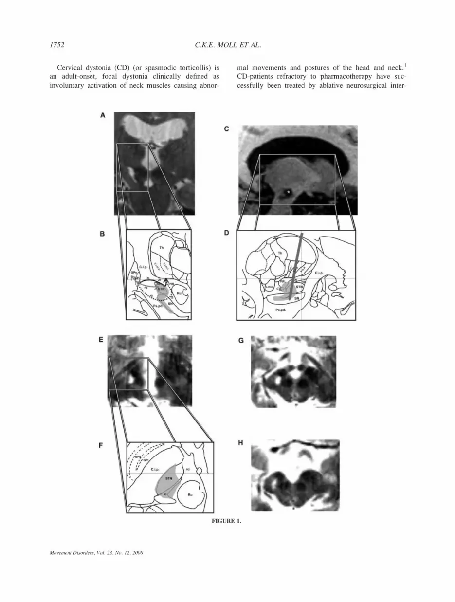
FIGURE 1.
Movement Disorders, Vol. 23, No. 12, 2008
1752 C.K.E. MOLL ET AL.

ventions since the 1950s.2 Pallidotomy was the initial
procedure for treatment of CD in the 1950s,2 and,
today again, Leksell’s target in the posteroventrolateral
internal pallidum is approached in most deep brain
stimulation (DBS) procedures performed for segmental
and general dystonia.3,4 Nonetheless, in the late 1950s
and early 1960s the stereotactic targets in torticollis
surgery had changed and paralleled the preferred tar-
gets for Parkinson’s disease with a delay of a few
years. The internal pallidum was widely abandoned in
favor of thalamic surgery,5,6 including interventions
with deliberate targeting of the posterior subthalamic
region.2,7–10 In the early 1970s, Mundinger stressed the
long-term efficacy of interventions in the posterior sub-
thalamic region in CD surgery and their superiority to
pallidal and thalamic coagulations.11 Consequently, he
explored the use of therapeutic electrical stimulation of
deep brain structures as early as 1977 and showed that
unilateral subthalamic DBS suppressed abnormal head
movements in CD patients.12 Unfortunately, the impact
of this hardly recognized work was rather limited and
fell into oblivion together with a general decline of
functional brain surgery in the 1970s. The subthalamic
region has only rarely been tested in modern surgery
for CD despite favorable results from the first era of
stereotactic surgery.13
Here, we report on a long lasting (>30 years) ther-
apeutic effect of a unilateral stereotactic subthala-
motomy in a patient with CD. We aimed to identify
the neural substrates the destruction of which is
responsible for the long-term beneficial effect.
Therefore, we applied the stereotactic method on
high-resolution magnetic resonance images of the
patient’s brain.
CASE HISTORY
At the age of 29, this patient with an inconspicious
family history recognized a pulling sensation in the
neck, months before abnormal head posture became
apparent for the first time in the autumn of 1972. At
the onset, she developed intermittent horizontal head
turning towards the right side. No other body parts
were affected. Initially, the patient was capable to
suppress abnormal movements by supporting the chin
with both hands. Symptoms progressed rapidly within
weeks, leading to a constant rotation of the head to the
right. Medical treatment was not beneficial but caused
sleepiness and apathy. The patient became unable to
work as a secretary. Because of a relative nihilism in
the medical treatment of CD and a general therapeutic
optimism concerning functional stereotactic surgery at
that time, precedence was given to a stereotactic opera-
tion over the intradural rhizotomy of Foerster and
Dandy or a peripheral surgical procedure.
SURGICAL PROCEDURE IN 1973
A right stereotactic subthalamotomy, ipsilateral to
the direction of the abnormal head movement, was car-
ried out by one of the authors (CBO) in February
1973. The surgical procedure comprised the use of a
targeting device developed by Riechert and Mun-
dinger.2 Mundinger’s approach for coagulations in the
zona incerta (ZI), which intended to spare the subthala-
mic nucleus (STN), was chosen for this operation.9,11
To place the lesion as much as possible in the axial
part of the subthalamic region, the use of a curved
electrode was proposed, which could be protruded per-
pendicularly to the central shaft (Fig. 1D). The target
FIG. 1. Neuroimaging of the subthalamic lesion. A: 3 Tesla T2-weighted MRI. Coronal section at the level of the stereotactic lesion. Thelesioned area is hyperintense with respect to the surrounding brain and extends in a streak-like fashion along the border of the SN to the internalcapsule and cerebral peduncle. B: Magnified area of interest with a schematic representation of the subthalamic lesion coordinates (gray shadedarea) superimposed on a modified section taken from the atlas of Schaltenbrand and Bailey.16 Coronal section, 5 mm posterior to the mid-com-missural point. The three main output pathways of the pallidum have been indicated to illustrate that the lesion interrupts pallidofugal fibers atvarious levels. Note that the ansa lenticularis (AL) runs rostral to the present section, and is therefore displayed as a dashed line. Abbreviations:Th, Thalamus; V.o.a, Nc. ventralis oralis anterior thalami; V.o.p., Nc. ventralis oralis posterior thalami; V.i.m., Nc. ventralis intermedius thalami;ZI, Zona incerta; PRL, Prelemniscal radiation; H2, Forel’s Field H2; STN, Subthalamic nucleus; L.med., Lemniscus medialis; SN, Substantianigra; Ru, Red nucleus; C.i.p., Posterior internal capsule; Ps.pd., Pedunculus cerebri; Q, Fasciculus subthalamo-hypothalamicus (Sano); GPi, inter-nal segment of globus pallidus; GPe, external segment of globus pallidus; AL, Ansa lenticularis; FL, lenticular fasciculus; FS, subthalamic fasci-culus. C: 3 Tesla T1-weighted MRI (Flash 3D), sagittal section. The lesion area is hypointense with respect to the surrounding brain. The primarylesion cavity, which has its maximum diameter in this plane, is marked by an asterisk. Note the finger-like extension of the main lesion causedby a second coagulation using the curved electrode. D: Lesion extent superimposed on the corresponding sagittal slice (11 mm lateral to the mid-line) of the atlas of Schaltenbrand and Bailey.16 Schematic drawing of the putative position of the coagulation electrode including the curved leadpushed posteriorly (�8 mm) out of the trunk of the electrode. Abbreviations as above. E: 3 Tesla T2-weighted MRI. Horizontal section at thelevel of the posterior commissure. Note the encroachment of the hyperintense lesion onto the STN, which is in one plane with the posterior com-missure and is clearly identifiable on the intact side. F: Lesion extent superimposed on a horizontal slice of the atlas of Schaltenbrand and Bai-ley16 (3.5 mm below AC-PC). Abbreviations as above. G: 3 Tesla T2-weighted MRI. Horizontal section at the level of the mammillary bodies, 3mm inferior to the section in E. Note that the lesion mainly involves the STN-SN complex, neighboring structures to an extent (C.i.p., ZI), butspares the red nucleus. H: 3 Tesla T2-weighted MRI. Horizontal section 2-mm inferior to the section in G. Note the affection of nigral volumeand of the neighboring cerebral peduncle, which appears slimmer compared to the intact side.
3
1753SUBTHALAMOTOMY IN CERVICAL DYSTONIA
Movement Disorders, Vol. 23, No. 12, 2008

site was determined in relation to a baseline connecting
the lower border of Monro’s foramen and the posterior
commissure, which were visualized by means of a ste-
reotactic ventriculography. The target coordinates as
derived from the stereotactic atlas of Andrew and Wat-
kins14 were: 12 mm behind Monro’s foramen, 8 mm
lateral to the middle plane, and 5 mm below the base-
line. After drilling of a trephine opening, the electrode
with a tip diameter of 2 mm was inserted to the target
level with approach angles of 108 in the frontal and
288 in the sagittal plane (rostral inclination), respec-
tively. A temperature-controlled high-frequency coagu-
lator (F.L. Fischer, Freiburg, Germany) was used to
produce the lesion. The parameters used for monopolar
electrocoagulation were 120 mA and �20 V for a pe-
riod of 20 to 30 seconds, resulting in an approximate
temperature of 708C for the tissue inactivation.
POSTOPERATIVE COURSE
The effect of the stereotactic operation was not
immediately apparent. Initially, the malposition of the
head persisted and the patient suffered from transient
side-effects, such as nonrhythmic rapid low amplitude
twitches of the left hand and slight clumsiness of the
left arm. With a delay of 3 to 4 months, however,
involuntary head turning gradually subsided, and after
6 months both dystonic symptoms and side-effects had
disappeared completely. The patient resumed her work
in a business company. For 30 years, she remained
healthy and showed no signs of cervical dystonia. In
2002, she experienced a 2-day period of generalized,
involuntary muscle-twitching. Around the same time,
she noticed difficulties with writing. In 2005, she
noticed involuntary head turning towards the right
side, which progressed relatively slowly over several
months. Symptoms deteriorated when she walked or
during stress. Medical treatment with trihexyphenidyl
(2 mg/d) was ineffective. She was referred to our hos-
pital for further assessment and local botulinum toxin
injections.
CLINICAL ASSESSMENT AND
CURRENT TREATMENT
The neurological examination was inconspicious,
apart from abnormal movements and postures. The
patient had intermittent right-sided torticollis up to 808and a slight right laterocollis with elevation and for-
ward displacement of the right shoulder. Writing was
slow, stiff and scrawly. During writing there was dorsal
extension of the wrist and abduction of the forearm
and overflow dystonia affecting the right upper arm
and shoulder. When stretching out the arms in front
the patient showed a moderate dystonic posturing of
the left arm, superimposed with mild irregular myo-
clonic jerks and continuous mild chorea. Currently,
overactive neck muscles are treated with local botuli-
num-toxin injections with an improvement of �50%.
HIGH-RESOLUTION MRI
To determine the localization relative to stereotactic
and anatomical landmarks on the one hand and to esti-
mate the precise extent of the stereotactic lesion on the
other, we obtained high-resolution images using 3 Tesla
MRI (Trio 3T, Siemens Medical Solutions, Erlangen,
Germany). After acquisition of a rapid 3D image local-
izer, we carried out partial volume scans of T2-
weighted, fast spin-echo images (spatial resolution: 1 31 3 1 mm3; TR: 5180 ms; TE: 96 ms) in the frontal
(Fig. 1A) and horizontal plane (Fig. 1E,G,D), centering
upon the visualization of the meso-diencephalic junc-
tion. The subsequently acquired sequence of T1-
weighted FLASH 3D images (spatial resolution: 1 3 1
3 1 mm3; TR: 15 ms; TE: 4.9 ms) covered the whole
brain and allowed for a detailed visualization of the an-
terior and posterior commissure (AC, PC), and of the
3rd ventricle structures that were used as reference
points in the stereotactic intervention (Fig. 1C). We
determined the position and boundaries of the coagula-
tion foci, relative to these landmarks (iPlan 1.1, Brain-
LAB Inc., Westchester, IL). The lesion area was hyper-
intense on T2-weighted images and hypointense on
T1-weighted images with respect to the surrounding
brain. To estimate the lesion volume, we used an
open-source image navigation and display software.15
For a definition of the anatomical structures, we used
the Schaltenbrand and Bailey atlas with Hassler’s
delimitation and nomenclature of thalamic structures as
a visual guide.16
STEREOTACTIC RECONSTRUCTION
OF THE LESION
A primary lesion was placed beneath the ventral
base of the rostral ventral tier thalamic nuclei. This
spherical lesion comprised a volume of ca. 90 mm3.
The dorsal margin of this lesion was: 13 mm lateral,
2.5 mm posterior, and 2 mm below AC-PC. It had its
center 5 mm below the AC-PC plane, 12 mm lateral,
and 5 mm posterior to the midcommissural point. The
coagulation focus covered a good portion of the caudal
ZI, the prelemniscal radiation, and invaded the poste-
rior edge of the STN (Fig. 1A–F). Running antero-
1754 C.K.E. MOLL ET AL.
Movement Disorders, Vol. 23, No. 12, 2008

laterally to the red nucleus and posteromedial to the
STN, it extended into the prerubral field and covered
parts of both the thalamic and mesencephalic part of
Forel’s field H (Fig. 1G). An arcuated enlargement of
the primary coagulation focus was produced by pro-
truding the curved electrode (Fig. 1A–D). This finger-
like lesion extended over a distance of �8 mm along
the lateral border of the SN to the cerebral peduncle. It
continued lateral to the red nucleus and encroached
upon the midbrain tegmentum (coordinates of the
lesion center: 10 mm lateral, 10 mm posterior, and 11
mm below AC-PC). On frontal T2-weighted MR
images, this lesion appeared as a streak-like extension
of the main lesion. It included a volume of �60 mm3
and covered �50% of nigral volume (Fig. 1A,G). The
most inferior extension of this lesion was 8 mm lateral,
11 mm posterior, and 12.5 mm below AC-PC. In hori-
zontal sections, the right cerebral peduncle appeared
slimmer in comparison to the opposite side, indicating
fiber degeneration due to affection of descending corti-
cofugal and/or traversing pallidofugal fibers (Fig. 1H).
DISCUSSION
This report demonstrates that unilateral lesioning of
the posterior subthalamic region may suffice to com-
pletely abolish CD over several decades. We feel it is
plausible to relate the clinical course of the present
case to the surgical procedure rather than to the natural
history of CD. Although spontaneous remissions can
occur in primary CD they are quite uncommon and
usually patients relapse within 5 years.17 In our patient,
symptoms subsided for 30 years. Also, improvement
following surgery was delayed which is the typical
scenario following functional surgery in dystonia
patients.3,11
In addition to a well documented follow up period of
three decades after surgery, this case is remarkable
because it offers the opportunity to determine the exact
lesion location and to discuss pathophysiological con-
cepts of early stereotactic surgery in the light of modern
imaging techniques. The discussion will therefore focus
on the potential significance of the structures that were
implicated in the described lesion, for CD surgery.
The lesion area comprises Forel’s field which resem-
bles campotomies as propagated by Spiegel and
Wycis.10 Because of its close anatomical association
with fibers from the dentate nucleus, a lesion in Forel’s
field will likely have implicated also the cerebellotha-
lamic system.18
The ZI was involved in the lesion area, particularly its
caudal aspect posteromedial to the STN. In addition to
pallidothalamic fiber bundles, ascending fibers originating
in the interstitial nucleus of Cajal that pass through or ter-
minate in the region of the ZI are of potential relevance in
the context of neurosurgery for CD.19,20 The interstitial
nucleus has long been implicated in the pathophysiology
of CD,21,22 since stimulation of both the interstitial
nucleus23 or intersititothalamic fibers in the ZI region
produces rotatory movements in laboratory animals.24
The primary coagulation focus clearly invaded the
posterior moiety of the STN. This is of particular inter-
est, since in contrast to contemporary subthalamic sur-
gery, which focuses on the STN proper, earlier subtha-
lamotomies attempted to avoid injury of this nucleus
because of concerns to produce hemiballism.
The transient weakness of the left arm occurring
postoperatively indicated the affection of corticospinal
fibers. However, the impairment of capsular fibers was
not sufficient to interfere with the patient’s postopera-
tive activity as a secretary. In view of the extensive
distribution of pallidofugal fibers in the internal cap-
sule,18 it appears likely that the present lesion also
involved a considerable number of them.
In summary, the present case report demonstrates
that isolated lesions of the ZI were hardly possible,
since the lesion inevitably affected neighboring struc-
tures. It is impossible to determine a single anatomical
structure, the destruction of which might be responsible
for the long-term beneficial effect on involuntary head
movements seen in this patient. However, the common
and most prominent anatomical denominator of subtha-
lamic structures implicated in the lesion described here
is their close anatomical association to the pallidofugal
complex. The lesion has interrupted the bulk of pallid-
ofugal fiber tracts where they pass through the internal
capsule at the level of the ZI.
Electrical stimulation of the posterior subthalamic
region has recently been shown to alleviate parkinso-
nian symptoms.25,26 The potential efficacy of subthala-
mic stimulation also for CD surgery is indicated by a
recent report of a patient with medically refractory
CD, who showed marked improvement of dystonian
symptoms following bilateral STN-DBS.13
In agreement with previous researchers7,9,10,25,27 we
hypothesize, that the posterior subthalamic region may
be considered a promising target for stereotactic proce-
dures in various movement disorders, including CD.
Acknowledgments: The authors wish to thank K. Muellerfor help with acquisition of the MR images and G. Englerand A. Sharott for helpful suggestions concerning the manu-script. The authors thank P. Vogel for referring the patient toour Department.
1755SUBTHALAMOTOMY IN CERVICAL DYSTONIA
Movement Disorders, Vol. 23, No. 12, 2008

REFERENCES
1. Fahn S, Bressman SB, Marsden CD. Classification of dystonia.Adv Neurol 1998;78:1–10.
2. Riechert T. Stereotactic brain operations. Methods, clinicalaspects, indications. Bern, Stuttgart, Vienna: Huber; 1980.
3. Bittar RG, Yianni J, Wang S, et al. Deep brain stimulation forgeneralised dystonia and spasmodic torticollis. J Clin Neurosci2005;12:12–16.
4. Krauss JK, Pohle T, Weber S, Ozdoba C, Burgunder JM. Bilat-eral stimulation of globus pallidus internus for treatment of cervi-cal dystonia. Lancet 1999;354:837–838.
5. Cooper IS. Effect of thalamic lesions upon torticollis N Engl JMed 1964;270:967–972.
6. Loher TJ, Pohle T, Krauss JK. Functional stereotactic surgery fortreatment of cervical dystonia: review of the experience from thelesional era. Stereotact Funct Neurosurg 2004;82:1–13.
7. Andy OJ, Jurko MF, Sias FR, Jr. Subthalamotomy in treatmentof Parkinsonian tremor. J Neurosurg 1963;20:860–870.
8. Bertrand C, Siegfried J. Extrapyramidal disorders—Session II.Proc 6th Symp Int Soc Res Stereoencephalotomy, Tokyo 1973.Confin Neurol 1975;37:9.
9. Mundinger F. Stereotaxic interventions on the zona incerta areafor treatment of extrapyramidal motor disturbances and theirresults. Confin Neurol 1965;26:222–230.
10. Spiegel EA, Wycis HT, Szekely EG, Adams DJ, Flanagan M,Baird HW, III. Campotomy in various extrapyramidal disorders.J Neurosurg 1963;20:871–884.
11. Mundinger F, Riechert T, Disselhoff J. Long-term results ofstereotactic treatment of spasmodic torticollis. Confin Neurol1972;34:41–50.
12. Mundinger F. [New stereotactic treatment of spasmodic torticol-lis with a brain stimulation system (author’s transl)]. Med Klin1977;72:1982–1986.
13. Chou KL, Hurtig HI, Jaggi JL, Baltuch GH. Bilateral subthala-mic nucleus deep brain stimulation in a patient with cervicaldystonia and essential tremor. Mov Disord 2005;20:377–380.
14. Andrew J, Watkins ES. A stereotaxic atlas of the human thala-mus and adjacent structures. A variability study. Baltimore:Williams & Wilkins; 1969.
15. Rosset A, Spadola L, Ratib O. OsiriX: an open-source softwarefor navigating in multidimensional DICOM images. J Digit Imag2004;17:205–216.
16. Schaltenbrand G, Bailey P. Introduction to stereotaxis with anatlas of the human brain. Stuttgart: Georg Thieme Verlag; 1959.
17. Dauer WT, Burke RE, Greene P, Fahn S. Current concepts onthe clinical features, aetiology and management of idiopathic cer-vical dystonia. Brain 1998;121(Part 4):547–560.
18. Smith MC. Stereotactic operations for Parkinson’s disease—ana-tomical observations. In: Williams D, editor. Modern trends inneurology. London: Butterworths; 1967. p 21–52.
19. Hassler R. Uber die afferenten Bahnen und Thalamuskerne desmotorischen Systems des Großhirns. II. Mitteilung Arch PsychiatNervenkr 1949;182:786–818.
20. Kokkoroyannis T, Scudder CA, Balaban CD, Highstein SM,Moschovakis AK. Anatomy and physiology of the primate inter-stitial nucleus of Cajal I. efferent projections. J Neurophysiol1996;75:725–739.
21. Munchau A, Corna S, Gresty MA, et al. Abnormal interactionbetween vestibular and voluntary head control in patients withspasmodic torticollis. Brain 2001;124(Part 1):47–59.
22. Hassler R, Hess WR. Experimentelle und anatomische Befundeuber die Drehbewegungen und ihre nervosen Apparate. ArchPsychiat Nervenkr 1954;192:488–526.
23. Klier EM, Wang H, Constantin AG, Crawford JD. Midbrain controlof three-dimensional head orientation. Science 2002;295:1314–1316.
24. Hyde JE, Toczek S. Functional relation of interstitial nucleus torotatory movements evoked from zona incerta stimulation.J Neurophysiol 1962;25:455–466.
25. Plaha P, Ben-Shlomo Y, Patel NK, Gill SS. Stimulation of thecaudal zona incerta is superior to stimulation of the subthalamicnucleus in improving contralateral parkinsonism. Brain 2006;129(Part 7):1732–1747.
26. Kitagawa M, Murata J, Uesugi H, et al. Two-year follow-up ofchronic stimulation of the posterior subthalamic white matter fortremor-dominant Parkinson’s disease. Neurosurgery 2005;56:281–289; discussion 281–289.
27. Struppler A, Lucking CH, Erbel F. Neurophysiological findingsduring stereotactic operation in thalamus and subthalamus. Con-fin Neurol 1972;34:70–73.
Intact Presupplementary MotorArea Function in Early, Untreated
Parkinson’s Disease
W. R. Wayne Martin, MD,1* Marguerite Wieler, MSc,1
Myrlene Gee, PhD,2 Christopher C. Hanstock, PhD,2
and Richard M. Camicioli, MD1
1Division of Neurology, University of Alberta, Edmonton,Alberta, Canada; 2Department of Biomedical Engineering,
University of Alberta, Edmonton, Alberta, Canada
Abstract: Although motor symptoms of Parkinson’s dis-ease (PD) are initially responsive to dopamine replace-ment therapy, nonresponsive features develop over time,suggesting that impaired dopaminergic function alonemay not be wholly responsible for all the motor featuresof the disease. Previous studies suggest impaired functionin the presupplementary motor area (pre-SMA) in PD.Our objective was to determine whether pre-SMA abnor-malities are present in untreated patients with early dis-ease. We measured N-acetyl aspartate (NAA)/creatine(Cr) and choline (Cho)/Cr ratios in pre-SMA in 26untreated patients with early PD (disease duration 3.0 62.0 yr) and 15 control subjects with single voxel magneticresonance spectroscopy. Neither NAA/Cr nor Cho/Crratios differed significantly between groups. These obser-vations suggest that, although pre-SMA function isimpaired in moderately advanced PD, it is relativelyspared in early disease. We suggest that pre-SMAdysfunction is in part responsible for the dopaminenonresponsive features associated with disease pro-gression. � 2008 Movement Disorder Society
Key words: Parkinson’s disease; supplementary motorarea; magnetic resonance spectroscopy
Additional Supporting Information may be found in the onlineversion of this article.
*Correspondence to: Dr. W. R. Wayne Martin, Movement Disor-ders Clinic, Glenrose Rehabilitation Hospital, 10230-111 Avenue,Edmonton, AB, Canada T5G 0B7. E-mail: [email protected]
Potential conflict of interest: None reported.Received 17 September 2007; Revised 21 February 2008;
Accepted 31 March 2008Published online 15 August 2008 in Wiley InterScience (www.
interscience.wiley.com). DOI: 10.1002/mds.22101
1756 W.R.W. MARTIN ET AL.
Movement Disorders, Vol. 23, No. 12, 2008

The major pathophysiological substrate underlying
the motor dysfunction of Parkinson’s disease (PD) is
loss of dopaminergic neurons projecting from substantia
nigra compacta to striatum and the resulting striatal
dopamine depletion. Although many motor symptoms of
early PD are responsive to dopamine replacement, over
time dopamine nonresponsive features including freezing
of gait and impaired balance develop. This suggests that
abnormal dopaminergic function alone is not completely
responsible for all motor features of the disease.
Cortical motor areas are closely involved in control
of normal movement and there is evidence of cortical
dysfunction in PD. The presupplementary motor area
(pre-SMA) is underactive on functional imaging,1–4
and significant loss of pre-SMA neurons has been
reported in PD.5 We have reported magnetic resonance
spectroscopy (MRS) findings from pre-SMA consistent
with this neuronal loss in patients with moderately
severe disease.6 Our objective in the present study was
to determine whether pre-SMA changes are present in
patients with early, untreated PD.
PATIENTS AND METHODS
Twenty-six patients with early PD were recruited
and matched for age and gender with 15 healthy con-
trol subjects. All patients fulfilled standard criteria for
clinical diagnosis of PD.7 None were treated with levo-
dopa, dopamine agonists or other PD medications prior
to the completion of the studies described in this arti-
cle. Clinical signs and symptoms were rated with the
Unified Parkinson’s Disease Rating Scale (UPDRS)8
by one of two raters (MW and WM) with a high inter-
rater reliability (intraclass correlation coefficient for
motor UPDRS 5 0.99, determined in a separate group
of PD patients). The study was approved by the
Human Research Ethics Board of the University of
Alberta; all subjects gave written informed consent.
MRS data were acquired using a Siemens Sonata
1.5T system. Imaging included a sagittal gradient-echo
sequence (repetition time (TR) 199 ms, echo time (TE)
4.6 ms, slice thickness 5 mm), a coronal T2-weighted
sequence (TR 6330 ms, TE 83 ms, slice thickness
3 mm), and a T1-weighted axial 3D magnetization
prepared rapid acquisition gradient echo (MPRAGE)
sequence (TR 2120 ms, TE 3.9 ms, TI (inversion time)
1100 ms with 176 slices, each 1 mm thick). After
locating the anterior commissure (AC)–posterior com-
missure (PC) line on a sagittal image, a line perpendic-
ular to the AC–PC line was placed through the AC
and a 2 3 2 3 2 cm3 voxel positioned such that its
posterior edge abutted the perpendicular with the voxel
rotated counter clockwise to place its superior border
parallel to the cortical surface (see Fig. 1). Coronal
and axial images were used to ensure that the voxel
was positioned in the midline. Water suppressed spec-
tra were obtained using a PRESS sequence (TR 51600 ms, TE 5 80 ms, 64 averages, 1024 data size)
and peak areas calculated with LC model,9 a program
that accepts the raw MRS data and provides a ‘‘black-
box’’ analysis with little user input. The metabolite ba-
sis set spectra used for the analysis were generated by
numerical simulation. From the peak areas for N-acetylaspartate (NAA), choline (Cho), and creatine/phospho-
creatine (Cr), the metabolite ratios NAA/Cr and Cho/
Cr were calculated. A typical spectrum from a control
subject is illustrated in Figure 2.
The MRS voxel was segmented into its gray matter
(GM), white matter (WM), and cerebrospinal fluid
(CSF) components using MPRAGE images with SPM2
(http://www.fil.ion.ucl.ac.uk/spm/) in Matlab 7.0 (The
Mathworks, Natick, MA). The spectroscopy voxel was
placed on each segment using an in-house program to
transform the coordinates of the voxel into the frame
of reference of the segmented images.
Statistical analyses were performed using SPSS ver-
sion 14.0 for Windows (SPSS, Chicago, IL). Compari-
sons of controls and patients were made using a two-
tailed t-test. Relationships between metabolite ratios
and UPDRS scores were determined using linear
regression; the Pearson correlation coefficient, r, is
reported. A value of P < 0.05 was required for statisti-
cal significance.
RESULTS
There was no significant difference with respect to
age or gender distribution between patients and con-
FIG. 1. Sagittal image showing pre-SMA voxel placement. AC, an-terior commissure; PC, posterior commissure.
1757PRE-SMA IN PARKINSON’S DISEASE
Movement Disorders, Vol. 23, No. 12, 2008

trols. Disease duration was 3.0 6 2.0 yr (since symp-
tom onset) and motor UPDRS score 15.1 6 7.0. Clini-
cal features are summarized in Table 1.
There was no significant difference in either NAA/
Cr (1.50 6 0.15 vs. 1.48 6 0.16) or the Cho/Cr (0.70
6 0.07 vs. 0.68 6 0.11) ratio between patients and
controls. Similarly, segmentation of voxels into GM,
WM, and CSF fractions showed no significant dif-
ference (GM vol: 4314 6 811 mm3 vs. 4199 6719 mm3; WM vol: 1472 6 261 mm3 vs. 1423 6 422
mm3; CSF vol: 2297 6 803 mm3 vs. 2402 6 904 mm3).
NAA/Cr did not correlate significantly with total,
motor, or axial UPDRS scores (P 5 0.235, 0.208, 0.191,
respectively). There was no correlation between NAA/
Cr and Hoehn/Yahr score (P 5 0.266; see Supporting
Information Fig. 1). There was a trend toward a corre-
lation between Cho/Cr and clinical features (total
UPDRS: r 5 0.36, P 5 0.085; motor UPDRS: r 50.39, P 5 0.072; axial UPDRS: r 5 0.36, P 5 0.086).
A typical patient spectrum, similar in quality to the
normal spectrum in Figure 2, is illustrated in Support-
ing Information Figure 2.
DISCUSSION
This group of patients with untreated PD showed no
significant abnormalities in pre-SMA metabolite ratios
in comparison to age-matched controls. This is in con-
trast to our previous study showing reduced NAA/Cr
in pre-SMA6 in a patient population with longer dis-
ease duration. The critical difference in this study was
the disease duration of 3.0 6 2.0 yr (measured from
symptom onset) when compared with 8.4 6 4.4 yr in
the previous publication. The second difference is
patient age with those in this study having a mean age
of 60.7 6 8.5 yr vs. 71.2 6 4.4 yr in the previous
study. Age difference is unlikely to explain the find-
ings, however, since control group ages (56.8 6 7.6 yr
in this study vs. 71.5 6 4.9 yr in the previous study)
were closely matched to the ages of the patient groups.
A third difference is that patients in this study were
untreated in contrast to those in the previous study.
Lucetti et al. have reported that dopamine agonist
treatment can modify motor cortex spectra, normaliz-
ing the reduced Cho/Cr ratio that they observed in
early PD.10 We are unaware, however, of any reports
of dopaminergic treatment producing the reduction in
metabolite ratios that we reported previously.
The trend toward significance in the correlation
between Cho/Cr and UPDRS score raises the possibil-
ity that membrane turnover increases as PD progresses,
perhaps as an early indicator of a neurodegenerative
process in pre-SMA. Most brain Cho is bound in mem-
brane phospholipids such as phosphatidylcholine.
Bound Cho, however, is not mobile and therefore
largely invisible in proton MRS. The Cho peak in pro-
ton spectra of the brain is thought to consist largely of
cytosolic glycerophosphocholine and phosphocholine,
products of membrane breakdown.11 Previous studies
in neurodegenerative dementias have also reported
TABLE 1. Demographics and clinical status
Controls Patients
n (female, male) 15 (4, 7) 26 (8, 14)Age (yrs) 56.8 6 7.6 60.7 6 8.5Years since symptom onset – 3.0 6 2.0UPDRS motor score – 15.1 6 7.0Tremor subscorea – 2.7 6 2.0Bradykinesia subscoreb – 6.3 6 3.5Rigidity subscorec – 3.2 6 2.2Axial subscored – 3.3 6 1.7
Hoehn and Yahr score – 1.6 6 0.5
Figures represent mean 6 SD.aUPDRS items 20–21.bUPDRS items 23–26, 31.cUPDRS item 22.dUPDRS items 18, 19, 27–30.
FIG. 2. Proton spectrum from a 2 3 2 3 2 cm3 voxel located onthe midline in the region of the supplementary motor area of a con-trol subject. The data shown in (a) highlight the metabolite peaksfrom N-acetylaspartate (NAA), Creatine plus phosphocreatine (Cr/PCr), and choline-containing compounds (Cho) and illustrate boththe experimental data (gray) and the LCModel fit (black). The datashown in (b) represent the residual noise following subtraction of thefit from the experimental data.
Movement Disorders, Vol. 23, No. 12, 2008
1758 W.R.W. MARTIN ET AL.

increased Cho/Cr, suggested as being due to increased
turnover of membrane constituents.12
The nonprimary motor area in area 6 on the postero-
medial frontal cortex is divided into SMA proper and
pre- or rostral SMA.13 Although SMA proper is active
in the initiation and execution of movement, pre-SMA is
thought to play a role in motor planning and organization
of complex movements.5,13 Pre-SMA is active prior to
movement onset and is likely the source of the early com-
ponent of the Bereitschaftspotential, which precedes vol-
untary movement by a second or more.14 Brain imaging
studies have implicated the pre-SMA in motor task learn-
ing with a correlation between pre-SMA activity and the
complexity of a learned motor sequence.15
Abnormal pre-SMA function has been reported in
PD. Changes in the Bereitschaftspotential are present,
with a pattern suggesting a failure of pre-SMA activa-
tion.14 Decreased pre-SMA activation,2 reversible by
levodopa administration in patients with early dis-
ease,16,17 is evident with functional imaging. Although
a significant loss of cortico-cortical projecting pyrami-
dal neurons from the pre-SMA has been reported in
PD, this observation was based on pathology from
patients with a long duration of disease.5
Our data are consistent with the notion that although
pre-SMA activity is preserved in early PD, abnormal-
ities develop with disease progression. We suggest that
pre-SMA dysfunction may be in part responsible for
the development of dopamine nonresponsive motor
features associated with disease progression.
Acknowledgments: This work was supported by the Cana-dian Institutes for Health Research. We gratefully acknowl-edge the willing cooperation of our subjects.
REFERENCES
1. Fukuda M, Mentis M, Ma Y, et al. Networks mediating the clin-ical effects of pallidal brain stimulation for Parkinson’s disease:a PET study of resting-state glucose metabolism. Brain 2001;124:1601–1609.
2. Sabatini U, Boulanouar K, Martin F, et al. Cortical motor reor-ganization in akinetic patients with Parkinson’s disease: a func-tional MRI study. Brain 2000;123:394–403.
3. Thobois S, Dominey P, Decety J, et al. Motor imagery in normalsubjects and in asymmetrical Parkinson’s disease: a PET study.Neurology 2000;55:996–1002.
4. Cunnington R, Egan G, O’Sullivan J, Hughes A, Bradshaw J,Colebatch J. Motor imagery in Parkinson’s disease: a PET study.Mov Disord 2001;16:849–857.
5. Macdonald V, Halliday GM. Selective loss of pyramidal neuronsin the presupplementary motor cortex in Parkinson’s disease.Mov Disord 2002;17:1166–1173.
6. Camicioli RM, Hanstock CC, Bouchard TP, Gee M, Fisher NJ,Martin WRW. Magnetic resonance spectroscopic evidence forpresupplementary motor area neuronal dysfunction in Parkinson’sdisease. Mov Disorders 2007;22:382–386.
7. Calne DB, Snow BJ, Lee C. Criteria for diagnosing Parkinson’sdisease. Ann Neurol 1992;32(Suppl):S125–S127.
8. Fahn S, Elton RL. Unified Parkinson’s Disease Rating Scale. In:Fahn S, Marsden CD, Goldstein M, Calne DB, editors. Recentdevelopments in Parkinson’s disease, Vol. 2. Florham Park, NJ:Macmillan Healthcare; 1987. p 153–163.
9. Provencher SW. Estimation of metabolite concentrations fromlocalized in vivo proton NMR spectra. Magn Reson Med 1993;30:672–679.
10. Lucetti C, Del Dott P, Gambaccini G, et al. Influences ofdopaminergic treatment on motor cortex in Parkinson disease: aMRI/MRS study. Mov Disord 2007;22:2170–2175.
11. Klein J. Membrane breakdown in acute and chronic neurodegen-eration: focus on choline-containing phospholipids. J NeuralTransm 2000;107:1027–1063.
12. Kantarci K, Petersen RC, Boeve BF, et al. 1HMR spectroscopyin common dementias. Neurology 2004;63:1393–1398.
13. Picard N, Strick PL. Imaging the premotor areas. Curr Opin Neu-robiol 2001;11:663–672.
14. Colebatch JG. Bereitschaftspotential and movement-relatedpotentials: origin, significance, and application in disorders ofhuman movement. Mov Disord 2007;22:601–610.
15. Boecker H, Dagher A, Ceballos-Baumann AO, et al. Role of thehuman rostral supplementary motor area and the basal ganglia inmotor sequence control: investigations with H215O PET. J Neu-rophysiol 1998;79:1070–1080.
16. Haslinger B, Erhard P, Kampfe N, et al. Event-related functionalmagnetic resonance imaging in Parkinson’s disease before andafter levodopa. Brain 2001;124:558–570.
17. Buhmann C, Glauche V, Sturenburg HJ, Oechsner M, Weiller C,Buchel C. Pharmacologically modulated fMRI–cortical respon-siveness to levodopa in drug-naive hemiparkinsonian patients.Brain 2003;126:451–461.
1759PRE-SMA IN PARKINSON’S DISEASE
Movement Disorders, Vol. 23, No. 12, 2008

Reversal of Head Drop AfterDiscontinuation of Olanzapine in
a DLB Patient
Marcel J.H. Aries, MD,1 Hans Debruyne, MD,1
Sebastiaan Engelborghs, MD, PhD,1,2,3,4
Nathalie Le Bastard, MSc,2 Nore Somers, MSc,1
Dagmar Gorissen, MD,5 Barbara A. Pickut, MD,1,4
and Peter Paul De Deyn, MD, PhD1,2,4*
1Department of Neurology and Memory Clinic,ZNA-Middelheim and ZNA-Hoge Beuken, Antwerp, Belgium;2Laboratory of Neurochemistry and Behaviour, Departmentof Biomedical Sciences, Institute Born-Bunge, University of
Antwerp, Antwerp, Belgium; 3Department of NursingSciences, Faculty of Medicine, University of Antwerp,
Antwerp, Belgium; 4Department of Health Care Sciences,University College, Antwerp, Belgium;
5Department of Physical Medicine and Rehabilitation,ZNA-Middelheim and ZNA-Hoge Beuken, Antwerp, Belgium
Video
Abstract: We present a 72-year-oldpatient with probablediffuse Lewy body disease and visual hallucinations, whodeveloped subacute reversible ‘‘dropped head syndrome’’and parkinsonian signs after the introduction of olanzapineat a total daily dose of 10 mg. One week after olanzapinewas withdrawn, the patient’s posture started to improve.Further improvement was achieved after dopaminergicsubstitution. Clinical and electrophysiological observationsmight indicate neck extensor myopathy due to axial rigid-ity or focal neck dystonia, induced by dopamine receptorblockade.
� 2008 Movement Disorder Society
Key words: diffuse Lewy body disease; olanzapine;dropped head syndrome; axial rigidity; dystonia
The term ‘‘dropped head syndrome’’ refers to a con-
dition characterized by severe weakness of the neck
extensors, resulting in neck flexion. Dropped head is
often part of a generalized neuromuscular disorder
such as myasthenia gravis, polymyositis, or amyotro-
phic lateral sclerosis. Antecollis has also been
described in multiple system atrophy and Parkinson’s
disease. The term ‘‘isolated neck extensor myopathy’’
is used when no underlying etiology is identified.1–3
The pathogenesis of dropped head syndrome among
extrapyramidal disorders is still under debate. Present
case series are not conclusive with regard to suggested
mechanisms like noninflammatory myopathy, rigidity,
traction injury, and axial dystonia.1,4–6
CASE REPORT
A 72-year-old woman was admitted to a geriatric
ward of a general hospital due to severe confusion,
delusions, and visual hallucinations, which had been
seen for 3 weeks. As a psychotic depression was sus-
pected, the patient’s general practitioner started the
atypical antipsychotic agent olanzapine that was upti-
trated to a total daily dose of 10 mg. Noticeable head
flexion started after 2 days of olanzapine treatment.
Her medical history revealed depression and anxiety
disorder, treated with various antidepressants. The fam-
ily reported a progressive fluctuating cognitive decline,
which had progressed over the period of 1 year with
prominent delusions. Two years earlier, the patient had
experienced well-formed visual hallucinations that dis-
appeared spontaneously after less than 1 week. Brady-
kinesia had been observed for about 1 year. At our
memory clinic, the atypical antipsychotic was with-
drawn in 9 days after its start. At that time, the patient
was still in a severe confusional state and could lift her
head maximally 208 from her chest. The MMSE score
at admission was 6 of 30. Her neck rigidity was severe
and painful, particularly in case of passive backward
extension. The head was slightly rotated to the right
side (see video, Segment 1). The sternocleidomastoi-
deus (SCM), trapezius, and splenius muscles did not
reveal apparent muscle contractions on palpation. In
addition, the clinical neurological examination showed
bradykinesia, shuffling gait with festination, bilateral
cogwheel rigidity, and postural instability. Laboratory
results revealed the increased serum creatine kinase
(CK) levels of 3.500 U/L (normal range: 50–150 U/L),
which completely normalized during the next 2 days.
Our patient was diagnosed as suffering from probable
diffuse Lewy body disease (DLB), according to the
consensus guidelines for the clinical diagnosis of
DLB.7 In addition, a cervical collar was given, and
rivastigmine was uptitrated to a total daily dose of 12
mg, with a beneficiary effect on the visual hallucina-
tions.
Additional Supporting Information may be found in the onlineversion of this article.
*Correspondence to: Dr. Peter Paul De Deyn, Laboratory ofNeurochemistry and Behaviour, Institute Born-Bunge, University ofAntwerp, Universiteitsplein 1, BE-2610 Antwerp, Belgium.E-mail: [email protected]
Potential conflict of interest: None reported.Received 5 December 2007; Revised 27 February 2008; Accepted
19 March 2008Published online 25 July 2008 in Wiley InterScience (www.
interscience.wiley.com). DOI: 10.1002/mds.22182
1760 M.J.H. ARIES ET AL.
Movement Disorders, Vol. 23, No. 12, 2008

Twenty-two days after the onset of the dropped
head syndrome, electromyography showed spontane-
ous muscle activity consisting of positive sharp
waves, fibrillation potentials, and complex repetitive
discharges in cervical paraspinal muscles. One week
after olanzapine was withdrawn, the patient’s posture
started to improve slightly (see video, Segment 2).
Close observation revealed that the patient was able
to drink with nearly full neck extension, which might
be interpreted as being a sensory trigger (see video,
Segment 3). Two weeks after olanzapine was with-
drawn, we prescribed a total daily dose of 375-mg
benserazide-levodopa because of disabling painful
axial rigidity. After 5 weeks of treatment, the
improvement of head flexed posture and motor symp-
toms was remarkable, though fluctuating during the
day. Rigidity of dorsal cervical muscles with head
ptosis was noticed when lying in a supine position
(see video, Segment 4). Repeated needle electromyog-
raphy disclosed marked myopathic changes in cervical
paraspinal muscles and sustained contraction of right
splenius capitis muscle.
Entacapone was added at a total daily dose of
600 mg because of peak dose dyskinesias, following
the dose increment of L-dopa. After 10 weeks, the
patient’s posture continued to improve to the point of
an approximation of a classical parkinsonian slightly
flexed stance (see video, Segment 5). Aripiprazole
10 mg was deem necessary to treat persistent delusions
with good clinical results and no subsequent increase
in extrapyramidal symptoms or dropped head in the
subsequent 7 months.
DISCUSSION
Different factors may contribute to the pathophysiol-
ogy of the disabling anterior curvature of the cervical
spine in Parkinsonism. In this case, we considered a
malignant neuroleptic syndrome (MNS) or an acute
myopathy of dorsal neck muscles to explain muscle
weakness, elevated CK levels, and persistent rigidity.8
The CK levels values, however, normalized within 2
days, and hyperthermia and autonomic dysregulations
were lacking, making the diagnosis of MNS, unlikely.
In our patient, repeatedly, electromyography showed
myopathic changes in paravertebral extensor muscles,
suggesting a plausible explanation for head ptosis. A
monophasic noninflammatory process restricted to the
paraspinal musculature has been suggested by several
authors.3,5,6 However, these changes may be nonspe-
cific or secondary, resulting from mechanical strain
rather than a specific myopathic disorder.3,4
Psychotic symptoms in DLB patients are particularly
difficult to treat due to their extreme sensitivity to anti-
cholinergic and antidopaminergic medications. This
increased sensitivity could be due to the relative loss
of dopaminergic neurons, particularly in the substantia
nigra, and the failure of postsynaptic striatal neurons to
upregulate D2 receptors in response to a dopaminergic
deficit or D2-blocking drugs.9 The dropped head syn-
drome was probably induced by the atypical antipsy-
chotic olanzapine, as symptoms improved after with-
drawal of the antipsychotic. The coexistence with
extrapyramidal signs and the effects of dopaminergic
replacing medication suggest another provoking and
sustaining factor than the myopathy. An unbalanced
muscle rigidity between the anterior and the posterior
neck muscles is suspected, which was supported by
surface EMG of the neck showing that reflex muscle
activity of SCM muscles was elicited during the act of
neck extension.10 Neck examination in our patient
indeed revealed the fluctuating painful axial rigidity,
and video segment 3 shows that the patient was not
able to extend the neck in supine position. Also, the
theory of a focal dystonia has been proposed.11
Although convincing dystonic spasms of the anterior
neck muscles, as in our patient, are often lacking, clini-
cally undetectable contractions in deep neck flexor
musculature might be relevant. Improvement of
dropped head by drinking suggests the presence of sen-
sory trigger mechanisms as described in cervical dysto-
nia.12 In our patient, sustained contraction of the right
neck rotator splenius capitis might have been indicative
for cervical dystonia in deeper anterior neck muscles.
Another possible explanation is suggested in a recent
publication showing that in 23 of 38 treatment-naive
patients with torticollis, the SCM to be activated failed
to work adequately during an effort to achieve maxi-
mum rotation to the contralateral direction of torticollis
(so-called negative dystonia).13 This can explain why a
subset of Parkinson patients, with a well-preserved
strength of paraspinal muscles, show prominent
dropped head.1
To the best of our knowledge, this is the first case
report of reversible dropped head syndrome in DLB. In
2006, an analog case report described a depressive
patient who developed mild parkinsonian signs and
camptocormia after the introduction of olanzapine.11
Similarities between dropped head and camptocormia,
which are characterized by anterior curvature of the
thoracolumbar spine, suggest similar pathophysiologi-
cal processes affecting the paraspinal muscles of differ-
ent parts of the spine.4
1761REVERSAL OF HEAD DROP AFTER OLANZAPINE DISCONTINUATION
Movement Disorders, Vol. 23, No. 12, 2008

LEGENDS TO VIDEO
Segment 1. Seven days after treatment with olanza-
pine, severe head ptosis is present. The patient can lift
her head maximally 208. The head is slightly rotated to
the right side. Backward extension of the neck is lim-
ited and painful.
Segment 2. One week after olanzapine was with-
drawn, the patient’s posture started to improve slightly.
Backward extension is still limited.
Segment 3. Ten days after olanzapine withdrawal,
further spontaneous improvement was noticed, with the
patient being able to drink with nearly full neck exten-
sion, indicating a sensory trigger.
Segment 4. Painful rigidity of dorsal cervical
muscles is noticed when lying in a supine position.
Segment 5. Ten weeks after the olanzapine was
withdrawn, the patient’s posture continued to improve
to a classical parkinsonian slightly flexed stance.
Acknowledgments: This research was supported by the
Special Research Fund of the University of Antwerp, Sticht-
ing Alzheimer Onderzoek, the Thomas Riellaerts Research
Fund, the Institute Born-Bunge, the agreement between the
Institute Born-Bunge and the University of Antwerp, the cen-
tral Biobank facility of the Institute Born-Bunge, University
of Antwerp, Medical Research Foundation Antwerp, Neuro-
search Antwerp, the Fund for Scientific Research, Flanders
(FWO-F) (grant no G.0127.07), and the Institute for Promo-
tion of Innovation through Science and Technology in Flan-
ders (IWT-Vlaanderen). NLB is a PhD fellow of the FWO-F/
L’Oreal-UNESCO, and SE is a postdoctoral fellow of the
FWO-F. We are grateful to Mr. F. van Coppenolle (Univer-
sity of Antwerp) for the photographic work.
REFERENCES
1. Kashira K, Ohno M, Tomito S. Dropped head syndrome in Par-kinson’s disease. Mov Disord 2006;21:1213–1216.
2. Fujimoto K. Dropped head in Parkinson’s disease. J Neurol 2006;253:21–26.
3. Katz JS, Wolfe GI, Burns DK, Bryan WW, Fleckenstein JL, Bar-ohn RJ. Isolated neck extensor myopathy: a common cause ofdropped head syndrome. Neurology 1996;46:917–921.
4. Umapathi T, Chaudhry V, Cornblath D, Drachman D, Griffin J,Kunci R. Head drop and camptocormia. J Neurol Neurosurg Psy-chiatry 2002;73:1–7.
5. Ashmark H, Eeg Olafsson KE, Johansson A, et al. Parkinson andneck extensor myopathy: a new syndrome or coincidental find-ings? Arch Neurol 2001;58:232–237.
6. Oerlemans WGH, Visser de M. Dropped head syndrome andbent spine syndrome: two separate clinical entities or differentmanifestations of axial myopathy. J Neurol Neurosurg Psychiatry1998;65:258–259.
7. McKeith IG, Galasko D, Kosaka K, Perry EK, Dickson DW,Hansen LA. Consensus guidelines for the clinical and pathologicdiagnosis of dementia with Lewy bodies (DLB): report of theconsortium on DLB international workshop. Neurology 1996;47:1113–1124.
8. Adityanjee, Sajatovic M, Munshi KR. Neuropsychiatric sequelaeof neuroleptic malignant syndrome. Clin Neuropharmacol 2005;28:197–204.
9. Baskys A. Lewy body dementia: the litmus test for neuroleptic sen-sitivity and extrapyramidal symptoms. J Clin Psychiatry 2004;65:16–22.
10. Vela L, Moron DJ, Sanchez C, Pareja JA, Baron M. Camptocor-mia induced by atypical antipsychotics and resolved by electro-convulsive therapy. Mov Disord 2006;21:1977–1979.
11. Yoshiyama Y, Takama J, Hattori T. The dropped head sign inParkinsonism. J Neurol Sci 1999;167:22–25.
12. Schramm A, Reiners K, Naumann M. Complex mechanisms ofsensory tricks in cervical dystonia. Mov Disord 2004;19:452–458.
13. Mezaki T. Dystonia redefined as central non-paretic loss of con-trol of muscle action: a concept including inability to activatemuscles required for a specific movement, or ‘negative dystonia’.Med Hypotheses 2007, doi:10.1016/j.mehy. 2007.03.014.
Hemidystonia Secondary toThalamic Hemorrhage Treated
with GPi Stimulation
Tadashi Hamasaki, MD, PhD,* Kazumichi Yamada,MD, PhD, and Jun-ichi Kuratsu, MD, PhD
Department of Neurosurgery, Kumamoto University MedicalSchool, Honjo, Kumamoto, Japan
Abstract: There have been few reports on the surgicaltreatment of secondary hemidystonias, most of which aredue to basal ganglia stroke or trauma. We present 2patients with hemidystonia secondary to thalamic hemor-rhage whom we successfully treated with unilateral globuspallidus internus (GPi) stimulation. Case 1 is a 56-year-old man with abnormal posturing and intolerable musclecontraction pain in the left arm. Case 2 is a 73-year-oldwoman who developed severe abnormal posturing in theright arm and gait disturbance due to hyperextension ofthe right leg. The dystonic symptoms of both patientswere refractory to medication. Three months after theinception of high frequency GPi stimulation, the motorscores on the Burke-Fahn-Marsden Dystonia Rating Scalewere improved by 49.2% and 34.3% in Cases 1 and 2,respectively. We suggest GPi stimulation as a possiblealternative to treat secondary hemidystonia. � 2008Movement Disorder Society
Key words: GPi-DBS; thalamus; secondary hemidystonia
Additional Supporting Information may be found in the onlineversion of this article.
*Correspondence to: Dr. Tadashi Hamasaki, Department of Neuro-surgery, Kumamoto University Medical School, 1-1-1 Honjo, Kuma-moto 860-8556, Japan. E-mail: [email protected]
Received 8 February 2008; Revised 8 April 2008; Accepted 22May 2008
Published online 25 July 2008 in Wiley InterScience (www.
interscience.wiley.com). DOI: 10.1002/mds.22183
1762 T. HAMASAKI ET AL.
Movement Disorders, Vol. 23, No. 12, 2008

Most hemidystonias develop after a latency period
as a result of insults affecting the contralateral brain1,2
and abnormal postures and movements have a deleteri-
ous effect on the activities of daily life. Reported etiol-
ogies include stroke, trauma, perinatal injury, infection,
brain tumor, and arterio-venous malformation, located
in the basal ganglia, thalamus, cerebral cortex, or inter-
nal capsule.2 Medical treatment is ineffective in most
cases although benzodiazepines and anti-cholinergic
agents may produce some effect.2–5 Few patients bene-
fit from the injection of botulinum toxin A because the
affected muscles are distributed widely in the body.
Deep brain stimulation (DBS) of the globus pallidus
internus (GPi) represents a powerful alternative for the
treatment of medically refractory dystonia such as gen-
eralized-6 and segmental dystonia.7 However, contro-
versy continues to surround the effectiveness of GPi-
DBS in other dystonia subtypes. We present 2 patients
with hemidystonia secondary to thalamic hemorrhage
who were effectively treated with GPi-DBS.
PATIENTS AND METHODS
Case Presentation
Case 1: This 56-year-old man who suffered a right
thalamic hemorrhage at the age of 52 (Fig. 1A) noticed
abnormal posturing of the left arm and leg 3 months
after the hemorrhage. He subsequently developed ex-
cessive muscle contraction and associated muscle pain
in the left shoulder and arm. Medication including clo-
nazepam and carbamazepine was minimally effective.
On our initial examination, he was alert and well-ori-
ented. Cranial nerve study returned unremarkable
results. His left arm and leg were mildly weak with
spastic tone and hyperreflexia. The dystonic posture
primarily affected his left hand with hyperextension of
all fingers (videotape segment 1). Forced abduction of
the left upper arm, flexion of the forearm, and hyperex-
tension of the leg were observed especially during
walking. There was mild sensory loss on the left side.
He reported severe muscle pain distributed over the
face, neck, shoulder, and proximal part of the arm. His
preoperative motor score on the Burke-Fahn-Marsden
Dystonia Rating Scale (BFMDRS) was 29.5 (Fig. 2A).
FIG. 1. Magnetic resonance images obtained at admission. (A) Case 1. (B) Case 2. Arrows indicate scar lesions in the medial to posterior portionof the thalamus. Thalamic atrophy is apparent in both cases.
FIG. 2. Preoperative and postoperative motor scores on the Burke-Fahn-Marsden Dystonia Rating Scale. (A) Case 1. (B) Case 2.
Movement Disorders, Vol. 23, No. 12, 2008
1763GPi STIMULATION FOR SECONDARY HEMIDYSTONIA

Case 2: This 73-year-old woman developed a left
thalamic hemorrhage (Fig. 1B) when she was 63 years
old. Several months later, she noticed abnormal postur-
ing of the right arm; it was initially intermittent but
progressed over the course of several years despite
medical therapy including clonazepam. On admission,
her mental status was normal. She had difficulty in
opening her eyes due to tonic and phasic contraction
of the face muscles. Her right arm and leg were mildly
weak. There was a strong tendency for adduction with
internal rotation of the upper right arm and flexion of
the forearm. This forced her arm behind her back even
at rest (videotape segment 3). She had no sensory dis-
turbances. Her gait was severely disturbed primarily
due to hyperextension of the right leg. Her preopera-
tive BFMDRS motor score was 35 (Fig. 2B).
Both patients provided prior written informed
consent for the acquisition and offline and/or online
distribution of preoperative and postoperative videotape.
Surgery
Case 1: Magnetic resonance imaging (MRI)-guided
stereotactic surgery was performed under local anes-
thesia with selective 3rd ventriculography and micro-
electrode recording.8 The target was the right GPi,
which we assumed to be at a point 20 mm lateral to
the midline, 2 mm anterior to the mid-commissure
point, and 1 mm dorsal to the floor of the 3rd ventri-
cle. The most ventral contact of a quadripolar DBS
electrode(Model 3387, Medtronic) was placed in the
most ventral part of the GPi with the aid of micro-
electrode recording.
Case 2: Left GPi-DBS was performed; except for
the use of Framelink (Medtronic) to calculate the coor-
dinates, the procedure was as in Case 1. Test stimula-
tion at the tentative target (2 mA, 100 lsec, 100 Hz)
elicited immediate effect and she could open her hand
more easily.
RESULTS
Test stimulation via the implanted electrodes allevi-
ated the excessive muscle contractions and associated
muscle pain in the left arm in Case 1 and improved
somewhat the forced abnormal posturing of the right
arm in Case 2. After a few days of test stimulation, a
pulse generator (SOLETRA Model 7426, Medtronic)
was implanted subcutaneously in the subclavian region.
There were no adverse effects or deterioration of
neurological symptoms attributable to the surgical
procedures.
In Case 1, the stimulation parameters at discharge
were: bipolar stimulation with contact 1(2) 2(1),
130 Hz, pulse duration 210 lsec, and amplitude
0.3V. The active contacts were subsequently moved
to a more ventral position and the voltage was
increased [contact 0(2) 1(1), 210 lsec, 130 Hz, and
1.5–1.8 V] because these settings most effectively
alleviated the dystonic posture. With respect to his
muscle contraction pain, the visual analogue scale
was improved by 80%. The abnormal posturing of
his left arm was improved significantly during move-
ment and even at rest (videotape segment 2). Three
months after the inception of GPi-DBS, his BFMDRS
motor score was 15, representing a 49.2% improve-
ment over his preoperative score of 29.5 (Fig. 2A).
At the latest visit 22 months postoperatively, his dys-
tonic symptoms in the left extremities and associated
muscle pain were similar to those three months after
surgery.
In Case 2, the initial stimulation parameters were
as follows: unipolar stimulation with Case (1), con-
tact 0(2) 1(2), 130 Hz, pulse duration 450 lsec, andamplitude 2.4 V. A subsequent decrease in the volt-
age to 2.0 V elicited no deterioration of her dystonic
symptoms. Her facial dystonia was improved. The
abnormal posturing of her right arm was improved
moderately and she was able to move her arm unas-
sisted from the back to the side of the trunk (video-
tape segment 4). She continued to exhibit severe
gait disturbance although it was slightly improved
with some improvement of the hyperextension of her
right leg. Three months after the start of unilateral
GPi-DBS, her BFMDRS motor score was 23, consti-
tuting a 34.3% improvement over her preoperative
score of 35 (Fig. 2B). At the latest visit 10 months
postoperatively, the patient’s condition was un-
changed compared with that observed 3 months
postoperatively, except for the arm dystonia slightly
worsened.
DISCUSSION
We used the BFMDRS to evaluate the effectiveness
of unilateral GPi-DBS in patients with hemidystonia
secondary to thalamic hemorrhage. The improvement
rates were inferior to that we achieved in other dysto-
nia subtypes where the average rate of improvement
was 98% for DYT1 generalized dystonia,9 87% for tar-
dive dystonia,10 80% for segmental axial dystonia,11
and 69% for primary cervical dystonia (unpublished
observation). It is worth noting that the effectiveness
of GPi-DBS was more modest in Case 2 where the dis-
1764 T. HAMASAKI ET AL.
Movement Disorders, Vol. 23, No. 12, 2008

ease duration was longer than that of Case 1. This sup-
ports the observation of Ochiai et al.12 that the better
improvement can be achieved in the patients with
shorter (less than 5 years) disease duration. As was
true in our case, a pulse duration exeeding 200 lsecwas more effective than shorter durations.11,13,14
Of 29 patients treated by stereotactic surgery in the
literature, only 5 patients with secondary hemidystonia
underwent GPi stimulation.2,15 Fernandez et al.16
reported a patient with Benedikt’s syndrome who
developed hemidystonia as a complication of microvas-
cular decompression for trigeminal neuralgia. They
performed GPi-DBS because the adverse effects were
greater than the beneficial effects of benzodiazepine.
However, dystonia was improved significantly for only
1–3 hour after the start of stimulation. Loher et al.17
reported a patient with hemidystonia due to severe
closed head injury. On the basis of clinical symptoms
they postulated that the lesion responsible for dystonia
was in the upper brain stem, however, MRI demon-
strated no focal lesion. GPi-DBS was performed
9 years after the injury because neither medication nor
thalamotomy was effective. They reported marked
improvement of their patient’s hemidystonia and asso-
ciated pain 2 months after the inception of GPi-DBS
with sustained improvement 4 years after surgery.
Their report is unique in that GPi stimulation was
effective despite the prolonged disease duration. How-
ever, these early surgical cases were not informative in
terms of fine adjustments of the stimulation parameters
and they did not report postoperative quantitative eval-
uation of the dystonia symptoms. In their series of 10
cases with generalized dystonia, Diamond et al.18
encountered a patient with hemidystonia secondary to
a basal ganglia hemorrhage. GPi-DBS produced a
22.2% improvement on the Unified Dystonia Rating
Scale (UDRS). The mean rate of improvement in our 2
cases was 42%, indicating that GPi stimulation can be
used to address secondary hemidystonia although com-
pared to other dystonia subtypes, the results may be in-
ferior.
A number of clinical and experimental studies sup-
port the concept that dystonia is a disorder of sensori-
motor integration.19 According to Murase et al.,20 elec-
trophysiological studies suggested that patients with
dystonia encounter problems in interpreting propriosep-
tive sensory inputs before or during movement. Indeed,
in 22 patients with thalamic stroke, analysis of three-
dimensional MRI showed that the Vc was one of the
thalamic nuclei more frequently lesioned in patients
with dystonia than patients without involuntary move-
ment.21 In our cases, insult to the thalamic sensory
nuclei (Fig. 1) and possibly subsequent aberrant reor-
ganization of the somatosensory pathway3 may have
resulted in abnormal sensory inputs to the somatosen-
sory area, producing a fixed input–output mismatch in
the ‘‘motor subroutine’’22 dedicated to movement on
the contralateral side of the body.
In conclusion, our surgical results suggest that GPi-
DBS is a possible alternative to treat secondary hemi-
dystonia although reports of surgical cases are still
scarce. Studies on more patients are underway in our
laboratory to identify optimum parameter settings and
long-term outcomes, and to compare the course of
treated patients with the natural course of secondary
hemidystonia.
LEGENDS TO THE VIDEOTAPE
Segment 1. Case 1: Preoperatively, this 56-year-old
man with a history of right thalamic hemorrhage
(Case 1) exhibits dystonic posturing of the left hand
and arm at rest and during movement. Hyperextension
of the left leg is also observed during walking.
Segment 2. Fourteen days after the inception of right
GPi-DBS, there was alleviation of his dystonic postures
and movements, and he is able to open his left hand
and to stand straight. His dystonic pain on the left side
of the face and left shoulder has almost disappeared.
His BFMDRS motor score is improved by 49.2%.
Segment 3. Case 2: Preoperatively, this 73-year-old
woman with a history of left thalamic hemorrhage
exhibits severe dystonic posture of the right arm. It is
forced behind her back even at rest. Her right face and
leg are moderately affected.
Segment 4. After two weeks of continuous stimula-
tion of the left GPi, her dystonic symptoms are signifi-
cantly alleviated. Her BFMDRS motor score is
improved by 34.3%.
Acknowledgments: We thank Drs. T. Takezaki andR. Kojima for their assistance with the surgery, and the pre-operative and postoperative evaluation of the patients. K.Yamada and T. Hamasaki did the research project, surgery,and the data analysis. T. Hamasaki prepared the first draft ofthis manuscript. Review and critique were done byK.Yamada and J. Kuratsu.
REFERENCES
1. Pettigrew LC, Jankovic J. Hemidystonia: A report of 22 patientsand review of the literature. J Neurol Neurosurg Psychiatry1985;48:650–657.
2. Chuang C, Fahn S, Frucht SJ. The natural history and treatmentof acquired hemidystonia: report of 33 cases and review of theliterature. J Neurol Neurosurg Psychiatry 2002;72:59–67.
1765GPi STIMULATION FOR SECONDARY HEMIDYSTONIA
Movement Disorders, Vol. 23, No. 12, 2008

3. Burke RE, Fahn S, Gold AP. Delayed-onset dystonia in patientswith ‘‘static’’ encephalopathy. J Neurol Neurosurg Psychiatry 1980;43:789–797.
4. Factor SA, Sanchez-Ramos J, Weiner WJ. Delayed-onset dysto-nia associated with corticospinal tract dysfunction. Mov Disord1988;3:201–210.
5. Nardocci N, Zorzi G, Grisoli M, Rumi V, Broggi G, Angelini L.Acquired hemidystonia in childhood: a clinical and neuroradio-logical study of 13 patients. Pediatric Neurol 1996;15:108–113.
6. Vidailhet M, Vercueil L, Houeto JL, et al.French Stimulation duPallidum Interne dans la Dystonie (SPIDY) Study Group. Bilat-eral deep-brain stimulation of globus pallidus in primary general-ized dystonia. N Eng J Med 2005;352:459–467.
7. Wohrle JC, Weigel R, Grips E, Blahak C, Capelle HH, KraussJK. Risperidone-responsive segmental dystonia and pallidal deepbrain stimulation. Neurology 2003;61:546–548.
8. Goto S, Yamada K. Long term continuous bilateral pallidal stim-ulation produces stimulation independent relief of cervical dysto-nia. J Neurol Neurosurg Psychiatry 2004;75:1506–1507.
9. Goto S, Yamada K, Shimazu H, et al. Impact of bilateral pallidalstimulation on DYT1-generalized dystonia in Japanese patients.Mov Disord 2006;21:1785–1787.
10. Yamada K, Goto S, Yoshida S, et al. Bilateral pallidal stimula-tion for tardive dystonia. Funct Neurosurg 2005;44:115–119.
11. Muta D, Goto S, Nishikawa S, et al. Bilateral pallidal stimulationfor idiopathic segmental dystonia advanced from Meige syn-drome refractory to bilateral thalamotomy. Mov Disord 2001;16:774–777.
12. Ochiai T, Taira T, Goto S, Hayashi M, Hori T. An effect and in-dication of GPi-DBS for dystonia—from experience of 41 GPi-DBS cases. Funct Neurosurg 2007;46:166–170.
13. Kumar R, Dagher A, Hutchison WD, Lang AE, Lozano AM.Globus pallidus deep brain stimulation for generalized dystonia:clinical and PET study. Neurology 1999;53:871–874.
14. Coubes P, Roubertie A, Vayssiere N, Hemm S, Echenne B.Treatment of DYT1-generalized dystonia by stimulation of theinternal globus pallidus. Lancet 2000;355:2220–2221.
15. Hutchison WD, Lang AE, Dostrovsky JO, Lozano AM. Pallidalneuronal activity: implications for models of dystonia. Ann Neu-rol 2003;53:480–488.
16. Fernandez HH, Friedman JH, Centofanti JV. Benedikt’s syn-drome with delayed-onset rubral tremor and hemidystonia: acomplication of Douloureux surgery. Mov Disord 1999;14:695–697.
17. Loher TJ, Hasdemir MG, Burgunder JM, Krauss JK. Long-termfollow-up study of chronic globus pallidus internus stimulationfor posttraumatic hemidystonia. J Neurosurg 2000;92:547–460.
18. Diamond A, Shahed J, Azher S, Dat-Vuong K, Jankovic J.Globus pallidus deep brain stimulation in dystonia. Mov Disord2006;21:692–694.
19. Abbruzzese G, Berardelli A. Sensorimotor integration in move-ment disorders. Mov Disord 2003;18:231–240.
20. Murase N, Kaji R, Shimizu H, et al. Abnormal premovementgaiting of somatosensory input in writer’s cramp. Brain 2000;123:1813–1829.
21. Lehericy S, Grand S, Pollak P, et al. Clinical characteristics andtopography of lesions in movement disorders due to thalamiclesions. Neurology 2001;57:1055–1066.
22. Kaji R, Shibasaki H, Kimura J. Writer’s cramp: a disorder ofmotor subroutine? Ann Neurol 1995;38:837–838.
Autonomic Dysfunction inDifferent Subtypes of Multiple
System Atrophy
Claudia Schmidt, MD,1 Birgit Herting, MD,2
Silke Prieur,1 Susann Junghanns, MD,2
Katherine Schweitzer, MD,3 Christoph Globas, MD,3
Ludger Schols, MD,3 Heinz Reichmann, MD,2
Daniela Berg, MD,3 and Tjalf Ziemssen, MD,1*
1Autonomic and neuroendocrinological laboratory,University of Dresden, Germany; 2Parkinson Research group,Department of Neurology, University of Dresden, Germany;3Department of Neurodegenerative Disorders, Hertie Instituteof Clinical Brain Research, University of Tubingen, Germany
Abstract: Multiple system atrophy (MSA) can clinically bedivided into the cerebellar (MSA-C) and the parkinsonian(MSA-P) variant. However, till now, it is unknownwhether autonomic dysfunction in these two entities dif-fers regarding severity and profile. We compared the pat-tern of autonomic dysfunction in 12 patients with MSA-Cand 26 with MSA-P in comparison with 27 age- and sex-matched healthy controls using a standard battery of au-tonomic function tests and a structured anamnesis of theautonomic nervous system. MSA-P patients complainedsignificantly more often about the symptoms of autonomicdysfunctions than MSA-C patients, especially regardingvasomotor, secretomotor, and gastrointestinal subsystems.However, regarding cardiovascular, sudomotor pupil, uro-genital, and sleep subsystems, there were no significantquantitative or qualitative differences as analyzed by au-tonomic anamnesis and testing. Our results suggest thatthere are only minor differences in the pattern of auto-nomic dysfunction between the two clinical MSAphenotypes. � 2008 Movement Disorder Society
Key words: autonomic nervous system; multiple systematrophy; autonomic dysfunction; MSA subtypes
Multiple system atrophy (MSA) is a sporadic neuro-
degenerative disorder that usually manifests in the
early sixth decade of life and progresses relentlessly
with a mean survival of 9 years.1 Clinically, MSA
is dominated by autonomic failure, which may be asso-
ciated with either levodopa (L-dopa)-unresponsive
Additional Supporting Information may be found in the onlineversion of this article.
*Correspondence to: Dr. Tjalf Ziemssen, Autonomic and Neuro-endocrinological Laboratory, Department of Neurology, University ofTechnology Dresden, Fetscherstrabe 74, D-01307 Dresden, Germany.E-mail: [email protected]
Received 31 January 2008; Revised 19 May 2008; Accepted 22May 2008
Published online 25 July 2008 in Wiley InterScience (www.
interscience.wiley.com). DOI: 10.1002/mds.22187
1766 C. SCHMIDT ET AL.
Movement Disorders, Vol. 23, No. 12, 2008
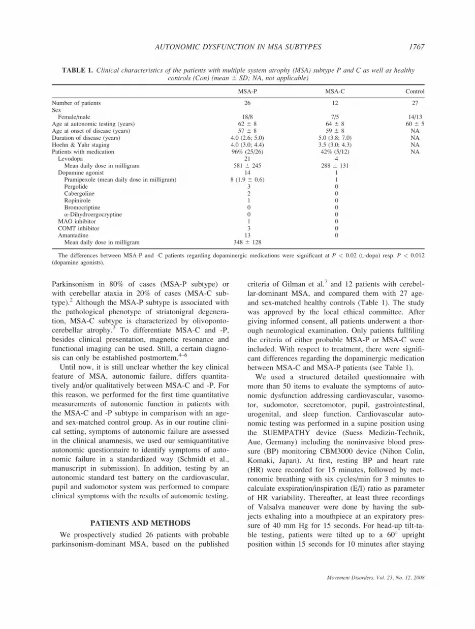
Parkinsonism in 80% of cases (MSA-P subtype) or
with cerebellar ataxia in 20% of cases (MSA-C sub-
type).2 Although the MSA-P subtype is associated with
the pathological phenotype of striatonigral degenera-
tion, MSA-C subtype is characterized by olivoponto-
cerebellar atrophy.3 To differentiate MSA-C and -P,
besides clinical presentation, magnetic resonance and
functional imaging can be used. Still, a certain diagno-
sis can only be established postmortem.4–6
Until now, it is still unclear whether the key clinical
feature of MSA, autonomic failure, differs quantita-
tively and/or qualitatively between MSA-C and -P. For
this reason, we performed for the first time quantitative
measurements of autonomic function in patients with
the MSA-C and -P subtype in comparison with an age-
and sex-matched control group. As in our routine clini-
cal setting, symptoms of autonomic failure are assessed
in the clinical anamnesis, we used our semiquantitative
autonomic questionnaire to identify symptoms of auto-
nomic failure in a standardized way (Schmidt et al.,
manuscript in submission). In addition, testing by an
autonomic standard test battery on the cardiovascular,
pupil and sudomotor system was performed to compare
clinical symptoms with the results of autonomic testing.
PATIENTS AND METHODS
We prospectively studied 26 patients with probable
parkinsonism-dominant MSA, based on the published
criteria of Gilman et al.7 and 12 patients with cerebel-
lar-dominant MSA, and compared them with 27 age-
and sex-matched healthy controls (Table 1). The study
was approved by the local ethical committee. After
giving informed consent, all patients underwent a thor-
ough neurological examination. Only patients fullfiling
the criteria of either probable MSA-P or MSA-C were
included. With respect to treatment, there were signifi-
cant differences regarding the dopaminergic medication
between MSA-C and MSA-P patients (see Table 1).
We used a structured detailed questionnaire with
more than 50 items to evaluate the symptoms of auto-
nomic dysfunction addressing cardiovascular, vasomo-
tor, sudomotor, secretomotor, pupil, gastrointestinal,
urogenital, and sleep function. Cardiovascular auto-
nomic testing was performed in a supine position using
the SUEMPATHY device (Suess Medizin-Technik,
Aue, Germany) including the noninvasive blood pres-
sure (BP) monitoring CBM3000 device (Nihon Colin,
Komaki, Japan). At first, resting BP and heart rate
(HR) were recorded for 15 minutes, followed by met-
ronomic breathing with six cycles/min for 3 minutes to
calculate exspiration/inspiration (E/I) ratio as parameter
of HR variability. Thereafter, at least three recordings
of Valsalva maneuver were done by having the sub-
jects exhaling into a mouthpiece at an expiratory pres-
sure of 40 mm Hg for 15 seconds. For head-up tilt-ta-
ble testing, patients were tilted up to a 608 upright
position within 15 seconds for 10 minutes after staying
TABLE 1. Clinical characteristics of the patients with multiple system atrophy (MSA) subtype P and C as well as healthycontrols (Con) (mean 6 SD; NA, not applicable)
MSA-P MSA-C Control
Number of patients 26 12 27SexFemale/male 18/8 7/5 14/13
Age at autonomic testing (years) 62 6 8 64 6 8 60 6 5Age at onset of disease (years) 57 6 8 59 6 8 NADuration of disease (years) 4.0 (2.6; 5.0) 5.0 (3.8; 7.0) NAHoehn & Yahr staging 4.0 (3.0; 4.4) 3.5 (3.0; 4.3) NAPatients with medication 96% (25/26) 42% (5/12) NALevodopa 21 4
Mean daily dose in milligram 581 6 245 288 6 131Dopamine agonist 14 1
Pramipexole (mean daily dose in milligram) 8 (1.9 6 0.6) 1Pergolide 3 0Cabergoline 2 0Ropinirole 1 0Bromocriptine 0 0a-Dihydroergocryptine 0 0
MAO inhibitor 1 0COMT inhibitor 3 0Amantadine 13 0
Mean daily dose in milligram 348 6 128
The differences between MSA-P and -C patients regarding dopaminergic medications were significant at P < 0.02 (L-dopa) resp. P < 0.012(dopamine agonists).
Movement Disorders, Vol. 23, No. 12, 2008
1767AUTONOMIC DYSFUNCTION IN MSA SUBTYPES

TABLE
2.Semiqua
ntitativeau
tono
mic
anam
nesisin
MSA
-P,-C
patients,an
dhealthycontrols,which
wereperformed
byastan
dardized
clinical
interview
usingou
row
nau
tono
mic
question
naire
MSA-P
patients
MSA-C
patients
Controls
PvalueMSA-P
versuscontrol
PvalueMSA-C
versuscontrol
PvalueMSA-P
versusMSA-C
Cardiovascularfunction
78%
(18/23)
55%
(6/11)
33%
(9/27)
0.002
NS
NS
Mostfrequent
symptom
Presyncope65%
(15/23)
Presyncope36%
(4/11)
Presyncope30%
(8/27)
NS
NS
NS
Postural(8/23)
Postural(4/11)
Postural(4/27)
Heatintolerance
(8/23)
Postexercise
(3/27)
Vasomotorfunction
96%
(22/23)
64%
(7/11)
37%
(10/27)
<0.0001
NS
0.029
Mostfrequent
symptom
Cold
hands65%
(15/23)
Cold
hands45%
(5/11)
Cold
hands26%
(7/27)
0.01
NS
NS
Sudomotorfunction
57%
(13/23)
45%
(5/11)
37%
(10/27)
NS
NS
NS
Mostfrequent
symptom
Dry
skin
35%
(8/23)
Dry
skin
27%
(3/11)
Dry
skin
30%
(8/27)
NS
NS
NS
Localizedhyperhidrosis
26%
(6/23)
Localizedhyperhidrosis
18%
(2/11)
Localizedhyperhidrosis4%
(1/27)
0.039
NS
NS
Secretomotorfunction
96%
(22/23)
64%
(7/11)
37%
(10/27)
<0.0001
NS
0.029
Mostfrequent
symptom
Hypersalivation70%
(16/23)
Hypersalivation45%
(5/11)
Hypersalivation(0/27)
<0.0001
0.001
NS
Dry
eyes
26%
(7/27)
NS
Pupilfunction
78%
(18/23)
82%
(9/11)
22%
(6/27)
<0.0001
0.001
NS
Mostfrequent
symptom
Impairedvisionduring
night52%
(12/23)
Impairedvisionduring
night36%
(4/11)
Impairedvisionduringnight
19%
(5/27)
0.017
NS
NS
Gastrointestinal
function
96%
(22/23)
64%
(7/11)
19%
(5/27)
<0.0001
0.017
0.029
Mostfrequent
symptom
Constipation78%
(18/23)
Constipation55%
(6/11)
Constipation11%
(3/27)
<0.0001
0.009
NS
Bloatingsensations15%
(4/27)
Urogenital
function
100%
(23/23)
100%
(11/11)
37%
(10/27)
<0.0001
<0.0001
NS
Mostfrequent
symptom
Incontinence
70%
(mixed)
(16/23)
Incontinence
73%
(mixed)(8/11)
Stressincontinence
22%
(6/27)
0.001
0.008
NS
Sleep
function
83%
(19/23)
64%
(7/11)
41%
(11/27)
0.004
NS
NS
Mostfrequent
symptom
Snorring70%
(16/23)
Snorring73%
(8/11)
Snorring22%
(6/27)
0.001
0.008
NS
Overallfrequency
ofcomplaints
regardingindividual
autonomic
functionsystem
s(Cardiovascular,vasomotor,sudomotor,secretomotor,pupil,gastrointestinal,urogenital,sleepfunction)
aswellas
mostfrequentsymptomsin
theindividual
functionsystem
sispresented.Pvalues
areprovided
forcomparisonofthedifferentgroups.
1768 C. SCHMIDT ET AL.
Movement Disorders, Vol. 23, No. 12, 2008

at least for 10 minutes in resting supine position. Sym-
pathetic skin response (SSR) evoked by acoustic stimu-
lation was recorded from both palms at least three
times using the SUEMPATHY device (Suess Medizin-
Technik, Aue, Germany). Recording of the pupil dia-
meter in darkness by near-infrared TV (television)
pupillography was performed 20 minutes after dark ad-
aptation using the compact integrated pupillograph
(AMTech GmbH, Weinheim) in a standardized way.8
Devices for automatic 24-hour recording of ambulatory
BP (Boso GmbH, Jungingen, Germany) were used at
various measurement intervals (every 15 minutes dur-
ing the day and every 30 minutes at night).
The statistical analysis of the data was performed by
SPSS. For the descriptive part, mean 6 standard devia-
tion was calculated. Considering the small number of
patients, mean and median (first quartile; third quartile)
were compared. If both differed more than 10%, me-
dian was used; in other cases, mean 6 standard devia-
tion was used. For the explorative analysis (interindi-
vidual comparison), we used the Mann–Whitney–Wil-
coxon test using medians. For the crosstab analysis
(binary data), Fisher’s test was applied. Significance
was defined P < 0.05.
RESULTS
Regarding standardized semiquantitative anamnesis
of the autonomic nervous system, the most frequent
symptom of autonomic failure was urogenital dysfunc-
tion (both MSA groups 100% vs. 37% in the age-
matched control group, P < 0.0001) (Table 2). In
addition, control persons described a totally different
pattern of urogenital symptoms (stress incontinence).
Sudomotor dysfunction (e.g., dry skin and localized
hyperhdrosis) was the least specific symptom of auto-
nomic dysfunction as it was reported to be pathological
by many healthy controls as well. Interestingly, MSA-
P patients complained significantly more often about
symptoms of autonomic dysfunction than MSA-C
patients in vasomotor, secretomotor, and gastrointestinal
systems (P < 0.029). In addition, MSA-P patients
complained about the impairment of significantly
higher numbers of pathologically involved autonomic
subsystems than MSA-C patients when the numbers of
pathologically changed autonomic subsystems were
counted (7 of 8 vs. 6 of 8 involved autonomic systems,
P < 0.008).
Concerning the results of autonomic testing, resting
supine HR and BP were significantly different between
the MSA patients and controls, but not between MSA-P
and -C patients (Table 3). Parameters of parasympa-
thetic function as E/I ratio during metronomic breathing
and Valsalva ratio were pathological in all MSA
patients with a nonsignificant tendency toward more
severe parasympathetic involvement of MSA-P
patients. Sympathetic dysfunction in the Valsalva ma-
neuver (missing phase IIb and overshot of phase IV)
was also similar between MSA-C and -P patients. Or-
thostatic testing revealed orthostatic hypotension in
nearly 60% of both MSA subgroup patients with a non-
significant tendency for a higher proportion of sympto-
matic orthostatic hypotension in MSA-C patients (15
vs. 25%). For both, MSA-C and -P patients, increase of
HR during orthostasis was significantly lower than in
the control group. In both patient groups, 24-hour am-
bulatory BP recording revealed similar pathological BP
behavior with nondipping and even an increase in BP
during night. SSR testing was the only method demon-
strating small significant differences between MSA-P
and -C patients: 48% MSA-P patients presented with
missing SSR at both sides in comparison with 22%
MSA-C patients (P < 0.049) and 4% healthy controls
(P < 0.001). In addition, significantly more MSA-P
(91%) than MSA-C (43%) patients showed pathological
P0 latencies (P < 0.047). Although nearly all SSR
parameters besides amplitude differed significantly
between MSA-P patients and healthy controls, there
were no significant differences between MSA-C
patients and controls. In contrast, pupillography demon-
strated no difference regarding pupil diameter in dark-
ness between MSA-P and -C patients with a tendency
of lower pupil diameter in darkness in MSA-C patients,
which was significant when compared with the healthy
controls (P < 0.018). Pupil light reflex testing revealed
no significant differences between the MSA-P and -C
patients (data not shown).
DISCUSSION
Autonomic anamnesis and function tests are manda-
tory in the diagnosis and clinical follow-up of patients
with MSA. Findings of significant autonomic failure
early in the course of the disease make the diagnosis
of MSA more likely. Autonomic failure is the key
symptom in both MSA subtypes, corresponding to his-
topathological supraspinal and spinal findings.10 Cell
loss is reported in the dorsal motor nucleus of vagus
and involves catecholaminergic neurons of the ventro-
lateral medulla. All cases of MSA with predominant
pathology in either the striatonigral or olivopontocere-
bellar system show significant loss of intermediolateral
cells in the spinal cord.11
Till now, only clinical anamnesis of autonomic fail-
ure has been assessed comparative studies between
1769AUTONOMIC DYSFUNCTION IN MSA SUBTYPES
Movement Disorders, Vol. 23, No. 12, 2008

TABLE
3.Auton
omic
testingof
thecardiovascular,pu
pilan
dsudo
motor
system
inMSA
-P,-C
patients,an
dhealthycontrols,as
describedin
materials
andmethods
MSA-P
patients
MSA-C
patients
Controls
PvalueMSA-P
versuscontrol
PvalueMSA-C
versuscontrol
PvalueMSA-P
versusMSA-C
Restingheartrate
(Bpm)
766
10
766
13
666
90.001
0.019
NS
Restingsystolic/diastolicbloodpressure
(mmHg)
1446
22/776
16
1456
17/806
11
1276
13/706
10
0.002/0.037
0.003/0.005
NS
P.withhypertensionat
supinerest
35%
(9/26)
58%
(7/12)
15%
(4/27)
NS
0.017
NS
E/I-ratio
duringmetronomic
breathing
1.056
0.03
1.086
0.07
1.186
0.09
0.000
0.003
NS
P.withpathological
E/I-ratio
87%
(13/15)
56%
(5/9)
4%
(1/27)
0.005
0.002
NS
Valsalvaratio
1.18(1.11;1.27)
1.26(1.07;1.42)
1.36(1.27;1.43)
0.003
NS
NS
P.withpathological
ValsalvaphaseII
(missingphaseIIb)
53%
(9/17)
44%
(4/9)
4%
(1/25)
0.000
0.012
NS
P.withpathological
ValsalvaphaseIV
(missingbloodpressure
overshot)
88%
(15/17)
88%
(8/9)
28%
(7/25)
0.000
0.004
NS
FallofsystolicBPduringHUT[absolute
(mm
Hg)/relative]
230(2
40,220)
235(2
46;213)
210(2
15;25)
0.000
0.012
NS
220%
(228%,214%)
221%
(229%;28%)
27%
(212%;24%)
FallofdiastolicBPduringHUT[absolute
(mm
Hg)/relative]
215(2
15;210)
215(2
20;0)
0(2
5;25)
0.000
0.027
NS
217%
(221%;210%)
217%
(222%;0)
0%
(28%;6%)
FallofmedianBPduringHUT[absolute
(mm
Hg)/relative]
220(2
27;215)
223(2
28;24)
23(2
8;22)
0.000
0.022
NS
218%
(225%;213%)
220%
(224%;24%)
23%
(29%;2%)
P.withorthostatic
hypotension
58%
(15/26)
58%
(7/12)
0%
0.000
0.000
NS
P.withsymptomatic
orthostatic
hypotension
15%
(4/26)
25%
(3/12)
0%
0.051
0.021
NS
Relativeincrease
ofheartrate
duringHUT
15%
(9%;20%)
10%
(7%;15%)
23%
(15%;29%)
0.040
0.005
NS
24-h
BP:Missingnocturnal
bloodpressure
dipping
70%
(14/20)
60%
(3/5)
8%
(2/26)
0.000
0.020
NS
24-h
BP:Reversedcircadianbloodpressure
45%
(9/20)
60%
(3/5)
4%
(1/26)
0.001
0.008
NS
P.withmissingSSRat
oneside
17%
(4/23)
0%
0%
0.024
NS
NS
P.withmissingSSRat
both
sides
48%
(11/23)
22%
(2/9)
4%
(1/27)
0.000
NS
0.049
MeanSSRP0latency
(ms)
1.94(1.89;2.05)
1.61(1.52;2.03)
1.45(1.25;1.68)
0.000
NS
NS
P.withpathological
SSRP0latency
92%
(11/12)
43%
(3/7)
31%
(8/26)
0.001
NS
0.047
MeanSSRN1latency
(ms)
3.06(3.01;3.20)
2.43(2.21;2.88)
2.22(1.91;2.75)
0.002
NS
NS
MeanSSRP0am
plitude(lm)
315(165;462)
239(166;397)
443(233;694)
NS
NS
NS
P.withpathological
SSRam
plitude
50%
(6/12)
57%
(4/7)
19%
(5/26)
NS
NS
NS
Pupildiameter
indarkness(m
m)
4.876
0.93
4.336
1.12
5.226
0.72
NS
0.018
NS
P.withpathological
pupildiameter
indarkness
31%
(5/16)
38%
(3/8)
7%
(2/27)
NS
NS
NS
Values
aregiven
asmedian(1.quartile;3.quartile)
Pvalues
areprovided
forcomparisonofthedifferentgroups.
HUT,head-uptilt;significantorthostatic
hypotensionwas
defined
bytheactual
guidelines.9
SSR,sympathetic
skin
response.
1770 C. SCHMIDT ET AL.
Movement Disorders, Vol. 23, No. 12, 2008
MSA-P and -C patients. Although one histopathologi-
cal study with 42 postmortem-confirmed cases showed
no differences regarding symptoms of autonomic and
urinary failure,3 another study with 100 MSA cases
reported significantly more abundant clinical signs of
autonomic dysfunction in the olivopontocerebellar
(MSA-C) than in the striatonigral (MSA-P) phenotype.12
Both studies only relied on retrospective chart reviews.
Another imaging study with 45 MSA patients described
in 74% (MSA-P) and 85% (MSA-C) symptoms sugges-
tive of autonomic dysfunction (mostly orthostatic hypo-
tension and urinary symptoms).13 Another small study
regarding SSR in MSA-P and -C demonstrated preser-
vation of SSR in up to 30% in MSA patients, more in
the MSA-C than in the MSA-P group.14
As autonomic failure can be diagnosed by symptom-
focused anamnesis, which is routinely used method in
the clinical setting, and autonomic testing, which is
only available in specialized centers, we used both
approaches to compare autonomic failure between
MSA-C and -P patients.
In our study, we were able to demonstrate that it is
possible to differentiate both MSA-P and -C patients
from healthy age- and sex-matched controls by both
approaches. Interestingly, using our semiquantitative
autonomic questionnaire, we were able to demonstrate
significant differences between MSA-P and -C patients.
MSA-P patients reported symptoms of autonomic failure
in more autonomic subsystems and more frequently in
the vasomotor, secretomotor, and gastrointestinal sub-
system, which cannot be assessed by standard autonomic
testing. In contrast, cardiovascular, sudomotor, and pupil
subsystem were not significantly different between
MSA-P and -C patients as assessed by both approaches
used in this study, semiquantitative anamnesis, and auto-
nomic testing. Even regarding SSR, only minor differen-
ces could be shown. So, if both approaches could be
compared, autonomic testing and semiquantitative an-
amnesis demonstrated similar results.
Comparing MSA-C and -P patients, we can report
for the first time a quantitative comparison, which
demonstrates differences in some autonomic subsys-
tems (by standardized anamnesis), whereas other sub-
systems (evaluated by anamnesis and autonomic test
battery) are similarly involved in MSA-P and -C
patients.
As restriction of this study, the number of patients
investigated in each group was small. Moreover, we
used clinical criteria for diagnosis and did not have
pathologic confirmation. Further prospective studies on
MSA patients with larger groups of patients and with
neuropathological correlation are needed to determine
accurate diagnostic values. There maybe an influence
of dopaminergic medication, which was significant dif-
ferent between MSA-P and -C patients (Table 1). But,
we regard this effect only minor from the literature
and our unpublished data.15,16 So, Mesec et al.17 could
not find any significant influence of dopaminergic med-
ication in 50 PD patients as well as Bouhaddi et al.18
in de novo versus long-term-treated PD patients. In
addition, Senard et al. only reported minor changes by
dopaminergic stimulation.19 In some MSA-P patients,
it was possible to stop dopaminergic medication the
day before testing, whereas in some patients it was not
possible because of quite-pronounced OFF symptoms.
The patients we investigated were at least half a year
on stable dopaminergic medication. In this situation,
we decided to analyze in OFF whereas possible and
where not to perform testing under dopaminergic medi-
cation. In addition, we could not demonstrate differen-
ces in a subgroup analysis comparing patients with and
without dopaminergic medication (data not shown).
Still, frequency and severity of autonomic symptoms
show that not only standard autonomic testing, but also
autonomic anamnesis, which could be performed in
nearly any clinical setting should be a mandatory part
of the diagnostic procedure of MSA patients.
REFERENCES
1. Geser F, Wenning GK. The diagnosis of multiple system atro-phy. J Neurol 2006;253 (Suppl 3):iii2–iii15.
2. Wenning GK, Geser F. Multiple system atrophy. Rev Neurol2003;159(5, Part 2):3S31–3S38.
3. Jellinger KA, Seppi K, Wenning GK. Grading of neuropathologyin multiple system atrophy: proposal for a novel scale. Mov Dis-ord 2005;20 (Suppl 12):S29–S36.
4. da Rocha AJ, Maia AC, Jr, da Silva CJ, et al. Pyramidal tractdegeneration in multiple system atrophy: the relevance of mag-netization transfer imaging. Mov Disord 2007;22:238–244.
5. Schulz JB, Klockgether T, Petersen D, et al. Multiple system at-rophy: natural history, MRI morphology, and dopamine receptorimaging with 123IBZM-SPECT. J Neurol Neurosurg Psychiatr1994;57:1047–1056.
6. Taniwaki T, Nakagawa M, Yamada T, et al. Cerebral metabolicchanges in early multiple system atrophy: a PET study. J NeurolSci 2002;200:79–84.
7. Gilman S, Low PA, Quinn N, et al. Consensus statement on thediagnosis of multiple system atrophy. J Auton Nerv Syst 1998;74:189–192.
8. Schmidt C, Herting B, Prieur S, et al. Pupil diameter in dark-ness differentiates progressive supranuclear palsy (PSP) fromother extrapyramidal syndromes. Mov Disord 2007;22:2123–2126.
9. Consensus statement on the definition of orthostatic hypotension,pure autonomic failure, and multiple system atrophy. The Con-sensus Committee of the American Autonomic Society and theAmerican Academy of Neurology. Neurology 1996;46:1470.
10. Ozawa T. Morphological substrate of autonomic failure and neu-rohormonal dysfunction in multiple system atrophy: impact ondetermining phenotype spectrum. Acta Neuropathol (Berl) 2007;114:201–211.
Movement Disorders, Vol. 23, No. 12, 2008
1771AUTONOMIC DYSFUNCTION IN MSA SUBTYPES

11. Oppenheimer DR. Lateral horn cells in progressive autonomicfailure. J Neurol Sci 1980;46:393–404.
12. Ozawa T, Paviour D, Quinn NP, et al. The spectrum of patholog-ical involvement of the striatonigral and olivopontocerebellarsystems in multiple system atrophy: clinicopathological correla-tions. Brain 2004;127(Part 12):2657–2671.
13. Abdo WF, van de Warrenburg BP, Kremer HP, Bloem BR, Ver-beek MM. CSF biomarker profiles do not differentiate betweenthe cerebellar and parkinsonian phenotypes of multiple system at-rophy. Parkinsonism Relat Disord 2007;13:480–482.
14. Magnificio F, Misra VP, Murray NMF, Mathias CJ. The labora-tory detection of autonomic dysfuntion in multiple system atro-phy—the role of the sympathetic skin response. Neurology 1997;48 (Suppl):A190.
15. Goldstein DS. Dysautonomia in Parkinson’s disease: neurocardio-logical abnormalities. Lancet Neurol 2003;2:669–676.
16. Jost WH. [Autonomic dysregulations in Parkinson’s syndrome].Schweizerische Rundschau Medizin Praxis 5 Rev Suisse MedPRAXIS 2005;94:1639–1646.
17. Mesec A, Sega S, Kiauta T. The influence of the type, duration,severity and levodopa treatment of Parkinson’s disease on cardio-vascular autonomic responses. Clin Auton Res 1993;3:339–344.
18. Bouhaddi M, Vuillier F, Fortrat JO, et al. Impaired cardiovascu-lar autonomic control in newly and long-term-treated patientswith Parkinson’s disease: involvement of L-dopa therapy. AutonNeurosci 2004;116:30–38.
19. Senard JM, Verwaerde P, Rascol O, Montastruc JL. Effects ofacute levodopa administration on blood pressure and heart vari-ability in never treated parkinsonians. Hypertens Res 1995;18(Suppl 1):S175–S177.
Ultrasonographic Findings ofShoulder Disorders in Patients
with Parkinson’s Disease
Seong-Beom Koh, MD, PhD,1 Jee-Hoon Roh, MD,1
Ji Hyun Kim, MD, PhD,1 Kyungmi Oh, MD, PhD,1
Byung-Jo Kim, MD, PhD,1 Gui-Sang Kim, MD,2
Byung-Kyu Park, MD, PhD,2 Se-Ju Kim, MD, PhD2,and Joon-Shik Yoon, MD, PhD2*
1Department of Neurology Korea University Collegeof Medicine Guro-Ku, Seoul, Republic of Korea;
2Department of Physical Medicine and RehabilitationKorea University College of Medicine, Guro-Ku, Seoul,
Republic of Korea
Abstract: The prevalence of a history of shoulder com-plaints is higher in patients with Parkinson’s disease (PD)than in healthy individuals. The aims of this study were toevaluate shoulder disease in PD patients with ultrasonogra-phy (US) and to identify the relationship between the USfindings of shoulder disease and the motor signs in patientswith PD. Thirty-three PD patients completed a shoulderdisability questionnaire, a musculoskeletal examination,and US of the shoulder. Twenty-two patients had abnormalUS findings. Tendon tearing was the most common abnor-mal US finding (22/22), and the supraspinatus tendon wasthe most common site involved (15/22). Patients with ten-don tearing had a significantly longer duration of diseasethan patients without tendon tearing (P 5 0.027). Patientswith adhesive capsulitis had a significantly higher rigidityscore than patients without adhesive capsulitis (P 5 0.035).Disease duration and rigidity were the contributing factorsfor the development of tendon tearing and adhesive capsu-litis, respectively. � 2008 Movement Disorder Society
Key words: shoulder disorders; Parkinson’s disease; ultra-sonography
Shoulder pain is an important medical and socioeco-
nomic problem. Pain and stiffness in the shoulder may
lead to an inability to work and/or to carry out activ-
ities of daily living. The self-reported prevalence of
shoulder pain is estimated to be 16 to 26%; shoulder
pain is the third most common cause of consultations
for musculoskeletal conditions in primary care.1,2
Ultrasonography (US) has been shown to be a useful
diagnostic tool in patients with shoulder pain and/or a
*Correspondence to: Dr. Joon-Shik Yoon, Department of PhysicalMedicine and Rehabilitation, Korea University College of Medicineat Guro Hospital, 80 Guro-Dong, Guro-Ku, Seoul, Republic ofKorea. E-mail: [email protected]
Received 28 January 2008; Revised 27 May 2008; Accepted 31May 2008
Published online 25 July 2008 in Wiley InterScience (www.
interscience.wiley.com). DOI: 10.1002/mds.22191
1772 S.-B. KOH ET AL.
Movement Disorders, Vol. 23, No. 12, 2008

limited range of shoulder motion. It allows for the
careful assessment of a wide range of changes involv-
ing different anatomic structures of the shoulder girdle,
such as the rotator cuff tendons, the long head of the
biceps tendon, and the soft tissues.3,4
Parkinson’s disease (PD) is a common neurodege-
nerative disorder, affecting 1.47% of the population
over 60 years of age in Korea.5 In one previous report,
the PD population, as compared with healthy individu-
als, showed a higher prevalence of both a history of
shoulder complaints and frozen shoulder.6
The primary aim of this study was to identify the
US findings associated with shoulder disease in
patients with PD. The second aim was to identify the
relationship between the US findings of shoulder dis-
ease and the cardinal motor signs of PD.
PATIENTS AND METHODS
Patient Selection
Thirty-three patients with PD were enrolled in our
study. The patients were selected from the Neurology
Department of the Korea University Guro Hospital. All
patients met the clinical diagnostic criteria for PD, as
described by the United Kingdom Parkinson’s Disease
Society Brain Bank.7 The patients were on levodopa
therapy with a definite and lasting good response. We
excluded patients who reported previous shoulder joint
trauma or shoulder surgery. Patients who could not
complete a questionnaire and an US study because of
cognitive impairment (Mini-Mental Status Examination
<24) or who had severe medical comorbidities were
also excluded. Written informed consent was obtained
from each patient for participation in the study. This
study was approved by the Institutional Review Board
of the Korea University Guro Hospital.
Between April 1, 2006 and May 1, 2006, 62 patients
were prospectively enrolled in this study, and 33
patients were ultimately examined. Twenty-nine
patients were excluded due to cognitive impairment (n
5 12), previous shoulder trauma or surgery (n 5 4),
rheumatoid arthritis [n 5 8 (2 patients also had cogni-
tive impairment)], and an incomplete study (n 5 7).
METHODS
Neurologic Evaluation
Patients underwent a neurologic examination and a
clinical assessment. The Hoehn and Yahr (H&Y) stage
was determined8 and the degree of disease severity
was quantified by the Unified Parkinson’s Disease Rat-
ing Scale (UPDRS)9 in the on state by one movement
disorder specialist.
Self-Questionnaire
All enrolled patients were asked to complete a ques-
tionnaire with information pertaining to their shoulder
disability.10
Musculoskeletal Examination and US
Patients underwent a musculoskeletal examination
and an US. The examiner was blinded to the patient’s
history and the findings of the neurologic examination.
Real-time sonography was performed with a HDI 5000
(Philips, Holland) equipped with a 7 to 12 MHz linear-
array transducer. The patient was seated on an exami-
nation chair, behind which the examiner stood.
According to the description of a previous report, five
standard US views were selected (anterior transverse,
anterior longitudinal, lateral transverse, lateral longitu-
dinal, and posterior transverse).11 The US criteria for
complete tendon tears (TT) were as follows: tendon
nonvisualization, anechoic or hypoechoic discontinuity,
and deltoid herniation (Fig. 1). The criteria of partial ten-
don tears were as follows: focal heterogenous hypoecho-
genecity and an incomplete hypoechoic defect (Fig. 2).
We made note of secondary signs of tears, such as sub-
deltoid bursitis, cortical irregularity of the greater tuber-
osity, and the cartilage sign. By using dynamic US, ad-
hesive capsulitis (AC) was defined as a difficulty in
obtaining adequate images of the subscapularis tendon
due to a reduced region of external rotation of the sub-
scapularis tendon and increased fluid in the dependent
portion of the bicipital tendon sheath.12 We also defined
FIG. 1. Longitudinal ultrasonography shows tendon nonvisualiza-tion, indicating a full thickness tear of the supraspinatus tendon(arrow). The retracted tendon end is detected (curved arrow). Theoverlying deltoid muscle (D) is seen on the underlying humerus (H).
1773US FINDINGS OF SHOULDER DISORDERS IN PD PATIENTS
Movement Disorders, Vol. 23, No. 12, 2008

myofacial pain syndrome (MPS) as a tender point within
a taut band of the shoulder girdle muscle, a characteristic
referred pain, pain on sustained compression over
the tender point, or a local twitch response within the
band. We classified shoulder disease according to the
anatomic structures of the shoulder girdle and a patho-
logic process by US, such as the TT, AC, and a nonpa-
thologic process, such as MPS.
Statistical Analysis
Data were tabulated and analyzed using SPSS, version
10.0 for Windows (SPSS, Chicago, IL). An independent
t-test was used to compare disease duration, H&Y stage,
UPDRS part 3 (total, tremor, rigidity, and bradykinesia
scores) and the shoulder pain questionnaire score accord-
ing to the classification of shoulder diseases. The correla-
tion between the shoulder pain questionnaire score and
each cardinal motor sign score of UPDRS part 3 was
evaluated using Spearman’s rho test.
RESULTS
Clinical Characteristics
The study participants included 19 males and 23
females with a mean age of 67.27 6 6.72 years (range,
54–80 years). The median age of disease onset was
64.0 years, and the mean duration of disease was 3.2
6 2.6 years (range, 1–12 years; Table 1). Among the
33 patients, 3 patients did not have shoulder pain.
Ultrasonographic Findings
Among the 33 patients, 22 patients had abnormal
US findings on the dominant side. Among the 22
patients, 10 patients also had abnormal US findings on
the nondominant side.
All 22 patients with abnormal US findings had a TT
(22/22) and 9 patients had AC (9/22). All nine patients
with AC also had a TT. The anatomic locations of the
TT were the supraspinatus tendon (15/22), a combina-
tion of the supraspinatus and subscapular tendons (6/
22), and a combination of the supraspinatus and biceps
tendons (1/22). Eleven patients had MPS. Among three
patients without shoulder pain, one patient had a TT of
the supraspinatus tendon and two patients had AC.
Among the 10 patients with abnormal US findings on
the nondominant side, 8 patients had a TT (8/10),
3 patients had AC (3/10), and 1 patient had a com-
bined TT and AC. On the dominant side, the majority
of patients (18/22) with a TT had complete tendon
tearing and 4 patients had partial tendon tearing. On
the nondominant side, only one patient had complete
tendon tearing and other seven patients had partial ten-
don tearing.
Clinical Features and Cardinal Motor Signs
of PD According to Shoulder Diseases
Tendon Tear
The mean duration of disease was 3.82 6 2.82 years
for the group with a TT and 2.05 6 1.56 years for the
group without a TT. In the group with a TT, the mean
duration of disease was significantly longer than the
group without a TT (P 5 0.027). There was no signifi-
cant difference between the two groups with respect to
other clinical profiles (Table 2).
Adhesive Capsulitis
The mean rigidity score of UPDRS 3 was 15.11 68.43 for the group with AC and 3.46 6 2.23 for the
group without AC. In the group with AC, the mean
UPDRS 3 rigidity score was significantly higher than
in the group without AC (P 5 0.035). There was no
significant difference between the two groups with
respect to other clinical profiles (Table 2).
Myofacial Pain Syndrome
There was no significant difference between patients
with MPS and patients without MPS in terms of the
TABLE 1. Demographic and clinical characteristics of the33 patients with Parkinson’s disease
Mean 6 SD or number
Age (yr) 67.27 6 6.72Male/female 10/23Disease duration (yr) 3.23 6 2.59Hoehn & Yahr Stage 2.11 6 0.68
FIG. 2. Transverse ultrasonography of the supraspinatus tendonshows a partial thickness tear (arrow) in the supraspinatus tendon(S). The overlying deltoid muscle (D) is seen.
1774 S.-B. KOH ET AL.
Movement Disorders, Vol. 23, No. 12, 2008

shoulder disability questionnaire score, H&Y stage, or
UPDRS 3 tremor and bradykinesia scores.
Shoulder Disability Questionnaire and Cardinal
Motor Signs of Parkinsonism
There was no significant correlation between the
shoulder disability questionnaire score and the UPDRS
3 total score, and there was no significant correlation
between the shoulder disability questionnaire and each
UPDRS 3 cardinal sign score.
DISCUSSION
In this study of PD patients, abnormal US findings
were common and a TT was the most common finding;
MPS and AC were also common findings. A TT was
also observed in patients without shoulder pain (33%).
The epidemiology of symptomatic and asymptomatic
rotator cuff tears has been poorly characterized. Previ-
ous studies involving cadavers have demonstrated a
high prevalence of rotator cuff tears, ranging from
9.4% (18 of 192) to 39% (39 of 100).13 Indeed, rotator
cuff tears are a very common pathologic finding and
may be part of the natural ageing process.14 Reilly
et al. also reported that in an unselected cadaver popu-
lation, the prevalence of full-thickness and partial
thickness tears was 11.75 and 18.49%, respectively
(total tears, 30.24%).15 In a previous study with US,
the prevalence of rotator cuff tears was 65% (15 of 23)
in patients who were more than 70 years of age.16 In
another study with US, Yamaguchi et al.17 reported
that there was a strong correlation between the onset
of rotator cuff tears and increasing age. In our study,
the prevalence of TT in the PD patients group was
66.7% (22 of 33) and there was no significant age dif-
ference between the TT and non-TT groups. The prev-
alence and degree of TT on the dominant side were
common and severe, compared to the nondominant
side. With these findings, we suggest that shoulder dis-
ease in PD patients is common compared to the gen-
eral population. However, in our study there was no
age-matched healthy control group. This was a main li-
mitation in our study.
In the present study, the most frequent location of a
TT was the supraspinatus tendon; a TT involving the
supraspinatus and subscapularis tendons was also
common in patients with PD. In the general popula-
tion, the supraspinatus tendon is the most frequent
tendon that is torn.18 The supraspinatus tendon is
located on the acromiohumeral joint and is covered
by the deltoid muscle. When the shoulder joint is im-
mobilized for an extended period of time, the joint
capsule of the acromiohumeral joint undergoes atro-
phy. Therefore, in PD patients the adducted posture
of the shoulder, rigidity, and poverty of movement
might contribute to the tearing of the supraspinatus
tendon. PD of long duration might be a risk factor for
a TT of the rotator cuff.
AC was also a common finding in patients with PD.
AC is characterized by pain and loss of stiffness in the
shoulder and affects approximately 2% of the general
population.19 In the present study, AC affected 9
(26.4%) of 34 patients with PD. The UPDRS 3 rigidity
score was higher in patients with AC than in patients
without AC. Rigidity and the associated immobiliza-
tion might contribute to the development of AC, and
AC may also be an aggravating factor for rigidity.
However, in the present study, there was no signifi-
cant correlation between the shoulder disability ques-
tionnaire score and the UPDRS motor scores. The
small sample size and predominance of early stage PD
(approximately 76% for H-Y stage �2.5; median H&Y
stage 5 2) could limit an accurate assessment of the
relationship between the shoulder disability score and
the cardinal motor signs of PD.
In conclusion, US abnormalities of the shoulder joint
are common in patients with PD, and a supraspinatus
TT was the most common finding in our study. The
TABLE 2. Clinical features in PD patients with shoulder diseases
Tendon tearing Adhesive capsulitis
Yes No Yes No
Number of Patients 22 11 9 24Age 67.36 6 6.19 67.09 6 8.01 66.90 6 7.36 67.43 6 6.60SDQ scores 31.86 6 26.50* 13.91 6 20.12* 31.89 6 23.15 23.63 6 26.73Disease duration (yr) 3.82 6 2.82* 2.05 6 1.56* 4.00 6 1.87 2.94 6 2.79UPDRS 3, total 13.73 6 8.32 13.18 6 8.53 15.11 6 8.43 12.96 6 8.30UPDRS 3, tremor 1.95 6 1.53 1.82 6 1.25 1.67 6 1.12 2.00 6 1.53UPDRS 3, rigidity 4.27 6 2.76 3.55 6 2.21 5.56 6 2.96* 3.46 6 2.23*UPDRS 3, bradykinesia 3.82 6 4.52 4.36 6 4.57 5.00 6 5.77 3.63 6 3.97
*P < 0.05.
1775US FINDINGS OF SHOULDER DISORDERS IN PD PATIENTS
Movement Disorders, Vol. 23, No. 12, 2008

duration of disease was the main contributing factor
for the development of a TT, whereas rigidity was a
more important factor in the development of AC.
REFERENCES
1. Urwin M, Symmons D, Allison T, et al. Estimating the burdenof musculoskeletal disorders in the community: the comparativeprevalence of symptoms at different anatomical sites, and therelation to social deprivation. Ann Rheum Dis 1998;57:649–655.
2. Winters JC, Sobel JS, Groenier KH, Arendzen JH, Meyboom-deJong B. The long-term course of shoulder complaints: a prospec-tive study in general practice. Rheumatology (Oxford) 1999;38:160–163.
3. Ozcakar L, Vanderstraeten G, Parlevliet T. Sonography and visu-alizing rotator cuff injuries in older people. J Am Geriatr Soc2005;53:1840–1841.
4. Iagnocco A, Filippucci E, Meenagh G, et al. Ultrasound imagingfor the rheumatologist. I. Ultrasonography of the shoulder ClinExp Rheumatol 2006;24:6–11.
5. Seo WK, Koh SB, Kim BJ, et al. Prevalence of Parkinson’s dis-ease in Korea. J Clin Neurosci 2007;14:1155–1157.
6. Cleeves L, Findley L. Frozen shoulder and other shoulder distur-bancies in Parkinson’s disease. J Neurol Neurosurg Psychiatry1989;52:813–814.
7. Yughes AJ, Daniel SE, Lees AJ. The clinical features of Parkin-son’s disease in 100 histologically proven cases. Adv Neurol1993;60:595–599.
8. Hoehn MM, Yahr MD. Parkinsonism: onset, progression andmortality. Neurology 1967;17:427–442.
9. The Unified Parkinson’s Disease Rating Scale (UPDRS): statusand recommendations. Mov Disord 2003;18(7):738–750.
10. van der Windt DA, van der Heijden GJ, de Winter AF, KoesBW, Deville W, Bouter LM. The responsiveness of the shoulderdisability questionnaire. Ann Rheum Dis 1998;57:82–87.
11. Moosmayer S, Smith HJ. Diagnostic ultrasound of the shoulder–a method for experts only? Results from an orthopedic surgeonwith relative inexpensive compared to operative findings. ActaOrthop 2005;76:503–508.
12. McNally EG. Practical musculoskeletal ultrasound. Philadelphia:Elsevier; 2005.
13. Fukuda H, Hamada K, Yamanaka K. Pathology and pathogenesisof bursal-side rotator cuff tears viewed from en bloc histologicsections. Clin Orthop Relat Res 1990(254):75–80.
14. Worland RL, Lee D, Orozco CG, SozaRex F, Keenan J. Correla-tion of age, acromial morphology, and rotator cuff tear pathologydiagnosed by ultrasound in asymptomatic patients. J SouthOrthop Assoc 2003;12:23–26.
15. Reilly P, Macleod I, Macfarlane R, Windley J, Emery RJ. Deadmen and radiologists don’t lie: a review of cadaveric and radio-logical studies of rotator cuff tear prevalence. Ann R Coll SurgEngl 2006;88:116–121.
16. Milgrom C, Schaffler M, Gilbert S, van Holsbeeck M. Rotator-cuff changes in asymptomatic adults. The effect of age, handdominance and gender. J Bone Joint Surg Br 1995;77:296–298.
17. Yamaguchi K, Ditosios K, Middleton WD, et al. The diagnosisand morphologic features of rotator cuff disease. A comparisonof asymptomatic and symptomatic shoulders. J Bone Joint SurgAm 2006;88:1699–1704.
18. Kim HA, Kim SH, Seo YI. Ultrasonographic findings of painfulshoulders and correlation between physical examination and ultra-sonographic rotator cuff tear. Mod Rheumatol 2007;17:213–219.
19. Hunt SA, Kwon YW, Zuckerman JD. The rotator interval: anat-omy, pathology, and strategies for treatment. J Am Acad OrthopSurg 2007;15:218–227.
Elevated Serotonin TransporterBinding in Depressed Patientswith Parkinson’s Disease:A Preliminary PET Study
with [11C]DASB
Isabelle Boileau, PhD,1* Jerry J. Warsh, MD,2
Mark Guttman, MD,1,3 Jean A. Saint-Cyr, PhD,1,4
Tina McCluskey, MSc,1 Pablo Rusjan, PhD,5
Sylvain Houle, MD,5 Alan A. Wilson, PhD,5
Jeffrey H. Meyer, MD, PhD,5
and Stephen J. Kish, PhD1
1Human Neurochemical Pathology Laboratory, Centre forAddiction and Mental Health, University of Toronto, Toronto,Ontario, Canada; 2Laboratory of Cellular and MolecularPathophysiology and Mood Disorders Program, Centre for
Addiction and Mental Health, University of Toronto, Toronto,Ontario, Canada; 3Centre for Movement Disorders,
Markham, Ontario, Canada; 4Division of Brain Imaging &Behaviour Systems, Toronto Western Research Institute,University of Toronto, Toronto, Ontario, Canada; 5VivianM. Rakoff PET Imaging Centre, Centre for Addiction and
Mental Health, University of Toronto, Toronto, Ontario, Canada
Abstract: This study investigated whether abnormalities inserotonin transporter binding occur in Parkinson’s dis-ease (PD) patients with concurrent depression. We esti-mated serotonin transporter levels in seven clinicallydepressed early-stage PD patients and in seven healthymatched-control subjects during a single positron emis-sion tomography (PET) scan with the serotonin trans-porter radioligand, [11C]DASB. Depressed PD patientsdisplayed a wide-spread increase (8–68%) in [11C]DASBspecific binding outside of the striatum, which was signifi-cant in dorsolateral (37%) and prefrontal (68%) cortices.Elevated [11C]DASB binding was positively correlatedwith depressive symptoms but not with disease severity orduration. Compatible with recent PET/[11C]DASB findingsin major depression, the present preliminary data suggestthat increased [11C]DASB binding, possibly reflectinggreater serotonin transporter density (up-regulation), mightbe a pathological feature of depression in Parkinson’sdisease—and possibly a characteristic of depressive illness ingeneral. � 2008 Movement Disorder Society
Key words: Parkinson’s disease; depression; serotonintransporter; positron emission tomography; [11C]DASB
*Correspondence to: Dr. Isabelle Boileau, Centre for Addictionand Mental Health, 250 College Street, Toronto, Ontario, CANADAM5T 1R8. E-mail: [email protected]
Pontential conflict of interest: None reported.Received 17 March 2008; Revised 15 May 2008; Accepted 11
June 2008Published online 25 July 2008 in Wiley InterScience (www.
interscience.wiley.com). DOI: 10.1002/mds.22212
1776 I. BOILEAU ET AL.
Movement Disorders, Vol. 23, No. 12, 2008

Parkinson’s disease (PD) is a neurodegenerative dis-
order characterized by motor and nonmotor features
including cognitive and neuropsychiatric disturbances.
Depression, assumed to be present in �40% of PD
patients,1 is not only the most common nonmotor
symptom of (early) PD but also among the most taxing
on quality of life.2 Postmortem studies3 and in vivo
neurochemical imaging investigations studies4–6 sug-
gest a brain serotonergic deficiency in PD which could
in part explain mood and cognitive disturbances
(including depression) in some PD patients. However,
to date, only one post-mortem neuropathological study
has provided evidence that depression in PD might be
related to a loss of (raphe) serotonergic neurons.7 To
our knowledge, no imaging study of serotonin trans-
porter (5-HTT) levels, a key serotonin neuron marker,
has been conducted in PD with diagnosed concurrent
depression using a highly specific and selective radio-
ligand. The goal of the present positron emission tomo-
graphy (PET) study was to assess 5-HTT binding in
clinically depressed, nondemented patients with PD
using [11C]DASB ([11C]-3-amino-4-(2-dimethylamino-
methyl-phenylsulfanyl)-benzonitrile) a highly specific
radioactive probe.8
PATIENTS AND METHODS
Seven patients (4M, 3F) meeting UK Brain-Bank
Criteria for idiopathic PD (mean 6 SD age of 61.1 69.8 years) and seven gender-matched controls (mean 6SD age of 59.5 6 6.7 years) participated in the
research ethic’s board approved study. PD patients
were mostly in early clinical stages of the disease
(mean 6 SD disease duration: 4 6 2.9 years; Hoehn
and Yahr scale (H&Y): 2 H&Y stage I, 4 H&Y stage
II, 1 H&Y stage III; mean 6 SD Unified Parkinson’s
Disease Rating Scale on medication: 21.1 6 0.34) and
responded well to the treatment. All PD subjects (and
no control subjects) met criteria for current depressive
disorders (4 major, 2 minor, and 1 sub-syndromal
depression) during a structured clinical interview for
DSM-IV Axis I Disorders (SCID) with a psychiatrist.
FIG. 1. (A) Increased [11C]DASB BPND in DLPFC and PFC indepressed PD relative to healthy matched controls. (B) Positive cor-relation (Pearson) between [11C]DASB BPND and depressive symp-toms (HAM-21).
TABLE 1. Mean (and SD) of [11C]DASB BPND in 10 brain ROIs of normal control subjects (n 5 7) andpatients with PD and concurrent depression (n 5 7)
Regions of interest
Control (n 5 7) PD (n 5 7)
%Difference P uncorrectedMean SD Mean SD
DLPFC 0.32 0.07 0.42 0.04 37 y0.001PFC 0.29 0.11 0.48 0.07 68 y0.002OFC 0.42 0.08 0.49 0.11 16 0.21Anterior cingulate 0.35 0.07 0.46 0.14 31 0.08Insular cortex 0.36 0.16 0.50 0.13 39 0.09Cuneus 0.38 0.15 0.51 0.13 37 0.08Caudate nucleus 1.18 0.34 1.16 0.09 21 0.89Putamen 1.11 0.39 1.05 0.09 25 0.72Thalamus 1.71 0.66 2.18 0.52 21 0.28Midbrain 3.33 1.46 3.61 0.89 8 0.67
ySignificant difference after adjustment using Bonferroni correction criterion (P < 0.005).
Movement Disorders, Vol. 23, No. 12, 2008
1777[11C]DASB BINDING IN DEPRESSED PARKINSON’S

None of the subjects, PD or controls, had ever received
antidepressant medication and none showed evidence
of dementia based on a nonsignificant difference (P 50.4) between pre-morbid intelligence, as estimated by
the National Adult Reading Test (controls mean 6 SD:
119.59 6 5.6; PD mean 6 SD: 116.81 6 4.2), and IQ
as estimated with the Wechsler Adult Intelligence
Scale (controls mean 6 SD: 121.29 6 7.6; PD mean
6 SD: 112.29 6 9.3). PET measurements were con-
ducted when the patients were in a normal medicated
state (3 Levodopa/carbidopa therapy, 1 L-dopa/carbi-
dopa and dopamine agonist therapy, 1 dopamine ago-
nist therapy, 1 L-dopa/carbidopa and anticholinergic
therapy, and 1 amantadine therapy). All subjects com-
pleted rater-administered mood questionnaires includ-
ing the Hamilton Depression Scale (HAM-21) and the
Inventory of Depressive Symptomatology (IDS) and a
battery of standardized cognitive tests (data not shown).
[11C]DASB synthesis and image acquisition protocol
have been described in detail elsewhere.9,10 All sub-
jects participated in a 90-min PET scan (using GEMS
2048-15B PET camera, Scanditronix Medical, General
electric, Uppsala, Sweden) after a bolus injection of
362.23 MBq of [11C]DASB (purity >95%; specific ac-
tivity 29.82 6 8.9 GBq/umol at the time of injection).
Images were corrected for attenuation using a 68Ge
transmission scan and were reconstructed by the fil-
tered back projection (Hanning filter).
PET data were analyzed using a region of interest
(ROI)-based method, which uses proton density
weighted magnetic resonance imaging (Signa 1.5 T
scanner, GE Medical Systems, Milwaukee, WI) and an
in-house software11 (ROMI) to delineate ROIs. Briefly,
automatically defined ROIs were aligned and re-sliced
to match the dimensions of the PET images, and
[11C]DASB ROI time activity curves were extracted.
To address the potential issues of bias from the volume
loss in the patient sample, time activity data was cor-
rected for the effect of partial volume (PVC) using the
Mueller-Gartner algorithm.12 [11C]DASB nondisplace-
able binding potential (BPND: distribution volume ratio
of a receptor containing region to a nonreceptor region
minus 1) was estimated in each ROI using the nonin-
vasive Logan method13 implemented under Pmod (Ver-
sion 2.8). The cerebellar cortex, a tissue devoid of sig-
nificant 5-HTT binding, served as the reference tissue.
The rationale for this method has been described in
detail, and reliability data have been previously pub-
lished.14 Differences in [11C]DASB BPND values
between control and PD were analyzed using Bonfer-
roni-corrected independent-sample Student t-test (alphalevel corrected for 10 ROI comparisons; P 5 0.005).
RESULTS
When compared to the controls, mean total scores
on the two depression rating scales were significantly
higher for the PD group (HAM-21 mean 6 SD: con-
trols, 1.4 6 1.3; PD, 15.8 6 4.1, P 5 0.00; IDS mean
6 SD: controls, 4.1 6 3.5; PD, 29.5 6 5.5, P 50.00).
As shown in Table 1, [11C]DASB BPND was higher
in PD relative to controls (range 8–68%; mean 6 SD
33 6 17%) in all the regions investigated except the
striatum. Increased [11C]DASB BPND in PD was statis-
tically significant in the dorsolateral prefrontal (DLPFC;
P corrected 5 0.01) and prefrontal (PFC; P corrected 50.02) cortices. Before PVC, the increases were smaller
(and more variable) corresponding to 24.7% in DLPFC
and 39.6% in PFC (N.S.). PVC appeared to result in a
greater relative gain in regional (cortical) activity in
patients compared to controls; possibly because of
inter-group differences in cortical ROI volume (gray
matter volume was slightly, by less than 10%, reduced
in frontal cortex of patients, hence theoretically prone
to greater contamination from partial volume effect).
Inspection of the individual data revealed only a slight
overlap between the patients and controls [11C]DASB
BPND values in the DLPFC and PFC (Fig. 1A). Statis-
tically significant correlations (Spearman’s q; Fig. 1Bs)between depressive symptom rating and [11C]DASB
BPND were observed in the orbitofrontal cortex (OFC;
q 5 0.96, P < 0.001), the cingulate cortex (q 5 0.81,
P 5 0.027), the insula (q 5 0.85, P 5 0.014), and
also the putamen (q 5 0.85, P 5 0.014); such that
BPND increased with depression symptom severity
(HAM-21). Only the OFC correlation survived correc-
tion for the multiple comparisons (P corrected <0.001). [11C]DASB BPND before PVC also correlated
with the depression severity (OFC: q 5 0.89, P 50.007; insula q 5 0.71, P 5 0.07).
DISCUSSION
This is the first study to directly investigate 5-HTT
binding using a highly specific 5-HTT radioligand in clin-
ically depressed PD. Notwithstanding the many limita-
tions of our study, for example, small sample size, possi-
ble influence of anti-parkinsonian medication on
[11C]DASB BPND,15 heterogeneity of depression severity
in the PD sample, noise added by the PVC algorithm,
between-group brain morphological differences and high
[11C]DASB BPND variability in low 5-HTT binding
regions, our data indicate that [11C]DASB BPND at the 5-
HTT is increased and positively correlated with an index
of depressive symptomatology in PD.
1778 I. BOILEAU ET AL.
Movement Disorders, Vol. 23, No. 12, 2008

These findings appear to ‘‘deviate’’ from the results
of postmortem3 and imaging4–6 studies which typically
find a moderate, though variable, decrease in serotonin
markers in PD. Although, this difference can tenta-
tively be accounted for in part by the inclusion of
more-advanced PD in previous studies3,5–6 (note that
the patients in the present study were generally early
stage); it can also be suggested that the apparent
increase (up-regulation) of 5-HTT might have masked
the transporter loss and conversely, increased 5-HTT
levels might have been underestimated due to a paral-
lel loss of serotonin neuropil (or 5-HTT down-regula-
tion). In this context, it could be argued that a substan-
tial up-regulation in 5-HTT in the cerebral cortex (the
region showing statistically significant changes) would
have to occur in order to counter the PD-related
loss of the transporter. However, the status of brain 5-
HTT in early (nondepressed) PD is uncertain as it is
limited to a study of only five subjects4 and did not
include measurement of the transporter in the PFC
brain areas.
Our findings (specifically in the cingulate, insula,
thalamus, and PFC but not in the striatum) are com-
patible with some recent PET reports that current epi-
sodes of depression16 and pessimism in major depres-
sion17 are associated with the elevated 5-HTT bind-
ing. However, they are at odds with other findings of
either decreased or no change in 5-HTT levels in
major depression18 (for review see Ref. 19). Possible
discrepancies among these studies could be explained
by differences in extent of withdrawal from antide-
pressant medication (note that the subjects of our
investigation had no previous exposure to antidepres-
sant medication) or presence of comorbid psychiatric
pathology.
The in vivo evidence that physiological fluctuations
in endogenous serotonin concentrations in humans do
not affect the interaction between [11C]DASB and 5-
HTT,20,21 suggest that the observed between-group
effect more likely reflects a change in 5-HTT density
(up-regulation) and/or affinity for [11C]DASB. This
argues in favor of the hypothesis that the mechanism
contributing to reduced extracellular concentrations of
serotonin—and presumably to symptoms of depres-
sion—could be excessive serotonin clearance capacity
resulting from the increased 5-HTT density. Whether
increased 5-HTT is an inherited vulnerability or a com-
pensatory response to PD or to other secondary (envi-
ronmental or biological) factors is unknown; the etiol-
ogy of elevated 5-HTT levels in PD with comorbid
depression and in depression, whether common or in-
dependent, is still an open-ended question.
In conclusion, our imaging data suggest that the
depression in PD might be associated with the above-
normal brain 5-HTT concentration. In this context, our
finding adds to the emerging literature that increased
the brain 5-HTT availability might be a common fea-
ture of the depressive episodes.
Acknowledgments: This study was supported by MichaelJ Fox Foundation, Canada CIHR. Dr. Guttman is supportedby the National Parkinson Foundation (Miami) Inc. Dr.Meyer acknowledges the Ontario Mental Health FoundationIntermediate Research Fellowship.
REFERENCES
1. Poewe W, Luginger E. Depression in Parkinson’s disease:impediments to recognition and treatment options. Neurology 1999;52(7 Suppl 3):S2–S6.
2. Schrag A, Jahanshahi M, Quinn N. How does Parkinson’s dis-ease affect quality of life? A comparison with quality of life inthe general population. Mov Disord 200;15:1112–1118.
3. Kish SJ. Biochemistry of Parkinson’s disease: is a brain seroto-nergic deficiency a characteristic of idiopathic Parkinson’s dis-ease? Adv Neurol 2003;91:39–49.
4. Albin RL, Koeppe RA, Bohnen NI, Wernette K, Kilbourn MA,Frey KA. Spared caudal brainstem SERT binding in earlyParkinson’s disease. J Cereb Blood Flow Metab 2008;28:441–444.
5. Guttman M, Boileau I, Warsh J, et al. Brain serotonin transporterbinding in non-depressed patients with Parkinson’s disease. Eur JNeurol 2007;14:523–528.
6. Kerenyi L, Ricaurte GA, Schretlen DJ, et al. Positron emissiontomography of striatal serotonin transporters in Parkinson dis-ease. Arch Neurol 2003;60:1223–1229.
7. Paulus W, Jellinger K. The neuropathologic basis of differentclinical subgroups of Parkinson’s disease. J Neuropathol ExpNeurol 1991;50:743–755.
8. Houle S, Ginovart N, Hussey D, Meyer JH, Wilson AA. Imagingthe serotonin transporter with positron emission tomography: ini-tial human studies with [11C]DAPP and [11C]DASB. Eur J NuclMed 2001;27:1719–1722.
9. Wilson AA, Ginovart N, Schmidt M, Meyer JH, Threkleld PG,Houle S. Novel radiotracers for imaging the serotonin transporterby positron emission tomography: synthesis, radiosynthesis, andin vitro and ex vivo evaluation of (11)C-labeled 2-(phenylthio)araalkylamines. J Med Chem 2000;43:3103–3110.
10. Ginovart N, Wilson AA, Meyer JH, Hussey D, Houle S. Positronemission tomography quantification of [11C]-DASB binding tothe human serotonin transporter: modeling strategies. J CerebBlood Flow Metab 2001;21:1342–1353.
11. Rusjan PM, Mamo D, Ginovart N, et al. An automated methodfor the extraction of regional data from PET images. PsychiatryRes 2006;147:79–89.
12. Muller-Gartner HW, Links JM, Prince JL, et al. Measurement ofradiotracer concentration in brain gray matter using positronemission tomography: MRI-based correction for partial volumeeffects. J Cereb Blood Flow Metab 2000;12:571–583.
13. Logan J, Fowler JS, Volkow ND, Wang G-J, Ding Y-S, Alexoff DL.Distribution volume ratios without blood sampling from graphicalanalysis of PET data. J Cereb Blood FlowMetab 1996;16:834–840.
14. Meyer JH. Imaging the serotonin transporter during majordepressive disorder and antidepressant treatment. J PsychiatryNeurosci 2007;32:86–102.
15. Kvernmo T, Hartter S, Burger E. A review of the receptor-bind-ing and pharmacokinetic properties of dopamine agonists. ClinTher 2006;28:1065–1078.
1779[11C]DASB BINDING IN DEPRESSED PARKINSON’S
Movement Disorders, Vol. 23, No. 12, 2008

16. Cannon DM, Ichise M, Rollis D, et al. Elevated serotonin trans-porter binding in major depressive disorder assessed using posi-tron emission tomography and [11C]DASB; comparison withbipolar disorder. Biol Psychiatry 2007;62:870–877.
17. Meyer JH, Houle S, Sagrati S, et al. Brain serotonin transporterbinding potential measured with carbon 11-labeled DASB posi-tron emission tomography: effects of major depressive episodesand severity of dysfunctional attitudes. Arch Gen Psychiatry 2004;61:1271–1279.
18. Parsey RV, Hastings RS, Oquendo MA, et al. Lower serotonintransporter binding potential in the human brain during majordepressive episodes. Am J Psychiatry 2006;163:52–58.
19. Meyer JH. Applying neuroimaging ligands to study major de-pressive disorders. Semin Nucl Med 2008;38:287–304.
20. Talbot PS, Frankle WG, Hwang DR, et al. Effects of reducedendogenous 5-HT on the in vivo binding of the serotonin trans-porter radioligand 11C-DASB in healthy humans. Synapse 2005;55:164–175.
21. Praschak-Rieder N, Wilson AA, Hussey D, et al. Effects of tryp-tophan depletion on the serotonin transporter in healthy humans.Biol Psychiatry 2005;58:825–830.
What Relation Is There BetweenDeep Brain Stimulation and
Coping Strategies inParkinson’s Disease?
Sebastien Montel, PhD*and Catherine Bungener, PhD
Laboratory of Clinical Psychopathology andNeuropsychology, University of Paris Descartes,
Paris, France
Abstract: We investigated the effect of the deep brainstimulation (DBS) on coping strategies while takingdepression into account. Patients with Parkinson’s disease(PD) were divided into three groups matched for sex, age,and disease severity: one, just before DBS, another at 12months post DBS, and a group of patients not being con-sidered for DBS. Each patient was asked to complete twoself-reports about their coping styles: The ways of copingcheck list and the coping with health, injuries, and prob-lems scale. The Montgomery and Asberg depression rat-ing scale was assessed by a psychologist. After control fordepression, significant differences were noticed concerningtwo coping strategies: instrumental (P < 0.01) and emo-tional (P < 0.05) ones, with higher instrumental copingstrategies (seeking more information) for patients priorDBS and higher emotional strategies (avoidance, emo-tional preoccupation) for patients not being consideredfor surgery. These results confirmed our clinical im-pression that coping strategies differ as a function of thesurgical status of PD patients. � 2008 Movement DisorderSociety
Key words: coping strategies; deep brain stimulation; Par-kinson’s disease
Although deep brain stimulation (DBS) of the sub-
thalamic nucleus (STN) effectively improves some un-
pleasant, distressing symptoms of Parkinson’s disease
(PD) like dyskinesia, tremor or rigidity,1 the effect of
such an intervention on nonmotor symptoms (e.g. mood)
are more debatable.2
When patients encounter difficulties related to their
medical condition, they resort to specific coping strat-
egies. Coping strategies have been defined as ‘‘theoverall cognitive and behavioral efforts to master,
*Correspondence to: Sebastien Montel, 258 rue Marcadet Bat K,75018 Paris, France. E-mail: [email protected] conflict of interest: None reported.Received 15 February 2008; Revised 9 June 2008; Accepted 16
June 2008Published online 25 July 2008 in Wiley InterScience (www.
interscience.wiley.com). DOI: 10.1002/mds.22217
1780 S. MONTEL AND C. BUNGENER
Movement Disorders, Vol. 23, No. 12, 2008

reduce or tolerate inside or outside demands whichthreaten or surpass personal resources.’’3
To our knowledge, few papers have studied coping
strategies in PD and none have done in relation to
DBS. From the studies on PD under medication, no
specific style of coping emerged but it seemed that
some strategies like avoidance or emotional strategies
were linked to poor adjustment.4–6
Therefore, we examined the coping strategies used
by three different groups of PD patients: those who
had undergone STN DBS, those awaiting DBS, and
patients not being considered for surgery. Because of
the frequency of depression in PD and its relation-
ship with coping strategies,5,7 we also controlled for
depression.
PATIENTS AND METHODS
Between September 2004 and December 2006, 135
PD patients, distributed into three different groups
were selected: 6 months before DBS (N 5 45), 12
months after DBS (N 5 45), and patients not being
considered for surgery (N 5 45). All were matched for
sex, age, and disease severity. All participants were
treated in the Department of Neurology of the Salpe-
triere Hospital and were present at the hospital in the
context of their treatment follow-up. All subjects (men
and women) aged from 18 to 85, were invited to par-
ticipate in this study. The exclusion criteria consisted
of having a cognitive impairment (MMSE < 24)8 or a
mental disease9 except for depression. All patients
gave their written, informed consent.
All the neurological data was collected by a neurolo-
gist. Disease severity and motor signs were measured
using the Hoehn and Yahr stages10 and the Unified
Parkinson’s Disease Rating Scale (UPDRS).11 Their
cognitive state was assessed using the Mini Mental
State Examination.8
Then, patients were interviewed by a psychologist in
order to collect their sociodemographic data. Over the
course of the interview, the psychologist checked for
mental disorders: current and past depression, anxiety,
psychotic disorders and substance abuse (mini interna-
tional neuropsychiatric interview (M.I.N.I)9,12), severity
of depression (Montgomery and Asberg depression rat-
ing scale, MADRS13). Finally, each subject was asked
to complete two self-report coping questionnaires: the
French version of the ways of coping checklist
(WCC)14,15 and the coping with health, injuries and
problems scale (CHIP).16 The WCC is a 29-item self-
questionnaire which assesses five types of coping strat-
egies: solving problem, avoidance, seeks social support,
positive re-evaluation, and self-blamed. All of them are
summarized by two broad strategies: problem-focused
coping which implies the efforts made by the subject
to overcome his difficulty, and emotion-focused coping
which are associated with emotional regulation.
Respondents were asked to report on the coping behav-
iors they typically used when encountering ‘‘a stressful
situation.’’
The CHIP is specific to subjects suffering from so-
matic diseases. This 32-item questionnaire identifies
four strategies: diversion (e.g. thinking about agreeable
things), palliative (e.g. taking a rest to feel better),
instrumental (e.g. looking for more information), and
emotional preoccupation (e.g. feeling angry because of
the disease). Respondents rate the extent to which they
engage in various types of coping activities in order to
deal with a disease using a 5-point Likert-type scale.
The higher the score, the more that type of strategy is
used.
These two instruments have been used for the
assessment of coping strategies in PD.6 Avoidance,
self-blamed, and emotion-focused coping (WCC) are
strongly associated to emotional preoccupation (CHIP)
(P < 0.05), while solving problem, seeks social sup-
port and problem-focused coping (WCC) are associated
to instrumental strategies (CHIP) (P < 0.05).
We used a multiple analysis of variance model
(MANOVA) for the comparison of coping strategies
between groups. An analysis of covariance (MAN-
COVA) was carried out in order to control for depres-
sion. We also carried out a post-hoc analysis (Tukey
test) to specify each effect which was found. The 1997
version of Statistica software Release 5.1 was used to
perform the analysis.
RESULTS
The clinical characteristics of each group are dis-
played in Table 1.
Sixty percent of all the subjects were men. The
mean age was 58.7 (69.3). The mean for H&Y stages
was 1.5 (60.7) (on stage) and 2.3 (60.9) (off stage).
The UPDRS scores differed significantly according to
the groups. The lowest scores were observed for
patients who benefited from DBS.
The MANOVA showed that daily treatment dosage
and dyskinesia were substantially lower 12 months
after DBS (P < 0.01 and P < 0.05). The disease dura-
tion did not vary as a function of the group (P 50.22).
Twelve percent of the stimulated patients had a his-
tory of major depression disorders, while such was the
1781DBS AND COPING STRATEGIES IN PD
Movement Disorders, Vol. 23, No. 12, 2008

case for 13% of the patients waiting to undergo DBS
and 28% of the patients not being considered for DBS
(P < 0.05). The depression scores on the MADRS
varied significantly from group to group (P < 0.01).
Indeed, higher scores were noted for patients under
medication (Mean 6 SD 9.2 6 4.4), while patients
stimulated and those awaiting DBS had lower scores
(respectively Mean 6 SD 5.7 6 4.4 and 5.6 6 3.6).
A MANOVA enabled us to compare the coping
styles of the different groups (Table 2). Given the
impact of depression on the variables studied, we con-
sidered it to be covariate (MANCOVA). After control-
ling for depression, substantial differences were
observed concerning two coping strategies: instrumen-
tal (P < 0.01) and emotional (P < 0.05) ones. The
post-hoc analysis allowed us to specify each effect.
Indeed, patients awaiting stimulation used significantly
more instrumental coping strategies (e.g. seeking more
information) than did patients not being considered for
surgery (P < 0.05) and those who had been stimulated
(P < 0.01), while patients not being considered for
surgery used significantly more emotional coping strat-
egies (e.g. avoidance, emotional preoccupation) than
did the two others groups (P < 0.05). Patients not
being considered for surgery used significantly more
instrumental strategies than those who had been stimu-
lated (P < 0.05).
Higher score in instrumental coping strategies
(CHIP) than in emotional coping strategies were
observed for all groups.
As for all the other strategies, (CHIP or WCC) dif-
ferences observed between the different groups did not
hold up after controlling for depression.
DISCUSSION
We confirmed previous data1,17,18 concerning the
improvement of motor signs and consequently the
decrease of daily dosage treatments thanks to DBS. It
seems that the effectiveness of DBS on motor signs is
now clearly established, even undisputable.
We observed that score of depression remained
moderate. Indeed, patients who benefited from DBS
could see their motor improvement and those who
TABLE 1. Clinical charasteristics of the population as a function of the groups
Awaiting for DBS 12 M after DBSNot being considered
for DBSN 5 45 N 5 45 N 5 45
UPDRS on state Means (SD) 10.6 (4.7) 6.7 (5.7) 18.2 (10.3)*UPDRS off state Means (SD) 32.6 (7.3)* 13.6 (10.8) 23.7 (15.6)**Disease duration Means (SD) 10.1 (6.5) 11.9 (5.1) 9.9 (4.5)Treatment dosage mg Means (SD) 1160 (669.1)* 447 (322.6) 1002 (554.6)*Dyskinesia % 82** 50 76**
*P < 0.05.**P < 0.01.
TABLE 2. Coping strategies as a function of the groups
Awaiting for DBS 12 M after DBSNot beingconsidered
N 5 45 N 5 45 for DBS N 5 45
PFC (WCC) Means (SD) 38.3 (8.2) 36.6 (7.9) 36.1 (8.1)EFC (WCC) Means (SD) 47 (10.8) 44.6 (10.2) 49.2 (9.3)*PS (WCC) Means (SD) 23.8 (5.9) 21.8 (5.7) 22.2 (6.5)AWT (WCC) Means (SD) 14.4 (5.5) 14.3 (5.2) 16.5 (5.1)*SSS (WCC) Means (SD) 14.5 (4) 14.8 (3.5) 13.9 (3.5)PR (WCC) Means (SD) 23.8 (4.5) 21.8 (3.4) 22.2 (4)SB (WCC) Means (SD) 8.8 (4.6) 8.5 (4) 10.5 (4)D (CHIP) Means (SD) 27.5 (6) 27 (5.3) 26 (6.1)P (CHIP) Means (SD) 24 (5.3) 23 (4.1) 24 (5.4)I (CHIP) Means (SD) 31 (5.8)** 27 (4.8) 29 (4.7)*E (CHIP) Means (SD) 21 (7.6) 20 (7.5) 24 (7.1)*
PFC, problem focused coping strategies; EFC, emotional focused coping strategies; PS, problem solving;AWT, avoidance with wishful thinking; SSS, seeks social support; PR, positive re-evaluation; SB, self-blamed;D, distraction; P, palliative; I, instrumental; E, emotional preoccupation.
*P < 0.05.**P < 0.01.
Movement Disorders, Vol. 23, No. 12, 2008
1782 S. MONTEL AND C. BUNGENER

were awaiting stimulation put all their hope in this sur-
gery. We can imagine that these two situations would
have a positive effect on mood. Concerning patients
not being considered for DBS, their scores on MADRS
were higher than the two others groups, but remained
moderate.
Even when matched regarding disease severity,
patients not considered for DBS presented more fre-
quent histories of depression. We do not have a clear
explanation to propose, but we must reiterate that
severe depression is one of the exclusion criteria for
DBS.
Concerning coping strategies, we noticed substantial
differences in instrumental and emotional strategies
according to the groups. In fact, patients awaiting DBS
used significantly more instrumental coping strategies
than did the other patients. This might be due to the
fact that just before DBS, patients are always looking
for more information about their disease or the latest
information about the surgical procedure. However,
once they have benefited from the best treatment cur-
rently available, it seems that their coping style
changes. Indeed, as observed clinically, after surgery,
patients are not free from all problems but their preoc-
cupations change. They seek other means to attain their
well-being or they consider their well-being differently.
However, data showed higher score in instrumental
coping strategies (CHIP) than in emotional coping
strategies for all groups. Two explanations can be pro-
posed. First, these results can be associated with lower
scores of depression for all groups. Second, whatever
the patients’ surgical status, they need information and
look for the improvement of their QoL.
Patients not being considered for DBS used signifi-
cantly more emotional coping strategies than did
patients awaiting DBS and patients who had already
been stimulated. This result matched with the clinical
data which showed that patients under treatment suf-
fered from more motor signs than did patients who
were stimulated and then tended to use more emotional
strategies which are generally used more often in such
circumstances.6 We can hypothesize that emotional
strategies which include avoidance and self-blame do
not correspond to the mood of patients who are confi-
dent in a successful outcome to their surgery and who
are impatiently awaiting symptom improvement.
Indeed, since they are confident, they prefer to be
active in order to be ready for change. As most of the
studies4–6 emphasized, patients with higher scores of
depression used more emotional coping strategies.
However, we think that all groups of PD patients,
whether they have already been stimulated, are await-
ing stimulation or not, would benefit from a psycholog-
ical intervention focused on coping strategies in order
to help them adapt their coping style as a function of
their medical situation. It would be useful for patients
not being considered for DBS who have to cope with
their motor disabilities every day, for those impatiently
awaiting DBS in order to help them maintain realistic
expectations about the procedure, and for patients al-
ready stimulated in order to help them adapt to their
new life and sometimes to the disappointment of what
were over-optimistic expectations of DBS.
Acknowledgments: We thank Drs. Anne-Marie Bonnet,Marie-Laure Welter, and Sadek Yaici as well as the nursesof the ‘‘Federation des maladies du systeme nerveux’’ of thePitie-Salpetriere hospital for their assistance during thisresearch.
REFERENCES
1. Houeto JL, Mallet L, Mesnage V, et al. Subthalamic stimulationin Parkinson disease: behavior and social adaptation. Arch Neu-rol 2006;63:1090–1095.
2. Drapier S, Raoul S, Drapier D, et al. Only physical aspects ofquality of life are significantly improved by bilateral subthalamicstimulation in Parkinson’s disease. J Neurol 2005;252:583–588.
3. Lazarus R, Folkman S. Stress, appraisal and coping. New York:Springer; 1984.
4. Moore KA, Seeney F. Biopsychosocial predictors of depressivemood in people with Parkinson’s disease. Behav Med 2007;33:29–37.
5. Ehmann TS, Beninger RJ, Gawel MJ, et al. Coping, social sup-port, and depressive symptoms in Parkinson’s disease. J GeriatrPsychiatry Neurol 1990;3:85–90.
6. Montel S, Bonnet AM, Bungener C. Quality of life in relation tocoping, mood and dyskinesia in Parkinson’s Disease. J GeriatrPsychiatry Neurol (in press).
7. Ravina B, Camicioli R, Como PG, et al. The impact of depres-sive symptoms in early Parkinson disease. Neurology 2007;69:342–347.
8. Folstein MF, Folstein SE, Mc Hugh PR. Mini mental scale: apractical method for grading the cognitive state of patients forthe clinician. J Psychiatr Res 1975;12:189–198.
9. American Psychiatric Association. Diagnostic and statistical man-ual of mental disorders, 4th ed. Revised (DSM IV-TR). Washing-ton, DC: American Psychiatric Association; 2000.
10. Hoehn M, Yarh M. Parkinsonism: onset, progression and mortal-ity. Neurology 1967;17:427–442.
11. Fahn S, Elton R,Members of the UPDRS Development Commit-tee. In: Fahn S, Marsden CD, Calne DB, Goldstein M, editors.Recent developments in Parkinson’s disease, Vol 2. FlorhamPark, NJ: Macmillan Health Care Information; 1987. p 153–163,293–304.
12. Lecrubier Y, Weiller E, Hergueta T, et al. M.I.N.I. 5.0.0, FrenchVersion, DSM-IV, 1999.
13. Montgomery SA, Asberg M. A new depression scale designed tobe sensitive to change. Br J Psychiatry 1979;34:382–389.
14. Vitaliano PP, Russo J, Carr JE, et al. The ways of coping check-list: revision and psychometric properties. Multivariate BehavRes 1985;20:3–26.
15. Paulhan I, Nuissier J, Quintard B, et al. La mesure du coping.Traduction et validation de la version Francaise de l’echelleVitaliano. Ann Med Psychol 1994;152:292–299.
1783DBS AND COPING STRATEGIES IN PD
Movement Disorders, Vol. 23, No. 12, 2008

16. Endler NS, Parker JDA, Summerfelt LJ. Coping with healthproblems: developing a reliable and valid multidimensional mea-sure. Psychol Assess 1998;10:195–205.
17. Krack P, Batir A, Van Blercom N, et al. Five-year follow-up ofbilateral stimulation of the subthalamic nucleus in advanced Par-kinson’s disease. N Engl J Med 2003;349:1925–1934.
18. Tir M, Devos D, Blond S, et al. Exhaustive, one-year follow-upof subthalamic nucleus deep brain stimulation in a large, single-center cohort of Parkinsonian patients. Neurosurgery 2007;61:297–304.
Arm Swing Is Reduced inIdiopathic Cervical Dystonia
Georg Kagi, MD, Petra Schwingenschuh, MD,and Kailash P. Bhatia, MD*
Sobell Department of Motor Neuroscience and MovementDisorders, Institute of Neurology, University College London,
National Hospital for Neurology and Neurosurgery,Queen Square, London, United Kingdom
Video
Abstract: Arm swing is typically reduced in people withParkinsonism, and also in those with pyramidal dysfunc-tion. We have previously observed that patients with focalarm dystonia can also have reduced arm swing. However,arm swing has not been formally studied in adult-onsetprimary cervical dystonia (AOPCD). We assessed armswing in 100 consecutive patients diagnosed with AOPCDand 50 healthy controls. Reduced arm swing was morecommon in patients with AOPCD compared with healthycontrols (55% vs. 6%, P < 0.001) and was more oftenabnormal on the same side as the direction of head turn-ing (P < 0.05). Women with AOPCD had more oftenreduced arm swing compared with men (P 5 0.002).Reduced arm swing is common in AOPCD. It may indi-cate segmental spread of subtle motor dysfunction or itmay be a feature of dystonia per se. � 2008 MovementDisorder Society
Key words: adult-onset primary cervical dystonia; armswing; Parkinson’s disease
Adult-onset primary cervical dystonia (AOPCD) is
the most common form of adult-onset focal dystonia1
and is characterized by sustained, involuntary contrac-
tions of the neck muscles resulting in abnormal pos-
tures and sometimes tremor of the head.2 The inci-
dence is sex- and age-dependent. Women are affected
1.5 to 1.9 times more often than men, and in both men
and women, the peak incidence is in the fifth decade.
In 70 to 90% of cases, disease onset is between the
fourth and sixth decade of life.2–5 Extracervical dysto-
nia (oromandibular, blepharospasm, focal hand dysto-
nia, or axial dystonia) is found in �20%2,4,6 and a pos-
tural or kinetic hand tremor in �25% of patients with
AOPCD.
Asymmetry of arm swing is recognized in a number
of neurological disorders such as hemiparesis or Par-
kinson’ disease. Indeed, the diagnosis of Parkinson’s
disease is often supported clinically by the finding of
unilateral loss of arm swing. From our clinical experi-
ence, we have observed that arm swing may be
reduced in patients with focal arm dystonia, for exam-
ple, writer’s cramp. Furthermore, we have recently
reported the presence of reduced arm swing in patients
with adult-onset dystonic arm tremor, many of whom
were misdiagnosed with Parkinson’s disease.7 We
wondered how common reduced arm swing might be
in other dystonic conditions, and therefore, we for-
mally investigated arm swing in 100 consecutive
patients with AOPCD.
PATIENTS AND METHODS
We recruited 100 consecutive patients with AOPCD,
who were attending a botulinum toxin clinic at the
National Hospital for Neurology and Neurosurgery.
We also recruited 50 healthy controls matched for age,
gender, and handedness. In the patients, we specifically
recorded the direction of head turn and any abnormal-
ity of arm swing. Torticollis was defined as head devi-
ation in the horizontal, laterocollis as deviation in the
coronal, and ante-/retrocollis as deviation in the sagittal
plane. The side of head deviation was defined as the
side of torticollis or, if absent, the side of laterocollis.
For the assessment of arm swing, patients were asked
to walk a standardized distance of 6 m back and forth
twice, with a gait velocity judged by them to be
‘‘normal walking speed.’’ Arm swing was observed by
an experienced movement disorders neurologist (GK),
and patients were also videotaped. Arm swing was
classified as either ‘‘normal’’ when there was a good
amplitude on both sides without asymmetry (see video,
segment 1), as ‘‘mildly’’ reduced when there was some
Additional Supporting Information may be found in the onlineversion of this article.
*Correspondence to: Dr. Kailash P. Bhatia, Sobell Department ofMotor Neuroscience and Movement Disorders, Institute of Neurol-ogy, University College of London, Queen Square, London WC1N3BG, UK. E-mail: [email protected] conflict of interest: None reported.Received 3 April 2008; Revised 21 May 2008; Accepted 16 June
2008Published online 25 July 2008 in Wiley InterScience (www.
interscience.wiley.com). DOI: 10.1002/mds.22216
1784 G. KAGI ET AL.
Movement Disorders, Vol. 23, No. 12, 2008
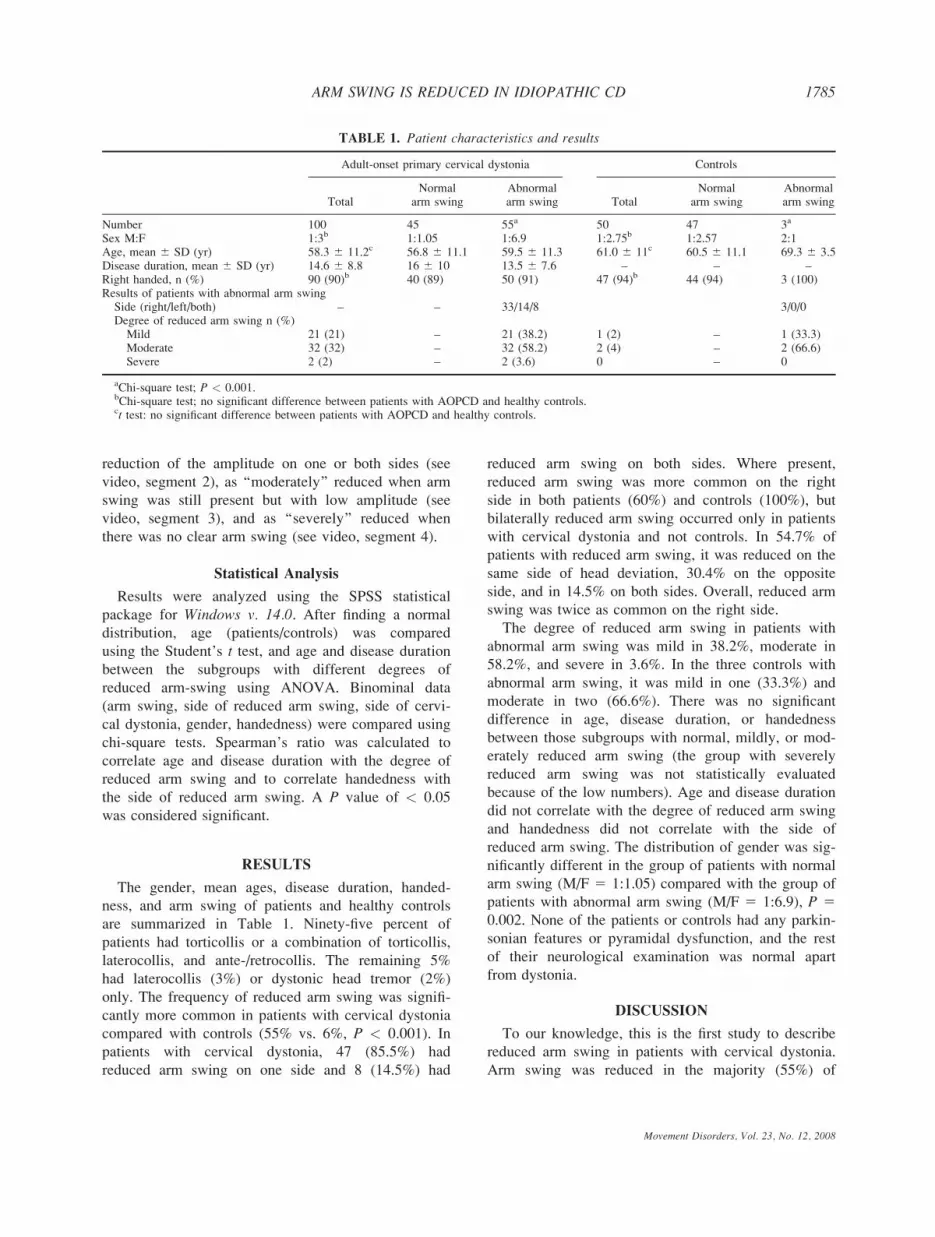
reduction of the amplitude on one or both sides (see
video, segment 2), as ‘‘moderately’’ reduced when arm
swing was still present but with low amplitude (see
video, segment 3), and as ‘‘severely’’ reduced when
there was no clear arm swing (see video, segment 4).
Statistical Analysis
Results were analyzed using the SPSS statistical
package for Windows v. 14.0. After finding a normal
distribution, age (patients/controls) was compared
using the Student’s t test, and age and disease duration
between the subgroups with different degrees of
reduced arm-swing using ANOVA. Binominal data
(arm swing, side of reduced arm swing, side of cervi-
cal dystonia, gender, handedness) were compared using
chi-square tests. Spearman’s ratio was calculated to
correlate age and disease duration with the degree of
reduced arm swing and to correlate handedness with
the side of reduced arm swing. A P value of < 0.05
was considered significant.
RESULTS
The gender, mean ages, disease duration, handed-
ness, and arm swing of patients and healthy controls
are summarized in Table 1. Ninety-five percent of
patients had torticollis or a combination of torticollis,
laterocollis, and ante-/retrocollis. The remaining 5%
had laterocollis (3%) or dystonic head tremor (2%)
only. The frequency of reduced arm swing was signifi-
cantly more common in patients with cervical dystonia
compared with controls (55% vs. 6%, P < 0.001). In
patients with cervical dystonia, 47 (85.5%) had
reduced arm swing on one side and 8 (14.5%) had
reduced arm swing on both sides. Where present,
reduced arm swing was more common on the right
side in both patients (60%) and controls (100%), but
bilaterally reduced arm swing occurred only in patients
with cervical dystonia and not controls. In 54.7% of
patients with reduced arm swing, it was reduced on the
same side of head deviation, 30.4% on the opposite
side, and in 14.5% on both sides. Overall, reduced arm
swing was twice as common on the right side.
The degree of reduced arm swing in patients with
abnormal arm swing was mild in 38.2%, moderate in
58.2%, and severe in 3.6%. In the three controls with
abnormal arm swing, it was mild in one (33.3%) and
moderate in two (66.6%). There was no significant
difference in age, disease duration, or handedness
between those subgroups with normal, mildly, or mod-
erately reduced arm swing (the group with severely
reduced arm swing was not statistically evaluated
because of the low numbers). Age and disease duration
did not correlate with the degree of reduced arm swing
and handedness did not correlate with the side of
reduced arm swing. The distribution of gender was sig-
nificantly different in the group of patients with normal
arm swing (M/F 5 1:1.05) compared with the group of
patients with abnormal arm swing (M/F 5 1:6.9), P 50.002. None of the patients or controls had any parkin-
sonian features or pyramidal dysfunction, and the rest
of their neurological examination was normal apart
from dystonia.
DISCUSSION
To our knowledge, this is the first study to describe
reduced arm swing in patients with cervical dystonia.
Arm swing was reduced in the majority (55%) of
TABLE 1. Patient characteristics and results
Adult-onset primary cervical dystonia Controls
TotalNormal
arm swingAbnormalarm swing Total
Normalarm swing
Abnormalarm swing
Number 100 45 55a 50 47 3a
Sex M:F 1:3b 1:1.05 1:6.9 1:2.75b 1:2.57 2:1Age, mean 6 SD (yr) 58.3 6 11.2c 56.8 6 11.1 59.5 6 11.3 61.0 6 11c 60.5 6 11.1 69.3 6 3.5Disease duration, mean 6 SD (yr) 14.6 6 8.8 16 6 10 13.5 6 7.6 – – –Right handed, n (%) 90 (90)b 40 (89) 50 (91) 47 (94)b 44 (94) 3 (100)Results of patients with abnormal arm swingSide (right/left/both) – – 33/14/8 3/0/0Degree of reduced arm swing n (%)
Mild 21 (21) – 21 (38.2) 1 (2) – 1 (33.3)Moderate 32 (32) – 32 (58.2) 2 (4) – 2 (66.6)Severe 2 (2) – 2 (3.6) 0 – 0
aChi-square test; P < 0.001.bChi-square test; no significant difference between patients with AOPCD and healthy controls.ct test: no significant difference between patients with AOPCD and healthy controls.
1785ARM SWING IS REDUCED IN IDIOPATHIC CD
Movement Disorders, Vol. 23, No. 12, 2008

patients with AOPCD compared with 6% of healthy
controls. Reduced arm swing was particularly common
in women with AOPCD (even after correcting for the
sex bias that cervical dystonia itself is more common
in women) and more commonly affected the right side.
Loss of the associated synergistic arm movements
when walking is seen in akinetic-rigid syndromes, py-
ramidal tract lesions affecting the arm, and acute cere-
bellar lesions. Unilateral loss of associated arm swing
when walking may be the first clinical sign in early
Parkinson’s disease.8,9 However, none of our patients
with AOPCD had features of Parkinsonism or pyrami-
dal dysfunction even after a median disease duration of
14.6 years. This leads us to conclude that the decreased
arm swing we observed can be part and parcel of dys-
tonia itself.
We have previously reported that patients with
adult-onset dystonic tremor affecting the arms (which
was often associated with dystonic postures of the
arms) frequently have reduced arm swing.7 We and
others have also previously noted anecdotally that
reduced arm swing can occur in patients with pure
focal arm dystonia without associated tremor. How-
ever, we did not observe signs of dystonia or tremor in
the arms of any of our patients with AOPCD, even
those with reduced arm swing. It is possible that the
reduced arm swing in these patients represents a seg-
mental spread of subtle motor system dysfunction to
the arm, which is sufficient to cause reduced arm
swing, but is not sufficient to cause overt dystonic pos-
tures or tremor.
It is well recognized that a degree of asymmetry in
arm swing is physiological and therefore present in
healthy subjects. A recent study in normal subjects
using quantitative gait analysis has shown that the right
arm tends to swing less than the left regardless of
handedness.10 Nevertheless, it is interesting that in our
patients with AOPCD as well as in our controls we
have reproduced this pattern with reduced arm swing
seen predominantly on the right side. However, there
was no correlation with handedness.
Why reduced arm swing should occur much more in
women with AOPCD than in men is not clear. One
could speculate that focal dystonia may have a higher
tendency to spread from the focal (cervical) dystonia
toward a segmental distribution in women.
In summary, we have shown that reduced arm swing
is common in patients with AOPCD, and we propose
that it is part and parcel of primary focal/segmental
dystonia affecting the neck. The importance of this
observation, in combination with our previously pub-
lished data in adult-onset dystonic tremor, is that
reduced arm swing appears to be a frequent occurrence
in patients with dystonia.7 It can therefore present a
chance for clinical confusion, given the historical link
between reduced arm swing and Parkinson’s disease.
Although it is unlikely that clinicians would misdiag-
nose a patient with clear cervical dystonia and reduced
arm swing as Parkinson’s disease (in contrast to the
clear risk of doing so in a patient with dystonic tremor
of the arm), there are other situations where awareness
of the presence of reduced arm swing in patients with
AOPCD are more important. One of the most impor-
tant clinical decisions to make when assessing a patient
with dystonia is to decide whether they have primary
dystonia (defined as dystonia 1/2 tremor as the only
clinical signs) or whether they have a secondary/degen-
erative dystonia. In this context, the misattribution of
reduced arm swing as a sign of possible secondary/de-
generative dystonia becomes important, as it could
lead to unnecessary investigation and diagnostic confu-
sion. Clinicians should therefore be aware that, in
patients with movement disorders, reduced arm swing
is not just a sign associated with Parkinsonism, and
can occur in the context of uncomplicated primary
dystonia.
LEGENDS TO THE VIDEO
Segment 1. This woman has mild torticollis to the
right side and normal arm swing when walking.
Segment 2. This man has torticollis to the right and
laterocollis to the left side and mildly reduced arm
swing on the right side when walking.
Segment 3. This woman has torticollis to the right side
and moderately reduced arm swing on the right side
when walking.
Segment 4. This woman has torticollis to the right side
and severely reduced arm swing on the right side
when walking.
Segment 5. This woman has torticollis to the right side
and bilaterally reduced arm swing slightly more pro-
nounced on the right side.
Acknowledgments: GK was supported by a grant from theSwiss Parkinson’s Disease Association.
REFERENCES
1. Nutt JG, Muenter MD, Aronson A, Kurland LT, Melton LJ, III.Epidemiology of focal and generalized dystonia in Rochester,Minnesota. Mov Disord 1988;3:188–194.
2. Jankovic J, Leder S, Warner D, Schwartz K. Cervical dystonia:clinical findings and associated movement disorders. Neurology1991;41:1088–1091.
3. Duane DD. Spasmodic torticollis. Adv Neurol 1988;49:135–150.
Movement Disorders, Vol. 23, No. 12, 2008
1786 G. KAGI ET AL.

4. Chan J, Brin MF, Fahn S. Idiopathic cervical dystonia: clinicalcharacteristics. Mov Disord 1991;6:119–126.
5. Rondot P, Marchand MP, Dellatolas G. Spasmodic torticol-lis—review of 220 patients. Can J Neurol Sci 1991;18:143–151.
6. Dauer WT, Burke RE, Greene P, Fahn S. Current concepts onthe clinical features, aetiology and management of idiopathic cer-vical dystonia. Brain 1998;121:547–560.
7. Schneider SA, Edwards MJ, Mir P, et al. Patients with adult-onset dystonic tremor resembling parkinsonian tremor have scans
without evidence of dopaminergic deficit (SWEDDs). Mov Dis-ord 2007;22:2210–2215.
8. Thomson PD. Clinical neurological assessment of balance andgait disorders. In: Bronstein AM, Brandt T, Nutt J, WoollacotMH, editors. Clinical disorders of balance, posture and gait.London: Arnold; 2004. p 93–98.
9. Rubino FA. Gait disorders. Neurologist 2002;8:254–262.10. Kuhtz-Buschbeck JP, Brockmann K, Gilster R, Koch A, Stolze
H. Asymmetry of arm-swing not related to handedness. Gait Pos-ture 2008;27:447–454.
Movement Disorders, Vol. 23, No. 12, 2008
1787ARM SWING IS REDUCED IN IDIOPATHIC CD