Embed Size (px)
Citation preview

Aa
II*oO
R
acwPscrhmdagwN1ltcTncveiTind
fgtna1
mUSm
Biochemical and Biophysical Research Communications 277, 179–185 (2000)
doi:10.1006/bbrc.2000.3651, available online at http://www.idealibrary.com on
rsenic Trioxide Inhibits Neuroblastoma Growth in Vivond Promotes Apoptotic Cell Death in Vitro
ngrid Øra,*,† Lennart Bondesson,‡ Carolin Jonsson,* June Ljungberg,*sabella Porn-Ares,§ Stanislaw Garwicz,† and Sven Påhlman*,1
Department of Laboratory Medicine, Division of Molecular Medicine, ‡Division of Clinical Pathology, and §Divisionf Experimental Pathology, University Hospital MAS, Malmo, Sweden; and †Department of Pediatrics,ncology-Hematology Section, Lund University Hospital, Lund, Sweden
eceived September 12, 2000
achieved at the concentrations tested. We suggest thatAip
t
itmdpIalmp(cvhtco
bRtcmwtrcdtw
Recent clinical studies have shown that inorganicrsenic trioxide (As2O3) at low concentrations inducesomplete remission with minimal toxicity in patientsith refractory acute promyelocytic leukemia (APL).reclinical studies suggest that As2O3 induces apopto-is and possibly differentiation in APL cells. Like APLells, neuroblastoma (NB) cells are thought to be ar-ested at an early stage of differentiation, and cells ofighly malignant tumors fail to undergo spontaneousaturation. Both APL and NB cells can respond with
ifferentiation to retinoic acid (RA) treatment in vitrond probably also in vivo. For that reason we investi-ated the effect of As2O3 alone and in combinationith RA on NB cell lines. In vitro, the number of viableB cells was reduced at As2O3 concentrations aroundmM after 72 h exposure. The IC50 in six different cell
ines treated for 3 days was in the 1.5 to 5 mM concen-ration interval, the most sensitive being SK-N-BE(2)ells derived from a chemotherapy resistant tumor.he combined treatment with RA (1 and 3 mM) showedo consistent additional effect with regard to inducedell death. The effect of As2O3 on NB cell number in-olved As2O3-induced apoptotic pathways (decreasedxpression of Bcl-2 and stimulation of caspase-3 activ-ty) with no clear evidence of induced differentiation.he in vivo effect of As2O3 on NB growth was also
nvestigated in nude mice bearing tumors of xe-ografted NB cells. Although tumor growth was re-uced by As2O3 treatment, complete remission was not
Abbreviations used: APL, acute promyelocytic leukemia; FCS,etal calf serum; GAP-43, growth-associated protein-43; GAPDH,lyceraldehyde-3-phosphate-dehydrogenase; MTT, 3[4,5-dimethyl-hiazol-2-yl]-2,5-diphenyltetrazolium; NB, neuroblastoma; NPY,europeptide tyrosine; PBS, phosphate-buffered saline; RA, retinoiccid; RAR, retinoic acid receptor; TPA, 12-O-tetradecanoyl phorbol-3-acetate; TH, tyrosine hydroxylase.
1 To whom requests for reprints should be addressed at Depart-ent of Laboratory Medicine, Division of Molecular Medicine, Lundniversity, University Hospital MAS, Entrance 78, S-205 02 Malmo,weden. Fax: 146-40337322. E-mail: [email protected].
179
s2O3, in combination with existing treatment modal-ties, might be a treatment approach for high risk NBatients. © 2000 Academic Press
Key Words: animal tumor model; apoptosis; arsenicrioxide; differentiation; neuroblastoma.
Advanced NB still remains one of the most challeng-ng problems in pediatric oncology despite aggressivereatment, and the need for new or additional treat-ent strategies is obvious. More than 60% of the chil-
ren over 1 year of age with neuroblastoma (NB)resent with disseminated disease at diagnosis (1).ntensive therapeutic interventions including multiplegent chemotherapy, surgery, radiotherapy, and auto-oguous bone marrow transplantation have resulted in
inor improvement on the overall survival for theseatients, especially for patients older than two years2, 3). In vitro, retinoic acid (RA) induces maturation ofultured NB cells (4–6), but until recently positive inivo effects of RA have not been reported. However,igh-dose RA treatment after autologous bone marrowransplantation showed encouraging results in a re-ent combined Children Cancer Group/Pediatric Oncol-gy Group double randomized study (7).Acute promyelocytic leukemia (APL) is characterized
y the translocation t (15, 17) which generates a PML/ARa fusion protein involving the retinoic acid recep-
or (RARa), a nuclear transcription factor normallyontributing to myeloid differentiation (8, 9). For al-ost ten years, patients with APL have been treatedith RA, which induces differentiation by revoking the
ranscriptional repression of the fusion protein, thuseflecting a successful example of translational medi-ine (10, 11). Some years ago it was reported that lowoses of arsenic trioxide (As2O3) are effective in thereatment of patients with relapsed APL (12) and thisas recently confirmed in an independent clinical
0006-291X/00 $35.00Copyright © 2000 by Academic PressAll rights of reproduction in any form reserved.

study (13). Two children, 9 and 13, were included intsctcdtsaidodeott
vssaisgda(cardtepc
M
Wfesmfita
Bepil1atMS
vested by trypsinization of confluent cultures, washed in cold PBSa
(PiuccvaaMrmm
fhcafilp(atIc
2iw(ctcB15Tuaa(a
mtwtAtecu
(admFAH1op
Vol. 277, No. 1, 2000 BIOCHEMICAL AND BIOPHYSICAL RESEARCH COMMUNICATIONS
his study and the treatment was given without severeide effects (13). In vitro, the 50% survival rate of APLells after As2O3 treatment for 3 days lies in a concen-ration interval between 1 to 2 mM (14). At these con-entrations, As2O3 induces apoptosis in APL cells byown-regulation of the Bcl-2 protein (14) and activa-ion of caspases 1 and 3 (13). In early reports it washown that As2O3 downregulates PML/RARa (15, 16),nd based on morphology and differentiation markerst was suggested that low concentrations of As2O3 in-uce differentiation of these APL cells (15). However,ther studies have concluded that As2O3 does not in-uce differentiation of APL cells (16), and that theffect of As2O3 is independent on the down-regulationf PML or the RAR fusion protein (17), and at presenthe effect of As2O3 on APL cell differentiation appearso be an open issue.
The effect of low doses of As2O3 on tumor cell sur-ival is not restricted to APL cells, As2O3 inhibits cellurvival in a variety of cancer cells including cells ofolid tumors (18–20). Like in APL, the NB tumor cellsre blocked at an immature differentiation stage andn cultured NB cells this differentiation block can inome cell lines be released by drugs or combinations ofrowth factors (4–6, 21, 22). Furthermore, NB cells areerived from the sympathetic nervous system (23–25),nd like sympathetic neuroblasts, NB express Bcl-226–29). It is likely that down-regulation of Bcl-2 in NBells will promote induction of an apoptotic process,nd in NB tumors there appears to be a positive cor-elation at the cellular level between apoptotic celleath and low Bcl-2 expression (26, 27). The similari-ies in the biology of APL and NB cells, including Bcl-2xpression, a potential indirect target for As2O3,rompted us to investigate the effect of As2O3 on NBell survival and differentiation in vitro and in vivo.
ATERIALS AND METHODS
Drugs and chemicals. As2O3 (Sigma Chemicals, Inc, Milwaukee,I) was dissolved in 1 M NaOH and kept as a stock of 30 mM As2O3
or up to 14 days. This stock was further diluted in Eagle’s minimalssential medium for cell culture studies and in phosphate bufferedaline (PBS) for animal injections. RA (Sigma) was prepared in a 2M stock in absolute ethanol and added to the culture medium to anal concentration of 1 or 3 mM. The phorbol ester 12-O-etradecanoyl phorbol-13-acetate (TPA) (Sigma), was dissolved inbsolute ethanol and kept as a 1.6 mM stock.
Cell lines and culture conditions. Human NB cell lines SK-N-E(2), IMR-32, SMS-MSN, and SH-SY5Y were cultured in minimalssential medium supplemented with 10% fetal calf serum (FCS),enicillin (100 IU/ml), and streptomycin (50 mg/ml) in a humidifiedncubator containing 95% air 1 5% CO2 atmosphere. The NB cellines LA-N-1, and LA-N-2 were grown in RPMI-1640 medium and0% FCS with the same supplements as above. For cell morphologynd differentiation assays 106 cells/10 cm culture dish were exposedo different concentrations of As2O3 alone or in combination with RA.orphological changes were followed by phase contrast microscopy.
K-N-BE(2) cells used for the animal studies were routinely har-
180
nd kept on ice until subcutaneous injection in nude mice.
MTT reduction assay. The assay is based on the uptake of MTT3[4,5-dimethylthiazol-2-yl]-2,5-diphenyltetrazolium bromide (MTT,romega, Madison, WI) by viable cells and its conversion to an
nsoluble dye via the action of succinate dehydrogenase, a reactionsed to quantify number of viable cells. For the MTT assays 10,000ells were seeded in 96-well microculture plates and three wellsontaining cells with no drugs were kept for measuring control celliability. Six concentrations of As2O3 ranging from 0.3 to 40 mM withnd without 1 mM RA were tested in triplicates. The cells withdditives were grown in a cell incubator for 72 h before addition ofTT solution. The cells were incubated at 37°C for 4 h and the
eaction was developed according to the supplier’s protocol (Pro-ega). The absorbance at 570 nm was recorded using an ELISAicroplate reader.
RNA and protein hybridization analyses. NB cell lines exposedor three days to 1 or 3 mM As2O3 with and without 1 mM RA, werearvested and total RNA was extracted according to standard pro-edures. Ten micrograms of total RNA was electrophoretically sep-rated on agarose gels containing formaldehyde and blotted ontolters as described (22). The filters were hybridized with 32P-dCTP-
abeled cDNA probes of neuropeptide Y (NPY), growth-associatedrotein-43 (GAP-43) and glyceraldehyde-3-phosphate-dehydrogenaseGAPDH). Hybridizing mRNA was visualized by autoradiographynd the bands were scanned using Fuji Film Science Imaging Sys-ems LAS-100. The results were quantified using Science Lab 97mage Gauge Ver. 3.0 relating NPY and GAP-43 mRNA to theorresponding GAPDH mRNA levels.
For Western blot analyses, cells were seeded day 0 at a density of3 106/10 cm dish and one day later As2O3 was added. After 3 days
n As2O3 the medium was removed and the dishes were washed twiceith ice-cold PBS before harvest. The cells were lysed in RIPA-buffer
10 mM Tris–HCl, 160 mM NaCl, 1% Triton X100, 1% Na deoxy-holate, 0.1% SDS, 1 mM EGTA, 1 mM EDTA, and protease inhibi-ors (“Complete, EDTA-free,” Boehringer-Mannheim), and proteinontent in the cleared supernatant was determined by the method ofradford. For SDS–PAGE, 120 mg of each sample were loaded on a0% polyacrylamide gel. After blotting, the filter was blocked with% fat-free dried milk in Tris–HCl pH 8.0, 150 mM NaCl and 0.05%ween 20. The anti-Bcl-2 antiserum (Santa Cruz Biotechnology) wassed in a 1/500 dilution and the anti-tyrosine hydroxylase (TH)ntibody (Boehringer-Mannheim) was diluted 1/500. The secondarynti-rabbit and anti-mouse horseradish peroxidase-linked antibodiesAmersham) were diluted 1/5000. The peroxidase reaction was visu-lized with Super Signal (Pierce) by exposure of X-ray film.
Fluorometric assay for caspase-3 activity. Caspase-3 activity waseasured in cell lysates from non-treated and arsenic trioxide
reated NB cells. Cells were grown in 6-well plates, 0.5 3 106 cells/ell, for 48 and 72 h with and without As2O3 in increasing concen-
rations. Cleavage of the fluorogenic caspase-3 substrate DEVD-MC (Upstate Biotechnology, Lake Placid) was performed according
o manufacturer’s instructions, using a Fluorostar plate reader andxcitation and emission wavelengths of 390 and 460 nm. Theaspase-3 inhibitor DEVD-CHO (3 mM; Pharmingen, San Diego) wassed to confirm assay-specificity.
Experimental animals. Athymic mice of the NMRI strain (nu/nu)Bogstad, Denmark) were housed in controlled environment and fedd libidum. Cages were autoclaved and changed weekly. All proce-ures were carried out in accordance with the regional ethical com-ittee for animal research, which approved the study (M101-99).emale mice 6 weeks of age weighing 20–25 g at arrival were used.fter one week of acclimatization, the mice were anesthetized withalothan and injected on the left side of upper back with 5 to 10 306 SK-N-BE(2) cells suspended in PBS to a volume of 200 ml. Groupsf 5 mice were housed in each cage. The tumor take was high in theresented experiments, 39 of 40 animals had tumor-take after 7–12
days. At a tumor size of approximately 0.1 cm3, As2O3 diluted in PBStrteiw
ebbfiai9bmrabtM(11m
R
A
sIStwtiaatdrtAtcpctoocwfSsmc
Ata
tSo1Mf
Vol. 277, No. 1, 2000 BIOCHEMICAL AND BIOPHYSICAL RESEARCH COMMUNICATIONS
o 400 ml was given intravenously daily in the tail vein. Control miceeceived 400 ml PBS without drug. The animals were randomized inhree groups receiving 0, 200, or 400 mg As2O3. Tumor volume wasstimated every second day measured when the mouse was securedn a Plexiglass holder. After three weeks the mice were sacrificedith CO gas, and animals and dissected tumors were weighed.
Tissue preparation, immunohistochemistry, and TdT-dUTP nicknd labeling TUNEL). Mouse tumors were immediately fixed in 4%uffered formaldehyde at 4°C and processed for paraffin embeddingefore sectioning. Four to five micrometer sections were deparaf-nized in xylene, rehydrated and incubated in 0.3% H2O2 and meth-nol for 20 min to block endogenous peroxidase activity. To enhancemmunohistochemical staining the sections were microwaved to5°C in 0.01 mM sodium citrate buffer (pH 7.3) for 10–15 min. Afterlocking of nonspecific binding with 1% bovine serum albumin for 20in the optimally diluted primary antibody was incubated for 1 h at
oom temperature. Slides were rinsed and incubated 30 min withvidin-biotin-peroxidase conjugated secondary antibody. Diamino-enzidine was used as chromogen. The primary antibodies and dilu-ions were as follows: anti-human monoclonal anti-TH (Boehringerannheim) at 1:25 dilution, anti human monoclonal anti-Ki-67
Dako) at 1:100 dilution, anti-human monoclonal anti Bcl-2 (Dako) at:100 dilution, and anti-mouse polyclonal anti CD 34 (Chemicon) at:100 dilution. The TUNEL staining was performed according to theanufacturer’s protocol (Boehringer-Mannheim).
ESULTS
s2O3 Induces Cell Death of Cultured HumanNB Cells
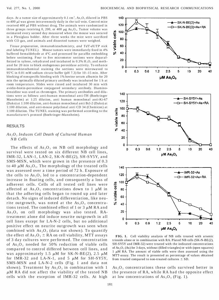
The effects of As2O3 on NB cell morphology andurvival were tested on six different NB cell lines,MR-32, LAN-1, LAN-2, SK-N-BE(2), SH-SY5Y, andMS-MSN, which were grown in the presence of 0.3o 40 mM As2O3. The morphology of the treated cellsas assessed over a time period of 72 h. Exposure of
he cells to As2O3 led to a concentration-dependentncrease in floating cells, and consequently a loss ofdherent cells. Cells of all tested cell lines wereffected at As2O3 concentrations down to 1 mM inhat the adhering cells began to round up and lateretach. No signs of induced differentiation, like neu-ite outgrowth, was noted at the As2O3 concentra-ions tested. The combined effect of 1 or 3 mM RA ands2O3 on cell morphology was also tested. RA-
reatment alone did induce neurite outgrowth in allell lines except for LA-N-2 cells, but no additionalositive effect on neurite outgrowth was seen whenombined with As2O3 (data not shown). To quantifyhe effect of As2O3 6 RA on cell viability, MTT assaysf 3 day cultures were performed. The concentrationf As2O3 needed for 50% reduction of viable cellsompared to control differed between cell lines, andas approximately 1.5 mM for SK-N-BE(2), 2.5 mM
or IMR-32 and LA-N-1, and 5 mM for SH-SY5Y,MS-MSN and LA-N-2 cells (Fig. 1 and data nothown). Treatment by As2O3 in combination with 1M RA did not affect the viability of the tested NBells with the exception of IMR-32 cells. At high
181
s2O3 concentrations these cells survived better inhe presence of RA, while RA had the opposite effectt low concentrations of As2O3 (Fig. 1).
FIG. 1. Cell viability analysis of NB cells treated with arsenicrioxide alone or in combination with RA. Plated NB cells (SK-N-BE(2),H-SY5Y and IMR-32) were treated with the indicated concentrationsf As2O3 (As) for 3 days, without (filled triangles) or with (open squares)mM RA. The amount of viable cells were then assessed using theTT-assay. The result is presented as percentage of values obtained
rom treated compared to non-treated cultures 6 SD.

A
tpvsmtdaorvidrtFssatwsr
AtactirsNi
tween treated and non-treated tumors, and based onCdn
P
tecept2Sot(itScfc3mawSoett
ncbstumpgclstbcPpsTbm
nwrv(a
Vol. 277, No. 1, 2000 BIOCHEMICAL AND BIOPHYSICAL RESEARCH COMMUNICATIONS
s2O3 Represses in Vivo Growth of XenotransplantedHuman NB Cells
In our hands, all cultured human NB cells so farested form localized solid tumors when xenotrans-lanted into nude mice (data not shown). To test the inivo effects of As2O3 on NB growth, mice were injectedubcutaneously with SK-N-BE(2) cells, which form theost aggressively growing tumors of the NB cell lines
ested. The animals were randomly assigned to threeifferent groups when the tumors reached a volume ofpproximately 0.1 cm3. Group 1 animals received PBSnly, group 2 received 200 mg As2O3, and group 3eceived 400 mg As2O3, as daily injections in the tailein. Tumors in control mice receiving PBS grew rap-dly, and these animals were sacrificed after 10 to 14ays of treatment due to large tumors (Fig. 2). Miceeceiving As2O3 showed a dose-dependent inhibition ofumor growth compared to the control mice as shown inig. 2. Mice treated with 200 mg As2O3 did not showigns of severe side effects, except that they seemedlightly apathetic 1 to 2 min after injection of As2O3. Nonimal in this group lost weight during the As2O3
reatment, as compared to the control animals injectedith PBS alone. Animals treated with 400 mg As2O3
uffered from side effects (anorexia, transient edema)esulting in weight loss at the end of treatment.
In an attempt to mechanistically understand thes2O3-induced repressed growth of the NB xenograft
umors, differences in number of proliferating andpoptotic cells, and the differentiation status of tumorells in the dissected tumors, were assessed. Sections ofumors from non-treated and treated animals weremmunohistochemically stained with antibodies di-ected towards Ki-67, Bcl-2, and TH, respectively, andections were also stained with the TUNEL technique.one of these stainings resulted in statistically signif-
cant differences in the number of positive cells be-
FIG. 2. In vivo growth of xenotransplanted human NB cells inude mice and treatment with arsenic trioxide. SK-N-BE(2) cellsere injected subcutaneously into the mice. When tumors had
eached the volume of approximately 0.1 cm3, treatment with solventehicle (PBS; open squares), or 200 (filled triangles), and 400 mgopen circles) As2O3 was initiated. In each treatment group 6 to 7nimals were used. Tumor sizes are given in cm3 6 SD.
182
D 34 immunohistochemistry there were no apparentifferences in the vascularization of the tumors (dataot shown).
henotypic Effects of As2O3 on Cultured NB Cells
To further investigate possible mechanisms behindhe in vivo effects of As2O3, cultured NB cells werexposed for 3 days to 1 and 3 mM As2O3 without or inombination with 1 mM RA and examined for potentialffects on the differentiation status of these cells. Ex-ression of two well-characterized markers of sympa-hetic ganglionic differentiation, NPY and GAP-43 (21,4), was analyzed by Northern blot hybridization. InK-N-BE(2) and IMR-32 cells a discrete up-regulationf NPY mRNA was observed with 1 mM As2O3, whilehe GAP-43 mRNA levels were unchanged or reducedFig. 3). In the presence of 1 mM RA GAP-43 expressionncreased in both cell lines, which is in accordance withhe morphological differentiation seen, while only theK-N-BE(2) cells had increased NPY expression. Theombination of RA and As2O3 consistently did not af-ect the expression levels of these marker genes whenompared to the expression induced by RA alone (Fig.). The expression levels of another differentiationarker gene for the sympathetic lineage, TH, was not
ffected by low As2O3 concentrations, while treatmentith 2 and 4 mM As2O3 reduced the TH levels in theH-SY5Y and IMR-32 cells (Fig. 4). Based on morphol-gy and marker gene expression data, we conclude thatxposure of human NB cells to doses of As2O3 affectinghe survival of these cells, did not induce differentia-ion in the cell lines investigated.
Neuroblasts of the developing human sympatheticervous system express Bcl-2 and the expression in-reases with developmental age (26). In these neuro-lasts, Bcl-2 expression is positively correlated to cellurvival, which was demonstrated in vitro, and in aransgenic mouse model where overexpression of Bcl-2nder the influence of the neuron-specific enolase pro-oter resulted in overgrowth and enlargement of sym-
athetic ganglia (30, 31). Based on the similarities inene expression in sympathetic neuroblasts and in NBells, we analyzed the Bcl-2 protein levels in NB cellines treated with As2O3 at concentrations affecting theurvival of these cells. As shown in Fig. 4, in two ofhree tested cell lines the Bcl-2 protein concentrationegan to decrease at 1 to 2 mM As2O3, with SK-N-BE(2)ells being more sensitive than SH-SY5Y cells.horbolester-differentiated SH-SY5Y cells served as aositive control with increased Bcl-2 expression in theympathetically differentiated, TPA-treated, cells (26).he third cell line, IMR-32, did not show an apprecia-le decrease in Bcl-2 protein when treated with up to 4M As2O3 (Fig. 4), despite that As2O3 at this concen-

t(
Altcc7aTtwtsag
D
uPtahpapvcpttT
vebi
mau(Ad(NBfmwdmst(mBcs
aAdcWdTt
Neea
Vol. 277, No. 1, 2000 BIOCHEMICAL AND BIOPHYSICAL RESEARCH COMMUNICATIONS
ration had a considerable capacity to kill IMR-32 cellssee Fig. 2).
To test whether the decrease in Bcl-2 protein seen ins2O3-treated SK-N-BE(2) and SH-SY5Y cells was
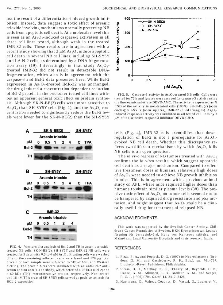
inked to an induced apoptotic process, caspase-3 ac-ivity was measured in the arsenic trioxide-treated NBells. In both SK-N-BE(2) and SH-SY5Y cells an in-reased caspase-3 activity was recorded after 48 and2 h. Also in IMR-32 cells a slight increase in caspase-3ctivity was detected (Fig. 5, 48 h data not shown).hus, the reduced Bcl-2 protein levels and the activa-ion of caspase-3 in As2O3-treated NB cells, togetherith lack of data supporting that these cells differen-
iate, suggest that the reduced number of viable cellseen in vitro (Fig. 1) at least in part is explained bypoptotic cell death, and not by differentiation-inducedrowth arrest.
ISCUSSION
In traditional Chinese medicine arsenic has beensed as treatment of different cancers for centuries.urified As2O3 suitable for intravenous administra-ion, to overcome the side effects associated with oraldministration, has been used in China since 1970 andas been proven to be an effective second line drug foratients with APL. Daily infusions of 10 mg As2O3 indult patients with relapse of APL, give peak arseniclasma levels of 4–6 mM in these patients (32). Initro, 1 to 2 mM As2O3 is needed to obtain a 50% APLell death after 3 days of treatment (15, 16). In theresent study, sensitivity to As2O3 was found in all sixested NB cell lines, with 50% cell death obtained be-ween 1.5 to 5 mM As2O3 depending on cell line tested.he drug had an inhibitory effect on tumor growth in
FIG. 3. Expression of sympathetic neuronal differentiation markeB cells were treated for 3 days with 1 or 3 mM As2O3 with or withou
xpression of NPY and GAP-43 was analyzed by Northern blot hybxpression was quantified by scanning the autoradiographs, and thefter compensation for the GAPDH signal.
183
ivo at As2O3 doses giving no apparent, early, sideffects. Thus, similar to APL cells, NB cells appear toe sensitive to As2O3 both in vitro and in vivo at clin-cally achievable doses.
The studied NB cell lines are established from highlyalignant tumors, and all but one, (SH-SY5Y) have an
mplified N-myc gene. The cell lines established fromntreated primary tumors or infiltrated bone marrowIMR-32, LA-N-1, LA-N-2) were not more sensitive tos2O3 than those derived from patients with residualisease or relapse after treatment with chemotherapySK-N-BE(2), SH-SY5Y, a neuroblastic clone of the SK--SH cell line) (33–36). We chose to use the SK-N-E(2) cell line in the animal model studies, a cell line
orming very aggressively growing tumors in nudeice without signs of metastatic spread. This cell lineas established from a 2-year-old boy with residualisease after induction therapy and these cells areore resistant to chemotherapy than those of a corre-
ponding cell line established at diagnosis (33). Fur-hermore, these cells have a high p53 protein levelsdata to be published) and in keeping with this, a
utated p53 (37). As demonstrated here, the SK-N-E(2) cells were sensitive to As2O3 suggesting minimal
ross-resistance between As2O3 and chemotherapy re-istance (36).NB cell lines frequently respond to RA by differenti-
tion and inhibited growth. Combination of RA withs2O3, did not consistently affect As2O3-induced celleath, and RA-induced phenotypic changes, like in-reased GAP-43 expression, were unaltered by As2O3.e further conclude that As2O3 alone does not alter the
ifferentiation status of the cultured NB cells tested.hus, the reduced number of viable cells in culturesreated with As2O3 alone or in combination with RA is
in arsenic trioxide and RA-treated NB cells. SK-N-BE(2) and IMR-32mM RA as indicated in the figure. Total RNA was prepared and thezations. GAPDH served as RNA loading control. NPY and GAP-43mbers in the figure give the expression relative to non-treated cells,
rst 1
ridinu
not the result of a differentiation-induced growth inhi-btcitIrcattfcetoosAce
creflN
cctoishibtc
A
dfM
RttopbsaaB
tt6cim
Vol. 277, No. 1, 2000 BIOCHEMICAL AND BIOPHYSICAL RESEARCH COMMUNICATIONS
ition. Instead, data suggest a toxic effect of arsenicrioxide involving mechanisms normally protecting NBells from apoptotic cell death. At a molecular level thiss seen as an As2O3-induced caspase-3 activation in allhree cell lines tested, although weak in the treatedMR-32 cells. These results are in agreement with aecent study showing that 2 mM As2O3 induce apoptoticell death in several NB cell lines, including SH-SY5Ynd LA-N-2 cells, as determined by a DNA fragmenta-ion assay (19). Interestingly, in that study As2O3-reated IMR-32 did not result in detectable DNA-ragmentation, which also is in agreement with theaspase-3 and Bcl-2 data presented here. While Bcl-2xpression in As2O3-treated IMR-32 was unchanged,he drug induced a concentration dependent reductionf Bcl-2 protein in the two other tested cell lines with-ut an apparent general toxic effect on protein synthe-is. Although SK-N-BE(2) cells were more sensitive tos2O3 than SH-SY5Y cells (Fig. 1), and the As2O3 con-entration needed to significantly reduce the Bcl-2 lev-ls were lower for the SK-N-BE(2) than the SH-SY5Y
FIG. 4. Western blot analysis of Bcl-2 and TH in arsenic trioxide-reated NB cells. SK-N-BE(2), SH-SY5Y and IMR-32 NB cells werereated for 3 days with 0.5 to 4 mM As2O3. Floating cells were washedff and the remaining adherent cells were lysed and 120 mg totalrotein of each sample were subjected to SDS–PAGE and Westernlotting. The protein blots were incubated with an anti-Bcl-2 anti-erum and an anti-TH antibody, which detected a 26 kDa (Bcl-2) and60 kDa (TH) immunoreactive protein, respectively. Non-treated
nd 16 nM TPA-treated SH-SY5Y cells served as positive controls forCL-2 expression.
184
ells (Fig. 4), IMR-32 cells exemplifies that down-egulation of Bcl-2 is not a prerequisite for As2O3-voked NB cell death. Whether this discrepancy re-ects two different mechanisms by which As2O3 killsB cells is an open question.The in vivo regress of NB tumors treated with As2O3
onfirms the in vitro results, which suggest apoptoticell death as a major mechanism. Compared to effec-ive treatment doses in humans, relatively high dosesf As2O3 were needed to achieve NB growth inhibitionn mice. This is in agreement with a previous animaltudy on APL, where mice required higher doses thanumans to obtain similar plasma levels (38). The pos-
tive toxic effect of As2O3 on tumor cells seemed not toe hampered by acquired drug resistance and p53 mu-ation, and might suggest that As2O3 could be a clini-ally useful drug for treatment of relapsed NB.
CKNOWLEDGMENTS
This work was supported by the Swedish Cancer Society, Chil-ren’s Cancer Foundation of Sweden, HKH Kronprinsessan Lovisasorening for barnasjukvård, Hans von Kantzows stiftelse, and
almo and Lund University Hospitals and their research funds.
EFERENCES
1. Pizzo, P. A., and Poplack, D. G. (1997) in Neuroblastoma (Bro-deur, G. M., and Castleberry, R. P., Eds.), pp. 761–797,Lippincott-Raven Publishers, Philadelphia.
2. Stram, D. O., Matthay, K. K., O’Leary, M., Reynolds, C. P.,Haase, G. M., Atkinson, J. B., Brodeur, G. M., and Seeger,R. C. J. (1996) Clin. Oncol. 14, 2417–2426.
3. Hartmann, O., Valteau-Couanet, D., Vassal, G., Lapierre, V.,
FIG. 5. Caspase-3 activity in As2O3-treated NB cells. Cells werereated for 72 h and lysates were assayed for caspase-3 activity usinghe fluorogenic substrate DEVD-AMC. The activity is expressed as %SD of the activity in non-treated cells (100%). SK-N-BE(2) (open
ircles); SH-SY5Y (open squares); IMR-32 (filled triangles). As2O3-nduced caspase-3 activity was inhibited in all tested cell lines by 3M of the selective caspase-3 inhibitor DEVD-CHO.

Brugieres, L., Delgado, R., Couanet, D., Lumbroso, J., and Ben-
1
11
1
1
1
1
1
1
1
2
Kim, B. K., and Lee, Y. Y. (1999) Biochem. Biophys. Res. Com-
2
2
2
2
2
2
2
2
2
3
3
3
3
3
3
3
3
3
Vol. 277, No. 1, 2000 BIOCHEMICAL AND BIOPHYSICAL RESEARCH COMMUNICATIONS
hamou, E. (1999) Bone Marrow Transpl. 23, 789–795.4. Påhlman, S., Ruusala, A-I., Abrahamsson, L., Mattsson,
M. E. K., and Esscher, T. (1984) Cell Diff. 14, 135–144.5. Giannini, G., Dawson, M. I., Zhang, X., and Thiele, C. J. (1997)
J. Biol. Chem. 272, 26693–2671.6. Matsou, T., and Thiele, C. J. (1998) Oncogene 16, 3337–3343.7. Matthay, K. K., Villablanca, J. G., Seeger, R. C., Stram, D. O.,
Harris, R. E., Ransey, N. K., Swift, P., Shimada, H., Black, C. T.,Brodeur, G. M., Gerbing, R. B., and Reynolds, C. P. (1999)N. Engl. J. Med. 341, 1165–1173.
8. Tsai, S., and Collins, S. (1993) Proc. Natl. Acad. Sci. USA 90,7153–7157.
9. Labrecque, J., Allan, D., Chambon, P., Iscove, N. N., Lohnes, D.,and Hoang, T. (1998) Blood 92, 607–615.
0. Huang, M., Ye, Y., Chen, R., Chai, J., Lu, J., Zhoa, L., Gu, L., andWang, Z. (1988) Blood 72, 567–572.
1. Slack, J. (1999) Curr. Opin. Oncol. 11, 9–13.2. Sun, H. D., Ma, L., Hu, X. C., and Zhanf, T. D. (1992) Chin.
J. Comb. Trad. Chin. Med. West. Med. 12, 170–171.3. Soignet, S. L., Maslak, P., Wang, Z-G., Jhanwar, S., Calleja, E.,
Dardashti, L. J., Corso, D., DeBlasio, A., Gabrilove, J., Schein-berg, D. A., Pandolfi, P. P., and Warrel, R. P. (1998) N. Engl.J. Med. 339, 1341–1348.
4. Chen, G-Q., Zhu, J., Shi, X. G., Ni, J. H., Zhong, H. J., Si, G. Y.,Jin, X. L., Tang, W., Li, X. S., Xong, S. M., Shen, Z. X., Sun, G. L.,Ma, J., Zhang, P., Zhang, T. D., Gazin, C., Naoe, T., Chen, S. J.,Wang, Z. Y., and Chen, Z. (1996) Blood 88, 1052–1061.
5. Chen, G-Q., Shi, X-G., Tang, W., Xiong, S-M., Zhu, J., Cai, X.,Han, Z-G., Ni, J-H., Shi, G-Y., Jia, P-M., Liu, M-M., Niu, C., Ma,J., Zhang, P., Zhang, T-D., Paul, P., Naoe, T., Kitamura, K.,Miller, W., Waxman, S., Wang, Z-Y., de The, H., Chen, S-J., andChen, Z. (1997) Blood 89, 3345–3353.
6. Shao, W., Fanelli, M., Ferrara, F. F., Riccioni, R., Rosenauer, A.,Davison, K., Lamph, W. W., Waxman, S., Pelicci, P. P., Lo Locco,F., Avvisati, G., Testa, U., Peschle, C., Gambacorti-Passerini, C.,Nervi, C., and Miller, W. H. (1998) J. Natl. Cancer Inst. 90,124–133.
7. Wang, Z. G., Rivi, R., Delva, L., Delva, L., Konig, A., Scheinberg,D. A., Gambacorti-Passerini, C., Gabrilove, J. L., Warrell, R. P.,and Pandolfi, P. P. (1998) Blood 92, 1497–1504.
8. Rousselot, P., Labaume, S., Marolleau, J-P., Larghero, J.,Noguera, M-H., Brouet, J-C., and Fermand, J-P. (1999) CancerRes. 59, 1041–1048.
9. Akao, Y., Nakagawa, Y., and Akiyama, K. (1999) FEBS Letts.455, 59–62.
0. Seol, J. G., Park, W. H., Kim, E. S., Jung, C. W., Hyun, J. M.,
185
mun. 65, 400–404.1. Påhlman, S., Meyerson, G., Lindgren, E., Schalling, M., and
Johansson, I. (1991) Proc. Natl. Acad. Sci. USA 88, 9994–9998.2. Lavenius, E., Parrow, V., Nånberg, E., and Påhlman, S. (1994)
Growth Factors 10, 29–39.3. Hoehner, J. C., Gestblom, C., Hedborg, F., Sandstedt, B., Olsen,
L., and Påhlman, S. (1996) Lab. Invest. 7, 659–675.4. Gestblom, C., Hoehner, J. C., Hedborg, F., Sandstedt, B., and
Påhlman, S. (1997) Am. J. Pathol. 150, 107–117.5. Påhlman, S., and Hedborg, F. (2000) in Neuroblastoma (Bro-
deur, G. M., Sawada, T., Voute, P. A., and Tsuchida, Y., Eds.), pp.9–19, Elsevier, Amsterdam.
6. Hoehner, J. C., Hedborg, F., Jernberg-Wiklund, H., Olsen, L.,and Påhlman, S. (1995) Int. J. Cancer 62, 19–24.
7. Hoehner, J. C., Gestblom, C., Olsen, L., and Påhlman, S. (1997)Br. J. Cancer 75, 1185–1194.
8. Krajewski, S., Chatten, J., Hanada, M., and Reed, J. C. (1995)Lab. Invest. 71, 42–45.
9. Ikegaki, N., Katsumata, M., Tsujimoto, Y., Nakagawara, A., andBrodeur, G. M. (1995) Cancer Lett. 91, 161–168.
0. Garcia, I., Martinou, I., Tsujimoto, Y., and Martinou, J. C. (1992)Science 258, 302–304.
1. Farlie, P. G., Dringen, R., Rees, S. M., Kannourakis, G., andBernard, O. (1995) Proc. Natl. Acad. Sci. USA 92, 4397–4401.
2. Shen, Z. X., Chen, G. Q., Ni, J. H., Li, X. S., Xiong, S. M., Qiu,Q. Y., Zhu, J., Tang, W., Sun, G. L., Yang, K. Q., Chen, Y., Zhou,L., Fang, Z. W., Wang, Y. T., Ma, J., Zhang, P., Zhang, T. D.,Chen, S. J., Chen, Z., and Wang, Z. Y. (1997) Blood 89, 3354–3360.
3. Keshelava, N., Seeger, R. C., Groshen, S., and Reynolds, C. P.(1998) Cancer Res. 58, 5396–5405.
4. Tumilowicz, J. J., Nichols, W. W., Cholon, J. J., and Greene, A. E.(1970) Cancer Res. 30, 2110–2118.
5. Biedler, J. L., Helson, L., and Spengler, B. A. (1973) Cancer Res.33, 2643–2652.
6. Seeger, R. C., Rayner, S. A., Banerjee, A., Chung, H., Laug,W. E., Neustein, H. B., and Benedict, W. F. (1977) Cancer Res.37, 1364–1371.
7. Keshelava, N., Zou, J. J., Luna, M. C., Waidyaratne, N. S.,Triche, T. J., Gomer, C. J., and Reynolds, C. P. (2000) in Ad-vances in Neuroblastoma Research 2000 Conference. Abstractbook p. 16.
8. Lallemand-Breitenbach, V., Guillemin, M-C., Janin, A., Daniel,M-T., Degos, L., Kogan, S. C., Bishop, J. M., and de The, H.(1999) J. Exp. Med. 189, 1043–1052.


