Embed Size (px)
Citation preview
Artemin Crystal Structure Reveals Insights into Heparan Sulfate Binding‡
Laura Silvian,* Ping Jin, Paul Carmillo, P. Ann Boriack-Sjodin, Carolyn Pelletier, Mia Rushe,∧ BangJian Gong,Dinah Sah,| Blake Pepinsky, and Anthony Rossomando
Department of Drug DiscoVery, Biogen Idec, Inc., 12 Cambridge Center, Cambridge, Massachusetts 02142
ReceiVed January 6, 2006; ReVised Manuscript ReceiVed March 31, 2006
ABSTRACT: Artemin (ART) promotes the growth of developing peripheral neurons by signaling througha multicomponent receptor complex comprised of a transmembrane tyrosine kinase receptor (cRET) anda specific glycosylphosphatidylinositol-linked co-receptor (GFRR3). Glial cell line-derived neurotrophicfactor (GDNF) signals through a similar ternary complex but requires heparan sulfate proteoglycans(HSPGs) for full activity. HSPG has not been demonstrated as a requirement for ART signaling. Wecrystallized ART in the presence of sulfate and solved its structure by isomorphous replacement. Thestructure reveals ordered sulfate anions bound to arginine residues in the pre-helix and amino-terminalregions that were organized in a triad arrangement characteristic of heparan sulfate. Three residues in thepre-helix were singly or triply substituted with glutamic acid, and the resulting proteins were shown tohave reduced heparin-binding affinity that is partly reflected in their ability to activate cRET. This studysuggests that ART binds HSPGs and identifies residues that may be involved in HSPG binding.
The glial cell line-derived neurotrophic factor (GDNF)1
family of ligands (GFLs) are neurotrophic growth factorsthat promote the survival of distinct populations of centraland peripheral neurons. Four GFLs have been described todate: artemin [ART (1), also known as neublastin (2) andenovin (3)], neurturin [NTN (4)], persephin [PSP (5)], andGDNF (6). ART exhibits a restricted pattern of expressionin the developing vasculature and sclerotomes (7), whereasGDNF is expressed in multiple tissues during developmentand in adulthood (8).
GFLs are biologically active covalently linked homo-dimers, in which each monomer has a “cystine knot”topology. This cystine knot was identified first through thestructure of transforming growth factorâ2 [TGF-â2 (9, 10)]and can be recognized from the primary sequence by acharacteristic cysteine-spacing motif. These proteins containtwo antiparallelâ strands (“fingers”), which are linked bythe cystine knot. In the TGF-â superfamily of cystine-knotproteins, including osteogenic protein 1 [OP-1 or BMP7(11)], bone morphogenic protein 2 [BMP-2 (12)], and GDNF,
the dimer arrangement is antiparallel and includes aninterchain disulfide bond. A helix is inserted in the loopbetween the two fingers, termed the “wrist”, and packsagainst the flat fingers of the other monomer in a “hand-shake” arrangement. Other cystine-knot proteins differ fromthe TGF-â family; they do not contain a helical “wrist”, andtheir dimerization interfaces are different. These structuresinclude platelet-derived growth factor [PDGF (13)], nervegrowth factor [NGF (14)], and vascular endothelial growthfactor [VEGF (15)], among others. Of the GFLs, GDNF isthe only family member whose crystal structure has beensolved [PDB ID 1AGQ;16]. The GDNF crystal containstwo covalent homodimers in its asymmetric unit. The twoGDNF homodimers differ in the relative hinge angle betweenthe “finger-tips” and “wrist” within their respective mono-mers.
The homodimeric GFLs can activate cRET tyrosine kinaseby forming a complex containing the cRET tyrosine kinasereceptor and a preferred high-affinity GPI-linked co-receptor(GFRR) in a proposed stoichiometry of GFL homodimer-GFRR2-RET2 (17). Four GFRR co-receptors have beenidentified (GFRR1-4) (18-21). Each preferentially bindsto one of the four GFLs; however, crossover may occur insome instances (22). ART interacts selectively with the co-receptor GFRR3, whereas GDNF interacts selectively withGFRR1 (1, 17, 23).
A variety of experiments suggest that the finger loops ofGFLs interact directly with their respective GFRR co-receptors. On the basis of mutagenesis experiments, GFRR1was shown to bind the acidic and hydrophobic finger loopsof GDNF (24). In a domain-swapping experiment betweenGFLs, the preference of ART for co-receptor GFRR3 wasascribed to the specific sequences in the pre-helix region ofthe wrist and theâ strands of finger 2 (22). In support ofthe finger loops being essential, a recent model of the
‡ Coordinates and structure factors have been deposited in the ProteinData Bank with PDB ID 2ASK.
* To whom correspondence should be addressed. Telephone: (617)679-2208. Fax: (617) 679-2616. E-mail: [email protected].
∧ New address: Research, Novartis Institutes for Biomedical Re-search Inc., 250 Mass Ave., Cambridge, MA 02139.
| New address: Alnylam Pharmaceuticals, Inc., 300 Third St.,Cambridge, MA 02142.
1 Abbreviations: GDNF, glial cell line-derived neurotrophic factor;GFL, GDNF family of ligands; ART, artemin; ART113, full-lengthhuman artemin with 113 amino acids; NTN, neurturin; PSP, persephin;TGF-â2, transforming growth factorâ2; OP-1, osteogenic protein 1;BMP-2, bone morphogenic protein 2; PDGF, platelet-derived growthfactor; NGF, nerve growth factor; VEGF, vascular endothelial growthfactor; GFR-R, GPI-linked co-receptor; HSPG, heparan sulfate pro-teoglycan; GAG, glycosaminoglycan; HGF/SF, hepatocyte growthfactor/scatter factor; FGF-1, fibroblast growth factor 1; bFGF, basicfibroblast growth factor; EGF, epidermal growth factor; KIRA, kinasereceptor activation enzyme-linked immunosorbent assay.
6801Biochemistry2006,45, 6801-6812
10.1021/bi060035x CCC: $33.50 © 2006 American Chemical SocietyPublished on Web 05/13/2006

GFRR1/GDNF binding surface has been proposed thatincorporates mutagenesis data and places essential argininesin proximity to acidic residues in the finger loops of GDNF(25).
The ability of many cytokines to promote receptor dimer-ization and signaling is often facilitated by membrane-boundheparan sulfate proteoglycans (HSPGs). Heparan sulfate islocalized on cell surfaces and the extracellular matrix and issynthesized as a proteoglycan composed of a protein coreand multiple glycosaminoglycan (GAG) chains. Heparin, incontrast, is a densely sulfonated glycosaminoglycan thatresembles the GAG chains of the heparan sulfate proteogly-can (26) and is often used in in vitro studies. In previousstudies, interactions between fibroblast growth factor 1 (FGF-1) and hepatocyte growth factor/scatter factor (HGF/SF) withtheir tyrosine kinase receptors FGFR2 and c-Met, respec-tively, were shown to be facilitated by GAGs (27, 28). Recentstudies have indicated that GDNF signaling is mediated byheparan sulfate. In one study, the phosphorylation state ofcRET was altered after either heparinase III treatment or theaddition of exogenous heparin, suggesting that GAGs medi-ate a direct interaction between GDNF and its receptors (29).More specifically, the 2-O sulfate moieties are required (30).Other studies have reported that GDNF-induced upregulationof the tyrosine-hydroxylase gene mRNA was enhanced bythe addition of heparin (31). Therefore, it is of interest todetermine whether the interactions of other members of theGDNF family with their receptors might also involve GAGs.
We present here the crystal structure of human ART andcompare it to the previously solved GDNF structure. TheART structure contains three sulfate anions that are arrangedin a triad motif and are suggestive of a GAG-binding site.On the basis of this structure, we generated four ART variantsin which arginine residues that bind these sulfates were singlyor triply substituted with glutamate and assayed for heparin-binding and heparin-dependent stimulation of signalingactivity. These data identify a heparin-binding site on theART surface that is distinct from the receptor-binding region.
EXPERIMENTAL PROCEDURES
Protein Expression and Crystallization.ART and seleno-methionine-ART were expressed inEscherichia colias His-tagged fusion proteins with a Lysyl-endoprotease cleavagesite immediately adjacent to the start of the mature 113 aminoacid sequence. Selenomethionine-labeled ART was expressedin a methionine auxotrophic host using a standard procedurein which the methionine in minimal media was substitutedwith selenomethionine (32).
E. coli host BL21(DE3) plysS expressing either wild-typeART or mutant ART, or a selenomethionine auxotrophic hostB834 (Novagen) expressing selenomethionine-incorporatedART were lysed in phosphate-buffered saline (5 mM NaPO4,150 mM NaCl, pH 6.5) using a Gaulin press. The foldingprocedures for mutant and selenomethionine-incorporatedART were essentially the same as wild-type ART givenbelow but performed on a 1/20th scale. After centrifugation(30 min at 10 000 rpm) to isolate inclusion bodies, the pelletswere washed 2 times with 20× volumes buffer [0.02 M Tris-HCl at pH 8.5 and 0.5 mM ethylenediaminetetraacetic acid(EDTA)] and then washed 2 times with the same buffercontaining Triton X-100 (2%, v/v) followed by two additionalbuffer washes without detergent. The final pellets were
suspended in 50 mL 6 M guanidine hydrochloride, 0.1 MTris-HCl at pH 8.5, 0.1 M dithiothreitol (DTT), and 1 mMEDTA and homogenized using a polytron homogenizerfollowed by overnight stirring at room temperature. Thesolubilized proteins were clarified by centrifugation prior todenaturing chromatography through 5.5 L of Superdex 200preparative resin (Amersham Biosciences) equilibrated with0.05 M glycine/H3PO4 at pH 8.0 and eluted with 2 Mguanidine-HCl at 20 mL/min.
Fractions containing ART were pooled and concentratedapproximately 5-fold to 250 mL using an Amicon 2.5-Lstirred cell concentrator. After filtration to remove anyprecipitate, the concentrated protein was subjected to re-naturing sizing chromatography through Superdex 200equilibrated with 0.1 M Tris-HCl at pH 7.8, 0.5 M guanidine-HCl, 8.8 mM reduced glutathione, and 0.22 mM oxidizedglutathione. The column was run using 0.5 M guanidine-HCl at 20 mL/min. Fractions containing renatured ART wereidentified by sodium dodecyl sulfate-polyacrylamide gelelectrophoresis (SDS-PAGE) to determine the presence ofthe dimeric product under nonreducing conditions, pooled,and stored at 4°C for further processing.
The N-terminal histidine tag was removed enzymaticallywith lysyl-endopeptidase to produce 113 amino acid ART(ART113) or with trypsin to produce the 104 amino acidform (ART104). The protein sample was made with 0.1 Msodium chloride, 25 mMN-2-hydroxyethylpiperazine-N′-2-ethanesulfonic acid (HEPES) at pH 8.0, and 0.15 Mguanidine-HCl and lysyl-endopeptidase (WAKO) added ata 1:600 (w/w) ratio of protease/ART. To generate ART104,trypsin (1:2000, w/w) was substituted for lysyl-endopeptidaseusing the same buffer. The samples were stirred at roomtemperature for 2 h, and the digest was subjected to Ni-NTA chromatography (Qiagen). The flow through from thischromatography step was subjected to further purificationusing SP-Sepharose (Amersham Biosciences). Eluted ARTwas aliquoted and stored at-70 °C.
For crystallization, ART113 was concentrated to 17 mg/mL in 0.8 M arginine. Crystals were grown with the hanging-drop vapor diffusion method using a sparce-matrix conditionmade of 1.25 M magnesium sulfate and 0.1 M 2-(N-morpholino)ethanesulfonic acid (MES) at pH 6.5 and 20°C.The most reproducible crystals were obtained by microseed-ing. The crystals were cryoprotected by the addition ofincreasing amounts of ethylene glycol to the well solutionin intervals of 5% every minute to a final concentration of1.25 M magnesium sulfate, 0.1 M MES at pH 6.5, and 30%(v/v) ethylene glycol and then frozen by quick transfer intoliquid nitrogen.
Data Collection and Structure Determination and Refine-ment.Crystals approximately 100µm on each side frozenat -180 °C diffracted to 1.55 Å at beamline X4A at theNational Synchrotron Light Source (Upton, NY). Dataprocessing with the HKL program package (33) revealed thecrystals belong to aC2 space group with one covalent dimerper asymmetric unit and approximate cell dimensionsa )115,b ) 34, andc ) 56 Å andR ) γ ) 90° andâ ) 99°.
The crystal structure was solved by combining multipleisomorphous replacement experiments on soaked crystals anda single anomalous dispersion experiment at the seleno-methionine f′′ peak (Table 1). Data processing was carriedout using the hkl suite version 1.98 (33). First, the two seleno-
6802 Biochemistry, Vol. 45, No. 22, 2006 Silvian et al.

methionine sites were located by inspection of isomorphousand anomalous difference Patterson maps. The remainingheavy-metal sites were located using SOLVE (34). Inspectionof resulting Fourier maps suggested that the hand of thephases needed to be flipped to produce the correct hand. Thephases were then improved with RESOLVE (34), resultingin a figure of merit of 0.56 for data to 2 Å resolution, andresulting maps to 3.2 Å were of sufficient quality to tracethe ART model.
Alternating cycles of model building with O (35) andrefinement with CNX (36) against the selenomet3 data setusing a mlhl target resulted in a complete model of the ARTprotein, excluding the first amino-terminal 12 amino acidsand including 235 water molecules and 6 sulfate anions. Torefine against the native data, this model was placed bymolecular replacement using MOLREP (37) and then refinedto 1.55 Å resolution against a maximum-likelihood structurefactor (mlf) target. A Ramachandron plot calculated inPROCHECK (38) reveals no amino acids in the disallowedor generously allowed regions. The coordinates have beendeposited in the Protein Data Bank under accession code2ASK.
Construction of Mutant ART Molecules.Mutations R48E,R49E, and R51E and the triple mutant (R48E, R49E, andR51E) were generated by PCR site-directed mutagenesis(Quik Change, Strategene) within the plasmid pCMB098.The following mutant oligonucleotides were used as prim-ers: R48E (5′-TGTTCAGGATCTTGTGAACGTGCACGT-TCTCCG-3′), R49E (5′-TCAGGATCTTGTCGTGAAG-CACGTTCTCCGCAT-3′), and R51E (5′-TCTTGTCGTCGTGCAGAATCTCCGCATGATCTA-3′), and the pres-ence of the mutation was confirmed by DNA sequencing.The mutants were purified using a scaled-down version ofthe method described for the purification of wild-type ART.The purified proteins were subjected to cystine mapping bymass spectrometry to confirm that they were folded properly.
Heparin-Sepharose Chromatography Assay.Each of theART variants and wild-type ART was individually subjectedto heparin-Sepharose (Amersham-Pharmacia) using similarconditions. Approximately 100µg of ART was loaded on 1mL of resin in binding buffer (5 mM phosphate at pH 6.5/150 mM sodium chloride). The resin was washed with 5column volumes of binding buffer followed by elution over20 column volumes using a linear salt gradient from 150mM to 1 M sodium chloride. ART was monitored by UVabsorbance at 280 nm.
Heparin Enzyme-Linked Immunosorbent Assay (ELISA).The 96-well plates were coated with 0.2µg (100µL/well inPBS) anti-ART monoclonal antibody (P3B3), incubated atroom temperature, and washed 3 times with TBST (50 mMTris-HCl at pH 7.5, 0.15 M sodium chloride, and 0.1% TritonX-100). The plate was blocked with a 1× Casein solution(Pierce) for 60 min at room temperature, followed by threeTBST washes. During the blocking step, a solution of 20ng/mL wild-type or mutant ART in TBST with 0.05% BSAas a carrier was prepared. In addition, a 1:10 dilution seriesof biotin-heparin (Celsus laboratories; BH0323 is a mixturewith an average molecular weight of 5 kDa) starting from 2mg/mL was made, and 5µL of each concentration was mixedwith 45 µL of each of the diluted ART solutions (20 ng/mL) in separate tubes, followed by a 1 hroom-temperatureincubation. After this incubation, 90µL of PBST was addedto each well along with 10µL of the ART/biotin-heparinmixture and incubated at room temperature for 1 h. Theplates were then washed with TBST 3 times for 30 s eachwash. A 1:8000 dilution of streptavidin-HRP (SouthernBiotech) was prepared in PBST (PBS with 0.1% TritonX-100), and 100µL of this solution was added to each welland incubated at room temperature for 30 min. Again, thewells were washed with four 30 s TBST washes. The
Table 1: Data Collection and Refinement Statisticsa
data set
native1.0 ÅX4A
Semet11.54 Årotatinganode
Semet21.54 Årotatinganode
Semet30.997 Å
X4A
PtCl41 mM
4 h1.00 ÅX4A
IrCl310 mM
72 h1.54 Årotatinganode
IrCl610 mM
18 h1.54 Årotatinganode
resolution (Å) 50-1.55 35-2.0 35-2.1 35-1.8 35-1.6 35-2.1 35-2.8observations (total) 249 796 79 616 176 072 206 193 393 166 68 007 32 826observations (unique) 30 149 13 698 12 154 19 457 52 053 12 464 5325Rsym (%)b 0.064
(0.310)0.077(0.209)
0.099(0.271)
0.071(0.279)
0.071(0.338)
0.085(0.314)
0.085(0.162)
completeness (%) 96.6(95.4)
96.4(93.5)
99.0(97.6)
99.4(98.5)
98.1(99.3)
99.6(99.8)
99.7(99.3)
Riso (%)c 8.0 8.5 8.3 20.1 10.8 18.0number of sites 2 2 1 2 1 3
Refinement: Native Data Setresolution (Å) 20-1.55total number of reflections 28 955number of total atoms (waters/sulfates) 1790 (235/6)Rcryst/Rfree (%)d,e 0.228/0.256rmsd bond length (Å) 0.007rmsd bond angle (deg) 1.39meanB factor (Å2) 24.3
a The values in parentheses are for the highest resolution shell.b Rsym ) ∑|Ii - ⟨Ii⟩|/∑⟨Ii⟩, whereIi is the observed intensity and⟨Ii⟩ is the averageintensity over symmetry-equivalent measurements.c Riso ) ∑||FPH| - |FP||/∑|FP|, whereFPH andFP are the derivative and native structure factors,respectively.d Rcryst ) ∑||Fobs| - |Fcalc||/∑|Fobs|, whereFobs is the observed andFcalc is the calculated structure factors.e Rfree is theRcryst computedfrom 10% of the reflections that were randomly selected and omitted from the refinement.
Structure of Human Artemin Biochemistry, Vol. 45, No. 22, 20066803

detection substrate was prepared by mixing equal parts ofSuperSignal Luminol/Enhancer solution with stable peroxidesolution (Pierce). A total of 100µL of the substrate wasadded to each well and incubated for 1 min in the dark withgentle shaking. Light emission was measured using an ABITropix luminometer.
Kinase Receptor ActiVation (KIRA) ELISA.ART activitywas determined by its ability to stimulate cRET phos-phorylation in NB41A3-mRL3 cells, an adherent murineneuroblastoma cell line that expresses cRET and GFRR3.The KIRA was performed as described (23), except that, forthe experiments performed in the presence of heparin, thecells were incubated with 250µg/mL sodium heparin (Celsuslaboratories, PH-0300 mixture with an average molecularweight of approximately 12.5 kDa) prior to stimulation bythe ART or an ART variant.
RESULTS
The structure of human ART was solved using a re-combinant form of the protein with 113 amino acids(ART113) expressed inE. coli (39). To prepare ART forcrystallization, the protein was refolded, purified, andconcentrated in the presence of a buffer containing high
L-arginine concentrations. Crystals were formed only in thepresence of sulfate anions. To solve the structure, weattempted molecular replacement with standard programs(Amore/CNX 6D searches). This method failed using eitherthe monomeric or dimeric GDNF or an ART homologymodel based on the GDNF structure as a search probe.Instead, phases sufficient for clear density interpretation wereobtained by combining data collected from several multipleisomorphous replacement experiments with data from asingle anomalous dispersion experiment on a crystal ofselenomethionine-incorporated ART (Table 1). The modelwas converted to the native unit cell by molecular replace-ment and then refined against the native data set to 1.55 Åresolution. A single covalent dimer was observed in theasymmetric unit. The amino-terminal 12 residues are disor-dered in each monomer; both monomers start at residue 13.There are six sulfate anions modeled in the electron densitywith temperature factors that are generally higher than theaverageB factor of the protein atoms (24.3 Å2): S1, 24.1;S2, 43.7; S3, 34.2; S4, 32.4; S5, 21.0; and S6, 24.1 Å2.
The ART dimer is in the shape of a letter S, in whichsulfate anions decorate the central positively charged regionand one of the branches of the S-shaped structure. The ART
FIGURE 1: Structure and properties of the ART covalent dimer. (A) Representative portion of a 2Fo - Fc electron-density map of ART. Themap is contoured at 1.2σ, and the model is the final refined model. (B) Stereoview of theR-carbon trace of the ART covalent dimer (greenand yellow chains). Every 10th carbon is labeled. The sulfates are labeled and represented by red and yellow sticks. (C) Molecular interactionsof the three sulfates of the cluster which may mimic heparan sulfate with the arginine/side chains of ART. (D) Measurement of the distancebetween sulfates within single saccharide units in heparin from the structure of FGF1 complexed to FGFR2 and heparin (PDB 1E0O;40).
6804 Biochemistry, Vol. 45, No. 22, 2006 Silvian et al.

dimer is covalently linked through Cys 80. Each of the ARTmonomers is organized by three-disulfide bonds that definethe characteristic cystine knot of TGF-â superfamily mem-bers (Figure 1A). Six sulfate ions with appropriate tetrahedralgeometry have been modeled in the ART electron density(Figure 1B); three sulfates clustered together may partlymimic the binding mode of heparan sulfate. The sulfates areseparated by approximately 8-9 Å and are arranged at thevertices of an approximate equilateral triangle (Figure 1C).They are tethered to the protein by three positively chargedarginine residues from the pre-helix region and a fourtharginine near the amino terminus (Arg 14). A single pivotalresidue, Arg 48, interacts with all three sulfates in this cluster(S2, S6, and S3). Its backbone amide hydrogen bonds withsulfate S2, while its side chain forms a bifurcated hydrogenbond between sulfates S6 and S3 (Figure 1C). Additionalresidues within the helix (Arg 49, Arg 51, and Ser 46) andelsewhere (Arg 14, Ser 73, and carbonyl 72) providesupplementary interactions with each individual sulfate.
Sulfate S3 is bound by the side chains of R48 and R14;sulfate S6 is bound by the side chains of R48 and R51; andsulfate S2 is bound by the side chain of R49. The remainingsulfates are not clustered together (Figure 1B); therefore, itremains unclear whether they could also mimic heparansulfate.
While the overall fold of the ART covalent homodimer issimilar to GDNF (16), the shape and possibly flexibility ofthe elongated homodimer differs. Unlike GDNF, orderedsegments in the pre-helix and post-helix regions of ARTappear to increase the hinge angle between the “fingers” and“wrist” of each monomer (parts A and B of Figure 2). Thehinge angle is influenced by the sequence composition inthe post-helix and pre-helix regions. The post-helix loop ofART contains a triple proline insertion that is absent in otherGFLs (Figure 3). GDNF has a positively charged post-helixloop that is disordered in one of the two homodimerscontained within the asymmetric unit (16). The pre-helixregion of ART contains a positively charged heparin
FIGURE 2: Differences between ART and GDNF. (A) Toothpaste representation of the ART dimer superimposed on a GDNF dimer. TheART dimer (yellow and green) and the GDNF dimer 1 (pink and blue) are superimposed by overlaying the helices of monomer 1 of eachdimer. (B) Superposition of the two ART monomers and four GDNF monomers by superimposing the twoâ-strand-containing fingers. Thestructure of each ART monomer (yellow and green) significantly differs from each GDNF monomer (purple and blue, red and pink) inpre-helix region (blue box) and post-helix (pink box) regions, causing a rotation of the helix. The post-helix region of one of the GDNFdimers in the asymmetric unit is partially disordered. The pre-helix region is bound by a sulfate cluster in the ART structure (yellowstructures near the blue box). (C) Superposition of the “fingers” of the monomers of representative proteins in the cystine knot superfamily,indicating differences in the hinge between “fingers” and “wrists”: ART (2ASK; yellow), GDNF (1AGQ; magenta), Tgf-â2 (1TFG; red),BMP-2 (1REU; dark blue), and OP-1 or BMP-7 (1BMP; cyan). (D) Electrostatic charge distribution of one of the GDNF dimers (left panel)highlighting the positive charge in the post-helix region compared to the electrostatic charge distribution in the pre-helix of the ART dimer(right panel). Relative to the orientation of the proteins in A, GDNF is viewed from the top and ART is viewed from the bottom. Bothproteins are contoured at the same potential ranges. The positive charge is blue, and the negative charge is red. Sulfates in ART are denotedwith red and yellow sticks.
Structure of Human Artemin Biochemistry, Vol. 45, No. 22, 20066805

consensus sequence XBBXBX, where B is a basic residueand X is a hydropathic residue (40), and is observed to bindsulfates (see results below), while the pre-helix region ofGDNF is negatively charged.
One result of this change in the hinge angle within themonomer is that the shape of the ART homodimer differsfrom that of GDNF (Figure 2A). We measured the hingeangle for available proteins in the TGF-â superfamily bycalculating the angle between the helical axis and a linedrawn perpendicular to the disulfide bonds of the cystineknot. This angle is 83° in ART, approximately 90° in eachof the two independent GDNF molecules, and greater than90° for TGF-â2 or OP-1 (Figure 2C). This observation isnot surprising because these proteins bind different receptorsand co-receptors. The hinge angle could impart different co-receptor specificities by altering receptor interactions thatoccur through the tips of their “fingers”.
The enhanced curvature of each ART monomer increasesthe buried surface area of the ART dimer compared toGDNF. Each ART monomer buries approximately 1093 Å2
of the surface area at the dimer interface, while each GDNFmonomer buries only 899 Å2 for the first dimer and 874 Å2
for the second dimer in the asymmetric unit. The differencein the hinge angle explains why molecular replacement withstandard programs using the GDNF monomers or dimers assearch probes failed to identify the correct structural solution.
ART also differs from GDNF in its overall charge andelectrostatic distribution. The calculated pI of human ARTis 11.3, while the pI of human GDNF is approximately 8.This difference is reflected in the distribution of localpositively charged clusters. GDNF displays a highly localizedpositive charge centered at the post-helix region withnegatively charged amino acids at its finger tips (Figure 2D)(16). In contrast, ART is more positively charged, and thecharge is scattered more ubiquitously (Figure 2D). In ART,three of the four residues preceding the helix are arginines.GDNF contains two negatively charged residues in this pre-helix region (Figure 3).
The three-sulfate cluster has characteristics that suggestthe sulfates mimic the binding mode of heparan sulfate. From
the crystal structure of the FGF1/FGFR2/heparin ternarycomplex, the sulfates within a saccharide-repeating unit ofheparin are separated by 8-8.8 Å (41) (Figure 1D). Thesemeasurements closely match the distance between sulfatesS3 and S6 (8.8 Å), sulfates S2 and S6 (8.8 Å), and sulfatesS3 and S2 (8.1 Å). We reasoned that the mutation of pre-helix arginines (Arg 48, Arg 49, and Arg 51) or deletion ofamino-terminal sequences to remove side chains that contactthe sulfate cluster should reduce heparan sulfate binding andthat these variants can be used to address a possible role forheparan sulfate in ART signaling.
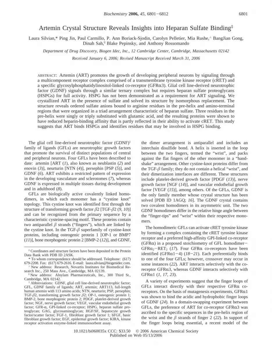
A sequence alignment of mammalian ART orthologuesshows that, while the pre-helix and amino-terminal argininesare highly conserved, arginines that are in the finger-tipregions of human ART are not conserved (Figure 4A). Toprobe the role of scattered positively charged residues ofhuman ART113 in forming an extended heparin-binding site,we compared human and rat ART113 in heparin binding.The calculated pI of rat ART is 10.0, while human ARThas a pI of 11.3. A homology model of rat ART based onthe human ART structure indicates that the positive chargedistribution of human ART and rat ART is similar in thegroove between the two pre-helix regions of the monomer.Rat ART is less positively charged at its finger tips (Figure4B). However, when we compared the binding affinity offull-length human ART to that of full-length rat ART in aheparin-based ELISA, we observed no measurable differencein their affinities for heparin (data not shown), suggestingthat the charge on the human ART finger tips is not requiredfor heparin binding. We then tested the effect of removingthe first nine amino-terminal residues of human ART onheparin binding. The truncated form demonstrated a<10-fold reduction in binding affinity (data not shown), indicatingthat, although unstructured, this region partly contributes toheparin binding. However, the pre-helix region of ART,which is observed to bind sulfates and exhibit high-sequenceconservation, may play a more central role in forming theheparin-binding site.
To determine if the pre-helix region may be part of aheparin-binding site, we separately converted to glutamate
FIGURE 3: Sequence alignment of the human GFL members. The secondary-structural elements within the ART structure are identifiedabove the sequence by designations forR helices (coil) andâ strands (arrows). The ART residues interacting with sulfates and the residuespredicted to comprise the expanded heparan-sulfate-binding site (including Arg14) are black. Residues in the pre-helix region are identifiedby black boxes, and residues in the post-helix region are identified by gray boxes. The ART proline insertion is indicated by asterisks.
6806 Biochemistry, Vol. 45, No. 22, 2006 Silvian et al.
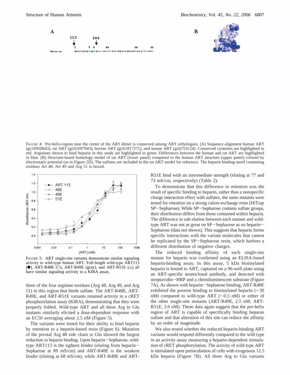
three of the four arginine residues (Arg 48, Arg 49, and Arg51) in this region that binds sulfate. The ART-R48E, ART-R49E, and ART-R51E variants retained activity in a cRETphosphorylation assay (KIRA), demonstrating that they wereproperly folded. Wild-type ART and all three Arg to Glumutants similarly elicited a dose-dependent response withan EC50 averaging about 2.5 nM (Figure 5).
The variants were tested for their ability to bind heparinby retention to a heparin-based resin (Figure 6). Mutationof the pivotal Arg 48 side chain to Glu showed the largestreduction in heparin binding. Upon heparin-Sepharose, wild-type ART113 is the tightest binder (eluting from heparin-Sepharose at 89 mS/cm) and ART-R48E is the weakestbinder (eluting at 68 mS/cm), while ART-R49E and ART-
R51E bind with an intermediate strength (eluting at 77 and73 mS/cm, respectively) (Table 2).
To demonstrate that this difference in retention was theresult of specific binding to heparin, rather than a nonspecificcharge interaction effect with sulfates, the same mutants weretested for retention on a strong cation-exchange resin (HiTrapSP-Sepharose). While SP-Sepharose contains sulfate groups,their distribution differs from those contained within heparin.The difference in salt elution between each mutant and wild-type ART was not as great on SP-Sepharose as on heparin-Sepharose (data not shown). This suggests that heparin formsspecific interactions with the variant molecules that cannotbe replicated by the SP-Sepharose resin, which harbors adifferent distribution of negative charges.
The reduced binding affinity of each single-sitemutant for heparin was confirmed using an ELISA-basedheparin-binding assay. In this assay, 5 kDa biotinylatedheparin is bound to ART, captured on a 96-well plate usingan ART-specific monoclonal antibody, and detected withstreptavidin-HRP and a chemiluminescent substrate (Figure7A). As shown with heparin-Sepharose binding, ART-R48Eexhibited the poorest binding to biotinylated heparin (∼30nM) compared to wild-type ART (∼0.5 nM) or either ofthe other single-site mutants (ART-R49E, 2.5 nM; ART-R51E, 3.0 nM). These data again suggest that the pre-helixregion of ART is capable of specifically binding heparansulfate and that alteration of this site can reduce the affinityby an order of magnitude.
We also tested whether the reduced heparin-binding ARTvariants would respond differently compared to the wild typein an activity assay measuring a heparin-dependent stimula-tion of cRET phosphorylation. The activity of wild-type ARTis stimulated upon preincubation of cells with exogenous 12.5kDa heparin (Figure 7B). All three Arg to Glu variants
FIGURE 4: Pre-helix-region near the center of the ART dimer is conserved among ART orthologues. (A) Sequence alignment human ART(gi|16950643), rat ART (gi|61097943), bovine ART (gi|61817271), and mouse ART (gi|6753124). Conserved cysteines are highlighted inred. Arginines shown to bind heparin in this study are highlighted in green. Differences between the human and rat ART are highlightedin blue. (B) Structure-based homology model of rat ART (lower panel) compared to the human ART structure (upper panel) colored byelectrostatic potential (as in Figure 2D). The sulfates are included in the rat ART model for reference. The heparin binding motif containingresidues Art 48, Art 49 and Arg 51 is boxed.
FIGURE 5: ART single-site variants demonstrate similar signalingactivity to wild-type human ART. Full-length wild-type ART113(b), ART-R48E (O), ART-R49E (gray), and ART-R51E (4) allhave similar signaling activity in a KIRA assay.
Structure of Human Artemin Biochemistry, Vol. 45, No. 22, 20066807

similarly demonstrate enhanced activity upon heparin pre-incubation, as represented by ART-R48E (Figure 7B). Thesestudies suggest that the effect of mutating one amino acidon an extensive heparin-binding surface of the molecule istoo minor to trigger an effect on the signaling cascade.
To further pursue this hypothesis, we constructed an ARTvariant containing all three mutations (ART-R48E,R49E,R51E) to determine the cumulative effect of these mutationsupon heparin binding. We measured its affinity for heparinand its activity in the heparin-dependent stimulation of thecRET phosphorylation assay. The triple mutant boundbiotinylated heparin with at least 3 orders of magnitude loweraffinity than the wild type and 2 orders of magnitude loweraffinity compared to the single ART-R48E mutation (Figure8A and Table 2). It binds to heparin-Sepharose more weaklythan the single-site mutants, eluting at 57 mS/cm. Even inthe absence of exogenous heparin, the triple mutant displayedan improved EC50 value for cRET activation compared towild-type ART (EC50 of 0.8 nM rather than 3.5 nM; Table2). In the presence of heparin, the triple mutant does notdemonstrate any heparin-dependent stimulation of activityas had been observed for wild-type ART (Figure 8B). Thesestudies show that the extensive heparin-binding surface ofthe molecule can be altered by the triple mutant and pointto the central groove of ART as a crucial region of heparinbinding.
DISCUSSION
Heparin binding has a profound effect on the pharmaco-kinetic and pharmacodynamic properties of proteins such as
HGF/SF (42), which was the basis for our investigation ofwhether the heparin-binding site could be identified and re-engineered. In solving the structure of human ART, we
FIGURE 6: ART variants display different heparin-specific chromatic retention profiles. Elution profile of ART variants from a heparin-Sepharose column. The elution peak of wild-type ART113 (D) is compared to that of ART-R48E (A), ART-R49E (C), and ART-R51E (B).
Table 2: Comparison of ART Variants in Heparin Binding andKIRA Assaysa
ARTform
EC50heparinbinding(nM)
Heparin-Sepharose
elutionconductivity
(mS/cm)
KIRAEC50
withoutheparin(nM)
KIRAEC50with
heparin(nM)
KIRA∆EC50
ART 113 0.5 88.9 3.5 0.5 748E 30.0 68.0 2.0 0.5 449E 2.5 77.0 3.0 0.5 651E 3.0 72.5 2.0 0.4 548,49,51E 5000.0 57.0 0.8 0.8 1
a The KIRA ∆EC50 describes the fold improvement in cRETactivation upon the addition of heparin.
FIGURE 7: Behavior of single-site variants relative to wild-type ARTin heparin-binding and heparin-dependent stimulation of cRETphosphorylation. (A) Heparin ELISA for wild-type ART113 (b)and each ART single-site variant: R48E (O), R49E (gray), andR51E (4). Each sample was analyzed in triplicate. Error bars denotethe standard error of the mean (SEM). (B) Stimulation of signalingactivity by ART-R48E upon the addition of heparin. KIRA of wild-type ART113 in the absence (b) and presence (O) of 250 µg/mLheparin compared to ART-R48E in the absence (gray) and presence(4) of 250 µg/mL heparin. Similar curves to ART-R48E wereobserved for ART-R49E and ART-R51E.
6808 Biochemistry, Vol. 45, No. 22, 2006 Silvian et al.

observed a triad of bound sulfate ions that resembled thespacing of sulfates within heparin and identified this regionas a putative heparin-binding site. To test if ART is a heparin-binding protein, we generated point mutations at each ofthese sulfate-binding residues, expressed the proteins, andcharacterized the effects of the mutations in heparin-bindingassays using soluble and immobilized heparin. We demon-strate that ART is a heparin-binding protein and localize thekey residues for heparin binding within the pre-helix regionof ART.
GFLs have predicted heparin-binding site(s) in differentregions of their sequences that would result in their placementon different locations along the surface of the molecule(Figures 2D and 3). Within the two sequence stretchesbetween the “wrist” and “fingers”, ART has a putativeheparin-binding site (CRRARS) in its pre-helix region, whileGDNF (SRSRRL) and NTN (RRVRKLRRER) have puta-tive heparin-binding sites in their post-helix loops. Thisplaces the heparin-binding sites on opposite sides of thestructure. PSP, on the other hand, has no obvious heparin-binding sites in the hinge (Figure 3). The locations and
strengths of these heparin-binding clusters may have differentfunctional consequences on the role of heparin binding tothese cytokines both in vivo and in cell-based assays.
Sequence comparisons of ART orthologues demonstratethat the amino-terminal and pre-helix arginines are highlyconserved and thus could represent a conserved HSPG-binding function. The two sulfate-decorated pre-helix regionsof each human ART dimer are connected by a positivelycharged central groove that is appropriate in dimension tobind heparin sulfate oligosaccharides. Human ART, incontrast to rat ART, has positive charges scattered throughoutits finger regions that are not conserved (Figure 4B). Thesecharge differences do not effect heparin binding. Thisobservation, along with the identification of bound sulfateions in the ART pre-helix region, suggests that heparinbinding is localized to a specific region and not because ofa general electrostatic effect.
Although the amino terminus was not detected in our ARTstructure, we tested its contribution in heparin binding byproteolytically generating a truncated variant (ART104) withresidues 1-9 removed and measuring its ability to bindheparin relative to the full-length ART113. The truncationvariant has a 10-fold lower affinity for heparin (data notshown), suggesting that the amino terminus may also formpart of the heparin-binding site. The amino terminus is likelyto be in close proximity to the pre-helix region and to thepositively charged groove, because the amino-terminal Arg14contributes to sulfate ion binding in conjunction with thearginines identified in the pre-helix region (Figure 1C). NTNand PSP also have positively charged amino-terminalsequences,RRRAGPRRRRAR and RLRR, respectively(Figure 3), that may also act as heparan-sulfate-bindingregions.
The heparin-binding variants provide a tool to determinewhether the heparin-binding and/or signaling activity of ARTcan be modulated. All three single-point mutations reduced5 kDa heparin affinity and decreased retention on heparin-Sepharose, and the triple mutant showed the greatest decreasein affinity (Table 2). Interestingly, among the single-sitemutants, the magnitude of reduction in heparin affinitycorrelates with the location and number of interactions thateach Arg makes with the sulfates in the structure. Arg 48 islocated in the center of the sulfate triangle, and its side chaininteracts with sulfates S3 and S6, while Arg 49 and Arg 51are located on the periphery of the sulfate triangle and eachmakes only one salt bridge to a single sulfate ion. The R48Emutation produced the largest reduction in heparin binding,which is consistent with its central structural role.
While the 5 kDa heparin mixture, used in the heparinELISA, was ideally suited to probe subtle binding effects ofthe single-point mutations in ART, a 12.5 kDa heparinmixture, more closely resembling the long GAG chainsfound in vivo, was used in the receptor activation assay.Given the large amount and size of heparan sulfate on thecell surface in the form of HSPGs, 5 kDa heparin wasconsidered ill-suited to test the effect of heparin binding tothe mutants in the cRET activation assay and 12.5 kDaheparin was used.
The addition of 12.5 kDa heparin resulted in a similarcRET activity enhancement for either wild-type ART or eachof the three ART single-site mutants but resulted in noenhancement for the triple mutant. The triple mutant
FIGURE 8: Behavior of triple-mutant ART relative to the wild typein heparin-binding and heparin-dependent stimulation of cRETphosphorylation. (A) Heparin ELISA with biotinylated 5 kDaheparin for wild-type ART113 (b), ART-R48E (O), and ART-R48E,R49E,R51E (gray). Each sample was analyzed in triplicate.Error bars denote the SEM. (B) Stimulation of signaling activityby the ART triple mutant upon the addition of heparin comparedto that of wild-type ART. KIRA of wild-type ART113 in theabsence (b) and presence (O) of 250µg/mL heparin compared toART-R48E,R49E,R51E in the absence (gray) and presence (4) of250 µg/mL heparin.
Structure of Human Artemin Biochemistry, Vol. 45, No. 22, 20066809

demonstrated enhanced cRET activation over wild-type ARTin the absence of heparin. One hypothesis for the lack ofresponse of the single-site mutations relative to the wild typeis that their modifications were not sufficiently potent orextensive to produce effects on heparin binding that couldbe detected in the cell-signaling assay. Unlike a single-sitemutation, the triple mutation appears to partly mimic heparinbinding to wild-type ART by increasing the potency of cRETactivation and preventing exogenous heparin from bindingand further stimulating activity to the extent observed forthe single-point mutants or wild-type ART. These datasupport the hypothesis that Arg 48, Arg 49, and Arg 51contained within the pre-helix region of ART may play arole in heparin binding.
ART may have a different response to specific cell-surface-bound HSPGs in vivo than to soluble heparin added ex vivo.Heparin and heparan sulfate are not identical. There are subtledifferences in their saccharide sequences. Heparan sulfateis composed of more varied saccharide units. Heparin is moresubstituted with sulfo groups. While heparin has an averageof 2.7 negative charges per disaccharide, there are fewer than2 negative charges for heparan sulfate (43). The amino acidsR48, R49, and R51 may contribute more significantly towardbinding the highly sulfonated heparin than in binding theprotein-bound heparan sulfate. Many growth factors, suchas basic fibroblast growth factor (bFGF), have been shownto be specific for only a small subset of the diverse array ofheparan sulfate variants (44).
The enhancement in cRET activation upon the additionof exogenous heparin to the ART-signaling assay contrastswith studies by Barnett and co-workers or Davies and co-workers, who observed that the addition of exogenousheparan sulfate or heparin to either Madin-Darby caninekidney or PC-12 rat adrenal medullary phaeochromocytomacells inhibits GDNF signaling (29, 45). Their explanationwas that the exogenous GAG prevented the HSPGs at thecell surface from binding GDNF and recruiting GDNF tothe cell membrane. The discrepancy between those studiesand ours is partly reconcilable by identification of differencesbetween the two systems. GDNF and ART have distinct co-receptors and form distinct signaling complexes. In theabsence of heparin, the affinity of ART for GFRR3/cRETis significantly lower than the affinity of GDNF for GFRR-1/cRET, as measured by their abilities to bind to GFRR1-Igor GFRR3-Ig immobilized to a Biacore chip surface (23).Binding to soluble heparin may increase the avidity of ARTor help orient it for proper receptor binding, resulting in anincrease in its apparent binding to its receptors. ART is morebasic than GDNF and may bind more nonspecifically to cellmembranes without the countercharge of heparin. Heparinbinding to wild-type ART or construction of the triple R toE mutation may orient ART properly so that it canproductively bind to its receptors. Last, as demonstrated here,the pre-helix and post-helix heparin recognition motifs inART and GDNF map to opposite surfaces (Figure 2D);binding exogenous heparin may interfere with the recruitmentfunction of HSPGs in the GDNF case but not in the ARTcase. Beyond its potential role in recruiting GFLs to the cellsurface, HSPGs may increase the avidity of GFL binding ordirectly mediate interactions between the GFLs and theirreceptors. While a heparin-dependent receptor binding activ-ity has been reported for bFGF, HGF, a splice variant of
PDGF, heparin-binding epidermal growth factor (EGF),VEGF, and neuregulins, its role in most of these cases isnot well-understood (reviewed in ref44). Further studies arenecessary to specifically address which of these or otherpossible mechanisms of action are relevant for ART.
We favor a model in which certain syndecans or glypicansare upregulated to direct ART to cellular membranes thatcontain signaling receptors and orient it in such a way topromote receptor binding. Syndecans, which are the majorform of membrane-associated HSPGs on many cells, havebeen shown to have variable heparan sulfate glycosamino-glycan structures, and the pattern of syndecan expressiondiffers in a cell-specific manner (46). The potential toregulate growth factor activity by temporally regulating theexpression levels of syndecans is an accepted mechanismof action for other growth factors. Rapid changes in theexpression of syndecans have been observed during criticalperiods of development and correlate with a change inrelative expression levels of growth factors bFGF and acidicfibroblast growth factor (aFGF) (44).
In summary, we have successfully demonstrated that ARTis a heparin-binding protein and employed a rational,structure-based design to reduce heparin binding withoutcompromising biological function. In addition, the crystalstructure of ART shows clear differences from GDNF thatcould account for its selectivity for its specific GFRR co-receptor. The structure-activity studies for ART should beinvaluable in designing novel ART-based therapeutics withimproved pharmaceutical properties.
ACKNOWLEDGMENT
We gratefully acknowledge Craig Ogata at the X4Cbeamline at Brookhaven National Laboratory for help insetting up data collection. We thank Ami Horne for help inconstructing the mutant ART clones and Adrian Whitty andRich Cate for rewarding discussions and a critical readingof the manuscript. The authors declare no financial conflictof interest in publication of this manuscript.
REFERENCES
1. Baloh, R. H., Tansey, M. G., Lampe, P. A., Fahrner, T. J.,Enomoto, H., Simburger, K. S., Leitner, M. L., Araki, T., Johnson,E. M., Jr., and Milbrandt, J. (1998) Artemin, a novel member ofthe GDNF ligand family, supports peripheral and central neuronsand signals through the GFRR3-RET receptor complex,Neuron21, 1291-1302.
2. Rosenblad, C., Gronborg, M., Hansen, C., Blom, N., Meyer, M.,Johansen, J., Dago, L., Kirik, D., Patel, U. A., Lundberg, C.,Trono, D., Bjorklund, A., and Johansen, T. E. (2000)In ViVoprotection of nigral dopamine neurons by lentiviral gene transferof the novel GDNF-family member neublastin/artemin,Mol. CellNeurosci. 15, 199-214.
3. Masure, S., Geerts, H., Cik, M., Hoefnagel, E., van den Kieboom,G., Tuytelaars, A., Harris, S., Lesage, A. S., Leysen, J. E., vander Helm, L., Verhasselt, P., Yon, J., and Gordon, R. D. (1999)Enovin, a member of the glial cell-line-derived neurotrophic factor(GDNF) family with growth promoting activity on neuronal cells.Existence and tissue-specific expression of different splice variants,Eur. J. Biochem. 266, 892-902.
4. Kotzbauer, P. T., Lampe, P. A., Heuckeroth, R. O., Golden, J. P.,Creedon, D. J., Johnson, E. M., Jr., and Milbrandt, J. (1996)Neurturin, a relative of glial-cell-line-derived neurotrophic factor,Nature 384, 467-470.
5. Milbrandt, J., de Sauvage, F. J., Fahrner, T. J., Baloh, R. H.,Leitner, M. L., Tansey, M. G., Lampe, P. A., Heuckeroth, R. O.,
6810 Biochemistry, Vol. 45, No. 22, 2006 Silvian et al.

Kotzbauer, P. T., Simburger, K. S., Golden, J. P., Davies, J. A.,Vejsada, R., Kato, A. C., Hynes, M., Sherman, D., Nishimura,M., Wang, L. C., Vandlen, R., Moffat, B., Klein, R. D., Poulsen,K., Gray, C., Garces, A., Johnson, E. M., Jr., et al. (1998)Persephin, a novel neurotrophic factor related to GDNF andneurturin,Neuron 20, 245-253.
6. Lin, L. F., Doherty, D. H., Lile, J. D., Bektesh, S., and Collins, F.(1993) GDNF: A glial cell line-derived neurotrophic factorfor midbrain dopaminergic neurons,Science 260, 1130-1132.
7. Honma, Y., Araki, T., Gianino, S., Bruce, A., Heuckeroth, R. O.,Johnson, E. M., and Milbrandt, J. (2002) ART is a vascular-derived neurotrophic factor for developing sympathetic neurons,Neuron 35, 267-282.
8. Choi-Lundberg, D. L., and Bohn, M. C. (1995) Ontogeny anddistribution of glial cell line-derived neurotrophic factor (GDNF)mRNA in rat,Brain Res. DeV. Brain Res. 85, 80-88.
9. Daopin, S., Piez, K. A., Ogawa, Y., and Davies, D. R. (1992)Crystal structure of transforming growth factor-â2: An unusualfold for the superfamily,Science 257, 369-373.
10. Schlunegger, M. P., and Grutter, M. G. (1992) An unusual featurerevealed by the crystal structure at 2.2 Å resolution of humantransforming growth factor-â2, Nature 358, 430-434.
11. Griffith, D. L., Keck, P. C., Sampath, T. K., Rueger, D. C., andCarlson, W. D. (1996) Three-dimensional structure of recombinanthuman osteogenic protein 1: Structural paradigm for the trans-forming growth factorâ superfamily,Proc. Nat. Acad. Sci. U.S.A.93, 878-883.
12. Scheufler, C., Sebald, W., and Huelsmeyer, M. (1999) Crystalstructure of human bone morphogenic protein-2 at 2.7 Å resolu-tion, J. Mol. Biol. 287, 103-115.
13. Oefner, C., D’Arcy, A., Winkler, F. K., Eggimann, B., and Hosang,M. (1992) Crystal structure of human platelet-derived growthfactor BB,EMBO J. 11, 3921-3926.
14. McDonald, N. Q., Lapatto, R., Murray-Rust, J., Gunning, J.,Wlodawer, A., and Blundell, T. L. (1991) New protein foldrevealed by a 2.3 Å resolution crystal structure of nerve growthfactor,Nature 354, 411.
15. Muller, Y. A., Christinger, H. W., Keyt, B. A., and deVos, A. M.(1997) The crystal structure of vascular endothelial growth factor(VEGF) refined to 1.93 Å resolution: Multiple copy flexibilityand receptor binding,Structure 5, 1325.
16. Eigenbrot, C., and Gerber, N. (1997) X-ray structure of glial cell-derived neurotrophic factor at 1.9 Å resolution and implicationsfor receptor binding,Nat. Struct. Biol. 4, 435-438.
17. Jing, S., Wen, D., Yu, Y., Host, P. L., Luo, Y., Fang, M., Tamir,R., Antonio, L., Hu, Z., Cupples, R., Louis, J. C., Hu, S., Altrock,B. W., and Fox, G. M. (1996) GDNF-induced activation of theret protein tyrosine kinase is mediated by GDNFR-R, a novelreceptor for GDNF,Cell 85, 113-1124.
18. Baloh, R. H., Tansey, M. G., Golden, J. P., Creedon, D. J.,Heuckeroth, R. O., Keck, C. L., Zimonjic, D. B., Popescu, N. C.,Johnson, E. M., Jr., and Milbrandt, J. (1997) TrnR2, a novelreceptor that mediates neurturin and GDNF signaling through Ret,Neuron 18, 793-802.
19. Jing, S., Yu, Y., Fang, M., Hu, Z., Holst, P. L., Boone, T., Delaney,J., Schultz, H., Zhou, R., and Fox, G. M. (1997) GFRR-2 andGFRR-3 are two new receptors for ligands of the GDNF family,J. Biol. Chem. 272, 33111-33117.
20. Sanicola, M., Hession, C., Worley, D., Carmillo, P., Ehrenfels,C., Walus, L., Robinson, S., Jaworski, G., Wei, H., Tizard, R.,Whitty, A., Pepinsky, R. B., and Cate, R. L. (1997) Glial cellline-derived neurotrophic factor-dependent RET activation can bemediated by two different cell-surface accessory proteins,Proc.Natl. Acad. Sci. U.S.A. 94, 6238-6243.
21. Nishino, J., Mochida, K., Ohfuji, Y.Shimazaki, T., Meno, C.,Ohishi, S., Matsuda, Y., Fujii, H., Saijoh, Y., and Hamada, H.(1999) GFRR3, a component of the artemin receptor, is requiredfor migration and survival of the superior cervical ganglion,Neuron 23, 725-736.
22. Baloh, R. H., Tansey, M. G., Johnson, E. M., Jr., and Milbrandt,J. (2000) Functional mapping of receptor specificity domains ofglial cell line-derived neurotrophic factor (GDNF) family ligandsand production of GFRR-1 RET-specific agonists,J. Biol. Chem.275, 3412-3420.
23. Carmillo, P, Dago, L., Day, E. S., Worley, D. S., Rossomando,A., Walus, L., Orozco, O., Buckley, C., Miller, S., Tse, A., Cate,R. L., Rosenblad, C., Sah, D. W., Gronborg, M., and Whitty, A.(2005) Glial cell line-derived neurotrophic factor (GDNF) receptor
R-1 (GFRR1) is highly selective for GDNF versus artemin,Biochemistry 44, 2545-2554.
24. Eketjall, S., Fainzilber, M., Murray-Rust, J., and Ibanez, C. F.(1999) Distinct structural elements in GDNF mediate binding toGFRR1 and activation of the GFRR1-cRET receptor complex,EMBO J. 18, 5901-5910.
25. Leppa¨nen, V. M., Bespalov, M. M., Runeberg-Roos, P., Puurand,U., Merits, A., Saarma, M., and Goldman, A. (2004) The structureof GFRR1 domain 3 reveals new insights into GDNF binding andRET activation,EMBO J. 23, 1452-1462.
26. Hileman, R. E., Fromm, J. R., Weiler, J. M., and Linhardt, R. J.(1998) Glycosaminoglycan-protein interactions: Definition ofconsensus sites in glycosaminoglycan binding proteins,Bioessays20, 156-167.
27. Rapraeger, A. C., Krufka, A., and Olwin, B. B. (1991) Requirementof heparan sulfate for bFGF-mediated fibroblast growth andmyoblast differentiation,Science 252, 1705-1708.
28. Lyon, M., Deakin, J. A., Rahmoune, H., Fernig, D. G., Nakamura,T., and Gallagher, J. T. (1998) Hepatocyte growth factor/scatterfactor binds with high affinity to dermatan sulfate,J. Biol. Chem.273, 271-278.
29. Barnett, M., W., Fisher, C. E., Perona-Wright G., and Davies, J.A. (2002) Signaling by glial cell line-derived neurotrophic factor(GDNF) requires heparan sulfate glycosaminoglycan,J. Cell Sci.115, 4495-4503.
30. Rickard, S. M., Mummery, R. S., Mulloy, B., and Rider, C. C.(2003) The binding of human glial cell line-derived neurotrophicfactor to heparin and heparan sulfate: Importance of 2-O-sulfategroups and effect on its interaction with its receptor, GFRR1,Glycobiology 13, 419-426.
31. Tanaka, M., Xiao, H., and Kiuchi, K. (2002) Heparin facilitatesglial cell line-derived neurotrophic factor signal transduction,NeuroReport 13, 1913-1916.
32. Leahy, D. J., Hendrickson, W. A., Aukhil, I., and Erickson, H. P.(1992) Structure of a fibronectin type III domain from tenascinphased by MAD analysis of the selenomethionyl protein,Science258, 987-991.
33. Otwinowski, Z., and Minor, W. (1997) Processing X-ray diffractiondata collected in oscillation mode,Methods Enzymol. 276, 307-326.
34. Terwilliger, T. C., and Berendzen, J. (1999) Automated MAD andMIR structure solution,Acta Crystallogr., Sect. D: Biol. Crys-tallogr. 55 (part 4), 849-861.
35. Jones, T. A., Zou, J. Y., Cowan, S. W., and Kjeldgaard, M. (1991)Improved methods for building protein models in electron densitymaps and the location of errors in these models,Acta Crystallogr.,Sect. A: Found. Crystallogr. 47(part 2) 110-119.
36. Brunger, A. T., Adams, P. D., Clore, G. M., DeLano, W. L., Gros,P., Grosse-Kunstleve, R. W., Jiang, J.-S., Kuszewski, J., Nilges,M., Pannu, N. S., Read, R. J., Rice, L. M., Simonson, T., andWarren, G. L. (1998) Crystallography and NMR system: A newsoftware suite for macromolecular structure determination,Acta Crystallogr., Sect. D: Biol. Crystallogr. 54(part 5), 905-921.
37. Vagin, A., and Teplyakov, A. (1997) MOLREP: An automatedprogram for molecular replacement,J. Appl. Crystallogr. 30,1022-1025.
38. Laskowski, R. A., MacArthur, M. W., Moss, D. S., and Thornton,J. M. (1993) Procheck: A program to check stereochemical qualityof protein structures,J. Appl. Crystallogr. 26, 283-290.
39. Gardell, L. R., Wang, R., Ehrenfels, C., Ossipov, M. H.,Rossomando, A. J., Miller, S., Buckley, C., Cai, A. K., Tse, A.,Foley, S. F., Gong, B., Walus, L., Carmillo, P., Worley, D.,Huang, C., Engber, T., Pepinsky, B., Cate, R. L., Vanderah, T.W., Lai, J., Sah, D. W., and Porreca, F. (2003) Multiple actionsof systemic ART in experimental neuropathy,Nat. Med. 9, 1383-1389.
40. Cardin, A. D., and Weintraub, H. J. (1989) Molecular modelingof protein-glycosaminoglycan interactions,Arterioscler. Thromb.Vasc. Biol. 9, 21-32.
41. Pellegrini, L., Burke, D. F., von Delft, F., Mulloy, B., and Blundell,T. L. (2000) Crystal structure of fibroblast growth factorreceptor ectodomain bound to ligand and heparin,Nature 407,1029-1034.
42. Hartmann, G., Prospero, T., Brinkmann, V., Ozcelik, O., Winter,G., Hepple, J., Batley S., Bladt, F., Sachs, M., Birchmeier, C.,
Structure of Human Artemin Biochemistry, Vol. 45, No. 22, 20066811

Birchmeier, W., and Gherardi, E. (1997) Engineered mutants ofHGF/SF with reduced binding to heparan sulphate proteoglycans,decreased clearance and enhanced activityin ViVo, Curr. Biol. 8,125-134.
43. Munoz, E. M, and Linhardt, R. J. (2004) Heparin-binding domainsin vascular biology,Arterioscler. Thromb. Vasc. Biol. 24, 1549-1557.
44. Carey, D. J. (1997) Syndecans: Multifunctional cell-surface co-receptors,Biochem. J. 327, 1-16.
45. Davies, J. A., Yates, E. A., and Turnbull, J. E. (2003) Structuraldeterminants of heparan sulphate modulation of GDNF signaling,Growth Factors 21, 109-119.
46. Kim, C. W., Goldberger, O. A., Gallow, R. L., and Bernfield, M.(1994) Members of the syndecan family of heparin sulfateproteoglycans are expressed in distinct cell-, tissue- and develop-ment-specific patterns,Mol. Biol. Cell 5, 797-805.
BI060035X
6812 Biochemistry, Vol. 45, No. 22, 2006 Silvian et al.


