Embed Size (px)
Citation preview
Alma Mater Studiorum – Università di Bologna
DOTTORATO DI RICERCA IN
Scienze Biotecnologiche e Farmaceutiche
Ciclo XXXI
Settore Concorsuale: 03/D1
Settore Scientifico Disciplinare: CHIM/08
Novel Antitumoral Strategies
Inducing Membrane Lipid and DNA Damage:
Artificial Chemical Nucleases & the ‘ClickGene’ Project
Presentata da: Geοrgia Menounou
Coordinatore Dottorato Supervisore: Prof. Marinella Roberti &
Prof. Santi Mario Spampinato Dr. Carla Ferreri
Esame finale anno 2019
- 2 -
Contents
Abstract ............................................................................................................................................................................ - 5 -
Abbreviations and Acronyms .................................................................................................................................. - 9 -
Chapter 1: Membrane Lipidomics – A Novel Approach in Antitumoral Strategies ...................... - 14 -
1.1. Introduction ............................................................................................................................................................ - 14 -
1.2. Cell Membrane Lipids ............................................................................................................................................ - 18 -
1.3. Oxidative - Free Radical Chemistry and Fatty Acid Moieties ................................................................................ - 22 -
1.4. Transition Metal-Based Drugs................................................................................................................................ - 24 -
1.5. Lipid Geometry ...................................................................................................................................................... - 28 -
1.5.1. Cis-trans Isomerization ........................................................................................................................................... - 30 -
1.5.2. Lipid Peroxidation .................................................................................................................................................. - 34 -
1.6. Cell Membrane and Lipidomic Analysis ................................................................................................................ - 37 -
1.7. Liposomes: a Simulation of Cell Membranes ........................................................................................................ - 38 -
1.8. Thesis Overall Objectives ....................................................................................................................................... - 41 -
Chapter 2: Model Studies of Cu-TPMA-Phen-Induced Lipid Damage in Liposome
Membranes ................................................................................................................................................................... - 42 -
2.1. Materials and Methods ........................................................................................................................................... - 44 -
2.1.1. Liposome Experiments ........................................................................................................................................... - 44 -
2.1.2. GC Analysis ........................................................................................................................................................... - 45 -
2.2. Results and Discussion ........................................................................................................................................... - 46 -
2.2.1. Building-up a Membrane Biomimetic Model ......................................................................................................... - 46 -
2.2.2. MUFA-containing Vesicles .................................................................................................................................... - 48 -
2.2.3. MUFA- and PUFA-containing Vesicles ................................................................................................................. - 59 -
2.3. Conclusions ............................................................................................................................................................ - 64 -
Chapter 3: Trans Lipid Library; Synthesis of Docosahexaenoic Acid (DHA) Monotrans
Isomers - Regioisomer Identification & Model Studies in DHA-Containing Liposomes .............. - 65 -
3.1. Materials and Methods ........................................................................................................................................... - 70 -
3.2. Epoxidation of Methyl All-(Z)-4,7,10,13,16,19-docosahexaenoate ....................................................................... - 72 -
3.2.1. Synthesis of Epoxydocosapentaenoic Acid Methyl Ester (EDP-Me) Regioisomers .............................................. - 72 -
- 3 -
3.2.2. Transformation of EDP-Me Regioisomers to Monotrans DHA-Me Isomers ......................................................... - 75 -
3.3. Synthesis of Monotrans DHA-Me Isomers by Photolysis ...................................................................................... - 77 -
3.4. Isomerization of Fish Oil by Photolysis ................................................................................................................. - 78 -
3.5. Analysis of Monotrans DHA Isomers from Commercially Available Supplements .............................................. - 78 -
3.6. LUVET Preparation................................................................................................................................................ - 79 -
3.6.1. Isomerisation vs Peroxidation of PC in LUVET .................................................................................................... - 79 -
3.7. Results and Discussion ........................................................................................................................................... - 81 -
3.7.1. Dual Synthetic Approach for DHA Transformation and Monotrans DHA Isomer Identification .......................... - 81 -
3.7.2. NMR Study of the EDP Isomers and the Monotrans DHA Isomers ...................................................................... - 85 -
3.7.3. Isomerization of DHA-Containing Fish Oil and Determination of the Monotrans DHA Isomer Content in
Commercially Available Supplements ............................................................................................................................... - 88 -
3.7.4. Model Studies in DHA-Containing Liposomes and Cu-TPMA-Phen-Induced Lipid Damage .............................. - 91 -
3.8. Conclusions ............................................................................................................................................................ - 96 -
Chapter 4: Novel Artificial Chemical Nucleases Induce DNA Cleavage ............................................. - 97 -
4.1. Introduction ............................................................................................................................................................ - 97 -
4.2. Copper Complexes as Intercalators with Endonuclease Reactivity ...................................................................... - 100 -
4.3. AMN’s Reactivity and Formation of Lesions....................................................................................................... - 102 -
4.4. Materials and Methods ......................................................................................................................................... - 105 -
4.4.1. DNA Binding Studies ........................................................................................................................................... - 105 -
4.4.2. DNA Damage Studies .......................................................................................................................................... - 105 -
4.4.3. Identification and Quantification of 8-oxo-Pus and cdPus Lesions In The Presence of AMNs ........................... - 107 -
4.5. Results and Discussion ......................................................................................................................................... - 110 -
4.5.1. DNA Binding Studies - Topoisomerase I-Mediated Relaxation. ......................................................................... - 110 -
4.5.2. Artificial Chemical Nuclease Activity.................................................................................................................. - 114 -
4.5.3. Oxidative DNA Cleavage in the Presence of Non-Covalent DNA Binding Agents ............................................ - 116 -
4.5.4. DNA Damage Mechanism.................................................................................................................................... - 117 -
4.5.5. DNA Repair Enzyme Recognition ....................................................................................................................... - 119 -
4.5.6. AMNs-induced Oxidative Cleavage in Supercoiled Plasmid DNA ..................................................................... - 124 -
4.6. Conclusions .......................................................................................................................................................... - 129 -
Appendix ...................................................................................................................................................................... - 131 -
Part I – Studies on Lipid Damage ................................................................................................................................. - 131 -
5.1. UV-VIS Spectra ................................................................................................................................................... - 131 -
5.2. Gas Chromatography (GC) ................................................................................................................................... - 132 -
5.3. Thin Layer Chromatography (T.L.C.) .................................................................................................................. - 134 -
- 4 -
5.4. NMR Assignments of EDP Isomers Methyl Esters .............................................................................................. - 135 -
5.5. Ag-Thin Layer Chromatography .......................................................................................................................... - 153 -
5.6. Gas Chromatographic Analysis of DHA Isomers................................................................................................. - 155 -
5.7. Gas Chromatography Mass Spectrometry (GC-MS) ........................................................................................... - 157 -
5.8. 13
C – NMR Spectra ............................................................................................................................................... - 161 -
5.9. Analysis of Omega-3 Containing Supplements .................................................................................................... - 163 -
5.10. Gas Chromatographic Identification of FAMEs in 20 % 18:0-22:6 PC/16:0-18:1 PC Composition of Vesicles - 167 -
Part II – Studies on DNA Damage ................................................................................................................................ - 168 -
5.11. Agarose Gel Electrophoresis ............................................................................................................................... - 168 -
5.12. High Pressure Liquid Chromatography (H.P.L.C.) ............................................................................................. - 170 -
5.13. Liquid Chromatography Tandem–mass Spectrometry (LC-MS/MS) ................................................................. - 171 -
Acknowledgements .................................................................................................................................................. - 197 -
- 5 -
Abstract
This PhD project was carried out within the frame of a Marie Curie Network with the
acronym ‘ClickGene’, which focuses on the development of next-generation gene silencing
therapeutics. The ‘ClickGene’ project studies the gene knockout mechanism induced by new
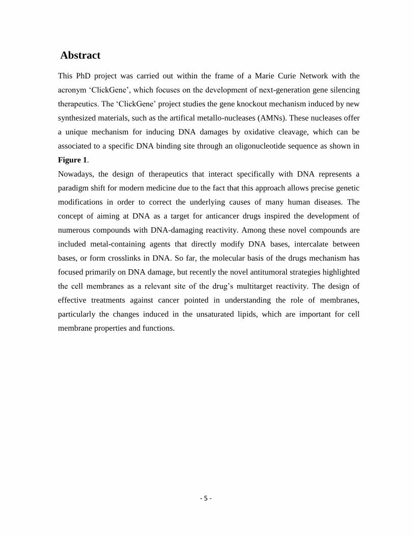
synthesized materials, such as the artifical metallo-nucleases (AMNs). These nucleases offer
a unique mechanism for inducing DNA damages by oxidative cleavage, which can be
associated to a specific DNA binding site through an oligonucleotide sequence as shown in
Figure 1.
Nowadays, the design of therapeutics that interact specifically with DNA represents a
paradigm shift for modern medicine due to the fact that this approach allows precise genetic
modifications in order to correct the underlying causes of many human diseases. The
concept of aiming at DNA as a target for anticancer drugs inspired the development of
numerous compounds with DNA-damaging reactivity. Among these novel compounds are
included metal-containing agents that directly modify DNA bases, intercalate between
bases, or form crosslinks in DNA. So far, the molecular basis of the drugs mechanism has
focused primarily on DNA damage, but recently the novel antitumoral strategies highlighted
the cell membranes as a relevant site of the drug’s multitarget reactivity. The design of
effective treatments against cancer pointed in understanding the role of membranes,
particularly the changes induced in the unsaturated lipids, which are important for cell
membrane properties and functions.
- 6 -
Figure 1. Artificial chemical nucleases induce lipid modifications and targeted oxidative DNA
scission via oxidative/free radical mechanisms.
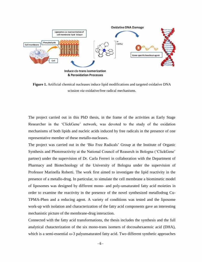
The project carried out in this PhD thesis, in the frame of the activities as Early Stage
Researcher in the ‘ClickGene’ network, was devoted to the study of the oxidation
mechanisms of both lipids and nucleic acids induced by free radicals in the presence of one
representative member of these metallo-nucleases.
The project was carried out in the ‘Bio Free Radicals’ Group at the Institute of Organic
Synthesis and Photoreactivity at the National Council of Reaserch in Bologna (‘ClickGene’
partner) under the supervision of Dr. Carla Ferreri in collaboration with the Department of
Pharmacy and Biotechnology of the University of Bologna under the supervision of
Professor Marinella Roberti. The work first aimed to investigate the lipid reactivity in the
presence of a metallo-drug. In particular, to simulate the cell membrane a biomimetic model
of liposomes was designed by different mono- and poly-unsaturated fatty acid moieties in
order to examine the reactivity in the presence of the novel synthesized metallodrug Cu-
TPMA-Phen and a reducing agent. A variety of conditions was tested and the liposome
work-up with isolation and characterization of the fatty acid components gave an interesting
mechanistic picture of the membrane-drug interaction.
Connected with the fatty acid transformations, the thesis includes the synthesis and the full
analytical chatacterization of the six mono-trans isomers of docosahexaenoic acid (DHA),
which is a semi-essential ω-3 palyunsaturated fatty acid. Two different synthetic approaches
- 7 -
were combined to obtain a full identification of the six mono-trans isomers and the analysis
was based on gas chromatography (GC) and nuclear magnetic resonance (NMR). The
characterization was novel and essential for building-up a molecular reference library. The
usefulness of such library was tested in an analytical application to identify trans content in
nutraceutical formulas.
The second part of the project was carried out at Dublin City University (a ‘ClickGene’
partner) in the National Institute for Cellular Biotechnology under the supervision of
Professor Andrew Kellett. The work focused on artificial metallo-nuclease (AMN) activity
and their ability for precise cleavage of DNA. The cleavage occurs by DNA oxidation,
mediated by reactive oxygen species (ROS) and contributes toward therapeutic utility by
damaging the genome of cancer cells to impede faithful cell replication. In these
experiments, the damage profiles were studied using major groove recognition elements,
spin-trapping scavengers of reactive oxygen species (ROS), and DNA repair enzymes
with glycosylase and/or endonuclease activity. Finally, the DNA damage fragments
produced in DCU were isolated, purified and enzymatically digested to single nucleosides.
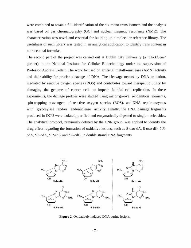
The analytical protocol, previously defined by the CNR group, was applied to identify the
drug effect regarding the formation of oxidative lesions, such as 8-oxo-dA, 8-oxo-dG, 5′R-
cdA, 5′S-cdA, 5′R-cdG and 5′S-cdG, in double strand DNA fragments.
Figure 2. Oxidatively induced DNA purine lesions.
- 8 -
The work carried out in this PhD thesis is multidisciplinary, providing novel insights in
antitumoral strategies and highlighting the dual effect of the drug towards membrane lipids
and DNA. In-depth knowledge of several disciplines was acquired related to cutting-edge
research in liposome formulations, membrane lipidomic approach, nucleic acid chemistry,
gene therapy and diagnostics. Finally, the results contribute in the fields of free radicals in
chemistry, bioinorganic chemistry, biology, pharmacology and medicine.
- 9 -
Abbreviations and Acronyms
A
AB
AcONa
Ag-TLC
Adenine
Abasic Site
Sodium Acetate
Silver Ion Thin Layer Chromatography
AgNO3 Silver Nitrate
ALA α-linolenic Acid
ARA Arachidonic Acid
AMN
AP
BER
BHT
Bipy
BSA
Artificial Metallo-Nuclease
Apurine / Apyrimidine
Base Excision Repair
Butylated Hydroxytoluene
Bipyridine
Bovine Serum Albumin
C6D6
cdA
cdG
Deuterated Benzene
5',8-Cyclo-2'-Deoxyadenosine
5',8-Cyclo-2'-Deoxyguanosine
CDCl3 Deuterated Chloroform
Chol
cPu
Cu-Oda
Cholesterol
Cyclopurine Lesions
Copper-octanedioate
Cu-TPMA-Bipy Copper-tris-(2-pyridylmethyl)amine-2,2′-bipyridine
- 10 -
Cu-TPMA-DPQ Copper-tris-(2-pyridylmethyl)amine-dipyrido[3,2-f:2′,3′h]quinoxaline
Cu-TPMA-DPPZ Copper-tris-(2-pyridylmethyl)amine- dipyrido[3,2-a:2′,3′-c]phenazine
Cu-TPMA-Phen
C
Copper-tris-(2-pyridylmethyl)amine-1,10-phenanthroline
Cytosine
DGLA Dihomo-γ-linolelic Acid
DHA Docosahexanoic Acid
DHA-Me
dI
DLS
DMF
DMPO
DMSO
Docosahexanoic Acid Methyl Ester
Deoxyinosine
Dynamic Light Scattering
Dimethylformamide
5,5-Dimethyl-1-Pyrroline N-Oxide
Dimethyl Sulfoxide
DNA
DSB
dU
Deoxyribonucleic Acid
Double-Strand Break
Deoxyuracil
EDTA Ethylenediamine Tetraacetic Acid
EDP
ET
Endo
ESI
EtBr
Epoxy-Docosapentaenoic Acid
Electron Transfer
Endonuclease
Electrospray Ionisation
Ethidium Bromide
FA Fatty Acid
FAME Fatty Acid Methyl Ester
- 11 -
FaPy
Formamidopyrimidine
FDA Food and Drug Administration
FID
Fpg
Flame Ionization Detector
Formamidopyrimidine DNA Glycosylase
GC Gas Chromatography
GC-MS
G
Gas Chromatography – Mass Spectrometry
Guanine
GUV
hAAG
Giant Unilamellar Vesicle
Human Alkyladenine DNA Glycosylase
HMBC
HEPES
HPLC
Heteronuclear Multiple Bond Correlation
4-(2-Hydroxyethyl)-1-Piperazineethanesulfonic Acid
High Pressure Liquid Chromatography
HSQC Heteronuclear Single-Quantum Coherence
KOH
L
Potassium Hydroxide
Linear
LA
LC/MS
Linoleic Acid
Liquid Chromatography–Mass Spectrometry
LC-PUFA Long Chain Polyunsaturated Fatty Acid
LUV Large Unilamellar Vesicle
LUVET
2-ME
Me-Tar-U
Me
Large Unilamellar Vesicle by Extrusion Technique
2-Mercaptoethanol
Methyltartronylurea
Methyl
- 12 -
m-CPBA
MeOH
MG
MgCl
Meta-Chloroperoxybenzoic Acid
Methanol
Methyl Green
Magnesium Chloride
MLV
MRM
Multilamellar Vesicle
Multiple-Reaction Monitoring
MUFA Monounsaturated Fatty Acid
m/z Mass-to-charge Ratio
Na2SO4
NaCl
Sodium Sulphate
Sodium Chloride
NaHCO3
NEB
Sodium Bicarbonate
New England Biolabs
NH4OH Ammonium Hydroxide
NMR
OH
OC
8-oxo-dA
8-oxo-dG
18:0-22:6 PC
Nuclear Magnetic Resonance
Hydroxy
Open Circular
8-oxo-2'-deoxyadenosine
8-oxo-2'-deoxyguanosine
1-stearoyl-2-docosahexaenoyl-sn-glycero-3-phosphocholine
PC
PD
PDA
PDE
Phosphatidylcholine
Phendione
Photometric Diode Array
Phosphodiesterase
- 13 -
POPC
Pu
1-palmitoyl-2-oleoylphosphatidylcholine
Purines
pUC19
Py
Plasmid DNA
Pyrimidines
Rf Retention Factor
ROS Reactive Oxygen Species
RSS
SC
SDS
SSB
Reactive Sulfur Species
Supercoiled
Sodium Dodecyl Sulfate
Single-Strand Break
SUV
TAE
TBE
Small Unilamellar Vesicle
Tris/Acetate/EDTA
Tris/Borate/EDTA
TFA
T
Trans Fatty Acid
Thymine
TLC Thin Layer Chromatography
UFA
U
UV
V
Unsaturated Fatty Acid
Uracil
Ultraviolet
Volt
WHO
ZnCl2
World Health Organization
Zinc Chloride
- 14 -
Chapter 1: Membrane Lipidomics – A Novel Approach in
Antitumoral Strategies
1.1. Introduction
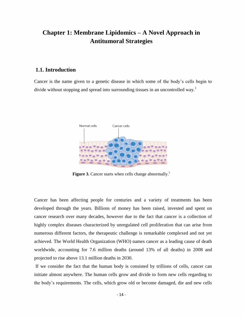
Cancer is the name given to a genetic disease in which some of the body’s cells begin to
divide without stopping and spread into surrounding tissues in an uncontrolled way.1
Figure 3. Cancer starts when cells change abnormally.2
Cancer has been affecting people for centuries and a variety of treatments has been
developed through the years. Billions of money has been raised, invested and spent on
cancer research over many decades, however due to the fact that cancer is a collection of
highly complex diseases characterized by unregulated cell proliferation that can arise from
numerous different factors, the therapeutic challenge is remarkable complexed and not yet
achieved. The World Health Organization (WHO) names cancer as a leading cause of death
worldwide, accounting for 7.6 million deaths (around 13% of all deaths) in 2008 and
projected to rise above 13.1 million deaths in 2030.
If we consider the fact that the human body is consisted by trillions of cells, cancer can
initiate almost anywhere. The human cells grow and divide to form new cells regarding to
the body’s requirements. The cells, which grow old or become damaged, die and new cells
- 15 -
are formed. This physiological process might be disrupted when cancer is developed and
new cells form when they are not needed. In particular, the moment of the cell division
which requires not only chromosome duplication but also membrane phospholipid
recruitment is nowadays looked at with attention, since it is evident that in the tumor
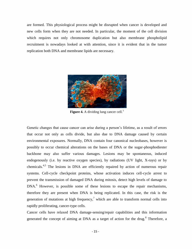
replication both DNA and membrane lipids are necessary.
Figure 4. A dividing lung cancer cell.3
Genetic changes that cause cancer can arise during a person’s lifetime, as a result of errors
that occur not only as cells divide, but also due to DNA damage caused by certain
environmental exposures. Normally, DNA contain four canonical nucleobases, however is
possibly to occur chemical alterations on the bases of DNA or the sugar-phosphodiester
backbone may also suffer various damages. Lesions may be spontaneous, induced
endogenously (i.e. by reactive oxygen species), by radiations (UV light, X-rays) or by
chemicals.4,5
The lesions in DNA are efficiently repaired by action of numerous repair
systems. Cell-cycle checkpoint proteins, whose activation induces cell-cycle arrest to
prevent the transmission of damaged DNA during mitosis, detect high levels of damage to
DNA.6 However, is possible some of these lesions to escape the repair mechanisms,
therefore they are present when DNA is being replicated. In this case, the risk is the
generation of mutations at high frequency,7 which are able to transform normal cells into
rapidly proliferating, cancer-type cells.
Cancer cells have relaxed DNA damage-sensing/repair capabilities and this information
generated the concept of aiming at DNA as a target of action for the drug.8 Therefore, a
- 16 -
variety of anticancer drugs were inspired and the development of compounds, such as
cisplatin,9 doxorubicin,
10 5-fluorouracil,
11 etoposide,
12 and gemcitabine
13 that react
chemically with DNA is growing constantly.14
Among them, the most common metallodrug
is cisplatin with clinical application as chemotherapeutic agent. However, this compound is
severely limited by its toxic side effects such as kidney damage, increased risk of getting an
infection, tiredness, weakness and hair loss.15,16,17
This has spurred chemists to employ
different strategies in the development of new metal-based anticancer agents with different
mechanisms of action. The research interest was aimed towards compounds able to directly
modify DNA bases, intercalate between bases, or form crosslinks in DNA leading to stalled
replication, fork progression and subsequent cell death via apoptosis. 18,19,20
In addition, the scientific advancements revealed the deleterious effects of metal-catalyzed
reactive oxygen species (ROS) in biological systems and relate them with various
pathological conditions, such as cancer. The ROS-dependent activation of apoptotic cell
death, highlight the potential use of ROS as an antitumor agent. This valuable information
led in the development of the next generation therapeutic compounds with ability in a
targeted ROS production in the vicinity of nucleic acids. Complexes of redox active metals,
principally copper and iron, represent an important group of metallodrugs, such as the
artificial chemical nucleases.21
The ligand environment in these complexes allows for the
tuning of charge, redox potential, chirality and geometry to optimize DNA binding. Under
aerobic conditions, or in the presence of appropriate intracellular oxidants, these compounds
can undergo Fenton or Haber–Weiss chemistry to generate reactive oxygen species (ROS).
If the complex is bound to DNA, then there is a high probability that ROS will oxidize
DNA, leading to strand breakages.19
A well-studied example of ROS-active
chemotherapeutic drugs is the metal-activated bleomycin, which is able to induce DNA
single- and double-strand scission via formation of an intermediate metal-complex,
requiring a metal cofactor such as copper or iron.22,23
Due to these properties bleomycin is
used in cancer treatments and Hodgkin's lymphoma. This in cellulo catalytic production of
ROS by copper(II) and iron(II) complexes is recognized as a major mechanistic model in
design of effective inorganic formulated drugs, giving rise in the development of such
promising materials.
- 17 -
The idea of a unique anticancer strategy named “oxidation therapy” has been developed by
inducing oxidative and free radical chemistry for cytotoxic oxystress as cancer treatment.24
Overproduction of highly reactive oxygen metabolites can initiate lethal chain reactions,
which involve oxidation and damage to every structure that is present and is able to be
modified within the cell. Therefore, both DNA and unsaturated lipid components of cell
membranes, which are crucial for cellular integrity and survival, can be damaged giving rise
to senescent, degenerative or fatal lesions in cells offering a powerful therapeutic modality
for future anticancer therapy.25
In fact, the novel antitumoral strategies examine the chemotherapeutic drug’s reactivity as a
synergic effect towards DNA and cell membrane lipids. Recent studies by our group, both in
cell cultures and liposomes as representation of the cell membrane, with the anticancer drug
bleomycin report the formation of a bleomycin-iron complex, highlighting that the free
radical reactivity does not concern only DNA cleavage mechanism, but involves also the
moiety of unsaturated lipids, which are present in membranes.26,27
Cell membrane plays a
crucial role in several biological processes and stress situations can enhance the production
of free radicals.. As described previously, among antitumor active metallodrugs copper(II)
complexes are particularly interesting due to the redox behavior of Cu+2 ⟶ Cu
+1.
28 This in
situ electron transfer can give rise in the formation of other radical species. The presence of
thiols, as biologically relevant reducing agents lead in generation of thiyl radicals, which are
able to cause damages involving among others the membrane lipids.29
These highly active
species are able to trigger a cascade of reactions, leading in permanent modifications on the
structure of the membrane constituents.30
The monounsaturated fatty acids (MUFAs) and the
polyunsaturated fatty acids (PUFAs) are highly abundant and essential structural
components of the cell membrane with important effects on the structure and physical
properties of localized membrane domains. MUFA and PUFA moieties contain one or more
double bonds respectively, with cis geometry in their backbone. In the presence of sulfur-
centered radicals, the intraconversion of the cis geometry of unsaturated fatty acids moieties
is possible to occur and as a result, the corresponding trans lipids are formed. This change in
membrane architecture is possible to reduce the membrane permeability and fluidity, affect
the membrane-associated enzymes and altered the ion transport. In addition, the PUFA
moieties can be partitioned in oxidative and lipid peroxidation pathways leading in a
- 18 -
decreased PUFA content in the membrane.31
The structural differences in the fatty acid
moieties result in alterations and direct the cell to an apoptotic pathway. The changes in the
membrane lipid compositions can be monitored by fatty acid-based lipidomics.32
This type
of analysis in the research field of lipid structures and functions represents a powerful
diagnostic tool for detection of membrane impairments and correlate them with pathological
conditions.
Finally, model studies using liposomes as a simpler representation of the cell membrane
allow the lipid damage evaluation on the monounsaturated fatty acids (MUFA) and
polyunsaturated fatty acids (PUFA)-containing phospholipids, under biologically related
free radical and oxidative conditions.33
This approach allows an insight of the metallodrug’s
chemical reactivity that mediates ROS damage to the moieties of phospholipids, in the
presence of reductant in order to achieve the electron transfer and the generation of radicals.
By building up biomimetic models, the radical and oxidative processes can be followed up
using a variety of reaction conditions, thereby providing a molecular basis to observe and
study these processes.34
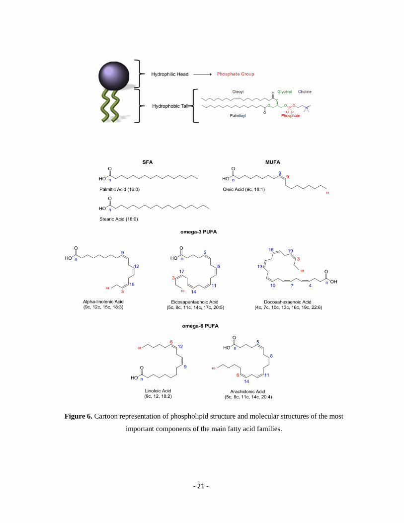
1.2. Cell Membrane Lipids
The membranes constitute the cell boundaries, as well as the boundaries of organelles within
the cell. Membranes are consisted by a hydrophobic matrix, formed by an oriented double
layer of phospholipids to which proteins are bound in different forms. In 1935, Danielli and
Davson proposed as representation, the model of a double layer of phospholipids as the basic
structural element of membranes.35
It was assumed that all membranes had a uniform
thickness and a constant lipid-protein ratio with symmetrical internal – external surfaces. Due
to limitations of this model in 1972 Singer and Nicolson proposed the structure and dynamics
of biological membranes with the view of a ‘fluid mosaic’ model as framed in Figure 5.36
- 19 -
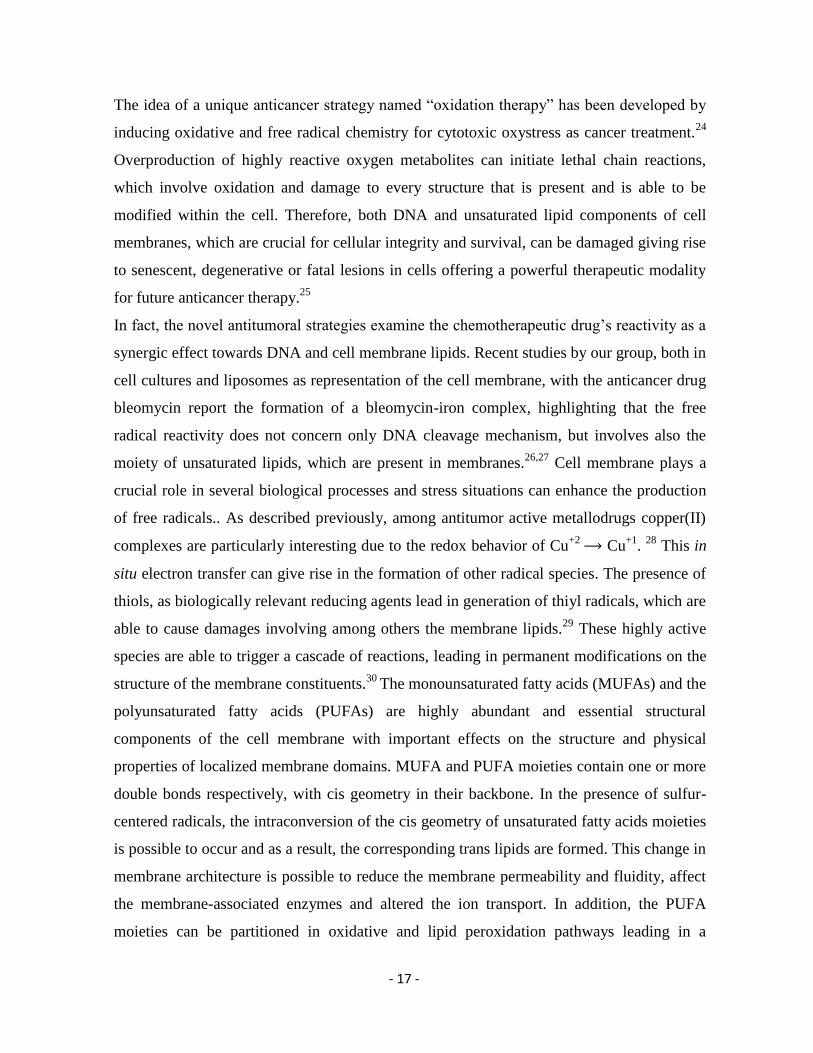
Figure 5. Cartoon representation of cell membranes as ‘fluid mosaic’.37
Cell membrane is a mixture of fluid state lipids, which are organized in a double layer or
bilayer, along with proteins and other molecules. As far as concerns the membrane lipids, they
are amphipathic and they possess both a hydrophobic and a hydrophilic moiety. This occurs in
phospholipids, glycolipids and sterols.38
Because of this amphipathic character, in an aqueous
medium they can organize themselves on both sides of an imaginary plane, with the
hydrophobic portions facing each other and the polar moieties oriented to the outer, aqueous
space.39
Both lipids and proteins are in constant motion (hence the fluid mosaic name
mentioned above) and they rotate around their long axis, under physiological condition. The
most abundant lipid components are the phospholipids consisting of a L-glycerol molecule, in
which the hydroxyl groups in positions 1 and 2 are esterified with two fatty acids and the
hydroxyl group in position 3 is esterified with a phosphate group that is bound to small polar
molecules such as choline, serine, ethanolamine or inositol. As presented in Figure 6, in the
phospholipid structure the hydrophobic tails are consisted by fatty acid residues with different
lengths of aliphatic chain, which can be either saturated or unsaturated.40
Taking into account
the structure of the hydrocarbon chain the fatty acids can be distinguished in:
Saturated fatty acids (SFAs), when the aliphatic chain contains no double bond
Monounsaturated fatty acids (MUFAs), which have one double bond in the fatty acid
chain with all of the remained carbon atoms being single-bonded and
Polyunsaturated fatty acids (PUFAs), in which the constituent hydrocarbon chain
possesses two or more carbon–carbon double bonds.41
- 20 -
In fact, depending on the position of the double bonds, PUFAs are either omega-3 or -6
methylene-interrupted fatty acids.42
It is important to mention that this classification of
PUFAs in two different families reflects the fact that they are synthetized starting from a
common precursor for each family. The precursor for the series of omega-3 linoleic acid (LA,
9c, 12c, 18:2) and for the omega-6 series is a-linolenic acid (ALA, 9c, 12c, 15c, 18:3). Both
fatty acids, linoleic and a-linolenic, are considered essentials, which means that they cannot
be produced via enzymatic pathways, but need to be taken by the diet.43,44
- 21 -
Figure 6. Cartoon representation of phospholipid structure and molecular structures of the most
important components of the main fatty acid families.
- 22 -
1.3. Oxidative - Free Radical Chemistry and Fatty Acid Moieties
There are several definitions of ‘free radical’, as well as debates about whether the word ‘free’
is superfluous. A simple definition is that a free radical is any specie capable of independent
existence (hence the term ‘free’) that contains one or more unpaired electrons. The presence
of one or more unpaired electrons usually causes free radicals to be attracted slightly to a
magnetic field (i.e. to be paramagnetic) and sometimes makes them highly reactive, although
the chemical reactivity of radicals varies widely. Many free radicals exist in living systems,
although most molecules in vivo are non-radicals. If the above rule is taken into account then
the diatomic oxygen molecule can be considered itself a free radical since it has two unpaired
electrons, each located in a different π* antibonding orbital. This is the most stable state or
ground state of O2 and is the form it takes in the air around us. If one electron is added to the
ground-state O2 molecule, it enters one of the π* antibonding orbitals and the product is
superoxide radical, O2•–
. Addition of another electron to O2•–
will give O2–2
the peroxide ion, a
non-radical. Species, like the ones described previously, that derived from molecular oxygen
and they are more reactive than O2 itself, are well known as reactive oxygen species (ROS).45
The term includes not only superoxide and some other oxygen radicals, but also some non-
radical derivatives of O2, such as H2O2 and hypochlorous acid (HOCl). Hence, all oxygen
radicals are ROS, but not all ROS are oxygen radicals. ‘Reactive’ is a relative term; O2•–
and
H2O2 are selective in their reactions with biological molecules, leaving most of them
unscathed, whereas OH• attacks everything around it.
46 The term reactive species has been
expanded to include reactive nitrogen, chlorine, bromine, iron and sulphur species.
Oxidative stress occurs when the production of reactive oxygen species (ROS) is greater than
the body's ability to detoxify the reactive intermediates.47
This imbalance leads to oxidative
damage to DNA, lipids, proteins, molecules and genes within the body.48
Since the body is
incapable of keeping up with the detoxification of the free radicals, the damage continues to
spread. The body naturally produces antioxidants like superoxide dismutase, catalase and an
assortment of peroxidase enzymes, as a means of defending itself against free radicals.49
The
antioxidants neutralize the free radicals, thereby rendering them harmless to other cells.
Antioxidants have the remarkable ability to repair damaged molecules by donating hydrogen
atoms to the molecules. Some antioxidants even have a chelating effect decreasing the free
radical production that is catalyzed by metals.50
In this situation, the antioxidant contains the
- 23 -
metal molecules so strongly that the chemical reaction necessary to create a free radical never
occurs.
Production of O2•–
in the presence of NADH or NADPH can occur for the presence of an
NADPH oxidase enzyme. The O2•–
/H2O2 produced have the potential to damage nuclear
components, in particular by conversion into OH• the damage to DNA and consequent
mutation can occur.51,52
Plasma membranes contain redox systems that transfer electrons from
NADH to external electron acceptors, such as ascorbate but there are cases that also the cell
membranes suffer from permanent modifications of their components. The above data
evidenced the necessity to apply a novel strategy when considering the oxidative – free
radical chemistry within the cell, since targets of the produced radicals is probable to be both
DNA and membrane lipids each one resulting in a modification leading in pathologic
conditions or in the case of cancer tumors to induce an apoptotic response.53,54
- 24 -
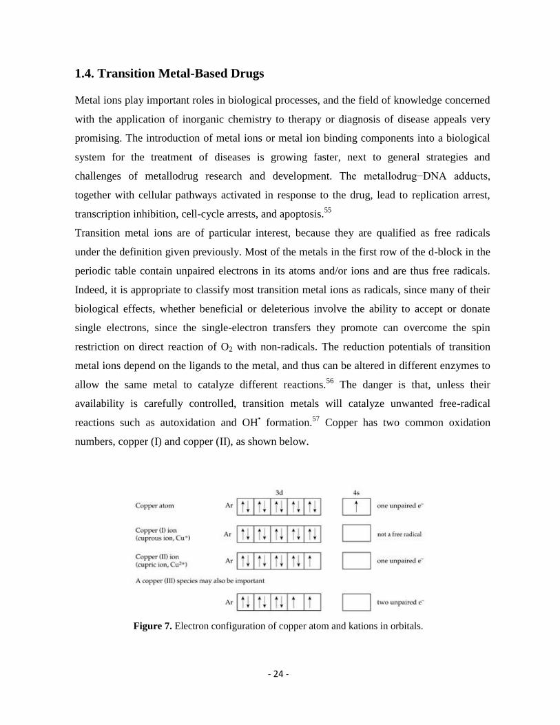
1.4. Transition Metal-Based Drugs
Metal ions play important roles in biological processes, and the field of knowledge concerned
with the application of inorganic chemistry to therapy or diagnosis of disease appeals very
promising. The introduction of metal ions or metal ion binding components into a biological
system for the treatment of diseases is growing faster, next to general strategies and
challenges of metallodrug research and development. The metallodrug−DNA adducts,
together with cellular pathways activated in response to the drug, lead to replication arrest,
transcription inhibition, cell-cycle arrests, and apoptosis.55
Transition metal ions are of particular interest, because they are qualified as free radicals
under the definition given previously. Most of the metals in the first row of the d-block in the
periodic table contain unpaired electrons in its atoms and/or ions and are thus free radicals.
Indeed, it is appropriate to classify most transition metal ions as radicals, since many of their
biological effects, whether beneficial or deleterious involve the ability to accept or donate
single electrons, since the single-electron transfers they promote can overcome the spin
restriction on direct reaction of O2 with non-radicals. The reduction potentials of transition
metal ions depend on the ligands to the metal, and thus can be altered in different enzymes to
allow the same metal to catalyze different reactions.56
The danger is that, unless their
availability is carefully controlled, transition metals will catalyze unwanted free-radical
reactions such as autoxidation and OH• formation.
57 Copper has two common oxidation
numbers, copper (I) and copper (II), as shown below.
Figure 7. Electron configuration of copper atom and kations in orbitals.
- 25 -
Copper (I) readily undergoes self-reaction if it accumulates; Cu+ + Cu
+ → Cu + Cu
2+.
The one-electron difference between Cu+
and Cu2+
, or Cu2+
and Cu (III), allows copper to
promote radical reactions. Under appropriate conditions, for example, copper ions interact
rapidly with O2•–
.
Cu2+
+ O2•–
→ Cu+ +O2 k = (5 – 8)×10
9 M
–1 s
–1
H+ + Cu
+ + HO2
• → Cu
2+ + H2O2 k ≈ 10
9 M
–1 s
–1
O2•–
+ Cu+ + H2O→ Cu
2+ + OH
– + HO
–2 k ≈ 10
9 M
–1 s
–1
Net: O2•–
+ O2•–
+ 2H+ → H2O2 + O2
The copper, by changing its oxidation number, is catalyzing the conversion of two O2•–
radicals and two H+ ions to H2O2 and O2. It is known, that several copper ions chelate - react
with biological molecules. Among these bioactive molecules are the thiols, which act as
antioxidants. It is highly probable for thiols to react with copper in vivo, leading in formation
of free radicals. Thiyl radicals are formed when thiols react with carbon-centered radicals,
with transition metal ions or with several oxygen radicals, including OH•, RO
•, CO3
•–, RO2
•.58
For example,
RSH + OH• → RS
• + H2O
RSH + RO2• → RS
• + ROOH
RSH + Cu2+
→ RS• + Cu
+ + H
+
Once the thiyl radical in the presence of copper (II) is generated, is often assumed that RS•
radical is essentially inert and disappear by dimerization,59
e.g. for the glutathione thiyl
radical,
GS• +GS
• →GSSG k = 1.5 × 10
9M
–1s
–1
Copper can participate in oxidative or free radical pathways giving rise in formation of ROS,
which are able to initiate lipid and DNA damage.60
Due to these properties, a variety of
- 26 -
copper-coordination complexes with nuclease reactivity and site-directed DNA cleavage has
been developed. Artificial chemical nucleases are biomimetic and rely on simple systems,
which incorporate well-known enzymatic properties, such as metal coordination, general acid-
base catalysis, and nucleophilic attack to hydrolyze activated phosphodiesters. A well-studied
chemical nuclease is 1,10-phenanthroline-copper, and the ability of this redox-active
coordination complex to cleave DNA has been established. One of the striking features of the
reaction was the specificity of 1,10-phenanthroline ligand and the absolute requirement for
copper ion. In fact, both the redox chemistry of the copper ion as well as the extrinsic
stereoelectronic and steric properties of phenanthroline copper complexes are of equal
importance in the mechanism.61
This new generation of drugs does not concern only copper, a wide range of redox
metallodrugs has been established in anticancer therapies. Among them, the most popular is
cisplatin due to the ability to crosslink with the purine bases on the DNA, interfering with
DNA repair mechanisms, causing DNA damage, and subsequently inducing apoptosis in
cancer cells.62
Nowadays, cisplatin is used as a chemotherapy medication to treat a number of
cancers.
Finally yet importantly, bleomycin is another drug that is used and it has a unique mechanism
of antitumor activity. Bleomycin has both metal binding and DNA binding sites and its
activity to generate radical species, such as hydroxyl radicals, in the presence of ferrous or
copper(I) ions and molecular oxygen, is known since long time. The mechanism of its action
includes the production of oxidative DNA strand scission via formation of OH radicals The
mechanism includes the binding of five nitrogen atoms with divalent metals such as iron.
Molecular oxygen, bound by the iron, can produce highly reactive free radicals and Fe(III).
The free radicals produce DNA single-strand breaks at 3'-4' bonds in deoxyribose.63
In conclusion, platinum-based drugs such as cisplatin are powerful anticancer agents, they
have undesirable side effects and are effective against only a few kinds of cancers.64
There is,
therefore, a need for new drugs with an improved spectrum of efficacy and lower toxicity.
Complexes of copper, gold and silver (coinage metals) are potential candidates to fulfill this
need.65
Although metallodrugs are used in antitumoral therapies, the mechanism of reactivity
is not yet fully explored in many cases. A precise understanding of both the DNA binding
properties and the chemical reactivity of any scission reagent is essential for site selectivity
- 27 -
potential application of more synthetic restriction endonucleases. Needless to be mentioned,
the development of anticancer drugs based on these metals is currently a very active field.
- 28 -
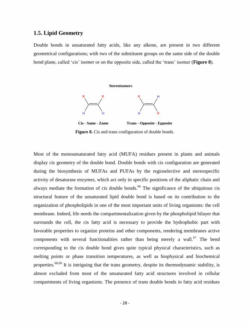
1.5. Lipid Geometry
Double bonds in unsaturated fatty acids, like any alkene, are present in two different
geometrical configurations; with two of the substituent groups on the same side of the double
bond plane, called ‘cis’ isomer or on the opposite side, called the ‘trans’ isomer (Figure 8).
Figure 8. Cis and trans configuration of double bonds.
Most of the monounsaturated fatty acid (MUFA) residues present in plants and animals
display cis geometry of the double bond. Double bonds with cis configuration are generated
during the biosynthesis of MUFAs and PUFAs by the regioselective and stereospecific
activity of desaturase enzymes, which act only in specific positions of the aliphatic chain and
always mediate the formation of cis double bonds.66
The significance of the ubiquitous cis
structural feature of the unsaturated lipid double bond is based on its contribution to the
organization of phospholipids in one of the most important units of living organisms: the cell
membrane. Indeed, life needs the compartmentalization given by the phospholipid bilayer that
surrounds the cell, the cis fatty acid is necessary to provide the hydrophobic part with
favorable properties to organize proteins and other components, rendering membranes active
components with several functionalities rather than being merely a wall.67
The bend
corresponding to the cis double bond gives quite typical physical characteristics, such as
melting points or phase transition temperatures, as well as biophysical and biochemical
properties.68,69
It is intriguing that the trans geometry, despite its thermodynamic stability, is
almost excluded from most of the unsaturated fatty acid structures involved in cellular
compartments of living organisms. The presence of trans double bonds in fatty acid residues
H
R R
H
Cis - Same - Zame
H
R H
R
Trans - Opposite - Epposite
Stereoisomers
- 29 -
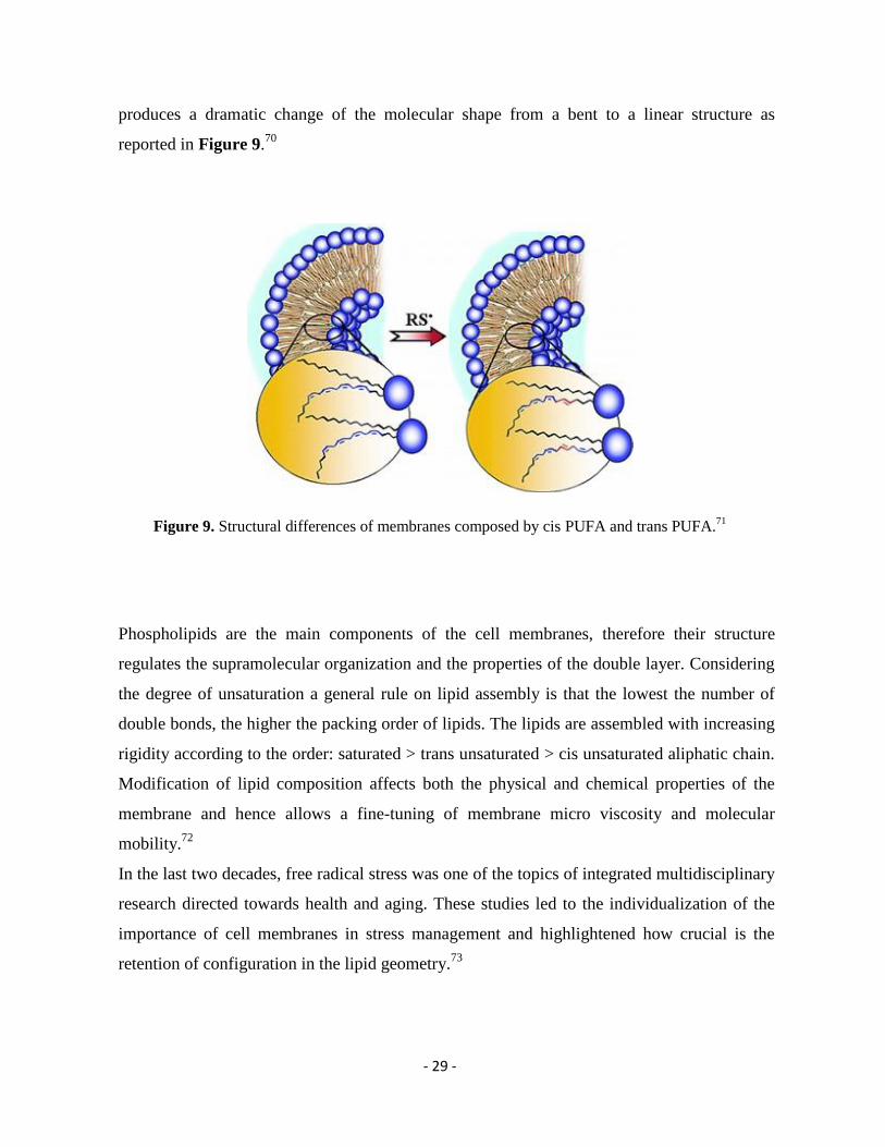
produces a dramatic change of the molecular shape from a bent to a linear structure as
reported in Figure 9.70
Figure 9. Structural differences of membranes composed by cis PUFA and trans PUFA.71
Phospholipids are the main components of the cell membranes, therefore their structure
regulates the supramolecular organization and the properties of the double layer. Considering
the degree of unsaturation a general rule on lipid assembly is that the lowest the number of
double bonds, the higher the packing order of lipids. The lipids are assembled with increasing
rigidity according to the order: saturated > trans unsaturated > cis unsaturated aliphatic chain.
Modification of lipid composition affects both the physical and chemical properties of the
membrane and hence allows a fine-tuning of membrane micro viscosity and molecular
mobility.72
In the last two decades, free radical stress was one of the topics of integrated multidisciplinary
research directed towards health and aging. These studies led to the individualization of the
importance of cell membranes in stress management and highlightened how crucial is the
retention of configuration in the lipid geometry.73
- 30 -
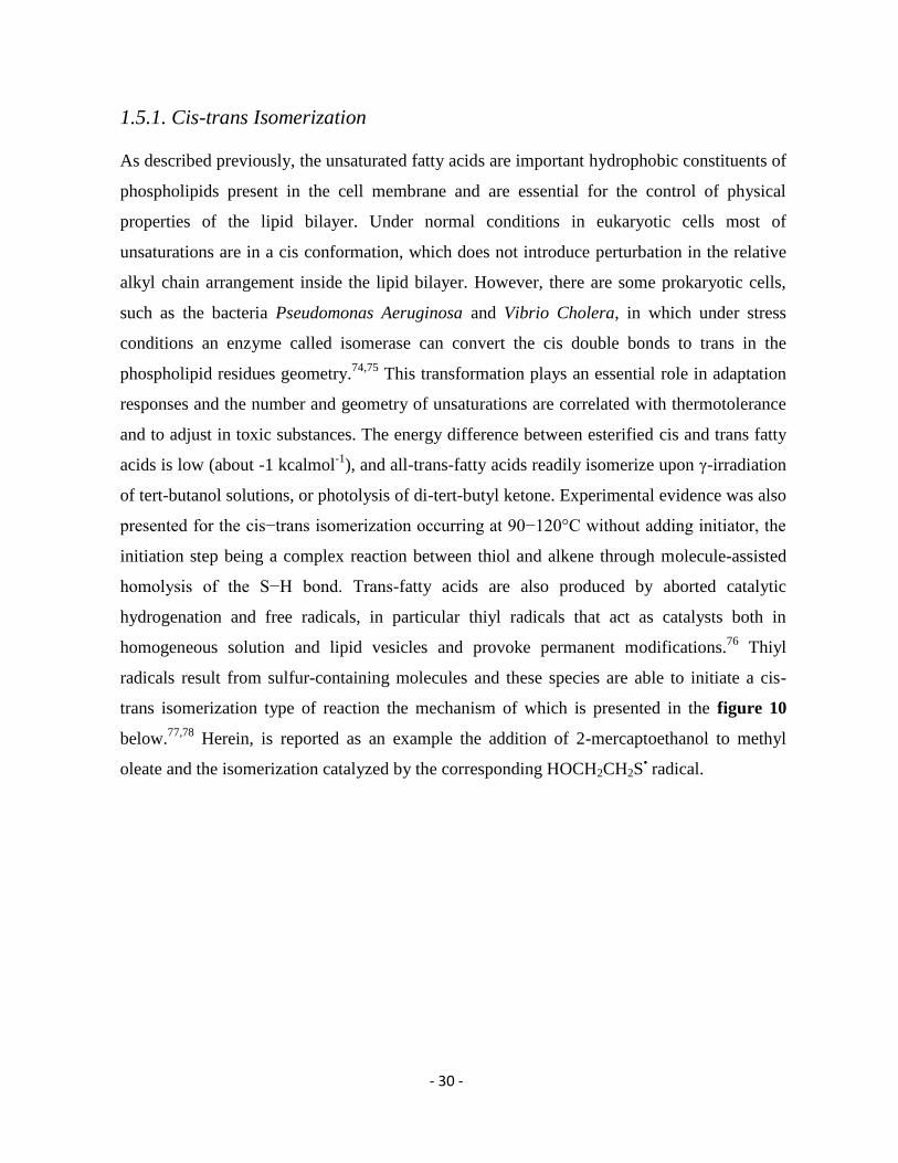
1.5.1. Cis-trans Isomerization
As described previously, the unsaturated fatty acids are important hydrophobic constituents of
phospholipids present in the cell membrane and are essential for the control of physical
properties of the lipid bilayer. Under normal conditions in eukaryotic cells most of
unsaturations are in a cis conformation, which does not introduce perturbation in the relative
alkyl chain arrangement inside the lipid bilayer. However, there are some prokaryotic cells,
such as the bacteria Pseudomonas Aeruginosa and Vibrio Cholera, in which under stress
conditions an enzyme called isomerase can convert the cis double bonds to trans in the
phospholipid residues geometry.74,75
This transformation plays an essential role in adaptation
responses and the number and geometry of unsaturations are correlated with thermotolerance
and to adjust in toxic substances. The energy difference between esterified cis and trans fatty
acids is low (about -1 kcalmol-1
), and all-trans-fatty acids readily isomerize upon γ-irradiation
of tert-butanol solutions, or photolysis of di-tert-butyl ketone. Experimental evidence was also
presented for the cis−trans isomerization occurring at 90−120°C without adding initiator, the
initiation step being a complex reaction between thiol and alkene through molecule-assisted
homolysis of the S−H bond. Trans-fatty acids are also produced by aborted catalytic
hydrogenation and free radicals, in particular thiyl radicals that act as catalysts both in
homogeneous solution and lipid vesicles and provoke permanent modifications.76
Thiyl
radicals result from sulfur-containing molecules and these species are able to initiate a cis-
trans isomerization type of reaction the mechanism of which is presented in the figure 10
below.77,78
Herein, is reported as an example the addition of 2-mercaptoethanol to methyl
oleate and the isomerization catalyzed by the corresponding HOCH2CH2S• radical.
- 31 -
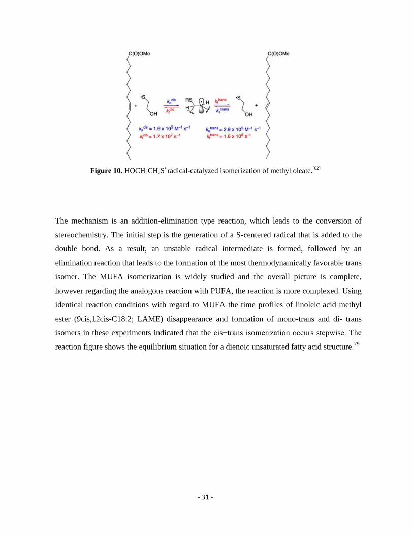
Figure 10. HOCH2CH2S• radical-catalyzed isomerization of methyl oleate.
[62]
The mechanism is an addition-elimination type reaction, which leads to the conversion of
stereochemistry. The initial step is the generation of a S-centered radical that is added to the
double bond. As a result, an unstable radical intermediate is formed, followed by an
elimination reaction that leads to the formation of the most thermodynamically favorable trans
isomer. The MUFA isomerization is widely studied and the overall picture is complete,
however regarding the analogous reaction with PUFA, the reaction is more complexed. Using
identical reaction conditions with regard to MUFA the time profiles of linoleic acid methyl
ester (9cis,12cis-C18:2; LAME) disappearance and formation of mono-trans and di- trans
isomers in these experiments indicated that the cis−trans isomerization occurs stepwise. The
reaction figure shows the equilibrium situation for a dienoic unsaturated fatty acid structure.79
- 32 -
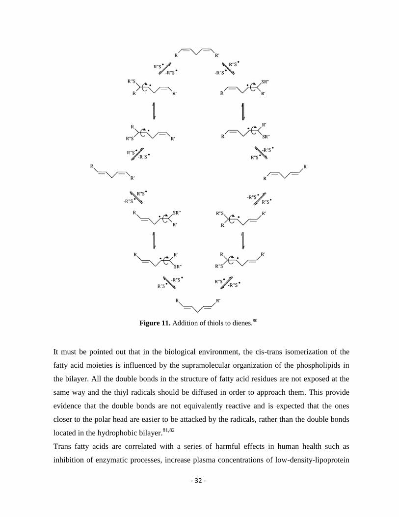
Figure 11. Addition of thiols to dienes.80
It must be pointed out that in the biological environment, the cis-trans isomerization of the
fatty acid moieties is influenced by the supramolecular organization of the phospholipids in
the bilayer. All the double bonds in the structure of fatty acid residues are not exposed at the
same way and the thiyl radicals should be diffused in order to approach them. This provide
evidence that the double bonds are not equivalently reactive and is expected that the ones
closer to the polar head are easier to be attacked by the radicals, rather than the double bonds
located in the hydrophobic bilayer.81,82
Trans fatty acids are correlated with a series of harmful effects in human health such as
inhibition of enzymatic processes, increase plasma concentrations of low-density-lipoprotein

- 33 -
cholesterol and reduce concentrations of high-density-lipoprotein, which results in relation
between the blood lipid concentrations and the risk of coronary artery disease.83
- 34 -
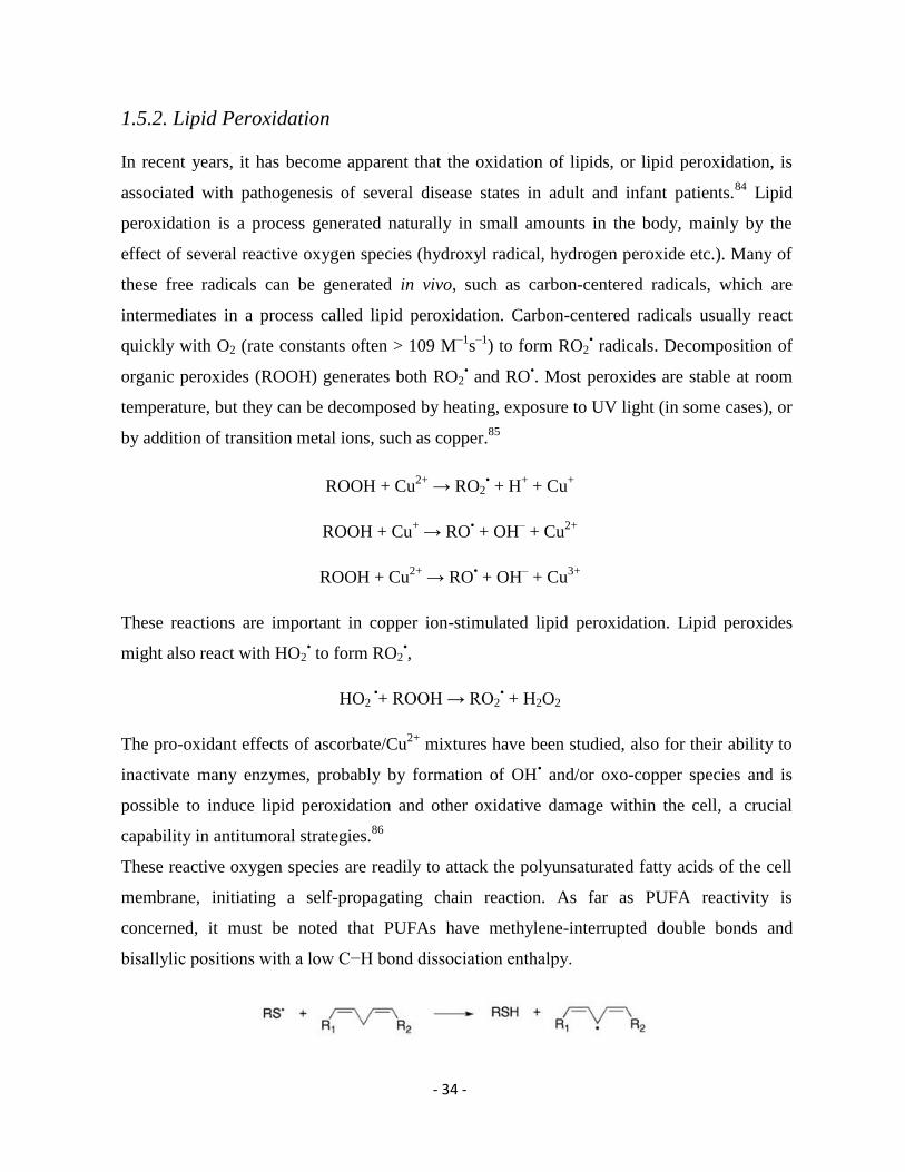
1.5.2. Lipid Peroxidation
In recent years, it has become apparent that the oxidation of lipids, or lipid peroxidation, is
associated with pathogenesis of several disease states in adult and infant patients.84
Lipid
peroxidation is a process generated naturally in small amounts in the body, mainly by the
effect of several reactive oxygen species (hydroxyl radical, hydrogen peroxide etc.). Many of
these free radicals can be generated in vivo, such as carbon-centered radicals, which are
intermediates in a process called lipid peroxidation. Carbon-centered radicals usually react
quickly with O2 (rate constants often > 109 M–1
s–1
) to form RO2• radicals. Decomposition of
organic peroxides (ROOH) generates both RO2• and RO
•. Most peroxides are stable at room
temperature, but they can be decomposed by heating, exposure to UV light (in some cases), or
by addition of transition metal ions, such as copper.85
ROOH + Cu2+
→ RO2• + H
+ + Cu
+
ROOH + Cu+ → RO
• + OH
– + Cu
2+
ROOH + Cu2+
→ RO• + OH
– + Cu
3+
These reactions are important in copper ion-stimulated lipid peroxidation. Lipid peroxides
might also react with HO2• to form RO2
•,
HO2 •+ ROOH → RO2
• + H2O2
The pro-oxidant effects of ascorbate/Cu2+
mixtures have been studied, also for their ability to
inactivate many enzymes, probably by formation of OH• and/or oxo-copper species and is
possible to induce lipid peroxidation and other oxidative damage within the cell, a crucial
capability in antitumoral strategies.86
These reactive oxygen species are readily to attack the polyunsaturated fatty acids of the cell
membrane, initiating a self-propagating chain reaction. As far as PUFA reactivity is
concerned, it must be noted that PUFAs have methylene-interrupted double bonds and
bisallylic positions with a low C−H bond dissociation enthalpy.
- 35 -
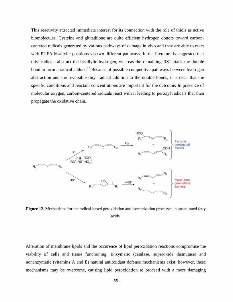
This reactivity attracted immediate interest for its connection with the role of thiols as active
biomolecules. Cysteine and glutathione are quite efficient hydrogen donors toward carbon-
centered radicals generated by various pathways of damage in vivo and they are able to react
with PUFA bisallylic positions via two different pathways. In the literature is suggested that
thiyl radicals abstract the bisallylic hydrogen, whereas the remaining RS• attack the double
bond to form a radical adduct.87
Because of possible competitive pathways between hydrogen
abstraction and the reversible thiyl radical addition to the double bonds, it is clear that the
specific conditions and reactant concentrations are important for the outcome. In presence of
molecular oxygen, carbon-centered radicals react with it leading to peroxyl radicals that then
propagate the oxidative chain.
Figure 12. Mechanisms for the radical-based peroxidation and isomerization processes in unsaturated fatty
acids.
Alteration of membrane lipids and the occurence of lipid peroxidation reactions compromise the
viability of cells and tissue functioning. Enzymatic (catalase, superoxide dismutase) and
nonenzymatic (vitamins A and E) natural antioxidant defense mechanisms exist; however, these
mechanisms may be overcome, causing lipid peroxidation to proceed with a more damaging
- 36 -
effect.88
Since lipid peroxidation is a self-propagating chain-reaction, the initial oxidation of only
a few lipid molecules can result in significant tissue damage.89
Extensive research in the field of
lipid peroxidation determined the alteration of the physiological functions in cell membranes and
the important role in cellular membrane damage.90
Lipid peroxidation has been implicated in
disease states such as atherosclerosis, asthma, Parkinson's disease, kidney damage and others.91
Beyond ROS involvement in carcinogenesis, increased ROS level can inhibit tumor cell growth
via formation of their products which are also the lipid peroxides. Indeed, in tumors in advanced
stages, a further increase of oxidative stress, such as that occurs when using several anticancer
drugs and radiation therapy, can overcome the antioxidant defenses of cancer cells and drive them
to apoptosis.92
- 37 -
1.6. Cell Membrane and Lipidomic Analysis
The healthcare approach has improved a lot in the last decades due to the understanding of
signaling and response pathways at the cellular level. As described previously, it is of high
importance the fatty acid residues of phospholipids to maintain their natural assembly and
structural integrity. Membranes are necessary to not only divide and form the living cell, but
represent also a crucial site for regulating exchange of nutrients, oxygen and stimuli inside the
cell or even between different cells.93,94
The cell membrane is a relevant site for receiving and
emitting signals, inducing an adaptive response in the lipid constituents. The division of lipids
into lipid classes reinforce the knowledge regarding the membrane composition and its regulation
by the appropriate mix of fatty acid residues. The total mapping of the membrane components
allowed a different perspective for understanding the relationship between structures and
functions, as well as their connection to the status of the organism in a more dynamic and
functional way. The new technological advancements gave birth in ‘Lipidomics’ that took the
place of the “old” lipidology with a suffix “-omics,” in order to express the correlation with other
“-omics” technologies, such as genomics and proteomics.95
Lipidomics address the lipid diversity
needed for life by analyzing lipid molecules in a ‘dynamic’ context, following up the changes
that occur to lipids in a cell compartment or in whole organism, under physiological or
pathological conditions. The fatty acid monitoring opened new frontiers to health prevention and
disease treatment. To date, the lipidomic analytical strategies are applied to a wide variety of
biological samples such as blood, plasma, serum, urine and biological tissues derived from animal
models or clinical patients. This powerful diagnostic tool provides information on quantification
and qualification of fatty acid constituents for monitoring the membrane remodeling under
different conditions.96
The lipidomic approach is very promising due to a very important
achievement that includes the contribution in the field of biomarker discovery.97
Finally,
lipidomics enhance the understanding of the increase or decrease of a lipid level and its
combination with the development of a disease and in fact, can play a key role in risk prediction
and therapeutic monitoring for several pathologies.98
- 38 -
1.7. Liposomes: a Simulation of Cell Membranes
All the scientific achievements in the field of lipidomics pointed out the importance of cell
membrane and its components. Once it was clear that fatty acid residues represent important
vectors of free radicals reactivity with all the harmful consequences mentioned above, the study
of the lipid behavior under various conditions became a necessity. A simpler representation is the
liposomes, which serve as excellent model membranes.99
Liposomes were first described in the
mid-60s, they are self-closed structures composed of amphiphilic lipids that form a bilayer
encompassing an aqueous compartment and have been extensively used as cell membrane
models.100,101
Liposomes composed of phospholipids present a membrane structure similar to the
cellular one, in which the lipophilic hydrocarbon region is sandwiched between two ordered polar
head group regions. Phospholipids are amphipathic molecules, many of which form liposomal
structures spontaneously when confronted with excess water. Research on liposome technology
has progressed from conventional vesicles to ‘second-generation liposomes’, in which long-
circulating liposomes are obtained by modulating the lipid composition, size, and charge of the
vesicle.102,103
Generally, liposomes are definite as spherical vesicles with particle sizes ranging
from 30 nm to several micrometers,. The liposome size can vary from very small (0.025 μm) to
large (2.5 μm) vesicles. Moreover, liposomes may have one or bilayer membranes. Based on their
size and number of bilayers, liposomes can also be classified into one of two categories: (1)
multilamellar vesicles (MLV) and (2) unilamellar vesicles. Unilamellar vesicles can also be
classified into two categories: (1) large unilamellar vesicles (LUV) and (2) small unilamellar
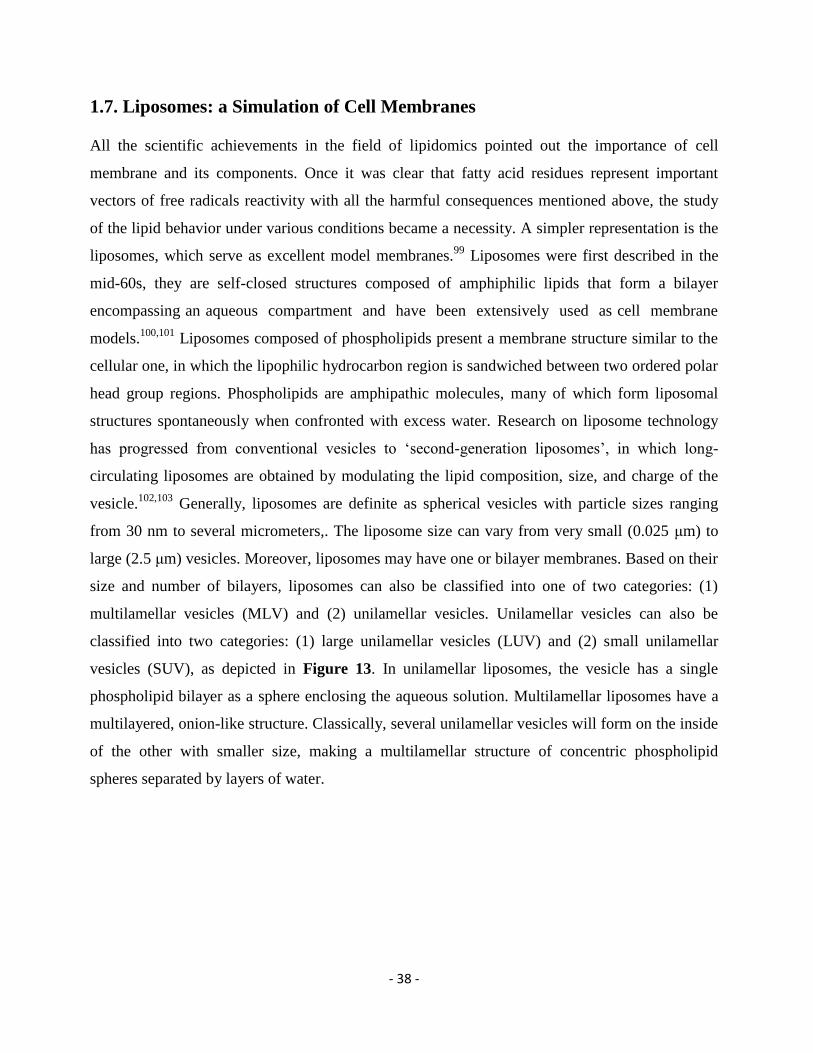
vesicles (SUV), as depicted in Figure 13. In unilamellar liposomes, the vesicle has a single
phospholipid bilayer as a sphere enclosing the aqueous solution. Multilamellar liposomes have a
multilayered, onion-like structure. Classically, several unilamellar vesicles will form on the inside
of the other with smaller size, making a multilamellar structure of concentric phospholipid
spheres separated by layers of water.
- 39 -
Figure 13. Liposome classification and diameter sizes. (SUV = small unilamellar vesicle; LUV = large
unilamellar vesicle; GUV = giant unilamellar vesicle; MLV = multilamellar vesicle)
There is a variety of methods for liposome preparation such as sonication,104
french pressure cell:
extrusion,105
freeze-thawed liposomes,106
lipid film hydration by hand shaking,107
non-hand
shaking or freeze drying,108
micro-emulsification,109
membrane extrusion110
etc, all methods have
four common stages:111
1. Drying down lipids from organic solvent.
2. Dispersing the lipid in aqueous media.
3. Purifying the resultant liposome.
4. Analyzing the final product.
Liposomes have been used in a broad range of pharmaceutical applications showing particular
promise as intracellular delivery systems for anti-sense molecules, ribosomes, proteins/peptides,
and DNA.112
However, despite the liposomes primary use as drug deliverable systems this is not
their only application.113
Liposome models can be used in studies of lipid peroxidation or cis-trans
isomerization due to the fact that they are a stimulation of the cell membrane, in order to avoid the
Unilamellar Liposomes Multilamellar Liposomes
SUV
< 100 nm
LUV
> 100 nm
GUV
> 300 nm
MLV
> 100 nm
- 40 -
complication of possible interference of cell components other than the membrane lipids.114,115
They exhibit ion discrimination, osmotic swelling, and response to a variety of agents that
accelerate or retard loss of ions or molecules from the spherules in a way that at least qualitatively
mimics their action on natural membrane-bounded structures. Among others, cell membranes act
as a physiological barrier for a drug in its path to reach the site of action. The diffusion through
cell membrane phospholipid bilayer, is a key step in the absorption and distribution of a drug
as well as, ultimately, its action in the organism. The molecule must enter the membrane within
the polar head group region, diffuse through the lipophilic hydrocarbon double layer and emerge
throughout the head group region on the inner side. Even in cases where a specific transporter is
involved, the drug’s ability to interact with the membrane is often highly correlated with
structural changes in her components. This ability depends on the drug’s hydrophilic/lipophilic
equilibrium. The study of the interaction between a drug and the membrane lipids of unilamellar
liposomes is a novel aspect in antitumoral treatments. Nowadays, this aspect is highlightened
more and more, providing many distinct advantages and bringing out several important features
regarding the reaction mechanism and the consequent membrane damage under free radical
conditions. Applications of liposomes in medicine and pharmacology can be divided into
diagnostic and therapeutic applications; liposomes containing various markers or drugs, and their
use as a tool, a model, or reagent in the basic studies of cell interactions, recognition processes,
and mode of action of certain substances.116,117,118,119
- 41 -
1.8. Thesis Overall Objectives
The overall objective of this PhD thesis was to explore the oxidative/free radical damage
mechanism induced my novel synthesized metallodrugs using biomimetic models in order to
simulate the biological system and exploring mechanistic scenarios with targeted experiments of
lipid cis-trans isomerization and DNA damages.
The damage mechanism is mediated by ROS, which are able to lead in permanent oxidative
modifications in cellular components. Due to this reason the research achievements presented in
this thesis focused on the two most important targets of the antitumoral metallodrug’s reactivity;
membrane lipids and DNA. Therefore, the experimental work can be divided in two parts
according to the aspect that is studied.
Evaluation of the lipid damage in the presence of the artificial metallo-nucleases
[Cu(TPMA)(N,Nʹ)]2+
. For the purposes of these studies, liposome models were employed
as representation of the cell membranes. Alterations in the chemical structure of fatty acid
moieties in the presence of a novel metallodrug and a reducing agent are discussed in
Chapter 2 as well as, in Chapter 3. In addition, in Chapter 3 is presented the first
complete synthetic strategy and analytical characterization of the most electron rich fatty
acid; the docosahexaenoic acid (DHA) which was essential for further investigation
regarding DHA’s reactivity in lipid damage.
Evaluation of the DNA oxidation profiles and damage mechanism induced by a novel type
of [Cu(TPMA)(N,Nʹ)]2+
artificial metallo-nucleases. The detailed examination regarding
the DNA binding, cleavage ability as well as radical trapping experiments to identify the
dominant free radical(s) giving rise to SSBs and DSBs of AMNs was determined with
supercoiled DNA. The results obtainded during the course this analysis are discussed in
Chapter 4.
- 42 -
Chapter 2: Model Studies of Cu-TPMA-Phen-Induced Lipid Damage in
Liposome Membranes
Among metal complexes of therapeutic use as antitumor drugs, copper(II) complexes have a
particular place due to the fact that they can undergo redox activity with in vivo formation of
Cu(I) and, together with DNA binding properties to cause DNA cleavage. 120
The increased
requirement of copper in cancer cells for redox metabolism and its oxidative properties to
generate reactive oxygen species (ROS) are the biological and chemical basis, respectively, for
the anticancer activity of these metallodrugs acting as artificial nucleases for the sequence specific
disruption of gene function.121 The artificial chemical nuclease Cu-TPMA-Phen of the novel
series [Cu(TPMA)(N,Nʹ)]2 that was used for the studies on lipid damage was synthesized, purified
and fully characterized by ESR Nicolo Fantoni in Dublin City University a ‘ClickGene’ partner,
under the supervision of Professor Andrew Kellett.
As mentioned above, an important aspect of the Cu-TPMA-Phen chemical reactivity is the fact
that it has a redox behavior of Cu+2 ⟶ Cu
+1 and this in situ electron transfer can give rise in the
formation of radical species.122
In the presence of thiols, which are active biomolecules, this redox
potential of the copper(II) complex can generate S-centered radicals, able to cause biological
damages involving among others the membrane lipids. 123,124
The outcome of the generation of
thiyl radicals, is that these species can either catalyse a cis-trans isomerisation of double bonds
and/or initiate a peroxidation process involving the unsaturated fatty acid residues of
phospholipids.124
Phospholipids are a large family of compounds, they are considered as building
blocks of the membrane and naturally in eukaryotic cells, the double bond geometry of
unsaturated fatty acid residues is cis.125
However, the intervention of sulfur-centered radicals, can
lead in a cis–trans isomerization, giving rise to the formation of the more thermodynamically
stable trans isomer.126
The free radical-based drug effect inspired biomimetic studies in membrane
models, composed of liposomes containing monounsaturated fatty acid residues (such as oleic
acid, 18:1 cis-9) in the presence of the potential drug [Cu(TPMA)(Phenanthroline)](ClO4)2] and
different thiols as reducing agents to cause the recycling of the redox state of the complex and
- 43 -
incubation at 37°C. In our experiments, we were interested in studying the transformation of cis
geometry into trans, under a variety of different conditions. The study of the biomimetic model in
a radical-catalyzed cis-trans isomerization is of high importance, since the trans lipid geometry
resembles that of saturated lipids and is possible to lead in a permanent modification of the
membrane and formation of a more rigid bilayer packing.127,70
On the other hand, it is known that the monounsaturated fatty acid moieties are less prone to a
radical-based oxidative degradation compared with the polyunsaturated fatty acids.128
In order to
evaluate the peroxidation pathway, liposomes formed by soybean lecithin containing different
percentages of SFA, MUFA and PUFA fatty acids were tested in the presence of the copper
complex and thiol. These vesicles offer the advantage of studying the competition of free radical
transformations towards peroxidation and isomerization processes, since the polyunsaturated fatty
acids can partition in both pathways. 129
Comparing the results we obtained by the use of these
two different liposomal compositions under a variety of conditions concerning the effect of
concentration both for the copper(II)complex and the thiol, the different thiols used, the addition
of well known antioxidants and the presence or absence of oxygen an interesting scenario of this
potential metallodrug reactivity was furnished.
The biomimetic model of liposomes was designed to follow the fatty acid fate after reaction with
thiyl radicals, formed in the presence of the Cu-TPMA-Phen and a thiol compound. The model
studies underlined the crucial role of membranes in antitumoral treatments and the fact that
membranes are not just spectators but important vectors of the antitumoral’s drug reactivity.130,131
- 44 -
2.1. Materials and Methods
2.1.1. Liposome Experiments
The first step in our experimental procedure is the addition of POPC in chloroform (53 mg
dissolved in 3 mL), followed by evaporation in a test tube under argon stream. The thin film that
is formed, remains under vacuum until it is completely free of solvent and then 1 mL tridistilled
water is added, in order to obtain a final concentration of 70 mM phospholipid content.132
The
solution is vortexed vigorously under argon stream for 7 min and this procedure yields at large,
multilamellar liposomes (LMV). Our next step is downsizing LMV dispersions in unilamellar
vesicles (LUV) with a mean diameter of 156-158 nm using the extrusion technique (LUVET) and
a 200 nm polycarbonate membrane filter.133
This method is chosen since not only it helps prevent
the membranes from fouling and improves the homogeneity of the size distribution of the final
suspension, but also because this membrane model represents in the closest way the membrane
bilayer. The size of the liposomes is measured using DLS and the LUVET stock suspensions are
transferred into a vial and stored at 4°C for a maximum of 2 weeks.
The total volume of every reaction is 1 mL with phospholipid concentration of 1 mM. More
specifically an aliquot of 14.5 μL fatty acid content from the stock solution is added in tridistilled
water in the reaction vessel. To the liposome suspension, the copper complex is transferred (0.15
mM) and the reaction remains under stirring for 2 min. From stock solutions in tridistilled water
and final concentration in the reaction 10 mM, the thiol (2-mercaptoethanol or l-cysteine or
reduced glutathione) is added drop wise (0.5 mm/min) using a syringe pump. Each reaction vessel
is incubated at 37°C and in order to follow the formation of trans fatty acid residues, samples are
analyzed at different times, as reported in Tables 1-9.
The work-up of the vesicles is made with 2:1 chloroform/methanol, extracting and collecting the
organic phases dried over anhydrous sodium sulfate and evaporating the solvent under vacuum at
room temperature. The phospholipids are treated with 0.5 M KOH/MeOH, in a transesterification
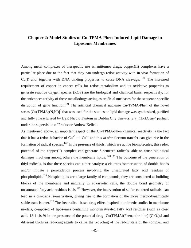
type of reaction for 10 min at room temperature, as shown in Scheme 1.134
- 45 -
Scheme 1. Mechanism of transesterification reaction in which the phosphoryl group of phospholipids is
exchanged with the methyl group of methanol under alkaline conditions.
The reaction is quenched with addition of tridistilled water and an extraction with n-hexane
follows. The organic layers containing the corresponding fatty acid methyl esters are analyzed by
GC for the examination of the FAME content.135
For the experiments under anaerobic conditions,
all the solutions are degassed with argon for 15 min and the addition of all reagents take place
under argon. The anaerobic conditions are maintained during the incubation period by creating
pressure of argon inside the reaction vial.
2.1.2. GC Analysis
Fatty acid methyl esters were analyzed by gas chromatograph (Agilent 6850, Milan) equipped
with a 60 m Χ 0.25 mm Χ 0.25 μm (50%-cyanopropyl)-methylpolysiloxane column (DB23,
Agilent, U.S.A.). The instrument has a flame ionization detector (FID) that requires air (450
mL/min) and hydrogen (40 mL/min) and is maintained at a temperature of 250 °C and applying
injection temperature at 230 °C. From an initial temperature of 165 °C held for 3 min, followed by
an increase of 1 °C/min up to 195 °C, held for 40 min. A final ramp, with a temperature increase
of 10 °C/min up to a maximum temperature of 240 °C, was maintained for 10 min for column
purge. A constant pressure mode (29 psi) was chosen with helium as carrier gas. Methyl esters
were identified by comparison with the retention times of commercially available standards or
trans fatty acid references.
- 46 -
2.2. Results and Discussion
2.2.1. Building-up a Membrane Biomimetic Model
In order to simulate the cell membrane phospholipid behavior, large unilamellar vesicles were
formed by extrusion technique (LUVET). In our experiments, two different lipid compositions
were used at concentration of phospholipid content 1 mM. Firstly, the synthetic phospholipid 1-
palmitoyl-2-oleoyl phosphatidyl choline (POPC) was chosen due to the fact that it represents the
simplest biomimetic model, containing only two different phospholipid moieties of saturated and
monounsaturated fatty acids. Moreover, the preparation of vesicles from soybean lecithin was
made, containing different percentages of SFA, MUFA and PUFA fatty acids residues.
POPC vesicles are proved ideal for the study of cis-trans isomerization since they are consisted of
the MUFA moiety of oleic acid (9 cis-18:1). Oleic acid can react with sulfur centered radicals and
be transformed into its trans geometrical isomer (see Appendix Figure S2), which is elaidic acid
(9 trans-18:1).136
For the best estimation of the copper complex contribution to lipid
isomerization, also control experiments were carried out in the presence of the potential drug,
without addition of thiol, in which case no transformation was observed. In addition, the
efficiency of different thiols, such as 2-mercaptoethanol, l-cysteine and glutathione (reduced) was
evaluated, under anaerobic and aerobic conditions. Due to its amphiphilic character and the ability
to diffuse freely in the membrane bilayer, 2-mercaptoethanol gave the highest yields of
isomerization in every condition tested.
In the experiments with lecithin liposomes the transformations of MUFA (oleic acid, 9 cis-18:1)
and PUFA (linoleic acid, 9cis,12cis-18:2) residues to their corresponding geometrical isomers, as
well as the PUFA consumption by oxidative degradation were evaluated. Depending from the
reaction conditions, isomerization and peroxidation processes were examined and compared. The
yield of isomerization for each fatty acid MUFA or PUFA, along with the consumption of PUFA
was based on the peak areas obtained by GC analysis. Gas chromatography is proved to be a
valuable analytical tool for the identification of each geometrical isomer providing a clear
separation (see Appendix Figure S3). The estimation of PUFA peroxidation, it is based on the
fact that lecithin is composed, among others, by the saturated fatty acid residue of palmitic acid
(16:0). Due to the lack of double bonds in its moiety, palmitic acid is unreactive under free
- 47 -
radical conditions and can play the role of internal standard. The quantification of consumption is
possible using the GC calibration curves made for each fatty acid that was analyzed vs the
palmitic acid residue.
In order to understand the mechanism of this metallodrug, it is crucial to gain an insight to the
electron transfer that occurs between the metal and the thiol compound. For this reason, the UV-
VIS spectra of the Cu-TPMA-Phen, before and after addition of different concentrations of thiol
were obtained, as presented in Appendix in Figure S1.
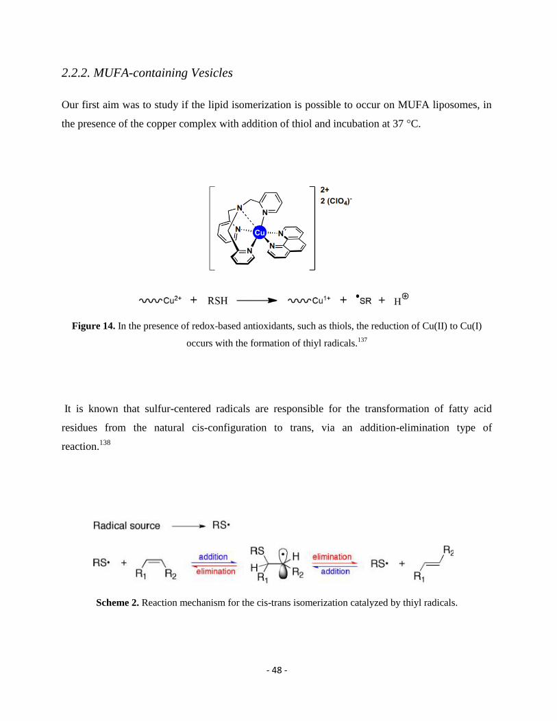
- 48 -
2.2.2. MUFA-containing Vesicles
Our first aim was to study if the lipid isomerization is possible to occur on MUFA liposomes, in
the presence of the copper complex with addition of thiol and incubation at 37 °C.
Figure 14. In the presence of redox-based antioxidants, such as thiols, the reduction of Cu(II) to Cu(I)
occurs with the formation of thiyl radicals.137
It is known that sulfur-centered radicals are responsible for the transformation of fatty acid
residues from the natural cis-configuration to trans, via an addition-elimination type of
reaction.138
Scheme 2. Reaction mechanism for the cis-trans isomerization catalyzed by thiyl radicals.
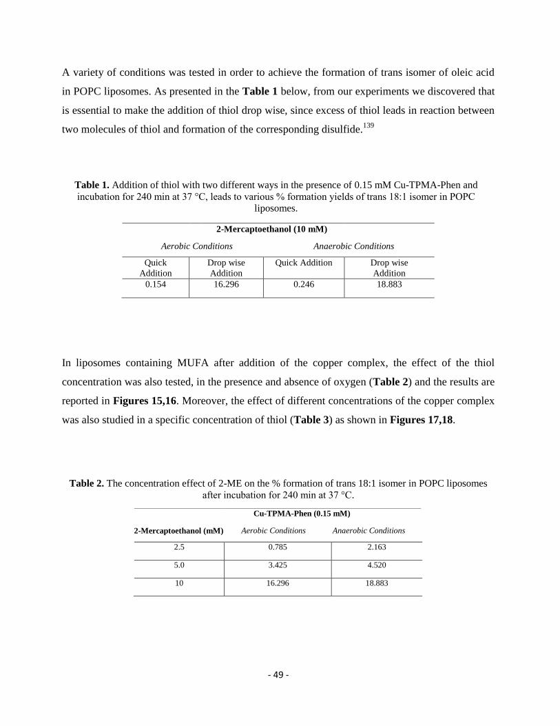
- 49 -
A variety of conditions was tested in order to achieve the formation of trans isomer of oleic acid
in POPC liposomes. As presented in the Table 1 below, from our experiments we discovered that
is essential to make the addition of thiol drop wise, since excess of thiol leads in reaction between
two molecules of thiol and formation of the corresponding disulfide.139
Table 1. Addition of thiol with two different ways in the presence of 0.15 mM Cu-TPMA-Phen and
incubation for 240 min at 37 °C, leads to various % formation yields of trans 18:1 isomer in POPC
liposomes.
2-Mercaptoethanol (10 mM)
Aerobic Conditions Anaerobic Conditions
Quick
Addition
Drop wise
Addition
Quick Addition Drop wise
Addition
0.154 16.296 0.246 18.883
In liposomes containing MUFA after addition of the copper complex, the effect of the thiol
concentration was also tested, in the presence and absence of oxygen (Table 2) and the results are
reported in Figures 15,16. Moreover, the effect of different concentrations of the copper complex
was also studied in a specific concentration of thiol (Table 3) as shown in Figures 17,18.
Table 2. The concentration effect of 2-ME on the % formation of trans 18:1 isomer in POPC liposomes
after incubation for 240 min at 37 °C.
Cu-TPMA-Phen (0.15 mM)
2-Mercaptoethanol (mM) Aerobic Conditions Anaerobic Conditions
2.5 0.785 2.163
5.0 3.425 4.520
10 16.296 18.883
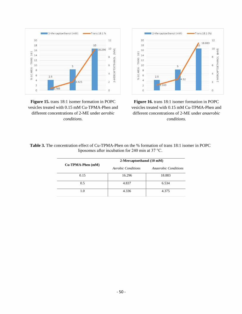
- 50 -
Figure 15. trans 18:1 isomer formation in POPC
vesicles treated with 0.15 mM Cu-TPMA-Phen and
different concentrations of 2-ΜE under aerobic
conditions.
Figure 16. trans 18:1 isomer formation in POPC
vesicles treated with 0.15 mM Cu-TPMA-Phen and
different concentrations of 2-ΜE under anaerobic
conditions.
Table 3. The concentration effect of Cu-TPMA-Phen on the % formation of trans 18:1 isomer in POPC
liposomes after incubation for 240 min at 37 °C.
Cu-TPMA-Phen (mM)
2-Mercaptoethanol (10 mM)
Aerobic Conditions Anaerobic Conditions
0.15 16.296 18.883
0.5 4.837 6.534
1.0 4.336 4.375
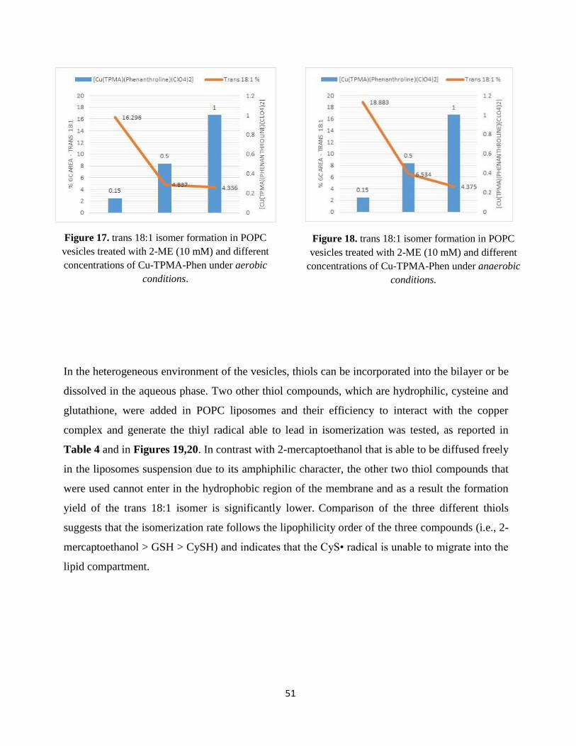
51
Figure 17. trans 18:1 isomer formation in POPC
vesicles treated with 2-ΜE (10 mM) and different
concentrations of Cu-TPMA-Phen under aerobic
conditions.
Figure 18. trans 18:1 isomer formation in POPC
vesicles treated with 2-ΜE (10 mM) and different
concentrations of Cu-TPMA-Phen under anaerobic
conditions.
In the heterogeneous environment of the vesicles, thiols can be incorporated into the bilayer or be
dissolved in the aqueous phase. Two other thiol compounds, which are hydrophilic, cysteine and
glutathione, were added in POPC liposomes and their efficiency to interact with the copper
complex and generate the thiyl radical able to lead in isomerization was tested, as reported in
Table 4 and in Figures 19,20. In contrast with 2-mercaptoethanol that is able to be diffused freely
in the liposomes suspension due to its amphiphilic character, the other two thiol compounds that
were used cannot enter in the hydrophobic region of the membrane and as a result the formation
yield of the trans 18:1 isomer is significantly lower. Comparison of the three different thiols
suggests that the isomerization rate follows the lipophilicity order of the three compounds (i.e., 2-
mercaptoethanol > GSH > CySH) and indicates that the CyS• radical is unable to migrate into the
lipid compartment.
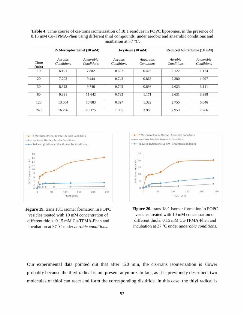
52
Table 4. Time course of cis-trans isomerization of 18:1 residues in POPC liposomes, in the presence of
0.15 mM Cu-TPMA-Phen using different thiol compounds, under aerobic and anaerobic conditions and
incubation at 37 °C.
Time
(min)
2- Mercaptoethanol (10 mM) l-cysteine (10 mM) Reduced Glutathione (10 mM)
Aerobic
Conditions
Anaerobic
Conditions
Aerobic
Conditions
Anaerobic
Conditions
Aerobic
Conditions
Anaerobic
Conditions
10 6.191 7.882 0.627 0.428 2.122 1.124
20 7.202 9.444 0.743 0.866 2.380 1.997
30 8.322 9.746 0.745 0.893 2.623 3.111
60 9.381 11.642 0.782 1.171 2.631 3.388
120 13.604 18.883 0.827 1.322 2.755 5.046
240 16.296 20.175 1.005 2.963 2.953 7.266
Figure 19. trans 18:1 isomer formation in POPC
vesicles treated with 10 mM concentration of
different thiols, 0.15 mM Cu-TPMA-Phen and
incubation at 37 0C under aerobic conditions.
Figure 20. trans 18:1 isomer formation in POPC
vesicles treated with 10 mM concentration of
different thiols, 0.15 mM Cu-TPMA-Phen and
incubation at 37 0C under anaerobic conditions.
Our experimental data pointed out that after 120 min, the cis-trans isomerization is slower
probably because the thiyl radical is not present anymore. In fact, as it is previously described, two
molecules of thiol can react and form the corresponding disulfide. In this case, the thiyl radical is
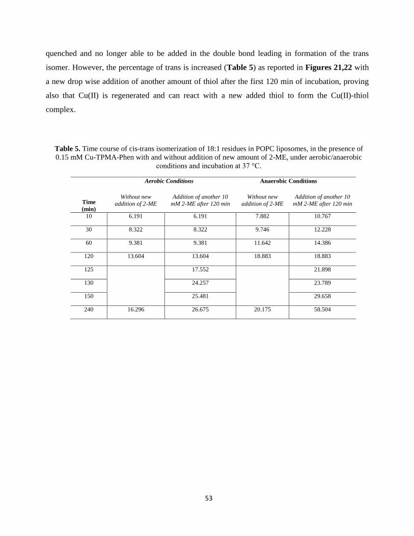
53
quenched and no longer able to be added in the double bond leading in formation of the trans
isomer. However, the percentage of trans is increased (Table 5) as reported in Figures 21,22 with
a new drop wise addition of another amount of thiol after the first 120 min of incubation, proving
also that Cu(II) is regenerated and can react with a new added thiol to form the Cu(II)-thiol
complex.
Table 5. Time course of cis-trans isomerization of 18:1 residues in POPC liposomes, in the presence of
0.15 mM Cu-TPMA-Phen with and without addition of new amount of 2-ME, under aerobic/anaerobic
conditions and incubation at 37 °C.
Time
(min)
Aerobic Conditions Anaerobic Conditions
Without new
addition of 2-ME
Addition of another 10
mM 2-ME after 120 min
Without new
addition of 2-ME
Addition of another 10
mM 2-ME after 120 min
10 6.191 6.191 7.882 10.767
30 8.322 8.322 9.746 12.228
60 9.381 9.381 11.642 14.386
120 13.604 13.604 18.883 18.883
125
17.552 21.898
130 24.257 23.789
150 25.481 29.658
240 16.296 26.675 20.175 58.504
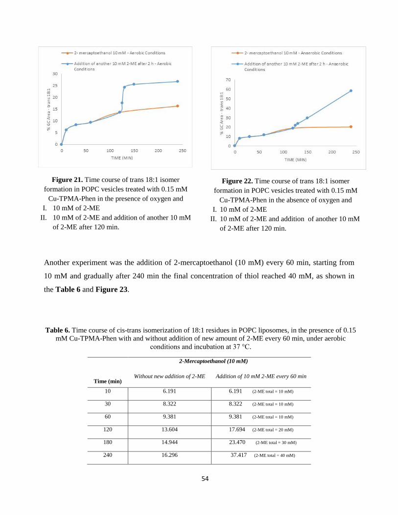
54
Figure 21. Time course of trans 18:1 isomer
formation in POPC vesicles treated with 0.15 mM
Cu-TPMA-Phen in the presence of oxygen and
I. 10 mM of 2-ME
II. 10 mM of 2-ME and addition of another 10 mM
of 2-ME after 120 min.
Figure 22. Time course of trans 18:1 isomer
formation in POPC vesicles treated with 0.15 mM
Cu-TPMA-Phen in the absence of oxygen and
I. 10 mM of 2-ME
II. 10 mM of 2-ME and addition of another 10 mM
of 2-ME after 120 min.
Another experiment was the addition of 2-mercaptoethanol (10 mM) every 60 min, starting from
10 mM and gradually after 240 min the final concentration of thiol reached 40 mM, as shown in
the Table 6 and Figure 23.
Table 6. Time course of cis-trans isomerization of 18:1 residues in POPC liposomes, in the presence of 0.15
mM Cu-TPMA-Phen with and without addition of new amount of 2-ME every 60 min, under aerobic
conditions and incubation at 37 °C.
Time (min)
2-Mercaptoethanol (10 mM)
Without new addition of 2-ME
Addition of 10 mM 2-ME every 60 min
10 6.191 6.191 (2-ME total = 10 mM)
30 8.322 8.322 (2-ME total = 10 mM)
60 9.381 9.381 (2-ME total = 10 mM)
120 13.604 17.694 (2-ME total = 20 mM)
180 14.944 23.470 (2-ME total = 30 mM)
240 16.296 37.417 (2-ME total = 40 mM)
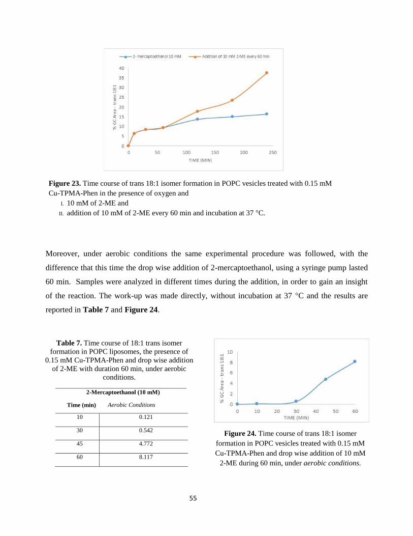
55
Figure 23. Time course of trans 18:1 isomer formation in POPC vesicles treated with 0.15 mM
Cu-TPMA-Phen in the presence of oxygen and
I. 10 mM of 2-ME and
II. addition of 10 mM of 2-ME every 60 min and incubation at 37 °C.
Moreover, under aerobic conditions the same experimental procedure was followed, with the
difference that this time the drop wise addition of 2-mercaptoethanol, using a syringe pump lasted
60 min. Samples were analyzed in different times during the addition, in order to gain an insight
of the reaction. The work-up was made directly, without incubation at 37 °C and the results are
reported in Table 7 and Figure 24.
Table 7. Time course of 18:1 trans isomer
formation in POPC liposomes, the presence of
0.15 mM Cu-TPMA-Phen and drop wise addition
of 2-ME with duration 60 min, under aerobic
conditions.
2-Mercaptoethanol (10 mM)
Time (min) Aerobic Conditions
10 0.121
30 0.542
45 4.772
60 8.117
Figure 24. Time course of trans 18:1 isomer
formation in POPC vesicles treated with 0.15 mM
Cu-TPMA-Phen and drop wise addition of 10 mM
2-ME during 60 min, under aerobic conditions.
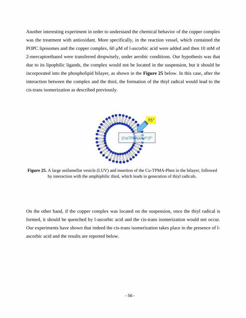
- 56 -
Another interesting experiment in order to understand the chemical behavior of the copper complex
was the treatment with antioxidant. More specifically, in the reaction vessel, which contained the
POPC liposomes and the copper complex, 60 μM of l-ascorbic acid were added and then 10 mM of
2-mercaptoethanol were transferred dropwisely, under aerobic conditions. Our hypothesis was that
due to its lipophilic ligands, the complex would not be located in the suspension, but it should be
incorporated into the phospholipid bilayer, as shown in the Figure 25 below. In this case, after the
interaction between the complex and the thiol, the formation of the thiyl radical would lead to the
cis-trans isomerization as described previously.
Figure 25. A large unilamellar vesicle (LUV) and insertion of the Cu-TPMA-Phen in the bilayer, followed
by interaction with the amphiphilic thiol, which leads in generation of thiyl radicals.
On the other hand, if the copper complex was located on the suspension, once the thiyl radical is
formed, it should be quenched by l-ascorbic acid and the cis-trans isomerization would not occur.
Our experiments have shown that indeed the cis-trans isomerization takes place in the presence of l-
ascorbic acid and the results are reported below.
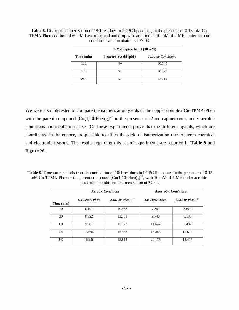
- 57 -
Table 8. Cis- trans isomerization of 18:1 residues in POPC liposomes, in the presence of 0.15 mM Cu-
TPMA-Phen addition of 60 μM l-ascorbic acid and drop wise addition of 10 mM of 2-ME, under aerobic
conditions and incubation at 37 °C.
We were also interested to compare the isomerization yields of the copper complex Cu-TPMA-Phen
with the parent compound [Cu(1,10-Phen)2]2+
in the presence of 2-mercaptoethanol, under aerobic
conditions and incubation at 37 °C. These experiments prove that the different ligands, which are
coordinated in the copper, are possible to affect the yield of isomerization due to stereo chemical
and electronic reasons. The results regarding this set of experiments are reported in Table 9 and
Figure 26.
Table 9. Time course of cis-trans isomerization of 18:1 residues in POPC liposomes in the presence of 0.15
mM Cu-TPMA-Phen or the parent compound [Cu(1,10-Phen)2]2+
, with 10 mM of 2-ME under aerobic -
anaerobic conditions and incubation at 37 °C.
Time (min)
Aerobic Conditions Anaerobic Conditions
Cu-TPMA-Phen
[Cu(1,10-Phen)2]2+
Cu-TPMA-Phen
[Cu(1,10-Phen)2]2+
10 6.191 10.936 7.882 3.670
30 8.322 13.331 9.746 5.135
60 9.381 15.173 11.642 6.482
120 13.604 15.558 18.883 11.613
240 16.296 15.814 20.175 12.417
2-Mercaptoethanol (10 mM)
Time (min) l-Ascorbic Acid (μM) Aerobic Conditions
120 No 10.740
120 60 10.591
240 60 12.219
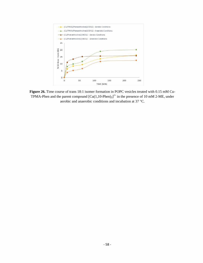
- 58 -
Figure 26. Time course of trans 18:1 isomer formation in POPC vesicles treated with 0.15 mM Cu-
TPMA-Phen and the parent compound [Cu(1,10-Phen)2]2+
in the presence of 10 mM 2-ME, under
aerobic and anaerobic conditions and incubation at 37 °C.
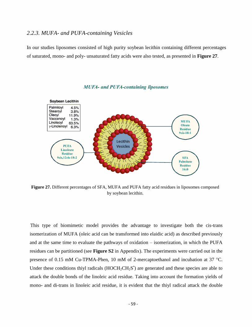
- 59 -
2.2.3. MUFA- and PUFA-containing Vesicles
In our studies liposomes consisted of high purity soybean lecithin containing different percentages
of saturated, mono- and poly- unsaturated fatty acids were also tested, as presented in Figure 27.
Figure 27. Different percentages of SFA, MUFA and PUFA fatty acid residues in liposomes composed
by soybean lecithin.
This type of biomimetic model provides the advantage to investigate both the cis-trans
isomerization of MUFA (oleic acid can be transformed into elaidic acid) as described previously
and at the same time to evaluate the pathways of oxidation – isomerization, in which the PUFA
residues can be partitioned (see Figure S2 in Appendix). The experiments were carried out in the
presence of 0.15 mM Cu-TPMA-Phen, 10 mM of 2-mercaptoethanol and incubation at 37 °C.
Under these conditions thiyl radicals (HOCH2CH2S•) are generated and these species are able to
attack the double bonds of the linoleic acid residue. Taking into account the formation yields of
mono- and di-trans in linoleic acid residue, it is evident that the thiyl radical attack the double
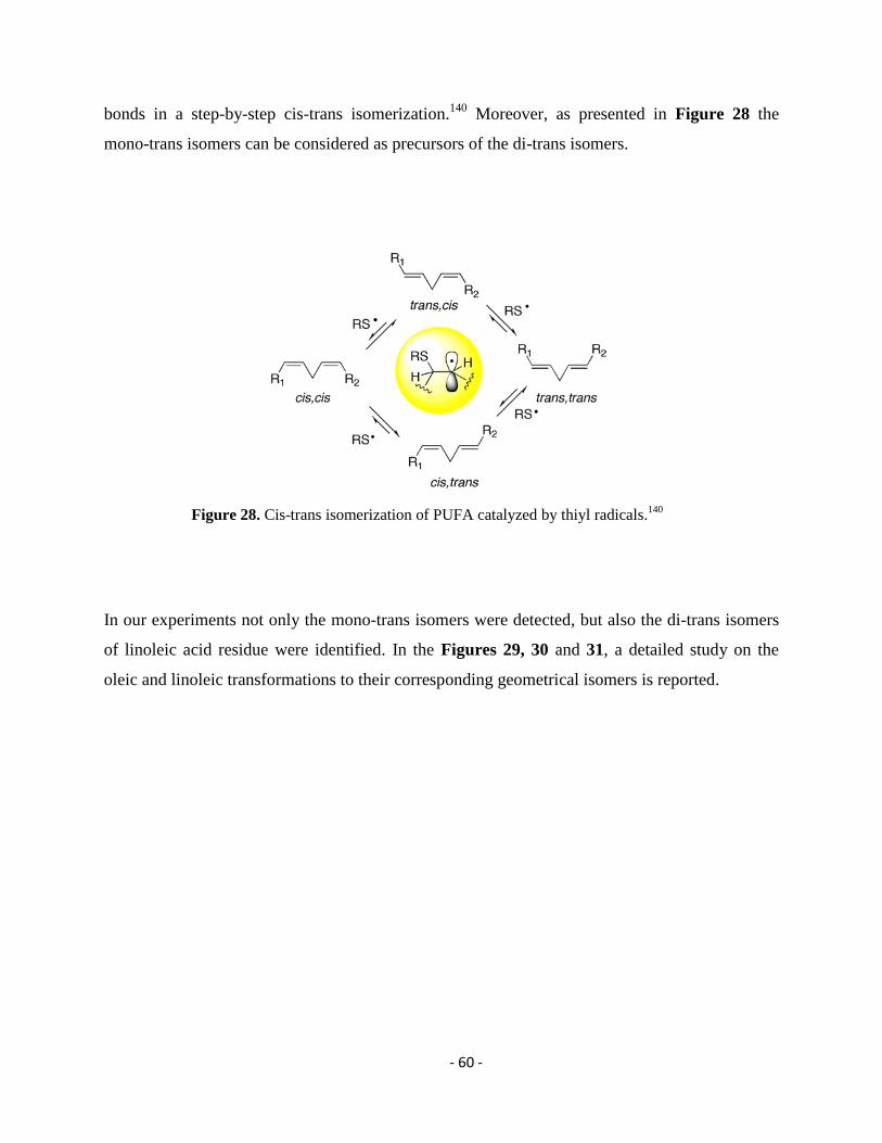
- 60 -
bonds in a step-by-step cis-trans isomerization.140
Moreover, as presented in Figure 28 the
mono-trans isomers can be considered as precursors of the di-trans isomers.
Figure 28. Cis-trans isomerization of PUFA catalyzed by thiyl radicals.140
In our experiments not only the mono-trans isomers were detected, but also the di-trans isomers
of linoleic acid residue were identified. In the Figures 29, 30 and 31, a detailed study on the
oleic and linoleic transformations to their corresponding geometrical isomers is reported.
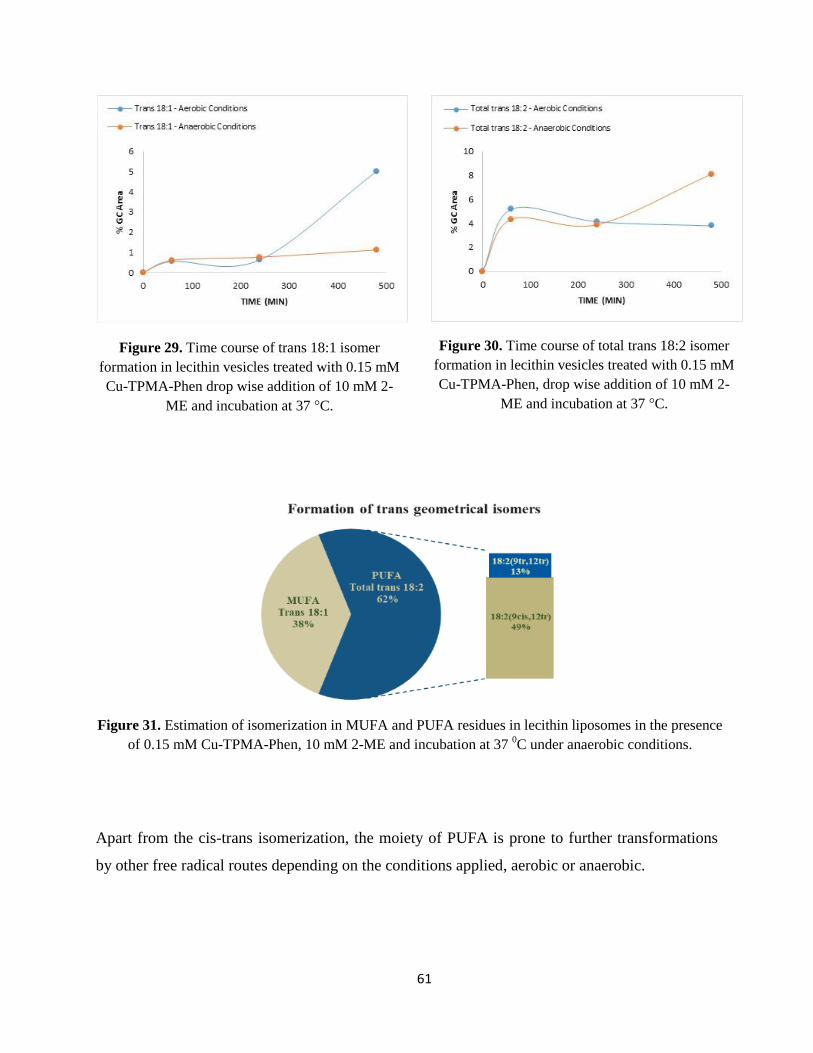
61
Figure 29. Time course of trans 18:1 isomer
formation in lecithin vesicles treated with 0.15 mM
Cu-TPMA-Phen drop wise addition of 10 mM 2-
ME and incubation at 37 °C.
Figure 30. Time course of total trans 18:2 isomer
formation in lecithin vesicles treated with 0.15 mM
Cu-TPMA-Phen, drop wise addition of 10 mM 2-
ME and incubation at 37 °C.
Figure 31. Estimation of isomerization in MUFA and PUFA residues in lecithin liposomes in the presence
of 0.15 mM Cu-TPMA-Phen, 10 mM 2-ME and incubation at 37 0C under anaerobic conditions.
Apart from the cis-trans isomerization, the moiety of PUFA is prone to further transformations
by other free radical routes depending on the conditions applied, aerobic or anaerobic.
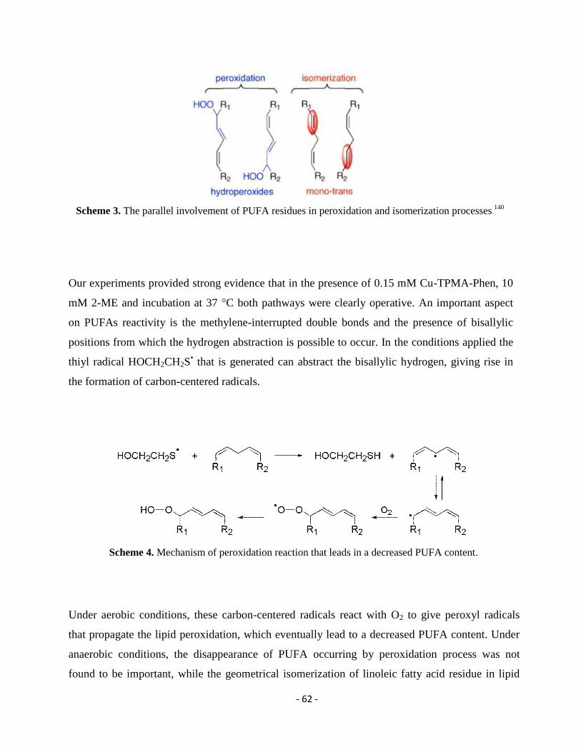
- 62 -
Scheme 3. The parallel involvement of PUFA residues in peroxidation and isomerization processes.
140
Our experiments provided strong evidence that in the presence of 0.15 mM Cu-TPMA-Phen, 10
mM 2-ME and incubation at 37 °C both pathways were clearly operative. An important aspect
on PUFAs reactivity is the methylene-interrupted double bonds and the presence of bisallylic
positions from which the hydrogen abstraction is possible to occur. In the conditions applied the
thiyl radical HOCH2CH2S• that is generated can abstract the bisallylic hydrogen, giving rise in
the formation of carbon-centered radicals.
Scheme 4. Mechanism of peroxidation reaction that leads in a decreased PUFA content.
Under aerobic conditions, these carbon-centered radicals react with O2 to give peroxyl radicals
that propagate the lipid peroxidation, which eventually lead to a decreased PUFA content. Under
anaerobic conditions, the disappearance of PUFA occurring by peroxidation process was not
found to be important, while the geometrical isomerization of linoleic fatty acid residue in lipid
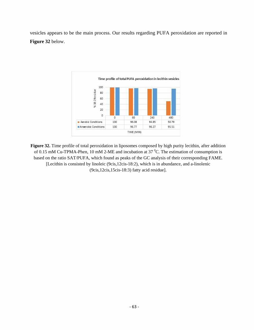
- 63 -
vesicles appears to be the main process. Our results regarding PUFA peroxidation are reported in
Figure 32 below.
Figure 32. Time profile of total peroxidation in liposomes composed by high purity lecithin, after addition
of 0.15 mM Cu-TPMA-Phen, 10 mM 2-ME and incubation at 37 0C. The estimation of consumption is
based on the ratio SAT/PUFA, which found as peaks of the GC analysis of their corresponding FAME.
[Lecithin is consisted by linoleic (9cis,12cis-18:2), which is in abundance, and a-linolenic
(9cis,12cis,15cis-18:3) fatty acid residue].
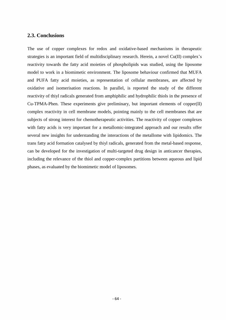
- 64 -
2.3. Conclusions
The use of copper complexes for redox and oxidative-based mechanisms in therapeutic
strategies is an important field of multidisciplinary research. Herein, a novel Cu(II) complex’s
reactivity towards the fatty acid moieties of phospholipids was studied, using the liposome
model to work in a biomimetic environment. The liposome behaviour confirmed that MUFA
and PUFA fatty acid moieties, as representation of cellular membranes, are affected by
oxidative and isomerisation reactions. In parallel, is reported the study of the different
reactivity of thiyl radicals generated from amphiphilic and hydrophilic thiols in the presence of
Cu-TPMA-Phen. These experiments give preliminary, but important elements of copper(II)
complex reactivity in cell membrane models, pointing mainly to the cell membranes that are
subjects of strong interest for chemotherapeutic activities. The reactivity of copper complexes
with fatty acids is very important for a metallomic-integrated approach and our results offer
several new insights for understanding the interactions of the metallome with lipidomics. The
trans fatty acid formation catalysed by thiyl radicals, generated from the metal-based response,
can be developed for the investigation of multi-targeted drug design in anticancer therapies,
including the relevance of the thiol and copper-complex partitions between aqueous and lipid
phases, as evaluated by the biomimetic model of liposomes.
- 65 -
Chapter 3: Trans Lipid Library; Synthesis of Docosahexaenoic Acid (DHA) Monotrans Isomers - Regioisomer Identification & Model Studies in DHA-
Containing Liposomes
Continuing our studies on the artificial chemical nuclease’s reactivity towards the unsaturated
fatty acid residues, I thought it would be interesting to investigate vesicles composed by the
most electron-rich polyunsaturated fatty acid, due to an increased probability of lipid damage;
the docosahexaenoic acid (DHA) which contains six cis double bonds.
However, in the literature there were not enough data regarding the characterization of the six
monotrans DHA regioisomers and they were an open issue to address for analytical,
biological, and nutraceutical applications. Therefore, my research aimed in building-up a
library of the monotrans DHA isomers. The studies included the preparation, separation, and
first identification of each isomer by a dual approach consisting of the following:
1. the direct thiyl radical-catalyzed isomerization of cis-DHA methyl ester and
2. the two-step synthesis from cis-DHA methyl ester via monoepoxides as intermediates,
which are separated and identified by nuclear magnetic resonance spectroscopy,
followed by elimination for the unequivocal assignment of the double bond position.
The results were published in Chemical Research in Toxicology141
Menounou G.‡,Giacometti
G.,‡ Scanferlato R., Dambruoso P., Sansone A., Tueros I., Amézaga J.,Chatgilialoglu C. and
Ferreri C.; 2018 31 (3), 191-200, Trans Lipid Library: Synthesis of Docosahexaenoic Acid
(DHA) Monotrans Isomers and Regioisomer Identification in DHA-Containing Supplements.
(DOI: 10.1021/acs.chemrestox.8b00021)
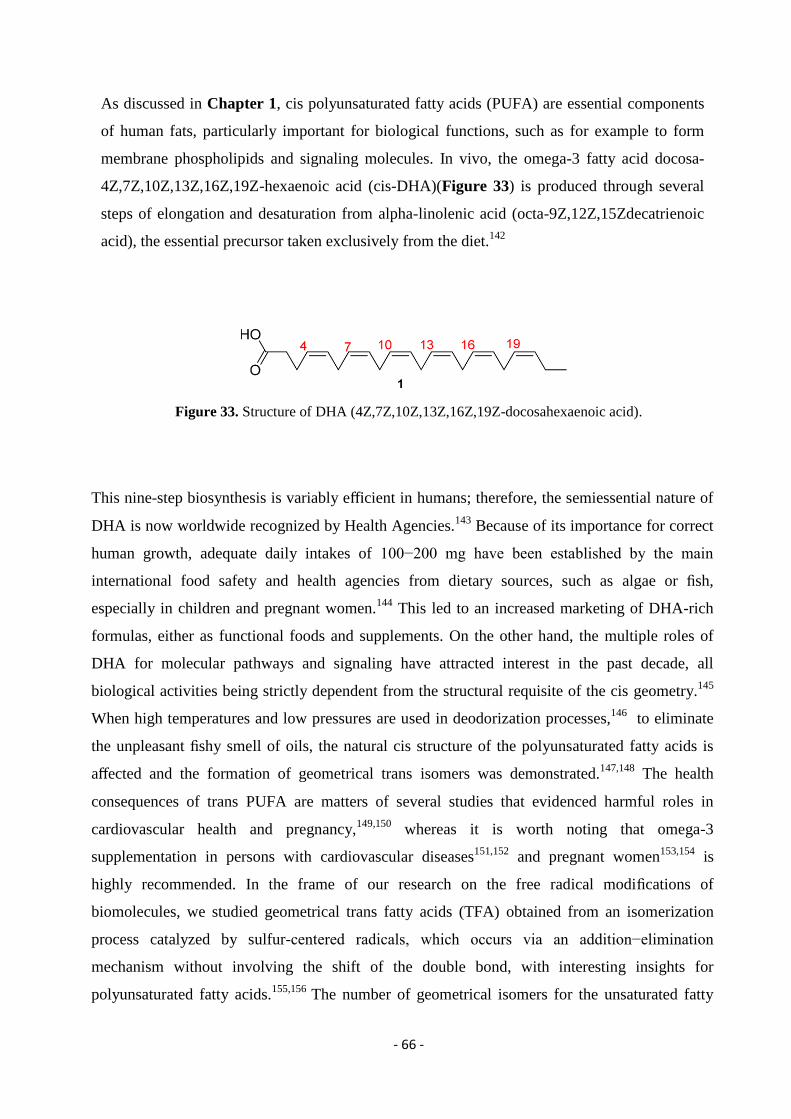
- 66 -
As discussed in Chapter 1, cis polyunsaturated fatty acids (PUFA) are essential components
of human fats, particularly important for biological functions, such as for example to form
membrane phospholipids and signaling molecules. In vivo, the omega-3 fatty acid docosa-
4Z,7Z,10Z,13Z,16Z,19Z-hexaenoic acid (cis-DHA)(Figure 33) is produced through several
steps of elongation and desaturation from alpha-linolenic acid (octa-9Z,12Z,15Zdecatrienoic
acid), the essential precursor taken exclusively from the diet.142
Figure 33. Structure of DHA (4Z,7Z,10Z,13Z,16Z,19Z-docosahexaenoic acid).
This nine-step biosynthesis is variably efficient in humans; therefore, the semiessential nature of
DHA is now worldwide recognized by Health Agencies.143
Because of its importance for correct
human growth, adequate daily intakes of 100−200 mg have been established by the main
international food safety and health agencies from dietary sources, such as algae or fish,
especially in children and pregnant women.144
This led to an increased marketing of DHA-rich
formulas, either as functional foods and supplements. On the other hand, the multiple roles of
DHA for molecular pathways and signaling have attracted interest in the past decade, all
biological activities being strictly dependent from the structural requisite of the cis geometry.145
When high temperatures and low pressures are used in deodorization processes,146
to eliminate
the unpleasant fishy smell of oils, the natural cis structure of the polyunsaturated fatty acids is
affected and the formation of geometrical trans isomers was demonstrated.147,148
The health
consequences of trans PUFA are matters of several studies that evidenced harmful roles in
cardiovascular health and pregnancy,149,150
whereas it is worth noting that omega-3
supplementation in persons with cardiovascular diseases151,152
and pregnant women153,154
is
highly recommended. In the frame of our research on the free radical modifications of
biomolecules, we studied geometrical trans fatty acids (TFA) obtained from an isomerization
process catalyzed by sulfur-centered radicals, which occurs via an addition−elimination
mechanism without involving the shift of the double bond, with interesting insights for
polyunsaturated fatty acids.155,156
The number of geometrical isomers for the unsaturated fatty
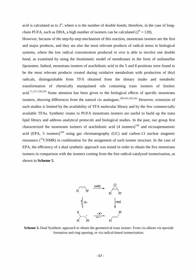
- 67 -
acid is calculated as to 2n, where n is the number of double bonds; therefore, in the case of long-
chain PUFA, such as DHA, a high number of isomers can be calculated (26 = 128).
However, because of the step-by-step mechanism of this reaction, monotrans isomers are the first
and major products, and they are also the most relevant products of radical stress in biological
systems, where the low radical concentration produced in vivo is able to involve one double
bond, as examined by using the biomimetic model of membranes in the form of unilamellar
liposomes. Indeed, monotrans isomers of arachidonic acid in the 5 and 8 positions were found to
be the most relevant products created during oxidative metabolism with production of thiyl
radicals, distinguishable from TFA obtained from the dietary intake and metabolic
transformation of chemically manipulated oils containing trans isomers of linoleic
acid.71,157,158,159
Some attention has been given to the biological effects of specific monotrans
isomers, showing differences from the natural cis analogues.160,161,162,163
However, extension of
such studies is limited by the availability of TFA molecular library and by the few commercially
available TFAs. Synthetic routes to PUFA monotrans isomers are useful to build up the trans
lipid library and address analytical protocols and biological studies. In the past, our group first
characterized the monotrans isomers of arachidonic acid (4 isomers)140
and eicosapentaenoic
acid (EPA, 5 isomers)164
using gas chromatography (GC) and carbon-13 nuclear magnetic
resonance (13
CNMR) in combination for the assignment of each isomer structure. In the case of
EPA, the efficiency of a dual synthetic approach was tested in order to obtain the five monotrans
isomers in comparison with the isomers coming from the free radical-catalyzed isomerization, as
shown in Scheme 5.
Scheme 5. Dual Synthetic approach to obtain the geometrical trans isomer: From cis-alkene via epoxide
formation and ring opening, or via radical-based isomerization.
- 68 -
The strategy of separation and assignment of the monoepoxide structures, prior to the
elimination, successfully worked for the first unambiguous determination of the trans alkene
position, integrating previous data in the literature.165,166,167
We considered the dual approach
shown in Scheme 5 to be useful also for the synthesis of the monotrans DHA isomers. Only the
4E regioisomer is commercially available as methyl ester and can be used as GC reference,
whereas in literature monotrans DHA isomers were analyzed by several authors after
deodorization/isomerization reactions of DHA-rich oils, assaying various conditions of GC
analysis but lacking a specific regioisomer assignment.147,148,162,166,167
As far as the synthesis of
the DHA monoepoxides is concerned, the classical epoxidation reaction can lead to these
compounds,168
which are named epoxydocosapentaenoic acid (EDP). 4,5-EDP and 19,20-EDP
were known165,169,170,171
also due to their biological relevance as metabolites of cytochrome
P450.172,173
The strategy of the separation of the intermediate monoepoxide regioisomers was successful to
unambiguously individuate the epoxidation of the double bond position, using mono- and
bidimensional nuclear magnetic resonance (NMR) experiments. Consequently, after ring
opening, dibromide formation and elimination from each monoepoxide regioisomer double bond
was unequivocally established, thus allowing for the assignment of the corresponding peaks in
the GC analysis. This double approach clarified that the direct isomerization by free radical-
catalyzed reaction gives the six isomers as exclusive products within a few minutes. The
usefulness of such identification was then assayed for analysis of fish oil and its isomerization
products, also in view of applying the analytical route to the purity of fish oil preparations, which
is a hot topic of food research.148,174,175
Trans-containing triglycerides were studied and it was
highlightened that NMR represents a powerful analytical methodology to apply directly on the
oil without transformation to fatty acid methyl esters.176
Moreover, DHA due to its unique effect of altering membrane composition, is often regarded as
the major omega-3 fatty acid involved in anticancer activity.177
Although use of DHA as an
anticancer drug to prevent or treat human cancer in clinical settings has not yet been well
established, recent studies suggest that DHA can be very effective as an adjuvant with other
anticancer agents.178
There are studies reporting that oxidized DHA leads to DNA adduct
formation, therefore this induced oxidative DNA damage triggers cell cycle arrest and apoptosis
in cancer cells.179,180
Several in vitro and animal studies suggest combining DHA with other
anticancer agents often improves efficacy of anticancer drugs as well as reduces therapy-
associated side effects.181
Docosahexaenoic acid (DHA), in vitro and in vivo, used along with
anticancer drugs, has improved cancer treatment outcome. In fact, there are clinical studies in the
- 69 -
literature reporting positive results with omega-3 supplements in oncologic patients.182
There are
beneficial effects of omega-3 fatty acids supplements in patients undergoing chemotherapy
and/or radiotherapy on different outcomes, being the preservation of body composition the most
evident.183
Incorporation of DHA in cellular membranes improves drug uptake, whereas
increased lipid peroxidation is another mechanism for DHA-mediated enhanced efficacy of
anticancer drugs.184,185
In 1998, Hawkins et al. reported that apoptotic death of human Mia-PaCa-
2 pancreatic cancer cells induced by PUFAs varies with double bond number and involves an
oxidative mechanism.186
They found correlations between the number of fatty acid double bonds
and the proportion of apoptotic cells. This information trigger our experimental design towards
DHA-containing liposomes as biomimetic model of cell membranes, since DHA represents the
most electron rich and crucial PUFA.
The monotrans DHA library and GC characterizations were firstly applied to the analysis of
omega-3 containing supplements in capsules commercially available in Italy and Spain,
evidencing trans isomer contamination in the oil ingredients. Secondly, the cis-trans
isomerization process was examined in DHA-containing liposomes in the presence of Cu-
TPMA-Phen and 2-mercaptoethanol as reducing agent to evaluate the altered configuration of
DHA.
- 70 -
3.1. Materials and Methods
Chloroform, methanol, n-hexane, 2-propanol, and acetonitrile were purchased from Merck
(HPLC purity). Absolute ethanol was purchased from BDH Prolabo VWR (AnalaR
NORMAPUR) and 2-mercaptoethanol was purchased from Aldrich. Silver nitrate, 30% aq
NHOH and anhydrous sodium sulfate (Na2SO4) were purchased from Carlo Erba. All fatty acid
methyl esters (FAME) used as reference standard for GC analyses were purchased from Aldrich,
Fluka, or Sigma and used without further purification. 4E,7Z,10Z,13Z,16Z,19Z DHA-
methylester was purchase from Lipidox (Lidingo, Sweden). Deuterated benzene and deuterated
CDCl3 were purchased from Eurisotop (France). Silica-gel thin-layer chromatography (analytical
and preparative) was performed on Merck silica gel 60 plates (0.25 and 2 mm thickness,
respectively).
Regarding the experimenrs with liposomes, vesicles composed by 20% 18:0-22:6 PC/POPC in 1
mM concentration were prepared. Samples were degassed under argon for 15 min. In addition,
the stock solution of 2-mercaptoethanol was prepared (the water was degassed for 15 min and
then 2-ME was added under argon stream). The anaerobic conditions were maintained during the
incubation period by creating pressure of argon inside the reaction vial.
GC Analyses. Fatty acid methyl esters were analyzed by gas chromatograph (Agilent 6850,
Milan) equipped with a 60 m Χ 0.25 mm Χ 0.25 μm (50%-cyanopropyl)-methylpolysiloxane
column (DB23, Agilent, U.S.A.). The instrument has a flame ionization detector (FID) that
requires air (450 mL/min) and hydrogen (40 mL/min) and is maintained at a temperature of 250
°C. Two identical equipment conditions were used for the analyses, applying the same injection
temperature (230 °C) but different oven conditions and carrier gas.
Method A: from an initial temperature of 165 °C held for 3 min, followed by an increase of 1
°C/min up to 195 °C, held for 40 min. A final ramp, with a temperature increase of 10 °C/min up
to a maximum temperature of 240 °C, was maintained for 10 min for column purge. A constant
pressure mode (29 psi) was chosen with helium as carrier gas. Methyl esters were identified by
comparison with the retention times of commercially available standards or trans fatty acid
references, obtained as described elsewhere.
Method B: temperature started from 195 °C, held for 26 min, followed by an increase of 10
°C/min up to 205 °C, held for 13 min, followed by a second increase of 30 °C/min up to 240 °C,
- 71 -
held for 10 min. A constant pressure mode (29 psi) was chosen with hydrogen as carrier gas.
GC/MS spectra were recorded on a Clarus 500 GC apparatus equipped with a Clarus 560S mass
spectrometer (GC/MS transfer line temperature 230 °C) and a 60 m Χ 0.25 mm Χ 0.25 μm
(50%cyanopropylphenyl)-dimethylpolysiloxane column (DB225 ms, Agilent, U.S.A.), using an
injector temperature of 230 °C, a split ratio of 50:1, and helium as carrier gas at a constant flow
of 1.2 mL/min, with the following oven conditions: temperature started from 195 °C, held for 52
min, followed by an increase of 3 °C/min up to 205 °C, held for 10 min, followed by a second
increase of 3 °C/min up to 225 °C, held for 15 min and a final increase of 5 °C/min up to 230 °C,
held for 10 min.
Photolysis. Photolysis was carried out in a quartz photochemical reactor (Sigma-Aldrich)
equipped with a 5.5 W low-pressure mercury lamp. The temperature was maintained at (22 ±
2)°C by means of a thermostat bath. Nuclear Magnetic Resonance (NMR). All NMR spectra
were collected with a fully automated Agilent NMR system, consisting of a 54 mm bore, 500
MHz (11.7 T) Premium Shielded superconducting magnet, a DD2 Performa IV NMR console,
and the Agilent OneNMR probe. All samples were dissolved in C6D6 and transferred in 3 mm
thin wall NMR tubes (Wilmad 335-PP-8). After having calibrated each sample of pw90, the
following seven NMR experiments were collected
on each sample (2D experiments were collected with 50% NUS and F2 sw (1H): 10 ppm, from
−0.5 to 9.5 ppm):
1H NMR spectra: pw90 was calibrated for each sample, sw: −0.5 to 9.5 ppm
one gHSQC: F1 sw (13
C) 135 ppm, from 5.0 to 140 ppm; nt = 32; ni = 512; res: 33 Hz (0.26
ppm.) F2 sw (1H): 10 ppm, from −0,5 to 9,5 ppm, NUS: 50%.
two bsgHSQCAD: first F1 sw (13
C): 60 ppm, from 5.0 to 65 ppm; nt = 32; ni = 512; res: 15
Hz (0.12 ppm). F2 sw (1H): 10 ppm, from −0.5 to 9.5 ppm, NUS: 50%. second: F1 sw (
13C):
20 ppm, from 120.0 to 140 ppm; nt = 32; ni = 512; res: 5 Hz (0.04 ppm). F2 sw (1H): 10
ppm, from −0.5 to 9.5 ppm, NUS: 50%.
one gHMBCAD: F1 sw (13
C): 175 ppm, from 5.0 to 180 ppm; nt = 64; ni = 512; res: 43 Hz
(0.34 ppm). F2 sw (from −0.5 to 9.5 ppm, NUS: 50%.
two bsgHMBC: first F1 sw (13
C): 60 ppm, from 5.0 to 65 ppm; nt = 64; ni = 512; res: 15 Hz
(0.12 ppm). F2 sw (1H): 10 ppm, from −0.5 to 9.5 ppm, NUS: 50%. second: F1sw(
13C): 60
ppm, from 120.0 to 180 ppm; nt = 64; ni = 512; res: 15 Hz (0.12 ppm). F2 sw (1H): 10 ppm,
from −0.5 to 9.5 ppm, NUS: 50%.
- 72 -
3.2. Epoxidation of Methyl All-(Z)-4,7,10,13,16,19-docosahexaenoate
3.2.1. Synthesis of Epoxydocosapentaenoic Acid Methyl Ester (EDP-Me)
Regioisomers
A 5.84 mM solution of meta-chloroperoxybenzoic acid in dichloromethane (2.5 mL
corresponding to 0.0146 mmol; 2.51 mg) was added dropwise to a solution of DHA methyl ester
1 (50 mg; 0.146 mmol) in dichloromethane (2.5 mL). After the addition was completed, the
mixture was stirred for 10 min at room temperature under argon atmosphere, following the
formation of monoepoxides with traces of diepoxides by TLC. Eluent: 7/3 n-hexane/diethyl
ether; DHA-Me Rf = 0.9, first mixture (5, 6 and traces of 4) R= 0.7, second mixture (3 and 7) Rf
= 0.6, pure isomer (2) Rf = 0.5): bisepoxides Rf ≤ 0.4). Work-up was carried out by addition of 5
mL of ice-cold NaHCO3 (25% w/v) stirred for 2 min. Then, the reaction mixture was transferred
in a separating funnel, the aqueous layer was discarded and the organic layer was washed two
times with NaHCO3, deionized H2O and dried over Na2SO4. The crude was purified with flash
chromatography (eluent: 9/1 n-hexane/diethyl ether) to give a first fraction containing an
inseparable mixture (colorless oil; 8 mg; 0.0223 mmol; 15.3% yield) containing 13,14-EDP-Me
(5), 16,17EDP-Me (6) with traces of 10,11-EDP 4 and of residual starting material (1).
Structures 5 and 6 in Figure 34, were found in a 29:71 ratio, respectively, as calculated by the
integration of protons of C-22 on 1H NMR spectrum (trace II in Figure 37). EDP-Me
regioisomers 5 and 6 were further purified by preparative TLC using 100 mL toluene/500 μL
isopropanol as the eluent: (Rf = 0.5, pure isomer 6, and Rf = 0.4, pure isomer 5) to give pure 6 (3
mg; 0.0084 mmol; 37.5% yield) with only traces of 4.
The second fraction contained an inseparable mixture of 7,8-EDPMe (3) and 19,20-EDP-Me (7)
(colorless oil; 7 mg; 0.0195 mmol; 13.4% yield) found in a 43:57 ratio, respectively, as
calculated by the integration of protons of C-22 on 1H NMR spectrum (trace III in Figure 37).
The subsequent fraction contained pure 4,5EDP-Me (2) regioisomer (colorless oil; 5 mg; 0.0134
mmol; 9.2% yield) identified as shown in Figure 37 (trace IV) for the C22 triplet at 1H NMR.
The starting material DHA-Me (1) recovered was 30 mg (0.0877 mmol; 60% recovered yield).
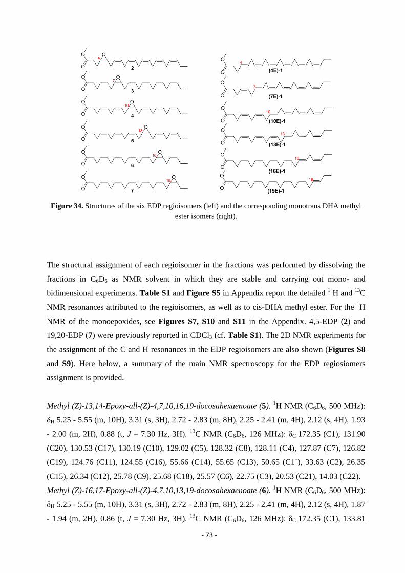
- 73 -
Figure 34. Structures of the six EDP regioisomers (left) and the corresponding monotrans DHA methyl
ester isomers (right).
The structural assignment of each regioisomer in the fractions was performed by dissolving the
fractions in C6D6 as NMR solvent in which they are stable and carrying out mono- and
bidimensional experiments. Table S1 and Figure S5 in Appendix report the detailed 1 H and
13C
NMR resonances attributed to the regioisomers, as well as to cis-DHA methyl ester. For the 1H
NMR of the monoepoxides, see Figures S7, S10 and S11 in the Appendix. 4,5-EDP (2) and
19,20-EDP (7) were previously reported in CDCl3 (cf. Table S1). The 2D NMR experiments for
the assignment of the C and H resonances in the EDP regioisomers are also shown (Figures S8
and S9). Here below, a summary of the main NMR spectroscopy for the EDP regiosiomers
assignment is provided.
Methyl (Z)-13,14-Epoxy-all-(Z)-4,7,10,16,19-docosahexaenoate (5). 1H NMR (C6D6, 500 MHz):
δΗ 5.25 - 5.55 (m, 10H), 3.31 (s, 3H), 2.72 - 2.83 (m, 8H), 2.25 - 2.41 (m, 4H), 2.12 (s, 4H), 1.93
- 2.00 (m, 2H), 0.88 (t, J = 7.30 Hz, 3H). 13
C NMR (C6D6, 126 MHz): δC 172.35 (C1), 131.90
(C20), 130.53 (C17), 130.19 (C10), 129.02 (C5), 128.32 (C8), 128.11 (C4), 127.87 (C7), 126.82
(C19), 124.76 (C11), 124.55 (C16), 55.66 (C14), 55.65 (C13), 50.65 (C1`), 33.63 (C2), 26.35
(C15), 26.34 (C12), 25.78 (C9), 25.68 (C18), 25.57 (C6), 22.75 (C3), 20.53 (C21), 14.03 (C22).
Methyl (Z)-16,17-Epoxy-all-(Z)-4,7,10,13,19-docosahexaenoate (6). 1H NMR (C6D6, 500 MHz):
δΗ 5.25 - 5.55 (m, 10H), 3.31 (s, 3H), 2.72 - 2.83 (m, 8H), 2.25 - 2.41 (m, 4H), 2.12 (s, 4H), 1.87
- 1.94 (m, 2H), 0.86 (t, J = 7.30 Hz, 3H). 13
C NMR (C6D6, 126 MHz): δC 172.35 (C1), 133.81
- 74 -
(C20), 130.19 (C13), 129.02 (C5), 128.40 (C10), 128.32 (C8), 128.11 (C4), 127.87 (C7), 127.70
(C11), 124.76 (C14), 123.66 (C19), 55.79 (C17), 55.65 (C16), 50.65 (C1`), 33.63 (C2), 26.34
(C15), 26.22 (C18), 25.78 (C12), 25.66 (C9), 25.57 (C6), 22.75 (C3), 20.53 (C21), 14.00 (C22).
Methyl (Z)-7,8-Epoxy-all-(Z)-4,10,13,16,19-docosahexaenoate (3). 1H NMR (C6D6, 500 MHz):
δH 5.35 - 5.51 (m, 10H), 3.30 (s, 3H), 2.69 - 2.85 (m, 8H), 2.19 - 2.32 (m, 4H), 2.08 - 2.14 (m, J
= 7.30 Hz, 4H), 1.93 - 2.04 (m, 2H), 0.89 (t, J = 7.50 Hz, 3H). 13
C NMR (C6D6, 126 MHz): δC
172.30 (C1), 131.82 (C20), 130.17 (C11), 130.08 (C4), 128.35 – 127.80 (C13, C14, C16 and
C17), 127.06 (C19), 125.77 (C5), 124.78 (C10), 55.65 (C8), 55.63 (C7), 50.66 (C1`), 33.51 (C2),
26.35 (C9), 26.23 (C6), 25.78 (C12), 25.75 – 25.60 (C15), 25.58 (C18), 22.83 (C3), 20.55 (C21),
14.07 (C22).
Methyl (Z)-19,20-Epoxy-all-(Z)-4,7,10,13,16-docosahexaenoate (7). 1H NMR (C6D6, 500 MHz):
δH 5.25 - 5.52 (m, 10H), 3.31 (s, 3H), 2.73 - 2.86 (m, 9H), 2.60 (dt, J = 4.20, 6.40 Hz, 1H), 2.25 -
2.37 (m, 3H), 2.04 - 2.16 (m, J = 7.30 Hz, 3H), 1.40 (ddq, J = 7.30, 7.30, 14.10 Hz, 1H), 1.28
(ddq, J = 6.10, 7.30, 13.50 Hz, 1H), 0.84 (t, J = 7.70 Hz, 3H). 13
C NMR (C6D6, 126 MHz): δC
172.32 (C1), 130.03 (C16), 129.06 (C5), 128.35 – 127.80 (C7, C8, C10, C11, C13 and C14),
128.08 (C4), 124.96 (C17), 57.38 (C20), 55.75 (C19), 50.65 (C1`), 33.64 (C2), 26.31 (C18),
25.79 (C15), 25.75 – 25.60 (C9 and C12), 25.58 (C6), 22.76 (C3), 21.10 (C21), 10.45 (C22).
Methyl (Z)-4,5-Epoxy-all-(Z)-7,10,13,16,19-docosahexaenoate (2). 1H NMR (C6D6, 500 MHz):
δH 5.23 - 5.55 (m, 10H), 3.29 (s, 3H), 2.65 - 2.88 (m, 10H), 2.13 - 2.27 (m, 3H), 1.95 - 2.09 (m,
3H), 1.68 - 1.76 (m, 1H), 1.58 - 1.68 (m, 1H), 0.89 (t, J = 7.63 Hz, 3H). 13
C NMR (C6D6, 126
MHz): δC 172.35 (C1), 131.89 (C20), 130.27 (C8), 128.63/128.38/128.11/128.96 (C13, C14, C16
and C17), 128.42 (C11), 127.95 (C10), 127.19 (C19), 124.78 (C7), 56.07 (C5), 55.32 (C4), 50.85
(C1`), 30.76 (C2), 26.38 (C6), 25.86 (C9), 25.75 (C12 and C15), 25.66 (C18), 23.42 (C3), 20.66
(C21), 14.17 (C22).
Methyl All-(Z)-4,7,10,13,16,19-Docosahexaenoate (1). 1H NMR
1H NMR (C6D6, 500 MHz): δH
5.24 - 5.47 (m, 12H), 3.31 (s, 3H), 2.73 - 2.88 (m, 10H), 2.29 (dddd, J = 0.80, 1.40, 7.34, 14.70
Hz, 1H), 2.29 (dddd, J = 0.80, 1.40, 7.30, 14.70 Hz, 1H), 2.11 (t, J = 7.40 Hz, 2H), 1.95 - 2.03
(m, 2H), 0.89 (t, J = 7.61 Hz, 3H). 13
C NMR (C6D6, 126 MHz): dC 174.32 (C1), 131.78 (C20),
129.08 (5), 128.50 (C17), 128.22 (C14), 128.15 (C8, C10 and C11), 128.11 (C7, C13 and C19),
128.06 (C4), 127.91 (C16), 50.65 (C1`), 33.65 (C2), 25.67 (C15), 25.66 (C9), 25.65 (C18), 25.57
(C6), 25.56 (C12), 22.75 (C3), 20.54 (C21), 14.08 (C22).
- 75 -
3.2.2. Transformation of EDP-Me Regioisomers to Monotrans DHA-Me Isomers
The reaction is described for 16,17-EDP-Me and 13,14-EDP-Me (first fraction of the EDP
chromatographic separation) and the same steps were followed for the other isolated EDP-Me
fractions.
Step 1. (Bromination of Epoxide)
To a freshly prepared solution of triphenylphosphine dibromide (7 mg; 0.0167 mmol) in dry
dichloromethane (50 μL) a solution containing a mixture of EDP regioisomers, 6 and 5, (3 mg;
0.0084 mmol containing traces of 10,11-EDP 4) in dry dichloromethane (100 μL) and pyridine
(1 μL) was added, stirring at 0 °C under argon atmosphere. The mixture was left stirring
overnight at 5 °C, and then was quenched with a 1 M aqueous solution of hydrochloric acid (0.5
mL) and extracted three times with chloroform/ethanol (4/1). The reaction was monitored by
TLC for the ring opening as dibromide (7/3 n-hexane/diethyl ether, starting material Rf = 0.7 and
dibromide product R = 0.4). The organic layers were collected, dried over Na2SO4 and
evaporated under vacuum. The crude containing methyl 16,17-dibromo-(4Z,7Z,10Z,13Z,19Z)-
docosapentaenoate and 13,14-dibromo-(4Z,7Z,10Z,16Z,19Z)-docosapentaenoate (with traces of
the 10,11-dibromoderivative) was used for the next step. The same reaction was also performed
for the inseparable mixture of the EDP-Me regioisomers 3 and 7 and for the EDP-ME
regioisomer 4. In all cases, the reaction crude was used for the next step.
Step 2. (Elimination)
To an ice-cooled slurry of activated zinc (4.7 mg; 0.072 mmol), acetic acid (2 μL), and N,N-
dimethylformamide (150 μL), a solution of methyl 16,17-dibromo(4Z,7Z,10Z,13Z,19Z)-
docosapentaenoate and traces of 13,14-dibromo-(4Z,7Z,10Z,16Z,19Z)-docosapentaenoate (3 mg;
0.0060 mmol) in N,N-dimethylformamide (100 μL) was added, and the mixture was stirred at 0
°C for 14 h. The reaction was monitored by TLC (eluent: 7/3 n-hexane/diethyl ether) evidencing
the formation of alkenes (R = 0.9) in the presence of traces of starting material (Rf = 0.4). At this
point, the reaction was stopped by addition of a 1 M aqueous hydrochloric acid solution (1 mL),
and the precipitate was filtered off. The filtrate was extracted three times with a mixture of
chloroform/ methanol (3/1). The organic layers were collected and dried over Na2SO4. The
solvent was removed under vacuum to afford a colorless oil (1.5 mg; 0.0044 mmol; 73% yield).
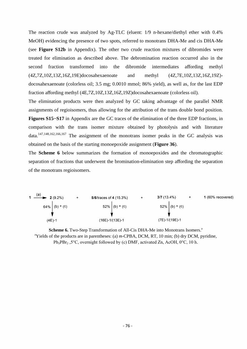
- 76 -
The reaction crude was analyzed by Ag-TLC (eluent: 1/9 n-hexane/diethyl ether with 0.4%
MeOH) evidencing the presence of two spots, referred to monotrans DHA-Me and cis DHA-Me
(see Figure S12b in Appendix). The other two crude reaction mixtures of dibromides were
treated for elimination as described above. The debromination reaction occurred also in the
second fraction transformed into the dibromide intermediates affording methyl
(4Z,7Z,10Z,13Z,16Z,19E)docosahexaenoate and methyl (4Z,7E,10Z,13Z,16Z,19Z)-
docosahexaenoate (colorless oil; 3.5 mg; 0.0010 mmol; 86% yield), as well as, for the last EDP
fraction affording methyl (4E,7Z,10Z,13Z,16Z,19Z)docosahexaenoate (colorless oil).
The elimination products were then analyzed by GC taking advantage of the parallel NMR
assignments of regioisomers, thus allowing for the attribution of the trans double bond position.
Figures S15−S17 in Appendix are the GC traces of the elimination of the three EDP fractions, in
comparison with the trans isomer mixture obtained by photolysis and with literature
data.147,148,162,166,167
The assignment of the monotrans isomer peaks in the GC analysis was
obtained on the basis of the starting monoepoxide assignment (Figure 36).
The Scheme 6 below summarizes the formation of monoepoxides and the chromatographic
separation of fractions that underwent the bromination-elimination step affording the separation
of the monotrans regioisomers.
Scheme 6. Two-Step Transformation of All-Cis DHA-Me into Monotrans Isomers.a
aYields of the products are in parentheses: (a) m-CPBA, DCM, RT, 10 min; (b) dry DCM, pyridine,
Ph3PBr2 ,5°C, overnight followed by (c) DMF, activated Zn, AcOH, 0°C, 10 h.
- 77 -
3.3. Synthesis of Monotrans DHA-Me Isomers by Photolysis
A 15 mM solution of DHA-Me ester (20 mg, 0.058 mmol) in 2-propanol (3.87 mL) was
transferred into a quartz photochemical reactor (Sigma-Aldrich) equipped with a 5.5 W low-
pressure mercury lamp. The reaction mixture was degassed with argon for 20 min, then an
aliquot of a previously degassed stock solution of 2-mercaptoethanol in 2-propanol was added
(0.03 mmol) and the UV lamp was turned on for 5 min at the temperature of 22 ± 2 °C, kept by
means of a thermostat bath. The reaction was monitored by Ag-TLC, spraying the plate with
cerium ammonium sulfate/ammonium molybdate reagent (CAM), to evidence the formation of
the monotrans fraction together with ditrans, tritrans products, in order to stop the reaction when
monotrans isomers were prevalently formed in the presence of remaining cis-DHA (see Figure
S12a in Appendix). The solvent was removed under vacuum, and addition of chloroform with
further evaporation under vacuum helped the removal of 2-mercaptoethanol. The crude of the
reaction was subsequently dissolved in 1 mL of n-hexane and loaded onto a preparative Ag-TLC
plate. A rapid and efficient separation was obtained using 1:9 n-hexane/diethyl ether with 0.4%
MeOH as mobile phase. After elution, the plate portion corresponding to the monotrans isomer
fraction (Rf = 0.58), detected by spraying a small portion of the plate with CAM, was scraped
off. Silica was washed with chloroform (3 Χ 5 mL). The solvent was evaporated to give a solid
material, which is the Ag-fatty acid complex insoluble in n-hexane. This material was dissolved
in a 5% aqueous solution of NH4OH, vigorously stirred (600 rpm) for 10 min and extracted with
aliquots of n-hexane (3 Χ 5 mL). Finally, the organic phase was dried over Na2SO4, filtered and
evaporated. The unreacted all-cis DHA-Me was recovered and subjected to a second
isomerization cycle. This procedure leaded to the collection of a colorless oil that corresponded
to the monotrans DHA methyl ester isomer mixture (5.2 mg, 0.0152 mmol, and overall yield
from two isomerization cycles 26%). GC analysis was carried out under the conditions described
in Materials and Methods (see trace II, Figure 36 or trace II in Figure 38). 13
C NMR (Figures
S13 and S14 in Appendix) of the mixture of monotrans DHA-Me isomers obtained from the
isomerization and from the purification were also performed.
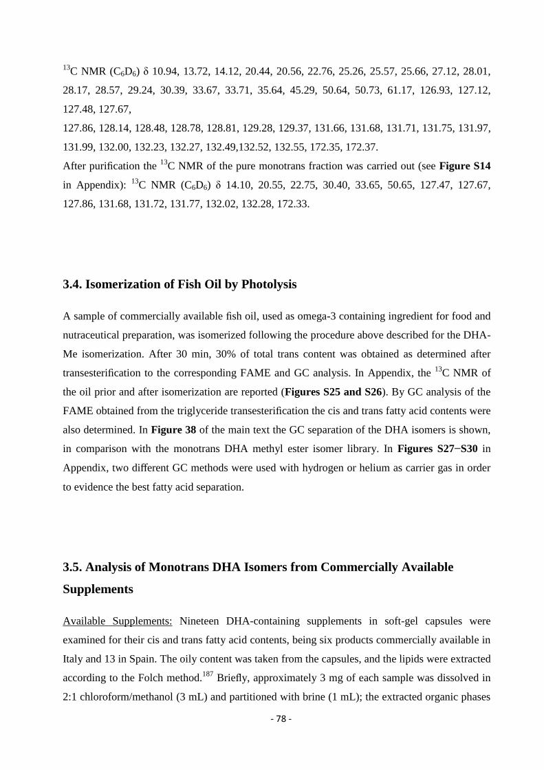
- 78 -
13C NMR (C6D6) δ 10.94, 13.72, 14.12, 20.44, 20.56, 22.76, 25.26, 25.57, 25.66, 27.12, 28.01,
28.17, 28.57, 29.24, 30.39, 33.67, 33.71, 35.64, 45.29, 50.64, 50.73, 61.17, 126.93, 127.12,
127.48, 127.67,
127.86, 128.14, 128.48, 128.78, 128.81, 129.28, 129.37, 131.66, 131.68, 131.71, 131.75, 131.97,
131.99, 132.00, 132.23, 132.27, 132.49,132.52, 132.55, 172.35, 172.37.
After purification the 13
C NMR of the pure monotrans fraction was carried out (see Figure S14
in Appendix): 13
C NMR (C6D6) δ 14.10, 20.55, 22.75, 30.40, 33.65, 50.65, 127.47, 127.67,
127.86, 131.68, 131.72, 131.77, 132.02, 132.28, 172.33.
3.4. Isomerization of Fish Oil by Photolysis
A sample of commercially available fish oil, used as omega-3 containing ingredient for food and
nutraceutical preparation, was isomerized following the procedure above described for the DHA-
Me isomerization. After 30 min, 30% of total trans content was obtained as determined after
transesterification to the corresponding FAME and GC analysis. In Appendix, the 13
C NMR of
the oil prior and after isomerization are reported (Figures S25 and S26). By GC analysis of the
FAME obtained from the triglyceride transesterification the cis and trans fatty acid contents were
also determined. In Figure 38 of the main text the GC separation of the DHA isomers is shown,
in comparison with the monotrans DHA methyl ester isomer library. In Figures S27−S30 in
Appendix, two different GC methods were used with hydrogen or helium as carrier gas in order
to evidence the best fatty acid separation.
3.5. Analysis of Monotrans DHA Isomers from Commercially Available
Supplements
Available Supplements: Nineteen DHA-containing supplements in soft-gel capsules were
examined for their cis and trans fatty acid contents, being six products commercially available in
Italy and 13 in Spain. The oily content was taken from the capsules, and the lipids were extracted
according to the Folch method.187
Briefly, approximately 3 mg of each sample was dissolved in
2:1 chloroform/methanol (3 mL) and partitioned with brine (1 mL); the extracted organic phases
- 79 -
(3 Χ 3 mL) containing lipids were joined and dried with anhydrous Na2SO4. After filtration, the
solvent was eliminated using a rotary evaporator and the oily residue was left under vacuum for
10 min to remove any possible trace of water. The lipid extract was checked by TLC (mobile
phase: n-hexane/diethyl ether 7:3) and then treated with freshly prepared 0.5 M KOH/MeOH
solution for 30 min at room temperature under stirring. Reaction was quenched using brine and
fatty acid methyl esters (FAME) were extracted using n-hexane (3x 3 mL); the organic phase
was collected and dried with anhydrous Na2SO4. After filtration and solvent evaporation, the
FAME mixture was dissolved in 10 μL n-hexane and 1 μL was injected for GC analysis. Tables
S2 and S3 in Appendix report the FAME content: products 1−6 are omega-3 supplements
present in the Italian market and products 7−19 are present in the Spanish market.
3.6. LUVET Preparation
A mixture of 20 % 1-stearoyl-2-docosahexaenoyl-sn-glycero-3-phosphocholine/1-palmitoyl-2-
oleoylphosphatidylcholine in chloroform solution (111 μL of 18:0-22:6 PC with 11.1 mg of 16:0-
18:1 PC dissolved in 1 mL chloroform) was evaporated to a thin film in a test tube under a stream
of argon and then kept under high-vacuum for 30 min at room temperature.
In order to obtain a final concentration of 70 mM phospholipid content, 250 μL tridistilled water
were added and by vortexing for 7 min under an argon atmosphere multilamellar vesicles were
formed. Large unilamellar vesicles (LUV) with a mean diameter of 156-158 nm were prepared by
extrusion technique using LiposoFast and a 200 nm polycarbonate membrane filter as described
previously.133
The size of the liposomes was measured using dynamic light scattering (DLS)
methodology. The LUVET stock suspensions were transferred into a vial and stored at 4 °C for a
maximum of two days.
3.6.1. Isomerisation vs Peroxidation of PC in LUVET
The total volume for every reaction was 1 mL of LUVET stock (phospholipid concentration of 1
mM). More specifically an aliquot of 14.5 μL fatty acid content from the stock solution was added
in tridistilled water in the reaction vessel. To the liposome suspension, the copper complex was
transferred (0.15 mM) and the reaction remained under stirring for 2 min. From stock solutions in
- 80 -
tridistilled water and final concentration in the reaction 10 mM, the thiol, 2-mercaptoethanol was
added to the reaction drop wise (0.5 mM/min) using a syringe pump. Each reaction vessel was
incubated at 37 °C and, in order to follow the formation of trans fatty acid residues, samples were
analysed at different times. The work-up of the vesicles was made with 2:1 chloroform/methanol,
extracting and collecting the organic phases dried over anhydrous sodium sulphate and
evaporating the solvent under vacuum at room temperature. The phospholipid extracts were
treated with 0.5 M KOH/MeOH, in a transesterification type of reaction for 10 min at ambient
temperature. The reaction was quenched with addition of tridistilled water and an extraction with
n-hexane followed. The organic layer containing the corresponding fatty acid methyl esters
(FAME) was analysed by GC for the determination of the content.
- 81 -
3.7. Results and Discussion
3.7.1. Dual Synthetic Approach for DHA Transformation and Monotrans DHA
Isomer Identification
According to Scheme 5 we approached the dual synthetic strategy first by carrying out the direct
radical-catalyzed isomerization of DHA methyl ester, as described previously by our group for
EPA,163
thus obtaining a mixture of the six monotrans DHA isomers (Figure 34). Ag-TLC was
used to purify the monotrans DHA isomers from the starting material (Figure 35, lane C).
Figure 35. Ag-TLC (eluent: 1/9 n-hexane/diethyl ether with 0.4% MeOH) of the transformation of one
monoepoxide fraction (second fraction) via bromination and elimination reactions. A: standard all-cis
DHA-Me. B: elimination reaction showing the presence of all-cisDHA-Me and monotrans isomers (19E
and 7E) of DHA-Me. C: mixture of monotrans DHA-Me obtained by thiyl-radical catalyzed
isomerization after purification as reported in Experimental Section.
The recognition of each monotrans isomer was not realizable at this stage, however satisfactory
separation of these isomers could be obtained by gas chromatography, the GC trace showing five
out of six separable peaks, meaning that only two isomers are superimposed. It is also worth
noting that one monotrans DHA isomer elutes similarly to cis-DHA (Figure 36, GC trace II).
GC analysis, under the conditions described in Materials and Methods (trace II, Figure 36) gave
a pattern of peaks similar to those described in literature.147,148,166,168
At this point the assignment
of each monotrans isomer could not be performed. Only by elution of the commercially available
4E-DHA-Me, the third eluting peak could be assigned to this isomer (Figure 36, trace V). By the
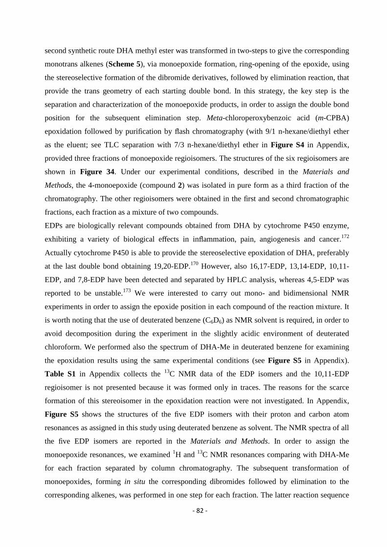
- 82 -
second synthetic route DHA methyl ester was transformed in two-steps to give the corresponding
monotrans alkenes (Scheme 5), via monoepoxide formation, ring-opening of the epoxide, using
the stereoselective formation of the dibromide derivatives, followed by elimination reaction, that
provide the trans geometry of each starting double bond. In this strategy, the key step is the
separation and characterization of the monoepoxide products, in order to assign the double bond
position for the subsequent elimination step. Meta-chloroperoxybenzoic acid (m-CPBA)
epoxidation followed by purification by flash chromatography (with 9/1 n-hexane/diethyl ether
as the eluent; see TLC separation with 7/3 n-hexane/diethyl ether in Figure S4 in Appendix,
provided three fractions of monoepoxide regioisomers. The structures of the six regioisomers are
shown in Figure 34. Under our experimental conditions, described in the Materials and
Methods, the 4-monoepoxide (compound 2) was isolated in pure form as a third fraction of the
chromatography. The other regioisomers were obtained in the first and second chromatographic
fractions, each fraction as a mixture of two compounds.
EDPs are biologically relevant compounds obtained from DHA by cytochrome P450 enzyme,
exhibiting a variety of biological effects in inflammation, pain, angiogenesis and cancer.172
Actually cytochrome P450 is able to provide the stereoselective epoxidation of DHA, preferably
at the last double bond obtaining 19,20-EDP.170
However, also 16,17-EDP, 13,14-EDP, 10,11-
EDP, and 7,8-EDP have been detected and separated by HPLC analysis, whereas 4,5-EDP was
reported to be unstable.173
We were interested to carry out mono- and bidimensional NMR
experiments in order to assign the epoxide position in each compound of the reaction mixture. It
is worth noting that the use of deuterated benzene (C6D6) as NMR solvent is required, in order to
avoid decomposition during the experiment in the slightly acidic environment of deuterated
chloroform. We performed also the spectrum of DHA-Me in deuterated benzene for examining
the epoxidation results using the same experimental conditions (see Figure S5 in Appendix).
Table S1 in Appendix collects the 13
C NMR data of the EDP isomers and the 10,11-EDP
regioisomer is not presented because it was formed only in traces. The reasons for the scarce
formation of this stereoisomer in the epoxidation reaction were not investigated. In Appendix,
Figure S5 shows the structures of the five EDP isomers with their proton and carbon atom
resonances as assigned in this study using deuterated benzene as solvent. The NMR spectra of all
the five EDP isomers are reported in the Materials and Methods. In order to assign the
monoepoxide resonances, we examined 1H and
13C NMR resonances comparing with DHA-Me
for each fraction separated by column chromatography. The subsequent transformation of
monoepoxides, forming in situ the corresponding dibromides followed by elimination to the
corresponding alkenes, was performed in one step for each fraction. The latter reaction sequence
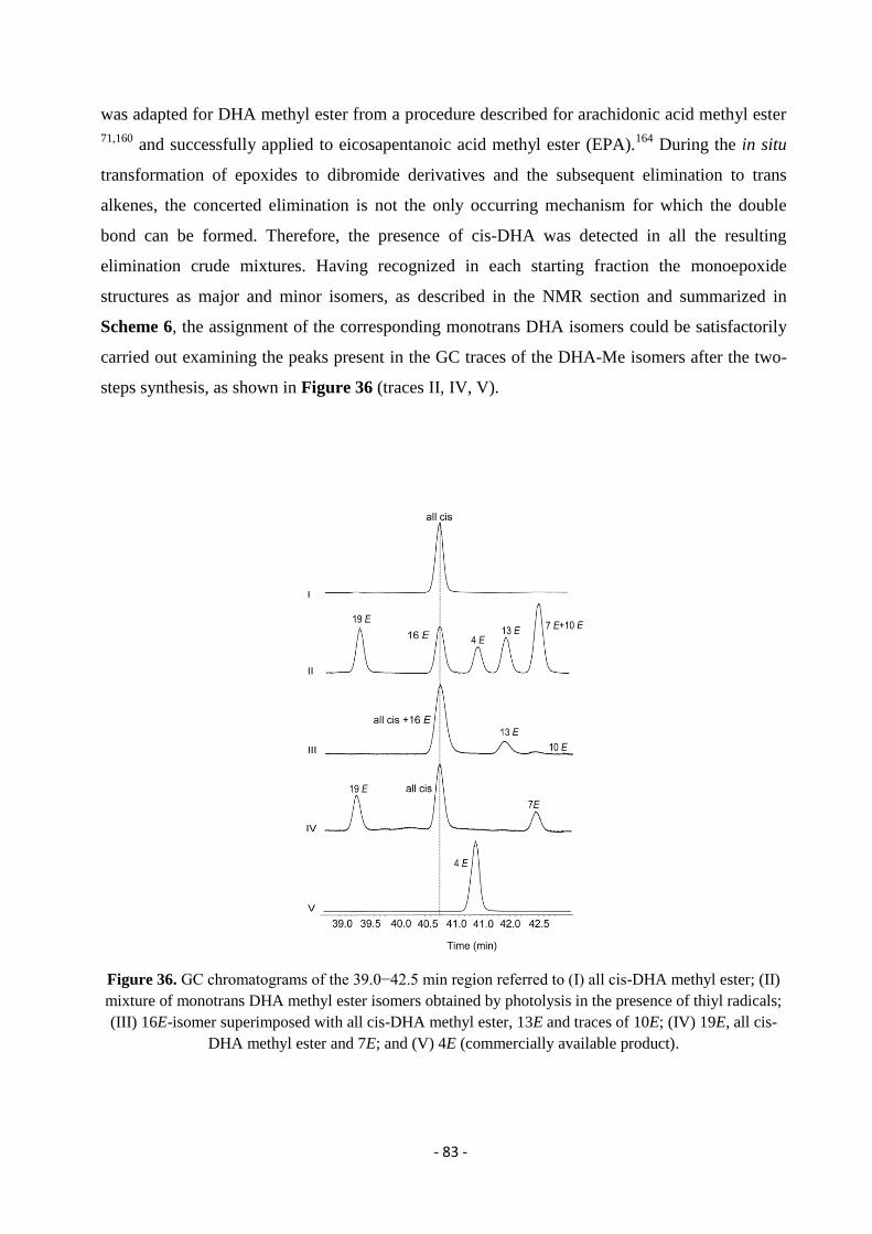
- 83 -
was adapted for DHA methyl ester from a procedure described for arachidonic acid methyl ester
71,160 and successfully applied to eicosapentanoic acid methyl ester (EPA).
164 During the in situ
transformation of epoxides to dibromide derivatives and the subsequent elimination to trans
alkenes, the concerted elimination is not the only occurring mechanism for which the double
bond can be formed. Therefore, the presence of cis-DHA was detected in all the resulting
elimination crude mixtures. Having recognized in each starting fraction the monoepoxide
structures as major and minor isomers, as described in the NMR section and summarized in
Scheme 6, the assignment of the corresponding monotrans DHA isomers could be satisfactorily
carried out examining the peaks present in the GC traces of the DHA-Me isomers after the two-
steps synthesis, as shown in Figure 36 (traces II, IV, V).
Figure 36. GC chromatograms of the 39.0−42.5 min region referred to (I) all cis-DHA methyl ester; (II)
mixture of monotrans DHA methyl ester isomers obtained by photolysis in the presence of thiyl radicals;
(III) 16E-isomer superimposed with all cis-DHA methyl ester, 13E and traces of 10E; (IV) 19E, all cis-
DHA methyl ester and 7E; and (V) 4E (commercially available product).
- 84 -
The isomer overlapping with DHA-Me is the 16E monotrans DHA-Me, and the 7E and 10E
monotrans DHA-Me are also superimposed (lanes (III) and (IV) corresponding to the conversion
of these two monoepoxide fractions according to Scheme 6). It is worth recalling that the 10E
isomer was obtained from the traces of the 10,11-EDP, formed with the lowest yield among all
EDP isomers, as ascertained by the study of the NMR spectra reported in the following section.
Finally, the following GC order of elution could be assigned:
19E <16E = all-cis < 4E <13E <7E =10E
This is the first time the assignment of monotrans geometrical isomers of the omega-3 fatty acid
DHA is made, by a combination of Ag-TLC, NMR spectroscopy, gas chromatography, and this
result contributes to the library of noncommercially available trans isomers developed by us for
the long-chain polyunsaturated fatty acids (LC-PUFA) omega-6 and omega-3.
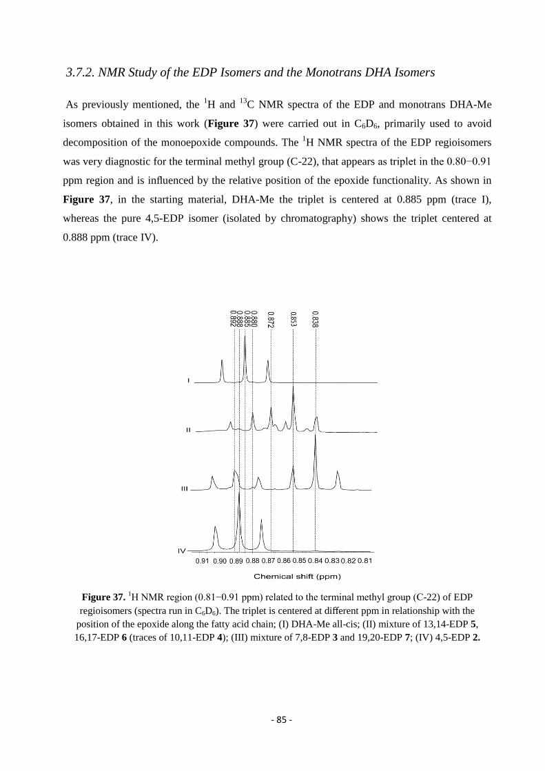
- 85 -
3.7.2. NMR Study of the EDP Isomers and the Monotrans DHA Isomers
As previously mentioned, the 1H and
13C NMR spectra of the EDP and monotrans DHA-Me
isomers obtained in this work (Figure 37) were carried out in C6D6, primarily used to avoid
decomposition of the monoepoxide compounds. The 1H NMR spectra of the EDP regioisomers
was very diagnostic for the terminal methyl group (C-22), that appears as triplet in the 0.80−0.91
ppm region and is influenced by the relative position of the epoxide functionality. As shown in
Figure 37, in the starting material, DHA-Me the triplet is centered at 0.885 ppm (trace I),
whereas the pure 4,5-EDP isomer (isolated by chromatography) shows the triplet centered at
0.888 ppm (trace IV).
Figure 37. 1H NMR region (0.81−0.91 ppm) related to the terminal methyl group (C-22) of EDP
regioisomers (spectra run in C6D6). The triplet is centered at different ppm in relationship with the
position of the epoxide along the fatty acid chain; (I) DHA-Me all-cis; (II) mixture of 13,14-EDP 5,
16,17-EDP 6 (traces of 10,11-EDP 4); (III) mixture of 7,8-EDP 3 and 19,20-EDP 7; (IV) 4,5-EDP 2.
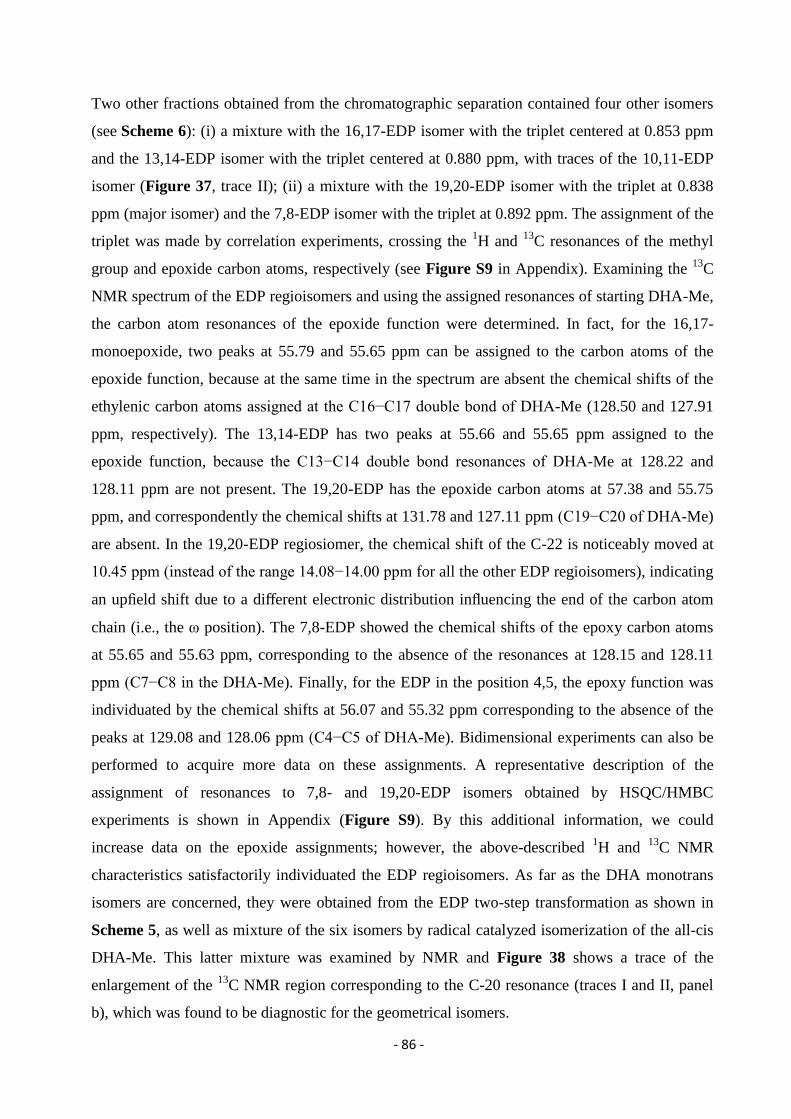
- 86 -
Two other fractions obtained from the chromatographic separation contained four other isomers
(see Scheme 6): (i) a mixture with the 16,17-EDP isomer with the triplet centered at 0.853 ppm
and the 13,14-EDP isomer with the triplet centered at 0.880 ppm, with traces of the 10,11-EDP
isomer (Figure 37, trace II); (ii) a mixture with the 19,20-EDP isomer with the triplet at 0.838
ppm (major isomer) and the 7,8-EDP isomer with the triplet at 0.892 ppm. The assignment of the
triplet was made by correlation experiments, crossing the 1H and
13C resonances of the methyl
group and epoxide carbon atoms, respectively (see Figure S9 in Appendix). Examining the 13
C
NMR spectrum of the EDP regioisomers and using the assigned resonances of starting DHA-Me,
the carbon atom resonances of the epoxide function were determined. In fact, for the 16,17-
monoepoxide, two peaks at 55.79 and 55.65 ppm can be assigned to the carbon atoms of the
epoxide function, because at the same time in the spectrum are absent the chemical shifts of the
ethylenic carbon atoms assigned at the C16−C17 double bond of DHA-Me (128.50 and 127.91
ppm, respectively). The 13,14-EDP has two peaks at 55.66 and 55.65 ppm assigned to the
epoxide function, because the C13−C14 double bond resonances of DHA-Me at 128.22 and
128.11 ppm are not present. The 19,20-EDP has the epoxide carbon atoms at 57.38 and 55.75
ppm, and correspondently the chemical shifts at 131.78 and 127.11 ppm (C19−C20 of DHA-Me)
are absent. In the 19,20-EDP regiosiomer, the chemical shift of the C-22 is noticeably moved at
10.45 ppm (instead of the range 14.08−14.00 ppm for all the other EDP regioisomers), indicating
an upfield shift due to a different electronic distribution influencing the end of the carbon atom
chain (i.e., the ω position). The 7,8-EDP showed the chemical shifts of the epoxy carbon atoms
at 55.65 and 55.63 ppm, corresponding to the absence of the resonances at 128.15 and 128.11
ppm (C7−C8 in the DHA-Me). Finally, for the EDP in the position 4,5, the epoxy function was
individuated by the chemical shifts at 56.07 and 55.32 ppm corresponding to the absence of the
peaks at 129.08 and 128.06 ppm (C4−C5 of DHA-Me). Bidimensional experiments can also be
performed to acquire more data on these assignments. A representative description of the
assignment of resonances to 7,8- and 19,20-EDP isomers obtained by HSQC/HMBC
experiments is shown in Appendix (Figure S9). By this additional information, we could
increase data on the epoxide assignments; however, the above-described 1H and
13C NMR
characteristics satisfactorily individuated the EDP regioisomers. As far as the DHA monotrans
isomers are concerned, they were obtained from the EDP two-step transformation as shown in
Scheme 5, as well as mixture of the six isomers by radical catalyzed isomerization of the all-cis
DHA-Me. This latter mixture was examined by NMR and Figure 38 shows a trace of the
enlargement of the 13
C NMR region corresponding to the C-20 resonance (traces I and II, panel
b), which was found to be diagnostic for the geometrical isomers.
- 87 -
Figure 38. (Panel a) Partial GC traces of: (I) all-cis DHA-Me; (II) the six DHA-Me monotrans isomers
and all-cis DHA-Me; (III) FAME obtained from the transesterification of DHA-containing fish oil. The
presence of monotrans DHA isomers as contaminants is visible when the trace is expanded (Χ 10); (IV)
DHA-Me isomers obtained after isomerization and transesterification of fish oil (this mixture contains
mono-, di-, and tri-trans isomers). (Panel b) Enlargement of the 13
C NMR region (131.5−132.5 pm)
corresponding to the C-20 ethylenic carbon atom of: (I) all-cis DHA-Me; (II) the six DHA-Me monotrans
isomers and all-cis DHA-Me; (III) DHA-containing triglycerides from fish oil; (IV) DHA-containing
triglycerides from fish oil after isomerization (this mixture contains mono-, di-, and tri-trans isomers).
The C-20 atom is the most deshielded among the olefinic peaks, and its distance from the
position of the trans double bond in the fatty acid chain influences its chemical shift. Five
different chemical shifts for this carbon atom could be individuated in the monotrans isomer
mixture isolated from the isomerization (trace II in Figure 38, panel b), one of them representing
two overlapped resonances, and they are clearly different from the C-20 of DHA-Me appearing
at 131.78 ppm (cf. trace I in Figure 38, panel b). The importance of the last vinylic carbon atom
of the PUFA fatty acid chain was previously described for arachidonic and eicosapentaenoic
acids. 157,164
The present data support the use of NMR spectroscopy for the analysis of natural
and marine oils, as already shown by other groups188,189,190
and recently reviewed.191
The herein
described chemical synthesis of monotrans DHA isomers, as well as our previously published
- 88 -
work on trans lipid library,71,140,156,157
certainly increase availability of reference compounds and
will help to gather more information by the powerful NMR methodology.
3.7.3. Isomerization of DHA-Containing Fish Oil and Determination of the
Monotrans DHA Isomer Content in Commercially Available Supplements
Natural oils are important sources of cis PUFA, and trans isomers of EPA and DHA are
contaminants after deodorization or distillation procedures that are used in order to eliminate the
fishy odor. PUFA isomerization in oils has been described by heating procedures147,167
or p-
toluene sulfinic acid treatment,174
whereas we reported the isomerization of some oils by thiyl
radical catalyzed process,176
but not of fish oils. The isomerization of a sample of DHA-
containing fish oil commercially available as raw material for food or supplements was
performed. The reaction was carried out as described in the Materials and Methods, and GC
analysis was performed after transesterification of the triglycerides into the corresponding
FAMEs. Figure 38 (panel a) shows the GC regions corresponding to FAMEs from fish oil in the
starting material (trace III), and it is worth noting that in this sample monotrans DHA isomers are
already present as contaminants, as it can be seen by comparison with the FAME of the oil after
free radical-catalyzed isomerization (trace IV), and related cis and monotrans DHA-Me isomers
(traces I and II). In the same Figure 38 (panel b), the enlarged region of the 13
C NMR spectra
corresponding to the C-20 (ethylenic carbon atom) resonance is shown for the DHA-containing
triglycerides of fish oil (trace III) in comparison with the all-cis DHA-Me (trace I). It is
gratifying to see that the C-20 chemical shift is similar for FAME and triglycerides that contains
the DHA moieties. Moreover, after fish oil isomerization, trans-DHA-containing triglycerides
show strong similarity of the C20 resonances for the monotrans DHA-Me isomers that were
previously described. In Appendix, the full 13
C NMR spectra of the fish oil triglycerides before
and after the isomerization are shown (Figures S25 and S26). It is worth mentioning that the GC
resolution of monotrans isomers achieved under our analytical conditions, using a specific oven
program with a 60 m Χ 0.25 mm Χ 0.25 μm (50%-cyanopropyl)-methylpolysiloxane column,
were obtained after testing several gas chromatographic conditions, in particular concerning the
carrier gas. Using natural sources of fatty acids as marine oils, it was envisaged that other long-
chain fatty acids can elute in the region of the DHA-Me and its monotrans isomers. This was the
- 89 -
case of C22-PUFAs, such as 22:5 ω-3 (DPA) and 22:6 ω-6, as well as long chain saturated and
monounsaturated fatty acids, such as C24:0 and C24:1. In Appendix, Figures S27−30 show the
peak resolution connected with the two GC experimental conditions that are described in
Materials and Methods. We were aware that a very careful study was carried out with a 200 m
SLB IL111 ionic liquid column verifying the accuracy of the proposed method using Standard
Reference Material (SRM) 3275 “Omega-3 and Omega-6 Fatty Acids in Fish Oil” from the
National Institute of Standards and Technology (NIST).175
We could compare the reported
results with our method using hydrogen as carrier gas (Figure S29 in Appendix), and it was
gratifying to see that interferences with other fatty acids can be avoided also under our
conditions. As already shown for EPA isomers,164
the use of hydrogen as carrier gas together
with specific oven temperature program is satisfactory, in terms of no interferences by C22-
PUFAs and long chain saturated and monounsaturated fatty acids.
The combination of GC and 13
C NMR analyses, the latter using the resonance of the C-20, can
be of diagnostic value for individuating monotrans DHA isomers, with the advantage that 13
C
NMR can be performed directly on the triglycerides without any transformation. In this case, it is
also worth noting that by studying the region between 130.40 and 132.60 ppm, also in different
NMR solvents, the overview of omega-6 and omega-3 monotrans isomer content can be
obtained. This overall picture can be relevant for further developments in metabolomics research.
The analysis of the triglyceride fractions contained in DHA-containing supplements available on
the markets in Italy and Spain was performed. As matter of fact, analytical protocol to run
quality control of supplements are increasingly addressed for the safety of producers and
consumers, and researchers raised problems of oxidized or trans contaminants by evaluating
commercial products of different countries.174,175
Table 10 shows our results in terms of fatty
acids, such as omega-3 DPA, DHA, the DHA monotrans isomers and the monounsaturated 24:1,
detected in some representative supplements available in Italian and Spanish markets, presenting
low and high-content of monotrans DHA isomers.
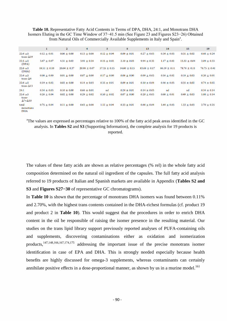
- 90 -
Table 10. Representative Fatty Acid Contents in Terms of DPA, DHA, 24:1, and Monotrans DHA
Isomers Eluting in the GC Time Window of 37−41.5 min (See Figure 23 and Figures S23−26) Obtained
from Natural Oils of Commercially Available Supplements in Italy and Spaina.
aThe values are expressed as percentages relative to 100% of the fatty acid peak areas identified in the GC
analysis. In Tables S2 and S3 (Supporting Information), the complete analysis for 19 products is
reported.
The values of these fatty acids are shown as relative percentages (% rel) in the whole fatty acid
composition determined on the natural oil ingredient of the capsules. The full fatty acid analysis
referred to 19 products of Italian and Spanish markets are available in Appendix (Tables S2 and
S3 and Figures S27−30 of representative GC chromatograms).
In Table 10 is shown that the percentage of monotrans DHA isomers was found between 0.11%
and 2.70%, with the highest trans contents contained in the DHA-richest formulas (cf. product 19
and product 2 in Table 10). This would suggest that the procedures in order to enrich DHA
content in the oil be responsible of raising the isomer presence in the resulting material. Our
studies on the trans lipid library support previously reported analyses of PUFA-containing oils
and supplements, discovering contaminations either as oxidation and isomerization
products,147,148,166,167,174,175
addressing the important issue of the precise monotrans isomer
identification in case of EPA and DHA. This is strongly needed especially because health
benefits are highly discussed for omega-3 supplements, whereas contaminants can certainly
annihilate positive effects in a dose-proportional manner, as shown by us in a murine model.161
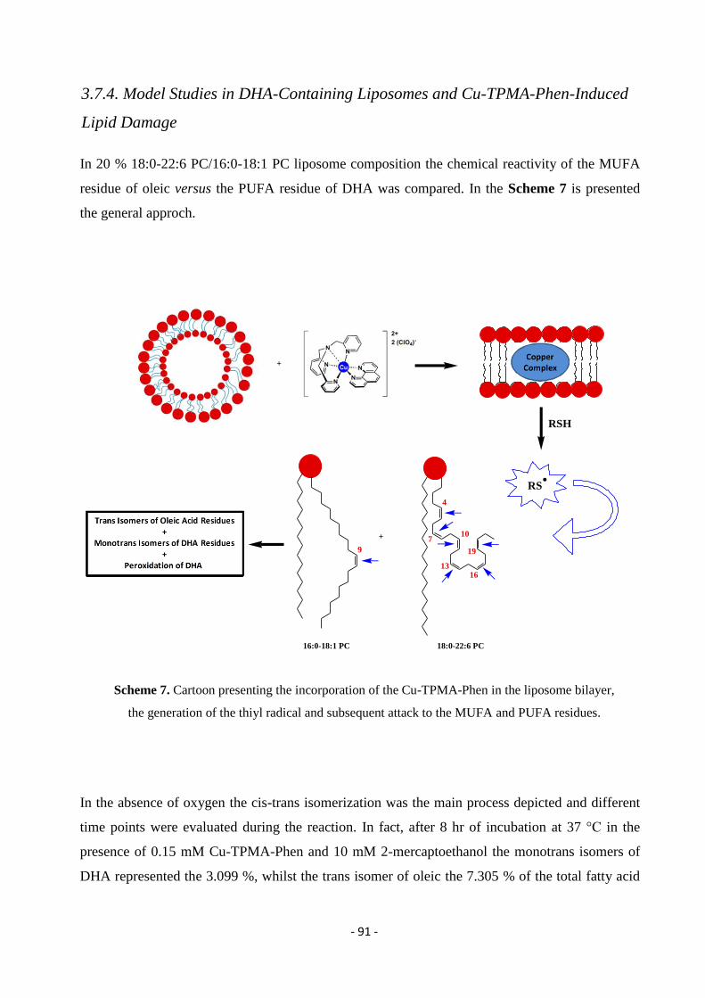
- 91 -
3.7.4. Model Studies in DHA-Containing Liposomes and Cu-TPMA-Phen-Induced
Lipid Damage
In 20 % 18:0-22:6 PC/16:0-18:1 PC liposome composition the chemical reactivity of the MUFA
residue of oleic versus the PUFA residue of DHA was compared. In the Scheme 7 is presented
the general approch.
Scheme 7. Cartoon presenting the incorporation of the Cu-TPMA-Phen in the liposome bilayer,
the generation of the thiyl radical and subsequent attack to the MUFA and PUFA residues.
In the absence of oxygen the cis-trans isomerization was the main process depicted and different
time points were evaluated during the reaction. In fact, after 8 hr of incubation at 37 °C in the
presence of 0.15 mM Cu-TPMA-Phen and 10 mM 2-mercaptoethanol the monotrans isomers of
DHA represented the 3.099 %, whilst the trans isomer of oleic the 7.305 % of the total fatty acid
+
RSH
RS
+
9
4
710
1316
19
16:0-18:1 PC 18:0-22:6 PC
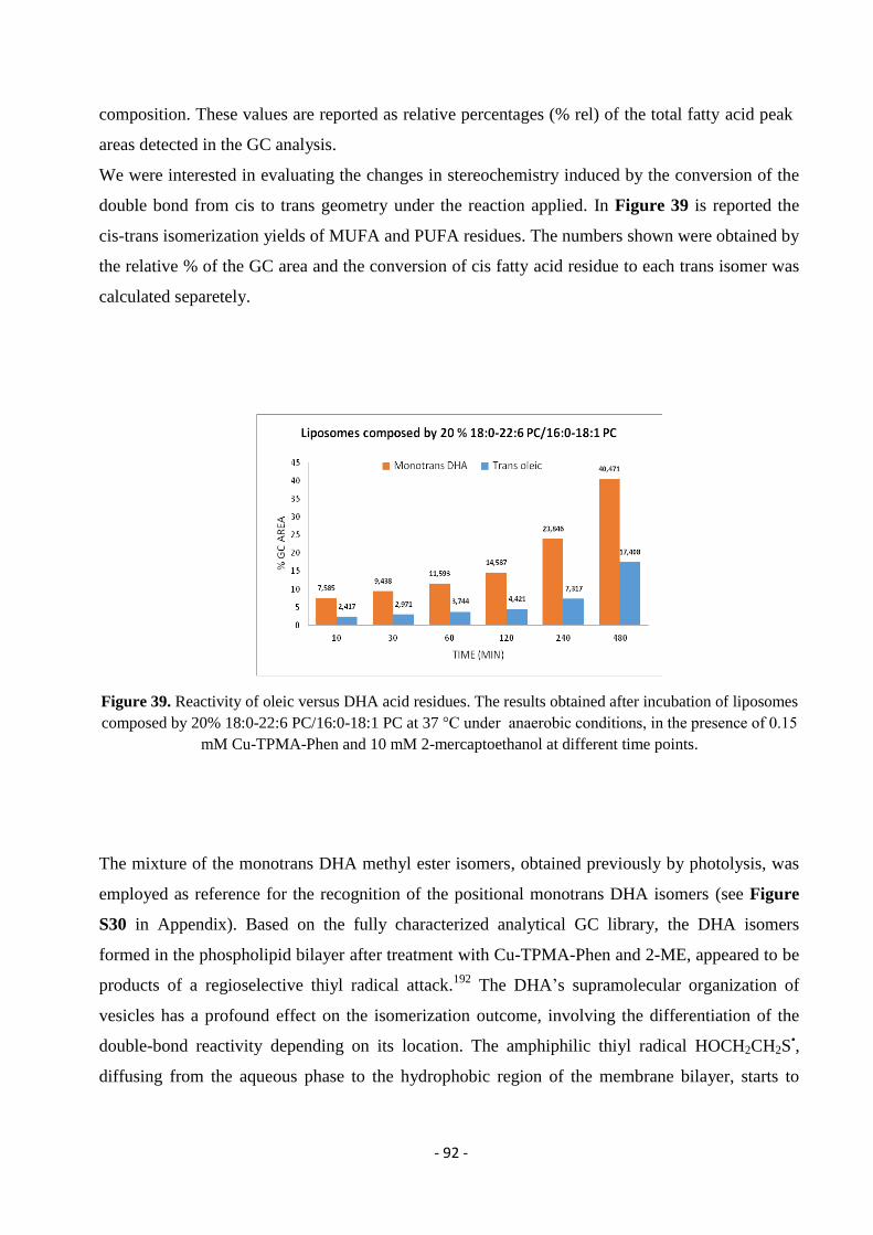
- 92 -
composition. These values are reported as relative percentages (% rel) of the total fatty acid peak
areas detected in the GC analysis.
We were interested in evaluating the changes in stereochemistry induced by the conversion of the
double bond from cis to trans geometry under the reaction applied. In Figure 39 is reported the
cis-trans isomerization yields of MUFA and PUFA residues. The numbers shown were obtained by
the relative % of the GC area and the conversion of cis fatty acid residue to each trans isomer was
calculated separetely.
Figure 39. Reactivity of oleic versus DHA acid residues. The results obtained after incubation of liposomes
composed by 20% 18:0-22:6 PC/16:0-18:1 PC at 37 °C under anaerobic conditions, in the presence of 0.15
mM Cu-TPMA-Phen and 10 mM 2-mercaptoethanol at different time points.
The mixture of the monotrans DHA methyl ester isomers, obtained previously by photolysis, was
employed as reference for the recognition of the positional monotrans DHA isomers (see Figure
S30 in Appendix). Based on the fully characterized analytical GC library, the DHA isomers
formed in the phospholipid bilayer after treatment with Cu-TPMA-Phen and 2-ME, appeared to be
products of a regioselective thiyl radical attack.192
The DHA’s supramolecular organization of
vesicles has a profound effect on the isomerization outcome, involving the differentiation of the
double-bond reactivity depending on its location. The amphiphilic thiyl radical HOCH2CH2S•,
diffusing from the aqueous phase to the hydrophobic region of the membrane bilayer, starts to
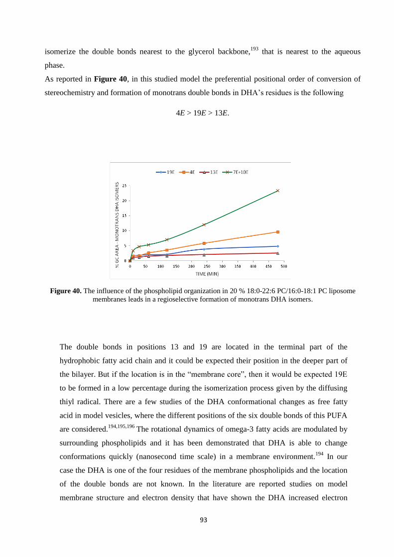
93
isomerize the double bonds nearest to the glycerol backbone,193
that is nearest to the aqueous
phase.
As reported in Figure 40, in this studied model the preferential positional order of conversion of
stereochemistry and formation of monotrans double bonds in DHA’s residues is the following
4E > 19E > 13E.
Figure 40. The influence of the phospholipid organization in 20 % 18:0-22:6 PC/16:0-18:1 PC liposome
membranes leads in a regioselective formation of monotrans DHA isomers.
The double bonds in positions 13 and 19 are located in the terminal part of the
hydrophobic fatty acid chain and it could be expected their position in the deeper part of
the bilayer. But if the location is in the “membrane core”, then it would be expected 19E
to be formed in a low percentage during the isomerization process given by the diffusing
thiyl radical. There are a few studies of the DHA conformational changes as free fatty
acid in model vesicles, where the different positions of the six double bonds of this PUFA
are considered.194,195,196
The rotational dynamics of omega-3 fatty acids are modulated by
surrounding phospholipids and it has been demonstrated that DHA is able to change
conformations quickly (nanosecond time scale) in a membrane environment.194
In our
case the DHA is one of the four residues of the membrane phospholipids and the location
of the double bonds are not known. In the literature are reported studies on model
membrane structure and electron density that have shown the DHA increased electron
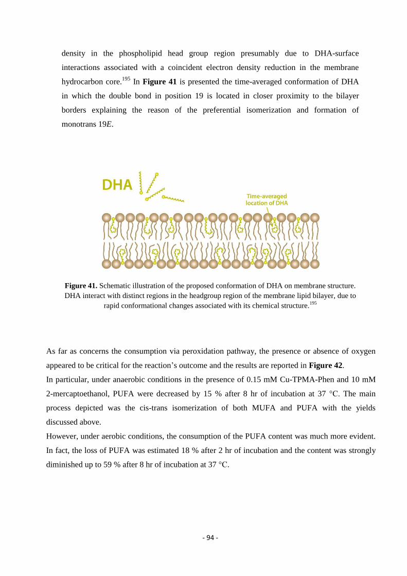
- 94 -
density in the phospholipid head group region presumably due to DHA-surface
interactions associated with a coincident electron density reduction in the membrane
hydrocarbon core.195
In Figure 41 is presented the time-averaged conformation of DHA
in which the double bond in position 19 is located in closer proximity to the bilayer
borders explaining the reason of the preferential isomerization and formation of
monotrans 19E.
Figure 41. Schematic illustration of the proposed conformation of DHA on membrane structure.
DHA interact with distinct regions in the headgroup region of the membrane lipid bilayer, due to
rapid conformational changes associated with its chemical structure.195
As far as concerns the consumption via peroxidation pathway, the presence or absence of oxygen
appeared to be critical for the reaction’s outcome and the results are reported in Figure 42.
In particular, under anaerobic conditions in the presence of 0.15 mM Cu-TPMA-Phen and 10 mM
2-mercaptoethanol, PUFA were decreased by 15 % after 8 hr of incubation at 37 °C. The main
process depicted was the cis-trans isomerization of both MUFA and PUFA with the yields
discussed above.
However, under aerobic conditions, the consumption of the PUFA content was much more evident.
In fact, the loss of PUFA was estimated 18 % after 2 hr of incubation and the content was strongly
diminished up to 59 % after 8 hr of incubation at 37 °C.
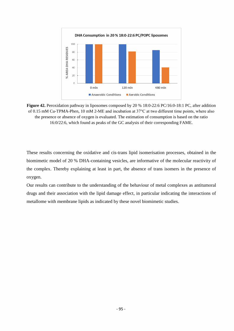
- 95 -
Figure 42. Peroxidation pathway in liposomes composed by 20 % 18:0-22:6 PC/16:0-18:1 PC, after addition
of 0.15 mM Cu-TPMA-Phen, 10 mM 2-ME and incubation at 37°C at two different time points, where also
the presence or absence of oxygen is evaluated. The estimation of consumption is based on the ratio
16:0/22:6, which found as peaks of the GC analysis of their corresponding FAME.
These results concerning the oxidative and cis-trans lipid isomerisation processes, obtained in the
biomimetic model of 20 % DHA-containing vesicles, are informative of the molecular reactivity of
the complex. Thereby explaining at least in part, the absence of trans isomers in the presence of
oxygen.
Our results can contribute to the understanding of the behaviour of metal complexes as antitumoral
drugs and their association with the lipid damage effect, in particular indicating the interactions of
metallome with membrane lipids as indicated by these novel biomimetic studies.
- 96 -
3.8. Conclusions
In conclusion, here we showed the extension of the trans lipid library to include an important long
chain PUFA such as DHA, with the individuation of its structural changes due to chemical
treatment as well as free radical reactivity, converting the cis geometry into trans. It is expected that
this new knowledge will successfully integrate other analytical methodologies for the quality
control of trans fats in health care and food products needed by regulatory bodies all over the world.
The valuable spectra obtained and fully characterized were in benefit of our research towards the
investigation of the artificial chemical nuclease’s Cu-TPMA-Phen interaction with DHA fatty acid
residues in model studies. The monotrans mixture obtained from the free radical catalyzed
isomerization of DHA methyl ester was used as reference for recognition of the positional isomers
that were formed in the presence of a thiol and the artificial chemical nuclease.
The above described results represent preliminary observations of the regioselective cis-trans
isomerization of DHA in biomimetic models of cellular membranes and furnish an aspect of the
mechanistic scenario of DHA’s important role in antitumoral treatments. Our studies provide an
insight of the novel metallodrug Cu-TPMA-Phen molecular mechanism via free radical production
in the presence of DHA composed liposomes. Using a 20 % 18:0-22:6 PC/16:0-18:1 PC liposome
composition, the ability of MUFA and PUFA residues to partition between isomerisation and
consumption processes was investigated and it was evidenced that the supramolecular organization
of phospholipids in the membrane is related to the geometrical isomerism.
As a chemical biology model for antitumoral strategies, liposomes highlight the role of cell
membranes, with synergic roles for chemotherapeutic effects. Fatty acid recruitment and membrane
formation are attracting a lot of interest in cancer, and in this context the loss of the natural cis
geometry and the oxidation-induced lipid remodelling are worthy of deeper studies in anticancer
strategies. Finally, our results indicate the dual drug’s effect towards not only the DNA but also the
membrane lipids.
- 97 -
Chapter 4: Novel Artificial Chemical Nucleases Induce DNA Cleavage
The project described in this chapter was carried out in the frames of my secondment at
Dublin City University, in the National Institute for Cellular Biotechnology under the
supervision of Professor Andrew Kellett. The synthesis, purification and full spectroscopic
characterization of the complexes used was achieved by ESR Nicolo Fantoni. The results
obtained by my work were focused on the AMN’s series [Cu(TPMA)(N,Nʹ)]2
reactivity and
the induced DNA damage. I examined the ability of these materials for precise cleavage of
DNA along with the oxidation properties. Finally, DNA damage fragments were produced in
DCU laboratories after treatment with AMN. The isolation, purification and enzymatic
digestion to single nucleosides was achieved in CNR, Bologna along with the studies of
lesions by stable isotope dilution LC-MS / MS analysis.
The majority of the results presented in this Chapter were published in Chem. Eur. J.; Zuin
Fantoni, N. , Molphy, Z. , Slator, C. , Menounou, G. , Toniolo, G. , Mitrikas, G. , McKee, V. ,
Chatgilialoglu, C. and Kellett, A. (2018), Polypyridyl‐based Copper Phenanthrene
Complexes: A New Type of Stabilized Artificial Chemical Nuclease.
(doi:10.1002/chem.201804084).
4.1. Introduction
A very promising strategy against diseases such a cancer, is the insertion of molecules
between the planar bases of DNA with ultimate goal to inhibit DNA replication in rapidly
growing cancer cells.19
This process is well known as intercalation and the outcome is
correlated with structural distortions towards the DNA helix. Small molecules that cleave
DNA play important roles in gene editing and cancer chemotherapy such as the metallo-
intercalators, which are complexes of a metal cation with polycyclic aromatic ligands.196
The
mechanism of intercalation was proposed for the first time by Leonard Lerman in 1961, as it
follows.197
In aqueous isotonic solution, the cationic intercalator is attracted electrostatically
to the surface of the polyanionic DNA. The negative charges of oxygen atoms in the
phosphate groups across the DNA’s structure are balanced by sodium and/or magnesium
cations, which play the role of a "condensation cloud". In the case of metallo-intercalators,
the metal center displaces these cations that surround DNA and as a result, a weak
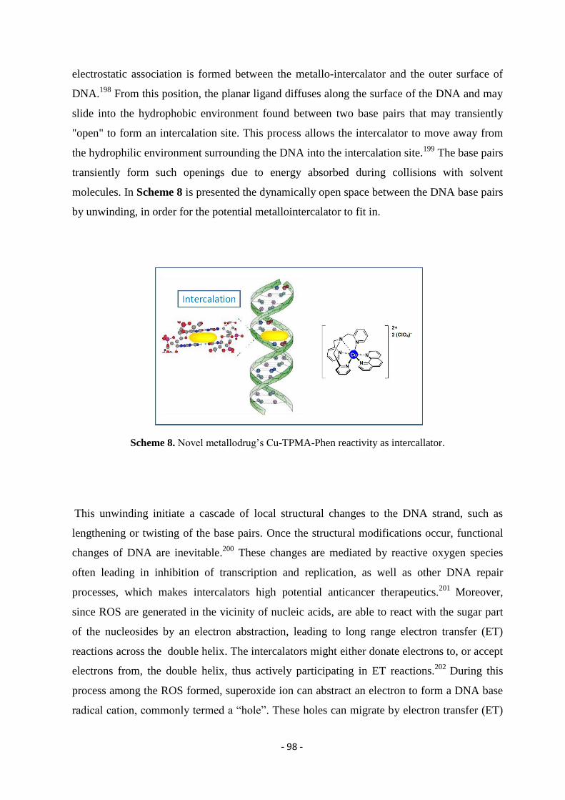
- 98 -
electrostatic association is formed between the metallo-intercalator and the outer surface of
DNA.198
From this position, the planar ligand diffuses along the surface of the DNA and may
slide into the hydrophobic environment found between two base pairs that may transiently
"open" to form an intercalation site. This process allows the intercalator to move away from
the hydrophilic environment surrounding the DNA into the intercalation site.199
The base pairs
transiently form such openings due to energy absorbed during collisions with solvent
molecules. In Scheme 8 is presented the dynamically open space between the DNA base pairs
by unwinding, in order for the potential metallointercalator to fit in.
Scheme 8. Novel metallodrug’s Cu-TPMA-Phen reactivity as intercallator.
This unwinding initiate a cascade of local structural changes to the DNA strand, such as
lengthening or twisting of the base pairs. Once the structural modifications occur, functional
changes of DNA are inevitable.200
These changes are mediated by reactive oxygen species
often leading in inhibition of transcription and replication, as well as other DNA repair
processes, which makes intercalators high potential anticancer therapeutics.201
Moreover,
since ROS are generated in the vicinity of nucleic acids, are able to react with the sugar part
of the nucleosides by an electron abstraction, leading to long range electron transfer (ET)
reactions across the double helix. The intercalators might either donate electrons to, or accept
electrons from, the double helix, thus actively participating in ET reactions.202
During this
process among the ROS formed, superoxide ion can abstract an electron to form a DNA base
radical cation, commonly termed a “hole”. These holes can migrate by electron transfer (ET)
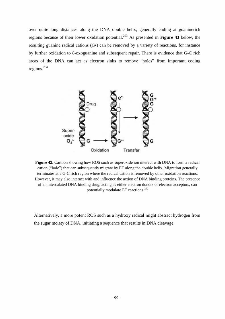
- 99 -
over quite long distances along the DNA double helix, generally ending at guaninerich
regions because of their lower oxidation potential.203
As presented in Figure 43 below, the
resulting guanine radical cations (G•) can be removed by a variety of reactions, for instance
by further oxidation to 8-oxoguanine and subsequent repair. There is evidence that G-C rich
areas of the DNA can act as electron sinks to remove “holes” from important coding
regions.204
Figure 43. Cartoon showing how ROS such as superoxide ion interact with DNA to form a radical
cation (“hole”) that can subsequently migrate by ET along the double helix. Migration generally
terminates at a G-C rich region where the radical cation is removed by other oxidation reactions.
However, it may also interact with and influence the action of DNA binding proteins. The presence
of an intercalated DNA binding drug, acting as either electron donors or electron acceptors, can
potentially modulate ET reactions.202
Alternatively, a more potent ROS such as a hydroxy radical might abstract hydrogen from
the sugar moiety of DNA, initiating a sequence that results in DNA cleavage.
- 100 -
4.2. Copper Complexes as Intercalators with Endonuclease Reactivity
In general, metallo-intercalators mimic the cleavage portion of natural endonuclease
machinery by cutting DNA molecules at or near specific base sequences.205
As they are
versatile and amenable to engineering, coordination compounds with artificial metallo-
nuclease (AMN) activity are an attractive alternative to natural nucleases where, despite their
sequence specificity, limited selectivity impedes precise cleavage of longer DNA
fragments.143,206,207
The advantage of metallo-nucleases is the fact that induce cleavage in
DNA by targeting the cancer cells and impeding faithful cell replication by an oxidation
pathway, mediated by formation of ROS.21,208,209
Once the DNA damage occurs is generally
irreversible and relies on binding of the activated AMN to its requisite target site via groove
binding, insertion or intercalation interactions.210
Copper compounds are attractive choices for
AMNs given their biologically-accessible redox properties and wide structural variability.65
Another factor is the bioavailability and essentiality of copper to the human body where a
daily uptake of between 1.0 – 3.0 mg is required.211
Copper ion homeostasis may, therefore,
facilitate both tolerance and the active transport of exogenous copper based AMNs.212
A wide
range of copper(II) AMNs are known,213,214,215
but one of best studied examples is Sigman’s
reagent [Cu(1,10-phenanthroline)2]2+
(Cu-Phen), which oxidises DNA in the presence of a
reductant and/or oxidant. The mechanism of Cu-Phen, in its reduced Cu(I) form, includes
intercalation in A-T rich sites in the minor groove before abstracting a hydrogen atom from
the C1′ position of deoxyribose to mediate strand excision.216,217,218,219,220
Inspired by Cu-Phen, heteroleptic systems were designed with phenazine intercalators (e.g.
dipyridoquinoxaline (DPQ), dipyridophenazine (DPPZ), and benzo[i]dipyridophenazine
(DPPN)) 221,222
together with TPMA due to the stabilisation and coordination flexibility of this
ligand, as presented in Figure 44.
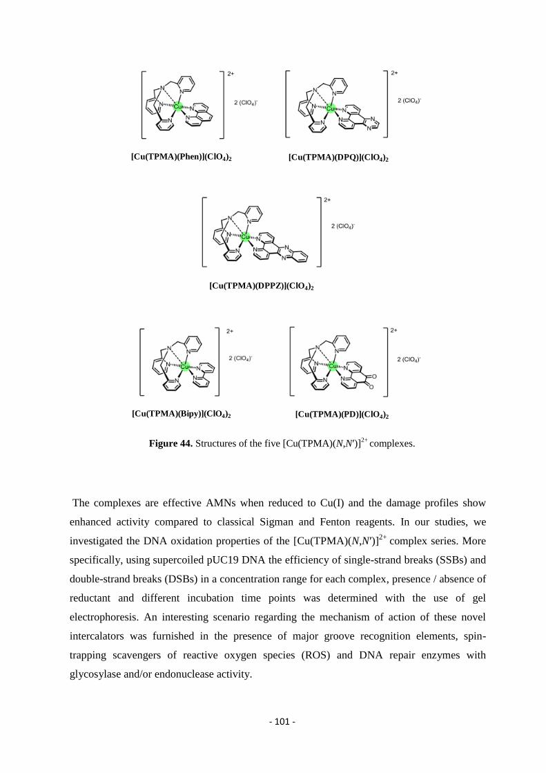
- 101 -
Figure 44. Structures of the five [Cu(TPMA)(N,Nʹ)]2+
complexes.
The complexes are effective AMNs when reduced to Cu(I) and the damage profiles show
enhanced activity compared to classical Sigman and Fenton reagents. In our studies, we
investigated the DNA oxidation properties of the [Cu(TPMA)(N,Nʹ)]2+
complex series. More
specifically, using supercoiled pUC19 DNA the efficiency of single-strand breaks (SSBs) and
double-strand breaks (DSBs) in a concentration range for each complex, presence / absence of
reductant and different incubation time points was determined with the use of gel
electrophoresis. An interesting scenario regarding the mechanism of action of these novel
intercalators was furnished in the presence of major groove recognition elements, spin-
trapping scavengers of reactive oxygen species (ROS) and DNA repair enzymes with
glycosylase and/or endonuclease activity.
[Cu(TPMA)(Bipy)](ClO4)2 [Cu(TPMA)(PD)](ClO4)2
[Cu(TPMA)(Phen)](ClO4)2 [Cu(TPMA)(DPQ)](ClO4)2
[Cu(TPMA)(DPPZ)](ClO4)2
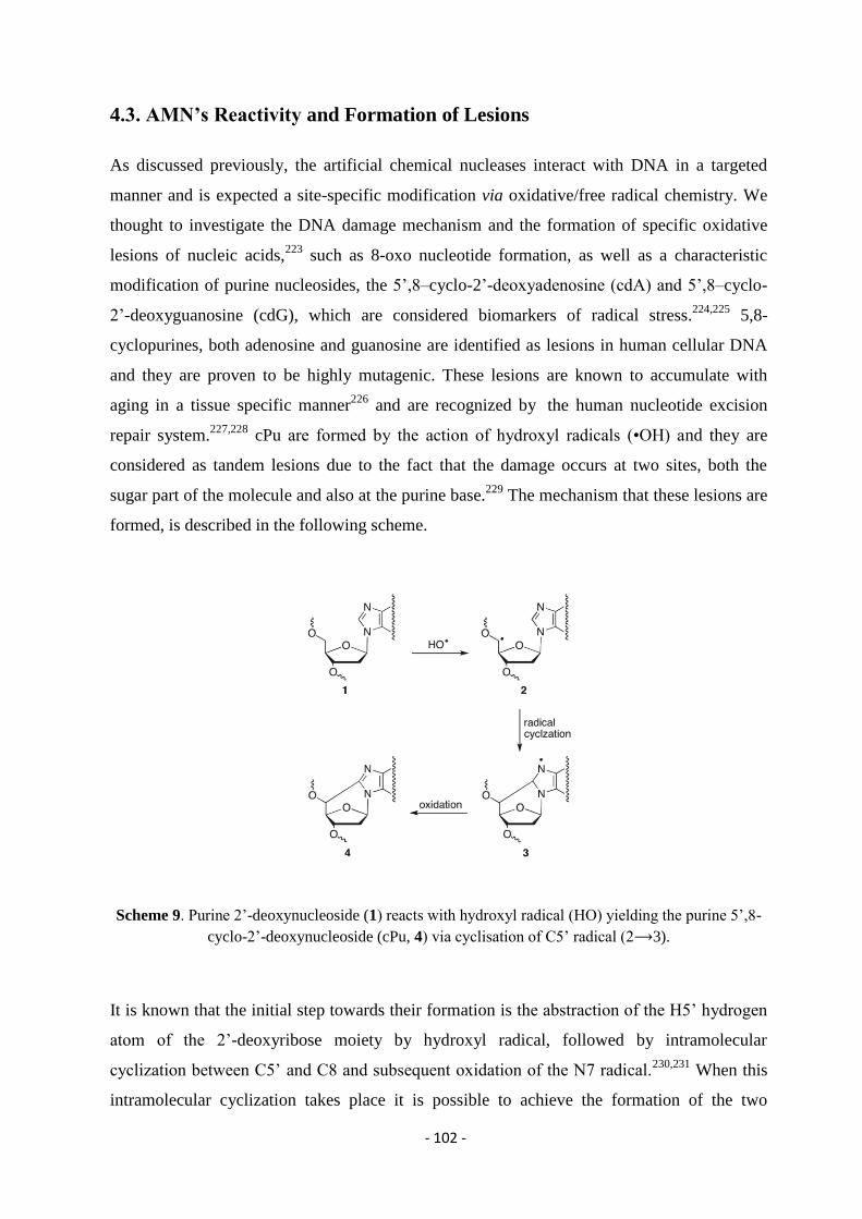
- 102 -
4.3. AMN’s Reactivity and Formation of Lesions
As discussed previously, the artificial chemical nucleases interact with DNA in a targeted
manner and is expected a site-specific modification via oxidative/free radical chemistry. We
thought to investigate the DNA damage mechanism and the formation of specific oxidative
lesions of nucleic acids,223
such as 8-oxo nucleotide formation, as well as a characteristic
modification of purine nucleosides, the 5’,8–cyclo-2’-deoxyadenosine (cdA) and 5’,8–cyclo-
2’-deoxyguanosine (cdG), which are considered biomarkers of radical stress.224,225
5,8-
cyclopurines, both adenosine and guanosine are identified as lesions in human cellular DNA
and they are proven to be highly mutagenic. These lesions are known to accumulate with
aging in a tissue specific manner226
and are recognized by the human nucleotide excision
repair system.227,228
cPu are formed by the action of hydroxyl radicals (•OH) and they are
considered as tandem lesions due to the fact that the damage occurs at two sites, both the
sugar part of the molecule and also at the purine base.229
The mechanism that these lesions are
formed, is described in the following scheme.
Scheme 9. Purine 2’-deoxynucleoside (1) reacts with hydroxyl radical (HO) yielding the purine 5’,8-
cyclo-2’-deoxynucleoside (cPu, 4) via cyclisation of C5’ radical (2⟶3).
It is known that the initial step towards their formation is the abstraction of the H5’ hydrogen
atom of the 2’-deoxyribose moiety by hydroxyl radical, followed by intramolecular
cyclization between C5’ and C8 and subsequent oxidation of the N7 radical.230,231
When this
intramolecular cyclization takes place it is possible to achieve the formation of the two
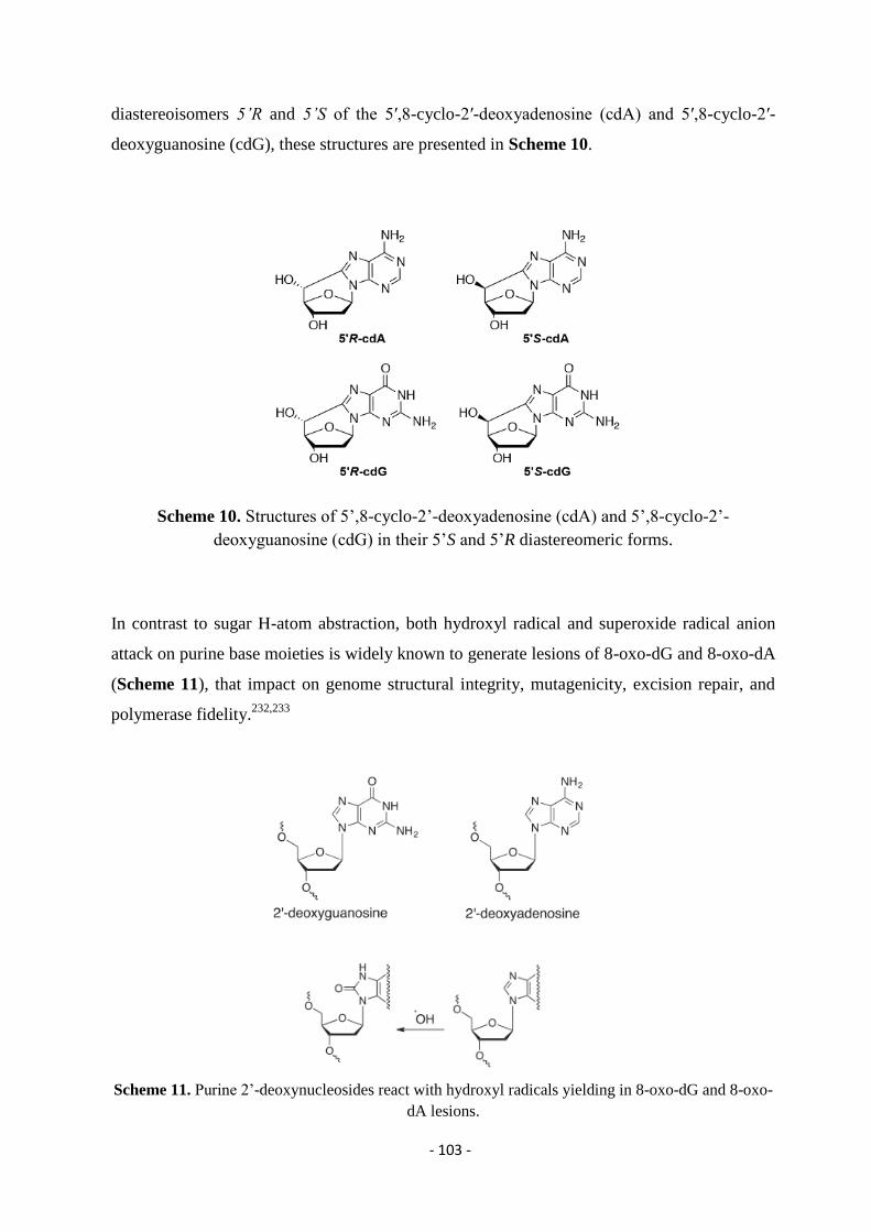
- 103 -
diastereoisomers 5’R and 5’S of the 5′,8-cyclo-2′-deoxyadenosine (cdA) and 5′,8-cyclo-2′-
deoxyguanosine (cdG), these structures are presented in Scheme 10.
Scheme 10. Structures of 5’,8-cyclo-2’-deoxyadenosine (cdA) and 5’,8-cyclo-2’-
deoxyguanosine (cdG) in their 5’S and 5’R diastereomeric forms.
In contrast to sugar H-atom abstraction, both hydroxyl radical and superoxide radical anion
attack on purine base moieties is widely known to generate lesions of 8-oxo-dG and 8-oxo-dA
(Scheme 11), that impact on genome structural integrity, mutagenicity, excision repair, and
polymerase fidelity.232,233
Scheme 11. Purine 2’-deoxynucleosides react with hydroxyl radicals yielding in 8-oxo-dG and 8-oxo-
dA lesions.
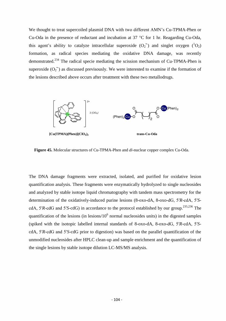
- 104 -
We thought to treat supercoiled plasmid DNA with two different AMN’s Cu-TPMA-Phen or
Cu-Oda in the presence of reductant and incubation at 37 °C for 1 hr. Reagarding Cu-Oda,
this agent’s ability to catalyze intracellular superoxide (O2•-) and singlet oxygen (
1O2)
formation, as radical species mediating the oxidative DNA damage, was recently
demonstrated.234
The radical specie mediating the scission mechanism of Cu-TPMA-Phen is
superoxide (O2•-) as discussed previsously. We were interested to examine if the formation of
the lesions described above occurs after treatment with these two metallodrugs.
Figure 45. Molecular structures of Cu-TPMA-Phen and di-nuclear copper complex Cu-Oda.
The DNA damage fragments were extracted, isolated, and purified for oxidative lesion
quantification analysis. These fragments were enzymatically hydrolyzed to single nucleosides
and analyzed by stable isotope liquid chromatography with tandem mass spectrometry for the
determination of the oxidatively-induced purine lesions (8-oxo-dA, 8-oxo-dG, 5′R-cdA, 5′S-
cdA, 5′R-cdG and 5′S-cdG) in accordance to the protocol established by our group.235,236
The
quantification of the lesions (in lesions/106 normal nucleosides units) in the digested samples
(spiked with the isotopic labelled internal standards of 8-oxo-dA, 8-oxo-dG, 5′R-cdA, 5′S-
cdA, 5′R-cdG and 5′S-cdG prior to digestion) was based on the parallel quantification of the
unmodified nucleosides after HPLC clean-up and sample enrichment and the quantification of
the single lesions by stable isotope dilution LC-MS/MS analysis.
[Cu(TPMA)(Phen)](ClO4)2 trans-Cu-Oda
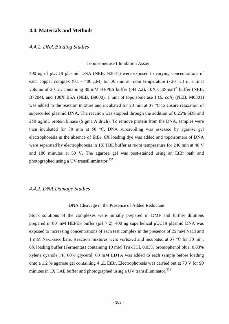
- 105 -
4.4. Materials and Methods
4.4.1. DNA Binding Studies
Topoisomerase I Inhibition Assay
400 ng of pUC19 plasmid DNA (NEB, N3041) were exposed to varying concentrations of
each copper complex (0.1 - 400 µM) for 30 min at room temperature (~20 °C) in a final
volume of 20 µL containing 80 mM HEPES buffer (pH 7.2), 10Χ CutSmart® buffer (NEB,
B7204), and 100Χ BSA (NEB, B9000). 1 unit of topoisomerase I (E. coli) (NEB, M0301)
was added to the reaction mixture and incubated for 20 min at 37 °C to ensure relaxation of
supercoiled plasmid DNA. The reaction was stopped through the addition of 0.25% SDS and
250 µg/mL protein kinase (Sigma Aldrich). To remove protein from the DNA, samples were
then incubated for 30 min at 50 °C. DNA supercoiling was assessed by agarose gel
electrophoresis in the absence of EtBr. 6Χ loading dye was added and topoisomers of DNA
were separated by electrophoresis in 1Χ TBE buffer at room temperature for 240 min at 40 V
and 180 minutes at 50 V. The agarose gel was post-stained using an EtBr bath and
photographed using a UV transilluminator.237
4.4.2. DNA Damage Studies
DNA Cleavage in the Presence of Added Reductant
Stock solutions of the complexes were initially prepared in DMF and further dilutions
prepared in 80 mM HEPES buffer (pH 7.2). 400 ng superhelical pUC19 plasmid DNA was
exposed to increasing concentrations of each test complex in the presence of 25 mM NaCl and
1 mM Na-L-ascorbate. Reaction mixtures were vortexed and incubated at 37 °C for 30 min.
6Χ loading buffer (Fermentas) containing 10 mM Tris-HCl, 0.03% bromophenol blue, 0.03%
xylene cyanole FF, 60% glycerol, 60 mM EDTA was added to each sample before loading
onto a 1.2 % agarose gel containing 4 µL EtBr. Electrophoresis was carried out at 70 V for 90
minutes in 1Χ TAE buffer and photographed using a UV transilluminator.221
- 106 -
DNA Cleavage in the Presence of Non-Covalent DNA Binding Agents
400 ng pUC19 was pre-incubated with 25 mM NaCl, 1 mM Na-L-ascorbate and 8 µM of
methyl green in 80 mM HEPES for 45 min at 37 °C. Samples were vortexed and varying
concentrations of test complexes were then introduced (Cu-TPMA: 10, 15, 20, 25 µM and
Cu-TPMA-Phen / CuTPMA-DPQ / Cu-TPMA-DPPZ: 1, 2.5, 5, 7.5 µM). The reaction
mixture was further incubated at 37 °C for 30 min and subjected to gel electrophoresis
(prepared and stained as previously described).238
DNA Cleavage in the Presence of ROS Scavangers
The assay was conducted according to the method recently reported by Slator et al.234
Briefly,
to a final volume of 20 µL, 80 mM HEPES, 25 mM NaCl, 1 mM Na-L-ascorbate, and 400 ng
of pUC19 DNA were treated with varying drug concentrations (Cu-TPMA: 5, 10, 15, 20, 25
µM and Cu-TPMAPhen / Cu-TPMA-DPQ / Cu-TPMA-DPPZ: 1, 2, 3, 4, 5 µM) in the
presence ROS scavengers; 4,5dihydroxy-1,3-benzenedisulfonic acid (Tiron) (10 mM), D-
Mannitol (10 mM), N′-dimethylthiourea (10 mM), L-Methionine (10 mM), KI (10 mM),
DMSO (10 %) and EDTA (100 µM).239
Reactions were vortexed and incubated at 37 °C for
30 min and electrophoresis carried out as previously stated.
DNA Cleavage in the Presence of Repair Enzymes
Supercoiled pUC19 DNA (400 ng) in 8 mM HEPES, 25 mM NaCl, 1 mM Na-L-ascorbate in
a final volume of 20 μL, was pre-incubated with test series for 30 min at 37 °C at various
concentrations. The following reactions were supplemented with associated buffers as per
manufacturer recommendations; those with Fpg contained 1 mg/mL BSA and 1Χ NEB Buffer
1, Endo III with 1Χ NEB Buffer 1, Endo IV with 1Χ NEB Buffer 3, Endo V with 1Χ NEB
Buffer 4 and hAAG with 1Χ ThermoPol. Subsequently, 2U of repair enzymes FpG (NEB,
M0240S), endonuclease (Endo) III (NEB, M0268S), Endo IV (NEB, M0304S), Endo V
(NEB, M0305S) and hAAG (NEB, M0313S) were added to the reaction mixture and
incubated for 30 min, 37 °C. Samples were denatured with 0.25 % SDS, 250 µg/mL
proteinase K and heated to 50 °C for 20 min. Reactions solutions were loaded onto a 1.2%
- 107 -
agarose gel (4 µL EtBr) with 6Χ loading buffer and subjected to electrophoresis at 70 V for
70 min and imaged using UV transilluminator.
4.4.3. Identification and Quantification of 8-oxo-Pus and cdPus Lesions In The
Presence of AMNs
Nuclease P1 from Penicillium Citrinum, phosphodieasterase I and II, alkaline phosphatase
from bovine intestinal mucosa, DNase I and DNase II, benzonase 99%, BHT, deferoxamine
mesylate and pentostatin were purchased from Sigma-Aldrich, RNase T1 was from Thermo
Fisher Scientific and RNase A from Roche Diagnostic GmbH, (Mannheim, Germany). The 3
kDa cut-off filters were obtained from Millipore (Bedford, USA). Solvents (HPLC-grade)
were purchased from Fisher Scientific.
pUC19 Pasmid DNA Treated with Cu-TPMA-Phen or Cu-Oda to Induce Cleavage
Stock solutions of the complexes were initially prepared in DMF and further dilutions
prepared in 80 mM HEPES buffer (pH 7.2). 400 ng superhelical pUC19 plasmid DNA was
exposed to 2.5 μM concentration of Cu-TPMA-Phen or Cu-Oda in the presence of 25 mM
NaCl and 1 mM Na-L-ascorbate. Reaction mixtures were vortexed and incubated at 37 °C for
60 min. Reactions were repeated to generate sufficient stocks of nicked and linear pUC19 for
subsequent experiments. Nicked and linearized dsDNA were then purified from the
enzymatic reaction using QIAquick purification (QIAGEN, Baltimore, MD), eluted in pure
water, and quantified using the NanoDrop (ND-1000) spectrophotometer.240
Enzymatic Digestion of the dsDNA Samples (SC, OC, L) to Nucleosides
About 20 μg isolated DNA were dissolved in 100 μL of Ar flushed 10 mM Tris-HCl (pH 7.9),
containing 10 mM MgCl, 50 mM NaCl, 0.2 mM pentostatin, 5 μM BHT and 3 mM
deferoxamine and the internal standards were added ([15
N5]-5’S-cdA, [15
N5]-5’R-cdA, [15
N5]-
5’S-cdG, [15
N5]-5’R-cdG and [15
N5]-8-oxo-dA). Three units of benzonase (in 20 mM Tris-
HCl pH 8.0, 2 mM MgCl2 and 20 mM NaCl), 4 mU phosphodiesterase I, 3 U DNase I, 2 mU
of phosphodiesterase II and 2 U of alkaline phosphatase were added and the mixture was
- 108 -
incubated at 37 °C. After 21 h, 35 μL of Ar flushed buffer containing 0.3 M AcONa (pH 5.6)
and 10 mM ZnCl2 were added along with 0.5 U of Nuclease P1 (in 30 mM AcONa pH 5.3, 5
mM ZnCl2 and 50 mM NaCl), 4 mU PDE II and 125 mU of DNase II and the mixture was
further incubated at 37 °C for extra 21 h. A step-quenching with 1% formic acid solution
(final pH 7) was followed, the digestion mixture was placed in a microspin filter (3 kDa) and
the enzymes were filtered off by centrifugation at 14.000 g. (4 °C) for 20 min. Subsequently,
the filtrate was freeze-dried before HPLC analysis, clean-up and enrichment.241
Analysis by High-Performance Liquid Chromatography, Clean-up and Enrichment
HPLC-UV clean-up and enrichment of the enzyme free samples were performed on a 4.6 mm
x 150 mm Atlantis® dC18, 100 Å column (5 μm particle size, Waters) loaded with a 4.6 mm
x 20 mm Guard Column 2pK (Atlantis® dC18 5μm, Waters) on a Waters Alliance® HPLC
System (Waters e2695 Separations Module) including a Waters 2998 Photodiode Array
(PDA) detector. The Empower 3 software was used for the chromatographic instrument
control and data processing. The gradient program used an eluent composed by 2 mM
ammonium formate (solvent A), acetonitrile (solvent B) and methanol (solvent C) and the
flow rate was set at 1 mL/min. Briefly, the gradient program initiated with 99% solvent A,
increasing solvent B from 1% to 30% within 35 min, then immediately solvent C from 0% to
40% for 5 min, closing with initial conditions for 5 min re-equilibration. The fractions
containing the lesions were collected (the time windows were identified by injection of a pure
standard of a lesions mixture). The collected fractions were freeze-dried, pooled, freeze-dried
again, and redissolved in Milli-Q water before been injected for LC-MS/MS analysis. The
quantification of the unmodified nucleosides was based on their absorbance at 260 nm.233b
Measurement of Modified Nucleosides by LC-ESI-MS/MS
An LC-MS/MS system Finnigan TSQ Quantum Discovery Max triple-stage quadrupole mass
spectrometer (Thermo, USA), equipped with electrospray ionization (ESI) source in positive
mode was employed for the detection and quantification of the lesions in the enzymatically
digested DNA samples. Separation of target analytes was achieved with a Finnigan Surveyor
LC system, equipped with a Finnigan Surveyor AS autosampler (Thermo, USA). The
Xcalibur software 2.1 SP 1160 was used to control the mass spectrometric parameters and for
- 109 -
data acquisition. The collected fractions after HPLC analysis were subsequently injected to
the LC-MS/MS system loaded with a 2.1 mm × 150 mm Atlantis dC18, 100 A column (3 lm
particle size, Waters) guarded by a 2.1 mm × 10 mm Guard Column 2pK (Atlantis®
dC18 3
μm, Waters). The gradient elution program used for the chromatographic separation of the
DNA lesions initiated with 99% of 2 mM ammonium formate (solvent A) and 1% acetonitrile
(solvent B) (held for 1 min), increasing solvent B from 1% to 9.8% within 20 min and then
immediately to 15% solvent B (held for 5 min), closing with initial conditions for 10 min re-
equilibration. The flow rate remained constant at 0.2 mL/min, the injection volume was 30 μl
and column temperature was set at 30 °C. Detection was performed in multiple reaction
monitoring mode (MRM) using the two most intense and characteristic precursor/product ion
transitions for each DNA lesion.233b
Statistical Analysis
A two-tailed p-value < 0.05 was considered to indicate a statistically significant difference.
Statistical analysis was performed using t-test by comparing the means of DNA sample
treated with one of the two artificial chemical nucleases (Two-Sample Assuming Unequal
Variances) and error bars represent standard deviation of the mean, calculated from three
independent samples.*denotes a statistically significant difference (p ≤ 0.05), **denotes a
statistically significant difference p ≤ 0.001 and ***denotes a statistically significant
difference p ≤ 0.0001 between the groups.
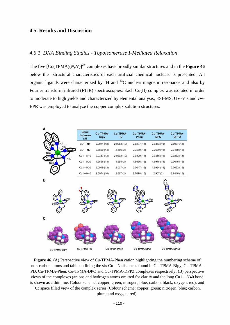
- 110 -
4.5. Results and Discussion
4.5.1. DNA Binding Studies - Topoisomerase I-Mediated Relaxation
The five [Cu(TPMA)(N,N')]2+
complexes have broadly similar structures and in the Figure 46
below the structural characteristics of each artificial chemical nuclease is presented. All
organic ligands were characterized by 1H and
13C nuclear magnetic resonance and also by
Fourier transform infrared (FTIR) spectroscopies. Each Cu(II) complex was isolated in order
to moderate to high yields and characterized by elemental analysis, ESI-MS, UV-Vis and cw-
EPR was employed to analyze the copper complex solution structures.
Figure 46. (A) Perspective view of Cu-TPMA-Phen cation highlighting the numbering scheme of
non-carbon atoms and table outlining the six Cu—N distances found in Cu-TPMA-Bipy, Cu-TPMA-
PD, Cu-TPMA-Phen, Cu-TPMA-DPQ and Cu-TPMA-DPPZ complexes respectively; (B) perspective
views of the complexes (anions and hydrogen atoms omitted for clarity and the long Cu1—N40 bond
is shown as a thin line. Colour scheme: copper, green; nitrogen, blue; carbon, black; oxygen, red); and
(C) space filled view of the complex series (Colour scheme: copper, green; nitrogen, blue; carbon,
plum; and oxygen, red).
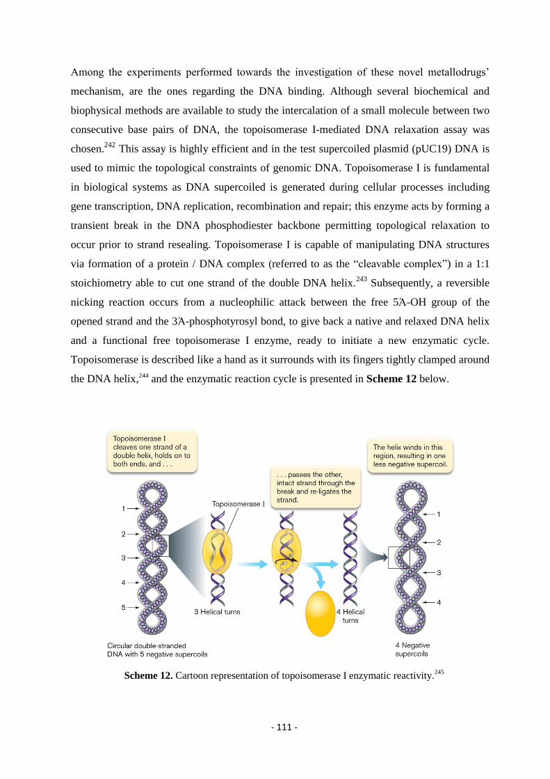
- 111 -
Among the experiments performed towards the investigation of these novel metallodrugs’
mechanism, are the ones regarding the DNA binding. Although several biochemical and
biophysical methods are available to study the intercalation of a small molecule between two
consecutive base pairs of DNA, the topoisomerase I-mediated DNA relaxation assay was
chosen.242
This assay is highly efficient and in the test supercoiled plasmid (pUC19) DNA is
used to mimic the topological constraints of genomic DNA. Topoisomerase I is fundamental
in biological systems as DNA supercoiled is generated during cellular processes including
gene transcription, DNA replication, recombination and repair; this enzyme acts by forming a
transient break in the DNA phosphodiester backbone permitting topological relaxation to
occur prior to strand resealing. Topoisomerase I is capable of manipulating DNA structures
via formation of a protein / DNA complex (referred to as the “cleavable complex”) in a 1:1
stoichiometry able to cut one strand of the double DNA helix.243
Subsequently, a reversible
nicking reaction occurs from a nucleophilic attack between the free 5Ά-OH group of the
opened strand and the 3Ά-phosphotyrosyl bond, to give back a native and relaxed DNA helix
and a functional free topoisomerase I enzyme, ready to initiate a new enzymatic cycle.
Topoisomerase is described like a hand as it surrounds with its fingers tightly clamped around
the DNA helix,244 and the enzymatic reaction cycle is presented in Scheme 12 below.
Scheme 12. Cartoon representation of topoisomerase I enzymatic reactivity.245
- 112 -
The unwinding assay using topo I relies on the ability of the enzyme to efficiently relax the
plasmid with its bound intercalator followed by the resupercoiling of the plasmid upon
removal of the intercalator.246
Circular DNA can assume different degrees of supercoiling
ranging from the fully relaxed form to the fully supercoiled form. The degree of supercoiling
defines distinct forms of the plasmid referred to as topoisomers. Topoisomers can be
separated as more or less discrete bands using gel electrophoresis since resistance to
migration through such gels decreases with degree of supercoiling. The degree of supercoiling
present after treatment reflects the degree of intercalation.247
In our studies unwinding by copper(II) complexes was examined between 0.1 – 400 µM and
the results pointed out that although Cu-TPMA-Phen could not unwind pUC19 below 50 µM,
Cu-TPMA-DPQ and Cu-TPMA-DPPZ complexes fully relaxed (0) the plasmid at 1.0 µM
with higher concentrations generating a positively (+) supercoiled DNA indicating a strong
propensity to intercalate as presented in Figure 47.
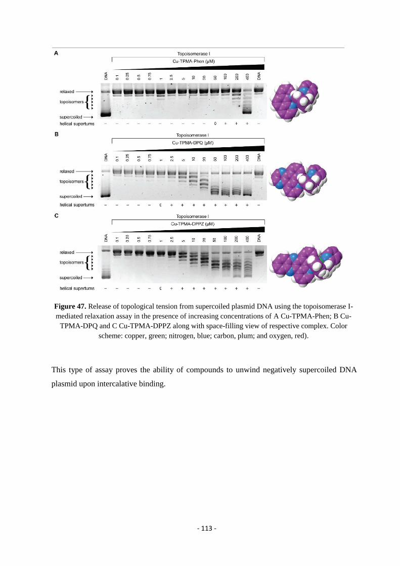
- 113 -
Figure 47. Release of topological tension from supercoiled plasmid DNA using the topoisomerase I-
mediated relaxation assay in the presence of increasing concentrations of A Cu-TPMA-Phen; B Cu-
TPMA-DPQ and C Cu-TPMA-DPPZ along with space-filling view of respective complex. Color
scheme: copper, green; nitrogen, blue; carbon, plum; and oxygen, red).
This type of assay proves the ability of compounds to unwind negatively supercoiled DNA
plasmid upon intercalative binding.
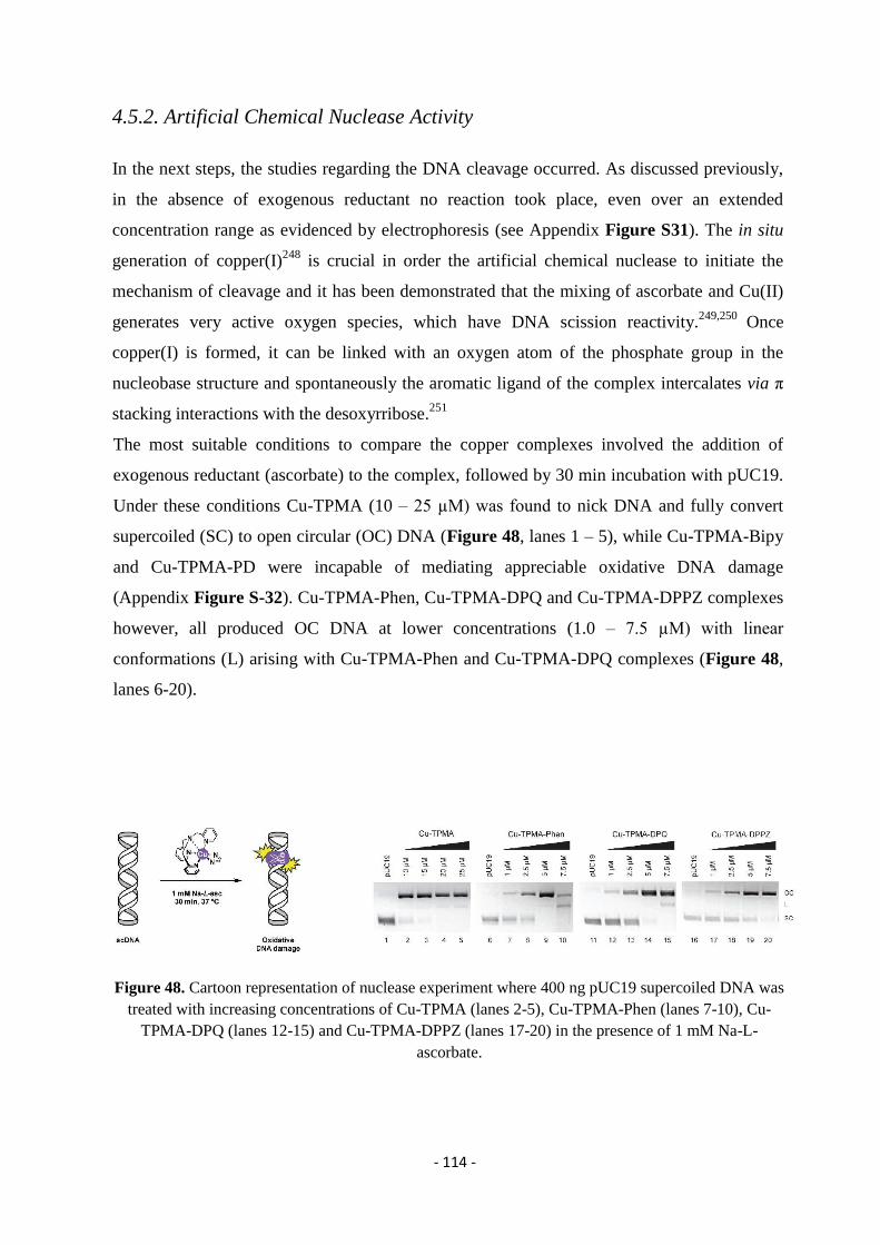
- 114 -
4.5.2. Artificial Chemical Nuclease Activity
In the next steps, the studies regarding the DNA cleavage occurred. As discussed previously,
in the absence of exogenous reductant no reaction took place, even over an extended
concentration range as evidenced by electrophoresis (see Appendix Figure S31). The in situ
generation of copper(I)248
is crucial in order the artificial chemical nuclease to initiate the
mechanism of cleavage and it has been demonstrated that the mixing of ascorbate and Cu(II)
generates very active oxygen species, which have DNA scission reactivity.249,250
Once
copper(I) is formed, it can be linked with an oxygen atom of the phosphate group in the
nucleobase structure and spontaneously the aromatic ligand of the complex intercalates via π
stacking interactions with the desoxyrribose.251
The most suitable conditions to compare the copper complexes involved the addition of
exogenous reductant (ascorbate) to the complex, followed by 30 min incubation with pUC19.
Under these conditions Cu-TPMA (10 – 25 µM) was found to nick DNA and fully convert
supercoiled (SC) to open circular (OC) DNA (Figure 48, lanes 1 – 5), while Cu-TPMA-Bipy
and Cu-TPMA-PD were incapable of mediating appreciable oxidative DNA damage
(Appendix Figure S-32). Cu-TPMA-Phen, Cu-TPMA-DPQ and Cu-TPMA-DPPZ complexes
however, all produced OC DNA at lower concentrations (1.0 – 7.5 µM) with linear
conformations (L) arising with Cu-TPMA-Phen and Cu-TPMA-DPQ complexes (Figure 48,
lanes 6-20).
Figure 48. Cartoon representation of nuclease experiment where 400 ng pUC19 supercoiled DNA was
treated with increasing concentrations of Cu-TPMA (lanes 2-5), Cu-TPMA-Phen (lanes 7-10), Cu-
TPMA-DPQ (lanes 12-15) and Cu-TPMA-DPPZ (lanes 17-20) in the presence of 1 mM Na-L-
ascorbate.
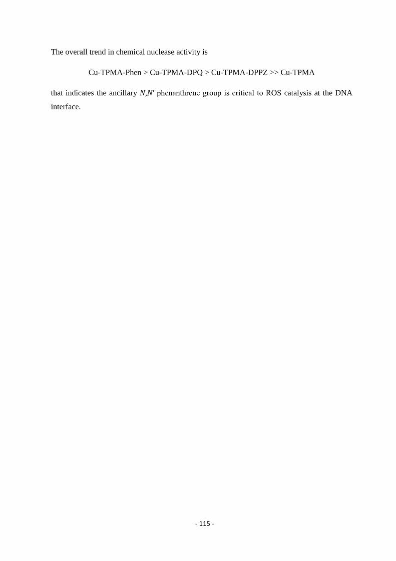
- 115 -
The overall trend in chemical nuclease activity is
Cu-TPMA-Phen > Cu-TPMA-DPQ > Cu-TPMA-DPPZ >> Cu-TPMA
that indicates the ancillary N,N′ phenanthrene group is critical to ROS catalysis at the DNA
interface.
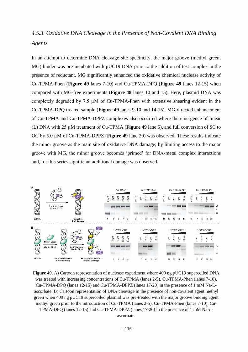
- 116 -
4.5.3. Oxidative DNA Cleavage in the Presence of Non-Covalent DNA Binding
Agents
In an attempt to determine DNA cleavage site specificity, the major groove (methyl green,
MG) binder was pre-incubated with pUC19 DNA prior to the addition of test complex in the
presence of reductant. MG significantly enhanced the oxidative chemical nuclease activity of
Cu-TPMA-Phen (Figure 49 lanes 7-10) and Cu-TPMA-DPQ (Figure 49 lanes 12-15) when
compared with MG-free experiments (Figure 48 lanes 10 and 15). Here, plasmid DNA was
completely degraded by 7.5 µM of Cu-TPMA-Phen with extensive shearing evident in the
Cu-TPMA-DPQ treated sample (Figure 49 lanes 9-10 and 14-15). MG-directed enhancement
of Cu-TPMA and Cu-TPMA-DPPZ complexes also occurred where the emergence of linear
(L) DNA with 25 µM treatment of Cu-TPMA (Figure 49 lane 5), and full conversion of SC to
OC by 5.0 µM of Cu-TPMA-DPPZ (Figure 49 lane 20) was observed. These results indicate
the minor groove as the main site of oxidative DNA damage; by limiting access to the major
groove with MG, the minor groove becomes ‘primed’ for DNA-metal complex interactions
and, for this series significant additional damage was observed.
Figure 49. A) Cartoon representation of nuclease experiment where 400 ng pUC19 supercoiled DNA
was treated with increasing concentrations of Cu-TPMA (lanes 2-5), Cu-TPMA-Phen (lanes 7-10),
Cu-TPMA-DPQ (lanes 12-15) and Cu-TPMA-DPPZ (lanes 17-20) in the presence of 1 mM Na-L-
ascorbate. B) Cartoon representation of DNA cleavage in the presence of non-covalent agent methyl
green when 400 ng pUC19 supercoiled plasmid was pre-treated with the major groove binding agent
methyl green prior to the introduction of Cu-TPMA (lanes 2-5), Cu-TPMA-Phen (lanes 7-10), Cu-
TPMA-DPQ (lanes 12-15) and Cu-TPMA-DPPZ (lanes 17-20) in the presence of 1 mM Na-L-
ascorbate.
- 117 -
4.5.4. DNA Damage Mechanism
The one-electron difference between Cu(II) and Cu(I) allows copper to promote radical
reactions, giving rise in the formation of ROS in the DNA interface. A variety of experiments
was carried out including pre-incubation with ROS specific scavengers such as 4,5-dihydroxy-
1,3-benzenedisulfonic acid (tiron), D-mannitol, L-methionine, and N,N′-dimethylthiourea
(DMTU). These compounds were employed to probe the role of superoxide (O2•−
), the hydroxyl
radical (•OH), hydrogen peroxide (H2O2), hypochlorous acid (HOCl) or a combination. The
objective of this investigation was to gain an insight of the generated species that represent an
important aspect of the mechanism. The results shown in Figure 50, point out that O2•−
is the
most prevalent radical specie involved in the cleavage mechanism as in all cases, pre-incubation
with tiron significantly impeded AMN activity. In these experiments, delayed onset of OC-DNA
formation, inhibition of L-DNA, and protection of SC-DNA were evident. H2O2 is also
generated as part of the mechanism as DMTU somewhat impeded chemical nuclease activity.
Figure 50. A) Cartoon representation of nuclease experiment where 400 ng pUC19 supercoiled DNA
was treated with increasing concentrations of Cu-TPMA (lanes 2-5), Cu-TPMA-Phen (lanes 7-10),
Cu-TPMA-DPQ (lanes 12-15) and Cu-TPMA-DPPZ (lanes 17-20) in the presence of 1 mM Na-L-
ascorbate. B) Table showing ROS scavenging species employed in this study where 400 ng pUC19
supercoiled DNA was treated with increasing concentrations of Cu-TPMA (lanes 1-5), Cu-TPMAPhen
(lanes 6-10), Cu-TPMA-DPQ (lanes 11-15) and Cu-TPMA-DPPZ (lanes 16-20) in the presence of 1
mM Na-L-ascorbate and 10 mM scavenging species 4,5-Dihydroxy-1,3-benzenedisulfonic acid
(Tiron).
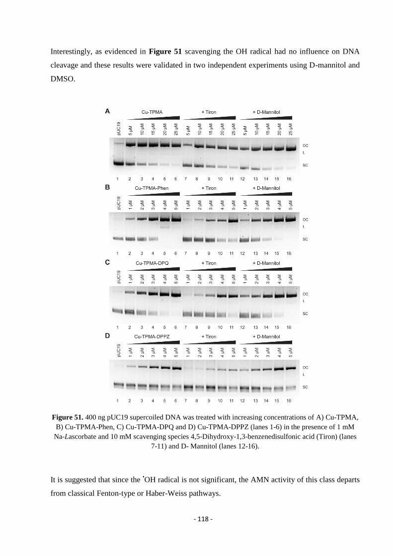
- 118 -
Interestingly, as evidenced in Figure 51 scavenging the OH radical had no influence on DNA
cleavage and these results were validated in two independent experiments using D-mannitol and
DMSO.
Figure 51. 400 ng pUC19 supercoiled DNA was treated with increasing concentrations of A) Cu-TPMA,
B) Cu-TPMA-Phen, C) Cu-TPMA-DPQ and D) Cu-TPMA-DPPZ (lanes 1-6) in the presence of 1 mM
Na-Lascorbate and 10 mM scavenging species 4,5-Dihydroxy-1,3-benzenedisulfonic acid (Tiron) (lanes
7-11) and D- Mannitol (lanes 12-16).
It is suggested that since the •OH radical is not significant, the AMN activity of this class departs
from classical Fenton-type or Haber-Weiss pathways.
- 119 -
4.5.5. DNA Repair Enzyme Recognition
In another interesting series of experiments, DNA repair enzymes with glycosylase/endonuclease
activity were used in order to demonstrate the distinctive DNA oxidation chemistry. In excision
repair (BER) mechanism, repair enzymes are intrinsic check-points able to restore spontaneous
DNA decomposition and mis-incorporation of mismatch sites, oxidized lesions and/or alkylated
bases during the cell cycle.252
A range of enzymes isolated from both prokaryotic and eukaryotic
sources are able to recognize specific nucleoside modifications and excize the damage site to
prime for subsequent repair processes. Cleavage experiments to identify the mechanism of
oxidative damage utilizing a selected range of repair enzymes. Repair proteins with sole or
combined lesion recognition, known as glycoylases, can incise DNA by removing the base and
generating abasic sites (AB). Glycoylases and several repair endonucleases also recognise AP
(apurine / apyrimidine) sites and mediate strand nicking adjacent to the base-free lesion (Figure
52) which can be identified by increased nicked, linear, and sheared forms of plasmid DNA. The
enzymes used with endonuclease activity are reported below, as well as the nucleotide sequence
recognized for cleavage for each restriction site.
Fpg (formamidopyrimidine [fapy]-DNA glycosylase) (also known as 8-oxoguanine DNA
glycosylase) acts both as a N-glycosylase and an AP-lyase. The N-glycosylase activity
releases damaged purines from double stranded DNA, generating an apurinic (AP site). The
AP-lyase activity cleaves both 3´ and 5´ to the AP site thereby removing the AP site and
leaving a 1 base gap. Some of the damaged bases recognized and removed by Fpg include 7,
8-dihydro-8-oxoguanine (8-oxoguanine), 8-oxoadenine, fapy-guanine, methy-fapy-guanine,
fapy-adenine, aflatoxin B1-fapy-guanine, 5-hydroxy-cytosine and 5-hydroxy-uracil.253,245
Endo III Endonuclease III (Nth) protein from E. coli acts as both N-glycosylase and a AP-
lyase. The N-glycosylase activity releases damaged pyrimidines from double-stranded DNA,
generating a basic (AP site). The AP-lyase activity of the enzyme cleaves 3´ to the AP site
leaving a 5´ phosphate and a 3´ ring opened sugar. Some of the damaged bases recognized
and removed by Endouclease III include urea, 5, 6 dihydroxythymine, thymine glycol, 5-
hydroxy-5 methylhydanton, uracil glycol, 6-hydroxy-5, 6-dihdrothimine and
methyltartronylurea.245,254
Endo IV Endonuclease IV can act on a variety of oxidative damage in DNA. The enzyme is
apurinic/apyrimidinic (AP) endonuclease that will hydrolyse intact AP sites in DNA. AP
- 120 -
sites are cleaved at the first phosphodiester bond that is 5´ to the lesion leaving a hydroxyl
group at the 3´ terminus and a deoxyribose 5´-phosphate at the 5´ terminus. The enzyme also
has a 3´ -diesterease activity and can release phosphoglycolaldehyde, intact deoxyribose 5-
phosphate and phosphate from the 3´ end of DNA.255,256
Endo V Endonuclease V is a repair enzyme found in E. coli that recognizes deoxyinosine, a
deamination product of deoxyadenosine in DNA. Endonuclease V, often called deoxyinosine
3´ endonuclease, recognizes DNA containing deoxyinosines (paired or not) on double-
stranded DNA, single-stranded DNA with deoxyinosines and to a lesser degree, DNA
containing abasic sites or urea, base mismatches, insertion/deletion mismatches, hairpin or
unpaired loops, flaps and pseudo-Y structures. It is believed that Endonuclease V needs
another protein to repair the DNA, as it does not remove the deoxyinsoine or the damaged
bases. Endonuclease V cleaves the second phosphodiester bonds 3´ to the mismatch of
deoxyinosine (2), leaving a nick with 3´-hydroxyl and 5´-phosphate.257,258,259,260
hAAG Human Alkyladenine DNA Glycosylase (hAAG) excises alkylated and oxidative
DNA damaged sites, including 3-methyladenine, 7-methylguanine, 1,N-ethenoadenine and
hypoxanthine. hAAG catalyzes the hydrolysis of the N-glycosidic bond to release the
damaged base. hAAG is also known as methylpurine DNA glycosylase (MPG) or 3-
methyladenine-DNA glycosylase (ANPG).254,261
Reaction conditions for Cu-TPMA-Phen, Cu-TPMA-DPQ, Cu-TPMA-DPPZ (0.5 – 2.5 µM)
complexes were optimized to initiate cleavage of pUC19 SC-DNA to OC and L forms (Figure
53) prior to repair enzyme addition (Figure 52A). These reactions were compared to 1 h
exposure of pUC19 to each complex in the absence of repair enzymes (lanes 1-4, Figure 52C
and D). Overall, enhanced cleavage activities in the order
hAAG > Endo V > Endo III > Fpg
were identified with limited change or inhibition occurring in the presence of Endo IV (Figure
52D). Control experiments involving Cu-Phen (0.5 – 2.5 µM), Cu-TPMA (1.0 – 20.0 µM) and
the hydroxyl radical (•OH) generated from a Cu+/2+
/ H2O2 / Fenton system were examined for
comparative purposes (Figure 52C and Figure S 33). Here, enhanced activity by Cu-Phen in the
presence of Endo III (APyrimidine) but not FpG (APurine) indicates preferential oxidative
- 121 -
damage of thymine residues. pUC19 in the presence of Fenton’s reagent underwent additional
cleavage by FpG, Endo III, and Endo IV which supports formation of oxidized purine and
pyrimidine bases together with abasic sites by the •OH radical. Interestingly, DNA damage by
phenanthroline-containing complexes (Cu-TPMA-Phen and Cu-Phen) was attenuated upon
addition of Endo IV and we propose this inhibition arises from covalent stabilization of the DNA
adduct Endo IV complex. Additionally, since Endo IV had limited effects with Cu-TPMA,
CuTPMA-DPQ, and Cu-TPMA-DPPZ this interaction seems phenanthroline ligand-
specific.219,262
These results are in good agreement with that work since trapping O2•−
with tiron
and stabilizing Endo IV with Phen complexes reduces oxidative DNA damage. hAAG enhanced
the cleavage activity of [Cu(TPMA)(N,N′)]2+
complexes but not [Cu(TPMA)]2+
and, when
combined with observations of additional DNA damage with Endo V, it seems reasonable to
suggest deoxyinosine (dI) or free-base hypoxanthine is generated by these ternary compounds. dI
is a naturally occurring noncanonical base that arises from deamination of adenine,263
however it
can be formed from radical generation of Ade-N6-yl.264
The Ade-N6-yl radical may also be
generated by Cu2+
/H2O2 with elegantly designed immuno-spin trapping experiments establishing
its formation via a 5,5ʹ-dimethyl-1-pyrroline N-oxide (DMPO) nucleoside adduct.265
The extent
of hAAG-mediated DNA damage observed here is dependent on the ancillary ligand choice with
bulkier intercalators giving rise to more extensive shearing (DPPZ > DPQ > Phen) suggesting a
possible alternative deoxyinosine oxidation pathway.
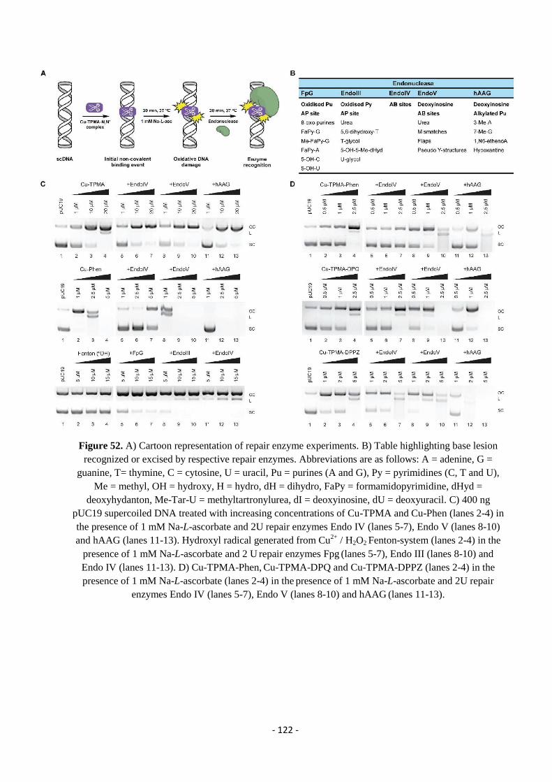
- 122 -
Figure 52. A) Cartoon representation of repair enzyme experiments. B) Table highlighting base lesion
recognized or excised by respective repair enzymes. Abbreviations are as follows: A = adenine, G =
guanine, T= thymine, C = cytosine, U = uracil, Pu = purines (A and G), Py = pyrimidines (C, T and U),
Me = methyl, OH = hydroxy, H = hydro, dH = dihydro, FaPy = formamidopyrimidine, dHyd =
deoxyhydanton, Me-Tar-U = methyltartronylurea, dI = deoxyinosine, dU = deoxyuracil. C) 400 ng
pUC19 supercoiled DNA treated with increasing concentrations of Cu-TPMA and Cu-Phen (lanes 2-4) in
the presence of 1 mM Na-L-ascorbate and 2U repair enzymes Endo IV (lanes 5-7), Endo V (lanes 8-10)
and hAAG (lanes 11-13). Hydroxyl radical generated from Cu2+
/ H2O2 Fenton-system (lanes 2-4) in the
presence of 1 mM Na-L-ascorbate and 2 U repair enzymes Fpg (lanes 5-7), Endo III (lanes 8-10) and
Endo IV (lanes 11-13). D) Cu-TPMA-Phen, Cu-TPMA-DPQ and Cu-TPMA-DPPZ (lanes 2-4) in the
presence of 1 mM Na-L-ascorbate (lanes 2-4) in the presence of 1 mM Na-L-ascorbate and 2U repair
enzymes Endo IV (lanes 5-7), Endo V (lanes 8-10) and hAAG (lanes 11-13).
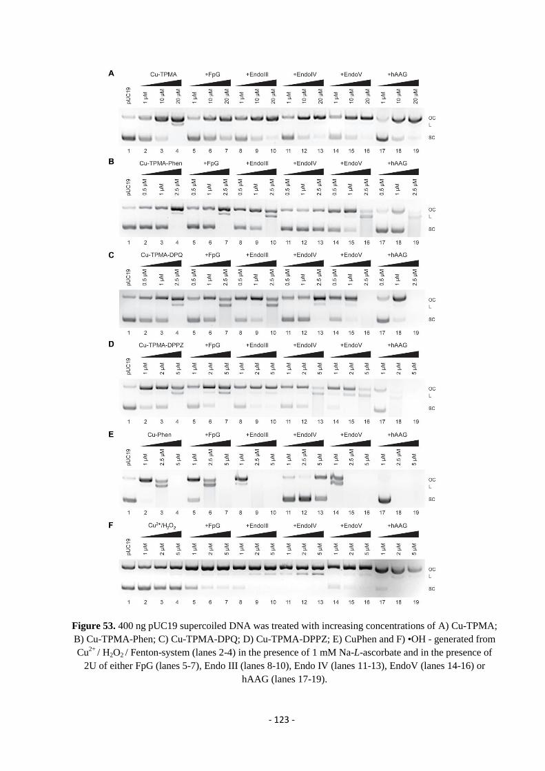
- 123 -
Figure 53. 400 ng pUC19 supercoiled DNA was treated with increasing concentrations of A) Cu-TPMA;
B) Cu-TPMA-Phen; C) Cu-TPMA-DPQ; D) Cu-TPMA-DPPZ; E) CuPhen and F) •OH - generated from
Cu2+
/ H2O2 / Fenton-system (lanes 2-4) in the presence of 1 mM Na-L-ascorbate and in the presence of
2U of either FpG (lanes 5-7), Endo III (lanes 8-10), Endo IV (lanes 11-13), EndoV (lanes 14-16) or
hAAG (lanes 17-19).
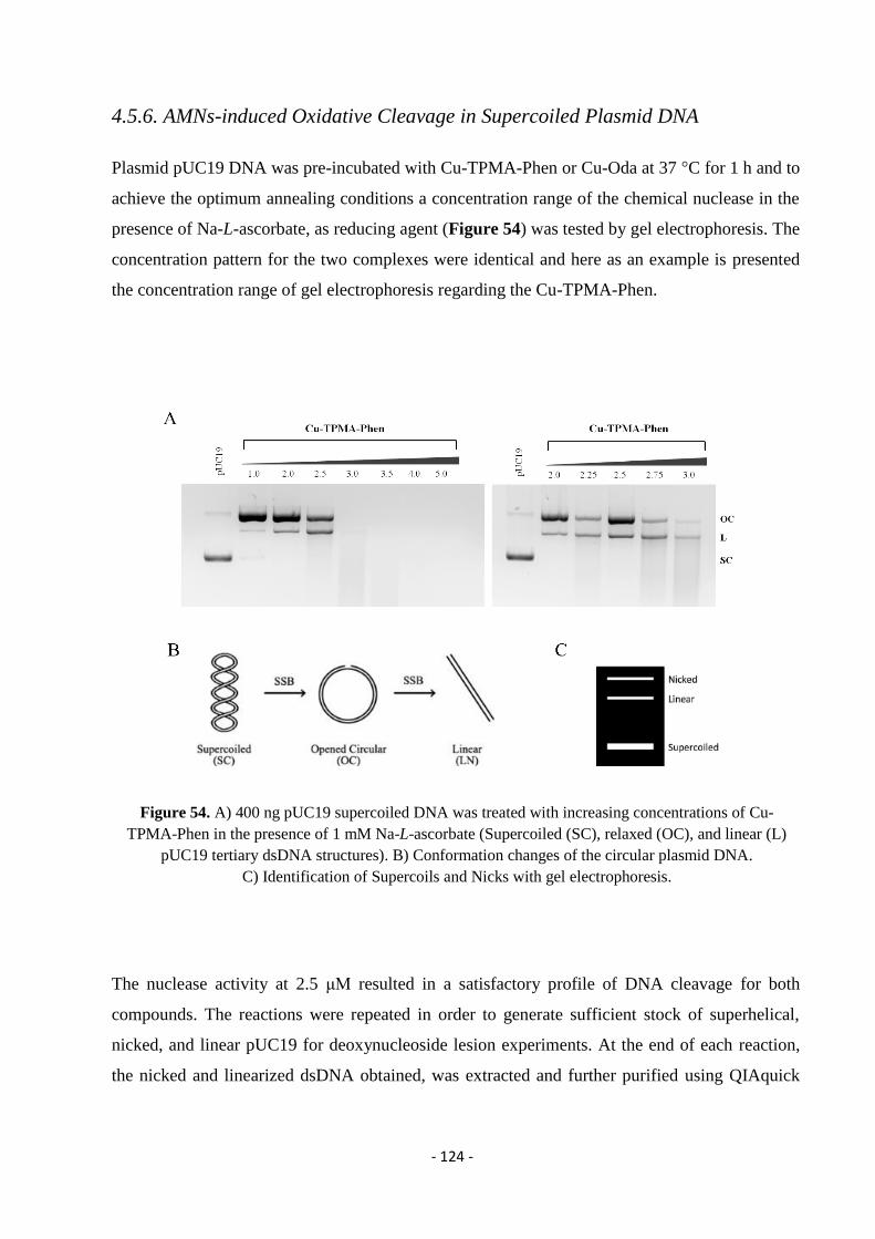
- 124 -
4.5.6. AMNs-induced Oxidative Cleavage in Supercoiled Plasmid DNA
Plasmid pUC19 DNA was pre-incubated with Cu-TPMA-Phen or Cu-Oda at 37 °C for 1 h and to
achieve the optimum annealing conditions a concentration range of the chemical nuclease in the
presence of Na-L-ascorbate, as reducing agent (Figure 54) was tested by gel electrophoresis. The
concentration pattern for the two complexes were identical and here as an example is presented
the concentration range of gel electrophoresis regarding the Cu-TPMA-Phen.
Figure 54. A) 400 ng pUC19 supercoiled DNA was treated with increasing concentrations of Cu-
TPMA-Phen in the presence of 1 mM Na-L-ascorbate (Supercoiled (SC), relaxed (OC), and linear (L)
pUC19 tertiary dsDNA structures). B) Conformation changes of the circular plasmid DNA.
C) Identification of Supercoils and Nicks with gel electrophoresis.
The nuclease activity at 2.5 μΜ resulted in a satisfactory profile of DNA cleavage for both
compounds. The reactions were repeated in order to generate sufficient stock of superhelical,
nicked, and linear pUC19 for deoxynucleoside lesion experiments. At the end of each reaction,
the nicked and linearized dsDNA obtained, was extracted and further purified using QIAquick
- 125 -
purification (QIAGEN, Baltimore, MD), eluted in pure water and quantified in triplicate using
the NanoDrop (ND-1000) spectrophotometer.
Quantification of Purine Lesions in dsDNA Conformers
The double stranded DNA samples were analysed for both detection and quantification of the
oxidative leisions 5′,8-cyclo-2′-deoxyadenosine (cdA) and 5′,8-cyclo-2′-deoxyguanosine (cdG),
7,8-dihydro-8-oxo-2′-deoxyguanosine (8-oxo-dG) and 7,8-dihydro-8-oxo-2′-deoxyadenosine (8-
oxo-dA). Many efforts have been devoted during the last two decades to the identification and
measurement of cdA and cdG lesions in DNA samples. As discussed previously, the initial step
for the formation of these lesions is H5′ abstraction by HO• followed by an intramolecular attack
of C5′ radical to the purine moiety. We were interested in evaluating these cyclopurine lesions
along with 8-oxo-dG and 8-oxo-dA, due to the fact that they are “pure” hydroxyl radical-derived
products and cannot derive from accidental oxidation of the material or other mechanistic
pathways. The absence or presence of these modified nucleosides would provide strong evidence
reagarding the artificial chemical nucleases mechanism and the ability of these materials to lead
in formation of hydroxyl radical. In the experiments plasmid pUC19 supercoiled DNA was
treated with 2.5 µM of Cu-TPMA-Phen or Cu-Oda in the presence of 1 mM Na-L-ascorbate and
the DNA samples were incubated at 37 °C for 1 hr. As reported in the Table 11 and 12 regarding
the cdPus lesions there were no differences among the control and the treated with copper
complexes samples.
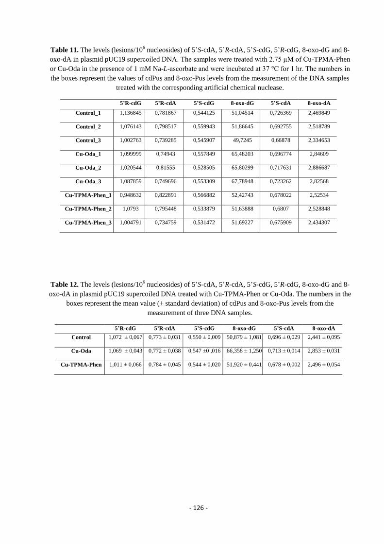
- 126 -
Table 11. The levels (lesions/106 nucleosides) of 5’S-cdA, 5’R-cdA, 5’S-cdG, 5’R-cdG, 8-oxo-dG and 8-
oxo-dA in plasmid pUC19 supercoiled DNA. The samples were treated with 2.75 µM of Cu-TPMA-Phen
or Cu-Oda in the presence of 1 mM Na-L-ascorbate and were incubated at 37 °C for 1 hr. The numbers in
the boxes represent the values of cdPus and 8-oxo-Pus levels from the measurement of the DNA samples
treated with the corresponding artificial chemical nuclease.
5’R-cdG 5’R-cdA 5’S-cdG 8-oxo-dG 5’S-cdA 8-oxo-dA
Control_1
1,136845 0,781867 0,544125 51,04514 0,726369 2,469849
Control_2
1,076143 0,798517 0,559943 51,86645 0,692755 2,518789
Control_3
1,002763 0,739285 0,545907 49,7245 0,66878 2,334653
Cu-Oda_1
1,099999 0,74943 0,557849 65,48203 0,696774 2,84609
Cu-Oda_2
1,020544 0,81555 0,528505 65,80299 0,717631 2,886687
Cu-Oda_3
1,087859 0,749696 0,553309 67,78948 0,723262 2,82568
Cu-TPMA-Phen_1
0,948632 0,822891 0,566882 52,42743 0,678022 2,52534
Cu-TPMA-Phen_2
1,0793 0,795448 0,533879 51,63888 0,6807 2,528848
Cu-TPMA-Phen_3
1,004791 0,734759 0,531472 51,69227 0,675909 2,434307
Table 12. The levels (lesions/106 nucleosides) of 5’S-cdA, 5’R-cdA, 5’S-cdG, 5’R-cdG, 8-oxo-dG and 8-
oxo-dA in plasmid pUC19 supercoiled DNA treated with Cu-TPMA-Phen or Cu-Oda. The numbers in the
boxes represent the mean value (± standard deviation) of cdPus and 8-oxo-Pus levels from the
measurement of three DNA samples.
5’R-cdG 5’R-cdA 5’S-cdG 8-oxo-dG 5’S-cdA 8-oxo-dA
Control
1,072 ± 0,067 0,773 ± 0,031 0,550 ± 0,009 50,879 ± 1,081 0,696 ± 0,029 2,441 ± 0,095
Cu-Oda
1,069 ± 0,043 0,772 ± 0,038 0,547 ±0 ,016 66,358 ± 1,250 0,713 ± 0,014 2,853 ± 0,031
Cu-TPMA-Phen
1,011 ± 0,066 0,784 ± 0,045 0,544 ± 0,020 51,920 ± 0,441 0,678 ± 0,002 2,496 ± 0,054
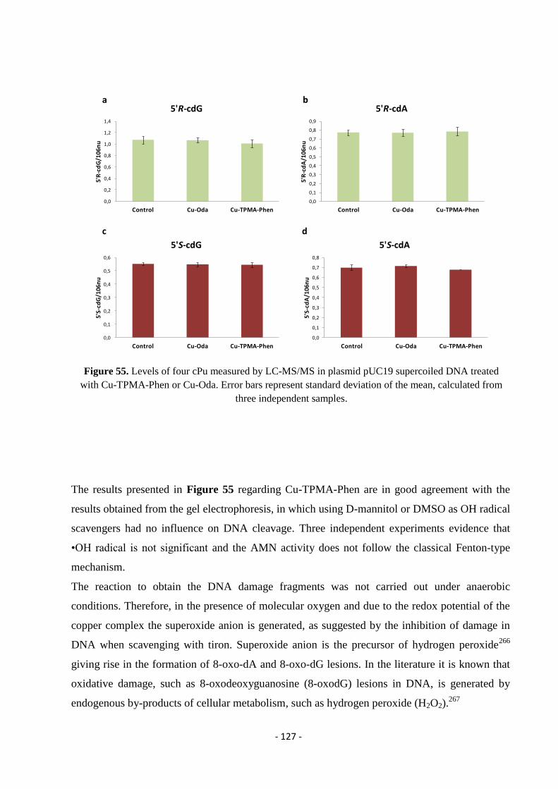
- 127 -
Figure 55. Levels of four cPu measured by LC-MS/MS in plasmid pUC19 supercoiled DNA treated
with Cu-TPMA-Phen or Cu-Oda. Error bars represent standard deviation of the mean, calculated from
three independent samples.
The results presented in Figure 55 regarding Cu-TPMA-Phen are in good agreement with the
results obtained from the gel electrophoresis, in which using D-mannitol or DMSO as OH radical
scavengers had no influence on DNA cleavage. Three independent experiments evidence that
•OH radical is not significant and the AMN activity does not follow the classical Fenton-type
mechanism.
The reaction to obtain the DNA damage fragments was not carried out under anaerobic
conditions. Therefore, in the presence of molecular oxygen and due to the redox potential of the
copper complex the superoxide anion is generated, as suggested by the inhibition of damage in
DNA when scavenging with tiron. Superoxide anion is the precursor of hydrogen peroxide266
giving rise in the formation of 8-oxo-dA and 8-oxo-dG lesions. In the literature it is known that
oxidative damage, such as 8-oxodeoxyguanosine (8-oxodG) lesions in DNA, is generated by
endogenous by-products of cellular metabolism, such as hydrogen peroxide (H2O2).267
0,0
0,2
0,4
0,6
0,8
1,0
1,2
1,4
Control Cu-Oda Cu-TPMA-Phen
5'R
-cd
G/1
06
nu
5'R-cdG
0,0
0,1
0,2
0,3
0,4
0,5
0,6
0,7
0,8
0,9
Control Cu-Oda Cu-TPMA-Phen
5'R
-cd
A/1
06
nu
5'R-cdA
0,0
0,1
0,2
0,3
0,4
0,5
0,6
Control Cu-Oda Cu-TPMA-Phen
5'S
-cd
G/1
06n
u
5'S-cdG
0,0
0,1
0,2
0,3
0,4
0,5
0,6
0,7
0,8
Control Cu-Oda Cu-TPMA-Phen
5'S
-cd
A/1
06n
u
5'S-cdA
a b
c d
- 128 -
Figure 56. Levels of 8-oxo-dA and 8-oxo-dG measured by LC-MS/MS in plasmid pUC19 supercoiled
DNA treated with Cu-TPMA-Phen or Cu-Oda. Statistical analysis was performed using t-test by
comparing the means of DNA sample treated with one of the two artificial chemical nucleases (Two-
Sample Assuming Unequal Variances) and error bars represent standard deviation of the mean, calculated
from three independent samples.*denotes a statistically significant difference (p ≤ 0.05), **denotes a
statistically significant difference p ≤ 0.001 and ***denotes a statistically significant difference p ≤
0.0001 between the groups.
The overall 8-oxo lesion formation, regardless of topology, followed 8-oxo-dG ≫ 8-oxo-dA
(Figure 56) after treatment with both AMNs. However, statistically significant difference was
observed only in the presence of the di-nuclear copper complex Cu-Oda. These results point out
that the ligand groups on the metallodrug play an important role. Although scavenger
experiments showed that both AMNs are able to generate superoxide radical anion, the bulky
ligand of TPMA can interfere and act as a block; ROS formation might not be in the vicinity of
nucleic acids therefore the oxidative DNA damage does not occur.
a b
**
0
0,5
1
1,5
2
2,5
3
control Cu-Oda Cu-TPMA-Phen
8-o
xo-d
A/1
06n
u
8-oxo-dA
0
10
20
30
40
50
60
70
80
control Cu-Oda Cu-TPMA-Phen
8-o
xo-d
G/1
06
nu
8-oxo-dG
**
a b
- 129 -
4.6. Conclusions
The experiments presented proved that copper(II) artificial chemical nucleases are very
promising materials as therapeutic agents especially when combined with specific scaffolds,
essential for controlling stability and reactivity. DNA binding was identified and
topoisomerase I-mediated relaxation revealed the importance of N,N′ ligand choice to
intercalation as Cu-TPMA-DPQ and Cu-TPMA-DPPZ complexes unwound supercoiled
pUC19 DNA with greater efficiency. Further analysis showed the minor groove as the
preferred site for AMN activity as pre-exposure to a major groove binder (methyl green)
enhanced overall oxidative damage sensitivity to supercoiled pUC19 DNA. In terms of the
oxidative mechanism, trapping experiments show that the complexes do not follow classical
Fenton-type or Haber-Weiss processes but instead generate superoxide- and, to a somewhat
lower extent, hydrogen peroxide during the catalytic ROS process. To delineate this activity
from that of classical copper-bis-1,10-phenanthroline (Cu-Phen), Fenton-type systems, and
the [Cu(TPMA)]2+
cation, DNA repair enzyme recognition experiments were conducted.
DNA damage lesions formed by Cu-TPMA-Phen, -DPQ, and -DPPZ complexes were not
recognized by FpG or Endo III, which suggests—in contrast with Cu-Phen and Fenton’s
reagent—an oxidation pathway independent of oxidized purine and pyrimidine bases,
respectively. The activity of Endo IV, which recognizes both apurine and apyrimidine sites,
was inhibited by Cu-Phen and Cu-TPMA-Phen and it appears likely, therefore, that a covalent
DNA-complex-enzyme adduct is realized. The role of deoxyinosine (dI) in the DNA damage
process was established since hAAG and Endo V both enhanced cleavage activity of
[Cu(TPMA)(N,N′)]2+
complexes; the ancillary ligand choice is important here with bulkier
intercalators giving rise to more extensive hAAG-mediated shearing in the order DPPZ >
DPQ > Phen.
In addition, cyclopurine lesions and 8-oxo-purines were identified and quantified on dsDNA
mixtures of supercoiled (SC), open circular (OC), and linear (L) conformation, after treatment
with Cu-TPMA-Phen or Cu-Oda in the presence of reductant. In the enzymatically digested
nucleosides an HPLC clean-up protocol was applied and the fractions of interest containing
the lesions, were collected and analyzed by LC-MS/MS. We focused on the four cPu lesions
that are correlated directly with HO•-induced DNA damage due to the fact that they are strong
candidate biomarkers in cancer diagnostics, particularly for their stability during the analytical
procedures. However, the levels of these lesions were identical with the control proving that
the hydroxyl radicals are not generated in the presence of these chemical nucleases. Finally,
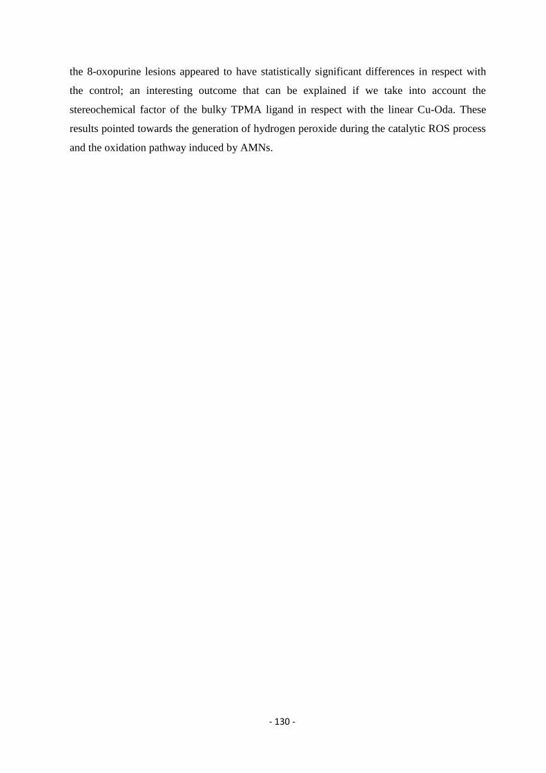
- 130 -
the 8-oxopurine lesions appeared to have statistically significant differences in respect with
the control; an interesting outcome that can be explained if we take into account the
stereochemical factor of the bulky TPMA ligand in respect with the linear Cu-Oda. These
results pointed towards the generation of hydrogen peroxide during the catalytic ROS process
and the oxidation pathway induced by AMNs.
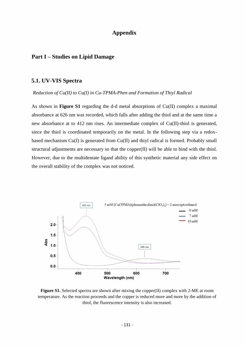
- 131 -
Appendix
Part I – Studies on Lipid Damage
5.1. UV-VIS Spectra
Reduction of Cu(II) to Cu(I) in Cu-TPMA-Phen and Formation of Thiyl Radical
As shown in Figure S1 regarding the d-d metal absorptions of Cu(II) complex a maximal
absorbance at 626 nm was recorded, which falls after adding the thiol and at the same time a
new absorbance at to 412 nm rises. An intermediate complex of Cu(II)-thiol is generated,
since the thiol is coordinated temporarily on the metal. In the following step via a redox-
based mechanism Cu(I) is generated from Cu(II) and thiyl radical is formed. Probably small
structural adjustments are necessary so that the copper(II) will be able to bind with the thiol.
However, due to the multidentate ligand ability of this synthetic material any side effect on
the overall stability of the complex was not noticed.
Figure S1. Selected spectra are shown after mixing the copper(II) complex with 2-ME at room
temperature. As the reaction proceeds and the copper is reduced more and more by the addition of
thiol, the fluorescence intensity is also increased.
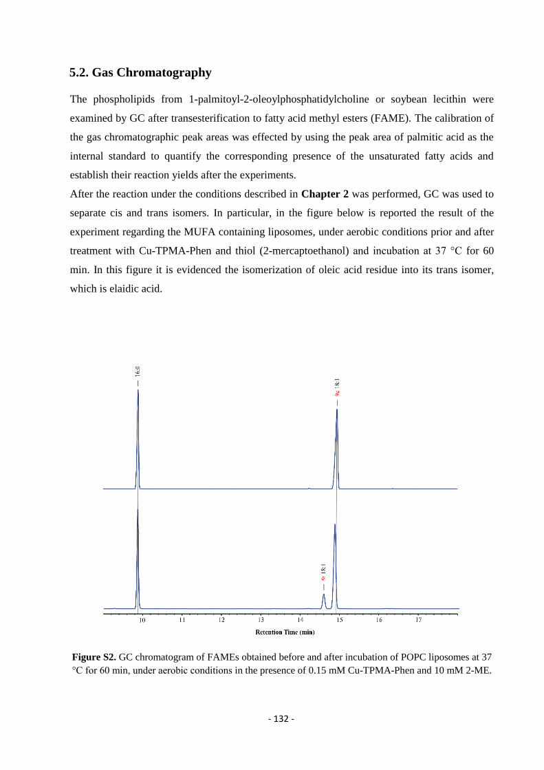
- 132 -
5.2. Gas Chromatography
The phospholipids from 1-palmitoyl-2-oleoylphosphatidylcholine or soybean lecithin were
examined by GC after transesterification to fatty acid methyl esters (FAME). The calibration of
the gas chromatographic peak areas was effected by using the peak area of palmitic acid as the
internal standard to quantify the corresponding presence of the unsaturated fatty acids and
establish their reaction yields after the experiments.
After the reaction under the conditions described in Chapter 2 was performed, GC was used to
separate cis and trans isomers. In particular, in the figure below is reported the result of the
experiment regarding the MUFA containing liposomes, under aerobic conditions prior and after
treatment with Cu-TPMA-Phen and thiol (2-mercaptoethanol) and incubation at 37 °C for 60
min. In this figure it is evidenced the isomerization of oleic acid residue into its trans isomer,
which is elaidic acid.
Figure S2. GC chromatogram of FAMEs obtained before and after incubation of POPC liposomes at 37
°C for 60 min, under aerobic conditions in the presence of 0.15 mM Cu-TPMA-Phen and 10 mM 2-ME.
- 133 -
In the experiments with the MUFA composed liposomes, no peroxidation was observed neither
in aerobic nor in anaerobic conditions, due to the fact that the MUFA moiety of oleic acid (9cis-
18:1) is not so prone to oxidation as PUFAs are. Therefore, this fatty acid residue can be
targetted by thiyl radical only in a catalysed isomerization pathway.
On the other hand, in lecithin liposomes the PUFA can be partitioned both in isomerization or
peroxidation pathways. We decided to monitor the 18:1 and 18:2 fatty acids under different
liposome incubation conditions, since these two fatty acids are the most representative
unsaturated moieties of the mixture. In Figure S3, is shown the GC chromatogram of lecithin
FAMEs without any treatment (starting material) trace I, also traces II and III, report the two
different profiles depending on the presence or absence of oxygen after treatment with Cu-
TPMA-Phen and thiol (2-mercaptoethanol) and incubation at 37 °C for 480 min.
Figure S3. GC chromatogram of FAMEs obtained before any treatment (trace I); after incubation of
lecithin liposomes at 37 °C for 480 min in the presence of 0.15 mM Cu-TPMA-Phen and 10 mM 2-
mercaptoethanol under anaerobic conditions (trace II); same reaction conditions but under aerobic
conditions (trace III).
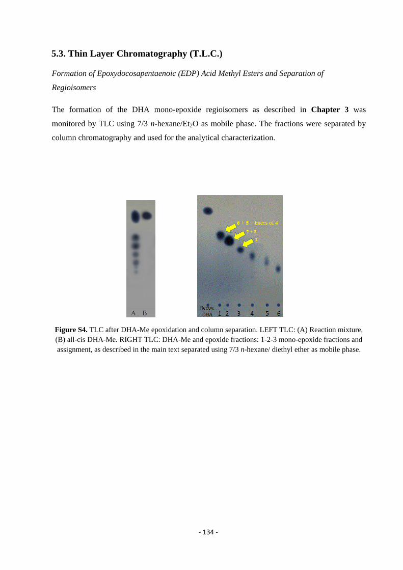
- 134 -
5.3. Thin Layer Chromatography (T.L.C.)
Formation of Epoxydocosapentaenoic (EDP) Acid Methyl Esters and Separation of
Regioisomers
The formation of the DHA mono-epoxide regioisomers as described in Chapter 3 was
monitored by TLC using 7/3 n-hexane/Et2O as mobile phase. The fractions were separated by
column chromatography and used for the analytical characterization.
Figure S4. TLC after DHA-Me epoxidation and column separation. LEFT TLC: (A) Reaction mixture,
(B) all-cis DHA-Me. RIGHT TLC: DHA-Me and epoxide fractions: 1-2-3 mono-epoxide fractions and
assignment, as described in the main text separated using 7/3 n-hexane/ diethyl ether as mobile phase.
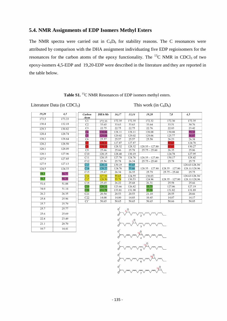
- 135 -
5.4. NMR Assignments of EDP Isomers Methyl Esters
The NMR spectra were carried out in C6D6 for stability reasons. The C resonances were
attributed by comparison with the DHA assignment individuating five EDP regioisomers for the
resonances for the carbon atoms of the epoxy functionality. The 13
C NMR in CDCl3 of two
epoxy-isomers 4,5-EDP and 19,20-EDP were described in the literature and they are reported in
the table below.
Table S1. 13
C NMR Resonances of EDP isomers methyl esters.
Literature Data (in CDCl3) This work (in C6D6)
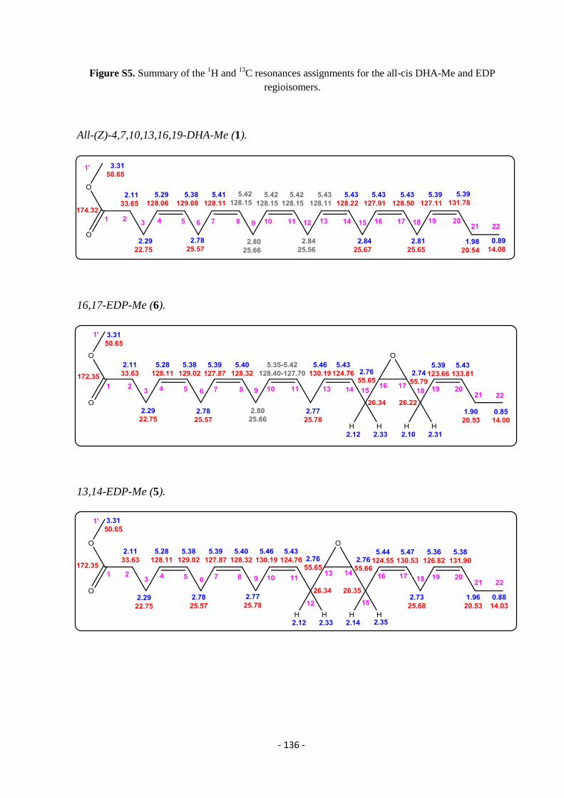
- 136 -
Figure S5. Summary of the 1H and
13C resonances assignments for the all-cis DHA-Me and EDP
regioisomers.
All-(Z)-4,7,10,13,16,19-DHA-Me (1).
16,17-EDP-Me (6).
13,14-EDP-Me (5).
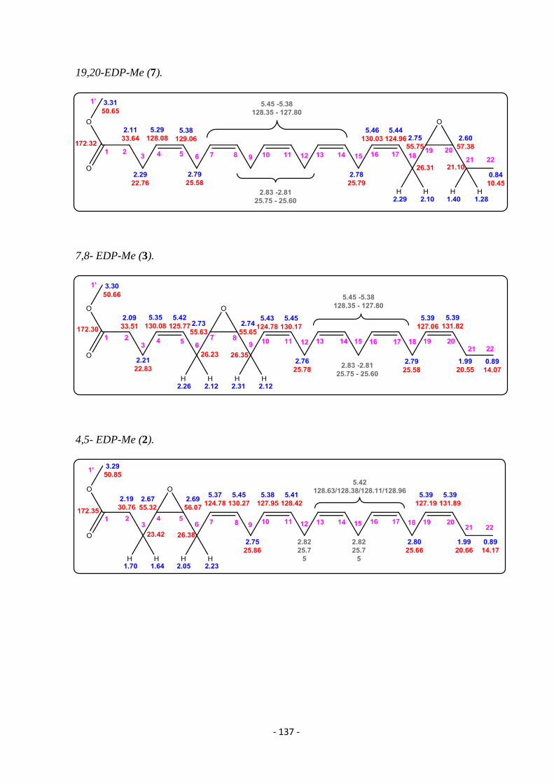
- 137 -
19,20-EDP-Me (7).
7,8- EDP-Me (3).
4,5- EDP-Me (2).
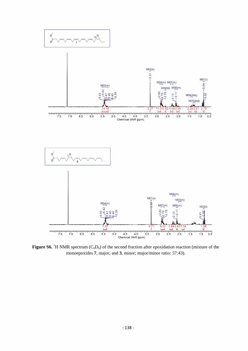
- 138 -
Figure S6. 1H NMR spectrum (C6D6) of the second fraction after epoxidation reaction (mixture of the
monoepoxides 7, major, and 3, minor; major/minor ratio: 57:43).
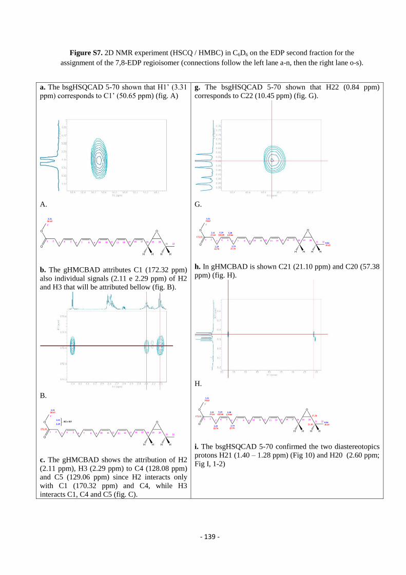
- 139 -
Figure S7. 2D NMR experiment (HSCQ / HMBC) in C6D6 on the EDP second fraction for the
assignment of the 7,8-EDP regioisomer (connections follow the left lane a-n, then the right lane o-s).
a. The bsgHSQCAD 5-70 shown that H1’ (3.31
ppm) corresponds to C1’ (50.65 ppm) (fig. A)
A.
b. The gHMCBAD attributes C1 (172.32 ppm)
also individual signals (2.11 e 2.29 ppm) of H2
and H3 that will be attributed bellow (fig. B).
B.
c. The gHMCBAD shows the attribution of H2
(2.11 ppm), H3 (2.29 ppm) to C4 (128.08 ppm)
and C5 (129.06 ppm) since H2 interacts only
with C1 (170.32 ppm) and C4, while H3
interacts C1, C4 and C5 (fig. C).
g. The bsgHSQCAD 5-70 shown that H22 (0.84 ppm)
corresponds to C22 (10.45 ppm) (fig. G).
G.
h. In gHMCBAD is shown C21 (21.10 ppm) and C20 (57.38
ppm) (fig. H).
H.
i. The bsgHSQCAD 5-70 confirmed the two diastereotopics
protons H21 (1.40 – 1.28 ppm) (Fig 10) and H20 (2.60 ppm;
Fig I, 1-2)

- 140 -
C.
d.The bsgHSQCAD 5-70 shows C2 (33.64 ppm)
and C3 (22.76 ppm) (fig. D).
D.
e. In bsgHSQCAD 110-140 the protons H4
(5.29 ppm) andH5 (5.38 ppm) are shown (fig.
E).
I1.
Fig I2.
l. The gHMCBAD shows C19 (55.75 ppm; Fig 12) and the
bsgHSQCAD 5-70 confirms H19 (2.75 ppm; Fig 13) (figs.
L1, L2)
L1.
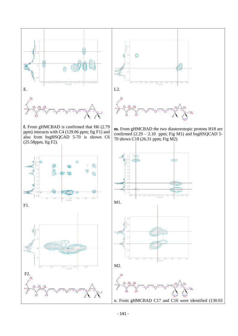
- 141 -
E.
f. From gHMCBAD is confirmed that H6 (2.79
ppm) interacts with C4 (129.06 ppm; fig F1) and
also from bsgHSQCAD 5-70 is shown C6
(25.58ppm, fig F2).
F1.
F2.
L2.
m. From gHMCBAD the two diastereotopic protons H18 are
confirmed (2.29 – 2.10 ppm; Fig M1) and bsgHSQCAD 5-
70 shows C18 (26.31 ppm; Fig M2)
M1.
M2.
n. From gHMCBAD C17 and C16 were identified (130.03
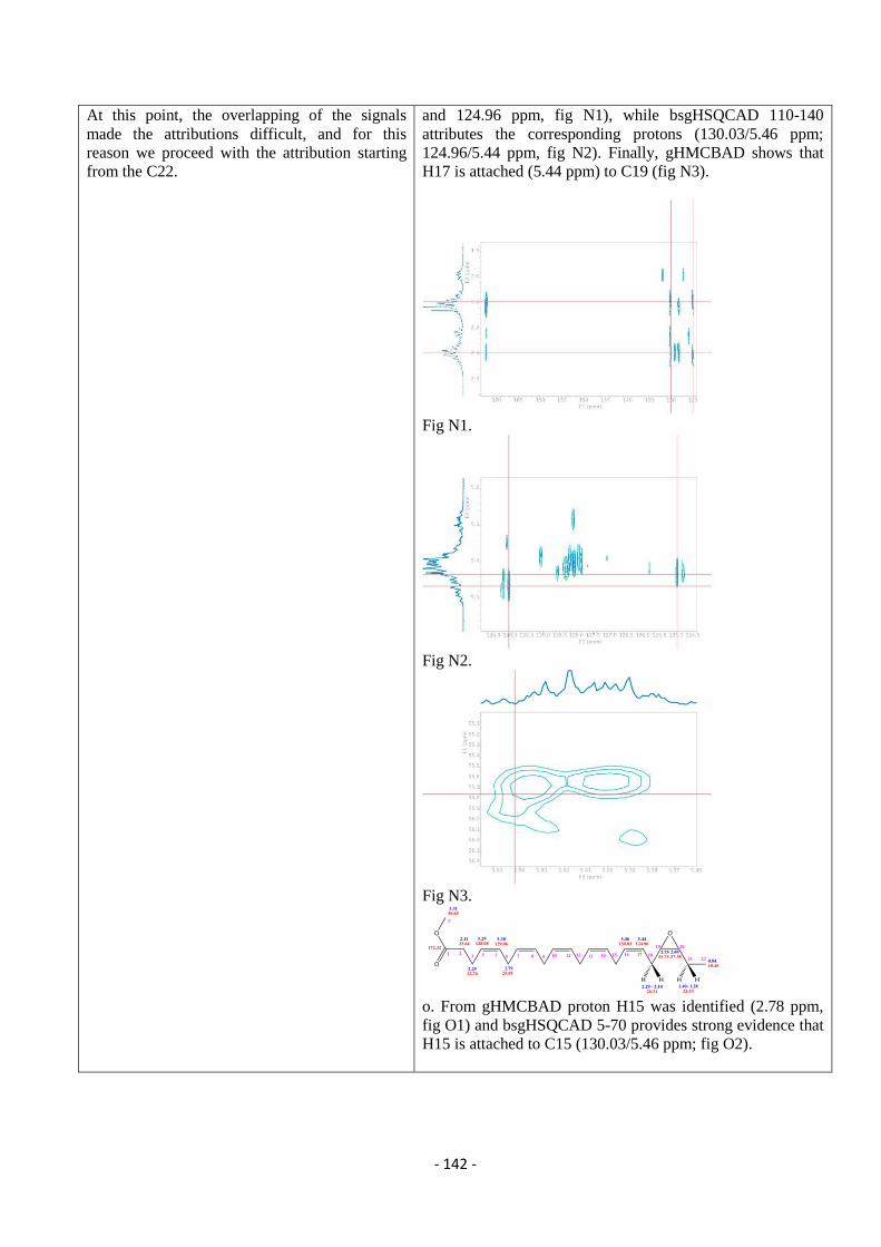
- 142 -
At this point, the overlapping of the signals
made the attributions difficult, and for this
reason we proceed with the attribution starting
from the C22.
and 124.96 ppm, fig N1), while bsgHSQCAD 110-140
attributes the corresponding protons (130.03/5.46 ppm;
124.96/5.44 ppm, fig N2). Finally, gHMCBAD shows that
H17 is attached (5.44 ppm) to C19 (fig N3).
Fig N1.
Fig N2.
Fig N3.
o. From gHMCBAD proton H15 was identified (2.78 ppm,
fig O1) and bsgHSQCAD 5-70 provides strong evidence that
H15 is attached to C15 (130.03/5.46 ppm; fig O2).
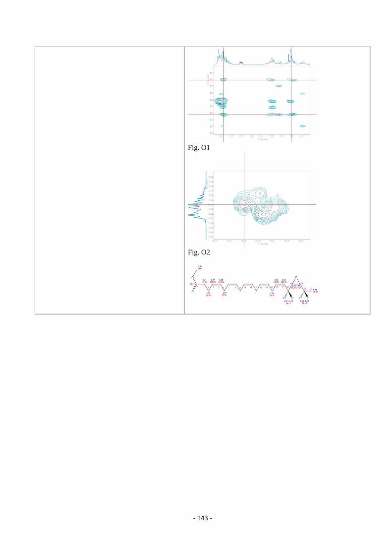
- 143 -
Fig. O1
Fig. O2
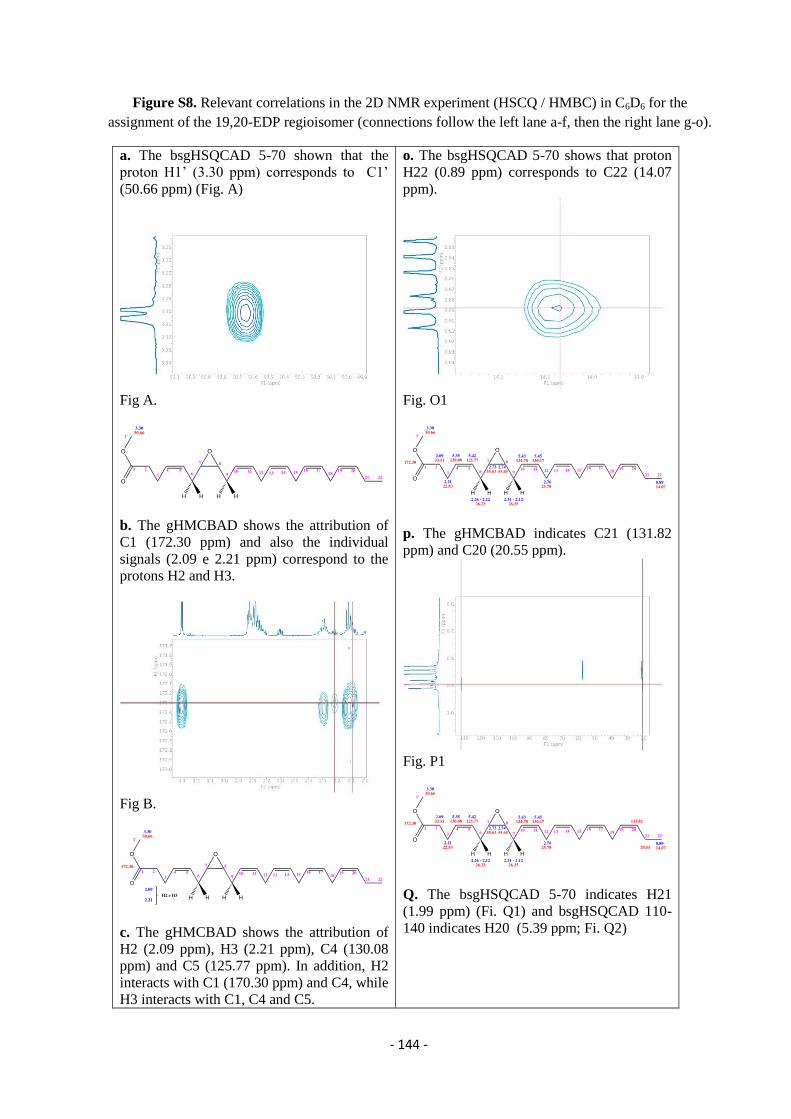
- 144 -
Figure S8. Relevant correlations in the 2D NMR experiment (HSCQ / HMBC) in C6D6 for the
assignment of the 19,20-EDP regioisomer (connections follow the left lane a-f, then the right lane g-o).
a. The bsgHSQCAD 5-70 shown that the
proton H1’ (3.30 ppm) corresponds to C1’
(50.66 ppm) (Fig. A)
Fig A.
b. The gHMCBAD shows the attribution of
C1 (172.30 ppm) and also the individual
signals (2.09 e 2.21 ppm) correspond to the
protons H2 and H3.
Fig B.
c. The gHMCBAD shows the attribution of
H2 (2.09 ppm), H3 (2.21 ppm), C4 (130.08
ppm) and C5 (125.77 ppm). In addition, H2
interacts with C1 (170.30 ppm) and C4, while
H3 interacts with C1, C4 and C5.
o. The bsgHSQCAD 5-70 shows that proton
H22 (0.89 ppm) corresponds to C22 (14.07
ppm).
Fig. O1
p. The gHMCBAD indicates C21 (131.82
ppm) and C20 (20.55 ppm).
Fig. P1
Q. The bsgHSQCAD 5-70 indicates H21
(1.99 ppm) (Fi. Q1) and bsgHSQCAD 110-
140 indicates H20 (5.39 ppm; Fi. Q2)
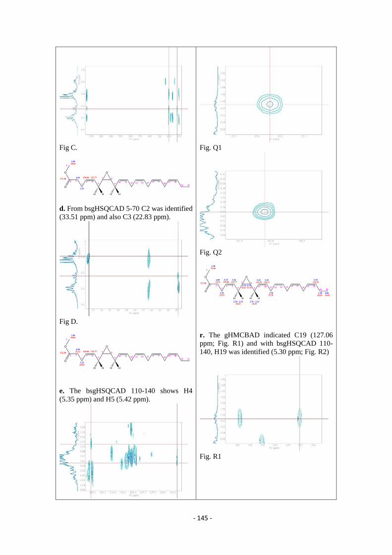
- 145 -
Fig C.
d. From bsgHSQCAD 5-70 C2 was identified
(33.51 ppm) and also C3 (22.83 ppm).
Fig D.
e. The bsgHSQCAD 110-140 shows H4
(5.35 ppm) and H5 (5.42 ppm).
Fig. Q1
Fig. Q2
r. The gHMCBAD indicated C19 (127.06
ppm; Fig. R1) and with bsgHSQCAD 110-
140, H19 was identified (5.30 ppm; Fig. R2)
Fig. R1
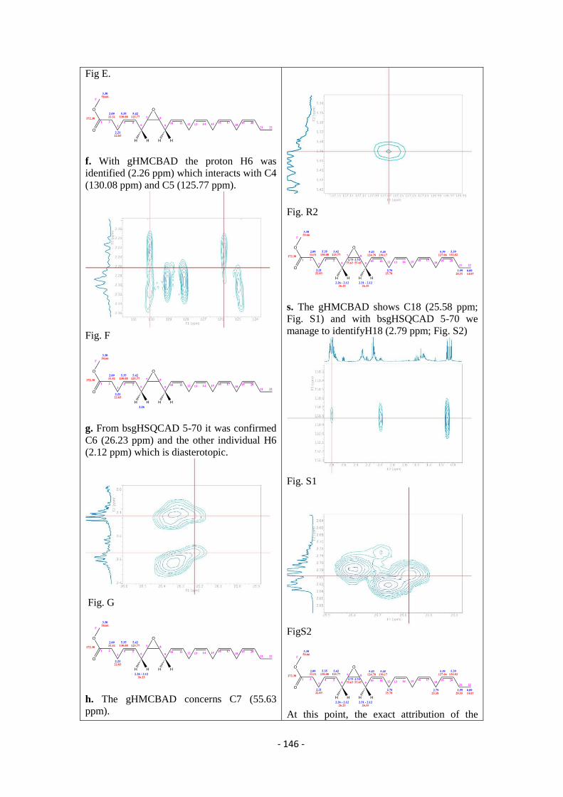
- 146 -
Fig E.
f. With gHMCBAD the proton H6 was
identified (2.26 ppm) which interacts with C4
(130.08 ppm) and C5 (125.77 ppm).
Fig. F
g. From bsgHSQCAD 5-70 it was confirmed
C6 (26.23 ppm) and the other individual H6
(2.12 ppm) which is diasterotopic.
Fig. G
h. The gHMCBAD concerns C7 (55.63
ppm).
Fig. R2
s. The gHMCBAD shows C18 (25.58 ppm;
Fig. S1) and with bsgHSQCAD 5-70 we
manage to identifyH18 (2.79 ppm; Fig. S2)
Fig. S1
FigS2
At this point, the exact attribution of the
- 147 -
Fig. H
i. With bsgHSQCAD 5-70 the proton H7 was
characterized (2.73 ppm).
Fig. I
l. At gHMCBAD C7 interacts with H9 (2.31
and 2.12 ppm) which are diastereotopic, and
also with C8 (55.65 ppm) (fig. L1). The
correlations proton – carbon are assigned
with bsgHSQCAD 5-70 (figs. L2-L3).
remaining resonances could not be done due
to the overlapping of the signals.
- 148 -
Fig. L1
Fig. L2
Fig. L3
M. In gHMCBAD the two protons H9
interact only with the two vinyl carbons C10
(124.78 ppm) and C11 (130.17 ppm) (Fi.
M1), which relative protons are attributed
with bsgHSQCAD 110-140 (Fig. M2), and
their position is attributed through HMCBAD
(Fig. M3)
- 149 -
Fig. M1
Fig. M2
Fig. M3
n. In gHMCBAD both C10 and C11 interact
with H12 (2.76 ppm; Fig. N1) whose carbon
(25.78 ppm) is attributed by bsgHSQCAD 5-
70 (Fig.N2).
- 150 -
Fig. N1
Fig. N2
The same 2D NMR experiments were also performed for the first and the third fraction
collected from the column in order to assign the peaks and characterize the epoxides. In the
next figures the protons NMR are presented.
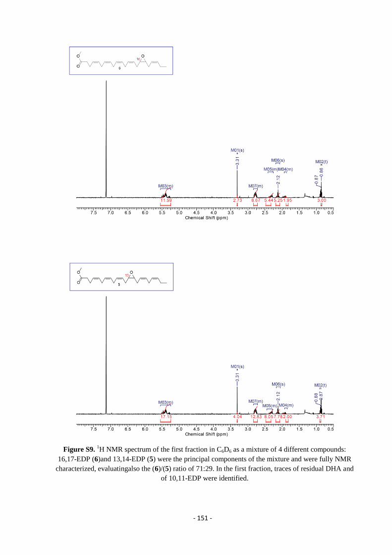
- 151 -
Figure S9. 1H NMR spectrum of the first fraction in C6D6 as a mixture of 4 different compounds:
16,17-EDP (6)and 13,14-EDP (5) were the principal components of the mixture and were fully NMR
characterized, evaluatingalso the (6)/(5) ratio of 71:29. In the first fraction, traces of residual DHA and
of 10,11-EDP were identified.
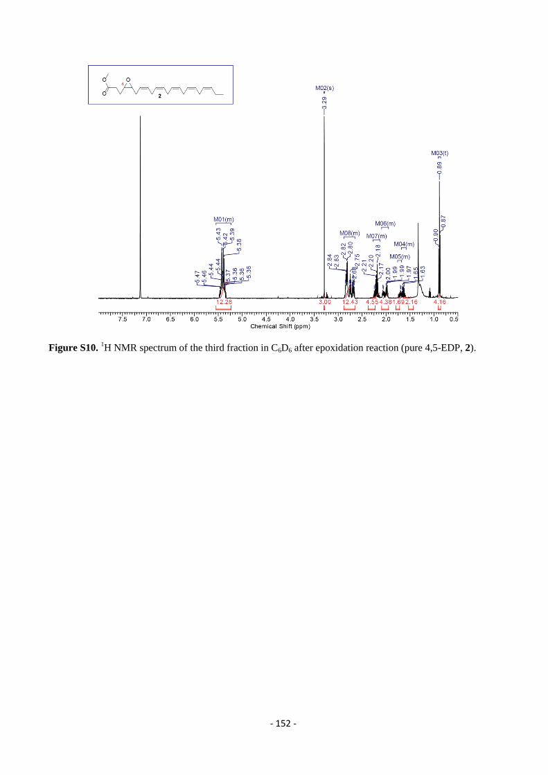
- 152 -
Figure S10. 1H NMR spectrum of the third fraction in C6D6 after epoxidation reaction (pure 4,5-EDP, 2).
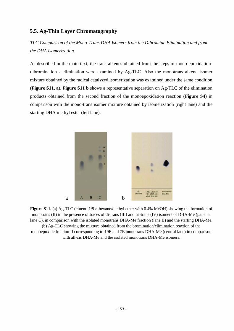
- 153 -
5.5. Ag-Thin Layer Chromatography
TLC Comparison of the Mono-Trans DHA Isomers from the Dibromide Elimination and from
the DHA Isomerization
As described in the main text, the trans-alkenes obtained from the steps of mono-epoxidation-
dibromination - elimination were examined by Ag-TLC. Also the monotrans alkene isomer
mixture obtained by the radical catalyzed isomerization was examined under the same condition
(Figure S11, a). Figure S11 b shows a representative separation on Ag-TLC of the elimination
products obtained from the second fraction of the monoepoxidation reaction (Figure S4) in
comparison with the mono-trans isomer mixture obtained by isomerization (right lane) and the
starting DHA methyl ester (left lane).
Figure S11. (a) Ag-TLC (eluent: 1/9 n-hexane/diethyl ether with 0.4% MeOH) showing the formation of
monotrans (II) in the presence of traces of di-trans (III) and tri-trans (IV) isomers of DHA-Me (panel a,
lane C), in comparison with the isolated monotrans DHA-Me fraction (lane B) and the starting DHA-Me.
(b) Ag-TLC showing the mixture obtained from the bromination/elimination reaction of the
monoepoxide fraction II corresponding to 19E and 7E monotrans DHA-Me (central lane) in comparison
with all-cis DHA-Me and the isolated monotrans DHA-Me isomers.
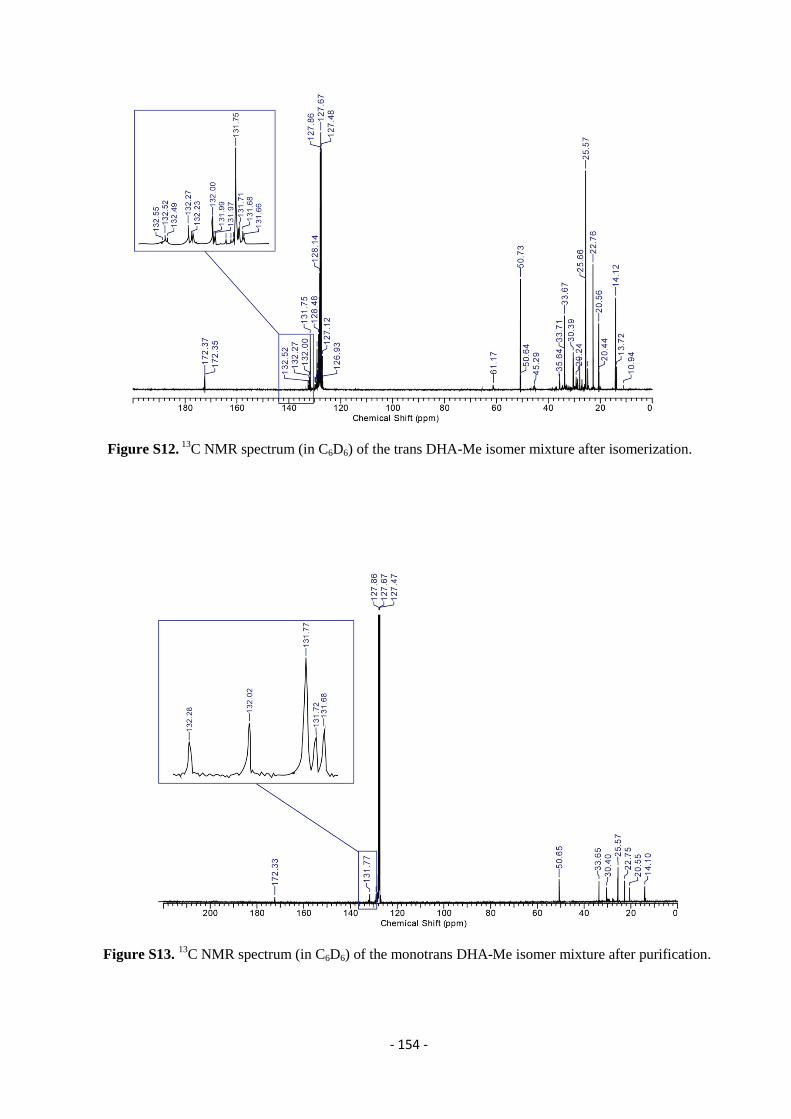
- 154 -
Figure S12. 13
C NMR spectrum (in C6D6) of the trans DHA-Me isomer mixture after isomerization.
Figure S13. 13
C NMR spectrum (in C6D6) of the monotrans DHA-Me isomer mixture after purification.
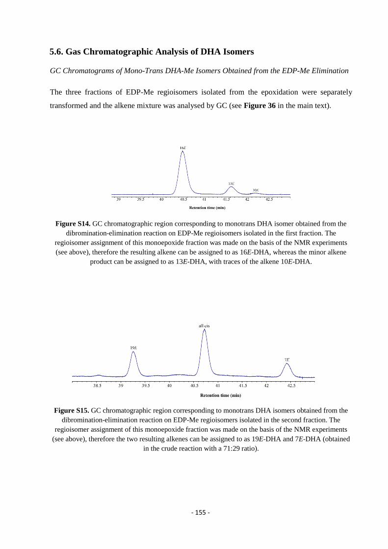
- 155 -
5.6. Gas Chromatographic Analysis of DHA Isomers
GC Chromatograms of Mono-Trans DHA-Me Isomers Obtained from the EDP-Me Elimination
The three fractions of EDP-Me regioisomers isolated from the epoxidation were separately
transformed and the alkene mixture was analysed by GC (see Figure 36 in the main text).
Figure S14. GC chromatographic region corresponding to monotrans DHA isomer obtained from the
dibromination-elimination reaction on EDP-Me regioisomers isolated in the first fraction. The
regioisomer assignment of this monoepoxide fraction was made on the basis of the NMR experiments
(see above), therefore the resulting alkene can be assigned to as 16E-DHA, whereas the minor alkene
product can be assigned to as 13E-DHA, with traces of the alkene 10E-DHA.
Figure S15. GC chromatographic region corresponding to monotrans DHA isomers obtained from the
dibromination-elimination reaction on EDP-Me regioisomers isolated in the second fraction. The
regioisomer assignment of this monoepoxide fraction was made on the basis of the NMR experiments
(see above), therefore the two resulting alkenes can be assigned to as 19E-DHA and 7E-DHA (obtained
in the crude reaction with a 71:29 ratio).
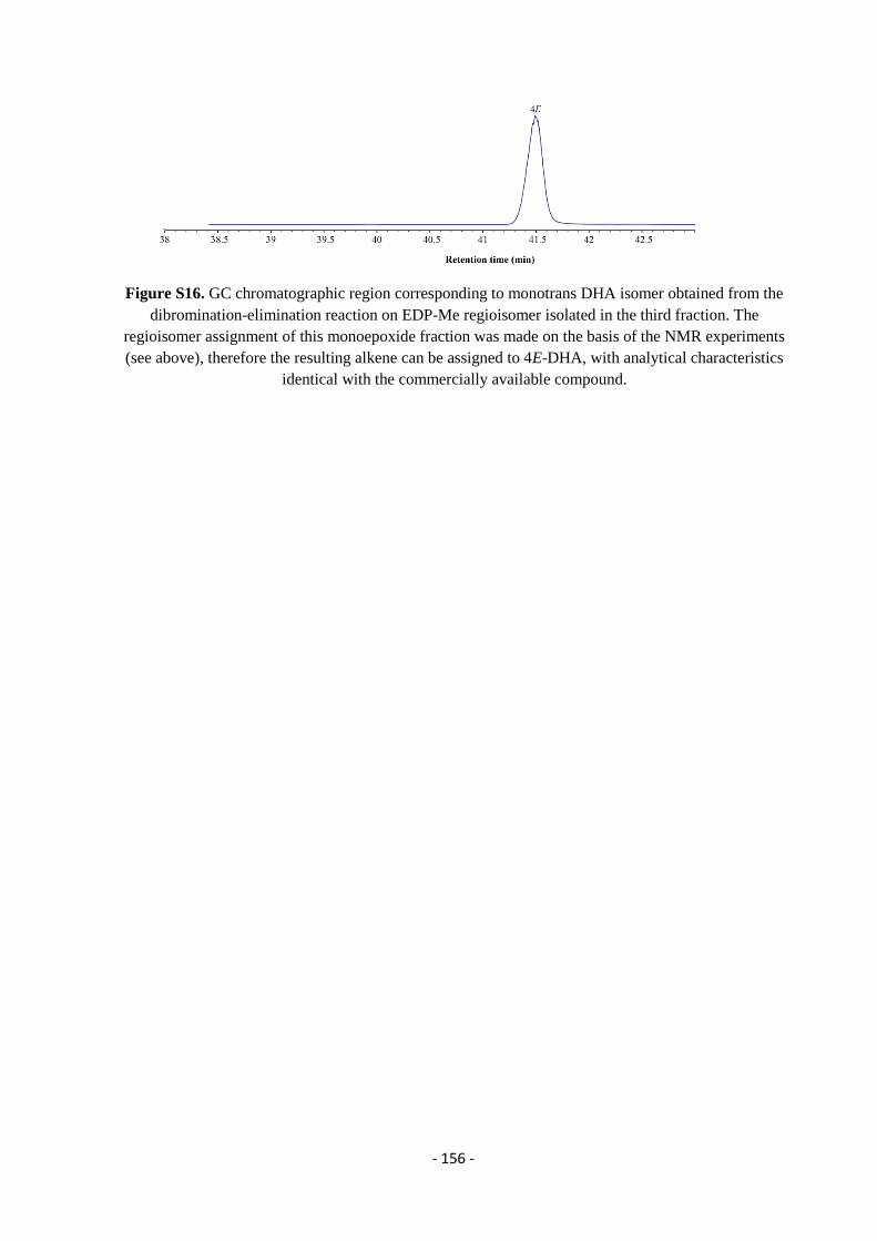
- 156 -
Figure S16. GC chromatographic region corresponding to monotrans DHA isomer obtained from the
dibromination-elimination reaction on EDP-Me regioisomer isolated in the third fraction. The
regioisomer assignment of this monoepoxide fraction was made on the basis of the NMR experiments
(see above), therefore the resulting alkene can be assigned to 4E-DHA, with analytical characteristics
identical with the commercially available compound.
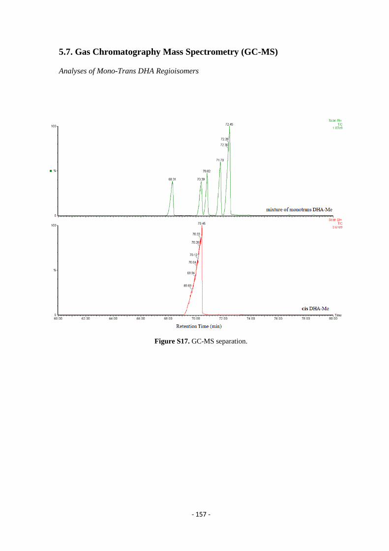
- 157 -
5.7. Gas Chromatography Mass Spectrometry (GC-MS)
Analyses of Mono-Trans DHA Regioisomers
Figure S17. GC-MS separation.
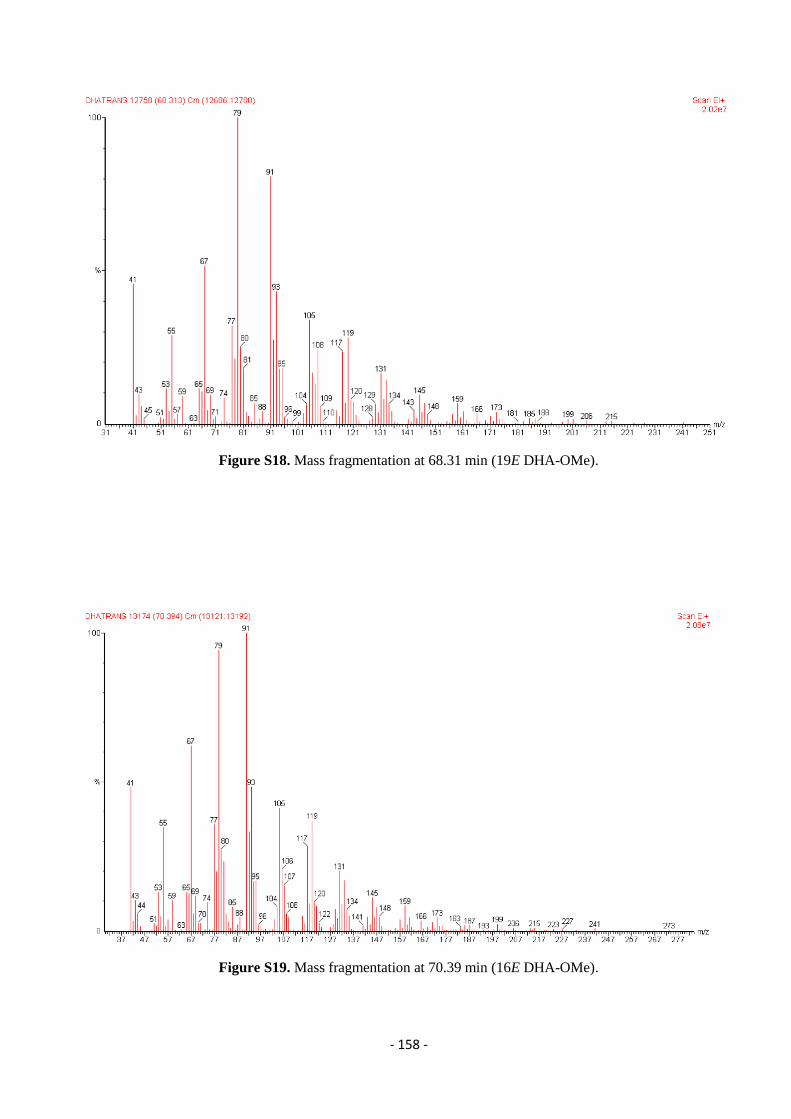
- 158 -
Figure S18. Mass fragmentation at 68.31 min (19E DHA-OMe).
Figure S19. Mass fragmentation at 70.39 min (16E DHA-OMe).
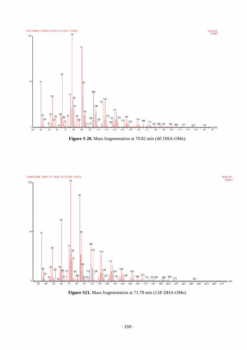
- 159 -
Figure S 20. Mass fragmentation at 70.82 min (4E DHA-OMe).
Figure S21. Mass fragmentation at 71.78 min (13E DHA-OMe).
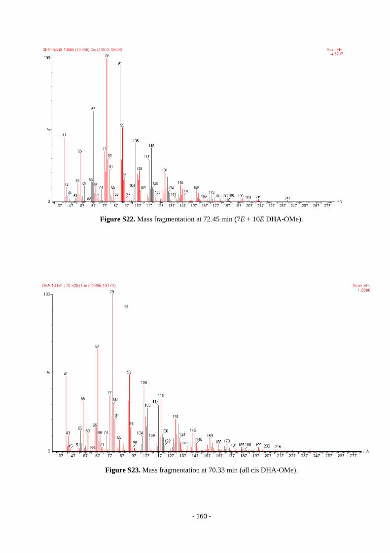
- 160 -
Figure S22. Mass fragmentation at 72.45 min (7E + 10E DHA-OMe).
Figure S23. Mass fragmentation at 70.33 min (all cis DHA-OMe).
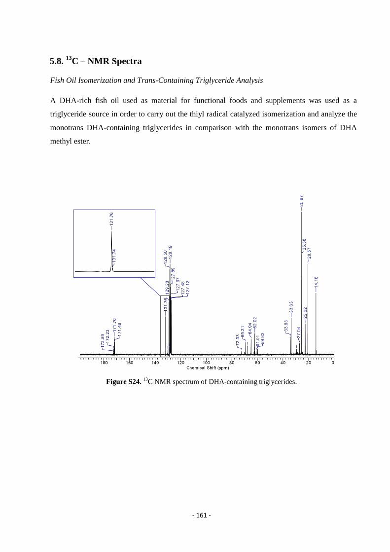
- 161 -
5.8. 13
C – NMR Spectra
Fish Oil Isomerization and Trans-Containing Triglyceride Analysis
A DHA-rich fish oil used as material for functional foods and supplements was used as a
triglyceride source in order to carry out the thiyl radical catalyzed isomerization and analyze the
monotrans DHA-containing triglycerides in comparison with the monotrans isomers of DHA
methyl ester.
Figure S24. 13
C NMR spectrum of DHA-containing triglycerides.
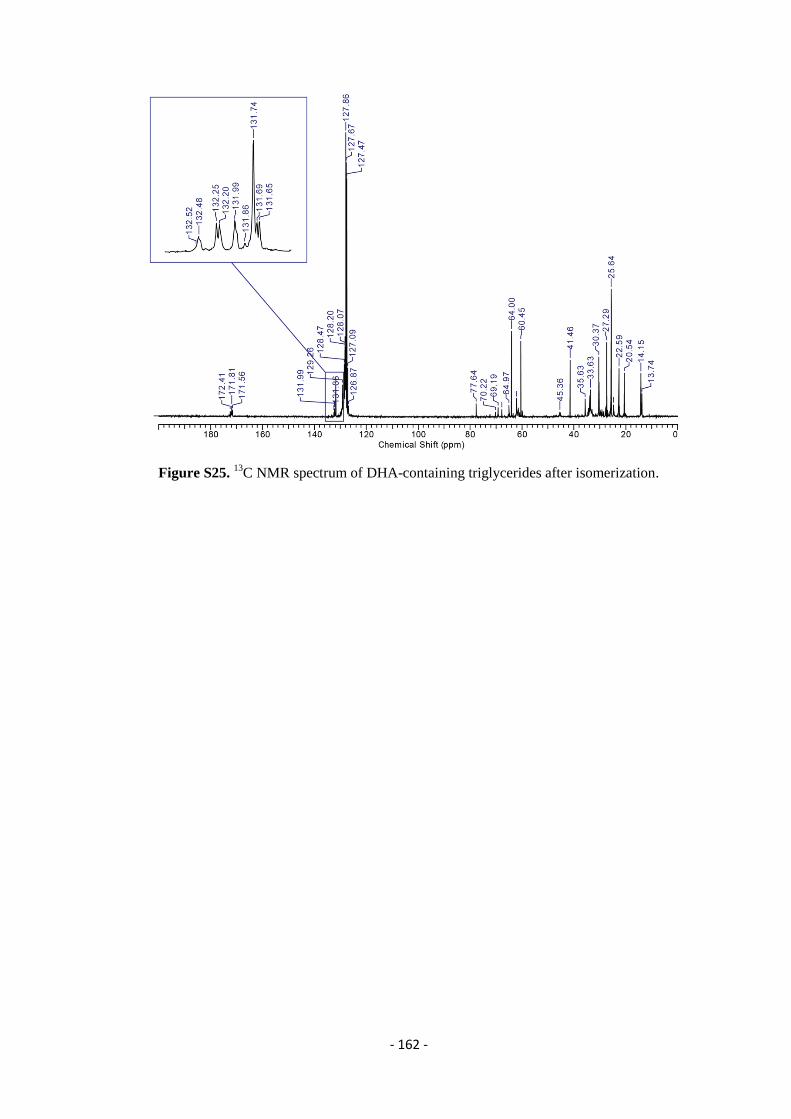
- 162 -
Figure S25. 13
C NMR spectrum of DHA-containing triglycerides after isomerization.
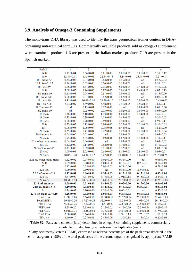
- 163 -
5.9. Analysis of Omega-3 Containing Supplements
The mono-trans DHA library was used to identify the trans geometrical isomer content in DHA-
containing nutraceutical formulas. Commercially available products sold as omega-3 supplement
were examined: products 1-6 are present in the Italian market, products 7-19 are present in the
Spanish market.
Table S2. Fatty acid content (%rel) determined in omega-3 containing supplements commercially
available in Italy. Analyses performed in triplicates (n=3).
*Fatty acid methyl esters (FAME) expressed as relative percentages of the peak areas detected in the
chromatograms (>98% of the total peak areas of the chromatogram recognized by appropriate FAME
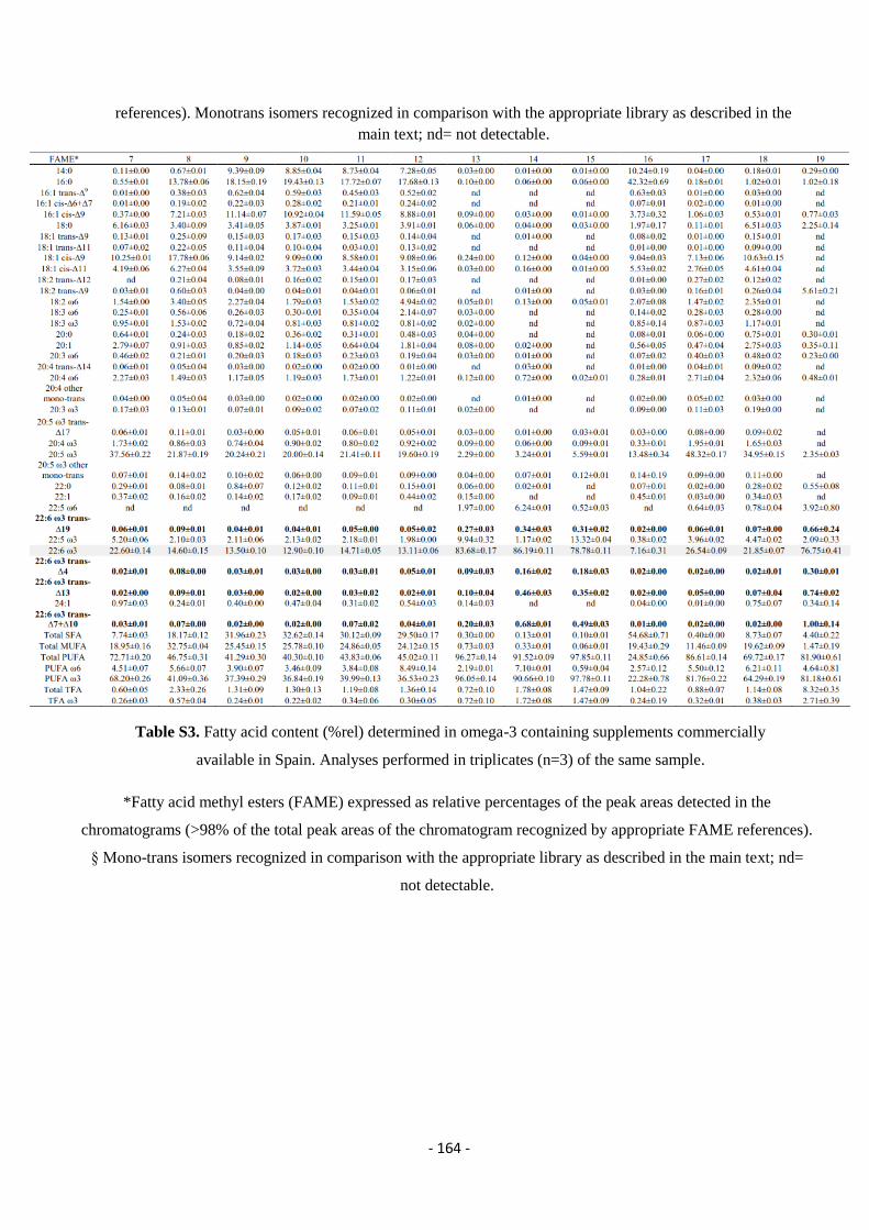
- 164 -
references). Monotrans isomers recognized in comparison with the appropriate library as described in the
main text; nd= not detectable.
Table S3. Fatty acid content (%rel) determined in omega-3 containing supplements commercially
available in Spain. Analyses performed in triplicates (n=3) of the same sample.
*Fatty acid methyl esters (FAME) expressed as relative percentages of the peak areas detected in the
chromatograms (>98% of the total peak areas of the chromatogram recognized by appropriate FAME references).
§ Mono-trans isomers recognized in comparison with the appropriate library as described in the main text; nd=
not detectable.
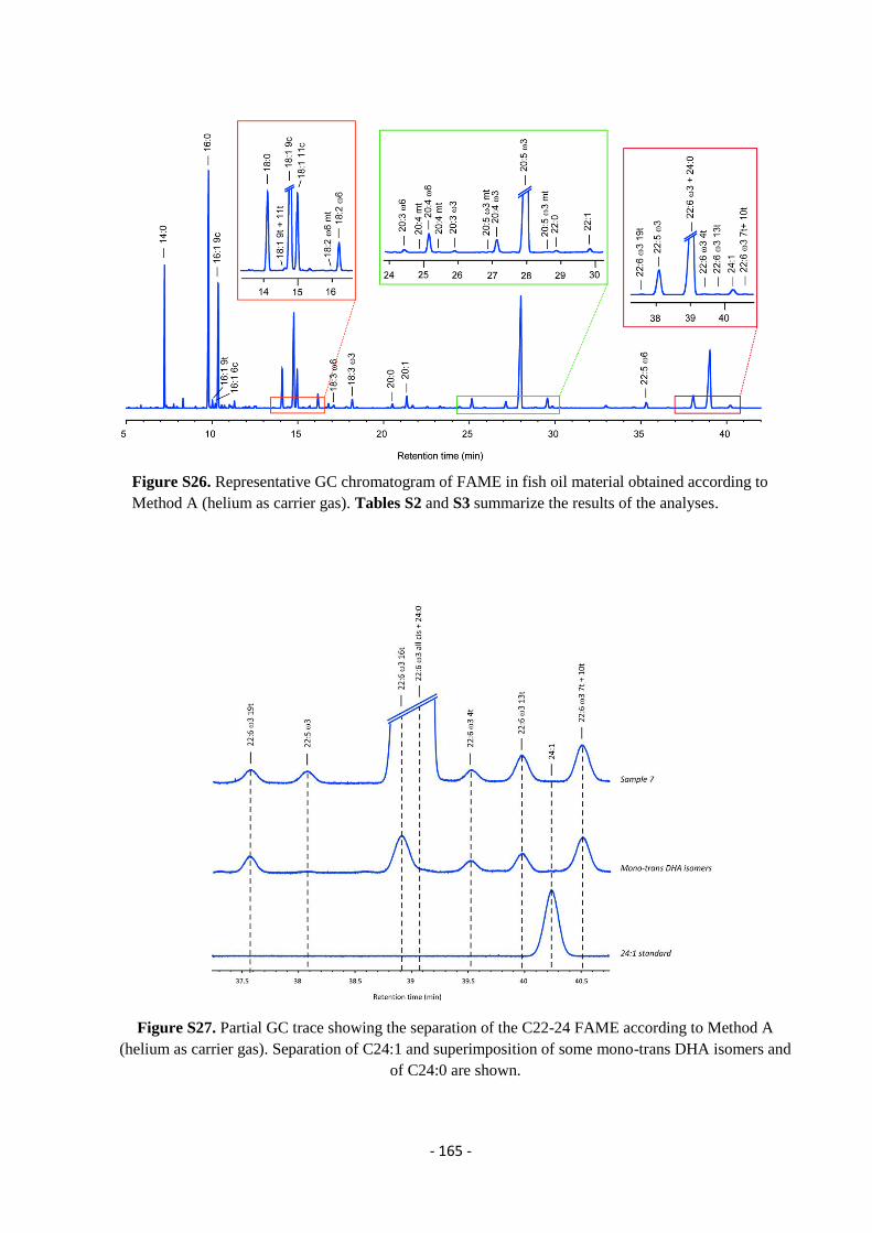
- 165 -
Figure S26. Representative GC chromatogram of FAME in fish oil material obtained according to
Method A (helium as carrier gas). Tables S2 and S3 summarize the results of the analyses.
Figure S27. Partial GC trace showing the separation of the C22-24 FAME according to Method A
(helium as carrier gas). Separation of C24:1 and superimposition of some mono-trans DHA isomers and
of C24:0 are shown.
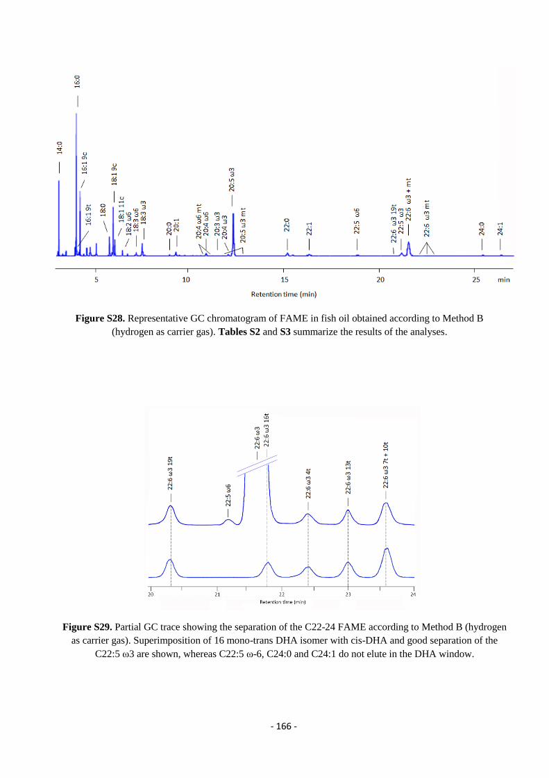
- 166 -
Figure S28. Representative GC chromatogram of FAME in fish oil obtained according to Method B
(hydrogen as carrier gas). Tables S2 and S3 summarize the results of the analyses.
Figure S29. Partial GC trace showing the separation of the C22-24 FAME according to Method B (hydrogen
as carrier gas). Superimposition of 16 mono-trans DHA isomer with cis-DHA and good separation of the
C22:5 ω3 are shown, whereas C22:5 ω-6, C24:0 and C24:1 do not elute in the DHA window.
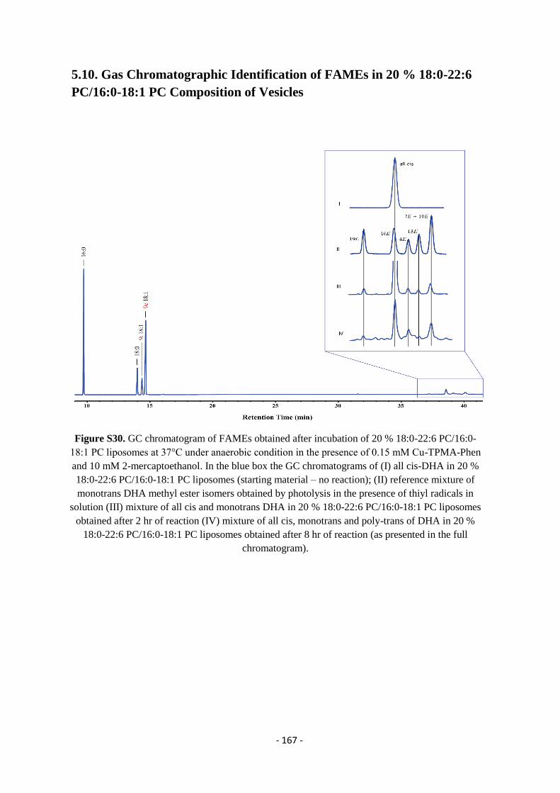
- 167 -
5.10. Gas Chromatographic Identification of FAMEs in 20 % 18:0-22:6
PC/16:0-18:1 PC Composition of Vesicles
Figure S30. GC chromatogram of FAMEs obtained after incubation of 20 % 18:0-22:6 PC/16:0-
18:1 PC liposomes at 37°C under anaerobic condition in the presence of 0.15 mM Cu-TPMA-Phen
and 10 mM 2-mercaptoethanol. In the blue box the GC chromatograms of (I) all cis-DHA in 20 %
18:0-22:6 PC/16:0-18:1 PC liposomes (starting material – no reaction); (II) reference mixture of
monotrans DHA methyl ester isomers obtained by photolysis in the presence of thiyl radicals in
solution (III) mixture of all cis and monotrans DHA in 20 % 18:0-22:6 PC/16:0-18:1 PC liposomes
obtained after 2 hr of reaction (IV) mixture of all cis, monotrans and poly-trans of DHA in 20 %
18:0-22:6 PC/16:0-18:1 PC liposomes obtained after 8 hr of reaction (as presented in the full
chromatogram).
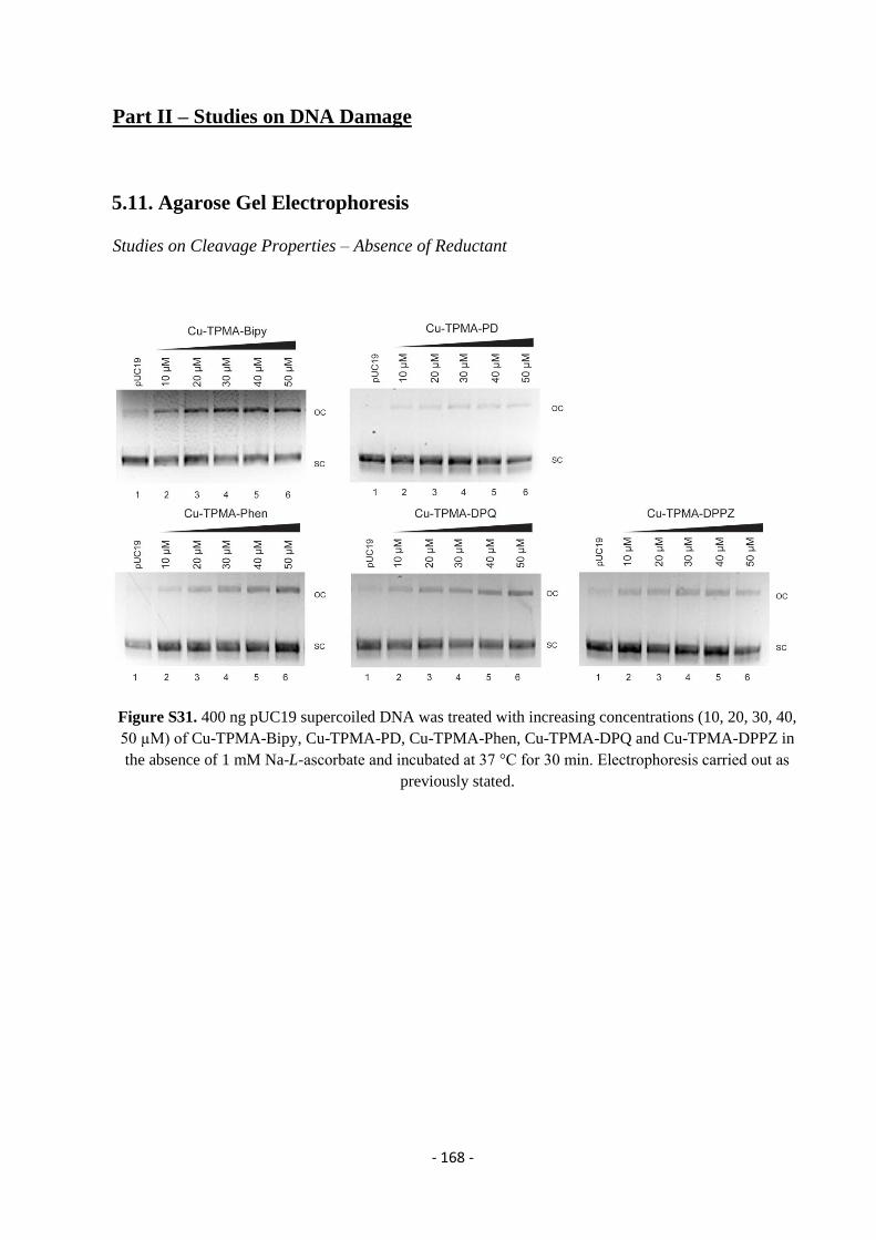
- 168 -
Part II – Studies on DNA Damage
5.11. Agarose Gel Electrophoresis
Studies on Cleavage Properties – Absence of Reductant
Figure S31. 400 ng pUC19 supercoiled DNA was treated with increasing concentrations (10, 20, 30, 40,
50 µM) of Cu-TPMA-Bipy, Cu-TPMA-PD, Cu-TPMA-Phen, Cu-TPMA-DPQ and Cu-TPMA-DPPZ in
the absence of 1 mM Na-L-ascorbate and incubated at 37 °C for 30 min. Electrophoresis carried out as
previously stated.
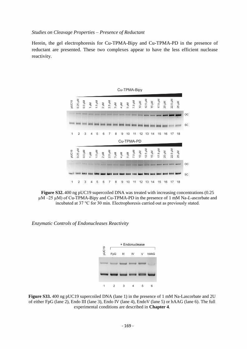
- 169 -
Studies on Cleavage Properties – Presence of Reductant
Herein, the gel electrophoresis for Cu-TPMA-Bipy and Cu-TPMA-PD in the presence of
reductant are presented. These two complexes appear to have the less efficient nuclease
reactivity.
Figure S32. 400 ng pUC19 supercoiled DNA was treated with increasing concentrations (0.25
µM –25 µM) of Cu-TPMA-Bipy and Cu-TPMA-PD in the presence of 1 mM Na-L-ascorbate and
incubated at 37 °C for 30 min. Electrophoresis carried out as previously stated.
Enzymatic Controls of Endonucleases Reactivity
Figure S33. 400 ng pUC19 supercoiled DNA (lane 1) in the presence of 1 mM Na-Lascorbate and 2U
of either FpG (lane 2), Endo III (lane 3), Endo IV (lane 4), EndoV (lane 5) or hAAG (lane 6). The full
experimental conditions are described in Chapter 4.
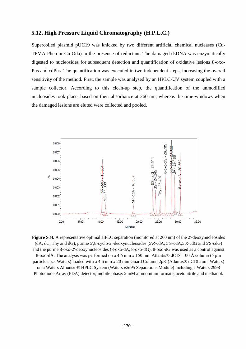
- 170 -
5.12. High Pressure Liquid Chromatography (H.P.L.C.)
Supercoiled plasmid pUC19 was knicked by two different artificial chemical nucleases (Cu-
TPMA-Phen or Cu-Oda) in the presence of reductant. The damaged dsDNA was enzymatically
digested to nucleosides for subsequent detection and quantification of oxidative lesions 8-oxo-
Pus and cdPus. The quantification was executed in two independent steps, increasing the overall
sensitivity of the method. First, the sample was analysed by an HPLC-UV system coupled with a
sample collector. According to this clean-up step, the quantification of the unmodified
nucleosides took place, based on their absorbance at 260 nm, whereas the time-windows when
the damaged lesions are eluted were collected and pooled.
Figure S34. A representative optimal HPLC separation (monitored at 260 nm) of the 2'-deoxynucleosides
(dA, dC, Thy and dG), purine 5',8-cyclo-2'-deoxynucleosides (5'R-cdA, 5'S-cdA,5'R-cdG and 5'S-cdG)
and the purine 8-oxo-2'-deoxynucleosides (8-oxo-dA, 8-oxo-dG). 8-oxo-dG was used as a control against
8-oxo-dA. The analysis was performed on a 4.6 mm x 150 mm Atlantis® dC18, 100 Å column (5 μm
particle size, Waters) loaded with a 4.6 mm x 20 mm Guard Column 2pK (Atlantis® dC18 5μm, Waters)
on a Waters Alliance ® HPLC System (Waters e2695 Separations Module) including a Waters 2998
Photodiode Array (PDA) detector; mobile phase: 2 mM ammomium formate, acetonitrile and methanol.
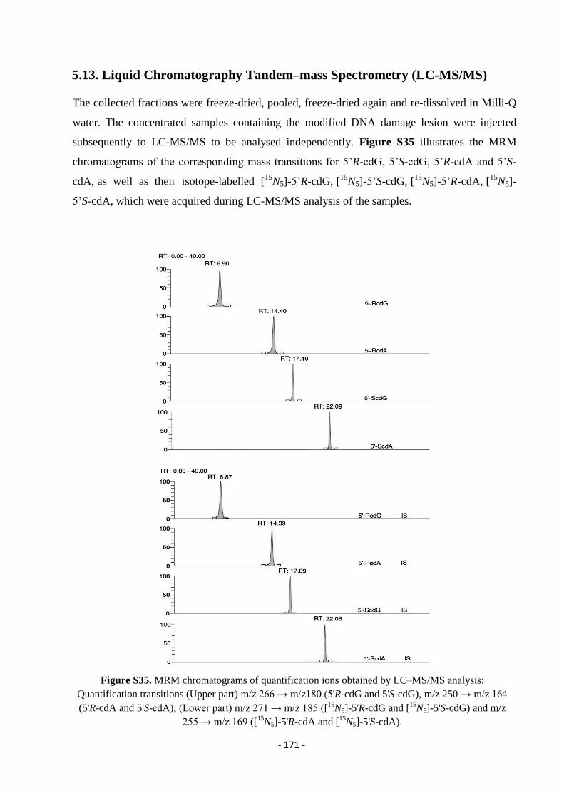
- 171 -
5.13. Liquid Chromatography Tandem–mass Spectrometry (LC-MS/MS)
The collected fractions were freeze-dried, pooled, freeze-dried again and re-dissolved in Milli-Q
water. The concentrated samples containing the modified DNA damage lesion were injected
subsequently to LC-MS/MS to be analysed independently. Figure S35 illustrates the MRM
chromatograms of the corresponding mass transitions for 5’R-cdG, 5’S-cdG, 5’R-cdA and 5’S-
cdA, as well as their isotope-labelled [15
N5]-5’R-cdG, [15
N5]-5’S-cdG, [15
N5]-5’R-cdA, [15
N5]-
5’S-cdA, which were acquired during LC-MS/MS analysis of the samples.
Figure S35. MRM chromatograms of quantification ions obtained by LC–MS/MS analysis:
Quantification transitions (Upper part) m/z 266 → m/z180 (5'R-cdG and 5'S-cdG), m/z 250 → m/z 164
(5'R-cdA and 5'S-cdA); (Lower part) m/z 271 → m/z 185 ([15
N5]-5'R-cdG and [15
N5]-5'S-cdG) and m/z
255 → m/z 169 ([15
N5]-5'R-cdA and [15
N5]-5'S-cdA).
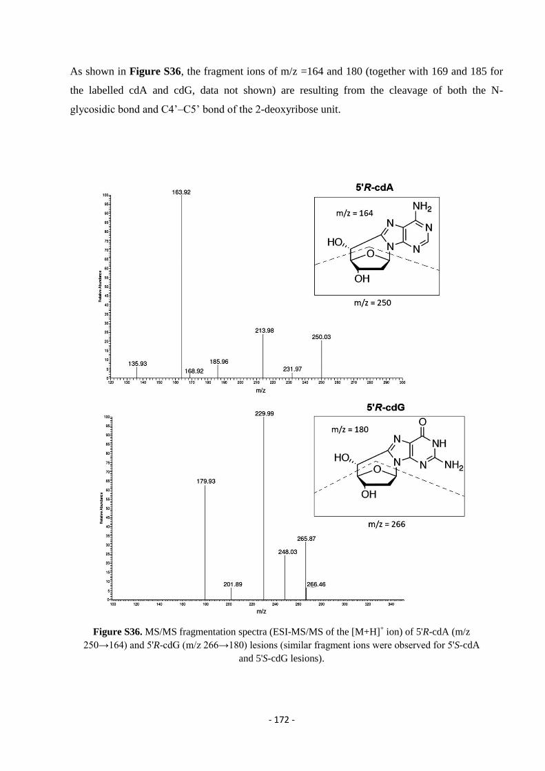
- 172 -
As shown in Figure S36, the fragment ions of m/z =164 and 180 (together with 169 and 185 for
the labelled cdA and cdG, data not shown) are resulting from the cleavage of both the N-
glycosidic bond and C4’–C5’ bond of the 2-deoxyribose unit.
Figure S36. MS/MS fragmentation spectra (ESI-MS/MS of the [M+H]+ ion) of 5'R-cdA (m/z
250→164) and 5'R-cdG (m/z 266→180) lesions (similar fragment ions were observed for 5'S-cdA
and 5'S-cdG lesions).
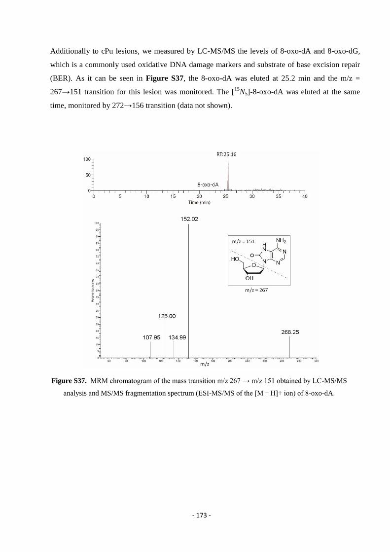
- 173 -
Additionally to cPu lesions, we measured by LC-MS/MS the levels of 8-oxo-dA and 8-oxo-dG,
which is a commonly used oxidative DNA damage markers and substrate of base excision repair
(BER). As it can be seen in Figure S37, the 8-oxo-dA was eluted at 25.2 min and the m/z =
267→151 transition for this lesion was monitored. The [15
N5]-8-oxo-dA was eluted at the same
time, monitored by 272→156 transition (data not shown).
Figure S37. MRM chromatogram of the mass transition m/z 267 → m/z 151 obtained by LC-MS/MS
analysis and MS/MS fragmentation spectrum (ESI-MS/MS of the [M + H]+ ion) of 8-oxo-dA.
- 174 -
References
1. GM., C., The Cell: A Molecular Approach. 2 ed.; Sinauer Associates: Sunderland
(MA), 2000.
2. Cancer Research UK. https://www.cancerresearchuk.org/.
3. Health, N. I. o.
4. Lodish H, B. A., Zipursky SL, et al., Molecular Cell Biology. 4 ed.; New York
2000.
5. Lunec, J., Free Radicals: Their Involvement in Disease Processes. Annals of
Clinical Biochemistry 1990, 27 (3), 173-182.
6. Bandyopadhyay, S.; Mehta, M.; Kuo, D.; Sung, M. K.; Chuang, R.; Jaehnig, E. J.;
Bodenmiller, B.; Licon, K.; Copeland, W.; Shales, M.; Fiedler, D.; Dutkowski, J.;
Guenole, A.; van Attikum, H.; Shokat, K. M.; Kolodner, R. D.; Huh, W. K.; Aebersold,
R.; Keogh, M. C.; Krogan, N. J.; Ideker, T., Rewiring of genetic networks in response to
DNA damage. Science 2010, 330 (6009), 1385-9.
7. Pagès, V.; Fuchs, R. P. P., How DNA lesions are turned into mutations within
cells? Oncogene 2002, 21, 8957.
8. Dar, A. C.; Das, T. K.; Shokat, K. M.; Cagan, R. L., Chemical genetic discovery
of targets and anti-targets for cancer polypharmacology. Nature 2012, 486, 80.
9. Baruah, H.; Barry, C. G.; Bierbach, U., Platinum-intercalator conjugates: from
DNA-targeted cisplatin derivatives to adenine binding complexes as potential modulators
of gene regulation. Curr Top Med Chem 2004, 4 (15), 1537-49.
10. Carvalho, C.; Santos, R. X.; Cardoso, S.; Correia, S.; Oliveira, P. J.; Santos, M. S.;
Moreira, P. I., Doxorubicin: the good, the bad and the ugly effect. Current medicinal
chemistry 2009, 16 (25), 3267-85.
11. Parker, W. B.; Cheng, Y. C., Metabolism and mechanism of action of 5-
fluorouracil. Pharmacol Ther 1990, 48 (3), 381-95.
12. Wozniak, A. J.; Ross, W. E., DNA damage as a basis for 4'-
demethylepipodophyllotoxin-9-(4,6-O-ethylidene-beta-D-glucopyranoside) (etoposide)
cytotoxicity. Cancer Res 1983, 43 (1), 120-4.
13. Lankadasari, M. B.; Aparna, J. S.; Mohammed, S.; James, S.; Aoki, K.; Binu, V.
S.; Nair, S.; Harikumar, K. B., Targeting S1PR1/STAT3 loop abrogates desmoplasia and
chemosensitizes pancreatic cancer to gemcitabine. Theranostics 2018, 8 (14), 3824-3840.
- 175 -
14. Pizarro, A. M.; Sadler, P. J., Unusual DNA binding modes for metal anticancer
complexes. Biochimie 2009, 91 (10), 1198-1211.
15. Brinkmann, M.; Tallone, E. M.; Wurschmidt, F.; Wulfing, C.; Dieckmann, K. P.,
[Myocardial infarction in a young patient with seminoma during chemotherapy with
cisplatinum, etoposide, and bleomycin]. Aktuelle Urol 2018.
16. Siddik, Z. H., Cisplatin: mode of cytotoxic action and molecular basis of
resistance. Oncogene 2003, 22 (47), 7265-79.
17. Oun, R.; Moussa, Y. E.; Wheate, N. J., The side effects of platinum-based
chemotherapy drugs: a review for chemists. Dalton Transactions 2018, 47 (19), 6645-
6653.
18. Interactions of Metallodrugs with DNA. In NMR of Biomolecules.
19. Deo, K. M.; Pages, B. J.; Ang, D. L.; Gordon, C. P.; Aldrich-Wright, J. R.,
Transition Metal Intercalators as Anticancer Agents—Recent Advances. International
Journal of Molecular Sciences 2016, 17 (11), 1818.
20. (a) Malina, J.; Novakova, O.; Vojtiskova, M.; Natile, G.; Brabec, V.,
Conformation of DNA GG Intrastrand Cross-Link of Antitumor Oxaliplatin and Its
Enantiomeric Analog. Biophysical Journal 2007, 93 (11), 3950-3962; (b) Bruijnincx, P.
C.; Sadler, P. J., New trends for metal complexes with anticancer activity. Current
opinion in chemical biology 2008, 12 (2), 197-206.
21. Sigman, D. S.; Mazumder, A.; Perrin, D. M., Chemical nucleases. Chemical
Reviews 1993, 93 (6), 2295-2316.
22. Fondello, C.; Agnetti, L.; Glikin, G. C.; Finocchiaro, L. M. E., Mechanisms
Enhancing the Cytotoxic Effects of Bleomycin plus Suicide or Interferon-beta Gene
Lipofection in Metastatic Human Melanoma Cells. Anticancer Agents Med Chem 2018.
23. Bolzan, A. D.; Bianchi, M. S., DNA and chromosome damage induced by
bleomycin in mammalian cells: An update. Mutat Res 2018, 775, 51-62.
24. Ben-Yoseph, O.; Ross, B. D., Oxidation therapy: the use of a reactive oxygen
species-generating enzyme system for tumour treatment. British Journal of Cancer 1994,
70 (6), 1131-1135.
25. Tan, L. T.-H.; Chan, K.-G.; Pusparajah, P.; Lee, W.-L.; Chuah, L.-H.; Khan, T.
M.; Lee, L.-H.; Goh, B.-H., Targeting Membrane Lipid a Potential Cancer Cure?
Frontiers in Pharmacology 2017, 8, 12.
- 176 -
26. Cort, A.; Ozben, T.; Melchiorre, M.; Chatgilialoglu, C.; Ferreri, C.; Sansone, A.,
Effects of bleomycin and antioxidants on the fatty acid profile of testicular cancer cell
membranes. Biochim Biophys Acta 2016, 1858 (2), 434-41.
27. Cort, A.; Ozben, T.; Sansone, A.; Barata-Vallejo, S.; Chatgilialoglu, C.; Ferreri,
C., Bleomycin-induced trans lipid formation in cell membranes and in liposome models.
Org Biomol Chem 2015, 13 (4), 1100-5.
28. Gutteridge, B. H. a. J. M. C., Free Radicals in Biology and Medicine. Fifth Edition
ed.; Oxford University Press: 2015.
29. Chatgilialoglu, C.; Ferreri, C., Trans lipids: the free radical path. Acc Chem Res
2005, 38 (6), 441-8.
30. Chen, C. L.; Tetri, L. H.; Neuschwander-Tetri, B. A.; Huang, S. S.; Huang, J. S.,
A mechanism by which dietary trans fats cause atherosclerosis. J Nutr Biochem 2011, 22
(7), 649-55.
31. Garcia, S. C.; Grotto, D.; Bulcão, R. P.; Moro, A. M.; Roehrs, M.; Valentini, J.; de
Freitas, F. A.; Paniz, C.; Bubols, G. B.; Charão, M. F., Evaluation of lipid damage related
to pathological and physiological conditions. Drug and Chemical Toxicology 2013, 36
(3), 306-312.
32. Ferreri, C.; Chatgilialoglu, C., Role of fatty acid-based functional lipidomics in the
development of molecular diagnostic tools. Expert Rev Mol Diagn 2012, 12 (7), 767-80.
33. Lucio, M.; Ferreira, H.; Lima, J. L.; Reis, S., Use of liposomes as membrane
models to evaluate the contribution of drug-membrane interactions to antioxidant
properties of etodolac. Redox Rep 2008, 13 (5), 225-36.
34. Thomas, A. H.; Catala, A.; Vignoni, M., Soybean phosphatidylcholine liposomes
as model membranes to study lipid peroxidation photoinduced by pterin. Biochim Biophys
Acta 2016, 1858 (1), 139-45.
35. Danielli, J. F.; Davson, H., A contribution to the theory of permeability of thin
films. Journal of Cellular and Comparative Physiology 1935, 5 (4), 495-508.
36. Singer, S. J.; Nicolson, G. L., The fluid mosaic model of the structure of cell
membranes. Science 1972, 175 (4023), 720-31.
37. Biology Wise. https://biologywise.com/.
38. Berg JM, T. J., Stryer L, Biochemistry. 5th edition ed.; W H Freeman: New York,
2002.
39. Alberts B, J. A., Lewis J, et al., Molecular Biology of the Cell. 4th edition ed.;
Garland Science: New York, 2002.
- 177 -
40. Cook, H. W., Chapter 5 - Fatty acid desaturation and chain elongation in
eukaryotes. In New Comprehensive Biochemistry, Vance, D. E.; Vance, J. E., Eds.
Elsevier: 1996; Vol. 31, pp 129-152.
41. Dominguez, L. J.; Barbagallo, M., Chapter 3 - Not All Fats Are Unhealthy. In The
Prevention of Cardiovascular Disease Through the Mediterranean Diet, Sánchez-
Villegas, A.; Sánchez-Tainta, A., Eds. Academic Press: 2018; pp 35-58.
42. Kaur, N.; Chugh, V.; Gupta, A. K., Essential fatty acids as functional components
of foods- a review. Journal of Food Science and Technology 2014, 51 (10), 2289-2303.
43. Simopoulos, A. P., An Increase in the Omega-6/Omega-3 Fatty Acid Ratio
Increases the Risk for Obesity. Nutrients 2016, 8 (3), 128.
44. Laposata, M., Fatty Acids: Biochemistry to Clinical Significance. American
Journal of Clinical Pathology 1995, 104 (2), 172-179.
45. Migdal, C.; Serres, M., [Reactive oxygen species and oxidative stress]. Med Sci
(Paris) 2011, 27 (4), 405-12.
46. Bergamini, C. M.; Gambetti, S.; Dondi, A.; Cervellati, C., Oxygen, reactive
oxygen species and tissue damage. Current pharmaceutical design 2004, 10 (14), 1611-
26.
47. Sharma, P.; Jha, A. B.; Dubey, R. S.; Pessarakli, M., Reactive Oxygen Species,
Oxidative Damage, and Antioxidative Defense Mechanism in Plants under Stressful
Conditions. Journal of Botany 2012, 2012, 26.
48. Gulati, K.; Rai, N.; Ray, A., Chapter 56 - Effects of Stress on Reproductive
and Developmental Biology. In Reproductive and Developmental Toxicology (Second
Edition), Gupta, R. C., Ed. Academic Press: 2017; pp 1063-1075.
49. Birben, E.; Sahiner, U. M.; Sackesen, C.; Erzurum, S.; Kalayci, O., Oxidative
Stress and Antioxidant Defense. The World Allergy Organization journal 2012, 5 (1), 9-
19.
50. Erden-Inal, M.; Sunal, E.; Kanbak, G., Age-related changes in the glutathione
redox system. Cell Biochem Funct 2002, 20 (1), 61-6.
51. Vazquez-Medina, J. P., Chapter 3 - Redox Signaling and the Onset of the
Inflammatory Cascade. In Immunity and Inflammation in Health and Disease, Chatterjee,
S.; Jungraithmayr, W.; Bagchi, D., Eds. Academic Press: 2018; pp 37-42.
52. Laurindo, F. R. M., Chapter 10 - Redox Cellular Signaling Pathways in
Endothelial Dysfunction and Vascular Disease. In Endothelium and Cardiovascular
- 178 -
Diseases, Da Luz, P. L.; Libby, P.; Chagas, A. C. P.; Laurindo, F. R. M., Eds. Academic
Press: 2018; pp 127-145.
53. Schieber, M.; Chandel, N. S., ROS Function in Redox Signaling and Oxidative
Stress. Current biology : CB 2014, 24 (10), R453-R462.
54. Ghosh, N.; Das, A.; Chaffee, S.; Roy, S.; Sen, C. K., Chapter 4 - Reactive Oxygen
Species, Oxidative Damage and Cell Death. In Immunity and Inflammation in Health and
Disease, Chatterjee, S.; Jungraithmayr, W.; Bagchi, D., Eds. Academic Press: 2018; pp
45-55.
55. Mjos, K. D.; Orvig, C., Metallodrugs in Medicinal Inorganic Chemistry. Chemical
Reviews 2014, 114 (8), 4540-4563.
56. Leon, I. E.; Cadavid-Vargas, J. F.; Di Virgilio, A. L.; Etcheverry, S. B.,
Vanadium, Ruthenium and Copper Compounds: A New Class of Nonplatinum
Metallodrugs with Anticancer Activity. Current medicinal chemistry 2017, 24 (2), 112-
148.
57. Babbs, C. F., Free radicals and the etiology of colon cancer. Free radical biology
& medicine 1990, 8 (2), 191-200.
58. Smith, R. C.; Reed, V. D.; Hill, W. E., Oxidation Of Thiols By Copper(II).
Phosphorus, Sulfur, and Silicon and the Related Elements 1994, 90 (1-4), 147-154.
59. Choi, J.; Yoon, N. M., Synthesis of Disulfides by Copper-Catalyzed
Disproportionation of Thiols. The Journal of Organic Chemistry 1995, 60 (11), 3266-
3267.
60. Marzano, C.; Pellei, M.; Tisato, F.; Santini, C., Copper complexes as anticancer
agents. Anticancer Agents Med Chem 2009, 9 (2), 185-211.
61. Sigman, D. S.; Kuwabara, M. D.; Chen, C. H.; Bruice, T. W., Nuclease activity of
1,10-phenanthroline-copper in study of protein-DNA interactions. Methods Enzymol
1991, 208, 414-33.
62. Dasari, S.; Tchounwou, P. B., Cisplatin in cancer therapy: molecular mechanisms
of action. European journal of pharmacology 2014, 0, 364-378.
63. Dorr, R. T., Bleomycin pharmacology: mechanism of action and resistance, and
clinical pharmacokinetics. Semin Oncol 1992, 19 (2 Suppl 5), 3-8.
64. Johnstone, T. C.; Park, G. Y.; Lippard, S. J., Understanding and Improving
Platinum Anticancer Drugs – Phenanthriplatin. Anticancer research 2014, 34 (1), 471-
476.
- 179 -
65. Santini, C.; Pellei, M.; Gandin, V.; Porchia, M.; Tisato, F.; Marzano, C., Advances
in copper complexes as anticancer agents. Chem Rev 2014, 114 (1), 815-62.
66. Cook, H. W.; McMaster, C. R., Chapter 7 Fatty acid desaturation and chain
elongation in eukaryotes. In New Comprehensive Biochemistry, Elsevier: 2002; Vol. 36,
pp 181-204.
67. Dowhan, W.; Bogdanov, M., Chapter 1 Functional roles of lipids in membranes.
In New Comprehensive Biochemistry, Elsevier: 2002; Vol. 36, pp 1-35.
68. Ferreri, C.; Pierotti, S.; Barbieri, A.; Zambonin, L.; Landi, L.; Rasi, S.; Luisi, P.
L.; Barigelletti, F.; Chatgilialoglu, C., Comparison of phosphatidylcholine vesicle
properties related to geometrical isomerism. Photochem Photobiol 2006, 82 (1), 274-80.
69. Cevc, G., Phospholipids Handbook. CRC Press.
70. Roach, C.; Feller, S. E.; Ward, J. A.; Shaikh, S. R.; Zerouga, M.; Stillwell, W.,
Comparison of cis and trans fatty acid containing phosphatidylcholines on membrane
properties. Biochemistry 2004, 43 (20), 6344-51.
71. Ferreri, C.; Samadi, A.; Sassatelli, F.; Landi, L.; Chatgilialoglu, C., Regioselective
Cis−Trans Isomerization of Arachidonic Double Bonds by Thiyl Radicals: The Influence
of Phospholipid Supramolecular Organization. Journal of the American Chemical Society
2004, 126 (4), 1063-1072.
72. Yang, X.; Sheng, W.; Sun, G. Y.; Lee, J. C. M., Effects of fatty acid unsaturation
numbers on membrane fluidity and α-secretase-dependent amyloid precursor protein
processing. Neurochemistry international 2011, 58 (3), 321-329.
73. Mihaljevic, B.; Tartaro, I.; Ferreri, C.; Chatgilialoglu, C., Linoleic acid
peroxidation vs. isomerization: a biomimetic model of free radical reactivity in the
presence of thiols. Org Biomol Chem 2011, 9 (9), 3541-8.
74. Heipieper, H. J.; Meinhardt, F.; Segura, A., The cis-trans isomerase of unsaturated
fatty acids in Pseudomonas and Vibrio: biochemistry, molecular biology and
physiological function of a unique stress adaptive mechanism. FEMS Microbiol Lett
2003, 229 (1), 1-7.
75. Diefenbach, R.; Heipieper, H.-J.; Keweloh, H., The conversion of cis into trans
unsaturated fatty acids in Pseudomonas putita P8: evidence for a role in the regulation of
membrane fluidity. Applied Microbiology and Biotechnology 1992, 38 (3), 382-387.
76. Ferreri, C.; Chatgilialoglu, C., Geometrical trans Lipid Isomers: A New Target for
Lipidomics. ChemBioChem 2005, 6 (10), 1722-1734.
- 180 -
77. Chatgilialoglu, C.; Ferreri, C.; Lykakis, I. N.; Wardman, P., Trans-fatty acids and
radical stress: what are the real culprits? Bioorg Med Chem 2006, 14 (18), 6144-8.
78. Ferreri, C.; Manco, I.; Faraone-Mennella, M. R.; Torreggiani, A.; Tamba, M.;
Chatgilialoglu, C., A biomimetic model of tandem radical damage involving sulfur-
containing proteins and unsaturated lipids. Chembiochem 2004, 5 (12), 1710-2.
79. Gardner, H. W., Oxygen radical chemistry of polyunsaturated fatty acids. Free
Radical Biology and Medicine 1989, 7 (1), 65-86.
80. Sprinz, H.; Adhikari, S.; Brede, O., Transformation of phospholipid membranes
by thiyl radicals via cis–trans isomerization of fatty acid residues. Advances in Colloid
and Interface Science 2001, 89-90, 313-325.
81. Yorihiro, Y.; Etsuo, N.; Yoshio, K.; Hiroyuki, S., Oxidation of lipids: 7. Oxidation
of phosphatidylcholines in homogeneous solution and in water dispersion. Biochimica et
Biophysica Acta (BBA) - Lipids and Lipid Metabolism 1984, 795 (2), 332-340.
82. Porter, N. A.; Caldwell, S. E.; Mills, K. A., Mechanisms of free radical oxidation
of unsaturated lipids. Lipids 1995, 30 (4), 277-90.
83. Ascherio, A.; Willett, W. C., Health effects of trans fatty acids. Am J Clin Nutr
1997, 66 (4 Suppl), 1006s-1010s.
84. Niki, E.; Yoshida, Y.; Saito, Y.; Noguchi, N., Lipid peroxidation: mechanisms,
inhibition, and biological effects. Biochem Biophys Res Commun 2005, 338 (1), 668-76.
85. Gutteridge, J. M. C.; Richmond, R.; Halliwell, B., Oxygen free-radicals and lipid
peroxidation: inhibition by the protein caeruloplasmin. FEBS Letters 1980, 112 (2), 269-
272.
86. Birben, E.; Sahiner, U. M.; Sackesen, C.; Erzurum, S.; Kalayci, O., Oxidative
stress and antioxidant defense. World Allergy Organ J 2012, 5 (1), 9-19.
87. Schoneich, C.; Dillinger, U.; von Bruchhausen, F.; Asmus, K. D., Oxidation of
polyunsaturated fatty acids and lipids through thiyl and sulfonyl radicals: reaction
kinetics, and influence of oxygen and structure of thiyl radicals. Arch Biochem Biophys
1992, 292 (2), 456-67.
88. D'Aquino, M.; Dunster, C.; Willson, R. L., Vitamin A and glutathione-mediated
free radical damage: competing reactions with polyunsaturated fatty acids and vitamin C.
Biochem Biophys Res Commun 1989, 161 (3), 1199-203.
89. Shils, M. E., Modern Nutrition in Health and Disease. 10th Revised edition ed.;
Lippincott Williams and Wilkins: Philadelphia, United States.
- 181 -
90. Wong-ekkabut, J.; Xu, Z.; Triampo, W.; Tang, I. M.; Tieleman, D. P.; Monticelli,
L., Effect of Lipid Peroxidation on the Properties of Lipid Bilayers: A Molecular
Dynamics Study. Biophysical Journal 2007, 93 (12), 4225-4236.
91. Mylonas, C.; Kouretas, D., Lipid peroxidation and tissue damage. In Vivo 1999,
13 (3), 295-309.
92. Barrera, G., Oxidative stress and lipid peroxidation products in cancer progression
and therapy. ISRN Oncol 2012, 2012, 137289.
93. Strandberg, E.; Tiltak, D.; Ehni, S.; Wadhwani, P.; Ulrich, A. S., Lipid shape is a
key factor for membrane interactions of amphipathic helical peptides. Biochimica et
Biophysica Acta (BBA) - Biomembranes 2012, 1818 (7), 1764-1776.
94. Deamer, D., The Role of Lipid Membranes in Life’s Origin. Life 2017, 7 (1), 5.
95. Yang, K.; Han, X., Lipidomics: Techniques, Applications, and Outcomes Related
to Biomedical Sciences. Trends Biochem Sci 2016, 41 (11), 954-969.
96. Zalba, S.; Ten Hagen, T. L., Cell membrane modulation as adjuvant in cancer
therapy. Cancer Treat Rev 2017, 52, 48-57.
97. Chatgilialoglu, C.; Ferreri, C.; Masi, A.; Melchiorre, M.; Sansone, A.; Terzidis, M.
A.; Torreggiani, A., Free Radicals in Chemical Biology: from Chemical Behavior to
Biomarker Development. Journal of visualized experiments : JoVE 2013, (74), 50379.
98. Lipidomic Profiles and Intervention Strategies in Prevention and Diseases. In
Membrane Lipidomics for Personalized Health.
99. Bangham, A. D.; Hill, M. W.; Miller, N. G. A., Preparation and Use of Liposomes
as Models of Biological Membranes. In Methods in Membrane Biology: Volume 1, Korn,
E. D., Ed. Springer US: Boston, MA, 1974; pp 1-68.
100. Maheux, S.; Frache, G.; Thomann, J. S.; Clément, F.; Penny, C.; Belmonte, T.;
Duday, D., Small unilamellar liposomes as a membrane model for cell inactivation by
cold atmospheric plasma treatment. Journal of Physics D: Applied Physics 2016, 49 (34),
344001.
101. Breslow, R., Biomimetic chemistry: a frontier at the chemistry/biology interface.
Chemistry & Biology 1998, 5 (2), R27-R28.
102. Akbarzadeh, A.; Rezaei-Sadabady, R.; Davaran, S.; Joo, S. W.; Zarghami, N.;
Hanifehpour, Y.; Samiei, M.; Kouhi, M.; Nejati-Koshki, K., Liposome: classification,
preparation, and applications. Nanoscale Research Letters 2013, 8 (1), 102.
103. Martino, C.; deMello, A. J., Droplet-based microfluidics for artificial cell
generation: a brief review. Interface Focus 2016, 6 (4), 20160011.
- 182 -
104. Mendez, R.; Banerjee, S., Sonication-Based Basic Protocol for Liposome
Synthesis. Methods in molecular biology (Clifton, N.J.) 2017, 1609, 255-260.
105. Hamilton, R. L., Jr.; Goerke, J.; Guo, L. S.; Williams, M. C.; Havel, R. J.,
Unilamellar liposomes made with the French pressure cell: a simple preparative and
semiquantitative technique. J Lipid Res 1980, 21 (8), 981-92.
106. Costa, A. P.; Xu, X.; Burgess, D. J., Freeze-Anneal-Thaw Cycling of Unilamellar
Liposomes: Effect on Encapsulation Efficiency. Pharmaceutical Research 2014, 31 (1),
97-103.
107. Vemuri, S.; Rhodes, C. T., Preparation and characterization of liposomes as
therapeutic delivery systems: a review. Pharm Acta Helv 1995, 70 (2), 95-111.
108. Wang, T.; Deng, Y.; Geng, Y.; Gao, Z.; Zou, J.; Wang, Z., Preparation of
submicron unilamellar liposomes by freeze-drying double emulsions. Biochimica et
Biophysica Acta (BBA) - Biomembranes 2006, 1758 (2), 222-231.
109. Dimov, N.; Kastner, E.; Hussain, M.; Perrie, Y.; Szita, N., Formation and
purification of tailored liposomes for drug delivery using a module-based micro
continuous-flow system. Scientific Reports 2017, 7 (1), 12045.
110. Ong, S. G. M.; Chitneni, M.; Lee, K. S.; Ming, L. C.; Yuen, K. H., Evaluation of
Extrusion Technique for Nanosizing Liposomes. Pharmaceutics 2016, 8 (4), 36.
111. Patil, Y. P.; Jadhav, S., Novel methods for liposome preparation. Chemistry and
Physics of Lipids 2014, 177, 8-18.
112. Sercombe, L.; Veerati, T.; Moheimani, F.; Wu, S. Y.; Sood, A. K.; Hua, S.,
Advances and Challenges of Liposome Assisted Drug Delivery. Frontiers in
Pharmacology 2015, 6, 286.
113. Lasic, D. D., Novel applications of liposomes. Trends in Biotechnology 1998, 16
(7), 307-321.
114. Chatterjee, S. N.; Agarwal, S., Liposomes as membrane model for study of lipid
peroxidation. Free radical biology & medicine 1988, 4 (1), 51-72.
115. Jana, A. K.; Agarwal, S.; Chatterjee, S. N., The Induction of Lipid Peroxidation in
Liposomal Membrane by Ultrasound and the Role of Hydroxyl Radicals. Radiation
Research 1990, 124 (1), 7-14.
116. Alavi, M.; Karimi, N.; Safaei, M., Application of Various Types of Liposomes in
Drug Delivery Systems. Advanced Pharmaceutical Bulletin 2017, 7 (1), 3-9.
117. Maeda, N.; Ikeda, K.; Matsumoto, M.; Namba, Y., Advanced lipid technology. J
Liposome Res 2017, 27 (3), 221-227.
- 183 -
118. Daraee, H.; Etemadi, A.; Kouhi, M.; Alimirzalu, S.; Akbarzadeh, A., Application
of liposomes in medicine and drug delivery. Artif Cells Nanomed Biotechnol 2016, 44 (1),
381-91.
119. Mishra, G. P.; Bagui, M.; Tamboli, V.; Mitra, A. K., Recent Applications of
Liposomes in Ophthalmic Drug Delivery. Journal of Drug Delivery 2011, 2011, 863734.
120. Slator, C.; Molphy, Z.; McKee, V.; Long, C.; Brown, T.; Kellett, A., Di-copper
metallodrugs promote NCI-60 chemotherapy via singlet oxygen and superoxide
production with tandem TA/TA and AT/AT oligonucleotide discrimination. Nucleic Acids
Research 2018, 46 (6), 2733-2750.
121. Slator, C.; Barron, N.; Howe, O.; Kellett, A., [Cu(o-phthalate)(phenanthroline)]
Exhibits Unique Superoxide-Mediated NCI-60 Chemotherapeutic Action through
Genomic DNA Damage and Mitochondrial Dysfunction. ACS Chem Biol 2016, 11 (1),
159-71.
122. Molphy, Z.; Slator, C.; Chatgilialoglu, C.; Kellett, A., DNA oxidation profiles of
copper phenanthrene chemical nucleases. Front Chem 2015, 3, 28.
123. Chatgilialoglu, C.; Ferreri, C.; Guerra, M.; Samadi, A.; Bowry, V. W., The
Reaction of Thiyl Radical with Methyl Linoleate: Completing the Picture. J Am Chem Soc
2017, 139 (13), 4704-4714.
124. Ferreri, C.; Costantino, C.; Perrotta, L.; Landi, L.; Mulazzani, Q. G.;
Chatgilialoglu, C., Cis−Trans Isomerization of Polyunsaturated Fatty Acid Residues in
Phospholipids Catalyzed by Thiyl Radicals. Journal of the American Chemical Society
2001, 123 (19), 4459-4468.
125. Lipid Isomerization. In Encyclopedia of Radicals in Chemistry, Biology and
Materials.
126. Ferreri, C.; Panagiotaki, M.; Chatgilialoglu, C., Trans fatty acids in membranes:
the free radical path. Mol Biotechnol 2007, 37 (1), 19-25.
127. Maulucci, G.; Cohen, O.; Daniel, B.; Sansone, A.; Petropoulou, P. I.; Filou, S.;
Spyridonidis, A.; Pani, G.; De Spirito, M.; Chatgilialoglu, C.; Ferreri, C.; Kypreos, K. E.;
Sasson, S., Fatty acid-related modulations of membrane fluidity in cells: detection and
implications. Free Radic Res 2016, 50 (sup1), S40-s50.
128. Pradenas, G. A.; Paillavil, B. A.; Reyes-Cerpa, S.; Perez-Donoso, J. M.; Vasquez,
C. C., Reduction of the monounsaturated fatty acid content of Escherichia coli results in
increased resistance to oxidative damage. Microbiology 2012, 158 (Pt 5), 1279-83.
- 184 -
129. Godoy, C. A.; Valiente, M.; Pons, R.; Montalvo, G., Effect of fatty acids on self-
assembly of soybean lecithin systems. Colloids Surf B Biointerfaces 2015, 131, 21-8.
130. Alves, A. C.; Ribeiro, D.; Nunes, C.; Reis, S., Biophysics in cancer: The relevance
of drug-membrane interaction studies. Biochim Biophys Acta 2016, 1858 (9), 2231-2244.
131. Beloribi-Djefaflia, S.; Vasseur, S.; Guillaumond, F., Lipid metabolic
reprogramming in cancer cells. Oncogenesis 2016, 5, e189.
132. Tremi, I.; Anagnostopoulos, D.; Spyratou, E.; Gkeka, P.; Georgakilas, A. G.;
Chatgilialoglu, C., Effect of 5-trans Isomer of Arachidonic Acid on Model Liposomal
Membranes Studied by a Combined Simulation and Experimental Approach. 2018, 251
(3), 475-489.
133. De Leo, V.; Ruscigno, S.; Trapani, A.; Di Gioia, S.; Milano, F.; Mandracchia, D.;
Comparelli, R.; Castellani, S.; Agostiano, A.; Trapani, G.; Catucci, L.; Conese, M.,
Preparation of drug-loaded small unilamellar liposomes and evaluation of their potential
for the treatment of chronic respiratory diseases. Int J Pharm 2018, 545 (1-2), 378-388.
134. Ichihara, K. i.; Fukubayashi, Y., Preparation of fatty acid methyl esters for gas-
liquid chromatography. Journal of Lipid Research 2010, 51 (3), 635-640.
135. Eder, K., Gas chromatographic analysis of fatty acid methyl esters. Journal of
Chromatography B: Biomedical Sciences and Applications 1995, 671 (1), 113-131.
136. Funari, S. S.; Barcelo, F.; Escriba, P. V., Effects of oleic acid and its congeners,
elaidic and stearic acids, on the structural properties of phosphatidylethanolamine
membranes. J Lipid Res 2003, 44 (3), 567-75.
137. Huston, P.; Espenson, J. H.; Bakac, A., Reactions of Thiyl Radicals with
Transition-Metal Complexes. Journal of the American Chemical Society 1992, 114 (24),
9510-9516.
138. Chatgilialoglu, C.; Ferreri, C.; Ballestri, M.; Mulazzani, Q. G.; Landi, L., cis−trans
Isomerization of Monounsaturated Fatty Acid Residues in Phospholipids by Thiyl
Radicals. Journal of the American Chemical Society 2000, 122 (19), 4593-4601.
139. Nagy, P., Kinetics and Mechanisms of Thiol–Disulfide Exchange Covering Direct
Substitution and Thiol Oxidation-Mediated Pathways. Antioxidants & Redox Signaling
2013, 18 (13), 1623-1641.
140. Chatgilialoglu, C.; Ferreri, C.; Melchiorre, M.; Sansone, A.; Torreggiani, A., Lipid
geometrical isomerism: from chemistry to biology and diagnostics. Chem Rev 2014, 114
(1), 255-84.
- 185 -
141. Menounou, G.; Giacometti, G.; Scanferlato, R.; Dambruoso, P.; Sansone, A.;
Tueros, I.; Amézaga, J.; Chatgilialoglu, C.; Ferreri, C., Trans Lipid Library: Synthesis of
Docosahexaenoic Acid (DHA) Monotrans Isomers and Regioisomer Identification in
DHA-Containing Supplements. Chemical research in toxicology 2018, 31 (3), 191-200.
142. Siddiqui, R. A.; Harvey, K.; Stillwell, W., Anticancer properties of oxidation
products of docosahexaenoic acid. Chemistry and physics of lipids 2008, 153 (1), 47-56.
143. Armitage, B., Photocleavage of Nucleic Acids. Chemical Reviews 1998, 98 (3),
1171-1200.
144. Opinion of the Panel on dietetic products, nutrition and allergies (NDA) on a
request from the Commission related to scientific and technical guidance for the
preparation and presentation of the application for authorisation of a health claim. EFSA
Journal 2007, 5 (7), 530.
145. Sanders, T. A., DHA status of vegetarians. Prostaglandins Leukot Essent Fatty
Acids 2009, 81 (2-3), 137-41.
146. Kemény, Z.; Recseg, K.; Hénon, G.; Kővári, K.; Zwobada, F., Deodorization of
vegetable oils: Prediction of trans polyunsaturated fatty acid content. Journal of the
American Oil Chemists' Society 2001, 78 (9), 973-979.
147. Fournier, V.; Destaillats, F.; Hug, B.; Golay, P. A.; Joffre, F.; Juaneda, P.; Semon,
E.; Dionisi, F.; Lambelet, P.; Sebedio, J. L.; Berdeaux, O., Quantification of
eicosapentaenoic and docosahexaenoic acid geometrical isomers formed during fish oil
deodorization by gas-liquid chromatography. J Chromatogr A 2007, 1154 (1-2), 353-9.
148. Sciotto, C.; Mjos, S. A., Trans isomers of EPA and DHA in omega-3 products on
the European market. Lipids 2012, 47 (7), 659-67.
149. Liu, X.-R.; Deng, Z.-Y.; Hu, J.-N.; Fan, Y.-W.; Liu, R.; Li, J.; Peng, J.-T.; Su, H.;
Peng, Q.; Li, W.-F., Erythrocyte membrane trans-fatty acid index is positively associated
with a 10-year CHD risk probability. British Journal of Nutrition 2013, 109 (9), 1695-
1703.
150. Anderson, A. K.; McDougald, D. M.; Steiner-Asiedu, M., Dietary trans fatty acid
intake and maternal and infant adiposity. Eur J Clin Nutr 2010, 64 (11), 1308-15.
151. Aung, T.; Halsey, J.; Kromhout, D.; Gerstein, H. C.; Marchioli, R.; Tavazzi, L.;
Geleijnse, J. M.; Rauch, B.; Ness, A.; Galan, P.; Chew, E. Y.; Bosch, J.; Collins, R.;
Lewington, S.; Armitage, J.; Clarke, R., Associations of Omega-3 Fatty Acid Supplement
Use With Cardiovascular Disease Risks: Meta-analysis of 10 Trials Involving 77917
Individuals. JAMA cardiology 2018, 3 (3), 225-234.
- 186 -
152. Ethan M Balk, M., MPH, Gaelen P Adam, MLIS, Valerie Langberg, ScM at al.,
Omega-3 Fatty Acids and Cardiovascular Disease: An Updated Systematic Review.
Evidence Reports/Technology Assessments Agency for Healthcare Research and Quality
(US), 2016 Aug; Vol. No. 223.
153. Bisgaard, H.; Stokholm, J.; Chawes, B. L.; Vissing, N. H.; Bjarnadottir, E.;
Schoos, A. M.; Wolsk, H. M.; Pedersen, T. M.; Vinding, R. K.; Thorsteinsdottir, S.;
Folsgaard, N. V.; Fink, N. R.; Thorsen, J.; Pedersen, A. G.; Waage, J.; Rasmussen, M. A.;
Stark, K. D.; Olsen, S. F.; Bonnelykke, K., Fish Oil-Derived Fatty Acids in Pregnancy
and Wheeze and Asthma in Offspring. The New England journal of medicine 2016, 375
(26), 2530-9.
154. Sydne J Newberry, M., PhD, Mei Chung, MPH, PhD, Marika Booth, MS et al.,
Omega-3 Fatty Acids and Maternal and Child Health: An Updated Systematic Review.
Evidence Reports/Technology Assessments: Agency for Healthcare Research and Quality
(US), 2016 Oct.; Vol. No. 224.
155. Chatgilialoglu, C.; Ferreri, C.; Melchiorre, M.; Sansone, A.; Torreggiani, A., Lipid
geometrical isomerism: from chemistry to biology and diagnostics. Chem Review 2014,
114 (1), 255-84.
156. Chatgilialoglu, C.; Ferreri, C.; Guerra, M.; Samadi, A.; Bowry, V. W., The
Reaction of Thiyl Radical with Methyl Linoleate: Completing the Picture. Journal of the
American Chemical Society 2017, 139 (13), 4704-4714.
157. Ferreri, C.; Faraone Mennella, M. R.; Formisano, C.; Landi, L.; Chatgilialoglu, C.,
Arachidonate geometrical isomers generated by thiyl radicals: the relationship with trans
lipids detected in biological samples. Free radical biology & medicine 2002, 33 (11),
1516-26.
158. Zambonin, L.; Ferreri, C.; Cabrini, L.; Prata, C.; Chatgilialoglu, C.; Landi, L.,
Occurrence of trans fatty acids in rats fed a trans-free diet: a free radical-mediated
formation? Free radical biology & medicine 2006, 40 (9), 1549-1556.
159. Ferreri, C.; Kratzsch, S.; Brede, O.; Marciniak, B.; Chatgilialoglu, C., Trans lipid
formation induced by thiols in human monocytic leukemia cells. Free radical biology &
medicine 2005, 38 (9), 1180-7.
160. Souabni, H.; Thoma, V.; Bizouarn, T.; Chatgilialoglu, C.; Siafaka-Kapadai, A.;
Baciou, L.; Ferreri, C.; Houée-Levin, C.; Ostuni, M. A., trans Arachidonic acid isomers
inhibit NADPH-oxidase activity by direct interaction with enzyme components.
Biochimica et Biophysica Acta (BBA) - Biomembranes 2012, 1818 (9), 2314-2324.
- 187 -
161. Aoun, M.; Feillet-Coudray, C.; Fouret, G.; Chabi, B.; Crouzier, D.; Ferreri, C.;
Chatgilialoglu, C.; Wrutniak-Cabello, C.; Cristol, J. P.; Carbonneau, M. A.; Coudray, C.,
Rat liver mitochondrial membrane characteristics and mitochondrial functions are more
profoundly altered by dietary lipid quantity than by dietary lipid quality: effect of
different nutritional lipid patterns. Br J Nutr 2012, 107 (5), 647-59.
162. Grandgirard, A.; Piconneaux, A.; Sebedio, J. L.; O'Keefe, S. F.; Semon, E.; Le
Quére, J. L., Occurrence of geometrical isomers of eicosapentaenoic and docosahexaenoic
acids in liver lipids of rats fed heated linseed oil. Lipids 1989, 24 (9), 799-804.
163. Vendel Nielsen, L.; Krogager, T. P.; Young, C.; Ferreri, C.; Chatgilialoglu, C.;
Nørregaard Jensen, O.; Enghild, J. J., Effects of Elaidic Acid on Lipid Metabolism in
HepG2 Cells, Investigated by an Integrated Approach of Lipidomics, Transcriptomics and
Proteomics. PLoS ONE 2013, 8 (9), e74283.
164. Ferreri, C.; Grabovskiy, S. A.; Aoun, M.; Melchiorre, M.; Kabal'nova, N.; Feillet-
Coudray, C.; Fouret, G.; Coudray, C.; Chatgilialoglu, C., Trans fatty acids: chemical
synthesis of eicosapentaenoic acid isomers and detection in rats fed a deodorized fish oil
diet. Chemical research in toxicology 2012, 25 (3), 687-94.
165. Vatèle, J.-M.; Doan, H. D.; Chardigny, J.-M.; Sébédio, J.-L.; Grandgirard, A.,
Synthesis of methyl (5Z, 8Z, 11Z, 14Z, 17E)-eicosapentaenoate and methyl (4Z, 7Z, 10Z,
13Z, 16Z, 19E)-docosahexaenoate. Chemistry and Physics of Lipids 1994, 74 (2), 185-
193.
166. Mjos, S. A., Properties of trans isomers of eicosapentaenoic acid and
docosahexaenoic acid methyl esters on cyanopropyl stationary phases. J Chromatogr A
2005, 1100 (2), 185-92.
167. Mjøs, S. A.; Solvang, M., Geometrical isomerisation of eicosapentaenoic and
docosahexaenoic acid at high temperatures. European Journal of Lipid Science and
Technology 2006, 108 (7), 589-597.
168. VanRollins, M.; Frade, P. D.; Carretero, O. A., Synthesis of epoxide and vicinal
diol regioisomers from docosahexaenoate methyl esters. J Lipid Res 1989, 30 (2), 275-86.
169. Jakobsen, M. G.; Vik, A.; Hansen, T. V., Concise syntheses of three ω-3
polyunsaturated fatty acids. Tetrahedron Letters 2012, 53 (44), 5837-5839.
170. Lucas, D.; Goulitquer, S.; Marienhagen, J.; Fer, M.; Dreano, Y.; Schwaneberg, U.;
Amet, Y.; Corcos, L., Stereoselective epoxidation of the last double bond of
polyunsaturated fatty acids by human cytochromes P450. J Lipid Res 2010, 51 (5), 1125-
33.
- 188 -
171. Kato, T.; Ishimatu, T.; Aikawa, A.; Taniguchi, K.; Kurahashi, T.; Nakai, T.,
Preparation of the enantiomers of 19-epoxy docosahexaenoic acids and their 4-hydroxy
derivatives. Tetrahedron: Asymmetry 2000, 11 (4), 851-860.
172. Zhang, G.; Kodani, S.; Hammock, B. D., Stabilized epoxygenated fatty acids
regulate inflammation, pain, angiogenesis and cancer. Prog Lipid Res 2014, 53, 108-23.
173. Fer, M.; Dreano, Y.; Lucas, D.; Corcos, L.; Salaun, J. P.; Berthou, F.; Amet, Y.,
Metabolism of eicosapentaenoic and docosahexaenoic acids by recombinant human
cytochromes P450. Arch Biochem Biophys 2008, 471 (2), 116-25.
174. Albert, B. B.; Derraik, J. G.; Cameron-Smith, D.; Hofman, P. L.; Tumanov, S.;
Villas-Boas, S. G.; Garg, M. L.; Cutfield, W. S., Fish oil supplements in New Zealand are
highly oxidised and do not meet label content of n-3 PUFA. Sci Rep 2015, 5, 7928.
175. Srigley, C. T.; Rader, J. I., Content and composition of fatty acids in marine oil
omega-3 supplements. J Agric Food Chem 2014, 62 (29), 7268-78.
176. Samadi, A.; Andreu, I.; Ferreri, C.; Dellonte, S.; Chatgilialoglu, C., Thiyl radical-
catalyzed isomerization of oils: An entry to the trans lipid library. Journal of the
American Oil Chemists' Society 2004, 81 (8), 753-758.
177. Gao, K.; Liang, Q.; Zhao, Z.-H.; Li, Y.-F.; Wang, S.-F., Synergistic anticancer
properties of docosahexaenoic acid and 5-fluorouracil through interference with energy
metabolism and cell cycle arrest in human gastric cancer cell line AGS cells. World
Journal of Gastroenterology 2016, 22 (10), 2971-2980.
178. Siddiqui, R. A.; Harvey, K. A.; Xu, Z.; Bammerlin, E. M.; Walker, C.; Altenburg,
J. D., Docosahexaenoic acid: a natural powerful adjuvant that improves efficacy for
anticancer treatment with no adverse effects. BioFactors (Oxford, England) 2011, 37 (6),
399-412.
179. D’Eliseo, D.; Velotti, F., Omega-3 Fatty Acids and Cancer Cell Cytotoxicity:
Implications for Multi-Targeted Cancer Therapy. Journal of Clinical Medicine 2016, 5
(2), 15.
180. Park, M.; Kim, H., Anti-cancer Mechanism of Docosahexaenoic Acid in
Pancreatic Carcinogenesis: A Mini-review. Journal of Cancer Prevention 2017, 22 (1), 1-
5.
181. Merendino, N.; #xf2; Costantini, L.; Manzi, L.; Molinari, R.; D'Eliseo, D.; Velotti,
F., Dietary ω-3 Polyunsaturated Fatty Acid DHA: A Potential Adjuvant in the
Treatment of Cancer. BioMed Research International 2013, 2013, 11.
- 189 -
182. de Aguiar Pastore Silva, J.; Emilia de Souza Fabre, M.; Waitzberg, D. L., Omega-
3 supplements for patients in chemotherapy and/or radiotherapy: A systematic review.
Clinical nutrition (Edinburgh, Scotland) 2015, 34 (3), 359-66.
183. Morland, S. L.; Martins, K. J. B.; Mazurak, V. C., n-3 polyunsaturated fatty acid
supplementation during cancer chemotherapy. Journal of Nutrition & Intermediary
Metabolism 2016, 5, 107-116.
184. Newell, M.; Baker, K.; Postovit, L. M.; Field, C. J., A Critical Review on the
Effect of Docosahexaenoic Acid (DHA) on Cancer Cell Cycle Progression. International
Journal of Molecular Sciences 2017, 18 (8), 1784.
185. Ng, Y.; Barhoumi, R.; B.Tjalkens, R.; Fan, Y.-Y.; Kolar, S.; Wang, N.; R.Lupton,
J.; S.Chapkin, R., The role of docosahexaenoic acid in mediating mitochondrial
membrane lipid oxidation and apoptosis in colonocytes. Carcinogenesis 2005, 26 (11),
1914-1921.
186. Hawkins, R. A.; Sangster, K.; Arends, M. J., Apoptotic death of pancreatic cancer
cells induced by polyunsaturated fatty acids varies with double bond number and involves
an oxidative mechanism. The Journal of pathology 1998, 185 (1), 61-70.
187. Folch, J.; Lees, M.; Sloane Stanley, G. H., A simple method for the isolation and
purification of total lipides from animal tissues. The Journal of biological chemistry 1957,
226 (1), 497-509.
188. Dais, P.; Misiak, M.; Hatzakis, E., Analysis of marine dietary supplements using
NMR spectroscopy. Analytical Methods 2015, 7 (12), 5226-5238.
189. Siddiqui, N.; Sim, J.; Silwood, C.; Toms, H.; A. Iles, R.; Grootveld, M.,
Multicomponent analysis of encapsulated marine oil supplements using high-resolution 1
H and. 2018.
190. Knothe, G.; Kenar, J. A., Determination of the fatty acid profile by 1H-NMR
spectroscopy. European Journal of Lipid Science and Technology 2004, 106 (2), 88-96.
191. Alexandri, E.; Ahmed, R.; Siddiqui, H.; Choudhary, M. I.; Tsiafoulis, C. G.;
Gerothanassis, I. P., High Resolution NMR Spectroscopy as a Structural and Analytical
Tool for Unsaturated Lipids in Solution. Molecules 2017, 22 (10).
192. Ferreri, C.; Pierotti, S.; Barbieri, A.; Zambonln, L.; Landi, L.; Rasl, S.; Luisi, P.
L.; Barigelletti, F.; Chatgilialoglu, C., Comparison of Phosphatidylcholine Vesicle
Properties Related to Geometrical Isomerism†. Photochemistry and photobiology 2006,
82 (1), 274-280.
- 190 -
193. Chatgilialoglu, C.; Ferreri, C.; Lykakis, I. N.; Mihaljević, B., Biomimetic Thiyl
Radical Chemistry by γ-Irradiation of Micelles and Vesicles Containing Unsaturated Fatty
Acids. Israel Journal of Chemistry 2014, 54 (3), 242-247.
194. Soubias, O.; Gawrisch, K., Docosahexaenoyl Chains Isomerize on the Sub-
Nanosecond Time Scale. Journal of the American Chemical Society 2007, 129 (21), 6678-
6679.
195. Sherratt, S. C. R.; Mason, R. P., Eicosapentaenoic acid and docosahexaenoic acid
have distinct membrane locations and lipid interactions as determined by X-ray
diffraction. Chemistry and Physics of Lipids 2018, 212, 73-79.
196. Liu, H. K.; Sadler, P. J., Metal complexes as DNA intercalators. Accounts of
chemical research 2011, 44 (5), 349-59.
197. Lerman, L. S., Structural considerations in the interaction of DNA and acridines. J
Mol Biol 1961, 3, 18-30.
198. Luzzati, V.; Masson, F.; Lerman, L. S., Interaction of DNA and proflavine: A
small-angle X-ray scattering study. Journal of Molecular Biology 1961, 3 (5), 634-639.
199. Lerman, L. S., THE STRUCTURE OF THE DNA-ACRIDINE COMPLEX.
Proceedings of the National Academy of Sciences of the United States of America 1963,
49 (1), 94-102.
200. K.R, S. G.; Mathew, B. B.; Sudhamani, C. N.; Naik, H. S. B., Mechanism of DNA
Binding and Cleavage. Biomedicine and Biotechnology 2014, 2 (1), 1-9.
201. Brana, M. F.; Cacho, M.; Gradillas, A.; de Pascual-Teresa, B.; Ramos, A.,
Intercalators as anticancer drugs. Current pharmaceutical design 2001, 7 (17), 1745-80.
202. Baguley, B. C.; Wakelin, L. P.; Jacintho, J. D.; Kovacic, P., Mechanisms of action
of DNA intercalating acridine-based drugs: how important are contributions from electron
transfer and oxidative stress? Current medicinal chemistry 2003, 10 (24), 2643-9.
203. Giese, B., Long-distance electron transfer through DNA. Annual review of
biochemistry 2002, 71, 51-70.
204. Conwell, E. M.; Basko, D. M., Hole traps in DNA. J Am Chem Soc 2001, 123
(46), 11441-5.
205. Prasher, P.; Sharma, M., Medicinal Chemistry of Acridine and its Analogues.
MedChemComm 2018.
206. Mancin, F.; Scrimin, P.; Tecilla, P., Progress in artificial metallonucleases.
Chemical Communications 2012, 48 (45), 5545-5559.
- 191 -
207. Chen, J.; Stubbe, J., Bleomycins: towards better therapeutics. Nature reviews.
Cancer 2005, 5 (2), 102-12.
208. Pitié, M.; Pratviel, G., Activation of DNA Carbon−Hydrogen Bonds by Metal
Complexes. Chemical Reviews 2010, 110 (2), 1018-1059.
209. Larragy, R.; Fitzgerald, J.; Prisecaru, A.; McKee, V.; Leonard, P.; Kellett, A.,
Protein engineering with artificial chemical nucleases. Chemical Communications 2015,
51 (65), 12908-12911.
210. Komor, A. C.; Barton, J. K., The path for metal complexes to a DNA target.
Chemical Communications 2013, 49 (35), 3617-3630.
211. McC. Howell, J. G., J. M., Copper in Animals and Man. CRC Press: 1987; Vol. 1.
212. Boal, A. K.; Rosenzweig, A. C., Structural biology of copper trafficking. Chem
Rev 2009, 109 (10), 4760-79.
213. Cowan, J. A., Metal Activation of Enzymes in Nucleic Acid Biochemistry.
Chemical Reviews 1998, 98 (3), 1067-1088.
214. Cowan, J. A., Chemical nucleases. Current opinion in chemical biology 2001, 5
(6), 634-42.
215. Yu, Z.; Han, M.; Cowan, J. A., Toward the Design of a Catalytic Metallodrug:
Selective Cleavage of G-Quadruplex Telomeric DNA by an Anticancer Copper–
Acridine–ATCUN Complex(). Angewandte Chemie (International ed. in English) 2015,
54 (6), 1901-1905.
216. Sigman, D. S.; Graham, D. R.; D'Aurora, V.; Stern, A. M., Oxygen-dependent
cleavage of DNA by the 1,10-phenanthroline . cuprous complex. Inhibition of Escherichia
coli DNA polymerase I. The Journal of biological chemistry 1979, 254 (24), 12269-72.
217. Chen, T.; Greenberg, M. M., Model Studies Indicate That Copper Phenanthroline
Induces Direct Strand Breaks via β-Elimination of the 2‘-Deoxyribonolactone
Intermediate Observed in Enediyne Mediated DNA Damage. Journal of the American
Chemical Society 1998, 120 (15), 3815-3816.
218. Sigman, D. S., Chemical nucleases. Biochemistry 1990, 29 (39), 9097-9105.
219. Bales, B. C.; Pitié, M.; Meunier, B.; Greenberg, M. M., A Minor Groove Binding
Copper-Phenanthroline Conjugate Produces Direct Strand Breaks via β-Elimination of 2-
Deoxyribonolactone. Journal of the American Chemical Society 2002, 124 (31), 9062-
9063.
- 192 -
220. Bales, B. C.; Kodama, T.; Weledji, Y. N.; Pitié, M.; Meunier, B.; Greenberg, M.
M., Mechanistic studies on DNA damage by minor groove binding copper–
phenanthroline conjugates. Nucleic Acids Research 2005, 33 (16), 5371-5379.
221. Molphy, Z.; Prisecaru, A.; Slator, C.; Barron, N.; McCann, M.; Colleran, J.;
Chandran, D.; Gathergood, N.; Kellett, A., Copper phenanthrene oxidative chemical
nucleases. Inorganic chemistry 2014, 53 (10), 5392-404.
222. Molphy, Z.; Slator, C.; Chatgilialoglu, C.; Kellett, A., DNA oxidation profiles of
copper phenanthrene chemical nucleases. Frontiers in Chemistry 2015, 3, 28.
223. Radiation-Induced and Oxidative DNA Damages Frontiers in Chemistry 2015.
224. Chatgilialoglu, C.; Ferreri, C.; Masi, A.; Melchiorre, M.; Sansone, A.; Terzidis, M.
A.; Torreggiani, A., Free radicals in chemical biology: from chemical behavior to
biomarker development. Journal of visualized experiments : JoVE 2013, (74).
225. Dizdaroglu, M.; Jaruga, P., Mechanisms of free radical-induced damage to DNA.
Free Radical Research 2012, 46 (4), 382-419.
226. Khan, I.; Suhasini, A. N.; Banerjee, T.; Sommers, J. A.; Kaplan, D. L.; Kuper, J.;
Kisker, C.; Brosh, R. M., Jr., Impact of Age-Associated Cyclopurine Lesions on DNA
Repair Helicases. PLOS ONE 2014, 9 (11), e113293.
227. Kropachev, K.; Ding, S.; Terzidis, M. A.; Masi, A.; Liu, Z.; Cai, Y.;
Kolbanovskiy, M.; Chatgilialoglu, C.; Broyde, S.; Geacintov, N. E.; Shafirovich, V.,
Structural basis for the recognition of diastereomeric 5′,8-cyclo-2′-deoxypurine lesions by
the human nucleotide excision repair system. Nucleic Acids Research 2014, 42 (8), 5020-
5032.
228. Brooks, P. J., The cyclopurine deoxynucleosides: DNA repair, biological effects,
mechanistic insights, and unanswered questions. Free radical biology & medicine 2017,
107, 90-100.
229. Terzidis, M. A.; Chatgilialoglu, C., An ameliorative protocol for the quantification
of purine 5′,8-cyclo-2′-deoxynucleosides in oxidized DNA. Frontiers in Chemistry 2015,
3, 47.
230. Krokidis, M. G.; Terzidis, M. A.; Efthimiadou, E.; Zervou, S.-K.; Kordas, G.;
Papadopoulos, K.; Hiskia, A.; Kletsas, D.; Chatgilialoglu, C., Purine 5′,8-cyclo-2′-
deoxynucleoside lesions: formation by radical stress and repair in human breast epithelial
cancer cells. Free Radical Research 2017, 51 (5), 470-482.
- 193 -
231. Jaruga, P.; Dizdaroglu, M., 8,5′-Cyclopurine-2′-deoxynucleosides in DNA:
Mechanisms of formation, measurement, repair and biological effects. DNA Repair 2008,
7 (9), 1413-1425.
232. (a) Haghdoost, S.; Czene, S.; Naslund, I.; Skog, S.; Harms-Ringdahl, M.,
Extracellular 8-oxo-dG as a sensitive parameter for oxidative stress in vivo and in vitro.
Free Radic Res 2005, 39 (2), 153-62; (b) Roszkowski, K.; Jozwicki, W.; Blaszczyk, P.;
Mucha-Malecka, A.; Siomek, A., Oxidative damage DNA: 8-oxoGua and 8-oxodG as
molecular markers of cancer. Medical science monitor : international medical journal of
experimental and clinical research 2011, 17 (6), Cr329-33.
233. (a) Wang, D.; Kreutzer, D. A.; Essigmann, J. M., Mutagenicity and repair of
oxidative DNA damage: insights from studies using defined lesions. Mutation
Research/Fundamental and Molecular Mechanisms of Mutagenesis 1998, 400 (1), 99-
115; (b) Krokidis, M. G.; Terzidis, M. A., Purine 5',8-cyclo-2'-deoxynucleoside lesions:
formation by radical stress and repair in human breast epithelial cancer cells. 2017, 51 (5),
470-482.
234. Slator, C.; Molphy, Z.; McKee, V.; Long, C.; Brown, T.; Kellett, A., Di-copper
metallodrugs promote NCI-60 chemotherapy via singlet oxygen and superoxide
production with tandem TA/TA and AT/AT oligonucleotide discrimination. Nucleic Acids
Res 2018, 46 (6), 2733-2750.
235. Terzidis, M. A.; Ferreri, C.; Chatgilialoglu, C., Radiation-induced formation of
purine lesions in single and double stranded DNA: revised quantification. Front Chem
2015, 3, 18.
236. Terzidis, M. A.; Prisecaru, A.; Molphy, Z.; Barron, N.; Randazzo, A.; Dumont, E.;
Krokidis, M. G.; Kellett, A.; Chatgilialoglu, C., Radical-induced purine lesion formation
is dependent on DNA helical topology. Free Radic Res 2016, 50 (sup1), S91-s101.
237. Nitiss, J. L.; Soans, E.; Rogojina, A.; Seth, A.; Mishina, M., Topoisomerase
Assays. Current Protocols in Pharmacology 2012, CHAPTER, Unit3.3-Unit3.3.
238. Tabassum, S.; Al-Asbahy, W. M.; Afzal, M.; Arjmand, F.; Bagchi, V., Molecular
drug design, synthesis and structure elucidation of a new specific target peptide based
metallo drug for cancer chemotherapy as topoisomerase I inhibitor. Dalton transactions
(Cambridge, England : 2003) 2012, 41 (16), 4955-64.
239. Zhou, W.; Wang, X.; Hu, M.; Zhu, C.; Guo, Z., A mitochondrion-targeting copper
complex exhibits potent cytotoxicity against cisplatin-resistant tumor cells through
multiple mechanisms of action. Chemical Science 2014, 5 (7), 2761-2770.
- 194 -
240. Molphy, Z.; Slator, C.; Chatgilialoglu, C.; Kellett, A., DNA oxidation profiles of
copper phenanthrene chemical nucleases. Frontiers in Chemistry 2015, 3 (28).
241. Terzidis, M. A.; Prisecaru, A.; Molphy, Z.; Barron, N.; Randazzo, A.; Dumont, E.;
Krokidis, M. G.; Kellett, A.; Chatgilialoglu, C., Radical-induced purine lesion formation
is dependent on DNA helical topology. Free Radical Research 2016, 50 (sup1), S91-
S101.
242. Slator, C.; Molphy, Z.; McKee, V.; Kellett, A., Triggering autophagic cell death
with a di-manganese(II) developmental therapeutic. Redox biology 2017, 12, 150-161.
243. Peixoto, P.; Bailly, C.; David-Cordonnier, M. H., Topoisomerase I-mediated DNA
relaxation as a tool to study intercalation of small molecules into supercoiled DNA.
Methods in molecular biology (Clifton, N.J.) 2010, 613, 235-56.
244. Drug-DNA Interaction Protocols Second ed.; Humana Press: 2010; p IX, 311.
245. Hatahet, Z.; Kow, Y. W.; Purmal, A. A.; Cunningham, R. P.; Wallace, S. S., New
substrates for old enzymes. 5-Hydroxy-2'-deoxycytidine and 5-hydroxy-2'-deoxyuridine
are substrates for Escherichia coli endonuclease III and formamidopyrimidine DNA N-
glycosylase, while 5-hydroxy-2'-deoxyuridine is a substrate for uracil DNA N-
glycosylase. The Journal of biological chemistry 1994, 269 (29), 18814-20.
246. Keller, W., Determination of the number of superhelical turns in simian virus 40
DNA by gel electrophoresis. Proceedings of the National Academy of Sciences of the
United States of America 1975, 72 (12), 4876-80.
247. Webb, M. R.; Ebeler, S. E., A gel electrophoresis assay for the simultaneous
determination of topoisomerase I inhibition and DNA intercalation. Analytical
biochemistry 2003, 321 (1), 22-30.
248. Hadi, S. M.; Ullah, M. F.; Shamim, U.; Bhatt, S. H.; Azmi, A. S., Catalytic
Therapy of Cancer by Ascorbic Acid Involves Redox Cycling of Exogenous/Endogenous
Copper Ions and Generation of Reactive Oxygen Species. Chemotherapy 2010, 56 (4),
280-284.
249. Chiou, S. H.; Chang, W. C.; Jou, Y. S.; Chung, H. M.; Lo, T. B., Specific
cleavages of DNA by ascorbate in the presence of copper ion or copper chelates. Journal
of biochemistry 1985, 98 (6), 1723-6.
250. Kazakov, S. A.; Astashkina, T. G.; Mamaev, S. V.; Vlassov, V. V., Site-specific
cleavage of single-stranded DNAs at unique sites by a copper-dependent redox reaction.
Nature 1988, 335 (6186), 186-8.
- 195 -
251. Galindo-Murillo, R.; García-Ramos, J. C.; Ruiz-Azuara, L.; Cheatham, I. I. I. T.
E.; Cortés-Guzmán, F., Intercalation processes of copper complexes in DNA. Nucleic
Acids Research 2015, 43 (11), 5364-5376.
252. Lindahl, T., Instability and decay of the primary structure of DNA. Nature 1993,
362, 709.
253. Tchou, J.; Bodepudi, V.; Shibutani, S.; Antoshechkin, I.; Miller, J.; Grollman, A.
P.; Johnson, F., Substrate specificity of Fpg protein. Recognition and cleavage of
oxidatively damaged DNA. The Journal of biological chemistry 1994, 269 (21), 15318-
24.
254. Dizdaroglu, M.; Laval, J.; Boiteux, S., Substrate specificity of the Escherichia coli
endonuclease III: Excision of thymine- and cytosine-derived lesions in DNA produced by
radiation-generated free radicals. Biochemistry 1993, 32 (45), 12105-12111.
255. Abner, C. W.; Lau, A. Y.; Ellenberger, T.; Bloom, L. B., Base Excision and DNA
Binding Activities of Human Alkyladenine DNA Glycosylase Are Sensitive to the Base
Paired with a Lesion. Journal of Biological Chemistry 2001, 276 (16), 13379-13387.
256. Levin, J. D.; Johnson, A. W.; Demple, B., Homogeneous Escherichia coli
endonuclease IV. Characterization of an enzyme that recognizes oxidative damage in
DNA. Journal of Biological Chemistry 1988, 263 (17), 8066-71.
257. Yao, M.; Hatahet, Z.; Melamede, R.; Kow, Y. W., Purification and
characterization of a novel deoxyinosine-specific enzyme, deoxyinosine 3′ endonuclease,
from Escherichia coli. 1994; Vol. 269, p 16260-8.
258. Yao, M.; Kow, Y. W., Strand-specific cleavage of mismatch-containing DNA by
deoxyinosine 3'-endonuclease from Escherichia coli. Journal of Biological Chemistry
1994, 269 (50), 31390-6.
259. Yao, M.; Kow, Y. W., Cleavage of Insertion/Deletion Mismatches, Flap and
Pseudo-Y DNA Structures by Deoxyinosine 3′-Endonuclease from Escherichia coli.
Journal of Biological Chemistry 1996, 271 (48), 30672-30676.
260. Yao, M.; Kow, Y. W., Further Characterization of Escherichia coliEndonuclease
V: MECHANISM OF RECOGNITION FOR DEOXYINOSINE, DEOXYURIDINE,
AND BASE MISMATCHES IN DNA. Journal of Biological Chemistry 1997, 272 (49),
30774-30779.
261. Dianov, G.; Lindahl, T., Preferential recognition of I.T base-pairs in the initiation
of excision-repair by hypoxanthine-DNA glycosylase. Nucleic Acids Research 1991, 19
(14), 3829-3833.
- 196 -
262. Fantoni, N.; Lauria, T.; Kellett, A., Genome Engineering with Synthetic Copper
Nucleases. 2015; Vol. 26.
263. Keane, P. M.; Hall, J. P.; Poynton, F. E.; Poulsen, B. C.; Gurung, S. P.; Clark, I.
P.; Sazanovich, I. V.; Towrie, M.; Gunnlaugsson, T.; Quinn, S. J.; Cardin, C. J.; Kelly, J.
M., Inosine Can Increase DNA′s Susceptibility to Photo-oxidation by a RuII Complex due
to Structural Change in the Minor Groove. Chemistry – A European Journal 2017, 23
(43), 10344-10351.
264. Cadet, J.; Wagner, J. R., DNA base damage by reactive oxygen species, oxidizing
agents, and UV radiation. Cold Spring Harbor perspectives in biology 2013, 5 (2).
265. Bhattacharjee, S.; Deterding, L. J.; Chatterjee, S.; Jiang, J.; Ehrenshaft, M.;
Lardinois, O.; Ramirez, D. C.; Tomer, K. B.; Mason, R. P., Site-specific radical formation
in DNA induced by Cu(II)-H(2)O(2) oxidizing system, using ESR, immuno-spin trapping,
LC-MS, and MS/MS. Free radical biology & medicine 2011, 50 (11), 1536-45.
266. Loschen, G.; Azzi, A.; Richter, C.; Flohe, L., Superoxide radicals as precursors of
mitochondrial hydrogen peroxide. FEBS Lett 1974, 42 (1), 68-72.
267. Robert, C.; Rassool, F. V., Chapter Three - HDAC Inhibitors: Roles of DNA
Damage and Repair. In Advances in Cancer Research, Grant, S., Ed. Academic Press:
2012; Vol. 116, pp 87-129.
- 197 -
Acknowledgements
Oscar Wilde once wrote
“There are only two tragedies in life: one is not getting what one wants, and the other is
getting it.”
Inspired by this quote I always believed that there can be two tragedies in the life of a
chemist; one is the experimental strategy not to work and the other is…to work! A
strategy that works as planned can teach nothing, after all chemistry cannot be designed
in papers and no doubt that it cannot be predicted. If that was the case then chemists
would not need benches, but offices…not pipettes but pencils! That is exactly the beauty of
chemistry that every experiment has a story to tell and all we have to do is be careful and
respectful enough to listen…there is no greater joy than an experiment that leads to an
‘undesirable’ and an ‘unexpected’ result, in this way new questions arise and new
pathways must be considered. The greatest blessing for a chemist is that during his
experimental adventures he is not alone, on the contrary is a member of a group with
ultimate goal to help and support each other. In my case I had the luck to become part of
the ‘Bio Free Radicals’ Group at the National Council of Reaserch in Bologna. I have no
words to thank the group leaders Dr. Carla Ferreri and especially Dr. Chryssostomos
Chatgilialoglu for the opportunity to join their group, the valuable advices and all the
thoughtful discussions when experimental difficulties arose. I am particularly grateful to
Professor Marinella Roberti for the guidance all these years, as well as on organizing the
thesis. I would like to offer my special thanks to Dr. Annalisa Masi and Dr. Marios
Krokidis. It was a pleasure collaborating with them and an educative experience I will
never forget.
My sincere thanks also goes Dr. Andrew Kellett in Dublin City University, who provided
me the opportunity to join his group during my secondment giving me access to his
laboratory and to his projects. In his group I had the chance to be guided by Dr. Zara
Molphy and Dr. Creina Slator. I am not sure if I am more grateful for all the knowledge,
the access to new techniques, the sharing of their experience, the support and the time
- 198 -
these two young ladies dedicated on me or for the fact that I made two true friends that I
will never forget. I am also grateful to Nicolo Fantoni for synthesizing and sharing the
novel metallodrugs that were used in my studies.
I would like to thank my fellow labmates Maria Louka and Gianluca Toniolo for the
stimulating discussions, the help, the support and most of all for all the sleepless nights
sharing our thoughts and troubles. As Dr. Carla Ferreri used to say ‘our group is
international’ and during these three years of hard work I had the unique opportunity to
collaborate inside the lab and make friends people from all over the world. I hold a
special place in my heart for Dr. Antonia Malenou, Dr. Tadhg Mcgivern, Giuseppe
Avella, Vasilis Tsopanakis, Mohammed Fanokh, Aysegul Hanikoglu, Rebecca Anderson,
Francesco Mastro and Anna Vita Larocca.
It would not feel right to not acknowledge the people who taught me what chemistry is in
my earliest steps, what working on a chemical bench is, how to be part of a lab family and
what respecting and loving your science truly means. I am referring to all my dear
Professors from the University of Crete. Particularly, I always think of Professor
Haralambos Katerinopoulos as my mentor in the beauty of synthetic organic chemistry
and there are not enough words to express my gratitude for the education he provided me
with, or to express what an inspiration he has been for me ever since.
In the pathway that a scientist follows apart from the difficulties on the research and his
quest to balance things in the laboratory, it is absolutely necessary to balance his own
life. In my case that would have been impossible without the support of my family, my
beloved parents Maria Maravelaki and Marinos Menounos. I am truly grateful for the
support, the guidance, the advices, the ethos, the honesty, the respect and all the moral
values they nourished me with. I am also grateful because in this journey of PhD and
specifically in the ‘ClickGene Family’ I met the love of my life Stefano Croce, who was
always patient and supportive with me. Stefano was always there to hear my complaints
and tolerate my difficult behavior due to stressful periods and honestly I do not know if I
would have managed to achieve my goal without his unconditional love.
In addition, I would like to thank my family, maybe not by blood but definitely at heart,
Sophia Sarpaki, Eirini Ieridou, Nikos Tsakiris and Thomas Themelis for hearing my
thoughts, fears and feelings as well as for their support and love.

- 199 -
Last but not least, a very special gratitude goes out to Marie Skłodowska-Curie
Innovative Training Network (ITN) ClickGene [H2020-MSCA-ITN-2014-642023] for
providing the funding for my PhD.


