Embed Size (px)
Citation preview

Aryl hydrocarbon receptor negatively regulatesdendritic cell immunogenicity via a kynurenine-dependent mechanismNam Trung Nguyena,b, Akihiro Kimurab,1, Taisuke Nakahamab, Ichino Chinenb, Kazuya Masudab, Keiko Noharac,Yoshiaki Fujii-Kuriyamad, and Tadamitsu Kishimotoa,b,2
aLaboratory of Immune Regulation, Osaka University World Premier International-Immunology Frontier Research Center, Osaka 565-0871, Japan; bLaboratoryof Immune Regulation, Osaka University Graduate School of Frontier Biosciences, Osaka 565-0871, Japan; cEnvironmental Health Sciences Division, NationalInstitute for Environmental Studies, Tsukuba 305-8506, Japan; and dMedical Research Institute, Tokyo Medical and Dental University, Tokyo 113-8510, Japan
Contributed by Tadamitsu Kishimoto, September 29, 2010 (sent for review June 24, 2010)
Although an immunoregulatory role of aryl hydrocarbon receptor(Ahr) has been demonstrated in T cells and macrophages, little isknown about its function in dendritic cells (DC). Here, we showthat lipopolysaccharide (LPS) and CpG stimulate Ahr expression inbone marrow-derived dendritic cells (BMDC). Furthermore, wefound that Ahr is required to induce indoleamine 2,3-dioxygenase(IDO) expression, an immunosuppressive enzyme that catabolizestryptophan into kynurenine (Kyn) and other metabolites in DC. Inthe presence of LPS or CpG, Ahr-deficient (Ahr−/−) mature BMDCinduced immune responses characterized by reduced Kyn and IL-10production compared with results observed with tolerogenic ma-ture WT BMDC. In a coculture system with LPS- or CpG-stimulatedBMDC and naive T cells, Ahr−/− BMDC inhibited naive T-cell differ-entiation into regulatory T (Treg) cells, which likely facilitated Th17cell development and promoted naive T-cell proliferation. Addi-tion of synthetic L-Kyn to the coculture system skewed the differ-entiation of naive T cells to Treg cells rather than Th17 cells. Takentogether, our resultsdemonstrateapreviouslyunknownnegativelyregulatory role for Ahr in DC-mediated immunogenesis in the pres-ence of LPS or CpG, which, in turn, alters the Kyn-dependent gen-eration of Treg cells and Th17 cells from naive T cells.
dioxin receptor | tryptophan catabolism | immune regulation
The precise regulatory mechanisms governing the generationof regulatory T (Treg) cells and IL-17–producing helper T
(Th17) cells in response to Toll-like receptor (TLR) activationare not fully understood. Differentiation of naive T cells intoTreg and Th17 cells is regulated by a combination of extracellularcytokines and intracellular transcription factors. Aryl hydrocar-bon receptor (Ahr), a ligand-activated transcription factor thatmediates dioxin toxicity, has recently emerged as an importantfactor in the regulation of immune responses (1). Ahr belongs tothe PER-ARNT-SIM superfamily of proteins (2, 3). Cytoplasmicinactive Ahr forms a complex with the chaperone Hsp90 and thecochaperones Ahr-interacting protein and phosphoprotein p23(4–6). Ahr ligands, such as 2,3,7,8-tetrachlorodibenzo-p-dioxin(TCDD), diffuse into the cell and bind to the cytosolic Ahrcomplex, leading to translocation of the ligand−Ahr complex intothe nucleus. In the nucleus, ligand−Ahr complexes dimerize withAhr nuclear translocator and bind to dioxin response elements inthe promoters of target genes, including those that encode cyto-chrome family proteins and Ahr repressor (7, 8). Ahr activationby TCDD causes various toxic responses, including cellulardamage and carcinogenesis (9, 10). It was previously reported thatAhr activation by TCDD or the UV photoproduct of trypto-phan 6-formylindolo[3,2-b]carbazole induces the development ofFoxp3+ Treg or Th17 cells, respectively (11–13). In line with theseresults, our group demonstrated that Ahr activation in T cells byIL-6 plus TGF-β and in macrophages by such TLR ligands as li-popolysaccharide (LPS) promotes Th17 cell differentiation byregulating the activation of signal transducer and activator of
transcription 1 (Stat1) and the expression of proinflammatory cy-tokines, including IL-6, TNF-α, and IL-12 (14, 15).In addition to macrophages, dendritic cells (DC) serve as pro-
fessional antigen-presenting cells (APC) that induce the dif-ferentiation of naive T cells into effector T cells (16). Theimmunoregulatory roles of Ahr in DC, however, are not fully un-derstood. Among the specialized subsets of DC, regulatory DC(DCreg) play a pivotal role in immune responses by regulatinga complex cytokine network. DCreg can be divided into immu-nogenic and tolerogenic DC, although no distinguishing marker iscurrently available (17). The functions of DCreg in T-cell activa-tion are mainly mediated by regulatory factors, such as IL-10 andindoleamine 2,3-dioxygenase (IDO) (18). IDO is an immuno-suppressive enzyme that consumes oxygen and catalyzes the es-sential amino acid tryptophan (Trp) into kynurenine (Kyn)(Fig. S1). These subsequent metabolites along this metabolicpathway—collectively referred to as kynurenines—are created bydownstream enzymes that are specifically expressed in differentcell types (19). IDO activity is induced in APC by IFN-γ, LPS, andCpG (20–25). The resulting reduction in Trp levels limits thegrowth of microorganisms and inhibits T-cell proliferation. Inaddition, Kyn causes apoptosis of effector T cells, most notablyTh1 cells (26–28), whereas the combined effect of Trp starvationand Kyn production induces Treg cell development (29).Previous reports showed that Ahr signaling is required for the
expression of IDO in DC (30, 31). These studies, however, didnot examine how Ahr in DC regulates the differentiation of Thcell lineages, such as Treg and Th17 cells. In addition, aromaticamino acids (e.g., Trp) can serve as Ahr agonists to promoteTh17 differentiation (32). These studies suggest that interactionsbetween Ahr and IDO are crucial for modulating immuneresponses. Interestingly, kynurenines have been demonstrated tobe effective in the treatment of several Treg cell-mediated au-toimmune conditions, such as experimental murine autoimmuneencephalomyelitis and collagen-induced arthritis (33, 34). Theroles of Ahr-induced signaling in these murine disease modelsare, however, unknown.In the current study, we test the hypothesis that LPS and CpG
activate Ahr in bone marrow-derived dendritic cells (BMDC),and thereby regulate IDO expression and DC function to drivethe differentiation of naive T cell into Treg and Th17 cells.
Author contributions: N.T.N. and T.K. designed research; N.T.N. performed research; T.N.,I.C., K.M., K.N., and Y.F.-K. contributed new reagents/analytic tools; N.T.N., A.K., and T.K.analyzed data; and N.T.N. and T.K. wrote the paper.
The authors declare no conflict of interest.1Present address: Department of Microbiology and Immunology, School of Medicine, KeioUniversity, Tokyo 160-8582, Japan.
2To whom correspondence should be addressed. E-mail: [email protected].
This article contains supporting information online at www.pnas.org/lookup/suppl/doi:10.1073/pnas.1014465107/-/DCSupplemental.
www.pnas.org/cgi/doi/10.1073/pnas.1014465107 PNAS | November 16, 2010 | vol. 107 | no. 46 | 19961–19966
IMMUNOLO
GY

ResultsAhr Is Expressed in Response to LPS or CpG Stimulation in BMDC.Wepreviously reported that Ahr contributes to Th17 cell generationfrom naive T cells that have been stimulated with IL-6 and TGF-β (14). In macrophages, Ahr expression is induced by LPSstimulation, and an absence of the protein in Ahr−/− mice aug-ments IL-6 levels, leading to increased LPS sensitivity (15). Theroles of Ahr in DC, however, are not fully understood. There-fore, we investigated Ahr expression in DC in the presence ofTLR4-LPS or TLR9-CpG signaling. BMDC from WT mice werestimulated with LPS or CpG for 24 h. Quantitative real-timePCR (qPCR) and Western blot analysis revealed that Ahr wasnot constitutively expressed in BMDC, whereas both LPS andCpG stimulated Ahr expression in these cells (Fig. 1 A and B).The Ahr was also expressed in splenic DC stimulated with LPSor CpG for 24 h (Fig. S2B).
IL-10 Production Was Reduced in Ahr−/− BMDC Stimulated with LPS orCpG. Next, we used ELISAs to examine the profile of cytokineproduction in WT and Ahr−/− BMDC stimulated with LPS orCpG for 24 h. Fig. 2 shows that the levels of such inflammatorycytokines as IL-6, TNF-α, IL-12p40, and TGF-β did not differbetween the WT and Ahr−/− samples. Interestingly, the level ofIL-10, an anti-inflammatory cytokine, was significantly reducedin Ahr−/− BMDC treated with LPS or CpG, compared withresults observed in WT BMDC. The level of IL-10 was dra-matically declined in Ahr−/− splenic DC treated with LPS orCpG, compared with results observed in WT splenic DC (Fig.S2C). It is likely that the absence of Ahr suppressed a regulatoryfunction of DCreg by inhibiting IL-10 production in response toLPS or CpG stimulation.
IDO Was Not Expressed in Ahr−/− BMDC in Response to LPS or CpGStimulation. Expression of functional IDO is induced in responseto IFN-γ, LPS, and CpG in macrophages and several subsets ofDC, including myeloid BMDC (23, 35–37). TCDD-induced Ahrexpression was recently reported to mediate IDO expression inDC (30). Therefore, we asked whether IDO expression inBMDC in response to LPS or CpG stimulation depended on Ahrexpression. To answer this question, we used qPCRs to assayIDO mRNA expression in WT and Ahr−/− BMDC stimulatedwith either LPS or CpG alone or in combination with TCDD for24 h. As shown in Fig. 3A, TCDD, LPS, or CpG stimulated IDO
mRNA expression in WT BMDC. The increased IDO expressionobserved in WT BMDC was completely suppressed in Ahr−/−
BMDC stimulated with LPS or CpG. LPS or CpG was able tostimulate IDO mRNA expression in WT splenic DC but not inAhr−/− splenic DC (Fig. S2D).IFN-γ and TLR-4 ligands, such as LPS, induce functional IDO
expression in BMDC (17). We verified the activity of IDO bymeasuring Kyn levels in cell culture supernatant using a colori-metric method. WT and Ahr−/− BMDC were stimulated withLPS or CpG, and culture supernatants were harvested after 18 h.As shown in Fig. 3B, the levels of Kyn in culture supernatant of
B- LPS CpG
Ahr (~95 kDa)
G3PDH (37 kDa)
WT BMDC
A
- LPS CpG0
2
4
6
8
Fol
d ch
ange
10
12
- LPS CpG
WT BMDC Ahr-/- BMDC
Fig. 1. LPS and CpG stimulated Ahr expression in BMDC. Bone marrow cellswere cultured in the presence of GM-CSF for 9 d. MACS-sorted CD11c+ BMDCwere then harvested and stimulated with LPS or CpG for 24 h. (A) Ahr mRNAexpression was examined using qPCRs. (B) Cells were lysed and analyzed byWestern blotting for Ahr and G3PDH. Representative data were obtainedfrom one of two experiments.
WTAhr-/-
0500
10001500200025003000
- LPS CpG
IL-6
(pg
/ml)
0500
10001500200025003000
TN
F-
(pg/
ml)
- LPS CpG
010,00015,00020,00025,00030,00035,000
IL-1
2p40
(pg
/ml)
- LPS CpG0
200
400
600
8001000
1200
TG
F-
1(p
g/m
l)
- LPS CpG
0100200300400500
600
IL-1
0 (p
g/m
l)
- LPS CpG
** ***
WTAhr-/-
WTAhr-/- WT
Ahr-/-
WTAhr-/-
Fig. 2. IL-10 production was inhibited in Ahr−/− BMDC stimulated with LPSor CpG. WT and Ahr−/− BMDC were stimulated with LPS or CpG and culturesupernatants were harvested after 24 h. Levels of IL-6, TNF-α, IL-12p40,TGF-β1, and IL-10 were measured using ELISA. Data show mean ± SD fromthree independent experiments. **P < 0.01; ***P < 0.005.
B
medium LPS CpG0
2
4
6
8
Kyn
(µ
M)
WTAhr-/-
10*
n.s.
- LPS TCDD LPS CpG CpG - LPS TCDD LPS CpG CpG+TCDD +TCDD +TCDD +TCDD
WT BMDC Ahr-/- BMDC
A
0
5
10
15
Fol
d ch
ange
Fig. 3. IDO was not expressed in Ahr−/− BMDC stimulated with LPS or CpG.(A) WT and Ahr−/− BMDC were stimulated with LPS or CpG either alone or incombination with TCDD for 24 h. IDO mRNA expression was examined usingqPCR. (B) To measure Kyn levels, WT and Ahr−/− BMDC were cultured incomplete RPMI medium 1640 plus L-Trp (100 μM). Cells were then stimulatedwith LPS or CpG, and culture supernatants were harvested after 18 h. Kynlevels were measured in culture supernatants using a colorimetric assay.Data show mean ± SD from three independent experiments. *P < 0.05; n.s.,not significant.
19962 | www.pnas.org/cgi/doi/10.1073/pnas.1014465107 Nguyen et al.

WT BMDC stimulated with LPS were higher than that in culturesupernatant of Ahr−/− BMDC. In a previous study, Kyn was notdetected in culture supernatant of WT BMDC stimulated withCpG due to high-affinity uptake of BMDC to Kyn (38). Wedetected a low level of Kyn in culture supernatant of CpG-stimulated WT BMDC, however.
Ahr in DC Regulates the Generation of Treg and Th17 Cells from NaiveT Cells. IDO+ DC play an important role in immune tolerance, aprocess that involves Treg cell development (20, 21). We pre-viously reported that an absence of Ahr in T cells significantlyimpaired Treg cell development (14). Here, we determinedwhether Ahr activation in DC participates in the differentiationof naive T cells into various Th cell lineages. To accomplish thisgoal, we used an in vitro BMDC–T cell-coculture system. WTand Ahr−/− BMDC were stimulated with LPS or CpG for 24 h.Naive T cells were isolated from WT mice and cocultured withthe stimulated WT and Ahr−/− BMDC. After 4 d, Foxp3 ex-pression in T cells was analyzed by FACS. Foxp3 expression wassignificantly suppressed in T cells that were activated by Ahr−/−
BMDC compared with results observed in T cells activated byWT BMDC (Fig. 4A).The altered development of Treg cells may have affected Th17
and Th1 cell generation from naive T cells. Therefore, we ex-amined the generation of Th17 and Th1 cells by assessing in-tracellular expression of IL-17 and IFN-γ in T cells. Aftercoculture with BMDC for 4 d, T cells were treated with phorbol12-myristate 13-acetate (PMA)/ionomycin, and analyzed for in-
tracellular IL-17 and IFN-γ using FACS. More IL-17–producingT cells were observed in cocultures containing Ahr−/− BMDCthan in those containing WT BMDC (Fig. 4B).As shown in Fig. 3B, Kyn levels were reduced in Ahr−/−BMDC.
Compared with CpG, LPS stimulated WT BMDC to producemore Kyn. Nevertheless, even the low levels of Kyn produced byCpG-stimulated WT BMDC may regulate DC function. More-over, Kyn has been shown to restore the regulatory functions ofDC lacking functional IDO (39). Thus, we hypothesized that re-duced Kyn levels caused by an absence of IDO in LPS- or CpG-stimulated Ahr−/− BMDC inhibit Treg cell generation from naiveT cells. To test this hypothesis, synthetic L-Kyn was added to theBMDC–T cell-coculture system. At a concentration of 50 μM, L-Kyn did not affect T-cell viability (Fig. S3). Interestingly, exoge-nous Kyn reversed the generation of Treg cells from naive T cells.As shown in Fig. 4A, the percentage of Foxp3+ Treg cells in-creased in cocultures containing Kyn-treated Ahr−/− BMDCrather than untreated Ahr−/− BMDC. The increased number ofTreg cells probably caused a reduction in the number of Th17cells in the Kyn-treated cocultures (Fig. 4B).We quantified IL-17 levels in coculture supernatant using
ELISAs. IL-17 levels were higher in Ahr−/− BMDC–T cell-cocultures comparedwithWTBMDC–Tcell-cocultures (Fig. 4C).Exogenous L-Kyn suppressed the level of IL-17 inAhr−/−BMDC–T cell-cocultures. Collectively, our results suggest that an absenceof Ahr is likely to impair the regulatory activities of DC and con-sequently to skew naive T-cell differentiation toward Th17 cellsand away fromTreg cells, possibly via an IDO-relatedmechanism.
C
naive T cells
- + Kyn+ LPS-
stimulated BMDC
- + Kyn+ CpG-
stimulated BMDC
WTAhr-/-
0
200
400
600
800
1000
IL-1
7 (p
g/m
l)
-- + Kyn
100
2.3%
28.8%
80
27.5%
80
11.5% 26.4%
80
21.7%
80
naive T cellsA
10.7%
80
25.2%
80
20.8%
80
Cou
nts
LPS-stimulated BMDC
CpG-stimulatedBMDC
naive T cell only
2.8%
- + Kyn
+ WT BMDC + Ahr-/- BMDC + WT BMDC + Ahr-/- BMDC
+ Kyn + Kyn
80
100
Foxp3
B naive T cells
2.5 0.1
82.3 15.1
2.0 0.1
87.0 10.9
4.3 0.1
78.7 16.9
1.9 0.1
79.1 18.9
1.3 0.0
84.7 14.0
1.2 0.0
86.6 12.2
2.7 0.0
84.8 12.5
1.4
85.6
IFN-γ
naive T cell only
+ WT BMDC + Ahr-/- BMDC + WT BMDC + Ahr-/- BMDC
+ Kyn + Kyn
LPS-stimulated BMDC
CpG-stimulatedBMDC
0.7 0.1
98.1 1.1
0.6
98.0 1.3
- + Kyn
IL-1
7
13.0
0.0
0.1
Fig. 4. Absence of Ahr in BMDC inhibited Treg development and facilitated Th17 cell generation from naive T cells. WT and Ahr−/− BMDC were stimulatedwith LPS or CpG for 24 h. MACS-sorted naive T cells fromWTmice were then cocultured with or without stimulated BMDC for 4 d. Synthetic L-Kyn (50 μM) wasadded to some samples as indicated. (A) After 4 d coculture, Foxp3 expression in T cells was examined using FITC-conjugated anti-mouse Foxp3 antibodies andstained cells were analyzed using FACS. (B) After 4 d coculture, T cells were stimulated with PMA/ionomycin for 5 h; GolgiStop was added for the final 2 h.T cells were then intracellularly stained for IL-17 and IFN-γ and analyzed using FACS. Data are shown as dot blots. (C) Levels of IL-17 in coculture supernatantsof LPS- or CpG-stimulated WT and Ahr−/− BMDC with naive T cells after 4 d. Supernatants were harvested from BMDC–T-cell coculture after 4 d. IL-17 wasmeasured in coculture supernatants using ELISA. These results are representative of three independent experiments.
Nguyen et al. PNAS | November 16, 2010 | vol. 107 | no. 46 | 19963
IMMUNOLO
GY
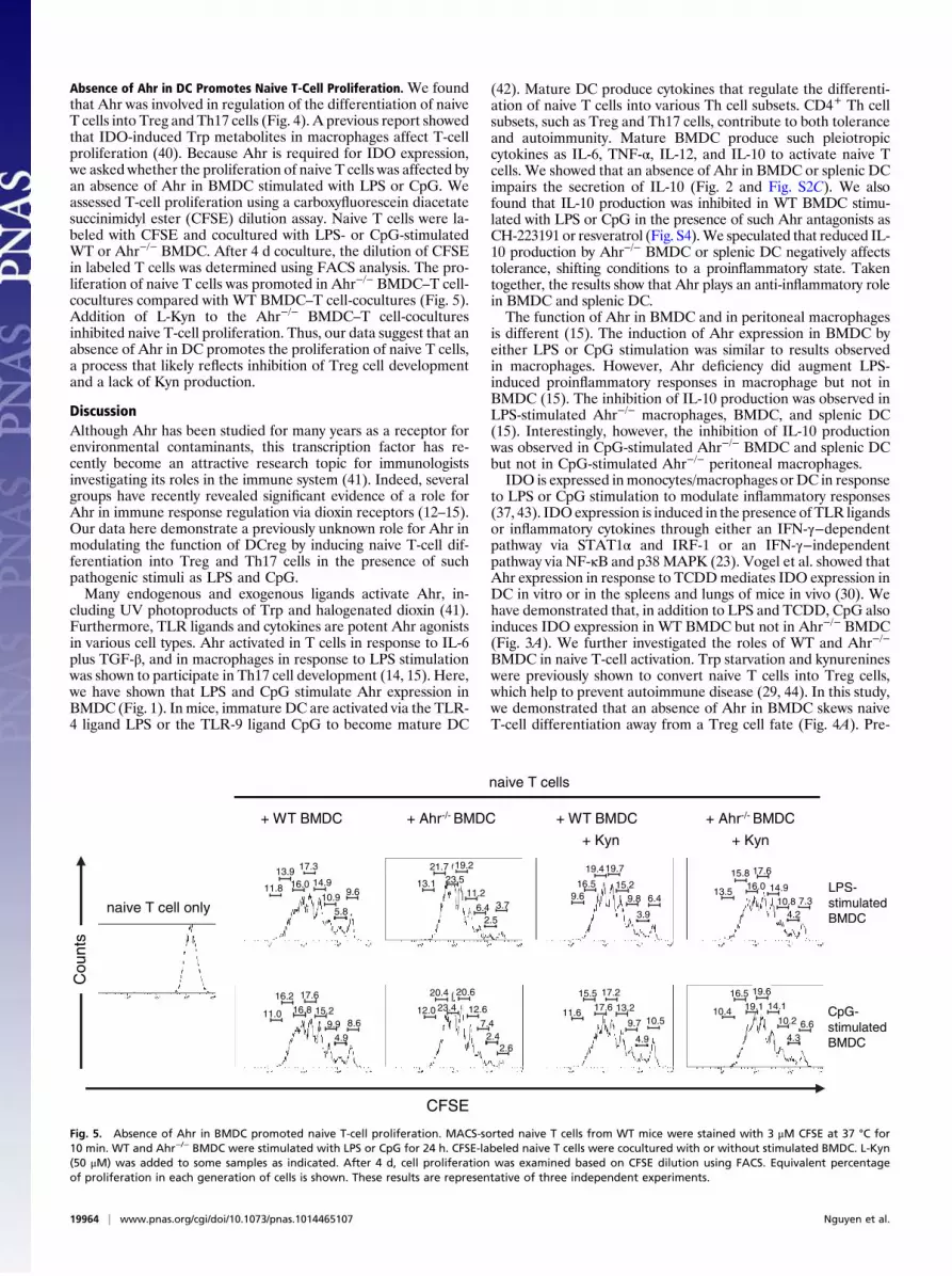
Absence of Ahr in DC Promotes Naive T-Cell Proliferation. We foundthat Ahr was involved in regulation of the differentiation of naiveT cells into Treg and Th17 cells (Fig. 4). A previous report showedthat IDO-induced Trp metabolites in macrophages affect T-cellproliferation (40). Because Ahr is required for IDO expression,we asked whether the proliferation of naive T cells was affected byan absence of Ahr in BMDC stimulated with LPS or CpG. Weassessed T-cell proliferation using a carboxyfluorescein diacetatesuccinimidyl ester (CFSE) dilution assay. Naive T cells were la-beled with CFSE and cocultured with LPS- or CpG-stimulatedWT or Ahr−/− BMDC. After 4 d coculture, the dilution of CFSEin labeled T cells was determined using FACS analysis. The pro-liferation of naive T cells was promoted in Ahr−/− BMDC–T cell-cocultures compared with WT BMDC–T cell-cocultures (Fig. 5).Addition of L-Kyn to the Ahr−/− BMDC–T cell-coculturesinhibited naive T-cell proliferation. Thus, our data suggest that anabsence of Ahr in DC promotes the proliferation of naive T cells,a process that likely reflects inhibition of Treg cell developmentand a lack of Kyn production.
DiscussionAlthough Ahr has been studied for many years as a receptor forenvironmental contaminants, this transcription factor has re-cently become an attractive research topic for immunologistsinvestigating its roles in the immune system (41). Indeed, severalgroups have recently revealed significant evidence of a role forAhr in immune response regulation via dioxin receptors (12–15).Our data here demonstrate a previously unknown role for Ahr inmodulating the function of DCreg by inducing naive T-cell dif-ferentiation into Treg and Th17 cells in the presence of suchpathogenic stimuli as LPS and CpG.Many endogenous and exogenous ligands activate Ahr, in-
cluding UV photoproducts of Trp and halogenated dioxin (41).Furthermore, TLR ligands and cytokines are potent Ahr agonistsin various cell types. Ahr activated in T cells in response to IL-6plus TGF-β, and in macrophages in response to LPS stimulationwas shown to participate in Th17 cell development (14, 15). Here,we have shown that LPS and CpG stimulate Ahr expression inBMDC (Fig. 1). In mice, immature DC are activated via the TLR-4 ligand LPS or the TLR-9 ligand CpG to become mature DC
(42). Mature DC produce cytokines that regulate the differenti-ation of naive T cells into various Th cell subsets. CD4+ Th cellsubsets, such as Treg and Th17 cells, contribute to both toleranceand autoimmunity. Mature BMDC produce such pleiotropiccytokines as IL-6, TNF-α, IL-12, and IL-10 to activate naive Tcells. We showed that an absence of Ahr in BMDC or splenic DCimpairs the secretion of IL-10 (Fig. 2 and Fig. S2C). We alsofound that IL-10 production was inhibited in WT BMDC stimu-lated with LPS or CpG in the presence of such Ahr antagonists asCH-223191 or resveratrol (Fig. S4).We speculated that reduced IL-10 production by Ahr−/− BMDC or splenic DC negatively affectstolerance, shifting conditions to a proinflammatory state. Takentogether, the results show that Ahr plays an anti-inflammatory rolein BMDC and splenic DC.The function of Ahr in BMDC and in peritoneal macrophages
is different (15). The induction of Ahr expression in BMDC byeither LPS or CpG stimulation was similar to results observedin macrophages. However, Ahr deficiency did augment LPS-induced proinflammatory responses in macrophage but not inBMDC (15). The inhibition of IL-10 production was observed inLPS-stimulated Ahr−/− macrophages, BMDC, and splenic DC(15). Interestingly, however, the inhibition of IL-10 productionwas observed in CpG-stimulated Ahr−/− BMDC and splenic DCbut not in CpG-stimulated Ahr−/− peritoneal macrophages.IDO is expressed inmonocytes/macrophages or DC in response
to LPS or CpG stimulation to modulate inflammatory responses(37, 43). IDOexpression is induced in the presence of TLR ligandsor inflammatory cytokines through either an IFN-γ−dependentpathway via STAT1α and IRF-1 or an IFN-γ−independentpathway via NF-κB and p38 MAPK (23). Vogel et al. showed thatAhr expression in response to TCDDmediates IDO expression inDC in vitro or in the spleens and lungs of mice in vivo (30). Wehave demonstrated that, in addition to LPS and TCDD, CpG alsoinduces IDO expression in WT BMDC but not in Ahr−/− BMDC(Fig. 3A). We further investigated the roles of WT and Ahr−/−
BMDC in naive T-cell activation. Trp starvation and kynurenineswere previously shown to convert naive T cells into Treg cells,which help to prevent autoimmune disease (29, 44). In this study,we demonstrated that an absence of Ahr in BMDC skews naiveT-cell differentiation away from a Treg cell fate (Fig. 4A). Pre-
6.64.3
10.2
14.1
19.6
19.110.4
16.5
10.5
4.9
9.7
13.2
17.2
17.611.6
15.5
2.62.4
7.4
12.6
20.6
23.412.0
20.4
8.6
4.99.9
15.2
17.6
16.811.0
16.2
7.34.2
10.814.9
17.6
16.013.5
15.8
6.4
3.9
9.815.2
19.4
9.616.5
19.7
3.7
2.56.4
11.2
19.2
23.513.1
21.7
naive T cells
+ WT BMDC + Ahr-/- BMDC + WT BMDC + Ahr-/- BMDC
+ Kyn + Kyn
LPS-stimulated BMDC
CpG-stimulatedBMDC
9.6
5.8
10.9
14.9
17.3
16.011.8
13.9
CFSE
Cou
nts
naive T cell only
Fig. 5. Absence of Ahr in BMDC promoted naive T-cell proliferation. MACS-sorted naive T cells from WT mice were stained with 3 μM CFSE at 37 °C for10 min. WT and Ahr−/− BMDC were stimulated with LPS or CpG for 24 h. CFSE-labeled naive T cells were cocultured with or without stimulated BMDC. L-Kyn(50 μM) was added to some samples as indicated. After 4 d, cell proliferation was examined based on CFSE dilution using FACS. Equivalent percentageof proliferation in each generation of cells is shown. These results are representative of three independent experiments.
19964 | www.pnas.org/cgi/doi/10.1073/pnas.1014465107 Nguyen et al.

vious reports of the regulatory roles of Ahr in the development ofFoxp3+ Treg cells have described inconsistent results. Activationof Ahr concomitant with an increase in the Treg cell populationwas demonstrated by our group and Quintana et al. (12) andKimura et al. (14). In a graft-versus-host disease model, however,Marshall et al. showed a decrease in Foxp3+ T-cell numbersduring Ahr activation (45). Recently, Mezrich et al. demonstratedthat Kyn induced the generation of Treg from naive T cells in anAhr-dependent manner (46). In our results, however, we foundthat Ahr−/− BMDC skewed the differentiation of naive T cellstoward a Th17 cell fate, possibly due to an inhibition of Treg celldevelopment (Fig. 4B). We hypothesized that inhibition of Tregcell development in the absence of Ahr in DC disrupts Th17 cellgeneration from naive T cells. Therefore, the Ahr-dependentmechanism that triggers the generation of Treg and Th17 cellsrequires further study. The role of Ahr in regulating Th17cell development has been previously reported (13, 14). Th17 cellsrespond to extracellular bacteria, mediate inflammation, andcause pathologic effects in certain autoimmune diseases. Imbal-ance in the ratio of Treg cells to Th17 cells dysregulates lym-phocyte activation and immune responses, which may contributeto various autoimmune disorders. Moreover, inhibition of Tregcell development due to an absence of Ahr in DC seems to pro-mote naive T-cell proliferation (Fig. 5).Deficiencies in IDO activity impair Trp degradation, and
consequently disrupt immune tolerance (43). IDO activates Tregcells and inhibits the conversion of Treg cells into Th17-like cells(36). Romani et al. suggested that kynurenines produced viaIDO negatively regulate IL-17 and IL-23 levels and inhibit au-toimmunity via IL-17 signaling (47). On the other hand, IDO hasbeen shown to drive autoimmune responses via B cells (48).Thus, IDO may contribute to both tolerance and autoimmunitydepending on the type of immune cells. Published evidencesuggests that IDO-induced Trp metabolites are Ahr ligands thatmediate the development of Treg cells (29, 49). In our BMDC–T-cell coculture system, synthetic L-Kyn inhibited naive T-celldifferentiation into Treg cells.In summary, we have identified a previously uncharacterized
role for Ahr in regulating tolerogenic or immunogenic activitiesof DC in the presence of LPS or CpG. We have also demon-strated that an absence of Ahr in stimulated BMDC skews naiveT-cell differentiation toward Th17 cells and away from Treg cells,possibly due to a lack of Kyn. However, the precise mechanism ofhow Kyn is able to induce Foxp3 and inhibit IL-17 needs furtherinvestigation. Our findings underscore the potential for thera-peutic targeting of the link between Ahr and kynurenines in thetreatment of immune cell-mediated inflammatory diseases.
Materials and MethodsMice. C57BL/6J WT mice were obtained from CLEA. Ahr−/− males and Ahr−/+
females (C57BL/6J background) were provided by Yoshiaki Fujii-Kuriyama(University of Tsukuba, Tsukuba, Japan), kept and mated in our laboratoryto generate Ahr−/− mice. To identify homozygous Ahr−/− mice, newbornmice were genotyped using PCRs performed with DNA obtained from thetails and specific primers. WT and Ahr−/− mice 6–8 wk old were used forthese experiments. All mice were maintained under specific, pathogen-freeconditions. All animal experiments were performed in accordance withprotocols approved by the Institutional Animal Care and Use Committees ofthe Graduate School of Frontier Biosciences, Osaka University.
BMDC Generation and Stimulation Conditions. BMDCwere generated fromWTand Ahr−/− mice as follows. Briefly, bone marrow cells were flushed fromtibiae and femurs of 6- to 8-wk-old C57BL/6J WT and Ahr−/− mice. The totalbone marrow cells were counted (Fig. S5A). Then, 2 × 10⁷ cells were seededin 150-mm diameter cell culture dishes (Corning) in 30 mL RPMI me-dium 1640 (Sigma) supplemented with 10% FCS, 100 μg/mL streptomycin,100 U/mL penicillin G, and 2.5 mM β-mercaptoethanol (complete RPMI me-dium 1640). On day 3, 30 mL complete RPMI medium 1640 containing10 ng/mL GM-CSF (Peprotech) was added to the dishes. On day 6, 30 mLculture was removed and centrifuged. The pellet was resuspended in 30 mLfresh complete RPMI medium 1640 containing 10 ng/mL GM-CSF andreturned to the same dish. On day 9, more than 80% of the nonadherentand loosely adherent cells expressed CD11c. BMDC were purified usinga MACS column and CD11c MicroBeads (Miltenyi Biotec) and ≈90% of thepurified BMDC were CD11b+ and CD11c+ (Fig. S5B). BMDC were seeded in 10mL complete RPMI medium 1640 (5 × 10⁶ cells/mL) and stimulated for 24 hwith or without LPS (1 μg/mL; Sigma), phosphorothioate-modified CpG-ODN(CpG) (1 μM; Gene Design Inc.), and TCDD (160 nM; Cerilliant) as indicated.Stimulated BMDC were considered mature if they expressed high levels ofMHC class II and CD86 (Fig. S5C).
qPCR. Total RNA was prepared by RNeasy (Quiagen) according to the man-ufacturer’s protocol. Reverse transcription of mRNA was performed in athermal cycler (Applied Biosystems). The mouse Ahr probe (Gene ExpressionAssays: Mm00478930_m1, Applied Biosystems) and mouse IDO probe (GeneExpression Assays: Mm00492586_m1, Applied Biosystems) were used. Forreference, we quantified mouse GAPDH (Applied Biosystems). The qPCR wascarried out in an ABI PRISM 7900 HT (Applied Biosystems). Cycling conditionswere 50 °C for 2 min and 95 °C for 10 min, followed by 40 cycles of 95 °C for15 s and 60 °C for 1 min. We applied the comparative ΔΔCt method nor-malized to GAPDH for IDO and Ahr mRNA quantitative analysis. The value ofunstimulated cells was set at one and was used to calculate the fold changein stimulated cells.
DC–T-Cell Coculture. Naive T cells were purified from the spleens ofWT C57BL/6J mice using a CD4+ T-cell isolation kit and CD62L MicroBeads (MiltenyiBiotec). Purified naive T cells (CD4+CD62Lhi) (1 × 10⁵ cells) were coculturedfor 4 d with or without LPS- and CpG-stimulated WT or Ahr−/− BMDC (1 × 10⁴
cells) in triplicate wells of a round-bottomed 96-well plate in a total volumeof 200 μL/well. Naive T cells in the cocultures were stimulated with theDynabeads Mouse T-activator CD3/CD28 (Invitrogen). L-Kyn (50 μM; Sigma)was added when indicated. After 4 d, IL-17 levels in the culture supernatantswere measured using ELISAs (R&D Systems). T cells were stained with trypanblue, and the viability of the cells was assessed using an automated cellcounter (Invitrogen).
Intracellular Cytokine and Foxp3 Staining. After cocultured with LPS- or CpG-stimulated BMDC, T cells were stimulated with 50 ng/mL PMA (Calbiochem)and 800 ng/mL ionomycin (Calbiochem) for 5 h. GolgiStop (BD Pharmingen)was added for the last 2 h. Samples were then fixed and permeabilized withCytofix/Cytoperm (BD Pharmingen). Cells were intracellularly stained with PE-conjugated anti–IL-17 antibodies (eBioscience) and FITC-conjugated anti–IFN-γ antibodies (eBioscience). For Foxp3 staining, T cells were fixed andpermeabilized in Fixation/Permeabilization buffer (eBioscience) for 30 minat 4 °C before intracellular staining with FITC-antimouse Foxp3 antibodies(eBioscience). FACS was performed with a Cytomics FC500 system (Beck-man Coulter).
Statistical Analysis. Student t tests were used to analyze data for differences,and P values <0.05 were considered to be significant.
ACKNOWLEDGMENTS. Thisworkwas supportedby Programfor PromotionofFundamental Studies inHealth Sciences of theNational InstituteofBiomedicalInnovation and Chugai-Roche Pharmaceutical Co. Ltd (Tokyo, Japan).
1. Stevens EA, Mezrich JD, Bradfield CA (2009) The aryl hydrocarbon receptor:
A perspective on potential roles in the immune system. Immunology 127:299–
311.2. Ema M, et al. (1992) cDNA cloning and structure of mouse putative Ah receptor.
Biochem Biophys Res Commun 184:246–253.3. Burbach KM, Poland A, Bradfield CA (1992) Cloning of the Ah-receptor cDNA reveals
a distinctive ligand-activated transcription factor. Proc Natl Acad Sci USA 89:8185–
8189.4. Perdew GH (1988) Association of the Ah receptor with the 90-kDa heat shock protein.
J Biol Chem 263:13802–13805.
5. Ma Q, Whitlock JP, Jr (1997) A novel cytoplasmic protein that interacts with the Ah
receptor, contains tetratricopeptide repeat motifs, and augments the transcriptional
response to 2,3,7,8-tetrachlorodibenzo-p-dioxin. J Biol Chem 272:8878–8884.6. Kazlauskas A, Poellinger L, Pongratz I (1999) Evidence that the co-chaperone p23
regulates ligand responsiveness of the dioxin (Aryl hydrocarbon) receptor. J Biol
Chem 274:13519–13524.7. Schrenk D (1998) Impact of dioxin-type induction of drug-metabolizing enzymes on
the metabolism of endo- and xenobiotics. Biochem Pharmacol 55:1155–1162.8. Mimura J, Ema M, Sogawa K, Fujii-Kuriyama Y (1999) Identification of a novel
mechanism of regulation of Ah (dioxin) receptor function. Genes Dev 13:20–25.
Nguyen et al. PNAS | November 16, 2010 | vol. 107 | no. 46 | 19965
IMMUNOLO
GY

9. Pitot HC, Goldsworthy T, Campbell HA, Poland A (1980) Quantitative evaluation ofthe promotion by 2,3,7,8-tetrachlorodibenzo-p-dioxin of hepatocarcinogenesis fromdiethylnitrosamine. Cancer Res 40:3616–3620.
10. Tomita S, et al. (2003) T cell-specific disruption of arylhydrocarbon receptor nucleartranslocator (Arnt) gene causes resistance to 2,3,7,8-tetrachlorodibenzo-p-dioxin-induced thymic involution. J Immunol 171:4113–4120.
11. Funatake CJ, Marshall NB, Steppan LB, Mourich DV, Kerkvliet NI (2005) Cutting edge:Activation of the aryl hydrocarbon receptor by 2,3,7,8-tetrachlorodibenzo-p-dioxingenerates a population of CD4+ CD25+ cells with characteristics of regulatory T cells.J Immunol 175:4184–4188.
12. Quintana FJ, et al. (2008) Control of T(reg) and T(H)17 cell differentiation by the arylhydrocarbon receptor. Nature 453:65–71.
13. Veldhoen M, et al. (2008) The aryl hydrocarbon receptor links TH17-cell-mediatedautoimmunity to environmental toxins. Nature 453:106–109.
14. Kimura A, Naka T, Nohara K, Fujii-Kuriyama Y, Kishimoto T (2008) Aryl hydrocarbonreceptor regulates Stat1 activation and participates in the development of Th17 cells.Proc Natl Acad Sci USA 105:9721–9726.
15. Kimura A, et al. (2009) Aryl hydrocarbon receptor in combination with Stat1 regulatesLPS-induced inflammatory responses. J Exp Med 206:2027–2035.
16. Banchereau J, Steinman RM (1998) Dendritic cells and the control of immunity.Nature 392:245–252.
17. Popov A, Schultze JL (2008) IDO-expressing regulatory dendritic cells in cancer andchronic infection. J Mol Med 86:145–160.
18. Steinman RM, Hawiger D, Nussenzweig MC (2003) Tolerogenic dendritic cells. AnnuRev Immunol 21:685–711.
19. Moffett JR, Namboodiri MA (2003) Tryptophan and the immune response. ImmunolCell Biol 81:247–265.
20. Mellor AL, Munn DH (2004) IDO expression by dendritic cells: Tolerance andtryptophan catabolism. Nat Rev Immunol 4:762–774.
21. Munn DH, et al. (2002) Potential regulatory function of human dendritic cellsexpressing indoleamine 2,3-dioxygenase. Science 297:1867–1870.
22. Mellor AL, et al. (2005) Cutting edge: CpG oligonucleotides induce splenic CD19+
dendritic cells to acquire potent indoleamine 2,3-dioxygenase-dependent T cellregulatory functions via IFN Type 1 signaling. J Immunol 175:5601–5605.
23. Fujigaki H, et al. (2006) The signal transducer and activator of transcription 1α andinterferon regulatory factor 1 are not essential for the induction of indoleamine 2,3-dioxygenase by lipopolysaccharide: Involvement of p38 mitogen-activated proteinkinase and nuclear factor-kappaB pathways, and synergistic effect of severalproinflammatory cytokines. J Biochem 139:655–662.
24. Wingender G, et al. (2006) Systemic application of CpG-rich DNA suppresses adaptiveT cell immunity via induction of IDO. Eur J Immunol 36:12–20.
25. Hill M, et al. (2007) IDO expands human CD4+CD25high regulatory T cells by promotingmaturation of LPS-treated dendritic cells. Eur J Immunol 37:3054–3062.
26. Edinger AL, Thompson CB (2002) Antigen-presenting cells control T cell proliferationby regulating amino acid availability. Proc Natl Acad Sci USA 99:1107–1109.
27. Munn DH, et al. (1999) Inhibition of T cell proliferation by macrophage tryptophancatabolism. J Exp Med 189:1363–1372.
28. Mellor AL, et al. (2003) Cutting edge: Induced indoleamine 2,3 dioxygenaseexpression in dendritic cell subsets suppresses T cell clonal expansion. J Immunol 171:1652–1655.
29. Fallarino F, et al. (2006) The combined effects of tryptophan starvation andtryptophan catabolites down-regulate T cell receptor ζ-chain and induce a regulatoryphenotype in naive T cells. J Immunol 176:6752–6761.
30. Vogel CFA, Goth SR, Dong B, Pessah IN, Matsumura F (2008) Aryl hydrocarbonreceptor signaling mediates expression of indoleamine 2,3-dioxygenase. BiochemBiophys Res Commun 375:331–335.
31. Jux B, Kadow S, Esser C (2009) Langerhans cell maturation and contact hypersensitivityare impaired in aryl hydrocarbon receptor-null mice. J Immunol 182:6709–6717.
32. Veldhoen M, Hirota K, Christensen J, O’Garra A, Stockinger B (2009) Natural agonistsfor aryl hydrocarbon receptor in culture medium are essential for optimal dif-ferentiation of Th17 T cells. J Exp Med 206:43–49.
33. Platten M, et al. (2005) Treatment of autoimmune neuroinflammation witha synthetic tryptophan metabolite. Science 310:850–855.
34. Criado G, Šimelyte E, Inglis JJ, Essex D, Williams RO (2009) Indoleamine 2,3dioxygenase-mediated tryptophan catabolism regulates accumulation of Th1/Th17cells in the joint in collagen-induced arthritis. Arthritis Rheum 60:1342–1351.
35. Jung ID, et al. (2007) Differential regulation of indoleamine 2,3-dioxygenase bylipopolysaccharide and interferon gamma in murine bone marrow derived dendriticcells. FEBS Lett 581:1449–1456.
36. Baban B, et al. (2009) IDO activates regulatory T cells and blocks their conversion intoTh17-like T cells. J Immunol 183:2475–2483.
37. Fallarino F, et al. (2009) IDO mediates TLR9-driven protection from experimentalautoimmune diabetes. J Immunol 183:6303–6312.
38. Hara T, et al. (2008) High-affinity uptake of kynurenine and nitric oxide-mediatedinhibition of indoleamine 2,3-dioxygenase in bone marrow-derived myeloid dendriticcells. Immunol Lett 116:95–102.
39. Belladonna ML, et al. (2006) Kynurenine pathway enzymes in dendritic cells initiatetolerogenesis in the absence of functional IDO. J Immunol 177:130–137.
40. Frumento G, et al. (2002) Tryptophan-derived catabolites are responsible forinhibition of T and natural killer cell proliferation induced by indoleamine 2,3-dioxygenase. J Exp Med 196:459–468.
41. Marshall NB, Kerkvliet NI (2010) Dioxin and immune regulation: Emerging role of arylhydrocarbon receptor in the generation of regulatory T cells. Ann N Y Acad Sci 1183:25–37.
42. Akira S, Takeda K (2004) Toll-like receptor signalling. Nat Rev Immunol 4:499–511.43. Grohmann U, Fallarino F, Puccetti P (2003) Tolerance, DCs and tryptophan: Much ado
about IDO. Trends Immunol 24:242–248.44. Fallarino F, et al. (2006) Tryptophan catabolism generates autoimmune-preventive
regulatory T cells. Transpl Immunol 17:58–60.45. Marshall NB, Vorachek WR, Steppan LB, Mourich DV, Kerkvliet NI (2008) Functional
characterization and gene expression analysis of CD4+ CD25+ regulatory T cellsgenerated in mice treated with 2,3,7,8-tetrachlorodibenzo-p-dioxin. J Immunol 181:2382–2391.
46. Mezrich JD, et al. (2010) An interaction between kynurenine and the aryl hydro-carbon receptor can generate regulatory T cells. J Immunol 185:3190–3198.
47. Romani L, Zelante T, De Luca A, Fallarino F, Puccetti P (2008) IL-17 and therapeutickynurenines in pathogenic inflammation to fungi. J Immunol 180:5157–5162.
48. Scott GN, et al. (2009) The immunoregulatory enzyme IDO paradoxically drives B cell-mediated autoimmunity. J Immunol 182:7509–7517.
49. Heath-Pagliuso S, et al. (1998) Activation of the Ah receptor by tryptophan andtryptophan metabolites. Biochemistry 37:11508–11515.
19966 | www.pnas.org/cgi/doi/10.1073/pnas.1014465107 Nguyen et al.


