Embed Size (px)
Citation preview

ORIGINAL RESEARCHBRAIN
Assessment ofWhole-BrainWhiteMatter by DTI in AutosomalRecessive Spastic Ataxia of Charlevoix-SaguenayK.K. Oguz, G. Haliloglu, C. Temucin, R. Gocmen, A.C. Has, K. Doerschner, A. Dolgun and M. Alikasifoglu
ABSTRACT
BACKGROUND AND PURPOSE: Extension and characteristics of WM involvement other than the brain stem remain inadequatelyinvestigated in ARSACS. The aim of this study was to investigate whole-brain WM alterations in patients with ARSACS.
MATERIALS AND METHODS: Nine Turkish unrelated patients with ARSACS and 9 sex- and age-matched healthy control participantsunderwent neurologic examination, molecular studies, electrophysiologic studies, and DTI of the brain. TBSS was used for whole-brainvoxelwise analysis of FA, AD, RD, mean diffusivity of WM. Tractographies for the CST and TPF were also computed.
RESULTS: Molecular studies revealed 8 novel mutations (3 nonsense, 4 missense, and 1 frameshift insertion) and amissense variation in theSACS gene. Thick TPF displaced and compressed the CST in the pons. The TPF had increased FA, decreased RD, and increased AD, whichmay be attributed to hypertrophy and/or hypermyelination. Widespread decreased FA and increased RD, suggesting demyelination, wasfound in the limbic, commissural, and projection fibers. In addition to demyelination, CST coursing cranial and caudal to the pons alsoshowed a marked decrease in AD, suggesting axonal degeneration. Electrophysiologic studies revealed findings that concur with demy-elination and axonal involvement.
CONCLUSIONS: In addition to developmental changes of the TPF and their effects on the CST in the brain stem, axonal degenerationmainly along the pyramidal tracts and widespread demyelination in WM also occur in patients with ARSACS. Widespread tissue damagemay be associated with extensive loss of sacsin protein in the brain and may explain a wide range of progressive neurologic abnormalitiesin patients with ARSACS.
ABBREVIATIONS: AD � axial diffusivity; ARSACS � autosomal recessive spastic ataxia of Charlevoix-Saguenay; CST � corticospinal tracts; FA � fractionalanisotropy; RD� radial diffusivity; TBSS� tract-based spatial statistics; TPF� transverse pontine fibers
ARSACS (MIM 270550) is a distinctive form of hereditary,
early-onset spastic ataxia, which manifests mainly by early-
onset pyramidal tract and cerebellar involvement, peripheral neu-
ropathy, and hypermyelination of the retinal nerve fibers. Clinical
variations including later-onset mental retardation, ophthalmo-
plegia, skeletal deformities, and biochemical abnormalities have
been reported.1,2
Since the first description of patients in the Charlevoix-
Saguenay region of Quebec in 1978, the disease has been increas-
ingly defined worldwide, with numerous mutations and diverse
phenotypes.2 The responsible gene (SACS, MIM 604490) was lo-
cated on chromosome 13q and encodes the sacsin protein, which
is widely expressed throughout the brain with a predilection for
pyramidal neurons, Purkinje cells, thalamic and pontine nuclei,
and reticular formation.3 Confusion with cerebral palsy and sec-
ondary spastic diplegia, and variations in the phenotype lead to
underdiagnosis of ARSACS.4,5
Neuroimaging findings include slowly progressive atrophy of
the superior vermis, and atrophy of the cerebellar hemispheres,
cervical spinal cord, and cerebral cortex.5,6 The description of
characteristic bilateral, parallel, paramedian, T2 hypointense
stripes on MR imaging contributed to an increased diagnosis of
ARSACS.7
By measuring anisotropic diffusion of water in the WM, DTI
detects abnormalities in the myelin, axon, or orientation of fibers
within the bundle.8,9 Gazulla et al10,11 described nonquantitative
abnormalities of the TPF and CST by DTI in 2 studies with 5
patients. These authors suggested a developmental basis for
Received November 1, 2012; accepted after revision December 7.
From the National Magnetic Resonance Research Center (UMRAM) (K.K.O., A.C.H.,K.D.), Bilkent University, Bilkent, Ankara, Turkey; and Departments of Radiology(K.K.O., R.G.), Pediatric Neurology (G.H.), Neurology (C.T.), Biostatistics (A.D.), andMedical Genetics (M.A.), Faculty of Medicine, Hacettepe University, Sihhiye,Ankara, Turkey.
Please address correspondence to Kader K. Oguz, National Magnetic ResonanceResearch Centre (UMRAM), Bilkent University, Cyberplaza, CBlock Level 2, Bilkent-Ankara, Turkey; e-mail:[email protected]
Indicates article with supplemental on-line tables.
http://dx.doi.org/10.3174/ajnr.A3488
AJNR Am J Neuroradiol ●:● ● 2013 www.ajnr.org 1
Published April 18, 2013 as 10.3174/ajnr.A3488
Copyright 2013 by American Society of Neuroradiology.

ARSACS by showing interruption of the CST in the pons by large
TPF. More recently, Prodi et al12 showed involvement of several
additional structures such as the forceps minor and major, supe-
rior longitudinal fasciculus, and cingulum by using an ROI ap-
proach for these predetermined areas. None of these studies in-
vestigated whole-brain WM changes in ARSACS by multiple
diffusion indices.
TBSS enables an unbiased whole-brain voxelwise analysis of
multi-subject FA data by using a nonlinear registration algorithm,
projection of the individual data onto a mean alignment-invari-
ant tract representation, and stringent statistical analysis. It has
been increasingly used for the depiction of WM abnormalities,
especially in patient groups with diseases in which alterations in
WM fibers cannot be precisely predicted.13
Given the high expression of the sacsin protein in the brain, we
hypothesized that WM alterations are not limited to the brain
stem and the cerebellum, and TBSS would show more widespread
abnormality in patients with ARSACS. We also aimed to seek
electrophysiologic correlates of DTI findings.
MATERIALS AND METHODSParticipantsThe local institutional review board approved the study, and all
participants gave written informed consent. Patients were asked
to participate in this prospective study on observation of MR im-
aging findings and confirmation of ARSACS by genetic studies.
Nine Turkish patients with ARSACS (male/female, 6/3; age
range, 5– 42 years; mean age, 23.67 � 13.28 years) and 9 sex- and
age-matched control participants (male/female, 6/3; age range,
5– 41 years; mean age, 23.78 � 12.43 years) were included in the
study.
All patients underwent neurologic and ophthalmologic exam-
inations, molecular studies, electrophysiologic studies, structural
MR imaging, and DTI of the brain.
Healthy volunteers without previous neuropsychological or
systemic disease served as control participants. They had normal
findings on structural MR imaging.
Molecular StudiesThe method for molecular studies is given in the On-line Appen-
dix, Part 1.
Electrophysiologic StudiesNerve conduction studies and motor-evoked potential studies
from the tibialis anterior muscle and somatosensory-evoked po-
tential by stimulation of the posterior tibial nerve were per-
formed. Technical details are given in the On-line Appendix,
Part 2.
Image AcquisitionImaging studies were performed on a 1.5T MR imaging scanner
(Symphony TIM; Siemens, Erlangen, Germany) equipped with
an 8-channel head coil. Brain MR imaging included sagittal and
axial T1-weighted imaging, axial T2-weighted imaging, and T2*
gradient recalled-echo. DTI applied axial single-shot echo-planar
imaging (TR, 5814 ms; TE, 98 ms; maximal b factor, 1000 s/mm2;
30 independent directions; field of view, 230 � 230 mm; matrix,
128 � 128; number of sections, 50, with 3-mm thickness without
gap).
Image Processing and AnalysisTwo experienced neuroradiologists (K.K.O., R.G.) evaluated
brain MR imaging for the presence of atrophy of the infratentorial
structures and the cerebrum, T2 hypointense stripes in the pons,
T2 hyperintense stripes in the lateral thalami, and susceptibility
on T2* GRE images in consensus.
DTI data were analyzed by use of TBSS. All scans were cor-
rected for head motion and eddy currents by use of the affine
registration. B0 volumes of each participant were extracted and
averaged. The main diffusion tensor was fitted in each voxel with
the FSL DTIFit tool (http://fsl.fmrib.ox.ac.uk/fsl/fsl-4.1.9/fdt/
fdt_dtifit.html), and the FA, RD, AD, and mean diffusivity maps
were calculated. After registration and alignment of individual
maps to the average space as input for TBSS, the mean FA map
and then the thinned mean FA skeleton (which represents the
centers of all tracts common to the group) were computed. Then
voxelwise statistics on FA, RD, AD, and mean diffusivity were
performed by use of 500 permutations. The results were corrected
for multiple comparisons, and family-wise error– corrected maps
at P values � .05 were considered significant.
Because the TPF and CST in the brain stem were reported to be
morphologically altered, we first recognized and evaluated these
structures on a directionally encoded color FA map of each par-
ticipant, and we then outlined and calculated corresponding
ROIs. Supratentorial WM clusters with significant change on the
resulting TBSS maps were extracted as ROIs and registered and
overlaid onto an anatomic Montreal Neurological Institute tem-
plate. These ROIs were labeled according to Johns Hopkins Uni-
versity WM tractography and the International Consortium for
Brain Mapping DTI-81 WM atlases in FSL, and the mean diffu-
sion indices of the ROIs were calculated.
3D fiber tractographies of the CST and TPF were obtained
by MedINRIA (http://www-sop.inria.fr/asclepios/software/
MedINRIA). The threshold for stopping fiber propagation was
FA � 0.2 and angle � 70°. Seed points were located at the level of
the corona radiata and medulla oblongata for the CST and central
pons for the TPF.
Statistical AnalysisStatistical analyses were done with a specific software package
(SPSS for Windows, version 15.0; SSPS, Chicago, Illinois). Age
and sex differences between patients and control participants
were tested by use of the independent-sample Student t test
and the Fisher exact test, respectively. The independent Stu-
dent t test was used for testing diffusion measures between
patients and control participants. P values � .05 were consid-
ered significant.
RESULTSPatient Demographics and Clinical AssessmentDemographic and clinical data for patients are summarized in
On-line Table 1. Delays in motor skills (n�7), ataxic gait (n�4),
dystonia (n�1), and frequent falls (n�1) were presenting symp-
toms. Initial diagnoses of the patients were cerebral palsy (n�4),
2 Oguz ● 2013 www.ajnr.org
hereditary spastic paraparesis (n�4), and static cerebellar ataxia
(n�1). The parents were first cousins in 4 families. None of the
patients had a family history of similar clinical features. Neuro-
logic and ophthalmologic examination revealed spasticity (n�7),
extremity deformity (n�2), cerebellar ataxia (n�4), dysarthria
(n�4), nystagmus (n�1), peripheral neuropathy (n�7), and my-
elinated retinal fibers (n�2) or thickening of the peripapillary
retinal fibers (n�1).
Molecular FindingsMolecular results are summarized in On-line Table 1 (detailed
results in On-line Appendix, Part 3, and On-line Table 2). Eight
never-reported different mutations (3 nonsense, 4 missense, and
1 frameshift insertion) and a missense variation were identified in
9 patients from unrelated families.
Electrophysiologic StudiesResults of electrophysiologic studies are given in On-line Table 3.
Nerve conduction studies were obtained in patients; somatosen-
sory-evoked potential and motor-evoked potential studies were
obtained in 5 patients. Decreased nerve conduction velocities of
the motor nerves and prolonged motor distal latencies were found
in patients with ARSACS. F waves were either prolonged or ab-
sent. Sensory nerve action potentials were absent bilaterally in the
sural nerves. When sensory conduction velocities could be ob-
tained, they were reduced in the sensory nerve action potentials.
Although central motor conduction times were significantly pro-
longed, cortical motor-evoked potential responses were remark-
ably low in patients without spinal and cortical somatosensory–
evoked potential responses. Compound muscle action potential
of the motor nerves was also low, especially in the lower
extremities.
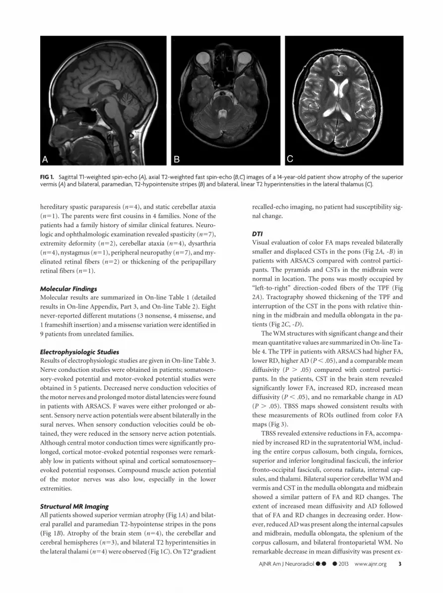
Structural MR ImagingAll patients showed superior vermian atrophy (Fig 1A) and bilat-
eral parallel and paramedian T2-hypointense stripes in the pons
(Fig 1B). Atrophy of the brain stem (n�4), the cerebellar and
cerebral hemispheres (n�3), and bilateral T2 hyperintensities in
the lateral thalami (n�4) were observed (Fig 1C). On T2*gradient
recalled-echo imaging, no patient had susceptibility sig-
nal change.
DTIVisual evaluation of color FA maps revealed bilaterally
smaller and displaced CSTs in the pons (Fig 2A, -B) in
patients with ARSACS compared with control partici-
pants. The pyramids and CSTs in the midbrain were
normal in location. The pons was mostly occupied by
“left-to-right” direction-coded fibers of the TPF (Fig
2A). Tractography showed thickening of the TPF and
interruption of the CST in the pons with relative thin-
ning in the midbrain and medulla oblongata in the pa-
tients (Fig 2C, -D).
The WM structures with significant change and their
mean quantitative values are summarized in On-line Ta-
ble 4. The TPF in patients with ARSACS had higher FA,
lower RD, higher AD (P � .05), and a comparable mean
diffusivity (P � .05) compared with control partici-
pants. In the patients, CST in the brain stem revealed
significantly lower FA, increased RD, increased mean
diffusivity (P � .05), and no remarkable change in AD
(P � .05). TBSS maps showed consistent results with
these measurements of ROIs outlined from color FA
maps (Fig 3).
TBSS revealed extensive reductions in FA, accompa-
nied by increased RD in the supratentorial WM, includ-
ing the entire corpus callosum, both cingula, fornices,
superior and inferior longitudinal fasciculi, the inferior
fronto-occipital fasciculi, corona radiata, internal cap-
sules, and thalami. Bilateral superior cerebellar WM and
vermis and CST in the medulla oblongata and midbrain
showed a similar pattern of FA and RD changes. The
extent of increased mean diffusivity and AD followed
that of FA and RD changes in decreasing order. How-
ever, reduced AD was present along the internal capsules
and midbrain, medulla oblongata, the splenium of the
corpus callosum, and bilateral frontoparietal WM. No
remarkable decrease in mean diffusivity was present ex-
FIG 1. Sagittal T1-weighted spin-echo (A), axial T2-weighted fast spin-echo (B,C) images of a 14-year-old patient show atrophy of the superiorvermis (A) and bilateral, paramedian, T2-hypointensite stripes (B) and bilateral, linear T2 hyperintensities in the lateral thalamus (C).
AJNR Am J Neuroradiol ●:● ● 2013 www.ajnr.org 3

cept for a few small clusters in the subcortical temporal WM
(Fig 3).
DISCUSSIONThree major results, which confirm and add to previous find-
ings10-12, can be derived from our data: 1) Large TPF with in-
creased FA and AD and decreased RD squeezed and displaced the
pontine CST with decreased FA, increased RD, and increased
mean diffusivity, as was also observed on tractography. 2) The
CST cranial and caudal to the pons showed an apparent reduction
in AD, suggestive of axonal degeneration. 3) The supratentorial
WM, including the limbic, commissural, and projection fibers,
was markedly involved, as demonstrated by reduced FA, in-
creased RD, and increased mean diffusivity, suggestive of
demyelination.
In agreement with Gazulla et al,10,11 the pontine CST was not
only engulfed by the TPF (TPF was thick because of hypermyeli-
nation and/or axonal hypertrophy), but was also dislocated on
directionally encoded FA images. The presence of hypermyeli-
nated or thickened peripapillar retinal fibers has been considered
a pathognomonic feature of ARSACS for quite some time.14 We
cannot confidently consider the thickening of the TPF and hyper-
myelination/peripapillary nerve thickening a result of identical
pathogenesis, given the rarity of patients with abnormal fundo-
scopic findings in our study, similar to other non-Quebec patient
populations. Furthermore, to our
knowledge, no evidence has been avail-
able regarding a relationship between
retinal nerve fiber layer thickening and
SACS mutation and the duration and se-
verity of ARSACS.15
AD (diffusion parallel to WM fibers)
and RD (diffusion perpendicular to WM
fibers) have been demonstrated to be
sensitive measures in the characteriza-
tion of tissue abnormalities. Experimen-
tal studies with immunohistostaining of
phosphorylated neurofilament and my-
elin basic protein have proven that re-
duced AD and increased RD correlated
with axonal and myelin damage, respec-
tively.16,17 Our finding of decreased AD
in the pyramidal pathways suggests an-
terograde and retrograde Wallerian de-
generation secondary to damage to the
CST by the TPF at the pons, very similar
to that observed after a pontine infarct
or a spinal cord injury.18,19 However,
because there was accompanying re-
duced FA and increased RD in the CST,
one can suggest that both axonal degen-
eration and demyelination occur along
the pyramidal tracts.
We also observed symmetric, linear,
and mild T2 hyperintensities in the lat-
eral thalami of 4 patients, which was re-
cently suggested as reflecting degenera-
tion of fibers of the external lamina consisting of afferent and
efferent fibers between the cortex and reticular thalamic nu-
clei.12,20 TBSS analysis was able to detect supratentorial abnor-
malities beyond the thalami and motor pathways and showed
widespread demyelination. Supporting our nerve conduction
studies and DTI findings, previous nerve conduction studies and
sural nerve biopsy examinations also revealed demyelinating neu-
ropathy with superimposed axonal involvement in patients with
ARSACS.11,21-23 Severe CST involvement, as assessed by motor-
evoked potential abnormalities, was attributed to the demyelina-
tion of the CST in an autopsy study.24 Our current TBSS study, on
the contrary, points out both axonal and myelin damage in the
CST. These features can also contribute to differentiation of AR-
SACS from other degenerative diseases of mainly axonal neurop-
athy, such as Friedreich ataxia.25 In our study, a widespread WM
abnormality is being documented first in ARSACS, in contrast to
more localized involvement of WM in the superior cerebellar pe-
duncles and peridentate area in Friedreich ataxia.26,27
Iron deposition in the basal ganglia and thalami and lipofus-
cin-like dens material within the lysosomes of swollen thalamic
and cerebellar cortical neurons were suggested to cause T2 hy-
pointensity in the pons and middle cerebellar peduncles.5,28,29
Our data do not support these suggestions because of lack of para-
magnetic susceptibility; T2 signal loss in the thalami or middle
FIG 2. Directionally encoded color FA image of the same patient as in Fig 1 (A) shows that the CST(blue, long arrow) is smaller and displaced in the pons by “left-to-right” direction-encoded TPF(red, short arrow). Tractographies of CST (blue) and TPF (red) depict interruption of CST by thickTPF (C). Corresponding FA image (B) and tractography (D) of a sex- and age-matched controlparticipant are also given.
4 Oguz ● 2013 www.ajnr.org

cerebellar peduncles; and DTI findings of material storage in
swollen neurons, such as reduced mean diffusivity.
Clinical variations such as mental retardation, ophthalmoplegia,
and lack of retinal nerve hypermyelination have been reported more
frequently in non-Quebec patients.2 In addition to developmental
abnormalities in the TPF and their mechanical effects on the CST,
axonal degeneration and, more extensively, demyelination in cere-
bral WM may explain a wide range of neurologic abnormalities other
than spasticity and the progressive nature of the disease. Recently,
Girard et al30 showed localization of sacsin to mitochondria and a
cascade of detrimental effects resulting in neuronal cell death by loss
of sacsin function in knockout mice. These authors further suggested
the presence of some common pathophysiologic features between
ARSACS and some other neurodegenerative diseases with mito-
chondrial impairment such as Alzheimer, Parkinson, and Hunting-
ton disease.30,31 Herein, by using TBSS, we present another common
feature of ARSACS with these diseases: extensive WM alterations in
the brain.32,33
Our current study was distinguished in several ways: First, to
the best of our knowledge, our study was the first that investigated
whole-brain WM in patients with ARSACS. Second, it quantified
the abnormal pattern of diffusion indices, including RD and AD,
indicating widespread WM disintegration. Last, although the
number of patients was limited, our patient cohort constituted
the largest population of patients with ARSACS in Turkey.34,35 In
our study, 8 different SACS mutations have been identified in 9
unrelated families referred to a single tertiary-care center. We
believe that recognition of imaging features, as well as the identi-
fication of new mutations, will increase the rate of diagnosis of
ARSACS in Turkey and other countries.
We did not measure the number of TPF and the CST because
changing DTI parameters can significantly alter the results, and
DTI has not been considered a precise way of measuring the
length and number of fibers.36 The small CST was not found to
have a significant change in the pons on TBSS maps. Because only
the major WM tracts are included in the skeleton, assessment of
small fiber tracts is difficult by TBSS. We overcame this problem
by outlining ROIs from directionally encoded FA images after
recognition of the morphologically altered structures.
CONCLUSIONSIn 9 Turkish patients with 8 new mutations, diffusion alterations
suggestive of widespread demyelination and axonal involvement,
mainly of the pyramidal tracts, have been demonstrated in addi-
tion to thickening of TPF and interruption of the CST in the pons.
These abnormalities may reflect tissue damage related to exten-
sive alterations in mitochondrial dynamics because of loss of sac-
sin function and may explain a wide range of neurologic abnor-
malities in patients with ARSACS.
ACKNOWLEDGMENTSThe authors thank Damagen personnel for their support in the mo-
lecular studies, Sueda Turk (Bilkent University, Department of Elec-
trics and Electronics Engineering) for contribution in data collection,
Prof. Dr. Meral Topcu (Hacettepe University, Department of Pedi-
atric Neurology) and Prof. Dr. Ersin Tan (Hacettepe University, De-
partment of Neurology) for their contribution of patients.
REFERENCES1. Van de Warrenburg BP, Sinke RJ, Kremer B. Recent advances in
hereditary spinocerebellar ataxias. J Neuropathol Exp Neurol2005;64:171– 80
FIG 3. TBSS (family-wise error–corrected threshold-cluster extend voxel p maps) display clusters with significantly different FA, RD, AD, andmean diffusivity compared with sex- and age-matched control participants at P� .05. For all diffusion measures, blue shows increased valuesand red shows decreased values. FA skeleton projected on a mean FA map is shown in green.
AJNR Am J Neuroradiol ●:● ● 2013 www.ajnr.org 5

2. Bouhlal Y, Amouri R, El Euch-Fayeche G, et al. Autosomal recessivespastic ataxia of Charlevoix-Saguenay: an overview. ParkinsonismRelat Disord 2011;17:418 –22
3. Parfitt DA, Michael GJ, Vermeulen EG, et al. The ataxia protein sac-sin is a functional co-chaperone that protects against polyglu-tamine-expanded ataxin-1. Hum Mol Genet 2009;18:1556 – 65
4. Bouchard JP, Barbeau A, Bouchard R, et al. Autosomal recessivespastic ataxia of Charlevoix-Saguenay. Can J Neurol Sci1978;5:61– 69
5. Bouchard JP, Richter A, Mathieu J, et al. Autosomal recessive spasticataxia of Charlevoix-Saguenay. Neuromuscul Disord 1998;8:474 –79
6. Anheim M, Fleury M, Monga B, et al. Epidemiological, clinical,paraclinical and molecular study of a cohort of 102 patients affectedwith autosomal recessive progressive cerebellar ataxia from Alsace,Eastern France: implications for clinical management. Neurogenet-ics 2010;11:1–12
7. Martin MH, Bouchard JP, Sylvain M, et al. Autosomal recessive spas-tic ataxia of Charlevoix-Saguenay: a report of MR imaging in 5 pa-tients. AJNR Am J Neuroradiol 2007;28:1606 – 08
8. Le Bihan D. Looking into the functional architecture of the brainwith diffusion MRI. Nat Rev Neurosci 2003;4:469 – 80
9. Mori S, Zhang J. Principles of diffusion tensor imaging and its ap-plications to basic neuroscience research. Neuron 2006;51:527–39
10. Gazulla J, Vela AC, Marın MA, et al. Is the ataxia of Charlevoix-Saguenay a developmental disease? Med Hypotheses 2011;77:347–52
11. Gazulla J, Benavente I, Vela AC, et al. New findings in the ataxia ofCharlevoix-Saguenay. J Neurol 2012;259:869 –78
12. Prodi E, Grisoli M, Panzeri M, et al. Supratentorial and pontine MRIabnormalities characterize recessive spastic ataxia of Charlevoix-Saguenay. A comprehensive study of an Italian series. Eur J Neurol2013;20:138 – 46
13. Smith SM, Jenkinson M, Johansen-Berg H, et al. Tract-based spatialstatistics: voxelwise analysis of multi-subject diffusion data. Neuro-image 2006;31:1487–505
14. Desserre J, Devos D, Sautiere BG, et al. Thickening of peripapillarretinal fibers for the diagnosis of autosomal recessive spastic ataxiaof Charlevoix-Saguenay. Cerebellum 2011;10:758 – 62
15. Vingolo EM, Di Fabio R, Salvatore S, et al. Myelinated retinal fibersin autosomal recessive spastic ataxia of Charlevoix-Saguenay. EurJ Neurol 2011;18:1187–90
16. Xie M, Wang Q, Wu TH, et al. Delayed axonal degeneration in slowWallerian degeneration mutant mice detected using diffusion ten-sor imaging. Neuroscience 2011;197:339 – 47
17. Kim JH, Wu TH, Budde MD, et al. Noninvasive detection of brain-stem and spinal cord axonal degeneration in an amyotrophic lateralsclerosis mouse model. NMR Biomed 2011;24:163– 69
18. Gupta RK, Saksena S, Chandra A, et al. Retrograde Wallerian degen-eration of cranial corticospinal tracts in cervical spinal cord injurypatients using diffusion tensor imaging. J Neurosci Res2008;86:2271– 80
19. Liang Z, Zeng J, Zhang C, et al. Longitudinal investigations on theanterograde and retrograde degeneration in the pyramidal tract
following pontine infarction with diffusion tensor imaging. Cere-brovasc Dis 2008;25:209 –16
20. Guillery RW, Harting JK. Structure and connections of the thalamicreticular nucleus: advancing views over half a century. J Comp Neu-rol 2003;463:360 –71
21. Takiyama Y. Autosomal recessive spastic ataxia of Charlevoix-Saguenay. Neuropathology 2006;26:368 –75
22. Vermeer S, Meijer RP, Pijl BJ, et al. ARSACS in the Dutchpopulation: a frequent cause of early-onset cerebellar ataxia. Neu-rogenetics 2008;9:207–14
23. Peyronnard JM, Charron L, Barbeau A. The neuropathy of Charlev-oix-Saguenay ataxia: an electrophysiological and pathologicalstudy. Can J Neurol Sci 1979;6:199 –203
24. Garcıa A, Criscuolo C, de Michele G, et al. Neurophysiological studyin a Spanish family with recessive spastic ataxia of Charlevoix-Saguenay. Muscle Nerve 2008;37:107–10
25. Pandolfo M. Friedreich’s ataxia: clinical aspects and pathogenesis.Semin Neurol 1999;19:311–21
26. Della Nave R, Ginestroni A, Tessa C, et al. Brain white matter tractsdegeneration in Friedreich ataxia. An in vivo MRI study using tract-based spatial statistics and voxel-based morphometry. Neuroimage2008;40:19 –25
27. Pagani E, Ginestroni A, Della Nave R, et al. Assessment of brain whitematter fiber bundle atrophy in patients with Friedreich ataxia. Ra-diology 2010;255:882– 89
28. Melberg A, Dahl N, Hetta J, et al. Neuroimaging study in autosomaldominant cerebellar ataxia, deafness, and narcolepsy. Neurology1999;53:2190 –92
29. Shimazaki H, Takiyama Y, Honda J, et al. Middle cerebellar pedun-cles and pontine T2 hypointensities in ARSACS. J Neuroimaging2012 Jan 23. [Epub ahead of print]
30. Girard M, Lariviere R, Parfitt DA, et al. Mitochondrial dysfunc-tion and Purkinje cell loss in autosomal recessive spastic ataxiaof Charlevoix-Saguenay (ARSACS). Proc Natl Acad Sci U S A2012;109:1661– 66
31. Johri A, Beal MF. Mitochondrial dysfunction in neurodegenerativediseases. J Pharmacol Exp Ther 2012;342:619 –30
32. Bohanna I, Georgiou-Karistianis N, Sritharan A, et al. Diffusion ten-sor imaging in Huntington’s disease reveals distinct patterns ofwhite matter degeneration associated with motor and cognitivedeficits. Brain Imaging Behav 2011;5:171– 80
33. Stebbins GT, Murphy CM. Diffusion tensor imaging in Alzhei-mer’s disease and mild cognitive impairment. Behav Neurol2009;21:39 – 49
34. Richter AM, Ozgul RK, Poisson VC, et al. Private SACS mutations inautosomal recessive spastic ataxia of Charlevoix–Saguenay (AR-SACS) families from Turkey. Neurogenetics 2004;5:165–70
35. Gucuyener K, Ozgul K, Paternotte C, et al. Autosomal recessive spas-tic ataxia of Charlevoix-Saguenay in two unrelated Turkish fami-lies. Neuropediatrics 2001;32:142– 46
36. Mukherjee P, Chung SW, Berman JI, et al. Diffusion tensor MR im-aging and fiber tractography: technical considerations. AJNR Am JNeuroradiol 2008;29:843–52
6 Oguz ● 2013 www.ajnr.org


