Embed Size (px)
Citation preview
ATR Regulates a G2-Phase Cell-Cycle Checkpointin Arabidopsis thaliana
Kevin Culligan,a,1 Alain Tissier,b and Anne Britta
a Section of Plant Biology, University of California, Davis, California 95616b Librophyt SAS, Centre de Cadarache, 13108 St. Paul-lez-Durance Cedex, France
Ataxia telangiectasia-mutated and Rad3-related (ATR) plays a central role in cell-cycle regulation, transmitting DNA damage
signals to downstream effectors of cell-cycle progression. In animals, ATR is an essential gene. Here, we find that
Arabidopsis (Arabidopsis thaliana) atr�/� mutants were viable, fertile, and phenotypically wild-type in the absence of
exogenous DNA damaging agents but exhibit altered expression of AtRNR1 (ribonucleotide reductase large subunit) and
alteration of some damage-induced cell-cycle checkpoints. atr mutants were hypersensitive to hydroxyurea (HU),
aphidicolin, and UV-B light but only mildly sensitive to g-radiation. G2 arrest was observed in response to g-irradiation in
both wild-type and atr plants, albeit with slightly different kinetics, suggesting that ATR plays a secondary role in response
to double-strand breaks. G2 arrest also was observed in wild-type plants in response to aphidicolin but was defective in atr
mutants, resulting in compaction of nuclei and subsequent cell death. By contrast, HU-treated wild-type and atr plants
arrested in G1 and showed no obvious signs of cell death. We propose that, in plants, HU invokes a novel checkpoint
responsive to low levels of deoxynucleotide triphosphates. These results demonstrate the important role of cell-cycle
checkpoints in the ability of plant cells to sense and cope with problems associated with DNA replication.
INTRODUCTION
The detection of DNA damage is an important aspect of damage
resistance. Cells do not routinely express all DNA repair
mechanisms, but instead upregulate these functions as required,
concomitantly slowing the cell cycle to permit time for repair
before S-phase (as replication past damaged sites may be
mutagenic) or before M-phase (after which point double-strand
breaks and lesions in daughter strand gaps may become far
more problematic to repair). Damage is generally detected by
protein complexes in the form of single-stranded DNA (Crowley
and Courcelle, 2002; Zou and Elledge, 2003), created when the
replication fork is physically blocked by DNA lesions or during
the process of DNA repair (e.g., homologous recombination,
nucleotide excision repair, and mismatch repair). These damage
sensors then activate downstream checkpoint mechanisms to
prevent progression into the next phase of the cell cycle as well
as upregulate DNA repair (Nyberg et al., 2002).
Some damage sensors also are involved in the regulation of
deoxynucleotide triphosphate (dNTP) pools, which may in turn
influence themode of DNA replication. One of the primary targets
for dNTP pool regulation is the enzyme ribonucleotide reductase
(RNR), which catalyzes the final reduction step in the production
of dNTPs. Genetic studies in Saccharomyces cerevisiae suggest
that the elevation of dNTP levels (through transcriptional and/or
posttranslational regulation of RNR) in response to DNA damage
may result in more efficient translesion DNA synthesis, enabling
the cell to complete replication in spite of the presence of
persisting lesions (Tanaka et al., 2000; Chabes et al., 2003; Yao
et al., 2003).
The proteins that sense damaged or single-stranded DNAs
and trigger these elaborate responses have been extensively
characterized in organisms ranging from bacteria to human
(Crowley and Courcelle, 2002; Melo and Toczyski, 2002). In
plants, however, the regulation of DNA replication and repair and
the cell cycle in response to DNA damage is only beginning to be
understood.
The Schizosaccharomyces pombe Rad3 homologs (ataxia
telangiectasia-mutated and Rad3-related [ATR] and ataxia
telangiectasia-mutated [ATM] in mammals, Mec1 in S. cerevi-
siae) are essential regulators of cell-cycle checkpoints, sensing
DNA damage and/or single-stranded DNA and activating
downstream effectors of cell-cycle progression and DNA repair.
ATM is activated primarily by DNA double-strand breaks. ATM-
deficient plants and animals are hypersensitive to g-irradiation
but not to replication-blocking agents such as UV-B light,
hydroxyurea (HU), or aphidicolin (Abraham, 2001; Garcia et al.,
2003; K. Culligan and A. Britt, unpublished data). Furthermore,
the Arabidopsis (Arabidopsis thaliana) ATM homolog is required
for the transcriptional induction of repair genes in response to
g-irradiation and for efficient progression through meiosis
(Garcia et al., 2003), though its role in plant cell-cycle responses
to DNA damage has not yet been determined. In animals,
activated ATM phoshorylates downstream components of
checkpoint pathways that include p53, BRCA1, NBS1, and
CHK2, initiating G1-, S-, or G2-phase arrest and/or apoptosis.
By contrast, ATR is activated primarily by agents that block
the progression of replication forks. ATR dominant-negative
1 To whom correspondence should be addressed. E-mail [email protected]; fax 530-752-5410.The author responsible for distribution of materials integral to thefindings presented in this article in accordance with the policy describedin the Instructions for Authors (www.plantcell.org) is: Kevin M. Culligan([email protected]).Article, publication date, and citation information can be found atwww.plantcell.org/cgi/doi/10.1105/tpc.018903.
This article is published in The Plant Cell Online, The Plant Cell Preview Section, which publishes manuscripts accepted for publication after they
have been edited and the authors have corrected proofs, but before the final, complete issue is published online. Early posting of articles reduces
normal time to publication by several weeks.
The Plant Cell Preview, www.aspb.orgª 2004 American Society of Plant Biologists 1 of 14
and conditional knockout mammalian cell lines display hyper-
sensitivity to UV-B light, HU, and aphidicolin but are also sen-
sitive to g-radiation. Thus, in animal cells, ATR is thought to
play a more generalized role in response to DNA damage than
ATM. Activated ATR phosphorylates CHK1 to initiate G2-phase
arrest but may also act at other points of the cell cycle, as
evidenced by ATR-dependent phosphorylation of BRCA1
(Tibbetts et al., 2000; Melo and Toczyski, 2002). However, the
determination of the full extent of ATR’s role in genome main-
tenance has been complicated by the fact that ATR homozygous
knockouts die early in embryogenesis (Brown and Baltimore,
2000; de Klein et al., 2000); cre/lox-induced nulls survive only
a few rounds of cell division, and the dominant-negative re-
duction-of-function cell lines have yielded conflicting results
(Cliby et al., 1998; Wright et al., 1998). Thus, a tractable genetic
system inwhich a null allele of ATR could be tolerated throughout
the development of a higher organism would be particularly
desirable.
Here, we investigate the effects of an ATR T-DNA insertion
allele in the plant Arabidopsis. The allele has a 6-kb insertion
within the kinase domain and is presumably null. We find that
the mutant plant (atr) is viable and developmentally normal in
the absence of exogenous DNA-damaging treatments but is
hypersensitive to the replication-blocking agents UV-B light,
HU, and aphidicolin. Thus, AtATR does appear to act as a
functional homolog of its mammalian counterpart. However, the
Arabidopsis atr mutants differ from mammalian mutants not
only in terms of their viability but also in their degree of sensitivity
to g-radiation.
In the course of these experiments, we also observed
a differential response to HU versus aphidicolin in wild-type
cells, with HU primarily inducing aG1 arrest, whereas aphidicolin
induced arrest in G2. This stands in contrast with the G2 arrest
induced by both agents in yeast and mammals (at concen-
trations that allowS-phase progression) and suggests that plants
may possess a novel G1 checkpoint response to low dNTP
levels. The G2 arrest in response to aphidicolin was abrogated in
atr mutants, suggesting that ATR regulates the G2 checkpoint
response to replication blocks, as it does inmammals.Moreover,
programmed cell death was observed in atrmutants in response
to aphidicolin, but not HU, under our experimental conditions.
We propose a model that suggests that the HU and aphidicolin
hypersensitivities of atrmutants are the result of its defect in the
G2 checkpoint response to blocked replication forks.
RESULTS
Identification of the ATROrtholog in Arabidopsis
ATR orthologs, including Mec1 of S. cerevisiae and Rad3 of S.
pombe, are members of a large gene family in eukaryotes that
encode Ser-Thr kinases, whose C-terminal catalytic domains
share similarity to yeast and mammalian phosphoinositide
3-kinases (Tibbetts and Abraham, 2000). We searched the
Arabidopsis genome for homologs of ATR, using the S. pombe
Rad3 C-terminal domain as a query sequence, and found
several candidate genes. Phylogenetic analysis of the corre-
sponding predicted full-length protein sequences indicated that
one was an ortholog of Target of Rapamycin (TOR), one an
ortholog of ATM, and one was an ortholog of ATR (Figure 1A).
No other candidate genes fell into these broad categories. The
ATR-orthologous sequence (previously reported in GenBank as
mRNA AtRad3 [AB040133], which we designate here as AtATR)
revealed high similarity throughout its entire length to other ATR
proteins, but its similarity to ATM and TOR sequences was
limited only to the highly conserved middle and C-terminal
regions. We confirmed the GenBank sequence and intron/exon
boundaries by isolating and sequencing cDNAs of AtATR
(�8.1 kb, ecotype Wassilewskija [Ws]) using RT-PCR, and the
resulting sequence predicted 17 exons encoding a protein of
2702 amino acids (Figure 1B).
Identification of T-DNA Insertion-Mutation Alleles
in AtATR
To study the function of ATR in Arabidopsis, we searched for
lines with T-DNA insertions within ATR. Three independent lines
were identified from different collections and were termed atr-1
(Arabidopsis Knockout Facility, ecotypeWs), atr-2 (SIGnAL data-
base, ecotype Columbia), and atr-3 (FLAG database, ecotype
Ws). atr-1 and atr-2 contain insertions in the middle of ATR, in
exons 7 and 10, respectively (Figure 1B). atr-3 contains a single,
�6-kb T-DNA insertion within the highly conserved C-terminal
kinase (PI3Kc) domain (Figures 1B to 1D), which causes a 30-bp
deletion adjacent to the catalyticAsp required for kinase function.
RT-PCR analysis of the C-terminal region of ATR confirms
that the transcript is absent in atr-3 (Figure 7). Thus, we believe
atr-3 is likely to be a null mutation and have focused the major-
ity of our characterization on this line (termed atr below).
Nonetheless, all three lines exhibit a similar recessive phenotype
(see below) that cosegregates with their respective T-DNA
insertions in ATR.
Neither AtATR nor AtATM Are Required during Normal
Somatic Development
All three atr homozygous alleles are phenotypically wild-type in
growth (of the root and shoot) and development (leaf and flower
development, seed set and viability) under standard conditions.
Thus, ATR is not essential for normal somatic development in
Arabidopsis. By contrast, ATR is an essential gene in animals;
ATR knockout mice die early in embryogenesis (Brown and
Baltimore, 2000; de Klein et al., 2000), whereas conditional
knockout human cell lines divide only a few times before dying
(Cortez et al., 2001; Brown and Baltimore, 2003). Because ATR
and ATM are paralogs known to play partially overlapping roles
in animal cells (Abraham, 2001), it is possible that ATM could
compensate for ATR deficiency in Arabidopsis. To test this
hypothesis, we established atr atm double mutants and found
no obvious phenotypic differences (in vegetative growth and
development) when compared with wild-type or single-mutant
plants, indicating that neither ATR nor ATM play an essential role
during normal vegetative (somatic) growth.
The double mutants, however, were completely sterile (Figure
2). atr mutant plants are fertile and produce normal gametes;
pollen staining for viable spores revealed no differences versus
2 of 14 The Plant Cell
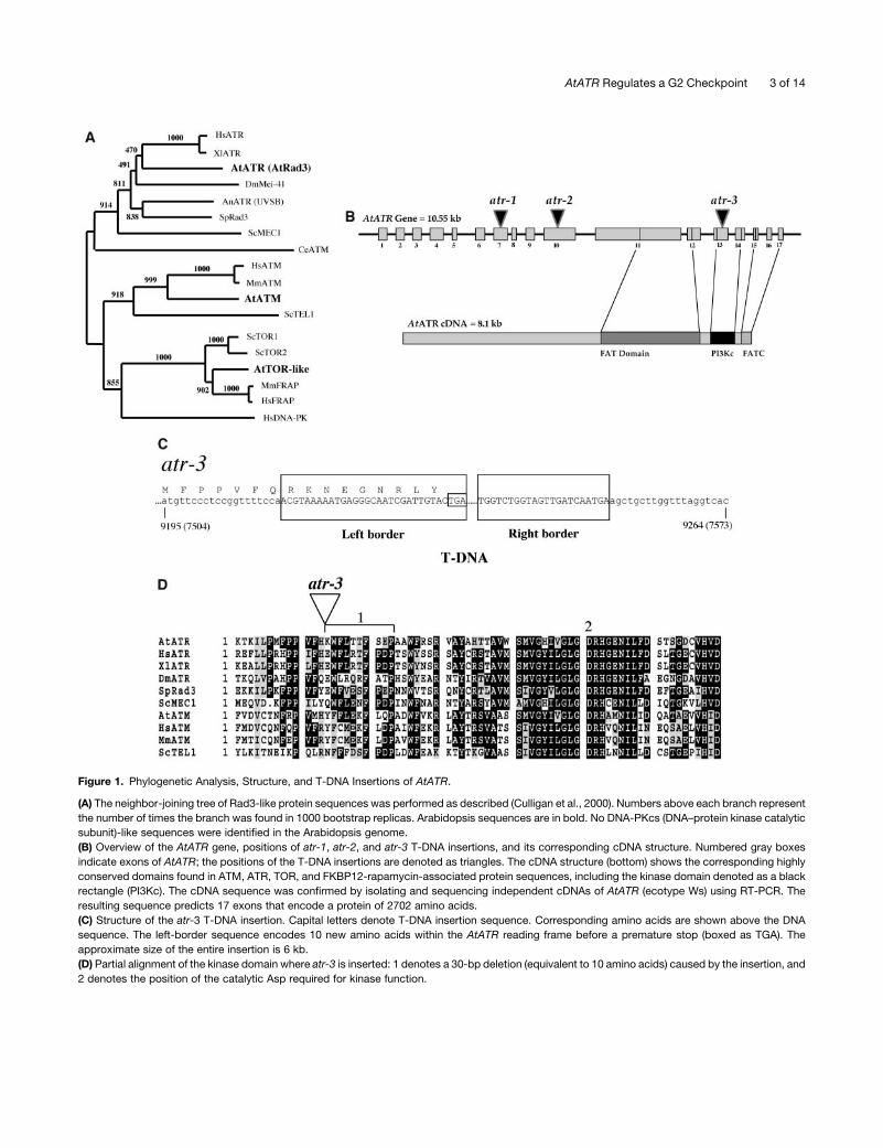
Figure 1. Phylogenetic Analysis, Structure, and T-DNA Insertions of AtATR.
(A) The neighbor-joining tree of Rad3-like protein sequences was performed as described (Culligan et al., 2000). Numbers above each branch represent
the number of times the branch was found in 1000 bootstrap replicas. Arabidopsis sequences are in bold. No DNA-PKcs (DNA–protein kinase catalytic
subunit)-like sequences were identified in the Arabidopsis genome.
(B) Overview of the AtATR gene, positions of atr-1, atr-2, and atr-3 T-DNA insertions, and its corresponding cDNA structure. Numbered gray boxes
indicate exons of AtATR; the positions of the T-DNA insertions are denoted as triangles. The cDNA structure (bottom) shows the corresponding highly
conserved domains found in ATM, ATR, TOR, and FKBP12-rapamycin-associated protein sequences, including the kinase domain denoted as a black
rectangle (PI3Kc). The cDNA sequence was confirmed by isolating and sequencing independent cDNAs of AtATR (ecotype Ws) using RT-PCR. The
resulting sequence predicts 17 exons that encode a protein of 2702 amino acids.
(C) Structure of the atr-3 T-DNA insertion. Capital letters denote T-DNA insertion sequence. Corresponding amino acids are shown above the DNA
sequence. The left-border sequence encodes 10 new amino acids within the AtATR reading frame before a premature stop (boxed as TGA). The
approximate size of the entire insertion is 6 kb.
(D) Partial alignment of the kinase domain where atr-3 is inserted: 1 denotes a 30-bp deletion (equivalent to 10 amino acids) caused by the insertion, and
2 denotes the position of the catalytic Asp required for kinase function.
AtATR Regulates a G2 Checkpoint 3 of 14
the wild type, and the siliques were full (normal seed set). atm
mutant plants, however, are partially sterile because of abundant
chromosomal fragmentation during meiosis, resulting in a re-
duction in viable pollen and reduced seed set (Garcia et al.,
2003). We suggest that AtATM and AtATR play partially redun-
dant roles during meiosis in Arabidopsis.
AtATR Is Required for Full Transcriptional Induction of the
Large Subunit of RNR, AtRNR1
In the budding yeast S. cerevisiae, Mec1 (the ATR ortholog)
regulates expression of RNR at both the transcriptional and
posttranscriptional levels (Zhao et al., 2001). Null alleles ofmec1
are lethal, but this lethality is suppressed by mutations in genes
that negatively regulate RNR (Huang et al., 1998; Zhao et al.,
1998), such as the transcriptional repressor Crt1 (Huang et al.,
1998) or the inhibitor of RNR activity Sml1 (Zhao et al., 1998).
Derepression of RNR transcription in response to DNA damage
is dependent on Mec1 and the downstream kinase Dun1, which
indirectly induces an inhibitory phosphorylation of Crt1 (Elledge
et al., 1992; Zhao and Rothstein, 2002). Whether Mec1 or ATR
also play a role in the normal cell-cycle specific regulation ofRNR
transcription in unchallenged cells remains unclear.
We therefore wanted to determine if RNR transcription is
regulated in an AtATR-dependent manner in challenged and/or
unchallenged plants. We searched GenBank and identified
the Arabidopsis homolog of RNR1, the large subunit of RNR
(accession number AF092841). Transcription of RNR1 has
already been shown to be regulated in a cell-cycle dependent
manner in tobacco (Nicotiana tabacum) (Chaboute et al., 1998,
2002), and transcription is induced by DNA damage in this plant
(M.E. Chaboute, personal communication). We probed RNA gel
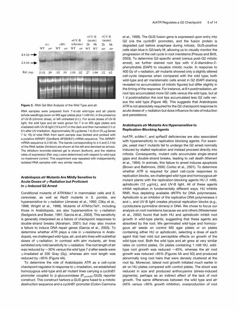
blots with AtRNR1 and found that AtRNR1 transcript is reduced
in atr mutants, down �40% versus the wild type under
standard conditions (Figure 3), suggesting that ATR is required
to support normal expression of RNR1. Plants treated with
chronic doses of UV-B light, sufficient to inhibit but not eliminate
growth, display a level of RNR1 transcript similar to that
observed under standard (UV-free) conditions, and again atr
plants exhibit reduced levels of RNR1 transcript. Interestingly,
plants treated with acute doses of UV-B light displayed
a transient inhibition of AtRNR1 transcript (at 2 h postirradia-
tion), followed by a return to standard condition levels at 6 h
postirradiation. This UV-induced suppression is not ATR de-
pendent, though again, the overall level of RNR1 transcript is
reduced (Figure 3).
Because we used HU (an inhibitor of RNR activity) as a DNA
replication-blocking agent (seebelow) to characterize thepheno-
typic effects of the atr mutant, we wanted to determine the
effects of HU treatment on RNR expression in whole plants.
Surprisingly, in the presence of HU,RNR1 expression is the same
in both the wild type and atr (Figure 3) with levels similar to the
wild type under standard conditions. This suggests that there
are additional ATR-independent levels of RNR1 regulation in
Arabidopsis and is consistent with previous studies showing that
HU induces RNR expression in plants (Chaboute et al., 1998).
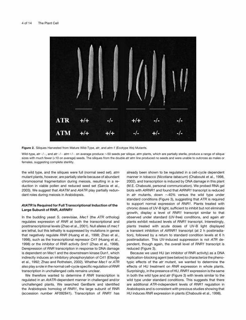
Figure 2. Siliques Harvested from Mature Wild-Type, atr, and atm-1 (Ecotype Ws) Mutants.
Wild-type, atr�/�, and atr�/� atm1/� on average produce �50 seeds per silique. atm plants, which are partially sterile, produce a range of silique
sizes with much fewer (<10 on average) seeds. The siliques from the double atr atm line produced no seeds and were unable to outcross as males or
females, suggesting complete sterility.
4 of 14 The Plant Cell
Arabidopsis atrMutants Are Mildly Sensitive to
Acute Doses of g-Radiation but Proficient
in g-Induced G2 Arrest
Conditional mutants of ATR/Mec1 in mammalian cells and S.
cerevisiae, as well as Rad3 mutants in S. pombe, are
hypersensitive to g-radiation (Jimenez et al., 1992; Cliby et al.,
1998; Wright et al., 1998). Mutants of ATM/scTel1, including
those in Arabidopsis, are also hypersensitive to g-radiation
(Sedgwick and Boder, 1991; Garcia et al., 2003). This sensitivity
is generally interpreted as a failure of checkpoint responses to
double-strand breaks (Abraham, 2001) but may also involve
a failure to induce DNA repair genes (Garcia et al., 2003). To
determine whether ATR plays a role in g-resistance in Arabi-
dopsis, we challengedwild-type, atr, and atm lines with sublethal
doses of g-radiation. In contrast with atm mutants, atr lines
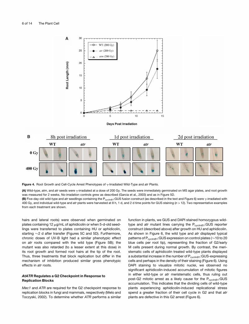
exhibited onlymild sensitivity to g-radiation. The root length of atr
was reduced by�30% versus the wild type 7 d after seeds were
g-irradiated at 200 Gray (Gy), whereas atm root length was
reduced by >95% (Figure 4A).
To determine the role of Arabidopsis ATR as a cell-cycle
checkpoint regulator in response to g-irradiation, we established
homozygous wild-type and atr mutant lines carrying a cyclinB1
promoter coupled to b-glucuronidase (PcyclinB1:GUS) reporter
construct. This construct harbors a GUS gene fused to a mitotic
destruction sequence and a cyclinB1 promoter (Colon-Carmona
et al., 1999). The GUS fusion gene is expressed upon entry into
G2 (via the cyclinB1 promoter), and the fusion protein is
degraded just before anaphase during mitosis. GUS-positive
cells stain blue in G2/early M, allowing us to visually monitor the
progression of the cell cycle in root meristems (Preuss and Britt,
2003). To determine G2-specific arrest (versus post-G2 mitotic
arrest), we further stained root tips with 49,6-diamidino-2-
phenylindole (DAPI) to visualize mitotic nuclei. In response to
400 Gy of g-radiation, atrmutants showed only a slightly altered
cell-cycle response when compared with the wild type; both
wild-type and atr meristematic cells arrest in G2 (DAPI staining
revealed no accumulation of mitotic figures) but differ slightly in
the timing of the response. For instance, at 8 h postirradiation, atr
root tips accumulated more G2 cells versus the wild type, but at
1 d postirradiation the root tips accumulated less G2 cells ver-
sus the wild type (Figure 4B). This suggests that Arabidopsis
ATR is not absolutely required for the G2 checkpoint response to
acute doses of g-radiation but does influence its rate of induction
and persistence.
Arabidopsis atrMutants Are Hypersensitive to
Replication-Blocking Agents
hsATR, scMec1, and spRad3 deficiencies are also associated
with hypersensitivity to replication blocking agents. For exam-
ple, yeast mec1 mutants fail to undergo the G2 arrest normally
induced by stalled replication and instead proceed directly into
mitosis. Consequently, mutant cells accumulate single-strand
gaps and double-strand breaks, leading to cell death (Weinert
et al., 1994). In animals, this failure to arrest induces apoptosis
(Brown and Baltimore, 2000; Cortez et al., 2001). To determine
whether ATR is required for plant cell-cycle responses to
replication blocks, we challenged wild-type and homozygous atr
mutant plants with the replication-blocking agents HU (1 mM),
aphidicolin (12 mg/mL), and UV-B light. All of these agents
inhibit replication in fundamentally different ways. HU inhibits
RNR, thus depleting available dNTPs for DNA polymerases.
Aphidicolin is an inhibitor of the replicative DNA polymerases d
and e, and UV-B light creates physical replication blocks (e.g.,
cyclobutane pyrimidine dimers) in DNA. We chose to focus our
analysis on root meristems because we and others (Wiedemeier
et al., 2002) found that both HU and aphidicolin inhibit root
growth in wild-type plants, suggesting that these agents are
absorbed by the root. We germinated wild-type and homozy-
gous atr seeds on control MS agar plates or on plates
containing either HU or aphidicolin, selecting a dose of each
agent that had mild but perceptible effects on growth of the
wild-type root. Both the wild type and atr grew at very similar
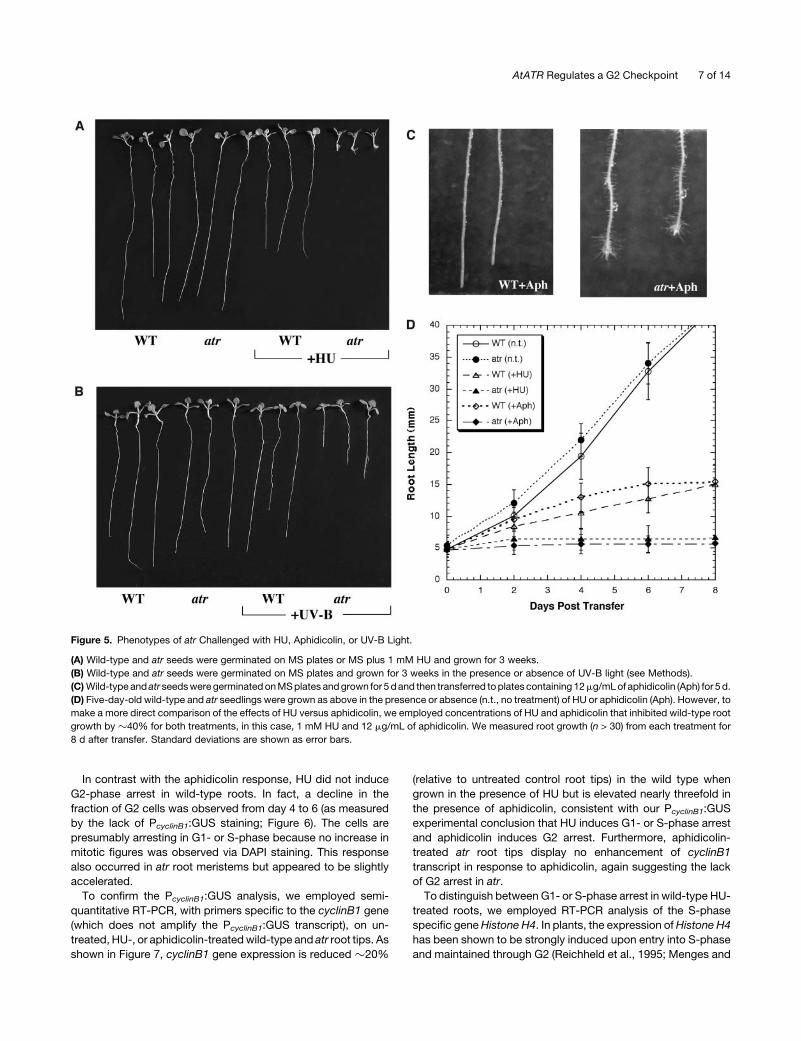
rates on control plates. On plates containing 1 mM HU, wild-
type root growth was reduced �40%, whereas the atr root
growth was reduced >95% (Figures 5A and 5D) and produced
abnormally long root hairs that were densely clustered at the
root tip. Moreover, lateral root growth initiated much earlier in
atr on HU plates compared with control plates. The shoot was
reduced in size and produced anthocyanins (stress-induced
pigments), perhaps as an indirect effect of the lack of root
growth. The same differences between the wild type and atr
(40% versus >95% growth inhibition, overproduction of root
Figure 3. RNA Gel Blot Analysis of the Wild Type and atr.
RNA samples were prepared from 7-d-old wild-type and atr plants
(whole seedlings) sown onMS agar plates plus 1mMHU, in the presence
of UV-B (chronic dose), or left untreated (n.t.). For acute doses of UV-B
light, the wild type and atr were grown for 7 d on MS agar plates and
irradiated with UV-B light (10 kJ/m2) in the dark and then harvested 2 h or
6 h after UV irradiation. Approximately 30 mg (lanes 1 to 6) or 25 mg (lanes
7 to 10) of total RNA from each sample was blotted and probed with
a putative AtRNR1 (GenBank AF092841) mRNA sequence. The AtRNR1
mRNA sequence is 2.65 kb. The bands corresponding to 4.4 and 2.4 kb
of the RNA ladder (Ambion) are shown at the left and denoted as arrows.
The ethidium bromide–stained gel is shown (bottom), and the relative
rates of expression (Rel. exp.) were determined with respect to wild-type
no-treatment control. This experiment was repeated with independently
isolated RNA samples with very similar results.
AtATR Regulates a G2 Checkpoint 5 of 14
hairs and lateral roots) were observed when germinated on
plates containing 12 mg/mL of aphidicolin or when 5-d-old seed-
lings were transferred to plates containing HU or aphidicolin,
starting �2 d after transfer (Figures 5C and 5D). Furthermore,
chronic doses of UV-B light had a similar phenotypic effect
on atr roots compared with the wild type (Figure 5B); the
mutant was also retarded (to a lesser extent at this dose) in
its root growth and formed root hairs at the tip of the root.
Thus, three treatments that block replication but differ in the
mechanism of inhibition produced similar gross phenotypic
effects in atr roots.
AtATR Regulates a G2 Checkpoint in Response to
Replication Blocks
Mec1 and ATR are required for the G2 checkpoint response to
replication blocks in fungi and mammals, respectively (Melo and
Toczyski, 2002). To determine whether ATR performs a similar
function in plants, we GUS and DAPI stained homozygous wild-
type and atr mutant lines carrying the PcyclinB1:GUS reporter
construct (described above) after growth on HU and aphidicolin.
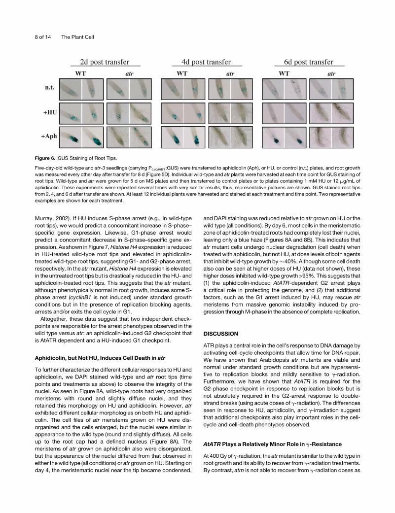
As shown in Figure 6, the wild type and atr displayed typical
patterns of PcyclinB1:GUS expression on control plates (�10 to 20
blue cells per root tip), representing the fraction of G2/early
M cells present during normal growth. By contrast, the meri-
stematic cells of aphidicolin treated wild-type plants displayed
a substantial increase in the number of PcyclinB1:GUS-expressing
cells and perhaps in the density of their staining (Figure 6). Using
DAPI staining to visualize mitotic nuclei, we observed no
significant aphidicolin-induced accumulation of mitotic figures
in either wild-type or atr meristematic cells, thus ruling out
post-G2 mitotic arrest as a likely cause for the PcyclinB1:GUS
accumulation. This indicates that the dividing cells of wild-type
plants experiencing aphidicolin-induced replicational stress
spend a greater fraction of their cell cycle in G2 and that atr
plants are defective in this G2 arrest (Figure 6).
Figure 4. Root Growth and Cell-Cycle Arrest Phenotypes of g-Irradiated Wild-Type and atr Plants.
(A) Wild-type, atm, and atr seeds were g-irradiated at a dose of 200 Gy. The seeds were immediately germinated on MS agar plates, and root growth
was measured for 2 weeks. No-irradation controls grew as described (Garcia et al., 2003) and as in Figure 5D.
(B) Five-day-old wild-type and atr seedlings containing the PcyclinB1:GUS fusion construct (as described in the text and Figure 6) were g-irradiated with
400 Gy, and individual wild-type and atr plants were harvested at 8 h, 1 d, and 2 d time points for GUS staining (n > 12). Two representative examples
from each treatment are shown.
6 of 14 The Plant Cell
In contrast with the aphidicolin response, HU did not induce
G2-phase arrest in wild-type roots. In fact, a decline in the
fraction of G2 cells was observed from day 4 to 6 (as measured
by the lack of PcyclinB1:GUS staining; Figure 6). The cells are
presumably arresting in G1- or S-phase because no increase in
mitotic figures was observed via DAPI staining. This response
also occurred in atr root meristems but appeared to be slightly
accelerated.
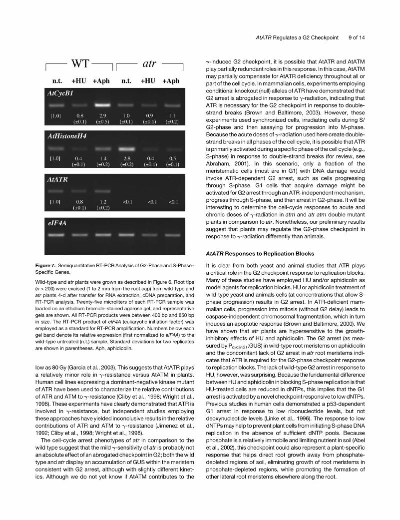
To confirm the PcyclinB1:GUS analysis, we employed semi-
quantitative RT-PCR, with primers specific to the cyclinB1 gene
(which does not amplify the PcyclinB1:GUS transcript), on un-
treated, HU-, or aphidicolin-treatedwild-type and atr root tips. As
shown in Figure 7, cyclinB1 gene expression is reduced �20%
(relative to untreated control root tips) in the wild type when
grown in the presence of HU but is elevated nearly threefold in
the presence of aphidicolin, consistent with our PcyclinB1:GUS
experimental conclusion that HU induces G1- or S-phase arrest
and aphidicolin induces G2 arrest. Furthermore, aphidicolin-
treated atr root tips display no enhancement of cyclinB1
transcript in response to aphidicolin, again suggesting the lack
of G2 arrest in atr.
To distinguish between G1- or S-phase arrest in wild-type HU-
treated roots, we employed RT-PCR analysis of the S-phase
specific geneHistone H4. In plants, the expression ofHistone H4
has been shown to be strongly induced upon entry into S-phase
and maintained through G2 (Reichheld et al., 1995; Menges and
Figure 5. Phenotypes of atr Challenged with HU, Aphidicolin, or UV-B Light.
(A) Wild-type and atr seeds were germinated on MS plates or MS plus 1 mM HU and grown for 3 weeks.
(B) Wild-type and atr seeds were germinated on MS plates and grown for 3 weeks in the presence or absence of UV-B light (see Methods).
(C)Wild-typeand atr seedsweregerminatedonMSplates andgrown for 5dand then transferred toplates containing 12mg/mLof aphidicolin (Aph) for 5d.
(D) Five-day-old wild-type and atr seedlings were grown as above in the presence or absence (n.t., no treatment) of HU or aphidicolin (Aph). However, to
make a more direct comparison of the effects of HU versus aphidicolin, we employed concentrations of HU and aphidicolin that inhibited wild-type root
growth by�40% for both treatments, in this case, 1 mM HU and 12 mg/mL of aphidicolin. We measured root growth (n > 30) from each treatment for
8 d after transfer. Standard deviations are shown as error bars.
AtATR Regulates a G2 Checkpoint 7 of 14
Murray, 2002). If HU induces S-phase arrest (e.g., in wild-type
root tips), we would predict a concomitant increase in S-phase–
specific gene expression. Likewise, G1-phase arrest would
predict a concomitant decrease in S-phase–specific gene ex-
pression. As shown in Figure 7,HistoneH4 expression is reduced
in HU-treated wild-type root tips and elevated in aphidicolin-
treated wild-type root tips, suggesting G1- and G2-phase arrest,
respectively. In the atrmutant,Histone H4 expression is elevated
in the untreated root tips but is drastically reduced in the HU- and
aphidicolin-treated root tips. This suggests that the atr mutant,
although phenotypically normal in root growth, induces some S-
phase arrest (cyclinB1 is not induced) under standard growth
conditions but in the presence of replication blocking agents,
arrests and/or exits the cell cycle in G1.
Altogether, these data suggest that two independent check-
points are responsible for the arrest phenotypes observed in the
wild type versus atr: an aphidicolin-induced G2 checkpoint that
is AtATR dependent and a HU-induced G1 checkpoint.
Aphidicolin, but Not HU, Induces Cell Death in atr
To further characterize the different cellular responses to HU and
aphidicolin, we DAPI stained wild-type and atr root tips (time
points and treatments as above) to observe the integrity of the
nuclei. As seen in Figure 8A, wild-type roots had very organized
meristems with round and slightly diffuse nuclei, and they
retained this morphology on HU and aphidicolin. However, atr
exhibited different cellular morphologies on both HU and aphidi-
colin. The cell files of atr meristems grown on HU were dis-
organized and the cells enlarged, but the nuclei were similar in
appearance to the wild type (round and slightly diffuse). All cells
up to the root cap had a defined nucleus (Figure 8A). The
meristems of atr grown on aphidicolin also were disorganized,
but the appearance of the nuclei differed from that observed in
either thewild type (all conditions) or atr grown onHU. Starting on
day 4, the meristematic nuclei near the tip became condensed,
and DAPI staining was reduced relative to atr grown on HU or the
wild type (all conditions). By day 6, most cells in the meristematic
zone of aphidicolin-treated roots had completely lost their nuclei,
leaving only a blue haze (Figures 8A and 8B). This indicates that
atr mutant cells undergo nuclear degradation (cell death) when
treatedwith aphidicolin, but not HU, at dose levels of both agents
that inhibit wild-type growth by�40%. Although some cell death
also can be seen at higher doses of HU (data not shown), these
higher doses inhibited wild-type growth >95%. This suggests that
(1) the aphidicolin-induced AtATR-dependent G2 arrest plays
a critical role in protecting the genome, and (2) that additional
factors, such as the G1 arrest induced by HU, may rescue atr
meristems from massive genomic instability induced by pro-
gression throughM-phase in the absence of complete replication.
DISCUSSION
ATR plays a central role in the cell’s response to DNA damage by
activating cell-cycle checkpoints that allow time for DNA repair.
We have shown that Arabidopsis atr mutants are viable and
normal under standard growth conditions but are hypersensi-
tive to replication blocks and mildly sensitive to g-radiation.
Furthermore, we have shown that AtATR is required for the
G2-phase checkpoint in response to replication blocks but is
not absolutely required in the G2-arrest response to double-
strand breaks (using acute doses of g-radiation). The differences
seen in response to HU, aphidicolin, and g-irradiation suggest
that additional checkpoints also play important roles in the cell-
cycle and cell-death phenotypes observed.
AtATR Plays a Relatively Minor Role in g-Resistance
At 400Gyof g-radiation, the atrmutant is similar to thewild type in
root growth and its ability to recover from g-radiation treatments.
By contrast, atm is not able to recover from g-radiation doses as
Figure 6. GUS Staining of Root Tips.
Five-day-old wild-type and atr-3 seedlings (carrying PcyclinB1:GUS) were transferred to aphidicolin (Aph), or HU, or control (n.t.) plates, and root growth
was measured every other day after transfer for 8 d (Figure 5D). Individual wild-type and atr plants were harvested at each time point for GUS staining of
root tips. Wild-type and atr were grown for 5 d on MS plates and then transferred to control plates or to plates containing 1 mM HU or 12 mg/mL of
aphidicolin. These experiments were repeated several times with very similar results; thus, representative pictures are shown. GUS stained root tips
from 2, 4, and 6 d after transfer are shown. At least 12 individual plants were harvested and stained at each treatment and time point. Two representative
examples are shown for each treatment.
8 of 14 The Plant Cell
low as 80Gy (Garcia et al., 2003). This suggests that AtATR plays
a relatively minor role in g-resistance versus AtATM in plants.
Human cell lines expressing a dominant-negative kinase mutant
of ATR have been used to characterize the relative contributions
of ATR and ATM to g-resistance (Cliby et al., 1998; Wright et al.,
1998). These experiments have clearly demonstrated that ATR is
involved in g-resistance, but independent studies employing
these approaches have yielded inconclusive results in the relative
contributions of ATR and ATM to g-resistance (Jimenez et al.,
1992; Cliby et al., 1998; Wright et al., 1998).
The cell-cycle arrest phenotypes of atr in comparison to the
wild type suggest that the mild g-sensitivity of atr is probably not
an absolute effect of an abrogatedcheckpoint inG2; both thewild
type and atr display an accumulation of GUSwithin themeristem
consistent with G2 arrest, although with slightly different kinet-
ics. Although we do not yet know if AtATM contributes to the
g-induced G2 checkpoint, it is possible that AtATR and AtATM
playpartially redundant roles in this response. In this case, AtATM
may partially compensate for AtATR deficiency throughout all or
part of the cell cycle. Inmammalian cells, experiments employing
conditional knockout (null) alleles of ATR have demonstrated that
G2 arrest is abrogated in response to g-radiation, indicating that
ATR is necessary for the G2 checkpoint in response to double-
strand breaks (Brown and Baltimore, 2003). However, these
experiments used synchronized cells, irradiating cells during S/
G2-phase and then assaying for progression into M-phase.
Because the acute doses of g-radiation used here create double-
strand breaks in all phases of the cell cycle, it is possible that ATR
isprimarily activatedduring a specificphaseof thecell cycle (e.g.,
S-phase) in response to double-strand breaks (for review, see
Abraham, 2001). In this scenario, only a fraction of the
meristematic cells (most are in G1) with DNA damage would
invoke ATR-dependent G2 arrest, such as cells progressing
through S-phase. G1 cells that acquire damage might be
activated forG2 arrest through anATR-independentmechanism,
progress through S-phase, and then arrest in G2-phase. It will be
interesting to determine the cell-cycle responses to acute and
chronic doses of g-radiation in atm and atr atm double mutant
plants in comparison to atr. Nonetheless, our preliminary results
suggest that plants may regulate the G2-phase checkpoint in
response to g-radiation differently than animals.
AtATR Responses to Replication Blocks
It is clear from both yeast and animal studies that ATR plays
a critical role in the G2 checkpoint response to replication blocks.
Many of these studies have employed HU and/or aphidicolin as
model agents for replication blocks. HUor aphidicolin treatment of
wild-type yeast and animals cells (at concentrations that allow S-
phase progression) results in G2 arrest. In ATR-deficient mam-
malian cells, progression into mitosis (without G2 delay) leads to
caspase-independent chromosomal fragmentation, which in turn
induces an apoptotic response (Brown and Baltimore, 2000). We
have shown that atr plants are hypersensitive to the growth-
inhibitory effects of HU and aphidicolin. The G2 arrest (as mea-
sured by PcyclinB1:GUS) in wild-type root meristems on aphidicolin
and the concomitant lack of G2 arrest in atr root meristems indi-
cates that ATR is required for the G2-phase checkpoint response
to replication blocks. The lackofwild-typeG2arrest in response to
HU, however,was surprising. Because the fundamental difference
betweenHUand aphidicolin in blockingS-phase replication is that
HU-treated cells are reduced in dNTPs, this implies that the G1
arrest is activated bya novel checkpoint responsive to lowdNTPs.
Previous studies in human cells demonstrated a p53-dependent
G1 arrest in response to low ribonucleotide levels, but not
deoxynucleotide levels (Linke et al., 1996). The response to low
dNTPsmay help to prevent plant cells from initiatingS-phaseDNA
replication in the absence of sufficient dNTP pools. Because
phosphate is a relatively immobile and limiting nutrient in soil (Abel
et al., 2002), this checkpoint could also represent a plant-specific
response that helps direct root growth away from phosphate-
depleted regions of soil, eliminating growth of root meristems in
phosphate-depleted regions, while promoting the formation of
other lateral root meristems elsewhere along the root.
Figure 7. Semiquantitative RT-PCR Analysis of G2-Phase and S-Phase–
Specific Genes.
Wild-type and atr plants were grown as described in Figure 6. Root tips
(n > 200) were excised (1 to 2 mm from the root cap) from wild-type and
atr plants 4-d after transfer for RNA extraction, cDNA preparation, and
RT-PCR analysis. Twenty-five microliters of each RT-PCR sample was
loaded on an ethidium bromide–stained agarose gel, and representative
gels are shown. All RT-PCR products were between 400 bp and 850 bp
in size. The RT-PCR product of eIF4A (eukaryotic initiation factor) was
employed as a standard for RT-PCR amplification. Numbers below each
gel band denote its relative expression (first normalized to eIF4A) to the
wild-type untreated (n.t.) sample. Standard deviations for two replicates
are shown in parentheses. Aph, aphidicolin.
AtATR Regulates a G2 Checkpoint 9 of 14
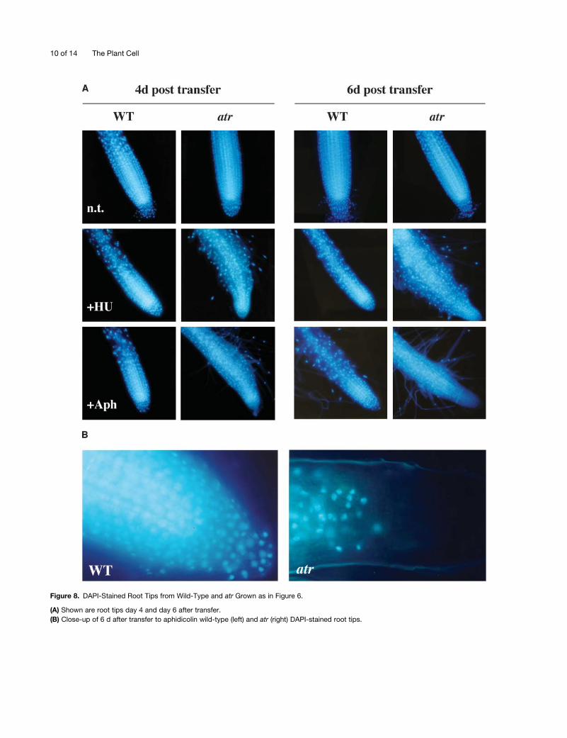
Figure 8. DAPI-Stained Root Tips from Wild-Type and atr Grown as in Figure 6.
(A) Shown are root tips day 4 and day 6 after transfer.
(B) Close-up of 6 d after transfer to aphidicolin wild-type (left) and atr (right) DAPI-stained root tips.
10 of 14 The Plant Cell
The cell death observed in atr in response to aphidicolin, but
not HU, suggests that theG1 (dNTP-dependent) checkpoint may
rescue atr from cell death. However, atr mutants are hypersen-
sitive to the effects of HU on the growth of the root, a fact that
may appear paradoxical given the hypothesis that this line is
proficient in the G1 arrest induced by this agent. For this reason,
we remind the reader that the HU-treated wild-type roots are
growing, though at a slightly reduced rate. In other words, the
cells are progressing through the entire cell cycle, although they
are spending a relatively longer fraction of that cycle in G1. We
thus propose the following model for Arabidopsis cell-cycle
checkpoint regulation in response to HU. HU treatment induces
two independent checkpoints in Arabidopsis: one in G1 in
response to low dNTPs (AtATR independent) and one in G2 in
response to replication blocks (AtATR dependent). HU-treated
wild-type and atr meristematic cells arrest in G1 in response to
low dNTP pools, thus retarding their entry into S-phase. The G1
checkpoint is eventually relieved after enough dNTPs accumu-
late to permit entry into S-phase, and some fraction of cells leak
through into S-phase. The resulting S-phase–induced depletion
of dNTPs in the presence of HU then results in a requirement for
AtATR-dependent G2 checkpoint activation. The inability of
atr to activate this G2 checkpoint results in defective DNA
replication and meristematic failure.
By contrast, aphidicolin-treated atr cells do not invoke the G1
checkpoint because they have normal levels of dNTPs and enter
S-phase without delay. atr cells then fail to arrest in G2 and
proceed into mitosis with incompletely replicated genomes,
perhaps to a much larger degree than HU-treated atr cells. This
results in amajority of the daughter G1 cells having an intolerable
amount of single-strand gaps and double-strand breaks, leading
to meristematic failure and cell death.
The cell death seen in atr was unexpected. Mutants in DNA
repair/maintenance genes in Arabidopsis, such asDNA ligase IV,
ERCC1, and telomerase, do not exhibit obvious cell death, even
in response to treatment with DNA damaging agents (Riha et al.,
2001; Friesner and Britt, 2003; Hefner et al., 2003). A prevailing
hypothesis is that plants, which have no p53 homolog, lack DNA
damage–induced cell death (apoptosis). We believe the atr
response to aphidicolin might be an example of programmed cell
death, as indicated by the fact that all cells in the root tip die,
including normally nondividing cells (e.g., epidermal cells), sug-
gesting that the signal for the cell death is diffusible. Alternatively,
it is possible that the cells surrounding the dying meristem are
recruited to reconstitute themissingmeristem, and this induction
of the cell cycle induces their death. In this case, death might
simply be the direct result of the chromosomal fragmentation
induced by progression through M-phase with an incompletely
replicated genome. In any event, it is clear that replication-
induced checkpoints play an important role in protecting cells
from the consequences of replicational stress.
AtATR-Dependent and AtATR-Independent Regulation
of AtRNR1 Transcription
The regulation of RNR in yeast is well defined and involves
transcriptional and posttranslational control (i.e., inhibitory
protein–protein interactions, modification of protein localization,
and allosteric regulation). For instance, the yeast RNR1 (large
subunit) gene is expressed during vegetative growth, upregu-
lated in late G1 and early S, and further upregulated in response
to DNA damage (Elledge et al., 1992). The expression patterns of
several RNR genes have been characterized in tobacco,
including an RNR1-like gene (Chaboute et al., 1998, 2002). The
reduced level of RNR1 transcript (Figure 3) we observed in atr
versus the wild type, in the absence of DNA damaging agents or
with chronic UV-B light, suggests that RNR1 is transcriptionally
maintained in Arabidopsis through both AtATR-dependent and
AtATR-independent mechanisms. Our analysis employing HU
on whole seedlings (Figure 3) further indicate that RNR1 is
regulated transcriptionally, and independently of AtATR, via
a feedback-type mechanism that boosts RNR1 transcripts when
dNTPs are low.
The rapid reduction of AtRNR1 transcript in response to acute
doses of UV-B in wild-type and atr plants suggests an additional
modeof transcriptional regulation forAtRNR1 in response toDNA
damage that is independent ofAtATR. In yeast,RNRupregulation
in response toDNAdamage is rapid, occurringwithin 30min after
treatment (Elledge and Davis, 1990; Elledge, 1993). It was thus
surprising that under our experimental conditions, an acute dose
of UV-B light caused a rapid reduction of AtRNR1 transcript
within 2 h after UV-B treatment, followed by a return to no-
treatment control levels within 6 h after UV-B treatment. This
response could represent a process bywhich inhibition ofAtRNR
expression leads to low dNTP levels and a concomitant arrest of
the cell cycle during DNA damage, possibly through the G1
checkpoint described above. A decrease in RNR expression
might also be expected to inhibit the potentially mutagenic
actions of bypass polymerases, which require high levels of
dNTPs for activity (Johnson et al., 2001; Chabes et al., 2003). One
plausible model for this mode of regulation would be that
immediately after experiencing acute DNA damage, cells initially
try to eliminate as much of the damage as possible via error-free
pathways. Initial downregulation of dNTPs would inhibit the
inherently mutagenic bypass polymerases and contribute to the
arrest of the cell cycle. Return to normal levels of dNTPs then
allows the cell cycle to progress and enables the cell to utilize
bypass polymerases as needed to complete replication. This
model is suggestive of the SOS response in Escherichia coli,
wherein error-free repair mechanisms are induced early, and
error-prone translesion synthesis is induced later (Goodman and
Woodgate, 2000; Rangaragan et al., 2002) in response to acute
induction of DNA damage.
AtATR Is Required for the Completion of Meiosis in the
Absence of AtATM
Given the partial sterility of atm mutant plants and nonsterility of
atr, the observation of complete sterility of the atr atm double
mutant suggests that ATR can partially complement ATM func-
tion during meiosis. Although it is not clear whether ATR has
adistinctmeiotic function or if ATR is simply filling in for the lack of
ATM (or vice versa), our experiments do at the very least suggest
that ATR is present and functional during meiosis in Arabidopsis.
In mammalian cells, in situ localization studies have shown that
both ATM and ATR are localized to meiotic chromosomes
AtATR Regulates a G2 Checkpoint 11 of 14
(Keegan et al., 1996;Moens et al., 1999). Further studies using atr
and atm single and double mutant plants will allow us to better
understand the role of ATR in plant meiosis, possibly leading to
additional insights into ATR function during animal meiosis.
Conclusions
We have shown that plants encode an ortholog of ATR and that
its role in G2 checkpoint regulation is similar in many respects to
its yeast and mammalian counterparts. The presence of ATM
and other downstream effectors of the cell cycle and DNA repair,
including BRCA1 (Lafarge and Montane, 2003) and a putative
CHK1 (S.B. Preuss, K. Culligan, and A. Britt, unpublished data)
for example, further suggest that G2 cell-cycle checkpoint
responses to DNA damage and replication blocks are regulated
in a similar fashion in plants and animals. This is not to say that
plant responses to DNA damage and replication blocks are
identical to that of animals; perhaps the most significant
difference between plants and animals is the lack of a p53-
dependent programmed cell death response to persisting DNA
damage. Plants may encode an analogous pathway for the
degradation of hopelessly compromised cells, as evidenced by
the cell death observed in atr, but this is probably a response to
massive disruption of DNA replication coupled to uninhibited
progression through the cell cycle. Perhaps because plants do
not die from cancer, a cell death response tominor levels of DNA
damage (such as those that occur spontaneously and persist in
repair defective mutants) would be counteradaptive. Because of
this insensitivity (versus animals) to persisting DNA damage,
plants may ultimately prove to be a robust model system for the
study of many fundamental processes in cell-cycle regulation
and DNA repair pathways in eukaryotes (for review, see Hays,
2002).
METHODS
Isolation of Mutants
The atr-1 allele (ecotypeWs) was isolated from the Arabidopsis Knockout
Facility as described (Krysan et al., 1999). The atr-2 allele (ecotype
Columbia; SALK_032841) was identified using the Salk SIGnAL Web site
(http://signal.salk.edu), and the seeds obtained from the ABRC. The atr-3
allele (ecotype Ws) was identified using the FLAG database (Genoplante;
http://genoplante-info.infobiogen.fr/ ).
Growth of Arabidopsis
For standard growth conditions, seeds were sown on 13 MS (GIBCO,
Cleveland, OH) phytagel (Sigma, St. Louis, MO) agar, pH 5.8, and grown
under cool-white lamps filtered through Mylar at an intensity of 100 to
150 mmol/m2/s with a 24-h day light cycle at 218C. HU (Sigma; 500 mM
stock in water) and aphidicolin (ICN Biomedicals, Basingstoke, UK;
20 mg/mL stock in DMSO) were added to MS plates (as above) to con-
centrations described in the text and figure legends. For experiments
using aphidicolin, all control and HU plates contained an equivalent
amount (0.05%) of DMSO to allow direct comparison.
UV-B and g-Irradiation of Arabidopsis
For chronic doses of UV-B light, plants were sown on MS plates and
grown with a 24-h light cycle under cool-white lamps, supplemented with
UV-B light using a mediumwave UV-B lamp (Spectronics, Westbury, NY;
peak intensity 312 nm) filtered through three sheets of cellulose acetate
(Golden State Plastics, Sacramento, CA; thickness 0.12 mm) to eliminate
the UV-C (<280 nm) fraction of emitted light. The cellulose acetate was
replaced every 3 d. The dose rate of UV-B was 2.5 J/s/m2. For acute
doses of UV-B, 5-d-old seedlings were irradiated with a UV trans-
illuminator (Fisher, Houston, TX), peak output at 305 nm, filtered through
a single cellulose acetate sheet. UV-B intensities were measured with
a UV-B radiometer with a peak response at 310 nm and a 50% response
at 280 and 340 nm (UV Products, San Gabriel, CA). For g-radiation
experiments, Arabidopsis seeds and plants were irradiated using a Cs137
source (Institute of Toxicology and Environmental Health, University of
California, Davis). Seeds were imbibed in water at 48C for 3 d, irradiated,
and then immediately placed on MS agar plates for germination and
placed in the growth chamber; plants were grown on MS plates (for 5 d),
irradiated, and then immediately returned to the growth chamber.
Preparation of RNA and RNA Gel Blotting
Total RNA was prepared from 7-d-old seedlings using an RNeasy plant
mini kit (Qiagen, Valencia, CA). RNA was blotted onto positively charged
membranes (BriteStar-Plus; Ambion, Austin, TX) and probed with a32P radioactively labeled AtRNR1 (GenBank AF092841) cDNA fragment.
This 761-bp fragment was generated with PCR primers RNR1 (59-AAG-
AATGGAGTGAGGAACTCTC-39) and RNR3 (59-CTCAAGCAACATTAA-
GCATTAG-39). The resulting bands were visualized on a Storm 860
PhosphorImager and quantified using ImageQuant software (Molecular
Dynamics, Sunnyvale, CA).
Semiquantitative RT-PCR
Wild-type and atr Arabidopsis plants were sown on MS agar plates,
grown for 5 d, then transferred to MS plates (n.t.), MS plates plus 1 mM
HU (1HU), or MS plates plus 12 mg/mL of aphidicolin (1Aph) for 4 d of
additional growth. All plants were verified for their respective pheno-
types described above. Approximately 1 to 2 mm of root tips, from >200
individual plants of each treatment, were excised and pooled together
for RNA extraction (RNeasy mini-prep; Qiagen). RNA samples were
DNase treated (DNase set; Qiagen) and quantified. To produce cDNA
for RT-PCR, 2 mg of total RNA from each condition described above
was reverse transcribed, employing random hexamer primers, with
a Superscript first-strand synthesis kit (Stratagene, La Jolla, CA) in a 40-
mL reaction according to the manufacturers protocol. RT-PCR was
performed in 50 mL reactions using Ex-Taq polymerase (Takara Bio,
Otsu, Shiga, Japan), 2 mL of cDNA, and the following gene-specific
primers: eIF4A-1 (59-CTCTCGCAATCTTCGCTCTTCTCTTT-39), eIF4A-5
(59-TCATAGATCTGGTCCTTGAAAC-39), CycB1-2 (59-GTCGCTTTCT-
TCTTAGTAGCCTTCT-39), CycB1-6 (59-GGCCTCCATTCACTCTCAA-
CAG-39), H4-5 (59-ATGTCTGGTCGTGGAAAGGGAG-39), H4-8 (59-AC\
CAAATTGCGTGTTTCCATTG-39), cATR-1 (59-CAGCGCCCAAAGAAGA-
TCATTC-39), and cATR-3 (59-GGCCCGCTGAGCATGTGGGTTC-39). All
primers (except Histone H4) spanned intron-exon boundaries to
eliminate the possibility of amplifying contaminating genomic DNA.
PCR-cycle parameters for all primer pairs were 20 s at 948C, 20 s at
628C, and 45 s at 728C. Primer pairs were first tested for log-linear
amplification using 18, 21, 24, 27, and 30 cycles of PCR. The optimized
number of cycles for each primer pair was: eIF4A, 23 cycles; cycB1, 27
cycles; Histone H4, 29 cycles; and ATR, 27 cycles. PCR products of
ethidium bromide–stained gels were quantified using ImageQuant
software as above.
GUS and DAPI Staining
GUS staining was performed as described (Jefferson et al., 1987; Preuss
and Britt, 2003). DAPI staining was performed by fixing roots in a solution
12 of 14 The Plant Cell
of ethanol and acetic acid (3:1). The roots were then washed two times in
PBS, pH 7.4, and stained in PBS containing 2.5mg/mL of DAPI (Molecular
Probes, Eugene, OR) for 20 min. The roots were then washed in PBS two
times and mounted on slides in 50% glycerol. Slides were visualized on
a Zeiss Axiophot microscope, and photographs were taken with a Zeiss
MC100 camera. DAPI staining was visualized with a standard UV
fluorescence filter set.
Sequence data from this article have been deposited with the EMBL/
GenBank data libraries under accession numbers AB040133 and
AF092841.
ACKNOWLEDGMENTS
We thank Ying Peng for assistance in isolating the atr atm double mutant
and Cheryl Whistler, Joanna Friesner, and Eli Hefner for critical reading of
the manuscript. We also thank the Arabidopsis Knockout Facility
(University of Wisconsin, Madison) and the SIGnAL (Salk Institute) and
FLAG databases for providing T-DNA knockout resources. This work was
supported by a USDA postdoctoral fellowship (2001-01860) to K.M.C.
and a National Science Foundation Grant (MCB-9983142) to A.B.B.
Received October 30, 2003; accepted February 13, 2004.
REFERENCES
Abel, S., Ticconi, C., and Delatorre, C. (2002). Phosphate sensing in
higher plants. Physiol. Plant 115, 1–8.
Abraham, R. (2001). Cell cycle checkpoint signaling through the ATM
and ATR kinases. Genes Dev. 15, 2177–2196.
Brown, E., and Baltimore, D. (2003). Essential and dispensable roles of
ATR in cell cycle arrest and genome maintenance. Genes Dev. 17,
615–628.
Brown, E.J., and Baltimore, D. (2000). ATR disruption leads to
chromosome fragmentation and early embryonic lethality. Genes
Dev. 15, 397–402.
Chabes, A., Clement, B., Domkin, V., Zhao, X., Rothstein, R., and
Thelander, L. (2003). Survival of DNA damage in yeast directly
depends on increased dNTP levels allowed by relaxed feedback
inhibition of ribonucleotide reductase. Cell 112, 391–401.
Chaboute, M., Combettes, B., Clement, B., Gigot, C., and Philipps,
G. (1998). Molecular characterization of tobacco ribonucleotide
reductase RNR1 and RNR2. Plant Mol. Biol. 38, 797–806.
Chaboute, M.E., Clement, B., and Philipps, G. (2002). S phase and
meristem-specific expression of the tobacco RNR1b gene is medi-
ated by an E2F element located in the 59 leader sequence. J. Biol.
Chem. 277, 17845–17851.
Cliby, W., Roberts, C., Cimprich, K., Stringer, C., Lamb, J.,
Schreiber, S., and Friend, S. (1998). Overexpression of a kinase-
inactive ATR protein causes sensitivity to DNA-damaging agents and
defects in cell cycle checkpoints. EMBO J. 17, 159–169.
Colon-Carmona, A., You, R., Haimovitch-Gal, T., and Doerner, P.
(1999). Spatio-temporal analysis of mitotic activity with a labile cyclin-
GUS fusion protein. Plant J. 20, 503–508.
Cortez, D., Guntuku, S., Qin, J., and Elledge, S.J. (2001). ATR and
ATRIP: Partners in checkpoint signaling. Science 294, 1713–1716.
Crowley, D.J., and Courcelle, J. (2002). Answering the call: Coping
with DNA damage at the most inopportune time. J. Biomed.
Biotechnol. 2, 66–74.
Culligan, K.M., Meyer-Gauen, G., Lyons-Weiler, J., and Hays, J.B.
(2000). Evolutionary origin, diversification and specialization of
eukaryotic MutS homolog mismatch repair proteins. Nucleic Acids
Res. 28, 463–471.
de Klein, A., Muijtjens, M., van Os, R., Verhoeven, Y., Smit, B., Carr,
A., Lehmann, A., and Hoeijmakers, J. (2000). Targeted disruption of
the cell-cycle checkpoint gene ATR leads to early embryonic lethality
in mice. Curr. Biol. 10, 479–482.
Elledge, S., Zhou, Z., and Allen, J.B. (1992). Ribonucleotide re-
ductase: Regulation, regulation, regulation. Trends Biochem. Sci. 17,
119–123.
Elledge, S.J. (1993). DNA damage and cell cycle regulation of
ribonucleotide reductase. Bioessays 15, 333–339.
Elledge, S.J., and Davis, R.W. (1990). Two genes differentially
regulated in the cell cycle and by DNA-damaging agents encode
alternative regulatory subunits of ribonucleotide reductase. Genes
Dev. 4, 740–751.
Friesner, J.D., and Britt, A.B. (2003). Ku80- and DNA ligase IV-deficient
plants are sensitive to ionizing radiation and defective in T-DNA
integration. Plant J. 34, 427–440.
Garcia, V., Bruchet, H., Camescasse, D., Fabienne, G., Bouchez,
D.L., and Tissier, A. (2003). AtATM is essential for meiosis
and the somatic response to DNA damage in plants. Plant Cell 15,
119–132.
Goodman, M.F., and Woodgate, R. (2000). The biochemical basis and
in vivo regulation of SOS-induced mutagenesis promoted by
Escherichia coli DNA polymerase V (UmuD’2C). Cold Spring Harb.
Symp. Quant. Biol. 65, 31–40.
Hays, J. (2002). Arabidopsis thaliana, a versatile model system for study
of eukaryotic genome-maintenance functions. DNA Repair 1,
579–600.
Hefner, E.A., Preuss, S.B., and Britt, A.B. (2003). Arabidopsis mutants
sensitive to gamma radiation include the homolog of the human repair
gene ERCC1. J. Exp. Bot. 54, 669–680.
Huang, M., Zhou, Z., and Elledge, S. (1998). The DNA replication and
damage checkpoint pathways induce transcription by inhibition of the
Crt1 repressor. Cell 94, 595–605.
Jefferson, R., Kavanagh, T.A., and Bevan, M.W. (1987). GUS fusions:
Beta-glucuronidase as a sensitive and versatile gene fusion marker in
higher plants. EMBO J. 6, 3901–3907.
Jimenez, G., Yucel, J., Rowley, R., and Subramani, S. (1992). The
rad31 gene of Schizosaccharomyces pombe is involved in multiple
checkpoint functions and in DNA repair. Proc. Natl. Acad. Sci. USA
89, 4952–4956.
Johnson, R., Haracska, L., Prakash, S., and Prakash, L. (2001). Role
of DNA polymerase zeta in the bypass of a (6-4) TT photoproduct.
Mol. Cell. Biol. 21, 3558–3563.
Keegan, K.S., et al. (1996). The Atr and Atm protein kinases associate
with different sites along meiotically pairing chromosomes. Genes
Dev. 10, 2423–2437.
Krysan, P.J., Young, J.C., and Sussman, M.R. (1999). T-DNA as an
insertional mutagen in Arabidopsis. Plant Cell 11, 2283–2290.
Lafarge, S., and Montane, M. (2003). Characterization of the
Arabidopsis thaliana ortholog of the human breast cancer suscepti-
bility gene 1:AtBRCA1, strongly induced by X-rays. Nucleic Acids
Res. 31, 1148–1155.
Linke, S., Clarkin, K., Di Leonardo, A., Tsou, A., and Wahl, G. (1996).
A reversible, p53-dependent G0/G1 cell cycle arrest induced by
ribonucleotide depletion in the absence of detectable DNA damage.
Genes Dev. 10, 934–947.
Melo, J., and Toczyski, D. (2002). A unified view of the DNA-damage
checkpoint. Curr. Opin. Cell Biol. 14, 237–245.
Menges, M., and Murray, J.A. (2002). Synchronous Arabidopsis
suspension cultures for analysis of cell-cycle gene activity. Plant J.
30, 203–212.
AtATR Regulates a G2 Checkpoint 13 of 14
Moens, P.B., Tarsounas, M., Morita, T., Habu, T., Rottinghaus, S.,
Freire, R., Jackson, S., Barlow, C., and Wynshaw-Boris, A. (1999).
The association of ATR protein with mouse meiotic chromosome
cores. Chromosoma 108, 95–102.
Nyberg, K.A., Michelson, R.J., Putnam, C.W., and Weinert, T.A.
(2002). Toward maintaining the genome: DNA damage and replication
checkpoints. Annu. Rev. Genet. 36, 617–656.
Preuss, S.B., and Britt, A.B. (2003). A DNA damage induced cell cycle
checkpoint in Arabidopsis. Genetics 164, 323–334.
Rangaragan, S., Woodgate, R., and Goodman, M.F. (2002). Replica-
tion restart in UV-irradiated Escherichia coli involving pols II, III, V,
PriA, RecA and RecFOR proteins. Mol. Microbiol. 43, 617–628.
Reichheld, J., Sonobe, S., Clement, B., Chaubet, N., and Gigot, C.
(1995). Cell cycle-regulated histone gene expression in synchronized
plant cells. Plant J. 7, 245–252.
Riha, K., McKnight, T., Griffing, L., and Shippen, D. (2001). Living with
genome instability: Plant responses to telomere dysfunction. Science
291, 1797–1800.
Sedgwick, P., and Boder, E. (1991). Ataxia-telangiectasia. In Hered-
itary Neuropathies and Spinocerebellar Atrophies, J.M.B.V. deJong,
ed (New York: Elsevier Science Publishing), pp. 347–423.
Tanaka, H., Arakawa, H., Yamaguchi, T., Shiraishi, K., Fukuda, S.,
Matsui, K., Takei, Y., and Nakamura, Y. (2000). A ribonucleotide
reductase gene involved in a p53-dependent cell-cycle checkpoint for
DNA damage. Nature 404, 42–49.
Tibbetts, R.S., and Abraham, R.T. (2000). PI3K-related kinases: Roles
in cell cycle regulation and DNA damage responses. In Signalling
Networks and Cell Cycle Control: The Molecular Basis of Cancer and
Other Diseases, J. Gutkind, ed (Totowa, NJ: Humana Press), pp.
267–301.
Tibbetts, R.S., Cortez, D., Brumbaugh, K.M., Scully, R., Livingston,
D., Elledge, S.J., and Abraham, R.T. (2000). Functional interactions
between BRCA1 and the checkpoint kinase ATR during genotoxic
stress. Genes Dev. 14, 2989–3002.
Weinert, T.A., Kiser, G.L., and Hartwell, L.H. (1994). Mitotic
checkpoint genes in budding yeast and the dependence of mitosis
on DNA replication and repair. Genes Dev. 8, 652–665.
Wiedemeier, A., Judy-March, J., Hocart, C., Wasteneys, G.O.,
Williamson, R., and Baskin, T. (2002). Mutant alleles of Arabidopsis
RADIALLY SWOLLEN 4 and 7 reduce growth anisotropy without
altering the transverse orientation of cortical microtubules or cellulose
microfibrils. Development 129, 4821–4830.
Wright, J., Keegan, K., Herendeen, D., Bentley, N., Carr, A.,
Hoekstra, M., and Concannon, P. (1998). Protein kinase mutants
of human ATR increase sensitivity to UV and ionizing radiation and
abrogate cell cycle checkpoint control. Proc. Natl. Acad. Sci. USA 95,
7445–7450.
Yao, R., Zhang, Z., An, X., Bucci, B., Perlstein, D.L., Stubbe, J., and
Huang, M. (2003). Subcellular localization of yeast ribonucleotide
reductase regulated by the DNA replication and damage checkpoint
pathways. Proc. Natl. Acad. Sci. USA 100, 6628–6633.
Zhao, X., Chabes, A., Domkin, V., Thelander, L., and Rothstein, R.
(2001). The ribonucleotide reductase inhibitor Sml1 is a new target of
the Mec1/Rad53 kinase cascade during growth and in response to
DNA damage. EMBO J. 20, 3544–3553.
Zhao, X., Muller, E.G., and Rothstein, R. (1998). A suppressor of two
essential checkpoint genes identifies a novel protein that negatively
affects dNTP pools. Mol. Cell 2, 329–340.
Zhao, X., and Rothstein, R. (2002). The Dun1 checkpoint kinase
phosphorylates and regulates the ribonucleotide reductase inhibitor
Sml1. Proc. Natl. Acad. Sci. USA 99, 3746–3751.
Zou, L., and Elledge, S.J. (2003). Sensing DNA damage through ATRIP
recognition of RPA-ssDNA complexes. Science 300, 1542–1548.
14 of 14 The Plant Cell
DOI 10.1105/tpc.018903; originally published online April 9, 2004;Plant Cell
Kevin Culligan, Alain Tissier and Anne Britt Arabidopsis thalianaATR Regulates a G2-Phase Cell-Cycle Checkpoint in
This information is current as of September 5, 2014
Permissions https://www.copyright.com/ccc/openurl.do?sid=pd_hw1532298X&issn=1532298X&WT.mc_id=pd_hw1532298X
eTOCs http://www.plantcell.org/cgi/alerts/ctmain
Sign up for eTOCs at:
CiteTrack Alerts http://www.plantcell.org/cgi/alerts/ctmain
Sign up for CiteTrack Alerts at:
Subscription Information http://www.aspb.org/publications/subscriptions.cfm
is available at:Plant Physiology and The Plant CellSubscription Information for
ADVANCING THE SCIENCE OF PLANT BIOLOGY © American Society of Plant Biologists


