Embed Size (px)
Citation preview

1941-3084American Heart Association. All rights reserved. Print ISSN: 1941-3149. Online ISSN: 2009 Copyright ©
Avenue, Dallas, TX 72514Circulation: Arrhythmia and Electrophysiology is published by the American Heart Association. 7272 Greenville
DOI: 10.1161/CIRCEP.108.798447 published online February 18, 2009;Circ Arrhythm Electrophysiol
A. Schweikert, David J. Burkhardt and Andrea NataleMauricio Arruda, Sakis Themistoclakis, Aldo Bonso, Antonio Rossillo, Antonio Raviele, Robert
Domenico Potenza, Raffaele Fanelli, Raimondo Massaro, Paul Wang, Amin Al-Ahmad, Salwa Beheiry, Gemma Pelargonio, Antonio Dello Russo, Michela Casella, Pietro Santarelli,Verma, Conor Barrett, Rong Bai, Dimpi Patel, Yaariv Khaykin, Richard Hongo, Steven Hao, Luigi Di Biase, Claude S. Elayi, Tamer S. Fahmy, David O. Martin, Chi Keong Ching, Atul
between different techniques.Atrial Fibrillation Ablation Strategies for Paroxysmal Patients: randomized comparison
World Wide Web at:
The online version of this article, along with updated information and services, is located on the
initial publication. Advance online articles must include the digital object identifier (DOIs) and date ofpublication priority; they are indexed by PubMed from initial publication. Citations to available prior to final publication). Advance online articles are citable and establishnot yet appeared in the paper journal (edited, typeset versions may be posted when Advance online articles have been peer reviewed and accepted for publication but have
http://www.lww.com/reprintsReprints: Information about reprints can be found online at
[email protected] West Camden Street, Baltimore, MD 21201-2436. Phone: 410-528-4050. Fax: 410-528-8550. E-mail:Permissions: Permissions & Rights Desk, Lippincott Williams & Wilkins, a division of Wolters Kluwer Health,
http://circep.ahajournals.org/site/subscriptions/Subscriptions: Information about subscribing to Circulation: Arrhythmia and Electrophysiology is online at
by guest on February 19, 2013circep.ahajournals.orgDownloaded from

Atrial Fibrillation Ablation Strategies for Paroxysmal Patients: randomized comparison between different techniques. Running title: Catheter ablation for paroxysmal AF First Author: Di Biase L., MD Luigi Di Biase 1,2, MD, Claude S. Elayi3, MD, Tamer S. Fahmy13, MD, David O. Martin4, MD, Chi Keong Ching11, MD, Atul Verma6, MD, Conor Barrett14, MD, Bai Rong15, MD, Dimpi Patel1, DO, Yaariv Khaykin6, MD, Richard Hongo5, MD, Steven Hao5, MD, Salwa Beheiry5, RN, Gemma Pelargonio7, MD, Antonio Dello Russo7, MD, Michela Casella7, MD, Pietro Santarelli7, MD, Domenico Potenza9, MD, Raffaele Fanelli9, MD, Raimondo Massaro9, MD, Paul Wang10, MD, Amin Al-Ahmad10, MD, Mauricio Arruda 12, MD, Sakis Themistoclakis8, MD, Aldo Bonso8, MD, Antonio, Rossillo8, MD, Antonio Raviele8, MD, Robert A. Schweikert16, MD, David J. Burkhardt1, MD, Andrea Natale1,10,12, MD. 1Texas Cardiac Arrhythmia Institute at St. David's Medical Center, Austin, Texas; 2Department of Cardiology, University of Foggia, Foggia, Italy; 3 Division of Cardiovascular Medicine, Gill Heart institute University of Kentucky, Lexington, Kentucky, USA; 4 Cleveland Clinic, Cleveland, USA; 5 Sutter Pacific Heart Centers, San Francisco, USA; 6 Southlake Regional Health Center, New market, Ontario, Canada; 7 Catholic University, Rome, Italy; 8 Hospital Umberto I Mestre, Italy; 9 Casa Sollievo Della Sofferenza, San Giovanni Rotondo, Foggia, Italy; 10 Stanford University, Palo Alto, CA, USA; 11 Department of Cardiology, National Heart Centre Singapore Mistri Wing, Singapore; 12 Case Western Reserve University School of Medicine, Cleveland, OH; 13 Department of Critical Care Medicine, Cairo University, Cairo, Egypt;
14 Cardiac Arrhythmia Service, Massachusetts General Hospital, Heart Center, Boston, MA, USA; 15 Department of Internal Medicine, Tong-Ji Hospital, Tong-Ji Medical College, Huazhong University of Science and Technology, Wuhan, China; 16 Akron General Hospital, Akron, Ohio, USA;
1 by guest on February 19, 2013circep.ahajournals.orgDownloaded from

Address for Correspondence:
Andrea Natale, MD Executive Medical Director of the Texas Cardiac Arrhythmia Institute
at St. David's Medical Center, Austin, Texas.
Consulting Professor, Division of Cardiology, Stanford University, Palo Alto,
California.
Clinical Associate Professor of Medicine, Case Western Reserve University,
Cleveland, Ohio
1015 East 32th Street, Austin, TX 78705, USA
Email: [email protected] phone: +15215448186
Fax: +15125448184 Word Count: 5770 Subject Code: [5] Arrhythmias, clinical electrophysiology, drugs; [22] Ablation/ICD/surgery Abbreviations
CFAE: complex fragmented and/or rapid atrial electrograms
AF: atrial fibrillation
AT: atrial tachyarrhythmia
PVAI: pulmonary vein antrum isolation
RA: right atrium
CS: coronary sinus
LA: left atrium
AADs: antiarrhythmic drugs
E.F.: Ejection fraction
EP lab: Electrophysiology laboratory.
EP: Electrophysiologist
2 by guest on February 19, 2013circep.ahajournals.orgDownloaded from

ABSTRACT
Background: Whether different ablation strategies affect paroxysmal AF long term
freedom from AF/AT is unclear. We sought to compare the effect of 3 different ablation
approaches on the long-term success in patients with paroxysmal AF.
Methods and Results: One hundred and three (103) consecutive patients with
paroxysmal AF scheduled for ablation and presenting in the electrophysiology laboratory
(EP lab) in AF were selected for this study. Patients were randomized to pulmonary vein
antrum isolation (PVAI) (35 pts) versus bi-atrial ablation of the complex fractionated
atrial electrograms (CFAEs) (34 pts) versus PVAI followed by CFAEs (34 pts). Patients
were given event recorder(s) and followed up at 3, 6, 9, 12 and 15 months post ablation.
There was no statistical significant difference between the groups in term of sex, age, AF
duration, LA size and EF. At one year follow up, freedom from AF/AT was documented
in 89% of patients in the PVAI group, 91% in the PVAI plus CFAEs group, and 23% in
the CFAE group (p< 0.001) after a single procedure and with AADs.
Conclusion: No difference in terms of success rate was seen between PVAI alone and
PVAI associated with defragmentation. CFAEs ablation alone had the smallest impact on
AF recurrences at one-year follow-up. These results suggest that antral isolation is
sufficient to treat most patients with paroxysmal AF.
Key Words: pulmonary vein antrum isolation, catheter ablation of atrial fibrillation,
radiofrequency, paroxysmal atrial fibrillation, randomized study
3 by guest on February 19, 2013circep.ahajournals.orgDownloaded from
INTRODUCTION
Catheter ablation has been shown to be a successful and effective therapy for
the treatment of atrial fibrillation (AF) (1). Although the pulmonary veins
(PVs) have been shown to play a major role in the initiation of AF, different
ablation strategies, including isolation of the pulmonary veins and ablation
of sites outside the pulmonary veins, have been proposed (2-7). However,
the relative benefit and success of each approach alone and in combination
has not been evaluated in randomized studies.
We sought to compare the effect of different ablation strategies on the AF
termination mode and the long term success of patients with paroxysmal
atrial fibrillation presenting to the electrophysiology laboratory (EP lab) in
AF.
We compared pulmonary vein antrum isolation (PVAI) alone, ablation of
complex fractionated atrial electrograms (CFAEs), and a hybrid strategy that
combines PVAI followed by ablation of complex fractionated atrial
electrograms.
4 by guest on February 19, 2013circep.ahajournals.orgDownloaded from

METHODS
Study population
We enrolled 103 consecutive patients with paroxysmal AF presenting to the
EP lab with spontaneous AF. The definition of paroxysmal AF followed the
guidelines suggested by the ACC/AHA/ ESC society.
Patients included in this study were enrolled for their first AF ablation by 6
different Institutions in the period between November 2004 and January
2007.
Patients were assigned a treatment based on the permuted block strategy.
The treatments were balanced within a block size of 3, with the block
randomly assigned to each center using a web-based centralized control
program.
Patients underwent PVAI only (group I n= 35), ablation of CFAE only
(group II n= 34), or a hybrid approach including PVAI plus CFAE (group III
n= 34).
Patients were enrolled if: 1) they had a history for at least one year of
paroxysmal AF, 2) they were refractory to at least 2 antiarrhythmic drugs
(AADs) and 3) they presented to the EP lab in spontaneous AF.
Patients who underwent a prior ablation procedure for AF were excluded
from this study.
5 by guest on February 19, 2013circep.ahajournals.orgDownloaded from

Before enrolling patients, the EP physicians, performing ablations at the
different institutions, assessed sample electrograms showing CFAE to ensure
uniformity in CFAE definition and identification.
Patients were enrolled from: 1) Sutter Pacific Heart Centers, San Francisco,
CA, USA (18 pts, 18%); 2) Catholic University, Rome, Italy; (22 pts, 21%);
3) Southlake Regional Health Center, Toronto, Canada; (21 pts, 20%); 4)
Casa Sollievo Della Sofferenza, Foggia, Italy (20 pts, 19,5%); 5) Hospital
Umberto I Mestre, Italy (20 pts, 19,5%) and 6) Stanford University, Palo
Alto (USA) (2 pts, 2%).
All patients signed an informed written consent prior to the procedure.
The Institutional Ethical Committees approved the study.
The authors had full access to and take full responsibility for the integrity of
the data. All authors have read and agree to the manuscript as written.
Ablation procedure.
All patients discontinued AADs at least five half lives before ablation.
Amiodarone therapy was discontinued 6 months before the procedure.
PVAI: The PVAI has been described in detail elsewhere (8, 9). Briefly, we
used a circular mapping catheter (Lasso, Biosense Webster, Diamond Bar,
CA, USA) and a 3.5 mm irrigated tip catheter (ThermoCool®) to ablate the
antrum of the pulmonary veins (PVs) and to achieve abolition of all
6 by guest on February 19, 2013circep.ahajournals.orgDownloaded from

electrograms. Intracardiac echocardiogram (ICE) was used to monitor the
trans-septal puncture and to define the anatomy of the pulmonary veins. An
esophageal probe was used to monitor the temperature in the esophagus
during ablation.
Radiofrequency energy output was titrated to a maximum of 45 W while
maintaining a catheter tip temperature of < 41°C. At each site energy was
delivered for 20 seconds. The maximum power over the esophagus and
within the coronary sinus (CS) was limited to 30 Watts and energy delivery
was discontinued when the esophageal temperature probe reached 39º C.
A 3-dimensional geometry of the left atrium (LA) was reconstructed with
the CARTO system (Biosense Webster, Diamond Bar, CA, USA) or the
NavX system (St. Jude Medical, St Paul, MN, USA) (figure 1 A-D).
The procedural end point for this ablation strategy was the local elimination
of all the pulmonary vein potentials along the antra or inside the veins (entry
and exit block).
The antrum included the entire posterior wall and extended anteriorly to the
right PVs along the left septum. Further ablation of the superior vena cava
(SVC) along the right atrium/SVC junction was also performed if mapping
revealed PV-like potentials around this region and when high output (30
mA) pacing did not capture the phrenic nerve (10).
7 by guest on February 19, 2013circep.ahajournals.orgDownloaded from

During ablation, the categories of AF termination (secondary endpoint)
considered have been: 1) conversion to SR, 2) organization into a regular
atrial tachyarrhythmia (AT), with a similar cycle length in both the atria and
CS, or 3) persistence of AF requiring cardioversion.
When AF organized into an AT, the latter arrhythmia was mapped and
ablated.
CFAEs only Group (Group II)
CFAEs were defined as: (1) atrial electrograms with two deflections or more
and /or with fractionated baseline complexes with continuous activity over
10 seconds recording time, (2) atrial electrograms with a cycle length ≤ 120
ms over a 10 seconds recording time. The ablation catheter was required to
be in a stable position when recording these electrograms (3, 11).
All operators assessed sample of CFAEs electrograms to ensure uniformity
in selecting ablation sites (Figure 2).
The left and right atria (including the CS) were mapped to identify areas
with electrical fractionation. These areas were ablated with the open
irrigation ablation catheter (same settings parameters as described above for
the PVAI group) until the CFAEs were completely eliminated.
The CFAEs were first ablated in the left atrium, then CS and right atrium,
respectively.
8 by guest on February 19, 2013circep.ahajournals.orgDownloaded from

The procedural endpoint of this ablation strategy was complete elimination
of the CFAEs potentials.
If atrial fibrillation terminated before elimination of all CFAEs, induction of
AF was attempted with pacing on and off isoproterenol (up to 20 mcg/min).
The categories of AF termination considered have been described above
(secondary endpoint).
If AF persisted after elimination of all CFAEs's sites, cardioversion was
utilized to restore sinus rhythm.
Hybrid approach (PVAI followed by Ablation of CFAE), (Group III).
This ablation strategy was a combination of the two previously described
approaches.
PVAI was followed by CFAEs ablation; therefore patients underwent
antrum isolation of all pulmonary veins and subsequently the elimination of
CFAEs in both atria.
The procedural endpoint for this strategy was the complete elimination of
CFAEs areas and electrical isolation of all the PV antra defined by entrance
and exit block. If AF terminated before CFAEs ablation or before all CFAEs
were ablated, induction of AF was performed with pacing on and off
isoproterenol (up to 20 mcg/min).
Modes of AF termination were the same as in Group I and II (secondary
9 by guest on February 19, 2013circep.ahajournals.orgDownloaded from

endpoint).
If AF persisted after PVAI plus CFAE, cardioversion was utilized to restore
sinus rhythm.
Primary endpoint
The primary endpoint of this study for all the ablation strategies was
freedom from atrial fibrillation defined as no episodes of AF/AT with or
without AADs that lasted more than one minute at the one year follow-up.
Episodes that occurred during the first two months (blanking period) after
the procedure were not considered as recurrences.
AADs were discontinued in all patients 2 months after the ablation when no
recurrences were present. In cases of recurrences, patients were given their
previously ineffective AADs. Patients with arrhythmia recurrence six
months beyond the first procedure and on AADs were offered a repeat
ablation.
Post ablation Management and Follow-Up
All patients were discharged on warfarin with a target INR of 2 to 3 and on
AADs previously ineffective, except for amiodarone.
Warfarin was continued for a minimum of 6 months after the ablation
procedure.
They were followed in the outpatient clinic at 3 months after the procedure
10 by guest on February 19, 2013circep.ahajournals.orgDownloaded from

and then every 3 months. Patients were also given an event recorder for 5
months. They were asked to record four times a week even if asymptomatic
and anytime they experienced symptoms. A 48-hour Holter monitor was
obtained at 3, 6, 9, 12 and 15 months post ablation.
Statistical Analysis
A permuted block randomization schedule with block size of 3 was
generated using a random number generator. Each permuted block was
assigned a number and each block was randomly assigned to a center.
Although Nadamanee et al (3) had reported high success with CFAEs, our
initial experience did not agree with his published results. We expected a
50% success rate using a CFAEs -only approach and, based on published
results from our experience, we expected an 80% success rate with a PVAI-
only approach. Under these assumptions, using a 1-tailed alpha of 5% and
80% power, a total of 32 patients would be required.
All continuous data are presented as “Mean +/- SD” and were compared by
student t-test or by ANOVA. Tukey-Kramer method for multiple
comparisons was used to compare the efficacy of the three procedures.
The analysis used the intention-to-treat principle. Categorical variables
comparison used χ² analysis. A p value < 0.05 was considered statistically
significant. (SPSS software version 11.0 Chicago, II, inc.).
11 by guest on February 19, 2013circep.ahajournals.orgDownloaded from
RESULTS
Patients Characteristics
Baseline characteristics of the three groups are presented in table 1. No
significant difference between groups in term of sex, age, AF duration, left
atrium size and ejection fraction (EF) was present. Previously ineffective
AADs are also reported in table 1.
Procedural Results
The procedural endpoint was achieved in all patients (100%) in each group.
The total fluoroscopy times of the groups were 65.6 ± 22.6 for group I, 59.9
± 24.7 for group II and 76.8 ± 21.8 for group III. (P = 0.8).
The duration of radiofrequency applications were 54 ±11 min for group I, 48
± 9 min for group II and 68 ± 14 min for group III ( P = 0.04).
The total number of patients with CFAEs ablated and the median number of
RF applications necessary to abolish CFAEs at each right and left atrial sites
are reported in table 2. (Figure 1)
Secondary endpoint: AF termination during ablation:
Organization into atrial tachyarrhythmia was 34% in group I, 16% in group
II and 29% in group III (P=0.158) with a mean cycle length 236.8 ± 32.9 ms;
conversion to sinus rhythm was seen in 60% (group I), 17% (group II), and
65% (group III) of patients respectively (p<0.001); persistence of AF
12 by guest on February 19, 2013circep.ahajournals.orgDownloaded from

requiring cardioversion was observed in 6% of group I, 67% of group II and
6% of group III. (p < 0.001) (Table 3).
When AF organized into AT, an attempt to map and terminate the AT during
ablation was performed each time. Conversion from an organized
flutter/tachycardia to SR was observed in 7 patients in group I, 2 patients in
group II and 6 patients in group III) (p = 0.2).
The majority of these ATs were located at the mitral valve level (16
patients), and in the posterior wall (11 patients) as demonstrated by
mapping/entrainment around the PVs .
Chronic follow-up/Primary endpoint
The primary endpoint of the study is reported in table 4 and 5 as freedom
from AF/AT after a single procedure with or without AADs at 1 year of
follow up.
In group I and group III freedom from AF/AT after 6 months was observed
in 94% of patients (14% requiring AADs), while in group II it was 59%
(11% requiring AADs). (p< 0.001).
After one year follow up (13.7±2.2 months), in group I and group III
freedom from AF/AT was seen in 89% (15% requiring AADs) and 91%
(15% requiring AADs) of patients respectively, while in group II was
achieved in 23% (11% requiring AADs) of patients (p< 0.001).(tables 4 and
13 by guest on February 19, 2013circep.ahajournals.orgDownloaded from

5).
The timing for a second procedure was at least 6 months after the first
procedure.
All 7 patients, belonging to group I and III and with primary endpoint
failure, accepted a second procedure after 7.1 ± 1.1 months from the first
procedure. Six out of the seven (86%) patients demonstrated no further
AF/AT at 9±7 months follow up from the second procedure without any
AADs.
Twenty two patients out of the 26 patients of group II failing the primary
endpoint accepted a second procedure after 7.3 ±1.1 months. These
procedures were performed using the PVAI-only approach. After a mean
follow up of 9 ±7 months from the second procedure 20 patients (91%),
were free from AF/AT without AADs.
Complications
No major complications have been observed in these groups of patients
during and after the procedures.
DISCUSSION
Main findings: This is the first prospective multicenter randomized study
comparing three ablation techniques in patients with paroxysmal AF.
CFAEs ablation alone had the smallest impact on both acute AF termination
14 by guest on February 19, 2013circep.ahajournals.orgDownloaded from

and freedom from AF/AT at one year follow up.
The hybrid strategy, which combines isolation of the PV antra and ablation
of CFAEs, was not associated with a better acute success rate (defined as
conversion to sinus rhythm) or chronic success rate (defined as event
freedom from AF or AT at 6-month and 1-year follow up), when compared
to PVAI alone.
Previous studies
The pulmonary veins are known for their preponderant role in triggering and
maintaining atrial fibrillation (2).
Segmental ostial pulmonary vein isolation maintains sinus rhythm in
approximatively 2/3 of the patients with paroxysmal atrial fibrillation (12,
13). Additional lesions such as mitral isthmus ablation (5) or antrum
isolation (14) have been reported to increase this success to approximately
90%. More recently, ablation targeting CFAEs has been shown to result in
sinus rhythm maintenance in approximately 80% of patients with
paroxysmal and persistent AF (3).
However, these results originated from a single center. To date, CFAEs
ablation for paroxysmal AF has only been reported in another publication
during the tailored approach described by Oral et al (15), but CFAEs
ablation was never performed alone and was not performed in all patients.
15 by guest on February 19, 2013circep.ahajournals.orgDownloaded from

Our results are different from the data published by Nadamanee et al (3) who
reported a success rate of 82% at 1-year follow up, in paroxysmal patients
who underwent CFAEs ablation alone (two of them with amiodarone) and
100% conversion to SR in paroxysmal patients including 8 patients (14%)
who required concomitant ibutilide administration during ablation.
The results of this study also indicate that CFAEs ablation alone has a
minimal impact on AF termination during ablation of patients with
paroxysmal AF and should not be considered as an alternative strategy
unless a better identification of critical CFAEs zones becomes widely
available and proven effective. In this respect, although we believe that the
technique utilized in this series of patients was really similar to the one
described by Nadamanee (3), it is possible that a difference exists in the
technique and extent of ablation used in our study.
General comments
Of note, after a short follow-up (≤ 6 months), CFAEs ablation alone
appeared to lead to an improvement in a significant number of patients;
however, most of this effect was lost by 12 months (tables 4 and 5).
This suggests that a longer follow up is required to assess the real impact on
AF recurrences following any ablation procedures.
The presence of sites other than the PVs which are able to initiate and
16 by guest on February 19, 2013circep.ahajournals.orgDownloaded from

maintain AF is probably responsible for the inability to treat all patients with
PV isolation alone and has prompted the utilization of different strategies
adjunctive to PV isolation.
These strategies include: creation of various ablation lines such as the mitral
isthmus (5), roof line (4), posterior LA wall lines (16), antrum isolation (14)
and cardiac autonomic denervation (6, 7).
Our results demonstrate a reduced efficacy of the CFAE ablation alone when
compared to PVAI alone and PVAI plus CFAE in the treatment of the
paroxysmal AF. In fact, the success rate of the PVAI approached 90% at
13.7±2.2 months of follow up with a single procedure with AADs, while the
success rate of CFAE ablation alone was 23%.
In our study, all patients had paroxysmal AF, which was present for at least
one year prior to ablation.
The lack of differences between PVAI and PVAI plus CFAEs, and the small
impact on success rate reached by CFAEs alone suggest that electrical
isolation of the PVs remains a cornerstone for catheter ablation of
paroxysmal AF.
Our results are consistent with the revised strategy reported by Morady et al
in a recent viewpoint (17). The authors state that they do not limit the
ablation to the arrhythmogenic veins as in their previous tailored approach
17 by guest on February 19, 2013circep.ahajournals.orgDownloaded from

(15), but they perform an antral ablation of all the pulmonary veins.
CFAEs ablation did not have a statistically significant additive effect when
combined with PVAI on AF termination mode (CFAE terminated AF during
ablation in 17% of cases, PVAI in 60%, and the combination of both in 65%
of cases).
This suggests that extra-antral CFAEs areas may be less relevant in
maintaining AF and that isolation of the antrum is necessary in nearly all
cases of paroxysmal atrial fibrillation to achieve long term success.
Of note, it is important to recognize that isolation of the antrum eliminates
many areas associated with fragmented electrograms. However, the poor
chronic success obtained with defragmentation alone reinforces the
importance of PVs isolation.
In agreement with our results, Morady et al (17) found that many of CFAEs,
which appeared critical for the maintenance of the AF, were in the antral
region, suggesting that CFAEs present in other areas (coronary sinus, left
atrial roof, and mitral annulus) should be considered as “innocent
bystanders” at least in the paroxysmal AF patients.
Based on our findings, additional sites of ablation should be reserved for
selected patients, and probably should not be driven by empirical targeting
of fragmented electrograms but by mapping triggers disclosed with
18 by guest on February 19, 2013circep.ahajournals.orgDownloaded from

administration of isoproterenol and/or adenosine.
Several other groups have shown that ablation strategies encompassing the
areas equivalent to the antrum achieve similar results and are better than
more limited approaches (18-20).
Study limitations:
Methods to identify CFAEs, although similar to the ones described by
Nademanee, (3) may be operator-dependent because they are based on visual
evaluation.
Software analysis tools to identify CFAEs were not used in our study.
However Scherr et al (21) demonstrated a high correlation between software
and visual identification of the CFAEs areas. In addition, the initial
description of defragmentation relied on visual identification of fragmented
electrograms.
Using a fixed block size of 3, it is possible to determine the assignment of
the third patient in the block before randomization. However, in a study such
as this, the operators cannot be blinded to the procedural endpoints, since
they had to know the type of procedure to perform. In addition, the
physicians performing the procedures were not involved in patient’s
recruitment and the outcome at follow-up was based on the objective
documentation of freedom from AF/AT.
19 by guest on February 19, 2013circep.ahajournals.orgDownloaded from

Finally the study was underpowered to detect a difference between PVAI
alone vs CFAEs plus PVAI; taking into account the adjustment for multiple
comparisons with a Family Wise Error Rate (FWER) <= 0.05, the sample
size required to provide 80% power to detect a difference of 5% (comparing
85% to 90%) between any two groups is 353 patients per group. While
acknowledging the statistical limitations, we deem that the data presented in
this study would serve as an important reference point for future studies
comparing the efficacy of PVAI+CFAEs procedures.
CONCLUSIONS
Ablation of the CFAEs alone had the smallest impact on both acute AF
termination and 1-year follow up success rate in patients with paroxysmal
AF. No difference in terms of acute and chronic success rates was observed
between PVAI alone and PVAI associated with defragmentation (CFAEs)
ablation. These results suggest that antral isolation and/or equivalent
strategies are sufficient to treat most patients with paroxysmal AF.
Acknowledgement
The authors would like to thanks Prasant Mohanty, MBBS, MPH for providing valuable
inputs in the statistical analysis performed for the study.
20 by guest on February 19, 2013circep.ahajournals.orgDownloaded from

Author Disclosures Drs D.O. Martin, P. Wang, A. Al-Ahmad, R.A. Schweikert, D.J. Burkhardt and A. Natale report receiving compensation from St. Jude Medical for participation in speaker’s bureaus. Drs P.Wang, A. Al-Ahmad, R.A. Schweikert and A. Natale report receiving compensation from Boston Scientific for participation in speaker’s bureaus. Drs A.Verma, Y. Khaykin, P. Wang, A. Al-Ahmad, R.A. Schweikert, A. Raviele and A. Natale report receiving compensation from Medtronic for participation in speaker’s bureaus. Drs A.Verma, Y. Khaykin, R.A. Schweikert, D.J. Burkhardt and A. Natale report receiving compensation from Biosense Webster for participation in speaker’s bureaus. Dr. P.Wang reports receiving compensation from Hansen Medical and Lifewatch for participation in speaker’s bureaus. Dr A. Al-Ahmad, reports serving as a consultant/advisory board to Lifewatch EBR Medical, CyberHeart. Drs S. Themistoclakis, A. Bonso and R.A. Schweikert, report serving as a consultant/advisory board to Biosense Webster. Dr. A. Al-Ahmad reports participation in a research grant from Siemens. Dr. A. Natale reports participation in a research grant from St. Jude Medical. Dr D.O. Martin, reports serving as a consultant/advisory board to Boston Scientific and Member Advisory Board for Medtronic. Dr. D.J. Burkhardt reports serving as Consultant/Advisory Board to Stereataxis All other authors have nothing to declare
21 by guest on February 19, 2013circep.ahajournals.orgDownloaded from

REFERENCES
1. Fuster V, Ryden LE, Cannom DS, Crijns HJ, Curtis AB, Ellenbogen KA, Halperin JL, Le Heuzey JY, Kay GN, Lowe JE, Olsson SB, Prystowsky EN, Tamargo JL, Wann S, Smith SC Jr, Jacobs AK, Adams CD, Anderson JL, Antman EM, Hunt SA, Nishimura R, Ornato JP, Page RL, Riegel B, Priori SG, Blanc JJ, Budaj A, Camm AJ, Dean V, Deckers JW, Despres C, Dickstein K, Lekakis J, McGregor K, Metra M, Morais J, Osterspey A, Zamorano JL. ACC/AHA/ESC 2006 guidelines for the management of patients with atrial fibrillation: a report of the American College of Cardiology/American Heart Association Task Force on Practice Guidelines and the European Society of Cardiology Committee for Practice Guidelines (Writing Committee to Revise the 2001 Guidelines for the Management of Patients With Atrial Fibrillation): developed in collaboration with the European Heart Rhythm Association and the Heart Rhythm Society. Circulation 2006; 114: 257–354.
2. Haïssaguerre M, Jaïs P, Shah DC, Takahashi A, Hocini M, Quiniou G,
Garrigue S, Le Mouroux A, Le Métayer P, Clémenty J. Spontaneous initiation of atrial fibrillation by ectopic beats originating in the pulmonary veins. N Engl J Med. 1998 ; 339: 659-66.
3. Nademanee K, McKenzie J, Kosar E, Schwab M, Sunsaneewitayakul
B, Vasavakul T, Khunnawat C, Ngarmukos T. A new approach for catheter ablation of atrial fibrillation: mapping of the electrophysiologic substrate. J Am Coll Cardiol. 2004 ;43: 2044-53.
4. Hocini M, Jaïs P, Sanders P, Takahashi Y, Rotter M, Rostock T, Hsu
LF, Sacher F, Reuter S, Clémenty J, Haïssaguerre M. Techniques, evaluation, and consequences of linear block at the left atrial roof in paroxysmal atrial fibrillation: a prospective randomized study. Circulation. 2005; 112 :3688-96.
5. Jaïs P, Hocini M, Hsu LF, Sanders P, Scavee C, Weerasooriya R,
Macle L, Raybaud F, Garrigue S, Shah DC, Le Metayer P, Clémenty J, Haïssaguerre M. Technique and results of linear ablation at the
22 by guest on February 19, 2013circep.ahajournals.orgDownloaded from

mitral isthmus. Circulation. 2004; 110 :2996-3002.
6. Pappone C, Santinelli V, Manguso F, Vicedomini G, Gugliotta F, Augello G, Mazzone P, Tortoriello V, Landoni G, Zangrillo A, Lang C, Tomita T, Mesas C, Mastella E, Alfieri O. Pulmonary vein denervation enhances long-term benefit after circumferential ablation for paroxysmal atrial fibrillation. Circulation. 2004 ; 109 :327-34.
7. Scanavacca M, Pisani CF, Hachul D, Lara S, Hardy C, Darrieux F,
Trombetta I, Negrão CE, Sosa E. Selective atrial vagal denervation guided by evoked vagal reflex to treat patients with paroxysmal atrial fibrillation. Circulation. 2006 ; 114 :876-85.
8. Verma A, Marrouche NF, Natale A. Pulmonary vein antrum isolation:
intracardiac echocardiography-guided technique. J Cardiovasc Electrophysiol. 2004; 15: 1335-40.
9. Kanj M, Wazni O, Natale A. How to do circular mapping catheter-
guided pulmonary vein antrum isolation: the Cleveland Clinic approach. Heart Rhythm. 2006; 3:866-9.
10. Arruda M, Mlcochova H, Prasad SK, Kilicaslan F, Saliba W, Patel D,
Fahmy T, Morales LS, Schweikert R, Martin D, Burkhardt D, Cummings J, Bhargava M, Dresing T, Wazni O, Kanj M, Natale A. Electrical isolation of the superior vena cava: an adjunctive strategy to pulmonary vein antrum isolation improving the outcome of AF ablation. J Cardiovasc Electrophysiol. 2007; 18:1261-6
11. Nademanee K, Schwab M, Porath J, Abbo A. How to perform
electrogram-guided atrial fibrillation ablation. Heart Rhythm. 2006; 3: 981-4.
12. Haïssaguerre M, Shah DC, Jaïs P, Hocini M, Yamane T, Deisenhofer
I, Chauvin M, Garrigue S, Clémenty J. Electrophysiological breakthroughs from the left atrium to the pulmonary veins. Circulation. 2000; 102:2463-5.
13. Oral H, Knight BP, Ozaydin M, Chugh A, Lai SW, Scharf C, Hassan
S, Greenstein R, Han JD, Pelosi F Jr, Strickberger SA, Morady F. Segmental ostial ablation to isolate the pulmonary veins during atrial
23 by guest on February 19, 2013circep.ahajournals.orgDownloaded from

fibrillation: feasibility and mechanistic insights. Circulation. 2002; 106 :1256-62.
14. Wazni OM, Marrouche NF, Martin DO, Verma A, Bhargava M,
Saliba W, Bash D, Schweikert R, Brachmann J, Gunther J, Gutleben K, Pisano E, Potenza D, Fanelli R, Raviele A, Themistoclakis S, Rossillo A, Bonso A, Natale A. Radiofrequency ablation vs antiarrhythmic drugs as first-line treatment of symptomatic atrial fibrillation: a randomized trial. JAMA. 2005; 293: 2634-40.
15. Oral H, Chugh A, Good E, Sankaran S, Reich SS, Igic P, Elmouchi D,
Tschopp D, Crawford T, Dey S, Wimmer A, Lemola K, Jongnarangsin K, Bogun F, Pelosi F Jr, Morady F. A tailored approach to catheter ablation of paroxysmal atrial fibrillation. Circulation.2006; 113:1824-31.
16. Sanders P, Hocini M, Jaïs P, Sacher F, Hsu LF, Takahashi Y, Rotter
M, Rostock T, Nalliah CJ, Clémenty J, Haïssaguerre M. Complete isolation of the pulmonary veins and posterior left atrium in chronic atrial fibrillation. Long-term clinical outcome. Eur Heart J. 2007; 15: 1862-71.
17. Morady F. Patient-specific ablation strategy for atrial fibrillation:
promises and difficulties. Heart Rhythm. 2007; 8:1094-6.
18. Ouyang F, Bänsch D, Ernst S, Schaumann A, Hachiya H, Chen M, Chun J, Falk P, Khanedani A, Antz M, Kuck KH. Complete isolation of left atrium surrounding the pulmonary veins: new insights from the double-Lasso technique in paroxysmal atrial fibrillation. Circulation. 2004; 110 :2090-6.
19. Arentz T, Weber R, Bürkle G, Herrera C, Blum T, Stockinger J,
Minners J, Neumann FJ, Kalusche D. Small or large isolation areas around the pulmonary veins for the treatment of atrial fibrillation? Results from a prospective randomized study. Circulation. 2007; 115:3057-63.
20. Marrouche NF, Dresing T, Cole C, Bash D, Saad E, Balaban K, Pavia
SV, Schweikert R, Saliba W, Abdul-Karim A, Pisano E, Fanelli R, Tchou P, Natale A. Circular mapping and ablation of the pulmonary
24 by guest on February 19, 2013circep.ahajournals.orgDownloaded from

vein for treatment of atrial fibrillation: impact of different catheter technologies. J Am Coll Cardiol. 2002; 40:464-74.
21. Scherr D, Dalal D, Cheema A, Cheng A, Henrikson CA, Spragg D,
Marine JE, Berger RD, Calkins H, Dong J. Automated detection and characterization of complex fractionated atrial electrograms in human left atrium during atrial fibrillation. Heart Rhythm. 2007; 4:1013-20.
25 by guest on February 19, 2013circep.ahajournals.orgDownloaded from

Clinical Characteristic
PVAI only (n=35)
CFAE only (n=34)
PVAI + CFAE (n=34)
P Value
Age 57 ± 8.1 59.9± 8.6 58.4 ± 7.5 P = 0.43
Male (%) 83 76 88 P = 0.44
HTN (%) 34 38 35 P = 0.51
AF duration (years) 5.3 ± 5.7 5.1± 4.1 5.3 ± 5 P= 0.61
LA size (cm) 4.3 ± 0.6 4.1 ± 0.5 4.4 ± 0.6 P= 0.38
LVEF (%) 55 ± 8 55.5± 6 54.6 ± 6 P= 0.89
Previously ineffective AA drugs
Amiodarone 2 (0.5 %) 2 (0.5%) 2 (0.5%) P =1.0
Sotalol 14 (40%) 14 (41%) 13 (38%) P = 0.9
> 1 class I AA drugs 21 (60%) 20 (58%) 21 (61%) P = 0.9
Table 1. Baseline Characteristics and previous ineffective AADs.
26 by guest on February 19, 2013circep.ahajournals.orgDownloaded from
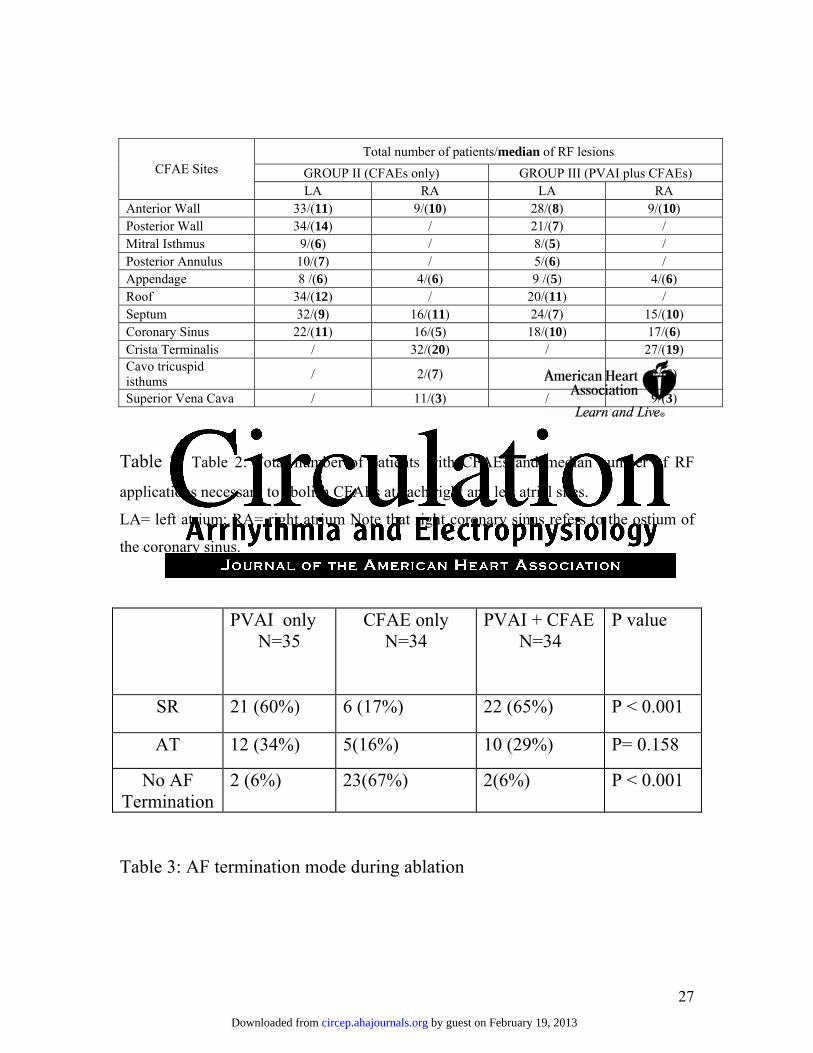
Total number of patients/median of RF lesions GROUP II (CFAEs only) GROUP III (PVAI plus CFAEs) CFAE Sites
LA RA LA RA Anterior Wall 33/(11) 9/(10) 28/(8) 9/(10) Posterior Wall 34/(14) / 21/(7) / Mitral Isthmus 9/(6) / 8/(5) / Posterior Annulus 10/(7) / 5/(6) / Appendage 8 /(6) 4/(6) 9 /(5) 4/(6) Roof 34/(12) / 20/(11) / Septum 32/(9) 16/(11) 24/(7) 15/(10) Coronary Sinus 22/(11) 16/(5) 18/(10) 17/(6) Crista Terminalis / 32/(20) / 27/(19) Cavo tricuspid isthums / 2/(7) / 2/(6)
Superior Vena Cava / 11/(3) / 9/(3)
Table 2: Table 2: Total number of patients with CFAEs and median number of RF
applications necessary to abolish CFAEs at each right and left atrial sites.
LA= left atrium; RA= right atrium Note that right coronary sinus refers to the ostium of
the coronary sinus.
PVAI only N=35
CFAE only N=34
PVAI + CFAE N=34
P value
SR 21 (60%) 6 (17%) 22 (65%) P < 0.001
AT 12 (34%) 5(16%) 10 (29%) P= 0.158
No AF Termination
2 (6%) 23(67%) 2(6%) P < 0.001
Table 3: AF termination mode during ablation
27 by guest on February 19, 2013circep.ahajournals.orgDownloaded from

PVAI onlyN=35
CFAE only N=34
PVAI + CFAE N=34
P value
*Freedom from AF/AT after 6 months follow up
33 pts (94%) 20 pts (59%) 32 pts (94%) <0.001
*Freedom from AF/AT after one year follow up
31 pts (89%) 8 pts (23%) 31 pts (91%) <0.001
Freedom from AF/AT after one year follow up
without AADs
26 pts (74%) 4 pts (12%) 26 pts (76%) <0.001
Table 4: Freedom from AF/AT at 6-months and 1-year follow up (13.7±2.2
months).
* With or without AADs
28 by guest on February 19, 2013circep.ahajournals.orgDownloaded from

A) Freedom from AF/AT after 6 months follow-up:
Compare Procedure
Difference between two
compared proportions
Standard Error (SEs)
The “q” statistic
Critical q
values for α = 0.05
P-value
1 vs 2 24.86 4.84 5.14 3.314 < 0.05 1 vs 3 0.22 4.84 0.05 3.314 > 0.05 3 vs 2 24.64 4.88 5.05 3.314 < 0.05
B) Freedom from AF/AT after one year follow-up
Compare Procedure
Difference between two
compared proportions
Standard Error (SEs)
The “q” statistic
Critical q
values for α = 0.05
P-value
3 vs 2 42.1 4.88 8.63 3.314 < 0.05 3 vs 1 2.29 4.84 0.47 3.314 > 0.05 1 vs 2 39.82 4.84 8.23 3.314 < 0.05
C) Freedom from AF/AT after one year follow up without AADs
Compare Procedure
Difference between two
compared proportions
Standard Error (SEs)
The “q” statistic
Critical q
values for α = 0.05
P-value
3 vs 2 39.51 4.88 8.1 3.314 < 0.05 3 vs 1 1.39 4.84 0.29 3.314 > 0.05 1 vs 2 38.12 4.84 7.88 3.314 < 0.05
Table 5: The Tukey-Kramer method for multiple comparisons was used to compare the
efficacy of the three procedures: PVAI only (procedure 1), CFAE only (procedure 2), and PVAI
+ CFAE (procedure 3).
29 by guest on February 19, 2013circep.ahajournals.orgDownloaded from

FIGURE LEGENDS Figure 1: Tridimensional map of the left atrium in the PA and AP views (Figures A, Fig B) and of the right atrium and CS in PA and AP views (Figures C and D) using Navx mapping system. The yellow dots indicate areas where CFAEs have been ablated in a patient belonging to the PVAI plus CFAEs group. Blue dots indicate lesions around the pulmonary vein antrum. Figure 2: Example of CFAEs (Complex fractionated atrial electrograms) on the ablation catheter proxysmal (abl 3-4) and distal (abl 1-2) that were targeted during the hybrid strategy. V1= precordial lead. CS 1-2= Coronary sinus distal lead
30 by guest on February 19, 2013circep.ahajournals.orgDownloaded from

A B
C D
Figure 1
by guest on February 19, 2013circep.ahajournals.org
Dow
nloaded from

Figure 2
by guest on February 19, 2013circep.ahajournals.org
Dow
nloaded from


