Embed Size (px)
Citation preview
Subscriber access provided by GWANGJU INST SCI & TECH
Langmuir is published by the American Chemical Society. 1155 Sixteenth StreetN.W., Washington, DC 20036
Letter
Au-Coated 3-D Nanoporous Titania Layer Prepared UsingPolystyrene-b-poly(2-vinylpyridine) Block Copolymer Nanoparticles
Won-Jeong Shin, Fevzihan Basarir, Tae-Ho Yoon, and Jae-Suk LeeLangmuir, 2009, 25 (6), 3344-3348• DOI: 10.1021/la804101a • Publication Date (Web): 17 February 2009
Downloaded from http://pubs.acs.org on March 20, 2009
More About This Article
Additional resources and features associated with this article are available within the HTML version:
• Supporting Information• Access to high resolution figures• Links to articles and content related to this article• Copyright permission to reproduce figures and/or text from this article
Au-Coated 3-D Nanoporous Titania Layer Prepared UsingPolystyrene-b-poly(2-vinylpyridine) Block Copolymer Nanoparticles
Won-Jeong Shin, Fevzihan Basarir, Tae-Ho Yoon, and Jae-Suk Lee*
Department of Materials Science and Engineering, Gwangju Institute of Science and Technology (GIST),261 Cheomdan-gwagiro (Oryong-dong), Buk-gu, Gwangju 500-712, Korea
ReceiVed December 12, 2008. ReVised Manuscript ReceiVed January 24, 2009
New nanoporous structures of Au-coated titania layers were prepared by using amphiphilic block copolymernanoparticles as a template. A 3-D template composed of self-assembled quaternized polystyrene-b-poly(2-vinylpyridine)(Q-PS-b-P2VP) block copolymer nanoparticles below 100 nm was prepared. The core-shell-type nanoparticles werewell ordered three-dimensionally using the vertical immersion method on the substrate. The polar solvents were addedto the polymer solution to prevent particle merging at 40 °C when considering the interaction between polymernanoparticles and solvents. Furthermore, Au-coated PS-b-P2VP nanoparticles were prepared using thiol-capped Aunanoparticles (3 nm). The 3-D arrays with Au-coated PS-b-P2VP nanoparticles as a template contributed to thepreparation of the nanoporous Au-coated titania layer. Therefore, the nanoporous Au-coated titania layer was fabricatedby removing PS-b-P2VP block copolymer nanoparticles by oxygen plasma etching.
Introduction
In recent years, porous semiconducting materials have attractedmuch attention because of various applications in electronic,catalytic, and electrochemical devices such as solar cells,1
electrocatalysts,2 and sensors.3,4 A convenient, simple methodfor fabricating porous materials is by using templates such as3-D latex arrays.5 By templating self-organized supramolecularassemblies of small molecules, surfactants, and block copolymers,it has been possible to prepare porous materials of various sizes.The porous materials generated by template removal have poresizes in the range of 100 nm to 10 µm.6 In particular, templatingagainst opalline arrays of colloidal spheres offers macroporousmaterials that exhibit precisely controlled pore sizes and highlyordered 3-D porous structures.7
There are several methods of fabricating 3-D arrays of particlessuch as sedimentation, evaporation, the Langmuir-Blodgettmethod, and others.8 All these fabrication methods are techniquesfor the formation of 3-D arrays from colloidal spheres or photoniccrystals. Colloidal spheres have been assembled into 3-D arrayswith relatively large domain sizes.7 Commercial polystyrene beadsand silica spheres are the two most commonly used templates,and methods such as calcination, dissolution, and etching maybe used to remove them.9 There have been many reports using3-D arrays of microspheres as templates. Xia et al. and Pine etal. have reported the successful fabrication of a well-ordered3-D particle layer and macroporous inorganic multilayer.6,7,9
They also described how metal nanoparticles coated withamorphous silica may be used to form spherical colloid
multilayers.10 Furthermore, Stein et al. demonstrated macroporousminerals from an ordered 3-D layer template using polystyrenespheres.11
There are various techniques used to control the size of particleused as templates. The most common technique for the preparationof colloidal particles is emulsion polymerization,12 whichgenerates 100-1000 nm particles. Mini- and microemulsionpolymerization are recently developed techniques that yieldcolloids in the ranges of 50-200 and 20-50 nm, respectively.13
Another method to control the size of polymeric particles isthrough self-assembly of amphiphilic block copolymers.14 Thismethod can control the size of polymeric particles in the 10-100nm range.
In this work, we describe a 3-D template composed of self-assembled amphiphilic diblock copolymer nanoparticles below100 nm. The core-shell nanoparticles were self-assembled andwell-ordered on the substrate using a vertical immersion method.With solvent evaporation, the liquid surface moves down, andarrays of nanoparticles form on the substrate. We also producedan Au-coated nanoporous titania layer after removing a templatefrom Au-coated diblock copolymer nanoparitcles and titaniacomposite.
Experimental SectionMaterials. Polystyrene-b-poly(2-vinylpyridine) (PS-b-P2VP)
block copolymer with a P2VP mole fraction (fP2VP) of 0.5 wassynthesized by living anionic polymerization15 in our previousmethod.16,17 The molecular weight of PS-b-P2VP is 126 kg mol-1,
* Corresponding author. Tel:+82-62-970-2306. Fax:+82-62-970-2304.E-mail: [email protected].
(1) Hagfelf, A.; Gratzel, M. Chem. ReV. 1995, 95, 49.(2) Moriguchi, I.; Maeda, H.; Teroka, Y.; Kagawa, S. Chem. Mater. 1997, 9,
1050.(3) Hoyer, P.; Masuda, H. J. Mater. Sci. Lett. 1996, 16, 1228.(4) Matsushita, S. I.; Miwa, T.; Tryk, D. A.; Fujishima, A. Langmuir 1998,
15, 6441.(5) Imhof, A.; Pine, D. J. Nature 1997, 389, 948.(6) Imhof, A.; Pine, D. J. AdV. Mater. 1998, 10, 697.(7) (a) Xia, Y.; Gates, B.; Yin, Y.; Lu, Y. AdV. Mater. 2000, 12, 693. (b) Gates,
B.; Yin, Y.; Xia, Y. Chem. Mater. 1999, 11, 2827.(8) (a) Zhou, Z.; Zhao, X. S. Langmuir 2004, 20, 1524. (b) Reculusa, S; Ravaine,
S. Chem. Mater. 2003, 15, 598.(9) (a) Subramanian, G.; Manoharan, V. M.; Thorne, J. D.; Pine, D. J. AdV.
Mater. 1999, 11, 1261. (b) Forster, S.; Antonietti, M. AdV. Mater. 1998, 10, 195.
(10) (a) Lu, Y.; Yin, Y.; Mayers, B. T.; Xia, Y. Nano Lett. 2002, 2, 183. (b)Lu, Y.; Yin, Y.; Li, Z.-Y.; Xia, Y. Nano Lett. 2002, 2, 785.
(11) Holland, B. T.; Blanford, C. F.; Stein, A. Science 1998, 281, 538.(12) Durbin, D. P.; El-Aasser, M. S.; Poehlein, G. W.; Vanderhoff, J. W.
J. Appl. Polym. Sci. 1979, 24, 703.(13) Zhang, G.; Niu, A.; Peng, S.; Jiang, M.; Tu, Y.; Li, M.; Wu, C. Acc. Chem.
Res. 2001, 34, 249.(14) (a) Thurmond, K. B.; Kowalewski, T.; Wooley, K. L. J. Am. Chem. Soc.
1997, 119, 6656. (b) Hadjichristidis, N.; Pispas, S. AdV. Polym. Sci. 2006, 200,37. (c) Loos, K.; Boker, A; Zettl, H.; Zhang, M.; Krausch, G.; Muller, A. H. E.Macromolecules 2005, 38, 873. (d) Zhou, C.; Jao, T.-C.; Winnik, M. A.; Wu, C.J. Phys. Chem. B 2002, 106, 1889.
(15) Ishizu, K. Prog. Polym. Sci. 1998, 23, 1483.(16) Cho, Y.-H.; Yang, J.-E.; Lee, J.-S. Mater. Sci. Eng., C 2004, 24, 293.(17) (a) Cho, Y.-H.; Cho, G.; Lee, J.-S. AdV. Mater. 2004, 17, 1815. (b) Goh,
H.-D.; Kang, N.-G.; Lee, J.-S. Langmuir 2007, 23, 12817.
3344 Langmuir 2009, 25, 3344-3348
10.1021/la804101a CCC: $40.75 2009 American Chemical SocietyPublished on Web 02/17/2009

and the polydispersity index (Mw/Mn) is 1.19. For the quaternizationof the pyridine unit, PS-b-P2VP (0.5 g) was dissolved in 50 mL ofmethyl ethyl ketone (MEK) and stirred for 30 min at roomtemperature. A 5-fold excess of CH3I compared to the molar ratioof the P2VP block was added to the solution. After several hours,the color of the reacted solution changed from clear colorless to anopaque light-yellow solution, which is evidence of quaternization.After another 5 days, the reaction mixture was poured into a largeamount of hexane, filtered, and dried under vacuum for 1 day.18
Quaternized PS-b-P2VP powder (Q-PS-b-P2VP, 0.0125 g) wasdissolved in 10 mL of toluene under vigorous stirring and sonicatedfor 3 days. To remove dust and large aggregated particles, the
nanoparticle solution was filtered with a 0.45 µm syringe membranefilter. Before using ITO glass, it was washed with acetone, methanol,and distilled water with sonication for 1 h each and then dried at80 °C. The ITO glass was immersed vertically without stirring inpolymer solution (nanoparticles in toluene) at 40 °C for 5 days.After 5 days, the multilayer of nanoparticles on the ITO glass wasdried at room temperature for 1 day before measurement. Thiol-capped Au nanoparticles were synthesized with dodecanethiol asdescribed elsewhere.20a The thiol-capped Au nanoparticles weredispersed in a 5 wt % Q-PS-b-P2VP nanoparticle solution for 12 h.Titanium(IV) isopropoxide (Aldrich, 97%) was mixed with ethanol(1:19 v/v) as a sol precursor solution for the preparation of theAu-coated nanoporous titania layer. The sol solution was used inquantities of 0.5s2 mL.
Characterizations. Fourier-transform infrared spectroscopy (Per-kin-Elmer System 2000) was used to confirm the quaternization.The 3-D polymer nanoparticle arrays were characterized with a field-emission scanning electron microscope (FE-SEM, Hitachi S-4700)at 10 kV. A field-emission transmission electron microscope (FE-TEM, JEOL JEM-2100F) operating at 200 kV was used to observethe nanoparticles. The TEM sample was prepared by dropping apolymer solution onto a carbon-coated copper grid and drying at 60°C. Before measurement, PS-b-P2VP was stained with I2 vapor for10 h. Elemental analysis was performed by an energy-dispersivespectrometer (EDS) attached to the FE-SEM.
Figure 1. Scheme of the preparation of 3-D arrays with Q-PS-b-P2VPnanoparticles by the vertical immersion method: (a) dispersion of thenanoparicles in toluene, (b) vertical immersion of the ITO glass substratein the 40 °C solution for 5 days, and (c) formation of 3-D arrays.
Figure 2. SEM images of 3-D arrays of Q-PS-b-P2VP nanoparticles: (a) 30° tilted cross-section when no polar solvent was added, (b) 30° tiltedcross-section when a small amount of water was added, (c) cross-section of image b, (d) 30° tilted cross-section when a small amount of ethanolwas added, (e) cross-section of image d, and (f) magnified 30° tilted surface of image d. (Inset) TEM images of Q-PS-b-P2VP nanoparticles for eachcase. The size of the nanoparticle is 80 ( 5 nm.
Letters Langmuir, Vol. 25, No. 6, 2009 3345
Results and Discussion
Formation of Block Copolymer Nanoparticle Arrays.Quaternization of pyridine nitrogen via a solution nucleophilicsubstitution reaction increases the hydrophilic properties of thePS-b-P2VP diblock copolymer.17 Quaternization was confirmedby the disappearance of the 1369 and 1303 cm-1 aromatic C-Nstretching vibrations in the Fourier transform infrared (FT-IR)spectrum (Supporting Information).18 Quaternized PS-b-P2VP(Q-PS-b-P2VP) has a hydrophobic polystyrene block and ahydrophilic poly(2-vinylpyridine) block. It is possible to formpolymeric micelles under selective solvent conditions (Figure1a). When Q-PS-b-P2VP was dispersed in toluene, core-shell-type polymeric nanoparticles were formed with a polystyreneshell.16,17 The average diameter of the nanoparticles in toluenewas observed to be 80 ( 5 nm from the TEM image in Figure2a. A polymer nanoparticle multilayer was prepared from Q-PS-b-P2VP by the immersion of an ITO-coated glass substratevertically in the polymer solution at 40 °C for 5 days (Figure 1b).With solvent evaporation, the liquid surface moves down, andQ-PS-b-P2VP nanoparticle arrays (3-D arrays) were self-assembled onto the ITO glass plate (Figure 1c). Immersing thesubstrate horizontally or diagonally did not produce the 3-D
arrayed nanoparticles. Horizontal immersion led to a thick filmwithout the maintenance of particle shape, and diagonal immersionyielded a nonuniform surface (Supporting Information).
Figure 2a shows 3-D arrays of self-assembled nanosizedparticles of <100 nm diameter. The polymer nanoparticlesassembled onto the substrate step by step to reduce the solutionsurface by evaporating the solvent. The shapes of Q-PS-b-P2VPnanoparticles were obviously not spheres, and each nanoparticlewas merged and resulted in films, as shown in Figure 2a. Becausethe shell part of the nanoparticles is hydrophobic polystyrene,the hydrophobic shells associated with each other in thehydrophobic solvent toluene. A polar solvent was added to thepolymer nanoparticle solution to prohibit particle merging. Wateris good candidate for effectively separating particles from eachother, but nonpolar solvent toluene does not mix well with water;therefore, only water (0.05 mL) was added to the polymer solution(10 mL).19 After the mixture was stirred with water for 12 h, ITOglass was immersed in solution vertically at 40 °C for 5 days.Figure 2b shows well-ordered 3-D arrays with separated polymernanoparticles. The ITO glass is covered with 3-D arrayseverywhere that was in contact with the polymer solution. Theaverage thickness of the 3-D arrays was 970 ( 30 nm as
(18) Zhu, J.; Eisenberg, A.; Lennox, R. B. Macromolecules 1992, 25, 6547.(19) Laid, D. R. Handbook of Organic SolVents; CRC Press; Boca Raton, FL,
1995.
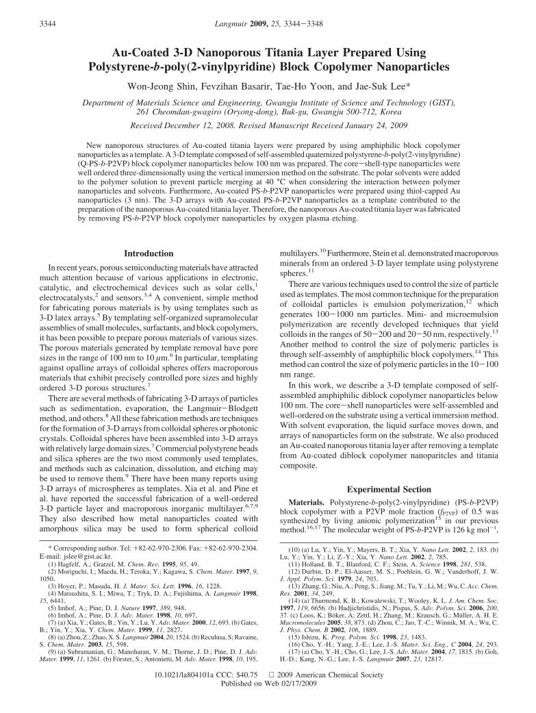
Figure 3. Scheme of preparing Au-coated nanoporous titania layer using Q-PS-b-P2VP nanoparticles coated with thiol-capped Au nanoparticles:(a) dispersion of thiol-capped Au-coated Q-PS-b-P2VP in toluene, (b) infiltration titania precursors to the solution, (c) self-assembling of nanoparticleswith titania precursors on the ITO glass at 40 °C for 5 days by vertical immersion method, (d) covering with the other glass plate to remove excesstitania precursors, (e) drying the composites at room temperature for 12 h, and (f) formation of Au-coated nanoporous of titania after removingQ-PS-b-P2VP by oxygen plasma etching.
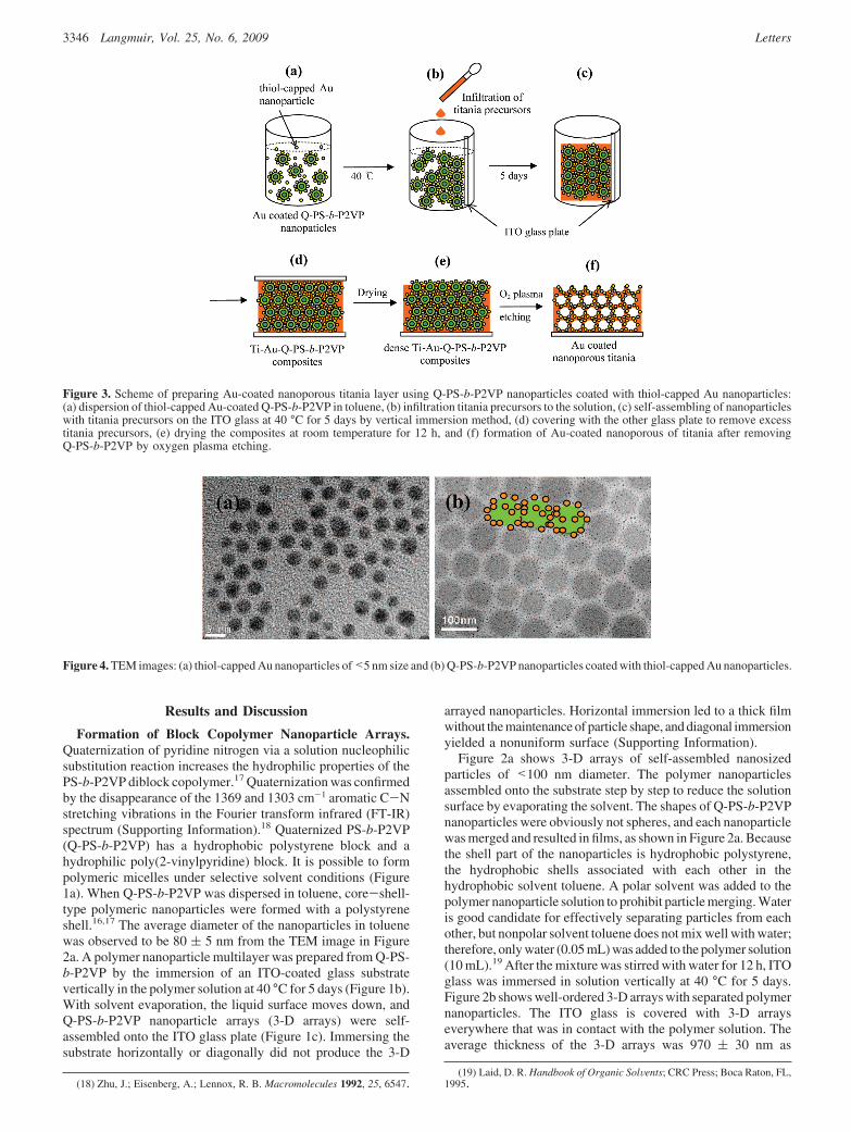
Figure 4. TEM images: (a) thiol-capped Au nanoparticles of<5 nm size and (b) Q-PS-b-P2VP nanoparticles coated with thiol-capped Au nanoparticles.
3346 Langmuir, Vol. 25, No. 6, 2009 Letters
determined from the cross-section of Figure 2c. Ethanol (3 mL)was also effective at separating particles from each other, asshown in Figure 2d. This indicates that added ethanol has aneffect on preventing nanoparticle merging for titania precursorinfiltration into the 3-D arrays. The polymer nanoparticles wereself-assembled very well on the surface of 3-D arrays (Figure2f). When 5 mL of ethanol was added, there was no improvementin preventing polymer nanoparticle merging. It is important tonote that with increasing ethanol content the total nanoparticlesolution concentration decreases. Because of this, nanoparticlesare not stacked enough on the substrate for 5 days. We foundthat the optimum condition was 3 mL of ethanol added to a 10mL polymer solution; this yielded an average thickness of 740(30 nm (Figure 2e). The thickness is controlled by the immersiontime or solution concentration, and the 3-D array structures werealso affected by the evaporation rate. TEM images (insets inFigure 2) show Q-PS-b-P2VP nanoparticles in toluene with polarsolvents. From these TEM images, no change in the core-shellstructure was observed. In the case of added ethanol, we observedalmost the same TEM image as in the case of water addition.
Synthesis of an Au-Coated Nanoporous Titania Layer.Figure 3 demonstrates the procedure for preparing an Au-coatednanoporous titania layer from Au-coated Q-PS-b-P2VP nano-
particle arrays. Q-PS-b-P2VP nanoparticles were coated withthiol-capped Au nanoparticles prepared by the well-known Brustmethod20 (Figure 3a). During 3-D stacking of the nanoparticleson ITO glass at 40 °C, an inorganic sol solution (3 mL) oftitanium(IV) isopropoxide in ethanol infiltrated the polymersolution (Figure 3b). As the titania precursor infiltrated thepolymer nanoparticles’ space, titania-Au-Q-PS-b-P2VP arrayswere formed from the precursor solution by capillary force.17
The thiol-capped Au nanoparticles were attached to the polymernanoparticle surfaces because of hydrophobic interaction21 (Figure3c) between PS and gold nanoparticles surrounded by alkyl chains.After 5 days, this immersed ITO glass was dried for 12 h underambient conditions and dense Ti-Au-Q-PS-b-P2VP compositeswere prepared by completing the sol-gel reaction22 (Figure 3e).Before drying, the second slide glass was used to eliminate excesssol solution on the surface of Ti-Au-Q-PS-b-P2VP compositesand to make open pores on the top layer to facilitate the removal
(20) (a) Brust, M.; Walker, M.; Bethell, D.; Schiffrin, D. J.; Whayman, R.J. Chem. Soc., Chem. Commun. 1994, 801. (b) Chaki, N. K.; Vijayamohanan,K. P. J. Phys. Chem. B 2005, 109, 2552.
(21) McNaught, A. D.; Wilkinson, A. IUPAC Compendium of ChemicalTerminology, 2nd ed.; Blackwell Scientific Publications; Oxford, U.K., 1997.
(22) Brinker, C. J.; Scherer, G. W. Sol-Gel Science: The Physics and Chemistryof Sol-Gel Processing; Academic Press; San Diego, CA, 1990.
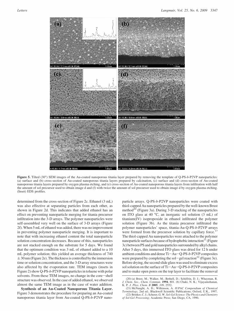
Figure 5. Tilted (30°) SEM images of the Au-coated nanoporous titania layer prepared by removing the template of Q-PS-b-P2VP nanoparticles:(a) surface and (b) cross-section of Au-coated nanoporous titania layers prepared by calcination, (c) surface and (d) cross-section of Au-coatednanoporous titania layers prepared by oxygen plasma etching, and (e) cross-section of Au-coated nanoporous titania layers from infiltration with halfthe amount of sol precursor used to obtain image d and (f) with twice the amount of sol precursor used to obtain image d by oxygen plasma etching.(Inset) EDS profiles.
Letters Langmuir, Vol. 25, No. 6, 2009 3347
of excess titania precursors as in our previous work17 (Figure3d). Finally, the templates were removed by etching, and thisresulted in Au-coated nanoporous titania layers (Figure 3f).
Figure 4a shows thiol-capped Au nanoparticles synthesizedby the Brust method, which have a 3-5 nm diameter, and Q-PS-b-P2VP nanoparticles uniformly coated with the metal nano-particles (Figure 4b). There are several methods of preparingporous materials by removing organic nanoparticles in the bulkcomposites with titania precursors such as calcination, dissolution,and etching. To remove the Q-PS-b-P2VP nanoparticles, theTi-Au-Q-PS-b-P2VP composite layer was sintered at 600 °Cfor 4 h, resulting in a nanoporous titania film.7 The SEM imagesof in Figure 5a,b reveal a nanoporous titania layer. Calcinationled to greater aggregation and growth of Au nanoparticles becauseof the high temperature, as shown in bright particles of Figure5a,b.23 Therefore, oxygen plasma etching was selected to removeQ-PS-b-P2VP nanoparticles and to avoid Au aggregation. TheTi-Au-Q-PS-b-P2VP composite film was exposed to oxygenplasma (100 W, 1.0 × 10⁻² Torr) for 20 min. Figure 5c,d showa nanoporous titania layer prepared from a template of Q-PS-b-P2VP nanoparticles arrayed three-dimensionally. It wasconfirmed that organic materials were completely removed bycomparing the energy-dispersive spectroscopy (EDS) profiles(inset images in Figure 5a,c) with the EDS profile of Ti-Au-Q-PS-b-P2VP composites (Supporting Information). The pore sizeof titania structure was approximately 80 nm. This means thatthe polymer nanoparticle size and the pore size of the titaniastructure are matched. The nanoporous titania layer in Figure 5ewas prepared from half the amount of precursor solution (1.5mL) used in Figure 5d, and the titania layer in Figure 5f was
prepared from twice the amount of precursor (6 mL). If less thanan optimum amount of titania precursor solution was to be used,then the nanoporous titania layer would be brittle and more porous.This indicates that the Au-coated nanoporous titania layer canbe controlled by the amount of titania precursor.
Conclusions
In general, porous inorganic materials are prepared mainlyfrom the sol-gel reaction of inorganic precursors and colloidal(0.1-1 µm) arrays as a template. In this work, we used theself-assembly of amphiphilic block copolymer nanoparticles asa template for nanoporous materials of the Au-coated titanialayer. The quaternization of PS-b-P2VP allowed polymernanoparticle be of the monodisperse core-shell type in toluene.We prepared 3-D Q-PS-b-P2VP block copolymer nanoparticlearrays made from about 80 nm nanoparticles on a substrate usinga vertical immersion method. By the addition of polar solvents,well-ordered 3-D nanoparticle arrays were obtained. An Au-coated nanoporous titania layer was also fabricated by removing3-D polymer nanoparticle arrays with oxygen plasma etchingand might be available in the fields of electrochemical and catalyticapplications.
Acknowledgment. This work was supported by the Programfor Integrated Molecular System (PIMS). We thank the KoreaBasic Science Institute (KBSI) for FE-TEM measurements.
Supporting Information Available: Confirmation of quaternizedblock copolymer, SEM images of structures by the substrate immersionmethod, and EDS profile of Ti-Au-Q-PS-b-P2VP composites. Thismaterial is available free of charge via the Internet at http://pubs.acs.org.
LA804101A(23) Daniel, M.-C.; Astruc, D. Chem. ReV. 2004, 104, 293.
3348 Langmuir, Vol. 25, No. 6, 2009 Letters