Embed Size (px)
Citation preview
B7-H1 Blockade Increases Survival of Dysfunctional CD8+
T Cells and Confers Protection against Leishmaniadonovani InfectionsTrupti Joshi1, Susana Rodriguez1, Vladimir Perovic1, Ian A. Cockburn2, Simona Stager1*
1 Department of Pharmacology and Molecular Sciences, The Johns Hopkins University School of Medicine, Baltimore, Maryland, United States of America, 2 Department of
Molecular Microbiology and Immunology, Malaria Research Institute, Johns Hopkins Bloomberg School of Public Health, Baltimore, Maryland, United States of America
Abstract
Experimental visceral leishmaniasis (VL) represents an exquisite model to study CD8+ T cell responses in a context of chronicinflammation and antigen persistence, since it is characterized by chronic infection in the spleen and CD8+ T cells arerequired for the development of protective immunity. However, antigen-specific CD8+ T cell responses in VL have so far notbeen studied, due to the absence of any defined Leishmania-specific CD8+ T cell epitopes. In this study, transgenicLeishmania donovani parasites expressing ovalbumin were used to characterize the development, function, and fate ofLeishmania-specific CD8+ T cell responses. Here we show that L. donovani parasites evade CD8+ T cell responses by limitingtheir expansion and inducing functional exhaustion and cell death. Dysfunctional CD8+ T cells could be partially rescued byin vivo B7-H1 blockade, which increased CD8+ T cell survival but failed to restore cytokine production. Nevertheless, B7-H1blockade significantly reduced the splenic parasite burden. These findings could be exploited for the design of newstrategies for immunotherapeutic interventions against VL.
Citation: Joshi T, Rodriguez S, Perovic V, Cockburn IA, Stager S (2009) B7-H1 Blockade Increases Survival of Dysfunctional CD8+ T Cells and Confers Protectionagainst Leishmania donovani Infections. PLoS Pathog 5(5): e1000431. doi:10.1371/journal.ppat.1000431
Editor: John M. Mansfield, University of Wisconsin-Madison, United States of America
Received November 7, 2008; Accepted April 15, 2009; Published May 15, 2009
Copyright: � 2009 Joshi et al. This is an open-access article distributed under the terms of the Creative Commons Attribution License, which permitsunrestricted use, distribution, and reproduction in any medium, provided the original author and source are credited.
Funding: This work was supported by the start up package to SS from the Johns Hopkins School of Medicine. IAC is a recipient of a fellowship from the JohnsHopkins Malaria Research Institute. The funders had no role in study design, data collection and analysis, decision to publish, or preparation of the manuscript.
Competing Interests: The authors have declared that no competing interests exist.
* E-mail: [email protected]
Introduction
Antigen-specific CD8+ T cell responses are essential for
protection and clearance of many microbial pathogens. CD8+ T
cells recognize peptides which are presented in the context of
major histocompatibility complex (MHC) class I via T cell
receptor (TCR). Rare naıve CD8+ T cells are activated in
secondary lymphoid tissues following encounter with dendritic
cells expressing peptide/MHCI complexes [1]. Once activated,
antigen-specific T cells typically undergo massive expansion,
differentiate into effector cells, and acquire the capacity to kill
and produce cytokines [2–5]. The magnitude of expansion largely
depends on the amount of antigen and/or the number of the naıve
precursors [6,7]. This robust proliferation is then followed by a
programmed contraction, which occurs independently of duration
of infection, magnitude of expansion or antigen dose [7]. Only 5–
10% of the cells present during the peak phase survive the
contraction, becoming long-lived memory cells [8]. Memory cells
show increased responsiveness and undergo dramatic clonal
expansion after reencounter with the same antigen, and thereby
confer protection [4,9].
This paradigm of T cell differentiation and memory formation
has been mainly derived from models of acute viral and bacterial
infections, such as Lymphocytic Choriomeningitis Virus (LCMV;
Armstrong strain), Vaccinia Virus and Listeria monocytogenes
[2,7,10–12]. Yet it may not apply to CD8+ T cell responses
generated in the presence of persistent antigen stimulation.
Indeed, several degrees of dysfunction, such as delays in expansion
and contraction, anergy, and suppression and exhaustion of
effector responses, have been observed during chronic diseases
[13–18]. The inhibitory receptor PD-1 and its ligand B7-H1 have
been shown to play an important role in the regulation of CD8+ T
cell function in anti-tumour and anti-microbial immunity, and also
in the early CD8+ T cell fate decisions [19–22]. This pathway
appears to induce T cell apoptosis and inhibits proliferation and
cytokine production upon TCR engagement in vitro [23,24]. In
vivo, B7-H1/PD-1 interaction was shown to control the initiation
and reversion of anergy, to inhibit T cell functions, and to be the
key pathway in the induction of exhaustion [21,25,26]. This
functionally inactivated phenotype has also been described in
humans, and shown to be reverted by treatment with blocking
antibodies to B7-H1, thereby restoring the capacity of CD8+ T
cells to control disease and decrease viral load [21].
Experimental visceral leishmaniasis (VL) represents an exquisite
model to study CD8+ T cell responses in a context of chronic
inflammation and antigen persistence. In mice, the two main
target organs of this disease are the liver and the spleen [27]. While
in the liver the infection is self-resolving due to the development of
a TH1-dominated granulomatous response, spleens infected with
Leishmania donovani, the causative agent of visceral leishmaniasis
(VL), stay chronically infected. Together with CD4+ T cells, CD8+
T cells have been shown to be essential for the control of primary
infections in various experimental models of Leishmaniasis [28–
31]. They also appear to be the main mediators of resistance to
PLoS Pathogens | www.plospathogens.org 1 May 2009 | Volume 5 | Issue 5 | e1000431
rechallenge and the major correlates of protection in vaccine-
induced immunity against several Leishmania species [30,32–35].
However, the onset of these responses seems to be delayed:
polyclonal CD8+ T cell responses are only detectable 3–4 weeks
into the infection in both L. major and L. donovani infected mice
[29,30]. Due to a lack of knowledge of Leishmania-specific CD8+
T cell epitopes, antigen-specific CD8+ T cell responses in VL have
thus far not been studied.
In this study, transgenic L. donovani parasites expressing
ovalbumin [36] were used to characterize the development,
function and fate of Leishmania-specific CD8+ T cell responses
during the course of infection. We show that L. donovani parasites
evade CD8+ T cell responses by limiting their expansion and
inducing functional exhaustion and cell death.
Results
Expansion, duration, and contraction of OT-I CD8+T cellresponses during Leishmania donovani infection
To determine the extent and significance of bystander activation
and distinguish it from antigen-specific responses, we first
compared the expansion of adoptively transferred OT-I CD8+ T
cells in mice infected with wild type (LV9) and Ovalbumin-
transgenic (PINK) Leishmania donovani parasites. In order to
visualize and analyze OT-I CD8+ T cell responses in LV9
infected mice, it was necessary to transfer 105 OT-I CD8+ T cells
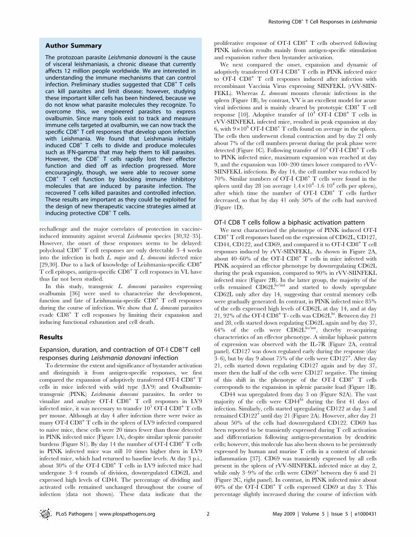
per mouse. Although at day 4 after infection there were twice as
many OT-I CD8+ T cells in the spleen of LV9 infected compared
to naive mice, these cells were 20 times fewer than those detected
in PINK infected mice (Figure 1A), despite similar splenic parasite
burdens (Figure S1). By day 14 the number of OT-I CD8+ T cells
in PINK infected mice was still 10 times higher then in LV9
infected mice, which had returned to baseline levels. At day 3 p.i.,
about 30% of the OT-I CD8+ T cells in LV9 infected mice had
undergone 3–4 rounds of division, downregulated CD62L and
expressed high levels of CD44. The percentage of dividing and
activated cells remained unchanged throughout the course of
infection (data not shown). These data indicate that the
proliferative response of OT-I CD8+ T cells observed following
PINK infection results mainly from antigen-specific stimulation
and expansion rather then bystander activation.
We next compared the onset, expansion and dynamic of
adoptively transferred OT-I CD8+ T cells in PINK infected mice
to OT-I CD8+ T cell responses induced after infection with
recombinant Vaccinia Virus expressing SIINFEKL (rVV-SIIN-
FEKL). Whereas L. donovani mounts chronic infections in the
spleen (Figure 1B), by contrast, VV is an excellent model for acute
viral infections and is mainly cleared by prototypic CD8+ T cell
response [10]. Adoptive transfer of 104 OT-I CD8+ T cells in
rVV-SIINFEKL infected mice, resulted in peak expansion at day
6, with 96106 OT-I CD8+ T cells found on average in the spleen.
The cells then underwent clonal contraction and by day 21 only
about 7% of the cell numbers present during the peak phase were
detected (Figure 1C). Following transfer of 104 OT-I CD8+ T cells
to PINK infected mice, maximum expansion was reached at day
9, and the expansion was 100–200 times lower compared to rVV-
SIINFEKL infections. By day 14, the cell number was reduced by
70%. Similar numbers of OT-I CD8+ T cells were found in the
spleen until day 28 (on average 1.46104–1.6 104 cells per spleen),
after which time the number of OT-I CD8+ T cells further
decreased, so that by day 41 only 50% of the cells had survived
(Figure 1D).
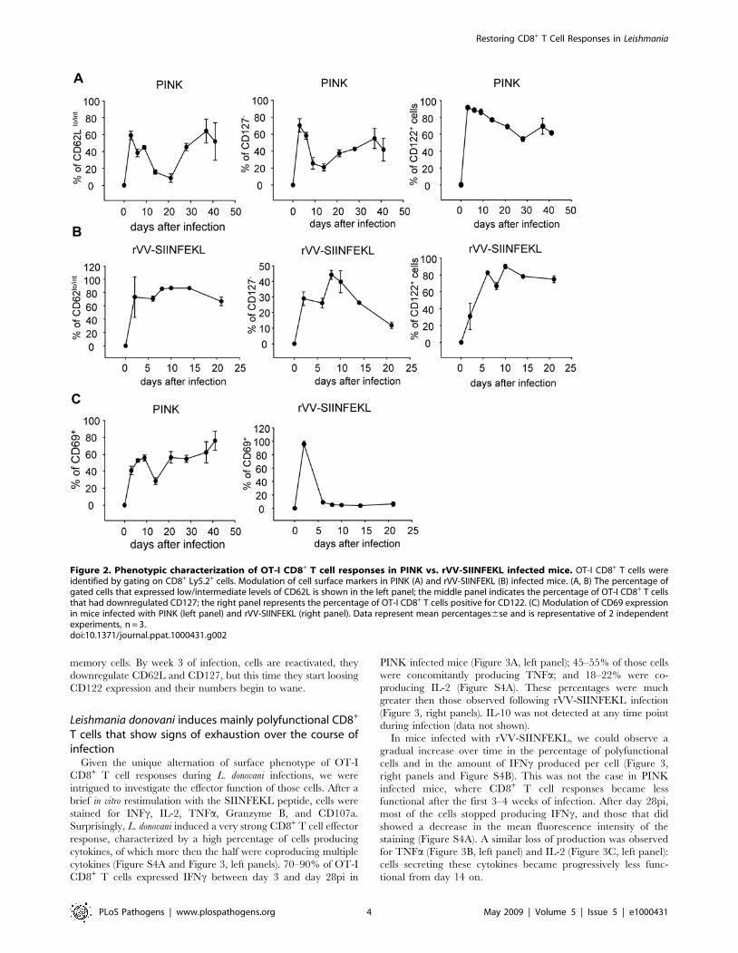
OT-I CD8 T cells follow a biphasic activation patternWe next characterized the phenotype of PINK induced OT-I
CD8+ T cell responses based on the expression of CD62L, CD127,
CD44, CD122, and CD69, and compared it to OT-I CD8+ T cell
responses induced by rVV-SIINFEKL. As shown in Figure 2A,
about 40–60% of the OT-I CD8+ T cells in mice infected with
PINK acquired an effector phenotype by downregulating CD62L
during the peak expansion, compared to 90% in rVV-SIINFEKL
infected mice (Figure 2B). In the latter group, the majority of the
cells remained CD62Llo/int and started to slowly upregulate
CD62L only after day 14, suggesting that central memory cells
were gradually generated. In contrast, in PINK infected mice 85%
of the cells expressed high levels of CD62L at day 14, and at day
21, 92% of the OT-I CD8+ T- cells was CD62Lhi. Between day 21
and 28, cells started down regulating CD62L again and by day 37,
64% of the cells were CD62Llo/int, thereby re-acquiring
characteristics of an effector phenotype. A similar biphasic pattern
of expression was observed with the IL-7R (Figure 2A, central
panel). CD127 was down regulated early during the response (day
3–6), but by day 9 about 75% of the cells were CD127+. After day
21, cells started down regulating CD127 again and by day 37,
more then the half of the cells were CD127 negative. The timing
of this shift in the phenotype of the OT-I CD8+ T cells
corresponds to the expansion in splenic parasite load (Figure 1B).
CD44 was upregulated from day 3 on (Figure S2A). The vast
majority of the cells were CD44hi during the first 41 days of
infection. Similarly, cells started upregulating CD122 at day 3 and
remained CD122+ until day 21 (Figure 2A). However, after day 21
about 50% of the cells had downregulated CD122. CD69 has
been reported to be transiently expressed during T cell activation
and differentiation following antigen-presentation by dendritic
cells; however, this molecule has also been shown to be persistently
expressed by human and murine T cells in a context of chronic
inflammation [37]. CD69 was transiently expressed by all cells
present in the spleen of rVV-SIINFEKL infected mice at day 2,
while only 3–9% of the cells were CD69+ between day 6 and 21
(Figure 2C, right panel). In contrast, in PINK infected mice about
40% of the OT-I CD8+ T cells expressed CD69 at day 3. This
percentage slightly increased during the course of infection with
Author Summary
The protozoan parasite Leishmania donovani is the causeof visceral leishmaniasis, a chronic disease that currentlyaffects 12 million people worldwide. We are interested inunderstanding the immune mechanisms that can controlinfection. Preliminary studies suggested that CD8+ T cellscan kill parasites and limit disease; however, studyingthese important killer cells has been hindered, because wedo not know what parasite molecules they recognize. Toovercome this, we engineered parasites to expressovalbumin. Since many tools exist to track and measureimmune cells targeted at ovalbumin, we can now track thespecific CD8+ T cell responses that develop upon infectionwith Leishmania. We found that Leishmania initiallyinduced CD8+ T cells to divide and produce moleculessuch as IFN-gamma that may help them to kill parasites.However, the CD8+ T cells rapidly lost their effectorfunction and died off as infection progressed. Moreencouragingly, though, we were able to recover someCD8+ T cell function by blocking immune inhibitorymolecules that are induced by parasite infection. Therecovered T cells killed parasites and controlled infection.These results are important as they could be exploited forthe design of new therapeutic vaccine strategies aimed atinducing protective CD8+ T cells.
Restoring CD8+ T Cell Responses in Leishmania
PLoS Pathogens | www.plospathogens.org 2 May 2009 | Volume 5 | Issue 5 | e1000431
the exception of day 14, when only 20% of the cells were CD69+
(Figure 2C, left panel).
We also monitored the proliferation of OT-1 CD8+ T cells by
assessing the CFSE dilution over the course of the infection (Figure
S3). Between day 2 and 6 after infection, the cells had undergone
several rounds of division, resulting in a complete dilution of the
CFSE staining (Figure S3). All OT-I CD8+ T cells present in the
spleen were CFSE2 until day 21. In mice infected with PINK,
OT-I CD8+ T cells had already undergone 4–5 rounds of division
at day 3 (Figure S3A), with maximal CFSE dilution observed at
day 6. Interestingly, CFSE dilution at day 6 was higher then at
days 9, 14, 21, and 28, indicating that the cells present at these
later time points had undergone fewer rounds of division then
those present at day 6. One possible explanation is that effector
cells present in the spleen at day 6 have migrated to the liver, the
other site of infection. To test this hypothesis, we enumerated the
OT-I CD8+ T cells present in the liver during the course of
infection (Figure S3B). A maximum of 1500 cells was detected
during peak expansion, suggesting that migration of effector cells
to the liver was not responsible for the disappearance of these cells
from the spleen. This suggests that effector cells that had expanded
at day 6 had possibly died and were replaced by newly recruited
and activated cells. Notably, these cells did not undergo more then
5–6 rounds of division, even during peak expansion.
Taken together, these results show that OT-I CD8+ T cells
following PINK infection display a biphasic activation pattern.
During the first 9 days of infection, before OT-I CD8+ T cells
undergo clonal contraction, they exhibit an effector phenotype;
this activation results in limited expansion. After the first wave of
activation, the majority of the cells that survived clonal contraction
were CD62Lhi CD44hi CD127+ CD122+ and KLRG12 (data not
shown). This phenotype is similar to that displayed by central
Figure 1. L. donovani induces limited expansion of CD8+ T cell. (A) 105 OT-I CD8+ T cells were adoptively transferred into congenic mice priorto infection with either wild type (LV9) or ovalbumin-transgenic (PINK) L. donovani amastigotes. Graph represents the average number6se of OT-ICD8+ T cells found in the spleen of each individual mouse during the course of infection. (B) Congenic mice received 104 OT-I CD8+ T cells prior toinfection with 26107 PINK amastigotes. Graph represents the average number6se of parasites found in the spleen of each individual mouse. Parasiteload was determined by limiting dilutions. (C) The same mice were sacrificed at different time points during infection and OT-I CD8+ T cells present inthe spleen were enumerated by gating on Ly5.2+ CD8+ cells. Graph represents the average number6se of cells found in the spleen of each individualmouse during the first 41 days of infection. (D) Congenic mice were infected with rVV-SIINFEKL (106 PFU) i.v. the day after receiving 104 OT-I CD8+ Tcells. Graph represents the average number6se of Ly5.2+ CD8+ cells found in the spleen of each individual mouse. All graphs show resultsrepresentative of 2–3 independent experiments; for all experiments, n = 3.doi:10.1371/journal.ppat.1000431.g001
Restoring CD8+ T Cell Responses in Leishmania
PLoS Pathogens | www.plospathogens.org 3 May 2009 | Volume 5 | Issue 5 | e1000431
memory cells. By week 3 of infection, cells are reactivated, they
downregulate CD62L and CD127, but this time they start loosing
CD122 expression and their numbers begin to wane.
Leishmania donovani induces mainly polyfunctional CD8+
T cells that show signs of exhaustion over the course ofinfection
Given the unique alternation of surface phenotype of OT-I
CD8+ T cell responses during L. donovani infections, we were
intrigued to investigate the effector function of those cells. After a
brief in vitro restimulation with the SIINFEKL peptide, cells were
stained for INFc, IL-2, TNFa, Granzyme B, and CD107a.
Surprisingly, L. donovani induced a very strong CD8+ T cell effector
response, characterized by a high percentage of cells producing
cytokines, of which more then the half were coproducing multiple
cytokines (Figure S4A and Figure 3, left panels). 70–90% of OT-I
CD8+ T cells expressed IFNc between day 3 and day 28pi in
PINK infected mice (Figure 3A, left panel); 45–55% of those cells
were concomitantly producing TNFa; and 18–22% were co-
producing IL-2 (Figure S4A). These percentages were much
greater then those observed following rVV-SIINFEKL infection
(Figure 3, right panels). IL-10 was not detected at any time point
during infection (data not shown).
In mice infected with rVV-SIINFEKL, we could observe a
gradual increase over time in the percentage of polyfunctional
cells and in the amount of IFNc produced per cell (Figure 3,
right panels and Figure S4B). This was not the case in PINK
infected mice, where CD8+ T cell responses became less
functional after the first 3–4 weeks of infection. After day 28pi,
most of the cells stopped producing IFNc, and those that did
showed a decrease in the mean fluorescence intensity of the
staining (Figure S4A). A similar loss of production was observed
for TNFa (Figure 3B, left panel) and IL-2 (Figure 3C, left panel):
cells secreting these cytokines became progressively less func-
tional from day 14 on.
Figure 2. Phenotypic characterization of OT-I CD8+ T cell responses in PINK vs. rVV-SIINFEKL infected mice. OT-I CD8+ T cells wereidentified by gating on CD8+ Ly5.2+ cells. Modulation of cell surface markers in PINK (A) and rVV-SIINFEKL (B) infected mice. (A, B) The percentage ofgated cells that expressed low/intermediate levels of CD62L is shown in the left panel; the middle panel indicates the percentage of OT-I CD8+ T cellsthat had downregulated CD127; the right panel represents the percentage of OT-I CD8+ T cells positive for CD122. (C) Modulation of CD69 expressionin mice infected with PINK (left panel) and rVV-SIINFEKL (right panel). Data represent mean percentages6se and is representative of 2 independentexperiments, n = 3.doi:10.1371/journal.ppat.1000431.g002
Restoring CD8+ T Cell Responses in Leishmania
PLoS Pathogens | www.plospathogens.org 4 May 2009 | Volume 5 | Issue 5 | e1000431
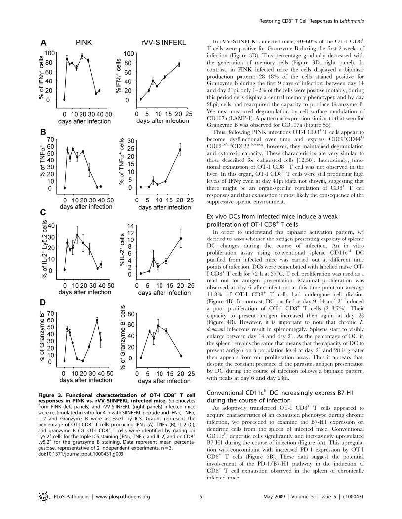
In rVV-SIINFEKL infected mice, 40–60% of the OT-I CD8+
T cells were positive for Granzyme B during the first 2 weeks of
infection (Figure 3D). This percentage gradually decreased with
the generation of memory cells (Figure 3D, right panel). In
contrast, in PINK infected mice the cells displayed a biphasic
production pattern: 28–48% of the cells stained positive for
Granzyme B during the first 9 days of infection; between day 14
and day 21pi, only 1–2% of the cells were positive (notably, during
this period cells display a central memory phenotype); and by day
28pi, cells had reacquired the capacity to produce Granzyme B.
We next measured degranulation by cell surface modulation of
CD107a (LAMP-1). A pattern of expression similar to that seen for
Granzyme B was observed for CD107a (Figure S5).
Thus, following PINK infections OT-I CD8+ T cells appear to
become dysfunctional over time and express CD69+CD44hi
CD62lo/intCD122 lo/neg, however, they maintained degranulation
and cytotoxic capacity. These characteristics are very similar to
those described for exhausted cells [12,38]. Interestingly, func-
tional exhaustion of OT-I CD8+ T cell was not observed in the
liver. In this organ, OT-I CD8+ T cells were still producing high
levels of IFNc even at day 41pi (data not shown), suggesting that
there might be an organ-specific regulation of CD8+ T cell
responses and that exhaustion is most likely the consequence of the
suppressive splenic environment.
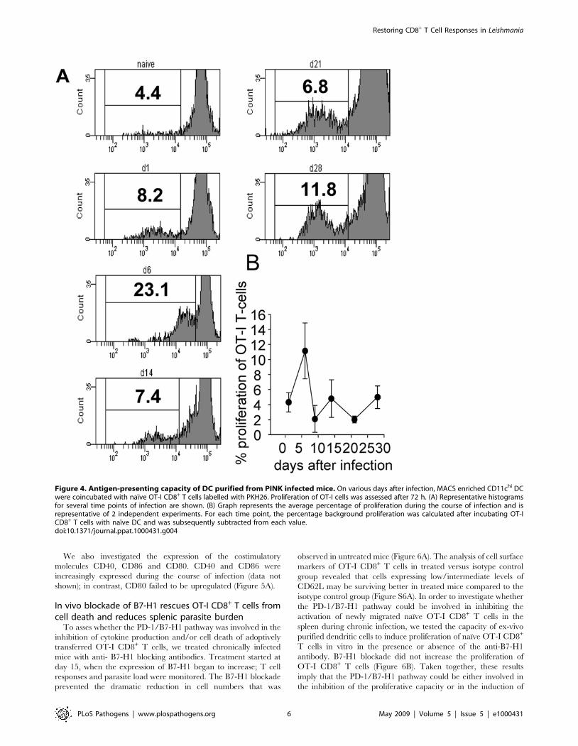
Ex vivo DCs from infected mice induce a weakproliferation of OT-I CD8+ T cells
In order to understand this biphasic activation pattern, we
decided to asses whether the antigen presenting capacity of splenic
DC changes during the course of infection. An in vitro
proliferation assay using conventional splenic CD11chi DC
purified from infected mice was carried out at different time
points of infection. DCs were coincubated with labelled naıve OT-
I CD8+ T cells for 72 h at 37uC. T cell proliferation was used as a
read out for antigen presentation. Maximal proliferation was
observed at day 6 after infection: at this time point on average
11.8% of OT-I CD8+ T cells had undergone cell division
(Figure 4B). In contrast, DC purified at day 9, 14 and 21 induced
a poor proliferation of OT-I CD8+ T cells (2–3.7%). Their
capacity to present antigen increased then again at day 28
(Figure 4B). However, it is important to note that chronic L.
donovani infections result in splenomegaly. Spleens start to visibly
enlarge between day 14 and day 21. As the percentage of DC in
the spleen remains the same that means that the capacity of DC to
present antigen on a population level at day 21 and 28 is greater
then appears from our proliferation assay. Thus it appears that,
despite the constant presence of the parasite, antigen presentation
by DC during the course of infection follows a biphasic pattern,
with peaks at day 6 and day 28pi.
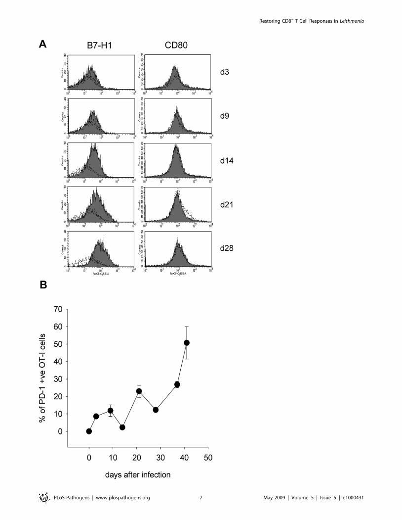
Conventional CD11chi DC increasingly express B7-H1during the course of infection
As adoptively transferred OT-I CD8+ T cells appeared to
acquire characteristics of an exhausted phenotype during chronic
infection, we proceeded to examine the B7-H1 expression on
dendritic cells from the spleen of infected mice. Conventional
CD11chi dendritic cells significantly and increasingly upregulated
B7-H1 during the course of infection (Figure 5A). This upregula-
tion was concomitant with increased PD-1 expression by OT-I
CD8+ T cells (Figure 5B). These data suggest the potential
involvement of the PD-1/B7-H1 pathway in the induction of
CD8+ T cell exhaustion observed in the spleen of chronically
infected mice.
Figure 3. Functional characterization of OT-I CD8+ T cellresponses in PINK vs. rVV-SIINFEKL infected mice. Splenocytesfrom PINK (left panels) and rVV-SIINFEKL (right panels) infected micewere restimulated in vitro for 4 h with SIINFEKL peptide and IFNc, TNFa,IL-2 and Granzyme B were assessed by ICS. Graphs represent thepercentage of OT-I CD8+ T cells producing IFNc (A), TNFa (B), IL-2 (C),and granzyme B (D). OT-I CD8+ T cells were identified by gating onLy5.2+ cells for the triple ICS staining (IFNc, TNFa, and IL-2) and on CD8+
Ly5.2+ for the granzyme B staining. Data represent mean percenta-ges6se, representative of 2 independent experiments, n = 3.doi:10.1371/journal.ppat.1000431.g003
Restoring CD8+ T Cell Responses in Leishmania
PLoS Pathogens | www.plospathogens.org 5 May 2009 | Volume 5 | Issue 5 | e1000431
We also investigated the expression of the costimulatory
molecules CD40, CD86 and CD80. CD40 and CD86 were
increasingly expressed during the course of infection (data not
shown); in contrast, CD80 failed to be upregulated (Figure 5A).
In vivo blockade of B7-H1 rescues OT-I CD8+ T cells fromcell death and reduces splenic parasite burden
To asses whether the PD-1/B7-H1 pathway was involved in the
inhibition of cytokine production and/or cell death of adoptively
transferred OT-I CD8+ T cells, we treated chronically infected
mice with anti- B7-H1 blocking antibodies. Treatment started at
day 15, when the expression of B7-H1 began to increase; T cell
responses and parasite load were monitored. The B7-H1 blockade
prevented the dramatic reduction in cell numbers that was
observed in untreated mice (Figure 6A). The analysis of cell surface
markers of OT-I CD8+ T cells in treated versus isotype control
group revealed that cells expressing low/intermediate levels of
CD62L may be surviving better in treated mice compared to the
isotype control group (Figure S6A). In order to investigate whether
the PD-1/B7-H1 pathway could be involved in inhibiting the
activation of newly migrated naıve OT-I CD8+ T cells in the
spleen during chronic infection, we tested the capacity of ex-vivo
purified dendritic cells to induce proliferation of naıve OT-I CD8+
T cells in vitro in the presence or absence of the anti-B7-H1
antibody. B7-H1 blockade did not increase the proliferation of
OT-I CD8+ T cells (Figure 6B). Taken together, these results
imply that the PD-1/B7-H1 pathway could be either involved in
the inhibition of the proliferative capacity or in the induction of
Figure 4. Antigen-presenting capacity of DC purified from PINK infected mice. On various days after infection, MACS enriched CD11chi DCwere coincubated with naıve OT-I CD8+ T cells labelled with PKH26. Proliferation of OT-I cells was assessed after 72 h. (A) Representative histogramsfor several time points of infection are shown. (B) Graph represents the average percentage of proliferation during the course of infection and isrepresentative of 2 independent experiments. For each time point, the percentage background proliferation was calculated after incubating OT-ICD8+ T cells with naıve DC and was subsequently subtracted from each value.doi:10.1371/journal.ppat.1000431.g004
Restoring CD8+ T Cell Responses in Leishmania
PLoS Pathogens | www.plospathogens.org 6 May 2009 | Volume 5 | Issue 5 | e1000431
Restoring CD8+ T Cell Responses in Leishmania
PLoS Pathogens | www.plospathogens.org 7 May 2009 | Volume 5 | Issue 5 | e1000431
cell death of effector CD8+ T cells. Further investigations are
needed in order to clarify this mechanism of action.
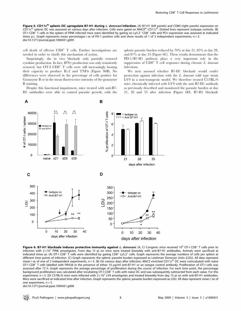
Surprisingly, the in vivo blockade only partially restored
cytokine production. In fact, IFNc production was only transiently
restored, but OT-I CD8+ T cells were still increasingly loosing
their capacity to produce IL-2 and TNFa (Figure S6B). No
differences were observed in the percentage of cells positive for
Granzyme B or in the mean fluorescence intensity of the granzyme
B staining.
Despite this functional impairment, mice treated with anti-B7-
H1 antibodies were able to control parasite growth, with the
splenic parasite burden reduced by 70% at day 21, 85% at day 28,
and 87% at day 35 (Figure 6C). These results demonstrate that the
PD-1/B7-H1 pathway plays a very important role in the
suppression of CD8+ T cell responses during chronic L. donovani
infections.
We next assessed whether B7-H1 blockade would confer
protection against infection with the L. donovani wild type strain
LV9 in a non-transgenic model. We therefore treated C57BL/6
mice chronically infected with LV9 with the anti B7-H1 antibody
as previously described and monitored the parasite burden at day
21, 28 and 35 after infection (Figure 6D). B7-H1 blockade
Figure 5. CD11chi splenic DC upregulate B7-H1 during L. donovani infection. (A) B7-H1 (left panels) and CD80 (right panels) expression onCD11chi splenic DC was assessed on various days after infection. Cells were gated on MHCIIhi CD11chi. Dotted lines represent isotyope controls. (B)OT-I CD8+ T cells in the spleen of PINK infected mice were identified by gating on Ly5.2+ CD8+ cells and PD-I expression was assessed at indicatedtimes p.i.. Graph represents mean percentages6se of PD-1 positive cells and show results of 1 of 3 independent experiments, n = 3.doi:10.1371/journal.ppat.1000431.g005
Figure 6. B7-H1 blockade induces protective immunity against L. donovani. (A, C) Congenic mice received 104 OT-I CD8+ T cells prior toinfection with 26107 PINK amastigotes. From day 15 pi on mice were treated biweekly with anti-B7-H1 antibodies. Animals were sacrificed atindicated times pi. (A) OT-I CD8+ T cells were identified by gating CD8+ Ly5.2+ cells. Graph represents the average numbers of cells per spleen atdifferent time points of infection. (C) Graph represents the splenic parasite burden expressed as Leishman Donovan Units (LDU). All data representmean6se of one of 2 independent experiments, n = 3. (B) On various days after infection, MACS enriched CD11chi DC were coincubated with naıveOT-I CD8+ T cells labelled with PKH26 in the presence of either 10 mg/ml anti-B7-H1 or an isotype control antibody. Proliferation of OT-I cells wasassessed after 72 h. Graph represents the average percentage of proliferation during the course of infection. For each time point, the percentagebackground proliferation was calculated after incubating OT-I CD8+ T cells with naıve DC and was subsequently subtracted from each value. For thisexperiment, n = 5. (D) C57BL/6 mice were infected with 26107 LV9 amastigotes and treated biweekly from day 15 pi on with anti-B7-H1 antibodies.Mice were sacrificed at indicated time after infection. Graph represents the splenic parasite burden expressed as LDU. All data represent mean6se ofone experiment, n = 5.doi:10.1371/journal.ppat.1000431.g006
Restoring CD8+ T Cell Responses in Leishmania
PLoS Pathogens | www.plospathogens.org 8 May 2009 | Volume 5 | Issue 5 | e1000431
significantly reduced the splenic parasite burden at day 21 (65%
reduction), 28 (57.5%), and 35 (71.4%), suggesting that endoge-
nous CD8+ T cell responses were most likely rescued by the
blockade. Interestingly, B7-H1 blockade also conferred protection
in the liver, but only at day 21 (53.2% reduction in the parasite
burden) and 28 (48.8% reduction), and was ineffective at day 35,
when parasite growth in this organ was under control and
infection had already significantly decreased (Figure S6C).
Superinfection at a distant site with rVV-SIINFEKL restoresOT-I CD8+ T cells responses and reduces parasite burden
To determine whether CD8+ T cells were the main mediators of
protection following B7-H1 blockade, we induced OT-I CD8+ T
cell responses by superinfecting chronically infected mice with
rVV-SIINFEKL at a distant site. Mice were challenged subcuta-
neously at day 32 pi with wild type vaccinia virus (VV) or rVV-
SIINFEKL, and euthanized 2, 6, and 9 days later. As expected,
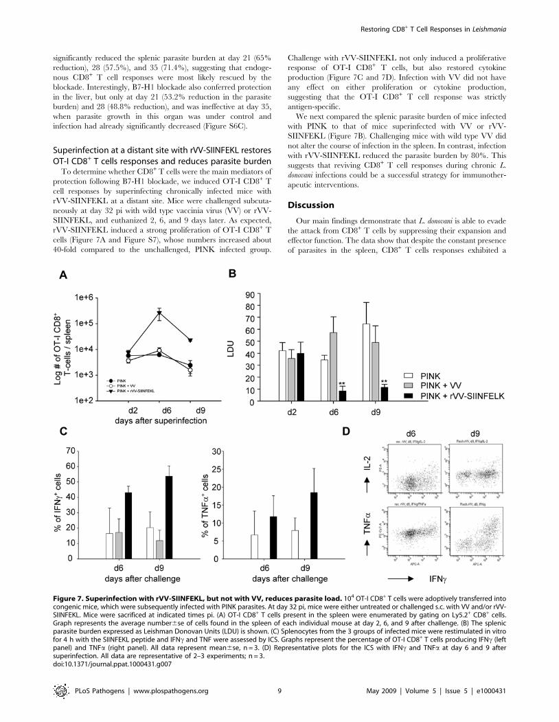
rVV-SIINFEKL induced a strong proliferation of OT-I CD8+ T
cells (Figure 7A and Figure S7), whose numbers increased about
40-fold compared to the unchallenged, PINK infected group.
Challenge with rVV-SIINFEKL not only induced a proliferative
response of OT-I CD8+ T cells, but also restored cytokine
production (Figure 7C and 7D). Infection with VV did not have
any effect on either proliferation or cytokine production,
suggesting that the OT-I CD8+ T cell response was strictly
antigen-specific.
We next compared the splenic parasite burden of mice infected
with PINK to that of mice superinfected with VV or rVV-
SIINFEKL (Figure 7B). Challenging mice with wild type VV did
not alter the course of infection in the spleen. In contrast, infection
with rVV-SIINFEKL reduced the parasite burden by 80%. This
suggests that reviving CD8+ T cell responses during chronic L.
donovani infections could be a successful strategy for immunother-
apeutic interventions.
Discussion
Our main findings demonstrate that L. donovani is able to evade
the attack from CD8+ T cells by suppressing their expansion and
effector function. The data show that despite the constant presence
of parasites in the spleen, CD8+ T cells responses exhibited a
Figure 7. Superinfection with rVV-SIINFEKL, but not with VV, reduces parasite load. 104 OT-I CD8+ T cells were adoptively transferred intocongenic mice, which were subsequently infected with PINK parasites. At day 32 pi, mice were either untreated or challenged s.c. with VV and/or rVV-SIINFEKL. Mice were sacrificed at indicated times pi. (A) OT-I CD8+ T cells present in the spleen were enumerated by gating on Ly5.2+ CD8+ cells.Graph represents the average number6se of cells found in the spleen of each individual mouse at day 2, 6, and 9 after challenge. (B) The splenicparasite burden expressed as Leishman Donovan Units (LDU) is shown. (C) Splenocytes from the 3 groups of infected mice were restimulated in vitrofor 4 h with the SIINFEKL peptide and IFNc and TNF were assessed by ICS. Graphs represent the percentage of OT-I CD8+ T cells producing IFNc (leftpanel) and TNFa (right panel). All data represent mean6se, n = 3. (D) Representative plots for the ICS with IFNc and TNFa at day 6 and 9 aftersuperinfection. All data are representative of 2–3 experiments; n = 3.doi:10.1371/journal.ppat.1000431.g007
Restoring CD8+ T Cell Responses in Leishmania
PLoS Pathogens | www.plospathogens.org 9 May 2009 | Volume 5 | Issue 5 | e1000431
biphasic activation pattern. The first wave of activation led to
limited expansion. The second wave resulted in cell death and
exhaustion of CD8+ T cells. B7-H1 blockade rescued CD8+ T cell
responses from cell death, but failed to completely restore cytokine
production. In spite of this, the parasite burden was considerably
reduced after treatment, suggesting that maintenance of effector
CD8+ T cell responses is crucial for the control of L. donovani
infections in the spleen.
The adoptive transfer experiments demonstrate for the first time
that L. donovani induces CD8+ T cell responses early during
infection. We were able to visualize this early response only
because we transferred 104 OT-I CD8+ T cells. Physiologically,
the number of naturally occurring naıve precursors for a
determined epitope is estimated to range between 50 and 1000
cells per mouse [6,39–42]. However, due to the limited expansion
capacity of CD8+ T cells in this model, transfer of such a low
number of cells did not allow us to perform an accurate analysis of
endogenous CD8+ T cell responses. This might explain why in
previous studies the onset of polyclonal responses has been
reported to be substantially delayed and could only be detected,
mainly in the liver, after 3–4 weeks of infection [30].
Although we transferred the same number of cells with the same
epitope specifcity, OT-I CD8+ T cells increased in numbers by
only 5 fold in Leishmania infected mice, compared to 900 fold in
mice infected with rVV-SIINFEKL. In other infection models,
expansions up to 50,000-fold were observed [2,5,39,40,43],
suggesting that Leishmania induces a very poor CD8+ T cell
expansion. When we assessed the antigen-presenting capacity of
DC purified from infected animals, we found that even during the
early stages of infection, DC were capable of inducing only a weak
proliferative response of naıve OT-I CD8+ T cells. This is not
surprising, since processing of Leishmania antigens for MHCI
presentation has been shown to be TAP-and proteosome-
independent [44], a pathway that is much less efficient then the
conventional ER-based, TAP-dependent pathway for Class I
presentation. We also noted that OT-I CD8+ T cells present in the
spleen at day 9 p.i. had undergone fewer rounds of division then
those detected at day 6 p.i., implying that between day 6 and 9
effector cells had died and were replaced with newly activated
CD8+ T cells. This suggests that expansion could also be limited
by cell death of effector cells. We also cannot rule out the
possibility of defective recruitment of CD8+ T cells into the spleen.
Leishmania infections are known to interfere with chemokine
expression [45–47], including CCL3 [48], a chemokine that was
recently shown to be involved in guiding CD8+ T cells to sites of
CD4+ T cell-dendritic cell interaction [49]. Hence, the limited
expansion of OT-I CD8+ T cells in L. donovani infected mice might
be due to a combination of several factors, including low antigen
load, poor recruitment of CD8+ T cells and/or increased cell
death of effector cells. Mechanisms responsible for this poor
expansion are currently under investigation.
In agreement with the previous literature, OT-I CD8+ T cell
expansion was followed by contraction at day 14 despite antigen
persistence [7]. This contraction was much steeper then in rVV-
SIINFEKL infected mice, suggesting that cells were dying more
rapidly. One of the most striking findings was that about 80% of
the cells that had survived contraction showed a central memory-
like phenotype, by expressing CD62Lhi, CD44hi, CD127+,
CD122+, CD692. These cells produced high amounts of IFNcupon restimulation and the majority were polyfunctional.
Additionally, they did not produce Granzyme B. The remaining
20% displayed an effector phenotype (CD62Llo, CD1272),
suggesting that effector memory cells were not generated. A
similar population of central memory-like cells has been recently
observed in mice infected with Trypanosoma cruzi [50]. Despite
being capable of antigen-independent survival, this population was
shown to be maintained for over a year in the presence of antigen
persistence. A recent report suggested a crucial role of T-bet as a
molecular switch between central- and effector memory cells
[51,52]. T-bet deficiency was shown to enhance generation of
central memory cells. As T-bet is also involved in the induction of
enhanced CD122 expression [53], and CD122 expression by OT-
I CD8+ T cells is gradually decreased during the course of VL, it is
possible that this molecule might not be properly induced in
Leishmania-specific CD8+ T cells.
Another interesting observation was the biphasic activation
pattern of OT-I CD8+ T cells, which reflects the variation in the
capacity of DC to present antigen during the course of infection.
This biphasic pattern can be in part explained by the biology of
the Leishmania infections. These protozoan parasites are obligate
intracellular pathogens that preferentially reside in macrophages,
but they can also be found in other cell types [54,55], including
DC [56]. During the first wave of expansion, the majority of the
cells capable of cross-presenting Leishmania antigen via MHCI is
most likely killed by CTLs so that at the end of contraction very
few DC presenting antigen survive and most of the parasites reside
in cells that are unable to cross-present antigen. For the second
wave of expansion, parasites will have first to be released from
those cells in order to be phagocytosed by DC and then killed and
processed for antigen presentation to CD8+ T cells. This explains
why between d14 and d21 p.i. DC showed a very poor antigen
presenting capacity. However, the amount of antigen presented
during this period, although little, could still be enough to
restimulate a memory response. Thus, OT-I CD8+ T cell
responses might be already impaired at this early stage of
infection. Indeed, the second wave of activation did not result in
expansion, but in functional exhaustion and cell death of the OT-I
CD8+ T cells.
This dysfunctional response could be a consequence of an
intrinsic problem following defective priming and/or could result
from a suppressive splenic environment. Although we can not rule
out that OT-I CD8+ T cell responses in L. donovani infected mice
might also have some intrinsic defects, our data support the second
scenario. Indeed conventional CD11chi splenic DC seemed to
increasingly express the inhibitory molecule B7-H1 and failed to
upregulate the costimulatory molecule CD80. B7-H1 is constitu-
tively expressed on subsets of macrophages, B-cells and thymo-
cytes, and can be induced on dendritic cells, endothelial and
epithelial cells [19,57]. Upregulation of B7-H1 on DC has been
observed during several chronic infections and in a wide range of
tumors [20,26,58,59]. Our results show that in vivo blockade of
B7-H1 during chronic L. donovani infection increased the survival
of OT-I CD8+ T cells. B7-H1 is thought to inhibit T cell
proliferation and cytokine production by ligation with the PD-1
receptor [23]. Through ligation with a yet unknown receptor, B7-
H1 can also induce programmed cell death of effector T cells [60].
Increased survival of OT-I CD8+ T cells after B7-H1 blockade
could therefore result from restoration of the proliferative capacity
or inhibition of induced cell death of effector CD8+ T cells.
In contrast to what has been recently reported in the literature
[21,26], in vivo blockade of B7-H1 during chronic VL did not
completely restore the functional capacity of exhausted OT-I
CD8+ T cells. This suggests that suppression of cytokine
production by CD8+ T cells during L. donovani infection might
be induced by mechanisms other than through the B7-H1/PD-1
pathway. A recent report has demonstrated a synergistic effect
between TGFb and the B7-H1/PD-1 axis in suppressing CD8+ T
cell responses [61]. As TGFb does not seem to play an important
Restoring CD8+ T Cell Responses in Leishmania
PLoS Pathogens | www.plospathogens.org 10 May 2009 | Volume 5 | Issue 5 | e1000431
role during chronic L. donovani infections [62], the possibility that
IL-10, which is elevated in both mouse and human VL [63–69],
could synergistically act with the B7-H1/PD-1 axis, needs to be
investigated. To our surprise, B7-H1 blockade resulted in
significant decrease in the parasite burden even if it failed to fully
restore IFNc production. While CD4+ T cells are clearly an
important source of IFNc in VL, recently we have shown that
therapeutic intervention with antigen-specific CD8+ T cells in
chronically infected mice dramatically reduced the parasite
burden [36], indicating that CD8+ T cells might play a much
more important role than previously thought. The current data
reinforce these findings by showing that OT-I CD8+ T cells
rescued from cell death by blocking B7-H1 or by superinfecting
mice with rVV-SIINFEKL resulted in host protection. The
mechanism of protection is not clear and might not merely rely
on IFNc production, as only 20% of the OT-I CD8+ T cells were
producing low amounts of IFNc at d35 pi. Nonetheless, most of
the cells were granzyme B positive and were degranulating upon
restimulation, suggesting that they have retained their cytotoxic
capacity. To date there is no evidence that CD8+ T cells can
mediate protection against L. donovani through their cytotoxic
activity.
In summary, this study shows that restoration of dysfunctional
CD8+ T cell responses induced by chronic L. donovani infections
results in disease control and host protection. This implies that
targeting CD8+ T cell responses by therapeutic vaccination could
be beneficial against chronic L. donovani infections. Moreover, these
findings might provide insights into the development of novel
strategies for therapeutic vaccination or other interventions aimed
at inducing CD8+ T cell responses, which might circumvent and/
or neutralize the immunosuppressive environment of the spleen.
Materials and Methods
Mice, parasites, and virusC57BL/6-Tg(OT-I)-RAG1tm1Mom mice were purchased from
Taconic; B6-Ly5.2 congenic mice were obtained from The
National Cancer Institute (Frederick, MD, USA), and B6.129S7-
Rag1tm1Mom/J from The Jackson Laboratory. All mice were housed
in the Johns Hopkins University animal facilities (Baltimore, MD)
under specific pathogen-free conditions and used at 6–8 weeks of
age. All experiments were approved by the Animal Care and Use
Committee of the Johns Hopkins University School of Medicine.
Ovalbumin-transgenic parasites were a gift from P.Kaye and
D.F. Smith (University of York, UK) and were generated as
previously described[36]. Wild type and ovalbumin transgenic
Leishmania donovani (strain LV9) parasites were maintained by serial
passage in B6.129S7-Rag1tm1Mom/J mice, and amastigotes were
isolated from the spleens of infected animals. Mice were infected
by injecting 26107 amastigotes intravenously via the lateral tail
vein. Hepatic and splenic parasite burdens were determined either
by limiting dilutions [31]or by examining methanol-fixed, Giemsa
stained tissue impression smears[70]. Data are presented as
number of parasites per spleen or as Leishman Donovan Units
(LDU).
The recombinant vaccinia virus (rVV) encoding SIINFEKL
(chicken ovalbumin 257–264) was a gift from F. Zavala (School of
Public Health, JHU, Baltimore) [71]. Mice were infected
intravenously or subcutaneously with 26106 pfu.
Adoptive transfer of OT-I cellsOT-I/RAG1 mice, transgenic for a T cell receptor specific for
chicken ovalbumin 257–264 presented by the MHC class I
molecule H-2 Kb, were used as T cell donors. CD8+ T cells were
enriched from splenocytes of naıve OT-I/RAG1 animals using
magnetic cell sorting (MACS), following manufacturers instruc-
tions (Miltenyi Biotech). Naıve CD8+ T cells were then sorted to
.98% purity using FACSVantage (Becton Dickinson) based on
their expression of CD44 and CD62L. After sorting, cells were
labelled with CFSE. Briefly, cells were resuspended at 56107/ml
in PBS and incubated with 2.5 mg/ml CFSE (Molecular Probes,
USA) for 10 min. at 37uC. The reaction was stopped by addition
of ice cold RPMI. Samples were then analyzed using a FACSDiva
(Becton Dickinson) for CFSE uptake prior to adoptive transfer.
Depending on the experiment, 16104 or 56104 cells were injected
into the lateral tail vein of B6-Ly5.2 congenic mice. Animals were
infected the day after with rVV-SIINFEKL and/or with wild type
or ovalbumin-transgenic Leishmania donovani.
Superinfection with rVV-SIINFEKL16104 sorted naıve OT-I CD8+ T cells were adoptively
transferred into B6-Ly5.2 congenic mice prior to infection with
26107 ovalbumin expressing L. donovani amastigotes. At day 32pi
mice were superinfected subcutaneously at the base of the tail with
26106 PFU of Vaccinia Virus (VV) or with recombinant VV
expressing the SIINFEKL peptide (rVV-SIINFEKL). Animals
were sacrificed at d2, d6 and d9 after infection with rVV-
SIINFEKL.
Flow cytometryOT-I CD8+ T cells were identified by staining splenocytes,
lymphnode cells and hepatic mononuclear cells with biotinylated
anti-CD45.2 antibody followed by PerCP-streptavidin (BD
Biosciences). The following antibodies were used to further
characterize the OT-I response: APC-conjugated anti-CD44 and
anti-CD8, PE-conjugated anti-CD62L, anti-CD69, anti-CD122,
anti-CD127 (all obtained from BD Biosciences), and anti-PD-1
(eBioscience). Splenocytes were also stained with APC-conjugated
anti-CD11c, FITC-conjugated anti-MHCII, PE-conjugated anti-
CD86, PE-Cy5.5 conjugated anti-CD80, and biotinylated anti-
B7H1 and anti-CD40, followed by PerCP-conjugated streptavidin
(all purchased by BD Biosciences). For all surface markers, cells
were directly stained following standard protocols. For intracellu-
lar staining, splenocytes were stimulated with the SIINFEKL
peptide for 4 hours in the presence of Brefeldin A and then stained
with biotinylated anti-CD45.2, followed by PerCP-conjugated
strepavidin. After fixation, cells were permeabilized and stained
with anti-Granzyme B (Invitrogen) or APC-conjugated anti-INFc(BD Biosciences), PE-conjugated anti IL-2 (BD Biosciences), and
PE-Cy7-conjugated anti-TNFa (eBioscience). Cells were also
stained with PE-conjugated anti-CD107 (eBioscience) following
the protocol described by Betts et al. [72].
Flowcytometric analysis was performed with a LSRII (Becton
Dickinson). One to two millions cells per sample were acquired
and analysed with the FACSDiva or with CellQuest software.
In vitro OT-I proliferation assaySpleen of naıve and ovalbumin-transgenic L. donovani infected
mice were digested with 0.4 mg/ml collagenase D for 30 minutes
at room temperature. Conventional CD11chi dendritic cells were
then enriched by MACS using CD11c microbeads (purity 80–
85%).
Dendritic cells were seeded at a concentration of 26104 cells/
well in a 96 wells plate.
After negative selection with anti-CD11c microbeads, CD8+ T
cells were purified from the spleen of naıve OT-I/RAG1 mice
using magnetic cell sorting (Miltenyi Biotech) (85–90% purity).
CD8+ OT-I T cells were then labelled using a red fluorescent cell
Restoring CD8+ T Cell Responses in Leishmania
PLoS Pathogens | www.plospathogens.org 11 May 2009 | Volume 5 | Issue 5 | e1000431
linker PKH26 (Sigma) in order to track proliferation. They were
then added to the ex-vivo purified dendritic cells at a
concentration of 105/well. 1 ng/ml of recombinant human IL-2
was also added to the wells. The proliferation of OT-I T cells was
assessed 72 h later by flowcytometry using FACSDiva (BD
Biosciences) and analysed with the FACSDiva software. Results
are expressed as percentage of OT-I CD8+ T cells that have
undergone one or more rounds of division. The percentage of cells
that entered division when incubated with DCs from a naive
animal was subtracted from this value.
B7H-1 blockadeAntagonistic mouse B7-H1 monoclonal antibody (clone 10B5)
was purified on a protein G column from the supernatant of the
hybridoma cell line. The hybridoma cell line was a gift from
L.Chen (Johns Hopkins University, School of Medicine, Balti-
more). Hamster IgG (Sigma) was used as isotype control. Mice
were treated every 4 days with 100 mg of antibody i.p. The first
treatment started at day 15 p.i. Before treatment, antibodies were
tested for functionally relevant LPS contamination, by assaying
their ability to synergize with IFNc for the induction of inducible
NO synthase [73]. No activity was detectable in such assays
(sensitivity,1 ng/ml LPS; data not shown).
Statistical analysisResults were analyzed using an unpaired Student t-test. P,0.05
was considered significant. All experiments were repeated at least
twice.
Supporting Information
Figure S1 Comparison of the splenic parasite burden in mice
infected with PINK vs. LV9. Parasite numbers were determined
by limiting dilutions.
Found at: doi:10.1371/journal.ppat.1000431.s001 (0.13 MB TIF)
Figure S2 Modulation of expression of cell surface markers
CD62L, CD69, CD127, CD122 at indicated times pi. Represen-
tative plots for PINK (A) and rVV-SIINFEKL (B) infected mice.
Found at: doi:10.1371/journal.ppat.1000431.s002 (1.35 MB TIF)
Figure S3 (A) CFSE dilution of OT-I CD8+ T cells on various
times pi. OT-I CD8+ T cells were identified by gating on Ly5.2+CD8+ cells. (B) Average numbers6se of OT-I CD8+ T cells found
in the liver at different time point of infection.
Found at: doi:10.1371/journal.ppat.1000431.s003 (0.35 MB TIF)
Figure S4 Cytokine production by adoptively transferred OT-I
CD8+ T-cells after infection with PINK (A) or rVV-SIINFEKL (B)
on various time points pi. Cytokine production was assessed by
ICS after 4 h stimulation with the SIINFEKL peptide. Represen-
tative plots for IL-2, IFNg, TNFa, and granzyme B stainigs are
shown.
Found at: doi:10.1371/journal.ppat.1000431.s004 (1.37 MB TIF)
Figure S5 On indicated times pi, splenocytes were restimulated
for 4 h with the SIINFEKL peptide and CD107a expression on
cells was assessed. Representative plots for each time point are
shown.
Found at: doi:10.1371/journal.ppat.1000431.s005 (0.17 MB TIF)
Figure S6 Congenic mice received 104 OT-I CD8+ T cells prior
to infection with 26107 PINK amastigotes. From day 15 pi on
mice were treated biweekly with anti-B7-H1 antibodies. Animals
were sacrificed at indicated times pi. (A) Modulation of CD62L
and CD122 expression on OT-I CD8+ T cells, identified by gating
on Ly5.2+ CD8+ cells. Mean percentages of cells expressing low/
intermediate levels of CD62L (left panel) and CD122 (right panel)
are shown. (B) Splenocytes from ant-B7-H1 treated and isotype
control treated mice were restimulated in vitro for 4 h with the
SIINFEKL peptide and IFNg and TNFa were assessed by ICS.
Graphs represent the percentage of OT-I CD8+ T cells producing
IFNg (left panel) and TNFa (right panel). All data represent
mean6se, n = 3. (C) C57BL/6 mice were infected with 26107
LV9 amastigotes and treated biweekly from day 15 pi on with anti-
B7-H1 antibodies. Mice were sacrificed at indicated time after
infection. Graph represents the hepatic parasite burden expressed
as LDU. All data represent mean6se of one experiment, n = 5.
Found at: doi:10.1371/journal.ppat.1000431.s006 (0.28 MB TIF)
Figure S7 Modulation of expression of CD62L, CD69, CD122,
and CD127 after superinfection with VV and/or rVV-SIIN-
FEKL. Representative plots for both groups at day 6 and 9 after
challenge are shown.
Found at: doi:10.1371/journal.ppat.1000431.s007 (0.47 MB TIF)
Acknowledgments
We thank Drs. Paul Kaye and Deborah Smith for the PINK parasites, Dr.
Abhay Satoskar for the LV9 strain, Dr. Lieping Chen for the 10B5
hybridoma, and Dr. Fidel Zavala for VV and rVV-SIINFEKL. We also
thank Dr. Victor Lewitsky for critical reading of the manuscript.
Author Contributions
Conceived and designed the experiments: TJ SS. Performed the
experiments: TJ SR VP IAC SS. Analyzed the data: TJ SR SS.
Contributed reagents/materials/analysis tools: IAC SS. Wrote the paper:
SS.
References
1. Heath WR, Carbone FR (2001) Cross-presentation, dendritic cells, tolerance
and immunity. Annu Rev Immunol 19: 47–64.
2. Butz EA, Bevan MJ (1998) Massive expansion of antigen-specific CD8+ T cells
during an acute virus infection. Immunity 8: 167–175.
3. Harty JT, Badovinac VP (2008) Shaping and reshaping CD8+ T cell memory.
Nat Rev Immunol 8: 107–119.
4. Kaech SM, Wherry EJ, Ahmed R (2002) Effector and memory T cell differentiation:
implications for vaccine development. Nat Rev Immunol 2: 251–262.
5. Murali-Krishna K, Altman JD, Suresh M, Sourdive DJ, Zajac AJ, et al. (1998)
Counting antigen-specific CD8 T cells: a reevaluation of bystander activation
during viral infection. Immunity 8: 177–187.
6. Badovinac VP, Haring JS, Harty JT (2007) Initial T cell receptor transgenic cell
precursor frequency dictates critical aspects of the CD8(+) T cell response to
infection. Immunity 26: 827–841.
7. Badovinac VP, Porter BB, Harty JT (2002) Programmed contraction of CD8(+)
T cells after infection. Nat Immunol 3: 619–626.
8. Sprent J, Tough DF (2001) T cell death and memory. Science 293: 245–248.
9. Wherry EJ, Teichgraber V, Becker TC, Masopust D, Kaech SM, et al. (2003)
Lineage relationship and protective immunity of memory CD8 T cell subsets.
Nat Immunol 4: 225–234.
10. Precopio ML, Betts MR, Parrino J, Price DA, Gostick E, et al. (2007)
Immunization with vaccinia virus induces polyfunctional and phenotypically
distinctive CD8(+) T cell responses. J Exp Med 204: 1405–1416.
11. Sarkar S, Kalia V, Haining WN, Konieczny BT, Subramaniam S, et al. (2008)
Functional and genomic profiling of effector CD8 T cell subsets with distinct
memory fates. J Exp Med 205: 625–640.
12. Wherry EJ, Ha SJ, Kaech SM, Haining WN, Sarkar S, et al. (2007) Molecular
signature of CD8+ T cell exhaustion during chronic viral infection. Immunity
27: 670–684.
13. Zajac AJ, Blattman JN, Murali-Krishna K, Sourdive DJ, Suresh M, et al. (1998)
Viral immune evasion due to persistence of activated T cells without effector
function. J Exp Med 188: 2205–2213.
14. Shin H, Wherry EJ (2007) CD8 T cell dysfunction during chronic viral infection.
Curr Opin Immunol 19: 408–415.
Restoring CD8+ T Cell Responses in Leishmania
PLoS Pathogens | www.plospathogens.org 12 May 2009 | Volume 5 | Issue 5 | e1000431
15. Luu RA, Gurnani K, Dudani R, Kammara R, van Faassen H, et al. (2006)
Delayed expansion and contraction of CD8+ T cell response during infection
with virulent Salmonella typhimurium. J Immunol 177: 1516–1525.
16. Leavey JK, Tarleton RL (2003) Cutting edge: dysfunctional CD8+ T cells reside
in nonlymphoid tissues during chronic Trypanosoma cruzi infection. J Immunol
170: 2264–2268.
17. Martin D, Tarleton R (2004) Generation, specificity, and function of CD8+ T
cells in Trypanosoma cruzi infection. Immunol Rev 201: 304–317.
18. Gajewski TF, Meng Y, Blank C, Brown I, Kacha A, et al. (2006) Immune
resistance orchestrated by the tumor microenvironment. Immunol Rev 213:
131–145.
19. Greenwald RJ, Freeman GJ, Sharpe AH (2005) The B7 family revisited. Annu
Rev Immunol 23: 515–548.
20. Curiel TJ, Wei S, Dong H, Alvarez X, Cheng P, et al. (2003) Blockade of B7-H1
improves myeloid dendritic cell-mediated antitumor immunity. Nat Med 9:
562–567.
21. Barber DL, Wherry EJ, Masopust D, Zhu B, Allison JP, et al. (2006) Restoring
function in exhausted CD8 T cells during chronic viral infection. Nature 439:
682–687.
22. Goldberg MV, Maris CH, Hipkiss EL, Flies AS, Zhen L, et al. (2007) Role of
PD-1 and its ligand, B7-H1, in early fate decisions of CD8 T cells. Blood 110:
186–192.
23. Freeman GJ, Long AJ, Iwai Y, Bourque K, Chernova T, et al. (2000)
Engagement of the PD-1 immunoinhibitory receptor by a novel B7 family
member leads to negative regulation of lymphocyte activation. J Exp Med 192:
1027–1034.
24. Brown JA, Dorfman DM, Ma FR, Sullivan EL, Munoz O, et al. (2003) Blockade
of programmed death-1 ligands on dendritic cells enhances T cell activation and
cytokine production. J Immunol 170: 1257–1266.
25. Tsushima F, Yao S, Shin T, Flies A, Flies S, et al. (2007) Interaction between B7-
H1 and PD-1 determines initiation and reversal of T cell anergy. Blood 110:
180–185.
26. Lukens JR, Cruise MW, Lassen MG, Hahn YS (2008) Blockade of PD-1/B7-H1
interaction restores effector CD8+ T cell responses in a hepatitis C virus core
murine model. J Immunol 180: 4875–4884.
27. Kaye PM, Svensson M, Ato M, Maroof A, Polley R, et al. (2004) The
immunopathology of experimental visceral leishmaniasis. Immunol Rev 201:
239–253.
28. Kaye PM, Cooke A, Lund T, Wattie M, Blackwell JM (1992) Altered course of
visceral leishmaniasis in mice expressing transgenic I-E molecules. Eur J Immunol
22: 357–364.
29. Belkaid Y, Von Stebut E, Mendez S, Lira R, Caler E, et al. (2002) CD8+ T cells
are required for primary immunity in C57BL/6 mice following low-dose,
intradermal challenge with Leishmania major. J Immunol 168: 3992–4000.
30. Stern JJ, Oca MJ, Rubin BY, Anderson SL, Murray HW (1988) Role of L3T4+and LyT-2+ cells in experimental visceral leishmaniasis. J Immunol 140:
3971–3977.
31. Ahmed S, Colmenares M, Soong L, Goldsmith-Pestana K, Munstermann L, et
al. (2003) Intradermal infection model for pathogenesis and vaccine studies of
murine visceral leishmaniasis. Infect Immun 71: 401–410.
32. Stager S, Alexander J, Kirby AC, Botto M, Rooijen NV, et al. (2003) Natural
antibodies and complement are endogenous adjuvants for vaccine-induced
CD8+ T cell responses. Nat Med 9: 1287–1292.
33. Gurunathan S, Sacks DL, Brown DR, Reiner SL, Charest H, et al. (1997)
Vaccination with DNA encoding the immunodominant LACK parasite antigen
confers protective immunity to mice infected with Leishmania major. J Exp Med
186: 1137–1147.
34. Basu R, Bhaumik S, Haldar AK, Naskar K, De T, et al. (2007) Hybrid cell
vaccination resolves Leishmania donovani infection by eliciting a strong CD8+cytotoxic T-lymphocyte response with concomitant suppression of interleukin-10
(IL-10) but not IL-4 or IL-13. Infect Immun 75: 5956–5966.
35. Colmenares M, Kima PE, Samoff E, Soong L, McMahon-Pratt D (2003)
Perforin and gamma interferon are critical CD8+ T cell-mediated responses in
vaccine-induced immunity against Leishmania amazonensis infection. Infect
Immun 71: 3172–3182.
36. Polley R, Stager S, Prickett S, Maroof A, Zubairi S, et al. (2006) Adoptive
immunotherapy against experimental visceral leishmaniasis with CD8+ T cells
requires the presence of cognate antigen. Infect Immun 74: 773–776.
37. Sancho D, Gomez M, Sanchez-Madrid F (2005) CD69 is an immunoregulatory
molecule induced following activation. Trends Immunol 26: 136–140.
38. Agnellini P, Wolint P, Rehr M, Cahenzli J, Karrer U, et al. (2007) Impaired
NFAT nuclear translocation results in split exhaustion of virus-specific CD8+ T
cell functions during chronic viral infection. Proc Natl Acad Sci U S A 104:
4565–4570.
39. Obar JJ, Khanna KM, Lefrancois L (2008) Endogenous naive CD8+ T cell
precursor frequency regulates primary and memory responses to infection.
Immunity 28: 859–869.
40. Blattman JN, Antia R, Sourdive DJ, Wang X, Kaech SM, et al. (2002)
Estimating the precursor frequency of naive antigen-specific CD8 T cells. J Exp
Med 195: 657–664.
41. Kedzierska K, Day EB, Pi J, Heard SB, Doherty PC, et al. (2006) Quantification
of repertoire diversity of influenza-specific epitopes with predominant public or
private TCR usage. J Immunol 177: 6705–6712.
42. Casrouge A, Beaudoing E, Dalle S, Pannetier C, Kanellopoulos J, et al. (2000)Size estimate of the alpha beta TCR repertoire of naive mouse splenocytes.
J Immunol 164: 5782–5787.
43. Busch DH, Pilip I, Pamer EG (1998) Evolution of a complex T cell receptorrepertoire during primary and recall bacterial infection. J Exp Med 188: 61–70.
44. Bertholet S, Goldszmid R, Morrot A, Debrabant A, Afrin F, et al. (2006)Leishmania antigens are presented to CD8+ T cells by a transporter associated
with antigen processing-independent pathway in vitro and in vivo. J Immunol
177: 3525–3533.
45. Gregory DJ, Olivier M (2005) Subversion of host cell signalling by the protozoan
parasite Leishmania. Parasitology 130 (Suppl): S27–35.
46. Katzman SD, Fowell DJ (2008) Pathogen-imposed skewing of mouse chemokine
and cytokine expression at the infected tissue site. J Clin Invest 118: 801–811.
47. Barbi J, Brombacher F, Satoskar AR (2008) T Cells from Leishmania major-susceptible BALB/c mice have a defect in efficiently up-regulating CXCR3
upon activation. J Immunol 181: 4613–4620.
48. Steigerwald M, Moll H (2005) Leishmania major modulates chemokine andchemokine receptor expression by dendritic cells and affects their migratory
capacity. Infect Immun 73: 2564–2567.
49. Castellino F, Huang AY, Altan-Bonnet G, Stoll S, Scheinecker C, et al. (2006)
Chemokines enhance immunity by guiding naive CD8+ T cells to sites of CD4+T cell-dendritic cell interaction. Nature 440: 890–895.
50. Bixby LM, Tarleton RL (2008) Stable CD8+ T cell memory during persistent
Trypanosoma cruzi infection. J Immunol 181: 2644–2650.
51. Intlekofer AM, Takemoto N, Kao C, Banerjee A, Schambach F, et al. (2007)
Requirement for T-bet in the aberrant differentiation of unhelped memory
CD8+ T cells. J Exp Med 204: 2015–2021.
52. Joshi NS, Cui W, Chandele A, Lee HK, Urso DR, et al. (2007) Inflammation
directs memory precursor and short-lived effector CD8(+) T cell fates via thegraded expression of T-bet transcription factor. Immunity 27: 281–295.
53. Intlekofer AM, Takemoto N, Wherry EJ, Longworth SA, Northrup JT, et al.
(2005) Effector and memory CD8+ T cell fate coupled by T-bet andeomesodermin. Nat Immunol 6: 1236–1244.
54. Peters NC, Egen JG, Secundino N, Debrabant A, Kimblin N, et al. (2008) In
vivo imaging reveals an essential role for neutrophils in leishmaniasis transmittedby sand flies. Science 321: 970–974.
55. Bogdan C, Donhauser N, Doring R, Rollinghoff M, Diefenbach A, et al. (2000)Fibroblasts as host cells in latent leishmaniosis. J Exp Med 191: 2121–2130.
56. Woelbing F, Kostka SL, Moelle K, Belkaid Y, Sunderkoetter C, et al. (2006)
Uptake of Leishmania major by dendritic cells is mediated by Fcgammareceptors and facilitates acquisition of protective immunity. J Exp Med 203:
177–188.
57. Chen L (2004) Co-inhibitory molecules of the B7-CD28 family in the control of
T cell immunity. Nat Rev Immunol 4: 336–347.
58. Trabattoni D, Saresella M, Biasin M, Boasso A, Piacentini L, et al. (2003) B7-H1is up-regulated in HIV infection and is a novel surrogate marker of disease
progression. Blood 101: 2514–2520.
59. Chen L, Zhang Z, Chen W, Zhang Z, Li Y, et al. (2007) B7-H1 up-regulation on
myeloid dendritic cells significantly suppresses T cell immune function in
patients with chronic hepatitis B. J Immunol 178: 6634–6641.
60. Dong H, Strome SE, Salomao DR, Tamura H, Hirano F, et al. (2002) Tumor-
associated B7-H1 promotes T cell apoptosis: a potential mechanism of immuneevasion. Nat Med 8: 793–800.
61. Wei S, Shreiner AB, Takeshita N, Chen L, Zou W, et al. (2008) Tumor-induced
immune suppression of in vivo effector T cell priming is mediated by the B7-H1/PD-1 axis and transforming growth factor beta. Cancer Res 68: 5432–5438.
62. Zubairi S, Sanos SL, Hill S, Kaye PM (2004) Immunotherapy with OX40L-Fc
or anti-CTLA-4 enhances local tissue responses and killing of Leishmaniadonovani. Eur J Immunol 34: 1433–1440.
63. Salhi A, Rodrigues V Jr, Santoro F, Dessein H, Romano A, et al. (2008)Immunological and genetic evidence for a crucial role of IL-10 in cutaneous
lesions in humans infected with Leishmania braziliensis. J Immunol 180:
6139–6148.
64. Nylen S, Maurya R, Eidsmo L, Manandhar KD, Sundar S, et al. (2007) Splenic
accumulation of IL-10 mRNA in T cells distinct from CD4+CD25+ (Foxp3)regulatory T cells in human visceral leishmaniasis. J Exp Med 204: 805–817.
65. Murray HW, Lu CM, Mauze S, Freeman S, Moreira AL, et al. (2002)
Interleukin-10 (IL-10) in experimental visceral leishmaniasis and IL-10 receptorblockade as immunotherapy. Infect Immun 70: 6284–6293.
66. Murphy ML, Wille U, Villegas EN, Hunter CA, Farrell JP (2001) IL-10mediates susceptibility to Leishmania donovani infection. Eur J Immunol 31:
2848–2856.
67. Maroof A, Beattie L, Zubairi S, Svensson M, Stager S, et al. (2008)Posttranscriptional regulation of il10 gene expression allows natural killer cells
to express immunoregulatory function. Immunity 29: 295–305.
68. Karp CL, el-Safi SH, Wynn TA, Satti MM, Kordofani AM, et al. (1993) In vivo
cytokine profiles in patients with kala-azar. Marked elevation of both interleukin-
10 and interferon-gamma. J Clin Invest 91: 1644–1648.
69. Ghalib HW, Piuvezam MR, Skeiky YA, Siddig M, Hashim FA, et al. (1993)
Interleukin 10 production correlates with pathology in human Leishmaniadonovani infections. J Clin Invest 92: 324–329.
70. Stager S, Smith DF, Kaye PM (2000) Immunization with a recombinant stage-
regulated surface protein from Leishmania donovani induces protection againstvisceral leishmaniasis. J Immunol 165: 7064–7071.
Restoring CD8+ T Cell Responses in Leishmania
PLoS Pathogens | www.plospathogens.org 13 May 2009 | Volume 5 | Issue 5 | e1000431
71. Norbury CC, Malide D, Gibbs JS, Bennink JR, Yewdell JW (2002) Visualizing
priming of virus-specific CD8+ T cells by infected dendritic cells in vivo. NatImmunol 3: 265–271.
72. Betts MR, Brenchley JM, Price DA, De Rosa SC, Douek DC, et al. (2003)
Sensitive and viable identification of antigen-specific CD8+ T cells by a flowcytometric assay for degranulation. J Immunol Methods 281: 65–78.
73. Proudfoot L, Nikolaev AV, Feng GJ, Wei WQ, Ferguson MA, et al. (1996)
Regulation of the expression of nitric oxide synthase and leishmanicidal activity
by glycoconjugates of Leishmania lipophosphoglycan in murine macrophages.
Proc Natl Acad Sci U S A 93: 10984–10989.
Restoring CD8+ T Cell Responses in Leishmania
PLoS Pathogens | www.plospathogens.org 14 May 2009 | Volume 5 | Issue 5 | e1000431