Embed Size (px)
Citation preview

1
Bcl-2 binds to and inhibits ryanodine receptors 1
Tim Vervliet1, Elke Decrock2, Jordi Molgó3, Vincenzo Sorrentino4, Ludwig Missiaen1, Luc 2
Leybaert2, Humbert De Smedt1, Nael Nadif Kasri5, Jan B. Parys1, Geert Bultynck1 3
4
Affiliations 5
1 KU Leuven, Laboratory of Molecular and Cellular Signaling, Department of Cellular and 6
Molecular Medicine, B-3000 Leuven, Belgium 7
2 University of Ghent, Physiology Group, Department Basic Medical Sciences, B-9000 Ghent, 8
Belgium 9
3 Institut de Neurobiologie Alfred Fessard, CNRS, Laboratoire de Neurobiologie et 10
Développement, 91198-Gif sur Yvette cedex, France 11
4 University of Siena, Molecular Medicine Section, Department of Molecular and 12
Developmental Medicine, and Interuniversitary Institute of Myology, 53100 Siena, Italy 13
5 Donders Institute for Brain, Radboud University Medical Center Cognition and Behaviour, 14
Department of Cognitive Neuroscience, 6500HB Nijmegen, The Netherlands 15
Corresponding author: 16
Name: Geert Bultynck 17
Address: Laboratory of Molecular and Cellular Signaling, 18
Department of Cellular and Molecular Medicine, KU Leuven 19
Campus Gasthuisberg, O&N I Herestraat 49 - bus 802, B-3000 Leuven 20
Belgium 21
e-mail: [email protected] 22
Telephone: +32 16 330215 23
24
© 2014. Published by The Company of Biologists Ltd.Jo
urna
l of C
ell S
cien
ceA
ccep
ted
man
uscr
ipt
JCS Advance Online Article. Posted on 24 April 2014

2
Summary 25
The anti-apoptotic B-cell lymphoma-2 (Bcl-2) protein not only counteracts apoptosis at 26
the mitochondria by scaffolding pro-apoptotic Bcl-2-family members, but also acts at 27
the endoplasmic reticulum, thereby controlling intracellular Ca2+ dynamics. Bcl-2 28
inhibits Ca2+ release by targeting the inositol 1,4,5-trisphosphate receptor (IP3R). 29
Sequence analysis revealed that the Bcl-2-binding site on the IP3R displays strong 30
homology with a conserved sequence present in all three ryanodine-receptor (RyR) 31
isoforms. We now report that, Bcl-2 co-immunoprecipitated with RyRs in ectopic 32
expression systems and in native rat hippocampi, indicating the existence of 33
endogenous RyR/Bcl-2 complexes. Purified RyR domains containing the putative Bcl-34
2-binding site bound full-length Bcl-2 in pull-down experiments and interacted with 35
Bcl-2’s BH4 domain in surface-plasmon-resonance experiments, suggesting a direct 36
interaction. Exogenous expression of full-length Bcl-2 or electroporation loading of 37
Bcl-2’s BH4-domain dampened RyR-mediated Ca2+ release in HEK293 cell models. 38
Finally, introducing the BH4-domain peptide into hippocampal neurons via a patch 39
pipette decreased RyR-mediated Ca2+ release. In conclusion, this study identifies Bcl-2 40
as a novel inhibitor of RyR-based intracellular Ca2+-release channels. 41
Keywords 42
Ca2+ signaling, Bcl-2, ryanodine receptor, hippocampus43
Jour
nal o
f Cel
l Sci
ence
Acc
epte
d m
anus
crip
t

3
Introduction 44
The B-cell lymphoma-2 (Bcl-2) family of proteins consists of both anti- and pro-45
apoptotic family members. For exerting their function, Bcl-2-family members depend 46
on the presence of one or more Bcl-2-homology (BH) domains (Letai, 2008). The role 47
of anti-apoptotic Bcl-2 proteins, which contain four BH domains, as critical gate 48
keepers of mitochondrial outer-membrane integrity, has been well established (Brunelle 49
and Letai, 2009; Chipuk and Green, 2008). This is achieved by scaffolding and 50
neutralizing pro-apoptotic proteins like Bax/Bak and BH3-only proteins via the 51
hydrophobic cleft, which is formed by the BH3, BH1 and BH2 domains. It has become 52
increasingly clear that Bcl-2 proteins also modulate intracellular Ca2+-signaling events 53
by directly targeting Ca2+-transport mechanisms at different cellular locations. At the 54
level of the endoplasmic reticulum (ER), the main intracellular Ca2+ store, Bcl-2-family 55
members target the inositol 1,4,5-trisphosphate (IP3) receptor (IP3R) (Monaco et al., 56
2012a; Oakes et al., 2005; Rong et al., 2008; White et al., 2005), sarco/endoplasmic-57
reticulum Ca2+-ATPases (SERCA) (Kuo et al., 1998) and Bax inhibitor 1 (BI-1) (Ahn et 58
al., 2010; Xu and Reed, 1998). At the mitochondrial outer membranes, Bcl-2 proteins 59
target the voltage-dependent anion channels (VDAC) (Arbel and Shoshan-Barmatz, 60
2010; Arbel et al., 2012; Plotz et al., 2012). More recently, Bcl-2 was shown to regulate 61
plasma-membrane Ca2+-ATPase (PMCA) activity (Ferdek et al., 2012). Therefore, Bcl-62
2’s function in cells seems to be tightly linked to its ability to modulate intracellular 63
Ca2+ homeostasis and dynamics. This is important, given the central role of both Bcl-2 64
and of Ca2+ signaling in cell-fate decisions, mitochondrial bio-energetics, autophagy, 65
ER stress and apoptosis (Chipuk et al., 2010; Giorgi et al., 2008; Kiviluoto et al., 2013). 66
Now, recent evidence indicates that the regulation of intracellular Ca2+ handling by Bcl-67
2-family proteins is also important for non-apoptotic functions, including 68
neuroplasticity, cellular migration, cell-cycle regulation and embryonic development 69
(Bonneau et al., 2013). 70
The molecular determinants underlying IP3R/Bcl-2-complex formation have been 71
identified (Rong et al., 2009; Rong et al., 2008). The BH4 domain of Bcl-2 has been 72
shown to be responsible for binding to a stretch of 20 amino acids located in the central, 73
modulatory domain of the IP3R. Moreover, Lys17, located in Bcl-2’s BH4 domain, 74
Jour
nal o
f Cel
l Sci
ence
Acc
epte
d m
anus
crip
t

4
seemed important for its binding to the IP3R and for regulating IP3R-mediated Ca2+ 75
release. Lys17 corresponds to Asp11 in the BH4 domain of Bcl-XL and this divergence 76
underlies a striking functional difference between the BH4-domain biology of Bcl-2 and 77
Bcl-XL in inhibiting IP3R channels and subsequent Ca2+ signaling (Monaco et al., 78
2012b). Sequence analysis of this Bcl-2-binding site on the IP3R revealed a significant 79
resemblance to a highly conserved stretch of 22 amino acids present in the ryanodine-80
receptor (RyR) channels, the other major class of tetrameric intracellular Ca2+-release 81
channels (Lanner et al., 2010). 82
Guided by this remarkable sequence homology, we now show that Bcl-2, via its BH4 83
domain, directly targets RyR channels in both ectopic expression systems and native rat 84
hippocampi, thereby inhibiting RyR-mediated Ca2+ release in RyR-expressing cell 85
models as well as in hippocampal neurons. 86
87
Jour
nal o
f Cel
l Sci
ence
Acc
epte
d m
anus
crip
t

5
Results 88
Bcl-2 interacts with RyR channels in HEK293 cell models and in rat hippocampal 89
brain lysates 90
The Bcl-2-binding site located in the central modulatory domain of the IP3R (a.a. 1389-91
1408 for mouse IP3R1) is well characterized (Rong et al., 2008). This binding site 92
shows great similarity with a region located in the central part of the RyR (a.a. 2448-93
2469 for rabbit RyR1). Since IP3Rs and RyRs share several binding partners important 94
for their regulation, it is plausible that Bcl-2 also affects RyR function. In order to verify 95
this, a co-immunoprecipitation approach in HEK293 cells stably overexpressing either 96
RyR1 or RyR3 (HEK RyR1 and HEK RyR3 respectively) was first set up. In these 97
clonal cell lines, RyR levels and endogenous Bcl-2 expression were assessed (Fig. 1A). 98
RyR1 protein-expression levels were lower compared to RyR3 in their respective cell 99
lines. The RyR antibody also detected a stable breakdown product of RyR1 and RyR3 100
resulting in a double signal in the RyR-stained immunoblot as described previously 101
(Xiao et al., 2002). Interestingly, overexpressing either RyR1 or RyR3 respectively 102
induced a 2.36 ± 0.30 and 2.77 ± 0.45 (averages ± s.d.) fold increase of endogenous 103
Bcl-2-protein levels in comparison to the HEK cells stably expressing the empty vector 104
(HEK mock). Immunoprecipitation of either RyR3 from HEK RyR3 cells (Fig. 1B) or 105
RyR1 from HEK RyR1 cells (Fig. 1C) resulted in the co-immunoprecipitation of 106
endogenous Bcl-2 (lanes 1-2) as well as of transiently overexpressed 3XFLAG-Bcl-2 107
(lanes 3-4). We previously described that the Bcl-2K17D mutant displayed much weaker 108
binding to the regulatory domain of the IP3R than wild-type Bcl-2 (Monaco et al., 109
2012b). Yet, 3XFLAG-Bcl-2K17D still co-immunoprecipitated with both RyR3 and 110
RyR1 proteins (Fig. 1D and E). Next, we examined whether endogenous RyR/Bcl-2 111
complexes were present in vivo. Hence, lysates from rat hippocampi, which express all 112
three known RyR isoforms, with RyR2 being the most abundantly expressed isoform 113
(Martin et al., 1998; Sharp et al., 1993), were prepared. In these lysates, Bcl-2 co-114
immunoprecipitated with the endogenous RyRs using the pan-RyR antibody, indicating 115
the presence of endogenous RyR/Bcl-2-protein complexes (Fig. 1F). 116
117
118
Jour
nal o
f Cel
l Sci
ence
Acc
epte
d m
anus
crip
t

6
Bcl-2 targets the central domain of the different RyR isoforms via its BH4 domain 119
The previous experiments established that Bcl-2 is found in RyR-protein complexes, but 120
neither clarified whether Bcl-2 directly binds to RyR channels nor identified the 121
molecular determinants underlying this interaction. Therefore, we exploited the 122
previously gained insights into the domains of IP3Rs and Bcl-2 responsible for 123
IP3R/Bcl-2-complex formation (Monaco et al., 2012a; Monaco et al., 2012b; Rong et 124
al., 2009). Fig. 2A shows the sequence comparison between the different IP3R and RyR 125
isoforms, focusing on the known Bcl-2-binding site on the IP3R (Rong et al., 2008). 126
RyR-protein domains covering approximately 400 a.a. of the central region and 127
containing the putative Bcl-2-binding site on RyR1, RyR2 and RyR3 (a.a. 2404-2827 128
for RyR1 (rabbit), a.a 2369-2794 for RyR2 (rabbit) and a.a. 2263-2688 for RyR3 129
(mink)) were cloned, expressed and purified as recombinant GST-fusion proteins. The 130
different purified GST-RyR domains were used in GST-pull-down assays, in 131
combination with cell lysates from COS-1 cells transiently overexpressing 3XFLAG-132
Bcl-2 (Fig. 2B). The previously characterized domain 3 of the IP3R1 was used as a 133
positive control (Rong et al., 2009). 3XFLAG-Bcl-2 was pulled down by the IP3R1 134
domain as well as by the different RyR domains (Fig. 2B). The binding of 3xFLAG-135
Bcl-2 to the purified GST-RyR domains was consistently higher than its binding to GST 136
(Supplementary Fig. 1). These data suggest that Bcl-2 interacts with all three RyR 137
isoforms via a binding site that is located in the central domain of the RyRs. 138
To assess whether Bcl-2 is able to directly bind to the purified RyR domains and clarify 139
whether this binding occurs via Bcl-2’s BH4 domain, we performed surface-plasmon-140
resonance (SPR) experiments. In addition, these data allow for a more quantitative 141
assessment of the RyR/Bcl-2 interaction. In these experiments, the binding of the 142
purified GST-RyR domains to a biotinylated version of Bcl-2’s BH4 domain was 143
monitored. All signals were corrected for background binding to a biotinylated 144
scrambled BH4 peptide immobilized to another channel on the same chip. Purified 145
GST-IP3R1 domain 3 (the positive control) and the respective GST-RyR domains were 146
used as analytes. A concentration-dependent increase in resonance units (RU) indicated 147
a specific binding to biotin-BH4-Bcl-2 for the GST-IP3R1 domain 3 as well as for the 148
various GST-RyR domains (Fig. 2C, D). Purified GST did not show significant binding 149
Jour
nal o
f Cel
l Sci
ence
Acc
epte
d m
anus
crip
t

7
to the BH4 domain of Bcl-2 (Fig. 2C). In all cases, the dissociation of the IP3R and RyR 150
domains from biotin-BH4-Bcl-2 was very slow. Fitted concentration-response curves 151
(Fig. 2D) were determined and used to obtain approximated EC50 values (Table 1). The 152
EC50 value for GST-IP3R1 domain 3 (0.38 µM) was very similar to those obtained for 153
GST-RyR2 domain (0.38 µM) and GST-RyR3 domain (0.37 µM). Only, the binding of 154
the GST-RyR1 domain seemed to display a lower affinity (EC50 value of 1.53 µM). 155
This indicated that the BH4 domain of Bcl-2 bound to the RyR2 and RyR3 with nearly 156
similar affinities as to IP3R1, while the affinity of Bcl-2 binding to RyR1 was lower. 157
Given that the binding of Bcl-2 to GST-IP3R1 domain 3 is critically dependent on the 158
presence of Lys17, we also monitored the binding of the different GST-RyR domains to 159
the biotin-BH4-Bcl-2K17D mutant. The binding of wild-type BH4-Bcl-2 and BH4-Bcl-160
2K17D to each GST-fusion protein were compared (Fig. 3) and the obtained EC50 values 161
are presented in Table 1. In agreement with our previous findings (Monaco et al., 162
2012b), binding of the GST-IP3R1 domain to biotin-BH4-Bcl-2K17D was severely 163
compromised in comparison to biotin-BH4-Bcl-2. In contrast, the binding of the GST-164
RyR domains was either not affected (in the case of RyR1) or only slightly affected (in 165
the case of RyR2 and RyR3). 166
Collectively, these data indicate that Bcl-2, via its BH4 domain, directly binds to a 167
central region in all three RyR isoforms. Although the BH4 domain was found to be 168
responsible for binding to both the RyR and the IP3R, the molecular determinants for 169
binding to the RyR were not identical to those for binding to IP3Rs. 170
171
Bcl-2 overexpression inhibits RyR-mediated Ca2+ release 172
We next set out to identify possible functional effects of the Bcl-2-RyR interaction. We 173
performed single-cell [Ca2+] measurements to assess the ability of full-size Bcl-2 to 174
inhibit RyR-mediated Ca2+ release in a cellular environment. The empty pCMV24 175
vector, a 3XFLAG-Bcl-2-or a 3XFLAG-Bcl-2K17D-containing vector was co-transfected 176
with an mCherry-expressing plasmid in HEK RyR3 cells. Fura-2-AM was used as a 177
cytosolic Ca2+ indicator in mCherry-positive cells. All these [Ca2+] measurements were 178
performed in the presence of an extracellular Ca2+ chelator (BAPTA) in order to study 179
Jour
nal o
f Cel
l Sci
ence
Acc
epte
d m
anus
crip
t

8
intracellular Ca2+-release events. Caffeine concentrations (1.5 mM) generating sub-180
maximal responses in these cells were used to induce RyR-mediated Ca2+ releases. A 181
typical experiment showing averaged calibrated single-cell [Ca2+] traces of Fura-2-182
loaded HEK RyR3 cells expressing the empty vector, 3XFLAG-Bcl-2 or 3XFLAG-Bcl-183
2K17D is shown in Fig. 4A. Overall, overexpression of 3XFLAG-Bcl-2 or 3XFLAG-Bcl-184
2K17D inhibited the caffeine-induced Ca2+ release by about 30% compared to the empty-185
vector control in (Fig. 4B). The ER Ca2+-store content was measured by blocking 186
SERCA using 1 µM thapsigargin in the presence of extracellular BAPTA and assessing 187
the total amount of Ca2+ released from the stores (area under the curve). These results 188
are summarized in Fig. 4C and indicate that overexpression of 3XFLAG-Bcl-2 or 189
3XFLAG-Bcl-2K17D did not significantly affect the ER Ca2+-store content in these cells. 190
Similar findings were obtained by overexpressing Bcl-2 or the Bcl-2K17D mutant in HEK 191
RyR1 cells (Supplementary Fig. 2). Because RyR1-expression levels were slightly 192
lower compared to RyR3 (Fig. 1A) and it was previously reported that, in contrast to the 193
HEK RyR3 cells, these HEK RyR1 cells are less sensitive to stimulation with caffeine 194
(Rossi et al., 2002) a higher concentration of caffeine (4.5 mM) was used to obtain 195
adequate sub-maximal responses. 196
To verify whether the caffeine-induced Ca2+ release was dependent on the RyR, similar 197
experiments were also performed using HEK mock cells lacking RyRs. Contrary to the 198
HEK RyR3 cells, administering caffeine did not generate a Ca2+ response in HEK mock 199
cells (Fig. 4D). Addition of 2 µM ionomycin resulted, however, in a rise in cytosolic 200
[Ca2+] in both cell lines, showing that the lack of a caffeine response in HEK mock cells 201
was due to the absence of RyRs. The ionomycin response was lower in the HEK RyR3 202
cells due to a partial depletion of the ER Ca2+ pool generated by the caffeine response 203
prior to the addition of ionomycin. 204
Since Bcl-2 is known to inhibit IP3Rs and Ca2+ release from IP3Rs can activate RyRs via 205
Ca2+-induced Ca2+ release, we wanted to exclude that Bcl-2’s inhibitory effect on the 206
caffeine-induced Ca2+ release occurred via an indirect effect on the IP3R. To completely 207
exclude this possibility, [Ca2+] measurements were performed in HEK RyR3 cells in the 208
presence of 2 µM xestospongin B (XeB), an IP3R inhibitor (Jaimovich et al., 2005). 209
Overexpression of 3XFLAG-Bcl-2 remained equally potent in inhibiting caffeine-210
Jour
nal o
f Cel
l Sci
ence
Acc
epte
d m
anus
crip
t

9
induced Ca2+ release in RyR3-expressing HEK cells in the presence of XeB (and thus 211
absence of IP3R activity) (Fig. 4E, F). Since Bcl-2 inhibits RyR-mediated Ca2+ release 212
in the presence of a pharmacological IP3R inhibitor and since the Bcl-2K17D mutant is 213
equally potent in inhibiting RyR-mediated Ca2+ release as wild-type Bcl-2, we postulate 214
that the Bcl-2-mediated inhibition of caffeine-induced Ca2+ release is due to an 215
inhibition of the RyRs and is not a result of inhibition of IP3Rs or altered ER-store 216
content. 217
The BH4 domain of Bcl-2 is sufficient to inhibit RyR-mediated Ca2+ release in HEK 218
RyR3 cells 219
The single-cell [Ca2+] measurements indicated that the RyR/Bcl-2 interaction inhibits 220
RyR-mediated Ca2+ release. In addition, the SPR data (Fig. 2) showed that the Bcl-221
2/RyR interaction occurs at least in part via the BH4 domain of Bcl-2. We next wanted 222
to identify whether the BH4 domain of Bcl-2 is sufficient to inhibit RyR channels. 223
Hence, we measured RyR-mediated Ca2+ release in Fluo-3-loaded HEK RyR3 cells 224
loaded with different concentrations of BH4-Bcl-2 peptide or a scrambled counterpart 225
(Fig. 5A, B). Entry of the peptide into the cells was mediated by electroporation 226
loading, as previously described (De Vuyst et al., 2008). Compared to the vehicle 227
control, electroporation loading of the cells with BH4-Bcl-2 (20 µM) caused a 228
prominent decrease in the caffeine-induced Ca2+ release. Performing the same 229
experiment with the scrambled BH4-Bcl-2 peptide (20 µM) did not alter caffeine-230
induced Ca2+ release (Fig. 5A). Electroporation loading of increasing concentrations of 231
BH4-Bcl-2 resulted in a concentration-dependent inhibition of the caffeine-induced 232
Ca2+ release, which was not observed utilizing the scrambled BH4-Bcl-2 (Fig. 5B). 233
234
The BH4 domain of Bcl-2 inhibits RyR-mediated Ca2+ release in hippocampal 235
neuronal cultures 236
The present data clearly show that the BH4 domain of Bcl-2 is sufficient to bind to and 237
inhibit RyRs. Since these experiments were all performed in cellular models 238
overexpressing RyRs, we next wanted to examine whether the BH4 domain of Bcl-2 is 239
also able to inhibit endogenous RyR channels. Given the presence of endogenous 240
Jour
nal o
f Cel
l Sci
ence
Acc
epte
d m
anus
crip
t

10
RyR/Bcl-2 complexes in rat hippocampal neurons (Fig. 1F), we opted to study the 241
regulation of RyR channels by the BH4 domain of Bcl-2 in these cells. 242
Dissociated hippocampal neurons were infected at 7 days in vitro (DIV) with an adeno-243
associated virus expressing GCaMP3. Single-cell [Ca2+] measurements were performed 244
between 14 and 18 DIV. GCaMP3 was used as a genetically encoded fluorescent Ca2+ 245
indicator (Yamada and Mikoshiba, 2012). Utilizing whole-cell voltage-clamp, the 246
membrane potential was clamped at -60 mV thereby preventing Ca2+ influx from the 247
extracellular space through voltage-gated Ca2+ channels. In this way, the measured 248
changes in fluorescence could be attributed to changes in intracellular Ca2+ release and 249
were not due to Ca2+ influx across the plasma membrane. At the same time the BH4 250
domain of Bcl-2, the scrambled BH4-domain peptide or the vehicle control (DMSO) 251
were introduced into the neurons via the patch pipette. A scheme of the experimental 252
protocol is provided in Fig. 6A. Local application of 10 mM caffeine resulted in RyR-253
mediated Ca2+ release. Fig. 6B shows representative images from a time-lapse 254
experiment and Fig. 6C shows a typical trace obtained for each condition. Introducing 255
the BH4 domain of Bcl-2 (20 µM) into the soma of the neurons led to a prominent 256
inhibition of the caffeine-induced Ca2+ release compared to introducing either the 257
vehicle (DMSO) control or the scrambled BH4 domain of Bcl-2 in the neurons (Fig. 258
6B-D). Pretreatment of the neurons with 50 µM ryanodine almost completely blocked 259
caffeine-induced Ca2+ release indicating that the observed Ca2+ release was attributed to 260
RyR activity (Fig. 6E). Taken together, these data indicate that the BH4 domain of Bcl-261
2 can inhibit native RyR channels in hippocampal neurons. 262
263
Jour
nal o
f Cel
l Sci
ence
Acc
epte
d m
anus
crip
t

11
Discussion 264
The major finding of this study is that RyR channels, an important class of intracellular 265
Ca2+-release channels, are novel targets for the anti-apoptotic Bcl-2 proteins in both 266
ectopic RyR-expressing cell systems and primary tissues like the hippocampus. We 267
showed that Bcl-2 via its BH4 domain directly binds to the central domain of the RyR 268
channels, thereby suppressing RyR-mediated Ca2+ release. These findings clearly 269
underpin the emerging role for Bcl-2 proteins in intracellular Ca2+ signaling by directly 270
targeting an increasing number of Ca2+-transporting systems at intracellular and 271
plasmalemmal membranes, including IP3Rs (Hanson et al., 2008; Rong et al., 2009; 272
Rong et al., 2008), SERCA (Kuo et al., 1998), VDAC (Arbel and Shoshan-Barmatz, 273
2010), BI-1 (Ahn et al., 2010; Xu and Reed, 1998) and PMCA (Ferdek et al., 2012). 274
The binding of Bcl-2 to the RyR shows a striking resemblance with the binding of Bcl-2 275
to the IP3R. The latter is interesting because IP3Rs and RyRs show many similarities at 276
both the structural and functional level (Furuichi et al., 1994; Seo et al., 2012). Both 277
intracellular Ca2+-release channels are modulated by the same cellular factors like Ca2+, 278
ATP and Mg2+ (Bezprozvanny et al., 1991; Bull et al., 2007; Dias et al., 2006; Maes et 279
al., 2001; Mak and Foskett, 1998). In addition, several kinases target both channels e.g. 280
PK (protein kinase) A, PKC, PKG and Ca2+/calmodulin-dependent protein kinase II 281
(Furuichi et al., 1994; Lanner et al., 2010; Vanderheyden et al., 2009). Different 282
regulatory proteins interact with both the IP3Rs and the RyRs. Calmodulin for example 283
regulates the Ca2+ sensitivity of IP3Rs (Kasri et al., 2004) and RyRs (Balshaw et al., 284
2001). Our data now clearly indicate that, in a similar way as for the IP3R (Rong et al., 285
2008), RyRs are also targets of Bcl-2. Importantly, the RyRs contain a sequence that has 286
approximately 60% homology to the Bcl-2-binding site located in the central, 287
modulatory region of IP3Rs (Rong et al., 2008). Bcl-2 binds to this site on the IP3R 288
through its BH4 domain (Rong et al., 2009). Our results (Fig. 2) indicate a similar 289
behavior for Bcl-2 with respect to the RyRs. This similarity extends to the functional 290
level as binding of the BH4 domain of Bcl-2 leads in both IP3Rs (Rong et al., 2009) and 291
RyRs to a suppression of channel-mediated Ca2+ release. 292
Sequence alignment revealed a 22 a.a. spanning region (a.a. 2309-2330, mink RyR3) in 293
the RyR that displays striking homology with the known Bcl-2-binding site of the IP3R. 294
Jour
nal o
f Cel
l Sci
ence
Acc
epte
d m
anus
crip
t
12
In addition, the proposed region is highly conserved across all RyR isoforms of 295
different species. It can be anticipated that regulation of RyRs by Bcl-2 is important, as 296
the proposed Bcl-2-binding site on the RyR is already known to be a crucial regulatory 297
site for RyR-channel stability. Structural coupling (RyR zipping) of this centrally 298
located site to the N-terminus is critical for adequate RyR functioning (Ikemoto and 299
Yamamoto, 2002; Yamamoto and Ikemoto, 2002). Disruption of this interaction (RyR 300
unzipping) generates leaky RyR channels and triggers irregular channel activity. This 301
central site is also part of a mutational hotspot in RyR1 and RyR2 involved in the onset 302
of malignant hyperthermia (Hwang et al., 2012) or arrhythmia (Yano, 2008), 303
respectively. Structural information about this mutational hotspot has been obtained 304
from 3D cryo-EM studies. In an elegant study, a green fluorescent protein (GFP) tag 305
was introduced at residue 2367 of RyR2 (Liu et al., 2005), about 60 amino acids 306
upstream of the here described Bcl-2-binding site. The location of the GFP insert was 307
mapped to a bridge area between domain 5 and domain 6 (Liu et al., 2005). This area is 308
known to be located at the cytoplasmic face of the RyR (Radermacher et al., 1994). 309
Thus, we anticipate that the region where the Bcl-2-binding site is located should be 310
accessible for interaction. 311
In addition, a proposed binding site for the 12-kDa and 12.6-kDa FK506-binding 312
proteins (FKBP12 and FKBP12.6) on the different RyR isoforms is located within the 313
binding domain for Bcl-2 on the RyRs identified in this study (Brillantes et al., 1994; 314
Bultynck et al., 2001b; Gaburjakova et al., 2001; Marx et al., 2000; Van Acker et al., 315
2004). Both FKBP12 and FKBP12.6 are immunophilins tightly associated with the 316
RyR, necessary for stabilizing the channel (Brillantes et al., 1994). In this way, these 317
proteins inhibit excessive Ca2+ leak via RyRs. Since Bcl-2 targets a site in close 318
proximity to a FKBP12/FKBP12.6-binding site, it is not surprising that Bcl-2 binding to 319
RyRs elicits functional consequences. It remains to be determined whether the binding 320
of immunophilins and Bcl-2-family proteins compete for the same, similar or 321
overlapping sites and whether RyR/Bcl-2 complexes are present in tissues containing 322
high levels of FKBP12/FKBP12.6, such as skeletal and cardiac muscle. In any case, 323
further experiments are needed, since additional FKBP12/FKBP12.6-binding sites on 324
RyR channels have been proposed. More specifically, for the cardiac RyR2 channel, 325
FKBP12.6 has been described to bind to both an N-terminal site (Masumiya et al., 326
Jour
nal o
f Cel
l Sci
ence
Acc
epte
d m
anus
crip
t

13
2003) and a C-terminal site (Zissimopoulos and Lai, 2005a; Zissimopoulos and Lai, 327
2005b). Further experiments investigating competition between Bcl-2 and 328
FKBP12/FKBP12.6 for the binding to the different RyR isoforms may therefore provide 329
additional insights in these apparent discrepancies. Furthermore, we previously reported 330
that despite sequence similarities between the proposed FKBP12-binding site on IP3Rs 331
and RyRs, their properties and secondary structure may be different (Bultynck et al., 332
2001a; Bultynck et al., 2001b). Hence, these differences may contribute to the fact that 333
in IP3Rs this site preferentially binds Bcl-2 over the Bcl-2K17D mutant, whereas for the 334
equivalent site in the RyRs there was a nearly similar binding of Bcl-2 and Bcl-2K17D. 335
Finally, other factors such as ATP may also influence Bcl-2 binding to the RyR as the 336
proposed site for Bcl-2 binding in RyRs has also been implicated in ATP binding 337
(Blayney et al., 2013; Zissimopoulos and Lai, 2005b). 338
The role of RyRs in cell-survival and cell-death decisions is much less well documented 339
than for the IP3R. RyRs however can similarly as IP3Rs, also mediate Ca2+ signaling 340
into mitochondria (Hajnoczky et al., 2002). The exact molecular mechanisms of this 341
Ca2+ transfer to the mitochondria remain poorly understood. A recent paper showed that 342
VDAC2 is coupled to RyR2 in the heart (Min et al., 2012), allowing a direct coupling of 343
RyR-mediated Ca2+ release to Ca2+ uptake in the mitochondria. Moreover, RyR-344
mediated Ca2+ signaling has been implicated in ATP production and metabolic 345
flexibility in the heart (Bround et al., 2013). Other studies implicated RyRs in the 346
regulation of apoptosis (Kim et al., 2002) and ER stress-mediated cell death (Luciani et 347
al., 2009; Ruiz et al., 2009) in various cell types, including neurons and pancreatic β 348
cells. The Bcl-2/RyR interaction described here could therefore provide an important 349
regulatory mechanism by which RyR activity controls cell fate. Further studies will be 350
needed to unravel the exact cell biological and/or physiological role of Bcl-2 binding to 351
RyRs in cell-fate decisions and functions beyond apoptosis. It is becoming increasingly 352
clear that Ca2+ signaling and RyRs play important roles in memory formation and 353
neurodegenerative diseases (Berridge, 2013; Berridge, 2011). The presence of RyR/Bcl-354
2 complexes in the hippocampus (Fig. 1F) and the observation that the BH4 domain of 355
Bcl-2 is able to inhibit RyR-mediated Ca2+ release in hippocampal neurons (Fig. 6), 356
may suggest that Bcl-2, via regulating RyR channels, has an important function in the 357
brain. 358
Jour
nal o
f Cel
l Sci
ence
Acc
epte
d m
anus
crip
t

14
In conclusion, we identified RyR channels as novel cellular targets for anti-apoptotic 359
Bcl-2 proteins. Our findings show that Bcl-2 targets and regulates the two main families 360
of intracellular Ca2+-release channels, IP3Rs and RyRs, in a similar way. This further 361
strengthens the role of Bcl-2 proteins as essential regulators of Ca2+-signaling events 362
and places RyR channels in the growing list of Ca2+-transport systems that are targeted 363
by Bcl-2. 364
365
Jour
nal o
f Cel
l Sci
ence
Acc
epte
d m
anus
crip
t

15
Materials and methods 366
Chemicals, antibodies and peptides 367
Unless otherwise specified, all chemicals were purchased from Sigma-Aldrich (St. 368
Louis, MO, USA). XeB was isolated from Xestospongia exigua as previously described 369
(Quirion et al., 1992). Mouse monoclonal anti-FLAG M2 antibody (1/3000) (Sigma-370
Aldrich), mouse monoclonal anti-GAPDH antibody (GAPDH-71.1) (1/50000) (Sigma-371
Aldrich), mouse monoclonal anti-RyR antibody (34C) (1/1000) (Thermo Scientific, 372
Rockford, IL, USA) and rabbit monoclonal anti-Bcl-2 antibody (1/1000) (50E3) (Cell 373
Signalling Technology, Boston, MA, USA) were used throughout this study. The 374
sequences of the peptides used in this study were: 375
Biotin-BH4-Bcl-2: Biotin-RTGYDNREIVMKYIHYKLSQRGYEW 376
Biotin-BH4-Bcl-2K17D: Biotin-RTGYDNREIVMDYIHYKLSQRGYEW 377
Biotin-scrambled BH4-Bcl-2: Biotin-WYEKQRSLHGIMYYVIEDRNTKGYR 378
These peptides were synthesized by Life Tein (Hillsborough, NJ, USA). The BH4-Bcl-2 379
and scrambled BH4-Bcl-2 peptides were also obtained without a biotin tag. 380
Plasmids and constructs 381
3XFLAG-Bcl-2 and 3XFLAG-Bcl-2K17D were obtained as described previously 382
(Monaco et al., 2012b). The rabbit RyR1 (7212-8481), RyR2 (7107-8382) and mink 383
RyR3 (6789-8064) GST-tagged constructs were developed using previously described 384
methods for cloning (Monaco et al., 2012b), utilizing the BamHI and EcoRI restriction 385
enzymes and the following primer sets: 386
RyR1: Forward: 5’GCGGCGGGATCCCACTTTGGGGAGGAGCCCCCTG3’ 387
Reverse: 5’GCGGCGGAATTCCTACCTGGCCTTCTCGATCGTCC3’ 388
RyR2: Forward: 5’GCGGCGGGATCCAGCAAAACACTTGATACGGAGGAG 389
3’ 390
Jour
nal o
f Cel
l Sci
ence
Acc
epte
d m
anus
crip
t
16
Reverse: 5’GCGGCGGAATTCCTATCGGGTTCTTTCAATCCTCC3’ 391
RyR3 Forward: 5’GCGGCGGGATCCAAGAGAGAAGTCATGGAGGACGG3’ 392
Reverse: 5’GCGGCGGAATTCCTATTTGGTCCTCTCCACAGACC3’ 393
Protein purification 394
GST-fusion-protein purification was performed as described previously (Bultynck et al., 395
2001b) except for the induction of protein synthesis, which was performed with 0.1 mM 396
isopropyl β-d-thiogalactoside for 20 hours at 14°C. After the purification, dialysis and 397
handling of the proteins was performed as described (Monaco et al., 2012b). 398
Cell culture and transfections 399
All media and supplements added to the medium used in this paper were purchased 400
from Life Technologies (Ghent, Belgium). HEK293 cells stably expressing an empty 401
pcDNA3.1(-) vector (HEK mock) or stably overexpressing RyR1 or RyR3 (Rossi et al., 402
2002) were cultured at 37°C in a 5% CO2 incubator in α-Minimum Essential Medium 403
supplemented with 10% fetal calf serum, 100 IU/ml penicillin, 100 μg/ml streptomycin, 404
2 mM glutamax and 800 µg/ml G418. COS-1 cells were cultured in Dulbecco’s 405
Modified Eagle’s medium, containing 10% fetal calf serum, 100 IU/ml penicillin, 100406
μg/ml streptomycin, 2.5 μg/ml fungizone and 2 mM glutamax at 37°C, 10% CO2. 407
One day after seeding, cells were transiently transfected with either empty p3XFLAG-408
Myc-CMV-24 or with the same vector containing Bcl-2 or Bcl-2K17D. JETprime 409
transfection reagent (Polyplus Transfections, Illkirch, France) was used according to the 410
manufacturer’s instruction. Two days later, HEK mock, HEK RyR1 or HEK RyR3 cells 411
were harvested and lysed using a CHAPS-based lysis buffer (pH 7.5, 50 mM Tris-HCl, 412
100 mM NaCl, 2 mM EDTA, 50 mM NaF, 1 mM Na3VO4, 1% CHAPS and protease 413
inhibitor tablets (Roche, Basel, Switzerland)). For COS-1 cells a Triton X-100 based 414
lysis buffer (pH 7.5, 25 mM HEPES, 100 mM NaCl, 1.5 mM MgCl2, 0.5 mM DTT, 415
10% glycerol, 1% Triton X-100 and protease inhibitor tablets) was used. Cells for 416
[Ca2+] measurements were seeded in 2-chamber slides and transfected two days later 417
using the X-tremeGENE HP DNA transfection reagent (Roche) according to the 418
Jour
nal o
f Cel
l Sci
ence
Acc
epte
d m
anus
crip
t

17
manufacturer’s protocol. As a selection marker pcDNA 3.1(-) mCherry vector was co-419
transfected at a 1:3 ratio to the p3XFLAG-Myc-CMV-24 vectors. 420
Dissociated hippocampal cultures 421
Dissociated hippocampal neurons were prepared as described previously (Nadif Kasri et 422
al., 2011). Briefly, embryonic day 18 rat hippocampi were dissected and washed with 423
ice-cold Hanks’ balanced salt solution (HBSS) without Mg2+ or Ca2+ (Life 424
Technologies) supplemented with 10 mM HEPES at pH 7.3. Following a 15 min 425
incubation with 0.25% trypsin at 37ºC, the hippocampi were again washed with the 426
HBSS solution. After removing the last wash, seeding medium (neurobasal medium 427
containing 10% FBS, 100 IU/ml penicillin, 100 µg/ml streptomycin and 2% B27 428
supplement) was added. Using polished Pasteur pipettes the hippocampi were 429
dissociated and seeded on polyethylene-treated cover slips at 50000 cells/coverslip. 430
Four hours later, half of the seeding medium was replaced with culturing medium 431
(neurobasal medium containing 2 mM glutamax, 100 IU/ml penicillin, 100 µg/ml 432
streptomycin and 2% B27 supplement). Half of the medium was replaced with culturing 433
medium every three days. 434
GST-pull downs 435
The purified GST-fusion proteins or parental GST (0.5 M) were incubated in the 436
Triton X-100 lysis buffer together with 70 µg of COS-1-cell lysates overexpressing the 437
3XFLAG-Bcl-2 protein (final volume 500 µl). The incubation was performed at 4°C 438
using a head-over-head rotator. After 1 hour, the GST-fusion proteins were immobilized 439
to glutathione-Sepharose® 4B beads (GE Healthcare, Diegem, Belgium). 1.5 to 2 hours 440
later, the Sepharose beads were washed 5 times using Triton X-100 lysis buffer. 441
Subsequently, the complexes were eluted in 40 µl 2X LDS (Life Technologies) 442
supplemented with 1/200 β-mercaptoethanol for 5 min at 95°C. Ten µl of the collected 443
eluate was used for immunoblot analysis. GelCode Blue (Thermo Scientific) was used 444
to determine the total amount of protein present on the PVDF membrane (Millipore, 445
Billerica, MA, USA). For quantification, the amount of 3XFLAG-Bcl-2 bound to the 446
different GST-fusion proteins was divided by the amount of GST-tagged protein present 447
Jour
nal o
f Cel
l Sci
ence
Acc
epte
d m
anus
crip
t

18
on the membrane corrected for their difference in molecular mass. Values are presented 448
relative to the amount bound to the positive control, GST-IP3R1 domain 3. 449
SPR analysis 450
SPR analysis was performed using a Biacore T100 (GE Healthcare). Immobilization to 451
the streptavidin-coated sensor chip (BR-1005-31; GE Healthcare) and SPR 452
measurements were performed as described previously (Monaco et al., 2012b). NaOH 453
(50 mM) with 0.0026% SDS was used as a regeneration buffer. Dose-response curves 454
were fitted using the Hill equation. For comparing the binding of the GST-tagged 455
domains to the wild-type BH4-Bcl-2 and the BH4-Bcl-2K17D mutant, the Vmax of the 456
fitted curves was fixed to the estimated value for the wild-type BH4 domain for each 457
GST-fusion domain. 458
Co-immunoprecipitation experiments 459
A co-immunoprecipitation kit (Thermo Scientific) was used. Five µg of either the RyR 460
antibody or a mouse IgG control antibody was covalently immobilized to 20 µl of the 461
resin according to the manufacturers’ protocol except for the final washing step, which 462
was performed using the CHAPS-based lysis buffer. Next, when using lysates of cells 463
overexpressing RyRs and 3XFLAG-Bcl-2 proteins, 200 µg of pre-cleared cell lysate 464
was incubated overnight at 4°C in CHAPS lysis buffer together with the resin 465
containing the antibody. For detection of interactions with endogenous Bcl-2 in the 466
RyR-overexpressing HEK cells, 400 µg of cell lysate was used without prior pre-467
clearing. The next day, the resin was washed 4-5 times with CHAPS lysis buffer, after 468
which the elution was performed by boiling the samples for 5 min at 95°C in 50 µl of 469
2X LDS supplemented with 1/200 β-mercaptoethanol. Twenty-one day-old rat 470
hippocampi were homogenized in the CHAPS lysis buffer and incubated for 30 min at 471
4°C. After centrifugation (4000xg) the supernatant was used for co-472
immunoprecipitation of endogenous Bcl-2/RyR complexes. The same protocol was 473
used as for the co-immunoprecipitations in the HEK RyR cells with endogenous Bcl-2 474
with the exception that, the amount of washes was reduced to two. 475
476
Jour
nal o
f Cel
l Sci
ence
Acc
epte
d m
anus
crip
t

19
Immunoblot analysis 477
Samples were prepared and used as previously described (Monaco et al., 2012b). For 478
visualization of RyRs, NuPAGE 3-8% Tris acetate gels were run. Detection was 479
performed using Pierce ECL Western Blotting Substrate (Thermo Scientific). For 480
developing, either CL-Xposure Films (Thermo Scientific) were used in combination 481
with an X-OMAT 1000 processor (Kodak, Zaventem, Belgium) or a Chemidoc™ MP 482
system (Bio Rad, Nazareth Eke, Belgium). 483
Electroporation loading and Ca2+ imaging 484
HEK RyR3 cells were grown as adherent monolayers to near confluency on 18 mm-485
diameter glass coverslips. Cell cultures were ester-loaded for 45 min with 10 µM Fluo-486
3-AM (Life Technologies) in HBSS with Ca2+ and Mg2+ (Life Technologies) 487
supplemented with 25 mM HEPES (HBSS-HEPES) and 0.01% pluronic F-127 (Life 488
Technologies) at room temperature, followed by de-esterification for 15 min. 489
Subsequently, a fine narrow zone of cells was loaded with Bcl-2 peptides and the 490
fluorescent dye Dextran TEXAS Red (100 µM; Life Technologies) using an in situ 491
electroporation technique, as described previously (De Vuyst et al., 2008; Decrock et 492
al., 2009; Monaco et al., 2012b). Briefly, cells were rinsed three times with HBSS-493
HEPES followed by three washes with a low conductivity electroporation buffer (4.02 494
mM KH2PO4, 10.8 mM K2HPO4, 1.0 mM MgCl2, 300 mM sorbitol, 2.0 mM HEPES, 495
pH 7.4). The cells were positioned 400 µm underneath a two-wire Pt-Ir electrode on the 496
microscopic stage and electroporated in the presence of a tiny amount of electroporation 497
solution (10 µl). Electroporation was performed with 50 kHz bipolar pulses at a field 498
strength of 2000 V/cm and applied as 15 trains of 10 pulses of 2 msec duration each. 499
After electroporation, cells were thoroughly washed with HBSS-HEPES and left 5 min 500
to recover before proceeding with the Ca2+ imaging. For the latter, cells were superfused 501
for 1 min with HBSS-HEPES, followed by 8 min with 1 mM caffeine in HBSS-HEPES. 502
Imaging was carried out using an inverted Nikon Eclipse TE300 fluorescence 503
microscope (Nikon, Brussels, Belgium) equipped with a x40 oil immersion objective 504
and an EM-CCD camera (QuantEM 512SC, Photometrics, Tuscon, AZ, USA). Images 505
(1/sec) were generated with custom-developed FluoFrames software written in 506
Jour
nal o
f Cel
l Sci
ence
Acc
epte
d m
anus
crip
t

20
Microsoft Visual C++ 6.0. Fluorescence-intensity changes in all cells were analyzed with 507
FluoFrames software. For each individual trace, the relative change of Fluo-3 508
fluorescence ((F-F0)/F0) was calculated. Subsequently, relative cytoplasmic [Ca2+] 509
changes were quantified as the area under the curve of the separate Ca2+ traces. Data 510
were normalized to the vehicle (DMSO) condition, which was set as 100%. 511
Fura-2-AM [Ca2+] measurements 512
A Zeiss Axio Observer Z1 Inverted Microscope equipped with a 20x air objective and a 513
high-speed digital camera (Axiocam Hsm, Zeiss, Jena, Germany) were used. HEK 514
RyR1 or HEK RyR3 cells co-transfected with 0.133 µg mCherry and 0.333 µg of the 515
3XFLAG constructs were loaded, two days after transfection, at room temperature, 516
using Fura-2-AM (1.25 μM; Biotium, Hayward, CA, USA) in modified Krebs buffer 517
(135 mM NaCl, 6.2 mM KCl, 1.2 mM MgCl2, 12 mM HEPES, pH 7.3, 11.5 mM 518
glucose and 2 mM CaCl2). After 30 min, de-esterification was allowed to occur for 30 519
min at room temperature. Before starting the [Ca2+] measurements, mCherry-positive 520
cells were selected. During the experiment, 3 mM BAPTA (Alfa Aesar, Ward Hill, MA, 521
USA) was added to buffer extracellular Ca2+. Caffeine and thapsigargin (Alomone Labs, 522
Jerusalem, Israel) responses were measured. For calibration, minimal and maximal 523
Fura-2 responses were subsequently determined using 2 µM of ionomycin (Enzo Life 524
Sciences, Farmingdale, NY, USA) supplemented with 50 mM EGTA or 500 mM CaCl2 525
respectively in modified Krebs buffer. When XeB (2 µM) was used, it was added to the 526
cell medium one hour prior to Fura-2-AM loading of the cell. XeB was also included 527
during all steps of the loading process. The cytosolic [Ca2+] was calculated using 528
[Ca2+] (nM) = Kd × (F380max/F380min) × (R–Rmin)/(Rmax–R), where Kd is the dissociation 529
constant of Fura-2 for Ca2+ at room temperature (220 nM). In each experiment, 15-20 530
mCherry-positive cells were measured, which was repeated on at least three different 531
days. Maximum peak values were calculated for each calibrated trace by subtracting the 532
baseline [Ca2+] from the maximum response, followed by averaging individual data 533
points. Replicate experiments within the same day were also averaged and used for 534
obtaining final averages for all days. For thapsigargin experiments the area under the 535
curve was determined by integrating the curves from the time point when thapsigargin 536
was added, until calibration was started 10 min later. 537
Jour
nal o
f Cel
l Sci
ence
Acc
epte
d m
anus
crip
t

21
Hippocampal [Ca2+] measurements 538
One week before measuring, a genetically encoded GCaMP3 Ca2+ indicator was 539
introduced in the neurons via adenoviral infection. 14 to 18 day-old hippocampal 540
neurons were used for these experiments. The cover slips were placed in the perfusion 541
chamber of a Slice Scope microscope (Scientifica, East Sussex, UK). The neurons were 542
perfused with heated (30ºC), oxygenated artificial cerebrospinal fluid (aCSF) 543
containing: 124 mM NaCl, 1.25 mM NaH2PO4, 3 mM KCl, 26 mM NaHCO3, 2 mM 544
CaCl2, 1 mM MgCl2 and 10 mM glucose at pH 7.4. Tetrodotoxin (1 µM) (Tocris 545
Bioscience, Bristol, UK) was added just before use. Patch pipettes with a 4 MΩ 546
resistance were pulled from borosilicate capillaries (Science Products GmBH, Hofheim, 547
Germany). These were filled with the following solution: 115 mM CsMeSO3, 20 mM 548
CsCl, 10 mM HEPES, 2.5 mM MgCl2, 4 mM Na2-ATP, 0.4 mM Na2-GTP, 10 mM Na-549
phosphocreatine and 0.1 mM EGTA. The vehicle (DMSO) or 20 µM of either the 550
scrambled BH4-Bcl-2 or the BH4-Bcl-2 peptide was added to this solution just before 551
the experiment. Utilizing whole-cell voltage clamp the membrane potential was 552
clamped at -60 mV using a MultiClamp 700B amplifier (Molecular Devices, Biberach 553
an der Riss, Germany). After five min of incubation with the peptide the [Ca2+] 554
measurement was started. Using a pneumatic drug-ejection system (PDES-02DX from 555
NPI, Tamm, Germany), a local 10 mM caffeine puff was administrated after 1 min 556
through a second patch pipette positioned 15-25 µm from the cell. Imaging was 557
performed using a CoolLED pE-2 excitation system (Life Sciences & Analytical, 558
Andover, UK) in combination with an ORCA-Flash2.8 C11440-10C camera 559
(Hamamatsu, Almere, the Netherlands). HCImage software (Hamamatsu) was used for 560
analyzing the [Ca2+] measurements. 561
Statistical analysis 562
When comparing two conditions, two-tailed, unpaired student t-tests were performed. 563
For comparing three or more groups, repeated measure ANOVA with Bonferroni post 564
test was performed. * indicates significantly different results when p<0.05. The exact p-565
values have been indicated in the figures, where feasible. 566
567
Jour
nal o
f Cel
l Sci
ence
Acc
epte
d m
anus
crip
t

22
Acknowledgements 568
We would like to thank Karol Ondrias and Zuzana Tomaskova for the helpful 569
discussions, and Marco Benevento, Martijn Selten, Wei Ba, Lubica Malekova, Kirsten 570
Welkenhuyzen, Giovanni Monaco, Marina Crabbé and Anja Florizoone for their 571
excellent technical assistance. This work was supported by the Research Foundation-572
Flanders (FWO) grants. 6.057.12 to GB and LL and G.0134.09N to LL, by the Research 573
Council of the KU Leuven via an OT START grant (STRT1/10/044) to GB, by the 574
Interuniversity Attraction Poles Program (Belgian Science Policy; P7/13 to JBP, GB 575
and LM and P7/10 to LL), by the “Donders Center for Neuroscience fellowship award 576
of the Radboud University Nijmegen Medical Center” to NNK and by the “FP7-Marie 577
Curie International Reintegration Grant” to NNK grant number 277091. TV was 578
supported by FWO travel grant V42613N. 579
The authors declare no competing interests. 580
581
Jour
nal o
f Cel
l Sci
ence
Acc
epte
d m
anus
crip
t

23
References 582
Ahn, T., Yun, C. H., Kim, H. R. and Chae, H. J. (2010). Cardiolipin, phosphatidylserine, and 583 BH4 domain of Bcl-2 family regulate Ca2+/H+ antiporter activity of human Bax inhibitor-1. Cell 584 Calcium. 47:387-96. 585 Arbel, N. and Shoshan-Barmatz, V. (2010). Voltage-dependent anion channel 1-based 586 peptides interact with Bcl-2 to prevent antiapoptotic activity. J Biol Chem. 285:6053-62. 587 Arbel, N., Ben-Hail, D. and Shoshan-Barmatz, V. (2012). Mediation of the antiapoptotic 588 activity of Bcl-xL protein upon interaction with VDAC1 protein. J Biol Chem. 287:23152-61. 589 Balshaw, D. M., Xu, L., Yamaguchi, N., Pasek, D. A. and Meissner, G. (2001). Calmodulin 590 binding and inhibition of cardiac muscle calcium release channel (ryanodine receptor). J Biol 591 Chem. 276:20144-20153. 592 Berridge, M. J. (2013). Calcium regulation of neural rhythms, memory and Alzheimer's 593 disease. J Physiol. Epub ahead of print. 594 Berridge, M. J. (2011). Calcium signalling and Alzheimer's disease. Neurochem Res. 36:1149-595 56. 596 Bezprozvanny, I., Watras, J. and Ehrlich, B. E. (1991). Bell-shaped calcium-response curves 597 of Ins(1,4,5)P3- and calcium-gated channels from endoplasmic reticulum of cerebellum. Nature. 598 351:751-4. 599 Blayney, L., Beck, K., MacDonald, E., D'Cruz, L., Nomikos, M., Griffiths, J., 600 Thanassoulas, A., Nounesis, G. and Lai, F. A. (2013). ATP interacts with the CPVT mutation-601 associated central domain of the cardiac ryanodine receptor. Biochim Biophys Acta-Gen Subj. 602 1830:4426-32. 603 Bonneau, B., Prudent, J., Popgeorgiev, N. and Gillet, G. (2013). Non-apoptotic roles of Bcl-604 2 family: the calcium connection. Biochim Biophys-Acta Mol Cell Res. 1833:1755-65. 605 Brillantes, A. B., Ondrias, K., Scott, A., Kobrinsky, E., Ondriasova, E., Moschella, M. C., 606 Jayaraman, T., Landers, M., Ehrlich, B. E. and Marks, A. R. (1994). Stabilization of 607 calcium release channel (ryanodine receptor) function by FK506-binding protein. Cell. 77:513-608 23. 609 Bround, M. J., Wambolt, R., Luciani, D. S., Kulpa, J. E., Rodrigues, B., Brownsey, R. W., 610 Allard, M. F. and Johnson, J. D. (2013). Cardiomyocyte ATP production, metabolic 611 flexibility, and survival require calcium flux through cardiac ryanodine receptors in vivo. J Biol 612 Chem. 288:18975-86. 613 Brunelle, J. K. and Letai, A. (2009). Control of mitochondrial apoptosis by the Bcl-2 family. J 614 Cell Sci. 122:437-41. 615 Bull, R., Finkelstein, J. P., Humeres, A., Behrens, M. I. and Hidalgo, C. (2007). Effects of 616 ATP, Mg2+, and redox agents on the Ca2+ dependence of RyR channels from rat brain cortex. 617 Am J Physiol Cell Physiol. 293:C162-71. 618 Bultynck, G., Rossi, D., Callewaert, G., Missiaen, L., Sorrentino, V., Parys, J. B. and De 619 Smedt, H. (2001a). The conserved sites for the FK506-binding proteins in ryanodine receptors 620 and inositol 1,4,5-trisphosphate receptors are structurally and functionally different. J Biol 621 Chem. 276:47715-24. 622 Bultynck, G., De Smet, P., Rossi, D., Callewaert, G., Missiaen, L., Sorrentino, V., De 623 Smedt, H. and Parys, J. B. (2001b). Characterization and mapping of the 12 kDa FK506-624 binding protein (FKBP12)-binding site on different isoforms of the ryanodine receptor and of 625 the inositol 1,4,5-trisphosphate receptor. Biochem J. 354:413-22. 626 Chipuk, J. E. and Green, D. R. (2008). How do BCL-2 proteins induce mitochondrial outer 627 membrane permeabilization? Trends Cell Biol. 18:157-64. 628 Chipuk, J. E., Moldoveanu, T., Llambi, F., Parsons, M. J. and Green, D. R. (2010). The 629 BCL-2 family reunion. Mol Cell. 37:299-310. 630 De Vuyst, E., De Bock, M., Decrock, E., Van Moorhem, M., Naus, C., Mabilde, C. and 631 Leybaert, L. (2008). In situ bipolar electroporation for localized cell loading with reporter dyes 632 and investigating gap junctional coupling. Biophys J. 94:469-79. 633
Jour
nal o
f Cel
l Sci
ence
Acc
epte
d m
anus
crip
t

24
Decrock, E., De Vuyst, E., Vinken, M., Van Moorhem, M., Vranckx, K., Wang, N., Van 634 Laeken, L., De Bock, M., D'Herde, K., Lai, C. P. et al. (2009). Connexin 43 hemichannels 635 contribute to the propagation of apoptotic cell death in a rat C6 glioma cell model. Cell Death 636 Differ. 16:151-63. 637 Dias, J. M., Szegedi, C., Jona, I. and Vogel, P. D. (2006). Insights into the regulation of the 638 ryanodine receptor: differential effects of Mg2+ and Ca2+ on ATP binding. Biochemistry. 639 45:9408-15. 640 Ferdek, P. E., Gerasimenko, J. V., Peng, S., Tepikin, A. V., Petersen, O. H. and 641 Gerasimenko, O. V. (2012). A novel role for Bcl-2 in regulation of cellular calcium extrusion. 642 Curr Biol. 22:1241-6. 643 Furuichi, T., Kohda, K., Miyawaki, A. and Mikoshiba, K. (1994). Intracellular channels. 644 Curr Opin Neurobiol. 4:294-303. 645 Gaburjakova, M., Gaburjakova, J., Reiken, S., Huang, F., Marx, S. O., Rosemblit, N. and 646 Marks, A. R. (2001). FKBP12 binding modulates ryanodine receptor channel gating. J Biol 647 Chem. 276:16931-5. 648 Giorgi, C., Romagnoli, A., Pinton, P. and Rizzuto, R. (2008). Ca2+ signaling, mitochondria 649 and cell death. Curr Mol Med. 8:119-30. 650 Hajnoczky, G., Csordas, G. and Yi, M. (2002). Old players in a new role: mitochondria-651 associated membranes, VDAC, and ryanodine receptors as contributors to calcium signal 652 propagation from endoplasmic reticulum to the mitochondria. Cell Calcium. 32:363-77. 653 Hanson, C. J., Bootman, M. D., Distelhorst, C. W., Wojcikiewicz, R. J. and Roderick, H. L. 654 (2008). Bcl-2 suppresses Ca2+ release through inositol 1,4,5-trisphosphate receptors and inhibits 655 Ca2+ uptake by mitochondria without affecting ER calcium store content. Cell Calcium. 44:324-656 38. 657 Hwang, J. H., Zorzato, F., Clarke, N. F. and Treves, S. (2012). Mapping domains and 658 mutations on the skeletal muscle ryanodine receptor channel. Trends Mol Med. 18:644-57. 659 Ikemoto, N. and Yamamoto, T. (2002). Regulation of calcium release by interdomain 660 interaction within ryanodine receptors. Front Biosci. 7:d671-83. 661 Jaimovich, E., Mattei, C., Liberona, J. L., Cardenas, C., Estrada, M., Barbier, J., Debitus, 662 C., Laurent, D. and Molgo, J. (2005). Xestospongin B, a competitive inhibitor of IP3-mediated 663 Ca2+ signalling in cultured rat myotubes, isolated myonuclei, and neuroblastoma (NG108-15) 664 cells. FEBS Lett. 579:2051-7. 665 Kasri, N. N., Parys, J. B., Callewaert, G., Missiaen, L. and De Smedt, H. (2004). 666 Calmodulin and calcium-release channels. Biol Res. 37:577-82. 667 Kim, B. C., Kim, H. T., Mamura, M., Ambudkar, I. S., Choi, K. S. and Kim, S. J. (2002). 668 Tumor necrosis factor induces apoptosis in hepatoma cells by increasing Ca2+ release from the 669 endoplasmic reticulum and suppressing Bcl-2 expression. J Biol Chem. 277:31381-9. 670 Kiviluoto, S., Vervliet, T., Ivanova, H., Decuypere, J. P., De Smedt, H., Missiaen, L., 671 Bultynck, G. and Parys, J. B. (2013). Regulation of inositol 1,4,5-trisphosphate receptors 672 during endoplasmic reticulum stress. Biochim Biophys Acta-Mol Cell Res. 1833:1612-24. 673 Kuo, T. H., Kim, H. R., Zhu, L., Yu, Y., Lin, H. M. and Tsang, W. (1998). Modulation of 674 endoplasmic reticulum calcium pump by Bcl-2. Oncogene. 17:1903-10. 675 Lanner, J. T., Georgiou, D. K., Joshi, A. D. and Hamilton, S. L. (2010). Ryanodine 676 receptors: structure, expression, molecular details, and function in calcium release. Cold Spring 677 Harb Perspect Biol. 2:a003996. 678 Letai, A. G. (2008). Diagnosing and exploiting cancer's addiction to blocks in apoptosis. Nat 679 Rev Cancer. 8:121-32. 680 Liu, Z., Wang, R., Zhang, J., Chen, S. R. and Wagenknecht, T. (2005). Localization of a 681 disease-associated mutation site in the three-dimensional structure of the cardiac muscle 682 ryanodine receptor. J Biol Chem. 280:37941-7. 683 Luciani, D. S., Gwiazda, K. S., Yang, T. L., Kalynyak, T. B., Bychkivska, Y., Frey, M. H., 684 Jeffrey, K. D., Sampaio, A. V., Underhill, T. M. and Johnson, J. D. (2009). Roles of IP3R 685
Jour
nal o
f Cel
l Sci
ence
Acc
epte
d m
anus
crip
t

25
and RyR Ca2+ channels in endoplasmic reticulum stress and beta-cell death. Diabetes. 58:422-686 32. 687 Maes, K., Missiaen, L., Parys, J. B., De Smet, P., Sienaert, I., Waelkens, E., Callewaert, G. 688 and De Smedt, H. (2001). Mapping of the ATP-binding sites on inositol 1,4,5-trisphosphate 689 receptor type 1 and type 3 homotetramers by controlled proteolysis and photoaffinity labeling. J 690 Biol Chem. 276:3492-7. 691 Mak, D. O. and Foskett, J. K. (1998). Effects of divalent cations on single-channel conduction 692 properties of Xenopus IP3 receptor. Am J Physiol Cell Physiol. 275:C179-88. 693 Martin, C., Chapman, K. E., Seckl, J. R. and Ashley, R. H. (1998). Partial cloning and 694 differential expression of ryanodine receptor calcium-release channel genes in human tissues 695 including the hippocampus and cerebellum. Neuroscience. 85:205-216. 696 Marx, S. O., Reiken, S., Hisamatsu, Y., Jayaraman, T., Burkhoff, D., Rosemblit, N. and 697 Marks, A. R. (2000). PKA phosphorylation dissociates FKBP12.6 from the calcium release 698 channel (ryanodine receptor): defective regulation in failing hearts. Cell. 101:365-76. 699 Masumiya, H., Wang, R., Zhang, J., Xiao, B. and Chen, S. R. (2003). Localization of the 700 12.6-kDa FK506-binding protein (FKBP12.6) binding site to the NH2-terminal domain of the 701 cardiac Ca2+ release channel (ryanodine receptor). J Biol Chem. 278:3786-92. 702 Min, C. K., Yeom, D. R., Lee, K. E., Kwon, H. K., Kang, M., Kim, Y. S., Park, Z. Y., Jeon, 703 H. and Kim do, H. (2012). Coupling of ryanodine receptor 2 and voltage-dependent anion 704 channel 2 is essential for Ca2+ transfer from the sarcoplasmic reticulum to the mitochondria in 705 the heart. Biochem J. 447:371-9. 706 Monaco, G., Beckers, M., Ivanova, H., Missiaen, L., Parys, J. B., De Smedt, H. and 707 Bultynck, G. (2012a). Profiling of the Bcl-2/Bcl-XL-binding sites on type 1 IP3 receptor. 708 Biochem Biophys Res Commun. 428:31-5. 709 Monaco, G., Decrock, E., Akl, H., Ponsaerts, R., Vervliet, T., Luyten, T., De Maeyer, M., 710 Missiaen, L., Distelhorst, C. W., De Smedt, H. et al. (2012b). Selective regulation of IP3-711 receptor-mediated Ca2+ signaling and apoptosis by the BH4 domain of Bcl-2 versus Bcl-Xl. Cell 712 Death Differ. 19:295-309. 713 Nadif Kasri, N., Nakano-Kobayashi, A. and Van Aelst, L. (2011). Rapid synthesis of the X-714 linked mental retardation protein OPHN1 mediates mGluR-dependent LTD through interaction 715 with the endocytic machinery. Neuron. 72:300-15. 716 Oakes, S. A., Scorrano, L., Opferman, J. T., Bassik, M. C., Nishino, M., Pozzan, T. and 717 Korsmeyer, S. J. (2005). Proapoptotic BAX and BAK regulate the type 1 inositol trisphosphate 718 receptor and calcium leak from the endoplasmic reticulum. Proc Natl Acad Sci U S A. 102:105-719 10. 720 Plotz, M., Gillissen, B., Hossini, A. M., Daniel, P. T. and Eberle, J. (2012). Disruption of the 721 VDAC2-Bak interaction by Bcl-xS mediates efficient induction of apoptosis in melanoma cells. 722 Cell Death Differ. 19:1928-38. 723 Quirion, J. C., Sevenet, T., Husson, H. P., Weniger, B. and Debitus, C. (1992). Two new 724 alkaloids from Xestospongia sp., a New Caledonian sponge. J Nat Prod. 55:1505-8. 725 Radermacher, M., Rao, V., Grassucci, R., Frank, J., Timerman, A. P., Fleischer, S. and 726 Wagenknecht, T. (1994). Cryo-electron microscopy and three-dimensional reconstruction of 727 the calcium release channel/ryanodine receptor from skeletal muscle. J Cell Biol. 127:411-23. 728 Rong, Y. P., Bultynck, G., Aromolaran, A. S., Zhong, F., Parys, J. B., De Smedt, H., 729 Mignery, G. A., Roderick, H. L., Bootman, M. D. and Distelhorst, C. W. (2009). The BH4 730 domain of Bcl-2 inhibits ER calcium release and apoptosis by binding the regulatory and 731 coupling domain of the IP3 receptor. Proc Natl Acad Sci U S A. 106:14397-402. 732 Rong, Y. P., Aromolaran, A. S., Bultynck, G., Zhong, F., Li, X., McColl, K., Matsuyama, 733 S., Herlitze, S., Roderick, H. L., Bootman, M. D. et al. (2008). Targeting Bcl-2-IP3 receptor 734 interaction to reverse Bcl-2's inhibition of apoptotic calcium signals. Mol Cell. 31:255-65. 735 Rossi, D., Simeoni, I., Micheli, M., Bootman, M., Lipp, P., Allen, P. D. and Sorrentino, V. 736 (2002). RyR1 and RyR3 isoforms provide distinct intracellular Ca2+ signals in HEK 293 cells. J 737 Cell Sci. 115:2497-504. 738
Jour
nal o
f Cel
l Sci
ence
Acc
epte
d m
anus
crip
t

26
Ruiz, A., Matute, C. and Alberdi, E. (2009). Endoplasmic reticulum Ca2+ release through 739 ryanodine and IP3 receptors contributes to neuronal excitotoxicity. Cell Calcium. 46:273-81. 740 Seo, M. D., Velamakanni, S., Ishiyama, N., Stathopulos, P. B., Rossi, A. M., Khan, S. A., 741 Dale, P., Li, C., Ames, J. B., Ikura, M. et al. (2012). Structural and functional conservation of 742 key domains in InsP3 and ryanodine receptors. Nature. 483:108-12. 743 Sharp, A. H., Mcpherson, P. S., Dawson, T. M., Aoki, C., Campbell, K. P. and Snyder, S. 744 H. (1993). Differential immunohistochemical localization of inositol 1,4,5-trisphosphate-745 sensitive and ryanodine-sensitive Ca2+ release channels in rat-brain. J Neurosci. 13:3051-3063. 746 Van Acker, K., Bultynck, G., Rossi, D., Sorrentino, V., Boens, N., Missiaen, L., De Smedt, 747 H., Parys, J. B. and Callewaert, G. (2004). The 12 kDa FK506-binding protein, FKBP12, 748 modulates the Ca2+-flux properties of the type-3 ryanodine receptor. J Cell Sci. 117:1129-37. 749 Vanderheyden, V., Devogelaere, B., Missiaen, L., De Smedt, H., Bultynck, G. and Parys, J. 750 B. (2009). Regulation of inositol 1,4,5-trisphosphate-induced Ca2+ release by reversible 751 phosphorylation and dephosphorylation. Biochim Biophys Acta-Mol Cell Res. 1793:959-70. 752 White, C., Li, C., Yang, J., Petrenko, N. B., Madesh, M., Thompson, C. B. and Foskett, J. 753 K. (2005). The endoplasmic reticulum gateway to apoptosis by Bcl-XL modulation of the 754 InsP3R. Nat Cell Biol. 7:1021-8. 755 Xiao, B., Masumiya, H., Jiang, D., Wang, R., Sei, Y., Zhang, L., Murayama, T., Ogawa, Y., 756 Lai, F. A., Wagenknecht, T. et al. (2002). Isoform-dependent formation of heteromeric Ca2+ 757 release channels (ryanodine receptors). J Biol Chem. 277:41778-85. 758 Xu, Q. and Reed, J. C. (1998). Bax inhibitor-1, a mammalian apoptosis suppressor identified 759 by functional screening in yeast. Mol Cell. 1:337-46. 760 Yamada, Y. and Mikoshiba, K. (2012). Quantitative comparison of novel GCaMP-type 761 genetically encoded Ca2+ indicators in mammalian neurons. Front Cell Neurosci. 6:41. 762 Yamamoto, T. and Ikemoto, N. (2002). Spectroscopic monitoring of local conformational 763 changes during the intramolecular domain-domain interaction of the ryanodine receptor. 764 Biochemistry. 41:1492-501. 765 Yano, M. (2008). Ryanodine receptor as a new therapeutic target of heart failure and lethal 766 arrhythmia. Circ J. 72:509-14. 767 Zissimopoulos, S. and Lai, F. A. (2005a). Interaction of FKBP12.6 with the cardiac ryanodine 768 receptor C-terminal domain. J Biol Chem. 280:5475-85. 769 Zissimopoulos, S. and Lai, F. A. (2005b). Central domain of the human cardiac muscle 770 ryanodine receptor does not mediate interaction with FKBP12.6. Cell Biochem Biophys. 43:203-771 19. 772 773 774
775 Jour
nal o
f Cel
l Sci
ence
Acc
epte
d m
anus
crip
t

27
Figure legends 776
777
Figure 1. Bcl-2 interacts with both overexpressed and endogenous RyRs 778
(A) Immunoblot showing the expression of RyRs, Bcl-2 and GAPDH (loading control) 779
in cell lysates from empty vector-expressing HEK cells (HEK mock), RyR1-expressing 780
HEK cells (HEK RyR1) and RyR3-expressing HEK cells (HEK RyR3). (B, C) 781
Immunoblots from co-immunoprecipitation experiments using HEK RyR3 (B) and 782
HEK RyR1 (C) cell lysates. RyR3 or RyR1 was immunoprecipitated using a pan-RyR 783
antibody. Co-immunoprecipitation of endogenous Bcl-2 with RyRs (lanes 1-2) was 784
assessed via immunoblotting using a Bcl-2 antibody. Co-immunoprecipitation of 785
ectopically expressed 3XFLAG-Bcl-2 (lanes 3-4) was assessed via immunoblotting 786
using a FLAG antibody. (D, E lanes 1-2) Similar experiments were performed as in B 787
and C (lanes 3-4) but utilizing the 3XFLAG-Bcl-2K17D mutant. Immunoprecipitations 788
using non-specific IgGs were included for every condition to assess the level of non-789
specific binding. 0.2 and 0.5 µg of total cell lysate was taken as input for the 3XFLAG-790
tagged proteins and the RyR, respectively (input). (F) Immunoblots showing a typical 791
co-immunoprecipitation experiment using lysates obtained from 21-day old rat 792
hippocampi. The endogenous RyRs were immunoprecipitated and the presence of 793
endogenous Bcl-2/RyR complexes was assessed using a Bcl-2 antibody. The IgG co-794
immunoprecipitation was used as negative control. 10 µg of total lysates was used as 795
input. Each experiment was performed at least three times utilizing each time a newly 796
prepared cell or hippocampal lysate. The double lines in panel B to E indicate were two 797
parts of the same immunoblot (and same exposure time) were merged. 798
799
Figure 2. Bcl-2 binds to RyRs through its BH4 domain 800
(A) Sequence alignment of the relevant sites on the three mouse IP3R isoforms (IP3R1: 801
a.a. 1389-1408, IP3R2: a.a. 1390-1409, IP3R3: a.a. 1380-1499) and the three RyR 802
isoforms (rabbit RyR1: a.a. 2448-2469, rabbit RyR2: a.a. 2415-2436, mink RyR3: a.a. 803
2309-2330) based on the known Bcl-2-binding site on the IP3R. Identical (green) or 804
similar (red) amino acids are indicated. (B) Example of the performed GST-pull-down 805
Jour
nal o
f Cel
l Sci
ence
Acc
epte
d m
anus
crip
t

28
experiments. Top: GelCode Blue staining of an immunoblot showing total amounts of 806
the pulled-down GST or GST-tagged proteins. Bottom: immunoblot stained with FLAG 807
antibody showing the amounts of pulled-down 3XFLAG-Bcl-2 protein. GST was used 808
as a negative control. 0.1 µg of total COS-1 lysates was used as input. The experiments 809
were repeated at least 5 times using at least 3 different batches of the GST-tagged 810
domains and each time a new COS-1 cell lysate. (C) Representative, background-811
corrected sensorgrams obtained from SPR experiments in which biotin-BH4-Bcl-2 812
immobilized to streptavidin-coated sensor chips was exposed to GST-tagged proteins 813
(1.1 M) or GST alone (5 M). Binding was expressed in resonance units (RU) as a 814
function of time. Binding of the GST-fusion proteins to the biotin-BH4-Bcl-2 was 815
corrected for non-specific binding by subtracting the response of these proteins to the 816
biotin-scrambled BH4 domain loaded in a different channel on the same sensor chip. 817
The first arrow indicates the start of the association phase (addition of the GST-fusion 818
proteins or GST diluted in running buffer), while the second arrow indicates the start of 819
the dissociation phase (running buffer alone). All experiments were performed using 820
different sensor chips and at least three different preparations of the GST-tagged 821
proteins. (D) Averages of the responses to the GST-tagged proteins for each tested 822
concentration were determined and used to fit dose response-curves. For clarity reasons, 823
the fitted curve corresponding to the RyR2 domain is depicted here as a dashed line. 824
Data points indicate averages ± s.e.m. (n=3). 825
826
Figure 3. Comparison of biotin-BH4-Bcl-2 and biotin-BH4-Bcl-2K17D for binding to 827
the GST-RyR domains 828
Binding of the different GST-domains to biotin-BH4-Bcl-2 and Biotin-BH4-Bcl-2K17D 829
was compared in SPR experiments similarly performed as in Fig. 2C and D. Biotin-830
BH4-Bcl-2K17D was immobilized to a different channel on the same sensor chip as 831
biotin-BH4-Bcl-2. Averages of the responses to the GST-tagged proteins for each 832
concentration were determined and plotted for both the wild-type BH4-Bcl-2 and the 833
BH4-Bcl-2K17D mutant. Data points indicate averages ± s.e.m. (n=3). Estimated EC50 834
values obtained from the fitted dose response-curves are shown in Table 1. 835
Jour
nal o
f Cel
l Sci
ence
Acc
epte
d m
anus
crip
t

29
Figure 4. Overexpression of Bcl-2 inhibits RyR-mediated Ca2+ release 836
Fura-2 loaded, transfected (mCherry-positive) HEK RYR3 cells were selected for 837
single-cell [Ca2+] measurements. (A) Average calibrated [Ca2+] traces (20 cells) from 838
HEK RyR3 cells containing the pCMV24 vector, 3XFLAG-Bcl-2 or 3XFLAG-Bcl-839
2K17D obtained in one experiment. The administration of BAPTA (3 mM) and caffeine 840
(1.5 mM) is indicated by the arrows. (B) Quantitative analysis of the caffeine responses 841
in HEK RyR3 cells; values show averages ± s.e.m. of at least 4 independent 842
experiments (n>100 cells). (C) Quantitative analysis of the ER Ca2+-store content. The 843
ER Ca2+-store content was determined similarly as in panel A, except that thapsigargin 844
(1 µM) was used instead of caffeine. The area under the curve (AUC) of the calibrated 845
traces was used for determining the total ER Ca2+-store content. The bar graph indicates 846
the average AUC ± s.e.m. of at least 3 independent experiments (n>80 cells) for each 847
condition. (D) Average calibrated [Ca2+] traces (20 cells) from HEK mock or HEK 848
RyR3 cells. The administration of caffeine (1.5 mM) and ionomycin (2 µM) is indicated 849
by the arrows. (E) Typical experiment depicting average calibrated [Ca2+] traces (20 850
cells) from empty-vector control cells and 3XFLAG-Bcl-2-expressing cells showing 851
caffeine-induced Ca2+ release in the presence of 2 µM of the IP3R inhibitor XeB. The 852
administration of BAPTA (3 mM) and caffeine (1.5 mM) is indicated by the arrows. 853
(F) Quantification of the caffeine responses for each condition in the presence of XeB. 854
Values depict averages of ± s.e.m. of at least 5 independent experiments (n>80 855
cells/condition). 856
857
Figure 5. The BH4 domain of Bcl-2 is sufficient for inhibiting RyR3-mediated Ca2+ 858
release 859
Single-cell Fluo-3 [Ca2+] measurements were performed in HEK RyR3 cells. The 860
vehicle (DMSO), the BH4 domain of Bcl-2 or the scrambled peptide were loaded via 861
electroporation (10, 20 or 40 µM), after which 1 mM of caffeine was used as stimulus. 862
(A) Typical traces obtained for each condition after electroporation loading with 20 µM 863
of the peptides or the vehicle. The arrow indicates the time where caffeine (1 mM) was 864
added. The traces are represented as (F-F0)/F0. (B) Quantitative analysis of all 865
Jour
nal o
f Cel
l Sci
ence
Acc
epte
d m
anus
crip
t

30
experiments. Averages of 5 independent experiments are given as relative responses ± 866
s.e.m. compared to the DMSO control. The caffeine responses were normalized to the 867
vehicle control. 868
869
Figure 6. The BH4 domain of Bcl-2 inhibits RyR-mediated Ca2+ release in 870
hippocampal neuronal cultures 871
Single-cell [Ca2+] measurements were performed on 14- to 18-day old dissociated 872
hippocampal neuronal cultures. GCaMP3-positive cells were selected for the 873
measurements. The vehicle (DMSO), a scrambled version of the BH4 domain of Bcl-2 874
(20 µM) or the BH4 domain of Bcl-2 (20 µM) were introduced into the cell via the 875
patch pipette. At the same time, the membrane potential was clamped at -60 mV. (A) 876
Scheme of the performed experiment starting from the time when whole-cell voltage-877
clamp was obtained. Caffeine (10 mM) was locally applied via a second patch pipette 878
positioned next to the cell. (B) Time lapse of a typical experiment for each tested 879
condition performed, focusing on the time when caffeine was applied locally. Scale bar, 880
5 μm. (C) Typical caffeine-induced Ca2+-release responses measured in the soma of one 881
neuron for each tested condition. The fluorescence was normalized to the baseline 882
fluorescence and represented as (F-F0)/F0. The arrow indicates the time point when 883
caffeine was applied. (D) Summary of all performed measurements for the scrambled 884
BH4 domain of Bcl-2 and the BH4 domain of Bcl-2 normalized to the vehicle control. 885
The circles indicate the average of all [Ca2+] measurements (2 to 4 cells) performed per 886
condition each day. [Ca2+] measurements performed on the same day are indicated in 887
the same color. The average ± s.e.m. of 6 independent experiments is indicated in black 888
(n=19 cells for each condition). (E) Typical single-cell [Ca2+] measurement performed 889
in neurons pretreated for 20 min with either the vehicle (DMSO) (black) or 50 µM 890
ryanodine (grey). The arrow indicates the time point when caffeine was applied. The 891
fluorescence was normalized to the baseline fluorescence and represented as (F-F0)/F0. 892
Experiments were performed at least 5 times. 893
894
Jour
nal o
f Cel
l Sci
ence
Acc
epte
d m
anus
crip
t

HE
K m
ock
HE
K R
yR1
HE
K R
yR3
anti-RyR
anti-GAPDH
anti-Bcl-2
anti-RyR
anti-Bcl-2
anti-RyR
anti-Bcl-2 anti-FLAG
HEK RyR3IP-RyR
IP-IgG3XFLAG-Bcl-2
+ - + - - + - + - - + +
IP-R
yR
IP-Ig
G
inpu
t
anti-RyR
anti-Bcl-2
Rat hippocampus
A B
D
1 2 3 4 1 2 3 4
Figure 1
inpu
t
C
anti-FLAG
IP-RyR
IP-IgG3XFLAG-Bcl-2
+ - + - - + - + - - + +
inpu
t
HEK RyR1
anti-RyR
anti-FLAG
HEK RyR3
IP-RyR
IP-IgG3XFLAG-Bcl-2K17D
+ - - + + +
1 2
inpu
t
anti-RyR
1 2
anti-FLAG
IP-RyR
IP-IgG3XFLAG-Bcl-2K17D
inpu
t
HEK RyR1
+ - - + + +
E F
Jour
nal o
f Cel
l Sci
ence
Acc
epte
d m
anus
crip
t

anti-FLAG
GS
T
GS
T-IP
3R1
dom
ain
3
GS
T-R
yR1
dom
ain
GS
T-R
yR2
dom
ain
GS
T-R
yR3
dom
ain
Inpu
t 3X
FLA
G-B
cl-2
A
B C
DGelCode BLue
IP3R1 (mouse) 1389 NVYTEIKC--NSLLPLDDIVRV 1408 IP3R2 (mouse) 1390 NVYTEIKC--NSLLPLDDIVRV 1409 IP3R3 (mouse) 1380 NVYTEIKC--TSLLPLEDVVTV 1399 RyR1 (rabbit) 2448 GEALRIRAILRSLVPLDDLVGI 2469 RyR2 (rabbit) 2415 GEAIRIRSILRSLIPLGDLVGV 2436 RyR3 (mink) 2309 GEAIRIRSILRSLVPTEDLVGI 2330
Figure 2
RU
1.1 µM GST-IP3R1 domain 31.1 µM GST-RyR1 domain1.1 µM GST-RyR2 domain1.1 µM GST-RyR3 domain5 µM GST
Time (sec)
GST-IP3R1 domain 3GST-RyR1 domainGST-RyR2 domainGST-RyR3 domain
RU
[GST domains] (M)
kDa 250 - 150 - 100 -
75 -
50 - 37 -
25 - 20 -
Jour
nal o
f Cel
l Sci
ence
Acc
epte
d m
anus
crip
t

[GST-RyR1 domain] (M) [GST-RyR2 domain] (M)
[GST-RyR3 domain] (M) [GST-IP3R1 domain 3] (M)
RU
RU
RU
RU
A B
C D
BH4-Bcl-2 BH4-Bcl-2K17D
Figure 3
Jour
nal o
f Cel
l Sci
ence
Acc
epte
d m
anus
crip
t

Time (sec)
[Ca2+
] (nM
)
Caf
fein
e-in
duce
d C
a2+ re
leas
e (n
M)
Thap
siga
rgin
-indu
ced
Ca2+
rele
ase
(AU
C)
A B C
D E
3XFLA
G-Bcl-
2
pCMV24
3XFLA
G-Bcl-
2
pCMV24
F
Time (sec)
[Ca2+
] (nM
)
Caf
fein
e-in
duce
dC
a2+ re
leas
e (n
M)
pCMV24
+XeB 3X
FLAG-
Bcl-2 +
XeB
* p=0.011
Figure 4
pCMV243XFLAG-Bcl-23XFLAG-Bcl-2K17D
HEK mockHEK RyR3
Time (sec)
pCMV24+XeB3XFLAG-Bcl-2+XeB
3XFLA
G-Bcl-
2K17
D
*p<0.01
3XFLA
G-Bcl-
2K17
D
*p<0.01
[Ca2+
] (nM
)
BA
PTA
caffe
ine
caffe
ine
iono
myc
in
BA
PTA
caffe
ine
Jour
nal o
f Cel
l Sci
ence
Acc
epte
d m
anus
crip
t

Rel
ativ
e ca
ffein
e-in
duce
dC
a2+ re
leas
e (%
)
(F-F
0)/F 0
Time (sec)
A B
vehiclescrambled BH4-Bcl-2 BH4-Bcl-2
scrambled BH4-Bcl-2 BH4-Bcl-2
10 µM 20 µM 40 µM
* p=0
.002
* p<0
.0001
Figure 5
caffe
ine
Jour
nal o
f Cel
l Sci
ence
Acc
epte
d m
anus
crip
t
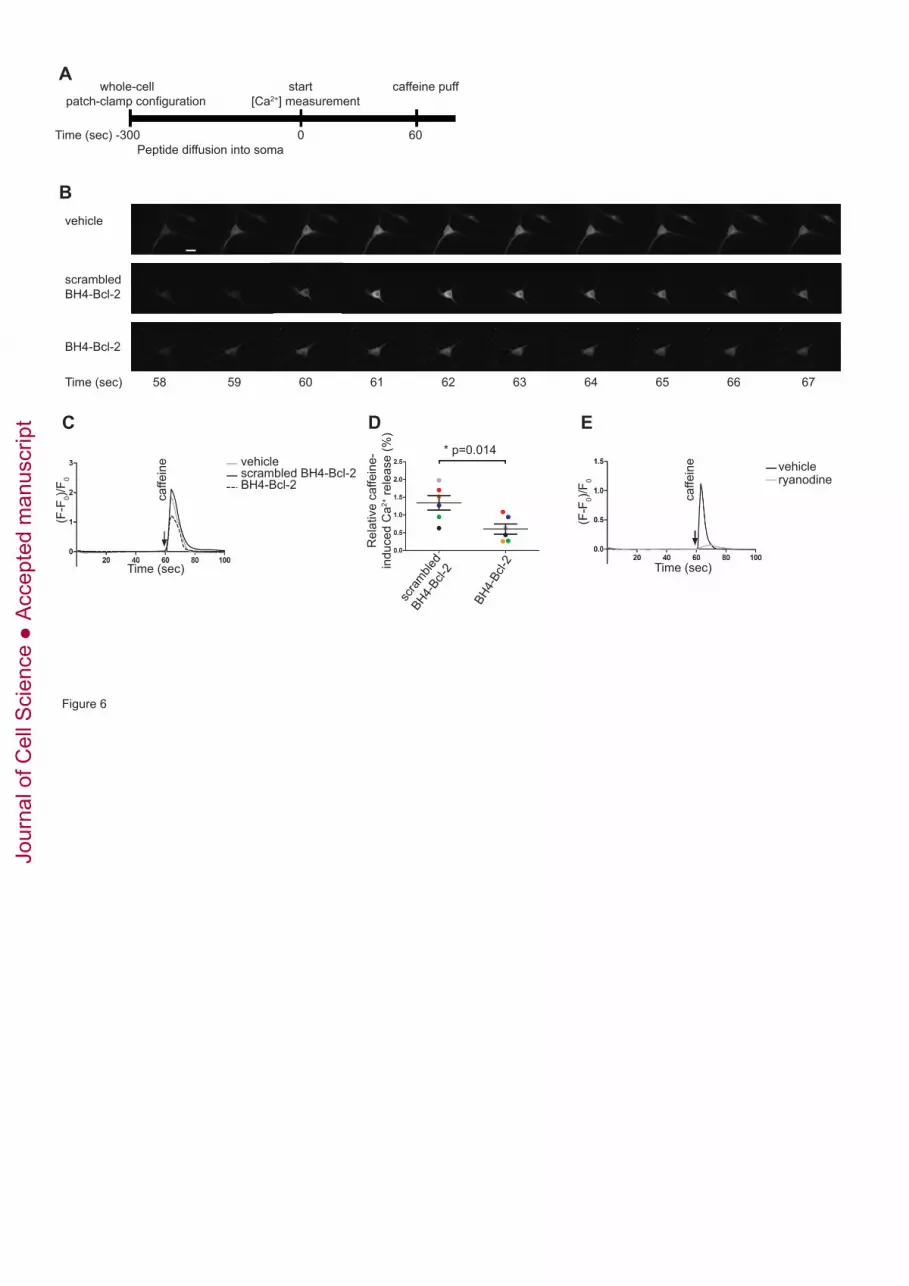
whole-cell start caffeine puff patch-clamp configuration [Ca2+] measurement
Time (sec) -300 0 60 Peptide diffusion into soma
vehicle
scrambled BH4-Bcl-2
BH4-Bcl-2
Time (sec) 58 59 60 61 62 63 64 65 66 67
(F-F
0)/F 0
Time (sec)
vehiclescrambled BH4-Bcl-2BH4-Bcl-2
A
B
C
Rel
ativ
e ca
ffein
e-in
duce
d C
a2+ re
leas
e (%
)
* p=0.014
scra
mbled
BH4-Bcl-
2
BH4-
Bcl-2
D
Figure 6
(F-F
0)/F 0
vehicleryanodine
E
Time (sec)
caffe
ine
caffe
ine
Jour
nal o
f Cel
l Sci
ence
Acc
epte
d m
anus
crip
t

Table 1: Affinity of biotin-BH4-Bcl-2 and biotin-BH4-Bcl-2K17D
for the used GST-tagged IP3R1 and RyR domains
Approximated EC50 values obtained from fitting using the
Hill equation of the data presented in figure 3.
Approximate EC50 values (µM)
BH4 BH4K17D
RyR1 domain 1.53 1.61
RyR2 domain 0.38 0.80
RyR3 domain 0.37 0.78
IP3R1 domain 3 0.38 1.87
Jour
nal o
f Cel
l Sci
ence
Acc
epte
d m
anus
crip
t


