Embed Size (px)
Citation preview
Beneficial Microorganisms in Agriculture, Food and the Environment: Safety Assessment and Regulation

This page intentionally left blank
Beneficial Microorganisms in Agriculture, Food and the Environment: Safety
Assessment and Regulation
Edited by
Ingvar Sundh
Swedish University of Agricultural Sciences, Uppsala BioCenter, Department of Microbiology, Uppsala, Sweden
Andrea Wilcks
CBRN-institute, Danish Emergency Management Agency, Birkerød, Denmark
and
Mark S. Goettel
Formerly of Agriculture and Agri-Food Canada, Lethbridge Research Centre, Lethbridge, Alberta, Canada
CABI is a trading name of CAB International
CABI CABINosworthy Way 38 Chauncey St.Wallingford Suite 1002Oxfordshire OX10 8DE Boston, MA 02111UK USA
Tel: +44 (0)1491 832111 Tel: +1 800 552 3083 (toll free)Fax: +44 (0)1491 833508 Tel: +1 (0)617 395 4051E-mail: [email protected] E-mail: [email protected]: www.cabi.org
© CAB International 2012. All rights reserved. No part of this publication may be reproduced in any form or by any means, electronically, mechanically, by photocopying, recording or otherwise, without the prior permission of the copyright owners.
A catalogue record for this book is available from the British Library, London, UK.
Library of Congress Cataloging-in-Publication Data
Beneficial microorganisms in agriculture, food and the environment : safetyassessment and regulation/edited by Ingvar Sundh, Andrea Wilcks, Mark S. Goettel. p. ; cm. Includes bibliographical references and index. ISBN 978-1-84593-810-9 (alk. paper) I. Sundh, Ingvar. II. Wilcks, Andrea. III. Goettel, Mark S. (Mark Stanislaw), 1954-[DNLM: 1. Environmental Microbiology. 2. Food Safety--methods. 3. Models, Immunological. QW 55]
363.19‘26--dc23 2012016959
ISBN-13: 978 1 84593 810 9
Commissioning editor: Claire ParfittEditorial assistant: Alexandra LainsburyProduction editor: Shankari Wilford
Typeset by SPi, Pondicherry, India.Printed and bound in the UK by CPI Group (UK) Ltd, Croydon, CR0 4YY.
Contents
Contributors ix
Preface xi
1 Microbes and the Law – Safety Assessment and Regulation of Beneficial Microorganisms 1Ingvar Sundh, Andrea Wilcks and Mark S. Goettel
PART I: FOOD AND FEED
2 Safety and Regulation of Microorganisms Added to the Food and Feed Chains, Including Probiotics – Introduction and Overview 12Stephen Wessels
3 Microbes for Human and Animal Consumption 27Atte von Wright
4 Antibiotic Resistance in Relation to Starter Cultures and Probiotics 41Andrea Wilcks and Angela H.A.M. van Hoek
5 Biopreservation of Food and Feed by Postharvest Biocontrol with Microorganisms 57Michael Wisniewski and Samir Droby
PART II: PEST CONTROL AGENTS AND PLANT GROWTH PROMOTERS
6 Safety and Regulation of Microbial Pest Control Agents and Microbial Plant Growth Promoters – Introduction and Overview 67Rüdiger Hauschild
7 Microbial Control of Invertebrate Pests 72Stefan T. Jaronski
8 Microbial Control of Plant Diseases 96Claude Alabouvette, Ulf Heilig and Christelle Cordier
v
9 Safety and Regulation of Microbial Control of Weeds 112Harry C. Evans and Marion K. Seier
10 Plant Growth Promotion with Microorganisms 138John G. Howieson and Sharon L. Fox
PART III: OTHER INDUSTRIAL APPLICATIONS
11 Regulation of Microorganisms Used for Bioremediation, Biorefinery and Other Bioindustrial Applications in the USA and Canada 153Jim Louter, John J. Sedivy and Mark Segal
PART IV: EVALUATING SAFETY
12 Determining the Safety of Microorganisms – Introduction and Overview 167Hans E.N. Bergmans
13 Virulence Genes in Risk Assessment of Beneficial Microorganisms: What Do Genome Sequences Tell Us? 180Trudy M. Wassenaar and Thomas Alter
14 Occupational Safety of Microbial Agents 197Anne Mette Madsen and Kira Tendal
PART V: MODEL TEST SYSTEMS
15 Model Systems for Testing Microbial Pathogenicity, Virulence and Toxicity – Introduction and Overview 217Andrea Wilcks, Mark S. Goettel and Ingvar Sundh
16 Nematode and Insect Models to Assay Microbial Infectivity, Virulence and Cytotoxicity 223C. Léopold Kurz and François Leulier
17 Assessing Potential Cytotoxicity of Biocontrol Microorganisms Using Invertebrate Assays 240Claudio Altomare, Barbara Pernfuss and Hermann Strasser
18 Assessing Genotoxic Effects of Microbial Products 256Milton A. Typas and Vassili N. Kouvelis
19 Assessing the Sensitization and Irritant Properties of Microorganisms 275Gregorio Loprieno
PART VI: INTERNATIONAL HARMONIZATION AND RISK PERCEPTION
20 International Conventions and Agreements – Consequences for International Trade and Utilization of Biological Matter, Including Microorganisms 293Carl-Gustaf Thornström
vi Contents
21 OECD Guidelines and Harmonization for Microbial Control Agents 308Jeroen J. Meeussen
22 Understanding Public Risk Perception for the Use of Beneficial Microorganisms 322Keith D. Warner
Index 335
Contents vii

This page intentionally left blank
Contributors
Claude Alabouvette, AGRENE, 47 rue Constant Pierrot, F-21000 Dijon, France. E-mail: [email protected]
Thomas Alter, Institute of Food Hygiene, Free University Berlin, Koenigsweg 69, D-14163 Berlin, Germany. E-mail: [email protected]
Claudio Altomare, Institute of Sciences of Food Production, National Research Council (CNR), Via G. Amendola 122, I-70126 Bari, Italy. E-mail: [email protected]
Hans E.N. Bergmans, Substances Expertise Center, National Institute for Public Health and the Environment (RIVM), GMO Office, Bilthoven, The Netherlands. E-mail: [email protected]
Christelle Cordier, AGRENE, 47 rue Constant Pierrot, F-21000 Dijon, France. E-mail: [email protected]
Samir Droby, Department of Postharvest Science, Volcani Center, Agricultural Research Organization (ARO), PO Box 6, Bet Dagan, 50250, Israel. E-mail: [email protected]
Harry C. Evans, CAB International, Europe–UK, Egham, Surrey TW20 9TY, UK. E-mail: [email protected]
Sharon L. Fox, Crops and Plants Research Institute (CaPRI), Division of Science, Murdoch University, South St, Murdoch, WA 6150, Australia. E-mail: [email protected]
Mark S. Goettel, formerly of Lethbridge Research Centre, Agriculture and Agri-Food Canada, 5403-1 Avenue South, PO Box 3000, Lethbridge, Alberta, Canada. E-mail: bstedit@ telusplanet.net
Rüdiger Hauschild, GAB Consulting GmbH, Hinter den Höfen 24, D-21769 Lamstedt, Germany. E-mail: [email protected]
Ulf Heilig, Consultant, 6 rue de Seine, F-78230 Le Pecq, France. E-mail: [email protected] G. Howieson, Crops and Plants Research Institute (CaPRI), Division of Science, Murdoch
University, South St, Murdoch, WA 6150, Australia. E-mail: [email protected] T. Jaronski, Northern Plains Research Laboratory (NPARL), United States Department
of Agriculture – Agricultural Research Service (USDA-ARS), 1500 Central Ave., Sidney, MT 59270, USA. E-mail: [email protected]
Vassili N. Kouvelis, Department of Genetics and Biotechnology, Faculty of Biology, University of Athens, Panepistimiopolis, Athens 15701, Greece. E-mail: [email protected]
C. Léopold Kurz, Centre d’Immunologie de Marseille-Luminy, Université de la Méditerranée, Case 906, 13288 Marseille cedex 9, France. E-mail: [email protected]
ix
François Leulier, Institut de Biologie du Développement de Marseille-Luminy, Université de la Méditerranée, Case 907, 13288 Marseille cedex 9, France. E-mail: [email protected]
Gregorio Loprieno, Dipartimento di Prevenzione ASL 2 Lucca, Piazza Aldo Moro, 55012 Capannori (LU), Italy. E-mail: [email protected]
Jim Louter, Biotechnology Section, Science and Technology Branch, Environment Canada, 200 Sacré Coeur blvd., Gatineau, Quebec K1A 0H3, Canada. E-mail: [email protected]
Anne Mette Madsen, The National Research Centre for the Working Environment, Lersø Parkallé 105, 2100 Copenhagen Ø, Denmark. E-mail: [email protected]
Jeroen J. Meeussen, Unit E3 – Chemicals, Contaminants, Pesticides, DG SANCO, European Commission, Rue Froissart 101, 4/90, 1049 Brussels, Belgium. E-mail: [email protected]
Barbara Pernfuss, Institute of Microbiology, Leopold-Franzens University of Innsbruck, Technikerstrasse 25, A-6020 Innsbruck, Austria. E-mail: [email protected]
John J. Sedivy, Novozymes Biologicals Inc., 5400 Corporate Circle, Salem, VA 24153, USA. E-mail: [email protected]
Mark Segal, United States Environmental Protection Agency (US EPA), 1201 Constitution Ave, Washington, DC 20460, USA. E-mail: [email protected]
Marion K. Seier, CAB International, Europe–UK, Egham, Surrey, TW20 9TY, UK. E-mail: [email protected]
Hermann Strasser, Institute of Microbiology, Leopold-Franzens University of Innsbruck, Technikerstrasse 25, A-6020 Innsbruck, Austria. E-mail: [email protected]
Ingvar Sundh, Department of Microbiology, Uppsala BioCenter, Swedish University of Agricultural Sciences, PO Box 7025, SE-750 07 Uppsala, Sweden. E-mail: [email protected]
Kira Tendal, The National Research Centre for the Working Environment, Lersø Parkallé 105, 2100 Copenhagen Ø, Denmark. E-mail: [email protected]
Carl-Gustaf Thornström, Uppsala BioCenter, Department of Plant Biology and Forest Genetics, Swedish University of Agricultural Sciences, PO Box 7080, SE-750 07 Uppsala, Sweden. E-mail: [email protected]
Milton A. Typas, Department of Genetics and Biotechnology, Faculty of Biology, University of Athens, Panepistimiopolis, Athens 15701, Greece. E-mail: [email protected]
Angela H.A.M. van Hoek, Laboratory for Zoonoses and Environmental Microbiology (LZO), National Institute for Public Health and the Environment (RIVM) – Division CIb, PO Box 1, NL-3720 BA Bilthoven, The Netherlands. E-mail: [email protected]
Atte von Wright, University of Eastern Finland, Institute of Public Health and Clinical Nutrition, PO Box 1627, FIN-70211 Kuopio, Finland. E-mail: [email protected]
Keith D. Warner, Religious Studies Department, and the Center for Science, Technology, and Society, Santa Clara University, 500 El Camino Real, Santa Clara, CA 95053, USA. E-mail: [email protected]
Trudy M. Wassenaar, Molecular Microbiology and Genomics Consultants, Tannenstrasse 7, D-55576 Zotzenheim, Germany. E-mail: [email protected]
Stephen Wessels, Environment and Toxicology, DHI, Agern Allé 5, DK-2970 Hørsholm, Denmark. E-mail: [email protected]
Andrea Wilcks, formerly CBRN-institute, Danish Emergency Management Agency, Datavej 16, DK-3460 Birkerød, Denmark. Present address: Danish Working Environment Authority, PO Box 1228, DK-0900 Copenhagen C, Denmark. E-mail: [email protected]
Michael Wisniewski, Appalachian Fruit Research Station, United States Department of Agriculture – Agricultural Research Service (USDA-ARS), Kearneysville, WV 25430 USA. E-mail: [email protected]
x Contributors
Preface
Microorganisms have been vital in the evolution of life on earth and they play paramount roles in the environment as well as for humankind. The earliest examples of active human exploita-tion of their activities are microbes that were ‘domesticated’ thousands of years ago for use in bread, alcohol fermentations and dairy products. Alongside societal development and strong growth in our understanding of the nature and properties of microorganisms, and the ability to culture single microbial isolates, several additional fields of application have surfaced within the agricultural, environmental and biotechnological sectors. However, with the realization during the last two centuries that many serious diseases are due to microorganisms causing infections or producing toxic compounds, it has become obvious that large-scale culture, mar-keting and utilization of single isolates of living microorganisms necessitate a careful safety assessment. Thus, a microorganism cannot be considered truly ‘beneficial’ until a certain level of safety has been established. As a consequence, many types of applications with microorgan-isms are presently subject to various regulatory measures.
The introduction of regulatory systems can have consequences for the inclination of research entrepreneurs and industry to venture into new projects with beneficial microorgan-isms. Long and costly registration procedures that are not commensurate with actual risks can unnecessarily hamper the development of useful microbiological products. Conversely, lack of appropriate regulation could potentially result in serious negative consequences for public health or for the environment. Many different types of legislation are applicable to the market-ing and use of microbiological products. General acts of legislation – e.g. regarding consumer, occupational or environmental safety – cover the marketing and use of beneficial microorgan-isms, and state that the producer or manufacturer of a product is responsible for an adequate safety assessment. In other specific cases, the submission of a data dossier followed by authori-zation is a prerequisite for market introduction.
The idea for this book emerged as a sequel to the conference ‘Microbes and the Law – Setting the Limits for Practical Use’ (http://www.mistra.org/program/dom/home/pressroom/newsarchive/news/microbesandthelawsettingthelimitsforpracticaluse.5.6b38234911d6cedb125800039217.html; accessed 12 January 2011), which was arranged by the research pro-gramme DOM (Domestication of Microorganisms: http://www.mistra.org/dom; accessed 12 January 2012) and was held at SLU (Swedish University of Agricultural Sciences) in Uppsala, Sweden, on 5–9 October 2008. Experts from science and industry, as well as from authorities responsible for the regulatory oversight of microorganisms, made presentations on recent
xi
developments in legislation and policy making as well as on new strategies for safety assess-ments of individual microbial isolates. A specific goal of the meeting was to create a forum for exchange of ideas and expertise across different scientific disciplines and fields of applications with microorganisms, because, to the best of our knowledge, that had never been done before.
The book does not aim to present a detailed account of regulations and data requirements or authorization processes for the many possible ways of utilizing microbes. As the main impe-tus for regulating microorganisms is to reach an acceptable level of safety with respect to human health and environment, the main aim of the book is instead to critically examine the safety principles that have governed the development of regulations in representative coun-tries, and to what extent these correctly mirror actual hazards and risks. After an introductory chapter by the editors, the book starts with chapters treating regulatory systems for typical application areas in representative jurisdictions. These are followed by others discussing vari-ous approaches for assessing the safety of microbes and their utilization, as well as new meth-odologies to determine pathogenicity, virulence and toxicity. Special chapters examine how international initiatives and agreements as well as public ‘risk perception’ can influence the implementation of microbial solutions to environmental or agricultural problems. We hope and believe that taking this generic approach will make this book useful to anyone interested in these topics, in any part of the world. The book is aimed at researchers (in academia as well as industry), postgraduate university students, regulators in governmental authorities respon-sible for risk assessment and authorization of microorganisms, personnel responsible for safety in microbiological laboratories, and non-governmental organizations within the agricultural, food and biotechnological sectors.
This book could not have materialized without input from several different scientific and regulatory disciplines. Such a cross-disciplinary work is, by necessity, dependent on contribu-tions from experts within a broad range of topics. Our sincere thanks are extended to all the authors who have contributed to this publication, which is the first in-depth treatment of safety assessment and current regulations and policies for microorganisms across the major types of their application. It is our hope that this cross-application approach will lead to the employ-ment of more relevant, efficient and harmonized systems for safety assessment and regulation of microbial products, thereby further facilitating safe utilization of microorganisms to the good of humankind as well as the environment.
Ingvar SundhUppsala, Sweden
Andrea WilcksBirkerød, Denmark
Mark S. GoettelLethbridge, Canada
January 2012
xii Preface
©CAB International 2012. Beneficial Microorganisms in Agriculture, Food and the Environment: Safety Assessment and Regulation (eds I. Sundh et al.) 1
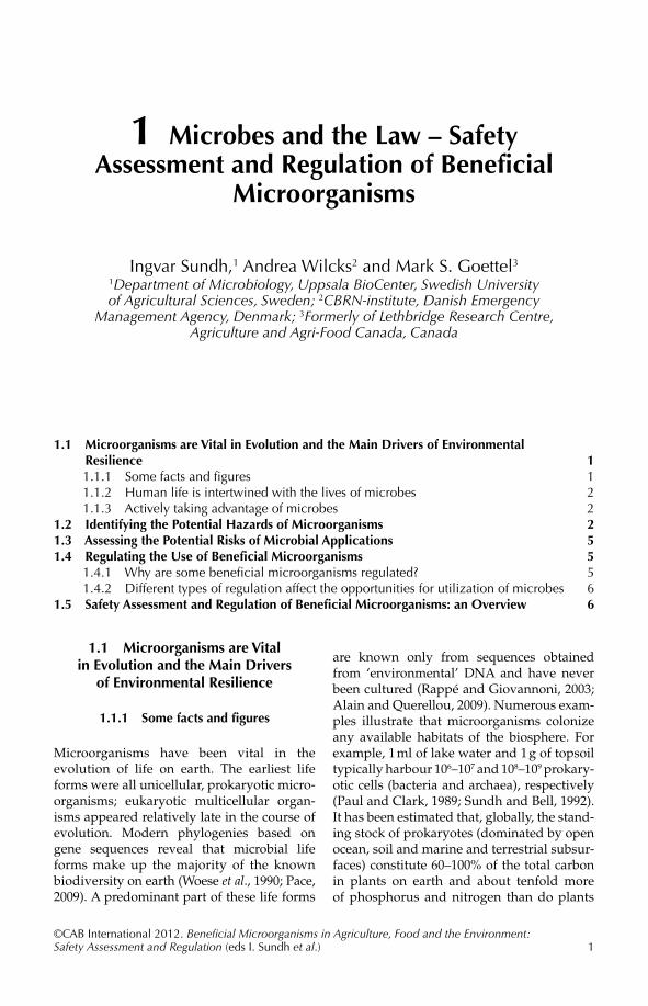
1.1 Microorganisms are Vital in Evolution and the Main Drivers of Environmental Resilience 11.1.1 Some facts and figures 1
1.1.2 Human life is intertwined with the lives of microbes 2 1.1.3 Actively taking advantage of microbes 2
1.2 Identifying the Potential Hazards of Microorganisms 21.3 Assessing the Potential Risks of Microbial Applications 51.4 Regulating the Use of Beneficial Microorganisms 5
1.4.1 Why are some beneficial microorganisms regulated? 5 1.4.2 Different types of regulation affect the opportunities for utilization of microbes 6
1.5 Safety Assessment and Regulation of Beneficial Microorganisms: an Overview 6
1 Microbes and the Law – Safety Assessment and Regulation of Beneficial
Microorganisms
Ingvar Sundh,1 Andrea Wilcks2 and Mark S. Goettel3
1Department of Microbiology, Uppsala BioCenter, Swedish University of Agricultural Sciences, Sweden; 2CBRN-institute, Danish Emergency
Management Agency, Denmark; 3Formerly of Lethbridge Research Centre, Agriculture and Agri-Food Canada, Canada
1.1 Microorganisms are Vital in Evolution and the Main Drivers
of Environmental Resilience
1.1.1 Some facts and figures
Microorganisms have been vital in the evolution of life on earth. The earliest life forms were all unicellular, prokaryotic micro-organisms; eukaryotic multicellular organ-isms appeared relatively late in the course of evolution. Modern phylogenies based on gene sequences reveal that microbial life forms make up the majority of the known biodiversity on earth (Woese et al., 1990; Pace, 2009). A predominant part of these life forms
are known only from sequences obtained from ‘environmental’ DNA and have never been cultured (Rappé and Giovannoni, 2003; Alain and Querellou, 2009). Numerous exam-ples illustrate that microorganisms colonize any available habitats of the biosphere. For example, 1 ml of lake water and 1 g of topsoil typically harbour 106–107 and 108–109 prokary-otic cells (bacteria and archaea), respectively (Paul and Clark, 1989; Sundh and Bell, 1992). It has been estimated that, globally, the stand-ing stock of prokaryotes (dominated by open ocean, soil and marine and terrestrial subsur-faces) constitute 60–100% of the total carbon in plants on earth and about tenfold more of phosphorus and nitrogen than do plants
2 I. Sundh et al.
(Whitman et al., 1998). Fungi and protists are not even included in these direct estimates of standing biomass and nutrient pools, but are generally considered to contribute less than prokaryotes in most ecosystems (Fierer et al.,2009). Aboveground plant compartments are substrates for epiphytic and endophytic microorganisms (Beattie and Lindow, 1999; Bayman, 2006; Whipps et al., 2008), and inver-tebrate animals harbour large numbers of microorganisms in their gastrointestinal tracts (König, 2006; Brinkman et al., 2008).
Collectively, microorganisms stand for an extremely wide metabolic versatility and are the main drivers of indispensable bio-geochemical processes and transformations (Falkowski et al., 2008). For instance, microbes are essential for the degradation of organic matter in soils and other ecosystems and, thereby, for nutrient recycling. Photosynthetic cyanobacteria and eukaryotic micro-algae pro vide the majority of the primary produc-tion of organic matter from CO2 in the upper water horizons of oceans and lakes, and in so doing have a major role in the global carbon budget (Chavez et al., 2011).
1.1.2 Human life is intertwined with the lives of microbes
Human life is intimately connected with the activities of microorganisms. Without any need for active intervention, we constantly live with microorganisms and by the services they pro-vide. Not only is humanity dependent on the way that microbes shape and maintain essen-tial functions in the environment, including agricultural production systems (i.e. they pro-vide what has been coined ‘ecosystem services’;Ducklow, 2008), but also on the direct contri-butions of micro bes within our bodies. This is because, in common with other vertebrate as well as invertebrate animals, our well-being depends on establishment of a balanced and functioning microbiota in the gastrointestinal tract (Eckburg et al., 2005). The intestinal tract of an adult human contains a factor of 10–100× more bacterial cells than the total number of body cells and 150× more microbial genes than there are in the total human genome (Zhu et al., 2010). Human skin and mucous membranes are
also inhabited by microorganisms, normally without any adverse effects on health (Cogen et al., 2008; Grice and Segre, 2011).
1.1.3 Actively taking advantage of microbes
That humanity actively takes advantage of specific microorganisms and the beneficial products of their metabolism is not new. The earliest documented examples come from thousands of years ago, when yeasts and lactic acid bacteria were used in the prepa-ration of bread, alcoholic beverages, and dairy products such as cheese (Caplice and Fitzgerald, 1999; Fox and McSweeney, 2004). By specific treatment and storage conditions of the raw materials, spontaneous develop-ment of particular microbial consortia could be stimulated, giving the food special, de -sired properties. In these early times, little was known about the nature of microorgan-isms and single strains could not be culti-vated in pure form and could thus not be added for specific purposes as such.
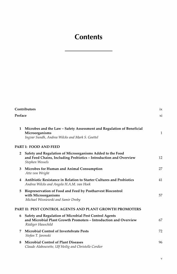
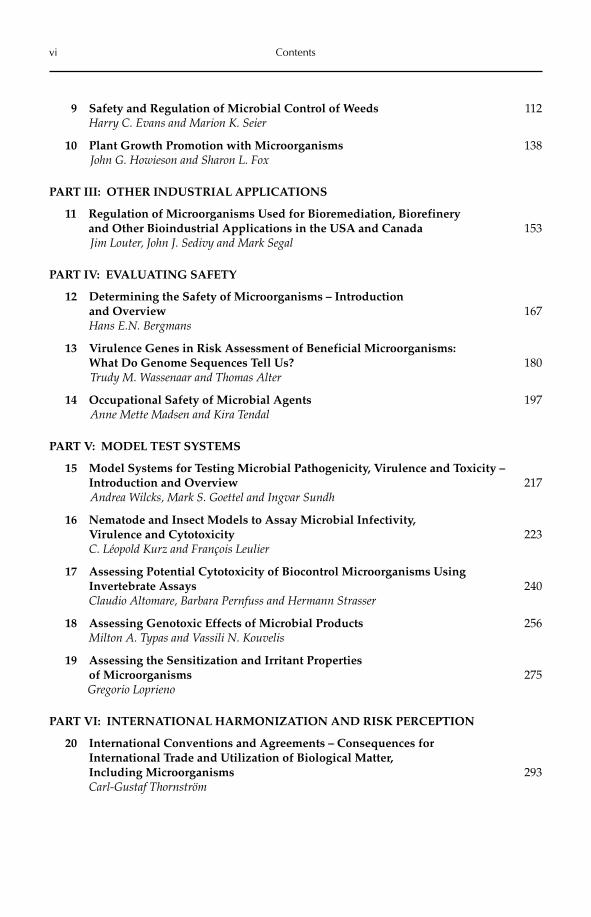
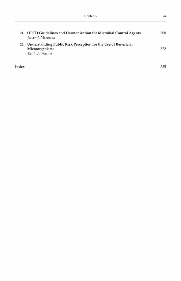
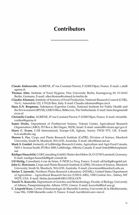
The invention of the microscope by Antonie van Leeuwenhoek by the end of the 17th century and the substantial progress in understanding of the biology of microorgan-isms during the 18th and particularly the 19th century made it possible to obtain, cultivate and study single isolates. Subsequent develop-ment of knowledge of the ecology, metabolic diversity and molecular biology of microorgan-isms during the 20th century paved the way for uncovering the great range of possibilities of utilizing microbes for specific purposes. By this time, it was possible not only to stimulate spontaneously occurring microbial communi-ties and consortia, but also to take single, well-characterized isolates into consideration. For instance, microorganisms can be used for many types of beneficial applications, as listed in Box 1.1 and exemplified in Figs. 1.1–1.4.
1.2 Identifying the Potential Hazards of Microorganisms
In spite of the indispensable roles of microor-ganisms in the maintenance of ecosystem functioning and human life, microbes can
Microbes and the Law 3
Box 1.1. Examples of various areas of beneficial applications with microorganisms, each with a few references for further reading.
1. Food and feed, including probiotics for humans and animals (example in Fig. 1.1)(Havenaar and Huis in’t Veld, 1992; Weinberg and Muck, 1996; Caplice and Fitzgerald, 1999; Fox and McSweeney, 2004).2. Antagonistic or pathogenic microbes for biological control of pests and diseases (examples in Figs 1.2–1.3)(Evans, 2000; Inglis et al. 2001; Haas and Defago, 2005).3. Addition of microorganisms as bioprophylactics to minimize the emission of chemical pollutants or for bioremediation of soils or sediments(Bouwer and Zehnder, 1993; Alexander, 1999; de Lorenzo, 2008).4. Plant growth-promoting microbial agents for, e.g. strengthening plant stress resistance or nutri-ent uptake(Kloepper et al., 1980; Glick, 1995; Moulin et al., 2001; Preston, 2004).5. Production of biofuels by microbial degradation of various waste fractions(Ahring, 2003; Hahn-Hägerdal et al., 2006; Karakashev et al., 2007; Weber et al., 2010).6. Biotechnological use of microbes for production of specific metabolites, enzymes, etc. (example in Fig. 1.4)(Lee et al. 2004; Chou, 2007; Ruiz et al., 2010).7. Fermentation of microbes for obtaining bulk biomass for use as, e.g. animal feed or production of biofuel(Kiessling and Askbrandt, 1993; Alper and Stephanopoulos, 2009; Romarheim et al., 2011; Shi et al., 2011).8. Medical use of microorganisms, e.g. for treatment of serious intestinal disorders(Tvede and Rask-Madsen, 1989; Cain and Karpa, 2011; Landy et al., 2011).
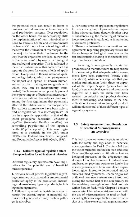
(a) (b)
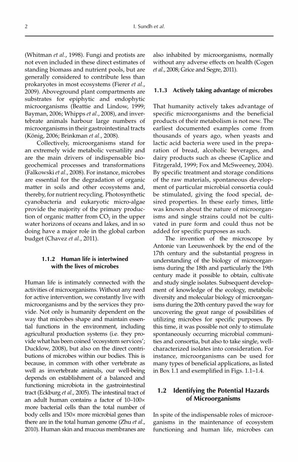
Fig. 1.1. The micrograph (a) shows sporangiophores and sporangiospores of the filamentous fungus Rhizopusoligosporus, which is used for preparation of the traditional Indonesian meal tempeh from fermented soybeans. To the right (b) is a petri dish containing barley tempeh made by the inoculation of barley with Rhizopus oligosporus. The fungus has completely covered the barley kernels. Photos: Xin-Mei Feng.
also have strong negative impacts. For instance, it has been estimated that 5–10% of the world’s food production is destroyed by the growth of spoilage fungi and/or bacteria (Pitt and Hocking, 1999; Gram et al., 2002), with substantially higher losses in less techni-cally developed countries. Besides causing a
general loss of food and feed commodities, a variety of spoilage microbes produce toxic compounds. Mycotoxin production by fungi (Richard, 2007) is highly significant in this respect, e.g. the carcinogenic aflatoxins and ochratoxins produced by some Aspergillusand Penicillium moulds. Other spoilage
4 I. Sundh et al.
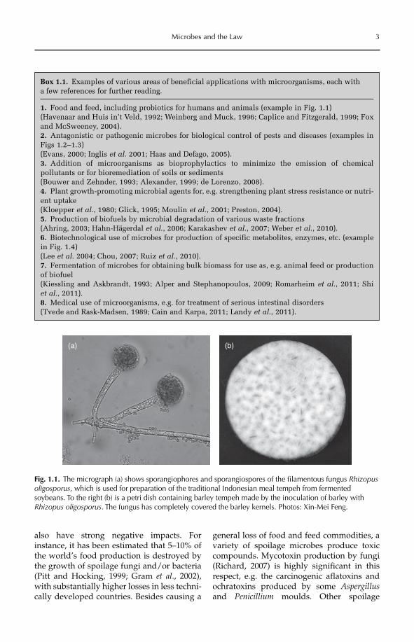
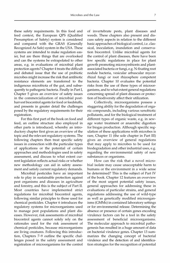
Fig. 1.2. Barley seed treated with the biological control product Cedomon® at a seed treatment facility in Sweden. Cedomon contains the bacterium Pseudomonas chlororaphis and controls soil-borne fungal diseases in the growing barley.
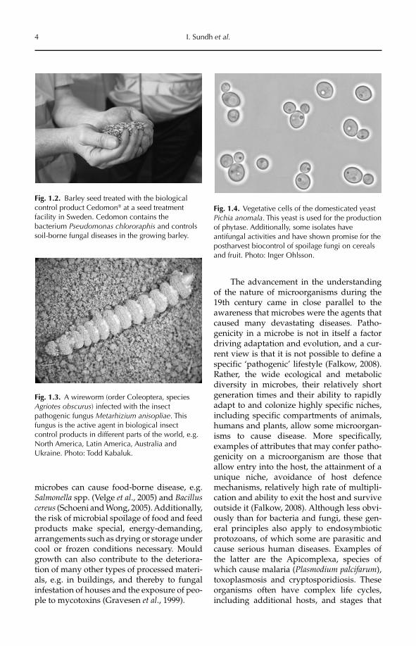
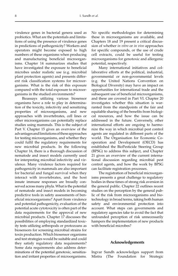
Fig. 1.3. A wireworm (order Coleoptera, species Agriotes obscurus) infected with the insect pathogenic fungus Metarhizium anisopliae. This fungus is the active agent in biological insect control products in different parts of the world, e.g. North America, Latin America, Australia and Ukraine. Photo: Todd Kabaluk.
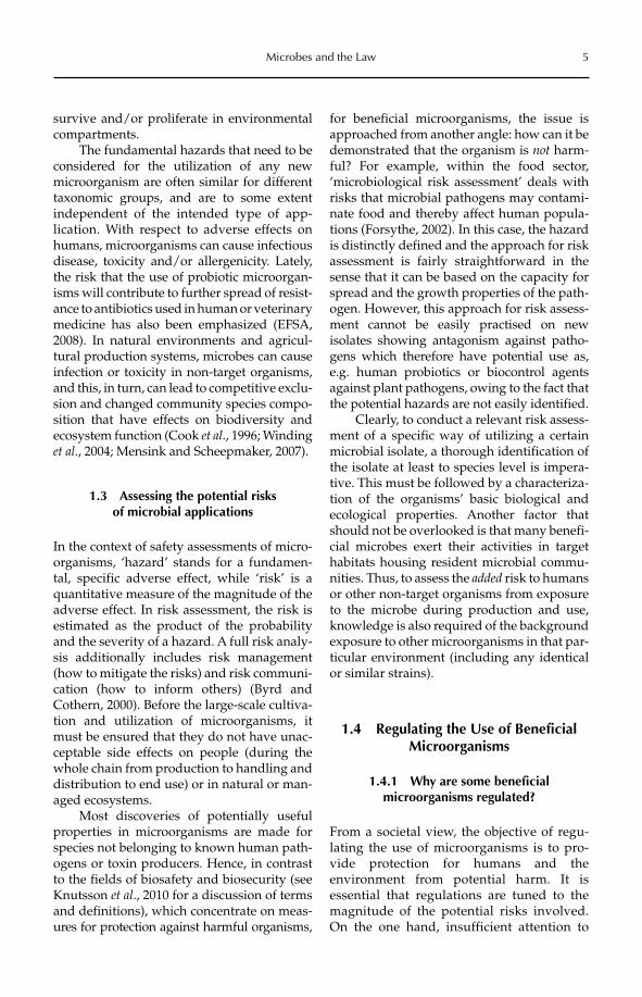
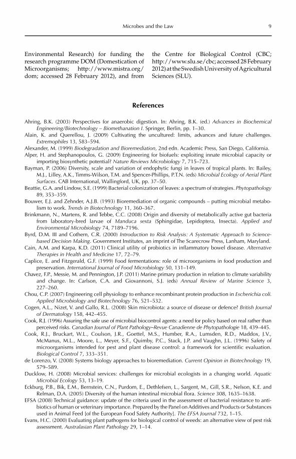
Fig. 1.4. Vegetative cells of the domesticated yeast Pichia anomala. This yeast is used for the production of phytase. Additionally, some isolates have antifungal activities and have shown promise for the postharvest biocontrol of spoilage fungi on cereals and fruit. Photo: Inger Ohlsson.
microbes can cause food-borne disease, e.g. Salmonella spp. (Velge et al., 2005) and Bacilluscereus (Schoeni and Wong, 2005). Additionally, the risk of microbial spoilage of food and feed products make special, energy-demanding, arrangements such as drying or storage under cool or frozen conditions necessary. Mould growth can also contribute to the deteriora-tion of many other types of processed materi-als, e.g. in buildings, and thereby to fungal infestation of houses and the exposure of peo-ple to mycotoxins (Gravesen et al., 1999).
The advancement in the understanding of the nature of microorganisms during the 19th century came in close parallel to the awareness that microbes were the agents that caused many devastating diseases. Patho-genicity in a microbe is not in itself a factor driving adaptation and evolution, and a cur-rent view is that it is not possible to define a specific ‘pathogenic’ lifestyle (Falkow, 2008). Rather, the wide ecological and metabolic diversity in microbes, their relatively short generation times and their ability to rapidly adapt to and colonize highly specific niches, including specific compartments of animals, humans and plants, allow some microorgan-isms to cause disease. More specifically, examples of attributes that may confer patho-genicity on a microorganism are those that allow entry into the host, the attainment of a unique niche, avoidance of host defence mechanisms, relatively high rate of multipli-cation and ability to exit the host and survive outside it (Falkow, 2008). Although less obvi-ously than for bacteria and fungi, these gen-eral principles also apply to endosymbiotic protozoans, of which some are parasitic and cause serious human diseases. Examples of the latter are the Apicomplexa, species of which cause malaria (Plasmodium palcifarum),toxoplasmosis and cryptosporidiosis. These organisms often have complex life cycles, including additional hosts, and stages that
Microbes and the Law 5
survive and/or proliferate in environmental compartments.
The fundamental hazards that need to be considered for the utilization of any new microorganism are often similar for different taxonomic groups, and are to some extent independent of the intended type of app-lication. With respect to adverse effects on humans, microorganisms can cause infectious disease, toxicity and/or allergenicity. Lately, the risk that the use of probiotic microorgan-isms will contribute to further spread of resist-ance to antibiotics used in human or veterinary medicine has also been emphasized (EFSA, 2008). In natural environments and agricul-tural production systems, microbes can cause infection or toxicity in non-target organisms, and this, in turn, can lead to competitive exclu-sion and changed community species compo-sition that have effects on biodiversity and ecosystem function (Cook et al., 1996; Winding et al., 2004; Mensink and Scheepmaker, 2007).
1.3 Assessing the potential risks of microbial applications
In the context of safety assessments of micro-organisms, ‘hazard’ stands for a fundamen-tal, specific adverse effect, while ‘risk’ is a quantitative measure of the magnitude of the adverse effect. In risk assessment, the risk is estimated as the product of the probability and the severity of a hazard. A full risk analy-sis additionally includes risk management (how to mitigate the risks) and risk communi-cation (how to inform others) (Byrd and Cothern, 2000). Before the large-scale cultiva-tion and utilization of microorganisms, it must be ensured that they do not have unac-ceptable side effects on people (during the whole chain from production to handling and distribution to end use) or in natural or man-aged ecosystems.
Most discoveries of potentially useful properties in microorganisms are made for species not belonging to known human path-ogens or toxin producers. Hence, in contrast to the fields of biosafety and biosecurity (see Knutsson et al., 2010 for a discussion of terms and definitions), which concentrate on meas-ures for protection against harmful organisms,
for beneficial microorganisms, the issue is approached from another angle: how can it be demonstrated that the organism is not harm-ful? For example, within the food sector, ‘microbiological risk assessment’ deals with risks that microbial pathogens may contami-nate food and thereby affect human popula-tions (Forsythe, 2002). In this case, the hazard is distinctly defined and the approach for risk assessment is fairly straightforward in the sense that it can be based on the capacity for spread and the growth properties of the path-ogen. However, this approach for risk assess-ment cannot be easily practised on new isolates showing antagonism against patho-gens which therefore have potential use as, e.g. human probiotics or biocontrol agents against plant pathogens, owing to the fact that the potential hazards are not easily identified.
Clearly, to conduct a relevant risk assess-ment of a specific way of utilizing a certain microbial isolate, a thorough identification of the isolate at least to species level is impera-tive. This must be followed by a characteriza-tion of the organisms’ basic biological and ecological properties. Another factor that should not be overlooked is that many benefi-cial microbes exert their activities in target habitats housing resident microbial commu-nities. Thus, to assess the added risk to humans or other non-target organisms from exposure to the microbe during production and use, knowledge is also required of the background exposure to other microorganisms in that par-ticular environment (including any identical or similar strains).
1.4 Regulating the Use of Beneficial Microorganisms
1.4.1 Why are some beneficial microorganisms regulated?
From a societal view, the objective of regu-lating the use of microorganisms is to pro-vide protection for humans and the environment from potential harm. It is essential that regulations are tuned to the magnitude of the potential risks involved. On the one hand, insufficient attention to
6 I. Sundh et al.
the potential risks can result in harm to humans, natural environments and agricul-tural production systems. Over-regulation, on the other hand, can unnecessarily stifle the implementation of new, microbial solu-tions to various health and environmental problems. Of the various acts of legislation that cover the utilization of microorganisms, the majority have their fundament in the way that the organisms are used, rather than in the organisms’ phylogeny or biological and ecological properties. This is reflected in the structural outline of this book, which has separate chapters for various fields of appli-cation. Exceptions to this are national ‘quar-antine’ legislations, which attempt to prevent the import and spread of known human, animal or plant pathogens (or goods with which they can be inadvertently trans-ported). Such measures can possibly prevent the export/import of beneficial microorgan-isms across national boundaries, and were among the first regulations that potentially affected the utilization of microorganisms. The earliest example we have been able to find of registration of a microorganism for use in a specific application is that of the insect pathogenic bacterium Paenibacillus popilliae (formerly Bacillus popilliae) for controlling populations of the Japanese beetle (Popillia japonica). This was regis-tered as a pesticide in the USA under FIFRA (the Federal Insecticide, Fungicide and Rodenticide Act) in 1948 (Cook, 1996).
1.4.2 Different types of regulation affect the opportunities for utilization of microbes
Different regulatory systems can have impli-cations for the potential use of beneficial microorganisms:
1. Various acts of general legislation regard-ing consumer, occupational or environmental protection apply to the production, market-ing and use of many types of products, includ-ing microorganisms.2. Different quarantine legislations aim to restrict the export/import of microbial cul-tures or of goods which may contain patho-genic microbes.
3. For some areas of application, regulations for a specific group of products encompass living microorganisms along with other types of substances, e.g. the marketing of microbial biocontrol agents is regulated, as is marketing of chemical pesticides.4. There are international conventions and agreements regarding proprietary issues and the exchange of biological resources or the fair and equitable sharing of the benefits aris-ing from their exploitation.
Some regulations generally dictate that producers and manufacturers are responsible for determining that adequate safety assess-ments have been performed (mostly point one above), while others stipulate that pre-market authorization (point three) or appro-val of the import/export (points two and four) of new microbial agents and products is required. As a rule, the chain from basic research on microbe and product develop-ment to marketing, and thence to the final utilization of a new microbiological product, will involve several of these different types of regulatory systems.
1.5 Safety Assessment and Regulation of Beneficial Microorganisms:
an Overview
This book covers numerous aspects associated with the safety and regulation of beneficial microorganisms. In Part I, Chapters 2–5 treat the use of microbial cultures in food and feed. Over time, the approach to utilization of micro-biological processes in the preparation and storage of food has been one of trial and error, and based on previous experience. Lately, live microorganisms have been formulated as pro-biotics, given in feed to domesticated animals and consumed by humans. Chapter 2 gives an outline of how new systems were introduced during the second half of the 20th century for the regulatory oversight of microbial products within food or feed, while Chapter 3 contains an analysis of the potential risks connected with the use of microorganisms in food or feed – including their use as probiotics – and a discus-sion of to what extent current regulations meet
Microbes and the Law 7
these safety requirements. In this food and feed context, the European QPS (Qualified Presumption of Safety) system is considered and compared with the GRAS (Generally Recognized As Safe) system in the USA. These systems are intended to make regulation eas-ier, but are there things that are overlooked and can the systems be extrapolated to other areas, e.g. in evaluations of microbial plant protection agents? Chapter 4 treats the difficult and debated issue that the use of probiotic microbes might increase the risk that antibiotic resistance elements are transferred to the indigenous microbiota of the gut, and subse-quently to pathogenic bacteria. Finally in Part I, Chapter 5 gives an overview of safety issues in the commercialization of microbial post-harvest biocontrol agents for food or feedstuffs, and presents in greater detail the challenges posed by the regulatory requirements for their registration.
For this first part of the book on food and feed, an outline structure also employed in other parts is introduced, whereby an intro-ductory chapter first gives an overview of the topic and the relevant regulatory systems. The following chapters then treat specific safety issues in connection with the particular types of applications or the potential of certain approaches and methodologies used in safety assessment, and discuss to what extent cur-rent legislation reflects actual risks or whether new methodology can aid in safety assess-ment and satisfy current regulatory demands.
Microbial pesticides have an important role to play in sustainable protection against pest organisms and diseases in agriculture and forestry, and this is the subject of Part II. Most countries have implemented strict regulations for microbial biocontrol agents, following similar principles to those used for chemical pesticides. Chapter 6 introduces the regulatory systems for microorganisms used to manage pest populations and plant dis-eases. However, risk assessments of microbial biocontrol agents cannot solely rely on the rationales used for the risk assessment of chemical pesticides, because microorganisms are living creatures. Following this introduc-tion, Chapters 7–9 outline the specific chal-lenges posed in the safety assessment and registration of microorganisms for the control
of invertebrate pests, plant diseases and weeds. These chapters also present and dis-cuss safety aspects in relation to the different basic approaches of biological control, i.e. clas-sical, inoculation, inundation and conserva-tion biocontrol. Unlike microbial agents for the control of plant diseases, there have been few specific regulations in place for plant growth promoting microsymbionts and plant- associated bacteria or fungi, e.g. N-fixating root nodule bacteria, vesicular arbuscular mycor-rhizal fungi or root rhizosphere competent bacteria. Chapter 10 evaluates the potential risks from the use of these types of microor-ganisms, and to what extent general regulations concerning spread of plant diseases or protec-tion of biodiversity affect their utilization.
Collectively, microorganisms possess a staggering ability for the degradation of orga-nic compounds, including various xenobiotic pollutants, and for the biological treatment of different types of organic waste, e.g. in sew-age water treatment or anaerobic digestion for biogas production. Although specific reg-ulation of these applications with microbes is rare, Chapter 11 (the sole chapter in Part III) gives an overview of general regulations that may apply to microbes to be used for biodegradation and other industrial uses, e.g. regarding the environmental safety of new substances or organisms.
How can the risk that a novel micro-bial isolate may cause unacceptable harm to humans or the environment in a wide sense be determined? This is the subject of Part IV of the book. Chapter 12 features an overview of the most urgent potential safety issues, general approaches for addressing these in evaluations of particular strains, and general regulations addressing the use of wild-type as well as genetically modified microorgan-isms (GMMs) in contained laboratory settings or for environmental release. Determining the absence or presence of certain genes encod ingvirulence factors can be a tool in the safety assessment of beneficial microorganisms. The molecular approach to microbial patho-genesis has resulted in a huge amount of data on bacterial virulence genes. Chapter 13 sum-marizes the changing concept of bacterial virulence and the detection of and identifica-tion strategies for the recognition of potential
8 I. Sundh et al.
virulence genes in bacterial genera used as probiotics. What are the potentials and limita-tions of using the presence of virulence genes in predictions of pathogenicity? Workers and operators might become exposed to high numbers of these organisms when producing and manufacturing beneficial microorgan-isms. Chapter 14 summarizes studies that have investigated the exposure to beneficial microbes under realistic use (e.g. microbial plant protection agents) and presents differ-ent risk classification systems for microor-ganisms. What is the risk of this exposure compared with the total exposure to microor-ganisms in the studied environments?
Bioassays utilizing various biosensor organisms have a role to play in determina-tion of the toxicity, infectivity and sensitizing properties of microorganisms, and new approaches with invertebrates, cell lines or other microorganisms can potentially replace studies using mammals. This is the subject of Part V. Chapter 15 gives an overview of the advantages and limitations of these approaches for testing microorganisms, and whether they could fulfil the regulatory requirements for new microbial products. In the following Chapter 16, there is a thorough discussion of nematode and insect models currently used for interpreting microbial infectivity and vir-ulence. Many virulence factors required for pathogenicity in mammals are also important for bacterial and fungal survival when they interact with invertebrates, and the host innate immune responses are broadly con-served across many phyla. What is the potential of nematode and insect models in becoming predictive tools in safety assessments of ben-eficial microorganisms? Apart from virulence and potential pathogenicity, evaluation of the potential acute cytotoxicity is often part of the data requirements for the approval of new microbial products. Chapter 17 discusses the possibilities of employing standardized toxic-ity tests utilizing arthropods or protozoans as biosensors for screening microbial strains for toxin production. Which biosensor organisms and test strategies would be suitable and could they satisfy regulatory data requirements? Some data requirements also address deter-minations of the potential genotoxic, sensitiza-tion and irritant properties of microorganisms.
No specific methodologies for determining these in microorganisms are available, and Chapters 18 and 19 present a critical discus-sion of whether in vitro or in vivo approaches for specific compounds, or the use of crude cell extracts, could be useful for testing microorganisms for genotoxic and allergenic potential, respectively.
Many international initiatives and col-laborative efforts at the political, industrial, governmental or non-governmental levels (e.g. the United Nations Convention on Biological Diversity) may have an impact on opportunities for international trade and the subsequent use of beneficial microorganisms, and these are covered in Part VI. Chapter 20 investigates whether this situation is war-ranted from the standpoints of the fair and equitable sharing of the benefits from biologi-cal resources, and how the issue can be addressed in the future. Conversely, other international efforts are ongoing to harmo-nize the way in which microbial pest control agents are regulated in different parts of the world. The Organisation for Economic Co-operation and Development (OECD) has established the BioPesticide Steering Group (BPSG) to address this subject, and Chapter 21 gives an overview of the current interna-tional discussion regarding microbial pest control agents, and how the work by BPSG can facilitate registration processes.
The registration of beneficial microorgan-isms presents a great challenge to regulatory bodies in these times of strong risk aversion in the general public. Chapter 22 outlines recent studies on the perception by the general pub-lic of the risk from microorganisms and bio-technology in broad terms, taking both human safety and environmental protection into account. What steps can governments and regulatory agencies take to avoid the fact that unfounded perception of risk unnecessarily hampers the implementation of new products with beneficial microbes?
Acknowledgements
Ingvar Sundh acknowledges support from Mistra (The Foundation for Strategic
Microbes and the Law 9
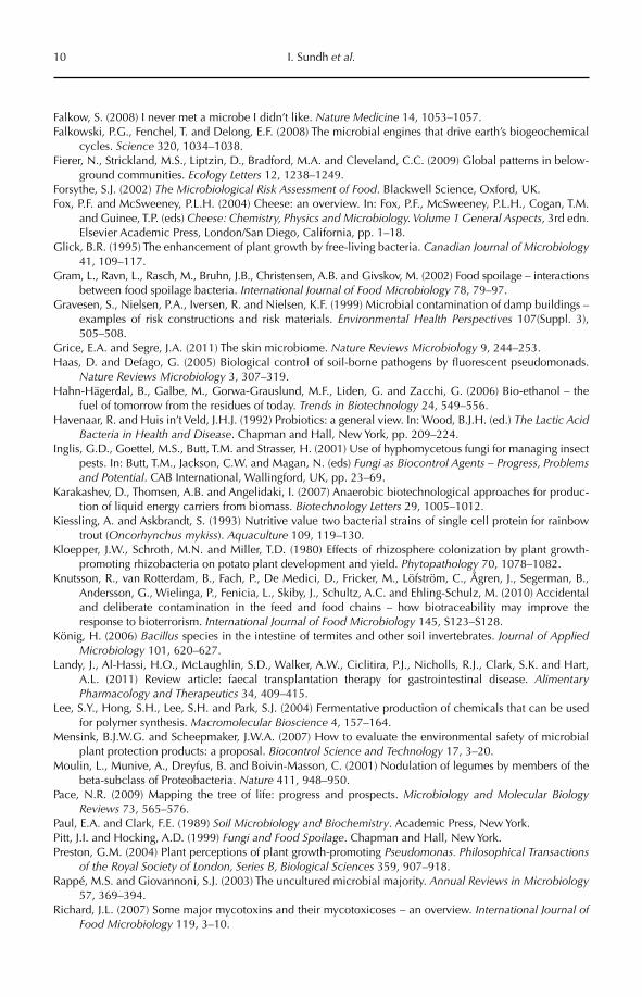
References
Ahring, B.K. (2003) Perspectives for anaerobic digestion. In: Ahring, B.K. (ed.) Advances in Biochemical Engineering/Biotechnology – Biomethanation I. Springer, Berlin, pp. 1–30.
Alain, K. and Querellou, J. (2009) Cultivating the uncultured: limits, advances and future challenges. Extremophiles 13, 583–594.
Alexander, M. (1999) Biodegradation and Bioremediation, 2nd edn. Academic Press, San Diego, California.Alper, H. and Stephanopoulos, G. (2009) Engineering for biofuels: exploiting innate microbial capacity or
importing biosynthetic potential? Nature Reviews Microbiology 7, 715–723.Bayman, P. (2006) Diversity, scale and variation of endophytic fungi in leaves of tropical plants. In: Bailey,
M.J., Lilley, A.K., Timms-Wilson, T.M. and Spencer-Phillips, P.T.N. (eds) Microbial Ecology of Aerial Plant Surfaces. CAB International, Wallingford, UK, pp. 37–50.
Beattie, G.A. and Lindow, S.E. (1999) Bacterial colonization of leaves: a spectrum of strategies. Phytopathology89, 353–359.
Bouwer, E.J. and Zehnder, A.J.B. (1993) Bioremediation of organic compounds – putting microbial metabo-lism to work. Trends in Biotechnology 11, 360–367.
Brinkmann, N., Martens, R. and Tebbe, C.C. (2008) Origin and diversity of metabolically active gut bacteria from laboratory-bred larvae of Manduca sexta (Sphingidae, Lepidoptera, Insecta). Applied and Environmental Microbiology 74, 7189–7196.
Byrd, D.M. III and Cothern, C.R. (2000) Introduction to Risk Analysis: A Systematic Approach to Science-based Decision Making. Government Institutes, an imprint of The Scarecrow Press, Lanham, Maryland.
Cain, A.M. and Karpa, K.D. (2011) Clinical utility of probiotics in inflammatory bowel disease. Alternative Therapies in Health and Medicine 17, 72–79.
Caplice, E. and Fitzgerald, G.F. (1999) Food fermentations: role of microorganisms in food production and preservation. International Journal of Food Microbiology 50, 131–149.
Chavez, F.P., Messie, M. and Pennington, J.P. (2011) Marine primary production in relation to climate variability and change. In: Carlson, C.A. and Giovannoni, S.J. (eds) Annual Review of Marine Science 3, 227–260.
Chou, C.P. (2007) Engineering cell physiology to enhance recombinant protein production in Escherichia coli. Applied Microbiology and Biotechnology 76, 521–532.
Cogen, A.L., Nizet, V. and Gallo, R.L. (2008) Skin microbiota: a source of disease or defence? British Journal of Dermatology 158, 442–455.
Cook, R.J. (1996) Assuring the safe use of microbial biocontrol agents: a need for policy based on real rather than perceived risks. Canadian Journal of Plant Pathology–Revue Canadienne de Phytopathologie 18, 439–445.
Cook, R.J., Bruckart, W.L., Coulson, J.R., Goettel, M.S., Humber, R.A., Lumsden, R.D., Maddox, J.V., McManus, M.L., Moore, L., Meyer, S.F., Quimby, P.C., Stack, J.P. and Vaughn, J.L. (1996) Safety of microorganisms intended for pest and plant disease control: a framework for scientific evaluation. Biological Control 7, 333–351.
de Lorenzo, V. (2008) Systems biology approaches to bioremediation. Current Opinion in Biotechnology 19, 579–589.
Ducklow, H. (2008) Microbial services: challenges for microbial ecologists in a changing world. Aquatic Microbial Ecology 53, 13–19.
Eckburg, P.B., Bik, E.M., Bernstein, C.N., Purdom, E., Dethlefsen, L., Sargent, M., Gill, S.R., Nelson, K.E. and Relman, D.A. (2005) Diversity of the human intestinal microbial flora. Science 308, 1635–1638.
EFSA (2008) Technical guidance: update of the criteria used in the assessment of bacterial resistance to anti-biotics of human or veterinary importance. Prepared by the Panel on Additives and Products or Substances used in Animal Feed [of the European Food Safety Authority]. The EFSA Journal 732, 1–15.
Evans, H.C. (2000) Evaluating plant pathogens for biological control of weeds: an alternative view of pest risk assessment. Australasian Plant Pathology 29, 1–14.
Environmental Research) for funding the research programme DOM (Domestication of Microorganisms; http://www.mistra.org/dom; accessed 28 February 2012), and from
the Centre for Biological Control (CBC; http://www.slu.se/cbc; accessed 28 February 2012) at the Swedish University of Agricultural Sciences (SLU).
10 I. Sundh et al.
Falkow, S. (2008) I never met a microbe I didn’t like. Nature Medicine 14, 1053–1057.Falkowski, P.G., Fenchel, T. and Delong, E.F. (2008) The microbial engines that drive earth’s biogeochemical
cycles. Science 320, 1034–1038.Fierer, N., Strickland, M.S., Liptzin, D., Bradford, M.A. and Cleveland, C.C. (2009) Global patterns in below-
ground communities. Ecology Letters 12, 1238–1249.Forsythe, S.J. (2002) The Microbiological Risk Assessment of Food. Blackwell Science, Oxford, UK.Fox, P.F. and McSweeney, P.L.H. (2004) Cheese: an overview. In: Fox, P.F., McSweeney, P.L.H., Cogan, T.M.
and Guinee, T.P. (eds) Cheese: Chemistry, Physics and Microbiology. Volume 1 General Aspects, 3rd edn. Elsevier Academic Press, London/San Diego, California, pp. 1–18.
Glick, B.R. (1995) The enhancement of plant growth by free-living bacteria. Canadian Journal of Microbiology41, 109–117.
Gram, L., Ravn, L., Rasch, M., Bruhn, J.B., Christensen, A.B. and Givskov, M. (2002) Food spoilage – interactions between food spoilage bacteria. International Journal of Food Microbiology 78, 79–97.
Gravesen, S., Nielsen, P.A., Iversen, R. and Nielsen, K.F. (1999) Microbial contamination of damp buildings –examples of risk constructions and risk materials. Environmental Health Perspectives 107(Suppl. 3), 505–508.
Grice, E.A. and Segre, J.A. (2011) The skin microbiome. Nature Reviews Microbiology 9, 244–253.Haas, D. and Defago, G. (2005) Biological control of soil-borne pathogens by fluorescent pseudomonads.
Nature Reviews Microbiology 3, 307–319.Hahn-Hägerdal, B., Galbe, M., Gorwa-Grauslund, M.F., Liden, G. and Zacchi, G. (2006) Bio-ethanol – the
fuel of tomorrow from the residues of today. Trends in Biotechnology 24, 549–556.Havenaar, R. and Huis in’t Veld, J.H.J. (1992) Probiotics: a general view. In: Wood, B.J.H. (ed.) The Lactic Acid
Bacteria in Health and Disease. Chapman and Hall, New York, pp. 209–224.Inglis, G.D., Goettel, M.S., Butt, T.M. and Strasser, H. (2001) Use of hyphomycetous fungi for managing insect
pests. In: Butt, T.M., Jackson, C.W. and Magan, N. (eds) Fungi as Biocontrol Agents – Progress, Problems and Potential. CAB International, Wallingford, UK, pp. 23–69.
Karakashev, D., Thomsen, A.B. and Angelidaki, I. (2007) Anaerobic biotechnological approaches for produc-tion of liquid energy carriers from biomass. Biotechnology Letters 29, 1005–1012.
Kiessling, A. and Askbrandt, S. (1993) Nutritive value two bacterial strains of single cell protein for rainbow trout (Oncorhynchus mykiss). Aquaculture 109, 119–130.
Kloepper, J.W., Schroth, M.N. and Miller, T.D. (1980) Effects of rhizosphere colonization by plant growth-promoting rhizobacteria on potato plant development and yield. Phytopathology 70, 1078–1082.
Knutsson, R., van Rotterdam, B., Fach, P., De Medici, D., Fricker, M., Löfström, C., Ågren, J., Segerman, B., Andersson, G., Wielinga, P., Fenicia, L., Skiby, J., Schultz, A.C. and Ehling-Schulz, M. (2010) Accidental and deliberate contamination in the feed and food chains – how biotraceability may improve the response to bioterrorism. International Journal of Food Microbiology 145, S123–S128.
König, H. (2006) Bacillus species in the intestine of termites and other soil invertebrates. Journal of Applied Microbiology 101, 620–627.
Landy, J., Al-Hassi, H.O., McLaughlin, S.D., Walker, A.W., Ciclitira, P.J., Nicholls, R.J., Clark, S.K. and Hart, A.L. (2011) Review article: faecal transplantation therapy for gastrointestinal disease. Alimentary Pharmacology and Therapeutics 34, 409–415.
Lee, S.Y., Hong, S.H., Lee, S.H. and Park, S.J. (2004) Fermentative production of chemicals that can be used for polymer synthesis. Macromolecular Bioscience 4, 157–164.
Mensink, B.J.W.G. and Scheepmaker, J.W.A. (2007) How to evaluate the environmental safety of microbial plant protection products: a proposal. Biocontrol Science and Technology 17, 3–20.
Moulin, L., Munive, A., Dreyfus, B. and Boivin-Masson, C. (2001) Nodulation of legumes by members of the beta-subclass of Proteobacteria. Nature 411, 948–950.
Pace, N.R. (2009) Mapping the tree of life: progress and prospects. Microbiology and Molecular Biology Reviews 73, 565–576.
Paul, E.A. and Clark, F.E. (1989) Soil Microbiology and Biochemistry. Academic Press, New York.Pitt, J.I. and Hocking, A.D. (1999) Fungi and Food Spoilage. Chapman and Hall, New York.Preston, G.M. (2004) Plant perceptions of plant growth-promoting Pseudomonas. Philosophical Transactions
of the Royal Society of London, Series B, Biological Sciences 359, 907–918.Rappé, M.S. and Giovannoni, S.J. (2003) The uncultured microbial majority. Annual Reviews in Microbiology
57, 369–394.Richard, J.L. (2007) Some major mycotoxins and their mycotoxicoses – an overview. International Journal of
Food Microbiology 119, 3–10.
Microbes and the Law 11
Romarheim, O.H., Overland, M., Mydland, L.T., Skrede, A. and Landsverk, T. (2011) Bacteria grown on natural gas prevent soybean meal-induced enteritis in Atlantic salmon. Journal of Nutrition 141, 124–130.
Ruiz, B., Chavez, A., Forero, A., Garcia-Huante, Y., Romero, A., Sanchez, M., Rocha, D., Sanchez, B., Rodriguez-Sanoja, R., Sanchez, S. and Langley, E. (2010) Production of microbial secondary metabolites: regulation by the carbon source. Critical Reviews in Microbiology 36, 146–167.
Schoeni, J.L. and Wong, A.C.L. (2005) Bacillus cereus food poisoning and its toxins. Journal of Food Protection68, 636–648.
Shi, S.B., Valle-Rodriguez, J.O., Siewers, V. and Nielsen, J. (2011) Prospects for microbial biodiesel produc-tion. Biotechnology Journal 6, 277–285.
Sundh, I. and Bell, R.T. (1992) Extracellular dissolved organic carbon released from phytoplankton as a source of carbon for heterotrophic bacteria in lakes of different humic content. Hydrobiologia 229, 93–106.
Tvede, M. and Rask-Madsen, J. (1989) Bacteriotherapy for chronic relapsing Clostridium difficile diarrhoea in six patients. The Lancet 8648, 1156–1160.
Velge, P., Cloeckaert, A. and Barrow, P. (2005) Emergence of Salmonella epidemics: the problems related to Salmonella enterica serotype[e] enteritidis and multiple antibiotic resistance in other major serotypes. Veterinary Research 36, 267–288.
Weber, C., Farwick, A., Benisch, F., Brat, D., Dietz, H., Subtil, T. and Boles, E. (2010) Trends and challenges in the microbial production of lignocellulosic bioalcohol fuels. Applied Microbiology and Biotechnology87, 1303–1315.
Weinberg, Z.G. and Muck, R.E. (1996) New trends and opportunities in the development and use of inocu-lants for silage. FEMS Microbiology Reviews 19, 53–68.
Whipps, J.M., Hand, P., Pink, D. and Bending, G.D. (2008) Phyllosphere microbiology with special reference to diversity and plant phenotype. Journal of Applied Microbiology 105, 1744–1755.
Whitman, W.B., Coleman, D.C. and Wiebe, W.J. (1998) Prokaryotes: the unseen majority. Proceedings of the National Academy of Sciences of the USA 95, 6578–6583.
Winding, A., Binnerup, S.J. and Pritchard, H. (2004) Non-target effects of bacterial biological control agents suppressing root pathogenic fungi. FEMS Microbiology Ecology 47, 129–141.
Woese, C.R., Kandler, O. and Wheelis, M.L. (1990) Towards a natural system of organisms: Proposal for the domains Archaea, Bacteria, and Eucarya. Proceedings of the National Academy of Sciences of the USA87, 4576–4579.
Zhu, B., Wang, X. and Li, L. (2010) Human gut microbiome: the second genome of human body. Protein and Cell 1, 718–725.
©CAB International 2012. Beneficial Microorganisms in Agriculture, Food and the Environment: 12 Safety Assessment and Regulation (eds I. Sundh et al.)
2.1 Introduction 122.2 History from Slop to Pure Culture 122.3 Constraints of the Law on Beneficial Microorganisms 14
2.3.1 EU harmonizes law on beneficial microorganisms 14 2.3.2 Reaction to discontent among EU consumers 16 2.3.3 Dilemma for regulators 17 2.3.4 US law and beneficial microorganisms 18
2.4 Food 182.4.1 Food cultures in the EU 18
2.4.2 Food cultures in the USA 19 2.4.3 Food cultures in Singapore 20
2.5 Feed 212.5.1 Feed probiotics in the EU 21
2.5.2 Feed probiotics in the USA 22 2.5.3 Feed probiotics in Singapore 23 2.5.4 Silage in the EU and USA 23
2.6 Conclusions 23
2 Safety and Regulation of Microorganisms Added to the Food
and Feed Chains, Including Probiotics – Introduction and Overview
Stephen WesselsEnvironment and Toxicology, DHI, Denmark
2.1 Introduction
Whether beneficial microorganisms ever reach the consumer is determined by a few – but very decisive – factors. Probably the single most decisive of these is how the appropriate authorities regulate the micro-organisms. This chapter will show how the way of regulating these microorganisms has developed to where it is today. The chapter has its primary focus on the European Union (EU), while developments in the USA and in Singapore are also elucidated. Although other regions of the world are not dealt with,
it goes without saying that regulatory deci-sions made for the enormous food and feed markets in the EU and the USA inevitably have an impact on decisions made for food and feed markets elsewhere in the world.
2.2 History from Slop to Pure Culture
Fermentation of foods is, indeed, an ancient process that can be traced back at least 8000 years, whether it was the fermentation of grapes, olives or cereal mash, or of goats’ or sheep’s milk (Walker, 2004; Fox and
Safety and Regulation of Microorganisms 13
McSweeney, 2004). Originally, these foods were fermented by the bacteria and fungi that were naturally present on the raw food or in the environment where they were left to ferment, i.e. fermentation by the autoch-thonous, or adventitious, microbiota. At least over the past century, however, the use of inoculation material containing the fer-menting microorganisms has been a known practice (Mogensen et al., 2002). Interestingly, after the existence of bacteria became known, and they were connected with dis-ease, the general public has been reluctant to acknowledge the need to add bacterial cultures to food. According to Rodgers (2008), even the Russian scientist Elie Metchnikoff, who systematized the use of probiotic bacteria in food, was aware of popular resistance to this concept. In his book The Prolongation of Life, Metchnikoff wrote, ‘A reader who has little knowledge of such matters may be surprised by my rec-ommendation to absorb large quantities of microbes, as the general belief is that microbes are all harmful. This belief, how-ever, is erroneous’ (cited in Rodgers, 2008). Likewise, in his treatise, Conn (1915), who was one of the founders of the American Society for Microbiology, noted that ‘Their [bacteria’s] presence is entirely consistent with the most perfect health, and, indeed, there are some reasons for believing that they are sometimes directly beneficial to health’. Conn (1915) also remarks on the unfortunate disrepute of bacteria, which does not seem to have changed significantly over the past century: ‘To most people the very word bacteria is almost equivalent to disease, and the thought of swallowing microbes in drinking water or milk is decid-edly repugnant and alarming. In the public mind it is only necessary to demonstrate that an article holds bacteria to throw it under condemnation’. It is the observation of the present author (SW) that, at least in Denmark, to this day the belief that all microorganisms are harmful is still very prevalent.
Systematic industrial use of starter cultures to ferment foods has only been practised since the middle of the 20th century (Mogensen et al., 2002). After the pasteurization of raw foods
became usual practice, it was necessary to inoculate these foods with a culture in order to initiate the desired fermentation. Then the use of starter cultures became common prac-tice. At first, starter cultures were isolates from previous fermentations that were maintained and propagated at the site of production, in a process graphically termed ‘back slopping’ (Brennan et al., 2002). As well as the safety aspects of this process, quality was also hard to maintain after inoculation with the undefined, multi-strain cultures that were used. Consequently, commercial culture producers started to specialize in specific cultures for specific foods. More and more starter cultures today are composed of single or multiple and defined strains. When it comes to probiotics, where regulatory requirements (especially in the EU) now demand greater documentation for efficacy, some industries invest great sums of money in the development of foods containing their own well-characterized strains of given spe-cies. This means that consumers now see claims on the labels of probiotic foods that even quote strain names (Chr. Hansen A/S, 2004; Saxelin, 2008).
In fact, emphasis by an industry on the desirability of its own particular strain of a beneficial microorganism is no moot point; another strain in the same species could have been implicated in disease. For instance, the literature abounds with reports of cases of endocarditis from which strains of Lactobacillusplantarum could be isolated and which may have been the causative agent (see, e.g. Zech et al., 1983; Davies et al., 1986; Bär et al., 1987). However, in most of the reported cases, the patient had suffered from a clinical manifes-tation that merited hospitalization such that the patient most probably was more suscepti-ble to infection by Lactobacillus spp. than when healthy. Salminen et al. (2006) have cat-alogued 85 cases from Finland of Lactobacillusbacteraemia. These workers found 11 differ-ent species of the genus, several of which are the documented species contained in probiot-ics. Even the strain L. rhamnosus GG, which has been widely used in commercial probiotic foods for more than two decades, has been shown to be associated with infant mortality in mice (Wagner et al., 1997).
14 S. Wessels
2.3 Constraints of the Law on Beneficial Microorganisms
Together, the EU and the USA are the domi-nant single-market systems in the world. Decisions on the safety of the food chain that are taken by their parliaments and their authorities inevitably influence policies in other countries and other regions of the world. Even the emerging economic giants in Asia, instead of being proactive and charting their own courses in food safety, are reactive to the goings on in the EU and USA. Thus, as we progress towards regulating beneficial microorganisms, whatever can be read out of the regulatory dynamics in the EU and USA might well predict what will happen to the regulation of these microorganisms in other parts of the world.
The differences between the EU and the USA in the way they regulate beneficial microorganisms reflect their different histo-ries. When the pharmaceutical industry was developing in the first half of the 20th century, the USA was already a century and a half old. At that time, and due to events in the phar-maceutical and chemical industries, the fed-eral (i.e. US-wide) food and drug authorities realized that the time had come to put con-straints on what should be added to food (US FDA, 1999). In contrast, the EU is not yet a single country but is becoming an ever closer union, by its own concerted design (OJEC, 1992). Interestingly, in very recent years, changes in the way the EU regulates benefi-cial microorganisms for food and feed have very directly mirrored the EU’s own develop-ment towards an ever closer union. In fact, the progression of events for beneficial micro-organisms could serve as the epitome of how the EU itself progresses towards its integra-tion to a single union and to an ever closer harmonization among the member states. This progression will become evident in the following sections.
Two forces in the EU that are quite dis-tant from the science of microbiology have had a strong influence on the way that bene-ficial microorganisms for the food chain are regulated. These forces are EU law and the European Commission’s reaction to con-sumer discontent. Because these two factors
will continue to determine developments in the use of beneficial microorganisms for years to come, they will be elucidated in the follow-ing three sections.
2.3.1 EU harmonizes law on beneficial microorganisms
Currently (in 2012), the EU has 27 member states, plus four countries associated with it in the European Free Trade Association (EFTA: Norway, Iceland, Lichtenstein, and Switzerland). EFTA countries must follow relevant EU legislation to be part of the EU single market. EU efforts at coordinating the use of beneficial microorganisms in the mem-ber states are a natural consequence of the general and increasing coordination across many fields of technology and politics in Europe. Historically, European efforts at coordinating the region started officially in 1951. Forty years later, the Treaty of Maas-tricht established the EU, with its official political ambitions of a union (European Commission, 2007). With the Treaty of Lisbon, from 1 December 2009, ‘the [European] Union’ became the single name for all references to EU matters: The Union shall replace and suc-ceed the European Community (OJEU, 2010). The consequences of this for the marketing of beneficial microorganisms should not be underestimated.
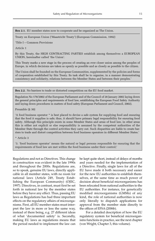
From 1991, Article 1 of the Treaty of Maastricht set the stage for very many devel-opments in the regulatory affairs of beneficial microorganisms for food and feed; see Box 2.1. Then, in 2002, EU Regulation No 178/2002 laid down the general principles for food law for all member states and established the European Food Safety Authority (EFSA) (OJEC, 2002). In that law, two of the most basic principles are the single EU food market and an absence of competition among the EU member states; see Box 2.2. These principles are the basis for the harmonization of the way the member states must regulate beneficial microorganisms for food and feed in the areas still open to member state legislation.
As a powerful instrument to harmoni-zation and to the ‘ever closer union’ (cf. Box 2.1), EU laws are now most often constructed as
Safety and Regulation of Microorganisms 15
Regulations and not as Directives. This change in construction was evident in the late 1990s and throughout the 2000s. Regulations are, so to speak, genuine EU laws, directly appli-cable in all member states, with no room for national laws (Article 249, Treaty Estab-lishing the European Community) (OJEC, 1997). Direc tives, in contrast, must first be set forth in national law by the member states before they have any effect. Thus, passing EU laws as Regulations has had three important effects on the regulatory affairs of microorga-nisms. First, all EU member states must inter-pret the law in more or less the same way, instead of there being, e.g. 27 different ideas of what ‘documented safety’ is. Secondly, making EU laws as regulations means that the period needed to implement the law can
be kept quite short, instead of delays of months and years needed for the implementation of a Directive. Finally, single laws for all of the EU have made it both necessary and easier for the new EU authorities to establish them-selves, at the same time as much power of decision about beneficial microorganisms has been relocated from national authorities to the EU authorities. For instance, for genetically modified microorganisms (GMMs) of any kind, the role of national authorities is now only literally to dispatch applications for approval from the member state directly to the offices of EFSA (2006b).
For a detailed description of how the EU regulatory system for beneficial microorgan-isms functions in practice, see the next chapter (von Wright, Chapter 3, this volume).
Box 2.1. EU member states now to cooperate and be organized as The Union.
Treaty on European Union (‘Maastricht Treaty’) (European Commission, 1992)
‘Title I – Common Provisions
Article 1
By this Treaty, the HIGH CONTRACTING PARTIES establish among themselves a EUROPEAN UNION, hereinafter called ‘the Union’.
This Treaty marks a new stage in the process of creating an ever closer union among the peoples of Europe, in which decisions are taken as openly as possible and as closely as possible to the citizen.
The Union shall be founded on the European Communities, supplemented by the policies and forms of cooperation established by this Treaty. Its task shall be to organise, in a manner demonstrating consistency and solidarity, relations between the Member States and between their peoples.’
Box 2.2. No barriers to trade or distorted competition on the EU food market.
Regulation No 178/2002 of the European Parliament and of the Council of 28 January 2002 laying down the general principles and requirements of food law, establishing the European Food Safety Authority and laying down procedures in matters of food safety (European Parliament and Council, 2002).
Preamble (§ 30)
‘A food business operator * is best placed to devise a safe system for supplying food and ensuring that the food it supplies is safe; thus, it should have primary legal responsibility for ensuring food safety. Although this principle exists in some Member States and areas of food law, in other areas this is either not explicit or else responsibility is assumed by the competent authorities of the Member State through the control activities they carry out. Such disparities are liable to create bar-riers to trade and distort competition between food business operators in different Member States.’
* Article 3
‘3. ‘food business operator’ means the natural or legal persons responsible for ensuring that the requirements of food law are met within the food business under their control;’
16 S. Wessels
2.3.2 Reaction to discontent among EU consumers
As well as the role of the EU in law making, consumer opinion in the EU has also been very influential in shaping the way that bene-ficial microorganisms for food and feed are regulated in the EU. Safe food and feed have obvious relevance for all individuals; thus, every individual is a consumer in discussions on food safety. During the 1990s, consumers in the various EU member states were dis-quieted by a series of food and feed contami-nation events. Because some of these events involved food and feed being traded between EU member states, the events seemed to reflect a lack of safety measures at the level of the EU. More specifically, they seemed to reflect a lack of accountability and responsi-bility on the part of the member state food safety systems (European Commission, 2000). Two examples will illustrate how consumer concerns resulted in the moving of the regula-tion of beneficial microorganisms from a member state level to the EU level.
In 1999, feed in Belgium was found to be contaminated with dioxins, which had spread to several categories of animals and to foods. It was believed that oil for feed had been stored in tanks previously used for industrial mineral oil (Tyler, 1999). Eggs and chickens containing the dioxins were sold in France, the Netherlands and Germany, and consum-ers in several countries were frightened by the event. Therefore, the EU Commission declared an EU-wide ban on the products. Eighteen months later, EU agricultural ministers adop-ted a regulation that put legally binding lim-its on dioxins in food. The EU Health and Consumer Protection Commis sioner David Byrne used the passing of the regulation to show the Commission’s emphatic opposition to the contamination incident: ‘I am pleased to see the Ministers recognize that we need to be uncompromising and severe on contami-nants in food. This new legislation, in setting legally binding limits, sets a new milestone in the EU’s feed and food safety strategy’ (European Commission, 2001).
In the late 1990s and 2000, the populace of the EU was affected by a number of zoon-oses, or infections in farm animals (usually
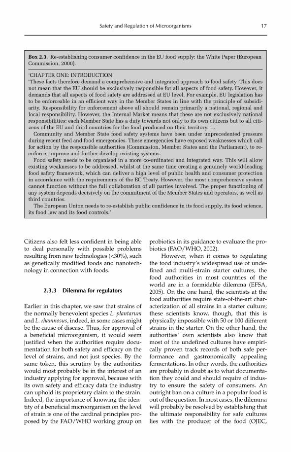
due to bacteria) that are transmitted to humans via the food chain. In 2002, the European Council supported a Directive and a Regulation aimed at monitoring and com-bating the bacteria, especially in the poultry industry. On the occasion of the support of the laws, Commissioner Byrne expressed clearly what the Commission also expected to achieve: ‘These proposals demonstrate how the Commission’s ‘farm to fork’ approach is being implemented in practice to ensure safe food for consumers’ (European Commission, 2002). The European Commission had pub-lished its White Paper on Food Safety in January 2000 (European Commission, 2000). The primary objective of the White Paper was to propose a way to establish an EU-wide sys-tem for food safety and to re-establish and maintain consumer confidence in the EU’s ability to ensure safe food. Box 2.3 quotes the White Paper and the European Commission’s reason for establishing the EU authorities for food and chemicals, i.e., EFSA in 2002 and the European Chemicals Agency (ECHA) in 2007.
During the same period of time as the two contamination events described above, the EU institutions were compiling the new Treaty of Nice (signed in 2001), which defined a new distribution of decision-making power for the EU. The treaty gave the European Parliament much more legislative power than it had had before (European Parliament, 2001). This meant that the members of the Parliament were now in a position to respond to the wishes of their constituencies and intensify EU efforts at guaranteeing a safe food supply. Less than a year after the Treaty of Nice was signed, the EU Parliament and Council had passed the Regulation that lays down the basic principles for EU food law and established the EU authority for food safety (OJEC, 2002).
Late in 2010, EFSA published a consumer opinion survey that elucidated where the EU public saw food-related risks (EFSA, 2010). The survey was the result of 26,600 personal interviews in all 27 EU member states; a simi-lar survey was conducted in 2005. It was apparent from the new survey that there had been no great increase in confidence that the food authorities (either in any member state or in EFSA) could ensure a safe food supply.
Safety and Regulation of Microorganisms 17
Citizens also felt less confident in being able to deal personally with possible problems resulting from new technologies (<30%), such as genetically modified foods and nanotech-nology in connection with foods.
2.3.3 Dilemma for regulators
Earlier in this chapter, we saw that strains of the normally benevolent species L. plantarumand L. rhamnosus, indeed, in some cases might be the cause of disease. Thus, for approval of a beneficial microorganism, it would seem justified when the authorities require docu-mentation for both safety and efficacy on the level of strains, and not just species. By the same token, this scrutiny by the authorities would most probably be in the interest of an industry applying for approval, because with its own safety and efficacy data the industry can uphold its proprietary claim to the strain. Indeed, the importance of knowing the iden-tity of a beneficial microorganism on the level of strain is one of the cardinal principles pro-posed by the FAO/WHO working group on
probiotics in its guidance to evaluate the pro-biotics (FAO/WHO, 2002).
However, when it comes to regulating the food industry’s widespread use of unde-fined and multi-strain starter cultures, the food authorities in most countries of the world are in a formidable dilemma (EFSA, 2005). On the one hand, the scientists at the food authorities require state-of-the-art char-acterization of all strains in a starter culture; these scientists know, though, that this is physically impossible with 50 or 100 different strains in the starter. On the other hand, the authorities’ own scientists also know that most of the undefined cultures have empiri-cally proven track records of both safe per-formance and gastronomically appealing fermentations. In other words, the authorities are probably in doubt as to what documenta-tion they could and should require of indus-try to ensure the safety of consumers. An outright ban on a culture in a popular food is out of the question. In most cases, the dilemma will probably be resolved by establishing that the ultimate responsibility for safe cultures lies with the producer of the food (OJEC,
Box 2.3. Re-establishing consumer confidence in the EU food supply: the White Paper (European Commission, 2000).
‘CHAPTER ONE: INTRODUCTION‘These facts therefore demand a comprehensive and integrated approach to food safety. This does not mean that the EU should be exclusively responsible for all aspects of food safety. However, it demands that all aspects of food safety are addressed at EU level. For example, EU legislation has to be enforceable in an efficient way in the Member States in line with the principle of subsidi-arity. Responsibility for enforcement above all should remain primarily a national, regional and local responsibility. However, the Internal Market means that these are not exclusively national responsibilities: each Member State has a duty towards not only to its own citizens but to all citi-zens of the EU and third countries for the food produced on their territory. … Community and Member State food safety systems have been under unprecedented pressure during recent feed and food emergencies. These emergencies have exposed weaknesses which call for action by the responsible authorities (Commission, Member States and the Parliament), to re-enforce, improve and further develop existing systems. Food safety needs to be organised in a more co-ordinated and integrated way. This will allow existing weaknesses to be addressed, whilst at the same time creating a genuinely world-leading food safety framework, which can deliver a high level of public health and consumer protection in accordance with the requirements of the EC Treaty. However, the most comprehensive system cannot function without the full collaboration of all parties involved. The proper functioning of any system depends decisively on the commitment of the Member States and operators, as well as third countries. The European Union needs to re-establish public confidence in its food supply, its food science, its food law and its food controls.’
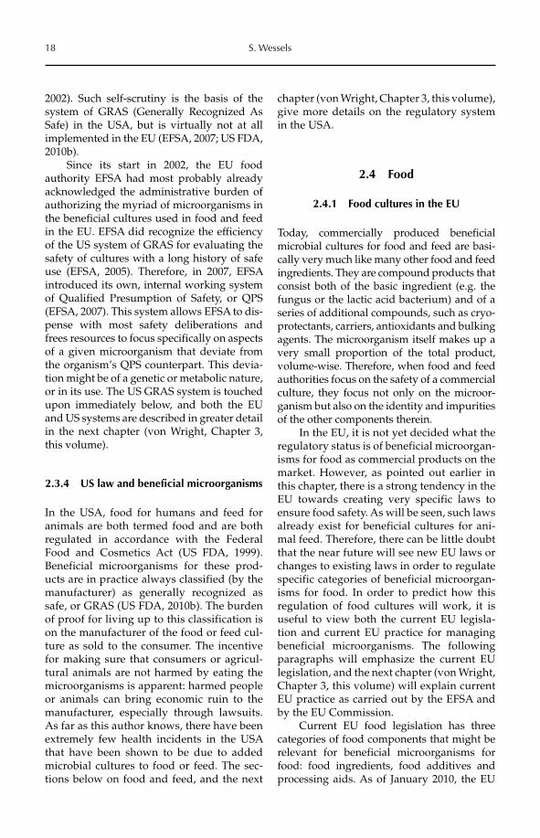
18 S. Wessels
2002). Such self-scrutiny is the basis of the system of GRAS (Generally Recognized As Safe) in the USA, but is virtually not at all implemented in the EU (EFSA, 2007; US FDA, 2010b).
Since its start in 2002, the EU food authority EFSA had most probably already acknowledged the administrative burden of authorizing the myriad of microorganisms in the beneficial cultures used in food and feed in the EU. EFSA did recognize the efficiency of the US system of GRAS for evaluating the safety of cultures with a long history of safe use (EFSA, 2005). Therefore, in 2007, EFSA introduced its own, internal working system of Qualified Presumption of Safety, or QPS (EFSA, 2007). This system allows EFSA to dis-pense with most safety deliberations and frees resources to focus specifically on aspects of a given microorganism that deviate from the organism’s QPS counterpart. This devia-tion might be of a genetic or metabolic nature, or in its use. The US GRAS system is touched upon immediately below, and both the EU and US systems are described in greater detail in the next chapter (von Wright, Chapter 3, this volume).
2.3.4 US law and beneficial microorganisms
In the USA, food for humans and feed for animals are both termed food and are both regulated in accordance with the Federal Food and Cosmetics Act (US FDA, 1999). Beneficial microorganisms for these prod-ucts are in practice always classified (by the manufacturer) as generally recognized as safe, or GRAS (US FDA, 2010b). The burden of proof for living up to this classification is on the manufacturer of the food or feed cul-ture as sold to the consumer. The incentive for making sure that consumers or agricul-tural animals are not harmed by eating the microorganisms is apparent: harmed people or animals can bring economic ruin to the manufacturer, especially through lawsuits. As far as this author knows, there have been extremely few health incidents in the USA that have been shown to be due to added microbial cultures to food or feed. The sec-tions below on food and feed, and the next
chapter (von Wright, Chapter 3, this volume), give more details on the regulatory system in the USA.
2.4 Food
2.4.1 Food cultures in the EU
Today, commercially produced beneficial microbial cultures for food and feed are basi-cally very much like many other food and feed ingredients. They are compound products that consist both of the basic ingredient (e.g. the fungus or the lactic acid bacterium) and of a series of additional compounds, such as cryo-protectants, carriers, antioxidants and bulking agents. The microorganism itself makes up a very small proportion of the total product, volume-wise. Therefore, when food and feed authorities focus on the safety of a commercial culture, they focus not only on the microor-ganism but also on the identity and impurities of the other components therein.
In the EU, it is not yet decided what the regulatory status is of beneficial microorgan-isms for food as commercial products on the market. However, as pointed out earlier in this chapter, there is a strong tendency in the EU towards creating very specific laws to ensure food safety. As will be seen, such laws already exist for beneficial cultures for ani-mal feed. Therefore, there can be little doubt that the near future will see new EU laws or changes to existing laws in order to regulate specific categories of beneficial microorgan-isms for food. In order to predict how this regulation of food cultures will work, it is useful to view both the current EU legisla-tion and current EU practice for managing beneficial microorganisms. The following paragraphs will emphasize the current EU legislation, and the next chapter (von Wright, Chapter 3, this volume) will explain current EU practice as carried out by the EFSA and by the EU Commission.
Current EU food legislation has three categories of food components that might be relevant for beneficial microorganisms for food: food ingredients, food additives and processing aids. As of January 2010, the EU
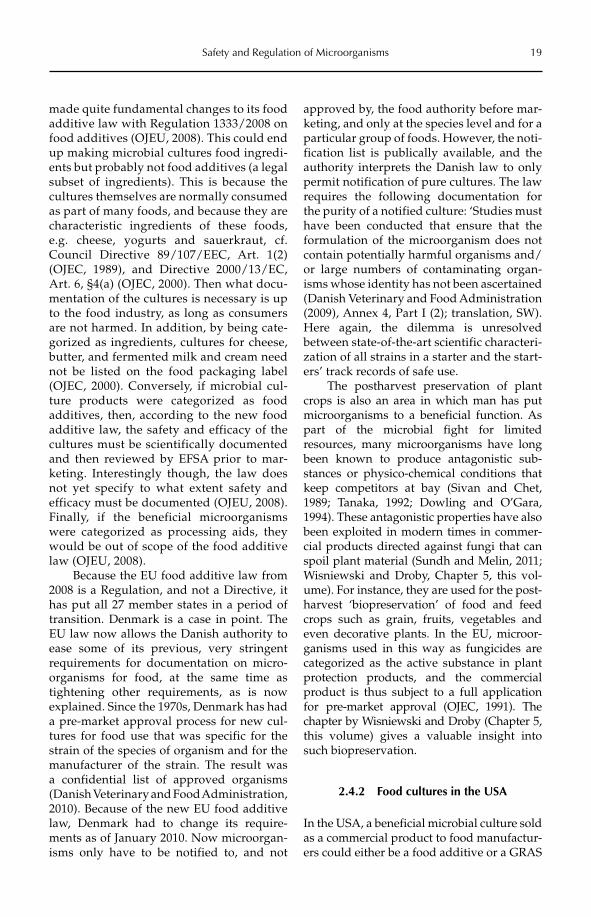
Safety and Regulation of Microorganisms 19
made quite fundamental changes to its food additive law with Regulation 1333/2008 on food additives (OJEU, 2008). This could end up making microbial cultures food ingredi-ents but probably not food additives (a legal subset of ingredients). This is because the cultures themselves are normally consumed as part of many foods, and because they are characteristic ingredients of these foods, e.g. cheese, yogurts and sauerkraut, cf. Council Directive 89/107/EEC, Art. 1(2) (OJEC, 1989), and Directive 2000/13/EC, Art. 6, §4(a) (OJEC, 2000). Then what docu-mentation of the cultures is necessary is up to the food industry, as long as consumers are not harmed. In addition, by being cate-gorized as ingredients, cultures for cheese, butter, and fermented milk and cream need not be listed on the food packaging label (OJEC, 2000). Conversely, if microbial cul-ture products were categorized as food additives, then, according to the new food additive law, the safety and efficacy of the cultures must be scientifically documented and then reviewed by EFSA prior to mar-keting. Interestingly though, the law does not yet specify to what extent safety and efficacy must be documented (OJEU, 2008). Finally, if the beneficial microorganisms were categorized as processing aids, they would be out of scope of the food additive law (OJEU, 2008).
Because the EU food additive law from 2008 is a Regulation, and not a Directive, it has put all 27 member states in a period of transition. Denmark is a case in point. The EU law now allows the Danish authority to ease some of its previous, very stringent requirements for documentation on micro-organisms for food, at the same time as tightening other requirements, as is now explained. Since the 1970s, Denmark has had a pre-market approval process for new cul-tures for food use that was specific for the strain of the species of organism and for the manufacturer of the strain. The result was a confidential list of approved organisms (Danish Veterinary and Food Administration, 2010). Because of the new EU food additive law, Denmark had to change its require-ments as of January 2010. Now microorgan-isms only have to be notified to, and not
approved by, the food authority before mar-keting, and only at the species level and for a particular group of foods. However, the noti-fication list is publically available, and the authority interprets the Danish law to only permit notification of pure cultures. The law requires the following documentation for the purity of a notified culture: ‘Studies must have been conducted that ensure that the formulation of the microorganism does not contain potentially harmful organisms and/or large numbers of contaminating organ-isms whose identity has not been ascertained (Danish Veterinary and Food Administration (2009), Annex 4, Part I (2); translation, SW). Here again, the dilemma is unresolved between state-of-the-art scientific characteri-zation of all strains in a starter and the start-ers’ track records of safe use.
The postharvest preservation of plant crops is also an area in which man has put microorganisms to a beneficial function. As part of the microbial fight for limited resources, many microorganisms have long been known to produce antagonistic sub-stances or physico-chemical conditions that keep competitors at bay (Sivan and Chet, 1989; Tanaka, 1992; Dowling and O’Gara, 1994). These antagonistic properties have also been exploited in modern times in commer-cial products directed against fungi that can spoil plant material (Sundh and Melin, 2011; Wisniewski and Droby, Chapter 5, this vol-ume). For instance, they are used for the post-harvest ‘biopreservation’ of food and feed crops such as grain, fruits, vegetables and even decorative plants. In the EU, microor-ganisms used in this way as fungicides are categorized as the active substance in plant protection products, and the commercial product is thus subject to a full application for pre-market approval (OJEC, 1991). The chapter by Wisniewski and Droby (Chapter 5, this volume) gives a valuable insight into such biopreservation.
2.4.2 Food cultures in the USA
In the USA, a beneficial microbial culture sold as a commercial product to food manufactur-ers could either be a food additive or a GRAS
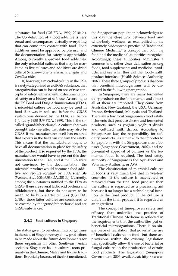
20 S. Wessels
substance for food (US FDA, 1999, 2010a,b). The US definition of a food additive is very broad and encompasses virtually everything that can come into contact with food. Food additives must be approved before use, and the documentation for safety is appreciable. Among currently approved food additives, the only microbial cultures that may be mar-keted as live cultures and additives are dried cells of Saccharomyces cerevisiae, S. fragilis and Candida utilis.
If, however, a microbial culture in the USA is safety-categorized as a GRAS substance, that categorization can be based on one of two con-cepts of safety: either scientific documentation of safety or a history of safe use. According to the US Food and Drug Administration (FDA), a microbial culture for food may be used in food if it was in safe use before the GRAS system was devised by the FDA, i.e. before 1 January 1958 (US FDA, 1999). This is the so-called ‘grandfather clause’. A culture that was brought into use after that date may also be GRAS if the manufacturer itself has ensured that experts in the field can confirm the safety. This means that the manufacturer ought to have all documentation in place for the safety of the product. If so requested by the FDA, the manufacturer would have to present this doc-umentation to the FDA, and if the FDA were not convinced by the documentation, the microbial product would become a food addi-tive and require scrutiny by FDA scientists (Wessels et al., 2004; US FDA, 2010b). Currently, among the substances notified to the FDA as GRAS, there are several lactic acid bacteria and bifidobacteria, but these do not seem to be meant to be bulk starter cultures (US FDA, 2010c); those latter cultures are considered to be covered by the ‘grandfather clause’ and are GRAS substances.
2.4.3 Food cultures in Singapore
The status given to beneficial microorganisms in the state of Singapore may allow predictions to be made about the future developments for these organisms in other South-east Asian societies. Singapore has its cultural roots pri-marily in the Chinese, Malay and Indian tradi-tions. Especially because of the first mentioned,
the Singaporean population acknowledges to this day the close link between food and whole-body wellness, as exemplified in the extremely widespread practice of Traditional Chinese Medicine,1 a concept that both the food and the medicinal authorities recognize. Accordingly, these authorities administer a common and rather clear delineation among foods, food supplements and medicinal prod-ucts, and use what they call the ‘food–health product interface’ (Health Sciences Authority, 2007). These three groups of products that con-tain beneficial microorganisms will be dis-cussed in the following paragraphs.
In Singapore, there are many fermented dairy products on the food market, and almost all of them are imported. They come from Australia, New Zealand, the USA, Germany, France, Switzerland, Malaysia and Thailand. There are a few local Singaporean food estab-lishments that produce cheese and fermented products, such as yoghurt, yoghurt drinks and cultured milk drinks. According to Singaporean law, the responsibility for safe dairy products lies either with the importer to Singapore or with the Singaporean manufac-turer (Singapore Government, 2002), and no pre-market approval of cultures or the fer-mented foods is required. The food safety authority of Singapore is the Agri-Food and Veterinary Authority, or AVA.
The classification of microbial cultures in foods is very much like that in Western countries. If the culture is inactivated or removed from the final food product, then the culture is regarded as a processing aid because it no longer has a technological func-tion in the final product. If the culture is viable in the final product, it is regarded as an ingredient.
The concept of time-proven safety and efficacy that underlies the practice of Traditional Chinese Medicine is reflected in the requirements that the authorities put on beneficial microorganisms. There is no sin-gle piece of legislation that governs the use of beneficial cultures in food, but there are provisions within the existing legislation that specifically allow the use of bacterial or fungal cultures in the production of certain food products. The legislation (Singapore Government, 2006; available at: http://www.
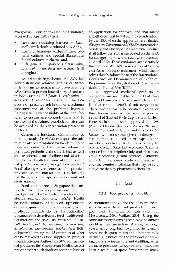
Safety and Regulation of Microorganisms 21
ava.gov.sg/ Legislation/ListOfLegislation/; accessed 26 April 2012) lists:
lactic acid-producing bacteria in • Lacto-bacillus milk drink or cultured milk drink;ripening, harmless acid-producing bac-•terial cultures and special filamentous fungal cultures in cheese; andL. bulgaricus• , Streptococcus thermophilus,L. acidophilus and Bacterium yoghurtii [sic!]in yoghurt.
As probiotic ingredients, the AVA has administratively allowed strains of Bifido-bacterium and Lactobacillus that have what the AVA terms ‘a proven long history of safe use in food (such as B. bifidum, L. acidophilus, L.delbrueckii, L. casei Shirota strain)’. The AVA does not prescribe minimum or maximum concentrations of the probiotic bacteria. Rather, it is the responsibility of the manufac-turer to ensure safe concentrations, and to ensure that the claimed probiotic function can be achieved by the concentration present in the food.
Concerning functional claims made for probiotic foods, the AVA does require the sub-mission of documentation for the claim. These rules are posted on the Internet, where the permitted probiotic claims are listed, as well as a requirement for labelling (and advertis-ing) the food with the name of the probiotic (http://www.ava.gov.sg/FoodSector/FoodLabelingAdvertisement). In practice, products on the market almost exclusively list the genus and species names and not strain names.
Food supplements in Singapore that con-tain beneficial microorganisms are adminis-tered primarily by the medicinal authority, the Health Sciences Authority (HSA) (Health Sciences Authority, 2007). Food supplements do not require a pre-market approval, while medicinal products do. On the authorities’ document that describes the food–health prod-uct interface, the HSA lists ‘Probiotics (in non-milk based products) including Lactobacillus,Streptococcus thermophilus, Bifidobacteria [bifi-dobacteria]’ among the 45 examples of what can be marketed as a food supplement product (Health Sciences Authority, 2007). For medici-nal products, the Singaporean Medicines Act prescribes that such products are the subject of
an application for approval, and that safety and efficacy must be ‘taken into consideration’ by the HSA when the application is evaluated (Singapore Government, 2008). Documentation of safety and efficacy of the medicinal product shall follow the guidances posted on the HSA homepage (http://www.hsa.gov.sg/; accessed 26 April 2012). These guidances are essentially the common ASEAN (Association of South-east Asian Nations) guidances, which them-selves closely follow those of the International Conference on Harmonisation of Technical Require ments for Registration of Pharma ceu-ticals for Human Use (ICH).
All approved medicinal products in Singapore are searchable on the HSA web site, and there are only two products on that list that contain beneficial microorganisms. These two appear to be identical except for their dosage forms: as capsule and as powder in a sachet (Lacteol Forte Capsule and Lacteol Forte Sachet, and were approved in 1998 (Aptalis Pharma [formerly Axcan Pharma], 2011). They contain lyophilized cells of Lacto-bacillus, with no species given, at dosages of 5 × 10⁹ and 1 × 10¹⁰ cells in the capsules and sachets, respectively. Both products may be sold as General Sales List Medicines (GSL), as opposed to Prescription Only and Pharmacy Only Medicines (Health Sciences Authority, 2011). GSL medicines can be compared with over-the-counter medicines that may be sold elsewhere than by pharmacies/chemists.
2.5 Feed
2.5.1 Feed probiotics in the EU
As mentioned above, the use of microorgan-isms to make beneficial products for man goes back thousands of years (Fox and McSweeney, 2004; Walker, 2004). Using the same microorganisms as feed may be almost as old as their use in food. Among the fungi, yeasts have long been exploited to ferment cereal mash, grape musts and other naturally derived substrates for the purposes of brew-ing, baking, winemaking and distilling. After all these processes (except baking), there has been a residue of spent fermentation mass,
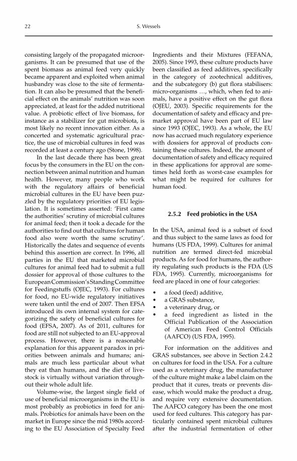
22 S. Wessels
consisting largely of the propagated microor-ganisms. It can be presumed that use of the spent biomass as animal feed very quickly became apparent and exploited when animal husbandry was close to the site of fermenta-tion. It can also be presumed that the benefi-cial effect on the animals’ nutrition was soon appreciated, at least for the added nutritional value. A probiotic effect of live biomass, for instance as a stabilizer for gut microbiota, is most likely no recent innovation either. As a concerted and systematic agricultural prac-tice, the use of microbial cultures in feed was recorded at least a century ago (Stone, 1998).
In the last decade there has been great focus by the consumers in the EU on the con-nection between animal nutrition and human health. However, many people who work with the regulatory affairs of beneficial microbial cultures in the EU have been puz-zled by the regulatory priorities of EU legis-lation. It is sometimes asserted: ‘First came the authorities’ scrutiny of microbial cultures for animal feed; then it took a decade for the authorities to find out that cultures for human food also were worth the same scrutiny’. Historically the dates and sequence of events behind this assertion are correct. In 1996, all parties in the EU that marketed microbial cultures for animal feed had to submit a full dossier for approval of those cultures to the European Commission’s Standing Committee for Feedingstuffs (OJEC, 1993). For cultures for food, no EU-wide regulatory initiatives were taken until the end of 2007. Then EFSA introduced its own internal system for cate-gorizing the safety of beneficial cultures for food (EFSA, 2007). As of 2011, cultures for food are still not subjected to an EU-approval process. However, there is a reasonable explanation for this apparent paradox in pri-orities between animals and humans; ani-mals are much less particular about what they eat than humans, and the diet of live-stock is virtually without variation through-out their whole adult life.
Volume-wise, the largest single field of use of beneficial microorganisms in the EU is most probably as probiotics in feed for ani-mals. Probiotics for animals have been on the market in Europe since the mid 1980s accord-ing to the EU Association of Specialty Feed
Ingredients and their Mixtures (FEFANA, 2005). Since 1993, these culture products have been classified as feed additives, specifically in the category of zootechnical additives, and the subcategory (b) gut flora stabilisers: micro-organisms …, which, when fed to ani-mals, have a positive effect on the gut flora (OJEU, 2003). Specific requirements for the documentation of safety and efficacy and pre-market approval have been part of EU law since 1993 (OJEC, 1993). As a whole, the EU now has accrued much regulatory experience with dossiers for approval of products con-taining these cultures. Indeed, the amount of documentation of safety and efficacy required in these applications for approval are some-times held forth as worst-case examples for what might be required for cultures for human food.
2.5.2 Feed probiotics in the USA
In the USA, animal feed is a subset of food and thus subject to the same laws as food for humans (US FDA, 1999). Cultures for animal nutrition are termed direct-fed microbial products. As for food for humans, the author-ity regulating such products is the FDA (US FDA, 1995). Currently, microorganisms for feed are placed in one of four categories:
a food (feed) additive,•a GRAS substance,•a veterinary drug, or•a feed ingredient as listed in the •Official Publication of the Association of American Feed Control Officials (AAFCO) (US FDA, 1995).
For information on the additives and GRAS substances, see above in Section 2.4.2 on cultures for food in the USA. For a culture used as a veterinary drug, the manufacturer of the culture might make a label claim on the product that it cures, treats or prevents dis-ease, which would make the product a drug, and require very extensive documentation. The AAFCO category has been the one most used for feed cultures. This category has par-ticularly contained spent microbial cultures after the industrial fermentation of other
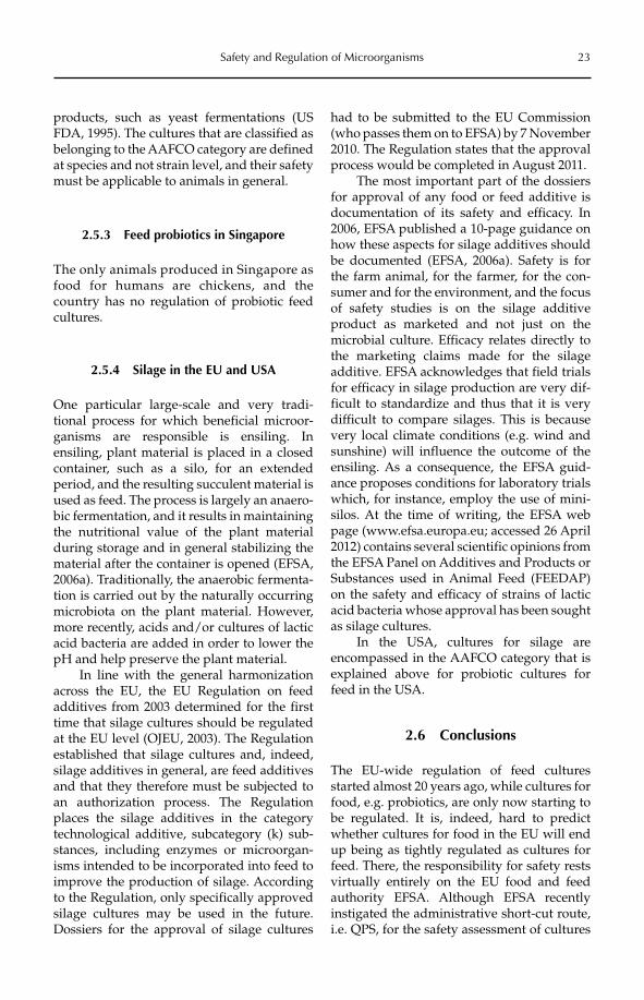
Safety and Regulation of Microorganisms 23
products, such as yeast fermentations (US FDA, 1995). The cultures that are classified as belonging to the AAFCO category are defined at species and not strain level, and their safety must be applicable to animals in general.
2.5.3 Feed probiotics in Singapore
The only animals produced in Singapore as food for humans are chickens, and the country has no regulation of probiotic feed cultures.
2.5.4 Silage in the EU and USA
One particular large-scale and very tradi-tional process for which beneficial microor-ganisms are responsible is ensiling. In ensiling, plant material is placed in a closed container, such as a silo, for an extended period, and the resulting succulent material is used as feed. The process is largely an anaero-bic fermentation, and it results in maintaining the nutritional value of the plant material during storage and in general stabilizing the material after the container is opened (EFSA, 2006a). Traditionally, the anaerobic fermenta-tion is carried out by the naturally occurring microbiota on the plant material. However, more recently, acids and/or cultures of lactic acid bacteria are added in order to lower the pH and help preserve the plant material.
In line with the general harmonization across the EU, the EU Regulation on feed additives from 2003 determined for the first time that silage cultures should be regulated at the EU level (OJEU, 2003). The Regulation established that silage cultures and, indeed, silage additives in general, are feed additives and that they therefore must be subjected to an authorization process. The Regulation places the silage additives in the category technological additive, subcategory (k) sub-stances, including enzymes or microorgan-isms intended to be incorporated into feed to improve the production of silage. According to the Regulation, only specifically approved silage cultures may be used in the future. Dossiers for the approval of silage cultures
had to be submitted to the EU Commission (who passes them on to EFSA) by 7 November 2010. The Regulation states that the approval process would be completed in August 2011.
The most important part of the dossiers for approval of any food or feed additive is documentation of its safety and efficacy. In 2006, EFSA published a 10-page guidance on how these aspects for silage additives should be documented (EFSA, 2006a). Safety is for the farm animal, for the farmer, for the con-sumer and for the environment, and the focus of safety studies is on the silage additive product as marketed and not just on the microbial culture. Efficacy relates directly to the marketing claims made for the silage additive. EFSA acknowledges that field trials for efficacy in silage production are very dif-ficult to standardize and thus that it is very difficult to compare silages. This is because very local climate conditions (e.g. wind and sunshine) will influence the outcome of the ensiling. As a consequence, the EFSA guid-ance proposes conditions for laboratory trials which, for instance, employ the use of mini-silos. At the time of writing, the EFSA web page (www.efsa.europa.eu; accessed 26 April 2012) contains several scientific opinions from the EFSA Panel on Additives and Products or Substances used in Animal Feed (FEEDAP) on the safety and efficacy of strains of lactic acid bacteria whose approval has been sought as silage cultures.
In the USA, cultures for silage are en compassed in the AAFCO category that is explained above for probiotic cultures for feed in the USA.
2.6 Conclusions
The EU-wide regulation of feed cultures started almost 20 years ago, while cultures for food, e.g. probiotics, are only now starting to be regulated. It is, indeed, hard to predict whether cultures for food in the EU will end up being as tightly regulated as cultures for feed. There, the responsibility for safety rests virtually entirely on the EU food and feed authority EFSA. Although EFSA recently instigated the administrative short-cut route, i.e. QPS, for the safety assessment of cultures
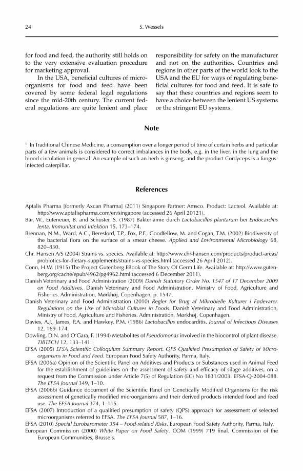
24 S. Wessels
for food and feed, the authority still holds on to the very extensive evaluation procedure for marketing approval.
In the USA, beneficial cultures of micro-organisms for food and feed have been covered by some federal legal regulations since the mid-20th century. The current fed-eral regulations are quite lenient and place
responsibility for safety on the manufacturer and not on the authorities. Countries and regions in other parts of the world look to the USA and the EU for ways of regulating bene-ficial cultures for food and feed. It is safe to say that these countries and regions seem to have a choice between the lenient US systems or the stringent EU systems.
Note
1 In Traditional Chinese Medicine, a consumption over a longer period of time of certain herbs and particular parts of a few animals is considered to correct imbalances in the body, e.g. in the liver, in the lung and the blood circulation in general. An example of such an herb is ginseng; and the product Cordyceps is a fungus-infected caterpillar.
References
Aptalis Pharma [formerly Axcan Pharma] (2011) Singapore Partner: Amsco. Product: Lacteol. Available at: http://www.aptalispharma.com/en/singapore (accessed 26 April 20121).
Bär, W., Euteneuer, B. and Schuster, S. (1987) Bakteriämie durch Lactobacillus plantarum bei Endocarditislenta. Immunitat und Infektion 15, 173–174.
Brennan, N.M., Ward, A.C., Beresford, T.P., Fox, P.F., Goodfellow, M. and Cogan, T.M. (2002) Biodiversity of the bacterial flora on the surface of a smear cheese. Applied and Environmental Microbiology 68, 820–830.
Chr. Hansen A/S (2004) Strains vs. species. Available at: http://www.chr-hansen.com/products/product-areas/probiotics-for-dietary-supplements/strains-vs-species.html (accessed 26 April 2012).
Conn, H.W. (1915) The Project Gutenberg EBook of The Story Of Germ Life. Available at: http://www.guten-berg.org/cache/epub/4962/pg4962.html (accessed 6 December 2011).
Danish Veterinary and Food Administration (2009) Danish Statutory Order No. 1547 of 17 December 2009 on Food Additives. Danish Veterinary and Food Administration, Ministry of Food, Agriculture and Fisheries. Administration, Mørkhøj, Copenhagen, p. 1547.
Danish Veterinary and Food Administration (2010) Regler for Brug af Mikrobielle Kulturer i Fødevarer. Regulations on the Use of Microbial Cultures in Foods. Danish Veterinary and Food Administration, Ministry of Food, Agriculture and Fisheries. Administration, Mørkhøj, Copenhagen.
Davies, A.J., James, P.A. and Hawkey, P.M. (1986) Lactobacillus endocarditis. Journal of Infectious Diseases12, 169–174.
Dowling, D.N. and O’Gara, F. (1994) Metabolites of Pseudomonas involved in the biocontrol of plant disease. TIBTECH 12, 133–141.
EFSA (2005) EFSA Scientific Colloquium Summary Report. QPS Qualified Presumption of Safety of Micro-organisms in Food and Feed. European Food Safety Authority, Parma, Italy.
EFSA (2006a) Opinion of the Scientific Panel on Additives and Products or Substances used in Animal Feed for the establishment of guidelines on the assessment of safety and efficacy of silage additives, on a request from the Commission under Article 7(5) of Regulation (EC) No 1831/2003. EFSA-Q-2004-088. The EFSA Journal 349, 1–10.
EFSA (2006b) Guidance document of the Scientific Panel on Genetically Modified Organisms for the risk assessment of genetically modified microorganisms and their derived products intended food and feed use. The EFSA Journal 374, 1–115.
EFSA (2007) Introduction of a qualified presumption of safety (QPS) approach for assessment of selected microorganisms referred to EFSA. The EFSA Journal 587, 1–16.
EFSA (2010) Special Eurobarometer 354 – Food-related Risks. European Food Safety Authority, Parma, Italy.European Commission (2000) White Paper on Food Safety. COM (1999) 719 final. Commission of the
European Communities, Brussels.
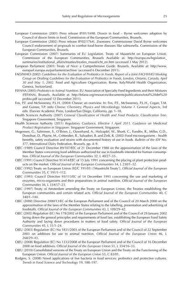
Safety and Regulation of Microorganisms 25
European Commission (2001) Press release IP/01/1698. Dioxin in food – Byrne welcomes adoption by Council of dioxin limits in food. Commission of the European Communities, Brussels.
European Commission (2002) Press release IP/02/1764). Zoonoses: Commissioner David Byrne welcomes Council endorsement of proposals to combat food-borne diseases like salmonella. Commission of the European Communities, Brussels.
European Commission (2007) Summaries of EU Legislation. Treaty of Maastricht on European Union. Commission of the European Communities, Brussels Available at: http://europa.eu/legislation_summaries/institutional_affairs/treaties/treaties_maastricht_en.htm (accessed 3 May 2012).
European Parliament (2001) Treaty of Nice: a Comprehensive Guide. Brussels. Available at: http://www.europarl.europa.eu/parliament/archive/ (accessed 6 December 2011).
FAO/WHO (2002) Guidelines for the Evaluation of Probiotics in Foods. Report of a Joint FAO/WHO Working Group on Drafting Guidelines for the Evaluation of Probiotics in Foods, London, Ontario, Canada, April 30 and May 1, 2002. Food and Agriculture Organization, Rome, Italy/World Health Organization, Geneva, Switzerland.
FEFANA (2005) Probiotics in Animal Nutrition. EU Association of Specialty Feed Ingredients and their Mixtures (FEFANA), Brussels. Available at: http://fefana.org/resources/documents/publications/total%20def%20probio.pdf (accessed 12 December 2011).
Fox, P.F. and McSweeney, P.L.H. (2004) Cheese: an overview. In: Fox, P.F., McSweeney, P.L.H., Cogan, T.M. and Guinee, T.P. (eds) Cheese: Chemistry, Physics and Microbiology. Volume 1. General Aspects, 3rd edn. Elsevier Academic Press, London/San Diego, California, pp. 1–18.
Health Sciences Authority (2007) General Classification of Health and Food Products. Classification Tree.Singapore Government, Singapore.
Health Sciences Authority (2011) Regulatory Guidance, Effective 1 April 2011. Guidance on Medicinal Product Registration in Singapore. Singapore Government, Singapore.
Mogensen, G., Salminen, S., O’Brien, J., Ouwehand, A., Holzapfel, W., Shortt, C., Fondén, R., Miller, G.D., Donohue, D., Playne, M., Crittenden, R., Salvadori, B. and Zink, R. (2002) Food microorganisms – health benefits, safety evaluation and strains with documented history of use in foods. Bulletin of the IDF No. 377, International Dairy Federation, Brussels, pp. 4–9.
OJEC (1989) Council Directive 89/107/EEC of 21 December 1988 on the approximation of the laws of the Member States concerning food additives authorized for use in foodstuffs intended for human consump-tion. Official Journal of the European Communities 32, L 40/27–33.
OJEC (1991) Council Directive 91/414/EEC of 15 July 1991 concerning the placing of plant protection prod-ucts on the market. Official Journal of the European Communities 34, L 230/1–32.
OJEC (1992) Treaty on European Union (92/C 191/01) [‘Maastricht Treaty’]. Official Journal of the European Communities 35, C 191/1–112.
OJEC (1993) Council Directive 93/113/EC of 14 December 1993 concerning the use and marketing of enzymes, micro-organisms and their preparations in animal nutrition. Official Journal of the European Communities 36, L 334/17–23.
OJEC (1997) Treaty of Amsterdam amending the Treaty on European Union, the Treaties establishing the European communities and certain related acts. Official Journal of the European Communities 40, C 340/1–144.
OJEC (2000) Directive 2000/13/EC of the European Parliament and of the Council of 20 March 2000 on the approximation of the laws of the Member States relating to the labelling, presentation and advertising of foodstuffs. Official Journal of the European Communities 43, L 109/29–42.
OJEC (2002) Regulation (EC) No 178/2002 of the European Parliament and of the Council of 28 January 2002 laying down the general principles and requirements of food law, establishing the European Food Safety Authority and laying down procedures in matters of food safety. Official Journal of the European Communities 45, L 31/1–24.
OJEU (2003) Regulation (EC) No 1831/2003 of the European Parliament and of the Council of 22 September 2003 on additives for use in animal nutrition. Official Journal of the European Union 46, L 268/29–43.
OJEU (2008) Regulation (EC) No 1333/2008 of the European Parliament and of the Council of 16 December 2008 on food additives. Official Journal of the European Union 51, L 354/16–33.
OJEU (2010) Consolidated versions of the Treaty on European Union and the Treaty on the Functioning of the European Union. Official Journal of the European Union 53, C 83/01.
Rodgers, S. (2008) Novel applications of live bacteria in food services: probiotics and protective cultures. Trends in Food Science and Technology 19, 188–197.
26 S. Wessels
Salminen, M.K., Rautelin, H., Tynkkynen, S., Poussa, T., Saxelin, M., Valtonen, V. and Jarvinen, A. (2006) Lactobacillus bacteremia, species identification, and antimicrobial susceptibility of 85 blood isolates. Clinical Infectious Diseases 42, e35–e44.
Saxelin, M. (2008) Research Behind the Probiotic Lactobacillus GG (LGG ®). Valio Ltd., Helsinki.Singapore Government (2002) Sale of Food Act, Chapter 283. Singapore Government.Singapore Government (2006) Sale of Food Act, Food Regulations, RG1, revised edn. Singapore Government.Singapore Government (2008) Medicines Act (Chapter 176). Singapore Government.Sivan, A. and Chet, I. (1989) The possible role of competition between Trichoderma harzianum and Fusarium
oxysporum on rhizosphere colonization. Phytopathology 79, 198–203.Stone, C.W. (1998) Yeast Products in the Feed Industry: A Practical Guide for Feed Professionals. Diamond V
Mills, Inc., Cedar Rapids, Iowa. Available at: http://www.vertumnus.info/DIAM.sci.Stone1998.pdf (accessed 26 April 2012).
Sundh, I. and Melin, P. (2011) Safety and regulation of yeasts used for biocontrol or biopreservation in the food or feed chain. Antonie Van Leeuwenhoek 99, 113–119.
Tanaka, Y. (1992) Antifungal agents. In: Omura, S. (ed.) The Search for Bioactive Compounds from Microorganisms. Springer-Verlag, Berlin, pp. 30–44.
Tyler, R. (1999) Dioxin contamination scandal hits Belgium. Effects spread through European Union and beyond. World Socialist Web Site, International Committee of the Fourth International. Available at: http://www.wsws.org/articles/1999/jun1999/belg-j08.shtml (accessed 26 April 2012).
US FDA (1995) Compliance Policy Guides. Sec. 689.100 Direct-Fed Microbial Products, US Food and Drug Administration, Washington, DC.
US FDA (1999) Federal Food, Drug, and Cosmetic Act. US Food and Drug Administration, Washington, DC.US FDA (2010a) Code of Federal Regulations, Title 21 – Food and Drugs. Part 172 – Food additives permitted
for direct addition to food for human consumption. US Food and Drug Administration, Washington, DC.US FDA (2010b) Code of Federal Regulations, Title 21 – Food and Drugs. Part 182 – Substances generally
recognized as safe. US Food and Drug Administration, Washington, DC.US FDA (2010c) GRAS Notice Inventory. US Food and Drug Administration, Washington, DC. Available at: http://
www.accessdata.fda.gov/scripts/fcn/fcnNavigation.cfm?rpt=grasListing (accessed 6 December 2011).Wagner, R.D., Warner, T., Roberts, L., Farmer, J. and Balish, E. (1997) Colonization of congenitally immuno-
deficient mice with probiotic bacteria. Infection and Immunity 65, 3345–3351.Walker, G.M. (2004) Metals in yeast fermentation processes. Advances in Applied Microbiology 54,
197–229.Wessels, S., Axelsson, L., Hansen, E.B., de Vuyst, L., Laulund, S., Lähteenmäki, L., Lindgren, S., Mollet, B.,
Salminen, S. and von Wright, A. (2004) The lactic acid bacteria, the food chain, and their regulation. Trends in Food Science and Technology 15, 498–505.
Wisniewski, M. and Droby, S. (2012) Biopreservation of food and feed by postharvest biocontrol with microor-ganisms. In: Sundh, I., Wilcks, A. and Goettel, M.S. (eds) Beneficial Microorganisms in Agriculture, Food and the Environment: Safety Assessment and Regulation. CAB International, Wallingford, UK, pp. 57–66.
von Wright, A. (2012) Microbes for human and animal consumption. In: Sundh, I., Wilcks, A. and Goettel, M.S. (eds) Beneficial Microorganisms in Agriculture, Food and the Environment: Safety Assessment and Regulation. CAB International, Wallingford, UK, pp. 27–40.
Zech, F., Buy, J.J. and De Plaen, J. (1983) Endocardite à Lactobacillus plantarum – Description d’un cas et revue de la literature [Endocarditis due to Lactobacillus plantarum. Description of a case and review of the literature]. Acta Clinica Belgica 38, 80–86.
©CAB International 2012. Beneficial Microorganisms in Agriculture, Food and the Environment: Safety Assessment and Regulation (eds I. Sundh et al.) 27
3.1 Introduction 273.2 Microorganisms Intentionally Added into Food or Feed 28
3.2.1 Lactic acid bacteria 28 3.2.2 Other bacteria used as food or feed cultures 28 3.2.3 Yeasts and filamentous fungi 29
3.3 Safety Aspects of Food and Feed-associated Microorganisms 293.4 Some Terms and Definitions Relevant to EU Legislation on the Safety of Food
and Feed Cultures 303.5 Starter Cultures – Additives, Processing Aids or Food Ingredients? 31
3.5.1 Situation in the EU 31 3.5.2 The regulatory framework in the USA 31
3.6 Food Microorganisms Specifically Regulated in the EU 32 3.6.1 Genetically modified microorganisms (GMMs) 32 3.6.2 Novel food legislation 33 3.6.3 Human probiotics and health claims 33
3.7 EU Legislation on Microorganisms Used as Feed Additives 33 3.7.1 Safety assessment of additives 34 3.7.2 Specific safety concerns 35 3.7.3 Efficacy studies 36
3.8 Towards a European Generic Risk Assessment Approach for Microorganisms: Qualified Presumption of Safety 36
3.9 Regulatory Situation in Canada, Asia and South America 373.10 Conclusions 37
3 Microbes for Human and Animal Consumption
Atte von WrightUniversity of Eastern Finland, Institute of Public Health and Clinical Nutrition, Finland
3.1 Introduction
Microorganisms have been utilized for mil-len nia to improve the keeping quality and the sen sory and nutritional properties of dif-ferent types of foods. Examples include fer-mented milks, cheeses, cured meats, leavened bakery products, pickled vegetables, wines
and beers, sauces and condiments, and even coffee and tea.
The traditional food production relied on spontaneous fermentations or back slop-ping, and factors such as raw materials, temperature, pH or salinity were used to determine the desired outcome of the often complicated processes. These traditional
28 A. von Wright
approaches are still applied, even in the developed world, to many artisanal prod-ucts and even to some industrial-scale pro-ductions. However, as a rule today, defined starter cultures of micro organisms are gen-erally used to ensure the proper production process and the high quality of the final products. The same applies to other types of microbial cultures (probiotics, protective cultures) which are discussed in more detail below.
The present review deals mainly with the legislative status and potential safety con-cerns of microorganisms intentionally added into food or feed in the European Union (EU). However, when relevant, the situation is com-pared with that of the USA to illustrate the different approaches in these two large single-market areas. For the sake of completeness, short descriptions of the regulatory frame-works in Canada, Asia and South America are also included.
3.2 Microorganisms Intentionally Added into Food or Feed
Starter cultures represent microorganisms that are intentionally added into food, and they represent species and strains that are generally of food origin. The same generally applies to the so-called protective cultures, which are not used in the actual production process, but are added to the final product to improve the microbial quality by preventing the growth of spoilage organisms. In con-trast, probiotic cultures, or microorganisms that are intended to enhance the well-being of the human or animal host by improving the properties of the endogenous microbiota, do not necessarily have a previous history in food use, but often represent originally intes-tinal species or strains.
In the following sections, some of the main microbial groups that are used in food and feed applications are briefly reviewed. For detailed discussions on the technological and safety aspects associated with these processes, the reader is advised to consult specific textbooks on the subject (e.g. Adams and Nout, 2001).
3.2.1 Lactic acid bacteria
Lactic acid bacteria (LAB) are Gram-positive, non-sporulating bacteria belonging to the phylum Firmicutes. LAB have a characteris-tic fermentative metabolism which produces lactic acid as the main fermentation product, either alone (homofermentative LAB) or together with acetic acid/ethanol and CO2
(heterofermentative LAB) (Axelsson, 2004). Morphologically they are either rods (Lacto-bacillus, Carnobacterium) or cocci (Aerococ cus,Enterococcus, Lactococcus, Leuconostoc, Pediococcus,Streptococcus, Tetragenococcus, Vagococcus, Weisella). Their natural habitats include plant material, the gastrointestinal tract, vaginal mucosa and other nutritionally rich niches suitable for these fastidious microorganisms.
The LAB genera used in starter cultures for food or feed include Lactobacillus,Lactococcus, Leuconostoc and Pediococcus. In so-called thermophilic starters, a streptococcal species, S. thermophilus, is also used, together with certain lactobacilli. For example, a typi-cal yoghurt starter consists of Lc. delbrueckiisubsp. bulgaricus and S. thermophilus, which are in symbiosis with each other while grow-ing in milk.
The LAB that are used as probiotic cul-tures often represent species and strains origi-nally isolated from the gastrointestinal tract (Lb. rhamnosus, Lb. acidophilus, Lb. reuteri, etc.).
3.2.2 Other bacteria used as food or feed cultures
Like LAB, most other bacteria that are inten-tionally used as starters or probiotics are Gram positive. Some, like staphylococci and bacilli, belong to the phylum Firmicutes, while propionic acid bacteria, brevibacteria, micrococci and bifidobacteria are included in the Actinobacteria.
Propionic acid bacteria are irregular, anaerobic rods (Ouwehand, 2004). The char-acteristic fermentation product is propionic acid, and Propionibacterium freudenreichii and its subspecies shermanii are used as aroma and flavour producers in Emmenthaler type cheese. These species are also applied as
Microbes for Human and Animal Consumption 29
silage starters and protective cultures because of the antifungal properties of propionate. Brevibacteria are aerobic pigmented bacteria that are applied as smear cultures on certain types of cheese (e.g. Limburger) to induce the proper surface ripening typical for these types of products (Weimer, 2000). Bifidobacteria (Crittenden, 2004) are anaerobic irregular rods that produce a mixture of lactic acid and ace-tic acid as the result of their characteristic fer-mentation. They are intestinal microorganisms and form the majority of the intestinal micro-biota of breastfed infants. They have not tra-ditionally been used in food production processes, but have gained popularity as pro-biotic cultures. Micrococci are used together with certain staphylococci (Staphylo coccus xylo-sus, Staph. carnosus) in meat starters because of their ability to reduce nitrate to nitrite, whichsubsequently reacts with myoglobin and forms the additional red colour of the meat (Geisen et al., 1992; García-Lopez et al., 2000).
Bacilli are a taxonomically heterogeneous group of spore-forming aerobic or facultatively anaerobic rods. With the exception of Bacillus subtilis subsp. natto used in ‘Natto’, a traditional Japanese food, they are generally not used in foods, although quite a number of Bacillus-based animal probiotics have been introduced into the market in the EU (e.g. Williams et al., 2009; European Commission, 2012).
3.2.3 Yeasts and filamentous fungi
Yeasts and filamentous fungi or moulds belong to eukaryotic microorganisms, meaning that their cellular organization resembles that of higher animals and plants. Fungal taxonomy is currently undergoing profound changes (Silva et al., 2000). However, most of the species used as food cultures are well characterized and established. For pragmatic reasons, higher fungi that do not form mycelium but live as single cells are called yeasts, and fungi that form hyphae or mycelium are called moulds.
Baker’s yeast, Saccharomyces cerevisiae, is by far the most well-known yeast, and its appli-cations in brewing and baking are ancient examples of the utilization of microorganisms in food processes (Viljoen and Heard, 2000). The usefulness of S. cerevisiae is based on its
ability to ferment hexoses anaerobically to ethanol and carbon dioxide and to produce aroma compounds, which give beer, wine and raised dough their characteristic properties. S. cerevisiae is also used as a feed additive (Büchi et al., 2010; European Commission, 2012).
Fungi of the genera Penicillium and Aspergillus are the most common filamen-tous fungi intentionally added to foods. For example, P. camemberti and P. roqueforti are species associated with white and blue mould cheeses, as their names already indi-cate. P. nalgiovense is used as surface cultures for certain types of sausages (Blank, 2000). A. oryzae is used in the production of many oriental foods like soy sauce or sake (Gomi, 2000). Rhizopus moulds are associated with another oriental delicacy, the soybean-based ‘tempeh’ (Nout, 2001).
3.3 Safety Aspects of Food and Feed-associated Microorganisms
The traditional food microorganisms usually have a long history of use – often dating back thousands of years. Thus their safety is either established or the risks associated with them have become generally accepted as a part of life. It is worth noting here that ethanol pro-duced by S. cerevisiae in beer and wine and for spirits is not generally considered as a micro-biological risk, although the misuse of this microbial metabolite is extremely prevalent and associated with more severe health and social consequences than the occasional food-borne infections caused by the actual food-associated pathogens.
Despite the general harmlessness of the microorganism species used in food cultures, even they are occasionally associated with bacteraemia, fungaemia, myocarditis or other types of infections, usually in patients having a severe underlying disease or immunodefi-ciency as predisposing factors (Gasser, 1994; Saxelin et al., 1996; Lherm et al., 2002). However, only rarely has there been an established con-nection between the infectious microorganism and any microbial food culture that the patient might have been exposed to via the diet. The few cases there are represent clinical isolates of probiotic strains indistinguishable from
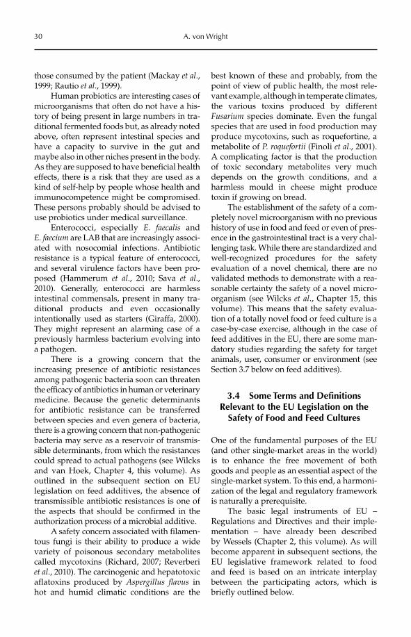
30 A. von Wright
those consumed by the patient (Mackay et al.,1999; Rautio et al., 1999).
Human probiotics are interesting cases of microorganisms that often do not have a his-tory of being present in large numbers in tra-ditional fermented foods but, as already noted above, often represent intestinal species and have a capacity to survive in the gut and maybe also in other niches present in the body. As they are supposed to have beneficial health effects, there is a risk that they are used as a kind of self-help by people whose health and immunocompetence might be compromised. These persons probably should be advised to use probiotics under medical surveillance.
Enterococci, especially E. faecalis and E. faecium are LAB that are increasingly associ-ated with nosocomial infections. Antibiotic resistance is a typical feature of enterococci, and several virulence factors have been pro-posed (Hammerum et al., 2010; Sava et al.,2010). Generally, enterococci are harmless intestinal commensals, present in many tra-ditional products and even occasionally intentionally used as starters (Giraffa, 2000). They might represent an alarming case of a previously harmless bacterium evolving into a pathogen.
There is a growing concern that the increasing presence of antibiotic resistances among pathogenic bacteria soon can threaten the efficacy of antibiotics in human or veterinary medicine. Because the genetic determinants for antibiotic resistance can be trans ferred between species and even genera of bacteria, there is a growing concern that non-pathogenic bacteria may serve as a reservoir of transmis-sible determinants, from which the resistances could spread to actual pathogens (see Wilcks and van Hoek, Chapter 4, this volume). As outlined in the subsequent section on EU legislation on feed additives, the absence of transmissible antibiotic resistances is one of the aspects that should be confirmed in the authorization process of a microbial additive.
A safety concern associated with filamen-tous fungi is their ability to produce a wide variety of poisonous secondary meta bolites called mycotoxins (Richard, 2007; Reverberi et al., 2010). The carcinogenic and hepatotoxic aflatoxins produced by Aspergillus flavus in hot and humid climatic conditions are the
best known of these and probably, from the point of view of public health, the most rele-vant example, although in temperate climates, the various toxins produced by different Fusarium species dominate. Even the fungal species that are used in food production may produce mycotoxins, such as roquefortine, a metabolite of P. roquefortii (Finoli et al., 2001). A complicating factor is that the production of toxic secondary metabolites very much depends on the growth conditions, and a harmless mould in cheese might produce toxin if growing on bread.
The establishment of the safety of a com-pletely novel microorganism with no previous history of use in food and feed or even of pres-ence in the gastrointestinal tract is a very chal-lenging task. While there are standardized and well-recognized procedures for the safety evaluation of a novel chemical, there are no validated methods to demonstrate with a rea-sonable certainty the safety of a novel micro-organism (see Wilcks et al., Chapter 15, this volume). This means that the safety evalua-tion of a totally novel food or feed culture is a case-by-case exercise, although in the case of feed additives in the EU, there are some man-datory studies regarding the safety for target animals, user, consumer or environment (see Section 3.7 below on feed additives).
3.4 Some Terms and Definitions Relevant to the EU Legislation on the
Safety of Food and Feed Cultures
One of the fundamental purposes of the EU (and other single-market areas in the world) is to enhance the free movement of both goods and people as an essential aspect of the single-market system. To this end, a harmoni-zation of the legal and regulatory framework is naturally a prerequisite.
The basic legal instruments of EU – Regulations and Directives and their imple-mentation – have already been described by Wessels (Chapter 2, this volume). As will become apparent in subsequent sections, the EU legislative framework related to food and feed is based on an intricate interplay between the participating actors, which is briefly outlined below.

Microbes for Human and Animal Consumption 31
The European Commission (henceforth ‘Commission’) is the government of the EU, and has both executive and – together with the European Parliament and the Council of the European Union – also legislative powers; it is the sole body that may initiate legislation. The Commission has several Directorates General, which form the executive branch of the Commission. Regarding microorganisms in food and feed, the Directorate General for Health and Consumers (SANCO) has the cen-tral regulatory role. SANCO is assisted by Standing Committees, the most important of which is the Standing Committee on the Food Chain and Animal Health (SCFCAH). The Standing Committee members represent member states and their interests. Each mem-ber state has a number of votes that are roughly proportional to the population of the country, with smaller countries, however, somewhat favoured. The decisions are made either unanimously or by a qualified majority. A qualified majority means that a decision requires votes from a combination of coun-tries that together make up the majority of the EU population in order to get accepted.
The Council of the European Union con-sists of the cabinet ministers of the member states. Which ministers participate in the meetings depends on the items on the agenda. For instance, if food and feed questions are discussed, then the ministers under whose responsibilities these matters fall will attend the meeting; for most member states, this is the minister of agriculture.
The EU has also several agencies, which have been established to perform specific tasks. The European Food Safety Authority (EFSA), established in 2002, performs risk assessments related to food and feed, while the risk man-agement measures belong to the Commission. The risk assessment is done by scientific panels consisting of independent experts, who must not represent their countries or institutions, but only science. Before the establishment of EFSA, the corresponding task was done by different Scientific Committees assisting SANCO, for instance the Scientific Committee for Food (SCF) or Scientific Committee on Animal Nutrition (SCAN). These committees are not to be confused with the Standing Committees already mentioned.
3.5 Starter Cultures – Additives, Processing Aids or Food Ingredients?
3.5.1 Situation in the EU
In the EU, there is presently no union-wide agreement on how the starter cultures used for food production should be legally defined and under what legislation they should be properly based. The two alternatives are either additives or processing aids. By definition, an additive is a substance that is intentionally added to a food for some specific technologi-cal purpose, and although an additive could also have some nutritional value, the intended use does not have to be nutritional. In con-trast, a processing aid is used to facilitate some process, and ideally it should no longer be present in the final product; there is no obliga-tion to list these among the constituents.
According to the recommendation of SCFCAH (SANCO, 2006), starters should be regarded mainly as processing aids. Only if a specific technological effect in food is sought, such as preservation, should a microbial cul-ture be considered as an additive. It is specifi-cally stated that probiotic cultures should not be classified as additives. So far, these recom-mendations have not led to any specific EU-wide harmonization of the national regu-lations. In view of recent developments, food cultures might be eventually classified as food ingredients, as pointed out by Wessels (Chapter 2, this volume).
While the situation with microbial cul-tures intended for food use currently is obscure in the EU, the case of microbial products intended for animals is very definite. These are unequivocally considered as feed additives, and this definition also covers the starters used for silage preparation. The relevant regulations are 1331/2003 EC and 767/2009 EC, and the requirements will be discussed in more detail under the specific sections on animal feed additives below (within Section 3.7).
3.5.2 The regulatory framework in the USA
In contrast to the EU, where the regulation of microbial cultures depends on whether they are
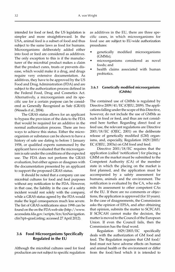
32 A. von Wright
intended for food or feed, the US legislation is simpler and more straightforward. In the USA, animal feed is a subset of food and thus subject to the same laws as food for humans. Microorganisms deliberately added either into food or feed are considered as additives. The only exception to this is if the manufac-turer of the microbial product makes a claim that the product cures, treats or prevents dis-ease, which would make it a drug, and drugs require very extensive documentation. As additives, they have to be approved by the US Food and Drug Administration (FDA) and are subject to the authorization process defined in the Federal Food, Drug and Cosmetics Act. Alternatively, a microorganism and its spe-cific use for a certain purpose can be consid-ered as Generally Recognized as Safe (GRAS) (Wessels et al., 2004).
The GRAS status allows for an applicant to bypass the provision of the data to the FDA that would be required for an additive in the normal authorization process. There are two ways to achieve this status. Either the micro-organism or substance can be shown to have a history of safe use dating to before 1 January 1958, or qualified experts summoned by the applicant have evaluated that the microorgan-ism is safe under the conditions of the expected use. The FDA does not perform the GRAS evaluation, but either agrees or disagrees with the documentation presented by an applicant to support the proposed GRAS status.
It should be noted that a company can use microbial cultures for food and feed purposes without any notification to the FDA. However, in that case, the liability in the case of a safety incident would rest solely with the company, while a GRAS status agreed by the FDA would make the legal consequences much less severe. The list of GRAS notifications since 1998 can be found on the on the FDA web site (http://www.accessdata.fda.gov/scripts/fcn/fcnNavigation.cfm?rpt=grasListing; accessed 27 April 2012).
3.6 Food Microorganisms Specifically Regulated in the EU
Although the microbial cultures used for food production are not subject to specific regulation
as additives in the EU, there are three spe-cific cases, in which microorganisms for food use are subject to EU-wide authorization procedures:
genetically modified microorganisms •(GMMs);microorganisms considered as novel •foods; andhealth claims associated with human •probiotics.
3.6.1 Genetically modified microorganisms (GMMs)
The contained use of GMMs is regulated by Directive 2009/41/EC (OJEU, 2009). The appli-cations falling under the scope of this Directive, however, do not include the use of GMMs as such in food or feed, and thus are not consid-ered here further. Regarding direct food or feed use, the relevant regulations are Directive 2001/18/EC (OJEC, 2001) on the deliberate release of genetically modified (GM) organ-isms, and, especially, Regulation 1829/2003/EC (OJEU, 2003a) on GM food and feed.
Directive 2001/18/EC requires that the application (called ‘notification’) for placing a GMM on the market must be submitted to the Competent Authority (CA) of the member state in which the placing on the market is first planned, and the application must be accompanied by a safety assessment for humans, animals and the environment. The notification is evaluated by the CA, who sub-mits its assessment to other competent CAs of the EU. If there are no comments or objec-tions, the application is approved (for 10 years). In the case of disagreements, the Commission asks the opinion of EFSA, and after obtaining that opinion, submits the matter to SCFCAH. If SCFCAH cannot make the decision, the matter is moved to the Council of the European Union. If even the Council fails, then the Commission has the final word.
Regulation 1829/2003/EC specifically deals with the authorization of GM food and feed. The Regulation requires that GM food/feed must not have adverse effects on human and animal health or the environment or differ from the food/feed which it is intended to
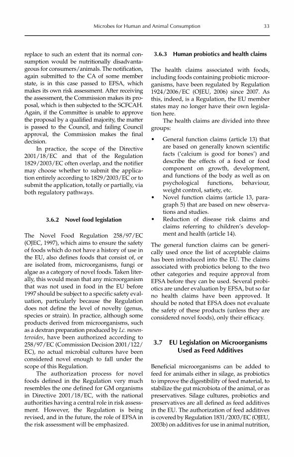
Microbes for Human and Animal Consumption 33
replace to such an extent that its normal con-sumption would be nutritionally disadvanta-geous for consumers/animals. The notification, again submitted to the CA of some member state, is in this case passed to EFSA, which makes its own risk assessment. After receiving the assessment, the Commission makes its pro-posal, which is then subjected to the SCFCAH. Again, if the Committee is unable to approve the proposal by a qualified majority, the matter is passed to the Council, and failing Council approval, the Commission makes the final decision.
In practice, the scope of the Directive 2001/18/EC and that of the Regulation 1829/2003/EC often overlap, and the notifier may choose whether to submit the applica-tion entirely according to 1829/2003/EC or to submit the application, totally or partially, via both regulatory pathways.
3.6.2 Novel food legislation
The Novel Food Regulation 258/97/EC (OJEC, 1997), which aims to ensure the safety of foods which do not have a history of use in the EU, also defines foods that consist of, or are isolated from, microorganisms, fungi or algae as a category of novel foods. Taken liter-ally, this would mean that any microorganism that was not used in food in the EU before 1997 should be subject to a specific safety eval-uation, particularly because the Regulation does not define the level of novelty (genus, species or strain). In practice, although some products derived from microorganisms, such as a dextran preparation produced by Lc. mesen-teroides, have been authorized according to 258/97/EC (Commission Decision 2001/122/EC), no actual microbial cultures have been considered novel enough to fall under the scope of this Regulation.
The authorization process for novel foods defined in the Regulation very much resembles the one defined for GM organisms in Directive 2001/18/EC, with the national authorities having a central role in risk assess-ment. However, the Regulation is being revised, and in the future, the role of EFSA in the risk assessment will be emphasized.
3.6.3 Human probiotics and health claims
The health claims associated with foods, including foods containing probiotic microor-ganisms, have been regulated by Regulation 1924/2006/EC (OJEU, 2006) since 2007. As this, indeed, is a Regulation, the EU member states may no longer have their own legisla-tion here.
The health claims are divided into three groups:
General function claims (article 13) that •are based on generally known scientific facts (‘calcium is good for bones’) and describe the effects of a food or food component on growth, development, and functions of the body as well as on psychological functions, behaviour, weight control, satiety, etc.Novel function claims (article 13, para-•graph 5) that are based on new observa-tions and studies.Reduction of disease risk claims and •claims referring to children’s develop-ment and health (article 14).
The general function claims can be generi-cally used once the list of acceptable claims has been introduced into the EU. The claims associated with probiotics belong to the two other categories and require approval from EFSA before they can be used. Several probi-otics are under evaluation by EFSA, but so far no health claims have been approved. It should be noted that EFSA does not evaluate the safety of these products (unless they are considered novel foods), only their efficacy.
3.7 EU Legislation on Microorganisms Used as Feed Additives
Beneficial microorganisms can be added to feed for animals either in silage, as probiotics to improve the digestibility of feed material, to stabilize the gut microbiota of the animal, or as preservatives. Silage cultures, probiotics and preservatives are all defined as feed additives in the EU. The authorization of feed additives is covered by Regulation 1831/2003/EC (OJEU, 2003b) on additives for use in animal nutrition,
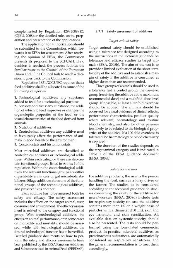
34 A. von Wright
complemented by Regulation 429/2008/EC (OJEU, 2008) on the detailed rules on the prep-aration and presentation of the applications.
The application for authorization should be submitted to the Commission, which for-wards it to EFSA for assessment. After receiv-ing the opinion of EFSA, the Commission presents its proposal to the SCFCAH. If no decision is reached, the process follows the familiar route to the Council of the European Union and, if the Council fails to reach a deci-sion, it goes back to the Commission.
Regulation 1831/2003/EC requires that a feed additive shall be allocated to some of the following categories:
1. Technological additives: any substance added to feed for a technological purpose.2. Sensory additives: any substance, the addi-tion of which to feed improves or changes the organoleptic properties of the feed, or the visual characteristics of the food derived from animals.3. Nutritional additives.4. Zootechnical additives: any additive used to favourably affect the performance of ani-mals in good health or the environment.5. Coccidiostats and histomonostats.
Most microbial additives are classified as zootechnical additives or technological addi-tives. Within each category, there are also cer-tain functional groups, listed in Annex I of the regulation. Within the zootechnological addi-tives, the relevant functional groups are either digestibility enhancers or gut microbiota sta-bilizers. Silage additives form one of the func-tional groups of the technological additives, and preservatives another.
Each additive has to be assessed both for safety and efficacy. The safety assessment includes the effects on the target animal, user, consumer and environment. The efficacy assess-ment is related to the category and functional group. With zootechnological additives, the effects on animal performance, or in some cases on morbidity and mortality, should be asses-sed, while with technological additives, the desired technological function has to be verified. Detailed guidance documents on how to per-form the safety and efficacy assessments have been published by the EFSA Panel on Additives and Substances used in Animal Feed (FEEDAP).
3.7.1 Safety assessment of additives
Target animal safety
Target animal safety should be established using a tolerance test designed according to the instructions in the technical guidance on tolerance and efficacy studies in target ani-mals (EFSA, 2008b). The aim of the test is to provide a limited evaluation of the short-term toxicity of the additive and to establish a mar-gin of safety if the additive is consumed at higher doses than are recommended.
Three groups of animals should be used in a tolerance test: a control group, the use-level group (receiving the additive at the maximum recommended dose) and a multifold-dose-level group. If possible, at least a tenfold overdose should be applied. The animals should be observed for visual evidence of clinical effects, performance characteristics, product quality where relevant, haematology and routine blood chemistry, and also for other parame-ters likely to be related to the biological prop-erties of the additive. If a 100-fold overdose is tolerated, no haematology or blood chemistry is required.
The duration of the studies depends on the target animal category and is indicated in Table 1 of the EFSA guidance document (EFSA, 2008b).
Safety for the user
For additive products, the user is the person handling the feed, such as a lorry driver or the farmer. The studies to be considered according to the technical guidance on stud-ies concerning the safety of the additive for users/workers (EFSA, 2008d) include tests for respiratory toxicity (in case the additive contains more than 1% on a weight basis of particles with a diameter £ 50 mm), skin and eye irritation, and skin sensitization. All available data on systemic toxicity should also be presented. The tests should be per-formed using the formulated commercial product. In practice, microbial additives, as proteinaceous substances, are automatically considered as respiratory sensitizers, and the general recommendation is to treat them accordingly.
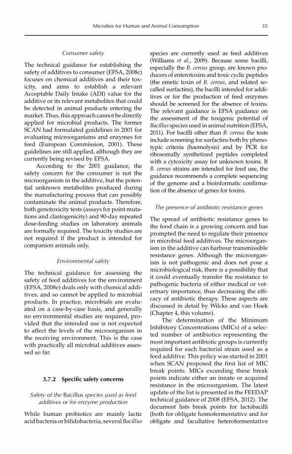
Microbes for Human and Animal Consumption 35
Consumer safety
The technical guidance for establishing the safety of additives to consumer (EFSA, 2008c) focuses on chemical additives and their tox-icity, and aims to establish a relevant Acceptable Daily Intake (ADI) value for the additive or its relevant metabolites that could be detected in animal products entering the market. Thus, this approach cannot be directly applied for microbial products. The former SCAN had formulated guidelines in 2001 for evaluating microorganisms and enzymes for feed (European Commission, 2001). These guidelines are still applied, although they are currently being revised by EFSA.
According to the 2001 guidance, the safety concern for the consumer is not the microorganism in the additive, but the poten-tial unknown metabolites produced during the manufacturing process that can possibly contaminate the animal products. Therefore, both genotoxicity tests (assays for point muta-tions and clastogenicity) and 90-day repeated dose-feeding studies on laboratory animals are formally required. The toxicity studies are not required if the product is intended for companion animals only.
Environmental safety
The technical guidance for assessing the safety of feed additives for the environment (EFSA, 2008e) deals only with chemical addi-tives, and so cannot be applied to microbial products. In practice, microbials are evalu-ated on a case-by-case basis, and generally no environmental studies are required, pro-vided that the intended use is not expected to affect the levels of the microorganism in the receiving environment. This is the case with practically all microbial additives asses-sed so far.
3.7.2 Specific safety concerns
Safety of the Bacillus species used as feed additives or for enzyme production
While human probiotics are mainly lactic acid bacteria or bifidobacteria, several Bacillus
species are currently used as feed additives (Williams et al., 2009). Because some bacilli, especially the B. cereus group, are known pro-ducers of enterotoxins and toxic cyclic peptides (the emetic toxin of B. cereus, and related so-called surfactins), the bacilli intended for addi-tives or for the production of feed enzymes should be screened for the absence of toxins. The relevant guidance is EFSA guidance on the assessment of the toxigenic potential of Bacillus species used in animal nutrition (EFSA, 2011). For bacilli other than B. cereus the tests include screening for surfactins both by pheno-typic criteria (haemolysis) and by PCR for ribosomally synthetized peptides completed with a cytoxicity assay for unknown toxins. If B. cereus strains are intended for feed use, the guidance recommends a complete sequencing of the genome and a bioinformatic confirma-tion of the absence of genes for toxins.
The presence of antibiotic resistance genes
The spread of antibiotic resistance genes to the food chain is a growing concern and has prompted the need to regulate their presence in microbial feed additives. The microorgan-ism in the additive can harbour transmissible resistance genes. Although the microorgan-ism is not pathogenic and does not pose a microbiological risk, there is a possibility that it could eventually transfer the resistance to pathogenic bacteria of either medical or vet-erinary importance, thus decreasing the effi-cacy of antibiotic therapy. These aspects are discussed in detail by Wilcks and van Hoek (Chapter 4, this volume).
The determination of the Minimum Inhibitory Concentrations (MICs) of a selec-ted number of antibiotics representing the most important antibiotic groups is currently required for each bacterial strain used as a feed additive. This policy was started in 2001 when SCAN proposed the first list of MIC break points. MICs exceeding these break points indicate either an innate or acquired resistance in the microorganism. The latest update of the list is presented in the FEEDAP technical guidance of 2008 (EFSA, 2012). The document lists break points for lactobacilli (both for obligate homofermentative and for obligate and facultative heterofermentative
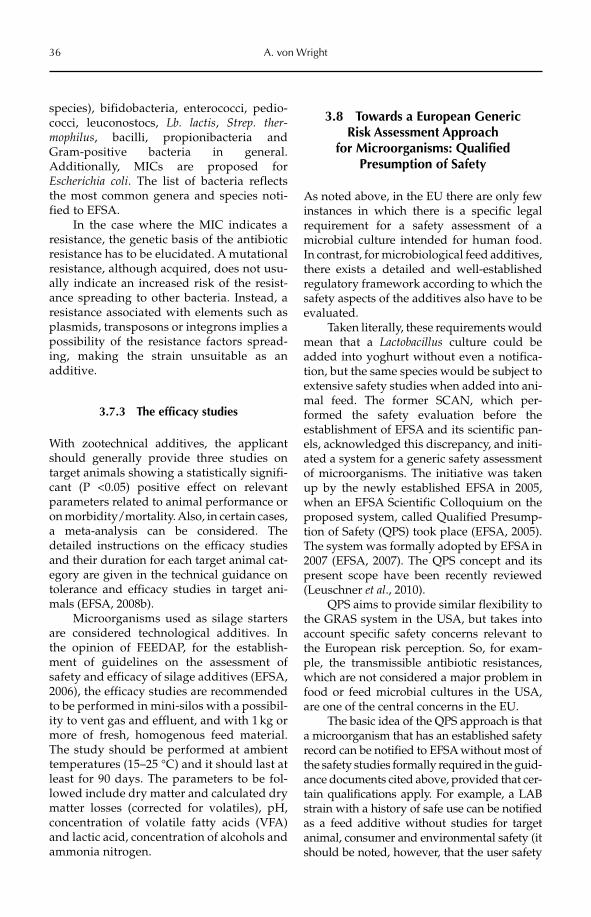
36 A. von Wright
species), bifidobacteria, enterococci, pedio-cocci, leuconostocs, Lb. lactis, Strep. ther-mophilus, bacilli, propionibacteria and Gram-positive bacteria in general. Additionally, MICs are proposed for Escherichia coli. The list of bacteria reflects the most common genera and species noti-fied to EFSA.
In the case where the MIC indicates a resistance, the genetic basis of the antibiotic resistance has to be elucidated. A mutational resistance, although acquired, does not usu-ally indicate an increased risk of the resist-ance spreading to other bacteria. Instead, a resistance associated with elements such as plasmids, transposons or integrons implies a possibility of the resistance factors spread-ing, making the strain unsuitable as an additive.
3.7.3 The efficacy studies
With zootechnical additives, the applicant should generally provide three studies on target animals showing a statistically signifi-cant (P <0.05) positive effect on relevant parameters related to animal performance or on morbidity/mortality. Also, in certain cases, a meta-analysis can be considered. The detailed instructions on the efficacy studies and their duration for each target animal cat-egory are given in the technical guidance on tolerance and efficacy studies in target ani-mals (EFSA, 2008b).
Microorganisms used as silage starters are considered technological additives. In the opinion of FEEDAP, for the establish-ment of guidelines on the assessment of safety and efficacy of silage additives (EFSA, 2006), the efficacy studies are recommended to be performed in mini-silos with a possibil-ity to vent gas and effluent, and with 1 kg or more of fresh, homogenous feed material. The study should be performed at ambient temperatures (15–25 °C) and it should last at least for 90 days. The parameters to be fol-lowed include dry matter and calculated dry matter losses (corrected for volatiles), pH, concentration of volatile fatty acids (VFA) and lactic acid, concentration of alcohols and ammonia nitrogen.
3.8 Towards a European Generic Risk Assessment Approach
for Microorganisms: Qualified Presumption of Safety
As noted above, in the EU there are only few instances in which there is a specific legal requirement for a safety assessment of a microbial culture intended for human food. In contrast, for microbiological feed additives, there exists a detailed and well-established regulatory framework according to which the safety aspects of the additives also have to be evaluated.
Taken literally, these requirements would mean that a Lactobacillus culture could be added into yoghurt without even a notifica-tion, but the same species would be subject to extensive safety studies when added into ani-mal feed. The former SCAN, which per-formed the safety evaluation before the establishment of EFSA and its scientific pan-els, acknowledged this discrepancy, and initi-ated a system for a generic safety assessment of microorganisms. The initiative was taken up by the newly established EFSA in 2005, when an EFSA Scientific Colloquium on the proposed system, called Qualified Presump-tion of Safety (QPS) took place (EFSA, 2005). The system was formally adopted by EFSA in 2007 (EFSA, 2007). The QPS concept and its present scope have been recently reviewed (Leuschner et al., 2010).
QPS aims to provide similar flexibility to the GRAS system in the USA, but takes into account specific safety concerns relevant to the European risk perception. So, for exam-ple, the transmissible antibiotic resistances, which are not considered a major problem in food or feed microbial cultures in the USA, are one of the central concerns in the EU.
The basic idea of the QPS approach is that a microorganism that has an established safety record can be notified to EFSA without most of the safety studies formally required in the guid-ance documents cited above, provided that cer-tain qualifications apply. For example, a LAB strain with a history of safe use can be notified as a feed additive without studies for target animal, consumer and environmental safety (it should be noted, however, that the user safety
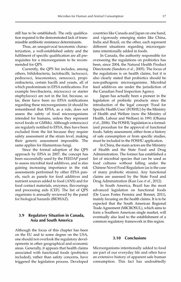
Microbes for Human and Animal Consumption 37
still has to be established). The only qualifica-tion required is the demonstrated lack of trans-missible antibiotic resistance determinants.
Thus, an unequivocal taxonomic charac-terization, a well-established safety and the fulfilment of specific qualification are all pre-requisites for a microorganism to be recom-mended for QPS.
Currently, the QPS list includes, among others, bifidobacteria, lactobacilli, lactococci, pediococci, leuconostocs, oenococci, propi-onibacteria, certain bacilli and yeasts, all of which predominate in EFSA notifications. For example brevibacteria, micrococci or starter staphylococci are not in the list, because, so far, there have been no EFSA notifications regarding these microorganisms (it should be remembered that EFSA, as a rule, does not assess the safety of food microorganisms intended for humans, unless they represent novel foods or GMMs). Although enterococci are regularly notified to EFSA, they have been excluded from the list because they require safety assessment at the strain level, making their generic assessment impossible. The same applies for filamentous fungi.
Since the formal adoption of the QPS approach by EFSA in 2007, the concept has been successfully used by the FEEDAP panel to assess microbial feed additives, and is also gaining increasing importance in the risk assessments performed by other EFSA pan-els, such as panels for food additives and nutrient sources added to food (ANS) and for food contact materials, enzymes, flavourings and processing aids (CEF). The list of QPS organisms is annually reviewed by the panel for biological hazards (BIOHAZ).
3.9 Regulatory Situation in Canada, Asia and South America
Although the focus of this chapter has been on the EU and to some degree on the USA, one should not overlook the regulatory devel-opments in other geographical and economic areas. Generally, it appears that health claims associated with functional foods (probiotics included), rather than safety concerns, have triggered the legislation process. Developed
countries like Canada and Japan on one hand, and vigorously emerging states like China, India and Brazil, on the other, each represent different situations regarding microorgan-isms intentionally added in foods.
In Canada, the authority responsible for overseeing the regulations on probiotics has been, since 2004, the Natural Health Product Directorate (Sanders et al., 2005). The focus of the regulations is on health claims, but it is also clearly stated that probiotics should be non-pathogenic microorganisms. Microbial feed additives are under the jurisdiction of the Canadian Feed Inspection Agency.
Japan has actually been a pioneer in the legislation of probiotic products since the introduction of the legal concept ‘Food for Specific Health Uses’ (FOSHU) by the Ministry of Health and Welfare (now the Ministry of Health, Labour and Welfare) in 1991 (Ohama et al., 2006). The FOSHU legislation is a formal-ized procedure for the approval of functional foods. Safety assessment, either from a history of safe consumption or from specific studies, must be included in the FOSHU application.
In China, the main actors are the Ministry of Health and the State Food and Drug Administration. The former has established a list of microbial species that can be used as food cultures without falling under the Chinese Novel Food Regulation (as is the case of many probiotic strains). Any functional claims are assessed by the State Food and Drug Administration (Kun Lee et al., 2012).
In South America, Brazil has the most advanced legislation on functional foods (De Luces Fortes Ferreira and Bonnet, 2011), mainly focusing on the health claims. It is to be expected that the South American Regional Trade Agreement (MICROSUL), which aims to form a Southern American single market, will eventually also lead to the establishment of a common regulatory framework in this region.
3.10 Conclusions
Microorganisms intentionally added to food are part of our everyday life and often have an extensive history of apparent safe human consumption. This fact has undoubtedly
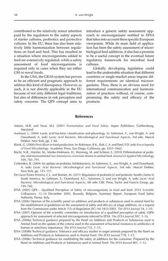
38 A. von Wright
contributed to the relatively minor attention paid by the regulators to the safety aspects of starter cultures, probiotics and protective cultures. In the EU, there has also been rela-tively little harmonization between regula-tions on food and feed. This has resulted in a situation where microorganisms added to feed are extensively regulated, while a safety assessment of food microorganisms is required only in cases when they are either GM or novel foods.
In the USA, the GRAS system has proven to be an efficient and pragmatic approach to address this kind of discrepancy. However, as such, it is not directly applicable in the EU because of not only different legal traditions, but also of differences in risk perception and safety concerns. The QPS concept aims to
introduce a generic safety assessment app-roach to microorganisms notified to EFSA that takes into account these specific European viewpoints. While its main field of applica-tion has been the safety assessment of micro-biological feed additives, it also has a promise to be a useful concept in the eventual future regulatory framework for microbial food cultures.
Globally developing legislation could lead to the undesirable situation that different countries or single-market areas impose dif-ferent requirements on identical microor-ganisms. Thus, there is an obvious need for international communication and harmoni-zation of practices without, of course, com-promising the safety and efficacy of the products.
References
Adams, M.R. and Nout, M.J. (2001) Fermentation and Food Safety. Aspen Publishers, Gaithersburg, Maryland.
Axelsson, L. (2004) Lactic acid bacteria: classification and physiology. In: Salminen, S., von Wright, A. and Ouwehand, A. (eds) Lactic Acid Bacteria: Microbiological and Functional Aspects, 3rd edn. Marcel Dekker, New York, pp. 1–66.
Blank, G. (2000) Penicillium in food production. In: Robinson, R.K., Batt, C.A. and Patel, P.D. (eds) Encyclopedia of Food Microbiology. Academic Press, San Diego, California, pp. 1655–1662.
Büchi, N.R., Hutzler, M., Mietke-Hofmann, H., Wenning, M. and Scherer, S. (2010) Differentiation of probi-otic and environmental Saccharomyces cerevisiae strains in animal feed. Journal of Applied Microbiology109, 783–791.
Crittenden, R. (2004) An update on probiotic bifidobacteria. In: Salminen, S., von Wright, A. and Ouwehand, A. (eds) Lactic Acid Bacteria: Microbiological and Functional Aspects, 3rd edn. Marcel Dekker, New York, pp. 125–157.
De Luces Fortes Ferreira, C.L. and Bonnet, M. (2011) Regulation of probiotic[s] and probiotic health claims in South America. In: Lahtinen, S., Ouwehand, A.C., Salminen, S. and von Wright, A. (eds) Lactic Acid Bacteria: Microbiological and Functional Aspects, 4th edn. CRC Press, Taylor and Francis Group, pp. 749–760.
EFSA (2005) QPS – Qualified Perception of Safety of microorganisms in food and feed. EFSA Scientific Colloquium, 13–14 December 2005, Brussels, Belgium, Summary Report. European Food Safety Authority, Parma, Italy.
EFSA (2006) Opinion of the scientific panel on additives and products or substances used in animal feed for the establishment of guidelines on the assessment of safety and efficacy of silage additives, on a request from the Commission under Article 7(5) of Regulation (EC) No 1831/2003. The EFSA Journal 34, 1–10.
EFSA (2007) Opinion of the scientific committee on introduction of a qualified perception of safety (QPS) approach for assessment of selected microorganisms referred to EFSA. The EFSA Journal 587, 1–16.
EFSA (2008a) Technical guidance prepared by the Panel on Additives and Products or Substances used in Animal Feed on the update of the criteria used in the assessment of bacterial resistance to antibiotics of human or veterinary importance. The EFSA Journal 732, 1–15.
EFSA (2008b) Technical guidance. Tolerance and efficacy studies in target animals prepared by the Panel on Additives and Products or Substances used in Animal Feed. The EFSA Journal 778, 1–13.
EFSA (2008c) Technical guidance for establishing the safety of additives for the consumer. Prepared by the Panel on Additives and Products or Substances used in Animal Feed. The EFSA Journal 801, 1–12.
Microbes for Human and Animal Consumption 39
EFSA (2008d) Technical guidance. Studies concerning the safety of use of the additive for users/workers pre-pared by the Panel on Additives and Products or Substances used in Animal Feed. The EFSA Journal 802, 1–2.
EFSA (2008e) Technical guidance for assessing the safety of feed additives for the environment prepared by the Panel on Additives and Products or Substances used in Animal Feed. The EFSA Journal 842, 1–28.
EFSA (2011) Technical Guidance on the assessment of the toxigenic potential of Bacillus species used in animal nutrition: ESFA Panel on Additives and Products of Substances used in Animal Feed (FEEDAP), The EFSA Journal 2445, 1–13.
EFSA (2012) Guidance on the assessment of bacterial susceptibility to antimicrobials of human and veterinary importance: EFSA Panel on Additives and Products or Substances used in Animal Feed (FEEDAP); TheEFSA Journal 2740, 1–10.
European Commission (2001) Guidelines for the Assessment of Additives in Feeding Stuffs, Part II. Enzymes and Micro-organisms. Available at: http://ec.europa.eu/food/fs/sc/scan/out68_en.pdf (accessed 27 April 2012).
European Commission (2012) European Union Register of Feed Additives pursuant of Regulation (EC) No 1831/2003. Appendices 3c and 4. Annex: List of Additives, Edn 142, Health and Consumers Directorate General, Brussels.
Finoli, C., Vecchio, A., Galli, A. and Dragoni, I. (2001) Roquefortine C occurrence in blue cheese. Journal of Food Protection 64, 246–251.
García-Lopez, M.-L., Sanots, J.-Á. and Otero, A. (2000) Micrococci. In: Robinson, R.K., Batt, C.A. and Patel, P.D. (eds) Encyclopedia of Food Microbiology. Academic Press, San Diego, California, pp. 1344–1350.
Gasser, F. (1994) Safety of lactic acid bacteria and their occurrence in human clinical infections. Bulletin de l’Institut Pasteur 92, 45–67.
Geisen, R., Lücke, F.-K. and Kröckel, L. (1992) Starter and protective cultures for meat and meat products. Fleishwitschaft 72, 894–898.
Giraffa, G. (2000) Enterococci from foods. FEMS Microbiology Review 26, 163–171.Gomi, K. (2000) Aspergillus oryzae. In: Robinson, R.K., Batt, C.A. and Patel, P.D. (eds) Encyclopedia of Food
Microbiology. Academic Press, San Diego, California, pp. 66–72.Hammerum, A.M., Lester, C.H. and Heuer, O.E. (2010) Antimicrobial resistant enterococci in animals and
meat: a human health hazard? Foodborne Pathogens and Disease 10, 1137–1146.Kun Lee, Y., Shao, W., Su, J., Yan, W., Ganguly, B., Rahayu, E.S., Chonan, O., Watanabe, K., Eong Ji, G., Soo
Park, M., Abd Rahim, R., Ling Foo, H., Tan, J.D., Chen, M-J. and Nitisinprasert, S. (2012) Probiotics regu-lation in Asian countries. In: Lahtinen, S., Ouwehand, A.C., Salminen, S. and von Wright, A. (eds) Lactic Acid Bacteria: Microbiological and Functional Aspects, 4th edn. CRC Press, Taylor and Francis Group, pp. 705–748.
Leuschner, R.G.K., Robinson, T.P., Hugas, M., Cocconcelli, P.S., Richard-Forget, F., Klein, G., Licht, T.R., Ngyen-The, C., Querol, A., Suarez, J.E., Thrane, U., Vlak, J.M. and von Wright, A. (2010) Qualified pre-sumption of safety (QPS): a generic risk assessment approach for biological agents notified to the European Food Safety Authority (EFSA). Trends in Food Science and Technology 21, 425–435.
Lherm, T., Monet, C., Nougière, B., Soulier, M., Larbi, D., Le Gall, C., Caen, D. and Malbrunot, C. (2002) Seven cases of fungaemia with Saccharomyces boulardii in critically ill patients. Intensive Care Medicine28, 797–801.
Mackay, A.D., Taylor, M.B., Kibbler, C.C. and Hamilton-Miller, J.M.T. (1999) Lactobacillus endocarditis caused by a probiotic organism. Clinical Microbiology and Infections 5, 290–292.
Nout, M.J.R. (2001) Fermented foods and their production. In: Adams, M.R. and Nout, M.J (eds) Fermentation and Food Safety. Aspen Publishers, Gaithersburg, Maryland, pp. 1–38.
Ohama, H., Ikeda, H. and Moriyama, H. (2006) Health foods and foods with health claims in Japan. Toxicology221, 95–111.
OJEC (1997) Regulation (EC) No 258/97 of the European Parliament and of the Council of 27 January 1997 concerning novel foods and novel food ingredients. Official Journal of the European Communities 40, L 43/1–6.
OJEC (2001) Directive 2001/18/EC of the European Parliament and of the Council of 12 March 2001 on the deliberate release into the environment of genetically modified organisms and repealing Council Directive 90/220/EEC. Official Journal of the European Communities 44, L 101/1–38.
OJEU (2003a) Regulation (EC) No 1829/2003 of The European Parliament and of the Council of 22 September 2003 on genetically modified food and feed. Official Journal of the European Union 46, L 268/1–23.
OJEU (2003b) Regulation (EC) No 1831/2003 of the European Parliament and of the Council of 22 September 2003 on additives for use in animal nutrition. Official Journal of the European Union 46, L 268/29–43.
40 A. von Wright
OJEU (2006) Regulation (EC) No 1924/2006 of the European Parliament and of the Council of 20 December 2006 on nutrition and health claims made on foods. Official Journal of the European Union 49, L 404/9–25.
OJEU (2008) Commission Regulation (EC) No 429/2008 of 25 April 2008 on detailed rules for the implementation of Regulation (EC) No 1831/2003 of the European Parliament and of the Council as regards the preparation and the presentation of applications and the assessment and the authorisation of feed additives. Official Journal of the European Union 51, L 133/1–65.
OJEU (2009) Directive 2009/41/EC of The European Parliament and of the Council of 6 May 2009 on the contained use of genetically modified micro-organisms. Official Journal of the European Union 52, L 125/75–97.
Ouwehand, A. (2004) The probiotic potential of propionibacteria. In: Salminen, S., von Wright, A. and Ouwehand, A. (eds) Lactic Acid Bacteria: Microbiological and Functional Aspects. Marcel Dekker, New York, pp. 159–174.
Rautio, M., Somer-Jousimies, H., Kauma, H., Pietarinen, I., Saxelin, M., Tynkkynen, S. and Koskela, M. (1999) Liver abscess due to a Lactobacillus rhamnosus strain indistinguishable from L. rhamnosus strain GG. Clinical Infectious Diseases 28, 1159–1160.
Reverberi, M., Riselli, A., Zjalic, S., Fabbri, A.A. and Fanelli, C. (2010) Natural functions of mycotoxins and control of their biosynthesis in fungi. Applied Microbiology and Biotechnology 87, 899–911.
Richard, J.L. (2007) Some major mycotoxins and their mycotoxicoses – an overview. International Journal of Food Microbiology 119, 3–10.
SANCO (2006) Summary record of the Standing Committee on the Food Chain and Animal Health held in Brussels on 14 December 2006: Section Toxicological Safety on the Food Chain. Document No. SANCO – D1(06)D/413447, Directorate General for Health and Consumers (SANCO), Brussels. Available at: http://ec.europa.eu/food/committees/regulatory/scfcah/toxic/summary23_en.pdf (accessed 27 April 2012).
Sanders, M.E., Tompkins, T., Heimbach, J.T. and Kolida, S. (2005) Weight of evidence needed to substantiate a health effect for probiotics and prebiotics. Regulatory considerations in Canada, E.U., and U.S. European Journal of Nutrition 44, 303–310.
Sava, I.G., Heikens, E. and Huebner, J. (2010) Pathogenesis and immunity in enterococcal infections. ClinicalMicrobiology and Infection 16, 533–540.
Saxelin, M., Chuang, N.-H., Chassy, B., Rautelin, H., Mäkelä, P.H., Salminen, S. and Gorbach, S.L. (1996) Lactobacilli and bacteremia in Southern Finland. Clinical Infectious Diseases 22, 564–566.
Silva, J., Gonzales, S., Palcios, J. and Oliver, G. (2000) Fungi. In: Robinson, R.K., Batt, C.A. and Patel, P.D. (eds) Encyclopedia of Food Microbiology. Academic Press, San Diego, California, pp. 850–898.
Viljoen, B.C. and Heard, G.M. (2000) Saccharomyces cerevisiae. In: Robinson, R.K., Batt, C.A. and Patel, P.D. (eds) Encyclopedia of Food Microbiology. Academic Press, San Diego, California, pp. 1918–1984.
Weimer, B. (2000) Brevibacterium. In: Robinson, R.K., Batt, C.A. and Patel, P.D. (eds) Encyclopedia of Food Microbiology. Academic Press, San Diego, California, pp. 309–314.
Wessels, S. (2012) Safety and regulation of microorganisms added to the food and feed chains, including probiotics – Introduction and overview. In: Sundh, I., Wilcks, A. and Goettel, M.S. (eds) BeneficialMicroorganisms in Agriculture, Food and the Environment: Safety Assessment and Regulation. CAB International, Wallingford, UK, pp. 12–26.
Wessels, S., Axelsson, L., Hansen, E.B., De Vuyst, L., Laulund, S., Lähteenmäki, L., Lindgren, S., Mollet, B., Salminen, S. and von Wright, A. (2004) The lactic acid bacteria, the food chain, and their regulation. Trends in Food Science and Technology 15, 498–505.
Wilcks, A., Goettel, M.S. and Sundh, I. (2012) Model systems for testing microbial pathogenicity, virulence and toxicity – introduction and overview. In: Sundh, I., Wilcks, A. and Goettel, M.S. (eds) BeneficialMicroorganisms in Agriculture, Food and the Environment: Safety Assessment and Regulation. CAB International, Wallingford, UK, pp. 217–222.
Williams, L.D., Burdock, G.A., Jiménez, G. and Castillo, M. (2009) Literature review of Toyoserin®, a non-toxigenic and non-pathogenic Bacillus cereus var. toyoi preparation. Regulatory Toxicology and Pharmacology 55, 236–246.
©CAB International 2012. Beneficial Microorganisms in Agriculture, Food and the Environment: Safety Assessment and Regulation (eds I. Sundh et al.) 41
4.1 Introduction 414.2 Antibiotic Resistance in Food-associated Bacteria 424.3 Phenotypic and Genotypic Methods for Assessing Antibiotic Resistance 43
4.3.1 Phenotypic methods 43 4.3.2 Genotypic methods 43 4.3.3 Phenotypic versus genotypic assessment 46
4.4 Horizontal Gene Transfer and Assessing Transferability 474.4.1 Mechanisms of transfer 47
4.4.2 Mobile elements found in strains used as starter cultures and probiotics 47 4.4.3 Methods/models to study transfer 50 4.4.4 Curing for antibiotic resistance 51
4.5 Conclusion 51
4 Antibiotic Resistance in Relation to Starter Cultures and Probiotics
Andrea Wilcks1 and Angela H.A.M. van Hoek2
1CBRN-institute, Danish Emergency Management Agency, Denmark; 2Laboratory for Zoonoses and Environmental Microbiology (LZO), National Institute for Public
Health and the Environment (RIVM), The Netherlands
4.1 Introduction
In recent years, discussions have been raised as to whether bacteria used as starter cultures (fermentation starters for, e.g. the production of meat or cheeses) and probiotics (live micro-organisms which when administered in ade-quate amounts confer a health benefit on the host; FAO/WHO, 2001) could act as reser-voirs for antibiotic resistance (AR) genes. When ingesting these bacteria, there is a risk that resistance elements could be transferred to the indigenous microbiota of the gut, and subsequently to pathogenic bacteria. This chapter summarizes current knowledge on AR genes and mobile elements in the genera used as starter cultures and probiotics, and on pitfalls in determining antibiotic resistance in specific strains.
A risk-based approach has been chosen in this chapter, focusing on the most impor-tant bacterial species used as starter cultures or probiotics and with AR genes relevant for human and animal health. Bacterial species of the lactic acid bacteria (LAB), bifidobacteria and coagulase-negative staphylococci are treated. We also focus on tetracycline and erythromycin, because they are two of the most widely used antibiotics in both clinical and animal therapy, and their resistance determinants are most commonly found among food-associated bacteria (Roberts, 2005, 2008; van Hoek et al., 2008a).
The determination of antibiotic resist-ance can be a challenging exercise. A gene may be detected, but is it functional and will it be expressed? Therefore, both phenotypic and genotypic analyses are important.
42 A. Wilcks and A.H.A.M. van Hoek
Approved standards for phenotypic and gen-otypic determination of the presence of anti-biotic resistance in food isolates are scarce, as are standards for the specification of mini-mum inhibitory concentration (MIC) values (Huys et al., 2010; Mayrhofer et al., 2010).
In a recently published technical guid-ance report (EFSA, 2008), the Panel on Additives and Products or Substances used in Animal Feed (FEEDAP) of the European Food Safety Authority (EFSA) defined MIC break points to categorize bacteria as susceptible or resistant. The report also classified antimicro-bial resistance into three categories: (i) intrin-sic or natural resistance inherent to a bacterial species; (ii) acquired resistance caused by the mutation of indigenous genes; and (iii) acquired resistance due to the acquisition of exogenous resistance genes. FEEDAP con-cluded that strains carrying the third class of resistance are unacceptable as animal feed additives (EFSA, 2008). This is also reflected in the EU Council Regulation 429/2008 (OJEU, 2008), where it is stated that microorganisms used for feed production should be free of resistance towards antibiotics relevant for humans and animals. If resistance is discov-ered, the genetic basis for the resistance shall be revealed, and the likelihood of transfer to the gut microbiota shall be assessed. Only if it can be shown that the antibiotic resistance is a result of a chromosomal mutation(s), and non-transferable, can the strain be used.
EFSA has taken responsibility in launch-ing the European initiative towards a ‘Quali-fied Presumption of Safety’ (QPS) concept (see von Wright, Chapter 3, this volume) which, like the ‘Generally Recognized as Safe’ (GRAS) system in the USA, is aimed to allow strains belonging to species with an established history and safety status to enter the market without extensive testing requirements. Thepresence of transmissible antibiotic resistance markers in the evaluation of strains is an important safety criterion. EFSA considers resistance to antibiotics, especially transfera-ble resistance, a safety concern and a decision criterion for determining a strain’s QPS sta-tus (EFSA, 2007). Distinguishing between intrinsic and acquired resistance is not always a straightforward process though and may require the comparison of antimicrobial
resistance patterns in many strains of the same species from different sources.
4.2 Antibiotic Resistance in Food-associated Bacteria
As mentioned previously, the risk presented by AR genes is highest when they are located on mobile elements – such as plasmids and transposons – and therefore prone to transfer to other bacteria. However, resistance towards kanamycin, streptomycin, trimethoprim and vancomycin is often intrinsic, or a result of the mutation of indigenous genes. For instance, mutations of the rpsL gene for the ribosomal protein S12 can be responsible for streptomycin resistance in, e.g. Bifidobacterium(Kiwaki and Sato, 2009; Sato and Iino, 2010). Resistance due to mutation can also occur to erythromycin and tetracycline, the two anti-biotics in focus in this chapter. An example is the erythromycin resistance of the commer-cial strain B. bifidum strain Yakult YIT 400, which is evidently linked to mutation in the 23S ribosomal RNA genes located on the chromosome (Sato and Iino, 2010), and there-fore the risk of transfer is minimal. Then again, acquired antibiotic resistance mechanisms are increasingly described in the literature on beneficial and/or starter culture bacteria. For example, a Belgian study found a poten-tially probiotic strain of Lactobacillus plantarumcontaining a tet(S) gene giving tetracycline resistance which was located on a plasmid of approximately 14 Kb (Huys et al., 2006).
Examples of acquired AR genes in Bifido-bacterium spp., Enterococcus spp., Lactobacillusspp., Lactococcus spp., Streptococcus thermophilusand coagulase-negative Staphylococcus spp. are presented in Table 4.1. It is evident that tetracycline and erythromycin resistance determinants are widespread in species con-sidered as beneficial and widely present in our food. The most frequently found tetracy-cline determinant is tet(M) (Roberts, 2005). In recent years, tet(W) has become nearly as prominent, although it has not been detected in all genera associated with food and feed (Cauwerts et al., 2006a; Kastner et al., 2006; Aires et al., 2007; van Hoek et al., 2008a). With
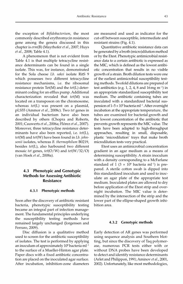
Antibiotic Resistance 43
the exception of Bifidobacterium, the most commonly described erythromycin resistance gene among the genera discussed in this chapter is erm(B) (Mayrhofer et al., 2007; Huys et al., 2008; Table 4.1).
A phenomenon that is not evident from Table 4.1 is that multiple tetracycline resist-ance determinants can be found in a single isolate. This was, for instance, demonstrated for the Sola cheese Lb. sakei isolate RitS 9 which possesses two different tetracycline resistance mechanisms, i.e. the ribosomal resistance protein Tet(M) and the tet(L) deter-minant coding for an efflux pump. Additional characterization revealed that tet(M) was located on a transposon on the chromosome, whereas tet(L) was present on a plasmid, pLS55 (Ammor et al., 2008c). Two tet genes in an individual bacterium have also been described by others (Chopra and Roberts, 2001; Cauwerts et al., 2006a; Flórez et al., 2007). Moreover, three tetracycline resistance deter-minants have also been reported, i.e. tet(L),tet(M) and tet(W) have been found in Lb. john-sonii isolates, whereas B. thermophilum B0219, besides tet(L), also harboured two different mosaic tet genes, tet(O/W) and tet(W/32/O)(van Hoek et al., 2008a).
4.3 Phenotypic and Genotypic Methods for Assessing Antibiotic
Resistance
4.3.1 Phenotypic methods
Soon after the discovery of antibiotic resistant bacteria, phenotypic susceptibility testing became an integral part of infection manage-ment. The fundamental principles underlying the susceptibility testing methods have remained largely unchanged (Jorgensen and Ferraro, 2009).
Disc diffusion is a qualitative method used to screen for the antibiotic susceptibility of isolates. The test is performed by applying an inoculum of approximately 108 bacteria ml−1
to the surface of a Mueller-Hinton agar plate. Paper discs with a fixed antibiotic concentra-tion are placed on the inoculated agar surface. After incubation, inhibition-zone diameters
are measured and used as indicator for the cut-off between susceptible, intermediate and resistant strains (Fig. 4.1).
Quantitative antibiotic resistance data can be generated by a broth (micro)dilution method or by the Etest. Phenotypic antimicrobial resist-ance data to a certain antibiotic is expressed as the MIC, which is defined as the lowest antibi-otic concentration that results in no visible growth of a strain. Broth dilution tests were one of the earliest antimicrobial susceptibility test-ing methods. Twofold dilutions are prepared of test antibiotics (e.g. 1, 2, 4, 8 and 16 mg m−1) in an appropriate standardized susceptibility test medium. The antibiotic containing tubes are inoculated with a standardized bacterial sus-pension of 1–5 × 10⁵ bacteria ml−1. After overnight incubation at the appropriate temperature, the tubes are examined for bacterial growth and the lowest concentration of the antibiotic that prevents growth represents the MIC value. The tests have been adapted to high-throughput approaches, resulting in small, disposable, plastic ‘microdilution’ trays that make broth microdilution tests very practical.
Etest uses an antimicrobial concentration gradient in an agar medium as a means of determining susceptibility. A strain inoculum with a density corresponding to a McFarlane standard of 1 (3 × 10⁸ bacteria ml−1) is pre-pared. A sterile cotton swab is dipped into this standardized inoculum and used to inoc-ulate an agar plate of the appropriate test medium. Inoculated plates are allowed to dry before application of the Etest strip and over-night incubation. The MIC value is deter-mined by the intersection of the strip and the lower part of the ellipse-shaped growth inhi-bition area.
4.3.2 Genotypic methods
Early detection of AR genes was performed using sequence analysis and Southern blot-ting, but since the discovery of Taq polymer-ase, numerous PCR tests either with or without DNA probes have been developed to detect and identify resistance determinants (Arlet and Philippon, 1991; Aminov et al., 2001, 2002). Unfortunately, like most methodologies,
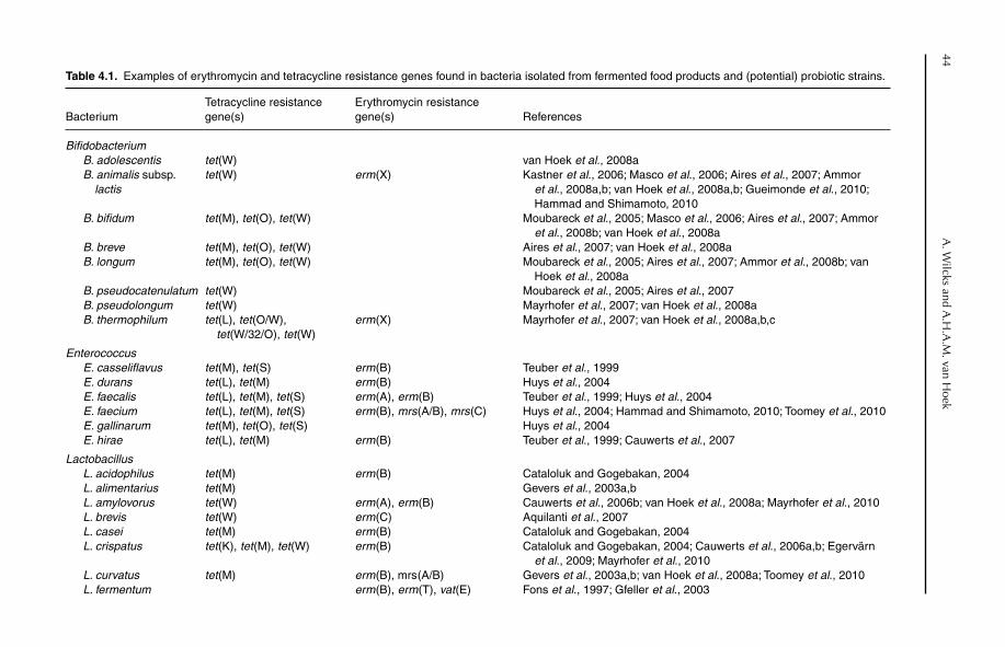
44 A
. Wilcks and A
.H.A
.M. van H
oek
Table 4.1. Examples of erythromycin and tetracycline resistance genes found in bacteria isolated from fermented food products and (potential) probiotic strains.
BacteriumTetracycline resistance gene(s)
Erythromycin resistance gene(s) References
BifidobacteriumB. adolescentis tet (W) van Hoek et al., 2008aB. animalis subsp.
lactistet(W) erm (X) Kastner et al., 2006; Masco et al., 2006; Aires et al., 2007; Ammor
et al., 2008a,b; van Hoek et al., 2008a,b; Gueimonde et al., 2010; Hammad and Shimamoto, 2010
B. bifidum tet (M), tet(O), tet(W) Moubareck et al., 2005; Masco et al., 2006; Aires et al., 2007; Ammor et al., 2008b; van Hoek et al., 2008a
B. breve tet (M), tet(O), tet(W) Aires et al., 2007; van Hoek et al., 2008aB. longum tet (M), tet(O), tet(W) Moubareck et al., 2005; Aires et al., 2007; Ammor et al., 2008b; van
Hoek et al., 2008aB. pseudocatenulatum tet (W) Moubareck et al., 2005; Aires et al., 2007B. pseudolongum tet (W) Mayrhofer et al., 2007; van Hoek et al., 2008aB. thermophilum tet (L), tet(O/W),
tet(W/32/O), tet(W)erm (X) Mayrhofer et al., 2007; van Hoek et al., 2008a,b,c
EnterococcusE. casseliflavus tet (M), tet(S) erm (B) Teuber et al., 1999E. durans tet (L), tet(M) erm (B) Huys et al., 2004E. faecalis tet (L), tet(M), tet(S) erm (A), erm (B) Teuber et al., 1999; Huys et al., 2004E. faecium tet (L), tet(M), tet(S) erm (B), mrs(A/B), mrs(C) Huys et al., 2004; Hammad and Shimamoto, 2010; Toomey et al., 2010E. gallinarum tet (M), tet(O), tet(S) Huys et al., 2004E. hirae tet (L), tet(M) erm (B) Teuber et al., 1999; Cauwerts et al., 2007
LactobacillusL. acidophilus tet (M) erm (B) Cataloluk and Gogebakan, 2004L. alimentarius tet (M) Gevers et al., 2003a,bL. amylovorus tet (W) erm (A), erm (B) Cauwerts et al., 2006b; van Hoek et al., 2008a; Mayrhofer et al., 2010L. brevis tet (W) erm (C) Aquilanti et al., 2007L. casei tet (M) erm (B) Cataloluk and Gogebakan, 2004L. crispatus tet (K), tet(M), tet(W) erm (B) Cataloluk and Gogebakan, 2004; Cauwerts et al., 2006a,b; Egervärn
et al., 2009; Mayrhofer et al., 2010L. curvatus tet (M) erm (B), mrs(A/B) Gevers et al., 2003a,b; van Hoek et al., 2008a; Toomey et al., 2010L. fermentum erm (B), erm (T), vat (E) Fons et al., 1997; Gfeller et al., 2003
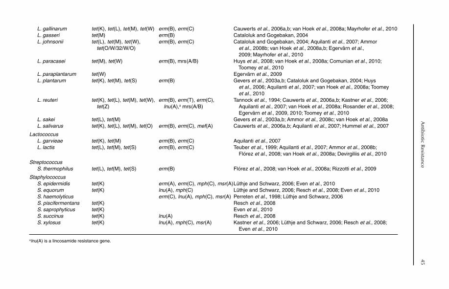
A
ntibiotic Resistance
45L. gallinarum tet (K), tet(L), tet(M), tet(W) erm (B), erm (C) Cauwerts et al., 2006a,b; van Hoek et al., 2008a; Mayrhofer et al., 2010L. gasseri tet (M) erm (B) Cataloluk and Gogebakan, 2004L. johnsonii tet(L), tet(M), tet(W),
tet(O/W/32/W/O)erm (B), erm (C) Cataloluk and Gogebakan, 2004; Aquilanti et al., 2007; Ammor
et al., 2008b; van Hoek et al., 2008a,b; Egervärn et al., 2009; Mayrhofer et al., 2010
L. paracasei tet (M), tet(W) erm (B), mrs(A/B) Huys et al., 2008; van Hoek et al., 2008a; Comunian et al., 2010; Toomey et al., 2010
L. paraplantarum tet (W) Egervärn et al., 2009L. plantarum tet (K), tet(M), tet(S) erm (B) Gevers et al., 2003a,b; Cataloluk and Gogebakan, 2004; Huys
et al., 2006; Aquilanti et al., 2007; van Hoek et al., 2008a; Toomey et al., 2010
L. reuteri tet (K), tet(L), tet(M), tet(W),tet(Z)
erm (B), erm (T), erm (C),lnu (A),a mrs(A/B)
Tannock et al., 1994; Cauwerts et al., 2006a,b; Kastner et al., 2006; Aquilanti et al., 2007; van Hoek et al., 2008a; Rosander et al., 2008; Egervärn et al., 2009, 2010; Toomey et al., 2010
L. sakei tet (L), tet(M) Gevers et al., 2003a,b; Ammor et al., 2008c; van Hoek et al., 2008aL. salivarus tet (K), tet(L), tet(M), tet(O) erm (B), erm (C), mef(A) Cauwerts et al., 2006a,b; Aquilanti et al., 2007; Hummel et al., 2007
LactococcusL. garvieae tet (K), tet(M) erm (B), erm (C) Aquilanti et al., 2007L. lactis tet (L), tet(M), tet(S) erm (B), erm (C) Teuber et al., 1999; Aquilanti et al., 2007; Ammor et al., 2008b;
Flórez et al., 2008; van Hoek et al., 2008a; Devirgiliis et al., 2010
StreptococcusS. thermophilus tet (L), tet(M), tet(S) erm (B) Flórez et al., 2008; van Hoek et al., 2008a; Rizzotti et al., 2009
StaphylococcusS. epidermidis tet (K) erm (A), erm (C), mph(C), msr(A)Lüthje and Schwarz, 2006; Even et al., 2010S. equorum tet (K) lnu (A), mph(C) Lüthje and Schwarz, 2006; Resch et al., 2008; Even et al., 2010S. haemolyticus erm (C), lnu (A), mph(C), msr(A) Perreten et al., 1998; Lüthje and Schwarz, 2006S. piscifermentans tet (K) Resch et al., 2008S. saprophyticus tet (K) Even et al., 2010S. succinus tet (K) lnu (A) Resch et al., 2008S. xylosus tet (K) lnu (A), mph(C), msr(A) Kastner et al., 2006; Lüthje and Schwarz, 2006; Resch et al., 2008;
Even et al., 2010
alnu(A) is a lincosamide resistance gene.
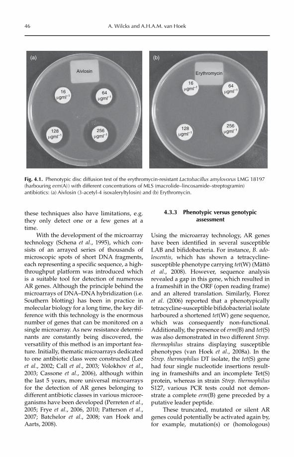
46 A. Wilcks and A.H.A.M. van Hoek
these techniques also have limitations, e.g. they only detect one or a few genes at a time.
With the development of the microarray technology (Schena et al., 1995), which con-sists of an arrayed series of thousands of microscopic spots of short DNA fragments, each representing a specific sequence, a high-throughput platform was introduced which is a suitable tool for detection of numerous AR genes. Although the principle behind the microarrays of DNA–DNA hybridization (i.e. Southern blotting) has been in practice in molecular biology for a long time, the key dif-ference with this technology is the enormous number of genes that can be monitored on a single microarray. As new resistance determi-nants are constantly being discovered, the versatility of this method is an important fea-ture. Initially, thematic microarrays dedicated to one antibiotic class were constructed (Lee et al., 2002; Call et al., 2003; Volokhov et al.,2003; Cassone et al., 2006), although within the last 5 years, more universal microarrays for the detection of AR genes belonging to different antibiotic classes in various microor-ganisms have been developed (Perreten et al.,2005; Frye et al., 2006, 2010; Patterson et al.,2007; Batchelor et al., 2008; van Hoek and Aarts, 2008).
4.3.3 Phenotypic versus genotypic assessment
Using the microarray technology, AR genes have been identified in several susceptible LAB and bifidobacteria. For instance, B. ado-lescentis, which has shown a tetracycline- susceptible phenotype carrying tet(W) (Mättö et al., 2008). However, sequence analysis revealed a gap in this gene, which resulted in a frameshift in the ORF (open reading frame) and an altered translation. Similarly, Florez et al. (2006) reported that a phenotypically tetracycline-susceptible bifidobacterial isolate harboured a shortened tet(W) gene sequence, which was consequently non-functional. Additionally, the presence of erm(B) and tet(S)was also demonstrated in two different Strep. thermophilus strains displaying susceptible phenotypes (van Hoek et al., 2008a). In the Strep. thermophilus DT isolate, the tet(S) gene had four single nucleotide insertions result-ing in frameshifts and an incomplete Tet(S) protein, whereas in strain Strep. thermophilusS127, various PCR tests could not demon-strate a complete erm(B) gene preceded by a putative leader peptide.
These truncated, mutated or silent AR genes could potentially be activated again by, for example, mutation(s) or (homologous)
Aivlosin
(a) (b)
16mgml–1
64mgml–1
256mgml–1
128mgml–1
Erythromycin
16mgml–1 64
mgml–1
256mgml–1128
mgml–1
Fig. 4.1. Phenotypic disc diffusion test of the erythromycin-resistant Lactobacillus amylovorus LMG 18197 (harbouring erm(A) ) with different concentrations of MLS (macrolide–lincosamide–streptogramin) antibiotics: (a) Aivlosin (3-acetyl-4 isovaleryltylosin) and (b) Erythromycin.
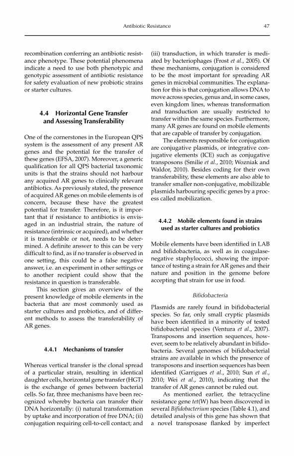
Antibiotic Resistance 47
recombination conferring an antibiotic resist-ance phenotype. These potential phenomena indicate a need to use both phenotypic and genotypic assessment of antibiotic resistance for safety evaluation of new probiotic strains or starter cultures.
4.4 Horizontal Gene Transfer and Assessing Transferability
One of the cornerstones in the European QPS system is the assessment of any present AR genes and the potential for the transfer of these genes (EFSA, 2007). Moreover, a generic qualification for all QPS bacterial taxonomic units is that the strains should not harbour any acquired AR genes to clinically relevant antibiotics. As previously stated, the presence of acquired AR genes on mobile elements is of concern, because these have the greatest potential for transfer. Therefore, is it impor-tant that if resistance to antibiotics is envis-aged in an industrial strain, the nature of resistance (intrinsic or acquired), and whether it is transferable or not, needs to be deter-mined. A definite answer to this can be very difficult to find, as if no transfer is observed in one setting, this could be a false negative answer, i.e. an experiment in other settings or to another recipient could show that the resistance in question is transferable.
This section gives an overview of the present knowledge of mobile elements in the bacteria that are most commonly used as starter cultures and probiotics, and of differ-ent methods to assess the transferability of AR genes.
4.4.1 Mechanisms of transfer
Whereas vertical transfer is the clonal spread of a particular strain, resulting in identical daughter cells, horizontal gene transfer (HGT) is the exchange of genes between bacterial cells. So far, three mechanisms have been rec-ognized whereby bacteria can transfer their DNA horizontally: (i) natural transformation by uptake and incorporation of free DNA; (ii) conjugation requiring cell-to-cell contact; and
(iii) transduction, in which transfer is medi-ated by bacteriophages (Frost et al., 2005). Of these mechanisms, conjugation is considered to be the most important for spreading AR genes in microbial communities. The explana-tion for this is that conjugation allows DNA to move across species, genus and, in some cases, even kingdom lines, whereas transformation and transduction are usually restricted to transfer within the same species. Furthermore, many AR genes are found on mobile elements that are capable of transfer by conjugation.
The elements responsible for conjugation are conjugative plasmids, or integrative con-jugative elements (ICE) such as conjugative transposons (Smilie et al., 2010; Wozniak and Waldor, 2010). Besides coding for their own transferability, these elements are also able to transfer smaller non-conjugative, mobilizable plasmids harbouring specific genes by a proc-ess called mobilization.
4.4.2 Mobile elements found in strains used as starter cultures and probiotics
Mobile elements have been identified in LAB and bifidobacteria, as well as in coagulase-negative staphylococci, showing the impor-tance of testing a strain for AR genes and their nature and position in the genome before accepting that strain for use in food.
Bifidobacteria
Plasmids are rarely found in bifidobacterial species. So far, only small cryptic plasmids have been identified in a minority of tested bifidobacterial species (Ventura et al., 2007). Transposons and insertion sequences, how-ever, seem to be relatively abundant in bifido-bacteria. Several genomes of bifidobacterial strains are available in which the presence of transposons and insertion sequences has been identified (Garrigues et al., 2010; Sun et al.,2010; Wei et al., 2010), indicating that the transfer of AR genes cannot be ruled out.
As mentioned earlier, the tetracycline resistance gene tet(W) has been discovered in several Bifidobacterium species (Table 4.1), and detailed analysis of this gene has shown that a novel transposase flanked by imperfect
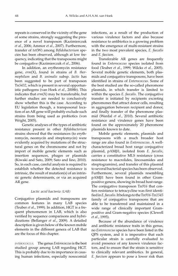
48 A. Wilcks and A.H.A.M. van Hoek
repeats is conserved in the vicinity of the gene of some strains, strongly suggesting the pres-ence of a novel transposon (Kazimierczak et al., 2006; Ammor et al., 2007). Furthermore, transfer of tet(W) among Bifidobacterium spe-cies has been observed, although at low fre-quency, indicating that the transposons might be conjugative (Kazimierczak et al., 2006).
In addition, an erythromycin resistance gene, erm(X), found in strains of B. ther-mophilum and B. animalis subsp. lactis has been suggested to be part of transposon Tn5432, which is present in several opportun-istic pathogens (van Hoek et al., 2008b). This indicates that erm(X) may be transferable, but further studies are needed to conclusively show whether this is the case. According to EU legislation though, a transposonal loca-tion of an AR gene will probably exclude such strains from being used as probiotics (von Wright, 2005).
Genetic analyses of the types of antibiotic resistance present in other Bifidobacteriumstrains showed that the resistances (to eryth-romycin, neomycin and streptomycin) were evidently acquired by mutations of the struc-tural genes on the chromosome and not by import of mobile genetic elements such as insertion sequences, phages or plasmids (Kiwaki and Sato, 2009; Sato and Iino, 2010). So, in each case, careful analysis is required to establish whether the detected resistance is intrinsic, the result of mutation(s) of an intrin-sic genetic determinants, or via an acquired AR gene.
Lactic acid bacteria (LAB)
Conjugative plasmids and transposons are common features in many LAB species (Teuber et al., 1999). In addition, HGT is a fre-quent phenomenon in LAB, which is also verified by sequence comparisons and hybri-dizations (Bellanger et al., 2009). A detailed description is given below of the known mobile elements in the different genera of LAB that are the focus of this chapter.
ENTEROCOCCUS. The genus Enterococcus is the best studied group among LAB regarding HGT. This is probably due to its importance in caus-ing human infections, especially nosocomial
infections, as a result of the production of various virulence factors and also because resistance to antibiotics is a growing problem with the emergence of multi-resistant strains in the two most prevalent species, E. faecalisand E. faecium.
Transferable AR genes are frequently found in Enterococcus species isolated from food (Teuber et al., 1999; Wilcks et al., 2005). Several mobile genetic elements, both plas-mids and conjugative transposons, have been identified in strains of Enterococcus. Some of the best studied are the so-called pheromone plasmids, in which transfer is limited to within the species E. faecalis. The conjugative transfer is initiated by recipients excreting pheromones that attract donor cells, resulting in aggregation between recipient and donor, and finally transfer of the pheromone plas-mid (Wardal et al., 2010). Several antibiotic resistance and virulence genes have been found on the approximately 20 pheromone plasmids known to date.
Mobile genetic elements, plasmids and transposons with a much broader host range are also found in Enterococcus. A well- characterized broad host range conjugative plasmid, pAMb1, isolated from E. faecalis,carries a constitutive MLS resistance (cross resistance to macrolides, lincosamides and streptogramins), and transfer of this plasmid to several bacterial species has been observed. Furthermore, several plasmids resembling pAMb1 have been found in other Gram-positive genera, showing its broad host range. The conjugative transposon Tn916 that con-fers resistance to tetracycline was first identi-fied in E. faecalis. It belongs to the Tn916-Tn1545family of conjugative transposons that are able to be transferred and maintained in a wide range of clinically important Gram-positive and Gram-negative species (Clewell et al., 1995).
Because of the abundance of virulence and antibiotic resistance traits in this genus, no Enterococcus species have been listed in the QPS system, and it is imperative that each particular strain is carefully evaluated to avoid presence of any known virulence fac-tors, and to ensure that the strain is sensitive to clinically relevant antibiotics. In general, E. faecium appears to pose a lower risk than
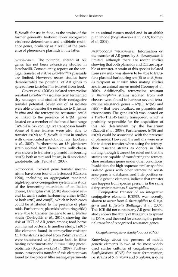
Antibiotic Resistance 49
E. faecalis for use in food, as the strains of the former generally harbour fewer recognized virulence determinants and antibiotic resist-ance genes, probably as a result of the pres-ence of pheromone plasmids in the latter.
LACTOBACILLUS. The potential spread of AR genes has not been extensively studied in lactobacilli. Consequently, reports on the con-jugal transfer of native Lactobacillus plasmids are limited. However, recent studies have demonstrated the potential of AR genes to spread from Lactobacillus isolated from food.
Gevers et al. (2003a) isolated tetracycline resistant Lactobacillus isolates from fermented dry sausages and studied their conjugative transfer potential. Seven out of 14 isolates were able to transfer the resistance to E. faecalis in vitro and the tetracycline resistance could be linked to the presence of tet(M) genes located on a member of the broad host range Tn916-Tn1545 conjugative transposon family. Some of these isolates were also able to transfer tet(M) to E. faecalis in vivo in studies with di-associated gnotobiotic rats (Jacobsen et al., 2007). Furthermore, an Lb. plantarumstrain isolated from French raw milk cheese was shown to transfer a plasmid harbouring erm(B), both in vitro and in vivo, in di-associated gnotobiotic rats (Feld et al., 2008).
LACTOCOCCUS. Several gene transfer mecha-nisms have been found in lactococci (Gasson, 1990), including an aggregation mediated high-frequency conjugation system. In a study of the fermenting microbiota of an Italian cheese, Devirgiliis et al. (2010) discovered sev-eral Lc. lactis strains harbouring either tet(M)or both tet(S) and erm(B), which in both cases could be attributed to the presence of plas-mids. Furthermore, plasmids harbouring tet(M) were able to transfer the gene to an E. faecalisstrain (Devirgiliis et al., 2010), showing the risk of HGT of AR genes among food-borne commensal bacteria. In another study, Tn916-like elements found in tetracycline resistant Lc. lactis strains isolated from Polish raw milk were transferred to E. faecalis both in filter mating experiments and in vivo, using gnoto-biotic rats (Boguslawska et al., 2009). Further-more, intraspecies transfer of this element was found to take place in filter mating experiments
in an animal rumen model and in an alfalfa plant model (Boguslawska et al., 2009; Toomey et al., 2009).
STREPTOCOCCUS THERMOPHILUS. Information on the transfer of AR genes by S. thermophilus is limited, although there are recent studies showing that both plasmids and ICE are capa-ble of transfer. A strain of this species isolated from raw milk was shown to be able to trans-fer a plasmid-harbouring erm(B) to an E. faeca-lis recipient in in vitro filter mating studies and in an animal rumen model (Toomey et al.,2009). Additionally, tetracycline resistant S. thermophilus strains isolated from soft cheeses were found to harbour several tetra-cycline resistance genes – tet(L), tet(M) and tet(S) – that were localized on plasmids and transposons. The gene tet(M) was located on a Tn916-Tn1545 family transposon, which is probably responsible for the acquisition of this AR determinant by the bacterium (Rizzotti et al., 2009). Furthermore, tet(S) and tet(M) could be associated with the presence of plasmids. However, the authors were una-ble to detect transfer when using the tetracy-cline resistant strains as donors in filter mating, though it cannot be ruled out that the strains are capable of transferring the tetracy-cline resistance genes under other conditions. In addition, the high sequence similarity of the isolated genes with other tetracycline resist-ance genes in databases, and their position on mobile genetic elements, indicate that transfer can happen from species present in the same dairy environment as S. thermophilus.
Conjugative transfer of an integrative conjugative element, ICESt3, was recently shown to occur from S. thermophilus to S. pyo-genes and E. faecalis (Bellanger et al., 2009). This ICE did not contain any AR gene, but the study shows the ability of this genus to spread its DNA, and the need for assessing the poten-tial transfer of recognized resistance genes.
Coagulase-negative staphylococci (CNS)
Knowledge about the presence of mobile genetic elements in two of the most widely used starter cultures of coagulase-negative Staphylococcus (CNS) for meat fermentation, i.e. strains of S. carnosus and S. xylosus, is quite
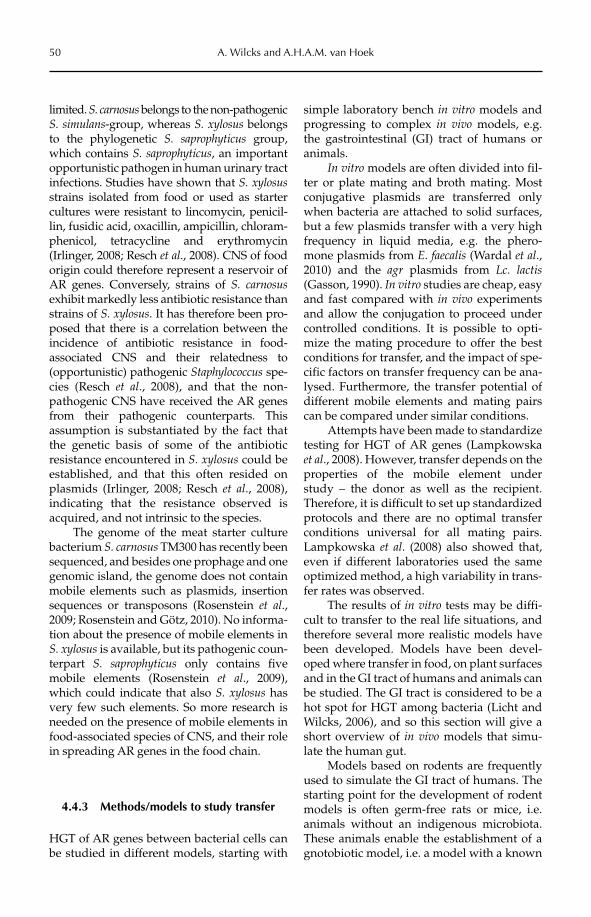
50 A. Wilcks and A.H.A.M. van Hoek
limited. S. carnosus belongs to the non-pathogenic S. simulans-group, whereas S. xylosus belongs to the phylogenetic S. saprophyticus group, which contains S. saprophyticus, an important opportunistic pathogen in human urinary tract infections. Studies have shown that S. xylosusstrains isolated from food or used as starter cultures were resistant to lincomycin, penicil-lin, fusidic acid, oxacillin, ampicillin, chloram-phenicol, tetracycline and erythromycin (Irlinger, 2008; Resch et al., 2008). CNS of food origin could therefore represent a reservoir of AR genes. Conversely, strains of S. carnosusexhibit markedly less antibiotic resistance than strains of S. xylosus. It has therefore been pro-posed that there is a correlation between the incidence of antibiotic resistance in food- associated CNS and their relatedness to (opportunistic) pathogenic Staphylococcus spe-cies (Resch et al., 2008), and that the non-pathogenic CNS have received the AR genes from their pathogenic counterparts. This assumption is substantiated by the fact that the genetic basis of some of the antibiotic resistance encountered in S. xylosus could be established, and that this often resided on plasmids (Irlinger, 2008; Resch et al., 2008), indicating that the resistance observed is acquired, and not intrinsic to the species.
The genome of the meat starter culture bacterium S. carnosus TM300 has recently been sequenced, and besides one prophage and one genomic island, the genome does not contain mobile elements such as plasmids, insertion sequences or transposons (Rosenstein et al.,2009; Rosenstein and Götz, 2010). No informa-tion about the presence of mobile elements in S. xylosus is available, but its pathogenic coun-terpart S. saprophyticus only contains five mobile elements (Rosenstein et al., 2009), which could indicate that also S. xylosus has very few such elements. So more research is needed on the presence of mobile elements in food-associated species of CNS, and their role in spreading AR genes in the food chain.
4.4.3 Methods/models to study transfer
HGT of AR genes between bacterial cells can be studied in different models, starting with
simple laboratory bench in vitro models and progressing to complex in vivo models, e.g. the gastrointestinal (GI) tract of humans or animals.
In vitro models are often divided into fil-ter or plate mating and broth mating. Most conjugative plasmids are transferred only when bacteria are attached to solid surfaces, but a few plasmids transfer with a very high frequency in liquid media, e.g. the phero-mone plasmids from E. faecalis (Wardal et al.,2010) and the agr plasmids from Lc. lactis(Gasson, 1990). In vitro studies are cheap, easy and fast compared with in vivo experiments and allow the conjugation to proceed under controlled conditions. It is possible to opti-mize the mating procedure to offer the best conditions for transfer, and the impact of spe-cific factors on transfer frequency can be ana-lysed. Furthermore, the transfer potential of different mobile elements and mating pairs can be compared under similar conditions.
Attempts have been made to standardize testing for HGT of AR genes (Lampkowska et al., 2008). However, transfer depends on the properties of the mobile element under study – the donor as well as the recipient. Therefore, it is difficult to set up standardized protocols and there are no optimal transfer conditions universal for all mating pairs. Lampkowska et al. (2008) also showed that, even if different laboratories used the same optimized method, a high variability in trans-fer rates was observed.
The results of in vitro tests may be diffi-cult to transfer to the real life situations, and therefore several more realistic models have been developed. Models have been devel-oped where transfer in food, on plant surfaces and in the GI tract of humans and animals can be studied. The GI tract is considered to be a hot spot for HGT among bacteria (Licht and Wilcks, 2006), and so this section will give a short overview of in vivo models that simu-late the human gut.
Models based on rodents are frequently used to simulate the GI tract of humans. The starting point for the development of rodent models is often germ-free rats or mice, i.e. animals without an indigenous microbiota. These animals enable the establishment of a gnotobiotic model, i.e. a model with a known
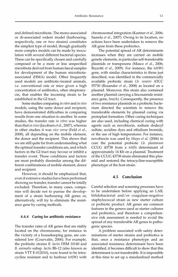
Antibiotic Resistance 51
and defined microbiota. The mono-associated or di-associated rodent model (harbouring, respectively, one or two strains) constitutes the simplest type of model, though gradually more complex models can be made by inocu-lation with several different bacterial species. These can be specifically chosen and carefully composed or be a more or less unspecified microbiota derived from human faecal samples for development of the human microbiota-associated (HMA) model. Other frequently used models are antibiotic-treated animals, i.e. conventional rats or mice given a high concentration of antibiotics, often streptomy-cin, that enables the incoming strain to be established in the GI tract.
Some studies comparing in vitro and in vivomodels, using the same donor and recipient, have demonstrated difficulties in comparing results from one situation to another. In some studies, the transfer rate in vitro was higher than that in vivo (Jacobsen et al., 2007), whereas in other studies it was vice versa (Feld et al.,2008), all depending on the mobile element, the donor and the recipient. This shows that we are still quite far from understanding what the optimal transfer conditions are, and which factors in the GI tract may favour or inhibit a transfer event. Those conditions and factors are most probably dissimilar among the dif-ferent combinations of mobile element, donor and recipient.
However, it should be emphasized that, even if extensive studies have been performed showing no transfer, transfer cannot be totally excluded. Therefore, in many cases, compa-nies will decide not to pursue the develop-ment of a strain harbouring AR genes or, alternatively, will try to eliminate the resist-ance gene by curing methods.
4.4.4 Curing for antibiotic resistance
The transfer rates of AR genes that are stably located on the chromosome, for instance in the vicinity of a housekeeping gene, are con-sidered low (Courvalin, 2006). For example, the probiotic strains B. lactis DSM 10140 and B. animalis subsp. lactis Bb-12 (also known as strain VTT E-012010), were found to be tetra-cycline resistant and to harbour tet(W) with
chromosomal integration (Kastner et al., 2006; Saarela et al., 2007). Owing to its location, no actions have been undertaken to remove the AR gene from these probiotics.
The potential spread of AR determinants increases when they are carried on mobile genetic elements, in particular self-transferable plasmids or transposons (Masco et al., 2006; Rizzotti et al., 2009). For instance, the tet(W) gene, with similar characteristics to those just described, was identified in the commercially available probiotic strain Lb. reuteri ATCC 55730 (Rosander et al., 2008) as located on a plasmid. Moreover, this strain also contained another plasmid carrying a lincosamide resist-ance gene, lnu(A). Consequently, the presence of two resistance plasmids in a probiotic bacte-rium directed the scientists to remove the transferable elements by plasmid curing via protoplast formation. Other curing techniques are also used, including chemical curing with agents such as novobiocin, sodium dodecyl sulfate, acridine dyes and ethidium bromide, or the use of high temperatures. For instance, novobiocin was used by Huys et al. (2006) to cure the potential probiotic Lb. plantarumCCUG 43738 from a tet(S) determinant of approximately 14 Kb on a plasmid. Treatment of the CCUG 43738 strain eliminated this plas-mid and restored the tetracycline-susceptible phenotype of the host strain.
4.5 Conclusion
Careful selection and screening processes have to be undertaken before applying an LAB, bifidobacterial and/or coagulase-negative staphylococcal strain as new starter culture or probiotic product. AR genes are common features in the genera used as starter cultures and probiotics, and therefore a comprehen-sive risk assessment is needed to avoid the spread of any transferable AR genes to patho-genic species.
A problem associated with safety deter-minations of starter strains and probiotics is that, once a resistance phenotype and an associated resistance determinant have been identified, it becomes difficult to show that this determinant is not transferable. It is impossible at this time to set up a standardized method
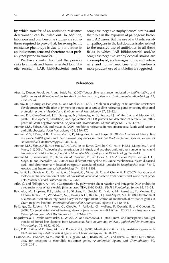
52 A. Wilcks and A.H.A.M. van Hoek
by which transfer of an antibiotic resistance determinant can be ruled out. In addition, laborious and cumbersome studies are some-times required to prove that, for example, the resistance phenotype is due to a mutation in an indigenous gene and therefore most prob-ably not prone to transfer.
We have clearly described the possible risks to animals and humans related to antibi-otic resistant LAB, bifidobacterial and/or
coagulase-negative staphylococcal strains, and their role in the exposure of pathogenic bacte-ria to AR genes. But the rise of antibiotic resist-ant pathogens in the last decades is also related to the massive use of antibiotics in all those fields in which LAB bifidobacterial and/or coagulase-negative staphylococcal strains are also employed, such as agriculture, and veteri-nary and human medicine, and therefore a more prudent use of antibiotics is suggested.
References
Aires, J., Doucet-Populaire, F. and Butel, M.J. (2007) Tetracycline resistance mediated by tet(W), tet(M), and tet(O) genes of Bifidobacterium isolates from humans. Applied and Environmental Microbiology 73, 2751–2754.
Aminov, R.I., Garrigues-Jeanjean, N. and Mackie, R.I. (2001) Molecular ecology of tetracycline resistance: development and validation of primers for detection of tetracycline resistance genes encoding ribosomal protection proteins. Applied and Environmental Microbiology 67, 22–32.
Aminov, R.I., Chee-Sanford, J.C., Garrigues, N., Teferedegne, B., Krapac, I.J., White, B.A. and Mackie, R.I. (2002) Development, validation, and application of PCR primers for detection of tetracycline efflux genes of Gram-negative bacteria. Applied and Environmental Microbiology 68, 1786–1793.
Ammor, M.S., Flórez, A.B. and Mayo, B. (2007) Antibiotic resistance in non-enterococcal lactic acid bacteria and bifidobacteria. Food Microbiology 24, 559–570.
Ammor, M.S., Flórez, A.B., Álvarez-Martín, P., Margolles, A. and Mayo, B. (2008a) Analysis of tetracycline resistance tet(W) genes and their flanking sequences in intestinal Bifidobacterium species. Journal of Antimicrobial Chemotherapy 62, 688–693.
Ammor, M.S., Flórez, A.B., van Hoek, A.H.A.M., de los Reyes-Gavilán, C.G., Aarts, H.J.M., Margolles, A. and Mayo, B. (2008b) Molecular characterization of intrinsic and acquired antibiotic resistance in lactic acid bacteria and bifidobacteria. Journal of Molecular Microbiology and Biotechnology 14, 6–15.
Ammor, M.S., Gueimonde, M., Danielsen, M., Zagorec, M., van Hoek, A.H.A.M., de los Reyes-Gavilán, C.G., Mayo, B. and Margolles, A. (2008c) Two different tetracycline resistance mechanisms, plasmid-carried tet(L) and chromosomally located transposon-associated tet(M), coexist in Lactobacillus sakei Rits 9. Applied and Environmental Microbiology 74, 1394–1401.
Aquilanti, L., Garofalo, C., Osimani, A., Silvestri, G., Vignaroli, C. and Clementi, F. (2007). Isolation and molecular characterization of antibiotic-resistant lactic acid bacteria from poultry and swine meat prod-ucts. Journal of Food Protection 70, 557–565.
Arlet, G. and Philippon, A. (1991) Construction by polymerase chain reaction and intragenic DNA probes for three main types of transferable b-lactamases (TEM, SHV, CARB). FEMS Microbiology Letters 82, 19–25.
Batchelor, M., Hopkins, K.L., Liebana, E., Slickers, P., Ehricht, R., Mafura, M., Aarestrup, F., Mevius, D., Clifton-Hadley, F.A., Woodward, M.J., Davies, R.H., Threlfall, E.J. and Anjum, M.F. (2008) Development of a miniaturised microarray-based assay for the rapid identification of antimicrobial resistance genes in Gram-negative bacteria. International Journal of Antimicrobial Agents 31, 440–451.
Bellanger, X., Roberts, A.P., Morel, C., Choulet, F., Pavlovic, G., Mullany, P., Decaris, B. and Guedon, G. (2009) Conjugative transfer of the integrative conjugative elements ICESt1 and ICESt3 from Streptococcusthermophilus. Journal of Bacteriology 191, 2764–2775.
Boguslawska, J., Zycka-Krzesinska, J., Wilcks, A. and Bardowski, J. (2009) Intra- and interspecies conjugal transfer of Tn916-like elements from Lactococcus lactis in vitro and in vivo. Applied and Environmental Microbiology 75, 6352–6360.
Call, D.R., Bakko, M.K., Krug, M.J. and Roberts, M.C. (2003) Identifying antimicrobial resistance genes with DNA microarrays. Antimicrobial Agents and Chemotherapy 47, 3290–3295.
Cassone, M., D’Andrea, M.M., Iannelli, F., Oggioni, M.R., Rossolini, G.M. and Pozzi, G. (2006) DNA micro-array for detection of macrolide resistance genes. Antimicrobial Agents and Chemotherapy 50, 2038–2041.
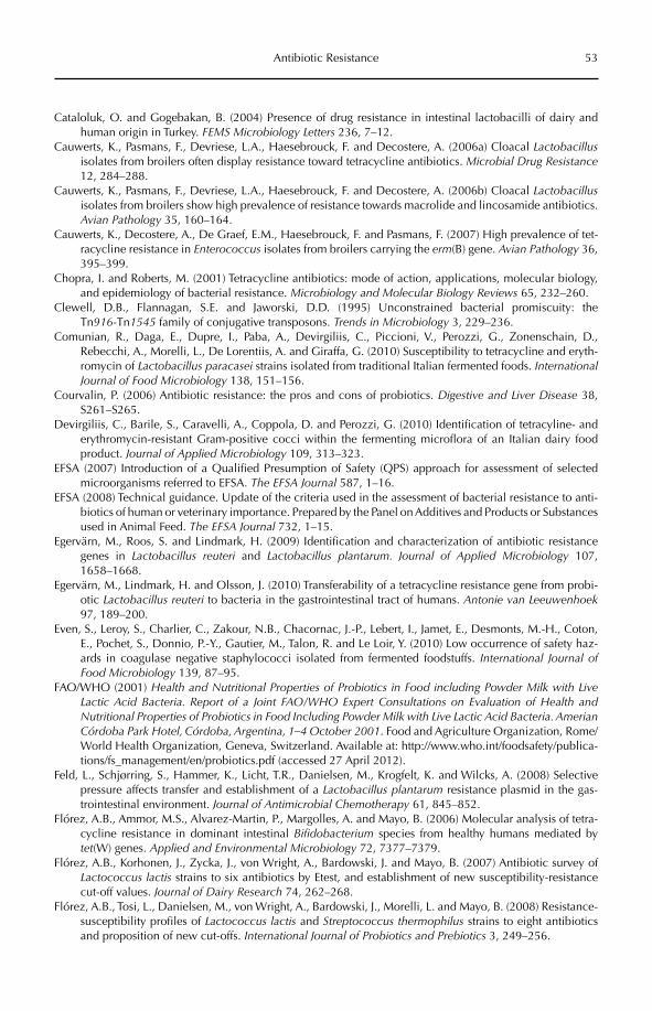
Antibiotic Resistance 53
Cataloluk, O. and Gogebakan, B. (2004) Presence of drug resistance in intestinal lactobacilli of dairy and human origin in Turkey. FEMS Microbiology Letters 236, 7–12.
Cauwerts, K., Pasmans, F., Devriese, L.A., Haesebrouck, F. and Decostere, A. (2006a) Cloacal Lactobacillusisolates from broilers often display resistance toward tetracycline antibiotics. Microbial Drug Resistance12, 284–288.
Cauwerts, K., Pasmans, F., Devriese, L.A., Haesebrouck, F. and Decostere, A. (2006b) Cloacal Lactobacillusisolates from broilers show high prevalence of resistance towards macrolide and lincosamide antibiotics. Avian Pathology 35, 160–164.
Cauwerts, K., Decostere, A., De Graef, E.M., Haesebrouck, F. and Pasmans, F. (2007) High prevalence of tet-racycline resistance in Enterococcus isolates from broilers carrying the erm(B) gene. Avian Pathology 36, 395–399.
Chopra, I. and Roberts, M. (2001) Tetracycline antibiotics: mode of action, applications, molecular biology, and epidemiology of bacterial resistance. Microbiology and Molecular Biology Reviews 65, 232–260.
Clewell, D.B., Flannagan, S.E. and Jaworski, D.D. (1995) Unconstrained bacterial promiscuity: the Tn916-Tn1545 family of conjugative transposons. Trends in Microbiology 3, 229–236.
Comunian, R., Daga, E., Dupre, I., Paba, A., Devirgiliis, C., Piccioni, V., Perozzi, G., Zonenschain, D., Rebecchi, A., Morelli, L., De Lorentiis, A. and Giraffa, G. (2010) Susceptibility to tetracycline and eryth-romycin of Lactobacillus paracasei strains isolated from traditional Italian fermented foods. InternationalJournal of Food Microbiology 138, 151–156.
Courvalin, P. (2006) Antibiotic resistance: the pros and cons of probiotics. Digestive and Liver Disease 38, S261–S265.
Devirgiliis, C., Barile, S., Caravelli, A., Coppola, D. and Perozzi, G. (2010) Identification of tetracyline- and erythromycin-resistant Gram-positive cocci within the fermenting microflora of an Italian dairy food product. Journal of Applied Microbiology 109, 313–323.
EFSA (2007) Introduction of a Qualified Presumption of Safety (QPS) approach for assessment of selected microorganisms referred to EFSA. The EFSA Journal 587, 1–16.
EFSA (2008) Technical guidance. Update of the criteria used in the assessment of bacterial resistance to anti-biotics of human or veterinary importance. Prepared by the Panel on Additives and Products or Substances used in Animal Feed. The EFSA Journal 732, 1–15.
Egervärn, M., Roos, S. and Lindmark, H. (2009) Identification and characterization of antibiotic resistance genes in Lactobacillus reuteri and Lactobacillus plantarum. Journal of Applied Microbiology 107, 1658–1668.
Egervärn, M., Lindmark, H. and Olsson, J. (2010) Transferability of a tetracycline resistance gene from probi-otic Lactobacillus reuteri to bacteria in the gastrointestinal tract of humans. Antonie van Leeuwenhoek97, 189–200.
Even, S., Leroy, S., Charlier, C., Zakour, N.B., Chacornac, J.-P., Lebert, I., Jamet, E., Desmonts, M.-H., Coton, E., Pochet, S., Donnio, P.-Y., Gautier, M., Talon, R. and Le Loir, Y. (2010) Low occurrence of safety haz-ards in coagulase negative staphylococci isolated from fermented foodstuffs. International Journal of Food Microbiology 139, 87–95.
FAO/WHO (2001) Health and Nutritional Properties of Probiotics in Food including Powder Milk with Live Lactic Acid Bacteria. Report of a Joint FAO/WHO Expert Consultations on Evaluation of Health and Nutritional Properties of Probiotics in Food Including Powder Milk with Live Lactic Acid Bacteria. Amerian Córdoba Park Hotel, Córdoba, Argentina, 1–4 October 2001. Food and Agriculture Organization, Rome/World Health Organization, Geneva, Switzerland. Available at: http://www.who.int/foodsafety/publica-tions/fs_management/en/probiotics.pdf (accessed 27 April 2012).
Feld, L., Schjørring, S., Hammer, K., Licht, T.R., Danielsen, M., Krogfelt, K. and Wilcks, A. (2008) Selective pressure affects transfer and establishment of a Lactobacillus plantarum resistance plasmid in the gas-trointestinal environment. Journal of Antimicrobial Chemotherapy 61, 845–852.
Flórez, A.B., Ammor, M.S., Alvarez-Martin, P., Margolles, A. and Mayo, B. (2006) Molecular analysis of tetra-cycline resistance in dominant intestinal Bifidobacterium species from healthy humans mediated by tet(W) genes. Applied and Environmental Microbiology 72, 7377–7379.
Flórez, A.B., Korhonen, J., Zycka, J., von Wright, A., Bardowski, J. and Mayo, B. (2007) Antibiotic survey of Lactococcus lactis strains to six antibiotics by Etest, and establishment of new susceptibility-resistance cut-off values. Journal of Dairy Research 74, 262–268.
Flórez, A.B., Tosi, L., Danielsen, M., von Wright, A., Bardowski, J., Morelli, L. and Mayo, B. (2008) Resistance-susceptibility profiles of Lactococcus lactis and Streptococcus thermophilus strains to eight antibiotics and proposition of new cut-offs. International Journal of Probiotics and Prebiotics 3, 249–256.
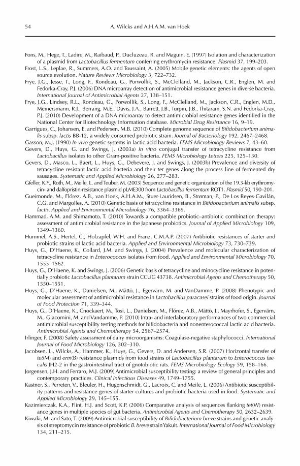
54 A. Wilcks and A.H.A.M. van Hoek
Fons, M., Hege, T., Ladire, M., Raibaud, P., Ducluzeau, R. and Maguin, E. (1997) Isolation and characterization of a plasmid from Lactobacillus fermentum conferring erythromycin resistance. Plasmid 37, 199–203.
Frost, L.S., Leplae, R., Summers, A.O. and Toussaint, A. (2005) Mobile genetic elements: the agents of open source evolution. Nature Reviews Microbiology 3, 722–732.
Frye, J.G., Jesse, T., Long, F., Rondeau, G., Porwollik, S., McClelland, M., Jackson, C.R., Englen, M. and Fedorka-Cray, P.J. (2006) DNA microarray detection of antimicrobial resistance genes in diverse bacteria. International Journal of Antimicrobial Agents 27, 138–151.
Frye, J.G., Lindsey, R.L., Rondeau, G., Porwollik, S., Long, F., McClelland, M., Jackson, C.R., Englen, M.D., Meinersmann, R.J., Berrang, M.E., Davis, J.A., Barrett, J.B., Turpin, J.B., Thitaram, S.N. and Fedorka-Cray, P.J. (2010) Development of a DNA microarray to detect antimicrobial resistance genes identified in the National Center for Biotechnology Information database. Microbial Drug Resistance 16, 9–19.
Garrigues, C., Johansen, E. and Pedersen, M.B. (2010) Complete genome sequence of Bifidobacterium anima-lis subsp. lactis BB-12, a widely consumed probiotic strain. Journal of Bacteriology 192, 2467–2468.
Gasson, M.J. (1990) In vivo genetic systems in lactic acid bacteria. FEMS Microbiology Reviews 7, 43–60.Gevers, D., Huys, G. and Swings, J. (2003a) In vitro conjugal transfer of tetracycline resistance from
Lactobacillus isolates to other Gram-positive bacteria. FEMS Microbiology Letters 225, 125–130.Gevers, D., Masco, L., Baert, L., Huys, G., Debevere, J. and Swings, J. (2003b) Prevalence and diversity of
tetracycline resistant lactic acid bacteria and their tet genes along the process line of fermented dry sausages. Systematic and Applied Microbiology 26, 277–283.
Gfeller, K.Y., Roth, M., Meile, L. and Teuber, M. (2003) Sequence and genetic organization of the 19.3-kb erythromy-cin- and dalfopristin-resistance plasmid pLME300 from Lactobacillus fermentum ROT1. Plasmid 50, 190–201.
Gueimonde, M., Flórez, A.B., van Hoek, A.H.A.M., Stuer-Lauridsen, B., Strøman, P., De Los Reyes-Gavilán, C.G. and Margolles, A. (2010) Genetic basis of tetracycline resistance in Bifidobacterium animalis subsp. lactis. Applied and Environmental Microbiology 76, 3364–3369.
Hammad, A.M. and Shimamoto, T. (2010) Towards a compatible probiotic–antibiotic combination therapy: assessment of antimicrobial resistance in the Japanese probiotics. Journal of Applied Microbiology 109, 1349–1360.
Hummel, A.S., Hertel, C., Holzapfel, W.H. and Franz, C.M.A.P. (2007) Antibiotic resistances of starter and probiotic strains of lactic acid bacteria. Applied and Environmental Microbiology 73, 730–739.
Huys, G., D’Haene, K., Collard, J.M. and Swings, J. (2004) Prevalence and molecular characterization of tetracycline resistance in Enterococcus isolates from food. Applied and Environmental Microbiology 70, 1555–1562.
Huys, G., D’Haene, K. and Swings, J. (2006) Genetic basis of tetracycline and minocycline resistance in poten-tially probiotic Lactobacillus plantarum strain CCUG 43738. Antimicrobial Agents and Chemotherapy 50, 1550–1551.
Huys, G., D’Haene, K., Danielsen, M., Mättö, J., Egervärn, M. and VanDamme, P. (2008) Phenotypic and molecular assessment of antimicrobial resistance in Lactobacillus paracasei strains of food origin. Journalof Food Protection 71, 339–344.
Huys, G., D’Haene, K., Cnockaert, M., Tosi, L., Danielsen, M., Flórez, A.B., Mättö, J., Mayrhofer, S., Egervärn, M., Giacomini, M. and Vandamme, P. (2010) Intra- and interlaboratory performances of two commercial antimicrobial susceptibility testing methods for bifidobacteria and nonenterococcal lactic acid bacteria. Antimicrobial Agents and Chemotherapy 54, 2567–2574.
Irlinger, F. (2008) Safety assessment of dairy microorganisms: Coagulase-negative staphylococci. InternationalJournal of Food Microbiology 126, 302–310.
Jacobsen, L., Wilcks, A., Hammer, K., Huys, G., Gevers, D. and Andersen, S.R. (2007) Horizontal transfer of tet(M) and erm(B) resistance plasmids from food strains of Lactobacillus plantarum to Enterococcus fae-calis JH2-2 in the gastrointestinal tract of gnotobiotic rats. FEMS Microbiology Ecology 59, 158–166.
Jorgensen, J.H. and Ferraro, M.J. (2009) Antimicrobial susceptibility testing: a review of general principles and contemporary practices. Clinical Infectious Diseases 49, 1749–1755.
Kastner, S., Perreten, V., Bleuler, H., Hugenschmidt, G., Lacroix, C. and Meile, L. (2006) Antibiotic susceptibil-ity patterns and resistance genes of starter cultures and probiotic bacteria used in food. Systematic and Applied Microbiology 29, 145–155.
Kazimierczak, K.A., Flint, H.J. and Scott, K.P. (2006) Comparative analysis of sequences flanking tet(W) resist-ance genes in multiple species of gut bacteria. Antimicrobial Agents and Chemotherapy 50, 2632–2639.
Kiwaki, M. and Sato, T. (2009) Antimicrobial susceptibility of Bifidobacterium breve strains and genetic analy-sis of streptomycin resistance of probiotic B. breve strain Yakult. International Journal of Food Microbiology134, 211–215.
Antibiotic Resistance 55
Lampkowska, J., Feld, L., Monaghan, A., Toomey, N., Schjørring, S., Jacobsen, B., van der Voet, H., Andersen, S.R., Bolton, D., Aarts, H., Krogfelt, K.A., Wilcks, A. and Bardowski, J. (2008) A standardized conjugation protocol to assess antibiotic resistance transfer between lactococcal species. International Journal of Food Microbiology 127, 172–175.
Lee, Y., Lee, C.-S., Kim, Y.-J., Chun, S., Park, S., Kim, Y.S. and Han, B.-D. (2002) Development of DNA chip for the simultaneous detection of various b-lactam antibiotic-resistant genes. Molecules and Cells 14, 192–197.
Licht, T.R. and Wilcks, A. (2006) Conjugative gene transfer in the gastrointestinal environment. Advances in Applied Microbiology 58, 77–95.
Lüthje, P. and Schwarz, S. (2006) Antimicrobial resistance of coagulase-negative staphylococci from bovine subclinical mastitis with particular reference to macrolide–lincosamide resistance phenotypes and geno-types. Journal of Antimicrobial Chemotherapy 57, 966–969.
Masco, L., Van Hoorde, K., De Brandt, E., Swings, J. and Huys, G. (2006) Antimicrobial susceptibility of Bifidobacterium strains from humans, animals and probiotic products. Journal of Antimicrobial Chemotherapy 58, 85–94.
Mättö, J., Maukonen, J., Alakomi, H.-L., Suihko, M.-L. and Saarela, M. (2008) Influence of oral doxycycline therapy on the diversity and antibiotic susceptibility of human intestinal bifidobacterial population. Journal of Applied Microbiology 105, 279–289.
Mayrhofer, S., Domig, K.J., Amtmann, E., van Hoek, A.H.A.M., Petersson, A., Mair, C., Mair, H.K. and Kneifel, W. (2007) Antibiotic susceptibility of Bifidobacterium thermophilum and Bifidobacterium pseudolongumisolates from animal sources. Journal of Food Protection 70, 119–124.
Mayrhofer, S., van Hoek, A.H.A.M., Mair, C., Huys, G., Aarts, H.J.M., Kneifel, W. and Domig, K.J. (2010) Antibiotic susceptibility of members of the Lactobacillus acidophilus group using broth microdilution and molecular identification of their resistance determinants. International Journal of Food Microbiology 144, 81–87.
Moubareck, C., Gavini, F., Vaugien, L., Butel, M.J. and Doucet-Populaire, F. (2005) Antimicrobial susceptibil-ity of bifidobacteria. Journal of Antimicrobial Chemotherapy 55, 38–44.
OJEU (2008) Commission Regulation (EC) No 429/2008 of 25 April 2008 on detailed rules for the implemen-tation of Regulation (EC) No 1831/2003 of the European Parliament and of the Council as regards the preparation and the presentation of applications and the assessment and the authorisation of feed addi-tives. Official Journal of the European Union 51, L 133/1–65.
Patterson, A.J., Colangeli, R., Spigaglia, P. and Scott, K.P. (2007) Distribution of specific tetracycline and eryth-romycin resistance genes in environmental samples assessed by macroarray detection. Environmental Microbiology 9, 703–715.
Perreten, V., Giampà, N., Schuler-Schmid, U. and Teuber, M. (1998) Antibiotic resistance genes in coagulase-negative staphylococci isolated from food. Systematic and Applied Microbiology 21, 113–120.
Perreten, V., Vorlet-Fawer, L., Slickers, P., Ehricht, R., Kuhnert, P. and Frey, J. (2005) Microarray-based detection of 90 antibiotic resistance genes of Gram-positive bacteria. Journal of Clinical Microbiology 43, 2291–2302.
Resch, M., Nagel, V. and Hertel, C. (2008) Antibiotic resistance of coagulase-negative staphylococci associ-ated with food and used in starter cultures. International Journal of Food Microbiology 127, 99–104.
Rizzotti, L., La Gioia, F., Dellaglio, F. and Torriani, S. (2009) Characterization of tetracycline-resistant Streptococcus thermophilus isolates from Italian soft cheeses. Applied and Environmental Microbiology 75, 4224–4229.
Roberts, M.C. (2005) Update on acquired tetracycline resistance genes. FEMS Microbiology Letters 245, 195–203.
Roberts, M. C. (2008) Update on macrolide-lincosamide-streptogramin, ketolide, and oxazolidinone resist-ance genes. FEMS Microbiology Letters 282, 147–159.
Rosander, A., Connolly, E. and Roos, S. (2008) Removal of antibiotic resistance gene-carrying plasmids from Lactobacillus reuteri ATCC 55730 and characterization of the resulting daughter strain, L. reuteri DSM 17938. Applied and Environmental Microbiology 74, 6032–6040.
Rosenstein, R. and Götz, F. (2010) Genomic differences between the food-grade Staphylococcus carnosusand pathogenic staphylococcal species. International Journal of Food Microbiology 300, 104–108.
Rosenstein, R., Nerz, C., Biswas, L., Resch, A., Raddatz, G., Schuster, S.C. and Götz, F. (2009) Genome analy-sis of the meat starter culture bacterium Staphylococcus carnosus TM300. Applied and Environmental Microbiology 75, 811–822.
Saarela, M., Maukonen, J., von Wright, A., Vilpponen-Salmela, T., Patterson, A.J., Scott, K.P., Hämynen, H. and Mättö, J. (2007) Tetracycline susceptibility of the ingested Lactobacillus acidophilus LaCH-5 and Bifidobacterium animalis subsp. lactis Bb-12 strains during antibiotic/probiotic intervention. InternationalJournal of Antimicrobial Agents 29, 271–280.
56 A. Wilcks and A.H.A.M. van Hoek
Sato, T. and Iino, T. (2010) Genetic analyses of the antibiotic resistance of Bifidobacterium bifidum strain Yakult YIT 4007. International Journal of Food Microbiology 137, 254–258.
Schena, M., Shalon, D., Davis, R.W., and Brown, P.O. (1995) Quantitative monitoring of gene expression patterns with a complementary DNA microarray. Science 270, 467–470.
Smilie, C., Garcillán-Barcia, M.P., Francia, M.V., Rocha, E.P.C. and de la Cruz, F. (2010) Mobility of plasmids. Microbiology and Molecular Biology Review 74, 434–452.
Sun, Z., Chen, X., Wang, J., Gao, P., Zhou, Z., Ren, Y., Sun, T., Wang, L., Meng, H., Chen, W. and Zhang, H. (2010) Complete genome sequence of probiotic Bifidobacterium animalis subsp. lactis strain V9. Journalof Bacteriology 192, 4080–4081.
Tannock, G.W., Luchansky, J.B., Miller, L., Connell, H., Thode-Andersen, S., Mercer, A.A. and Klaenhammer, T.R. (1994) Molecular characterization of a plasmid-borne (pGT633) erythromycin resistance determi-nant (ermGT) from Lactobacillus reuteri 100–63. Plasmid 31, 60–71.
Teuber, M., Meile, L. and Schwarz, F. (1999) Acquired antibiotic resistance in lactic acid bacteria from food. Antonie van Leeuwenhoek 76, 115–137.
Toomey, N., Monaghan, A., Fanning, S. and Bolton, D. (2009) Transfer of antibiotic resistance marker genes between lactic acid bacteria in model rumen and plant environments. Applied and Environmental Microbiology 75, 3146–3152.
Toomey, N., Bolton, D. and Fanning, S. (2010) Characterisation and transferability of antibiotic resistance genes from lactic acid bacteria isolated from Irish pork and beef abattoirs. Research in Microbiology 161, 127–135.
van Hoek, A.H.A.M. and Aarts, H.J.M. (2008) Microarray-based detection of antibiotic resistance genes in Salmonella. Food Analytical Methods 1, 95–108.
van Hoek, A.H.A.M., Margolles, A., Domig, K.J., Korhonen, J.M., Z∙ycka-Krzesin’ska, J., Bardowski, J.K., Danielsen, M., Huys, G., Morelli, L. and Aarts, H.J.M. (2008a) Molecular assessment of erythromycin and tetracycline resistance genes in lactic acid bacteria and bifidobacteria and their relation to the phe-notypic resistance. International Journal of Probiotics and Prebiotics 3, 271–280.
van Hoek, A.H.A.M., Mayrhofer, S., Domig, K.J. and Aarts, H.J.M. (2008b) Resistance determinant erm(X) is borne by transposon Tn5432 in Bifidobacterium thermophilum and Bifidobacterium animalis subsp. lactis. International Journal of Antimicrobial Agents 31, 544–548.
van Hoek, A.H.A.M., Mayrhofer, S., Domig, K.J., Flórez, A.B., Ammor, M.S., Mayo, B. and Aarts, H.J.M. (2008c) Mosaic tetracycline resistance genes and their flanking regions in Bifidobacterium thermophilumand Lactobacillus johnsonii. Antimicrobial Agents and Chemotherapy 52, 248–252.
Ventura, M., Canchaya, C., Fitzgerald, G.F., Gupta, R.S. and van Sinderen, D. (2007) Genomics as a means to understand bacterial phylogeny and ecological adaptation: the case of bifidobacteria. Antonie van Leeuwenhoek 91, 351–372.
Volokhov, D., Chizhikov, V., Chumakov, K. and Rasooly, A. (2003) Microarray analysis of erythromycin resist-ance determinants. Journal of Applied Microbiology 95, 787–798.
von Wright, A. (2005) Regulating the safety of probiotics – the European approach. Current Pharmaceutical Design 11, 17–23.
von Wright, A. (2012) Microbes for human and animal consumption. In: Sundh, I., Wilcks, A. and Goettel, M.S. (eds) Beneficial Microorganisms in Agriculture, Food and the Environment: Safety Assessment and Regulation. CAB International, Wallingford, UK, pp. 27–40.
Wardal, E., Sadowy, E. and Hryniewicz, W. (2010) Complex nature of enterococcal pheromone-responsive plasmids. Polish Journal of Microbiology 59, 79–87.
Wei, Y.X., Zhang, Z.Y., Liu, C., Zhu, Y.Z., Zhu, Y.Q., Zheng, H., Zhao, G.P., Wang, S. and Guo, X.K. (2010) Complete genome sequence of Bifidobacterium longum JDM301. Journal of Bacteriology 192, 4076–4077.
Wilcks, A., Andersen, S.R. and Licht, T.R. (2005) Characterization of transferable tetracycline resistance genes in Enterococcus faecalis isolated from raw food. FEMS Microbiology Letters 243, 15–19.
Wozniak, R.A. and Waldor, M.K. (2010) Integrative and conjugative elements: mosaic mobile genetic ele-ments enabling dynamic lateral gene flow. Nature Reviews Microbiology 8, 552–563.
©CAB International 2012. Beneficial Microorganisms in Agriculture, Food and the Environment: Safety Assessment and Regulation (eds I. Sundh et al.) 57
5.1 Introduction 575.2 Use of Natural Antagonists for Food Preservation 585.3 Development and Commercialization of Biocontrol Products 585.4 Mode of Action 595.5 Integration of Biocontrol Approaches 605.6 Safety of Biocontrol Agents Used on Food 615.7 Barriers to Commercialization 615.8 Concluding Remarks 63
5 Biopreservation of Food and Feed by Postharvest Biocontrol
with Microorganisms
Michael Wisniewski1 and Samir Droby2
1Appalachian Fruit Research Station, United States Department of Agriculture – Agricultural Research Service (USDA-ARS), USA; 2Department of Postharvest Science, Volcani Center, Agricultural Research
Organization (ARO), Israel
5.1 Introduction
The primary justification for conducting postharvest biocontrol research was to replace or reduce the use of synthetic chem-icals (Wilson and Wisniewski, 1989) because of concerns regarding their potential impact on human health (National Research Council, 1987), and especially on children’s health (National Research Council, 1993) and the environment. The discovery of biotypes of postharvest pathogens that were resistant to the major postharvest fungicides, as well as thepotential loss of registration for some of the commonly used postharvest fungicides, also added to the need for alternative strategies. There was an assumption that prospects for the success of postharvest biocontrol prod-ucts were greater than those of biocontrol agents developed to manage soil and foliar
diseases. Factors supporting this premise were the ability to better regulate the physi-cal environment (temperature, humidity, etc.) during postharvest processing and storage, the ability to target high numbers of the bio control agent directly to the desired loca-tion of activity, and the overall value of the commodity at the postharvest stage of pro-duction. In practice, however, despite the advantages noted, the impressive progress made in postharvest biocontrol research and the development of commercial products, accep-tance and widespread use of this approach as a postharvest disease management strat-egy is still limited in large-scale commercial settings (Wisniewski et al., 2001, 2007; Droby and Lechter, 2004).
In the early 1980s, one could find one or two publications per year on postharvest bio-control, while a current literature search on this
58 M. Wisniewski and S. Droby
topic will bring up at least a 100 related pub-lications per year, and over a 1000 articles covering the whole time period. It is now evi-dent that postharvest biocontrol has matured into a major field of research and commercial endeavour. Wilson and Pusey (1985) noted the potential for postharvest biocontrol, and Wilson and Wisniewski (1989) enumerated many of the first principles and concepts defining postharvest biocontrol research. Several reviews have also been written over the years (Janisiewicz, 1998; Wilson and Wisniewski, 1994; Droby et al., 2000, 2003b; El Ghaouth et al., 2004). More recently, Droby et al. (2009) evaluated the pro gress on this topic of research and, importantly, tried to identify the challenges and ideas that will generate research and product development in the next two decades.
5.2 Use of Natural Antagonists for Food Preservation
The use of fungi and bacteria to either modify food or preserve it has been an integral part of human civilization. To extend this concept into a scientific approach for managing post-harvest decay is perhaps very logical rather than far-fetched. For postharvest biocontrol, there has been an underlying hypothesis that there are species of microbes present on fruit and vegetable surfaces, as well as on har-vested grain, that are antagonistic to decay fungi. By identifying these species, and re -applying them to the surface of harvested com-modities in high numbers, one could extend the shelf life of the commodity without the use of a synthetic chemical.
In 1984, strain B-3 of Bacillus subtiliswas found to control brown rot of peaches caused by Monolinia fructicola (Pusey and Wilson, 1984) and the organism was patented. Subsequently, it was determined that the main mode of action of B-3 in controlling brown rot was the production of the antibi-otic iturin. It was felt, at that time, that there would be resistance to the applica-tion of an antibiotic-producing microorgan-ism on to food, and commercialization of B-3 was not pursued even though in pilot tests it
demonstrated control of brown rot compara-ble to that by synthetic fungicides (Pusey et al.,1988). Interestingly, from a scientific, commer-cial and registration standpoint, this viewpoint may not have been valid, as new concepts are being discovered about the actual role of anti-biotics in microbial biology (Mlot, 2009), and several biocontrol products have been devel-oped that utilize antibiotic-producing strains of B. subtilis.
In order to reduce the potential for select-ing antagonists that produce antibiotic com-pounds, and also the concerns regarding consumer acceptance of placing large num-bers of bacteria on a food surface, research on postharvest biocontrol has emphasized the identification and utilization of various spe-cies of yeasts (Droby et al., 2009), and a new in planta selection protocol was developed by Wilson et al. (1993) which has since become a standard approach for efficiently identifying potential new antagonists. Rather than in vitroscreening of organisms in Petri plates, which favours the identification of antibiotic- producing organisms, the method involves placing washing fluids obtained from the sur-face of fruit into fruit wounds that are subse-quently inoculated with a rot pathogen. Organisms are then isolated from wounds that do not develop infections. These are plated out, isolated in pure culture and identi-fied. Using this procedure, pure cultures of potential antagonists are produced and screened individually in fruit wounds to assess their potential as biocontrol agents. This pro-cedure has been used worldwide in many research programmes and has led to the iden-tification of numerous yeast antagonists, many of which display antagonistic activity to a broad spectrum of postharvest disease fungi on a wide variety of crops. A comprehensive list of identified antagonists has been recently published in a review by Sharma et al. (2009).
5.3 Development and Commercialization of Biocontrol
Products
At present, only a handful of postharvest bio-control products are commercially available
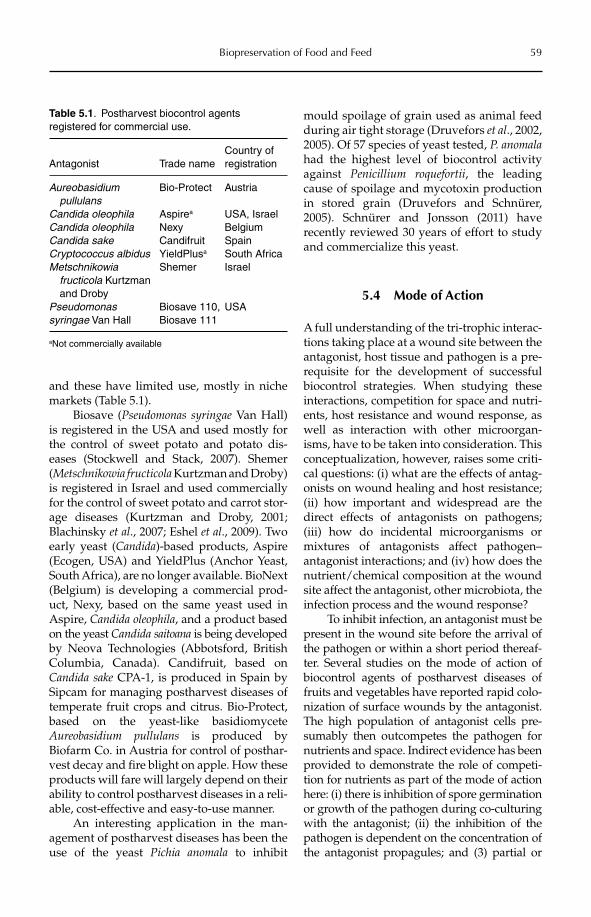
Biopreservation of Food and Feed 59
and these have limited use, mostly in niche markets (Table 5.1).
Biosave (Pseudomonas syringae Van Hall) is registered in the USA and used mostly for the control of sweet potato and potato dis-eases (Stockwell and Stack, 2007). Shemer (Metschnikowia fructicola Kurtzman and Droby) is registered in Israel and used commercially for the control of sweet potato and carrot stor-age diseases (Kurtzman and Droby, 2001; Blachinsky et al., 2007; Eshel et al., 2009). Two early yeast (Candida)-based products, Aspire (Ecogen, USA) and YieldPlus (Anchor Yeast, South Africa), are no longer available. BioNext (Belgium) is developing a commercial prod-uct, Nexy, based on the same yeast used in Aspire, Candida oleophila, and a product based on the yeast Candida saitoana is being developed by Neova Technologies (Abbotsford, British Columbia, Canada). Candifruit, based on Candida sake CPA-1, is produced in Spain by Sipcam for managing postharvest diseases of temperate fruit crops and citrus. Bio-Protect, based on the yeast-like basidiomycete Aureobasidium pullulans is produced by Biofarm Co. in Austria for control of posthar-vest decay and fire blight on apple. How these products will fare will largely depend on their ability to control postharvest diseases in a reli-able, cost-effective and easy-to-use manner.
An interesting application in the man-agement of postharvest diseases has been the use of the yeast Pichia anomala to inhibit
mould spoilage of grain used as animal feed during air tight storage (Druvefors et al., 2002, 2005). Of 57 species of yeast tested, P. anomalahad the highest level of biocontrol activity against Penicillium roquefortii, the leading cause of spoilage and mycotoxin production in stored grain (Druvefors and Schnürer, 2005). Schnürer and Jonsson (2011) have recently reviewed 30 years of effort to study and commercialize this yeast.
5.4 Mode of Action
A full understanding of the tri-trophic interac-tions taking place at a wound site between the antagonist, host tissue and pathogen is a pre-requisite for the development of successful biocontrol strategies. When studying these interactions, competition for space and nutri-ents, host resistance and wound response, as well as interaction with other microorgan-isms, have to be taken into consideration. This conceptualization, however, raises some criti-cal questions: (i) what are the effects of antag-onists on wound healing and host resistance; (ii) how important and widespread are the direct effects of antagonists on pathogens; (iii) how do incidental microorganisms or mixtures of antagonists affect pathogen–antagonist interactions; and (iv) how does the nutrient/chemical composition at the wound site affect the antagonist, other microbiota, the infection process and the wound response?
To inhibit infection, an antagonist must be present in the wound site before the arrival of the pathogen or within a short period thereaf-ter. Several studies on the mode of action of biocontrol agents of postharvest diseases of fruits and vegetables have reported rapid colo-nization of surface wounds by the antagonist. The high population of antagonist cells pre-sumably then outcompetes the pathogen for nutrients and space. Indirect evidence has been provided to demonstrate the role of competi-tion for nutrients as part of the mode of action here: (i) there is inhibition of spore germination or growth of the pathogen during co-culturing with the antagonist; (ii) the inhibition of the pathogen is dependent on the concentration of the antagonist propagules; and (3) partial or
Table 5.1. Postharvest biocontrol agents registered for commercial use.
Antagonist Trade nameCountry of registration
Aureobasidium pullulans
Bio-Protect Austria
Candida oleophila Aspirea USA, IsraelCandida oleophila Nexy BelgiumCandida sake Candifruit SpainCryptococcus albidus YieldPlusa South AfricaMetschnikowia fructicola Kurtzman and Droby
Shemer Israel
Pseudomonassyringae Van Hall
Biosave 110, Biosave 111
USA
aNot commercially available
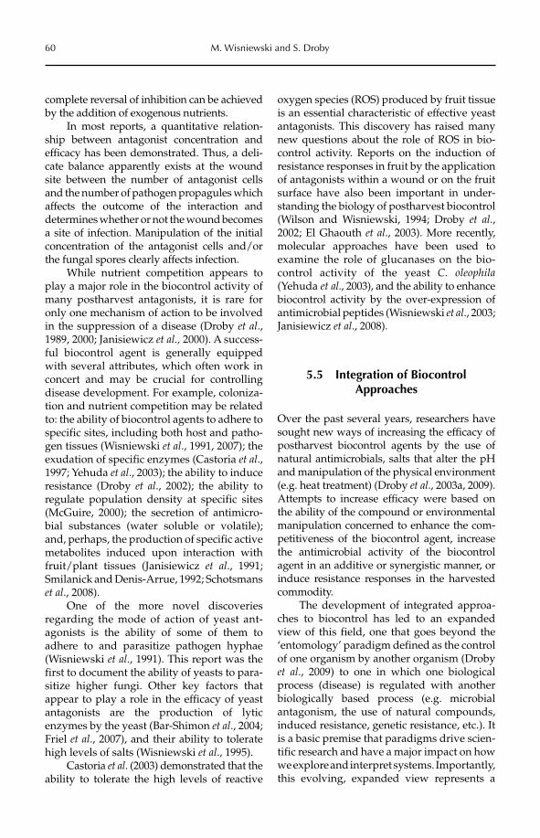
60 M. Wisniewski and S. Droby
complete reversal of inhibition can be achieved by the addition of exogenous nutrients.
In most reports, a quantitative relation-ship between antagonist concentration and efficacy has been demonstrated. Thus, a deli-cate balance apparently exists at the wound site between the number of antagonist cells and the number of pathogen propagules which affects the outcome of the interaction and determines whether or not the wound becomes a site of infection. Manipulation of the initial concentration of the antagonist cells and/or the fungal spores clearly affects infection.
While nutrient competition appears to play a major role in the biocontrol activity of many postharvest antagonists, it is rare for only one mechanism of action to be involved in the suppression of a disease (Droby et al.,1989, 2000; Janisiewicz et al., 2000). A success-ful biocontrol agent is generally equipped with several attributes, which often work in concert and may be crucial for controlling disease development. For example, coloniza-tion and nutrient competition may be related to: the ability of biocontrol agents to adhere to specific sites, including both host and patho-gen tissues (Wisniewski et al., 1991, 2007); the exudation of specific enzymes (Castoria et al.,1997; Yehuda et al., 2003); the ability to induce resistance (Droby et al., 2002); the ability to regulate population density at specific sites (McGuire, 2000); the secretion of antimicro-bial substances (water soluble or volatile); and, perhaps, the production of specific active metabolites induced upon interaction with fruit/plant tissues (Janisiewicz et al., 1991; Smilanick and Denis-Arrue, 1992; Schotsmans et al., 2008).
One of the more novel discoveries regarding the mode of action of yeast ant-agonists is the ability of some of them to adhere to and parasitize pathogen hyphae (Wisniewski et al., 1991). This report was the first to document the ability of yeasts to para-sitize higher fungi. Other key factors that appear to play a role in the efficacy of yeast antagonists are the production of lytic enzymes by the yeast (Bar-Shimon et al., 2004; Friel et al., 2007), and their ability to tolerate high levels of salts (Wisniewski et al., 1995).
Castoria et al. (2003) demonstrated that the ability to tolerate the high levels of reactive
oxygen species (ROS) produced by fruit tissue is an essential characteristic of effective yeast antagonists. This discovery has raised many new questions about the role of ROS in bio-control activity. Reports on the induction of resistance responses in fruit by the application of antagonists within a wound or on the fruit surface have also been important in under-standing the biology of postharvest biocontrol (Wilson and Wisniewski, 1994; Droby et al.,2002; El Ghaouth et al., 2003). More recently, molecular approaches have been used to examine the role of glucanases on the bio-control activity of the yeast C. oleophila(Yehuda et al., 2003), and the ability to enhance biocontrol activity by the over-expression of antimicrobial peptides (Wisniewski et al., 2003; Janisiewicz et al., 2008).
5.5 Integration of Biocontrol Approaches
Over the past several years, researchers have sought new ways of increasing the efficacy of postharvest biocontrol agents by the use of natural antimicrobials, salts that alter the pH and manipulation of the physical environment (e.g. heat treatment) (Droby et al., 2003a, 2009). Attempts to increase efficacy were based on the ability of the compound or environmental manipulation concerned to enhance the com-petitiveness of the biocontrol agent, increase the antimicrobial activity of the biocontrol agent in an additive or synergistic manner, or induce resistance responses in the harvested commodity.
The development of integrated approa-ches to biocontrol has led to an expanded view of this field, one that goes beyond the ‘entomology’ paradigm defined as the control of one organism by another organism (Droby et al., 2009) to one in which one biological process (disease) is regulated with another biologically based process (e.g. microbial antagonism, the use of natural compounds, induced resistance, genetic resistance, etc.). It is a basic premise that paradigms drive scien-tific research and have a major impact on how we explore and interpret systems. Importantly, this evolving, expanded view represents a
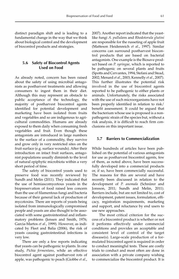
Biopreservation of Food and Feed 61
distinct paradigm shift and is leading to a fundamental change in the way that we think about biological control and the development of biocontrol products and strategies.
5.6 Safety of Biocontrol Agents Used on Food
As already noted, concern has been raised about the safety of using microbial antago-nists as postharvest treatments and allowing consumers to ingest them in their diet. Although this may represent an obstacle to public acceptance of the technology, the majority of postharvest biocontrol agents identified for potential development and marketing have been isolated from fruits and vegetables and so are indigenous to agri-cultural commodities. Humans are already exposed to them daily when consuming fresh vegetables and fruit. Even though these antagonists are introduced in large numbers to the surface of a commodity, they survive and grow only in very restricted sites on the fruit surface (e.g. surface wounds). After their introduction on intact fruit surfaces, antago-nist populations usually diminish to the level of natural epiphytic microbiota within a very short period of time.
The safety of biocontrol yeasts used to preserve food was recently reviewed by Sundh and Melin (2011). They indicated that the use of hemiascomycetous yeasts in the biopreservation of food raised less concern than the use of filamentous fungi and bacteria because of their general lack of production of mycotoxins. There are reports of yeasts being isolated from immunologically compromised people and yeasts are also thought to be asso-ciated with some gastrointestinal and inflam-matory problems (Jensen and Smith, 1976; Garcia-Martos et al., 1999). However, as indi-cated by Fleet and Balia (2006), the risk of yeasts causing gastrointestinal infections is very low.
There are only a few reports indicating that yeasts can be pathogenic to plants. In one study, Pichia fermentans, while an effective biocontrol agent against postharvest rots of apple, was pathogenic to peach (Giobbe et al.,
2007). Another report indicated that the yeast-like fungi A. pullulans and Rhodotorula glutiniare responsible for the russeting of apple fruit (Matteson Heidenreich et al., 1997). Similar concerns can surround postharvest biocon-trol products that are based on bacterial antagonists. One example is the Biosave prod-uct based on P. syringae, which is reported to be pathogenic on several plants and fruits (Spotts and Cervantes, 1994; Stefani and Stead, 2002; Menard et al., 2003; Kennelly et al., 2007). This further illustrates the potential risk involved in the use of biocontrol agents reported to be pathogenic to either plants or animals. Unfortunately, the risks associated with the use of such microorganisms have not been properly identified in relation to risk/benefit assessment. It could be argued that the bacterium whose use is proposed is a non-pathogenic strain of the species but, without a risk analysis, it is difficult to reach firm con-clusions on this important issue.
5.7 Barriers to Commercialization
While hundreds of articles have been pub-lished on the potential of various antagonists for use as postharvest biocontrol agents, few of them, as noted above, have been success-fully developed into a commercial products or, if so, have been commercially successful. The reasons for this are several and have recently been discussed in relation to the development of P. anomala (Schnürer and Jonsson, 2011; Sundh and Melin, 2011). Barriers include, but are not limited to, cost of development, patent issues, formulation, effi-cacy, registration requirements, marketing and support, and reluctance by end users to use new approaches.
The most critical criterion for the suc-cess of a biocontrol product is whether or not it performs effectively under commercial conditions and provides an acceptable and consistent level of control of the target disease(s). Large-scale production of a for-mulated biocontrol agent is required in order to conduct meaningful tests. These are costly trials to conduct and most often are done in association with a private company wishing to commercialize the biocontrol product. It is
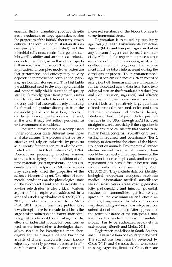
62 M. Wisniewski and S. Droby
essential that a formulated product, despite mass production of large quantities, retains the properties of the initial, laboratory-grown cultures. The formulation must retain its spe-cies purity (not be contaminated) and the microbial cells must retain their genetic sta-bility, cell viability and attributes as coloniz-ers on fruit surfaces, as well as other aspects of their mechanism of action. The commercial implications of complex modes of action are that performance and efficacy may be very dependent on production, formulation, pack-ing, application, storage, etc. This highlights the additional need to develop rapid, reliable and economically viable methods of quality testing. Currently, apart from growth assays (which may not reflect biocontrol activity), the only tests that are available rely on testing the formulated product directly on fruit (the commodity). This can be a long process if conducted in a comprehensive manner and, in the end, it may not reflect performance under commercial conditions.
Industrial fermentation is accomplished under conditions quite different from those in shake culture. The process must be cost-effective and rely on industrial by-products as nutrients; fermentation must also be com-pleted within 24–30 h (Hofstein et al., 1994). Downstream processing involves various steps, such as drying, and the addition of vol-ume materials (inert ingredients), adhesives, emulsifiers and adjuvants. All these actions may adversely affect the properties of the selected biocontrol agent. The effect of com-mercial conditions on the physiological state of the biocontrol agent and its activity fol-lowing rehydration is also critical. Various aspects of this topic were addressed in a series of articles by Abadias et al. (2000, 2001, 2003), and also in a recent article by Melin et al. (2011). Apart from these publications, few attempts have been made to address the large-scale production and formulation tech-nology of postharvest biocontrol agents. The effects of industrial production practices, as well as the formulation technologies them-selves, need to be investigated more thor-oughly for their impact on the biocontrol activity of chosen antagonists. Such knowl-edge may not only prevent a decrease in effi-cacy but actually lead to enhancement and
increased resistance of the biocontrol agents to environmental stress.
Registration is required by regulatory agencies (e.g. the US Environmental Protection Agency (EPA), and European agencies) before any biocontrol agent can be used commer-cially. Although the registration process is not as expensive or time consuming as it is for synthetic chemical fungicides, this require-ment must be taken into account during the development process. The registration pack-age must contain evidence of a clean record of safety (for both humans and the environment) for the biocontrol agent, data from basic toxi-cological tests on the formulated product (eye and skin irritation, ingestion) and efficacy data, including semi-commercial and com-mercial tests using relatively large quantities of food commodities treated under conditions that resemble commercial practices. The reg-istration of biocontrol products for posthar-vest use in the USA (through EPA) has been straightforward, especially if the organism is free of any medical history that would raise human health concerns. Typically, only Tier 1 testing is required, and occasionally Tier 2 testing, to determine the effect of the bicon-trol agent on animals. Environmental impact studies are not required at present; these would be very costly. In Europe, however, the situation is more complex and, until recently, registration has been difficult because data requirements are extensive (OJEC, 2001; OJEU, 2005). They include data on: identity; biological properties; analytical methods; medical information; medical surveillance; tests of sensitization, acute toxicity, genotox-icity, pathogenicity and infection potential; residues on commodities; persistence and spread in the environment; and effects on non-target organisms. The whole process is very demanding and may take 5–6 years from submission of the dossier. After approval of the active substance at the European Union level, practice has been that each formulated product has to be authorized separately in each country (Sundh and Melin, 2011).
Registration guidelines in South America are quite variable from one country to the next. The subject has been recently reviewed by Cotes (2011), and she notes that in some coun-tries, e.g. Argentina, Brazil and Chile, there are
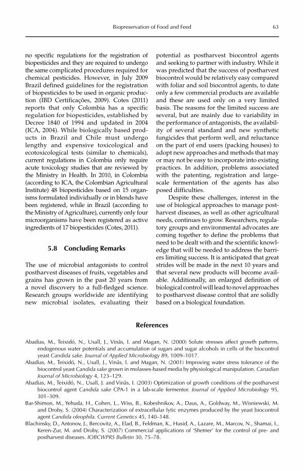
Biopreservation of Food and Feed 63
no specific regulations for the registration of biopesticides and they are required to undergo the same complicated procedures required for chemical pesticides. However, in July 2009 Brazil defined guidelines for the registration of biopesticides to be used in organic pro duc-tion (IBD Certificações, 2009). Cotes (2011) reports that only Colombia has a specific regulation for biopesticides, established by Decree 1840 of 1994 and updated in 2004 (ICA, 2004). While biologically based prod-ucts in Brazil and Chile must undergo lengthy and expensive toxicological and ecotoxico logical tests (similar to chemicals), current regulations in Colombia only require acute toxicology studies that are reviewed by the Ministry in Health. In 2010, in Colombia (according to ICA, the Colombian Agricultural Institute) 48 biopesticides based on 15 organ-isms formulated individually or in blends have been registered, while in Brazil (according to the Ministry of Agriculture), currently only four microorganisms have been registered as active ingredients of 17 biopesticides (Cotes, 2011).
5.8 Concluding Remarks
The use of microbial antagonists to control postharvest diseases of fruits, vegetables and grains has grown in the past 20 years from a novel discovery to a full-fledged science. Research groups worldwide are identifying new microbial isolates, evaluating their
poten tial as postharvest biocontrol agents and seeking to partner with industry. While it was predicted that the success of postharvest biocontrol would be relatively easy compared with foliar and soil biocontrol agents, to date only a few commercial products are available and these are used only on a very limited basis. The reasons for the limited success are several, but are mainly due to variability in the performance of antagonists, the availabil-ity of several standard and new synthetic fungicides that perform well, and reluctance on the part of end users (packing houses) to adopt new approaches and methods that may or may not be easy to incorporate into existing practices. In addition, problems associated with the patenting, registration and large-scale fermentation of the agents has also posed difficulties.
Despite these challenges, interest in the use of biological approaches to manage post-harvest diseases, as well as other agricultural needs, continues to grow. Researchers, regula-tory groups and environmental advocates are coming together to define the problems that need to be dealt with and the scientific knowl-edge that will be needed to address the barri-ers limiting success. It is anticipated that great strides will be made in the next 10 years and that several new products will become avail-able. Additionally, an enlarged definition of biological control will lead to novel approaches to postharvest disease control that are solidly based on a biological foundation.
References
Abadias, M., Teixidó, N., Usall, J., Vinãs, I. and Magan, N. (2000) Solute stresses affect growth patterns, endogenous water potentials and accumulation of sugars and sugar alcohols in cells of the biocontrol yeast Candida sake. Journal of Applied Microbiology 89, 1009–1017.
Abadias, M., Teixidó, N., Usall, J., Vinãs, I. and Magan, N. (2001) Improving water stress tolerance of the biocontrol yeast Candida sake grown in molasses-based media by physiological manipulation. CanadianJournal of Microbiology 4, 123–129.
Abadias, M., Teixidó, N., Usall, J. and Vinãs, I. (2003) Optimization of growth conditions of the postharvest biocontrol agent Candida sake CPA-1 in a lab-scale fermentor. Journal of Applied Microbiology 95, 301–309.
Bar-Shimon, M., Yehuda, H., Cohen, L., Wiss, B., Kobeshnikov, A., Daus, A., Goldway, M., Wisniewski, M. and Droby, S. (2004) Characterization of extracellular lytic enzymes produced by the yeast biocontrol agent Candida oleophila. Current Genetics 45, 140–148.
Blachinsky, D., Antonov, J., Bercovitz, A., Elad, B., Feldman, K., Husid, A., Lazare, M., Marcov, N., Shamai, I., Keren-Zur, M. and Droby, S. (2007) Commercial applications of ‘Shemer’ for the control of pre- and postharvest diseases. IOBCWPRS Bulletin 30, 75–78.
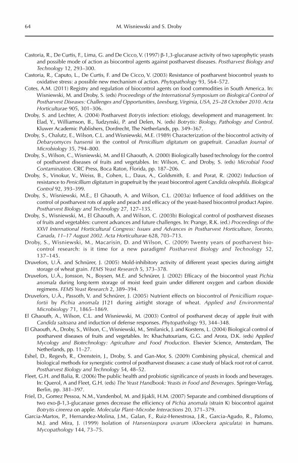
64 M. Wisniewski and S. Droby
Castoria, R., De Curtis, F., Lima, G. and De Cicco, V. (1997) b-1,3-glucanase activity of two saprophytic yeasts and possible mode of action as biocontrol agents against postharvest diseases. Postharvest Biology and Technology 12, 293–300.
Castoria, R., Caputo, L., De Curtis, F. and De Cicco, V. (2003) Resistance of postharvest biocontrol yeasts to oxidative stress: a possible new mechanism of action. Phytopathology 93, 564–572.
Cotes, A.M. (2011) Registry and regulation of biocontrol agents on food commodities in South America. In: Wisniewski, M. and Droby, S. (eds) Proceedings of the International Symposium on Biological Control of Postharvest Diseases: Challenges and Opportunities, Leesburg, Virginia, USA, 25–28 October 2010. Acta Horticulturae 905, 301–306.
Droby, S. and Lechter, A. (2004) Postharvest Botrytis infection: etiology, development and management. In: Elad, Y., Williamson, B., Tudzynski, P. and Delen, N. (eds) Botrytis: Biology, Pathology and Control.Kluwer Academic Publishers, Dordrecht, The Netherlands, pp. 349–367.
Droby, S., Chalutz, E., Wilson, C.L. and Wisniewski, M.E. (1989) Characterization of the biocontrol activity of Debaryomyces hansenii in the control of Penicillium digitatum on grapefruit. Canadian Journal of Microbiology 35, 794–800.
Droby, S., Wilson, C., Wisniewski, M. and El Ghaouth, A. (2000) Biologically based technology for the control of postharvest diseases of fruits and vegetables. In: Wilson, C. and Droby, S. (eds) Microbial Food Contamination. CRC Press, Boca Raton, Florida, pp. 187–206.
Droby, S., Vinokur, V., Weiss, B., Cohen, L., Daus, A., Goldsmith, E. and Porat, R. (2002) Induction of resistance to Penicillium digitatum in grapefruit by the yeast biocontrol agent Candida oleophila. Biological Control 92, 393–399.
Droby, S., Wisniewski, M.E., El Ghaouth, A. and Wilson, C.L. (2003a) Influence of food additives on the control of postharvest rots of apple and peach and efficacy of the yeast-based biocontrol product Aspire. Postharvest Biology and Technology 27, 127–135.
Droby, S., Wisniewski, M., El Ghaouth, A. and Wilson, C. (2003b) Biological control of postharvest diseases of fruits and vegetables: current advances and future challenges. In: Prange, R.K. (ed.) Proceedings of the XXVI International Horticultural Congress: Issues and Advances in Postharvest Horticulture, Toronto, Canada, 11–17 August 2002. Acta Horticulturae 628, 703–713.
Droby, S., Wisniewski, M., Macarisin, D. and Wilson, C. (2009) Twenty years of postharvest bio-control research: is it time for a new paradigm? Postharvest Biology and Technology 52, 137–145.
Druvefors, U.Ä. and Schnürer, J. (2005) Mold-inhibitory activity of different yeast species during airtight storage of wheat grain. FEMS Yeast Research 5, 373–378.
Druvefors, U.Ä., Jonsson, N., Boysen, M.E. and Schnürer, J. (2002) Efficacy of the biocontrol yeast Pichiaanomala during long-term storage of moist feed grain under different oxygen and carbon dioxide regimens. FEMS Yeast Research 2, 389–394.
Druvefors, U.Ä., Passoth, V. and Schnürer, J. (2005) Nutrient effects on biocontrol of Penicillium roque-fortii by Pichia anomala J121 during airtight storage of wheat. Applied and Environmental Microbiology 71, 1865–1869.
El Ghaouth, A., Wilson, C.L. and Wisniewski, M. (2003) Control of postharvest decay of apple fruit with Candida saitoana and induction of defense responses. Phytopathology 93, 344–348.
El Ghaouth, A., Droby, S., Wilson, C., Wisniewski, M., Smilanick, J. and Korstens, L. (2004) Biological control of postharvest diseases of fruits and vegetables. In: Khachatourians, G.G. and Arora, D.K. (eds) Applied Mycology and Biotechnology: Agriculture and Food Production. Elsevier Science, Amsterdam, The Netherlands, pp. 11–27.
Eshel, D., Regevb, R., Orenstein, J., Droby, S. and Gan-Mor, S. (2009) Combining physical, chemical and biological methods for synergistic control of postharvest diseases: a case study of black root rot of carrot. Postharvest Biology and Technology 54, 48–52.
Fleet, G.H. and Balia, R. (2006) The public health and probiotic significance of yeasts in foods and beverages. In: Querol, A and Fleet, G.H. (eds) The Yeast Handbook: Yeasts in Food and Beverages. Springer-Verlag, Berlin, pp. 381–397.
Friel, D., Gomez Pessoa, N.M., Vandenbol, M. and Jijakli, H.M. (2007) Separate and combined disruptions of two exo-b-1,3-glucanase genes decrease the efficiency of Pichia anomala (strain K) biocontrol against Botrytis cinerea on apple. Molecular Plant–Microbe Interactions 20, 371–379.
Garcia-Martos, P., Hernandez-Molina, J.M., Galan, F., Ruiz-Henestrosa, J.R., Garcia-Agudo, R., Palomo, M.J. and Mira, J. (1999) Isolation of Hanseniaspora uvarum (Kloeckera apiculata) in humans. Mycopathology 144, 73–75.
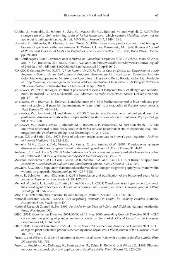
Biopreservation of Food and Feed 65
Giobbe, S., Marceddu, S., Scherm, B., Zara, G., Mazzarello, V.L., Budroni, M. and Migheli, Q. (2007) The strange case of a biofilm-forming strain of Pichia fermentans, which controls Monilinia brown rot on apple but is pathogenic on peach fruit. FEMS Yeast Research 7, 1389–1398.
Hofstein, R., Fridlender, B., Chalutz, E. and Droby, S. (1994) Large scale production and pilot testing of biocontrol agents of postharvest diseases. In: Wilson, C.L. and Wisniewski, M.E. (eds) Biological Control of Postharvest Diseases of Fruits and Vegetables: Theory and Practice. CRC Press, Boca Raton, Florida, pp. 89–100.
IBD Certificações (2009) Diretrizes para o Padrão de Qualidade ‘Orgânico IBD’, 17a Edição, Julho de 2009, doc. 8-1-2. Botucatu, São Paulo, Brazil. Available at: http://www.ibd.com.br/Media/arquivo_digital/e521889a-c16f-45df-baf8-13609d8a6891.pdf (accessed 30 April 2012).
ICA (2004) Resolución No. 00375 (27 de febrero de 2004). Por la Cual se Dictan las Disposiciones sobre Registro y Control de los Bioinsumos y Extractos Vegetales de Uso Agrícola en Colombia. Instituto Colombiano Agropecuario, Ministerio de Agricultura y Desarrollo Rural, Bogota, Colombia. Available at: http://www.agriculturaorganicaamericas.net/Documentos%20Oficiales%20CIAO/Registro%20de% 20bioinsumos%20y%20extractos.pdf (accessed 30 April 2012).
Janisiewicz, W. (1998) Biological control of postharvest diseases of temperate fruits: challenges and opportu-nities. In: Boland, G.J. and Kuykendall, L.D. (eds) Plant–Microbe Interactions. Marcel Dekker, New York, pp. 171–198.
Janisiewicz, W.J., Yourman, L., Roitman, J. and Mahoney, N. (1991) Postharvest control of blue mold and gray mold of apples and pears by dip treatments with pyrrolnitrin, a metabolite of Pseudomonas cepacia. Plant Disease 75, 490–494.
Janisiewicz, W.J., Tworkoski, T.J. and Sharer, C. (2000) Characterizing the mechanism of biological control of postharvest diseases on fruits with a simple method to study competition for nutrients. Phytopathology90, 1196–1200.
Janisiewicz, W.J., Bastos Pereira, I., Almeida, M.S., Roberts, D.P., Wisniewski, M. and Kurtenbach, E. (2008) Improved biocontrol of fruit decay fungi with Pichia pastoris recombinant strains expressing Psd1 anti-fungal peptide. Postharvest Biology and Technology 47, 218–225.
Jensen, D.P. and Smith, D.L. (1976) Fever of unknown origin secondary to brewer’s yeast ingestion. Archives of Internal Medicine 136, 332–333.
Kennelly, M.M., Cazorla, F.M., Vicente, A., Ramos, C. and Sundin, G.W. (2007) Pseudomonas syringaediseases of fruits trees: progress toward understanding and control. Plant Disease, 91, 4–17.
Kurtzman, C.P. and Droby, S. (2001) Metschnikowia fructicola, a new ascosporic yeast effective for biocontrol of postharvest fruit rots. Systematic and Applied Microbiology 24, 395–399.
Matteson Heidenreich, M.C., Corral-Garcia, M.R., Momol, E.A. and Burr, T.J. (1997) Russet of apple fruit caused by Aureobasidium pullulans and Rhodotorula glutinis. Plant Disease 81, 337–342.
McGuire, R.G. (2000) Population dynamics of postharvest decay antagonists growing epiphytically and within wounds on grapefruit. Phytopathology 90, 1217–1223.
Melin, P., Schnürer, J. and Håkanson, S. (2011) Formulation and stabilization of the biocontrol yeast Pichiaanomala. Antonie van Leeuwenhoek 99, 107–112.
Menard, M., Sutra, L., Luisetti, J., Prunier, J.P. and Gardan, L. (2003) Pseudomonas syringae pv. avii (pv. nov.), the causal agent of bacterial canker of wild cherries (Prunus avium) in France. European Journal of Plant Pathology 109, 565–576.
Mlot, C. (2009) Antibiotics in nature: beyond biological warfare. Science 324, 1637–1639.National Research Council (USA) (1987) Regulating Pesticides in Food: The Delaney Paradox. National
Academies Press, Washington DC.National Research Council (USA) (1993) Pesticides in the Diets of Infants and Children. National Academies
Press, Washington DC.OJEC (2001) Commission Directive 2001/36/EC of 16 May 2001 amending Council Directive 91/414/EEC
concerning the placing of plant protection products on the market. Official Journal of the European Communities 44, L 164/1–38.
OJEU (2005) Council Directive 2005/25/EC of 14 March 2005 amending Annex VI to Directive 91/414/EEC as regards plant protection products containing micro-organisms. Official Journal of the European Union48, L 90/1–34.
Pusey, L. and Wilson, C. (1984) Biocontrol of brown rot of stone fruits with a strain of Bacillus subtilis. Plant Disease 68, 753–756.
Pusey, L., Hotchkiss, M., Dulmage, H., Baumgardner, R., Zeher, E., Reilly, C. and Wilson, C. (1988) Pilot test for commercial production and application of Bacillus subtilis. Plant Disease 72, 622–626.
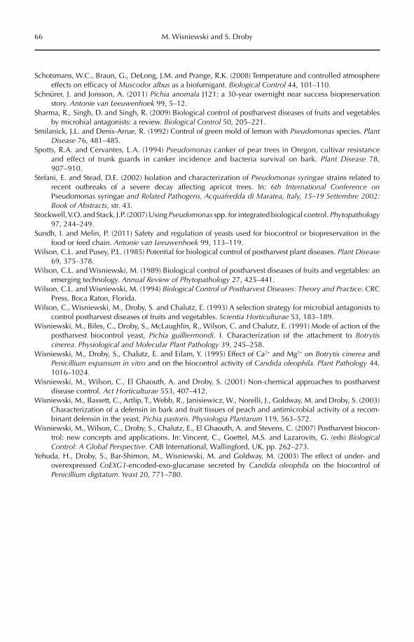
66 M. Wisniewski and S. Droby
Schotsmans, W.C., Braun, G., DeLong, J.M. and Prange, R.K. (2008) Temperature and controlled atmosphere effects on efficacy of Muscodor albus as a biofumigant. Biological Control 44, 101–110.
Schnürer, J. and Jonsson, A. (2011) Pichia anomala J121: a 30-year overnight near success biopreservation story. Antonie van Leeuwenhoek 99, 5–12.
Sharma, R., Singh, D. and Singh, R. (2009) Biological control of postharvest diseases of fruits and vegetables by microbial antagonists: a review. Biological Control 50, 205–221.
Smilanick, J.L. and Denis-Arrue, R. (1992) Control of green mold of lemon with Pseudomonas species. PlantDisease 76, 481–485.
Spotts, R.A. and Cervantes, L.A. (1994) Pseudomonas canker of pear trees in Oregon, cultivar resistance and effect of trunk guards in canker incidence and bacteria survival on bark. Plant Disease 78, 907–910.
Stefani, E. and Stead, D.E. (2002) Isolation and characterization of Pseudomonas syringae strains related to recent outbreaks of a severe decay affecting apricot trees. In: 6th International Conference onPseudomonas syringae and Related Pathogens, Acquafredda di Maratea, Italy, 15–19 Settembre 2002: Book of Abstracts, str. 43.
Stockwell, V.O. and Stack, J.P. (2007) Using Pseudomonas spp. for integrated biological control. Phytopathology97, 244–249.
Sundh, I. and Melin, P. (2011) Safety and regulation of yeasts used for biocontrol or biopreservation in the food or feed chain. Antonie van Leeuwenhoek 99, 113–119.
Wilson, C.L. and Pusey, P.L. (1985) Potential for biological control of postharvest plant diseases. Plant Disease69, 375–378.
Wilson, C.L. and Wisniewski, M. (1989) Biological control of postharvest diseases of fruits and vegetables: an emerging technology. Annual Review of Phytopathology 27, 425–441.
Wilson, C.L. and Wisniewski, M. (1994) Biological Control of Postharvest Diseases: Theory and Practice. CRC Press, Boca Raton, Florida.
Wilson, C., Wisniewski, M., Droby, S. and Chalutz, E. (1993) A selection strategy for microbial antagonists to control postharvest diseases of fruits and vegetables. Scientia Horticulturae 53, 183–189.
Wisniewski, M., Biles, C., Droby, S., McLaughlin, R., Wilson, C. and Chalutz, E. (1991) Mode of action of the postharvest biocontrol yeast, Pichia guilliermondi. I. Characterization of the attachment to Botrytis cinerea. Physiological and Molecular Plant Pathology 39, 245–258.
Wisniewski, M., Droby, S., Chalutz, E. and Eilam, Y. (1995) Effect of Ca2+ and Mg2+ on Botrytis cinerea and Penicillium expansum in vitro and on the biocontrol activity of Candida oleophila. Plant Pathology 44, 1016–1024.
Wisniewski, M., Wilson, C., El Ghaouth, A. and Droby, S. (2001) Non-chemical approaches to postharvest disease control. Act Horticulturae 553, 407–412.
Wisniewski, M., Bassett, C., Artlip, T., Webb, R., Janisiewicz, W., Norelli, J., Goldway, M. and Droby, S. (2003) Characterization of a defensin in bark and fruit tissues of peach and antimicrobial activity of a recom-binant defensin in the yeast, Pichia pastoris. Physiologia Plantarum 119, 563–572.
Wisniewski, M., Wilson, C., Droby, S., Chalutz, E., El Ghaouth, A. and Stevens, C. (2007) Postharvest biocon-trol: new concepts and applications. In: Vincent, C., Goettel, M.S. and Lazarovits, G. (eds) BiologicalControl: A Global Perspective. CAB International, Wallingford, UK, pp. 262–273.
Yehuda, H., Droby, S., Bar-Shimon, M., Wisniewski, M. and Goldway, M. (2003) The effect of under- and overexpressed CoEXG1-encoded-exo-glucanase secreted by Candida oleophila on the biocontrol of Penicillium digitatum. Yeast 20, 771–780.
©CAB International 2012. Beneficial Microorganisms in Agriculture, Food and the Environment: Safety Assessment and Regulation (eds I. Sundh et al.) 67
6.1 Introduction 676.2 Data Requirements 686.3 Regulatory Procedures 686.4 Regulation of Biocides 706.5 Conclusion 71
6 Safety and Regulation of Microbial Pest Control Agents and Microbial
Plant Growth Promoters – Introduction and Overview
Rüdiger HauschildGAB Consulting GmbH, Germany
6.1 Introduction
Increasingly, chemical pesticides are being taken off the market owing to newly discov-ered hazards towards human or environmen-tal health. In addition, there is increased public demand for organically grown food. Microbial pesticides have a potentially important role to play in our ability to protect our agriculture and forests in a sustainable manner, and most countries have strict regulations for them, as they do for chemical pesticides. However, risk assessments of microbial pesticides present unique challenges, because microorganisms are living creatures and not chemicals.
Traditional chemical plant protection products (PPPs) are increasingly criticized for different reasons, but mainly for their poten-tially detrimental effects on human health or the environment. Concerns about residues in crops play an important role in the perception of chemical PPPs, and microorganisms repre-sent a promising alternative as active sub-stances. Due to their natural occurrence in the
environment, at least some humans, and other non-target organisms, have always been expo sed to many of these organisms and, consequently, have had the opportunity to adapt to them. Most of the microorganisms used are not new to consumers or to the envi-ronment and, therefore, defence mechanisms against such biocontrol organisms have evolved among non-target organisms where they were necessary. However, microorgan-isms differ fundamentally from chemicals in their capacity to multiply in the environment and to produce substances that may be involved in their mode of action, and these may also have detrimental effects on non- target organisms.
This chapter gives a brief overview of the regulatory systems for microorganisms used to manage pest populations and plant dis-eases. In the four following chapters of this volume, Jaronski (Chapter 7), Alabouvette et al. (Chapter 8), Evans and Seier (Chapter 9) and Howieson and Fox (Chapter 10) discuss details on data requirements and safety
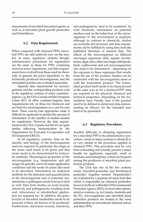
68 R. Hauschild
assessments of microbial biocontrol agents, as well as of microbial plant growth promoters and biofertilizers.
6.2 Data Requirements
When compared with chemical PPPs, micro-bial PPPs are still relatively new on the mar-ket. In many regulatory systems though, administrative procedures for registration are the same as those for PPPs containing chemical active ingredients, and data require-ments have evolved from those used for chem-icals. In general, the active ingredient, i.e. the technically produced microorganism, and the formulated product are evaluated separately.
Separate data requirements for microor-ganisms and the corresponding products exist in the regulatory systems of many countries/regions, e.g. the USA, Canada and the European Union (EU). In other countries/regions, data requirements rely on those for chemicals and are fixed for microorganisms on a case-by-case basis. These case-by-case approaches make it difficult for applicants to predict the amount of information or the number of studies needed for registration. However, the data require-ments in the USA, Canada and the EU are quite similar following harmonization by the Organisation for Economic Co-operation and Development (OECD).
In all regulatory systems, data on the identity and biology of the microorganism used are required. In particular, the origin of the strain used needs to be given and that strain needs to be characterized by molecu-lar methods. Physiological properties of the microorganism (e.g. temperature and pH ranges for growth), survival under application conditions and the mode of action also need to be described. Information on analytical methods for the detection and quan tification of the microorganism and of potential con-taminants or pathogens needs to be provided as well. Data from studies on acute toxicity, infectivity and pathogenicity resulting from oral, intravenous or intratracheal applica-tion are required too. In addition, the geno-toxicity of microbial metabolites needs to be assessed if these are known to be produced. Furthermore, short-term toxicity, infectivity,
and pathogenicity need to be examined. As with chemicals, information on potential residues and on the behaviour of the micro-organism in the environment is required, although in contrast to chemicals, studies are normally not necessary and data require-ments can be fulfilled by using data from the published literature or internal data. The effects of the microorganism on different non-target organisms (fish, aquatic inverte-brates, algae, bees, other non-target arthropods, birds, earthworms and soil micro organisms) also need to be determined – depending on the exposure to these organisms that results from the use of the product. Studies can be conducted with the microorganism alone or with the formulated product. The formu-lated product itself needs to be characterized in the same way as for a chemical PPP: data are required on the physical, chemical and technical properties, depending on the for-mulation type. Finally, uses of the product need to be defined in detail and data demon-strating its efficacy for the intended uses need to be presented.
6.3 Regulatory Procedures
Another difficulty in obtaining registration for a microbial PPP is the administrative pro-cedure, which in many systems is identical to or very similar to the procedure applied to chemical PPPs. This procedure may be very time-consuming and actually present a major hurdle for applicants, especially small or medium-sized enterprises, which are frequent among the producers of microbial plant pro-tection products.
The USA has one of the few systems where ‘microbial pesticides’ and ‘biochemical pesticides’ (together termed ‘biopesticides’) are evaluated by a separate division from that which is responsible for chemical pesticides; however, both are within the US Environmental Protection Agency (EPA). In most other admin-istrative systems, e.g. in Canada, Australia and the EU, microorganisms and microbial plant protection products are treated in the same administration as conventional chemical actives and their PPPs.
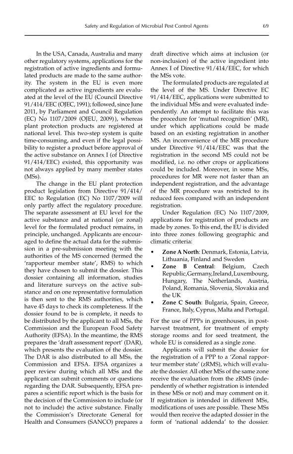
Safety and Regulation of Microbial Pest Control Agents 69
In the USA, Canada, Australia and many other regulatory systems, applications for the registration of active ingredients and formu-lated products are made to the same author-ity. The system in the EU is even more complicated as active ingredients are evalu-ated at the level of the EU (Council Directive 91/414/EEC (OJEC, 1991); followed, since June 2011, by Parliament and Council Regulation (EC) No 1107/2009 (OJEU, 2009) ), whereas plant protection products are registered at national level. This two-step system is quite time-consuming, and even if the legal possi-bility to register a product before approval of the active substance on Annex I (of Directive 91/414/EEC) existed, this opportunity was not always applied by many member states (MSs).
The change in the EU plant protection product legislation from Directive 91/414/EEC to Regulation (EC) No 1107/2009 will only partly affect the regulatory procedure. The separate assessment at EU level for the active substance and at national (or zonal) level for the formulated product remains, in principle, unchanged. Applicants are encour-aged to define the actual data for the submis-sion in a pre-submission meeting with the authorities of the MS concerned (termed the ‘rapporteur member state’, RMS) to which they have chosen to submit the dossier. This dossier containing all information, studies and literature surveys on the active sub-stance and on one representative formulation is then sent to the RMS authorities, which have 45 days to check its completeness. If the dossier found to be is complete, it needs to be dis tributed by the applicant to all MSs, the Commission and the European Food Safety Authority (EFSA). In the meantime, the RMS prepares the ‘draft assessment report’ (DAR), which presents the evaluation of the dossier. The DAR is also distributed to all MSs, the Commission and EFSA. EFSA organizes a peer review during which all MSs and the applicant can submit comments or questions regarding the DAR. Subsequently, EFSA pre-pares a scientific report which is the basis for the decision of the Commission to include (or not to include) the active substance. Finally the Commission’s Directorate General for Health and Consumers (SANCO) prepares a
draft directive which aims at inclusion (or non-inclusion) of the active ingredient into Annex I of Directive 91/414/EEC, for which the MSs vote.
The formulated products are regulated at the level of the MS. Under Directive EC 91/414/EEC, applications were submitted to the individual MSs and were evaluated inde-pendently. An attempt to facilitate this was the procedure for ‘mutual recognition’ (MR), under which applications could be made based on an existing registration in another MS. An inconvenience of the MR procedure under Directive 91/414/EEC was that the registration in the second MS could not be modified, i.e. no other crops or applications could be included. Moreover, in some MSs, procedures for MR were not faster than an independent registration, and the advantage of the MR procedure was restricted to its reduced fees compared with an independent registration.
Under Regulation (EC) No 1107/2009, applications for registration of products are made by zones. To this end, the EU is divided into three zones following geographic and climatic criteria:
Zone A North• : Denmark, Estonia, Latvia, Lithuania, Finland and SwedenZone B Central• : Belgium, Czech Republic, Germany, Ireland, Luxembourg, Hungary, The Netherlands, Austria, Poland, Romania, Slovenia, Slovakia and the UKZone C South• : Bulgaria, Spain, Greece, France, Italy, Cyprus, Malta and Portugal.
For the use of PPPs in greenhouses, in post-harvest treatment, for treatment of empty storage rooms and for seed treatment, the whole EU is considered as a single zone.
Applicants will submit the dossier for the registration of a PPP to a ‘Zonal rappor-teur member state’ (zRMS), which will evalu-ate the dossier. All other MSs of the same zone receive the evaluation from the zRMS (inde-pendently of whether registration is intended in these MSs or not) and may comment on it. If registration is intended in different MSs, modifications of uses are possible. These MSs would then receive the adapted dossier in the form of ‘national addenda’ to the dossier.
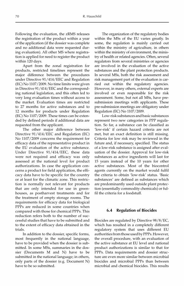
70 R. Hauschild
Following the evaluation, the zRMS releases the registration of the product within a year of the application (if the dossier was complete and no additional data were requested dur-ing evaluation). All other MS where registra-tion is applied for need to register the product within 120 days.
Apart from the zonal registration for products, restricted timelines represent the major difference between the procedures under Directive 91/414/EEC and Regulation (EC) No 1107/2009. No time limits were given in Directive 91/414/EEC and the correspond-ing national legislation, and this often led to very long evaluation times without access to the market. Evaluation times are restricted to 27 months for active substances and to 12 months for products under Regulation (EC) No 1107/2009. These times can be exten-ded by defined periods if additional data are requested from the applicant.
The other major difference between Directive 91/414/EEC and Regulation (EC) No 1107/2009 concerns the requirement for efficacy data of the representative product in the EU evaluation of the active substance. Under Directive 91/414/EEC, these data were not required and efficacy was only assessed at the national level for product authorizations. In case the application con-cerns a product for field application, the effi-cacy data have to be specific for the country or at least for the climatic zone. This restric-tion is normally not relevant for products that are only intended for use in green-houses, as postharvest treatments and for the treatment of empty storage rooms. The requirements for efficacy data for biological PPPs are reduced in some countries when compared with those for chemical PPPs. This reduction refers both to the number of suc-cessful studies that have to be submitted and to the extent of efficacy data obtained in the trials.
In addition to the dossier, specific forms, most frequently in the national language, have to be provided when the dossier is sub-mitted. In some MSs, summaries in the dos-sier (Documents M and N) have to be submitted in the national language; in others, only parts of the dossier (e.g. Document N) have to be so submitted.
The organization of the regulatory bodies within the MSs of the EU varies greatly. In some, the regulation is mainly carried out within the ministry of agriculture, in others within the ministry of environment, the minis-try of health or related agencies. Often, several regulators from several ministries or agencies are involved in the evaluation of the active substances and the plant protection products. In several MSs, both the risk assessment and risk management part of the evaluation is car-ried out within the regulatory agencies. However, in many others, external experts are involved or even responsible for the risk assessment. Some, but not all MSs, have pre-submission meetings with applicants. These pre-submission meetings are obligatory under Regulation (EC) No 1107/2009.
Low-risk substances and basic substances represent two new categories in PPP regula-tion. So far, a substance can be classified as ‘low-risk’ if certain hazard criteria are not met, but an exact definition is still missing. Criteria for low risk may be reviewed in the future and, if necessary, specified. The status of a low-risk substance is assigned after eval-uation of the dossier. Approval for low-risk substances as active ingredients will last for 15 years instead of the 10 years for other active substances. Most of the biocontrol agents currently on the market would fulfil the criteria to obtain ‘low-risk’ status. ‘Basic substances’ are defined as substances which are predominantly used outside plant protec-tion (essentially commodity chemicals) or ful-fil the criteria for a foodstuff.
6.4 Regulation of Biocides
Biocides are regulated by Directive 98/8/EC, which has resulted in a completely different regulatory system that uses different EU authorities from those used by PPPs. However, the overall procedure, with an evaluation of the active substance at EU level and national product authorizations is similar to that for PPPs. Data requirements and dossier struc-ture are even more similar between microbial biocides and microbial PPPs than between microbial and chemical biocides. This results
Safety and Regulation of Microbial Pest Control Agents 71
from the fact that the data requirements for microbial biocides were adapted from those for microbial plant protection products. Com-pared with PPPs, very few biocides contain microorganisms and experience of these among applicants and regulators is still quite limited.
6.5 Conclusion
Taken together, the registration of microbial plant protection products and biocides is
derived from that of their chemical counter-parts. Procedures for registration are the same for both microbial and chemical products, but data requirements have been adapted for microbials. However, improvements in data requirements are still necessary. The registra-tion of plant protection products (both micro-bials and chemicals) is still time-consuming, but the new EU regulation has for the first time defined timelines for the evaluation pro-cedure, as has been the case for many years in many non-EU countries.
References
Alabouvette, C., Heilig, U. and Cordier, C. (2012) Microbial control of plant diseases. In: Sundh, I., Wilcks, A. and Goettel, M.S. (eds) Beneficial Microorganisms in Agriculture, Food and the Environment: Safety Assessment and Regulation. CAB International, Wallingford, UK, pp. 96–111.
Evans, H.C. and Seier, M.K. (2012) Safety and regulation of microbial control of weeds. In: Sundh, I., Wilcks, A. and Goettel, M.S. (eds) Beneficial Microorganisms in Agriculture, Food and the Environment: Safety Assessment and Regulation. CAB International, Wallingford, UK, pp. 112–137.
Howieson, J.G. and Fox, S.L. (2012) Plant growth promotion with microorganisms. In: Sundh, I., Wilcks, A. and Goettel, M.S. (eds) Beneficial Microorganisms in Agriculture, Food and the Environment: Safety Assessment and Regulation. CAB International, Wallingford, UK, pp. 138–152.
Jaronski, S.F. (2012) Microbial control of invertebrate pests. In: Sundh, I., Wilcks, A. and Goettel, M.S. (eds) Beneficial Microorganisms in Agriculture, Food and the Environment: Safety Assessment and Regulation.CAB International, Wallingford, UK, pp. 72–95.
OJEC (1991) Council Directive 91/414/EEC of 15 July 1991 concerning the placing of plant protection prod-ucts on the market. Official Journal of the European Communities 34, L 230/1–32.
OJEC (1998) Directive 98/8/EC of the European Parliament and of the Council of 16 February 1998 concern-ing the placing of biocidal products on the market. Official Journal of the European Communities 41, L 123/1–63.
OJEU (2009) Regulation (EC) No 1107/2009 of the European Parliament and of the Council of 21 October 2009 concerning the placing of plant protection products on the market and repealing Council Directives 79/117/EEC and 91/414/EEC. Official Journal of the European Union 52, L 309/1–50.
©CAB International 2012. Beneficial Microorganisms in Agriculture, Food and the Environment: 72 Safety Assessment and Regulation (eds I. Sundh et al.)
7.1 Introduction 727.2 Microbial Arthropod Control Agents – A Brief Introduction 73
7.2.1 Use strategies of microbial pest control agents 73 7.2.2 Viruses 73 7.2.3 Bacteria 75 7.2.4 Protozoa 75 7.2.5 Fungi 76
7.3 Risk Assessment of Microbial Arthropod Control Agents 777.3.1 General issues 77
7.3.2 Human safety risk assessment 777.3.3 Environmental safety risk assessment 81
7.4 Additional Risk Assessment Considerations 877.4.1 Non-indigenous versus indigenous microorganisms 87
7.4.2 Genetically altered versus naturally occurring organisms 88 7.4.3 Use of microbial symbionts to control arthropods 89
7.5 Conclusion 89
7 Microbial Control of Invertebrate Pests
Stefan T. JaronskiNorthern Plains Agricultural Research Laboratory (NPARL),
United States Department of Agriculture – Agricultural Research Service (USDA-ARS), USA
7.1 Introduction
Arthropods, particularly insects, have a wide spectrum of microbial pathogens – viruses, bacteria, fungi, protozoans, as well as ento-mopathogenic bacterium–nematode symbi-oses. All these categories of microorganisms have been commercialized to control arthro-pods. More than 170 years ago, Augustino Bassi proposed using microorganisms as bio-logical control agents of insect pests. In 1873, LeConte recommended the deliberate study of insect diseases to control noxious insects. The earliest actual attempts to do so date back to the late 19th century when Metchnikoff and Krassilitchik attempted to produce an
insect pathogenic fungus on beer wort and disperse it to control the sugarbeet curculio (Steinhaus, 1949). In the late 1880s, in the USA, the Kansas Department of Agriculture attempted to control the chinch bug, Blissusleucopterus, by dispersing insects infected with another insect pathogenic fungus (Snow, 1891). Between 1939 and 1953, Paenibacillus(Bacillus) popilliae was applied throughout the eastern USA as part of a government pro-gramme to control the Japanese beetle, Popilliajaponica (Fleming, 1968); this bacterium was the first microbial to be registered in the USA in 1948 (US EPA, 2004). Bacillus thuringiensis(Bt), which was discovered to be pathogenic for insects in 1915, was developed in France
Microbial Control of Invertebrate Pests 73
in the 1930s as a control agent of various lepi-dopteran larvae, and was registered for that use in the USA in 1950 (US EPA, 1998a); this was followed by the use of Bt strains toxic for mosquito and blackfly larvae, and certain species of fungus gnats (US EPA, 1998b). During the past four decades, the develop-ment of microbial pest control agents (MPCAs; also called microbial biological control agents (MBCAs), a term that is used elsewhere in this book) to control arthropods has gained considerable momentum. In the 1960s and 1970s, Bt underwent commercial develop-ment in the USA for the control various larval lepidopteran pests, and subsequently for the control of Coleoptera and, as already noted, for the control of larval mosquitoes and black-flies. There are currently quite a number of commercial products, worldwide, based on this bacterium. In 2006, one industry trade group (the Biopesticide Industry Alliance; BPIA) reported an estimated the worldwide biopesticide market of approximately US$541 million (BPIA, 2010). The total global biopes-ticide market (including microbial pesticides, plant-incorporated protectants and others, which includes natural predators, entomopath-ogenic nematodes and parasitoids) was esti-mated to be US$1.6 billion in 2009 and predictedto increase to US$3.3 billion by 2014 (Lehr, 2010). There are several hundred microbial products presently extant worldwide (Kabaluk et al., 2010). In 2009, these products repre-sented about 2.5% of the overall pesticide market, but this was expected to grow to 3.0–4.5% by 2010, when the biopesticidesmarket was forecast to reach US$1 billion (IEN, 2009; BPIA, 2010); some 171 fungus-based insecticidal products alone had been identified at the time of a review made in 2007 (Faria and Wraight, 2007).
As with most things, there are risks in using microbes to control arthropod pests. The risks specific to these microorganisms are human pathogenicity and allergenicity, and direct or indirect effects on non-target verte-brates, invertebrates and plants. Common to all microbial arthropod control agents is the fact that they are animal pathogens, but each group of MPCAs poses slightly different potential risks, based upon its biology and ecology.
7.2 Microbial Arthropod Control Agents – A Brief Introduction
7.2.1 Use strategies of microbial pest control agents
Biological control agents, including microor-ganisms, can be used in several different pest control strategies (DeBach, 1974).
In classical biocontrol approaches, an exotic agent is released to control an exotic pest. The exotic agent is chosen for its ability to control the pest in the country of origin and is introduced to restore that equilibrium in the new region. An assumption is made that the agent will reproduce following introduc-tion, and will establish itself and spread through vertical and horizontal transmission, reaching an equilibrium with its host.
Conservation biocontrol consists of tak-ing measures to enhance the effectiveness of agents that are already present but which are in insufficient numbers to significantly affect the pest population. Such measures include providing physical refuges, introducing alter-nate hosts, etc.
Augmentation biocontrol involves peri-odic, supplementary releases of a domestic agent to maintain its effect on the target pest. The agent is already present but needs assist-ance to achieve the desired effect. This aug-mentation can be inoculative, using small numbers of an agent, in order to allow it to increase over time in order to prevent or delay a pest from increasing in numbers and crop damage. Augmentation can also be inunda-tive, whereby the agent is released in large numbers to rapidly overwhelm the target pest, much like a chemical pesticide. This last approach is the strategy most commonly used with MPCAs. Large quantities of an appro-priate stage of a microorganism are produced in vitro or in vivo, harvested, formulated and applied much like chemical pesticides.
7.2.2 Viruses
The entomopathogenic viruses important for insect pest control include four families, the Baculoviridae (nucleopolyhedrosis viruses,
74 S.T. Jaronski
NPVs; and granuloviruses, GVs), Reoviridae,specifically the genus Cypovirus (cytoplas-mic polyhedrosis viruses, CPVs), Iridoviridae (iridescent viruses), and Poxviridae (ento-mopoxviruses, EPVs). Of these families, the baculoviruses have been the most extensively developed commercially, beginning in the 1950s. The first successful registration and use in the USA was against the Douglas fir tussock moth in 1976 and the gypsy moth in 1978 (US EPA, 2002). Current major uses are against a number of Lepidoptera in forests and orchards. Control of lepidopteran pests in vegetable crops has lagged behind the two former uses in North America and Europe, but over 2 million hectares of soybean are treated annually in Brazil with an NPV of velvetbean caterpillar (Moscardi et al., 2007). Baculoviruses are double-stranded DNA (dsDNA) viruses contained within a polyhe-dral protein inclusion body; they characteris-tically develop within cell nuclei. Hosts include Lepidoptera, Hymenoptera, Diptera and some Crustacea. In general, however, host specificity is limited to one species or a group of closely related species (Granados and Federici, 1986; Blissard and Rohrmann, 1990). Historically, these viruses have been named based on their host, e.g. Autographacalifornica. There are multiple NPVs, but species-level systematics of the Baculoviridaeis now based on genomic DNA sequence pro-files as well as host range (Theilmann et al., 2005; Jehle et al., 2006) – Alphabaculovirus (lepidopteran-specific NPV), Gammabaculovirus(hymenopteran-specific NPV) and Deltabacu-lovirus (dipteran-specific NPV), while granu-losis viruses are now classified in the genus Betabaculovirus. This evolution of nomenclature has important implications for proper risk assessment. Considerable background infor-mation on the Baculoviridae may be found in a volume from the Organisation for Economic Co-operation and Development (OECD, 2006) and in McWilliam (2006).
The Poxviridae include important virus pathogens of vertebrates (e.g. smallpox in humans), but also include a subfamily, the Entomopoxvirinae (EPVs), which are restricted to arthropods, namely Coleoptera, Diptera, Lepidoptera and Orthoptera (ICTV, 2012; SIB, 2012). Perhaps the one that has been most
developed is the Melanoplus sanguinipes(migratory grasshopper) EPV, which was field tested by the US Department of Agriculture (USDA) in the 1990s, although it was never registered (Erlandson and Streett, 1997; Streett et al., 1997).
Iridoviruses are dsDNA, non-occluded, icosahedral viruses that have been proposed for the biological control of insect pests. They infect mainly invertebrates, but also some fish, amphibians and reptiles. These viruses are characterized by the iridescence that they con-fer on heavily infected invertebrates. Their biology has been summarized by Williams (1996). None have been developed to the point of commercialization. The Reoviridae, dsRNA viruses contained within an icosahedral body, include several important animal and human pathogens. One genus, however, Cypovirus,traditionally known as a cytoplasmic polyhe-drosis virus (CPV), is restric ted to insects, specifically Hymen optera, Lepidoptera, Cole-optera and Neuroptera. Cypovirus, which infects the gut epithelium, is readily transmit-ted through the frass of infected hosts, but causes only a chronic disease (Payne and Mertens, 1983; Zhou 2008). Only Cypovirus has been commercialized (the other genera within the Reoviridae being vertebrate viruses).
All these entomopathogenic viruses infect their hosts perorally, through ingestion. Within the insect gut, the protein occlusion body of the Baculoviridae and Entomopoxvirinaeis dissolved, releasing virions that pass through the gut peritrophic membrane and initially infect gut epithelial cells, reproduc-ing in the cell nuclei. The viruses proliferate from their initial infection sites to other tis-sues by means of nucleocapsids that bud out through the cellular membranes into the insect haemocoel (the open circulatory sys-tem). Insect hosts are typically killed by bacu-loviruses and CPVs within 5–7 days; the iridoviruses and EPVs more typically pro-duce chronic disease or have a slower lethality. In the later stages of infection by baculovi-ruses, CPVs, and EPVs the virions become occluded within a protein matrix. These occlusion bodies are released upon host cell lysis and can result in horizontal transmission of the virus upon death and disintegration of the host insect.
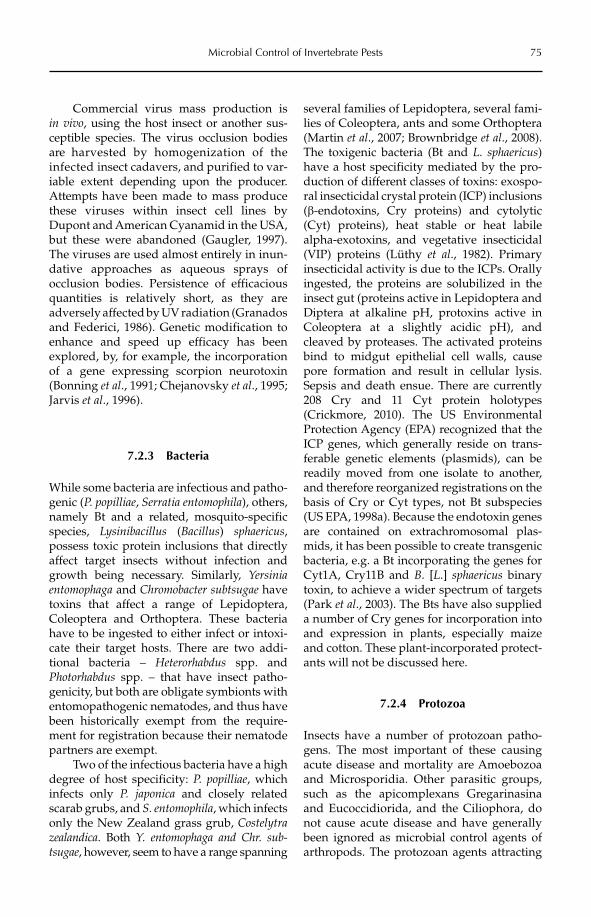
Microbial Control of Invertebrate Pests 75
Commercial virus mass production is in vivo, using the host insect or another sus-ceptible species. The virus occlusion bodies are harvested by homogenization of the infec ted insect cadavers, and purified to var-iable extent depending upon the producer. Attempts have been made to mass produce these viru ses within insect cell lines by Dupont and American Cyanamid in the USA, but these were abandoned (Gaugler, 1997). The viruses are used almost entirely in inun-dative app roaches as aqueous sprays of occlusion bodies. Persistence of efficacious quantities is relatively short, as they are adversely affected by UV radiation (Granados and Federici, 1986). Genetic modification to enhance and speed up efficacy has been explored, by, for example, the incorporation of a gene expressing scorpion neurotoxin (Bonning et al., 1991; Chejanovsky et al., 1995; Jarvis et al., 1996).
7.2.3 Bacteria
While some bacteria are infectious and patho-genic (P. popilliae, Serratia entomophila), others, namely Bt and a related, mosquito-specific species, Lysinibacillus (Bacillus) sphaericus,possess toxic protein inclusions that directly affect target insects without infection and growth being necessary. Similarly, Yersinia entomophaga and Chromobacter subtsugae have toxins that affect a range of Lepidoptera, Coleoptera and Orthoptera. These bacteria have to be ingested to either infect or intoxi-cate their target hosts. There are two addi-tional bacteria – Heterorhabdus spp. and Photorhabdus spp. – that have insect patho-genicity, but both are obligate symbionts with entomopathogenic nematodes, and thus have been historically exempt from the require-ment for registration because their nematode partners are exempt.
Two of the infectious bacteria have a high degree of host specificity: P. popilliae, which infects only P. japonica and closely related scarab grubs, and S. entomophila, which infects only the New Zealand grass grub, Costelytrazealandica. Both Y. entomophaga and Chr. sub-tsugae, however, seem to have a range spanning
several families of Lepidoptera, several fami-lies of Coleoptera, ants and some Orthoptera (Martin et al., 2007; Brownbridge et al., 2008). The toxigenic bacteria (Bt and L. sphaericus)have a host specificity mediated by the pro-duction of different classes of toxins: exospo-ral insecticidal crystal protein (ICP) inclusions (b-endotoxins, Cry proteins) and cytolytic (Cyt) proteins), heat stable or heat labile alpha-exotoxins, and vegetative insecticidal (VIP) proteins (Lüthy et al., 1982). Primary insecticidal activity is due to the ICPs. Orally ingested, the proteins are solubilized in the insect gut (proteins active in Lepidoptera and Diptera at alkaline pH, protoxins active in Coleoptera at a slightly acidic pH), and cleaved by proteases. The activated proteins bind to midgut epithelial cell walls, cause pore formation and result in cellular lysis. Sepsis and death ensue. There are currently 208 Cry and 11 Cyt protein holotypes (Crickmore, 2010). The US Environmental Protection Agency (EPA) recognized that the ICP genes, which generally reside on trans-ferable genetic elements (plasmids), can be readily moved from one isolate to another, and therefore reorganized registrations on the basis of Cry or Cyt types, not Bt subspecies (US EPA, 1998a). Because the endotoxin genes are contained on extrachromosomal plas-mids, it has been possible to create transgenic bacteria, e.g. a Bt incorporating the genes for Cyt1A, Cry11B and B. [L.] sphaericus binary toxin, to achieve a wider spectrum of targets (Park et al., 2003). The Bts have also supplied a number of Cry genes for incorporation into and expression in plants, especially maize and cotton. These plant-incorporated protect-ants will not be discussed here.
7.2.4 Protozoa
Insects have a number of protozoan patho-gens. The most important of these causing acute disease and mortality are Amoebozoa and Microsporidia. Other parasitic groups, such as the apicomplexans Gregarinasina and Eucoccidiorida, and the Ciliophora, do not cause acute disease and have generally been ignored as microbial control agents of arthropods. The protozoan agents attracting
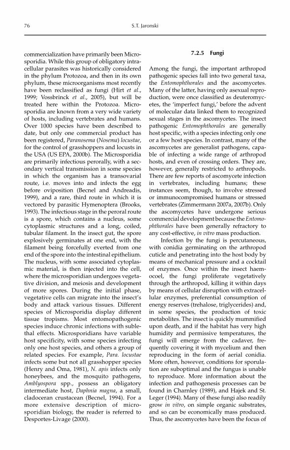
76 S.T. Jaronski
commercialization have primarily been Micro-sporidia. While this group of obligatory intra-cellular parasites was historically considered in the phylum Protozoa, and then in its own phylum, these microorganisms most recently have been reclassified as fungi (Hirt et al.,1999; Vossbrinck et al., 2005), but will be treated here within the Protozoa. Micro-sporidia are known from a very wide variety of hosts, including vertebrates and humans. Over 1000 species have been described to date, but only one commercial product has been registered, Paranosema (Nosema) locustae,for the control of grasshoppers and locusts in the USA (US EPA, 2000b). The Microsporidia are primarily infectious perorally, with a sec-ondary vertical transmission in some species in which the organism has a transovarial route, i.e. moves into and infects the egg before oviposition (Becnel and Andreadis, 1999), and a rare, third route in which it is vectored by parasitic Hymenoptera (Brooks, 1993). The infectious stage in the peroral route is a spore, which contains a nucleus, some cytoplasmic structures and a long, coiled, tubular filament. In the insect gut, the spore explosively germinates at one end, with the filament being forcefully everted from one end of the spore into the intestinal epithelium. The nucleus, with some associated cytoplas-mic material, is then injected into the cell, where the microsporidian undergoes vegeta-tive division, and meiosis and development of more spores. During the initial phase, vegetative cells can migrate into the insect’s body and attack various tissues. Different species of Microsporidia display different tissue tropisms. Most entomopathogenic species induce chronic infections with suble-thal effects. Microsporidians have variable host specificity, with some species infecting only one host species, and others a group of related species. For example, Para. locustaeinfects some but not all grasshopper species (Henry and Oma, 1981), N. apis infects only honeybees, and the mosquito pathogens, Amblyospora spp., possess an obligatory intermediate host, Daphnia magna, a small, cladoceran crustacean (Becnel, 1994). For a more extensive description of micro-sporidian biology, the reader is referred to Desportes-Livage (2000).
7.2.5 Fungi
Among the fungi, the important arthropod pathogenic species fall into two general taxa, the Entomophthorales and the ascomycetes. Many of the latter, having only asexual repro-duction, were once classified as deuteromyc-etes, the ‘imperfect fungi,’ before the advent of molecular data linked them to recognized sexual stages in the ascomycetes. The insect pathogenic Entomophthorales are generally host specific, with a species infecting only one or a few host species. In contrast, many of the ascomycetes are generalist pathogens, capa-ble of infecting a wide range of arthropod hosts, and even of crossing orders. They are, however, generally restricted to arthropods. There are few reports of ascomycete infection in vertebrates, including humans; these instances seem, though, to involve stressed or immunocompromised humans or stressed vertebrates (Zimmermann 2007a, 2007b). Only the ascomycetes have undergone serious commercial development because the Entomo-phthorales have been generally refractory to any cost-effective, in vitro mass production.
Infection by the fungi is percutaneous, with conidia germinating on the arthropod cuticle and penetrating into the host body by means of mechanical pressure and a cocktail of enzymes. Once within the insect haem-ocoel, the fungi proliferate vegetatively through the arthropod, killing it within days by means of cellular disruption with extracel-lular enzymes, preferential consumption of energy reserves (trehalose, triglycerides) and, in some species, the production of toxic metabolites. The insect is quickly mummified upon death, and if the habitat has very high humidity and permissive temperatures, the fungi will emerge from the cadaver, fre-quently covering it with mycelium and then reproducing in the form of aerial conidia. More often, however, conditions for sporula-tion are suboptimal and the fungus is unable to reproduce. More information about the infection and pathogenesis processes can be found in Charnley (1989), and Hajek and St. Leger (1994). Many of these fungi also readily grow in vitro, on simple organic substrates, and so can be economically mass produced. Thus, the ascomycetes have been the focus of
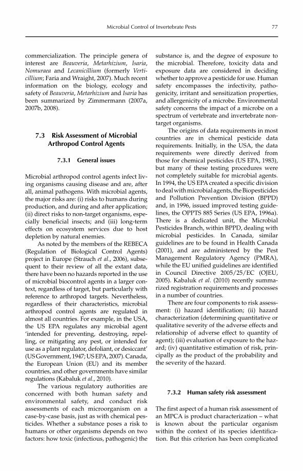
Microbial Control of Invertebrate Pests 77
commercialization. The principle genera of interest are Beauveria, Metarhizium, Isaria,Nomuraea and Lecanicillium (formerly Verti-cillium; Faria and Wraight, 2007). Much recent information on the biology, ecology and safety of Beauveria, Metarhizium and Isaria has been summarized by Zimmermann (2007a, 2007b, 2008).
7.3 Risk Assessment of Microbial Arthropod Control Agents
7.3.1 General issues
Microbial arthropod control agents infect liv-ing organisms causing disease and are, after all, animal pathogens. With microbial agents, the major risks are: (i) risks to humans during production, and during and after application; (ii) direct risks to non-target organisms, espe-cially beneficial insects; and (iii) long-term effects on ecosystem services due to host depletion by natural enemies.
As noted by the members of the REBECA (Regulation of Biological Control Agents) project in Europe (Strauch et al., 2006), subse-quent to their review of all the extant data, there have been no hazards reported in the use of microbial biocontrol agents in a larger con-text, regardless of target, but particularly with reference to arthropod targets. Nevertheless, regardless of their characteristics, microbial arthropod control agents are regulated in almost all countries. For example, in the USA, the US EPA regulates any microbial agent ‘intended for preventing, destroying, repel-ling, or mitigating any pest, or intended for use as a plant regulator, defoliant, or desiccant’ (US Government, 1947; US EPA, 2007). Canada, the European Union (EU) and its member countries, and other governments have similar regulations (Kabaluk et al., 2010).
The various regulatory authorities are concerned with both human safety and environmental safety, and conduct risk assessments of each microorganism on a case-by-case basis, just as with chemical pes-ticides. Whether a substance poses a risk to humans or other organisms depends on two factors: how toxic (infectious, pathogenic) the
substance is, and the degree of exposure to the microbial. Therefore, toxicity data and exposure data are considered in deciding whether to approve a pesticide for use. Human safety encompasses the infectivity, patho-genicity, irritant and sensitization properties, and allergenicity of a microbe. Environmental safety concerns the impact of a microbe on a spectrum of vertebrate and invertebrate non-target organisms.
The origins of data requirements in most countries are in chemical pesticide data requirements. Initially, in the USA, the data requirements were directly derived from those for chemical pesticides (US EPA, 1983), but many of these testing procedures were not completely suitable for microbial agents. In 1994, the US EPA created a specific division to deal with microbial agents, the Biopesticides and Pollution Prevention Division (BPPD) and, in 1996, issued improved testing guide-lines, the OPPTS 885 Series (US EPA, 1996a). There is a dedicated unit, the Microbial Pesticides Branch, within BPPD, dealing with microbial pesticides. In Canada, similar guidelines are to be found in Health Canada (2001), and are administered by the Pest Management Regulatory Agency (PMRA), while the EU unified guidelines are identified in Council Directive 2005/25/EC (OJEU, 2005). Kabaluk et al. (2010) recently summa-rized registration requirements and processes in a number of countries.
There are four components to risk assess-ment: (i) hazard identification; (ii) hazard characterization (determining quantitative or qualitative severity of the adverse effects and relationship of adverse effect to quantity of agent); (iii) evaluation of exposure to the haz-ard; (iv) quantitative estimation of risk, prin-cipally as the product of the probability and the severity of the hazard.
7.3.2 Human safety risk assessment
The first aspect of a human risk assessment of an MPCA is product characterization – what is known about the particular organism within the context of its species identifica-tion. But this criterion has been complicated
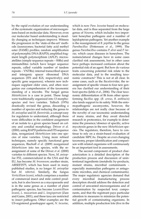
78 S.T. Jaronski
by the rapid evolution of our understanding of the systematic organization of microorgan-isms based on molecular data. However, even our molecular based understanding is stead-ily changing as techniques improve. Witness the progression in the ‘state-of-the-art’ meth-ods (isoenzymes; bacterial fatty acid methyl ester (FAME) profiles; random amplification of polymorphic DNA (RAPD); amplified frag-ment length polymorphism (AFLP); micros-atellites (simple sequence repeats – SSRs) and minisatellites (which have longer sequence repeats, called variable number of tandem repeats – VNTRs); internal transcribed spacer and intergenic spacer ribosomal DNA sequences (ITS and IGS, respectively); and specific gene sequences), wherein new tech-niques supplant older ones, and often reor-ganize our comprehension of the taxonomic standing of a microbe. The fungal genus Metarhizium is a case in point. These fungi were traditionally organized into 13 morpho-species and two varieties. Tulloch (1976) drastically revised the genus, discarding a number of species and reducing the genus to M. anisopliae and M. flavoviride, a concept easy for regulators to understand, although there were difficulties in the confident assignment of an isolate to a given species based on col-ony and conidial morphology. Driver et al.(2000), using RAPD patterns and ITS sequence data, reorganized Metarhizium into one spe-cies of nine varieties. Using more refined techniques, namely specific functional gene sequences, Bischoff et al. (2009) reorganized Metarhizium into ten species, with the re -assignment of some of the Driver et al. (2000) varieties to different species. Now, M. anisop-liae F52, commercialized in the USA and the EU, has become M. brunneum; another strain, ARSEF2575, which has been used in many published studies, is no longer M. anisopliaebut M. robertsii. Similarly, the fungus Verticillium lecanii, which comprises a number of commercial insect and mite control prod-ucts, but is also known as a mycoparasite and is in the same genus as a number of plant pathogenic species, has become Lecanicilliumlecanii, L. muscarium and L. longisporum (Zare and Gams, 2001), and these taxa are restricted to insect pathogens. Other examples are the US-registered grasshopper agent, N. locustae,
which is now Para. locustae based on molecu-lar data, and is thus separated from the large genus of Nosema, which includes two impor-tant honeybee pathogens and a number of lepidopteran pathogens. Yet another example is the reassignment of B. popilliae to the genus Paenibacillus (Pettersson et al., 1999). The genus Paenibacillus contains P. alvei and P. lar-vae, which cause diseases in honeybees. The nomenclatural changes have in some cases clarified risk assessments, but in other cases have perhaps increased confusion about the potential risk of an agent. But how do various traits concerning potential risk relate to the molecular data, and to the resulting taxo-nomic constructs? This is not at all clear. In some cases, such as the Baculoviridae, the re -assignment of specific viruses to four new gen-era has clarified our understanding of their host spectra (Jehle et al., 2006). The clear taxo-nomic differentiation of Bt from other spore-forming Bacillus spp. (B. cereus, B. anthracis)also lends support to its safety. With the ento-mopathogenic ascomycetes, however, the relationships are not really clear because of the novelty of our taxonomic understanding of many strains, and they await directed research in proteomics, for example to deter-mine the presence/absence of specific, active mycotoxin genes in the new Metarhizium spe-cies. The regulators, therefore, have to con-tinue to rely on a strain-based evaluation of candidate MPCAs. Despite an ever-changing taxonomy, proper identification and compari-son with related organisms will continuously be an important tool in assessments.
The second component of product char-acterization is a detailed description of the production process and discussion of unin-tentional ingredients (metabolic by-products, mutant strains, microbial contaminants – especially mammalian pathogens or antago-nistic microbes, and chemical contaminants). The major regulatory agencies demand that every microbial manufacturing process description should address the detection and control of unwanted microorganisms and of contamination by suspected toxic compo-nents, and that the registrant pays particular attention to measures to minimize the poten-tial growth of contaminating organisms. In addition, multiple production lots (five in the
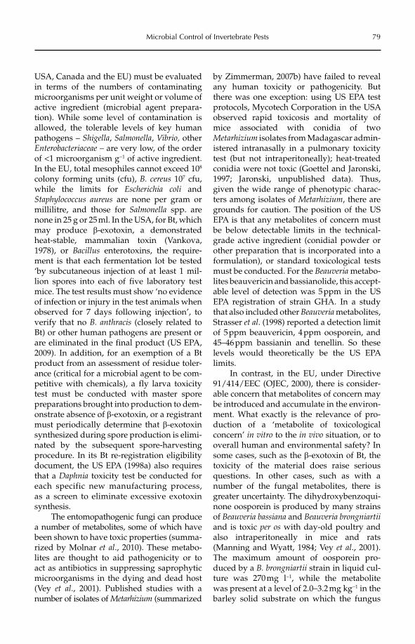
Microbial Control of Invertebrate Pests 79
USA, Canada and the EU) must be evaluated in terms of the numbers of contaminating microorganisms per unit weight or volume of active ingredient (microbial agent prepara-tion). While some level of contamination is allowed, the tolerable levels of key human pathogens – Shigella, Salmonella, Vibrio, other Enterobacteriaceae – are very low, of the order of <1 microorganism g−1 of active ingredient. In the EU, total mesophiles cannot exceed 108
colony forming units (cfu), B. cereus 107 cfu, while the limits for Escherichia coli and Staphylococcus aureus are none per gram or millilitre, and those for Salmonella spp. are none in 25 g or 25 ml. In the USA, for Bt, which may produce b-exotoxin, a demonstrated heat-stable, mammalian toxin (Vankova, 1978), or Bacillus enterotoxins, the require-ment is that each fermentation lot be tested ‘by subcutaneous injection of at least 1 mil-lion spores into each of five laboratory test mice. The test results must show ‘no evidence of infection or injury in the test animals when observed for 7 days following injection’, to verify that no B. anthracis (closely related to Bt) or other human pathogens are present or are eliminated in the final product (US EPA, 2009). In addition, for an exemption of a Bt product from an assessment of residue toler-ance (critical for a microbial agent to be com-petitive with chemicals), a fly larva toxicity test must be conducted with master spore preparations brought into production to dem-onstrate absence of b-exotoxin, or a registrant must periodically determine that b-exotoxinsynthesized during spore production is elimi-nated by the subsequent spore-harvesting procedure. In its Bt re-registration eligibility document, the US EPA (1998a) also requires that a Daphnia toxicity test be conducted for each specific new manufacturing process, as a screen to eliminate excessive exotoxin synthesis.
The entomopathogenic fungi can produce a number of metabolites, some of which have been shown to have toxic properties (summa-rized by Molnar et al., 2010). These metabo-lites are thought to aid pathogenicity or to act as antibiotics in suppressing saprophytic microorganisms in the dying and dead host (Vey et al., 2001). Published studies with a number of isolates of Metarhizium (summarized
by Zimmerman, 2007b) have failed to reveal any human toxicity or pathogenicity. But there was one exception: using US EPA test protocols, Mycotech Corporation in the USA observed rapid toxicosis and mortality of mice associated with conidia of two Metarhizium isolates from Madagascar admin-istered intranasally in a pulmonary toxicity test (but not intraperitoneally); heat-treated conidia were not toxic (Goettel and Jaronski, 1997; Jaronski, unpublished data). Thus, given the wide range of phenotypic charac-ters among isolates of Metarhizium, there are grounds for caution. The position of the US EPA is that any metabolites of concern must be below detectable limits in the technical-grade active ingredient (conidial powder or other preparation that is incorporated into a formulation), or standard toxicological tests must be conducted. For the Beauveria metabo-lites beauvericin and bassianolide, this accept-able level of detection was 5 ppm in the US EPA registration of strain GHA. In a study that also included other Beauveria metabolites, Strasser et al. (1998) reported a detection limit of 5 ppm beauvericin, 4 ppm oosporein, and 45–46 ppm bassianin and tenellin. So these levels would theoretically be the US EPA limits.
In contrast, in the EU, under Directive 91/414/EEC (OJEC, 2000), there is consider-able concern that metabolites of concern may be introduced and accumulate in the environ-ment. What exactly is the relevance of pro-duction of a ‘metabolite of toxicological concern’ in vitro to the in vivo situation, or to overall human and environmental safety? In some cases, such as the b-exotoxin of Bt, the toxicity of the material does raise serious questions. In other cases, such as with a number of the fungal metabolites, there is greater uncertainty. The dihydroxybenzoqui-none oosporein is produced by many strains of Beauveria bassiana and Beauveria brongniartiiand is toxic per os with day-old poultry and also intraperitoneally in mice and rats (Manning and Wyatt, 1984; Vey et al., 2001). The maximum amount of oosporein pro-duced by a B. brongniartii strain in liquid cul-ture was 270 mg l−1, while the metabolite was present at a level of 2.0–3.2 mg kg−1 in the barley solid substrate on which the fungus
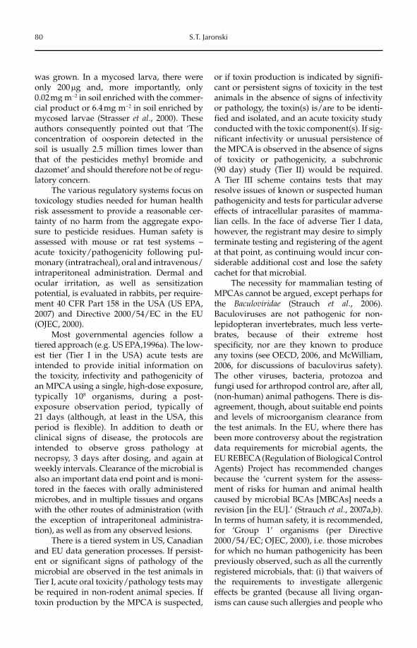
80 S.T. Jaronski
was grown. In a mycosed larva, there were only 200 mg and, more importantly, only 0.02 mg m−2 in soil enriched with the commer-cial product or 6.4 mg m−2 in soil enriched by mycosed larvae (Strasser et al., 2000). These authors consequently pointed out that ‘The concentration of oosporein detected in the soil is usually 2.5 million times lower than that of the pesticides methyl bromide and dazomet’ and should therefore not be of regu-latory concern.
The various regulatory systems focus on toxicology studies needed for human health risk assessment to provide a reasonable cer-tainty of no harm from the aggregate expo-sure to pesticide residues. Human safety is assessed with mouse or rat test systems – acute toxicity/pathogenicity following pul-monary (intratracheal), oral and intravenous/intraperitoneal administration. Dermal and ocular irritation, as well as sensitization potential, is evaluated in rabbits, per require-ment 40 CFR Part 158 in the USA (US EPA, 2007) and Directive 2000/54/EC in the EU (OJEC, 2000).
Most governmental agencies follow a tiered approach (e.g. US EPA,1996a). The low-est tier (Tier I in the USA) acute tests are intended to provide initial information on the toxicity, infectivity and pathogenicity of an MPCA using a single, high-dose exposure, typically 108 organisms, during a post- exposure observation period, typically of 21 days (although, at least in the USA, this period is flexible). In addition to death or clinical signs of disease, the protocols are intended to observe gross pathology at necropsy, 3 days after dosing, and again at weekly intervals. Clearance of the microbial is also an important data end point and is moni-tored in the faeces with orally administered microbes, and in multiple tissues and organs with the other routes of administration (with the exception of intraperitoneal administra-tion), as well as from any observed lesions.
There is a tiered system in US, Canadian and EU data generation processes. If persist-ent or significant signs of pathology of the microbial are observed in the test animals in Tier I, acute oral toxicity/pathology tests may be required in non-rodent animal species. If toxin production by the MPCA is suspected,
or if toxin production is indicated by signifi-cant or persistent signs of toxicity in the test animals in the absence of signs of infectivity or pathology, the toxin(s) is/are to be identi-fied and isolated, and an acute toxicity study conducted with the toxic component(s). If sig-nificant infectivity or unusual persistence of the MPCA is observed in the absence of signs of toxicity or pathogenicity, a subchronic (90 day) study (Tier II) would be required. A Tier III scheme contains tests that may resolve issues of known or suspected human pathogenicity and tests for particular adverse effects of intracellular parasites of mamma-lian cells. In the face of adverse Tier I data, however, the registrant may desire to simply terminate testing and registering of the agent at that point, as continuing would incur con-siderable additional cost and lose the safety cachet for that microbial.
The necessity for mammalian testing of MPCAs cannot be argued, except perhaps for the Baculoviridae (Strauch et al., 2006). Baculoviruses are not pathogenic for non-lepidopteran invertebrates, much less verte-brates, because of their extreme host specificity, nor are they known to produce any toxins (see OECD, 2006, and McWilliam, 2006, for discussions of baculovirus safety). The other viruses, bacteria, protozoa and fungi used for arthropod control are, after all, (non-human) animal pathogens. There is dis-agreement, though, about suitable end points and levels of microorganism clearance from the test animals. In the EU, where there has been more controversy about the registration data requirements for microbial agents, the EU REBECA (Regulation of Biological Control Agents) Project has recommended changes because the ‘current system for the assess-ment of risks for human and animal health caused by microbial BCAs [MBCAs] needs a revision [in the EU].’ (Strauch et al., 2007a,b). In terms of human safety, it is recommended, for ‘Group 1’ organisms (per Directive 2000/54/EC; OJEC, 2000), i.e. those microbes for which no human pathogenicity has been previously observed, such as all the currently registered microbials, that: (i) that waivers of the requirements to investigate allergenic effects be granted (because all living organ-isms can cause such allergies and people who
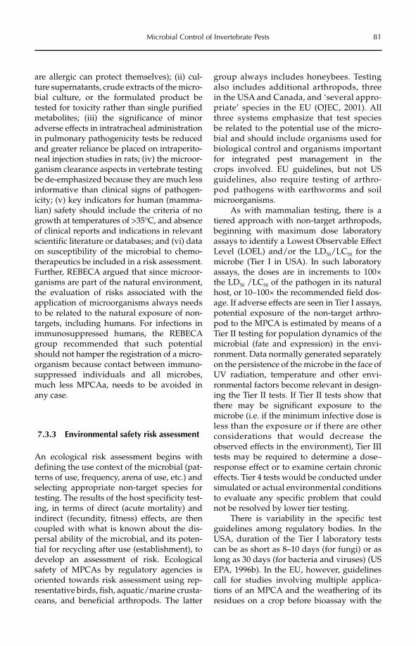
Microbial Control of Invertebrate Pests 81
are allergic can protect themselves); (ii) cul-ture supernatants, crude extracts of the micro-bial culture, or the formulated product be tested for toxicity rather than single purified metabolites; (iii) the significance of minor adverse effects in intratracheal administration in pulmonary pathogenicity tests be reduced and greater reliance be placed on intraperito-neal injection studies in rats; (iv) the microor-ganism clearance aspects in vertebrate testing be de-emphasized because they are much less informative than clinical signs of pathogen-icity; (v) key indicators for human (mamma-lian) safety should include the criteria of no growth at temperatures of >35°C, and absence of clinical reports and indications in relevant scientific literature or databases; and (vi) data on susceptibility of the microbial to chemo-therapeutics be inclu ded in a risk assessment. Further, REBECA argued that since microor-ganisms are part of the natural environment, the evaluation of risks associated with the application of microorganisms always needs to be related to the natural exposure of non-targets, including humans. For infections in immunosuppressed humans, the REBECA group recommended that such potential should not hamper the registration of a micro-organism because contact between immuno-suppressed individuals and all microbes, much less MPCAa, needs to be avoided in any case.
7.3.3 Environmental safety risk assessment
An ecological risk assessment begins with defining the use context of the microbial (pat-terns of use, frequency, arena of use, etc.) and selecting appropriate non-target species for testing. The results of the host specificity test-ing, in terms of direct (acute mortality) and indirect (fecundity, fitness) effects, are then coupled with what is known about the dis-persal ability of the microbial, and its poten-tial for recycling after use (establishment), to develop an assessment of risk. Ecological safety of MPCAs by regulatory agencies is oriented towards risk assessment using rep-resentative birds, fish, aquatic/marine crusta-ceans, and beneficial arthropods. The latter
group always includes honeybees. Testing also includes additional arthropods, three in the USA and Canada, and ‘several appro-priate’ species in the EU (OJEC, 2001). All three systems emphasize that test species be related to the potential use of the micro-bial and should include organisms used for biological control and organisms important for integrated pest management in the crops involved. EU guidelines, but not US guidelines, also require testing of arthro-pod pathogens with earthworms and soil microorganisms.
As with mammalian testing, there is a tiered approach with non-target arthropods, beginning with maximum dose laboratory assays to identify a Lowest Observable Effect Level (LOEL) and/or the LD50/LC50 for the microbe (Tier I in USA). In such laboratory assays, the doses are in increments to 100× the LD50 /LC50 of the pathogen in its natural host, or 10–100× the recommended field dos-age. If adverse effects are seen in Tier I assays, potential exposure of the non-target arthro-pod to the MPCA is estimated by means of a Tier II testing for population dynamics of the microbial (fate and expression) in the envi-ronment. Data normally generated separately on the persistence of the microbe in the face of UV radiation, temperature and other envi-ronmental factors become relevant in design-ing the Tier II tests. If Tier II tests show that there may be significant exposure to the microbe (i.e. if the minimum infective dose is less than the exposure or if there are other considerations that would decrease the observed effects in the environment), Tier III tests may be required to determine a dose–response effect or to examine certain chronic effects. Tier 4 tests would be conducted under simulated or actual environmental conditions to evaluate any specific problem that could not be resolved by lower tier testing.
There is variability in the specific test guidelines among regulatory bodies. In the USA, duration of the Tier I laboratory tests can be as short as 8–10 days (for fungi) or as long as 30 days (for bacteria and viruses) (US EPA, 1996b). In the EU, however, guidelines call for studies involving multiple applica-tions of an MPCA and the weathering of its residues on a crop before bioassay with the
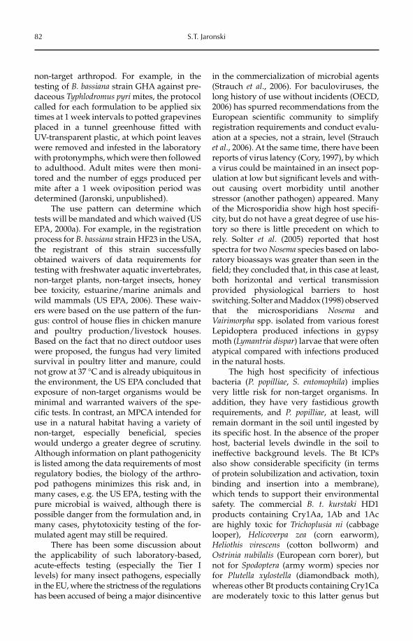
82 S.T. Jaronski
non-target arthropod. For example, in the testing of B. bassiana strain GHA against pre-daceous Typhlodromus pyri mites, the protocol called for each formulation to be applied six times at 1 week intervals to potted grapevines placed in a tunnel greenhouse fitted with UV-transparent plastic, at which point leaves were removed and infested in the laboratory with protonymphs, which were then followed to adulthood. Adult mites were then moni-tored and the number of eggs produced per mite after a 1 week oviposition period was determined (Jaronski, unpublished).
The use pattern can determine which tests will be mandated and which waived (US EPA, 2000a). For example, in the registration process for B. bassiana strain HF23 in the USA, the registrant of this strain successfully obtained waivers of data requirements for testing with freshwater aquatic invertebrates, non-target plants, non-target insects, honey bee toxicity, estuarine/marine animals and wild mammals (US EPA, 2006). These waiv-ers were based on the use pattern of the fun-gus: control of house flies in chicken manure and poultry production/livestock houses. Based on the fact that no direct outdoor uses were proposed, the fungus had very limited survival in poultry litter and manure, could not grow at 37 °C and is already ubiquitous in the environment, the US EPA concluded that exposure of non-target organisms would be minimal and warranted waivers of the spe-cific tests. In contrast, an MPCA intended for use in a natural habitat having a variety of non-target, especially beneficial, species would undergo a greater degree of scrutiny. Although information on plant pathogenicity is listed among the data requirements of most regulatory bodies, the biology of the arthro-pod pathogens minimizes this risk and, in many cases, e.g. the US EPA, testing with the pure microbial is waived, although there is possible danger from the formulation and, in many cases, phytotoxicity testing of the for-mulated agent may still be required.
There has been some discussion about the applicability of such laboratory-based, acute-effects testing (especially the Tier I levels) for many insect pathogens, especially in the EU, where the strictness of the regulations has been accused of being a major disincentive
in the commercialization of microbial agents (Strauch et al., 2006). For baculoviruses, the long history of use without incidents (OECD, 2006) has spurred recommendations from the European scientific community to simplify registration requirements and conduct evalu-ation at a species, not a strain, level (Strauch et al., 2006). At the same time, there have been reports of virus latency (Cory, 1997), by which a virus could be maintained in an insect pop-ulation at low but significant levels and with-out causing overt morbidity until another stressor (another pathogen) appeared. Many of the Microsporidia show high host specifi-city, but do not have a great degree of use his-tory so there is little precedent on which to rely. Solter et al. (2005) reported that host spectra for two Nosema species based on labo-ratory bioassays was greater than seen in the field; they concluded that, in this case at least, both horizontal and vertical transmission provided physiological barriers to host switching. Solter and Maddox (1998) observed that the microsporidians Nosema and Vairimorpha spp. isolated from various forest Lepidoptera produced infections in gypsy moth (Lymantria dispar) larvae that were often atypical compared with infections produced in the natural hosts.
The high host specificity of infectious bacteria (P. popilliae, S. entomophila) implies very little risk for non-target organisms. In addition, they have very fastidious growth requirements, and P. popilliae, at least, will remain dormant in the soil until ingested by its specific host. In the absence of the proper host, bacterial levels dwindle in the soil to ineffective background levels. The Bt ICPs also show considerable specificity (in terms of protein solubilization and activation, toxin binding and insertion into a membrane), which tends to support their environmental safety. The commercial B. t. kurstaki HD1 products containing Cry1Aa, 1Ab and 1Ac are highly toxic for Trichoplusia ni (cabbage looper), Helicoverpa zea (corn earworm), Heliothis virescens (cotton bollworm) and Ostrinia nubilalis (European corn borer), but not for Spodoptera (army worm) species nor for Plutella xylostella (diamondback moth), whereas other Bt products containing Cry1Ca are moderately toxic to this latter genus but
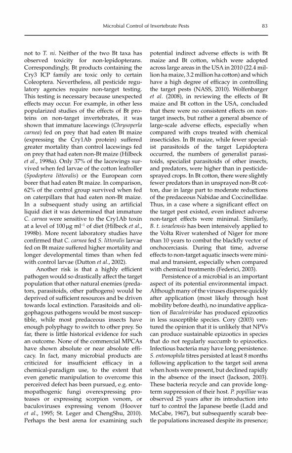
Microbial Control of Invertebrate Pests 83
not to T. ni. Neither of the two Bt taxa has observed toxicity for non-lepidopterans. Correspondingly, Bt products containing the Cry3 ICP family are toxic only to certain Coleoptera. Nevertheless, all pesticide regu-latory agencies require non-target testing. This testing is necessary because unexpected effects may occur. For example, in other less popularized studies of the effects of Bt pro-teins on non-target invertebrates, it was shown that immature lacewings (Chrysoperlacarnea) fed on prey that had eaten Bt maize (expressing the Cry1Ab protein) suffered greater mortality than control lacewings fed on prey that had eaten non-Bt maize (Hilbeck et al., 1998a). Only 37% of the lacewings sur-vived when fed larvae of the cotton leafroller (Spodoptera littoralis) or the European corn borer that had eaten Bt maize. In comparison, 62% of the control group survived when fed on caterpillars that had eaten non-Bt maize. In a subsequent study using an artificial liquid diet it was determined that immature C. carnea were sensitive to the Cry1Ab toxin at a level of 100 mg ml−1 of diet (Hilbeck et al.,1998b). More recent laboratory studies have confirmed that C. carnea fed S. littoralis larvae fed on Bt maize suffered higher mortality and longer developmental times than when fed with control larvae (Dutton et al., 2002).
Another risk is that a highly efficient pathogen would so drastically affect the target population that other natural enemies (preda-tors, parasitoids, other pathogens) would be deprived of sufficient resources and be driven towards local extinction. Parasitoids and oli-gophagous pathogens would be most suscep-tible, while most predaceous insects have enough polyphagy to switch to other prey. So far, there is little historical evidence for such an outcome. None of the commercial MPCAs have shown absolute or near absolute effi-cacy. In fact, many microbial products are criticized for insufficient efficacy in a chemical- paradigm use, to the extent that even genetic manipulation to overcome this perceived defect has been pursued, e.g. ento-mopathogenic fungi overexpressing pro-teases or expressing scorpion venom, or baculoviruses expressing venom (Hoover et al., 1995; St. Leger and ChengShu, 2010). Perhaps the best arena for examining such
potential indirect adverse effects is with Bt maize and Bt cotton, which were adopted across large areas in the USA in 2010 (22.4 mil-lion ha maize, 3.2 million ha cotton) and which have a high degree of efficacy in controlling the target pests (NASS, 2010). Wolfenbarger et al. (2008), in reviewing the effects of Bt maize and Bt cotton in the USA, concluded that there were no consistent effects on non-target insects, but rather a general absence of large-scale adverse effects, especially when compared with crops treated with chemical insecticides. In Bt maize, while fewer special-ist parasitoids of the target Lepidoptera occurred, the numbers of generalist parasi-toids, specialist parasitoids of other insects, and predators, were higher than in pesticide-sprayed crops. In Bt cotton, there were slightly fewer predators than in unsprayed non-Bt cot-ton, due in large part to moderate reductions of the predaceous Nabidae and Coccinellidae. Thus, in a case where a significant effect on the target pest existed, even indirect adverse non-target effects were minimal. Similarly, B. t. israelensis has been intensively applied to the Volta River watershed of Niger for more than 10 years to combat the blackfly vector of onchocerciasis. During that time, adverse effects to non-target aquatic insects were mini-mal and transient, especially when compared with chemical treatments (Federici, 2003).
Persistence of a microbial is an important aspect of its potential environmental impact. Although many of the viruses disperse quickly after application (most likely through host mobility before death), no inundative applica-tion of Baculoviridae has produced epizootics in less susceptible species. Cory (2003) ven-tured the opinion that it is unlikely that NPVs can produce sustainable epizootics in species that do not regularly succumb to epizootics. Infectious bacteria may have long persistence. S. entomophila titres persisted at least 8 months following application to the target soil arena when hosts were present, but declined rapidly in the absence of the insect (Jackson, 2003). These bacteria recycle and can provide long-term suppression of their host. P. popilliae was observed 25 years after its introduction into turf to control the Japanese beetle (Ladd and McCabe, 1967), but subsequently scarab bee-tle populations increased despite its presence;
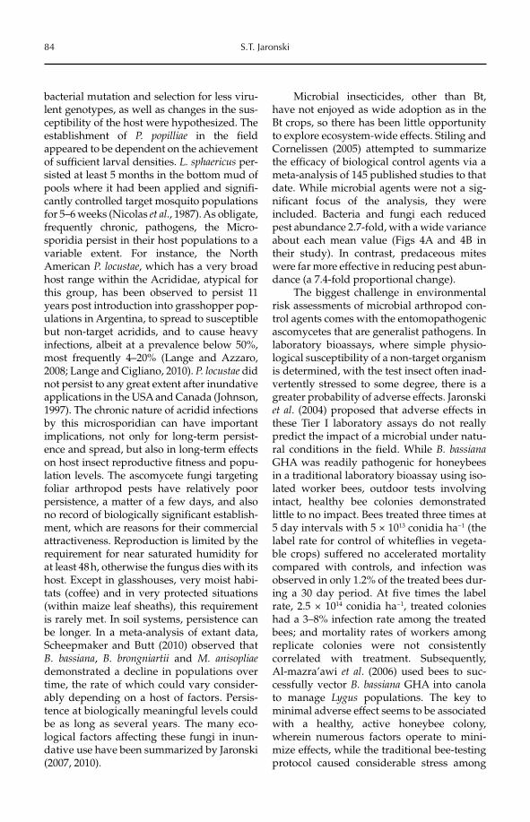
84 S.T. Jaronski
bacterial mutation and selection for less viru-lent genotypes, as well as changes in the sus-ceptibility of the host were hypothesized. The establishment of P. popilliae in the field appeared to be dependent on the achievement of sufficient larval densities. L. sphaericus per-sisted at least 5 months in the bottom mud of pools where it had been applied and signifi-cantly controlled target mosquito populations for 5–6 weeks (Nicolas et al., 1987). As obligate, frequently chronic, pathogens, the Micro-sporidia persist in their host populations to a variable extent. For instance, the North American P. locustae, which has a very broad host range within the Acrididae, atypical for this group, has been observed to persist 11 years post introduction into grasshopper pop-ulations in Argentina, to spread to susceptible but non-target acridids, and to cause heavy infections, albeit at a prevalence below 50%, most frequently 4–20% (Lange and Azzaro, 2008; Lange and Cigliano, 2010). P. locustae did not persist to any great extent after inundative applications in the USA and Canada (Johnson, 1997). The chronic nature of acridid infections by this microsporidian can have important implications, not only for long-term persist-ence and spread, but also in long-term effects on host insect reproductive fitness and popu-lation levels. The ascomycete fungi targeting foliar arthropod pests have relatively poor persistence, a matter of a few days, and also no record of biologically significant establish-ment, which are reasons for their commercial attractiveness. Reproduction is limited by the requirement for near saturated humidity for at least 48 h, otherwise the fungus dies with its host. Except in glasshouses, very moist habi-tats (coffee) and in very protected situations (within maize leaf sheaths), this requirement is rarely met. In soil systems, persistence can be longer. In a meta-analysis of extant data, Scheepmaker and Butt (2010) observed that B. bassiana, B. brongniartii and M. anisopliaedemonstrated a decline in populations over time, the rate of which could vary consider-ably depending on a host of factors. Persis-tence at biologically meaningful levels could be as long as several years. The many eco-logical factors affecting these fungi in inun-dative use have been summarized by Jaronski (2007, 2010).
Microbial insecticides, other than Bt, have not enjoyed as wide adoption as in the Bt crops, so there has been little opportunity to explore ecosystem-wide effects. Stiling and Cornelissen (2005) attempted to summarize the efficacy of biological control agents via a meta-analysis of 145 published studies to that date. While microbial agents were not a sig-nificant focus of the analysis, they were included. Bacteria and fungi each reduced pest abundance 2.7-fold, with a wide variance about each mean value (Figs 4A and 4B in their study). In contrast, predaceous mites were far more effective in reducing pest abun-dance (a 7.4-fold proportional change).
The biggest challenge in environmental risk assessments of microbial arthropod con-trol agents comes with the entomopathogenic ascomycetes that are generalist pathogens. In laboratory bioassays, where simple physio-logical susceptibility of a non-target organism is determined, with the test insect often inad-vertently stressed to some degree, there is a greater probability of adverse effects. Jaronski et al. (2004) proposed that adverse effects in these Tier I laboratory assays do not really predict the impact of a microbial under natu-ral conditions in the field. While B. bassianaGHA was readily pathogenic for honeybees in a traditional laboratory bioassay using iso-lated worker bees, outdoor tests involving intact, healthy bee colonies demonstrated little to no impact. Bees treated three times at 5 day intervals with 5 × 10¹³ conidia ha−1 (the label rate for control of whiteflies in vegeta-ble crops) suffered no accelerated mortality compared with controls, and infection was observed in only 1.2% of the treated bees dur-ing a 30 day period. At five times the label rate, 2.5 × 10¹⁴ conidia ha−1, treated colonies had a 3–8% infection rate among the treated bees; and mortality rates of workers among replicate colonies were not consistently correlated with treatment. Subsequently, Al-mazra’awi et al. (2006) used bees to suc-cessfully vector B. bassiana GHA into canola to manage Lygus populations. The key to minimal adverse effect seems to be associated with a healthy, active honeybee colony, wherein numerous factors operate to mini-mize effects, while the traditional bee-testing protocol caused considerable stress among
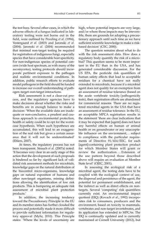
Microbial Control of Invertebrate Pests 85
the test bees. Several other cases, in which the adverse effects of a fungus indicated in lab-oratory testing were not borne out in the field, were outlined by Peveling et al. (1994), Vestergaard et al. (2003) and Jaronski et al.(2004). Jaronski et al. (2004) recommended that minimal non-target testing be required for registration of indigenous fungi, especially species that have considerable host specificity. For non-indigenous species of potential con-cern (wide host spectrum, as with many of the ascomycetes), testing protocols should incor-porate pertinent exposure to the pathogen and realistic environmental conditions. In addition, public research efforts to evaluate model pathogens in the field should be funded to increase our overall understanding of path-ogen–target–non-target interactions.
Risk assessment is not a clear-cut pro-cess, and the regulatory agencies have to make decisions about whether the risks and benefits are in enough balance to make a decision: ‘When the available data are inade-quate or non-conclusive, a prudent and cau-tious approach to environmental protection, health or safety could be to opt for the worst-case hypothesis. When such hypotheses are accumulated, this will lead to an exaggera-tion of the real risk but gives a certain assur-ance that it will not be underestimated’ (Ehlers, 2007).
At times, the regulatory process has not been transparent. Strauch et al. (2007a) noted: ‘It becomes very clear in an early stage of this action that the development of such proposals is hindered so far by: significant lack of vali-dated risk assessment methods for microbials, knowledge gaps on the natural distribution of the biocontrol micro-organisms, knowledge gaps on natural exposition of humans and other non-target organisms, missing defini-tions allowing the identification of low risk products. This is hampering an adequate risk assessment of microbial plant protection products.’
In addition, the increasing tendency toward the Precautionary Principle in the EU and its member states has further clouded the process and potentially made it more difficult to provide sufficient information for regula-tory approval (Myhr, 2010). This Principle states: ‘Where the levels of uncertainty are
high, where potential impacts are very large, and/or where those impacts may be irrevers-ible, there are grounds for adopting a precau-tionary approach until such time as we have sufficient scientific knowledge to make a risk-based decision’ (CEC, 2000).
The question remains about what to do with the risk assessment data. How does a regulatory body quantify the risk of a micro-bial? This question seems to be more impor-tant in the EU than in the USA, and has generated considerable discussion. For the US EPA, the pesticide risk quantifiers of human safety effects that lead to acceptable residues for a chemical have not really extended to microbials, because if a microbial agent does not qualify for an exemption from an assessment of residue tolerance (based on the acute vertebrate toxicity/pathogenicity tests), then registration efforts are abandoned for commercial reasons. There are no regis-tered microbial agents in the USA that have an assessment of residue tolerance. In the EU, an acceptable MPCA registration results in the statement ‘there are clear indications that it may be expected that [agent] does not have any harmful effects on human or animal health or on groundwater or any unaccepta-ble influence on the environment, … subject to compliance with the particular require-ments of Directive 91/414/EEC, for each [agent]-containing plant protection product for which Member States will grant or review the authorization. … Extension of the use pattern beyond those described above will require an evaluation at Member State level’ (OJEC, 2001).
In assessing the ecological risk of a microbial agent, the testing data have to be coupled with the ecological context of use, the dispersal and persistence of the agent, the potential for permanent establishment, and the indirect as well as direct effects on non-targets. Several ‘competing’ risk quantifiers currently exist. An environmental impact quotient (EIQ) (Kovach et al., 1992) incorpo-rates risk to consumers, producers and the environment, based on toxicity to mammals, vertebrates and non-target invertebrates, and its application has extended to MPCAs. The EIQ is continually updated and is currently maintained at Cornell University (2012), but
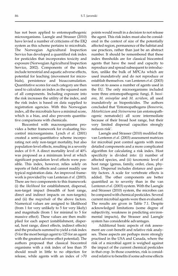
86 S.T. Jaronski
has not been applied to entomopathogenic microorganisms. Laengle and Strasser (2010) have levied a number of criticisms of the EIQ system as this scheme pertains to microbials. The Norwegian Agricultural Inspection Service has developed a quantified risk index for pesticides that incorporates toxicity and exposure (Norwegian Agricultural Inspection Service, 2002). Components of the index include terrestrial and aquatic adverse effects, potential for leaching (movement for micro-bials), persistence and bioaccumulation. Quantitative scores for each category are then used to calculate an index as the squared sum of all components. Including exposure into the risk increases the utility of the index, and the risk index is based on data supplied to registration agencies. With this Norwegian index, all the microbials have a minimal score, which is a bias, and also prevents quantita-tive comparisons with chemicals.
Biocontrol with macroorganisms pro-vides a better framework for evaluating bio-control microorganisms. Lynch et al. (2001) created a semi-quantitative scheme incorpo-rating not only non-target mortality, but also population-level effects, resulting in a severity index of 0–9. A direct mortality level of 40% was proposed as a minimum level at which significant population level effects were pos-sible. This index, however, relies solely on reports of field effects and cannot readily use typical registration data. An improved frame-work is provided by van Lenteran et al. (2003). There are two components to this framework: (i) the likelihood for establishment, dispersal, non-target impact (breadth of host range, direct and indirect impacts on non-targets); and (ii) the magnitude of the above factors. Numerical values are assigned to likelihood (from 1 for very unlikely to 5 for very likely) and magnitude (from 1 for minimal to 5 for massive effect). These values are then multi-plied for each aspect (establishment, disper-sal, host range, direct effects, indirect effects) and the products summed to yield a risk index (5 for the most benign agent to 125 for an agent with the greatest adverse effect potential). The authors proposed that classical biocontrol organisms with a risk index of less than 35 should result in little to no objection for release, while agents with an index of >70
points would result in a decision to not release the agent. This risk index must also be consid-ered in the context of size of the potentially affected region, permanence of the habitat and use practices, rather than just be an abstract number. It should be remembered that these index thresholds are for classical biocontrol agents that have the need and capacity to reproduce and spread subsequent to introduc-tion, unlike the bulk of MPCAs which are used inundatively and do not reproduce or establish themselves. van Lenteren et al. (2003) went on to assess a number of agents used in the EU. The only microorganisms included were three entomopathogenic fungi, B. bassi-ana, M. anisopliae and M. acridum, all used inundatively as biopesticides. The authors concluded that ‘Entomopathogens (Beauveria,Metarhizium and Steinernema (an entomopath-ogenic nematode) ) all score intermediate because of their broad host range, but their very limited dispersal capacities strongly reduces risk’.
Laengle and Strasser (2010) modified the van Lenteren et al. (2003) assessment matrices for microbial pest control agents with more detailed components and a more complicated algorithm for calculating a risk index. Host specificity is divided into: (i) number of affected species, and (ii) taxonomic level of host range (genus, family, order, class, phy-lum). Dispersal includes distance and quan-tity factors. A scale for vertebrate effects is added. The other components are better quantified as to severity than in the van Lenteren et al. (2003) system. With the Laengle and Strasser (2010) system, the microbes can be compared with chemical pesticides. Several current microbial agents were then evaluated. The results are given in Table 7.1. Despite acknowledged limitations (some degree of subjectivity, weakness in predicting environ-mental impacts), the Strasser and Laengle system has considerable advantages.
Additional basic aspects of risk assess-ment are cost–benefit and relative risk analy-ses. These aspects are perhaps more strongly practised in the USA and Canada, where the risk of a microbial agent is weighed against the impact of the current chemical pesticides in that crop. In those countries, risk is consid-ered relative to benefits if some adverse effects
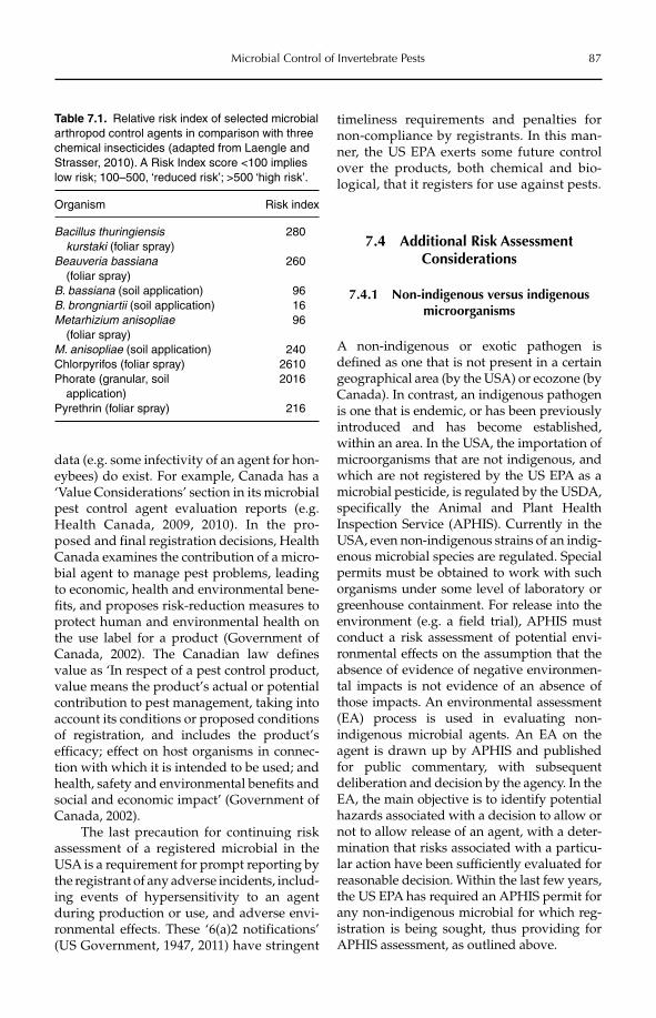
Microbial Control of Invertebrate Pests 87
data (e.g. some infectivity of an agent for hon-eybees) do exist. For example, Canada has a ‘Value Considerations’ section in its microbial pest control agent evaluation reports (e.g. Health Canada, 2009, 2010). In the pro-posed and final registration decisions, Health Canada examines the contribution of a micro-bial agent to manage pest problems, leading to economic, health and environmental bene-fits, and proposes risk-reduction measures to protect human and environmental health on the use label for a product (Government of Canada, 2002). The Canadian law defines value as ‘In respect of a pest control product, value means the product’s actual or potential contribution to pest management, taking into account its conditions or proposed conditions of registration, and includes the product’s efficacy; effect on host organisms in connec-tion with which it is intended to be used; and health, safety and environmental benefits and social and economic impact’ (Government of Canada, 2002).
The last precaution for continuing risk assessment of a registered microbial in the USA is a requirement for prompt reporting by the registrant of any adverse incidents, includ-ing events of hypersensitivity to an agent during production or use, and adverse envi-ronmental effects. These ‘6(a)2 notifications’ (US Government, 1947, 2011) have stringent
timeliness requirements and penalties for non-compliance by registrants. In this man-ner, the US EPA exerts some future control over the products, both chemical and bio-logical, that it registers for use against pests.
7.4 Additional Risk Assessment Considerations
7.4.1 Non-indigenous versus indigenous microorganisms
A non-indigenous or exotic pathogen is defined as one that is not present in a certain geographical area (by the USA) or ecozone (by Canada). In contrast, an indigenous pathogen is one that is endemic, or has been previously introduced and has become established, within an area. In the USA, the importation of microorganisms that are not indigenous, and which are not registered by the US EPA as a microbial pesticide, is regulated by the USDA, specifically the Animal and Plant Health Inspection Service (APHIS). Currently in the USA, even non-indigenous strains of an indig-enous microbial species are regulated. Special permits must be obtained to work with such organisms under some level of laboratory or greenhouse containment. For release into the environment (e.g. a field trial), APHIS must conduct a risk assessment of potential envi-ronmental effects on the assumption that the absence of evidence of negative environmen-tal impacts is not evidence of an absence of those impacts. An environmental assessment (EA) process is used in evaluating non- indigenous microbial agents. An EA on the agent is drawn up by APHIS and published for public commentary, with subsequent deliberation and decision by the agency. In the EA, the main objective is to identify potential hazards associated with a decision to allow or not to allow release of an agent, with a deter-mination that risks associated with a particu-lar action have been sufficiently evaluated for reasonable decision. Within the last few years, the US EPA has required an APHIS permit for any non-indigenous microbial for which reg-istration is being sought, thus providing for APHIS assessment, as outlined above.
Table 7.1. Relative risk index of selected microbial arthropod control agents in comparison with three chemical insecticides (adapted from Laengle and Strasser, 2010). A Risk Index score <100 implies low risk; 100–500, ‘reduced risk’; >500 ‘high risk’.
Organism Risk index
Bacillus thuringiensis kurstaki (foliar spray)
280
Beauveria bassiana (foliar spray)
260
B. bassiana (soil application) 96B. brongniartii (soil application) 16Metarhizium anisopliae (foliar spray)
96
M. anisopliae (soil application) 240Chlorpyrifos (foliar spray) 2610Phorate (granular, soil application)
2016
Pyrethrin (foliar spray) 216
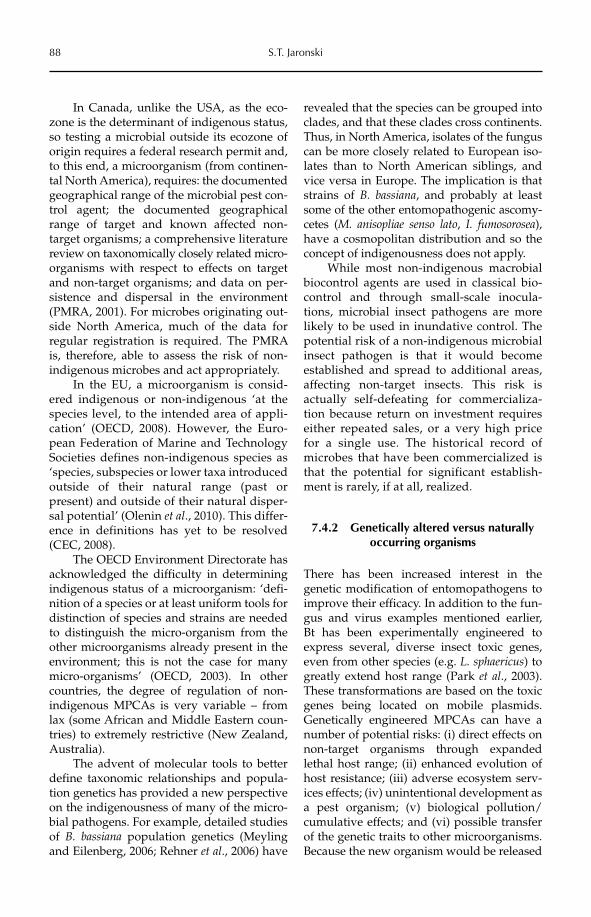
88 S.T. Jaronski
In Canada, unlike the USA, as the eco-zone is the determinant of indigenous status, so testing a microbial outside its ecozone of origin requires a federal research permit and, to this end, a microorganism (from continen-tal North America), requires: the documented geographical range of the microbial pest con-trol agent; the documented geographical range of target and known affected non- target organisms; a comprehensive literature review on taxonomically closely related micro-organisms with respect to effects on target and non-target organisms; and data on per-sistence and dispersal in the environment (PMRA, 2001). For microbes originating out-side North America, much of the data for regular registration is required. The PMRA is, therefore, able to assess the risk of non-indigenous microbes and act appropriately.
In the EU, a microorganism is consid-ered indigenous or non-indigenous ‘at the species level, to the intended area of appli-cation’ (OECD, 2008). However, the Euro-pean Federa tion of Marine and Technology Societies defines non-indigenous species as ‘species, subspecies or lower taxa introduced outside of their natural range (past or present) and outside of their natural disper-sal potential’ (Olenin et al., 2010). This differ-ence in definitions has yet to be resolved (CEC, 2008).
The OECD Environment Directorate has acknowledged the difficulty in determining indigenous status of a microorganism: ‘defi-nition of a species or at least uniform tools for distinction of species and strains are needed to distinguish the micro-organism from the other microorganisms already present in the environment; this is not the case for many micro-organisms’ (OECD, 2003). In other countries, the degree of regulation of non-indigenous MPCAs is very variable – from lax (some African and Middle Eastern coun-tries) to extremely restrictive (New Zealand, Australia).
The advent of molecular tools to better define taxonomic relationships and popula-tion genetics has provided a new perspective on the indigenousness of many of the micro-bial pathogens. For example, detailed studies of B. bassiana population genetics (Meyling and Eilenberg, 2006; Rehner et al., 2006) have
revealed that the species can be grouped into clades, and that these clades cross continents. Thus, in North America, isolates of the funguscan be more closely related to European iso-lates than to North American siblings, and vice versa in Europe. The implication is that strains of B. bassiana, and probably at least some of the other entomopathogenic ascomy-cetes (M. anisopliae senso lato, I. fumosorosea), have a cosmopolitan distribution and so the concept of indigenousness does not apply.
While most non-indigenous macrobial biocontrol agents are used in classical bio-control and through small-scale inocula-tions, microbial insect pathogens are more likely to be used in inundative control. The potential risk of a non-indigenous microbial insect pathogen is that it would become established and spread to additional areas, affecting non-target insects. This risk is actually self-defeating for commercializa-tion because return on investment requires either repeated sales, or a very high price for a single use. The historical record of microbes that have been commercialized is that the potential for significant establish-ment is rarely, if at all, realized.
7.4.2 Genetically altered versus naturally occurring organisms
There has been increased interest in the genetic modification of entomopathogens to improve their efficacy. In addition to the fun-gus and virus examples mentioned earlier, Bt has been experimentally engineered to express several, diverse insect toxic genes, even from other species (e.g. L. sphaericus) to greatly extend host range (Park et al., 2003). These transformations are based on the toxic genes being located on mobile plasmids. Genetically engineered MPCAs can have a number of potential risks: (i) direct effects on non-target organisms through expanded lethal host range; (ii) enhanced evolution of host resistance; (iii) adverse ecosystem serv-ices effects; (iv) unintentional development as a pest organism; (v) biological pollution/cumulative effects; and (vi) possible transfer of the genetic traits to other microorganisms. Because the new organism would be released
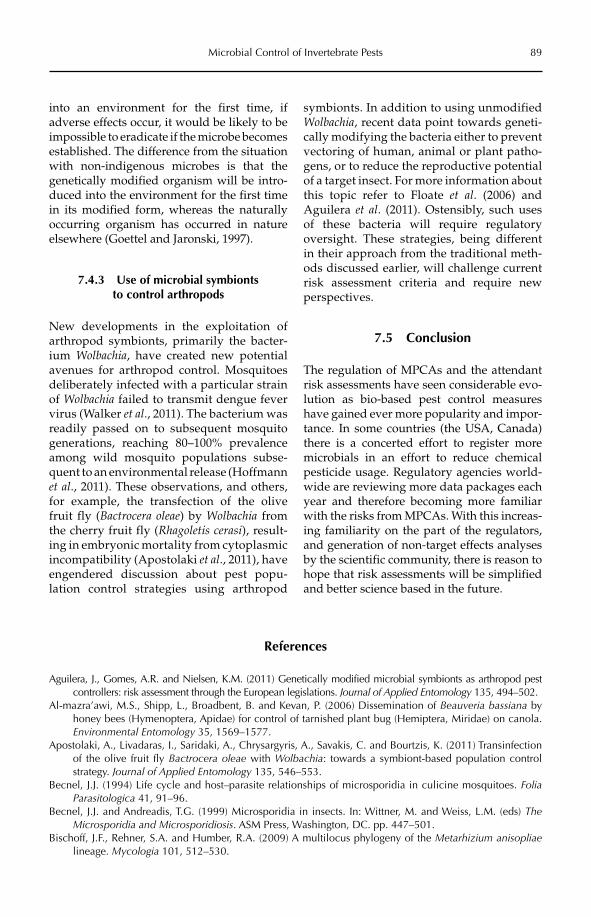
Microbial Control of Invertebrate Pests 89
into an environment for the first time, if adverse effects occur, it would be likely to be impossible to eradicate if the microbe becomes established. The difference from the situation with non-indigenous microbes is that the genetically modified organism will be intro-duced into the environment for the first time in its modified form, whereas the naturally occurring organism has occurred in nature elsewhere (Goettel and Jaronski, 1997).
7.4.3 Use of microbial symbionts to control arthropods
New developments in the exploitation of arthropod symbionts, primarily the bacter-ium Wolbachia, have created new potential avenues for arthropod control. Mosquitoes deliberately infected with a particular strain of Wolbachia failed to transmit dengue fever virus (Walker et al., 2011). The bacterium was readily passed on to subsequent mosquito generations, reaching 80–100% prevalence among wild mosquito populations subse-quent to an environmental release (Hoffmann et al., 2011). These observations, and others, for example, the transfection of the olive fruit fly (Bactrocera oleae) by Wolbachia from the cherry fruit fly (Rhagoletis cerasi), result-ing in embryonic mortality from cyto plasmic incompatibility (Apostolaki et al., 2011), have engendered discussion about pest popu-lation control strategies using arthropod
symbionts. In addition to using unmodified Wolbachia, recent data point towards geneti-cally modifying the bacteria either to prevent vectoring of human, animal or plant patho-gens, or to reduce the reproductive potential of a target insect. For more information about this topic refer to Floate et al. (2006) and Aguilera et al. (2011). Ostensibly, such uses of these bacteria will require regulatory oversight. These strategies, being different in their approach from the traditional meth-ods discussed earlier, will challenge current risk assessment criteria and require new perspectives.
7.5 Conclusion
The regulation of MPCAs and the attendant risk assessments have seen considerable evo-lution as bio-based pest control measures have gained ever more popularity and impor-tance. In some countries (the USA, Canada) there is a concerted effort to register more microbials in an effort to reduce chemical pesticide usage. Regulatory agencies world-wide are reviewing more data packages each year and therefore becoming more familiar with the risks from MPCAs. With this increas-ing familiarity on the part of the regulators, and generation of non-target effects analyses by the scientific community, there is reason to hope that risk assessments will be simplified and better science based in the future.
References
Aguilera, J., Gomes, A.R. and Nielsen, K.M. (2011) Genetically modified microbial symbionts as arthropod pest controllers: risk assessment through the European legislations. Journal of Applied Entomology 135, 494–502.
Al-mazra’awi, M.S., Shipp, L., Broadbent, B. and Kevan, P. (2006) Dissemination of Beauveria bassiana by honey bees (Hymenoptera, Apidae) for control of tarnished plant bug (Hemiptera, Miridae) on canola. Environmental Entomology 35, 1569–1577.
Apostolaki, A., Livadaras, I., Saridaki, A., Chrysargyris, A., Savakis, C. and Bourtzis, K. (2011) Transinfection of the olive fruit fly Bactrocera oleae with Wolbachia: towards a symbiont-based population control strategy. Journal of Applied Entomology 135, 546–553.
Becnel, J.J. (1994) Life cycle and host–parasite relationships of microsporidia in culicine mosquitoes. Folia Parasitologica 41, 91–96.
Becnel, J.J. and Andreadis, T.G. (1999) Microsporidia in insects. In: Wittner, M. and Weiss, L.M. (eds) TheMicrosporidia and Microsporidiosis. ASM Press, Washington, DC. pp. 447–501.
Bischoff, J.F., Rehner, S.A. and Humber, R.A. (2009) A multilocus phylogeny of the Metarhizium anisopliaelineage. Mycologia 101, 512–530.
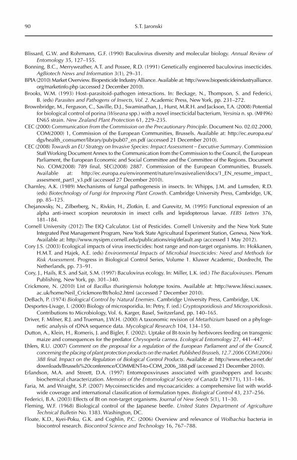
90 S.T. Jaronski
Blissard, G.W. and Rohrmann, G.F. (1990) Baculovirus diversity and molecular biology. Annual Review of Entomology 35, 127–155.
Bonning, B.C., Merryweather, A.T. and Possee, R.D. (1991) Genetically engineered baculovirus insecticides. AgBiotech News and Information 3(1), 29–31.
BPIA (2010) Market Overview. Biopesticide Industry Alliance. Available at: http://www.biopesticideindustryalliance.org/marketinfo.php (accessed 2 December 2010).
Brooks, W.M. (1993) Host–parasitoid–pathogen interactions. In: Beckage, N., Thompson, S. and Federici, B. (eds) Parasites and Pathogens of Insects, Vol. 2. Academic Press, New York, pp. 231–272.
Brownbridge, M., Ferguson, C., Saville, D.J., Swaminathan, J., Hurst, M.R.H. and Jackson, T.A. (2008) Potential for biological control of porina (Wiseana spp.) with a novel insecticidal bacterium, Yersinia n. sp. (MH96) EN65 strain. New Zealand Plant Protection 61, 229–235.
CEC (2000) Communication from the Commission on the Precautionary Principle. Document No. 02.02.2000, COM(2000) 1, Commission of the European Communities, Brussels. Available at: http://ec.europa.eu/dgs/health_consumer/library/pub/pub07_en.pdf (accessed 21 December 2010).
CEC (2008) Towards an EU Strategy on Invasive Species: Impact Assessment – Executive Summary. Commission Staff Working Document Annex to the Communication from the Commission to the Council, the European Parliament, the European Economic and Social Committee and the Committee of the Regions. Document No. COM(2008) 789 final, SEC(2008) 2887. Commission of the European Communities, Brussels. Available at: http://ec.europa.eu/environment/nature/invasivealien/docs/1_EN_resume_impact_assesment_part1_v3.pdf (accessed 27 December 2010).
Charnley, A.K. (1989) Mechanisms of fungal pathogenesis in insects. In: Whipps, J.M. and Lumsden, R.D. (eds) Biotechnology of Fungi for Improving Plant Growth. Cambridge University Press, Cambridge, UK, pp. 85–125.
Chejanovsky, N., Zilberberg, N., Rivkin, H., Zlotkin, E. and Gurevitz, M. (1995) Functional expression of an alpha anti-insect scorpion neurotoxin in insect cells and lepidopterous larvae. FEBS Letters 376, 181–184.
Cornell University (2012) The EIQ Calculator. List of Pesticides. Cornell University and the New York State Integrated Pest Management Program, New York State Agricultural Experiment Station, Geneva, New York. Available at: http://www.nysipm.cornell.edu/publications/eiq/default.asp (accessed 1 May 2012).
Cory J.S. (2003) Ecological impacts of virus insecticides: host range and non-target organisms. In: Hokkanen, H.M.T. and Hajek, A.E. (eds) Environmental Impacts of Microbial Insecticides: Need and Methods for Risk Assessment. Progress in Biological Control Series, Volume 1. Kluwer Academic, Dordrecht, The Netherlands, pp. 73–91.
Cory, J., Hails, R.S. and Sait, S.M. (1997) Baculovirus ecology. In: Miller, L.K. (ed.) The Baculoviruses. Plenum Publishing, New York, pp. 301–340.
Crickmore, N. (2010) List of Bacillus thuringiensis holotype toxins. Available at: http://www.lifesci.sussex.ac.uk/home/Neil_Crickmore/Bt/holo2.html (accessed 7 December 2010).
DeBach, P. (1974) Biological Control by Natural Enemies. Cambridge University Press, Cambridge, UK.Desportes-Livage, I. (2000) Biology of microsporidia. In: Petry, F. (ed.) Cryptosporidiosis and Microsporidiosis.
Contributions to Microbiology, Vol. 6, Karger, Basel, Switzerland, pp. 140–165.Driver, F. Milner, R.J. and Trueman, J.W.H. (2000) A taxonomic revision of Metarhizium based on a phyloge-
netic analysis of rDNA sequence data. Mycological Research 104, 134–150.Dutton, A., Klein, H., Romeris, J. and Bigler, F. (2002). Uptake of Bt-toxin by herbivores feeding on transgenic
maize and consequences for the predator Chrysoperla carnea. Ecological Entomology 27, 441–447.Ehlers, R.U. (2007) Comment on the proposal for a regulation of the European Parliament and of the Council,
concerning the placing of plant protection products on the market. Published Brussels, 12.7.2006 COM(2006) 388 final. Impact on the Regulation of Biological Control Products. Available at: http://www.rebeca-net.de/downloads/Brussels%20conference/COMMENT-to-COM_2006_388.pdf (accessed 21 December 2010).
Erlandson, M.A. and Streett, D.A. (1997) Entomopoxviruses associated with grasshoppers and locusts: biochemical characterization. Memoirs of the Entomological Society of Canada 129(171), 131–146.
Faria, M. and Wraight, S.P. (2007) Mycoinsecticides and mycoacaricides: a comprehensive list with world-wide coverage and international classification of formulation types. Biological Control 43, 237–256.
Federici, B.A. (2003) Effects of Bt on non-target organisms. Journal of New Seeds 5(1), 11–30.Fleming, W.F. (1968) Biological control of the Japanese beetle. United States Department of Agriculture
Technical Bulletin No. 1383. Washington, DC.Floate, K.D., Kyei-Poku, G.K. and Coghlin, P.C. (2006) Overview and relevance of Wolbachia bacteria in
biocontrol research. Biocontrol Science and Technology 16, 767–788.
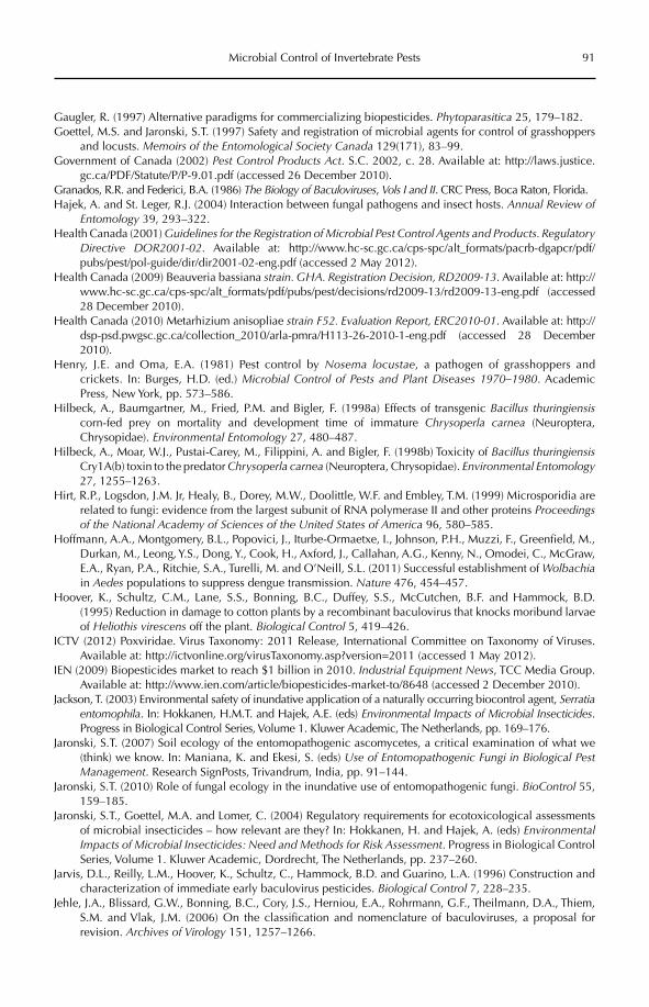
Microbial Control of Invertebrate Pests 91
Gaugler, R. (1997) Alternative paradigms for commercializing biopesticides. Phytoparasitica 25, 179–182.Goettel, M.S. and Jaronski, S.T. (1997) Safety and registration of microbial agents for control of grasshoppers
and locusts. Memoirs of the Entomological Society Canada 129(171), 83–99.Government of Canada (2002) Pest Control Products Act. S.C. 2002, c. 28. Available at: http://laws.justice.
gc.ca/PDF/Statute/P/P-9.01.pdf (accessed 26 December 2010).Granados, R.R. and Federici, B.A. (1986) The Biology of Baculoviruses, Vols I and II. CRC Press, Boca Raton, Florida.Hajek, A. and St. Leger, R.J. (2004) Interaction between fungal pathogens and insect hosts. Annual Review of
Entomology 39, 293–322.Health Canada (2001) Guidelines for the Registration of Microbial Pest Control Agents and Products. Regulatory
Directive DOR2001-02. Available at: http://www.hc-sc.gc.ca/cps-spc/alt_formats/pacrb-dgapcr/pdf/pubs/pest/pol-guide/dir/dir2001-02-eng.pdf (accessed 2 May 2012).
Health Canada (2009) Beauveria bassiana strain. GHA. Registration Decision, RD2009-13. Available at: http://www.hc-sc.gc.ca/cps-spc/alt_formats/pdf/pubs/pest/decisions/rd2009-13/rd2009-13-eng.pdf (accessed 28 December 2010).
Health Canada (2010) Metarhizium anisopliae strain F52. Evaluation Report, ERC2010-01. Available at: http://dsp-psd.pwgsc.gc.ca/collection_2010/arla-pmra/H113-26-2010-1-eng.pdf (accessed 28 December 2010).
Henry, J.E. and Oma, E.A. (1981) Pest control by Nosema locustae, a pathogen of grasshoppers and crickets. In: Burges, H.D. (ed.) Microbial Control of Pests and Plant Diseases 1970–1980. Academic Press, New York, pp. 573–586.
Hilbeck, A., Baumgartner, M., Fried, P.M. and Bigler, F. (1998a) Effects of transgenic Bacillus thuringiensiscorn-fed prey on mortality and development time of immature Chrysoperla carnea (Neuroptera, Chrysopidae). Environmental Entomology 27, 480–487.
Hilbeck, A., Moar, W.J., Pustai-Carey, M., Filippini, A. and Bigler, F. (1998b) Toxicity of Bacillus thuringiensisCry1A(b) toxin to the predator Chrysoperla carnea (Neuroptera, Chrysopidae). Environmental Entomology27, 1255–1263.
Hirt, R.P., Logsdon, J.M. Jr, Healy, B., Dorey, M.W., Doolittle, W.F. and Embley, T.M. (1999) Microsporidia are related to fungi: evidence from the largest subunit of RNA polymerase II and other proteins Proceedings of the National Academy of Sciences of the United States of America 96, 580–585.
Hoffmann, A.A., Montgomery, B.L., Popovici, J., Iturbe-Ormaetxe, I., Johnson, P.H., Muzzi, F., Greenfield, M., Durkan, M., Leong, Y.S., Dong, Y., Cook, H., Axford, J., Callahan, A.G., Kenny, N., Omodei, C., McGraw, E.A., Ryan, P.A., Ritchie, S.A., Turelli, M. and O’Neill, S.L. (2011) Successful establishment of Wolbachiain Aedes populations to suppress dengue transmission. Nature 476, 454–457.
Hoover, K., Schultz, C.M., Lane, S.S., Bonning, B.C., Duffey, S.S., McCutchen, B.F. and Hammock, B.D. (1995) Reduction in damage to cotton plants by a recombinant baculovirus that knocks moribund larvae of Heliothis virescens off the plant. Biological Control 5, 419–426.
ICTV (2012) Poxviridae. Virus Taxonomy: 2011 Release, International Committee on Taxonomy of Viruses. Available at: http://ictvonline.org/virusTaxonomy.asp?version=2011 (accessed 1 May 2012).
IEN (2009) Biopesticides market to reach $1 billion in 2010. Industrial Equipment News, TCC Media Group. Available at: http://www.ien.com/article/biopesticides-market-to/8648 (accessed 2 December 2010).
Jackson, T. (2003) Environmental safety of inundative application of a naturally occurring biocontrol agent, Serratia entomophila. In: Hokkanen, H.M.T. and Hajek, A.E. (eds) Environmental Impacts of Microbial Insecticides.Progress in Biological Control Series, Volume 1. Kluwer Academic, The Netherlands, pp. 169–176.
Jaronski, S.T. (2007) Soil ecology of the entomopathogenic ascomycetes, a critical examination of what we (think) we know. In: Maniana, K. and Ekesi, S. (eds) Use of Entomopathogenic Fungi in Biological Pest Management. Research SignPosts, Trivandrum, India, pp. 91–144.
Jaronski, S.T. (2010) Role of fungal ecology in the inundative use of entomopathogenic fungi. BioControl 55, 159–185.
Jaronski, S.T., Goettel, M.A. and Lomer, C. (2004) Regulatory requirements for ecotoxicological assessments of microbial insecticides – how relevant are they? In: Hokkanen, H. and Hajek, A. (eds) Environmental Impacts of Microbial Insecticides: Need and Methods for Risk Assessment. Progress in Biological Control Series, Volume 1. Kluwer Academic, Dordrecht, The Netherlands, pp. 237–260.
Jarvis, D.L., Reilly, L.M., Hoover, K., Schultz, C., Hammock, B.D. and Guarino, L.A. (1996) Construction and characterization of immediate early baculovirus pesticides. Biological Control 7, 228–235.
Jehle, J.A., Blissard, G.W., Bonning, B.C., Cory, J.S., Herniou, E.A., Rohrmann, G.F., Theilmann, D.A., Thiem, S.M. and Vlak, J.M. (2006) On the classification and nomenclature of baculoviruses, a proposal for revision. Archives of Virology 151, 1257–1266.
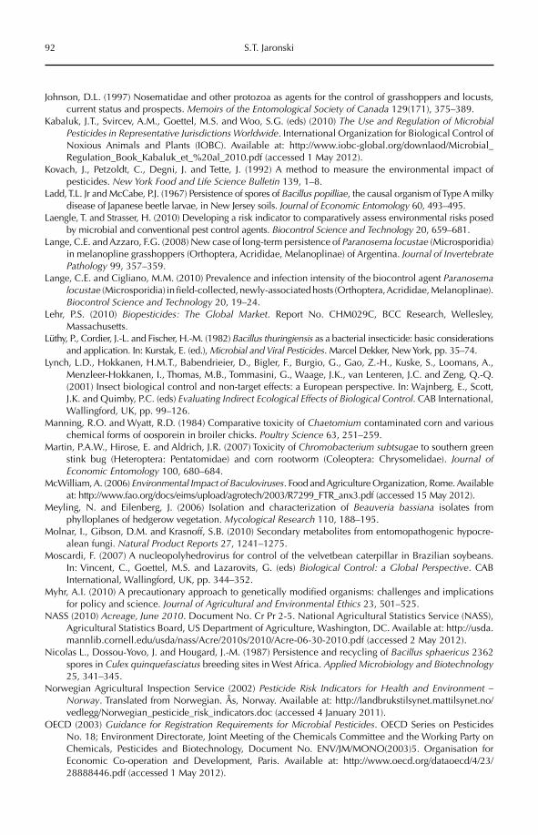
92 S.T. Jaronski
Johnson, D.L. (1997) Nosematidae and other protozoa as agents for the control of grasshoppers and locusts, current status and prospects. Memoirs of the Entomological Society of Canada 129(171), 375–389.
Kabaluk, J.T., Svircev, A.M., Goettel, M.S. and Woo, S.G. (eds) (2010) The Use and Regulation of Microbial Pesticides in Representative Jurisdictions Worldwide. International Organization for Biological Control of Noxious Animals and Plants (IOBC). Available at: http://www.iobc-global.org/downlaod/Microbial_Regulation_Book_Kabaluk_et_%20al_2010.pdf (accessed 1 May 2012).
Kovach, J., Petzoldt, C., Degni, J. and Tette, J. (1992) A method to measure the environmental impact of pesticides. New York Food and Life Science Bulletin 139, 1–8.
Ladd, T.L. Jr and McCabe, P.J. (1967) Persistence of spores of Bacillus popilliae, the causal organism of Type A milky disease of Japanese beetle larvae, in New Jersey soils. Journal of Economic Entomology 60, 493–495.
Laengle, T. and Strasser, H. (2010) Developing a risk indicator to comparatively assess environmental risks posed by microbial and conventional pest control agents. Biocontrol Science and Technology 20, 659–681.
Lange, C.E. and Azzaro, F.G. (2008) New case of long-term persistence of Paranosema locustae (Microsporidia) in melanopline grasshoppers (Orthoptera, Acrididae, Melanoplinae) of Argentina. Journal of Invertebrate Pathology 99, 357–359.
Lange, C.E. and Cigliano, M.M. (2010) Prevalence and infection intensity of the biocontrol agent Paranosema locustae (Microsporidia) in field-collected, newly-associated hosts (Orthoptera, Acrididae, Melanoplinae). Biocontrol Science and Technology 20, 19–24.
Lehr, P.S. (2010) Biopesticides: The Global Market. Report No. CHM029C, BCC Research, Wellesley, Massachusetts.
Lüthy, P., Cordier, J.-L. and Fischer, H.-M. (1982) Bacillus thuringiensis as a bacterial insecticide: basic considerations and application. In: Kurstak, E. (ed.), Microbial and Viral Pesticides. Marcel Dekker, New York, pp. 35–74.
Lynch, L.D., Hokkanen, H.M.T., Babendrieier, D., Bigler, F., Burgio, G., Gao, Z.-H., Kuske, S., Loomans, A., Menzleer-Hokkanen, I., Thomas, M.B., Tommasini, G., Waage, J.K., van Lenteren, J.C. and Zeng, Q.-Q. (2001) Insect biological control and non-target effects: a European perspective. In: Wajnberg, E., Scott, J.K. and Quimby, P.C. (eds) Evaluating Indirect Ecological Effects of Biological Control. CAB International, Wallingford, UK, pp. 99–126.
Manning, R.O. and Wyatt, R.D. (1984) Comparative toxicity of Chaetomium contaminated corn and various chemical forms of oosporein in broiler chicks. Poultry Science 63, 251–259.
Martin, P.A.W., Hirose, E. and Aldrich, J.R. (2007) Toxicity of Chromobacterium subtsugae to southern green stink bug (Heteroptera: Pentatomidae) and corn rootworm (Coleoptera: Chrysomelidae). Journal of Economic Entomology 100, 680–684.
McWilliam, A. (2006) Environmental Impact of Baculoviruses. Food and Agriculture Organization, Rome. Available at: http://www.fao.org/docs/eims/upload/agrotech/2003/R7299_FTR_anx3.pdf (accessed 15 May 2012).
Meyling, N. and Eilenberg, J. (2006) Isolation and characterization of Beauveria bassiana isolates from phylloplanes of hedgerow vegetation. Mycological Research 110, 188–195.
Molnar, I., Gibson, D.M. and Krasnoff, S.B. (2010) Secondary metabolites from entomopathogenic hypocre-alean fungi. Natural Product Reports 27, 1241–1275.
Moscardi, F. (2007) A nucleopolyhedrovirus for control of the velvetbean caterpillar in Brazilian soybeans. In: Vincent, C., Goettel, M.S. and Lazarovits, G. (eds) Biological Control: a Global Perspective. CAB International, Wallingford, UK, pp. 344–352.
Myhr, A.I. (2010) A precautionary approach to genetically modified organisms: challenges and implications for policy and science. Journal of Agricultural and Environmental Ethics 23, 501–525.
NASS (2010) Acreage, June 2010. Document No. Cr Pr 2-5. National Agricultural Statistics Service (NASS), Agricultural Statistics Board, US Department of Agriculture, Washington, DC. Available at: http://usda.mannlib.cornell.edu/usda/nass/Acre/2010s/2010/Acre-06-30-2010.pdf (accessed 2 May 2012).
Nicolas L., Dossou-Yovo, J. and Hougard, J.-M. (1987) Persistence and recycling of Bacillus sphaericus 2362 spores in Culex quinquefasciatus breeding sites in West Africa. Applied Microbiology and Biotechnology25, 341–345.
Norwegian Agricultural Inspection Service (2002) Pesticide Risk Indicators for Health and Environment – Norway. Translated from Norwegian. Ås, Norway. Available at: http://landbrukstilsynet.mattilsynet.no/vedlegg/Norwegian_pesticide_risk_indicators.doc (accessed 4 January 2011).
OECD (2003) Guidance for Registration Requirements for Microbial Pesticides. OECD Series on Pesticides No. 18; Environment Directorate, Joint Meeting of the Chemicals Committee and the Working Party on Chemicals, Pesticides and Biotechnology, Document No. ENV/JM/MONO(2003)5. Organisation for Economic Co-operation and Development, Paris. Available at: http://www.oecd.org/dataoecd/4/23/ 28888446.pdf (accessed 1 May 2012).
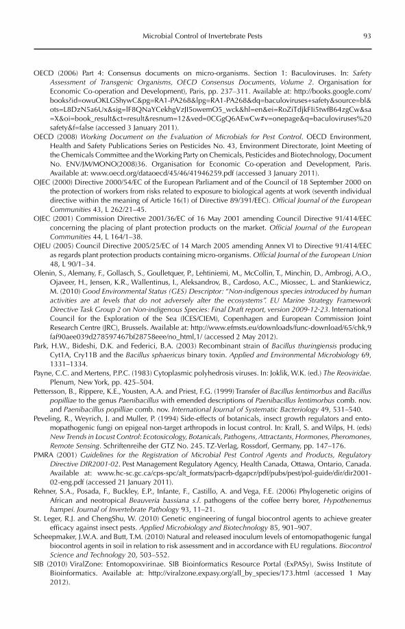
Microbial Control of Invertebrate Pests 93
OECD (2006) Part 4: Consensus documents on micro-organisms. Section 1: Baculoviruses. In: Safety Assessment of Transgenic Organisms, OECD Consensus Documents, Volume 2. Organisation for Economic Co-operation and Development), Paris, pp. 237–311. Available at: http://books.google.com/books?id=owuOKLGShywC&pg=RA1-PA268&lpg=RA1-PA268&dq=baculoviruses+safety&source=bl&ots=L8DzN5a6Ux&sig=lF8QNaYCekhgVzJI5owemO5_wck&hl=en&ei=RoZiTdjkFIi5twfB64zgCw&sa=X&oi=book_result&ct=result&resnum=12&ved=0CGgQ6AEwCw#v=onepage&q=baculoviruses%20safety&f=false (accessed 3 January 2011).
OECD (2008) Working Document on the Evaluation of Microbials for Pest Control. OECD Environment, Health and Safety Publications Series on Pesticides No. 43, Environment Directorate, Joint Meeting of the Chemicals Committee and the Working Party on Chemicals, Pesticides and Biotechnology, Document No. ENV/JM/MONO(2008)36. Organisation for Economic Co-operation and Development, Paris. Available at: www.oecd.org/dataoecd/45/46/41946259.pdf (accessed 3 January 2011).
OJEC (2000) Directive 2000/54/EC of the European Parliament and of the Council of 18 September 2000 on the protection of workers from risks related to exposure to biological agents at work (seventh individual directive within the meaning of Article 16(1) of Directive 89/391/EEC). Official Journal of the European Communities 43, L 262/21–45.
OJEC (2001) Commission Directive 2001/36/EC of 16 May 2001 amending Council Directive 91/414/EEC concerning the placing of plant protection products on the market. Official Journal of the European Communities 44, L 164/1–38.
OJEU (2005) Council Directive 2005/25/EC of 14 March 2005 amending Annex VI to Directive 91/414/EEC as regards plant protection products containing micro-organisms. Official Journal of the European Union48, L 90/1–34.
Olenin, S., Alemany, F., Gollasch, S., Goulletquer, P., Lehtiniemi, M., McCollin, T., Minchin, D., Ambrogi, A.O., Ojaveer, H., Jensen, K.R., Wallentinus, I., Aleksandrov, B., Cardoso, A.C., Miossec, L. and Stankiewicz, M. (2010) Good Environmental Status (GES) Descriptor: “Non-indigenous species introduced by human activities are at levels that do not adversely alter the ecosystems”. EU Marine Strategy Framework Directive Task Group 2 on Non-indigenous Species: Final Draft report, version 2009-12-23. International Council for the Exploration of the Sea (ICES/CIEM), Copenhagen and European Commission Joint Research Centre (JRC), Brussels. Available at: http://www.efmsts.eu/downloads/func-download/65/chk,9faf90aee039d278597467bf28758eee/no_html,1/ (accessed 2 May 2012).
Park, H.W., Bideshi, D.K. and Federici, B.A. (2003) Recombinant strain of Bacillus thuringiensis producing Cyt1A, Cry11B and the Bacillus sphaericus binary toxin. Applied and Environmental Microbiology 69, 1331–1334.
Payne, C.C. and Mertens, P.P.C. (1983) Cytoplasmic polyhedrosis viruses. In: Joklik, W.K. (ed.) The Reoviridae.Plenum, New York, pp. 425–504.
Pettersson, B., Rippere, K.E., Yousten, A.A. and Priest, F.G. (1999) Transfer of Bacillus lentimorbus and Bacilluspopilliae to the genus Paenibacillus with emended descriptions of Paenibacillus lentimorbus comb. nov. and Paenibacillus popilliae comb. nov. International Journal of Systematic Bacteriology 49, 531–540.
Peveling, R., Weyrich, J. and Muller, P. (1994) Side-effects of botanicals, insect growth regulators and ento-mopathogenic fungi on epigeal non-target arthropods in locust control. In: Krall, S. and Wilps, H. (eds) New Trends in Locust Control: Ecotoxicology, Botanicals, Pathogens, Attractants, Hormones, Pheromones, Remote Sensing. Schriftenreihe der GTZ No. 245. TZ-Verlag, Rossdorf, Germany, pp. 147–176.
PMRA (2001) Guidelines for the Registration of Microbial Pest Control Agents and Products, Regulatory Directive DIR2001-02. Pest Management Regulatory Agency, Health Canada, Ottawa, Ontario, Canada. Available at: www.hc-sc.gc.ca/cps-spc/alt_formats/pacrb-dgapcr/pdf/pubs/pest/pol-guide/dir/dir2001-02-eng.pdf (accessed 21 January 2011).
Rehner, S.A., Posada, F., Buckley, E.P., Infante, F., Castillo, A. and Vega, F.E. (2006) Phylogenetic origins of African and neotropical Beauveria bassiana s.l. pathogens of the coffee berry borer, Hypothenemushampei. Journal of Invertebrate Pathology 93, 11–21.
St. Leger, R.J. and ChengShu, W. (2010) Genetic engineering of fungal biocontrol agents to achieve greater efficacy against insect pests. Applied Microbiology and Biotechnology 85, 901–907.
Scheepmaker, J.W.A. and Butt, T.M. (2010) Natural and released inoculum levels of entomopathogenic fungal biocontrol agents in soil in relation to risk assessment and in accordance with EU regulations. BiocontrolScience and Technology 20, 503–552.
SIB (2010) ViralZone: Entomopoxvirinae. SIB Bioinformatics Resource Portal (ExPASy), Swiss Institute of Bioinformatics. Available at: http://viralzone.expasy.org/all_by_species/173.html (accessed 1 May 2012).
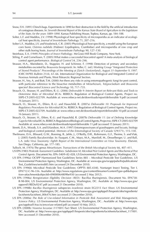
94 S.T. Jaronski
Snow, F.H. (1891) Chinch-bugs. Experiments in 1890 for their destruction in the field by the artificial introduction of contagious diseases. In: Seventh Biennial Report of the Kansas State Board of Agriculture to the Legislature of the State, for the years 1889–1890. Kansas Publishing House, Topkea, Kansas, pp. 184–188.
Solter, L.F. and Maddox, J.V. (1998) Physiological host specificity of microsporidia as an indicator of ecologi-cal host specificity. Journal of Invertebrate Pathology 71, 207–216.
Solter L.F., Maddox, J.V. and Vossbrinck, C.R. (2005) Physiological host specificity, a model using the European corn borer, Ostrinia nubilalis (Hubner) (Lepdioptera, Crambidae) and microsporidia of row crop and other stalk-boring hosts. Journal of Invertebrate Pathology 90, 127–130.
Steinhaus, E.A. (1949) Principles of Insect Pathology. McGraw-Hill Book Company, New York.Stiling, P. and Cornelissen, T. (2005) What makes a successful biocontrol agent? A meta-analysis of biological
control agent performance. Biological Control 34, 236–246.Strasser, H.A., Abendstein, D., Stuppner, H. and Schinner, F. (1998) Detection of primary and secondary
metabolites excreted by Beauveria brongniartii. In: Adler, C. (ed.) Working Group “Integrated Protection of Stored Products”. Proceedings of the Meeting at Zürich (Switzerland), August 31–September 9, 1997. IOBC/WPRS Bulletin 21(4), 61–66. International Organization for Biological and Integrated Control of Noxious Animals and Plants, West Palearctic Regional Section.
Strasser, H., Vey, A. and Butt, T.M. (2000) Are there any risks in using entomopathogenic fungi for pest control, with particular reference to the bioactive metabolites of Metarhizium, Tolypocladium and Beauveriaspecies? Biocontrol Science and Technology 10, 717–735.
Strauch, O., Strasser, H. and Ehlers, R.-U. (2006) Deliverable 9: Interim Report on Relevant Risks and Tools to Determine Risks of Microbial BCAs. REBECA: Regulation of Biological Control Agents, Project no. SSPE-CT-2005-022709. Available at: www.rebeca-net.de/downloads/report/deliverable 9.pdf (accessed 16 January 2011).
Strauch, O., Strasser, H., Ehlers, R.-U. and Hauschild, R. (2007a) Deliverable 10: Proposals for Improved Regulatory Procedures for Microbial BCAs. REBECA: Regulation of Biological Control Agents, Project no. SSPE-CT-2005-022709. Available at: www.rebeca-net.de/downloads/report/deliverable 10.pdf (accessed 16 January 2011).
Strauch, O., Strasser, H., Ehlers, R.-U. and Hauschild, R. (2007b) Deliverable 11: List of Defining Knowledge Gaps for Microbial BCAs. REBECA: Regulation of Biological Control Agents, Project no. SSPE-CT-2005-022709 Available at: www.rebeca-net.de/downloads/report/deliverable 11.pdf (accessed 15 January 2011).
Streett, D.A., Woods, S.A. and Erlandson, M.A. (1997) Entomopoxviruses of grasshoppers and locusts, biology and biological control potential. Memoirs of the Entomological Society of Canada 129(171), 115–130.
Theilmann, D.A., Blissard, G.W., Bonning, B., Jehle, J., O’Reilly, D.R., Rohrmann, G.F., Theime, S. and Vlak, J. (2005) Family Baculoviridae. In: Fauquet, C.M., Mayo, M.A., Maniloff, M., Desselberger, U. and Ball, L.A. (eds) Virus Taxonomy: Eighth Report of the International Committee on Virus Taxonomy. Elsevier, San Diego, California, pp. 177–185.
Tulloch, M. (1976) The genus Metarhizium. Transactions of the British Mycological Society 66, 407–411.US EPA (1983) Pesticide Assessment Guidelines: Subdivision M, Microbial Pest Control Agents and Biochemical Pest
Control Agents. Document No. EPA–540/9–82–028, US Environmental Protection Agency, Washington, DC.US EPA (1996a) OCSPP Harmonized Test Guidelines Series 885 – Microbial Pesticide Test Guidelines. US
Environmental Protection Agency, Washington, DC. Available at: www.epa.gov/ocspp/pubs/frs/publications/ Test_Guidelines/series885.htm (accessed 24 December 2010).
US EPA (1996b) Microbial Pesticide Test Guidelines: OPPTS 885.4340, Nontarget Insect Testing, Tier I. EPA712-C-96-336. Available at: http://www.regulations.gov/contentStreamer?contentType=pdf&disposition=attachment&objectId=09000064809be930 (accessed 2 May 2012).
US EPA (1998a) Reregistration Eligibility Decision (RED): Bacillus thuringiensis. Document No. EPA738- R-98-004, US Environmental Protection Agency, Washington, DC. Available at: http://www.epa.gov/oppsrrd1/REDs/0247.pdf (accessed 21 December 2010).
US EPA (1998b) Bacillus thuringiensis subspecies israelensis strain EG2215 Fact Sheet. US Environmental Protection Agency, Washington, DC. Available at: http://www.epa.gov/oppbppd1/biopesticides/ingredients/factsheets/factsheet_006476.htm (accessed 21 December 2010).
US EPA (2000a) The Role of Use-related Information in Pesticide Risk Assessment and Risk Management, Science Policy. US Environmental Protection Agency, Washington, DC.. Available at: http://www.epa.gov/oppfead1/trac/science/use-related.pdf (accessed 10 May 2012).
US EPA (2000b) Nosema locustae (117001) Fact Sheet. US Environmental Protection Agency, Washington, DC. Available at: http://www.epa.gov/oppbppd1/biopesticides/ingredients/factsheets/factsheet_117001.htm (accessed 21 December 2010).
Microbial Control of Invertebrate Pests 95
US EPA (2002) Occlusion bodies of nucleopolyhedrovirus (NPV) of various moths (PC codes 107300; 107302; 107303; 127885; 129078)[.] Occlusion bodies of granulovirus (GV) of codling moth and Indian meal moth (PC codes 129090 and 108896, respectively) Fact Sheet. US Environmental Protection Agency, Washington, DC. Available at: http://www.epa.gov/oppbppd1/biopesticides/ingredients/factsheets/factsheet_107300.htm (accessed 21 December 2010).
US EPA (2004) Bacillus popilliae spores (054502) Fact Sheet. US Environmental Protection Agency, Washington, DC. Available at: http://www.epa.gov/oppbppd1/biopesticides/ingredients/factsheets/factsheet_054502.htm (accessed 15 December 2010).
US EPA (2006) Biopesticide Registration Action Document, Beauveria bassiana HF23 (PC Code 090305). US Environmental Protection Agency, Washington, DC. Available at: http://www.epa.gov/oppbppd1/biopesticides/ingredients/tech_docs/brad_090305.pdf (accessed 16 December 2010).
US EPA (2007) Pesticides; Data Requirements for Biochemical and Microbial Pesticides: 40 CFR Part 158. US Environmental Protection Agency, Washington, DC. Available at: http://www.epa.gov/fedrgstr/EPA-PEST/2007/October/Day-26/p20828.htm (accessed 15 December 2010).
US EPA (2009) 40CFR 180.1011: Viable spores of the microorganism Bacillus thuringiensis Berliner, exemp-tion from the requirement of a tolerance. US Environmental Protection Agency, Washington, DC. Available at: http://www.law.cornell.edu/cfr/text/40/180.1011 (accessed 2 May 2012).
US Government (1947) Federal Insecticide, Fungicide and Rodenticide Act. Title 40, Code of Federal Regulations Chapter 1, Part 152. Washington, DC.
US Government (2011) Electronic Code of Federal Regulations: e-ECR. Title 40: Protection of Environment, Part 159 – Statements of Policies and Interpretations, Subpart D – Reporting Requirements for Risk/Benefit Information: 159.184 Toxic or adverse effect incident reports. Available at: http://ecfr.gpoaccess.gov/cgi/t/text/text-idx?c=ecfr&sid=350442cc63bfa6a590171b8a60b0ecdb&rgn=div8&view=text&node=40:24.0.1.1.10.2.1.13&idno=40 (accessed 2 May 2012).
van Lenteren, J.C., Babendrier, D., Bigler, F., Burgio, G., Hokkanen, H.M.T., Kuske, S., Loomans, A.J.M., Menzler-Hokkanen, I., van Rijn, P.C.J., Thomas, M.B., Tommasini, M.G. and Zeng, Q.-Q. (2003) Environmental risk assessment of exotic natural enemies used in inundative biological control. BioControl 48, 3–38.
Vankova, J. (1978) The heat-stable exotoxin of Bacillus thuringiensis. Folia Microbiologia 23, 162–174.Vestergaard, S., Cherry, A., Keller, S. and Goettel, M. (2003) Safety of hyphomycete fungi as microbial control
agents. In: Hokkanen, H.M.T. and Hajek, A.E. (eds) Environmental Impacts of Microbial Insecticides. Progress in Biological Control Series, Volume 1. Kluwer Academic, Dordrecht, The Netherlands, pp. 35–62.
Vey, A., Hoagland, R. and Butt, T.M. (2001) Toxic metabolites of fungal biocontrol agents. In: Butt, T.M., Jackson, C.W. and Magan, N. (eds) Fungi as Biocontrol Agents, Progress, Problems and Potential. CABI International, Wallingford, UK, pp. 311–346.
Vossbrinck, C.R. and Debrunner-Vossbrinck, B.A. (2005) Molecular phylogeny of the Microsporidia, ecologi-cal, ultrastructural and taxonomic considerations. Folia Parasitologica 52, 131–142.
Walker, T., Johnson, P.H., Moreira, L.A., Iturbe-Ormaetxe, I., Frentiu, F.D., McMeniman, C.J., Leong, Y.S., Dong, Y., Axford, J., Kriesner, P., Lloyd, A.L., Ritchie, S.A., O’Neill, S.L. and Hoffmann, A.A. (2011) The wMel Wolbachia strain blocks dengue and invades caged Aedes aegypti populations. Nature 476, 450–453.
Williams T. (1996) The iridoviruses. Advances in Virus Research 46, 345–412.Wolfenbarger, L.L., Naranjo, S.E., Lundgren, J.G., Bitzer, R.J. and Watrud, L.S. (2008) Bt crop effects on functional
guilds of non-target arthropods: a meta-analysis. PLoS ONE 3(5), e2118. doi, 10.1371/journal.pone.0002118.Zare, R. and Gams, W. (2001) A revision of Verticillium sect. Prostrata. III. Generic classification. Nova
Hedwigia 72, 329–337.Zhou, Z.H. (2008) Cypovirus. In: Patten, J.T. (ed.) Segmented Double-stranded RNA Viruses: Structure and
Molecular Biology. Caister Academic Press, Hethersett, Norwich, UK.Zimmermann, G. (2007a) Review on safety of the entomopathogenic fungi Beauveria bassiana and Beauveria
brongniartii. Biocontrol Science and Technology 17, 553–596.Zimmermann, G. (2007b) Review on safety of the entomopathogenic fungus Metarhizium anisopliae.
Biocontrol Science and Technology 17, 879–920.Zimmermann, G. (2008) The entomopathogenic fungi Isaria farinosa (formerly Paecilomyces farinosus) and
the Isaria fumosorosea species complex (formerly Paecilomyces fumosoroseus): biology, ecology and use in biological control. Biocontrol Science and Technology 18, 865–901.
©CAB International 2012. Beneficial Microorganisms in Agriculture, Food and the Environment: 96 Safety Assessment and Regulation (eds I. Sundh et al.)
8.1 Introduction 968.2 Identity of the Microbiological Control Agent 97
8.2.1 Bacteria 98 8.2.2 Fungi 99
8.3 Contaminants 1008.4 Secondary Metabolites 1008.5 Residues 1028.6 Effects on Human Health 1038.7 Fate and Behaviour in the Environment 1058.8 Effects on Non-target Organisms 1068.9 Conclusion 108
8 Microbial Control of Plant Diseases
Claude Alabouvette,1 Ulf Heilig2 and Christelle Cordier1
1AGRENE, Dijon, France; 2Consultant, Le Pecq, France
8.1 Introduction
At this beginning of the 21st century, human-ity is facing several challenges regarding food security in relation to climatic change and energy crisis, leading to a need for increasing agricultural production for both food and energy purposes. At the same time, there is an increased public demand for ‘safe’ foods, which does not necessarily means organic food, but food without pesticide residues. Thus crops have to be protected from pests and diseases with less use of chemical pesti-cides. In this context, there is a renewed inter-est in biological control using different types of antagonistic microorganisms.
In this chapter we consider the use of microbial biological control agents (MBCAs) to control plant diseases. But it seems impor-tant to first stress that the term ‘biopesticide’ or, alternatively, ‘microbial pesticide’, is not necessarily appropriate to describe an MBCA. The modes of action of MBCAs are complex,
often multiple and generally totally different from those of chemical pesticides. Most of them do not have ‘cidal’ effects in that they directly kill the target organisms; instead, they inhibit their growth, detoxify the toxins produced by the pathogens and induce plant defence reactions.
In many countries, the placing on the market of MBCAs is regulated the same way as that of chemical pesticides. Because MBCAs are living organisms, however, the hazards they might present are not of the same nature as those of chemical pesti-cides, and the risks have to be assessed differently.
In the European Union (EU), Regulation (EC) No 1107/2009 (OJEU, 2009) concerning the placing of plant protection products on the market, implemented on 14 June 2011, contains the text of reference which regulates the use of plant protection products, including chemicals as well as MBCAs. The Regulation follows the approach established in Council
Microbial Control of Plant Diseases 97
Directive 91/414/EEC (OJEC, 1991), which was amended by Commission Directive 2001/ 36/EC (OJEC, 2001) regarding the data require-ments for the inclusion in Annex I of microor-ganisms as active substances, and the national authorization of products (Annexes IIB and IIIB). Even if the latter Directive was written with the objective of adapting Directive 91/414/EEC to the case of living microorgan-isms, one must say that this corpus of regula-tion does not permit a rapid and satisfactory evaluation of risk linked to the application of biological control products. In fact, Directive 91/414/EEC was created in response to human health problems that appeared after many years of intensive use of chemical pes-ticides. It is not, therefore, adapted to the evaluation of the safety of MBCAs and there are no provisions for a timetable or a possible revision of data requirements for microbial active substances and plant protection prod-ucts containing them.
In contrast, several countries outside the EU, especially the USA, do not regulate MBCAs in the same way as chemical pesti-cides. The main differences between the EU regulations and those of other countries have recently been reviewed by Hauschild et al.(2011). With the objective of harmonization of the requirements for MBCA registration, the Organisation for Economic Co-operation and Development (OECD) is working on guidelines to identify the hazards and evalu-ate the risks linked to the use of MBCAs (see Meeussen, Chapter 21, this volume).
In this chapter, we outline the specific challenges posed in the safety assessment and registration of microorganisms being com-mercialized for the control of plant diseases, and critically discuss these in reference to the Regulation (EC) No 1107/2009, related legis-lative texts and OECD proposals.
Because MBCAs (fungi, bacteria or viru-ses) might be closely related to species that are known to be pathogenic, the first challenge is obviously to ascertain that the MBCA is not,and will never become, infectious or patho-genic for humans, animals or plants. One must also ensure that the secondary metabolites, and in particular toxins, if they are produced, are not harmful for man, or deleterious for non-target organisms or the environment
under the intended uses. In contrast to chemi-cals, MBCAs can survive and even multiply on plants and in the environment. So there is great concern about the behaviour and fate of the MBCAs after release. In relation to this capacity to survive and multiply, the identification of the hazards posed by possible residues is com-plex, as the residues consist not only of the liv-ing MBCAs themselves, but also of the secondary metabolites that the MBCAs can potentially produce on the plant parts that are used as food or feed. Finally, these basic ques-tions apply not only to the active substance, which is the microorganism itself, but also to possible microbial contaminants and metabo-lites of concern that might be present in the commercial product, which could be released into the environment.
Thus, the identification of hazards and assess ment of risks appears to be more com-plex when dealing with microbial biological control agents than with chemical control products.
8.2 Identity of the Microbiological Control Agent
Obviously, the first concern is whether the MBCA is pathogenic or infectious to humans and animals. Commission Regulation (EU) No 545/2011 (OJEU, 2011) of 10 June 2011 implementing Regulation (EC) No 1107/2009 concerning data requirements for active substances clearly states, like the previous Directive 2001/36/EC, that ‘the identification together with the characterization of the micro-organism provides the most important information and is a key point for decision making’. All experts agree with this state-ment: a correct identification of the microor-ganism is the first step to ensure the safety of the plant protection product containing it. Although there has been a debate about the level of identification of the microorganism and methods to be used, scientists agree that identification must be at the strain level, using the most accurate methodology available. Indeed, the biocontrol capacities are specific to the strain and not necessarily common to all strains belonging to the same (sub)spe-cies. This does not exclude the possibility
98 C. Alabouvette et al.
that, for the general parts of the dossier, data for different strains of the same species might be used when a great similarity within a spe-cies has been demonstrated. Nevertheless, as illustrated below, many species of microor-ganisms include both pathogenic and benefi-cial strains. It is necessary, then, to develop methods that enable the identification of the biocontrol strain from other strains belonging to the same species. Such tools are also needed for regulatory reasons to ensure that the prod-uct contains only the strain that has been approved. Identification of an MBCA at the species and strain level cannot rely only on phenotypic or physiological characters; today, several molecular methods are available that can be routinely used to identify MBCAs (Louws et al., 1999).
Beside the need to distinguish an MBCA from pathogenic strains, the ability to specifi-cally recognize a strain of an MBCA provides some protection for the owner of the strain. Because in Europe a microbial strain isolated from the environment cannot be patented, registration of a well-identified strain is a way to protect the plant protection product. Knowing that the data generated to establish the registration dossier are the property of the petitioner and typically protected for a period of 10 years, a competitor willing to market a product using the registered strain will have to negotiate access to the protected part of the dossier or conduct all the expensive studies needed to produce all the data specific to the dossier. Therefore, although not impossible, registration of a strain in practice prevents its use by another company for several years.
8.2.1 Bacteria
The use of several strains of bacteria as bio-logical control agents has been the cause of debate as to whether products containing these strains could be responsible for human infection. A well-documented example is that of Pantoea agglomerans; several strains of this species are among the most promising bio-control agents for a variety of bacterial and fungal plant diseases. Strain C9-1 of P. agglom-erans is registered in the USA and in Canada
to control fire blight of apple and pear, but commercial registration is not possible in Europe because P. agglomerans is listed as a biosafety level 2 organism owing to clinical reports that it is an opportunistic human pathogen. Rezzonico et al. (2009) conducted a comparison of many strains of this species, of both plant and clinical origin, with the aim of identifying genetic/phenotypic mark-ers using multi-locus phylogenetic analysis and fluorescent amplified length polymor-phism (fAFLP) fingerprinting. Sequence analysis revealed that a majority of the clini-cal isolates were improperly designated as P. agglomerans. In the P. agglomerans sensu stricto group, there was no discrete clustering of clinical/biocontrol strains and no marker was identified that was uniquely associated with clinical strains. Conversely, a putative biocontrol specific marker was identified which is only present in biocontrol strains. This marker could be used in the future for identification of biocontrol strains that can be expected to pose no risk to humans.
Another example concerns Stenotropho-monas maltophila. Some environmental strains could be developed as MBCAs, while others are known to be responsible for nosocomial infections. Minkwitz and Berg (2001) charac-terized 50 strains for phenotypic and molecu-lar characteristics. Using 16S ribosomal DNA sequencing, the isolates could be separated into three clusters, two of which consisted of isolates originating from the rhizosphere; the third cluster consisted of both clinical and environmental strains. In vitro characteriza-tion of the antagonism of the strains towards several pathogenic fungi and of their produc-tion of secondary metabolites demonstrated that all the strains, whatever their origin, pro-duced a large range of antifungal metabolites, which included antibiotics, siderophores and enzymes. How ever, as for other bacterial spe-cies, such as Pseudomonas spp., the antifungal mode of action was strain specific.
These two examples justify the need for a precise characterization of MBCAs at the infra-specific level in order to clearly distin-guish between bacterial strains potentially harmful to man (or mammals), and strains present in the environment that can be safely developed as biological control agents.
Microbial Control of Plant Diseases 99
8.2.2 Fungi
Fungal strains belonging to the genus Trichoderma/Hypocrea have been known for years as beneficial organisms with antagonis-tic activities against many plant pathogenic fungi. The genus Trichoderma is ubiquitous in soil, on organic debris, and on roots and other plant parts. It is mainly present in its anamor-phic stage, which produces many unicellu-lar microconidia. The different species of Trichoderma are very difficult to identify based on morphological characters, as described by Rifai (1969). Only recently has the use of molecular techniques targeting the sequence of the ribosomal DNA enabled a clear identi-fication at the genus and species levels. Indeed, these tools enable placement of the strains in a phylogenetic tree, thus revealing the relationship among species belonging to the same genus. This allows prediction of the potential hazards based on the proximity of a strain to known pathogenic species or strains. Cordier et al. (2007) compared the 18S rDNA sequences of Trichoderma strains to identify candidates that showed some abilities for bio-logical control. One potential MBCA strain examined was found to occur in the same cluster as several strains belonging to T. longi-brachiatum, a species known to include strains pathogenic to man. Consequently, based on this information, it was decided to terminate the development of this strain as an MBCA as it could potentially be pathogenic to humans.
Several preparations already on the market were supposed to contain strains of T. harzianum. But, based only on morphologi-cal characters, these strains were misidenti-fied. They have been recently re-identified as T. atroviride through sequencing of the ITS (internal transcribed spacer) region of the ribosomal DNA and of genes encoding dif-ferent functions, such as the elongation factor 1a. In order to distinguish these different strains belonging to the same species, it is absolutely necessary to develop a specific marker for each strain. This has been done for strain I 1237 of T. atroviride, which has recently been developed as a biocontrol agent (Cordier et al., 2007).
If the MBCA belongs to a species known to include strains pathogenic to plants,
non-target animals or man, the design of a SCAR (sequence characterized amplified region) marker is an absolute necessity as it will be the only way to accurately distin-guish it from other strains that are poten-tially pathogenic. One of the best examples comes from Fusarium oxysporum, which shows a great diversity of strains. F. oxyspo-rum can be found in many different environ-ments, including soil, where it is very common and occurs in high numbers. Most of the strains found, if not all, are good saprophytes, surviving for many years in the soil and in the rhizosphere of many plant species. F. oxysporum is well known to include very aggressive plant pathogens and also opportunistic human pathogens. The pathogenic soil-borne strains penetrate the roots of different plants and can provoke either rots or wilts. Strains responsible for wilts show narrow host specificity, usually attacking a single plant species, which has led to the dis tinction of pathovars or formae speciales. Interestingly, some strains are not only non-pathogenic but also provide dis-ease protection for certain plant species (Alabouvette et al., 2009). Several non- pathogenic strains have been proposed as MBCAs to control Fusarium diseases. In such a case, it is obviously necessary to develop a tool enabling precise identification of the strain in question. Recently, a SCAR marker has been designed to identify the well-known pro tective strain Fo47, and a method developed not only to detect but also to quantify this MBCA in the root of the plant (Edel-Hermann et al., 2011).
To conclude with this first aspect regarding the safety of MBCAs in relation to human, animal or plant infectivity or pathogenicity, one must stress that pres-ently there is no universal tool to address the precise identification of an MBCA at the species and strain levels. However, currently available technology provides several tools enabling characterization of a strain at the infraspecific level. Depend-ing on the species, it might be necessary to use several techniques or to sequence several DNA regions to reach a level of characterization sufficient to ensure the development of innocuous strains.
100 C. Alabouvette et al.
8.3 Contaminants
Products placed on the market contain not only the MBCA itself, and often residues of the cultivation medium, but also different addi-tives (e.g. co-formulants) and, possibly, micro-bial contaminants. Therefore, it is important to ensure that the contaminants are not hazard-ous to man or the environment. According to Commission Regulation (EU) No 545/2011 implementing Regulation (EC) No 1107/2009 as regards the data requirements for plant pro-tection products: ‘the content of other compo-nents (such as by-products, condensates, culture medium, etc.) and contaminating micro-organisms, derived from production process’ must be reported. This is a difficult challenge, because the level and the identity of contaminants might change from one produc-tion process or geographic site to another, and even from one batch to another. Thus, the regu-lations require that the applicant must subject both the production process and the product to continuous quality control.
From the microbiologist’s point of view, a plant protection product should contain the MBCA strain without any contaminants. Produced in pure culture, in fermentation reactors, the active substance should be clean. The presence of contaminants often means that the production process has not been well optimized. Contamination could potentially occur during the different steps of the pro-duction, harvesting and formulation pro-cesses, which are usually conducted under clean, but not necessarily aseptic, conditions. The technical active substance and the prepa-ration could be contaminated by ubiquitous microbiota from the surroundings. In such cases, the content of contaminants will be low compared with that of the MBCA and of no concern for human health. In some cases, for example in the case of viral MBCAs, which have to be produced in living insects, cells or bacterial cultures, it is difficult to avoid con-taminants. In addition, some preparations are formulated with components of natural ori-gin, such as clays, which might contain a complex microbiota. So these preparations might contain high levels of various types of contaminants. The regulations require that a petitioner reports the maximum content of
contaminants and their identification to ensure that the products do not contain plant or animal pathogens (Commission Regulation (EU) No 545/2011).
The level and nature of acceptable contaminants pose problems in relation to risk assessment for humans and non-target organisms. Most countries recommend methods for determination of the presence of contaminants of human concern and have limit levels for genera such as Listeria, Salmo-nella, Enterococcus, etc. At present though, there is no harmonized or agreed European list. The OECD BioPesticide Steering Group has prepared an issue paper on this subject (OECD, 2011b). This issue paper focuses on microbial contaminants of human and animal concern, primary human pathogens. For ref-erence and guidance to regulatory authorities as well as applicants, the issue paper includes a compilation of the methods available for screening microbial pest control products for the presence of pathogens and other contami-nating microorganisms. Table 8.1 presents a list of contaminants, with the tolerated con-tent limits proposed by OECD and proposals by industry representatives (Ravensberg, 2010). Very low limits should not be necessary for microbial contaminants that are normally present in soil and are introduced in relatively high densities with different agronomic inputs, such as manure.
8.4 Secondary Metabolites
According to Regulation (EU) No 544/2011: ‘relevant metabolites (i.e. if expected to be of concern to human health and/or the environ-ment) known to be formed by the micro-organism shall be identified and characterised at different states or growth stages of the micro-organism’. This point is one of the most controversial, as bacteria and fungi produce, usually at low concentrations, a large variety of secondary metabolites. These can include antibiotics, toxins, enzymes, hormone-like substances, surfactants and other types of compounds (Vining, 1990).
Consumer concerns regarding mycotox-ins entering the food chain have prompted closer scrutiny of the secondary metabolites
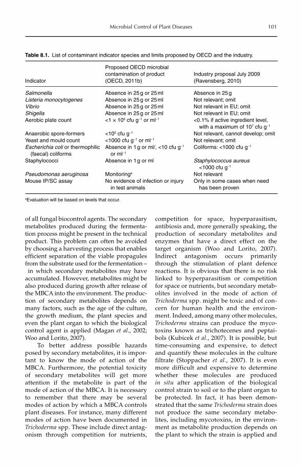
Microbial Control of Plant Diseases 101
of all fungal biocontrol agents. The secondary metabolites produced during the fermenta-tion process might be present in the technical product. This problem can often be avoided by choosing a harvesting process that enables efficient separation of the viable propagules from the substrate used for the fermentation – in which secondary metabolites may have accumulated. However, metabolites might be also produced during growth after release of the MBCA into the environment. The produc-tion of secondary metabolites depends on many factors, such as the age of the culture, the growth medium, the plant species and even the plant organ to which the biological control agent is applied (Magan et al., 2002; Woo and Lorito, 2007).
To better address possible hazards posed by secondary metabolites, it is impor-tant to know the mode of action of the MBCA. Furthermore, the potential toxicity of secondary metabolites will get more attention if the metabolite is part of the mode of action of the MBCA. It is necessary to remember that there may be several modes of action by which a MBCA controls plant diseases. For instance, many different modes of action have been documented in Trichoderma spp. These include direct antag-onism through competition for nutrients,
competition for space, hyperparasitism, antibiosis and, more generally speaking, the production of secondary metabolites and enzymes that have a direct effect on the target organism (Woo and Lorito, 2007). Indirect antagonism occurs primarily through the stimulation of plant defence reactions. It is obvious that there is no risk linked to hyperparasitism or competition for space or nutrients, but secondary metab-olites involved in the mode of action of Trichoderma spp. might be toxic and of con-cern for human health and the environ-ment. Indeed, among many other molecules, Trichoderma strains can produce the myco-toxins known as trichotecenes and peptai-bols (Kubicek et al., 2007). It is possible, but time-consuming and expensive, to detect and quantify these molecules in the culture filtrate (Stoppacher et al., 2007). It is even more difficult and expensive to determine whether these molecules are produced in situ after application of the biological control strain to soil or to the plant organ to be protected. In fact, it has been demon-strated that the same Trichoderma strain does not produce the same secondary metabo-lites, including mycotoxins, in the environ-ment as metabolite production depends on the plant to which the strain is applied and
Table 8.1. List of contaminant indicator species and limits proposed by OECD and the industry.
Indicator
Proposed OECD microbial contamination of product (OECD, 2011b)
Industry proposal July 2009 (Ravensberg, 2010)
Salmonella Absence in 25 g or 25 ml Absence in 25 gListeria monocytogenes Absence in 25 g or 25 ml Not relevant; omitVibrio Absence in 25 g or 25 ml Not relevant in EU; omitShigella Absence in 25 g or 25 ml Not relevant in EU; omitAerobic plate count <1 × 10⁵ cfu g−1 or ml−1 <0.1% if active ingredient level,
with a maximum of 107 cfu g−1
Anaerobic spore-formers <105 cfu g−1 Not relevant, cannot develop; omitYeast and mould count <1000 cfu g−1 or ml−1 Not relevant; omitEscherichia coli or thermophilic (faecal) coliforms
Absence in 1 g or ml/, <10 cfu g−1
or ml−1
Coliforms: <1000 cfu g−1
Staphylococci Absence in 1 g or ml Staphylococcus aureus <1000 cfu g−1
Pseudomonas aeruginosa Monitoringa Not relevantMouse IP/SC assay No evidence of infection or injury
in test animalsOnly in some cases when need has been proven
aEvaluation will be based on levels that occur.
102 C. Alabouvette et al.
the target pathogen that it confronts (Marra et al., 2006). It is important to stress, as well, that the secondary metabolites are only pro-duced locally and in very limited quanti-ties, and that strains naturally present in the environment also produce those same compounds. Thus, the relevance of the characterization and quantification of all toxic metabolites must be questioned, and it might not be necessary to spend much effort on this. Nevertheless, when the main mode of action is based on the production of antibiotics, there might be some concern over secondary metabolites with regard to human health and the environment. One must be certain that the use of an MBCA producing antibiotics will not contribute to the induction of multiple resistance to antibiotics used in human or veterinary medicine.
In conclusion, one must be realistic and admit that it is impossible to characterize all of the metabolites produced by an MBCA at the different stages of its life cycle, on all dif-ferent substrates or plants. So only those present at a quantifiable level in the techni-cal product, and known to be of concern, might be subject to risk assessment. The pro-duction of secondary metabolites will also be addressed below as it is one of the most important questions in relation to acceptable residue levels.
8.5 Residues
According to the data requirements for active substances consisting of microorganisms in Regulation (EU) No 545/2011 (Annex, Part B), residues are divided into viable and non- viable.The MBCA itself constitutes the major part of the viable residues, which also includemicrobial contaminants. In contrast to chem-ical pesticides, the MBCAs can establish and sometimes proliferate on or in food and feed. The metabolites are non-viable residues which can be produced by the MBCA on or in food and feed.
For the microorganism itself, if the tox-icity testing (see below) has led to the con-clusion that the MBCA is not of concern for
human health, the residual microorgan-isms should be of no concern either. The only question is whether the microorganism might have multiplied after release and reached a density far above the dose used in the toxicity testing. When a SCAR marker has been developed, it is quite easy to assess the population level of the MBCA in food or feed, but, in the absence of a specific marker, this is almost impossible, because the plant organs will often be colonized by natural populations of organisms belonging to the same species as the MBCA.
More difficult to address is the question of the secondary metabolites that may have been produced and still be present in food or feed. According to EU legislation, full experi-mental residue data are required if relevant quantities of the microorganism or of pro-duced metabolites, especially toxins, have been found to be persistent. As stated above, it is not realistic to try to characterize all the molecules that are present at the surface of, for example, a fruit. Thus, only the secondary metabolites known to be of concern for human health should be considered, and their level determined if an analytical method can be developed.
A recent example of the complexity of this question concerns the production of 2,3-deepoxy-2,3-didehydrorhizoxin (DDR) by Ps. chlororaphis. A strain of Ps. chlororaphis has been selected for its biocontrol capacities of several fungal root diseases of wheat. During the assessment for approval, it was established that it produces the toxic compound DDR. This secondary metabolite was isolated, char-acterized and its mutagenic potential demon-strated. As a consequence, questions arose as to whether the strain should be commercial-ized. A method of analysis was developed by the applicant and validated by the regu-lators. In the regulations, the quantification limit for DDR in the fermentation medium is 2 mg l−1 and the seed detection limit is 1 mgkg−1 (Commission Directive 2004/71/EC; OJEU, 2004). The highest amounts of DDR were detected at the end of the fermentation pro-cess, but because DDR decomposes rapidly, no detectable quantities of the metabolite could be found on treated seeds (Hökeberg, 2006). Hence, the risk might have been significant
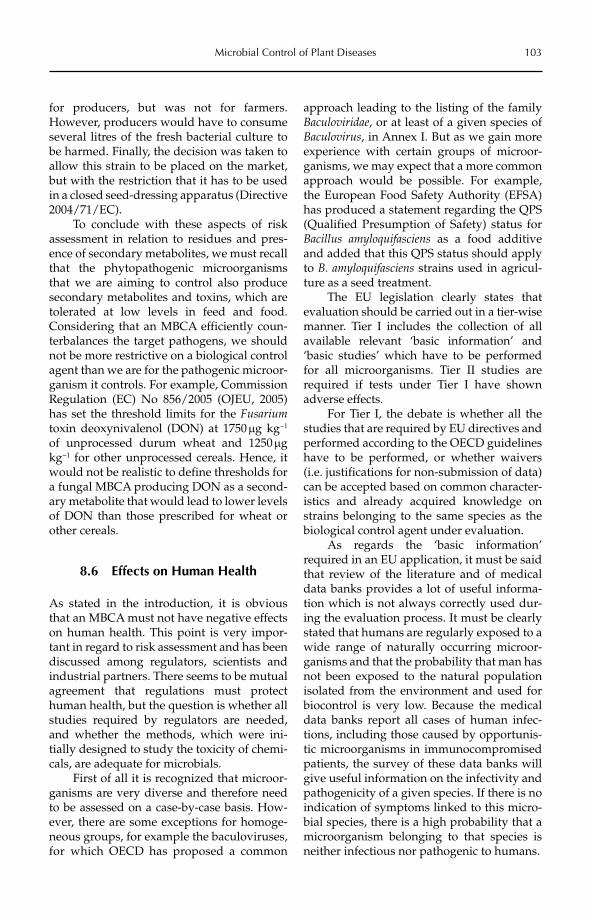
Microbial Control of Plant Diseases 103
for producers, but was not for farmers. However, producers would have to consume several litres of the fresh bacterial culture to be harmed. Finally, the decision was taken to allow this strain to be placed on the market, but with the restriction that it has to be used in a closed seed-dressing apparatus (Directive 2004/71/EC).
To conclude with these aspects of risk assessment in relation to residues and pres-ence of secondary metabolites, we must recall that the phytopathogenic microorganisms that we are aiming to control also produce secondary metabolites and toxins, which are tolerated at low levels in feed and food. Considering that an MBCA efficiently coun-terbalances the target pathogens, we should not be more restrictive on a biological control agent than we are for the pathogenic microor-ganism it controls. For example, Commission Regulation (EC) No 856/2005 (OJEU, 2005) has set the threshold limits for the Fusariumtoxin deoxynivalenol (DON) at 1750 mg kg−1
of unprocessed durum wheat and 1250 mgkg−1 for other unprocessed cereals. Hence, it would not be realistic to define thresholds for a fungal MBCA producing DON as a second-ary metabolite that would lead to lower levels of DON than those prescribed for wheat or other cereals.
8.6 Effects on Human Health
As stated in the introduction, it is obvious that an MBCA must not have negative effects on human health. This point is very impor-tant in regard to risk assessment and has been discussed among regulators, scientists and industrial partners. There seems to be mutual agreement that regulations must protect human health, but the question is whether all studies required by regulators are needed, and whether the methods, which were ini-tially designed to study the toxicity of chemi-cals, are adequate for microbials.
First of all it is recognized that microor-ganisms are very diverse and therefore need to be assessed on a case-by-case basis. How-ever, there are some exceptions for homoge-neous groups, for example the baculoviruses, for which OECD has proposed a common
approach leading to the listing of the family Baculoviridae, or at least of a given species of Baculovirus, in Annex I. But as we gain more experience with certain groups of microor-ganisms, we may expect that a more common approach would be possible. For example, the European Food Safety Authority (EFSA) has produced a statement regarding the QPS (Qualified Presumption of Safety) status for Bacillus amyloquifasciens as a food additive and added that this QPS status should apply to B. amyloquifasciens strains used in agricul-ture as a seed treatment.
The EU legislation clearly states that evaluation should be carried out in a tier-wise manner. Tier I includes the collection of all available relevant ‘basic information’ and ‘basic studies’ which have to be performed for all microorganisms. Tier II studies are required if tests under Tier I have shown adverse effects.
For Tier I, the debate is whether all the studies that are required by EU directives and performed according to the OECD guidelines have to be performed, or whether waivers (i.e. justifications for non-submission of data) can be accepted based on common character-istics and already acquired knowledge on strains belonging to the same species as the biological control agent under evaluation.
As regards the ‘basic information’ required in an EU application, it must be said that review of the literature and of medical data banks provides a lot of useful informa-tion which is not always correctly used dur-ing the evaluation process. It must be clearly stated that humans are regularly exposed to a wide range of naturally occurring microor-ganisms and that the probability that man has not been exposed to the natural population isolated from the environment and used for biocontrol is very low. Because the medical data banks report all cases of human infec-tions, including those caused by opportunis-tic microorganisms in immunocompromised patients, the survey of these data banks will give useful information on the infectivity and pathogenicity of a given species. If there is no indication of symptoms linked to this micro-bial species, there is a high probability that a microorganism belonging to that species is neither infectious nor pathogenic to humans.
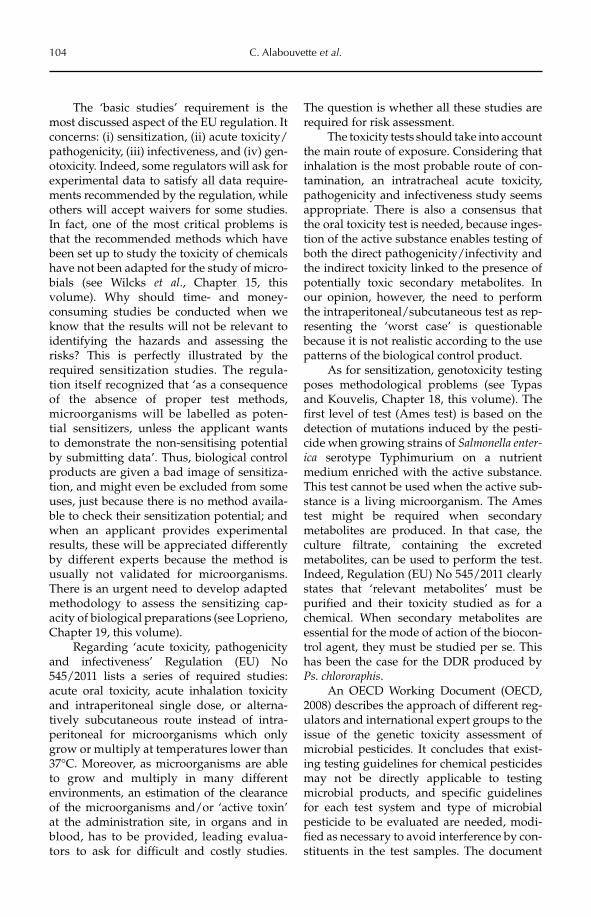
104 C. Alabouvette et al.
The ‘basic studies’ requirement is the most discussed aspect of the EU regulation. It concerns: (i) sensitization, (ii) acute toxicity/pathogenicity, (iii) infectiveness, and (iv) gen-otoxicity. Indeed, some regulators will ask for experimental data to satisfy all data require-ments recommended by the regulation, while others will accept waivers for some studies. In fact, one of the most critical problems is that the recommended methods which have been set up to study the toxicity of chemicals have not been adapted for the study of micro-bials (see Wilcks et al., Chapter 15, this volume). Why should time- and money- consuming studies be conducted when we know that the results will not be relevant to identifying the hazards and assessing the risks? This is perfectly illustrated by the required sensitization studies. The regula-tion itself recognized that ‘as a consequence of the absence of proper test methods, microorganisms will be labelled as poten-tial sensitizers, unless the applicant wants to demonstrate the non- sensitising potential by submitting data’. Thus, biological control products are given a bad image of sensitiza-tion, and might even be excluded from some uses, just because there is no method availa-ble to check their sensitization potential; and when an applicant provides experimental results, these will be appreciated differently by different experts because the method is usually not validated for microorganisms. There is an urgent need to develop adapted methodology to assess the sensitizing cap-acity of biological preparations (see Loprieno, Chapter 19, this volume).
Regarding ‘acute toxicity, pathogenicity and infectiveness’ Regulation (EU) No 545/2011 lists a series of required studies: acute oral toxicity, acute inhalation toxicity and intraperitoneal single dose, or alterna-tively subcutaneous route instead of intra-peritoneal for microorganisms which only grow or multiply at temperatures lower than 37°C. Moreover, as microorganisms are able to grow and multiply in many different environments, an estimation of the clearance of the microorganisms and/or ‘active toxin’ at the administration site, in organs and in blood, has to be provided, leading evalua-tors to ask for difficult and costly studies.
The question is whether all these studies are required for risk assessment.
The toxicity tests should take into account the main route of exposure. Considering that inhalation is the most probable route of con-tamination, an intratracheal acute toxicity, pathogenicity and infectiveness study seems appropriate. There is also a consensus that the oral toxicity test is needed, because inges-tion of the active substance enables testing of both the direct pathogenicity/infectivity and the indirect toxicity linked to the presence of potentially toxic secondary metabolites. In our opinion, however, the need to perform the intraperitoneal/subcutaneous test as rep-resenting the ‘worst case’ is questionable because it is not realistic according to the use patterns of the biological control product.
As for sensitization, genotoxicity testing poses methodological problems (see Typas and Kouvelis, Chapter 18, this volume). The first level of test (Ames test) is based on the detection of mutations induced by the pesti-cide when growing strains of Salmonella enter-ica serotype Typhimurium on a nutrient medium enriched with the active substance. This test cannot be used when the active sub-stance is a living microorganism. The Ames test might be required when secondary metabolites are produced. In that case, the culture filtrate, containing the excreted metabolites, can be used to perform the test. Indeed, Regulation (EU) No 545/2011 clearly states that ‘relevant metabolites’ must be purified and their toxicity studied as for a chemical. When secondary metabolites are essential for the mode of action of the biocon-trol agent, they must be studied per se. This has been the case for the DDR produced by Ps. chlororaphis.
An OECD Working Document (OECD, 2008) describes the approach of different reg-ulators and international expert groups to the issue of the genetic toxicity assessment of microbial pesticides. It concludes that exist-ing testing guidelines for chemical pesticides may not be directly applicable to testing microbial products, and specific guidelines for each test system and type of microbial pesticide to be evaluated are needed, modi-fied as necessary to avoid interference by con-stituents in the test samples. The document
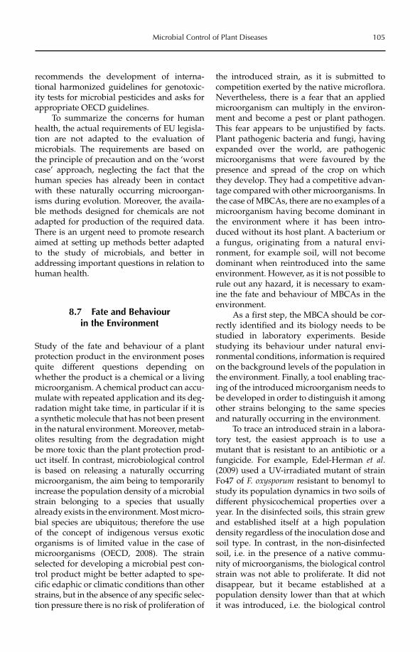
Microbial Control of Plant Diseases 105
recommends the development of interna-tional harmonized guidelines for genotoxic-ity tests for microbial pesticides and asks for appropriate OECD guidelines.
To summarize the concerns for human health, the actual requirements of EU legisla-tion are not adapted to the evaluation of microbials. The requirements are based on the principle of precaution and on the ‘worst case’ approach, neglecting the fact that the human species has already been in contact with these naturally occurring microorgan-isms during evolution. Moreover, the availa-ble methods designed for chemicals are not adapted for production of the required data. There is an urgent need to promote research aimed at setting up methods better adapted to the study of microbials, and better in addressing important questions in relation to human health.
8.7 Fate and Behaviour in the Environment
Study of the fate and behaviour of a plant protection product in the environment poses quite different questions depending on whether the product is a chemical or a living microorganism. A chemical product can accu-mulate with repeated application and its deg-radation might take time, in particular if it is a synthetic molecule that has not been present in the natural environment. Moreover, metab-olites resulting from the degradation might be more toxic than the plant protection prod-uct itself. In contrast, microbiological control is based on releasing a naturally occurring microorganism, the aim being to temporarily increase the population density of a microbial strain belonging to a species that usually already exists in the environment. Most micro-bial species are ubiquitous; therefore the use of the concept of indigenous versus exotic organisms is of limited value in the case of microorganisms (OECD, 2008). The strain selected for developing a microbial pest con-trol product might be better adapted to spe-cific edaphic or climatic conditions than otherstrains, but in the absence of any specific selec-tion pressure there is no risk of proliferation of
the introduced strain, as it is submitted to competition exerted by the native microflora. Nevertheless, there is a fear that an applied microorganism can multiply in the environ-ment and become a pest or plant pathogen. This fear appears to be unjustified by facts. Plant pathogenic bacteria and fungi, having expanded over the world, are pathogenic microorganisms that were favoured by the presence and spread of the crop on which they develop. They had a competitive advan-tage compared with other microorganisms. In the case of MBCAs, there are no examples of a microorganism having become dominant in the environment where it has been intro-duced without its host plant. A bacterium or a fungus, originating from a natural envi-ronment, for example soil, will not become dominant when reintroduced into the same environment. However, as it is not possible to rule out any hazard, it is necessary to exam-ine the fate and behaviour of MBCAs in the environment.
As a first step, the MBCA should be cor-rectly identified and its biology needs to be studied in laboratory experiments. Beside studying its behaviour under natural envi-ronmental conditions, information is required on the background levels of the population in the environment. Finally, a tool enabling trac-ing of the introduced microorganism needs to be developed in order to distinguish it among other strains belonging to the same species and naturally occurring in the environment.
To trace an introduced strain in a labora-tory test, the easiest approach is to use a mutant that is resistant to an antibiotic or a fungicide. For example, Edel-Herman et al.(2009) used a UV-irradiated mutant of strain Fo47 of F. oxysporum resistant to benomyl to study its population dynamics in two soils of different physicochemical properties over a year. In the disinfected soils, this strain grew and established itself at a high population density regardless of the inoculation dose and soil type. In contrast, in the non-disinfected soil, i.e. in the presence of a native commu-nity of microorganisms, the biological control strain was not able to proliferate. It did not disappear, but it became established at a population density lower than that at which it was introduced, i.e. the biological control
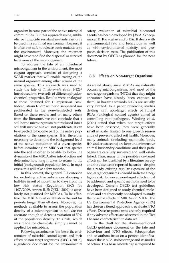
106 C. Alabouvette et al.
organism became part of the native microbial communities. But this approach using antibi-otic or fungicide resistant mutants can only be used in a confined environment because it is often not safe to release such mutants into the environment. Moreover, the mutation might have modified the dispersal or survival behaviour of the microorganism.
To address the fate of an introduced microorganism in the environment, the most elegant approach consists of designing a SCAR marker that will enable tracing of the natural organism among other strains of the same species. This approach was used to study the fate of T. atroviride strain I-1237 introduced into two soils of different physico-chemical properties. Results were analogous to those obtained for F. oxysporum Fo47. Indeed, strain I-1237 neither disappeared nor proliferated in the non-disinfected soils. Based on these results and on many others from the literature, we can conclude that a soil-borne microorganism reintroduced into a soil will survive but will not proliferate; it can be expected to become part of the native pop-ulations of the same species. It is, therefore, necessary to determine the background level of the native population of a given species before introducing an MBCA of that species into the soil in order to be able to follow the dynamics of the MBCA after introduction and determine how long it takes to return to the initial (background) population level. In most cases, this will take a few months.
In this context, the general EU criterion for excluding active substances showing a half-life in soil of more than 60 days from the low risk status (Regulation (EC) No 1107/2009, Annex II, 5; OJEU, 2009) is abso-lutely not justified for MBCAs. To be effec-tive, the MBCA must establish in the soil for periods longer than 60 days. Moreover, the methods available to assess the population density of a microorganism in soil are not accurate enough to detect a variation of 50% of the population density. This rule, which was made for chemicals, simply cannot be applied for microbials.
Following a seminar on ‘the fate in the envi-ronment of microbial control agents and their effects on non-target organisms’ (OECD, 2011a), a guidance document for the environmental
safety evaluation of microbial biocontrol agents has been developed by J.W.A. Scheep-maker, B. Karaoglan and S. Bär. It deals with environmental fate and behaviour as well as with environmental toxicity, and pro-poses decision trees. The publication of this document by OECD is planned for the near future.
8.8 Effects on Non-target Organisms
As stated above, since MBCAs are naturally occurring microorganisms, and most of the non-target organisms (NTOs) that they might encounter have already been exposed to them, so hazards towards NTOs are usually very limited. In a paper reviewing studies dealing with non-target effects of fungal BCAs (biological control agents) aimed at controlling root pathogens, Winding et al.(2004) stated that when non-target effects have been observed, they were generally small in scale, limited to one growth season and not proven to affect soil health. Moreover, many animals (including mammals, birds, fish and crustaceans) are kept under intensive animal husbandry conditions and their path-ogens are carefully surveyed and well estab-lished. Thus, many of the possible non-target effects can be identified by a literature survey and the absence of reported hazards – despite the already existing regular exposure of the non-target organisms – would indicate a neg-ligible risk. However, non-target effects must be addressed and specific methods need to be developed. Current OECD test guidelines have been designed to study chemical mole-cules and are frequently not adapted to study the possible effects of MBCAs on NTOs. The US Environmental Protection Agency (EPA) has chosen a tiered approach to evaluate NTO effects. Dose-response tests are only required if any adverse effects are observed in the Tier I hazard characterization data set.
In the draft for the above-mentioned OECD guidance document on the fate and behaviour and NTO effects, Scheepmaker and co-authors insist on a perfect identifica-tion of the MBCA, its host range and its modes of action. This basic knowledge is required to
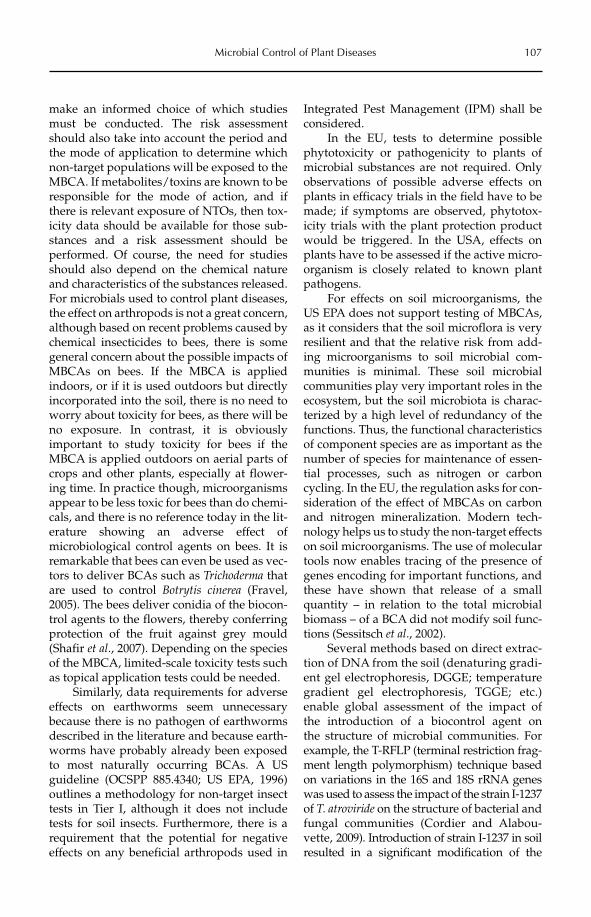
Microbial Control of Plant Diseases 107
make an informed choice of which studies must be conducted. The risk assessment should also take into account the period and the mode of application to determine which non-target populations will be exposed to the MBCA. If metabolites/toxins are known to be responsible for the mode of action, and if there is relevant exposure of NTOs, then tox-icity data should be available for those sub-stances and a risk assessment should be performed. Of course, the need for studies should also depend on the chemical nature and characteristics of the substances released. For microbials used to control plant diseases, the effect on arthropods is not a great concern, although based on recent problems caused by chemical insecticides to bees, there is some general concern about the possible impacts of MBCAs on bees. If the MBCA is applied indoors, or if it is used outdoors but directly incorporated into the soil, there is no need to worry about toxicity for bees, as there will be no exposure. In contrast, it is obviously important to study toxicity for bees if the MBCA is applied outdoors on aerial parts of crops and other plants, especially at flower-ing time. In practice though, microorganisms appear to be less toxic for bees than do chemi-cals, and there is no reference today in the lit-erature showing an adverse effect of microbiological control agents on bees. It is remarkable that bees can even be used as vec-tors to deliver BCAs such as Trichoderma that are used to control Botrytis cinerea (Fravel, 2005). The bees deliver conidia of the biocon-trol agents to the flowers, thereby conferring protection of the fruit against grey mould (Shafir et al., 2007). Depending on the species of the MBCA, limited-scale toxicity tests such as topical application tests could be needed.
Similarly, data requirements for adverse effects on earthworms seem unnecessary because there is no pathogen of earthworms described in the literature and because earth-worms have probably already been exposed to most naturally occurring BCAs. A US guideline (OCSPP 885.4340; US EPA, 1996) outlines a methodology for non-target insect tests in Tier I, although it does not include tests for soil insects. Furthermore, there is a requirement that the potential for negative effects on any beneficial arthropods used in
Integrated Pest Management (IPM) shall be considered.
In the EU, tests to determine possible phytotoxicity or pathogenicity to plants of microbial substances are not required. Only observations of possible adverse effects on plants in efficacy trials in the field have to be made; if symptoms are observed, phytotox-icity trials with the plant protection product would be triggered. In the USA, effects on plants have to be assessed if the active micro-organism is closely related to known plant pathogens.
For effects on soil microorganisms, the US EPA does not support testing of MBCAs, as it considers that the soil microflora is very resilient and that the relative risk from add-ing microorganisms to soil microbial com-munities is minimal. These soil microbial communities play very important roles in the ecosystem, but the soil microbiota is charac-terized by a high level of redundancy of the functions. Thus, the functional characteristics of component species are as important as the number of species for maintenance of essen-tial processes, such as nitrogen or carbon cycling. In the EU, the regulation asks for con-sideration of the effect of MBCAs on carbon and nitrogen mineralization. Modern tech-nology helps us to study the non-target effects on soil microorganisms. The use of molecular tools now enables tracing of the presence of genes encoding for important functions, and these have shown that release of a small quantity – in relation to the total microbial biomass – of a BCA did not modify soil func-tions (Sessitsch et al., 2002).
Several methods based on direct extrac-tion of DNA from the soil (denaturing gradi-ent gel electrophoresis, DGGE; temperature gradient gel electrophoresis, TGGE; etc.) enable global assessment of the impact of the introduction of a biocontrol agent on the structure of microbial communities. For example, the T-RFLP (terminal restriction frag-ment length polymorphism) technique based on variations in the 16S and 18S rRNA genes was used to assess the impact of the strain I-1237 of T. atroviride on the structure of bacterial and fungal communities (Cordier and Alabou-vette, 2009). Introduction of strain I-1237 in soil resulted in a significant modification of the
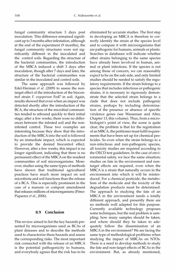
108 C. Alabouvette et al.
fungal community structure 3 days post inoculation. This difference remained signifi-cant up to 3 months after infestation, although at the end of the experiment (9 months), the fungal community structures were not sig-nificantly different in the inoculated and the control soils. Regarding the structure of the bacterial communities, the introduction of the MBCA induced a shift 3 days after inoculation, though after 7 days and later, the structure of the bacterial communities was similar in the inoculated and control soils.
The same approach was followed by Edel-Herman et al. (2009) to assess the non-target effect of the introduction of the biocon-trol strain F. oxysporum Fo47 in soil. Again, results showed that even when an impact was detected shortly after the introduction of the BCA, the structure of the microbial communi-ties tended to rebound quickly to their initial stage; after a few weeks, there were no differ-ences between the infested soil and the non-infested control. These two examples are interesting because they show that the intro-duction of the MBCA into the soil is followed by an immediate impact, which is expected to provide the desired biocontrol effect. However, after a few weeks, this impact is no longer significant, indicating that there is no permanent effect of the MBCA on the resident communities of soil microorganisms. More-over, studies using the same types of methods have shown that traditional agricultural practices have much more impact on soil microbiota and soil functions than the release of a BCA. This is especially prominent in the case of a manure or compost amendment that releases millions of microorganisms (Pérez-Piquerez et al., 2006).
8.9 Conclusion
This review aimed to list the key hazards pre-sented by microorganisms used as BCAs of plant diseases and to describe the methods used to characterize these hazards and assess the corresponding risks. The most important risk connected with the release of an MBCA is the potential pathogenicity to humans, and everybody agrees that the risk has to be
eliminated by accurate studies. The first step in developing an MBCA is therefore to cor-rectly identify the strain at the species level and to compare it with microorganisms that are pathogenic for humans, animals or plants. Searches in databases will indicate whether other strains belonging to the same species have already been involved in human, ani-mal or plant infections. If the species is not among those of concern, we can reasonably expect to be on the safe side, and only limited studies should be needed to satisfy the regu-latory requirements. If the strain belongs to a species that includes infectious or pathogenic strains, it is necessary to rigorously demon-strate that the selected strain belongs to a clade that does not include pathogenic strains, perhaps by including determina-tion of the presence or absence of specific virulence genes (see Wassenaar and Alter, Chapter 13, this volume). Thus, from a micro-biologist’s point of view, the scene is quite clear; the problem is that for the registration of an MBCA, the petitioner must fulfil require-ments that have been set up for chemical pes-ticides. So even when the strain belongs to a non-infectious and non-pathogenic species, all toxicity studies are required according to the OECD test guidelines. In the case of envi-ronmental safety, we face the same situation; studies on fate in the environment and non-target effects are required, even when the MBCA is a strain that naturally occurs in the environment into which it will be reintro-duced. For a chemical pesticide, the metabo-lism of the molecule and the toxicity of the degradation products must be determined. The approach to studying the fate of an MBCA in the environment needs a totally different approach, and presently there are no methods well adapted for this purpose. Currently available technology proposes some techniques, but the real problem is sam-pling: how many samples should be taken, and where should they be taken to ade-quately follow the dissemination of an MBCA in the environment? We are facing the same type of methodological problems when studying the impact of MBCAs on NTOs. There is a need to develop methods to study the fate and non-target effects of BCAs in the environment. But, as already mentioned,
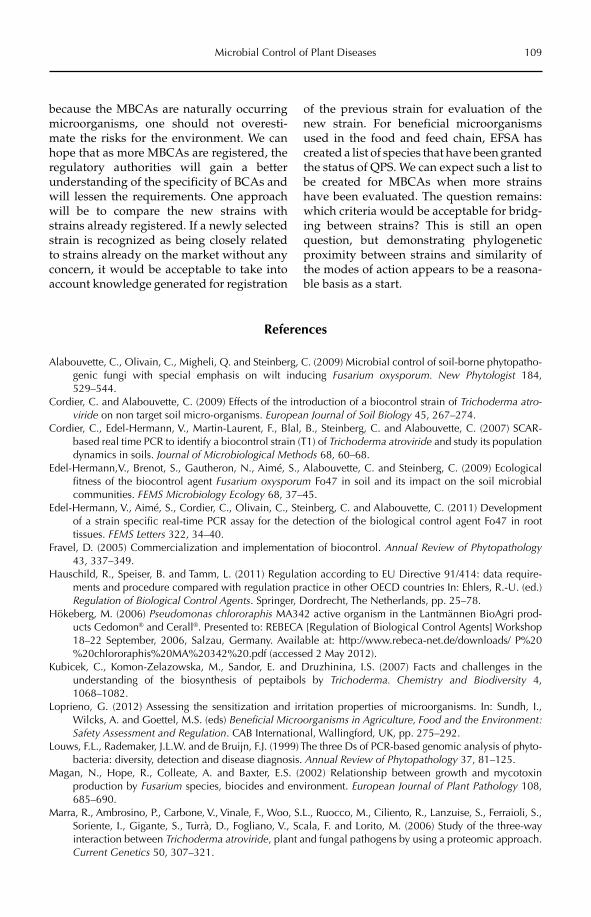
Microbial Control of Plant Diseases 109
because the MBCAs are naturally occurring microorganisms, one should not overesti-mate the risks for the environment. We can hope that as more MBCAs are registered, the regulatory authorities will gain a better understanding of the specificity of BCAs and will lessen the requirements. One approach will be to compare the new strains with strains already registered. If a newly selected strain is recognized as being closely related to strains already on the market without any concern, it would be acceptable to take into account knowledge generated for registration
of the previous strain for evaluation of the new strain. For beneficial microorganisms used in the food and feed chain, EFSA has created a list of species that have been granted the status of QPS. We can expect such a list to be created for MBCAs when more strains have been evaluated. The question remains: which criteria would be acceptable for bridg-ing between strains? This is still an open question, but demonstrating phylogenetic proximity between strains and similarity of the modes of action appears to be a reasona-ble basis as a start.
References
Alabouvette, C., Olivain, C., Migheli, Q. and Steinberg, C. (2009) Microbial control of soil-borne phytopatho-genic fungi with special emphasis on wilt inducing Fusarium oxysporum. New Phytologist 184, 529–544.
Cordier, C. and Alabouvette, C. (2009) Effects of the introduction of a biocontrol strain of Trichoderma atro-viride on non target soil micro-organisms. European Journal of Soil Biology 45, 267–274.
Cordier, C., Edel-Hermann, V., Martin-Laurent, F., Blal, B., Steinberg, C. and Alabouvette, C. (2007) SCAR-based real time PCR to identify a biocontrol strain (T1) of Trichoderma atroviride and study its population dynamics in soils. Journal of Microbiological Methods 68, 60–68.
Edel-Hermann,V., Brenot, S., Gautheron, N., Aimé, S., Alabouvette, C. and Steinberg, C. (2009) Ecological fitness of the biocontrol agent Fusarium oxysporum Fo47 in soil and its impact on the soil microbial communities. FEMS Microbiology Ecology 68, 37–45.
Edel-Hermann, V., Aimé, S., Cordier, C., Olivain, C., Steinberg, C. and Alabouvette, C. (2011) Development of a strain specific real-time PCR assay for the detection of the biological control agent Fo47 in root tissues. FEMS Letters 322, 34–40.
Fravel, D. (2005) Commercialization and implementation of biocontrol. Annual Review of Phytopathology43, 337–349.
Hauschild, R., Speiser, B. and Tamm, L. (2011) Regulation according to EU Directive 91/414: data require-ments and procedure compared with regulation practice in other OECD countries In: Ehlers, R.-U. (ed.) Regulation of Biological Control Agents. Springer, Dordrecht, The Netherlands, pp. 25–78.
Hökeberg, M. (2006) Pseudomonas chlororaphis MA342 active organism in the Lantmännen BioAgri prod-ucts Cedomon® and Cerall®. Presented to: REBECA [Regulation of Biological Control Agents] Workshop 18–22 September, 2006, Salzau, Germany. Available at: http://www.rebeca-net.de/downloads/ P%20%20chlororaphis%20MA%20342%20.pdf (accessed 2 May 2012).
Kubicek, C., Komon-Zelazowska, M., Sandor, E. and Druzhinina, I.S. (2007) Facts and challenges in the understanding of the biosynthesis of peptaibols by Trichoderma. Chemistry and Biodiversity 4, 1068–1082.
Loprieno, G. (2012) Assessing the sensitization and irritation properties of microorganisms. In: Sundh, I., Wilcks, A. and Goettel, M.S. (eds) Beneficial Microorganisms in Agriculture, Food and the Environment: Safety Assessment and Regulation. CAB International, Wallingford, UK, pp. 275–292.
Louws, F.L., Rademaker, J.L.W. and de Bruijn, F.J. (1999) The three Ds of PCR-based genomic analysis of phyto-bacteria: diversity, detection and disease diagnosis. Annual Review of Phytopathology 37, 81–125.
Magan, N., Hope, R., Colleate, A. and Baxter, E.S. (2002) Relationship between growth and mycotoxin production by Fusarium species, biocides and environment. European Journal of Plant Pathology 108, 685–690.
Marra, R., Ambrosino, P., Carbone, V., Vinale, F., Woo, S.L., Ruocco, M., Ciliento, R., Lanzuise, S., Ferraioli, S.,Soriente, I., Gigante, S., Turrà, D., Fogliano, V., Scala, F. and Lorito, M. (2006) Study of the three-way interaction between Trichoderma atroviride, plant and fungal pathogens by using a proteomic approach. Current Genetics 50, 307–321.
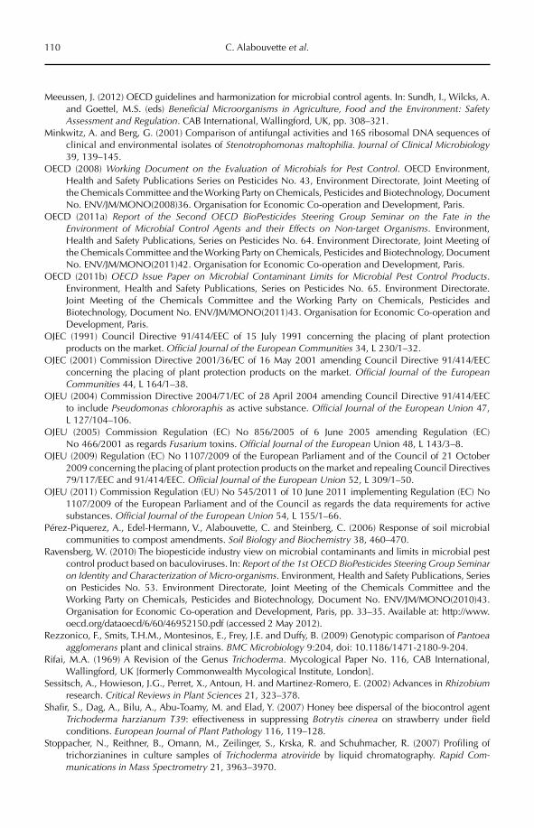
110 C. Alabouvette et al.
Meeussen, J. (2012) OECD guidelines and harmonization for microbial control agents. In: Sundh, I., Wilcks, A. and Goettel, M.S. (eds) Beneficial Microorganisms in Agriculture, Food and the Environment: Safety Assessment and Regulation. CAB International, Wallingford, UK, pp. 308–321.
Minkwitz, A. and Berg, G. (2001) Comparison of antifungal activities and 16S ribosomal DNA sequences of clinical and environmental isolates of Stenotrophomonas maltophilia. Journal of Clinical Microbiology39, 139–145.
OECD (2008) Working Document on the Evaluation of Microbials for Pest Control. OECD Environment, Health and Safety Publications Series on Pesticides No. 43, Environment Directorate, Joint Meeting of the Chemicals Committee and the Working Party on Chemicals, Pesticides and Biotechnology, Document No. ENV/JM/MONO(2008)36. Organisation for Economic Co-operation and Development, Paris.
OECD (2011a) Report of the Second OECD BioPesticides Steering Group Seminar on the Fate in the Environment of Microbial Control Agents and their Effects on Non-target Organisms. Environment, Health and Safety Publications, Series on Pesticides No. 64. Environment Directorate, Joint Meeting of the Chemicals Committee and the Working Party on Chemicals, Pesticides and Biotechnology, Document No. ENV/JM/MONO(2011)42. Organisation for Economic Co-operation and Development, Paris.
OECD (2011b) OECD Issue Paper on Microbial Contaminant Limits for Microbial Pest Control Products.Environment, Health and Safety Publications, Series on Pesticides No. 65. Environment Directorate. Joint Meeting of the Chemicals Committee and the Working Party on Chemicals, Pesticides and Biotechnology, Document No. ENV/JM/MONO(2011)43. Organisation for Economic Co-operation and Development, Paris.
OJEC (1991) Council Directive 91/414/EEC of 15 July 1991 concerning the placing of plant protection products on the market. Official Journal of the European Communities 34, L 230/1–32.
OJEC (2001) Commission Directive 2001/36/EC of 16 May 2001 amending Council Directive 91/414/EEC concerning the placing of plant protection products on the market. Official Journal of the European Communities 44, L 164/1–38.
OJEU (2004) Commission Directive 2004/71/EC of 28 April 2004 amending Council Directive 91/414/EEC to include Pseudomonas chlororaphis as active substance. Official Journal of the European Union 47, L 127/104–106.
OJEU (2005) Commission Regulation (EC) No 856/2005 of 6 June 2005 amending Regulation (EC) No 466/2001 as regards Fusarium toxins. Official Journal of the European Union 48, L 143/3–8.
OJEU (2009) Regulation (EC) No 1107/2009 of the European Parliament and of the Council of 21 October 2009 concerning the placing of plant protection products on the market and repealing Council Directives 79/117/EEC and 91/414/EEC. Official Journal of the European Union 52, L 309/1–50.
OJEU (2011) Commission Regulation (EU) No 545/2011 of 10 June 2011 implementing Regulation (EC) No 1107/2009 of the European Parliament and of the Council as regards the data requirements for active substances. Official Journal of the European Union 54, L 155/1–66.
Pérez-Piquerez, A., Edel-Hermann, V., Alabouvette, C. and Steinberg, C. (2006) Response of soil microbial communities to compost amendments. Soil Biology and Biochemistry 38, 460–470.
Ravensberg, W. (2010) The biopesticide industry view on microbial contaminants and limits in microbial pest control product based on baculoviruses. In: Report of the 1st OECD BioPesticides Steering Group Seminar on Identity and Characterization of Micro-organisms. Environment, Health and Safety Publications, Series on Pesticides No. 53. Environment Directorate, Joint Meeting of the Chemicals Committee and the Working Party on Chemicals, Pesticides and Biotechnology, Document No. ENV/JM/MONO(2010)43. Organisation for Economic Co-operation and Development, Paris, pp. 33–35. Available at: http://www.oecd.org/dataoecd/6/60/46952150.pdf (accessed 2 May 2012).
Rezzonico, F., Smits, T.H.M., Montesinos, E., Frey, J.E. and Duffy, B. (2009) Genotypic comparison of Pantoea agglomerans plant and clinical strains. BMC Microbiology 9:204, doi: 10.1186/1471-2180-9-204.
Rifai, M.A. (1969) A Revision of the Genus Trichoderma. Mycological Paper No. 116, CAB International, Wallingford, UK [formerly Commonwealth Mycological Institute, London].
Sessitsch, A., Howieson, J.G., Perret, X., Antoun, H. and Martinez-Romero, E. (2002) Advances in Rhizobiumresearch. Critical Reviews in Plant Sciences 21, 323–378.
Shafir, S., Dag, A., Bilu, A., Abu-Toamy, M. and Elad, Y. (2007) Honey bee dispersal of the biocontrol agent Trichoderma harzianum T39: effectiveness in suppressing Botrytis cinerea on strawberry under field conditions. European Journal of Plant Pathology 116, 119–128.
Stoppacher, N., Reithner, B., Omann, M., Zeilinger, S., Krska, R. and Schuhmacher, R. (2007) Profiling of trichorzianines in culture samples of Trichoderma atroviride by liquid chromatography. Rapid Com-munications in Mass Spectrometry 21, 3963–3970.
Microbial Control of Plant Diseases 111
US EPA (1996) Microbial Pesticide Test Guidelines: OPPTS 885.4340. Nontarget Insect Testing, Tier I. [OCSPP HarmonizedTest Guidelines, Series 885 – Microbial Pesticide Test Guidelines]. Document No. EPA712–C–96–336. US Environmental Protection Agency, Washington, DC. Available at: http://www.regulations.gov/contentStreamer?objectId=09000064809be930&disposition=attachment&contentType=pdf (accessed 2 May 2012).
Typas, M.A. and Kouvelis, V.N. (2012) Assessing genotoxic effects of microbial products. In: Sundh, I., Wilcks, A. and Goettel, M.S. (eds) Beneficial Microorganisms in Agriculture, Food and the Environ-ment: Safety Assessment and Regulation. CAB International, Wallingford, UK, pp. 256–274.
Vining, L.C. (1990) Functions of secondary metabolites. Annual Review of Microbiology 44, 395–427.Wassenaar, T.M. and Alter, T. (2012) Virulence genes in risk assessment of beneficial microorganisms: what do
genome sequences tell us? In: Sundh, I., Wilcks, A. and Goettel, M.S. (eds) Beneficial Microorganisms in Agriculture, Food and the Environment: Safety Assessment and Regulation. CAB International, Wallingford, UK, pp. 180–196.
Wilcks, A., Goettel, M.S. and Sundh, I. (2012) Model systems for testing microbial pathogenicity, virulence and toxicity – introduction and overview. In: Sundh, I., Wilcks, A. and Goettel, M.S. (eds) BeneficialMicroorganisms in Agriculture, Food and the Environment: Safety Assessment and Regulation. CAB International, Wallingford, UK, pp. 217–226.
Winding, A., Binnerup, S.J. and Pritchard H. (2004) Non-target effects of bacterial biological control agents suppressing root pathogenic fungi. FEMS Microbiology Ecology 47, 129–141.
Woo, S.L. and Lorito, M. (2007) Exploiting the interactions between fungal antagonists, pathogens and the plant for biocontrol. In: Vurro, M. and Gressel, J. (eds) Novel Biotechnologies for Biocontrol Agent Enhancement and Management. Springer, Dordrecht, The Netherlands.
©CAB International 2012. Beneficial Microorganisms in Agriculture, Food and the Environment: 112 Safety Assessment and Regulation (eds I. Sundh et al.)
9.1 Introduction 1129.2 Classical Biological Control (CBC) 113
9.2.1 Why do we need CBC? 113 9.2.2 How does it work? 113 9.2.3 How safe is it? 114 9.2.4 How is risk reduced? 116 9.2.5 How is it promulgated and regulated? 117 9.2.6 CBC in action: case studies of problematic safety and regulatory issues 121 9.2.7 Bureaucrats, botanists, beekeepers and other bottlenecks 124
9.3 Inundative (Bioherbicide) Biological Control (IBC) 126 9.3.1 Why do we need IBC? 126 9.3.2 How does it work? 126 9.3.3 Is it safe? 127 9.3.4 How is it regulated? 127 9.3.5 Case studies: regional variations 127
9.4 Overview: Where Are We? 129
9 Safety and Regulation of Microbial Control of Weeds
Harry C. Evans and Marion K. SeierCAB International, Egham, UK
9.1 Introduction
Microbial control of weeds is based almost exclusively on the use of fungi as biological control agents. Plant viruses are invariably difficult to manipulate, as most rely on other (unmanageable) biotic factors – mainly arthro-pod vectors – for successful transmission, while plant pathogenic bacteria are similarly prob-lematic in terms of their delivery and infection mechanisms. Hence, there are few examples of control involving bacterial and viral agents.
There are two distinct approaches to the microbial control of weeds – classical and inundative – each with very different safety
and regulatory issues to consider. Classical biological control (CBC) is directed at intro-duced alien plant species (neophytes) and involves the selection and movement of highly specialized or coevolved natural enemies from the centre of origin or native range of the target species into the exotic range where it has become weedy and problematic. The agent – invariably, a biotrophic fungal pathogen – is released, following approval based on a pest risk assessment, into the exotic ecosystem and it disperses naturally from predetermined inoculum sites, to establish and build up over time to epidemic (epiphytotic) proportions. The pathogen, once introduced in this way, is
Safety and Regulation of Microbial Control of Weeds 113
literally, therefore, a free agent and becomes a permanent part of the ecosystem. In principle, it cannot be eradicated or even controlled, if the need should ever arise. Thus, this strategy is entirely in the public domain – because all parts of society, as well as ecosystems, can be affected – particularly because public funds are most often involved. In sharp contrast, inunda-tive biological control (IBC) typically exploits indigenous plant pathogens targeted at locally important native or naturalized weeds. These pathogens are readily cultured (necrotrophic) fungi, mass produced and formulated as bio-herbicides under a patent, and funded from the private rather than the public sector. In this chapter, we compare the safety and regulatory issues between these two contrasting approaches to the microbial control of weeds.
9.2 Classical Biological Control (CBC)
9.2.1 Why do we need CBC?
Humankind has been moving plants and ani-mals around the world since the dawn of agri-culture, some 10,000 years ago. However, it is only in recent times – as trade and globaliza-tion have exploded – that the wholesale exchange and movement of exotic species has taken off and has now moved into overdrive: we are now entering the Homogocene era, loosely defined as the human-mediated or anthropogenic replacement of native biotas by non-native or alien species (Rosenzweig, 2001; Olden et al., 2004). Some of these species – introduced either deliberately or accidentally – have become problematic pests and a threat to both agriculture and biodiversity (Mooney and Hobbs, 2000). In fact, after habitat destruction by humans, invasive alien species (IAS), and exotic plant species, in particular, have been considered to pose the greatest threat to the world’s natural ecosystems (Mack et al., 2000; Mack and Lonsdale, 2002; Carey, 2002), partic-ularly in small-island systems (Trueman et al., 2010). Because these weedy plants can cover vast tracts of land, control by conventional means is logistically challenging, often prohibi-tively expensive and unsustainable. Moreover, in many cases, especially with aquatic and
riparian weeds, environmental constraints usu-ally preclude the use of chemical herbicides.
Frequently, the only choice considered for invasive alien weeds, once eradication has failed, has been that of containment, to try to reduce their impact on agricultural production, on the indigenous flora and fauna, and on society in general. Obviously, most countries, and certainly those in the developing world, do not have the foresight or political will, nor the infrastructure and resources to even attempt containment, especially of environmental weeds with no obvious economic impact; they prefer to ignore the problem of IAS or hope that it will go away. If this philosophy were followed to its logical conclusion, we would be well on the way to the predicted homogenization or McDonaldization of the world’s flora (Lövei, 1997; see Ritzer, 1998 for the general concepts of the latter terminology).
Nevertheless, there is the alternative management strategy of CBC, which presents a possibility to avert, or at least to temper, domination by invasive alien plant spe-cies. Potentially, CBC offers a cost-effective, environmentally benign and sustainable solution to the problem of invasive alien weeds; either functioning in isolation – as a one-off, ‘silver-bullet’ remedy – or integrated into a multi-component management plan. Some programmes have been spectacular successes and others have not, or have even been considered to have had negative impacts. Fortunately, the latter examples have been in the minority and all were entirely predictable (Evans, 2000). None of the purportedly failed CBC programmes have been analysed scien-tifically though, and until recently, the obvi-ous question was never asked: what would the situation have been without any biologi-cal control? This was addressed by Hoffmann and Moran (2008), and their study revealed the ‘subtle but very real benefits that accrue from otherwise seemingly ineffective agents’.
9.2.2 How does it work?
It has long been recognized, but only rela-tively recently crystallized into a hypothesis – the ‘enemy release hypothesis’ (Keane and
114 H.C. Evans and M.K. Seier
Crawley, 2002) – that plants, once released from their natural enemies, show a dramatic increase in vigour and fecundity. In plants with certain traits, such as the ability to colo-nize disturbed habitats, this increased fitness gives them a competitive edge over the native flora, allowing them to become weedy and invasive. The concept of CBC is simple: to address this imbalance by reducing the fit-ness through the release of natural enemies from the native range or centre of origin of the alien weed target. Evidence for this hypothesis was soon forthcoming from a study of invasive neophytes in the USA which showed that they had significantly fewer bio-trophic (coevolved) plant pathogens than in their native ranges (Mitchell and Power, 2003).
Obviously, ecosystem functioning is never so simplistic and, clearly, interlinking multi-trophic factors are involved in plant invasiveness, such as resource availability (Blumenthal, 2006). Another, recently recog-nized component can also be factored into the invasiveness equation – microbial endo-phytes. In the ‘endophyte-enemy release hypothesis’ (Evans, 2008), it is postulated that alien plants become invasive not only because they leave their natural enemies behind, but also that they arrive without, or with only a part of, their coevolved endo-phytes. Some of these endophytes are known to protect the plant host from both abiotic and biotic pressures, including natural enemies, but resources need to be allocated for this protection. So these neophytes have a double gain in fitness: no natural-enemy pressure; no ‘bodyguards’ to pay off. However, if a co -evolved natural enemy were to be introducedinto the system, the plant would be highly vulnerable to attack – not only because it lacked protective endophytes but also because it occurred in vigorous monocultures – the perfect setting for a natural-enemy popula-tion explosion. This could explain the phe-nomenon of the ‘silver bullet’, in which the introduction of a single natural enemy into the system can bring about the sudden and terminal decline of weed invasions. There are many such examples in the entomological lit-erature of spectacular control of seemingly intractable invasive alien weeds following
the release of insect natural enemies (Dodd, 1940; McFadyen, 1998; Syrett et al., 2000; van Wilgen et al., 2004; Page and Lacey, 2006), as well as of the control of exotic arthropod pests using parasitoids (Herren and Neuenschwander, 1991; Bokonou-Ganta et al., 2002), because the historical records for this type of control go back much further than the use of microbial agents. In recent times, though, similar successes have also been achieved with coevolved fungal pathogens of invasive weeds (Morin et al., 1996; Evans, 2002a; Barton et al., 2007).
Finally, in answering the question – ‘How does it work?’ – we only need to look at the historical records of plant-disease epiphy-totics when coevolved pathogens eventually catch up with their crop hosts growing in exotic monocultures, with catastrophic socio-economic results: potato blight, coffee rust, wheat rust, rubber blight, cacao diseases (Large, 1940; Quimby, 1982; Evans, 2002b; Agrios, 2005).
9.2.3 How safe is it?
In other words: how risky is biological con-trol? This question was posed by Simberloff and Stiling (1996a) specifically in relation to the general principles and practice of CBC, which they regarded as inherently risky, as well as the agents of CBC, which they assumed to be ‘guilty until proven innocent’. Thus, we live in an ever-increasingly safety-conscious or risk-averse world, and there are sound arguments both for and against this escalat-ing trend. Yet in the latter case, we could reach a situation where nobody will take a decision if there are any perceived risks, however small, that could rebound politically or finan-cially on the decision makers or on their organizations. Unfortunately, with CBC, there will always be risks because it is a natural sys-tem and nature is never 100% predictable (Evans, 1998). If the precautionary principle were to be followed to the letter – as set out in the 1992 Rio Declaration on Environment and Development (Sheppard et al., 2003) – CBC would be deemed to be unacceptably risky. Nevertheless, a comparative risk analysis shows that the probabilities of CBC agents
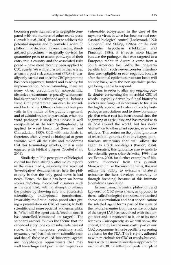
Safety and Regulation of Microbial Control of Weeds 115
becoming pests themselves is negligible com-pared with the number of other exotic pests (Lonsdale et al., 2001). In order to address this potential impasse and to provide a scientific platform for decision makers, existing stand-ardized procedures – originally devised for quarantine pests to assess pathways of their entry into a country and the associated risks posed – have more recently been applied to CBC agents. We will return to this theme later, as such a pest risk assessment (PRA) is usu-ally only carried out once the CBC programme has been approved, funded and is ready for implementation. Notwithstanding, there are many other, predominantly non-scientific, obstacles to surmount – especially with micro-bial as opposed to arthropod agents – before a weed CBC programme can even be consid-ered for funding. Often, a climate of fear pre-vails in the minds of the public in general, and of administrators in particular, when the word pathogen is used; this unease is well encapsulated in the term ‘pathophobia’, as applied to weed biocontrol (Freeman and Charudattan, 1985). CBC with microbials is, therefore, often viewed as biological or germ warfare, with all the risks and melodrama that this terminology invokes, or it is even equated with biblical plagues (Goettel et al.,2001).
Similarly, public perception of biological control has been strongly affected by reports in the mass media, especially the so-called ‘investigative’ documentaries; here the phil-osophy is that the only good news is bad news. Hence, the focus has been on horror stories depicting ‘biocontrol’ disasters, such as the cane toad, with no attempt to balance the picture by showing safe and successful, scientifically underpinned introductions. Invariably, the first question posed after giv-ing a presentation on CBC of weeds, to both scientific and non-specialist audiences alike, is: ‘What will the agent attack/feed on once it has controlled/eliminated its target?’. The standard answer follows the theme that the cane-toad story (one could substitute here rat snake, Indian mongoose, predatory snail, myxoma virus) has little or no scientific basis and that all these so-called ‘biocontrol agents’ are polyphagous opportunists that may well have huge and permanent impacts on
vulnerable ecosystems. In the case of the myxoma virus, in what has been termed neo-classical biological control (Lockwood, 1993; Simberloff and Stiling, 1996b), or the new encounter hypothesis (Hokkanen and Pimentel, 1984), it is even more bizarre because the pathogen that was targeted at a European rabbit in Australia came from a South American fox! Sadly, the long-term benefits from such new-encounter introduc-tions are negligible, or even negative, because after the initial epidemics, resistant hosts will bounce back, with the non-specialist patho-gen being unable to respond.
Thus, in order to allay any ongoing pub-lic doubts concerning the microbial CBC of weeds – typically driven by fungal biotrophs such as rust fungi – it is necessary to focus on the highly specialized nature of such plant–pathogen associations and to show, for exam-ple, that wheat rust has been around since the beginning of agriculture and has moved with its host around the world, but has never ‘shifted’ on to other plant species, even close relatives. This centres on the public ignorance of microbial genetics that envisages spon-taneous mutations that will allow the agent to attack non-targets (Barton, 2004). Unfortunately, this ignorance also extends to the scientific press (New Scientist, 1999; also see Evans, 2000, for further examples of bio-control ‘bloomers’ from this journal). Moreover, unlike the myxoma virus, the rust retains the ability to overcome whatever resistance the host develops (naturally or through breeding) because of this intimate (coevolved) association.
In conclusion, the central philosophy and keyword of CBC sensu stricto, as opposed to the so-called biological control examples cited above, is coevolution and host specialization: the selected agent forms part of the suite of the natural enemies from the centre of origin of the target IAS, has coevolved with that tar-get host and is restricted to it, or to its near relatives. Consequently, as we will show, the critical, and by far the most costly part of any CBC programme, is host-specificity screening as a basis for the PRA. This is rigidly adhered to with microbials for CBC of weeds, but con-trasts with the more laissez-faire approach for microbial CBC of arthropod pests and plant
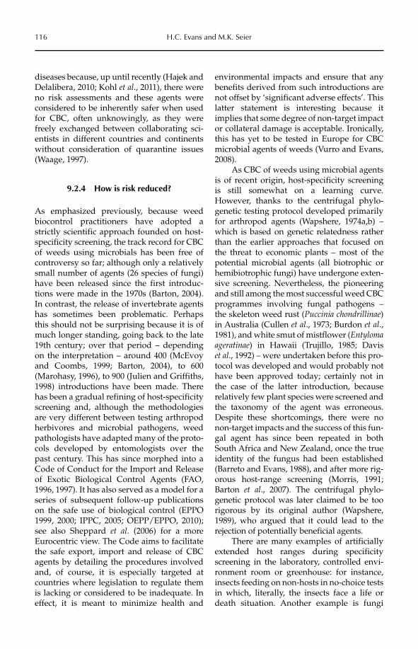
116 H.C. Evans and M.K. Seier
diseases because, up until recently (Hajek and Delalibera, 2010; Kohl et al., 2011), there were no risk assessments and these agents were considered to be inherently safer when used for CBC, often unknowingly, as they were freely exchanged between collaborating sci-entists in different countries and continents without consideration of quarantine issues (Waage, 1997).
9.2.4 How is risk reduced?
As emphasized previously, because weed biocontrol practitioners have adopted a strictly scientific approach founded on host-specificity screening, the track record for CBC of weeds using microbials has been free of controversy so far; although only a relatively small number of agents (26 species of fungi) have been released since the first introduc-tions were made in the 1970s (Barton, 2004). In contrast, the release of invertebrate agents has sometimes been problematic. Perhaps this should not be surprising because it is of much longer standing, going back to the late 19th century; over that period – depending on the interpretation – around 400 (McEvoy and Coombs, 1999; Barton, 2004), to 600 (Marohasy, 1996), to 900 (Julien and Griffiths, 1998) introductions have been made. There has been a gradual refining of host-specificity screening and, although the methodologies are very different between testing arthropod herbivores and microbial pathogens, weed pathologists have adapted many of the proto-cols developed by entomologists over the past century. This has since morphed into a Code of Conduct for the Import and Release of Exotic Biological Control Agents (FAO, 1996, 1997). It has also served as a model for a series of subsequent follow-up publications on the safe use of biological control (EPPO 1999, 2000; IPPC, 2005; OEPP/EPPO, 2010); see also Sheppard et al. (2006) for a more Eurocentric view. The Code aims to facilitate the safe export, import and release of CBC agents by detailing the procedures involved and, of course, it is especially targeted at countries where legislation to regulate them is lacking or considered to be inadequate. In effect, it is meant to minimize health and
environmental impacts and ensure that any benefits derived from such introductions are not offset by ‘significant adverse effects’. This latter statement is interesting because it implies that some degree of non-target impact or collateral damage is acceptable. Ironically, this has yet to be tested in Europe for CBC microbial agents of weeds (Vurro and Evans, 2008).
As CBC of weeds using microbial agents is of recent origin, host-specificity screening is still somewhat on a learning curve. However, thanks to the centrifugal phylo-genetic testing protocol developed primarily for arthropod agents (Wapshere, 1974a,b) – which is based on genetic relatedness rather than the earlier approaches that focused on the threat to economic plants – most of the potential microbial agents (all biotrophic or hemibiotrophic fungi) have undergone exten-sive screening. Nevertheless, the pioneering and still among the most successful weed CBC programmes involving fungal pathogens – the skeleton weed rust (Puccinia chondrillinae)in Australia (Cullen et al., 1973; Burdon et al.,1981), and white smut of mistflower (Entylomaageratinae) in Hawaii (Trujillo, 1985; Davis et al., 1992) – were undertaken before this pro-tocol was developed and would probably not have been approved today; certainly not in the case of the latter introduction, because relatively few plant species were screened and the taxonomy of the agent was erroneous. Despite these shortcomings, there were no non-target impacts and the success of this fun-gal agent has since been repeated in both South Africa and New Zealand, once the true identity of the fungus had been established (Barreto and Evans, 1988), and after more rig-orous host-range screening (Morris, 1991; Barton et al., 2007). The centrifugal phylo-genetic protocol was later claimed to be too rigorous by its original author (Wapshere, 1989), who argued that it could lead to the rejection of potentially beneficial agents.
There are many examples of artificially extended host ranges during specificity screening in the laboratory, controlled envi-ronment room or greenhouse: for instance, insects feeding on non-hosts in no-choice tests in which, literally, the insects face a life or death situation. Another example is fungi
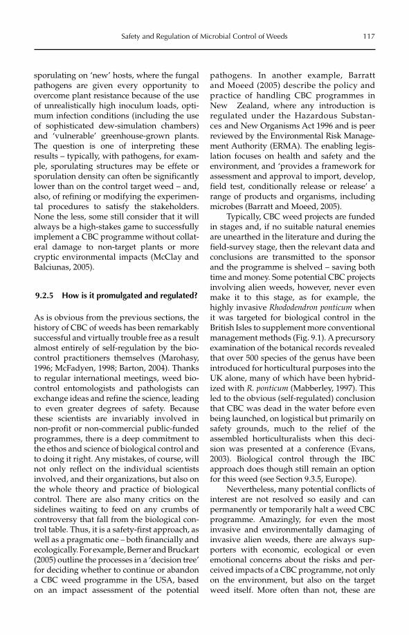
Safety and Regulation of Microbial Control of Weeds 117
sporulating on ‘new’ hosts, where the fungal pathogens are given every opportunity to overcome plant resistance because of the use of unrealistically high inoculum loads, opti-mum infection conditions (including the use of sophisticated dew-simulation chambers) and ‘vulnerable’ greenhouse-grown plants. The question is one of interpreting these results – typically, with pathogens, for exam-ple, sporulating structures may be effete or sporulation density can often be significantly lower than on the control target weed – and, also, of refining or modifying the experimen-tal procedures to satisfy the stakeholders. None the less, some still consider that it will always be a high-stakes game to successfully implement a CBC programme without collat-eral damage to non-target plants or more cryptic environmental impacts (McClay and Balciunas, 2005).
9.2.5 How is it promulgated and regulated?
As is obvious from the previous sections, the history of CBC of weeds has been remarkably successful and virtually trouble free as a result almost entirely of self-regulation by the bio-control practitioners themselves (Marohasy, 1996; McFadyen, 1998; Barton, 2004). Thanks to regular international meetings, weed bio-control entomologists and pathologists can exchange ideas and refine the science, leading to even greater degrees of safety. Because these scientists are invariably involved in non-profit or non-commercial public-funded programmes, there is a deep commitment to the ethos and science of biological control and to doing it right. Any mistakes, of course, will not only reflect on the individual scientists involved, and their organizations, but also on the whole theory and practice of biological control. There are also many critics on the sidelines waiting to feed on any crumbs of controversy that fall from the biological con-trol table. Thus, it is a safety-first approach, as well as a pragmatic one – both financially and ecologically. For example, Berner and Bruckart (2005) outline the processes in a ‘decision tree’ for deciding whether to continue or abandon a CBC weed programme in the USA, based on an impact assessment of the potential
pathogens. In another example, Barratt and Moeed (2005) describe the policy and practice of handling CBC programmes in New Zealand, where any introduction is regulated under the Hazardous Substan-ces and New Organisms Act 1996 and is peer reviewed by the Environmental Risk Manage-ment Authority (ERMA). The enabling legis-lation focuses on health and safety and the environment, and ‘provides a framework for assessment and approval to import, develop, field test, conditionally release or release’ a range of products and organisms, including microbes (Barratt and Moeed, 2005).
Typically, CBC weed projects are funded in stages and, if no suitable natural enemies are unearthed in the literature and during the field-survey stage, then the relevant data and conclusions are transmitted to the sponsor and the programme is shelved – saving both time and money. Some potential CBC projects involving alien weeds, however, never even make it to this stage, as for example, the highly invasive Rhododendron ponticum when it was targeted for biological control in the British Isles to supplement more conventional management methods (Fig. 9.1). A precursory examination of the botanical records revealed that over 500 species of the genus have been introduced for horticultural purposes into the UK alone, many of which have been hybrid-ized with R. ponticum (Mabberley, 1997). This led to the obvious (self-regulated) conclusion that CBC was dead in the water before even being launched, on logistical but primarily on safety grounds, much to the relief of the assembled horticulturalists when this deci-sion was presented at a conference (Evans, 2003). Biological control through the IBC approach does though still remain an option for this weed (see Section 9.3.5, Europe).
Nevertheless, many potential conflicts of interest are not resolved so easily and can permanently or temporarily halt a weed CBC programme. Amazingly, for even the most invasive and environmentally damaging of invasive alien weeds, there are always sup-porters with economic, ecological or even emotional concerns about the risks and per-ceived impacts of a CBC programme, not only on the environment, but also on the target weed itself. More often than not, these are
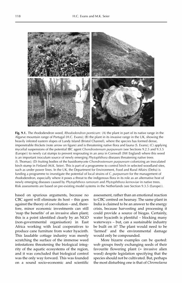
118 H.C. Evans and M.K. Seier
based on spurious arguments, because no CBC agent will eliminate its host – this goes against the theory of coevolution – and, there-fore, minor economic investments can still ‘reap the benefits’ of an invasive alien plant; this is a point identified clearly by an NGO (non-governmental organisation) in East Africa working with local cooperatives to produce cane furniture from water hyacinth. This laudable cottage industry was merely scratching the surface of the immense weed infestations threatening the biological integ-rity of the aquatic ecosystems of the region, and it was concluded that biological control was the only way forward. This was founded on a sound socio-economic and scientific
assessment, rather than an emotional reaction to CBC centred on hearsay. The same plant in India is claimed to be an answer to the energy crisis, because harvesting and processing it could provide a source of biogas. Certainly, water hyacinth is plentiful – blocking many waterways – but, can a sustainable industry be built on it? The plant would need to be ‘farmed’ and the environmental damage would only be compounded.
More bizarre examples can be quoted: web groups freely exchanging seeds of their favourite flowering plant (= invasive alien weed) despite legislation specifying that the species should not be cultivated. But, perhaps the most disturbing one is that of Chromolaena
Fig. 9.1. The rhododendron weed, Rhododendron ponticum: (A) the plant in part of its native range in the Algarve mountain range of Portugal (H.C. Evans); (B) the plant in its invasive range in the UK, showing the heavily infested eastern slopes of Lundy Island (Bristol Channel), where the species has formed dense, impenetrable thickets (note arrow on figure) and is threatening native flora and fauna (S. Evans); (C) applying mycelial suspensions of the potential IBC agent Chondrostereum purpureum (see Sections 9.2.5 and 9.3.5 (Europe) ) to newly cut stumps to prevent resprouting in an area in Cornwall (SW England) where this weed is an important inoculum source of newly emerging Phytophthora diseases threatening native trees (S. Thomas); (D) fruiting bodies of the basidiomycete Chondrostereum purpureum colonizing an inoculated birch stump in Finland (M.K. Seier). This is part of a programme to control birch in selected woodland sites, such as under power lines. In the UK, the Department for Environment, Food and Rural Affairs (Defra) is funding a programme to investigate the potential of local strains of C. purpureum for the management of rhododendron, especially where it poses a threat to the indigenous flora in its role as an alternative host of newly emerging diseases caused by Phytophthora ramorum and Phytophthora kernoviae in native trees. Risk assessments are based on pre-existing model systems in the Netherlands (see Section 9.3.5 (Europe) ).
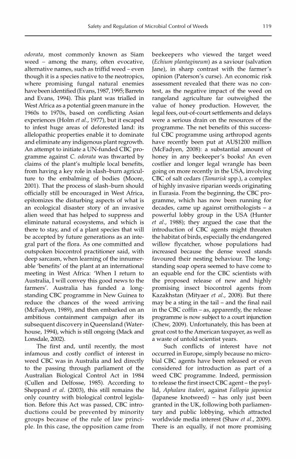
Safety and Regulation of Microbial Control of Weeds 119
odorata, most commonly known as Siam weed – among the many, often evocative, alternative names, such as triffid weed – even though it is a species native to the neotropics, where promising fungal natural enemies have been identified (Evans, 1987, 1995; Barreto and Evans, 1994). This plant was trialled in West Africa as a potential green manure in the 1960s to 1970s, based on conflicting Asian experiences (Holm et al., 1977), but it escaped to infest huge areas of deforested land: its allelopathic properties enable it to dominate and eliminate any indigenous plant regrowth. An attempt to initiate a UN-funded CBC pro-gramme against C. odorata was thwarted by claims of the plant’s multiple local benefits, from having a key role in slash–burn agricul-ture to the embalming of bodies (Moore, 2001). That the process of slash–burn should officially still be encouraged in West Africa, epitomizes the disturbing aspects of what is an ecological disaster story of an invasive alien weed that has helped to suppress and eliminate natural ecosystems, and which is there to stay, and of a plant species that will be accepted by future generations as an inte-gral part of the flora. As one committed and outspoken biocontrol practitioner said, with deep sarcasm, when learning of the innumer-able ‘benefits’ of the plant at an international meeting in West Africa: ‘When I return to Australia, I will convey this good news to the farmers’. Australia has funded a long- standing CBC programme in New Guinea to reduce the chances of the weed arriving (McFadyen, 1989), and then embarked on an ambitious containment campaign after its subsequent discovery in Queensland (Water-house, 1994), which is still ongoing (Mack and Lonsdale, 2002).
The first and, until recently, the most infamous and costly conflict of interest in weed CBC was in Australia and led directly to the passing through parliament of the Australian Biological Control Act in 1984 (Cullen and Delfosse, 1985). According to Sheppard et al. (2003), this still remains the only country with biological control legisla-tion. Before this Act was passed, CBC intro-ductions could be prevented by minority groups because of the rule of law princi-ple. In this case, the opposition came from
beekeepers who viewed the target weed (Echium plantagineum) as a saviour (salvation Jane), in sharp contrast with the farmer’s opinion (Paterson’s curse). An economic risk assessment revealed that there was no con-test, as the negative impact of the weed on rangeland agriculture far outweighed the value of honey production. However, the legal fees, out-of-court settlements and delays were a serious drain on the resources of the programme. The net benefits of this success-ful CBC programme using arthropod agents have recently been put at AU$1200 million (McFadyen, 2008): a substantial amount of honey in any beekeeper’s books! An even costlier and longer legal wrangle has been going on more recently in the USA, involving CBC of salt cedars (Tamarisk spp.), a complex of highly invasive riparian weeds originating in Eurasia. From the beginning, the CBC pro-gramme, which has now been running for decades, came up against ornithologists – a powerful lobby group in the USA (Hunter et al., 1988); they argued the case that the introduction of CBC agents might threaten the habitat of birds, especially the endangered willow flycatcher, whose populations had increased because the dense weed stands favoured their nesting behaviour. The long-standing soap opera seemed to have come to an equable end for the CBC scientists with the proposed release of new and highly promising insect biocontrol agents from Kazakhstan (Mityaer et al., 2008). But there may be a sting in the tail – and the final nail in the CBC coffin – as, apparently, the release programme is now subject to a court injunction (Chew, 2009). Unfortunately, this has been at great cost to the American taxpayer, as well as a waste of untold scientist years.
Such conflicts of interest have not occurred in Europe, simply because no micro-bial CBC agents have been released or even considered for introduction as part of a weed CBC programme. Indeed, permission to release the first insect CBC agent – the psyl-lid, Aphalara itadori, against Fallopia japonica(Japanese knotweed) – has only just been granted in the UK, following both parliamen-tary and public lobbying, which attracted worldwide media interest (Shaw et al., 2009). There is an equally, if not more promising
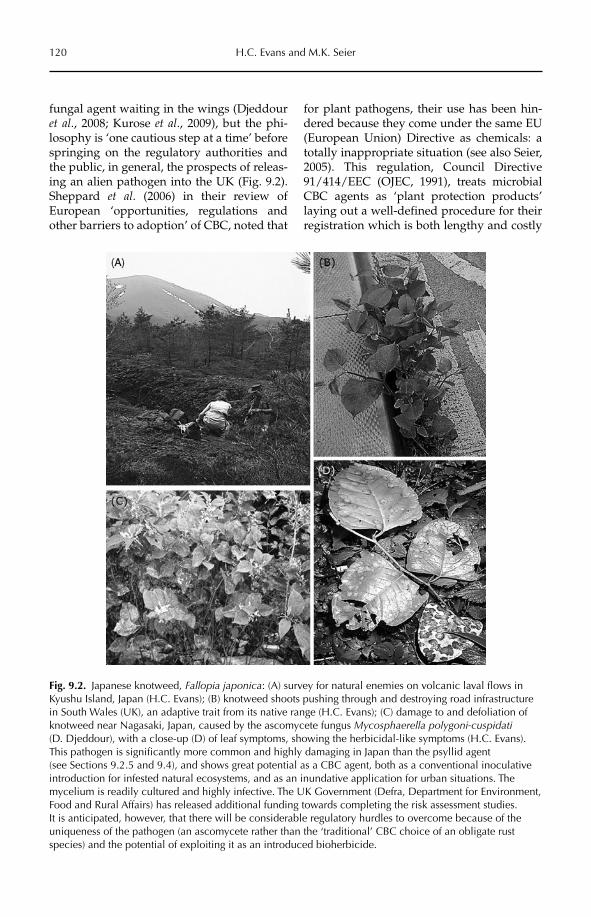
120 H.C. Evans and M.K. Seier
fungal agent waiting in the wings (Djeddour et al., 2008; Kurose et al., 2009), but the phi-losophy is ‘one cautious step at a time’ before springing on the regulatory authorities and the public, in general, the prospects of releas-ing an alien pathogen into the UK (Fig. 9.2). Sheppard et al. (2006) in their review of European ‘opportunities, regulations and other barriers to adoption’ of CBC, noted that
for plant pathogens, their use has been hin-dered because they come under the same EU (European Union) Directive as chemicals: a totally inappropriate situation (see also Seier, 2005). This regulation, Council Directive 91/414/EEC (OJEC, 1991), treats microbial CBC agents as ‘plant protection products’ laying out a well-defined procedure for their registration which is both lengthy and costly
Fig. 9.2. Japanese knotweed, Fallopia japonica: (A) survey for natural enemies on volcanic laval flows in Kyushu Island, Japan (H.C. Evans); (B) knotweed shoots pushing through and destroying road infrastructure in South Wales (UK), an adaptive trait from its native range (H.C. Evans); (C) damage to and defoliation of knotweed near Nagasaki, Japan, caused by the ascomycete fungus Mycosphaerella polygoni-cuspidati(D. Djeddour), with a close-up (D) of leaf symptoms, showing the herbicidal-like symptoms (H.C. Evans). This pathogen is significantly more common and highly damaging in Japan than the psyllid agent (see Sections 9.2.5 and 9.4), and shows great potential as a CBC agent, both as a conventional inoculative introduction for infested natural ecosystems, and as an inundative application for urban situations. The mycelium is readily cultured and highly infective. The UK Government (Defra, Department for Environment, Food and Rural Affairs) has released additional funding towards completing the risk assessment studies. It is anticipated, however, that there will be considerable regulatory hurdles to overcome because of the uniqueness of the pathogen (an ascomycete rather than the ‘traditional’ CBC choice of an obligate rust species) and the potential of exploiting it as an introduced bioherbicide.
Safety and Regulation of Microbial Control of Weeds 121
and based on data sets that have no relevance to CBC. Discussions are currently underway at the EU level on how this status quo could be simplified, aided by the outcomes of the pol-icy support action REBECA (Regulation for Biological Control Agents, 2006–2007) (Ehlers, 2008, 2011). However, how exactly the regula-tory process for microbial CBC agents will be handled by the EU remains uncertain until a test case is made.
9.2.6 CBC in action: case studies of problematic safety and regulatory issues
The examples selected in this section of in-house case studies show how the PRA is con-structed and the problems encountered during weed CBC programmes involving potential microbial agents, and how these are resolved: either successfully – in which case the programme concludes with the approval of the PRA by the receiving country and the introduction of the agent, or, unsuccessfully – in which case the programme is terminated or suspended pending further data.
Rubber-vine weed
The rubber-vine weed, Cryptostegia grandiflora(Asclepiadaceae), is an endemic Madagascan woody vine that has been described as the biggest threat to natural ecosystems in tropi-cal Australia (McFadyen and Harvey, 1990). It also became a major problem for graziers in northern Queensland where it competed with indigenous grasses, blocked rivers and denied cattle access to water by forming impenetra-ble barriers growing up to 30 m over the upper storey eucalypts. During surveys in Madagascar, the damaging leaf rust – Maravalia cryptostegiae, closely related to cof-fee rust – was found over the disparate western range of the plant (Evans, 1993). A strain of the rust from the north-west region was screened in negative-pressure quarantine facilities in the UK against a range of plant species (>70) selected by the Queensland authorities to reflect not only Australian and Madagascan asclepiads – in accordance with the centrifugal phylogenetic protocol (Wapshere, 1974a,b) – but also regional
tropical crops and anything related to coffee. In addition to standard visual and stereo-microscope observations of symptoms, it was also decided to include additional scanning electron microsope (SEM and light micro-scope examinations to determine the behav-iour of the rust, both on and within the leaf. The latter involved a clear-staining technique (Bruzzese and Hasan, 1983), which was not a normal part of the requested PRA but was included to obtain a fuller picture of plant–pathogen interactions in hosts and non-hosts: in other words, a value-added approach of scientific inquiry and rigorousness that tends to separate CBC of weeds from most other forms of biological control. Moreover, CBC screening offers an outstanding opportunity to follow plant–pathogen inter-actions in a highly disparate range of spe-cies: an area that normally receives scant attention in plant pathology (Heath, 1974, 2001). Inter estingly, five different resistance mechanisms – including two novel ones – were identified occurring on and within the non-host leaves (Evans and Tomley, 1994).
The selected rust strain defoliated the target weed and also sporulated on a closely related Madagascan genus, which did not elicit undue concern. However, towards the end of the 4 year screening period, a newly described asclepiad species in a closely related genus was received from Queensland and proved to be susceptible to the rust strain, albeit with limited rather than full sporula-tion, and with restricted internal mycelium development. All these host-range results, together with data on infection parameters and impacts (Evans and Fleureau, 1993), as well as taxonomic and hypothetical life cycle details (Evans, 1993), were included in the final report, which included an official PRA sent to the Australian Quarantine Inspection Service (AQIS) for permission to import the agent. AQIS is also tasked with sending the PRA to all Australian states for further scien-tific evaluation and amendments. Fearing the worst, a unique experiment was set up using a pollution-monitoring wind tunnel to better simulate natural infection in the field, espe-cially inoculum loads (see Section 9.2.4 on these artificial parameters). Rusted plants were placed at one end of the tunnel in front
122 H.C. Evans and M.K. Seier
of generated wind currents and selected ‘trap’ species were placed at the other. Fortunately, neither the Australian nor the Madagascan asclepiad ‘hosts’ showed infection, while sporulation occurred on the target weed: although at a lower intensity than in the pre-vious tests (Evans and Tomley, 1996). These results seemed to justify long-standing con-cerns that some agents could be rejected as unsafe because of the artificial pressure of the testing protocol and the overzealous inter-pretation of the results (Bruckart et al., 1985; Wapshere, 1989; Weidemann, 1991).
In fact, before these extra data were gen-erated, the Australian authorities had already approved the introduction of the rust on the grounds that: rubber-vine weed was on a rapidly advancing invasive front (covering 40,000 km2) in Queensland, and was posing a threat to the biodiversity of Northern Territory and its prestigious national parks; therefore, the danger of doing nothing far outweighed the risk of rust attack on a rare plant in a frag-ile ecosystem, which was geographically and climatically isolated from the rubber-vine infested areas. The rust – certified to be pure and free of contaminants (hyperparasites) – was despatched (as a ‘beneficial pest’) to Australia and passed through several gen-erations in quarantine before being released into experimental plots containing selected Madagascan and Australian asclepiads, inclu-ding the new endemic species, as well as Cryptostegia spp. The wind-tunnel experiment was vindicated because none of the other asclepiad genera were infected, although the rust sporulated only intermittently on the target weed, but consistently and heavily on C. madagascariensis, a species that, together with C. grandiflora, was found to form part of a rubber-vine complex in Madagascar (Marohasy and Forster, 1991). At the time of collection of rust strains, it was assumed that there was only one variable species of the host genus in Madagascar. Unfortunately, the selected strain from northern Madagascar was from C. madagascariensis, whereas the Australian invasive species, C. grandiflora,came from the south-west region. New collec-tions were made in this region of Madagascar, briefly rescreened in the UK – mainly to filter out natural enemies – and sent to Queensland.
The rust was released into the field, initially on bait plants. However, because of the urgency of the situation and the size of the infestation, inoculum was mass produced in greenhouses and sprayed from the ground in crude formulations, as well as from the air along the advancing front, due to its inacces-sibility (Tomley and Evans, 2004). This is the first use of aircraft as a release strategy in weed CBC – after a PRA for aerial application of the fungus was approved – and the first example of applying weed pathogens inun-datively for CBC. Aircraft have previously been used in CBC by applying parasitoids against an invasive alien mealy bug in Africa (Herren and Neuenschwander, 1991), as well as in IBC, for crop spraying of one of the pio-neering bioherbicides in the USA (TeBeest et al., 1992).
Independent monitoring by ecologists showed that rubber-vine populations were quickly reduced (up to 40%), following severe defoliation and the use of controlled burns to kill off otherwise fire-resistant plants, with virtually no seedling recruitment (Tomley and Evans, 2004). The weed has literally been stopped in its tracks, with no non-target effects. The benefits to Australian ecosystems cannot be measured financially, but the cost–benefit to agriculture in 2005 – a decade after release – has been put at over AU$230 million (Page and Lacey, 2006; McFadyen, 2008; Palmer et al., 2010) and, of course, this increases annually.
Bathurst burr
Bathurst burr, Xanthium spinosum, is a major problem to sheep farmers and the woollen industry in Australia (Parsons and Cuthbertson, 2001). During surveys in its Argentinian native range, the powdery mildew, Erysiphe cichora-cearum sensu lato, was found to be widespread, highly damaging and, apparently, host specific because it was not found on other Xanthiumspp. occurring in the same localities. As in the Cryptostegia case study above, a strain of the mildew was host-range tested in UK quarantine. The first screening against selected cultivars of sunflower – a major crop in Australia – as well as species of Xanthium, showed exceptional promise with symptoms only on the target
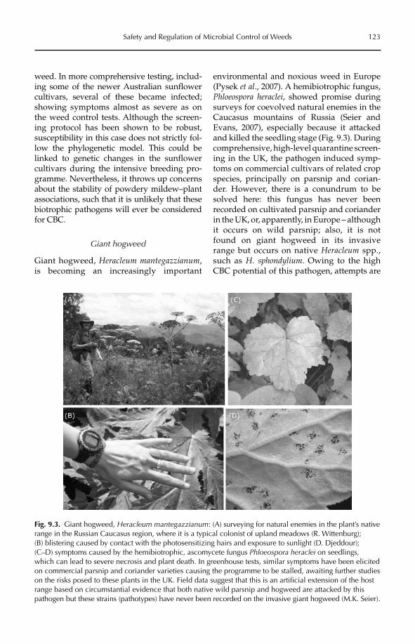
Safety and Regulation of Microbial Control of Weeds 123
weed. In more comprehensive testing, includ-ing some of the newer Australian sunflower cultivars, several of these became infected; showing symptoms almost as severe as on the weed control tests. Although the screen-ing protocol has been shown to be robust, susceptibility in this case does not strictly fol-low the phylogenetic model. This could be linked to genetic changes in the sunflower cultivars during the intensive breeding pro-gramme. Nevertheless, it throws up concerns about the stability of powdery mildew–plant associations, such that it is unlikely that these biotrophic pathogens will ever be considered for CBC.
Giant hogweed
Giant hogweed, Heracleum mantegazzianum,is becoming an increasingly important
environmental and noxious weed in Europe (Pysek et al., 2007). A hemibiotrophic fungus, Phloeospora heraclei, showed promise during surveys for coevolved natural enemies in the Caucasus mountains of Russia (Seier and Evans, 2007), especially because it attacked and killed the seedling stage (Fig. 9.3). During comprehensive, high-level quarantine screen-ing in the UK, the pathogen induced symp-toms on commercial cultivars of related crop species, principally on parsnip and corian-der. However, there is a conundrum to be solved here: this fungus has never been recorded on cultivated parsnip and coriander in the UK, or, apparently, in Europe – although it occurs on wild parsnip; also, it is not found on giant hogweed in its invasive range but occurs on native Heracleum spp., such as H. sphondylium. Owing to the high CBC potential of this pathogen, attempts are
Fig. 9.3. Giant hogweed, Heracleum mantegazzianum: (A) surveying for natural enemies in the plant’s native range in the Russian Caucasus region, where it is a typical colonist of upland meadows (R. Wittenburg); (B) blistering caused by contact with the photosensitizing hairs and exposure to sunlight (D. Djeddour); (C–D) symptoms caused by the hemibiotrophic, ascomycete fungus Phloeospora heraclei on seedlings, which can lead to severe necrosis and plant death. In greenhouse tests, similar symptoms have been elicited on commercial parsnip and coriander varieties causing the programme to be stalled, awaiting further studies on the risks posed to these plants in the UK. Field data suggest that this is an artificial extension of the host range based on circumstantial evidence that both native wild parsnip and hogweed are attacked by this pathogen but these strains (pathotypes) have never been recorded on the invasive giant hogweed (M.K. Seier).
124 H.C. Evans and M.K. Seier
being made to solve the puzzle through a series of cross-inoculation studies, but until this is resolved satisfactorily – based on scien-tific evidence that non-target plants are not at risk – the programme is temporarily, at least, at an impasse.
9.2.7 Bureaucrats, botanists, beekeepers and other bottlenecks
This title was used to illustrate the safety and legislative problems that weed biocontrol practitioners come up against before, during and after the implementation of CBC pro-grammes (Harris, 1985). It reflects the frustra-tions of a discipline that seems to oscillate between highs and lows. This 1980s low period had been preceded by the highs of the 1970s, when the use of microbials for the management of weeds had become more mainstream with the pioneering and hugely successful CBC programmes in Australia and the USA (Cullen et al., 1973; Trujillo, 1985). The pattern has been repeated over subse-quent decades, but with successful pro-grammes not being followed up by increased investment, as might logically be expected, and CBC entering another trough. Instead of increased confidence in the safety and eco-nomic benefits of CBC, periodic scare stories about the potential risks involved, distrust of its apparent primitive or simplistic tech-nology in a world of increasing sophistica-tion and, of course, unpredictable political manoeuvrings, have all contributed to gen-eral unease about this management strategy. If this is transmitted to donors and decision makers, then funding for new CBC initiatives becomes problematic. The following points summarize the present situation and future prospects of CBC of invasive alien weeds.
What have we learned?
That the central pillar, supporting the •PRA for weed CBC agents, host-range testing, is extremely robust. Some would argue that it is too robust and that useful agents could be rejected (Wapshere, 1989). The rubber-vine weed case study
(Section 9.2.6) illustrates this point perfectly. What we now know is that false positives are not uncommon in cen-trifugal phylogenetic screening but that such ‘negative’ results need to be inter-preted scientifically, based on the infor-mation presented. Whenever possible, this should be backed up with additional ‘high-tech’ data, for example, on host resistance mechanisms and simulated natural infection courts (wind tunnel experiments, more realistic inoculum concentrations). An informed rather than emotive decision can then be made within the context of the economics and the ecology of the exotic ecosystem into which the potential agent will be introduced. What we can take comfort in is that the current self-regulated sys-tem will detect any unpredicted, even bizarre, host shifts as in the Cryptostegiaand Xanthium examples described in Section 9.2.6.That strain selection is crucial to success. •Even though a strain exhibits high poten-tial in artificial screening tests, unless there is a coevolved association, then this potential will not be realized in the field situation (see Section 9.2.6, rubber-vine weed). Fortunately, high tech has now been embraced by biocontrol scien-tists, who routinely use molecular tech-niques to determine the weed’s centre of origin (as well as correct host identifica-tion!), in order to better access its co -evolved natural enemies – especially in the rust fungi, which frequently exhibit rigid host specificity, often at the bio-type–pathotype level.
What are the grey areas?
Biosecurity is now in common parlance, •even though the term is often misunder-stood or misapplied (Evans and Waller, 2010). For CBC of weeds sensu lato, this can have biological warfare connotations and, almost certainly, there have been covert CBC-type introductions against weedy (drug) plants. Fortunately, these seem to have been based on good science,
Safety and Regulation of Microbial Control of Weeds 125
although there is no international legislation (or international agreements) in place, apart from non-mandatory codes of conduct (IUCN, 1987; FAO, 1996, 1997; EPPO 1999, 2000), to stop individual countries from importing whatever potential CBC agents that they see fit. If high scientific standards are not maintained then ‘one error resulting in crop damage could jeopardize the future of all [CBC] programmes’ (Adams, 1988). Thus, for example, any country could officially import a microbial CBC agent targeted at an invasive weed through its quarantine service without the need to inform its neighbours. If this were based on poor science (i.e. not screened or non-specific), then the consequences for that region, or even that continent, could be disastrous as plant pathogens do not respect borders. This is not as far-fetched as it seems, because there is strong circumstantial evidence to indi-cate that at least two (unofficial) intro-ductions of pathogens for weed control have been made into Australia, probably by disaffected stakeholders. One of these agents (blackberry rust), quickly reached New Zealand (Marks et al.,1984; Cullen and Delfosse, 1985; Julien and Griffiths, 1998); more recently, the officially released bridal creeper rust (Puccinia myrsiphylli) in Australia was also reported from New Zealand(Waipara et al., 2006).Undue delays, unforeseen bottlenecks or •unnecessary bureaucracy could all con-tribute to stakeholder vigilantes taking the law into their own hands. For both the ‘rogue’ Australian introductions mentioned above, official releases were delayed pending further investigation. In the case of the blackberry rust, the confused taxonomy of the target weed (Rubus fruticosus agg.), plus the many commercial cultivars, complicated the science and, at one time, up to 19 patho-gen strains were being assessed for intro-duction (Bruzzese and Hasan, 1983). Undoubtedly, a rogue element of the farmers affected by weed infestations, and inspired by the earlier successes in
Australia with rust CBC agents, con-spired to release a European strain of the pathogen. Fortunately, no commercial soft fruit growers have been affected by the introduction.Recently enacted legislation in various •countries – with the Rio Convention on Bio-logical Diversity (CBD) as the catalyst – under the banner of the International Regime on Access and Benefit Sharing (Cock et al., 2010), could halt or seriously impede the implementation of CBC (see also Thornström, Chapter 20, this volume). This international regime and the supporting national legislations are directed at commercial research (bio-prospecting) and biopiracy, and are designed to stop the unregulated move-ment of genetic material across national borders, as well as to share the benefits between exporter and importer. CBC does not fall into this category, because it is non-commercial research with no patents to protect: ‘The benefits accrue to all, not the implementing agency or the government or group that paid for the research’ (Cock, 2010). However, a number of countries have already intro-duced laws on access and benefit shar-ing which are tailored as ‘anti-biopiracy’ legislation, and so are full of highly con-servative safeguards. If these are also applied to CBC, then exploratory sur-veys and the export of CBC agents will effectively be stopped or, at the very least, severely impeded. Often, the desig-nated bureaucracies empowered with administering the laws do not under-stand them, especially when microbial CBC organisms are involved. In other countries, there are simply no bureau-crats in a position to make such deci-sions, but they interpret the CBD to the full, in the sense that their biodiversity cannot be exported. It has even got to the absurd state where samples of the inva-sive alien plant Mikania micrantha in India could not initially be obtained for screen-ing, because the Indian quarantine authorities considered it to be ‘part of our biodiversity’ – even though the request was for an aid programme to manage a
126 H.C. Evans and M.K. Seier
weed threatening not only biodiversity hot spots in the Western Ghats, but also the major export crop of tea in Assam (Ellison and Evans, 2004). As Cock (2010) pointed out, the age of the hunter– gatherer in biological control has long gone. Unfortunately, the modern – some would say, more ethical (Delfosse, 2005) – CBC scientist could be faced with poten-tially insurmountable legislative barriers.
9.3 Inundative (Bioherbicide) Biological Control (IBC)
9.3.1 Why do we need IBC?
The motivations for IBC have been detailed comprehensively by Charudattan (2001), who pointed to changes in modern weed manage-ment practices and increasing public aware-ness of environmental issues. The main drivers are summarized here:
banning or phasing out of many tradi-•tional chemical herbicides, as well as general soil fumigants;the high cost of research and develop-•ment, as well as the registering, of new chemical herbicides;the scarcity of registered products for •niche markets;increasing weed resistance to chemical •herbicides;public resistance to herbicide-resistant •transgenic crops;increasing public preference for organic •or green produce; andgovernment initiatives and, in some •countries, mandates for reducing the use of chemical pesticides, of which herbi-cides account for almost 50% of the agro-chemical market.
In even more recent times, there has been increasing public concern that some of the best-selling herbicide products are not as safe as claimed, while the ethics of the major agro-chemical companies have been brought more and more into question. This has been re-enforced, in developed countries, by nos-talgic or never experienced images of the
diversity of the countryside before the rise of large-scale agriculture and the mass use of chemical pesticides, especially of herbicides.
What better, then, than ‘natural’ prod-ucts such as bioherbicides to fill the chemical- herbicide void?
9.3.2 How does it work?
In theory, a bioherbicide is applied and should function in much the same way as a chemical herbicide, because the microbial agents are often selected on their ability to produce plant toxins. Thus, the farmer expects results within days rather the months, years or even dec-ades that CBC can take. However, in practice, since the early successes in the 1970s (Templeton, 1982; TeBeest and Templeton, 1985) – mirroring those of weed CBC – IBC of weeds has been plagued by technical prob-lems: especially problems of mass produc-tion, formulation, shelf life and inconsistent field efficacy (Auld and Morin, 1995). In short, IBC has gone through the same periodic highs and lows as CBC and, as a management strat-egy in a commercial market, it has generally failed to compete with conventional chemical control as a player on the bigger agricultural stage. The beginnings were promising: select a pathogen, typically an indigenous necro-trophic (readily culturable) fungus, with a restricted host range, attacking an indigenous or naturalized weed of importance in local crops, and use relatively high-tech methods –often from the food industry – to formulate the product in a form suitable for application in small-scale field plots. This R&D phase was, more often than not, part of a publicly funded research project. To scale this up into a commercial product, venture capital was needed to cover not only the production costs but also for patenting, health and safety test-ing and registering; and, dealing with a living organism presented all manner of technical problems, many of which have still not been resolved. For example, in order to register a bioherbicide, efficacy data have to be submit-ted or made available, but, frequently, this has been impossible because the results were statistically inconsistent and depended largely on critical climatic conditions (such as
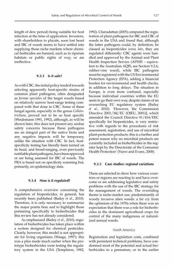
Safety and Regulation of Microbial Control of Weeds 127
length of dew period) being suitable for host infection at the time of application. Investors, with shareholders to placate, soon lost heart and IBC of weeds seems to have settled into supplying those niche markets where chemi-cal herbicides are banned, such as in riparian habitats or public rights of way, or are ineffective.
9.3.3 Is it safe?
As with CBC, the initial policy tended towards selecting apparently host-specific strains of common plant pathogens, often designated as formae speciales of the target weeds based on relatively narrow host-range testing com-pared with that done in CBC. Some of these fungal agents, especially in the genus Colleto-trichum, proved not to be so host specific (Weidemann 1991, 1992), although, as will be shown later, this does not present any undue safety concerns because these pathogens are an integral part of the native biota and any negative impacts will be temporary, unlike the situation with CBC. In fact, host-specificity testing has literally been turned on its head, and broad-ranging, even previously notifiable plant pathogens, have been approved or are being assessed for IBC of weeds. The PRA is based not on specificity screening but, primarily, on epidemiology data.
9.3.4 How is it regulated?
A comprehensive overview concerning the regulation of biopesticides, in general, has recently been published (Bailey et al., 2010). Therefore, it is only necessary to summarize the major points here and to highlight those pertaining specifically to bioherbicides that this review has not already considered.
As emphasized (Bailey et al., 2010), regu-lation of bioherbicides has taken place within a system designed for chemical pesticides. Clearly, however, this model is not appropri-ate for living organisms (Waage, 1997); this was a plea made much earlier when the pro-totype bioherbicides were testing the regula-tory system in the USA (Templeton, 1982,
1992). Charudattan (2005) compared the regis-tration of plant pathogens for IBC and CBC of weeds in the USA and found that, although the latter pathogens could, by definition, be classed as biopesticides sensu lato, they are regulated differently: CBC agents were han-dled and approved by the Animal and Plant Health Inspection Service (APHIS – equiva-lent to the Australian AQIS, see Section 9.2.6, rubber-vine weed), while IBC pathogens must be registered with the US Environmental Protection Agency (EPA), adding a financial burden for environmental and health checks, in addition to long delays. The situation in Europe, is even more confused, especially because individual countries within the EU seem to go their own way, despite claims of an overarching EU regulatory system (Bailey et al., 2010). ‘However, the Commission Directive 2001/36[/EC (OJEU, 2001)], which amended the Council Directive 91/414/EEC specifically for biopesticides, is very restric-tive with regards to the procedures of risk assessment, registration, and use of microbial plant-protection products: this is a further and potent reason why no microbial products are currently included as bioherbicides in the reg-ister kept by the Directorate of the Consumer Health Protection’ (Vurro and Evans, 2008).
9.3.5 Case studies: regional variations
These are selected to show how various coun-tries or regions are reacting to and have over-come or are addressing legislative and safety problems with the use of the IBC strategy for the management of weeds. The overriding theme is niche-market use, predominantly of woody invasive alien weeds: a far cry from the optimism of the 1970s when there was an expectation that there was a role for bioherbi-cides in the dominant agricultural crops for control of the many indigenous or natural-ized annual weeds.
North America
Registration and legislation costs, combined with persistent technical problems, have con-demned most of the potential and actual bio-herbicides to a premature, or in the earlier
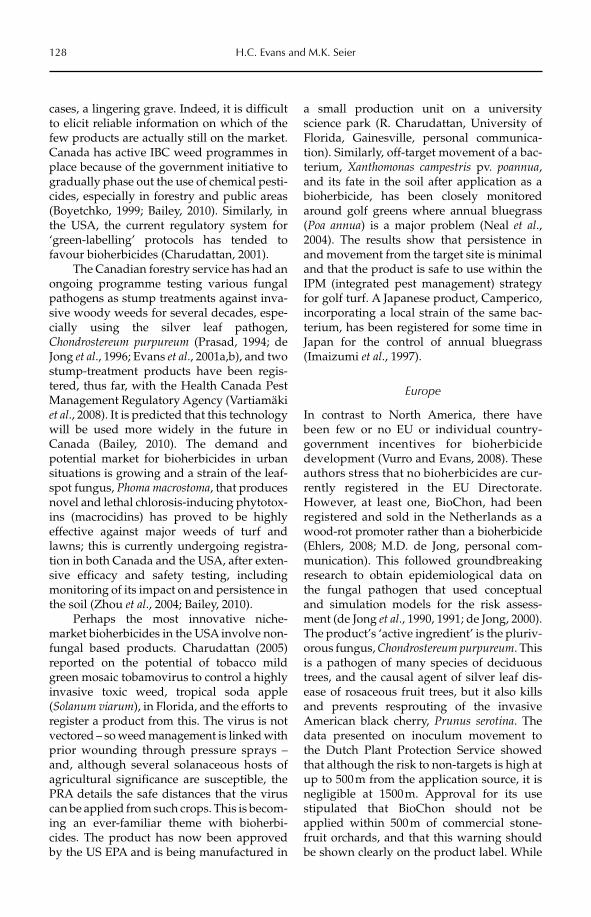
128 H.C. Evans and M.K. Seier
cases, a lingering grave. Indeed, it is difficult to elicit reliable information on which of the few products are actually still on the market. Canada has active IBC weed programmes in place because of the government initiative to gradually phase out the use of chemical pesti-cides, especially in forestry and public areas (Boyetchko, 1999; Bailey, 2010). Similarly, in the USA, the current regulatory system for ‘green-labelling’ protocols has tended to favour bioherbicides (Charudattan, 2001).
The Canadian forestry service has had an ongoing programme testing various fungal pathogens as stump treatments against inva-sive woody weeds for several decades, espe-cially using the silver leaf pathogen, Chondrostereum purpureum (Prasad, 1994; de Jong et al., 1996; Evans et al., 2001a,b), and two stump-treatment products have been regis-tered, thus far, with the Health Canada Pest Management Regulatory Agency (Vartiamäki et al., 2008). It is predicted that this technology will be used more widely in the future in Canada (Bailey, 2010). The demand and potential market for bioherbicides in urban situations is growing and a strain of the leaf-spot fungus, Phoma macrostoma, that produces novel and lethal chlorosis-inducing phytotox-ins (macrocidins) has proved to be highly effective against major weeds of turf and lawns; this is currently undergoing registra-tion in both Canada and the USA, after exten-sive efficacy and safety testing, including monitoring of its impact on and persistence in the soil (Zhou et al., 2004; Bailey, 2010).
Perhaps the most innovative niche- market bioherbicides in the USA involve non-fungal based products. Charudattan (2005) reported on the potential of tobacco mild green mosaic tobamovirus to control a highly invasive toxic weed, tropical soda apple (Solanum viarum), in Florida, and the efforts to register a product from this. The virus is not vectored – so weed management is linked with prior wounding through pressure sprays – and, although several solanaceous hosts of agricultural significance are susceptible, the PRA details the safe distances that the virus can be applied from such crops. This is becom-ing an ever-familiar theme with bioherbi-cides. The product has now been approved by the US EPA and is being manufactured in
a small production unit on a university science park (R. Charudattan, University of Florida, Gainesville, personal communica-tion). Similarly, off-target movement of a bac-terium, Xanthomonas campestris pv. poannua,and its fate in the soil after application as a bioherbicide, has been closely monitored around golf greens where annual bluegrass (Poa annua) is a major problem (Neal et al.,2004). The results show that persistence in and movement from the target site is minimal and that the product is safe to use within the IPM (integrated pest management) strategy for golf turf. A Japanese product, Camperico, incorporating a local strain of the same bac-terium, has been registered for some time in Japan for the control of annual bluegrass (Imaizumi et al., 1997).
Europe
In contrast to North America, there have been few or no EU or individual country- government incentives for bioherbicide development (Vurro and Evans, 2008). These authors stress that no bioherbicides are cur-rently registered in the EU Directorate. However, at least one, BioChon, had been registered and sold in the Netherlands as a wood-rot promoter rather than a bioherbicide (Ehlers, 2008; M.D. de Jong, personal com-munication). This followed groundbreaking research to obtain epidemiological data on the fungal pathogen that used conceptual and simulation models for the risk assess-ment (de Jong et al., 1990, 1991; de Jong, 2000). The product’s ‘active ingredient’ is the pluriv-orous fungus, Chondrostereum purpureum. This is a pathogen of many species of deciduous trees, and the causal agent of silver leaf dis-ease of rosaceous fruit trees, but it also kills and prevents resprouting of the invasive American black cherry, Prunus serotina. The data presented on inoculum movement to the Dutch Plant Protection Service showed that although the risk to non-targets is high at up to 500 m from the application source, it is negligible at 1500 m. Approval for its use stipulated that BioChon should not be applied within 500 m of commercial stone-fruit orchards, and that this warning should be shown clearly on the product label. While
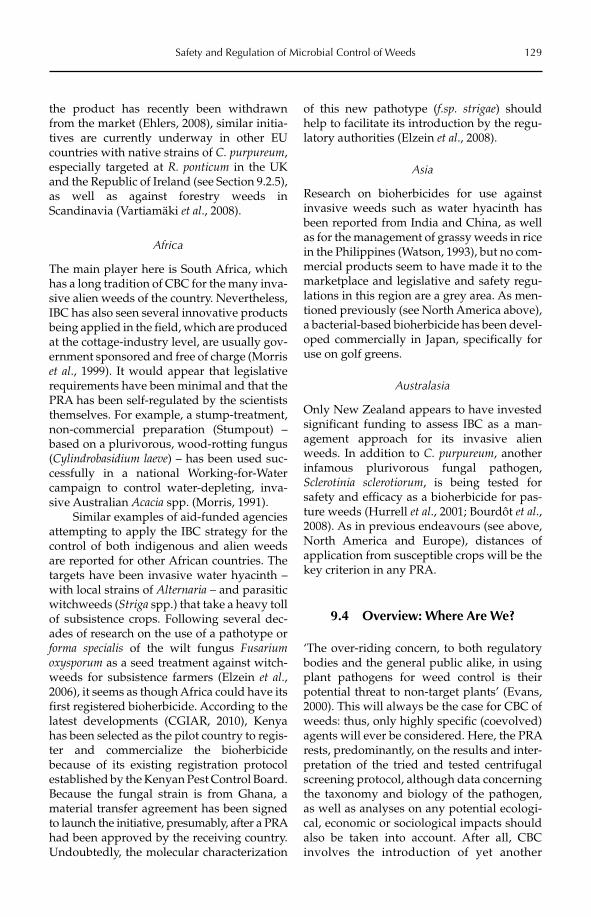
Safety and Regulation of Microbial Control of Weeds 129
the product has recently been withdrawn from the market (Ehlers, 2008), similar initia-tives are currently underway in other EU countries with native strains of C. purpureum,especially targeted at R. ponticum in the UK and the Republic of Ireland (see Section 9.2.5), as well as against forestry weeds in Scandinavia (Vartiamäki et al., 2008).
Africa
The main player here is South Africa, which has a long tradition of CBC for the many inva-sive alien weeds of the country. Nevertheless, IBC has also seen several innovative products being applied in the field, which are produced at the cottage-industry level, are usually gov-ernment sponsored and free of charge (Morris et al., 1999). It would appear that legislative requirements have been minimal and that the PRA has been self-regulated by the scientists themselves. For example, a stump-treatment, non-commercial preparation (Stumpout) – based on a plurivorous, wood-rotting fungus (Cylindrobasidium laeve) – has been used suc-cessfully in a national Working-for-Water campaign to control water-depleting, inva-sive Australian Acacia spp. (Morris, 1991).
Similar examples of aid-funded agencies attempting to apply the IBC strategy for the control of both indigenous and alien weeds are reported for other African countries. The targets have been invasive water hyacinth – with local strains of Alternaria – and parasitic witchweeds (Striga spp.) that take a heavy toll of subsistence crops. Following several dec-ades of research on the use of a pathotype or forma specialis of the wilt fungus Fusariumoxysporum as a seed treatment against witch-weeds for subsistence farmers (Elzein et al.,2006), it seems as though Africa could have its first registered bioherbicide. According to the latest developments (CGIAR, 2010), Kenya has been selected as the pilot country to regis-ter and commercialize the bioherbicide because of its existing registration protocol established by the Kenyan Pest Control Board. Because the fungal strain is from Ghana, a material transfer agreement has been signed to launch the initiative, presumably, after a PRA had been approved by the receiving country. Undoubtedly, the molecular characterization
of this new pathotype (f.sp. strigae) should help to facilitate its introduction by the regu-latory authorities (Elzein et al., 2008).
Asia
Research on bioherbicides for use against invasive weeds such as water hyacinth has been reported from India and China, as well as for the management of grassy weeds in rice in the Philippines (Watson, 1993), but no com-mercial products seem to have made it to the marketplace and legislative and safety regu-lations in this region are a grey area. As men-tioned previously (see North America above), a bacterial-based bioherbicide has been devel-oped commercially in Japan, specifically for use on golf greens.
Australasia
Only New Zealand appears to have invested significant funding to assess IBC as a man-agement approach for its invasive alien weeds. In addition to C. purpureum, another infamous plurivorous fungal pathogen, Sclerotinia sclerotiorum, is being tested for safety and efficacy as a bioherbicide for pas-ture weeds (Hurrell et al., 2001; Bourdôt et al.,2008). As in previous endeavours (see above, North America and Europe), distances of application from susceptible crops will be the key criterion in any PRA.
9.4 Overview: Where Are We?
‘The over-riding concern, to both regulatory bodies and the general public alike, in using plant pathogens for weed control is their potential threat to non-target plants’ (Evans, 2000). This will always be the case for CBC of weeds: thus, only highly specific (coevolved) agents will ever be considered. Here, the PRA rests, predominantly, on the results and inter-pretation of the tried and tested centrifugal screening protocol, although data concerning the taxonomy and biology of the pathogen, as well as analyses on any potential ecologi-cal, economic or sociological impacts should also be taken into account. After all, CBC involves the introduction of yet another
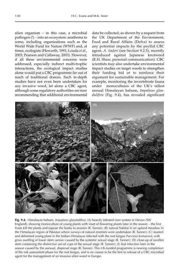
130 H.C. Evans and M.K. Seier
alien organism – in this case, a microbial pathogen (!) – into an ecosystem: anathema to some, including organizations such as the World Wide Fund for Nature (WWF) and, at times, ecologists (Howarth, 1991; Louda et al.,2003; Pearson and Callaway, 2003). However, if all these environmental concerns were addressed, especially indirect multi-trophic interactions, the ecological impact studies alone would put a CBC programme far out of reach of traditional donors. Such in-depth studies have not even been undertaken for any invasive weed, let alone a CBC agent, although some regulatory authorities are now recommending that additional environmental
data be collected, as shown by a request from the UK Department of the Environment, Food and Rural Affairs (Defra) to assess any potential impacts by the psyllid CBC agent, A. itadori (see Section 9.2.5), recently introduced against Japanese knotweed (R.H. Shaw, personal communication). CBC scientists may also undertake environmental impact studies on target weeds to strengthen their funding bid or to reinforce their argument for sustainable management. For example, monitoring the invertebrate fauna under monocultures of the UK’s tallest annual Himalayan balsam, Impatiens glan-dulifera (Fig. 9.4), has revealed significant
Fig. 9.4. Himalayan balsam, Impatiens glandulifera. (A) heavily infested river system in Devon (SW England), showing monoculture of young plants with inset of flowering plants later in the season – the first frosts kill the plants and expose the banks to erosion (R. Tanner); (B) natural habitat in an upland meadow in the Himalayan region of Pakistan where surveys of natural enemies were undertaken (R. Tanner); (C) stunted and deformed young plant in the Indian Himalayas infected with the rust fungus Puccinia komarovii, with gross swelling of lower stem (arrow) caused by the systemic sexual stage (R. Tanner); (D) close-up of swollen stem containing the distinctive aecial cups of the sexual stage (R. Tanner); (E) leaf infection later in the season caused by the asexual, dispersal stage (R. Tanner). This UK-funded programme is nearing completion of the risk assessment phase for the rust fungus, and is on course to be the first to release of a CBC microbial agent for the management of an invasive alien weed in Europe.
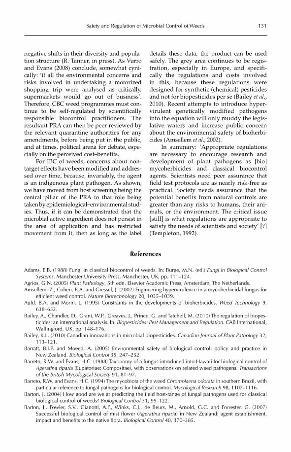
Safety and Regulation of Microbial Control of Weeds 131
negative shifts in their diversity and popula-tion structure (R. Tanner, in press). As Vurro and Evans (2008) conclude, somewhat cyni-cally: ‘if all the environmental concerns and risks involved in undertaking a motorized shopping trip were analysed as critically, supermarkets would go out of business’. Therefore, CBC weed programmes must con-tinue to be self-regulated by scientifically responsible biocontrol practitioners. The resultant PRA can then be peer reviewed by the relevant quarantine authorities for any amendments, before being put in the public, and at times, political arena for debate, espe-cially on the perceived cost–benefits.
For IBC of weeds, concerns about non-target effects have been modified and addres-sed over time, because, invariably, the agent is an indigenous plant pathogen. As shown, we have moved from host screening being the central pillar of the PRA to that role being taken by epidemiological–environmental stud-ies. Thus, if it can be demonstrated that the microbial active ingredient does not persist in the area of application and has restricted movement from it, then as long as the label
details these data, the product can be used safely. The grey area continues to be regis-tration, especially in Europe, and specifi-cally the regulations and costs involved in this, because these regulations were designed for synthetic (chemical) pesticides and not for biopesticides per se (Bailey et al., 2010). Recent attempts to introduce hyper-virulent genetically modified pathogens into the equation will only muddy the legis-lative waters and increase public concern about the environmental safety of bioherbi-cides (Amsellem et al., 2002).
In summary: ‘Appropriate regulations are necessary to encourage research and development of plant pathogens as [bio]mycoherbicides and classical biocontrol agents. Scientists need peer assurance that field test protocols are as nearly risk-free as practical. Society needs assurance that the potential benefits from natural controls are greater than any risks to humans, their ani-mals, or the environment. The critical issue [still] is what regulations are appropriate to satisfy the needs of scientists and society’ [?] (Templeton, 1992).
References
Adams, E.B. (1988) Fungi in classical biocontrol of weeds. In: Burge, M.N. (ed.) Fungi in Biological Control Systems. Manchester University Press, Manchester, UK, pp. 111–124.
Agrios, G.N. (2005) Plant Pathology, 5th edn. Elsevier Academic Press, Amsterdam, The Netherlands.Amsellem, Z., Cohen, B.A. and Gressel, J. (2002) Engineering hypervirulence in a mycoherbicidal fungus for
efficient weed control. Nature Biotechnology 20, 1035–1039.Auld, B.A. and Morin, L. (1995) Constraints in the developments of bioherbicides. Weed Technology 9,
638–652.Bailey, A., Chandler, D., Grant, W.P., Greaves, J., Prince, G. and Tatchell, M. (2010) The regulation of biopes-
ticides: an international analysis. In: Biopesticides: Pest Management and Regulation. CAB International, Wallingford, UK, pp. 148–176.
Bailey, K.L. (2010) Canadian innovations in microbial biopesticides. Canadian Journal of Plant Pathology 32, 113–121.
Barratt, B.I.P. and Moeed, A. (2005) Environmental safety of biological control: policy and practice in New Zealand. Biological Control 35, 247–252.
Barreto, R.W. and Evans, H.C. (1988) Taxonomy of a fungus introduced into Hawaii for biological control of Ageratina riparia (Eupatoriae: Compositae), with observations on related weed pathogens. Transactions of the British Mycological Society 91, 81–97.
Barreto, R.W. and Evans, H.C. (1994) The mycobiota of the weed Chromolaena odorata in southern Brazil, with particular reference to fungal pathogens for biological control. Mycological Research 98, 1107–1116.
Barton, J. (2004) How good are we at predicting the field host-range of fungal pathogens used for classical biological control of weeds? Biological Control 31, 99–122.
Barton, J., Fowler, S.V., Gianotti, A.F., Winks, C.J., de Beurs, M., Arnold, G.C. and Forrester, G. (2007) Successful biological control of mist flower (Ageratina riparia) in New Zealand: agent establishment, impact and benefits to the native flora. Biological Control 40, 370–385.
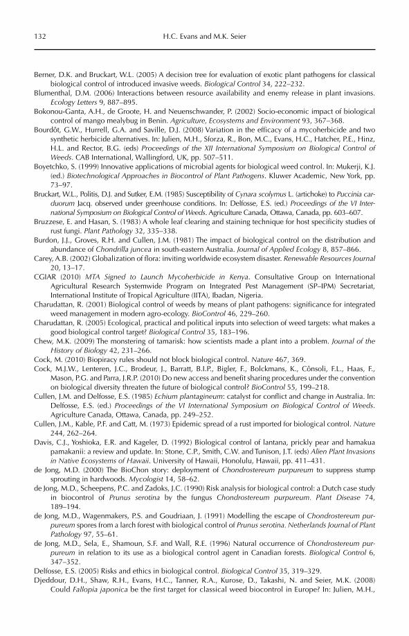
132 H.C. Evans and M.K. Seier
Berner, D.K. and Bruckart, W.L. (2005) A decision tree for evaluation of exotic plant pathogens for classical biological control of introduced invasive weeds. Biological Control 34, 222–232.
Blumenthal, D.M. (2006) Interactions between resource availability and enemy release in plant invasions. Ecology Letters 9, 887–895.
Bokonou-Ganta, A.H., de Groote, H. and Neuenschwander, P. (2002) Socio-economic impact of biological control of mango mealybug in Benin. Agriculture, Ecosystems and Environment 93, 367–368.
Bourdôt, G.W., Hurrell, G.A. and Saville, D.J. (2008) Variation in the efficacy of a mycoherbicide and two synthetic herbicide alternatives. In: Julien, M.H., Sforza, R., Bon, M.C., Evans, H.C., Hatcher, P.E., Hinz, H.L. and Rector, B.G. (eds) Proceedings of the XII International Symposium on Biological Control of Weeds. CAB International, Wallingford, UK, pp. 507–511.
Boyetchko, S. (1999) Innovative applications of microbial agents for biological weed control. In: Mukerji, K.J. (ed.) Biotechnological Approaches in Biocontrol of Plant Pathogens. Kluwer Academic, New York, pp. 73–97.
Bruckart, W.L., Politis, D.J. and Sutker, E.M. (1985) Susceptibility of Cynara scolymus L. (artichoke) to Puccinia car-duorum Jacq. observed under greenhouse conditions. In: Delfosse, E.S. (ed.) Proceedings of the VI Inter-national Symposium on Biological Control of Weeds. Agriculture Canada, Ottawa, Canada, pp. 603–607.
Bruzzese, E. and Hasan, S. (1983) A whole leaf clearing and staining technique for host specificity studies of rust fungi. Plant Pathology 32, 335–338.
Burdon, J.J., Groves, R.H. and Cullen, J.M. (1981) The impact of biological control on the distribution and abundance of Chondrilla juncea in south-eastern Australia. Journal of Applied Ecology 8, 857–866.
Carey, A.B. (2002) Globalization of flora: inviting worldwide ecosystem disaster. Renewable Resources Journal20, 13–17.
CGIAR (2010) MTA Signed to Launch Mycoherbicide in Kenya. Consultative Group on International Agricultural Research Systemwide Program on Integrated Pest Management (SP–IPM) Secretariat, International Institute of Tropical Agriculture (IITA), Ibadan, Nigeria.
Charudattan, R. (2001) Biological control of weeds by means of plant pathogens: significance for integrated weed management in modern agro-ecology. BioControl 46, 229–260.
Charudattan, R. (2005) Ecological, practical and political inputs into selection of weed targets: what makes a good biological control target? Biological Control 35, 183–196.
Chew, M.K. (2009) The monstering of tamarisk: how scientists made a plant into a problem. Journal of the History of Biology 42, 231–266.
Cock, M. (2010) Biopiracy rules should not block biological control. Nature 467, 369.Cock, M.J.W., Lenteren, J.C., Brodeur, J., Barratt, B.I.P., Bigler, F., Bolckmans, K., Cônsoli, F.L., Haas, F.,
Mason, P.G. and Parra, J.R.P. (2010) Do new access and benefit sharing procedures under the convention on biological diversity threaten the future of biological control? BioControl 55, 199–218.
Cullen, J.M. and Delfosse, E.S. (1985) Echium plantagineum: catalyst for conflict and change in Australia. In: Delfosse, E.S. (ed.) Proceedings of the VI International Symposium on Biological Control of Weeds.Agriculture Canada, Ottawa, Canada, pp. 249–252.
Cullen, J.M., Kable, P.F. and Catt, M. (1973) Epidemic spread of a rust imported for biological control. Nature244, 262–264.
Davis, C.J., Yoshioka, E.R. and Kageler, D. (1992) Biological control of lantana, prickly pear and hamakua pamakanii: a review and update. In: Stone, C.P., Smith, C.W. and Tunison, J.T. (eds) Alien Plant Invasions in Native Ecosystems of Hawaii. University of Hawaii, Honolulu, Hawaii, pp. 411–431.
de Jong, M.D. (2000) The BioChon story: deployment of Chondrostereum purpureum to suppress stump sprouting in hardwoods. Mycologist 14, 58–62.
de Jong, M.D., Scheepens, P.C. and Zadoks, J.C. (1990) Risk analysis for biological control: a Dutch case study in biocontrol of Prunus serotina by the fungus Chondrostereum purpureum. Plant Disease 74, 189–194.
de Jong, M.D., Wagenmakers, P.S. and Goudriaan, J. (1991) Modelling the escape of Chondrostereum pur-pureum spores from a larch forest with biological control of Prunus serotina. Netherlands Journal of Plant Pathology 97, 55–61.
de Jong, M.D., Sela, E., Shamoun, S.F. and Wall, R.E. (1996) Natural occurrence of Chondrostereum pur-pureum in relation to its use as a biological control agent in Canadian forests. Biological Control 6, 347–352.
Delfosse, E.S. (2005) Risks and ethics in biological control. Biological Control 35, 319–329.Djeddour, D.H., Shaw, R.H., Evans, H.C., Tanner, R.A., Kurose, D., Takashi, N. and Seier, M.K. (2008)
Could Fallopia japonica be the first target for classical weed biocontrol in Europe? In: Julien, M.H.,
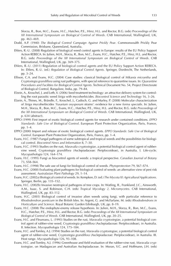
Safety and Regulation of Microbial Control of Weeds 133
Sforza, R., Bon, M.C., Evans, H.C., Hatcher, P.E., Hinz, H.L. and Rector, B.G. (eds) Proceedings of the XII Inter national Symposium on Biological Control of Weeds. CAB International, Wallingford, UK, pp. 463–469.
Dodd, A.P. (1940) The Biological Control Campaign Against Prickly Pear. Commonwealth Prickly Pear Commission, Brisbane, Queensland, Australia.
Ehlers, R.-U. (2008) Regulation of biological weed control agents in Europe: results of the EU Policy Support Action REBECA. In: Julien, M.H., Sforza, R., Bon, M.C., Evans, H.C., Hatcher, P.E., Hinz, H.L. and Rector, B.G. (eds) Proceedings of the XII International Symposium on Biological Control of Weeds. CAB International, Wallingford, UK, pp. 369–375.
Ehlers, R.-U. (2011) Regulation of biological control agents and the EU Policy Support Action REBECA. In: Ehlers, R.-U. (ed.) Regulation of Biological Control Agents. Springer, Dordrecht, The Netherlands, pp. 3–24.
Ellison, C.A. and Evans, H.C. (2004) Case studies: classical biological control of Mikania micrantha and Cryptostegia grandiflora using rust pathogens, with special reference to quarantine issues. In: QuarantineProcedures and Facilities for Biological Control Agents. Technical Document No. 54, Project Directorate of Biological Control, Bangalore, India, pp. 79–84.
Elzein, A., Kroschel, J. and Leth, V. (2006) Seed treatment technology: an attractive delivery system for control-ling the root parasitic weed Striga with mycoherbicides. Biocontrol Science and Technology 16, 3–26.
Elzein, A., Thines, M., Brändle, F., Kroschel, J., Cadisch, G. and Marley, P. (2008) Molecular characterization of Striga mycoherbicides ‘Fusarium oxysporum strains’: evidence for a new forma specialis. In: Julien, M.H., Sforza, R., Bon, M.C., Evans, H.C., Hatcher, P.E., Hinz, H.L. and Rector, B.G. (eds) Proceedings of the XII International Symposium on Biological Control of Weeds. CAB International, Wallingford, UK, p. 634 (abstract).
EPPO (1999) First import of exotic biological control agents for research under contained conditions. EPPOStandards: Safe Use of Biological Control. European Plant Protection Organization, Paris, France, pp. 1–4.
EPPO (2000) Import and release of exotic biological control agents. EPPO Standards: Safe Use of Biological Control. European Plant Protection Organization, Paris, France, pp. 1–4.
Evans, H.C. (1987) Fungal pathogens of some subtropical and tropical weeds and the possibilities for biologi-cal control. Biocontrol News and Information 8, 7–30.
Evans, H.C. (1993) Studies on the rust, Maravalia cryptostegiae, a potential biological control agent of rubber-vine weed, Cryptostegia grandiflora (Asclepiadaceae: Periplocoideae), in Australia. I. Life-cycle. Mycopathologia 124, 164–174.
Evans, H.C. (1995) Fungi as biocontrol agents of weeds: a tropical perspective. Canadian Journal of Botany73, S58–S64.
Evans, H.C. (1998) The safe use of fungi for biological control of weeds. Phytoprotection 79, S67–S74.Evans, H.C. (2000) Evaluating plant pathogens for biological control of weeds: an alternative view of pest risk
assessment. Australasian Plant Pathology 29, 1–14.Evans, H.C. (2002a) Biological control of weeds. In: Kempken, D. (ed.) The Mycota XI: Agricultural Applications.
Springer, Berlin, pp. 135–152.Evans, H.C. (2002b) Invasive neotropical pathogens of tree crops. In: Watling, R., Frankland, J.C., Ainsworth,
A.M., Isaac, S. and Robinson, C.H. (eds) Tropical Mycology 2, Micromycetes. CAB International, Wallingford, UK, pp. 83–112.
Evans, H.C. (2003) Biological control of invasive alien weeds using fungi, with particular reference to Rhododendron ponticum in the British Isles. In: Argent, G. and McFarlane, M. (eds) Rhododendrons in Horticulture and Science. Royal Botanic Garden Edinburgh, UK, pp. 8–19.
Evans, H.C. (2008) The endophyte-enemy release hypothesis. In: Julien, M.H., Sforza, R., Bon, M.C., Evans, H.C., Hatcher, P.E., Hinz, H.L. and Rector, B.G. (eds) Proceedings of the XII International Symposium on Biological Control of Weeds. CAB International, Wallingford, UK, pp. 20–25.
Evans, H.C. and Fleureaeu, L. (1993) Studies on the rust, Maravalia cryptostegiae, a potential biological con-trol agent of rubber-vine weed, Cryptostegia grandiflora (Asclepiadaceae: Periplocoideae), in Australia. II. Infection. Mycopathologia 124, 175–184.
Evans, H.C. and Tomley, A.J. (1994) Studies on the rust, Maravalia cryptostegiae, a potential biological control agent of rubber-vine weed, Cryptostegia grandiflora (Asclepiadaceae: Periplocoideae), in Australia. III. Host range. Mycopathologia 126, 93–108.
Evans, H.C. and Tomley, A.J. (1996) Greenhouse and field evaluation of the rubber-vine rust, Maravalia cryp-tostegiae, on Madagascan and Australian Asclepiadaceae. In: Moran, V.C. and Hoffmann, J.H. (eds)
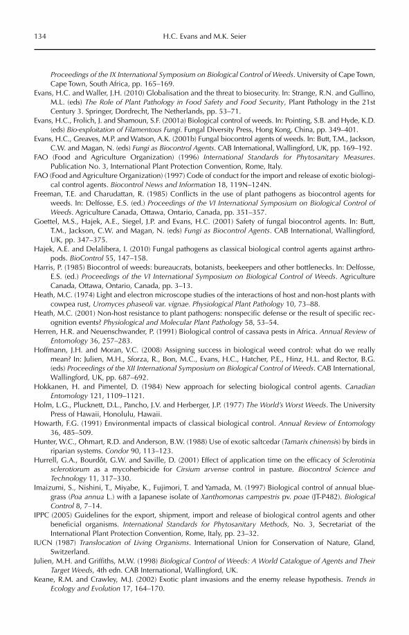
134 H.C. Evans and M.K. Seier
Proceedings of the IX International Symposium on Biological Control of Weeds. University of Cape Town, Cape Town, South Africa, pp. 165–169.
Evans, H.C. and Waller, J.H. (2010) Globalisation and the threat to biosecurity. In: Strange, R.N. and Gullino, M.L. (eds) The Role of Plant Pathology in Food Safety and Food Security, Plant Pathology in the 21st Century 3. Springer, Dordrecht, The Netherlands, pp. 53–71.
Evans, H.C., Frolich, J. and Shamoun, S.F. (2001a) Biological control of weeds. In: Pointing, S.B. and Hyde, K.D. (eds) Bio-exploitation of Filamentous Fungi. Fungal Diversity Press, Hong Kong, China, pp. 349–401.
Evans, H.C., Greaves, M.P. and Watson, A.K. (2001b) Fungal biocontrol agents of weeds. In: Butt, T.M., Jackson, C.W. and Magan, N. (eds) Fungi as Biocontrol Agents. CAB International, Wallingford, UK, pp. 169–192.
FAO (Food and Agriculture Organization) (1996) International Standards for Phytosanitary Measures.Publication No. 3, International Plant Protection Convention, Rome, Italy.
FAO (Food and Agriculture Organization) (1997) Code of conduct for the import and release of exotic biologi-cal control agents. Biocontrol News and Information 18, 119N–124N.
Freeman, T.E. and Charudattan, R. (1985) Conflicts in the use of plant pathogens as biocontrol agents for weeds. In: Delfosse, E.S. (ed.) Proceedings of the VI International Symposium on Biological Control of Weeds. Agriculture Canada, Ottawa, Ontario, Canada, pp. 351–357.
Goettel, M.S., Hajek, A.E., Siegel, J.P. and Evans, H.C. (2001) Safety of fungal biocontrol agents. In: Butt, T.M., Jackson, C.W. and Magan, N. (eds) Fungi as Biocontrol Agents. CAB International, Wallingford, UK, pp. 347–375.
Hajek, A.E. and Delalibera, I. (2010) Fungal pathogens as classical biological control agents against arthro-pods. BioControl 55, 147–158.
Harris, P. (1985) Biocontrol of weeds: bureaucrats, botanists, beekeepers and other bottlenecks. In: Delfosse, E.S. (ed.) Proceedings of the VI International Symposium on Biological Control of Weeds. Agriculture Canada, Ottawa, Ontario, Canada, pp. 3–13.
Heath, M.C. (1974) Light and electron microscope studies of the interactions of host and non-host plants with cowpea rust, Uromyces phaseoli var. vignae. Physiological Plant Pathology 10, 73–88.
Heath, M.C. (2001) Non-host resistance to plant pathogens: nonspecific defense or the result of specific rec-ognition events? Physiological and Molecular Plant Pathology 58, 53–54.
Herren, H.R. and Neuenschwander, P. (1991) Biological control of cassava pests in Africa. Annual Review of Entomology 36, 257–283.
Hoffmann, J.H. and Moran, V.C. (2008) Assigning success in biological weed control: what do we really mean? In: Julien, M.H., Sforza, R., Bon, M.C., Evans, H.C., Hatcher, P.E., Hinz, H.L. and Rector, B.G. (eds) Proceedings of the XII International Symposium on Biological Control of Weeds. CAB International, Wallingford, UK, pp. 687–692.
Hokkanen, H. and Pimentel, D. (1984) New approach for selecting biological control agents. CanadianEntomology 121, 1109–1121.
Holm, L.G., Plucknett, D.L., Pancho, J.V. and Herberger, J.P. (1977) The World’s Worst Weeds. The University Press of Hawaii, Honolulu, Hawaii.
Howarth, F.G. (1991) Environmental impacts of classical biological control. Annual Review of Entomology36, 485–509.
Hunter, W.C., Ohmart, R.D. and Anderson, B.W. (1988) Use of exotic saltcedar (Tamarix chinensis) by birds in riparian systems. Condor 90, 113–123.
Hurrell, G.A., Bourdôt, G.W. and Saville, D. (2001) Effect of application time on the efficacy of Sclerotiniasclerotiorum as a mycoherbicide for Cirsium arvense control in pasture. Biocontrol Science and Technology 11, 317–330.
Imaizumi, S., Nishini, T., Miyabe, K., Fujimori, T. and Yamada, M. (1997) Biological control of annual blue-grass (Poa annua L.) with a Japanese isolate of Xanthomonas campestris pv. poae (JT-P482). BiologicalControl 8, 7–14.
IPPC (2005) Guidelines for the export, shipment, import and release of biological control agents and other beneficial organisms. International Standards for Phytosanitary Methods, No. 3, Secretariat of the International Plant Protection Convention, Rome, Italy, pp. 23–32.
IUCN (1987) Translocation of Living Organisms. International Union for Conservation of Nature, Gland, Switzerland.
Julien, M.H. and Griffiths, M.W. (1998) Biological Control of Weeds: A World Catalogue of Agents and Their Target Weeds, 4th edn. CAB International, Wallingford, UK.
Keane, R.M. and Crawley, M.J. (2002) Exotic plant invasions and the enemy release hypothesis. Trends in Ecology and Evolution 17, 164–170.
Safety and Regulation of Microbial Control of Weeds 135
Kohl, J., Postma, J., Nicot, P., Ruocco, M. and Blum, B. (2011) Stepwise screening of microorganisms for commercial use in biocontrol of plant-pathogenic fungi and bacteria. Biological Control 57, 1–12.
Kurose, D., Evans, H.C., Djeddour, D.H., Cannon, P.F., Furoya, N. and Tsuchiya, K. (2009) Mycosphaerellaspecies as potential biological control agents of the invasive weed Fallopia japonica. Mycoscience 50, 179–189.
Large, E.C. (1940) The Advance of the Fungi. Cape, London.Lockwood, J.A. (1993) Environmental issues involved in biological control of rangeland grasshoppers
(Orthoptera: Acrididae) with exotic agents. Environmental Entomology 22, 503–518.Lonsdale, W.M., Briese, D.T. and Cullen, J.M. (2001) Risk analysis and weed biological control. In:
Wajnberg, E., Scott, J.K. and Quimby, P.C. (eds) Evaluating Indirect Ecological Effects of Biological Control. CAB International, Wallingford, UK, pp. 185–210.
Louda, S.M., Pemberton, R.W., Johnson, M.T. and Follet, P.A. (2003) Nontarget effects – the Achilles’ heel of biological control? Annual Review of Entomology 48, 365–396.
Lövei, G. (1997) Global change through invasion. Nature 388, 627–628.Mabberley, D.J. (1997) The Plant-Book, 2nd edn. Cambridge University Press, Cambridge, UK.Mack, R.N. and Lonsdale, W.M. (2002) Eradicating plants: hard-won lessons. In: Veitch, C.R. and Clout, M.N.
(eds) Turning the Tide: The Eradication of Invasive Species. Occasional Paper of the IUCN Species Survival Commission No. 27, Gland, Switzerland, pp. 163–172.
Mack, R.N., Simberloff, D., Lonsdale, W.M., Evans, H.C., Clout, M. and Bazzaz, F.A. (2000) Biotic invasions: causes, epidemiology, global consequences and control. Ecological Applications 10, 689–710.
Marks, G.C., Pascoe, I.G. and Bruzzese, E. (1984) First record of Phragmidium violaceum on blackberry in Victoria. Australasian Plant Pathology 13, 12–13.
Marohasy, J. (1996) Host shifts in biological weed control: real problems, semantic difficulties or poor sci-ence? International Journal of Pest Management 42, 71–75.
Marohasy, J. and Forster, P.I. (1991) A taxonomic revision of Cryptostegia R. Br. (Asclepiadaceae: Periplocoideae). Australian Systematic Botany 4, 571–577.
McClay, A.S. and Balciunas, J.K. (2005) The role of pre-release efficacy assessment in selecting biocontrol agents for weeds – applying the Anna Karenina principle. Biological Control 35, 197–207.
McEvoy, P.B. and Coombs, E.M. (1999) Biological control of plant invaders: regional patterns, field experi-ments and structured population models. Ecological Applications 9, 387–401.
McFadyen, R.E.C. (1989) Siam weed: a new threat to Australia’s north. Plant Protection Quarterly 4, 3–7.McFadyen, R.E.C. (1998) Biological control of weeds. Annual Review of Entomology 43, 369–393.McFadyen, R.E.C. (2008) Return on investment: determining the economic impact of biological control pro-
grammes. In: Julien, M.H., Sforza, R., Bon, M.C., Evans, H.C., Hatcher, P.E., Hinz, H.L. and Rector, B.G. (eds) Proceedings of the XII International Symposium on Biological Control of Weeds. CAB International, Wallingford, UK, pp. 67–74.
McFadyen, R.E. and Harvey, G.J. (1990) Distribution and control of rubber-vine, Cryptostegia grandiflora, a major weed in northern Queensland. Plant Protection Quarterly 5, 152–155.
Mitchell, C.E. and Power, A.G. (2003) Release of invasive plants from fungal and viral pathogens. Nature 421, 625–627.
Mityaer, I.D., Jashenko, R.V. and DeLoach, C.J. (2008) Tamarix biocontrol in US: new biocontrol agents from Kazakhstan. In: Julien, M.H., Sforza, R., Bon, M.C., Evans, H.C., Hatcher, P.E., Hinz, H.L. and Rector, B.G. (eds) Proceedings of the XII International Symposium on Biological Control of Weeds. CAB International, Wallingford, UK, p. 253 (abstract).
Mooney, H.A. and Hobbs, R.J. (2000) Invasive Species in a Changing World. Island Press, Washington, DC.Moore, D. (2001) Slayers, Saviors, Servants and Sex: An Exposé of Kingdom Fungi. Springer-Verlag, New York.Morin, L., Auld, B.A. and Smith, J.E. (1996) Rust epidemics, climate and control of Xanthium occidentale. In:
Moran,V.C. and Hoffmann, J.H. (eds) Proceedings of the IX International Symposium on Biological Control of Weeds. University of Cape Town, South Africa, pp. 385–391.
Morris, M.J. (1991) The use of plant pathogens for biological weed control in South Africa. Agriculture, Ecosystems and Environment 37, 239–255.
Morris, M.J., Wood, A.R. and Breeÿen den, A. (1999) Plant pathogens and biological control of weeds in South Africa: a review of projects and progress during the last decade. In: Olckers, T. and Hill, M.P. (eds) African Entomology Memoir No. 1, Entomological Society of Southern Africa, Hatfield, South Africa, pp. 129–137.
Neal, J.C., Williams, N.D. and Nelson, E.B. (2004) Evaluating off-target movement of Xanthomonas campes-tris pv. poannua following application as a biological agent for Poa annua on golf turf. In: Cullen, J.M.,
136 H.C. Evans and M.K. Seier
Briese, D.T., Kriticos, D.J., Lonsdale, W.M., Morin, L. and Scott, J.K. Proceedings of the XI International Symposium on Biological Control of Weeds. Commonwealth Scientific and Industrial Research Organisation, Canberra, Australia, pp. 301–304.
New Scientist (1999) Editorial: a very unholy war. No. 2203 (11 September 1989), p. 3.OEPP/EPPO (2010) Import and release of non-indigenous biological control agents. EPPO Standards: safe use
of biological control. Bulletin OEPP/EPPO Bulletin 40, 335–344.OJEC (1991) Council Directive 91/414/EEC of 15 July 1991 concerning the placing of plant protection prod-
ucts on the market. Official Journal of the European Communities 34, L 230/1–32. Available at: http://eur-lex.europa.eu/LexUriServ/LexUriServ.do?uri=OJ:L:1991:230:0001:0032:EN:PDF (accessed 3 May 2012).
OJEC (2001) Commission Directive 2001/36/EC of 16 May 2001 amending Council Directive 91/414/EEC concerning the placing of plant protection products on the market. Official Journal of the European Communities 44, L 164/1–38. Available at: http://eur-lex.europa.eu/LexUriServ/LexUriServ.do?uri=OJ:L:2001:164:0001:0038:EN:PDF(accessed 30 April 2012).
Olden, J.D., Poff, N.L., Douglas, M.R., Douglas, M.E. and Fausch, K.D. (2004) Ecological and evolutionary consequences of biotic homogenization. Trends in Ecology and Evolution 19, 18–24.
Page, A.R. and Lacey, K.L. (2006) Economic Impact Assessment of Australian Weed Biological Control.Technical Series 10, CRC for Australian Weed Management, Adelaide, South Australia, Australia.
Palmer, W.A., Heard, T.A. and Sheppard, A.W. (2010) A review of Australian classical biological control of weeds programs and research activities over the past 12 years. Biological Control 52, 271–287.
Parsons, W.T. and Cuthbertson, E.G. (2001) Noxious Weeds of Australia. CSIRO Publishing, Collingwood, Victoria, Australia.
Pearson, D.E. and Callaway, R.M. (2003) Indirect effects of host specific biological control agents. Trends in Ecology and Evolution 18, 456–461.
Prasad, R. (1994) Influence of several pesticides and adjuvants on Chondrostereum purpureum – a bioherbi-cide agent for control of forest weeds. Weed Technology 8, 445–449.
Pysek, P., Cock, M.J.W., Nentwig, W. and Ravn, H.P. (eds) (2007) Ecology and Management of Giant Hogweed.CAB International, Wallingford, UK.
Quimby, P.C. (1982) Impact of diseases on plant populations. In: Charudattan, R. and Walker, H.L. (eds) Biological Control of Weeds with Plant Pathogens. Wiley, New York, pp. 47–60.
Ritzer, G. (1998) The McDonaldization Thesis. Sage Publications, London.Rosenzweig, M.L. (2001) The four questions: what does the introduction of exotic species do to diversity?
Evolution and Ecology Research 3, 361–367.Seier, M.K. (2005) Fungal pathogens as classical biological control agents for invasive alien weeds – are they
a viable concept for Europe? In: Nentwig, W., Bascher, S., Cock, M.J.W., Hansörg, D., Gigon, A. and Wittenburg, R. (eds) Biological Invasions – from Ecology to Control. Neobiota 6, 165–176.
Seier, M.K. and Evans, H.C. (2007) Fungal pathogens associated with Heracleum mantegazzianum in its native and invaded distribution range. In: Pysek, P., Cock, M.J.W., Nentwig, W. and Ravn, H.P. (eds) Ecology and Management of Giant Hogweed. CAB International, Wallingford, UK, pp. 189–208.
Shaw, R.H., Bryner, S. and Tanner, R. (2009) The life history and host range of the Japanese knotweed psyllid, Aphalara itadori Shinji: potentially the first classical biological weed control agent for the European Union. Biological Control 49, 105–113.
Sheppard, A.W., Hill, R., DeClerck-Floate, R.A., McClay, A., Olckers, T., Quimby, P.C. and Zimmermann, H.G. (2003) A global review of risk–benefit–cost analysis for the introduction of classical biological control agents against weeds: a crisis in the making? Biocontrol News and Information 24, 91N–108N.
Sheppard, A.W., Shaw, R.H. and Sforza, R. (2006) Top 20 environmental weeds for classical biological control in Europe: a review of opportunities, regulations and other barriers to adoption. Weed Research 46, 93–117.
Simberloff, D. and Stiling, P. (1996a) Risks of species introduced for biological control. Biological Conservation78, 185–192.
Simberloff, D. and Stiling, P. (1996b) How risky is biological control? Ecology 77, 1965–1974.Syrett, P., Briese, D.T. and Hoffmann, J.H. (2000) Success in biological control of terrestrial weeds by arthro-
pods. In: Gurr, G. and Wratten, S. (eds) Biological Control: Measures of Success. Kluwer, Dordrecht, The Netherlands, pp. 189–230.
TeBeest, D.O. and Templeton, G.E. (1985) Mycoherbicides: progress in the biological control of weeds. PlantDisease 69, 6–10.
TeBeest, D.O., Yang, X.B. and Cisar, C.R. (1992) The status of biological control of weeds. Annual Review of Phytopathology 30, 637–657.
Safety and Regulation of Microbial Control of Weeds 137
Templeton, G.E. (1982) Biological herbicides: discovery, development and deployment. Weed Science 30, 430–433.
Templeton, G.E. (1992) Regulatory encouragement of biological weed control with plant pathogens. In: Charudattan, R. and Browning, H.W. (eds) Regulations and Guidelines: Critical Issues in Biological Control. University of Florida, Gainesville, Florida, pp. 61–63.
Thornström, C.-G. (2012) International conventions and agreements – consequences for international trade and utilization of biological matter, including microorganisms. In: Sundh, I., Wilcks, A. and Goettel, M.S. (eds) Beneficial Microorganisms in Agriculture, Food and the Environment: Safety Assessment and Regulation. CAB International, Wallingford, UK, pp. 293–307.
Tomley, A.J. and Evans, H.C. (2004) Establishment and preliminary impact of the Madagascan rust, Maravalia cryptostegiae, on the invasive alien weed, Cryptostegia grandiflora in Queensland, Australia. PlantPathology 53, 474–485.
Trueman, M., Atkinson, R., Guézou, A. and Wurm, P. (2010) Residence time and human-mediated propagule pressure at work in the alien flora of the Galapagos. Biological Invasions 12, 3949–3960.
Trujillo, E.E. (1985) Biological control of hamakua pa-makani with Cercosporella sp. in Hawaii. In: Delfosse, E. (ed.) Proceedings of the VI International Symposium on Biological Control of Weeds.Agriculture Canada, Ottawa, Ontario, Canada, pp. 661–671.
van Wilgen, B.W., de Wit, M.P., Andersen, H.J., Le Maitre, D.C., Kotze, I.M., Ndala, S., Brown, B. and Rapholo, M.B. (2004) Costs and benefits of biological control of invasive alien plants: case studies from South Africa. South African Journal of Science 100, 113–122.
Vartiamäki, H., Uotila, A., Vasaitis, R. and Hantula, J. (2008) Genetic diversity in Nordic and Baltic popula-tions of Chondrostereum purpureum: a potential herbicide biocontrol agent. Forest Pathology 38, 381–393.
Vurro, M. and Evans, H.C. (2008) Opportunities and constraints for the biological control of weeds in Europe. In: Julien, M.H., Sforza, R., Bon, M.C., Evans, H.C., Hatcher, P.E., Hinz, H.L. and Rector, B.G. (eds) Proceedings of the XII International Symposium on Biological Control of Weeds. CAB International, Wallingford, UK, pp. 455–462.
Waage, J. (1997) Biopesticides at the crossroads: IPM products or chemical clones? In: Evans, H.F. (ed.) Microbial Pesticides: Novelty or Necessity? British Crop Protection Society, Farnham, UK, pp. 11–20.
Waipara, N.W., McKenzie, E.H.C., Harman, H.M., Winks, C.J. and Park, D. (2006) First record of bridal creeper rust, Puccinia myrsiphylli, a classical biocontrol agent of the environmental weed bridal creeper, Asparagus asparagoides, in New Zealand. Australasian Plant Disease Notes 1, 23–24.
Wapshere, A.J. (1974a) Host specificity of phytophagous organisms and the evolutionary centres of plant genera and subgenera. Entomophaga 19, 301–309.
Wapshere, A.J. (1974b) A strategy for evaluating the safety of organisms for biological weed control. Annalsof Applied Biology 77, 201–211.
Wapshere, A.J. (1989) A testing sequence for reducing rejection of potential biological control agents for weeds. Annals of Applied Biology 114, 515–526.
Waterhouse, B.M. (1994) Discovery of Chromolaena odorata in northern Queensland, Australia. ChromolaenaNewsletter 9, 1–2.
Watson, A.K. (1993) Current status of bioherbicide development and prospects for rice in Asia. In: BiologicalControl and Integrated Management of Paddy and Aquatic Weeds. Food and Fertilizer Technology Center for the Asian and Pacific Region, Taipei, Taiwan, pp. 1–6.
Weidemann, G.J. (1991) Host-range testing: safety and science. In: TeBeest, D.O. (ed.) Microbial Control of Weeds. Chapman and Hall, New York, pp. 83–96.
Weidemann, G.J. (1992) Risk assessment: determining genetic relatedness and potential asexual gene exchange in biocontrol fungi. Plant Protection Quarterly 7, 166–168.
Zhou, L., Bailey, K.L. and Derby, J. (2004) Plant colonization and fate of the biocontrol fungus Phomamacrostoma. Biological Control 30, 634–644.
©CAB International 2012. Beneficial Microorganisms in Agriculture, Food and the Environment: 138 Safety Assessment and Regulation (eds I. Sundh et al.)
10.1 Introduction 13810.2 The Definition and History of Plant Growth Promotion by Microorganisms 139
10.2.1 Nodule bacteria (functional group 1) 139 10.2.2 Vesicular arbuscular mycorrhizae (VAM) and
ectomycorrhizae (functional group 1) 140 10.2.3 Rhizosphere plant growth promoting (PGP)
microbes (functional groups 2 and 6) 140 10.2.4 Plant-associated endophytes (functional group 2) 141
10.3 Mechanisms of Action of Plant Growth Promoting (PGP) Microbes 14110.4 Issues for PGP Microbes and the Law 142
10.4.1 Ownership 142 10.4.2 Protection of intellectual property (IP) and the inventive use of PGPs 144 10.4.3 Alteration of natural microbial biodiversity by application
of PGPs: does covering the globe with a few strains threaten biodiversity? 145 10.4.4 Quarantine transgressions 145 10.4.5 Efficacy and attempts to regulate PGP microbial products 146 10.4.6 Human health and environmental risk 147
10.5 Conclusions 148
10 Plant Growth Promotion with Microorganisms
John G. Howieson and Sharon L. FoxCrop and Plant Research Institute (CaPRI), Murdoch University, Australia
10.1 Introduction
Plant growth promotion by application of microorganisms has been a part of agricul-tural systems since at least Roman times. Ancient farmers were implored by Virgil (70–19 BC) to reinvigorate their fields by rotation of cereals with legumes such as lupins or vetches (Hamblin, 1998). These legumes became natu-rally nodulated by soil-borne root nodule bac-teria, and the ensuing symbiosis allowed the fixation of inert atmospheric dinitrogen gas to amino acids in the nodule, with subsequent export to the legume cells. Nitrogen fixation by nodule bacteria is an example of plant
growth promotion, as most soils are nitrogen deficient for plant growth and fixed nitrogen overcomes this deficiency. The nodule bacte-ria remain the most successful of the plant growth promoting (PGP) microorganisms and provide an excellent model to explore legal and ethical issues surrounding their usage.
However, a wide range of other PGP microorganisms are now in commerce, and this chapter attempts to address the issues raised by their exploitation. It should be noted that the overwhelming majority of PGP microbes in commerce have been devel-oped directly from the environment, without genetic manipulation. Hence, we are not
Plant Growth Promotion with Microorganisms 139
concerned in this chapter with the law as it relates to the release of genetically modified organisms. The key issues that do arise are: unintended manipulation of natural micro-bial biodiversity by the introduction of exotic genes; quarantine transgressions (as non- sterile carriers are traded globally); owner-ship of improved strains and intellectual property (IP); the human health questions invoked by the use of a bacterial genera linked to the incidence of human disease; and the uncertainty surrounding the efficacy of the various PGP microorganisms. This chapter will briefly outline the diversity of currently utilized PGP microorganisms and their mech-anisms of action, evaluate potential risks from their use, and assess to what extent general regulations concerning their spread or protec-tion of biodiversity can affect their utilization.
10.2 The Definition and History of Plant Growth Promotion
by Microorganisms
Microbes are widely deployed in the food, mining and agricultural industries. The agri-culturally significant organisms can be classi-fied into eight functional groups (Howieson, 2007), with the PGP microbes forming a sub-set of these:
1. Plant microsymbionts such as nodule bac-teria and mycorrhizal fungi2. Plant-associated microbes (i.e. those which elicit or potentiate a positive reaction or effect when in intimate proximity to a plant) such as cereal endophytes3. Rumen organisms4. Biocontrol agents (pathogens of weeds, fungi, insects or nematodes)5. Pathogens of plants or animals6. Agents for nutrient solubilization, biore-mediation or biodegradation7. Agents for the production of biofuels8. Agents facilitating DNA or gene transfer.
A microbial genus may be represented in more than one group, e.g. Agrobacterium is a common laboratory agent for transferring DNA (functional group 8), but also a common root pathogen (functional group 5).
This chapter will focus on the microbes in functional categories 1, 2 and 6, which elicit a growth-enhancing response when placed in close proximity to the leaves, seeds or rhizo-sphere of the target plant, or which are con-tained within the plant itself.
10.2.1 Nodule bacteria (functional group 1)
The root nodule bacteria (RNB) are found on all continents and nodulate the Leguminosae(Fabaceae), which is one of the largest families of flowering plants, with more than 18,000 species classified into 650 genera (Sprent, 2001). The RNB, as microsymbionts of leg-umes, were almost certainly the first group of PGP microbes to be studied at the microscopic level, and this was in the same decade (in 1883) that proof emerged that microbes such as Vibrio were the causative agents of serious human and animal illnesses, such as cholera. RNB were, in fact, manufactured as agricul-tural amendments within a few years of Beijerinck isolating and growing the bacteria in the Netherlands, and of the groundbreak-ing work of Hellriegel in Germany who iden-tified their role in legume nodulation and nitrogen fixation in 1887 (O’Hara et al., 2002). This was only 6 years after Koch first cultured bacteria on gelatine.
Early attempts at inoculation with RNB were rudimentary, such as moving soil from fields growing well-nodulated legumes to legume-free fields (Fred et al., 1932). In Canada, in the early part of the 20th century, farmers were encouraged to inoculate legume seed with a mixture of sieved, air-dried soil from fields containing well-nodulated plants of the target species. Within a couple of years, cul-tures of rhizobia were available in the market-places of Europe for farmers to inoculate a range of species, and inoculation was recom-mended in Australia (Guthrie, 1896). The first inoculant industries for RNB developed in the 1920s (Deaker et al., 2004). Thus began an unprecedented anthropogenic alteration of the soil microbial biota on the planet.
The symbiotic association between RNB and legumes is considered to be a vital bio-logical pillar in any terrestrial ecosystem (Reeve et al., 2010). This symbiosis annually
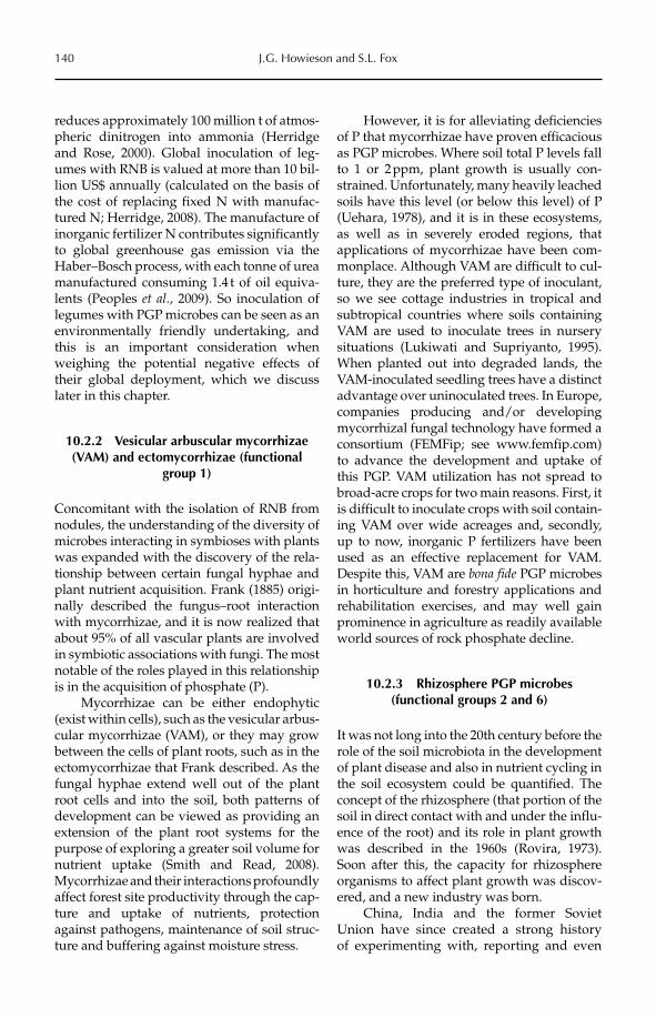
140 J.G. Howieson and S.L. Fox
reduces approximately 100 million t of atmos-pheric dinitrogen into ammonia (Herridge and Rose, 2000). Global inoculation of leg-umes with RNB is valued at more than 10 bil-lion US$ annually (calculated on the basis of the cost of replacing fixed N with manufac-tured N; Herridge, 2008). The manufacture of inorganic fertilizer N contributes significantly to global greenhouse gas emission via the Haber–Bosch process, with each tonne of urea manufactured consuming 1.4 t of oil equiva-lents (Peoples et al., 2009). So inoculation of legumes with PGP microbes can be seen as an environmentally friendly undertaking, and this is an important consideration when weighing the potential negative effects of their global deployment, which we discuss later in this chapter.
10.2.2 Vesicular arbuscular mycorrhizae (VAM) and ectomycorrhizae (functional
group 1)
Concomitant with the isolation of RNB from nodules, the understanding of the diversity of microbes interacting in symbioses with plants was expanded with the discovery of the rela-tionship between certain fungal hyphae and plant nutrient acquisition. Frank (1885) origi-nally described the fungus–root interaction with mycorrhizae, and it is now realized that about 95% of all vascular plants are involved in symbiotic associations with fungi. The most notable of the roles played in this relationship is in the acquisition of phosphate (P).
Mycorrhizae can be either endophytic (exist within cells), such as the vesicular arbus-cular mycorrhizae (VAM), or they may grow between the cells of plant roots, such as in the ectomycorrhizae that Frank described. As the fungal hyphae extend well out of the plant root cells and into the soil, both patterns of development can be viewed as providing an extension of the plant root systems for the purpose of exploring a greater soil volume for nutrient uptake (Smith and Read, 2008). Mycorrhizae and their interactions profoundly affect forest site productivity through the cap-ture and uptake of nutrients, protection against pathogens, maintenance of soil struc-ture and buffering against moisture stress.
However, it is for alleviating deficiencies of P that mycorrhizae have proven efficacious as PGP microbes. Where soil total P levels fall to 1 or 2 ppm, plant growth is usually con-strained. Unfortunately, many heavily leached soils have this level (or below this level) of P (Uehara, 1978), and it is in these ecosystems, as well as in severely eroded regions, that applications of mycorrhizae have been com-monplace. Although VAM are difficult to cul-ture, they are the preferred type of inoculant, so we see cottage industries in tropical and subtropical countries where soils containing VAM are used to inoculate trees in nursery situations (Lukiwati and Supriyanto, 1995). When planted out into degraded lands, the VAM-inoculated seedling trees have a distinct advantage over uninoculated trees. In Europe, companies producing and/or developing mycorrhizal fungal technology have formed a consortium (FEMFip; see www.femfip.com) to advance the development and uptake of this PGP. VAM utilization has not spread to broad-acre crops for two main reasons. First, it is difficult to inoculate crops with soil contain-ing VAM over wide acreages and, secondly, up to now, inorganic P fertilizers have been used as an effective replacement for VAM. Despite this, VAM are bona fide PGP microbes in horticulture and forestry applications and rehabilitation exercises, and may well gain prominence in agriculture as readily available world sources of rock phosphate decline.
10.2.3 Rhizosphere PGP microbes (functional groups 2 and 6)
It was not long into the 20th century before the role of the soil microbiota in the development of plant disease and also in nutrient cycling in the soil ecosystem could be quantified. The concept of the rhizosphere (that portion of the soil in direct contact with and under the influ-ence of the root) and its role in plant growth was described in the 1960s (Rovira, 1973). Soon after this, the capacity for rhizosphere organisms to affect plant growth was discov-ered, and a new industry was born.
China, India and the former Soviet Union have since created a strong history of experimenting with, reporting and even
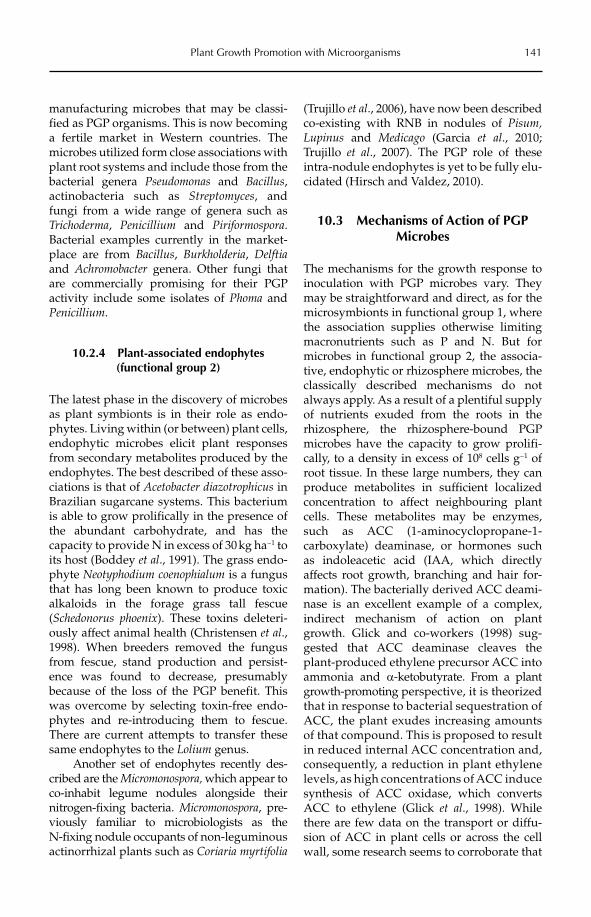
Plant Growth Promotion with Microorganisms 141
manufacturing microbes that may be classi-fied as PGP organisms. This is now becoming a fertile market in Western countries. The microbes utilized form close associations with plant root systems and include those from the bacterial genera Pseudomonas and Bacillus,actinobacteria such as Streptomyces, and fungi from a wide range of genera such as Trichoderma, Penicillium and Piriformospora.Bacterial examples currently in the market-place are from Bacillus, Burkholderia, Delftiaand Achromobacter genera. Other fungi that are commercially promising for their PGP activity include some isolates of Phoma and Penicillium.
10.2.4 Plant-associated endophytes (functional group 2)
The latest phase in the discovery of microbes as plant symbionts is in their role as endo-phytes. Living within (or between) plant cells, endophytic microbes elicit plant responses from secondary metabolites produced by the endophytes. The best described of these asso-ciations is that of Acetobacter diazotrophicus in Brazilian sugarcane systems. This bacterium is able to grow prolifically in the presence of the abundant carbohydrate, and has the capacity to provide N in excess of 30 kg ha−1 to its host (Boddey et al., 1991). The grass endo-phyte Neotyphodium coenophialum is a fungus that has long been known to produce toxic alkaloids in the forage grass tall fescue (Schedonorus phoenix). These toxins deleteri-ously affect animal health (Christensen et al.,1998). When breeders removed the fungus from fescue, stand production and persist-ence was found to decrease, presumably because of the loss of the PGP benefit. This was overcome by selecting toxin-free endo-phytes and re-introducing them to fescue. There are current attempts to transfer these same endophytes to the Lolium genus.
Another set of endophytes recently des-cribed are the Micromonospora, which appear to co-inhabit legume nodules alongside their nitrogen-fixing bacteria. Micromonospora, pre-viously familiar to microbiologists as the N-fixing nodule occupants of non-leguminousactinorrhizal plants such as Coriaria myrtifolia
(Trujillo et al., 2006), have now been described co-existing with RNB in nodules of Pisum,Lupinus and Medicago (Garcia et al., 2010; Trujillo et al., 2007). The PGP role of these intra-nodule endophytes is yet to be fully elu-cidated (Hirsch and Valdez, 2010).
10.3 Mechanisms of Action of PGP Microbes
The mechanisms for the growth response to inoculation with PGP microbes vary. They may be straightforward and direct, as for the microsymbionts in functional group 1, where the association supplies otherwise limiting macronutrients such as P and N. But for microbes in functional group 2, the associa-tive, endophytic or rhizosphere microbes, the classically described mechanisms do not always apply. As a result of a plentiful supply of nutrients exuded from the roots in the rhizosphere, the rhizosphere-bound PGP microbes have the capacity to grow prolifi-cally, to a density in excess of 108 cells g−1 of root tissue. In these large numbers, they can produce metabolites in sufficient localized concentration to affect neighbouring plant cells. These metabolites may be enzymes, such as ACC (1-aminocyclopropane-1-carboxylate) deaminase, or hormones such as indoleacetic acid (IAA, which directly affects root growth, branching and hair for-mation). The bacterially derived ACC deami-nase is an excellent example of a complex, indirect mechanism of action on plant growth. Glick and co-workers (1998) sug-gested that ACC deaminase cleaves the plant-produced ethylene precursor ACC into ammonia and a-ketobutyrate. From a plant growth-promoting perspective, it is theorized that in response to bacterial sequestration of ACC, the plant exudes increasing amounts of that compound. This is proposed to result in reduced internal ACC concentration and, consequently, a reduc tion in plant ethylene levels, as high concentrations of ACC induce synthesis of ACC oxidase, which converts ACC to ethylene (Glick et al., 1998). While there are few data on the transport or diffu-sion of ACC in plant cells or across the cell wall, some research seems to corroborate that
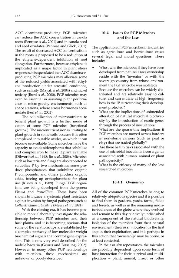
142 J.G. Howieson and S.L. Fox
ACC deaminase-producing PGP microbes can reduce the ACC concentration in canola roots (Penrose et al., 2001) and in canola seed and seed exudates (Penrose and Glick, 2001). The result of decreased ACC concentrations in the roots is proposed to be a reduction of the ethylene-dependent inhibition of root elongation. Furthermore, because ethylene is implicated as a major factor in plant stress responses, it is speculated that ACC deaminase-producing PGP microbes may alleviate some of the reduced yields associated with ethyl-ene production under stressful conditions, such as salinity (Mayak et al., 2004) and nickel toxicity (Burd et al., 2000). PGP microbes may even be essential in assisting plant perform-ance in micro-gravity environments, such as space stations, where stress hormones accu-mulate (Ferl et al., 2002).
The solubilization of micronutrients to benefit plant growth is a further mode of action of some PGP microbes (functional group 6). The micronutrient iron is limiting to plant growth in some soils because it is often complexed into stable oxides, as which it can become unavailable. Some microbes have the capacity to exude siderophores that solubilize and complex iron to make it plant available (Dilworth et al., 1998; Jin et al., 2006). Microbes such as bacteria and fungi are also reported to solubilize P by two mechanisms: some pro-duce phosphatases that solubilize organic P compounds; and others produce organic acids, freeing up orthophosphate for plant use (Kucey et al., 1989). Fungal PGP organ-isms are being developed from the genera Phoma and Penicillium. These have been shown to induce a systemic plant resistance against invasion by fungal pathogens such as Colletotrichum orbiculare (Meera et al., 1994).
With the cloning era, it has become pos-sible to more elaborately investigate the rela-tionship between PGP microbes and their host plants, and it is becoming obvious that some of the relationships are established by a complex pathway of low molecular weight biochemical signals that control gene expres-sion. This is now very well described for the nodule bacteria (Geurts and Bisseling, 2002). However, in many other PGP relationships with microbes, these mechanisms are unknown or poorly described.
10.4 Issues for PGP Microbes and the Law
The application of PGP microbes in industries such as agriculture and horticulture raises several legal and moral questions. These include:
Who owns the microbes if they have been •developed from nature? Does ownership reside with the ‘inventor’ or with the sovereign country from whose environ-ment the PGP microbe was isolated?Because the microbes can be widely dis-•tributed and are relatively easy to cul-ture, and can mutate at high frequency, how is the IP surrounding their develop-ment protected?What are the implications of unintended •alteration of natural microbial biodiver-sity by the introduction of exotic genes through the process of inoculation?What are the quarantine implications if •PGP microbes are moved across borders in non-sterile carriers (such as peat or clay) that are traded globally?Are there health risks associated with the •use of microbial inoculants from a genus associated with human, animal or plant pathogenicity?What is the efficacy of many of the less •researched microbes?
10.4.1 Ownership
All of the common PGP microbes belong to relatively ubiquitous species and it is possible to find them in gardens, yards, farms, fields and forests, as well as in the remaining undis-turbed areas of the globe where they evolved and remain to this day relatively undisturbed as a component of the natural biodiversity. Isolation of the microbes from their natural environment (their in situ location) is the first step in their exploitation, and it is perhaps in this action that ‘ownership’ may be borne, or at least contested.
In their in situ repositories, the microbes are probably dependent upon some form of host interaction for their survival and multi-plication – plant, animal, insect or other
Plant Growth Promotion with Microorganisms 143
microbe. Few PGP microbes are competent saprophytes in isolation from their hosts. Once taken from their natural habitat, cul-tured and stored, microbes become ex situ. Ex situ repositories are usually in the control of an institution or an industry that exploits them. The major difference from in situ repos-itories is that in these ex situ collections, the microbe is usually cultured in pure form, in the absence of any host, and the full metabolic requirements of the microbe must be met from artificial sources. In situ repositories are, of course, relatively inexpensive to maintain; however, there are substantial sociological, legislative and community consultation pro-cedures to work through to ensure they suc-ceed because they generally occupy land owned by the state. Ex situ collections are the converse – of relatively low diversity, expen-sive to maintain and in private ownership.
The Convention on Biological Diversity (CBD) of 1993 sets out a series of articles that assign principles of conservation of genetic resources and rights of ownership, which are recognized in international law. The CBD puts forward the argument that conservation of genetic resources, including microbes, is ‘a common concern of humankind’. It also sets principles for the fair and equitable sharing of the benefits arising from the use of genetic resources, notably those destined for com-mercial use. Importantly, the CBD is legally binding and countries that join it are obliged to implement its provisions.
The CBD is somewhat contentious in relation to PGP microbes in that it assigns sovereignty over natural resources to states (see Thornström, Chapter 20, this volume). The CBD also suggests that scientific experi-ments should be undertaken within the coun-try of origin of the genetic resources, where possible. This is likely to be a difficult or impossible undertaking with PGP microbes, because response to inoculation is likely to be species and environment specific and not all countries have the necessary research infra-structure for experimentation.
Hence, the issue of ownership of PGP microbes has its roots in the origin of the microbe according to the CBD. The in siturepositories may be tapped only after estab-lishment of a memorandum of understanding
and a material transfer agreement with the authorities in the sovereign state. Subsequent to 1993, to otherwise exploit these repositor-ies is an act of biopiracy. Microbes isolated before the 1993 agreement was enacted are not protected and this, combined with their ubiquitous nature, raises the issue of tracing the history of microbes and the date they were first identified or isolated (see Thornström, Chapter 20, this volume).
Differentiating strains of PGP microbes
While species of PGP microbes may be nearly ubiquitous, strains vary considerably. As an example, strains of RNB that belong to a sin-gle species and that nodulate a single species of legume can differ greatly in their nitrogen fixation and ecological properties. Molecular techniques, usually based upon some form of PCR (such as PCR-RFLP – PCR restriction fragment length polymorphism) can reliably differentiate microbial species at the strain level (Lee et al., 1997). However, there are obstacles to the application of PCR. First, not all microbes are readily amenable to PCR because specific primers are often required. Secondly, as we mentioned in the previous section, not all laboratories have the expertise to conduct this form of research. Techniques for reliably differentiating strains within the broad suite of PGP microbes (fungi, bacteria, archaea, viruses, algae, etc.) need to be devel-oped, standardized and broadly adopted.
As with differentiating microbes, despite the wealth of molecular tools available, micro-bial taxonomy is in a state of rapid flux as we learn more about the lateral transfer of genes on mobile genetic elements (Boucher et al.,2007). There is little consensus among micro-biologists on how to reliably classify many microbes below the genus level, particularly the bacteria (e.g. Young, 2010). Also, nomen-clatural changes have the potential to unwit-tingly confuse the origins of some PGP microbes.
Microbes replicate very quickly and the conditions under which they are cultured can lead to genetic change (drift), mainly through the loss of plasmids or DNA units bearing non-essential genes. Although bacte-ria and fungi can be readily freeze dried or
144 J.G. Howieson and S.L. Fox
lyophilized in glass ampoules for storage to minimize the opportunities for mutation, this is not always practicable. If a ‘protected or owned’ PGP microbe has undergone genetic drift, then perhaps it cannot be considered the same organism that was originally described in any invention.
10.4.2 Protection of intellectual property (IP) and the inventive use of PGPs
Protection by first to market
The contemporary attitude to protection of ownership of highly developed PGP microbes is to adhere to the principle of ‘first to mar-ket’. Often, the new PGP microbe, its mode of application and the acquisition of data that testify to its efficacy, conditions for manufac-ture and pathway to commercialization takes several (3–10) years. The protection of the innovation by being first to market is consid-erable, and this is becoming a more popular approach compared with patent protection.
Protection by patent – products or processes?
Microbes isolated from the environment can-not be patented per se because they are not considered an invention (Stix, 2006). In con-trast, a genetically modified microbe may be granted a product patent, because it can be argued that its modification is an invention. Thus, modifying Escherichia coli by insertion of genes to produce insulin in batch fermenta-tion is patentable.
However, patents have been granted for inventions based upon microbiological proc-esses, where the microbe produces a new or superior outcome, and these are referred to as ‘process patents’. So a brewer may patent the application of a specialized yeast strain to deliver particular outcomes (e.g. a special fla-vour; a high level of alcohol). A fungus that produces a unique antibiotic may be patented under the process that delivers or purifies this new compound (e.g. US Patent 3,169,100). Similarly, a new microbe isolated from the environment and not previously described, but which can be used in a process for the treatment of plants can now be counted as
an invention, and therefore can be patented. Examples in the RNB world are plentiful, with novel isolates being described almost daily. Ardley et al. (2011) described novel spe-cies of Microvirga isolated from the nodules of the legumes Lupinus texensis and Lotononisangolensis. These legumes, when taken from their natural environments (Texas and Angola, respectively) and introduced to agricultural environments, do not grow well without the application of their specific nodule bacteria, because of nitrogen deficiency. The applica-tion of these inoculants to the legumes to enhance growth and nitrogen fixation is theo-retically patentable.
In the case of the associative microbes, a process patent can also be considered when the application of the microbe alters the com-position of the plant physiologically and allows it ultimately to express different traits. For example, patent number 4,878,936 lodged in 1989 in the USA covers the application of ‘helper bacteria’ to increase the nodulation of legumes. This patent covers a method of inoc-ulating legumes with Bacillus cereus ATCC 53522 and its enhancing mutants. Included in the patent is a method of placing the nodula-tion-enhancing bacteria in the vicinity of the legume roots by coating the seed with the nodulation enhancing bacteria and an inert carrier. This helper bacterium results in increased nodule weights, numbers, size and longevity. More recently, a bacterial co-inocu-lant has been described that enhances the rate of nodulation by RNB on a common pasture species, Medicago truncatula (Fox et al., 2011). The earlier nodulation results in increased shoot yields and total N and could theoreti-cally be patented as a process for increasing nitrogen fixation.
In the case of the exploitation of the toxin-free grass endophytes described earlier, there is IP protection surrounding the tech-nology to remove the toxic strains of the fun-gus from fescue, and techniques to reintroduce new fungal strains into the seed, which then colonize the plant when it grows (Australian patent AU2003267883B2). In the case of this PGP, IP protection is based around the tech-niques of removal of one fungus and the introduction of its replacement. In the attempts to transfer the fungus to a new grass
Plant Growth Promotion with Microorganisms 145
genus, Lolium, this association is not found in nature and therefore also satisfies the ‘invention’ criteria.
10.4.3 Alteration of natural microbial biodiversity by application of PGPs: does
covering the globe with a few strains threaten biodiversity?
Without doubt, the greatest global change in the distribution of microbes has come about through human exploration of the world since the 16th century. Feedstuffs carried for animals on ships contained, for example, RNB and legume seeds, which were distrib-uted widely around the planet. Ships sailing from the Mediterranean basin introduced these legumes and their associated microbiota to sub-Saharan Africa, on to Australia, and more recently to both South and North America. Since then, the deliberate use of RNB as inoculants for legumes, particularly in the 20th century, has continued to perturb the natural distribution of PGP microbes. Massive changes have occurred in the tropics, subtropics and warm temperate zones of Africa, Asia and America, where Glycine max(soybean) inoculated with Bradyrhizobium japonicum now dominates grain legume pro-duction. There are nearly 70 million t of inoc-ulated soybeans produced annually in the USA, in addition to 34 and 53 million t in Argentina and Brazil, respectively (Hungria et al., 2006). The RNB inoculants for this crop have thus been distributed over more than 150 million ha of the American continent in the last 30 years. There is now a similar attempt to introduce soybean to Africa (Giller et al., 2010), and plantings across the globe have been almost entirely inoculated with less than five strains – the B. japonicum sero-groups associated with USDA123, USDA110, CB1809 and, more recently, B. elkanii strains SEMIA 597 and SEMIA 5019.
As for soybean in the Americas, large tracts of land have been cleared of their native vegetation in central Asia, temperate America and southern Australia, and planted to cool season forage legumes from two main gen-era, Trifolium (clovers) and Medicago (medics). Again, the majority of these legumes have
been inoculated with RNB at some stage in their production. The perennial forage crop M. sativa has wide adaptation to soil and cli-mate and, because of this, through human activities, it has spread from its centre of ori-gin (believed to be in the temperate zones of Persia) to become the dominant forage on all continents, carrying its RNB with it. Annual clovers and medics were established across 25 million ha of arable land throughout south-ern Australia over the last 150 years of the 19th and 20th centuries, with RNB inoculants available since 1896. As in the case of legumes in the tropics, this represents a massive global change in the distribution of RNB.
It is of concern that there are perhaps only 50 species of forage legumes and less than 15 species of grain legumes in transglo-bal commercial trade (Kelley et al., 2000), inoculated accordingly with a narrow suite of RNB. Is it prudent, from a gene conservation perspective, to cover the globe so com-pletely with only relatively few strains of RNB? We have evidence that these inocu-lants displace the original RNB, because of host preference for matched inoculants (Stepkowski et al., 2005). Not only may intro-duced inocula displace natural biodiversity, but we have recently learned that introduced PGP microbes may exchange their (exotic) DNA with indigenous microbes and, by doing so, greatly alter the properties of the recipient microbe. The recently described phenomenon of an exotic RNB inoculant strain which transferred its ‘symbiosis island’ to resident, but non-nodulating, microbes, concomitantly allowing these resident bacteria to become nodulating organisms (Nandasena et al., 2009), serves as a strong reminder of how easy it is to unwittingly alter the dynamics of the natural microbiota. What is the impact on the in situ conservation of microbial biodiversity in this example?
10.4.4 Quarantine transgressions
The supply and distribution of PGP microbes is becoming a transglobal trade, as fewer and larger companies dominate the industry, and oceans and authorities are no longer a substantial barrier to trade. Although
146 J.G. Howieson and S.L. Fox
the distribution of PGP microbes is subject to a wide range of regulations and laws that specify product integrity and phytosanitary con-ditions – which are administratively tedious – these conditions are actually able to be met quite simply in a biological sense: the paper-work must only specify the genus and species of the traded microbe. While some PGP microbes may be distributed as spores (e.g. Bacillus) or as dried powders (e.g. yeasts) for multiplication in the hands of a secondary manufacturer, most PGP microbes are distrib-uted in a hydrated, functional form (i.e. as vegetative cells) in carriers that sustain them. These carriers, such as finely ground and ameliorated peat, are very difficult to steri-lize, and although methods such as autoclav-ing or exposure to gamma radiation are invoked, carriers are rarely sterile. In fact, the quality assessment (QA) applied to RNB in many countries acknowledges that contami-nants must be less than 1000 cells g−1 (Deaker et al., 2004). Thus, trade in PGP microbes is often also trade and distribution in associated microbes of generally unknown form or function.
10.4.5 Efficacy and attempts to regulate PGP microbial products
PGP microbes that stimulate plant growth have gained substantial acceptance in the rural communities of many developing nations, despite a lack of clarity about their efficacy. It is speculated that the reason for this is that, in many rural settings, it has been normal practice to gather and distribute ani-mal wastes across fields to improve plant growth. These wastes contain decomposing microbes, as well as abundant N and P. The efficacy of this recycling is not disputed. However, the application of pure cultures of PGP microbes is seen by many as a sophisti-cated extension of the spreading of farmyard wastes (Dr Rosalind Deaker, University of Sydney, 2011, personal communication).
Because PGP microbes have become a large and lucrative market in broad-acre agriculture, they are a favourite target for misleading claims. There are several ways to
protect against false claims for the efficacy of PGP microbes, such as QA registration pro-cesses and the development of industry standards and best practice, but the current proliferation of dubious products indicates that the issue is not resolved. One of the major difficulties is that applications of PGP microbes carry the uncertainty of a complex biology with them, and quite often they sim-ply do not work. The multiple interactions between the genotype, or genetic make-up, of the target plant (G), the genotype of the PGP microbe (g) and the environment (E) in which it is applied can mitigate against success. There are many environmental factors that can reduce the efficacy of PGP microbes. Examples are hostile soils (dry or acid) in which a seed-applied PGP microbe cannot survive in sufficient numbers to colonize the rhizosphere. The endophytes are more resil-ient in this regard because they can be pro-tected within the seed, rather than on its surface. There are also examples in which a minor change in G can have a major change in efficacy. The natural loss of DNA from a symbiotically sensitive region in RNB is a notable example of such an alteration that completely removes the PGP effect (O’Hara et al., 2002). So, it is relatively common for PGP microbes to fail, even with the most effi-cacious of relationships, such as the legume–RNB association.
For the associative relationships, which are less stringent and in some ways less repro-ducible, efficacy is much harder to prove (and police). Some contemporary products of li -quid PGP microbial applications claim the presence of more than 20 species of microbe (e.g. SC27 Soil Inoculant and Super Microbial Organic Complex), often in addition to extracts of exotic plants such as seaweed. To reproduce statistically sound experiments to test these products under a wide range of G × g × E is very difficult, and expensive, so claims of efficacy are not always verifiable.
Because of this uncertainty, there have been attempts to ‘legislate’ and ‘register’ PGP microbes in the past, and this provides the opportunity for legal redress. Legislation is often under the Fertilizer or Pesticide Acts, and registration is policed under consumer protection acts, or similar, depending on
Plant Growth Promotion with Microorganisms 147
the country. In Canada, PGP inoculants are regulated under the Fertilisers Act (Louter et al., Chapter 11, this volume) and adminis-tered by the Canadian Food Inspection Agency, which demands proof of efficacy as part of the registration process. This offers some assurances to consumers that the prod-uct works as claimed by the manufacturer. However, the most effective contemporary form of protection seems to be adherence to quality standards. Membership of a manufac-turing group that adheres to this standard, identifiable by an emblem or statement, can imply that a product is quality assured. This places the responsibility more upon the user to undertake some form of due diligence before purchase. In Australia, a ‘Green tick’ logo is applied to inoculants which have:
1. labelling that shows the plant host for which the inoculant should be used;2. an explanation of application method;3. described storage conditions;4. an expiry date/shelf life;5. a guaranteed number of live RNB at point of sale; and6. a batch number.
A further layer of confusion in relation to the efficacy of PGP microbes is imposed by the fact that they are live cultures, and there-fore storage and shelf life become confound-ing considerations in their performance (i.e. the E component in G × g × E). Given that a sufficient number of microbes need to be present for most products to give effective results, this is an area where quality control programmes can be valuable. An example of this is the RNB inoculant industry, which has used research programmes to increase bacter-ial numbers in the inoculants, and then to marry them with carriers that provide a resil-ient shelf life to ensure the success of these inoculants. These quality control programmes can be supported by legislation, such as in Canada and France, or they can be voluntary, as in Australia and New Zealand (Herridge et al., 2002). Involvement of both the public and private sectors in establishing and man-aging a quality control programme for RNB inoculants has proven extremely successful in Australia, and may provide a good model for PGP microbes in other countries. In Canada,
over-regulation was found to have a negative impact on the development and deployment of biopesticides, whereas streamlining the process improved uptake (Bailey et al., 2010).
10.4.6 Human health and environmental risk
The use of PGP microbes on a commercial scale seems to have largely escaped the regu-latory scrutiny of microbes used in other areas, such as biocontrol agents (biopesti-cides) in functional group 4. This is somewhat ironic given that the microbes involved are often similar species. Perhaps this is a reflec-tion of the fact that the first PGP microbes were direct, growth-enhancing, nodulating bacteria, and these have traditionally been considered non-pathogenic.
The Organisation for Economic Co-operation and Development (OECD) has established a steering committee to help identify the issues associated with the use of microbials as biocontrol agents and to give guidance to member regulatory agencies with regard to the identification of microbes and appropriate risk assessment procedures (see Meeussen, Chapter 21, this volume). Microbes registered for use as biopesticides, such as the entomopathogenic fungus Metarhizium, are subject to risk assessment processes similar to those used for new chemical pesticides. In the USA, the US Environmental Protection Agency (EPA) now regulates these microbes through the Federal Insecticide, Fungicide and Rodenticide Act (see Jaronski, Chapter 7, this volume). Similarly, in Australia, biopesticides are regulated by the Australian Pesticides and Veterinary Medicine Authority (APVMA), and are also subject to registration and risk assessment processes.
While, as far as can be ascertained, there has been no directive from the OECD on the use or assessment of PGP microbes, there are examples in which some are assessed under existing regulations. For example, in the USA, microorganisms that fix nitrogen with legumes and thereby promote growth are regulated under the Toxic Substances Control Act (TSCA), and it would be reasonable for
148 J.G. Howieson and S.L. Fox
other PGP microbes to be included under this same guideline. The Canadian approach is more advanced, with all microbial inoculants submitted to the Canadian Food Inspection Agency subject to a science-based evaluation of product safety information, efficacy and labelling (Leggett et al., 2003). Similarly, in Australia, biological products require regis-tration as they fall under the provisions of the Agricultural and Veterinary Chemicals Code Act 1994 (AgVet Code). By definition, any product deemed to alter the physiology of a plant (i.e. promote plant growth) is registered as a chemical agricultural product. European legislation is country specific but there are generally two routes to registration – as a fer-tilizer or as a plant protection product. Registration in the latter category is necessary if the PGP is listed in Annex I (Active sub-stances authorized for incorporation in plant protection products) of the European Union (EU) Council Directive 91/414/EEC (OJEC, 1991). Registration as a fertilizer must be accompanied by data from at least ten effi-cacy and phytotoxicity experiments. A greater emphasis is placed on non-target behaviour and residual activities in the EU legislation (Directive 91/414/EEC) than in Australia, or in the Americas (Hauschild et al., 2011).
One difficulty with safety assessment for these organisms is the complication that arises from imprecise identification and description of different species of microor-ganisms, particularly the bacteria, as alluded to in Section 10.4.1 (Differentiating strains of PGP microbes). A relevant example is that of the early approval of several Burkholderia cepacia strains as biocontrol agents of fungal diseases and nematodes in the late 1980s. B. cepacia is commonly isolated from the spu-tum of patients with cystic fibrosis, but is generally considered to be a colonizing bac-terium rather than an infecting bacterium. However, a low proportion of patients colo-nized with B. cepacia may succumb to the ‘cepacia syndrome’, which leads to death, within weeks or months of colonization. At the time of the approval, the authorities were supplied with extensive biochemical and taxonomic characterization data which classified the strains into groups that were considered as environmental strains and
therefore non-pathogenic (Parke and Gurian-Sherman, 2001). As bacterial taxonomy pro-gressed, data became available that one of these strains actually possessed a DNA region known as the B. cepacia Epidemic Strain Marker (BCESM), which had been used to distinguish between the pathogenic-ity of clinically isolated strains. This put some doubt on the original approval and raised other questions as well, which resulted in the inoculant being voluntarily removed from sale. At what point can we be assured that we know enough about a microbe so that there can be reasonable certainty about its safety? It is worth remembering also that many inoculant delivery methods, or carri-ers, of PGP microbes, such as peat and clay, have a very pronounced fine fraction that readily becomes aerosolized.
The contemporary increase in genome sequencing capacity allows some positive progress on this front. As Burkholderia strains have recently been discovered as nodulating organisms of the forage legumes in South Africa (Garau et al., 2009), it is likely they will be developed as inoculants for new agricul-tural legumes in this decade (Howieson et al.,2008). We now have the ability to screen whole genomes for genes associated with pathogenicity and mammalian colonization (see Wassenaar and Alter, Chapter 13, this volume). In fact, this was recently achieved for four Burkholderia strains and the process has identified those which may be suitable for use as inoculants, with minimal human or environmental risk (Hirsch et al., 2009). This sequencing technology represents an oppor-tunity to take an important step towards mainstream acceptance of microbial inocu-lants by reducing the uncertainty that often accompanies new products.
10.5 Conclusions
The PGP microbes are a fundamental asset in many primary production settings, and they have been exploited for thousands of years. However, there exists a lack of clarity or focus on impediments relating to their continued refinement, such as quarantine transgres-sions, ownership, IP protection and alteration
Plant Growth Promotion with Microorganisms 149
to microbial biodiversity in the sovereignty in which they are applied. This is perhaps a reflection of the relative invisibility of many of these microorganisms, as much as a reflec-tion of their biological complexity. Disease-causing agents, by way of contrast, have received far more attention in research and policy, as crop losses and other detrimental effects are more visible, and economically quantifiable.
It is contended that the uptake of these beneficial products will be expedited by
developing and applying benchmarks, such as expectations of efficacy, accepted princi-ples of ownership and QA standards in their manufacture. The 1993 Convention on Biological Diversity (CBD) makes a start in dealing with issues of ownership and exchange of germplasm. However, we acknowledge that not all PGP microbes are efficacious in all settings, because of the vagaries of biological interactions when dealing with live products, and consumers and regulators must be aware of this.
References
Ardley, J.K., Parker, M.A., De Meyer, S., O’Hara, G.W., Reeve, W.G., Yates, R.J., Dilworth, M.J., Willems, A. and Howieson, J.G. (2011) Microvirga texensis sp. nov., Microvirga lotononensis sp. nov., and Microvirga zambiensis sp. nov. are alphaproteobacterial root nodule bacteria that specifically nodulate and fix nitrogen with geographically and taxonomically separate legume hosts. International Journal of Systematic and Evolutionary Microbiology, 23 December 2011, doi: 10.1099/ijs.0.035097-0 (published online ahead of print).
Bailey, K.L., Boyetchko, S.M. and Längle, T. (2010) Social and economic drivers shaping the future of biologi-cal control: a Canadian perspective on the factors affecting the development and use of microbial biopesticides. Biological Control 52, 221–229.
Boddey, R.M., Urquiaga, V., Reis, V. and Dobereiner, J. (1991) Biological nitrogen fixation associated with sugar cane. Plant and Soil 137, 111–117.
Boucher, Y., Maurizio, L., Koenig, J.E. and Stokes, H.W. (2007) Intergrons: mobilization platforms that pro-mote genetic diversity in bacteria. Trends in Microbiology 15, 301–309.
Burd, G.I., Dixon, D.G. and Glick, B.R. (2000) Plant growth-promoting bacteria that decrease heavy metal toxicity in plants. Canadian Journal of Microbiology 46, 237–245.
Christensen, M.J., Easton, H.S., Simpson, W.R. and Tapper, B.A. (1998) Occurrence of the fungal endophyte Neotyphodium coenophialum in leaf blades of tall fescue and implications for stock health. New Zealand Journal of Agricultural Research 41, 595–602.
Deaker, R., Roughley, R.J. and Kennedy, I.R. (2004) Legume seed inoculation technology – a review. SoilBiology and Biochemistry 36, 1275–1288.
Dilworth, M.J., Carson, K.C., Giles, R.G.F., Byrne, L.T. and Glenn, A.R. (1998) Rhizobium leguminosarum bv. viciae produces a novel trihydroxamate siderophore, vicibactin. Microbiology 144, 781–791.
Ferl, R., Wheeler, R., Levine, H.G. and Paul, A.-L. (2002) Plants in space. Current Opinion in Plant Biology 5, 258–263.
Fox, S.L., O’Hara, G.W. and Bräu, L. (2011) Enhanced nodulation and symbiotic effectiveness of Medicagotruncatula when co-inoculated with Pseudomonas fluorescens WSM3457 and Ensifer (Sinorhizobium)medicae WSM419. Plant and Soil 348, 245–254.
Frank, A.B. (1885) Über die auf Wurzelsymbiose beruhende Ernährung gewisser Bäume durch unterirdische Pilze. Berichte der Deutschen Botanischen Gesellschaft 3, 128–145.
Fred, P.D., Baldwin, I.L. and McCoy, F. (1932) Root Nodule Bacteria and Leguminous Plants (with Supplement).University of Wisconsin Studies in Science No. 5, University of Wisconsin, Madison, Wisconsin.
Garcia, L.C., Martínez-Molina, E. and Trujillo, M.E. (2010) Micromonospora pisi sp. nov., isolated from root nodules of Pisum sativum. International Journal of Systematic and Evolutionary Microbiology 60, 331–337.
Garau, G., Yates, R.J., Deiana, P. and Howieson, J.G. (2009) Novel strains of nodulating Burkholderia have a role in nitrogen fixation with papilionoid herbaceous legumes adapted to acid, infertile soils. Soil Biology and Biochemistry 41, 125–134.
Geurts, R. and Bisseling, T. (2002) Rhizobium nod factor perception and signalling. The Plant Cell 14 (Suppl.), S239–S249.
150 J.G. Howieson and S.L. Fox
Giller, K.E., Tittonell, P., Rufino, M.C., van Wijk, M.T., Zingore, S., Mapfumo, P., Adjei-Nsiah, S., Herrero, M., Chikowo, R., Corbeels, M., Rowe, E.C., Baijukya, F., Mwijage, A., Smith, J., Yeboah, E., van der Burg, W.J., Sanogo, O.M., Misikom, M., de Ridder, N., Karanja, S., Kaizzi, C., K’ungu, J., Mwale, M., Nwaga, D., Pacini, C. and Vanlauwe, B. (2010) Communicating complexity: integrated assessment of trade-offs concerning soil fertility management within African farming systems to support innovation and development. Agricultural Systems 104, 191–203.
Glick, B.R., Penrose, D.M. and Jiping, L. (1998) A model for the lowering of plant ethylene concentrations by plant growth-promoting bacteria. Journal of Theoretical Biology 190, 63–68.
Guthrie, F.B. (1896) Inoculation of soil for leguminous crops. Agricultural Gazette of New South Wales 7, 690–694.
Hamblin, J. (1998) Preface. In: Gladstones, J.S., Atkins, C.A. and Hamblin, J. (eds) Lupins as Crop Plants: Biology, Production, and Utilization. CAB International, Wallingford, UK, pp. x1.
Hauschild, R., Speiser, B. and Tamm, L. (2011) Regulation according to EU Directive 91/414[EEC]: data requirements and procedure compared with regulation practice in other OECD countries. In: Ehlers, R.-U. (ed.) Regulation of Biological Control Agents. Springer, Dordrecht, The Netherlands. pp. 25–77.
Herridge, D.F. (2008) Inoculation technology for legumes. In: Dilworth, J.M., James, E.K., Sprent, J.I. and Newton, W.E. (eds) Nitrogen Fixing Leguminous Symbioses. Nitrogen Fixation: Origins, Applications and Research Progress, Volume 7. Springer, Dordrecht, The Netherlands, pp. 77–115.
Herridge, D.F. and Rose, I.A. (2000) Breeding for enhanced nitrogen fixation in crop legumes. Field Crops Research 65, 229–248.
Herridge, D.F., Gemell, G. and Hartley, E. (2002) Legume inoculants and quality control. In: Herridge, D.F. (ed.) Inoculants and Nitrogen Fixation of Legumes in Vietnam: Proceedings of a Workshop Held in Hanoi, Vietnam 17–18 February 2001. ACIAR Proceedings 109e, Australian Centre for International Agricultural Research, Canberra, Australia, pp. 105–115.
Hirsch, A.M. and Valdez, M. (2010) Micromonospora: an important microbe for biomedicine and potentially for biocontrol and biofuels. Soil Biology and Biochemistry 42, 536–542.
Hirsch, A.M., Yerrapragada, S., Nandasena, K., Kano, S., Song, N., Yang, P., Khadem, R. and Weinstock, G. (2009) Genomic studies of plant associated Burkholderias – the beta alternative to Rhizobium for bio-logical nitrogen fixation. In: 15th Australian Nitrogen Fixation Conference, 8–13 November, Margaret River, Western Australia.
Howieson, J.G. (2007) Technical issues relating to AMGRs, including their characteristics, utilisation, preser-vation and distribution. FAO Technical Paper distributed as a draft information paper to the 11th Regular Session of Commission on Genetic Resources for Food and Agriculture (CGRFA), 11–15 June 2007.
Howieson, J.G., Yates, R.J., Foster, K., Real, D. and Besier, B. (2008) Prospects for the future use of legumes. In: Dilworth, M.J., James, E.K., Sprent, J.I. and Newton, W.E. (eds) Leguminous Nitrogen-Fixing Symbioses.Nitrogen Fixation: Origins, Applications and Research Progress, Volume 7. Springer, Dordrecht, The Netherlands, pp. 363–394.
Hungria, M., Campo, R.J., Mendes, I.C. and Graham, P.H. (2006) Contribution of biological nitrogen fixation to the N nutrition of grain crops in the tropics: the success of soybean (Glycine max L. Merr.) in South America. In: Singh, R.P., Shankar, N. and Jaiwal, P.K. (eds) Nitrogen Nutrition in Plant Productivity.Studium Press, Houston, Texas, pp. 43–93.
Jaronski, S.T. (2012) Microbial control of invertebrate pests. In: Sundh, I., Wilcks, A. and Goettel, M.S. (eds) Beneficial Microorganisms in Agriculture, Food and the Environment: Safety Assessment and Regulation.CAB International, Wallingford, UK, pp. 72–95.
Jin, C.W., He, Y.F., Tang, C., Wu, P. and Zheng, S.J. (2006) Mechanisms of microbially enhanced Fe acquisition in red clover (Trifolium pratense L.). Plant, Cell and Environment 29, 888–897.
Kelley, T.G., Parthasarathy Rao, P. and Grisko-Kelley, H. (2000) The pulse economy in the mid 1990s: a review of global and regional developments. In: Knight, R. (ed.) Linking Research and Marketing Opportunities for Pulses in the 21st Century. Current Science and Biotechnology in Agriculture, Volume 34, Kluwer Academic, Dordrecht, The Netherlands, pp. 1–30.
Kucey, R.M.N., Janzen, H.H. and Leggett, M.E. (1989) Microbially mediated increases in plant-available phosphorus. Advances in Agronomy 42, 199–228.
Lee, I.M., Bartoszyk, I.M., Gundersen-Rindal, D.E. and Davis, R.E. (1997) Phylogeny and classification of bacteria in the genera Clavibacter and Rathayibacter on the basis of 16S rRNA gene sequence analyses. Applied Environmental Microbiology 63, 2631–2636.
Leggett, M., Cross, J., Hnatowich, G. and Holloway, G. (2003) Challenges in commercializing a phosphate solubilizing microorganism: Penicillium bilaiae, a case history. In: Velázquez, E. and Rodríguez-Barrueco, C.
Plant Growth Promotion with Microorganisms 151
(eds) First International Meeting on Microbial Phosphate Solubilization, Salamanca, Spain, 16–19 July 2002. Developments in Plant and Soil Sciences, Springer, Dordrecht, The Netherlands, pp. 215–222.
Louter, J., Sedivy, J.J. and Segal, M. (2012) Regulation of microorganisms used for bioremediation, biorefinery and other bioindustrial applications in the USA and Canada. In: Sundh, I., Wilcks, A. and Goettel, M.S. (eds) Beneficial Microorganisms in Agriculture, Food and the Environment: Safety Assessment and Regulation. CAB International, Wallingford, UK, pp. 153–166.
Lukiwati, D.R. and Supriyanto, S. (1995) Performance of three VAM species from India for inoculum produc-tion in Centro and Puero. In: Prana, S.S. (ed.) International Workshop on Biotechnology and Development of Species for Industrial Timber Estates. Proceedings, LIPI (Indonesian Institute of Sciences), Bogor, Indonesia, 27–29 June, pp. 257–265.
Mayak, S., Tirosh, T. and Glick, B.R. (2004) Plant growth-promoting bacteria that confer resistance in tomato and pepper to salt stress. Plant Physiology and Biochemistry 167, 650–656.
Meera, M.S., Shivanna, M.B., Kageyama, K. and Hyakumachi, M. (1994) Plant growth promoting fungi from Zoysiagrass rhizosphere as potential inducers of systemic resistance in cucumbers. Phytopathology 84, 1399–1406.
Meeussen, J. (2012) OECD guidelines and harmonization for microbial control agents. In: Sundh, I., Wilcks, A. and Goettel, M.S. (eds) Beneficial Microorganisms in Agriculture, Food and the Environment: Safety Assessment and Regulation. CAB International, Wallingford, UK, pp. 308–321.
Nandasena, K.G., O’Hara, G.W., Tiwari, R.P., Willems, A. and Howieson, J.G. (2009) Mesorhizobium australi-cum sp. nov. and Mesorhizobium opportunistum sp. nov. isolated from Biserrula pelecinus L. growing in Australia. International Journal of Systematic and Evolutionary Microbiology 59, 2140–2147.
O’Hara, G.W., Howieson, J.G. and Graham, P.H. (2002) Nitrogen fixation and agricultural practise. In: Leigh, G.J. (ed.) Nitrogen Fixation at the Millenium. Elsevier Science, Amsterdam, pp. 391–420.
OJEC (1991) Council Directive 91/414/EEC of 15 July 1991 concerning the placing of plant protection prod-ucts on the market. Official Journal of the European Communities 34, L 230/1–32.
Parke, J.L. and Gurian-Sherman, D. (2001) Diversity of the Burkholderia cepacia complex and the impli-cations for risk assessment of biological control strains. Annual Review of Phytopathology 39, 225–258.
Penrose, D.M. and Glick, B.R. (2001) Levels of ACC and related compounds in exudate and extracts of canola seeds treated with ACC deaminase containing plant growth-promoting bacteria. Canadian Journal of Microbiology 47, 368–372.
Penrose, D.M., Moffatt, B.A. and Glick, B.R. (2001) Determination of 1-aminocyclopropane-1-carboxylic acid (ACC) to assess the effects of ACC deaminase containing bacteria on the roots of canola seedlings. Canadian Journal of Microbiology 47, 77–80.
Peoples, M.B., Hauggaard-Nielsen, H. and Jensen, E.S. (2009) The potential environmental benefits and risks derived from legumes in rotations. In: Emerich, D.W. and Krishnan, H.B. (eds) Nitrogen Fixation in Crop Production. Agronomy Monograph No. 52, Soil Science Society of America, Madison, Wisconsin, pp. 349–385.
Reeve, W.G., Chain, P., O’Hara, G.W., Ardley, J., Nandesena, K., Brau, L., Tiwari, R.P., Malfatti, S., Kiss, H., Lapidus, A., Copeland, A., Nolan, M., Land, M., Ivanova, N., Mavromatis, K., Markowitz, V., Kyrpides, N., Yates, R. and Howieson, J.G. (2010) Complete genome sequence of the Medicago microsymbiont Ensifer(Sinorhizobium) medicae strain WSM419. Standards in Genomic Sciences 2, 77–86.
Rovira, A.D. (1973) Zones of exudation along plant roots and spatial distribution of micro-organisms in the rhizophere. Pesticide Science 4, 361–366.
Smith, S.E. and Read, D.J. (2008) Mycorrhizal Symbiosis, 3rd edn. Academic Press (Elsevier), New York.Sprent, J. (2001) Nodulation in Legumes. Royal Botanic Gardens, Kew, London.Stepkowski, T., Moulin, L., Krzyzanska, A., McInnes, A., Law, I.J. and Howieson, J.G. (2005) European origin
of Bradyrhizobium populations infecting lupins and serradella in soils of Western Australia and South Africa. Applied and Environmental Microbiology 71, 7041–7052.
Stix, G. (2006) Owning the stuff of life. Scientific American 294 (2), 76–83.Thornström, C.-G. (2012) International conventions and agreements – consequences for international trade
and utilization of biological matter, including microorganisms. In: Sundh, I., Wilcks, A. and Goettel, M.S. (eds) Beneficial Microorganisms in Agriculture, Food and the Environment: Safety Assessment and Regulation. CAB International, Wallingford, UK, pp. xx–xx.
Trujillo, M.E., Kroppenstedt, R.M., Schumann, P., Carro, L. and Martínez-Molina, E. (2006) Micromonosporacoriariae sp. nov., isolated from root nodules of Coriaria myrtifolia. International Journal of Systematic and Evolutionary Microbiology 56, 2381–2385.
152 J.G. Howieson and S.L. Fox
Trujillo, M.E., Kroppenstedt, R.M., Fernandez-Molinero, C., Schumann, P. and Martínez-Molina, E. (2007) Micromonospora lupini sp. nov. and Micromonospora saelicesensis sp. nov., isolated from root nodules of Lupinus angustifolius. International Journal of Systematic and Evolutionary Microbiology 57, 2799–2804.
Uehara, G. (1978) Mineralogy of the predominant soils in tropical and sub-tropical regions. In: Andrew, C.S. and Kamprath, E.J. (eds) Mineral Nutrition of Legumes in Tropical and Sub-tropical Soils. Commonwealth Scientific and Industrial Research Organization (CSIRO), Melbourne, Victoria, Australia, pp. 21–36.
Wassenaar, T.M. and Alter, T. (2012) Virulence genes in risk assessment of beneficial microorganisms: what do genome sequences tell us? In: Sundh, I., Wilcks, A. and Goettel, M.S. (eds) Beneficial Microorganisms in Agriculture, Food and the Environment: Safety Assessment and Regulation. CAB International, Wallingford, UK, pp. 180–196.
Young, J.M. (2010) Sinorhizobium versus Ensifer: may a taxonomy subcommittee of the ICSP contradict the Judicial Commission? International Journal of Systematic and Evolutionary Microbiology 60, 1711–1713.
©CAB International 2012. Beneficial Microorganisms in Agriculture, Food and the Environment: Safety Assessment and Regulation (eds I. Sundh et al.) 153
11.1 Introduction 15311.2 Some examples of Bioindustrial Uses of Microorganisms 155
11.2.1 Bioremediation 155 11.2.2 Biorefineries 155
11.3 Legislation and Regulation 155 11.3.1 The USA 155 11.3.2 Canada 158
11.4 The Risk Assessment Process 161 11.4.1 The USA 161 11.4.2 Canada 162
11.5 Non-regulatory Programmes 164 11.5.1 The USA 164 11.5.2 Canada 164
11.6 Industrial Practices for the Production of Viable Microorganisms: Microorganism Production and Quality Control 164
11.7 Conclusion 165
11 Regulation of Microorganisms Used for Bioremediation, Biorefinery and Other Bioindustrial Applications
in the USA and Canada*
Jim Louter,1 John J. Sedivy2 and Mark Segal3
1Environment Canada, Science and Technology Branch, Biotechnology Section, Canada; 2Novozymes Biologicals Inc., USA; 3United States
Environmental Protection Agency, USA
11.1 Introduction
Microorganisms (such as bacteria and fungi) are naturally occurring, ubiquitous and ne -cessary for our environment to function. Thevast majority of these microorganisms are fundamentally harmless to humans, animals
and the environment. Clearly, there are sev-eral potentially harmful bacteria and fungi that do present a public health or environ-mental risk (e.g. Staphylococcus, Escherichiacoli, Salmonella, Fusarium graminearum), but it is easy to forget that most microorganisms are beneficial and necessary for our healthy
* The views expressed in this chapter are those of the authors and do not necessarily reflect the views or policies of their respective organizations.
154 J. Louter et al.
existence and survival. It is estimated that there are between 500 and 1000 different spe-cies of bacteria in and on our body, each hard at work digesting our food, outcompeting potentially pathogenic microbes, producing the vitamins needed to keep us healthy, and boosting our immune system to help ward off illness. In fact, over 90% of the cells in a nor-mal healthy human are bacterial cells! This is a good example of a symbiotic relationship whereby beneficial microorganisms contrib-ute to the metabolism of food in the gastroin-testinal tract and provide essential vitamins and nutrients, as well as performing the vital functions that keep all of us fit and well.
Away from our own bodies, microorgan-isms work to maintain critical life cycles in nature. They break down organic materials (e.g. in the compost heap), help plants to grow and thrive, and some even degrade toxic chem-icals. As our understanding of the benefit of microorganisms has grown, scientists have dis-covered ways of harnessing the capabilities of these microorganisms to perform various tasks: among others, to ferment wine, break down man-made waste, and enhance the effective-ness of cleaning and odour-control products. As a result, there has been an increase in the inclusion of such microorganisms in both industrial and consumer products. In parallel with this trend by commercial interests to increase the use of microorganisms in products, and the desire by consumers for ‘natural’ or biologically based products has come increas-ing awareness of the need for preventing, where possible, any adverse effects to humans or the environment that may arise from their use. Regulatory authorities are already in place in the USA, Canada and a number of other countries to assess the potential risks.
The uses of microorganisms for indus-trial applications such as biodegradation, bioremediation or biofuel production are just a few of the many possible uses for microor-ganisms outside the agricultural and food realms. In the USA and in Canada, regulation addresses any use of a microorganism that is not already regulated under a comparable, ‘commodity’-specific regime (such as food or a pesticide use).
In the USA, an important (but not the only) criterion for regulation is whether or
not the microorganism concerned has been produced using gene sequences from differ-ent taxonomic genera. Naturally occurring and intrageneric microorganisms are implic-itly on the Toxic Substances Control Act Chemical Substance Inventory and thus are not subject to pre-manufacturing review. The USA also has authority to regulate existing microorganisms already on the Inventory.
In Canada, there is no implicit listing of microorganisms on the equivalent list (the Domestic Substances List). Therefore, the range of microorganisms (naturally occurring and genetically modified) that are subject to ‘New Substances’ review is greater. As in most countries, this regulatory difference reflects a policy approach based on differing scientific, historical and social factors. In addition, in Canada, microorganisms that were in use between 1984 and 1986 (i.e. so-called ‘legacy’ strains) were considered to be existing and were exempt from the Regulations concerning new substances. In Canada, these are now in the process of being assessed for possible adverse effects.
The major use of microorganisms in industrial and commercial applications has been for the production of microbial fermen-tation products such as fine and speciality chemicals, predominantly enzymes. Enzymes are used as processing aids and catalysts for a variety of commercial and industrial applica-tions including laundry detergent, textiles, food, beverages, feed and biofuels. Viable microorganisms are also used in a wide vari-ety of industrial and consumer applications, including waste-water treatment, bioreme-diation, drain cleaners, grease-trap intercep-tors, floor cleaners, septic treatments, odour control, organic matter degradation and enhanced nutrient utilization.
In this chapter, the authors will discuss various attributes of the regulation of such bioindustrial microorganisms, including bio-based consumer products in the USA and Canada, beginning with a few examples and proceeding to a description of the regulatory systems and assessment processes in the two countries. A commercial perspective of the need for good quality control of bioproducts will complete the chapter.
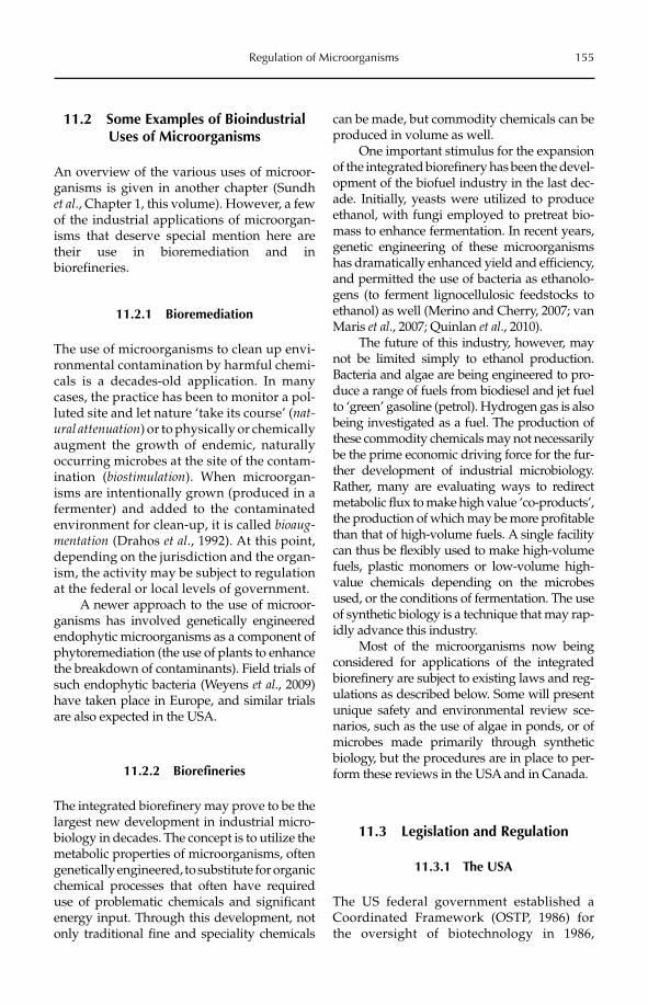
Regulation of Microorganisms 155
11.2 Some Examples of Bioindustrial Uses of Microorganisms
An overview of the various uses of microor-ganisms is given in another chapter (Sundh et al., Chapter 1, this volume). However, a few of the industrial applications of microorgan-isms that deserve special mention here are their use in bioremediation and in biorefineries.
11.2.1 Bioremediation
The use of microorganisms to clean up envi-ronmental contamination by harmful chemi-cals is a decades-old application. In many cases, the practice has been to monitor a pol-luted site and let nature ‘take its course’ (nat-ural attenuation) or to physically or chemically augment the growth of endemic, naturally occurring microbes at the site of the contam-ination (biostimulation). When microorgan-isms are intentionally grown (produced in a fermenter) and added to the contaminated environment for clean-up, it is called bioaug-mentation (Drahos et al., 1992). At this point, depending on the jurisdiction and the organ-ism, the activity may be subject to regulation at the federal or local levels of government.
A newer approach to the use of microor-ganisms has involved genetically engineered endophytic microorganisms as a component of phytoremediation (the use of plants to enhance the breakdown of contaminants). Field trials of such endophytic bacteria (Weyens et al., 2009) have taken place in Europe, and similar trials are also expected in the USA.
11.2.2 Biorefineries
The integrated biorefinery may prove to be the largest new development in industrial micro-biology in decades. The concept is to utilize the metabolic properties of microorganisms, often genetically engineered, to substitute for organic chemical processes that often have required use of problematic chemicals and significant energy input. Through this development, not only traditional fine and speciality chemicals
can be made, but commodity chemicals can be produced in volume as well.
One important stimulus for the expansion of the integrated biorefinery has been the devel-opment of the biofuel industry in the last dec-ade. Initially, yeasts were utilized to produce ethanol, with fungi employed to pretreat bio-mass to enhance fermentation. In recent years, genetic engineering of these microorganisms has dramatically enhanced yield and efficiency, and permitted the use of bacteria as ethanolo-gens (to ferment lignocellulosic feedstocks to ethanol) as well (Merino and Cherry, 2007; van Maris et al., 2007; Quinlan et al., 2010).
The future of this industry, however, may not be limited simply to ethanol production. Bacteria and algae are being engineered to pro-duce a range of fuels from biodiesel and jet fuel to ‘green’ gasoline (petrol). Hydrogen gas is also being investigated as a fuel. The production of these commodity chemicals may not necessarily be the prime economic driving force for the fur-ther development of industrial microbiology. Rather, many are evaluating ways to redirect metabolic flux to make high value ‘co-products’, the production of which may be more profitable than that of high-volume fuels. A single facility can thus be flexibly used to make high-volume fuels, plastic monomers or low-volume high-value chemicals depending on the microbes used, or the conditions of fermentation. The use of synthetic biology is a technique that may rap-idly advance this industry.
Most of the microorganisms now being considered for applications of the integrated biorefinery are subject to existing laws and reg-ulations as described below. Some will present unique safety and environmental review sce-narios, such as the use of algae in ponds, or of microbes made primarily through synthetic biology, but the procedures are in place to per-form these reviews in the USA and in Canada.
11.3 Legislation and Regulation
11.3.1 The USA
The US federal government established a Coordinated Framework (OSTP, 1986) for the oversight of biotechnology in 1986,
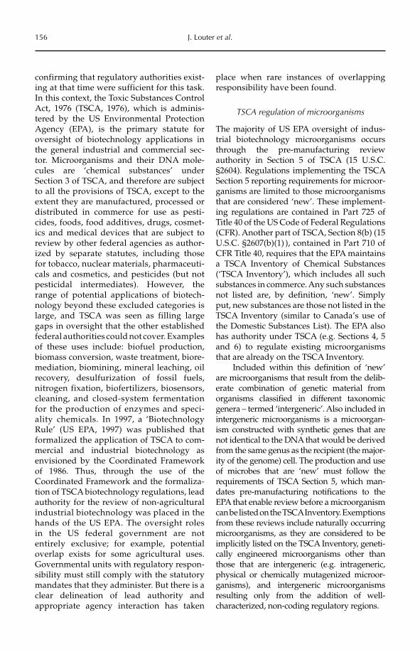
156 J. Louter et al.
confirming that regulatory authorities exist-ing at that time were sufficient for this task. In this context, the Toxic Substances Control Act, 1976 (TSCA, 1976), which is adminis-tered by the US Environmental Protection Agency (EPA), is the primary statute for oversight of biotechnology applications in the general industrial and commercial sec-tor. Microorganisms and their DNA mole-cules are ‘chemical substances’ under Section 3 of TSCA, and therefore are subject to all the provisions of TSCA, except to the extent they are manufactured, processed or distributed in commerce for use as pesti-cides, foods, food additives, drugs, cosmet-ics and medical devices that are subject to review by other federal agencies as author-ized by separate statutes, including those for tobacco, nuclear materials, pharmaceuti-cals and cosmetics, and pesticides (but not pesticidal intermediates). However, the range of potential applications of biotech-nology beyond these excluded categories is large, and TSCA was seen as filling large gaps in oversight that the other established federal authorities could not cover. Examples of these uses include: biofuel production, biomass conversion, waste treatment, biore-mediation, biomining, mineral leaching, oil recovery, desulfurization of fossil fuels, nitrogen fixation, biofertilizers, biosensors, cleaning, and closed-system fermentation for the production of enzymes and speci-ality chemicals. In 1997, a ‘Biotechnology Rule’ (US EPA, 1997) was published that formalized the application of TSCA to com-mercial and industrial biotechnology as envisioned by the Coordinated Framework of 1986. Thus, through the use of the Coordinated Framework and the formaliza-tion of TSCA biotechnology regulations, lead authority for the review of non-agricultural industrial biotechnology was placed in the hands of the US EPA. The oversight roles in the US federal government are not entirely exclusive; for example, potential overlap exists for some agricultural uses. Governmental units with regulatory respon-sibility must still comply with the statutory mandates that they administer. But there is a clear delineation of lead authority and appropriate agency interaction has taken
place when rare instances of overlapping responsibility have been found.
TSCA regulation of microorganisms
The majority of US EPA oversight of indus-trial biotechnology microorganisms occurs through the pre-manufacturing review authority in Section 5 of TSCA (15 U.S.C. §2604). Regulations implementing the TSCA Section 5 reporting requirements for microor-ganisms are limited to those microorganisms that are considered ‘new’. These implement-ing regulations are contained in Part 725 of Title 40 of the US Code of Federal Regulations(CFR). Another part of TSCA, Section 8(b) (15 U.S.C. §2607(b)(1) ), contained in Part 710 of CFR Title 40, requires that the EPA maintains a TSCA Inventory of Chemical Substances(‘TSCA Inventory’), which includes all such substances in commerce. Any such substances not listed are, by definition, ‘new’. Simply put, new substances are those not listed in the TSCA Inventory (similar to Canada’s use of the Domestic Substances List). The EPA also has authority under TSCA (e.g. Sections 4, 5 and 6) to regulate existing microorganisms that are already on the TSCA Inventory.
Included within this definition of ‘new’ are microorganisms that result from the delib-erate combination of genetic material from organisms classified in different taxonomic genera – termed ‘intergeneric’. Also included in intergeneric microorganisms is a microorgan-ism constructed with synthetic genes that are not identical to the DNA that would be derived from the same genus as the recipient (the major-ity of the genome) cell. The production and use of microbes that are ‘new’ must follow the requirements of TSCA Section 5, which man-dates pre-manufacturing notifications to the EPA that enable review before a microorganism can be listed on the TSCA Inventory. Exemptions from these reviews include naturally occurring microorganisms, as they are considered to be implicitly listed on the TSCA Inventory, geneti-cally engineered microorganisms other than those that are intergeneric (e.g. intrageneric, physical or chemically mutagenized microor-ganisms), and intergeneric microorganisms resulting only from the addition of well- characterized, non-coding regulatory regions.
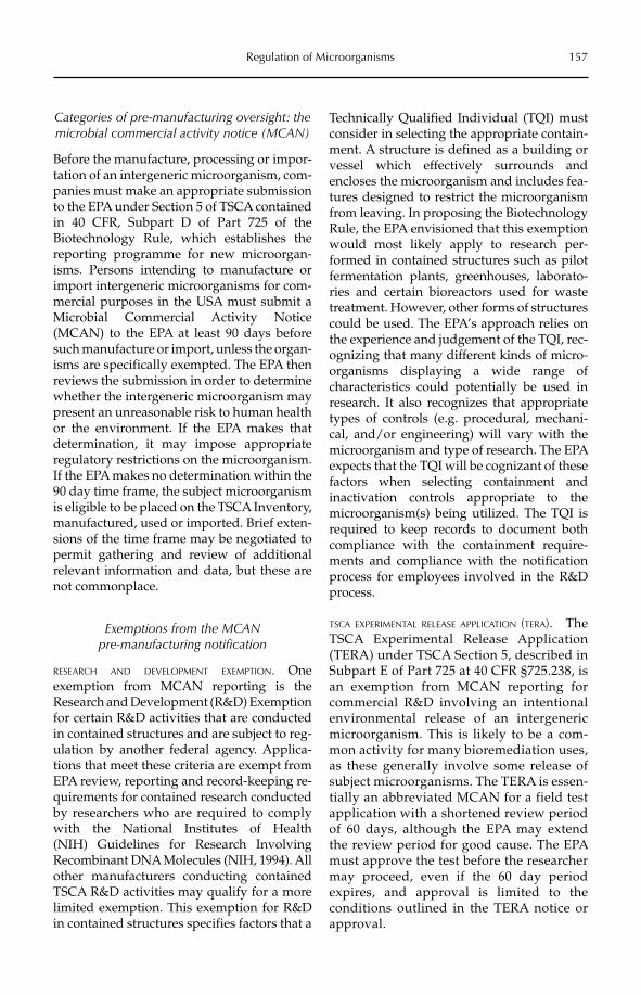
Regulation of Microorganisms 157
Categories of pre-manufacturing oversight: the microbial commercial activity notice (MCAN)
Before the manufacture, processing or impor-tation of an intergeneric microorganism, com-panies must make an appropriate submission to the EPA under Section 5 of TSCA contained in 40 CFR, Subpart D of Part 725 of the Biotechnology Rule, which establishes the reporting programme for new microorgan-isms. Persons intending to manufacture or import intergeneric microorganisms for com-mercial purposes in the USA must submit a Microbial Commercial Activity Notice (MCAN) to the EPA at least 90 days before such manufacture or import, unless the organ-isms are specifically exempted. The EPA then reviews the submission in order to determine whether the intergeneric microorganism may present an unreasonable risk to human health or the environment. If the EPA makes that determination, it may impose appropriate regulatory restrictions on the microorganism. If the EPA makes no determination within the 90 day time frame, the subject microorganism is eligible to be placed on the TSCA Inventory, manufactured, used or imported. Brief exten-sions of the time frame may be negotiated to permit gathering and review of additional relevant information and data, but these are not commonplace.
Exemptions from the MCAN pre-manufacturing notification
RESEARCH AND DEVELOPMENT EXEMPTION. Oneexemption from MCAN reporting is the Research and Development (R&D) Exemption for certain R&D activities that are conducted in contained structures and are subject to reg-ulation by another federal agency. Applica-tions that meet these criteria are exempt from EPA review, reporting and record-keeping re -quirements for contained research conducted by researchers who are required to comply with the National Institutes of Health (NIH) Guidelines for Research Involving Re combinant DNA Molecules (NIH, 1994). All other manufacturers conducting contained TSCA R&D activities may qualify for a more limited exemption. This exemption for R&D in contained structures specifies factors that a
Technically Qualified Individual (TQI) must consider in selecting the appropriate contain-ment. A structure is defined as a building or vessel which effectively surrounds and encloses the microorganism and includes fea-tures designed to restrict the microorganism from leaving. In proposing the Biotechnology Rule, the EPA envisioned that this exemption would most likely apply to research per-formed in contained structures such as pilot fermentation plants, greenhouses, laborato-ries and certain bioreactors used for waste treatment. However, other forms of structures could be used. The EPA’s approach relies on the experience and judgement of the TQI, rec-ognizing that many different kinds of micro-organisms displaying a wide range of characteristics could potentially be used in research. It also recognizes that appropriate types of controls (e.g. procedural, mechani-cal, and/or engineering) will vary with the microorganism and type of research. The EPA expects that the TQI will be cognizant of these factors when selecting containment and inactivation controls appropriate to the microorganism(s) being utilized. The TQI is required to keep records to document both compliance with the containment require-ments and compliance with the notification process for employees involved in the R&D process.
TSCA EXPERIMENTAL RELEASE APPLICATION (TERA). The TSCA Experimental Release Application (TERA) under TSCA Section 5, described in Subpart E of Part 725 at 40 CFR §725.238, is an exemption from MCAN reporting for commercial R&D involving an intentional environmental release of an intergeneric microorganism. This is likely to be a com-mon activity for many bioremediation uses, as these generally involve some release of subject microorganisms. The TERA is essen-tially an abbreviated MCAN for a field test application with a shortened review period of 60 days, although the EPA may extend the review period for good cause. The EPA must approve the test before the researcher may proceed, even if the 60 day period expires, and approval is limited to the conditions outlined in the TERA notice or approval.
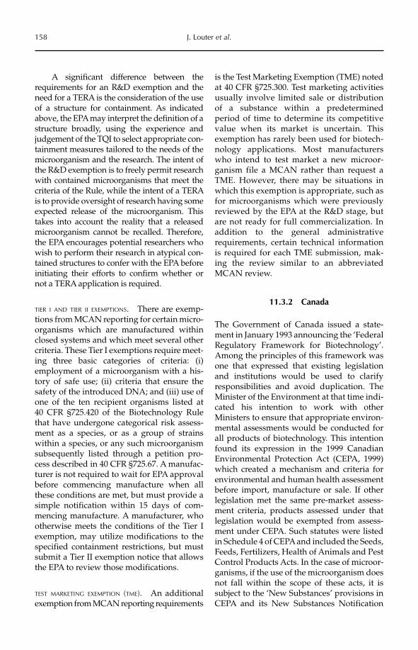
158 J. Louter et al.
A significant difference between the requirements for an R&D exemption and the need for a TERA is the consideration of the use of a structure for containment. As indicated above, the EPA may interpret the definition of a structure broadly, using the experience and judgement of the TQI to select appropriate con-tainment measures tailored to the needs of the microorganism and the research. The intent of the R&D exemption is to freely permit research with contained microorganisms that meet the criteria of the Rule, while the intent of a TERA is to provide oversight of research having some expected release of the microorganism. This takes into account the reality that a released microorganism cannot be recalled. Therefore, the EPA encourages potential researchers who wish to perform their research in atypical con-tained structures to confer with the EPA before initiating their efforts to confirm whether or not a TERA application is required.
TIER I AND TIER II EXEMPTIONS. There are exemp-tions from MCAN reporting for certain micro-organisms which are manufactured within closed systems and which meet several other criteria. These Tier I exemptions require meet-ing three basic categories of criteria: (i) employment of a microorganism with a his-tory of safe use; (ii) criteria that ensure the safety of the introduced DNA; and (iii) use of one of the ten recipient organisms listed at 40 CFR §725.420 of the Biotechnology Rule that have undergone categorical risk assess-ment as a species, or as a group of strains within a species, or any such microorganism subsequently listed through a petition pro-cess described in 40 CFR §725.67. A manufac-turer is not required to wait for EPA approval before commencing manufacture when all these conditions are met, but must provide a simple notification within 15 days of com-mencing manufacture. A manufacturer, who otherwise meets the conditions of the Tier I exemption, may utilize modifications to the specified containment restrictions, but must submit a Tier II exemption notice that allows the EPA to review those modifications.
TEST MARKETING EXEMPTION (TME). An additional exemption from MCAN reporting requirements
is the Test Marketing Exemption (TME) noted at 40 CFR §725.300. Test marketing activities usually involve limited sale or distribution of a substance within a predetermined period of time to determine its competitive value when its market is uncertain. This exemption has rarely been used for biotech-nology applications. Most manufacturers who intend to test market a new microor-ganism file a MCAN rather than request a TME. However, there may be situations in which this exemption is appropriate, such as for microorganisms which were previously reviewed by the EPA at the R&D stage, but are not ready for full commercialization. In addition to the general administrative requirements, certain technical information is required for each TME submission, mak-ing the review similar to an abbreviated MCAN review.
11.3.2 Canada
The Government of Canada issued a state-ment in January 1993 announcing the ‘Federal Regulatory Framework for Biotechnology’. Among the principles of this framework was one that expressed that existing legislation and institutions would be used to clarify responsibilities and avoid duplication. The Minister of the Environment at that time indi-cated his intention to work with other Ministers to ensure that appropriate environ-mental assessments would be conducted for all products of biotechnology. This intention found its expression in the 1999 Canadian Environmental Protection Act (CEPA, 1999) which created a mechanism and criteria for environmental and human health assessment before import, manufacture or sale. If other legislation met the same pre-market assess-ment criteria, products assessed under that legislation would be exempted from assess-ment under CEPA. Such statutes were listed in Schedule 4 of CEPA and included the Seeds, Feeds, Fertilizers, Health of Animals and Pest Control Products Acts. In the case of microor-ganisms, if the use of the microorganism does not fall within the scope of these acts, it is subject to the ‘New Substances’ provisions in CEPA and its New Substances Notification
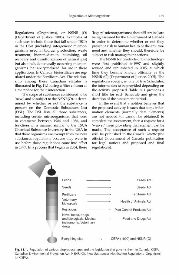
Regulation of Microorganisms 159
Regulations (Organisms), or NSNR (O) (Department of Justice, 2005). Examples of such uses include those that fall under TSCA in the USA (including intergeneric microor-ganisms used in biofuel production, waste treatment, bioremediation, biomining, oil recovery and desulfurization of natural gas) but also include naturally occurring microor-ganisms that are ‘produced’ for use in these applications. In Canada, biofertilizers are reg-ulated under the Fertilizers Act. The relation-ship among these Canadian statutes is illustrated in Fig. 11.1, using a filter column as a metaphor for their interaction.
The scope of substances considered to be ‘new’, and so subject to the NSNR(O), is deter-mined by whether or not the substance is present on the Domestic Substances List (DSL). The DSL lists all those substances, including certain microorganisms, that were in commerce between 1984 and 1986, and functions in a manner similar to the TSCA Chemical Substance Inventory in the USA in that these organisms are exempt from the new substances regulations because they were in use before those regulations came into effect in 1997. In a process that began in 2004, these
‘legacy’ microorganisms (about 65 strains) are being assessed by the Government of Canada in order to determine whether or not they present a risk to human health or the environ-ment and whether they should, therefore, be subject to risk management actions.
The NSNR for products of biotechnology were first published in1997 and slightly revised and renumbered in 2005, at which time they became known officially as the NSNR (O) (Department of Justice, 2005). The regulations specify, in one of five Schedules, the information to be provided depending on the activity proposed. Table 11.1 provides a short title for each Schedule and gives the duration of the assessment period.
In the event that a notifier believes that the proposed activity is such that some infor-mation elements (normally data elements) are not needed (or cannot be obtained) to complete the assessment, then a request for a ‘waiver’ from providing that element can be made. The acceptance of such a request will be published in the Canada Gazette (the official Government of Canada publication for legal notices and proposed and final regulations).
Pesticides Pest Control Products Act
Feeds Feeds Act
Seeds Seeds Act
Fertilizers Fertilizers Act
Veterinarybiologicals
Health of Animals Act
Food and Drugs ActNovel foods, drugsand biologicals, Medicalinstruments, Veterinarydrugs
Everything else CEPA (1999) and NSNR (O)
Fig. 11.1. Regulation of various bioproduct types and the legislation that governs them in Canada. CEPA, Canadian Environmental Protection Act; NSNR (O), New Substances Notification Regulations (Organisms) (of CEPA).
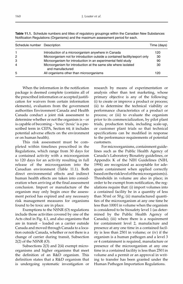
160 J. Louter et al.
When the information in the notification package is deemed complete (contains all of the prescribed information or accepted justifi-cation for waivers from certain information elements), evaluators from the government authorities Environment Canada and Health Canada conduct a joint risk assessment to determine whether or not the organism is – or is capable of becoming – ‘toxic’ (toxic’ is a pre-scribed term in CEPA, Section 64; it includes potential adverse effects on the environment or on human health).
This risk assessment must be com-pleted within timelines prescribed in the Regulations, which range from 30 days for a contained activity with a microorganism to 120 days for an activity resulting in full release of the microorganism into the Canadian environment (Table 11.1). Both direct environmental effects and indirect human health effects are taken into consid-eration when arriving at the final assessment conclusion. Import or manufacture of the organism may only begin once the assess-ment period has expired and any necessary risk management measures for organisms found to be toxic are in place.
Exemptions to the NSNR (O) regulations include those activities covered by one of the Acts cited in Fig. 4.1, and also organisms that are in transit – loaded on a carrier outside Canada and moved through Canada to a loca-tion outside Canada, whether or not there is a change of carrier during transit, Subsection 2(2) of the NSNR (O).
Subsections 2(3) and 2(4) exempt micro-organisms and higher organisms that meet the definition of an R&D organism. This definition states that a R&D organism that is undergoing systematic investigation or
research by means of experimentation or analysis other than test marketing, whose primary objective is any of the following: (i) to create or improve a product or process; (ii) to determine the technical viability or performance characteristics of a product or process; or (iii) to evaluate the organism prior to its commercialization, by pilot plant trials, production trials, including scale-up, or custo mer plant trials so that technical specificat ions can be modified in response to the performance requirements of potential customers.
For microorganisms, containment guide-lines such as the Public Health Agency of Canada’s Laboratory Biosafety guidelines, or Appendix K of the NIH Guidelines (NIH, 1994) are recognized as acceptable for ade-quate containment when applied correctly based on the risk level of the microorganism(s). Thresholds in volume are also in place; in order to be exempt from notification, the reg-ulations require that: (i) import volumes into a contained facility be in a quantity of less than 50 ml or 50 g; (ii) manufactured quanti-ties of the microorganism at any one time be less than 1000 l in volume when the organism is considered to be biosafety level 1 (as deter-mined by the Public Health Agency of Canada); (iii) where there is a requirement for containment level 2, manufacture and presence at any one time in a contained facil-ity is less than 250 l in volume; or (iv) if the organism is a human pathogen and a level 3 or 4 containment is required, manufacture or presence of the microorganism at any one time in a contained facility is less than 250 l in volume and a permit or an approval in writ-ing to transfer has been granted under the Human Pathogen Importation Regulations.
Table 11.1. Schedule numbers and titles of regulatory groupings within the Canadian New Substances Notification Regulations (Organisms) and the maximum assessment period for each.
Schedule number Description Time (days)
1 Introduction of a microorganism anywhere in Canada 1202 Microorganism not for introduction outside a contained facility/export only 303 Microorganism for introduction in an experimental field study 904 Microorganism for introduction at the same site where isolated
and manufactured30
5 All organisms other than microorganisms 120
Regulation of Microorganisms 161
11.4 The Risk Assessment Process
11.4.1 The USA
Under the pre-manufacturing review pro-gramme authorized by Section 5 of TSCA, within the time period specified by rule or statute, which varies according to the type of submission, the EPA conducts a risk assess-ment on the ‘new’ microorganism, under the paradigm that ‘Risk is a function of Hazard × Exposure’. Separate assessments for various elements of risk are integrated into a final risk assessment. The components include:
1. a verification of the identification of the subject microorganism;2. a human health hazard assessment;3. an ecological effects hazard assessment;4. a report that analyses the construction of the microorganism and summarizes the perti-nent chemical information and production volume;5. an analysis of the genetic construct that evaluates any potential hazards associated with the genetic modifications and the poten-tial for horizontal gene transfer;6. an engineering report that assesses manu-facturing processes affecting worker exposure and microbial releases to the environment through manufacturing or during field appli-cations; and7. an exposure assessment that evaluates the potential for survival, reproduction and spread of the microorganism, and the expo-sure of environmental receptors and of the general population.
Unlike review under CEPA in Canada, there is no specified schedule of information elements under Section 5 of TSCA for reviews of new substances. Rather, submitters must provide to the EPA all relevant data and infor-mation in their possession or reasonably ascertainable. These data must be sufficient to enable the EPA to complete a risk assessment. If a submission of any type contains insuffi-cient information to proceed with a review, the EPA may request the submitter to allow the review time to be extended in order that they may provide the necessary information. The EPA also has risk management options
that may be employed to mitigate the effect of uncertainty due to data or information limita-tions, as described in the last paragraph of this section.
Because TSCA is a risk–benefit statute (unlike CEPA in Canada), the risks of using the microorganism determined in the assess-ment are weighed against the benefits to soci-ety (which are evaluated in an economics analysis) to arrive at the final risk manage-ment decision. Several outcomes are possi-ble as a result of the review process. Under the pre-manufacturing review programme authorized by TSCA Section 5, the EPA may determine that there is sufficient informa-tion to conclude that the microorganism presents ‘no unreasonable risk of injury to human health or the environment’, in which case the Agency takes no regulatory action and the company may take action (e.g. com-mence manufacture, begin research or test the market) after the regulatory time frame expires. The time frames are firm, so that no action may take place until the time has expired, even if the EPA finishes its review early. In the case of the MCAN, if the EPA does not otherwise notify the submitter by the end of the 90 day period, the latter may commence its commercial activity without constraint.
Another outcome may be that there is sufficient information to determine that the microorganism presents ‘an unreasonable risk of injury to human health or the environ-ment’. In this case the EPA has a wide range of means to prohibit or restrict the production or use of the microorganism, from limits to size or method of production, to a complete ban on production and use. A last category of outcomes includes the case previously men-tioned where there is insufficient information to determine for certain that the risk is ‘unrea-sonable’, but there is sufficient information to imply that possibility exists for unreasonable risk and/or substantial/significant exposure. In such cases the Agency may negotiate a Section 5(e) Consent Order to restrict the use of the microorganism and to specify the data needed to lift the Consent Order. If the sub-mitter does not agree to negotiate with the EPA, the Agency may impose restrictions based on the information available and the submitter may choose to use the courts to
162 J. Louter et al.
seek relief. However, this last option has never been considered by any manufacturer seeking to produce a biotechnology-derived microorganism subject to TSCA. The need for Consent Orders has also proved to be a rare occurrence.
11.4.2 Canada
The risk assessment process, outcomes and risk management measures described in this section are applicable to all microorganisms subject to the NSNR (O). The initial source of information used in the risk assessment is that provided by a proponent through the notification package (schedule as dictated by the proposed activity; see Table 11.1), but evaluators also use in-house information and any additional information available in the public domain. This information helps to assist the evaluators in determining both potential hazards associated with the organ-ism and potential pathways of exposure to them, in order to estimate risk and the likeli-hood that an organism is – or is capable of becoming – ‘toxic’ as defined under Section 64 of the Act. Therefore, Section 64 effectively provides the end points to be addressed through risk assessment. If necessary, evalua-tion teams may contact external experts (both in Canada and abroad) to further inform the risk assessment without divulging any confi-dential business information. These consul-tations are generally driven by specific technical or scientific questions posed by the evaluation teams and are only used when in-house expertise cannot adequately address uncertainties.
The information elements listed in each schedule provide the minimum information that must be provided by the proponent so that evaluation staff may begin the risk assessment. In broad terms, the information required to be provided in a notification pack-age includes information in respect of: (i) the identification and characteristics of the organ-ism (including any genetic modifications); (ii) the manufacture and import details of the organism; (iii) the introduction of the organ-ism into the environment (including any con-tainment or other measures in place to prevent
introduction into the environment); (iv) the environmental fate of the organism; (v) the ecological effects of the organism; and (vi) the human health effects of the organ-ism. Note that there is no assessment of benefits or evaluation of performance claims under this act and regulation.
The evaluation and risk assessment is a joint responsibility between two Ministers: from Environment Canada, which conducts an assessment for environmental effects, including any effects on biodiversity; and from Health Canada, which conducts an assessment for indirect human health effects (that is, exposure resulting from the environ-mental release of the organism). As men-tioned earlier (Section 11.3.2), direct human health effects may be assessed under other legislation, such as the Food and Drugs Act, as appropriate. The ability to import or man-ufacture under CEPA does not exempt the notifier from any other laws or regulations that are in force in Canada and that may apply to the organism or activities involving the organism.
Currently, a memorandum of under-standing exists between Environment Canada and Health Canada for the risk assessment of organisms contained in products regulated under the Food and Drugs Act. Such assessments, conducted by Health Canada’s Environmental Assessment Unit, assesses environmental risk under CEPA on behalf of Environment Canada for organisms that have been notified under the Food and Drugs Act.
The Framework for Science-Based Risk Assessment of Microorganisms out-lines the systematic steps used by the Health Canada and Environment Canada sub-stances assessment programmes to conduct science-based risk assessments of microor-ganisms (Environment Canada and Health Canada, 2011). The assessment is based on whether the microorganism is toxic as defined under Section 64 of CEPA. In other words, it describes a framework of the basic concepts considered when conducting this assessment based on available information on hazard and exposure for both ‘existing’ (i.e. those on the Domestic Substance List) and ‘new’ microorganisms (i.e. those noti-fied under the NSNR (O) ). The approach is
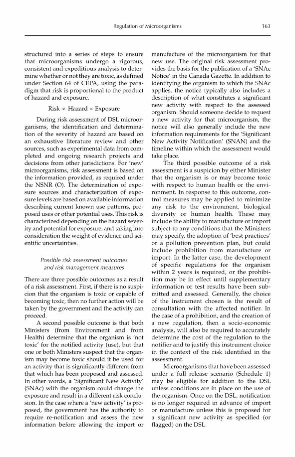
Regulation of Microorganisms 163
structured into a series of steps to ensure that microorganisms undergo a rigorous, consistent and expeditious analysis to deter-mine whether or not they are toxic, as defined under Section 64 of CEPA, using the para-digm that risk is proportional to the product of hazard and exposure.
Risk µ Hazard ´ Exposure
During risk assessment of DSL microor-ganisms, the identification and determina-tion of the severity of hazard are based on an exhaustive literature review and other sources, such as experimental data from com-pleted and ongoing research projects and decisions from other jurisdictions. For ‘new’ microorganisms, risk assessment is based on the information provided, as required under the NSNR (O). The determination of expo-sure sources and characterization of expo-sure levels are based on available information describing current known use patterns, pro-posed uses or other potential uses. This risk is characterized depending on the hazard sever-ity and potential for exposure, and taking into consideration the weight of evidence and sci-entific uncertainties.
Possible risk assessment outcomes and risk management measures
There are three possible outcomes as a result of a risk assessment. First, if there is no suspi-cion that the organism is toxic or capable of becoming toxic, then no further action will be taken by the government and the activity can proceed.
A second possible outcome is that both Ministers (from Environment and from Health) determine that the organism is ‘not toxic’ for the notified activity (use), but that one or both Ministers suspect that the organ-ism may become toxic should it be used for an activity that is significantly different from that which has been proposed and assessed. In other words, a ‘Significant New Activity’ (SNAc) with the organism could change the exposure and result in a different risk conclu-sion. In the case where a ‘new activity’ is pro-posed, the government has the authority to require re-notification and assess the new information before allowing the import or
manufacture of the microorganism for that new use. The original risk assessment pro-vides the basis for the publication of a ‘SNAc Notice’ in the Canada Gazette. In addition to identifying the organism to which the SNAc applies, the notice typically also includes a description of what constitutes a significant new activity with respect to the assessed organism. Should someone decide to request a new activity for that microorganism, the notice will also generally include the new information requirements for the ‘Significant New Activity Notification’ (SNAN) and the timeline within which the assessment would take place.
The third possible outcome of a risk assessment is a suspicion by either Minister that the organism is or may become toxic with respect to human health or the envi-ronment. In response to this outcome, con-trol measures may be applied to minimize any risk to the environment, biological diversity or human health. These may include the ability to manufacture or import subject to any conditions that the Ministers may specify, the adoption of ‘best practices’ or a pollution prevention plan, but could include prohibition from manufacture or import. In the latter case, the development of specific regulations for the organism within 2 years is required, or the prohibi-tion may be in effect until supplementary information or test results have been sub-mitted and assessed. Generally, the choice of the instrument chosen is the result of consultation with the affected notifier. In the case of a prohibition, and the creation of a new regulation, then a socio-economic analysis, will also be required to accurately determine the cost of the regulation to the notifier and to justify this instrument choice in the context of the risk identified in the assessment.
Microorganisms that have been assessed under a full release scenario (Schedule 1) may be eligible for addition to the DSL unless conditions are in place on the use of the organism. Once on the DSL, notification is no longer required in advance of import or manufacture unless this is proposed for a significant new activity as specified (or flagged) on the DSL.
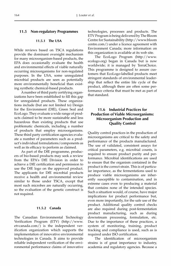
164 J. Louter et al.
11.5 Non-regulatory Programmes
11.5.1 The USA
While reviews based on TSCA regulations provide the dominant oversight mechanism for many microorganism-based products, the EPA does occasionally evaluate the health and environmental effects of viable naturally occurring microorganisms for non-regulated purposes. In the USA, some unregulated microbial products are seen as potentially more environmentally beneficial than exist-ing synthetic chemical-based products.
A number of third party certifying organ-izations have been established to fill this gap for unregulated products. These organiza-tions include (but are not limited to) Design for the Environment (DfE), Green Seal and EcoLogo. They evaluate a wide range of prod-ucts claimed to be more sustainable and less hazardous than existing products that use problematic chemicals, including a number of products that employ microorganisms. These third party certification agencies evalu-ate a number of parameters such as a prod-uct’s individual formulations/components as well as its efficacy to perform as claimed.
As part of the DfE programme, produc-ers of bio-based products may seek a review from the EPA’s DfE Division in order to achieve a DfE certification and permission to use the DfE logo on the approved product. The applicants for DfE microbial products receive a health and environmental review similar to those under TSCA, except that most such microbes are naturally occurring, so the evaluation of the genetic construct is not required.
11.5.2 Canada
The Canadian Environmental Technology Verification Program (ETV) (http://www.etvcanada.com/) is the independent ver-ification organization which supports the implementation of innovative environmental technologies in Canada. It aims to provide reliable independent verification of the envi-ronmental performance claims of innovative
technologies, processes and products. The ETV Program is being delivered by The Bloom Centre for Sustainability (http://www.bloom centre.com/) under a licence agreement with Environment Canada; more information on this organization is available at its web site.
The EcoLogo Program (http://www.ecologo.org) began in Canada but is now worldwide; it is managed by TerraChoice. This programme is designed to assure cus-tomers that EcoLogo-labelled products meet stringent standards of environmental leader-ship that reflect the entire life cycle of the product, although there are often some per-formance criteria that must be met as part of that standard.
11.6 Industrial Practices for Production of Viable Microorganisms:
Microorganism Production and Quality Control
Quality control practices in the production of microorganisms are critical to the safety and performance of the products manufactured. The use of validated, consistent assays for critical parameters, e.g. microbial counts, is required to ensure product purity and per-formance. Microbial identifications are used to ensure that the organism contained in the product is the correct strain. This is of particu-lar importance, as the fermentations used to produce viable microorganisms are inher-ently susceptible to contamination, and in extreme cases even to producing a material that contains none of the intended species. Such a situation would, of course, have major implications for product performance but, even more importantly, for the safe use of the product. Additional quality control checks are also required during post-fermentation product manufacturing, such as during downstream processing, formulation, etc. Due to the importance of these practices, a system of monitoring, training, product tracking and compliance is used, such as is required under ISO certification.
The identification of microorganism strains is of great importance to industry, academia and regulatory agencies. Because a
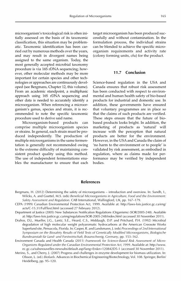
Regulation of Microorganisms 165
microorganism’s toxicological risk is often ini-tially assessed on the basis of its taxonomic classification, this situation may be problem-atic. Taxonomic identification has been car-ried out by numerous methods over the years, and may result in divergent names being assigned to the same organism. Today, the most generally accepted microbial taxonomy procedure is via 16S rDNA sequencing; how-ever, other molecular methods may be more important for certain species and other tech-nologies or approaches are continuously devel-oped (see Bergmans, Chapter 12, this volume). From an academic standpoint, a multiphasic approach using 16S rDNA sequencing and other data is needed to accurately identify a microorganism. When referencing a microor-ganism’s genus, species and strain, it is rec-ommended to note the specific taxonomic procedure used to derive said name.
Microorganism-based products can comprise multiple microorganism species or strains. In general, each strain must be pro-duced independently. The production of multiple microorganisms in the same fermen-tation is generally not recommended owing to the extreme difficulty of maintaining con-sistent product quality using this method. The use of independent fermentations ena-bles the manufacturer to ensure that each
target microorganism has been produced suc-cessfully and without contamination. In the formulation process, the individual strains can be blended to achieve the specific micro-organism requirements and activity rate (colony forming units, cfu) for the product.
11.7 Conclusion
Science-based regulation in the USA and Canada ensures that robust risk assessment has been conducted with respect to environ-mental and human health safety of bio-based products for industrial and domestic use. In addition, these governments have ensured that voluntary programmes are in place so that the claims of such products are verified. These steps ensure that the future of bio-based products looks bright. In addition, the marketing of products as ‘natural’ will increase with the perception that natural products are better for the environment. However, in the USA and Canada the claim of ‘no harm to the environment or to people’ is validated by risk assessment, as embodied in regulation, where as claims made for per-formance may be verified by independent bodies.
References
Bergmans, H. (2012) Determining the safety of microorganisms – introduction and overview. In: Sundh, I., Wilcks, A. and Goettel, M.S. (eds) Beneficial Microorganisms in Agriculture, Food and the Environment: Safety Assessment and Regulation. CAB International, Wallingford, UK, pp. 167–179.
CEPA (1999) Canadian Environmental Protection Act, 1999. Available at: http://laws-lois.justice.gc.ca/eng/acts/C-15.31/FullText.html (accessed 27 February 2012).
Department of Justice (2005) New Substances Notification Regulations (Organisms) (SOR/2005-248). Available at: http://laws-lois.justice.gc.ca/eng/regulations/SOR-2005-248/index.html (accessed 30 November 2011).
Drahos, D.J., Mueller, J.G., Lantz, S.E., Heard, C.S., Middaugh, D.P. and Pritchard, P.H. (1992) Microbial degradation of high molecular weight polyaromatic hydrocarbons at the American Creosote Works Superfund site, Pensacola, Florida. In: Casper, R. and Landsmann, J. (eds) Proceedings of 2nd International Symposium on the Biosafety Results of Field Tests of Genetically Modified Microorganisms, Biologische Bundesanstalt für Land- und Forstwirtschaft, Braunschweig, Germany, pp. 153–162.
Environment Canada and Health Canada (2011) Framework for Science-Based Risk Assessment of Micro-Organisms Regulated under the Canadian Environmental Protection Act, 1999. Available at: http://www.ec.gc.ca/subsnouvelles-newsubs/default.asp?lang=En&n=120842D5-1 (accessed 30 November 2011).
Merino, S., and Cherry, J. (2007) Progress and challenges in enzyme development for biomass utilization. In: Olsson, L. (ed.) Biofuels. Advances in Biochemical Engineering/Biotechnology, Vol. 108. Springer, Berlin/Heidelberg, pp. 95–120.
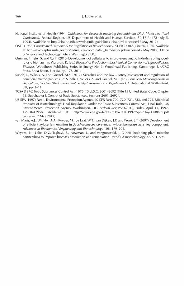
166 J. Louter et al.
National Institutes of Health (1994) Guidelines for Research Involving Recombinant DNA Molecules (NIH Guidelines). Federal Register, US Department of Health and Human Services, 59 FR 34472 (July 5, 1994). Available at: http://oba.od.nih.gov/rdna/nih_guidelines_oba.html (accessed 7 May 2012).
OSTP (1986) Coordinated Framework for Regulation of Biotechnology. 51 FR 23302, June 26, 1986. Available at: http://www.aphis.usda.gov/brs/fedregister/coordinated_framework.pdf (accessed 7 May 2012). Office of Science and Technology Policy, Washington, DC.
Quinlan, J., Teter, S. and Xu, F. (2010) Development of cellulases to improve enzymatic hydrolysis of lignocel-lulosic biomass. In: Waldron, K. (ed.) Bioalcohol Production: Biochemical Conversion of Lignocellulosic Biomass. Woodhead Publishing Series in Energy No. 3. Woodhead Publishing, Cambridge, UK/CRC Press, Boca Raton, Florida, pp. 178–201.
Sundh, I., Wilcks, A. and Goettel, M.S. (2012) Microbes and the law – safety assessment and regulation of beneficial microorganisms. In: Sundh, I., Wilcks, A. and Goettel, M.S. (eds) Beneficial Microorganisms in Agriculture, Food and the Environment: Safety Assessment and Regulation. CAB International, Wallingford, UK, pp. 1–11.
TCSA (1976) Toxic Substances Control Act, 1976, 15 U.S.C. 2601–2692 [Title 15 United States Code, Chapter 53, Subchapter I, Control of Toxic Substances, Sections 2601–2692].
US EPA (1997) Part II, Environmental Protection Agency, 40 CFR Parts 700, 720, 721, 723, and 725. Microbial Products of Biotechnology; Final Regulation Under the Toxic Substances Control Act; Final Rule. US Environmental Protection Agency, Washington, DC. Federal Register 62(70), Friday, April 11, 1997, 17910–17958. Available at: http://www.epa.gov/fedrgstr/EPA-TOX/1997/April/Day-11/t8669.pdf (accessed 7 May 2012).
van Maris, A.J., Winkler, A.A., Kuyper, M., de Laat, W.T., van Dijken, J.P. and Pronk, J.T. (2007) Development of efficient xylose fermentation in Saccharomyces cerevisiae: xylose isomerase as a key component. Advances in Biochemical Engineering and Biotechnology 108, 179–204.
Weyens, N., Lelie, D.V., Taghavi, S., Newman, L. and Vangronsveld, J. (2009) Exploiting plant–microbe partnerships to improve biomass production and remediation. Trends in Biotechnology 27, 591–598.
©CAB International 2012. Beneficial Microorganisms in Agriculture, Food and the Environment: Safety Assessment and Regulation (eds I. Sundh et al.) 167
12.1 Introduction 16712.2 The Role of Taxonomy and Related Subjects in the Determination of
the Biosafety of Microorganisms 168 12.2.1 The role of taxonomy 168 12.2.2 Clonal relationship between bacterial strains 169 12.2.3 Taxonomy– regulatory considerations 170
12.3 Virulence Factors – ‘Dominant’ Traits in Regulatory Considerations 17012.3.1 Understanding the concepts of pathogenicity and virulence 170
12.3.2 Virulence in a broader context 17112.4 Environmental Applications of Bacteria/Microorganisms 173
12.4.1 Safety evaluation of environmental applications of microorganisms: protection goals and adverse effects 174
12.4.2 Environmental safety evaluations – two case studies 17512.5 Concluding Remarks 176
12 Determining the Safety of Microorganisms – Introduction
and Overview
Hans E.N. BergmansNational Institute for Public Health and the Environment
(RIVM), The Netherlands
12.1 Introduction
The use of supposedly beneficial microbial strains in settings where they may or will be in direct unrestrained contact with humans, animals, plants or other microorganisms in the environment poses questions about their safety. The practice of ‘biosafety’ may in gen-eral be defined as endeavours to protect humans and the environment from harm that may arise from exposure to biological agents. Biosafety considerations for a given microorganism focus on the interactions that may occur between the microorganism and ‘target’ as well as ‘non-target’ organisms in the environment. These interactions may be
beneficial or neutral, or they may lead to adverse effects (where adverse effects on tar-get organisms may be intentional). The aim of this chapter is to give an overview of how to determine whether a microorganism is safe for use – that is, whether its interactions will be expected not to lead to unintended adverse effects on humans and (organisms in) the environment, and also to briefly introduce the regulatory systems that play a role in safety assessment and marketing.
The concept of biosafety is traditionally applied first and foremost to the safe use in laboratory settings of microorganisms that may be pathogenic, i.e. that may cause disease to humans (see, for instance, the laboratory
168 H.E.N. Bergmans
biosafety manual of the WHO, 2004). This manual, like other similar manuals, focuses on good laboratory practice when working with known pathogens at the necessary and mandatory biosafety level. The lowest risk group of microorganisms consists of organ-isms that are unlikely to cause human or ani-mal disease. Activities with these organisms should be performed under conditions of GMT (good microbiological techniques). GMT has two aspects: not only safeguarding laboratory workers from exposure to the micro organisms, but also safeguarding the micro organisms from harm caused by the environment, especially from contami-nation with other microorganisms (including viruses). The last aspect is of prime impor-tance, and applies also when work is done with micro organisms that are supposed to be safe, or are even beneficial.
As a general rule, it is not possible to show that a microorganism is completely ‘safe’. What can be shown with reasonable certainty is only its lack of specified unsafety:a microorganism can be presumed to be safe if it has been proved that it is not to be consid-ered pathogenic. Neither should the microor-ganism be toxigenic, and it should also be free of known allergens. In some fields of applica-tion of microorganisms, it is essential to dem-onstrate their safety, for instance in their industrial use as food or feed additives or for the production of food, feed or chemicals, e.g. in fermentation processes. The discussion document on the concept of the Qualified Presumption of Safety (QPS) of microorgan-isms, introduced by the European Food Safety Authority (EFSA, 2005; Leuschner et al., 2010), shows how such an approach can work in practice. A QPS evaluation of a microbial spe-cies takes into account the body of knowledge that is available: safety-related data on the species that are available from the scientific literature and from scientific databases, its history of use, e.g. in industrial applications, and what is known about its ecology as well as clinical aspects. The evaluation is done in a tiered approach, first taking into considera-tion whether the species has a known history of safe use under the circumstances of the intended use. If this does not yield enough data to draw a conclusion on the safety of
the species, its taxonomy may be taken into consideration to check whether there are known pathogens among taxonomically related microorganisms (usually at the taxo-nomic level of the species or genus). If that is the case, the presence or absence of known virulence factors of these pathogens is con-sidered to further underpin the status of the species as pathogenic or non-pathogenic. If strains are directly released into the envi-ronment, ecological considerations have to be taken into account, such as the survival and persistence of the microorganism in the environment.
This overview chapter will first discuss the role of taxonomy in the determination of the safety of microorganisms. The determi-nation of the presence of virulence factors will be the second topic, and safety consider-ations for ecological (environmental) applica-tions of microorganisms will be the third. The chapter will focus on the determination of safety of bacterial strains, but similar consid-erations apply to new strains of other groups of microorganisms.
12.2 The Role of Taxonomy and Related Subjects in the Determination of the
Biosafety of Microorganisms
12.2.1 The role of taxonomy
The OECD monograph on the use of taxon-omy in risk assessment of microorganisms (bacteria) (OECD, 2003; see also OECD, 2008) states that: ‘the taxonomic identification of a subject microorganism is a key element in any risk assessment for a biotechnology product. The use of taxonomy in risk assessment may be seen as having two components, 1) provid-ing a common frame of reference and 2) use in predictive analysis. In order that predictive analyses can take place, good identification of both the subject and a comparison micro-organism is needed. Inferences derived from a comparison bacterium’s characteristics may be used to help formulate questions for risk assessment of the subject microorgan-ism. Data for subject or comparison bacteria may be acquired directly through testing, or
Determining the Safety of Microorganisms 169
indirectly via interpretation of published, or otherwise available, information relevant to the issues of the case at hand. Selection of a comparison bacterium may be complex, but they can be used in risk assessment, given a good understanding of bacterial systematics and the relationships between the compari-son and subject bacteria’.
Although no formal methodology for bacterial taxonomy exists, Bergey’s Manual of Systematic Bacteriology (Bergey’s Manual Trust, 2011) is considered authoritative for the classification of bacterial strains. In the rec-ommended approach, the relatedness of bac-terial strains is primarily described on the basis of comparisons of their 16S RNA gene sequences, as well as on basic descriptive data such as microscopic and biochemical obser-vations, and general genomic data such as GC content and DNA/DNA hybridization data. Konstantinidis and Tiedje (2005) have taken this approach an important step for-ward towards what they call a genome-based taxonomy for prokaryotes; this is based on pairwise comparisons of whole genome sequences of bacterial strains that are now rapidly becoming available. These approaches are sufficient for establishing the taxonomic position in, for instance, the QPS approach. For the use of bacterial taxonomy in predictiveanalysis, it is, however, key to understand how genetic and phenotypic differences between bacterial strains come about. This requires, inter alia, a good understanding of the clonal relationship between bacterial strains.
12.2.2 Clonal relationship between bacterial strains
As bacterial reproduction is clonal, all off-spring bacteria derived from a single original bacterium should in principle be genetically identical, except for spurious mutations that occur (Barrick et al., 2009). There is ample evi-dence that such clonal relationships are found under natural conditions in specific niches that we can recognize and where selective pressure is high. Such clonal derivatives may usually be recognized and discriminated from other clones by similarities and differ-ences in their ‘housekeeping’ genes and
enzymes, identified by using multilocus sequence typing (MLST) techniques (Maiden, 2006).
Clones will tend to be stable because of selective pressure in their niche that acts against loss of functions through mutations. Where selective pressure is strong, we may expect to encounter bacterial populations that have a clearly clonal structure. Pathogenic bacteria are one example. Pathogens live in niches, in their hosts’ bodies, where they have to escape continuous attacks by the defence mechanisms of the host that constitute the selective pressure. It even appears, particu-larly in pathogens, that the selective pressure may lead to the development of so-called genetically monomorphic clones (Achtman, 2008). In these clones, genetic diversity is so low that this becomes a problem when typing them. Wirth et al. (2006) make a convincing case on how different genetic mechanisms may structure populations of Escherichia colias they shift from avirulent commensal strains to virulent to epidemic strains and vice versa. Two mechanisms are considered: the occur-rence of mutator strains that have enhanced levels of mutation, and horizontal gene trans-fer of virulence characters. Enhanced fre-quency of mutation will be a negative trait in populations of avirulent E. coli that are not under high selective pressure. Once an aviru-lent strain has picked up a virulence factor, for instance by horizontal gene transfer (HGT), it will interact more closely with the host and its defence systems. Under these circumstances, genetic diversification may play a more comprehensive role, and mutator strains may be more prevalent. Further viru-lence factors may also be picked up by HGT, leading to some strains becoming epidemic; these may be even more reliant on genetic diversification. Indeed, sudden bursts of genetic diversity are observed when epi-demic strains surface in bacterial populations (Maynard Smith et al., 1993).
From a taxonomic point of view, when we try to interpret bacterial diversity as the result of clonal descent and the occurrence of mutations, combined with exchange of traits that may occur through various processes of HGT (Heuer and Smalla, 2007), we have to rely on our knowledge about what has
170 H.E.N. Bergmans
happened to the individual bacterium in a population. There is a host of typing tech-niques that may help us here, to the point where some scientists (Achtman, 1996) speak of YATM (yet another typing method), or even TBCA (totally boring clonal analysis). In the present ‘postgenomic’ era, the possibili-ties for typing individual bacteria in popula-tions and following their genetic fate have exploded. In a review of the situation, Medini et al. (2008) make an excellent point for the application of genomics and other related available techniques to study bacterial populations.
12.2.3 Taxonomy: regulatory considerations
The amount of data that is becoming availa-ble from the plethora of typing techniques that exist pose a significant problem: which of this knowledge is ‘nice-to-know’ and which is ‘need-to-know’ as relates to the taxonomy of a strain from a regulatory perspective? The answer very much depends on the case under consideration. A new strain that has been freshly isolated from the environment, and subsequently studied and possibly actively modified in the laboratory by traditional strain improvement processes or by genetic modification, which will then be reintroduced into the environment, must of course be typed in order to determine to what genus and spe-cies it belongs. From this typing, probably by means of 16S RNA, and by comparisons with properties of its close taxonomic relatives, a first prediction can be made about the poten-tial pathogenicity of the strain. This will also allow an evaluation of the possibilities for HGT to close relatives of any new genes and traits that have been introduced into the strain. Strains that have been isolated from sources where pathogens are expected, for instance isolates from patients, will need to be fully typed, even further than at the species level, e.g. at the serotype level, or still further, at the molecular level, in order to predict their pathogenic status. In any case, strains derived from such sources will not be regarded as beneficial without rigorous testing. It is some-times suggested that a full sequencing and analysis of the genome of a strain should be
required for this purpose. It is, however, ques-tionable whether such a comprehensive approach will provide interpretable knowl-edge about the strain, as the exercise may lead to more ‘nice-to-know’ questions than ‘need-to-know’ answers. One reason to follow the full genomic approach could be to have a ref-erence available, if, unexpectedly, a problem turns up during the intended use of the strain. For instance, an attenuated derivative of an originally pathogenic strain that is used as a vaccine strain could revert to a pathogenic state in the process. In that case, it could be useful to trace how this reversion has occurred at the molecular level by comparing the genomes of the attenuated and the newly pathogenic strain. For such a case, however, the sequence data would not need to be available proactively; it would be sufficient to have a non-mutated sample (e.g. one kept at –80°C) of the original strain available that could be sequenced as need be.
12.3 Virulence Factors: ‘Dominant’ Traits in Regulatory Considerations
A major question in the determination of the safety of a microbial strain is whether the strain is pathogenic, or may become patho-genic during its use (for an extensive dis-cussion of the role of virulence factors in regulatory considerations, see OECD, 2011). In probably all regulatory systems, e.g. for microbial biocontrol organisms, occupational safety and microbes used in food/feed or genetically modified organisms, questions will be asked about the virulence of a strain in order to establish the safety of its use. In that sense, we could call virulence a ‘dominant’ trait in regulatory considerations. It is, there-fore, important to understand the concept of pathogenicity and its correlative ‘virulence’.
12.3.1 Understanding the concepts of pathogenicity and virulence
Pathogenicity is the potential of a microorgan-ism to live and replicate in the body of its host, and to cause harm, i.e. disease, resulting from
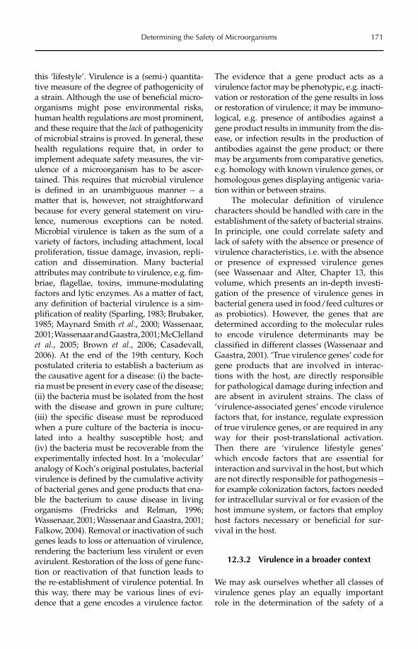
Determining the Safety of Microorganisms 171
this ‘lifestyle’. Virulence is a (semi-) quantita-tive measure of the degree of pathogenicity of a strain. Although the use of beneficial micro-organisms might pose environmental risks, human health regulations are most prominent, and these require that the lack of pathogenicity of microbial strains is proved. In general, these health regulations require that, in order to implement adequate safety measures, the vir-ulence of a microorganism has to be ascer-tained. This requires that microbial virulence is defined in an unambiguous manner – a matter that is, however, not straightforward because for every general statement on viru-lence, numerous exceptions can be noted. Microbial virulence is taken as the sum of a variety of factors, including attachment, local proliferation, tissue damage, invasion, repli-cation and dissemination. Many bacterial attributes may contribute to virulence, e.g. fim-briae, flagellae, toxins, immune- modulating factors and lytic enzymes. As a matter of fact, any definition of bacterial virulence is a sim-plification of reality (Sparling, 1983; Brubaker, 1985; Maynard Smith et al., 2000; Wassenaar, 2001; Wassenaar and Gaastra, 2001; McClelland et al., 2005; Brown et al., 2006; Casadevall, 2006). At the end of the 19th century, Koch postulated criteria to establish a bacterium as the causative agent for a disease: (i) the bacte-ria must be present in every case of the disease; (ii) the bacteria must be isolated from the host with the disease and grown in pure culture; (iii) the specific disease must be reproduced when a pure culture of the bacteria is inocu-lated into a healthy susceptible host; and (iv) the bacteria must be recoverable from the experimentally infected host. In a ‘molecular’ analogy of Koch’s original postulates, bacterial virulence is defined by the cumulative activity of bacterial genes and gene products that ena-ble the bacterium to cause disease in living organisms (Fredricks and Relman, 1996; Wassenaar, 2001; Wassenaar and Gaastra, 2001; Falkow, 2004). Removal or inactivation of such genes leads to loss or attenuation of virulence, rendering the bacterium less virulent or even avirulent. Restoration of the loss of gene func-tion or reactivation of that function leads to the re-establishment of virulence potential. In this way, there may be various lines of evi-dence that a gene encodes a virulence factor.
The evidence that a gene product acts as a virulence factor may be phenotypic, e.g. inacti-vation or restoration of the gene results in loss or restoration of virulence; it may be immuno-logical, e.g. presence of antibodies against a gene product results in immunity from the dis-ease, or infection results in the production of antibodies against the gene product; or there may be arguments from comparative genetics, e.g. homology with known virulence genes, or homologous genes displaying antigenic varia-tion within or between strains.
The molecular definition of virulence characters should be handled with care in the establishment of the safety of bacterial strains. In principle, one could correlate safety and lack of safety with the absence or presence of virulence characteristics, i.e. with the absence or presence of expressed virulence genes (see Wassenaar and Alter, Chapter 13, this volume, which presents an in-depth investi-gation of the presence of virulence genes in bacterial genera used in food/feed cultures or as probiotics). However, the genes that are determined according to the molecular rules to encode virulence determinants may be classified in different classes (Wassenaar and Gaastra, 2001). ‘True virulence genes’ code for gene products that are involved in interac-tions with the host, are directly responsible for pathological damage during infection and are absent in avirulent strains. The class of ‘virulence-associated genes’ encode virulence factors that, for instance, regulate expression of true virulence genes, or are required in any way for their post-translational activation. Then there are ‘virulence lifestyle genes’ which encode factors that are essential for interaction and survival in the host, but which are not directly responsible for pathogenesis –for example colonization factors, factors needed for intracellular survival or for evasion of the host immune system, or factors that employ host factors necessary or beneficial for sur-vival in the host.
12.3.2 Virulence in a broader context
We may ask ourselves whether all classes of virulence genes play an equally important role in the determination of the safety of a

172 H.E.N. Bergmans
strain. Clearly, the ‘true’ virulence genes are important, but virulence-associated genes need the presence of true virulence genes in order to affect safety, and the activity of viru-lence lifestyle genes should also be scruti-nized in order to determine their role in the safety characteristics of a strain. Interesting examples are the so-called housekeeping genes that are necessary for virulence, but are commonly present in both non-pathogenic and pathogenic strains. The aroA gene, which codes for the enzyme 5-enolpyruvylshikimate-3-phosphate synthetase, is widely present in bacteria and performs an essential step in the pathway that produces aromatic amino acids. Virulent strains of Salmonella can be intention-ally attenuated by inactivation of the aroAgene (Stocker, 2000), and this strategy is widely used for the construction of vaccine strains, in Gram-positive pathogenic bacteria (Bronze and Dale, 2010) as well as in Gram-negative bacteria (Salmonella). Clearly, aroAfulfils the ‘molecular’ criteria of a virulence gene, but it should not be considered as a virulence factor on its own, and the presence of the gene in an otherwise non-pathogenic strain will clearly not lead to specific virulence characteristics.
By the way they are defined, the pres-ence of ‘virulence’ factors is restricted to path-ogenic organisms. In our anthropomorphic view of pathogens, we tend to think that they cause harm to their hosts on purpose, but the view of pathogenicity as the result of a patho-genic lifestyle, as an adaptation to life in the host, is probably more realistic. It has been pointed out that factors similar to virulence factors are typically also found in soil bacte-ria, where they enable the soil-bound life-style of these bacteria (Casadevall, 2006). Casadevall argues that ‘each soil-dwelling microbe possesses a unique combination of characteristics, including some held widely in common such as the ability to form biofilms, acquire iron, or produce proteolytic enzymes’; these are all factors that can also contribute to the virulence of pathogenic bacteria. The picture that emerges is that throughout the environment, including the environment of human and animal hosts that we normally take into consideration when defining viru-lence factors, there is an enormous set of traits
that are important for the fitness, or viru-lence, of bacteria, that are dependent on their lifestyle. Where these bacteria are in close contact, a rapid exchange of these fitness/virulence factors may occur. This may lead to a change from a non-pathogenic to a patho-genic lifestyle, or to a change in the host of a pathogen because it has become adapted to the situation and challenges that occur in another host. Such a host jump may even occur between the kingdoms of living organ-isms. The possibility of host jumps is a major concern in the risk assessment of viruses. The possibility of cross-kingdom host jumps in bacteria is less well known, but is an impor-tant issue in the safety assessment of bacteria (Van Baarlen et al., 2007a).
An example of host jumps is found in the Burkholderia cepacia species complex, with B. cepacia as a type species. The genetic diversity in this complex is very large and it has been termed a ‘multireplicon complex’ (Mahenthiralingam et al., 2005). Formerly belonging to the genus Pseudomonas, Burk-holderia is commonly found in soil, e.g. in the plant rhizosphere and water. Members of the B. cepacia species complex have been identi-fied as plant commensals, where they may even be endophytic, when they do not only contribute to nitrogen fixation, but also have a role as opportunistic plant, animal and fungal pathogens. A common factor among human pathogenic Burkholderia species is the production of toxin-like virulence fac-tors, such as the pectinolytic enzymes that are found in the original onion pathogenic isolates but not in non-pathogenic represent-atives (Gonzales et al., 1997). Additional fac-tors that are associated with virulence are the Gram-negative bacterial endotoxins, different porins, the acyl-homoserine-L-lactone invol-ved in quorum sensing and other regulatory processes, and factors that promote the utili-zation of nutrient sources in the host (Baldwin et al., 2006).
The species B. cepacia is only one of a number of examples of microorganisms that apparently have made a cross-kingdom jump, a topic that is summarized by Van Baarlen et al. (2007a). These authors provide an over-view of the factors that are probably prerequi-sites for cross-kingdom jumps to occur. In the
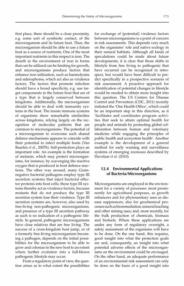
Determining the Safety of Microorganisms 173
first place, there should be a close proximity, e.g. some sort of symbiotic contact, of the microorganism and its future host. Then, the microorganism should be able to use a future host as a source of nutrients. One of the most important nutrients in this respect is iron. The dearth in the environment of iron in forms that can be utilized can be limiting for growth, and microorganisms produce factors that enhance iron utilization, such as haemolysins and siderophores, which act also as virulence factors. The factors that promote infection should have a broad specificity, e.g. use tar-get components in the future host that are of a type that is largely conserved between kingdoms. Additionally, the microorganism should be able to deal with immunity sys-tems in the host. The innate immune systems of organisms show remarkable similarities across kingdoms, relying largely on the rec-ognition of molecular patterns that are common to microorganisms. The potential of a microorganism to overcome such shared defence mechanisms appears to coincide with their potential to infect multiple hosts (Van Baarlen et al., 2007b). Self-protection plays an important role. An example is the formation of melanin, which may protect microorgan-isms, for instance, by scavenging the reactive oxygen that is produced in host defence reac-tions. The other way around, many Gram-negative bacterial pathogens employ type III secretion systems that inject bacterial effec-tor proteins into host cells; these type III sys-tems thereby act as virulence factors, because mutants that do not produce the type III secretion system lose their virulence. Type III secretion systems are, however, also used by free-living non-pathogenic microorganisms, and presence of a type III secretion pathway as such is no indication of a pathogenic life-style. In general, pathogenic microorganisms have close relatives that are free living. The success of a cross-kingdom host jump, or of a formerly free-living microorganism becom-ing a pathogen, depends on the initial possi-bilities for the microorganism to be able to grow and colonize in the new host to an extent where further evolution into a full-blown pathogenic lifestyle may occur.
From a regulatory point of view, the ques-tion arises as to what extent the possibilities
for exchange of (potential) virulence factors between microorganisms is a point of concern in risk assessments. This depends very much on the organisms’ role and native ecology in their natural habitats. Although all kinds of speculations could be made about future developments, it is clear that those shifts in lifestyle from free living to pathogenic that have occurred can be recognized in retro-spect, but would have been difficult to pre-dict specifically in a prospective scenario of risk assessment. A proactive approach for identification of potential changes in lifestyle would be needed to obtain more insight into this question. The US Centers for Disease Control and Prevention (CDC, 2011) recently initiated the ‘One Health Office’, which could be an important step in this direction, as it ‘facilitates and coordinates program activi-ties that seek to attain optimal health for people and animals by promoting global col-laboration between human and veterinary medicine while engaging the principles of public health and ecosystem health’. Another example is the development of a general method for early warning and surveillance systems of emerging zoonoses described by Havelaar et al. (2010).
12.4 Environmental Applications of Bacteria/Microorganisms
Microorganisms are employed in the environ-ment for a variety of processes: most promi-nently for agricultural purposes, as growth enhancers and for phytosanitary uses as dis-ease suppressors; also for geochemical pro-cesses such as bioremediation, mineral leaching and other mining uses; and, more recently, for the bulk production of chemicals, biomassand biofuels. Where these applications are under any form of regulatory oversight, a safety assessment of the organisms will have to be done. On the one hand, this requires, good insight into what the protection goals are and, consequently, an insight into what potential adverse effects of the microorgan-isms on the environment could be envisaged. On the other hand, an adequate performance of an environmental risk assessment can only be done on the basis of a good insight into
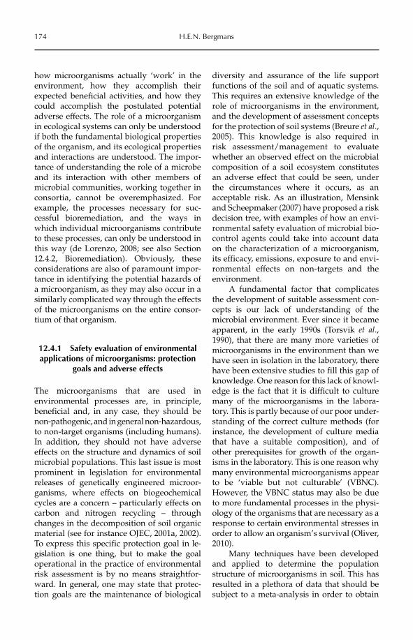
174 H.E.N. Bergmans
how microorganisms actually ‘work’ in the environment, how they accomplish their expected beneficial activities, and how they could accomplish the postulated potential adverse effects. The role of a microorganism in ecological systems can only be understood if both the fundamental biological properties of the organism, and its ecological properties and interactions are understood. The impor-tance of understanding the role of a microbe and its interaction with other members of microbial communities, working together in consortia, cannot be overemphasized. For example, the processes necessary for suc-cessful bioremediation, and the ways in which individual microorganisms contribute to these processes, can only be understood in this way (de Lorenzo, 2008; see also Section 12.4.2, Bioremediation). Obviously, these considerations are also of paramount impor-tance in identifying the potential hazards of a microorganism, as they may also occur in a similarly complicated way through the effects of the microorganisms on the entire consor-tium of that organism.
12.4.1 Safety evaluation of environmental applications of microorganisms: protection
goals and adverse effects
The microorganisms that are used in environmental processes are, in principle, beneficial and, in any case, they should be non- pathogenic, and in general non-hazardous, to non-target organisms (including humans). In addition, they should not have adverse effects on the structure and dynamics of soil microbial populations. This last issue is most prominent in legislation for environmental releases of genetically engineered microor-ganisms, where effects on biogeochemical cycles are a concern – particularly effects on carbon and nitrogen recycling – through changes in the decomposition of soil organic material (see for instance OJEC, 2001a, 2002). To express this specific protection goal in le -gislation is one thing, but to make the goal operational in the practice of environmental risk assessment is by no means straightfor-ward. In general, one may state that protec-tion goals are the maintenance of biological
diversity and assurance of the life support functions of the soil and of aquatic systems. This requires an extensive knowledge of the role of microorganisms in the environment, and the development of assessment concepts for the protection of soil systems (Breure et al.,2005). This knowledge is also required in risk assessment/management to evaluate whether an observed effect on the microbial composition of a soil ecosystem constitutes an adverse effect that could be seen, under the circumstances where it occurs, as an acceptable risk. As an illustration, Mensink and Scheepmaker (2007) have proposed a risk decision tree, with examples of how an envi-ronmental safety evaluation of microbial bio-control agents could take into account data on the characterization of a microorganism, its efficacy, emissions, exposure to and envi-ronmental effects on non-targets and the environment.
A fundamental factor that complicates the development of suitable assessment con-cepts is our lack of understanding of the microbial environment. Ever since it became apparent, in the early 1990s (Torsvik et al.,1990), that there are many more varieties of microorganisms in the environment than we have seen in isolation in the laboratory, there have been extensive studies to fill this gap of knowledge. One reason for this lack of knowl-edge is the fact that it is difficult to culture many of the microorganisms in the labora-tory. This is partly because of our poor under-standing of the correct culture methods (for instance, the development of culture media that have a suitable composition), and of other prerequisites for growth of the organ-isms in the laboratory. This is one reason why many environmental microorganisms appear to be ‘viable but not culturable’ (VBNC). However, the VBNC status may also be due to more fundamental processes in the physi-ology of the organisms that are necessary as a response to certain environmental stresses in order to allow an organism’s survival (Oliver, 2010).
Many techniques have been developed and applied to determine the population structure of microorganisms in soil. This has resulted in a plethora of data that should be subject to a meta-analysis in order to obtain
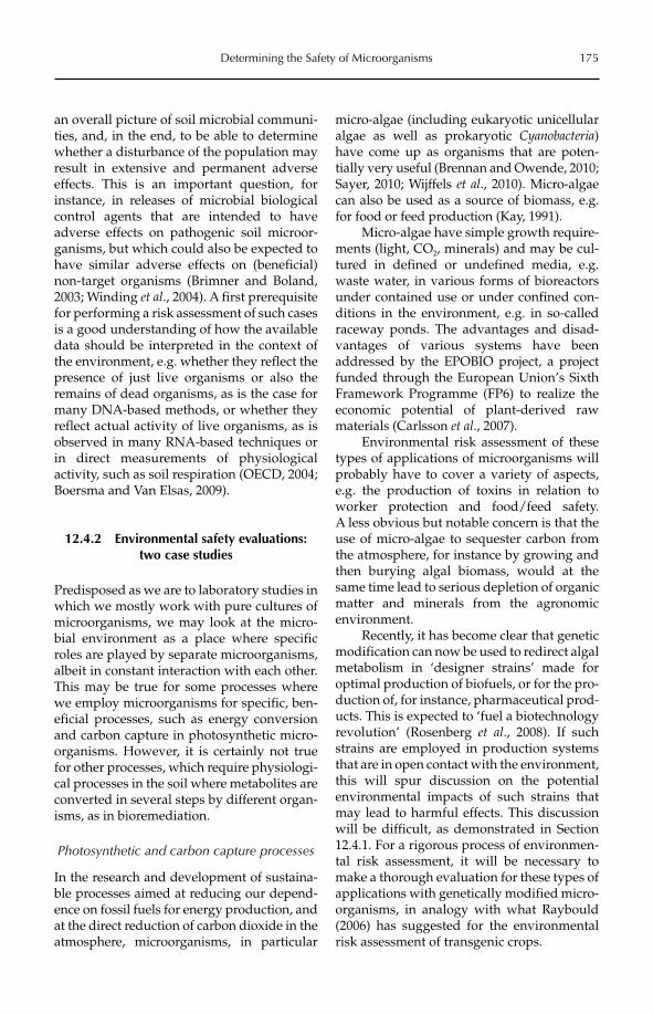
Determining the Safety of Microorganisms 175
an overall picture of soil microbial communi-ties, and, in the end, to be able to determine whether a disturbance of the population may result in extensive and permanent adverse effects. This is an important question, for instance, in releases of microbial biological control agents that are intended to have adverse effects on pathogenic soil microor-ganisms, but which could also be expected to have similar adverse effects on (beneficial) non-target organisms (Brimner and Boland, 2003; Winding et al., 2004). A first prerequisite for performing a risk assessment of such cases is a good understanding of how the available data should be interpreted in the context of the environment, e.g. whether they reflect the presence of just live organisms or also the remains of dead organisms, as is the case for many DNA-based methods, or whether they reflect actual activity of live organisms, as is observed in many RNA-based techniques or in direct measurements of physiological activity, such as soil respiration (OECD, 2004; Boersma and Van Elsas, 2009).
12.4.2 Environmental safety evaluations: two case studies
Predisposed as we are to laboratory studies in which we mostly work with pure cultures of microorganisms, we may look at the micro-bial environment as a place where specific roles are played by separate microorganisms, albeit in constant interaction with each other. This may be true for some processes where we employ microorganisms for specific, ben-eficial processes, such as energy conversion and carbon capture in photosynthetic micro-organisms. However, it is certainly not true for other processes, which require physiologi-cal processes in the soil where metabolites are converted in several steps by different organ-isms, as in bioremediation.
Photosynthetic and carbon capture processes
In the research and development of sustaina-ble processes aimed at reducing our depend-ence on fossil fuels for energy production, and at the direct reduction of carbon dioxide in the atmosphere, microorganisms, in particular
micro-algae (including eukaryotic unicellular algae as well as prokaryotic Cyanobacteria)have come up as organisms that are poten-tially very useful (Brennan and Owende, 2010; Sayer, 2010; Wijffels et al., 2010). Micro-algae can also be used as a source of biomass, e.g. for food or feed production (Kay, 1991).
Micro-algae have simple growth require-ments (light, CO2, minerals) and may be cul-tured in defined or undefined media, e.g. waste water, in various forms of bioreactors under contained use or under confined con-ditions in the environment, e.g. in so-called raceway ponds. The advantages and disad-vantages of various systems have been addressed by the EPOBIO project, a project funded through the European Union’s Sixth Framework Programme (FP6) to realize the economic potential of plant-derived raw materials (Carlsson et al., 2007).
Environmental risk assessment of these types of applications of microorganisms will probably have to cover a variety of aspects, e.g. the production of toxins in relation to worker protection and food/feed safety. A less obvious but notable concern is that the use of micro-algae to sequester carbon from the atmosphere, for instance by growing and then burying algal biomass, would at the same time lead to serious depletion of organic matter and minerals from the agronomic environment.
Recently, it has become clear that genetic modification can now be used to redirect algal metabolism in ‘designer strains’ made for optimal production of biofuels, or for the pro-duction of, for instance, pharmaceutical prod-ucts. This is expected to ‘fuel a biotechnology revolution’ (Rosenberg et al., 2008). If such strains are employed in production systems that are in open contact with the environment, this will spur discussion on the potential environmental impacts of such strains that may lead to harmful effects. This discussion will be difficult, as demonstrated in Section 12.4.1. For a rigorous process of environmen-tal risk assessment, it will be necessary to make a thorough evaluation for these types of applications with genetically modified micro-organisms, in analogy with what Raybould (2006) has suggested for the environmental risk assessment of transgenic crops.
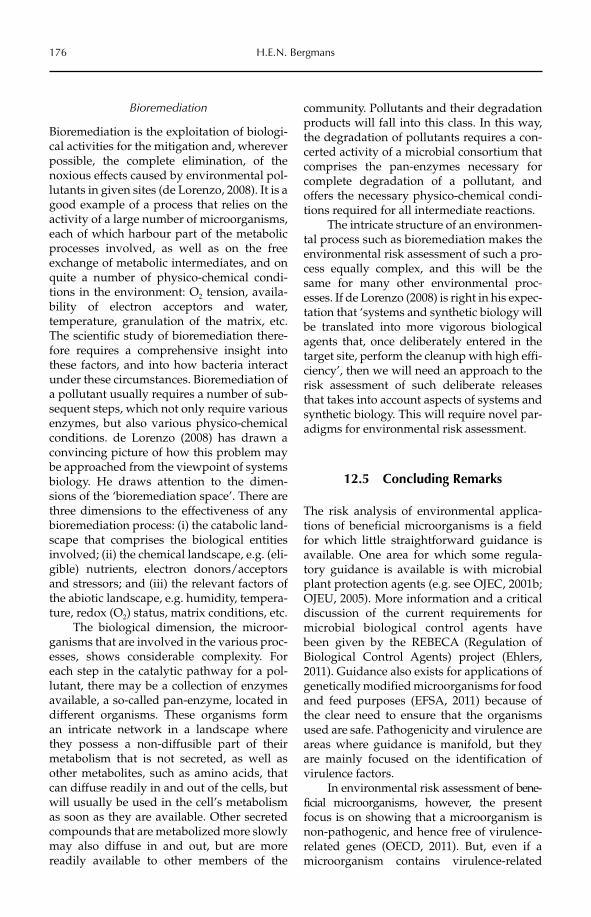
176 H.E.N. Bergmans
Bioremediation
Bioremediation is the exploitation of biologi-cal activities for the mitigation and, wherever possible, the complete elimination, of the noxious effects caused by environmental pol-lutants in given sites (de Lorenzo, 2008). It is a good example of a process that relies on the activity of a large number of microorganisms, each of which harbour part of the metabolic processes involved, as well as on the free exchange of metabolic intermediates, and on quite a number of physico-chemical condi-tions in the environment: O2 tension, availa-bility of electron acceptors and water, temperature, granulation of the matrix, etc. The scientific study of bioremediation there-fore requires a comprehensive insight into these factors, and into how bacteria interact under these circumstances. Bioremediation of a pollutant usually requires a number of sub-sequent steps, which not only require various enzymes, but also various physico-chemical conditions. de Lorenzo (2008) has drawn a convincing picture of how this problem may be approached from the viewpoint of systems biology. He draws attention to the dimen-sions of the ‘bioremediation space’. There are three dimensions to the effectiveness of any bioremediation process: (i) the catabolic land-scape that comprises the biological entities involved; (ii) the chemical landscape, e.g. (eli-gible) nutrients, electron donors/acceptors and stressors; and (iii) the relevant factors of the abiotic landscape, e.g. humidity, tempera-ture, redox (O2) status, matrix conditions, etc.
The biological dimension, the microor-ganisms that are involved in the various proc-esses, shows considerable complexity. For each step in the catalytic pathway for a pol-lutant, there may be a collection of enzymes available, a so-called pan-enzyme, located in different organisms. These organisms form an intricate network in a landscape where they possess a non-diffusible part of their metabolism that is not secreted, as well as other metabolites, such as amino acids, that can diffuse readily in and out of the cells, but will usually be used in the cell’s metabolism as soon as they are available. Other secreted compounds that are metabolized more slowly may also diffuse in and out, but are more readily available to other members of the
community. Pollutants and their degradation products will fall into this class. In this way, the degradation of pollutants requires a con-certed activity of a microbial consortium that comprises the pan-enzymes necessary for complete degradation of a pollutant, and offers the necessary physico-chemical condi-tions required for all intermediate reactions.
The intricate structure of an environmen-tal process such as bioremediation makes the environmental risk assessment of such a pro-cess equally complex, and this will be the same for many other environmental proc-esses. If de Lorenzo (2008) is right in his expec-tation that ‘systems and synthetic biology will be translated into more vigorous biological agents that, once deliberately entered in the target site, perform the cleanup with high effi-ciency’, then we will need an approach to the risk assessment of such deliberate releases that takes into account aspects of systems and synthetic biology. This will require novel par-adigms for environmental risk assessment.
12.5 Concluding Remarks
The risk analysis of environmental applica-tions of beneficial microorganisms is a field for which little straightforward guidance is available. One area for which some regula-tory guidance is available is with microbial plant protection agents (e.g. see OJEC, 2001b; OJEU, 2005). More information and a critical discussion of the current requirements for microbial biological control agents have been given by the REBECA (Regulation of Biological Control Agents) project (Ehlers, 2011). Guidance also exists for applications of genetically modified microorganisms for food and feed purposes (EFSA, 2011) because of the clear need to ensure that the organisms used are safe. Pathogenicity and virulence are areas where guidance is manifold, but they are mainly focused on the identification of virulence factors.
In environmental risk assessment of bene-ficial microorganisms, however, the presentfocus is on showing that a microorganism is non-pathogenic, and hence free of virulence-related genes (OECD, 2011). But, even if a microorganism contains virulence-related
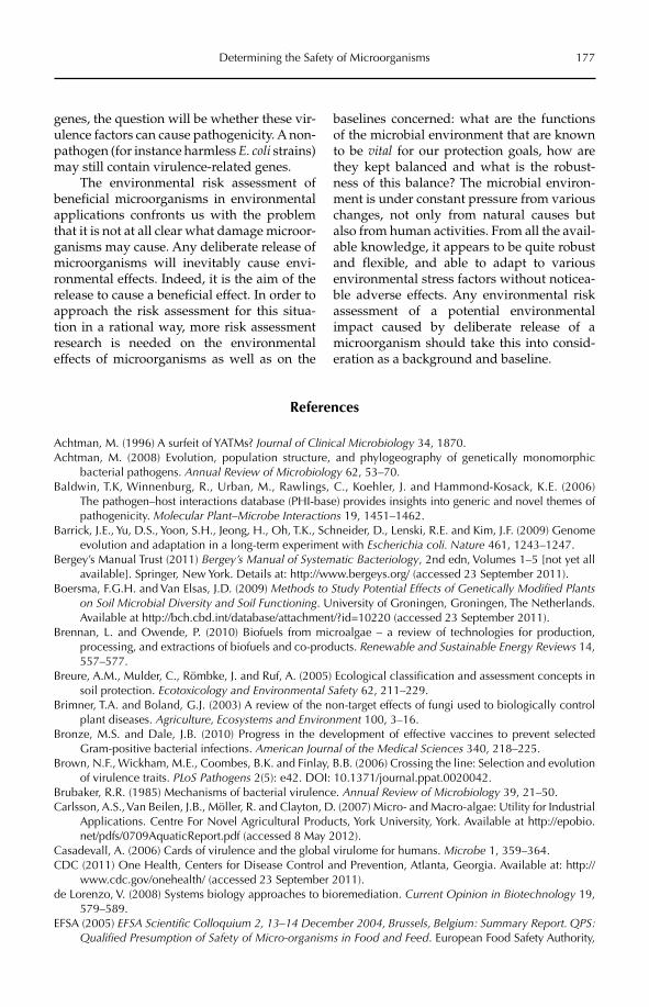
Determining the Safety of Microorganisms 177
genes, the question will be whether these vir-ulence factors can cause pathogenicity. A non-pathogen (for instance harmless E. coli strains) may still contain virulence-related genes.
The environmental risk assessment of beneficial microorganisms in environmental applications confronts us with the problem that it is not at all clear what damage microor-ganisms may cause. Any deliberate release of microorganisms will inevitably cause envi-ronmental effects. Indeed, it is the aim of the release to cause a beneficial effect. In order to approach the risk assessment for this situa-tion in a rational way, more risk assessment research is needed on the environmental effects of microorganisms as well as on the
baselines concerned: what are the functions of the microbial environment that are known to be vital for our protection goals, how are they kept balanced and what is the robust-ness of this balance? The microbial environ-ment is under constant pressure from various changes, not only from natural causes but also from human activities. From all the avail-able knowledge, it appears to be quite robust and flexible, and able to adapt to various environmental stress factors without noticea-ble adverse effects. Any environmental risk assessment of a potential environmental impact caused by deliberate release of a microorganism should take this into consid-eration as a background and baseline.
References
Achtman, M. (1996) A surfeit of YATMs? Journal of Clinical Microbiology 34, 1870.Achtman, M. (2008) Evolution, population structure, and phylogeography of genetically monomorphic
bacterial pathogens. Annual Review of Microbiology 62, 53–70.Baldwin, T.K, Winnenburg, R., Urban, M., Rawlings, C., Koehler, J. and Hammond-Kosack, K.E. (2006)
The pathogen–host interactions database (PHI-base) provides insights into generic and novel themes of pathogenicity. Molecular Plant–Microbe Interactions 19, 1451–1462.
Barrick, J.E., Yu, D.S., Yoon, S.H., Jeong, H., Oh, T.K., Schneider, D., Lenski, R.E. and Kim, J.F. (2009) Genome evolution and adaptation in a long-term experiment with Escherichia coli. Nature 461, 1243–1247.
Bergey’s Manual Trust (2011) Bergey’s Manual of Systematic Bacteriology, 2nd edn, Volumes 1–5 [not yet all available]. Springer, New York. Details at: http://www.bergeys.org/ (accessed 23 September 2011).
Boersma, F.G.H. and Van Elsas, J.D. (2009) Methods to Study Potential Effects of Genetically Modified Plants on Soil Microbial Diversity and Soil Functioning. University of Groningen, Groningen, The Netherlands. Available at http://bch.cbd.int/database/attachment/?id=10220 (accessed 23 September 2011).
Brennan, L. and Owende, P. (2010) Biofuels from microalgae – a review of technologies for production, processing, and extractions of biofuels and co-products. Renewable and Sustainable Energy Reviews 14, 557–577.
Breure, A.M., Mulder, C., Römbke, J. and Ruf, A. (2005) Ecological classification and assessment concepts in soil protection. Ecotoxicology and Environmental Safety 62, 211–229.
Brimner, T.A. and Boland, G.J. (2003) A review of the non-target effects of fungi used to biologically control plant diseases. Agriculture, Ecosystems and Environment 100, 3–16.
Bronze, M.S. and Dale, J.B. (2010) Progress in the development of effective vaccines to prevent selected Gram-positive bacterial infections. American Journal of the Medical Sciences 340, 218–225.
Brown, N.F., Wickham, M.E., Coombes, B.K. and Finlay, B.B. (2006) Crossing the line: Selection and evolution of virulence traits. PLoS Pathogens 2(5): e42. DOI: 10.1371/journal.ppat.0020042.
Brubaker, R.R. (1985) Mechanisms of bacterial virulence. Annual Review of Microbiology 39, 21–50.Carlsson, A.S., Van Beilen, J.B., Möller, R. and Clayton, D. (2007) Micro- and Macro-algae: Utility for Industrial
Applications. Centre For Novel Agricultural Products, York University, York. Available at http://epobio.net/pdfs/0709AquaticReport.pdf (accessed 8 May 2012).
Casadevall, A. (2006) Cards of virulence and the global virulome for humans. Microbe 1, 359–364.CDC (2011) One Health, Centers for Disease Control and Prevention, Atlanta, Georgia. Available at: http://
www.cdc.gov/onehealth/ (accessed 23 September 2011).de Lorenzo, V. (2008) Systems biology approaches to bioremediation. Current Opinion in Biotechnology 19,
579–589.EFSA (2005) EFSA Scientific Colloquium 2, 13–14 December 2004, Brussels, Belgium: Summary Report. QPS:
Qualified Presumption of Safety of Micro-organisms in Food and Feed. European Food Safety Authority,
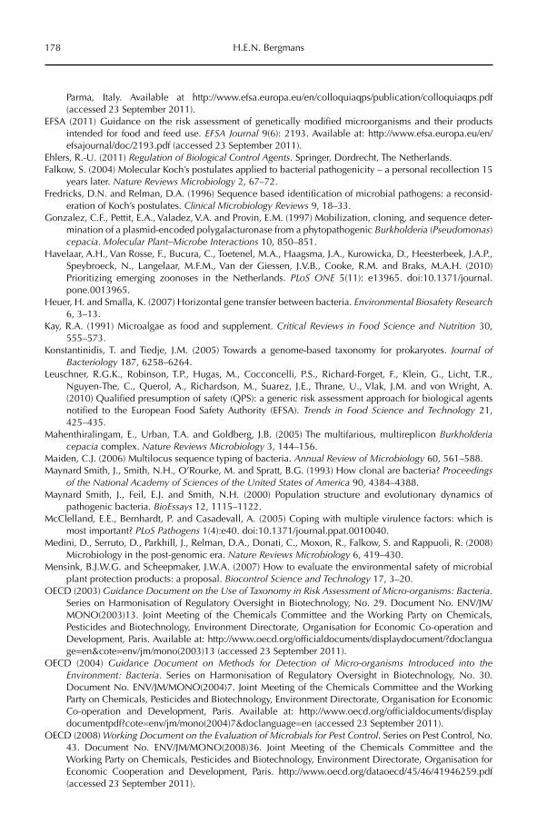
178 H.E.N. Bergmans
Parma, Italy. Available at http://www.efsa.europa.eu/en/colloquiaqps/publication/colloquiaqps.pdf (accessed 23 September 2011).
EFSA (2011) Guidance on the risk assessment of genetically modified microorganisms and their products intended for food and feed use. EFSA Journal 9(6): 2193. Available at: http://www.efsa.europa.eu/en/efsajournal/doc/2193.pdf (accessed 23 September 2011).
Ehlers, R.-U. (2011) Regulation of Biological Control Agents. Springer, Dordrecht, The Netherlands.Falkow, S. (2004) Molecular Koch’s postulates applied to bacterial pathogenicity – a personal recollection 15
years later. Nature Reviews Microbiology 2, 67–72.Fredricks, D.N. and Relman, D.A. (1996) Sequence based identification of microbial pathogens: a reconsid-
eration of Koch’s postulates. Clinical Microbiology Reviews 9, 18–33.Gonzalez, C.F., Pettit, E.A., Valadez, V.A. and Provin, E.M. (1997) Mobilization, cloning, and sequence deter-
mination of a plasmid-encoded polygalacturonase from a phytopathogenic Burkholderia (Pseudomonas)cepacia. Molecular Plant–Microbe Interactions 10, 850–851.
Havelaar, A.H., Van Rosse, F., Bucura, C., Toetenel, M.A., Haagsma, J.A., Kurowicka, D., Heesterbeek, J.A.P., Speybroeck, N., Langelaar, M.F.M., Van der Giessen, J.V.B., Cooke, R.M. and Braks, M.A.H. (2010) Prioritizing emerging zoonoses in the Netherlands. PLoS ONE 5(11): e13965. doi:10.1371/journal.pone.0013965.
Heuer, H. and Smalla, K. (2007) Horizontal gene transfer between bacteria. Environmental Biosafety Research6, 3–13.
Kay, R.A. (1991) Microalgae as food and supplement. Critical Reviews in Food Science and Nutrition 30, 555–573.
Konstantinidis, T. and Tiedje, J.M. (2005) Towards a genome-based taxonomy for prokaryotes. Journal of Bacteriology 187, 6258–6264.
Leuschner, R.G.K., Robinson, T.P., Hugas, M., Cocconcelli, P.S., Richard-Forget, F., Klein, G., Licht, T.R., Nguyen-The, C., Querol, A., Richardson, M., Suarez, J.E., Thrane, U., Vlak, J.M. and von Wright, A. (2010) Qualified presumption of safety (QPS): a generic risk assessment approach for biological agents notified to the European Food Safety Authority (EFSA). Trends in Food Science and Technology 21, 425–435.
Mahenthiralingam, E., Urban, T.A. and Goldberg, J.B. (2005) The multifarious, multireplicon Burkholderiacepacia complex. Nature Reviews Microbiology 3, 144–156.
Maiden, C.J. (2006) Multilocus sequence typing of bacteria. Annual Review of Microbiology 60, 561–588.Maynard Smith, J., Smith, N.H., O’Rourke, M. and Spratt, B.G. (1993) How clonal are bacteria? Proceedings
of the National Academy of Sciences of the United States of America 90, 4384–4388.Maynard Smith, J., Feil, E.J. and Smith, N.H. (2000) Population structure and evolutionary dynamics of
pathogenic bacteria. BioEssays 12, 1115–1122.McClelland, E.E., Bernhardt, P. and Casadevall, A. (2005) Coping with multiple virulence factors: which is
most important? PLoS Pathogens 1(4):e40. doi:10.1371/journal.ppat.0010040.Medini, D., Serruto, D., Parkhill, J., Relman, D.A., Donati, C., Moxon, R., Falkow, S. and Rappuoli, R. (2008)
Microbiology in the post-genomic era. Nature Reviews Microbiology 6, 419–430.Mensink, B.J.W.G. and Scheepmaker, J.W.A. (2007) How to evaluate the environmental safety of microbial
plant protection products: a proposal. Biocontrol Science and Technology 17, 3–20.OECD (2003) Guidance Document on the Use of Taxonomy in Risk Assessment of Micro-organisms: Bacteria.
Series on Harmonisation of Regulatory Oversight in Biotechnology, No. 29. Document No. ENV/JM/MONO(2003)13. Joint Meeting of the Chemicals Committee and the Working Party on Chemicals, Pesticides and Biotechnology, Environment Directorate, Organisation for Economic Co-operation and Development, Paris. Available at: http://www.oecd.org/officialdocuments/displaydocument/?doclanguage=en&cote=env/jm/mono(2003)13 (accessed 23 September 2011).
OECD (2004) Guidance Document on Methods for Detection of Micro-organisms Introduced into the Environment: Bacteria. Series on Harmonisation of Regulatory Oversight in Biotechnology, No. 30. Document No. ENV/JM/MONO(2004)7. Joint Meeting of the Chemicals Committee and the Working Party on Chemicals, Pesticides and Biotechnology, Environment Directorate, Organisation for Economic Co-operation and Development, Paris. Available at: http://www.oecd.org/officialdocuments/display documentpdf?cote=env/jm/mono(2004)7&doclanguage=en (accessed 23 September 2011).
OECD (2008) Working Document on the Evaluation of Microbials for Pest Control. Series on Pest Control, No. 43. Document No. ENV/JM/MONO(2008)36. Joint Meeting of the Chemicals Committee and the Working Party on Chemicals, Pesticides and Biotechnology, Environment Directorate, Organisation for Economic Cooperation and Development, Paris. http://www.oecd.org/dataoecd/45/46/41946259.pdf (accessed 23 September 2011).
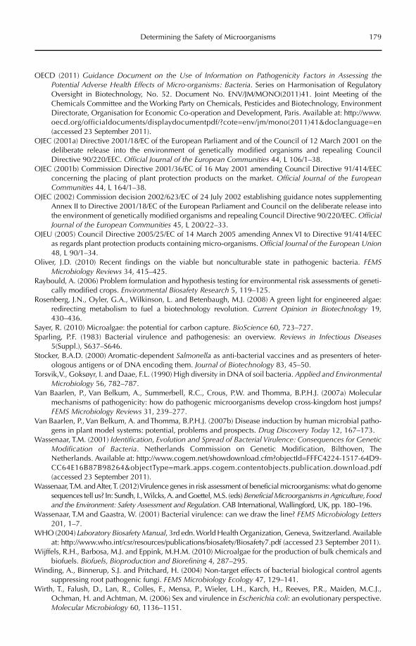
Determining the Safety of Microorganisms 179
OECD (2011) Guidance Document on the Use of Information on Pathogenicity Factors in Assessing the Potential Adverse Health Effects of Micro-organisms: Bacteria. Series on Harmonisation of Regulatory Oversight in Biotechnology, No. 52. Document No. ENV/JM/MONO(2011)41. Joint Meeting of the Chemicals Committee and the Working Party on Chemicals, Pesticides and Biotechnology, Environment Directorate, Organisation for Economic Co-operation and Development, Paris. Available at: http://www.oecd.org/officialdocuments/displaydocumentpdf/?cote=env/jm/mono(2011)41&doclanguage=en (accessed 23 September 2011).
OJEC (2001a) Directive 2001/18/EC of the European Parliament and of the Council of 12 March 2001 on the deliberate release into the environment of genetically modified organisms and repealing Council Directive 90/220/EEC. Official Journal of the European Communities 44, L 106/1–38.
OJEC (2001b) Commission Directive 2001/36/EC of 16 May 2001 amending Council Directive 91/414/EEC concerning the placing of plant protection products on the market. Official Journal of the European Communities 44, L 164/1–38.
OJEC (2002) Commission decision 2002/623/EC of 24 July 2002 establishing guidance notes supplementing Annex II to Directive 2001/18/EC of the European Parliament and Council on the deliberate release into the environment of genetically modified organisms and repealing Council Directive 90/220/EEC. OfficialJournal of the European Communities 45, L 200/22–33.
OJEU (2005) Council Directive 2005/25/EC of 14 March 2005 amending Annex VI to Directive 91/414/EEC as regards plant protection products containing micro-organisms. Official Journal of the European Union48, L 90/1–34.
Oliver, J.D. (2010) Recent findings on the viable but nonculturable state in pathogenic bacteria. FEMSMicrobiology Reviews 34, 415–425.
Raybould, A. (2006) Problem formulation and hypothesis testing for environmental risk assessments of geneti-cally modified crops. Environmental Biosafety Research 5, 119–125.
Rosenberg, J.N., Oyler, G.A., Wilkinson, L. and Betenbaugh, M.J. (2008) A green light for engineered algae: redirecting metabolism to fuel a biotechnology revolution. Current Opinion in Biotechnology 19, 430–436.
Sayer, R. (2010) Microalgae: the potential for carbon capture. BioScience 60, 723–727.Sparling, P.F. (1983) Bacterial virulence and pathogenesis: an overview. Reviews in Infectious Diseases
5(Suppl.), S637–S646.Stocker, B.A.D. (2000) Aromatic-dependent Salmonella as anti-bacterial vaccines and as presenters of heter-
ologous antigens or of DNA encoding them. Journal of Biotechnology 83, 45–50.Torsvik,V., Goksøyr, I. and Daae, F.L. (1990) High diversity in DNA of soil bacteria. Applied and Environmental
Microbiology 56, 782–787.Van Baarlen, P., Van Belkum, A., Summerbell, R.C., Crous, P.W. and Thomma, B.P.H.J. (2007a) Molecular
mechanisms of pathogenicity: how do pathogenic microorganisms develop cross-kingdom host jumps? FEMS Microbiology Reviews 31, 239–277.
Van Baarlen, P., Van Belkum, A. and Thomma, B.P.H.J. (2007b) Disease induction by human microbial patho-gens in plant model systems: potential, problems and prospects. Drug Discovery Today 12, 167–173.
Wassenaar, T.M. (2001) Identification, Evolution and Spread of Bacterial Virulence: Consequences for Genetic Modification of Bacteria. Netherlands Commission on Genetic Modification, Bilthoven, The Netherlands. Available at: http://www.cogem.net/showdownload.cfm?objectId=FFFC4224-1517-64D9-CC64E16B87B98264&objectType=mark.apps.cogem.contentobjects.publication.download.pdf (accessed 23 September 2011).
Wassenaar, T.M. and Alter, T. (2012) Virulence genes in risk assessment of beneficial microorganisms: what do genome sequences tell us? In: Sundh, I., Wilcks, A. and Goettel, M.S. (eds) Beneficial Microorganisms in Agriculture, Food and the Environment: Safety Assessment and Regulation. CAB International, Wallingford, UK, pp. 180–196.
Wassenaar, T.M and Gaastra, W. (2001) Bacterial virulence: can we draw the line? FEMS Microbiology Letters201, 1–7.
WHO (2004) Laboratory Biosafety Manual, 3rd edn. World Health Organization, Geneva, Switzerland. Available at: http://www.who.int/csr/resources/publications/biosafety/Biosafety7.pdf (accessed 23 September 2011).
Wijffels, R.H., Barbosa, M.J. and Eppink, M.H.M. (2010) Microalgae for the production of bulk chemicals and biofuels. Biofuels, Bioproduction and Biorefining 4, 287–295.
Winding, A., Binnerup, S.J. and Pritchard, H. (2004) Non-target effects of bacterial biological control agents suppressing root pathogenic fungi. FEMS Microbiology Ecology 47, 129–141.
Wirth, T., Falush, D., Lan, R., Colles, F., Mensa, P., Wieler, L.H., Karch, H., Reeves, P.R., Maiden, M.C.J., Ochman, H. and Achtman, M. (2006) Sex and virulence in Escherichia coli: an evolutionary perspective. Molecular Microbiology 60, 1136–1151.
©CAB International 2012. Beneficial Microorganisms in Agriculture, Food and the Environment: 180 Safety Assessment and Regulation (eds I. Sundh et al.)
13.1 Introduction 18013.2 Quality Requirements of Complete Genome Sequences 181
13.2.1 Quality of genome metadata 181 13.2.2 Quality of raw sequences and assembled genomes 182 13.2.3 Quality of annotated gene files 183
13.3 Definition of Virulence and Other Risk-related Properties 18313.4 Comparison of Genomes from Probiotic, Starter Culture and
Commensal Bacteria 186 13.4.1 Summary of publicly available genomes 186 13.4.2 Some statistics of sequenced genomes from probiotic and
other benign organisms 186 13.4.3 Presence of putative virulence genes in selected probiotic genomes 187 13.4.4 Presence of putative antibiotic resistance genes in
selected probiotic genomes 18813.5 Comparison of Complete Genomes from Pathogenic and Probiotic Microbes 19113.6 Conclusion 193
13 Virulence Genes in Risk Assessment of Beneficial Microorganisms: What Do
Genome Sequences Tell Us?
Trudy M. Wassenaar1 and Thomas Alter2
1Molecular Microbiology and Genomics Consultants, Zotzenheim, Germany; 2Institute of Food Hygiene, Free University Berlin, Germany
13.1 Introduction
Risk assessment of beneficial microorganisms depends on proper characterization of the genetic potential of the organisms in ques-tion. In this chapter, probiotics and starter cultures are used as an example to summa-rize the molecular information needed from these organisms for safety assessment. Pro-biotic bacteria have a (proven or assumed) beneficial effect on the host they colonize; starter cultures are deliberately added during food production, for instance for fermenta-tion purposes.
Virulence properties can be assessed by in vivo animal models of virulence, which can be based on LD50 studies or on disease sever-ity scores, in vitro models using cell lines to assess specific virulence mechanisms (inva-sion, toxicity, macrophage survival, etc.), or genetic characterization to determine the presence or absence of virulence genes.
The chapter deals with the genetic and genomic evidence for the absence of viru-lence properties in bacteria that are intended for beneficial use, with emphasis on pro-biotic and starter cultures that are suitable for human consumption. Legislation issues
Virulence Genes in Risk Assessment 181
related to the safety of probiotics are not treated here, as this topic has been reviewed previously (Wassenaar and Klein, 2008), and is also dealt with elsewhere in this book (von Wright, Chapter 3, this volume). In the past, virulence characterization was based on active searches for the presence of known virulence genes, for instance by PCR analysis. The shortcoming of this approach was that the presence or absence of virulence genes could only be demonstrated for those genes that were actively being searched for; there would be no information available about other genes that might be present. This draw-back has been resolved by the increasing availability of complete genome sequences.
In the era of genomics, the first step in the genetic characterization of (poten-tially) beneficial bacteria should be to deter-mine the DNA sequence of the organism’s complete genome. All genes present in the strain of interest can thus be identified and categorized. The logical next step would be to predict any recognizable poten-tial risk related to any possible virulence genes identified. Such predictions depend on inference from previous knowledge. A number of genome sequences have so far been obtained from organisms that have been given Generally Recognized As Safe (GRAS) status in the USA. For substances to obtain GRAS status, their use in food has to have a proven record of safety based either on published scientific evidence or on a long history of use (Mattia and Merker, 2008). Well-defined starter cultures and a number of probiotic bacterial strains have received GRAS status, e.g. Bifidobacterium lactis strain Bb12 or Streptococcus ther-mophilus strain Th4 (GRAS notice number 49) (US FDA, 2011). European regulation of the use of bacteria in the food chain differs to that in the USA, and the two regulatory systems have been compared elsewhere (Wessels et al., 2004). Starter cultures and probiotic bacterial strains will be used as examples of safe organisms, and because the distinction between them is not always clear (a number of probiotic bacteria are also in use as starter cultures), both types of bacteria are collectively described here as ‘beneficial bacteria’.
The genomes of safe bacteria, an increasing number of which are completely sequenced, can serve as a reference for genes or gene combinations that are unlikely to pose a risk to human health. Even more genome sequences are available from (entero)pathogenic bacteria, so that, in theory, a com-parison of pathogenic and beneficial bacte-rial genomes could identify those genes that are found exclusively in pathogenic organ-isms; these would be more likely to contrib-ute to virulence. The challenge would be to identify the critical combination of these genes that would result in pathogenic poten-tial. Such a comparison might also identify genes that are exclusively found in beneficial organisms, and these, in genomes that lack virulence genes, could in theory provide a ‘genetic signature’ for safe use.
Actual practice is more recalcitrant and this chapter discusses some of the advantages and difficulties related to safety assessments of beneficial bacteria based on genome sequences. First, we consider the quality requirements of a genome sequence for this approach to be of use.
13.2 Quality Requirements of Complete Genome Sequences
13.2.1 Quality of genome metadata
A bacterial genome sequence comprises both chromosomal DNA and any plasmid DNA; the term ‘genome’ is only synonymous to the term ‘chromosome’ when extrachromosomal autonomously replicating DNA is absent. Recently, the number of publicly available, completely sequenced bacterial genomes exceeded 1000, but when these sequences were reviewed, it was concluded that large differences exist in genome sequence quality as well as in their annotation (Lagesen et al.,2010). The quality of a published sequence depends on the number of ambiguous sequences it includes; a published bacterial genome can still contain unacceptably large numbers of these (represented as ‘N’ rather than one of the four DNA nucleotides). Another factor determining the quality of a
182 T.M. Wassenaar and T. Alter
genome sequence is how well the sequence was assembled from short reads; in quite a few instances, such assembly has introduced mistakes which are sometimes corrected in follow-up studies. Genome annotation, which describes the function of the predicted genes, varies in quality depending on how the genes were identified, and how careful their func-tional prediction (which is mainly done auto-matically) was manually checked.
There is no internationally accepted standard for the quality assessment of bacte-rial genome sequences, though efforts are made to standardize the metadata, i.e. the rel-evant information about the strain that was sequenced. To this extent, the minimum information about a genome sequence (MIGS) that is ideally to be provided with the sequence has been specified (Field et al., 2008). Nevertheless, a brief review of some of the Microbial Genome Projects at the NCBI (the US National Center for Biotechnology Information) web site (http://www.ncbi.nlm.nih.gov/genomes/lproks.cgi) illustrates that essential information on metadata is fre-quently incomplete or incorrect. For instance, the metadata summarized in the table ‘Organism info’ on the NCBI web site is too incomplete to be of any use. As an example of incorrect information, in the table of ‘Genomes in progress’, under Project Identification Number (PID) 46979, Enterococcus faecalisPC4.1 is listed, but the organism from which this sequence was derived is E. faecium PC4.1. Such mistakes are a nuisance, as the incorrect information is easily incorporated into auto-matically extracted data, and is not likely to be picked up unless all data are reviewed manually. It is anticipated that the mistake will be corrected when the genome sequence reaches completion; however, a number of genome sequences remain in public databases as ‘in progress’ for a long time, possibly for-ever. The oldest records of ‘Genomes in progress’ were last updated in 2002; these include a B. longum genome (a look at the GenBank file of this genome revealed it was last updated in 2003, but that information was not incorporated into the summarizing table at NCBI).
Even if the metadata information is cor-rect, it may be of limited use. For a genome
sequence of an E. faecalis strain isolated from urine and that most likely caused an infec-tion, it will be stated that the genome was derived from a pathogen; but in another indi-vidual this strain may behave as a commensal inhabitant of the gut. The same might apply to an E. faecium isolated from the blood of a bacteraemic patient with severe underlying conditions – a healthy individual could be colonized by the same strain without any symptoms. Whether such opportunistic path-ogens cause infection depends on host factors as well as on bacterial factors. This will have consequences for the way we interpret their gene content, as will be discussed below.
13.2.2 Quality of raw sequences and assembled genomes
At the time of writing, there are three com-monly used platforms for ‘next-generation’ sequencing of bacterial genomes: 454 pyro-sequencing (Roche Diagnostics), Illumina (Solexa) sequencing, and ABI SOLiD sequenc-ing (Life Technologies). Recently, the first bac-terial genomes have been sequenced using ‘third-generation’ technology, which is based on single-molecule sequencing, and this holds great promise for the future, as it uses very little material, is inexpensive, produces quite long read lengths, and can be stunningly fast, as was recently illustrated during the 2010 Haiti cholera outbreak (Chin et al., 2011).
The raw sequence data for the ‘next- generation’ sequencing methods consist of short reads: 100 nucleotides (or less) for Solexa and the ABI-SOLiD machines; and around 500 nucleotides or a bit longer for the 454 machines. These short pieces must be assembled into larger pieces or ‘contigs’ (i.e. contiguous pieces of DNA, where overlap-ping reads are combined like pieces in a puz-zle). The sequence coverage for many of these machines can easily be sufficient (40-fold, in some cases) to help minimize the number of assembled contigs. Assembly can be done with standard software such as Velvet or other methods for the short reads (Solexa and ABI-SOLiD; Miller et al., 2010), or with Newbler, which can be used for the 454 reads. Often, the high-throughput coverage of
Virulence Genes in Risk Assessment 183
Solexa is combined with the long reads of 454 to help in assembling genomes. Whereas complete assembly to a fully closed circular chromosome would be ideal, assembly gaps due to repeated sequences, notably those related to multiple rRNA loci, are inevitable. Gaps are also frequently the result of low sequencing coverage of particular DNA frag-ments, but this is less of a problem in third-generation sequencing technology (Nagarajan et al., 2010). Although it is possible to assem-ble a small bacterial genome into one piece based on a single run, this is usually the exception, rather than the rule. In practice (at present), the assembly of raw sequencing data from next-generation sequencing often results in over 100 contigs for many bacterial genomes, which means that numerous genes are possibly incompletely sequenced or even missed. A genome sequence can be estab-lished in draft form within weeks or even days, but closing all gaps can be demanding in terms of time and resources (Nagarajan et al., 2010). Finishing a genome sequence by closing all gaps will not only provide a com-plete picture on gene content, it also enables the correction of errors in contig assembly that occur quite frequently (Nagarajan et al.,2010). Nevertheless, it may not be necessary to close all gaps. Even though there are no regulations about what is acceptable, as a rule of thumb we propose to work with genome sequences containing as few contigs as possi-ble. Moreover, the number of ambiguous nucleotides should be minimized. The cut-off depends on the genomes being used and, of course, on what is needed, but it is advisable to exclude contigs shorter than 1 kbp, which is approximately the average length of a bac-terial gene. Gene finding on such short con-tigs becomes problematic because the chance of detecting multiple open reading frames (ORFs) increases with decreasing sequence length, and such ORFs are frequently artefacts.
13.2.3 Quality of annotated gene files
The next step is to generate an annotated genome, in which the protein coding and non-translated genes have been identified.
One popular method for finding proteins from a bacterial genome DNA sequence is Prodigal software that was developed at Oak Ridge National Laboratory, Tennessee (Hyatt et al., 2010). This method is quite fast, and in general can give annotations quite close to those found in annotated bacterial genomes in GenBank (the US National Institutes of Health genetic sequence database at NCBI), so that the resulting annotation file does not require too much manual curation. For a com-plete genome annotation, the non-translated genes should also be included. Transfer RNAs (tRNAs) can be found with the program tRNA-Scan-SE (Schattner et al., 2005), and rRNAs can be found with the program RNAmmer (Lagesen et al., 2007). Currently, finding other small non-coding RNA genes is more difficult. While programs to identify them exist, these genes are not yet part of standard genome annotations.
13.3 Definition of Virulence and Other Risk-related Properties
The key information relevant for microbial risk assessment based on complete genome sequences of beneficial bacteria, especially those aimed for human consumption, is whether virulence genes are present. If they are, the risk that these genes pose in terms of consumer safety needs to be estimated. The way virulence genes (which code for viru-lence factors) are usually defined and identi-fied has been extensively reviewed (Wassenaar and Gaastra, 2001; Wassenaar, 2004), and this information will not be repeated here. Of the possible methods to identify virulence genes, the one approach relevant for this contribu-tion is identification by sequence homology with known virulence genes.
One complication in the definition of virulence in general is that rather than describ-ing a specific gene functioning in a specific virulence pathway of a particular pathogen, virulence cannot always be separated from niche adaptation/survival strategies. One of the few distinctions between these two is active invasion, which has been shown for a number of pathogenic intestinal bacteria, but is never employed by commensal gut
184 T.M. Wassenaar and T. Alter
bacteria. However, not all (enteric) pathogens are invasive. Many of the gut commensals use the same strategy of attachment (adhesins, fimbriae/pili) and motility (flagella, type IV pili) to survive in the gastrointestinal tract as is employed by pathogens. Although in path-ogens any of these mechanisms may contrib-ute to pathogenicity (and would result in attenuation upon inactivation), for commen-sals they contribute to colonization fitness. For example, the genome sequence of Lactobacillus rhamnosus GG revealed the pres-ence of mucus-binding pili, which are more typically found on pathogenic bacteria (Kankainen et al., 2009). The genes responsi-ble for such actions may be called ‘virulence’ genes when they reside in pathogens, but are colonization genes when present in commen-sals. The subtlety of this distinction is lost in gene annotation, and from sequence similar-ity it cannot be identified whether the gene in question is ‘good’ or ‘bad’, as it depends on the context of the rest of the genome. Would it be easier to reason the other way around, and propose that a gene found in a commensal with no known virulence should not be called a virulence gene? Obviously, this proposition would not be compatible with the view that the function of a gene is dictated by its sequence and not (also) by its genetic back-ground – a view that is still at the basis of molecular genetics. Furthermore, this prag-matism will not work for the opportunistic pathogens, which contain genes that function in pathogenicity only under certain circum-stances, and under other conditions contrib-ute to a commensal lifestyle. Putatively benign organisms are unlikely to be true pathogens, but they might behave as oppor-tunistic pathogens in certain conditions (Wessels et al., 2004; Koretz, 2009; Verma et al.,2010), so the distinction needed in microbial risk assessment is that between true commen-sal organisms and mostly commensal organ-isms, which can, under exceptional circumstances, behave as opportunistic path-ogens. How then, do we interpret the genes that report significant homology both to genes found in a commensal and, with equal significance, to a virulence gene in a patho-gen’s genome? Unfortunately, there is no sim-ple answer to that question. Gene function
depends on context, and the challenge is to predict the correct function of such a gene in that particular organism, set in the context of the complete genetic content of that organ-ism. The problem is even more complex, because the outcome of colonization is an interplay between the microorganism and the host.
Probiotic activity is the combined result of interaction between the host, the intestinal microbiota present and the probiotic strain; similarly, a pathogenic phenotype is defined by the combination of host, residual microbi-ota and virulence properties of the pathogen. In other words, pathogenicity is not an objec-tive criterion but depends on the circum-stances. Even non-pathogenic species can, under very specific circumstances, cause dis-ease, such as in severely immunocompro-mised individuals or in critically ill patients (Koretz, 2009). Ingested probiotic bacteria can in principle translocate to cause infections, though this has rarely been observed in healthy individuals (Liong, 2008). Despite the broad usage of Bifidobacterium spp. as probi-otics in the food industry, infections by single Bifidobacterium species have been described, and this is mostly restricted to B. dentium(Meile et al., 2008). Further, while B. dentium is being recognized as a potential oral pathogen involved in caries, the majority of these bacte-ria live in a non-pathogenic relationship with their host (Lee and O’Sullivan, 2010). Then again, a recognized pathogen will not harm an individual who has fully developed immu-nity against it, no matter how many virulence genes it carries. Most intestinal pathogens can produce a range of conditions, from asympto-matic colonization to severe disease, depend-ing on the overall health of the host, the current immune status, the administered dose and, possibly, the residual microbiota. Therefore, it is impossible to objectively define the exact virulence potential of a given organism, as virulence depends on the circumstances.
Given this difficulty, it is challenging to prove whether one is dealing with a patho-gen, an opportunistic pathogen, a commensal or a (potential) beneficial organism, when the proof is based only on a bacterial genome sequence. The absence of virulence genes is
Virulence Genes in Risk Assessment 185
no guarantee of a non-pathogenic phenotype, and the presence of virulence genes is no guarantee of pathogenicity. For example, haemolysin A, a well-recognized virulence gene in pathogenic Escherichia coli, is present and even weakly expressed in an E. coli strain that has been in use as a probiotic for decades (Willenbrock et al., 2007). Clearly, the presence of this single virulence factor does not cause pathogenicity, and even after knowing the complete genome sequence, we would still not be able to predict virulence, or absence thereof. Another example is given by the genome sequence of the probiotic strain E. coliNissle 1917, which was found to be very simi-lar to that of the uropathogenic E. coli CFT073 (Grozdanov et al., 2004; Hancock et al., 2010); not knowing the strain’s properties and based on the information from its genome sequence only, one would judge Nissle as a potential uropathogenic E. coli. At present, if a genome sequence is the only available source of infor-mation it would be difficult to accurately pre-dict whether an E. coli isolate would be safe for human consumption.
Apart from virulence genes, the presence of antibiotic resistance genes is considered undesirable in probiotic or otherwise bene-ficial bacteria, and an antibiotic profile should be established in a safety assessment (Bernardeau et al., 2008; Wilcks and van Hoek, Chapter 4, this volume). The main reason that these genes are considered unde-sirable is not because they would hamper treatment following an infection (probiotic bacteria should not be able to cause an infec-tion in the first place), but that they could donate these resistance genes to potential pathogens. However, not all antibiotic resist-ances are transferable, so in addition to an antibiogram, gene transferability should also be determined. The probability of DNA trans-fers between species, or even between genera, is smaller than transfer within a species, but it may not be negligible. Moreover, probiotics may be used during or shortly after an acute infection, so the presence of pathogenic bacte-ria is likely, and these can then serve as poten-tial acceptors of resistance genes. Intake of probiotic bacteria may even be advised dur-ing an antibiotic course to minimize the side effects that the antibiotic can have on the
residual gut microbiota. This advice can be questioned, because first of all susceptible probiotic bacteria will not be able to colonize, but will be killed by the antibiotic and, sec-ondly, because the procedure may select for probiotic bacteria that have acquired resist-ance as a result of the selective pressure. To reduce the side effects of antibiotic use, a pro-biotic should be taken after the course was terminated.
A final risk may be perceived from genes that provide the capacity for DNA transfer, irrespective of the nature of the genes that could be transferred by this capacity. Transfer of plasmid or viral (phage or prophage) DNA occurs frequently between bacteria. DNA uptake from the residual microbiota to the probiotic bacteria would be undesirable if this could result in pathogenicity or antibiotic resistance – though it should be realized that in this scenario, such genes must be present in the microbiota in the first place, implying that pathogenic bacteria or resistant bacteria are already in residence. DNA donation from benign bacteria to the residual biota would be less of a problem because the probiotic bacte-ria are unlikely to contain genes that can be harmful, even if transferred to a different genetic background. Hence, the presence of prophage DNA (bacteriophage DNA that is inserted and integrated into the bacterial chromosome) is not necessarily problematic, but can – at low frequencies – contribute to horizontal gene transfer (HGT) by transfer-ring small fragments of chromosomal DNA to new hosts. Genome analysis of probiotic strains has revealed the presence of one or more prophage/prophage-like elements in many Lactobacillus spp., Lactococcus lactis and bifidobacteria (Ventura et al., 2005, 2006).
Bacterial conjugation involving conjuga-tive plasmids might further contribute to HGT, which can assist in the dissemination of, for example, antibiotic resistance genes; interspecies conjugative transfer of tetracy-cline and erythromycin resistance plasmids from probiotic bacteria has been demon-strated in vitro, and evidence for such trans-fers in vivo is accumulating (Jones et al., 2010). These mobile elements carry the genes required for processing the plasmid into a transfer-competent form (mob genes) and
186 T.M. Wassenaar and T. Alter
the genes involved in the formation of a trans-envelope machinery and a pilus struc-ture (tra or trb genes) (Filloux, 2010).
With all this knowledge and insight, how well do the genomes of proven safe bacteria perform? Are they really free of virulence and antibiotic resistance genes? Nearly all of the beneficial bacteria that have been completely sequenced were approved for their applica-tions before their genome sequences were available. As the next section will illustrate, these genomes sometimes contain genes whose presence might not have been expected.
13.4 Comparison of Genomes from Probiotic, Starter Culture and
Commensal Bacteria
13.4.1 Summary of publicly available genomes
As of December 2010, GenBank listed 1279 complete and 3529 in-progress bacterial genomes (plus 93 complete and 82 incomplete archaeal genomes). This is a moving target, and novel genomes are being sequenced, and made publicly available, at such a rate that the information given here represents only a moment in time. The most common bacte-rial phyla for probiotic research are the Actinobacteria and Firmicutes. The list includes 21 completed Actinobacteria genomes, and 375 genomes in progress. Of these, 15 finished and 26 ongoing genome sequences belong to the genus Bifidobacterium. Even more genomes of Firmicutes are available or in progress: 316 and 1236, respectively. The most relevant gen-era include Lactobacillus (28 finished, 120 in progress), Lactococcus (five and three), and Leuconostoc (four and seven). Two genera of Firmicutes contain well-known benign as well as pathogenic strains or species: Enterococcus(two finished genomes and 108 genomes in progress) and Streptococcus; for S. thermophilus(one of the two non-pathogenic members of this genus) there are four finished genomes but none listed as in progress.
These numbers illustrate the size of the currently available data sets and this is expected to increase in the near future.
Few microbiologists or risk researchers will have the computational skills and equipment to handle such large sets of data. Fortunately, some simple genome comparisons can be done using tools that are available online. Notably, the Integrated Microbial Genomes (IMG) system (http://img.jgi.doe.gov) pro-vides useful data for some quick comparisons and these were used for the analyses pre-sented in the next three sections.
13.4.2 Some statistics of sequenced genomes from probiotic and
other benign organisms
From the IMG web pages, 40 completely fin-ished genomes of beneficial bacteria were selected. These were from five genera that mostly contain species that are frequently used as probiotics or starter cultures. Three of the listed genera contain strains that have obtained GRAS status. All these genomes are captured here under the general term of ‘ben-eficial bacteria’ (Table 13.1). For all 40 genomes, the genome length and GC content, as well as the total number of genes, were extracted from the IMG web pages, and the percentage of genes for which a function could be predicted was calculated. The per-centage of genes with unknown function for which homologues could be detected, and genes that did not detect homologues in the current sequence database were also reported on. In addition, the percentage of genes that could be attributed to Clusters of Orthologous Groups (COG) categories was recorded for each genome. These findings are summarized in Table 13.1, reported as averages for each of the genera. The genome size of S. thermophilusis on average the smallest, while Lc. lactis has the largest average genome size. This differ-ence in size is only partly reflected in the number of genes: Bifidobacterium species have on average the lowest number of genes, but not the shortest genomes. A function could be predicted for 66.9–73.8% of all genes. The per-centage of genes for which a COG class could be identified was relatively constant, between 70.2 and 72.6%. Approximately one quarter of the genes showed homology with other genes of unknown function, whereas few genes did
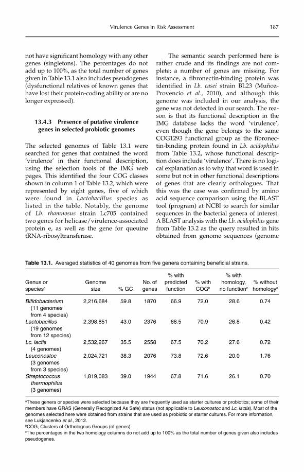
Virulence Genes in Risk Assessment 187
not have significant homology with any other genes (singletons). The percentages do not add up to 100%, as the total number of genes given in Table 13.1 also includes pseudogenes (dysfunctional relatives of known genes that have lost their protein-coding ability or are no longer expressed).
13.4.3 Presence of putative virulence genes in selected probiotic genomes
The selected genomes of Table 13.1 were searched for genes that contained the word ‘virulence’ in their functional description, using the selection tools of the IMG web pages. This identified the four COG classes shown in column 1 of Table 13.2, which were represented by eight genes, five of which were found in Lactobacillus species as listed in the table. Notably, the genome of Lb. rhamnosus strain Lc705 contained two genes for helicase/virulence-associated protein e, as well as the gene for queuine tRNA-ribosyltransferase.
The semantic search performed here is rather crude and its findings are not com-plete; a number of genes are missing. For instance, a fibronectin-binding protein was identified in Lb. casei strain BL23 (Muñoz-Provencio et al., 2010), and although this genome was included in our analysis, the gene was not detected in our search. The rea-son is that its functional description in the IMG database lacks the word ‘virulence’, even though the gene belongs to the same COG1293 functional group as the fibronec-tin-binding protein found in Lb. acidophilusfrom Table 13.2, whose functional descrip-tion does include ‘virulence’. There is no logi-cal explanation as to why that word is used in some but not in other functional descriptions of genes that are clearly orthologues. That this was the case was confirmed by amino acid sequence comparison using the BLAST tool (program) at NCBI to search for similar sequences in the bacterial genera of interest. A BLAST analysis with the Lb. acidophilus gene from Table 13.2 as the query resulted in hits obtained from genome sequences (genome
Table 13.1. Averaged statistics of 40 genomes from five genera containing beneficial strains.
Genus or speciesa
Genomesize % GC
No. of genes
% with predictedfunction
% with COGb
% with homology,
no functionc
% without homologyc
Bifidobacterium (11 genomes from 4 species)
2,216,684 59.8 1870 66.9 72.0 28.6 0.74
Lactobacillus (19 genomes from 12 species)
2,398,851 43.0 2376 68.5 70.9 26.8 0.42
Lc. lactis (4 genomes)
2,532,267 35.5 2558 67.5 70.2 27.6 0.72
Leuconostoc(3 genomes from 3 species)
2,024,721 38.3 2076 73.8 72.6 20.0 1.76
Streptococcusthermophilus(3 genomes)
1,819,083 39.0 1944 67.8 71.6 26.1 0.70
aThese genera or species were selected because they are frequently used as starter cultures or probiotics; some of their members have GRAS (Generally Recognized As Safe) status (not applicable to Leuconostoc and Lc. lactis). Most of the genomes selected here were obtained from strains that are used as probiotic or starter cultures. For more information, see Lukjancenko et al., 2012.bCOG, Clusters of Orthologous Groups (of genes).cThe percentages in the two homology columns do not add up to 100% as the total number of genes given also includes pseudogenes.
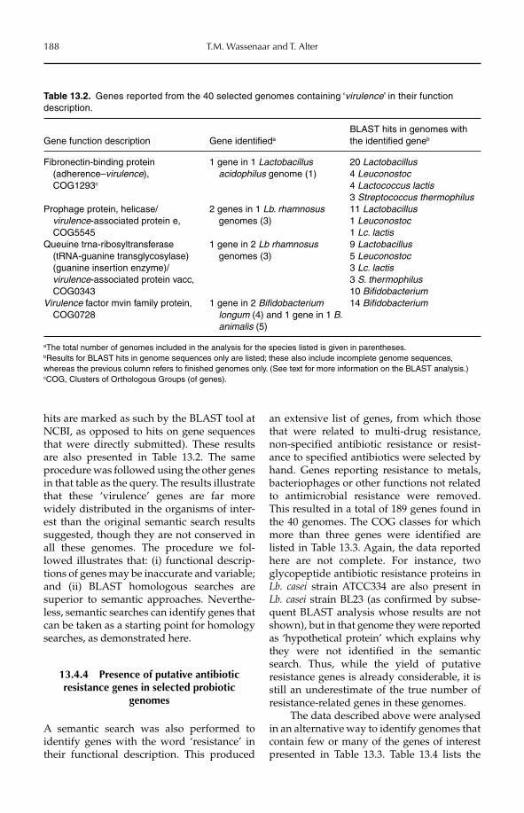
188 T.M. Wassenaar and T. Alter
hits are marked as such by the BLAST tool at NCBI, as opposed to hits on gene sequences that were directly submitted). These results are also presented in Table 13.2. The same procedure was followed using the other genes in that table as the query. The results illustrate that these ‘virulence’ genes are far more widely distributed in the organisms of inter-est than the original semantic search results suggested, though they are not conserved in all these genomes. The procedure we fol-lowed illustrates that: (i) functional descrip-tions of genes may be inaccurate and variable; and (ii) BLAST homologous searches are superior to semantic approaches. Neverthe-less, semantic searches can identify genes that can be taken as a starting point for homology searches, as demonstrated here.
13.4.4 Presence of putative antibiotic resistance genes in selected probiotic
genomes
A semantic search was also performed to identify genes with the word ‘resistance’ in their functional description. This produced
an extensive list of genes, from which those that were related to multi-drug resistance, non-specified antibiotic resistance or resist-ance to specified antibiotics were selected by hand. Genes reporting resistance to metals, bacteriophages or other functions not related to antimicrobial resistance were removed. This resulted in a total of 189 genes found in the 40 genomes. The COG classes for which more than three genes were identified are listed in Table 13.3. Again, the data reported here are not complete. For instance, two glycopeptide antibiotic resistance proteins in Lb. casei strain ATCC334 are also present in Lb. casei strain BL23 (as confirmed by subse-quent BLAST analysis whose results are not shown), but in that genome they were reported as ‘hypo thetical protein’ which explains why they were not identified in the semantic search. Thus, while the yield of putative resistance genes is already considerable, it is still an underestimate of the true number of resistance-related genes in these genomes.
The data described above were analysed in an alternative way to identify genomes that contain few or many of the genes of interest presented in Table 13.3. Table 13.4 lists the
Table 13.2. Genes reported from the 40 selected genomes containing ‘virulence’ in their function description.
Gene function description Gene identifieda
BLAST hits in genomes with the identified geneb
Fibronectin-binding protein (adherence–virulence),COG1293c
1 gene in 1 Lactobacillus acidophilus genome (1)
20 Lactobacillus4 Leuconostoc4 Lactococcus lactis3 Streptococcus thermophilus
Prophage protein, helicase/virulence-associated protein e, COG5545
2 genes in 1 Lb. rhamnosus genomes (3)
11 Lactobacillus1 Leuconostoc1 Lc. lactis
Queuine trna-ribosyltransferase (tRNA-guanine transglycosylase) (guanine insertion enzyme)/virulence-associated protein vacc, COG0343
1 gene in 2 Lb rhamnosus genomes (3)
9 Lactobacillus5 Leuconostoc3 Lc. lactis3 S. thermophilus10 Bifidobacterium
Virulence factor mvin family protein, COG0728
1 gene in 2 Bifidobacterium longum (4) and 1 gene in 1 B. animalis (5)
14 Bifidobacterium
aThe total number of genomes included in the analysis for the species listed is given in parentheses.bResults for BLAST hits in genome sequences only are listed; these also include incomplete genome sequences, whereas the previous column refers to finished genomes only. (See text for more information on the BLAST analysis.)cCOG, Clusters of Orthologous Groups (of genes).
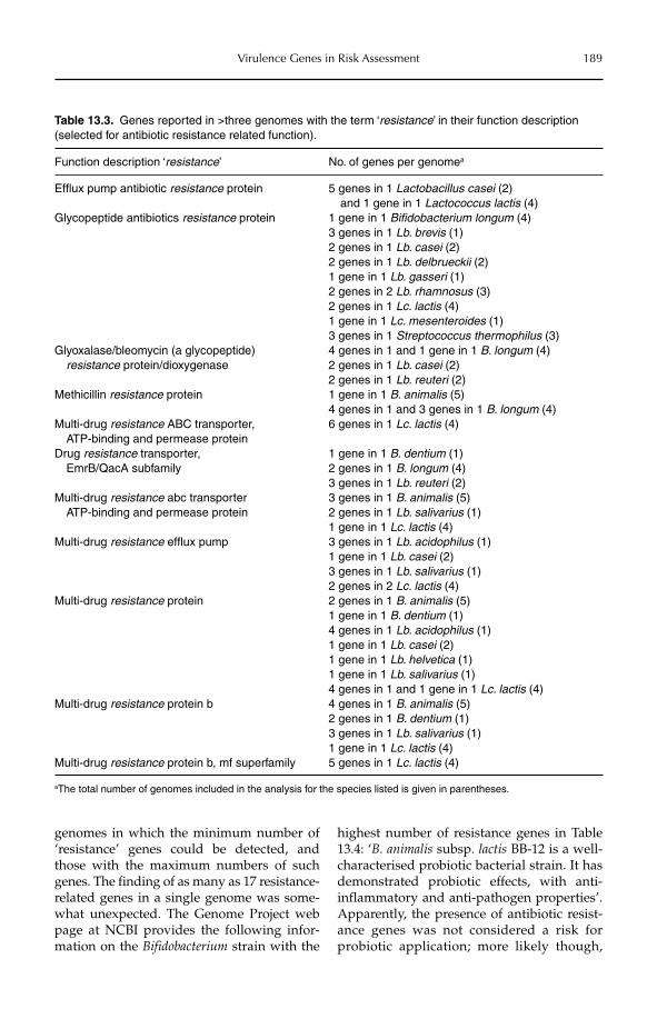
Virulence Genes in Risk Assessment 189
Table 13.3. Genes reported in >three genomes with the term ‘resistance’ in their function description (selected for antibiotic resistance related function).
Function description ‘resistance’ No. of genes per genomea
Efflux pump antibiotic resistance protein 5 genes in 1 Lactobacillus casei (2) and 1 gene in 1 Lactococcus lactis (4)
Glycopeptide antibiotics resistance protein 1 gene in 1 Bifidobacterium longum (4)3 genes in 1 Lb. brevis (1)2 genes in 1 Lb. casei (2)2 genes in 1 Lb. delbrueckii (2)1 gene in 1 Lb. gasseri (1)2 genes in 2 Lb. rhamnosus (3)2 genes in 1 Lc. lactis (4)1 gene in 1 Lc. mesenteroides (1)3 genes in 1 Streptococcus thermophilus (3)
Glyoxalase/bleomycin (a glycopeptide) resistance protein/dioxygenase
4 genes in 1 and 1 gene in 1 B. longum (4)2 genes in 1 Lb. casei (2)2 genes in 1 Lb. reuteri (2)
Methicillin resistance protein 1 gene in 1 B. animalis (5)4 genes in 1 and 3 genes in 1 B. longum (4)
Multi-drug resistance ABC transporter, ATP-binding and permease protein
6 genes in 1 Lc. lactis (4)
Drug resistance transporter, EmrB/QacA subfamily
1 gene in 1 B. dentium (1)2 genes in 1 B. longum (4)3 genes in 1 Lb. reuteri (2)
Multi-drug resistance abc transporter ATP-binding and permease protein
3 genes in 1 B. animalis (5)2 genes in 1 Lb. salivarius (1)1 gene in 1 Lc. lactis (4)
Multi-drug resistance efflux pump 3 genes in 1 Lb. acidophilus (1)1 gene in 1 Lb. casei (2)3 genes in 1 Lb. salivarius (1)2 genes in 2 Lc. lactis (4)
Multi-drug resistance protein 2 genes in 1 B. animalis (5)1 gene in 1 B. dentium (1)4 genes in 1 Lb. acidophilus (1)1 gene in 1 Lb. casei (2)1 gene in 1 Lb. helvetica (1)1 gene in 1 Lb. salivarius (1)4 genes in 1 and 1 gene in 1 Lc. lactis (4)
Multi-drug resistance protein b 4 genes in 1 B. animalis (5)2 genes in 1 B. dentium (1)3 genes in 1 Lb. salivarius (1)1 gene in 1 Lc. lactis (4)
Multi-drug resistance protein b, mf superfamily 5 genes in 1 Lc. lactis (4)
aThe total number of genomes included in the analysis for the species listed is given in parentheses.
genomes in which the minimum number of ‘resistance’ genes could be detected, and those with the maximum numbers of such genes. The finding of as many as 17 resistance-related genes in a single genome was some-what unexpected. The Genome Project web page at NCBI provides the following infor-mation on the Bifidobacterium strain with the
highest number of resistance genes in Table 13.4: ‘B. animalis subsp. lactis BB-12 is a well-characterised probiotic bacterial strain. It has demonstrated probiotic effects, with anti-inflammatory and anti-pathogen properties’. Apparently, the presence of antibiotic resist-ance genes was not considered a risk for probiotic application; more likely though,
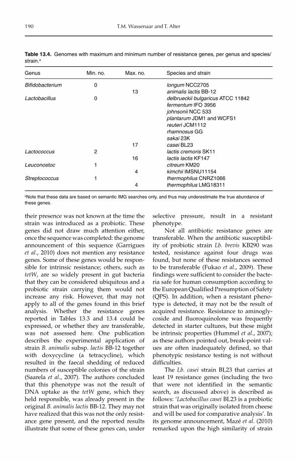
190 T.M. Wassenaar and T. Alter
their presence was not known at the time the strain was introduced as a probiotic. These genes did not draw much attention either, once the sequence was completed: the genome announcement of this sequence (Garrigues et al., 2010) does not mention any resistance genes. Some of these genes would be respon-sible for intrinsic resistance; others, such as tetW, are so widely present in gut bacteria that they can be considered ubiquitous and a probiotic strain carrying them would not increase any risk. However, that may not apply to all of the genes found in this brief analysis. Whether the resistance genes reported in Tables 13.3 and 13.4 could be expressed, or whether they are transferable, was not assessed here. One publication describes the experimental application of strain B. animalis subsp. lactis BB-12 together with doxycycline (a tetracycline), which resulted in the faecal shedding of reduced numbers of susceptible colonies of the strain (Saarela et al., 2007). The authors concluded that this phenotype was not the result of DNA uptake as the tetW gene, which they held responsible, was already present in the original B. animalis lactis BB-12. They may not have realized that this was not the only resist-ance gene present, and the reported results illustrate that some of these genes can, under
selective pressure, result in a resistant phenotype.
Not all antibiotic resistance genes are transferable. When the antibiotic susceptibil-ity of probiotic strain Lb. brevis KB290 was tested, resistance against four drugs was found, but none of these resistances seemed to be transferable (Fukao et al., 2009). These findings were sufficient to consider the bacte-ria safe for human consumption according to the European Qualified Presumption of Safety (QPS). In addition, when a resistant pheno-type is detected, it may not be the result of acquired resistance. Resistance to aminogly-coside and fluoroquinolone was frequently detected in starter cultures, but these might be intrinsic properties (Hummel et al., 2007); as these authors pointed out, break-point val-ues are often inadequately defined, so that phenotypic resistance testing is not without difficulties.
The Lb. casei strain BL23 that carries at least 19 resistance genes (including the two that were not identified in the semantic search, as discussed above) is described as follows: ‘Lactobacillus casei BL23 is a probiotic strain that was originally isolated from cheese and will be used for comparative analysis’. In its genome announcement, Mazé et al. (2010) remarked upon the high similarity of strain
Table 13.4. Genomes with maximum and minimum number of resistance genes, per genus and species/strain.a
Genus Min. no. Max. no. Species and strain
Bifidobacterium 0 longum NCC270513 animalis lactis BB-12
Lactobacillus 0 delbrueckii bulgaricus ATCC 11842fermentum IFO 3956johnsonii NCC 533plantarum JDM1 and WCFS1reuteri JCM1112rhamnosus GGsakai 23K
17 casei BL23Lactococcus 2 lactis cremoris SK11
16 lactis lactis KF147Leuconostoc 1 citreum KM20
4 kimchii IMSNU11154Streptococcus 1 thermophilus CNRZ1066
4 thermophilus LMG18311
aNote that these data are based on semantic IMG searches only, and thus may underestimate the true abundance of these genes.
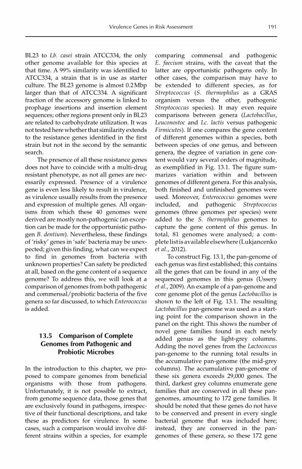
Virulence Genes in Risk Assessment 191
BL23 to Lb. casei strain ATCC334, the only other genome available for this species at that time. A 99% similarity was identified to ATCC334, a strain that is in use as starter culture. The BL23 genome is almost 0.2 Mbp larger than that of ATCC334. A significant fraction of the accessory genome is linked to prophage insertions and insertion element sequences; other regions present only in BL23 are related to carbohydrate utilization. It was not tested here whether that similarity extends to the resistance genes identified in the first strain but not in the second by the semantic search.
The presence of all these resistance genes does not have to coincide with a multi-drug resistant phenotype, as not all genes are nec-essarily expressed. Presence of a virulence gene is even less likely to result in virulence, as virulence usually results from the presence and expression of multiple genes. All organ-isms from which these 40 genomes were derived are mostly non-pathogenic (an excep-tion can be made for the opportunistic patho-gen B. dentium). Nevertheless, these findings of ‘risky’ genes in ‘safe’ bacteria may be unex-pected; given this finding, what can we expect to find in genomes from bacteria with unknown properties? Can safety be predicted at all, based on the gene content of a sequence genome? To address this, we will look at a comparison of genomes from both pathogenic and commensal/probiotic bacteria of the five genera so far discussed, to which Enterococcusis added.
13.5 Comparison of Complete Genomes from Pathogenic and
Probiotic Microbes
In the introduction to this chapter, we pro-posed to compare genomes from beneficial organisms with those from pathogens. Unfortunately, it is not possible to extract, from genome sequence data, those genes that are exclusively found in pathogens, irrespec-tive of their functional descriptions, and take these as predictors for virulence. In some cases, such a comparison would involve dif-ferent strains within a species, for example
comparing commensal and pathogenic E. faecium strains, with the caveat that the latter are opportunistic pathogens only. In other cases, the comparison may have to be extended to different species, as for Streptococcus (S. thermophilus as a GRAS organism versus the other, pathogenic Streptococcus species). It may even require comparisons between genera (Lactobacillus,Leuconostoc and Lc. lactis versus pathogenic Firmicutes). If one compares the gene content of different genomes within a species, both between species of one genus, and between genera, the degree of variation in gene con-tent would vary several orders of magnitude, as exemplified in Fig. 13.1. The figure sum-marizes variation within and between genomes of different genera. For this analysis, both finished and unfinished genomes were used. Moreover, Enterococcus genomes were included, and pathogenic Streptococcusgenomes (three genomes per species) were added to the S. thermophilus genomes to capture the gene content of this genus. In total, 81 genomes were analysed; a com-plete list is available elsewhere (Lukjancenko et al., 2012).
To construct Fig. 13.1, the pan-genome of each genus was first established; this contains all the genes that can be found in any of the sequenced genomes in this genus (Ussery et al., 2009). An example of a pan-genome and core genome plot of the genus Lactobacillus is shown to the left of Fig. 13.1. The resulting Lactobacillus pan-genome was used as a start-ing point for the comparison shown in the panel on the right. This shows the number of novel gene families found in each newly added genus as the light-grey columns. Adding the novel genes from the Lactococcuspan-genome to the running total results in the accumulative pan-genome (the mid-grey columns). The accumulative pan-genome of these six genera exceeds 29,000 genes. The third, darkest grey columns enumerate gene families that are conserved in all these pan-genomes, amounting to 172 gene families. It should be noted that these genes do not have to be conserved and present in every single bacterial genome that was included here; instead, they are conserved in the pan-genomes of these genera, so these 172 gene
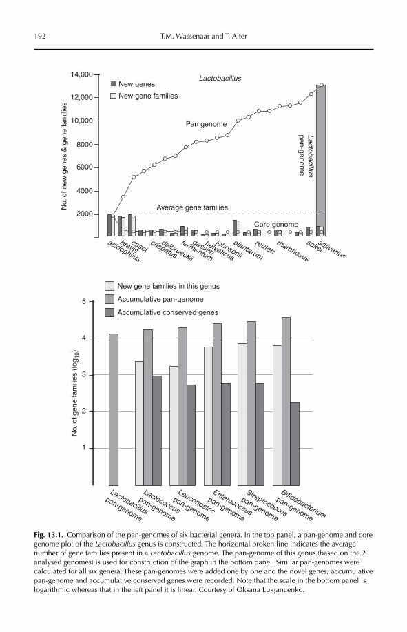
192 T.M. Wassenaar and T. Alter
New gene families in this genus
Accumulative pan-genome
Accumulative conserved genes
1
2
3
4
5
No.
of g
ene
fam
ilies
(lo
g 10)
No.
of n
ew g
enes
& g
ene
fam
ilies
Lactobacillus
pan-genome
Lactococcus
pan-genome
Leuconostoc
pan-genome
Enterococcus
pan-genome
Streptococcus
pan-genome
Bifidobacterium
pan-genome
Lactobacilluspan-genom
e
New genesLactobacillus
acidophilus
breviscasei
crispatus
delbrueckii
fermentum
gasserihelveticus
johnsonii
plantarum
reuterirhamnosus
sakeisalivarius
New gene families
Core genome
Pan genome
Average gene families2000
4000
6000
8000
10,000
12,000
14,000
Fig. 13.1. Comparison of the pan-genomes of six bacterial genera. In the top panel, a pan-genome and core genome plot of the Lactobacillus genus is constructed. The horizontal broken line indicates the average number of gene families present in a Lactobacillus genome. The pan-genome of this genus (based on the 21 analysed genomes) is used for construction of the graph in the bottom panel. Similar pan-genomes were calculated for all six genera. These pan-genomes were added one by one and the novel genes, accumulative pan-genome and accumulative conserved genes were recorded. Note that the scale in the bottom panel is logarithmic whereas that in the left panel it is linear. Courtesy of Oksana Lukjancenko.
Virulence Genes in Risk Assessment 193
families are present in at least one genome per genus. Compare this with the true core genome of all 81 genomes that has been described: there are only 63 gene families that are conserved and present in all of these genomes (not shown in the figure) (Lukjancenko et al., 2012). The core genome of the Lactobacillus genus contains 363 gene families, and that of Bifodobacterium 725 (O. Lukjancenko, unpublished data).
With this degree of variation within a genus (as Fig. 13.1 illustrates for Lactobacillus)and between genera, there is too much ‘noise’ that hides the ‘signal’ of virulence genes found in pathogens and absent in probiotics. If one were to compare the gene content between, say, a probiotic Lactobacillus strain and a pathogenic Enterococcus strain, there will be many genes found in the latter that are absent in the former, but that has little to do with their difference in phenotype: these genes make the difference between an Enter-ococcus and a Lactobacillus.
There is a way to tackle this problem. We have split the 81 genomes of interest into two groups: in one group we combined all patho-genic Firmicutes and in the other all non- pathogens (Firmicutes plus bifidobacteria). For these two collections, the pan-genomes and core genomes were calculated. We then compared to which COG classes these pan-genome genes and core-genome genes belonged, and compared the relative enrich-ment (i.e. conservation) to COG classes in the core genome with respect to the pan-genome. This identified significant differences between the enriched core gene families (genes conser-ved in all genomes of the collection) of the pathogens compared with the non-pathogens (Lukjancenko et al., 2012). The analysis identi-fied, for instance, that genes involved in post-translational modification and chaperones were over-represented in the core genome (compared with their pan-genome) of non-pathogens but not of pathogens. Conversely, genes involved in cell wall/membrane bio-synthesis secretion were over-represented in the core genome of pathogens but not in that of non-pathogens. The latter finding matches our knowledge of virulence genes, which are frequently cell surface structures or secreted proteins. Thus, without specifically searching
for a role in virulence, genes were identified in the core genome of pathogens that are likely to have a function in virulence. It is hypothesized that the genes identified as enriched in the core genome of non- pathogens would likewise play a role in establishing a commensal or possible probiotic relationship with the host.
Where do we go from here? The approach described above can be used as a proof of principle. When more genomes of probiotic, commensal and pathogenic strains become available, the method can be fine-tuned and optimized. That could possibly lead to a better understanding of the genes involved in either pathogenicity or a commensal/ probiotic relationships with the human host. A pragmatic approach could be to accept that presence of ‘risky’ genes in probiotic strains with a long history of safe use is not a prob-lem, as long as they are present in that par-ticular species. If we accept that view, a list of genes could be produced that, in particular genomic backgrounds, do not contribute to virulence, despite the fact that they can do so in other, putatively pathogenic organisms. This gives a means to weigh the risk these genes provide, so that novel genome sequences can be interpreted for the degree of safety of the organism in question based on gene content.
13.6 Conclusion
Is it feasible that in the near future we can predict the risk to human health for a bacte-rial isolate, based on its genome sequence only? Even though the properties of micro-organisms would be encoded in their genes, microbes will adjust their expression profile to external conditions. Genome sequences might reveal mechanisms by which micro-organisms introduce variation into their expression profiles by genetic polymor-phism, phase variation and antigenic varia-tion, but they cannot be used to predict expression patterns under all circumstances. Currently, whole genome expression profil-ing and in vivo phenotypic tests are still needed to describe the behaviour and safety of bacterial strains. Gene expression profil-ing using next-generation sequencing might
194 T.M. Wassenaar and T. Alter
ease that approach. The number of genes for which a function cannot be predicted should, it is hoped, decrease over time, at least for such genes that are conserved in multiple organisms; singleton genes with unknown function are more problematic. At present, their relevance to pathogenicity remains unknown. None the less, genome sequences can contribute to hazard identifi-cation by identifying and excluding poten-tial pathogenic microorganisms based on the pure presence of virulence factors and to
hazard characterization, by describing the microorganism’s lifestyle.
Acknowledgement
TMW acknowledges the support provided by the Safety and Environmental Assurance Centre at Unilever for part of this work. The assistance and advice of David W. Ussery and Oksana Lukjancenko was highly appreciated.
References
Bernardeau, M., Vernoux, J.P., Henri-Dubernet, S. and Guéguen, M. (2008) Safety assessment of dairy micro-organisms: the Lactobacillus genus. International Journal of Food Microbiology 126, 278–285.
Chin, C.S., Sorenson, J., Harris, J.B., Robins, W.P., Charles, R.C., Jean-Charles, R.R., Bullard, J., Webster, D.R., Kasarskis, A., Peluso, P., Paxinos, E.E., Yamaichi, Y., Calderwood, S.B., Mekalanos, J.J., Schadt, E.E. and Waldor, M.K. (2011) The origin of the Haitian cholera outbreak strain. New England Journal of Medicine364, 33–42.
Field, D., Garrity, G., Gray, T., Morrison, N., Selengut, J., Sterk, P., Tatusova, T., Thomson, N., Allen, M.J., Angiuoli, S.V., Ashburner, M., Axelrod, N., Baldauf, S., Ballard, S., Boore, J., Cochrane, G., Cole, J., Dawyndt, P., De Vos, P., dePamphilis, C., Edwards, R., Faruque, N., Feldman, R., Gilbert, J., Gilna, P., Glöckner, F.O., Goldstein, P., Guralnick, R., Haft, D., Hancock, D., Hermjakob, H., Hertz-Fowler, C., Hugenholtz, P., Joint, I., Kagan, L., Kane, M., Kennedy, J., Kowalchuk, G., Kottmann, R., Kolker, E., Kravitz, S., Kyrpides, N., Leebens-Mack, J., Lewis, S.E., Li, K., Lister, A.L., Lord, P., Maltsev, N., Markowitz, V., Martiny, J., Methe, B., Mizrachi, I., Moxon, R., Nelson, K., Parkhill, J., Proctor, L., White, O., Sansone, S.A., Spiers, A., Stevens, R., Swift, P., Taylor, C., Tateno, Y., Tett, A., Turner, S., Ussery, D., Vaughan, B., Ward, N., Whetzel, T., San Gil, I., Wilson, G. and Wipat, A. (2008) The minimum information about a genome sequence (MIGS) specification. Nature Biotechnology 26, 541–547.
Filloux, A. (2010) A variety of bacterial pili involved in horizontal gene transfer. Journal of Bacteriology 192, 3243–3245.
Fukao, M., Tomita, H., Yakabe, T., Nomura, T., Ike, Y. and Yajima, N. (2009) Assessment of antibiotic resistance in probiotic strain Lactobacillus brevis KB290. Journal of Food Protection 72, 1923–1929.
Garrigues, C., Johansen, E. and Pedersen, M.B. (2010) Complete genome sequence of Bifidobacterium animalissubsp. lactis BB-12, a widely consumed probiotic strain. Journal of Bacteriology 192, 2467–2468.
Grozdanov, L., Raasch, C., Schulze, J., Sonnenborn, U., Gottschalk, G., Hacker, J. and Dobrindt, U. (2004) Analysis of the genome structure of the nonpathogenic probiotic Escherichia coli strain Nissle 1917. Journal of Bacteriology 186, 5432–5441.
Hancock, V., Vejborg, R.M. and Klemm, P. (2010) Functional genomics of probiotic Escherichia coli Nissle 1917 and 83972, and UPEC strain CFT073: comparison of transcriptomes, growth and biofilm formation. Molecular Genetics and Genomics 284, 437–454.
Hummel, A.S., Hertel, C., Holzapfel, W.H. and Franz, C.M. (2007) Antibiotic resistances of starter and probiotic strains of lactic acid bacteria. Applied and Environmental Microbiology 73, 730–739.
Hyatt, D., Chen, G.L., Locascio, P.F., Land, M.L., Larimer, F.W. and Hauser, L.J. (2010) Prodigal: prokaryotic gene recognition and translation initiation site identification. BMC Bioinformatics 11: 119. doi:10.1186/ 1471-2105-11-119.
Jones, B.V., Sun, F. and Marchesi, J.R. (2010) Comparative metagenomic analysis of plasmid encoded func-tions in the human gut microbiome. BMC Genomics 11: 46. doi:10.1186/1471-2164-11-46.
Kankainen, M., Paulin, L., Tynkkynen, S., von Ossowski, I., Reunanen, J., Partanen, P., Satokari, R., Vesterlund, S., Hendrickx, A.P., Lebeer, S., De Keersmaecker, S.C., Vanderleyden, J., Hämäläinen, T., Laukkanen, S., Salovuori, N., Ritari, J., Alatalo, E., Korpela, R., Mattila-Sandholm, T., Lassig, A., Hatakka, K., Kinnunen, K.T., Karjalainen, H., Saxelin, M., Laakso, K., Surakka, A., Palva, A., Salusjärvi, T., Auvinen, P. and de Vos,
Virulence Genes in Risk Assessment 195
W.M. (2009) Comparative genomic analysis of Lactobacillus rhamnosus GG reveals pili containing a human-mucus binding protein. Proceedings of the National Academy of Sciences of the United States of America 106, 17193–17198.
Koretz, R.L. (2009) Probiotics, critical illness, and methodologic bias. Nutrition in Clinical Practice 24, 45–49.Lagesen, K., Hallin, P., Rødland, E.A., Staerfeldt, H.H., Rognes, T. and Ussery, D.W. (2007) RNAmmer:
consistent and rapid annotation of ribosomal RNA genes. Nucleic Acids Research 35, 3100–3108.Lagesen, K., Ussery, D.W. and Wassenaar, T.M. (2010) Genome Update: the thousandth genome – a caution-
ary tale. Microbiology 156, 603–608.Lee, J.H. and O’Sullivan, D.J. (2010) Genomic insights into bifidobacteria. Microbiology and Molecular
Biology Reviews 74, 378–416.Liong, M.T. (2008) Safety of probiotics: translocation and infection. Nutrition Reviews 66, 192–202.Lukjancenko, O., Ussery, D.W. and Wassenaar, T.M. (2012) Comparative genomics of Bifidobacterium,
Lactobacillus and related probiotic genera. Microbial Ecology 63, 651–673.Mattia, A. and Merker, R. (2008) Regulation of probiotic substances as ingredients in foods: premarket
approval or ‘generally recognized as safe’ notification. Clinical Infectious Disease 46, S115–S118.Mazé, A., Boël, G., Zúñiga, M., Bourand, A., Loux, V., Yebra, M.J., Monedero, V., Correia, K., Jacques, N.,
Beaufils, S., Poncet, S., Joyet, P., Milohanic, E., Casarégola, S., Auffray, Y., Pérez-Martínez, G., Gibrat, J.F., Zagorec, M., Francke, C., Hartke, A. and Deutscher, J. (2010) Complete genome sequence of the probi-otic Lactobacillus casei strain BL23. Journal of Bacteriology 192, 2647–2648.
Meile, L., Le Blay, G. and Thierry, A. (2008) Safety assessment of dairy microorganisms: Propionibacteriumand Bifidobacterium. International Journal of Food Microbiology 126, 316–320.
Miller, J.R., Koren, S. and Sutton, G. (2010) Assembly algorithms for next-generation sequencing data. Genomics 95, 315–327.
Muñoz-Provencio, D., Pérez-Martínez, G. and Monedero, V. (2010) Characterization of a fibronectin-binding protein from Lactobacillus casei BL23. Journal of Applied Microbiology 108, 1050–1059.
Nagarajan, N., Cook, C., Di Bonaventura, M., Ge, H., Richards, A., Bishop-Lilly, K.A., DeSalle, R., Read, T.D. and Pop, M. (2010) Finishing genomes with limited resources: lessons from an ensemble of microbial genomes. BMC Genomics 11: 242. doi:10.1186/1471-2164-11-242.
Saarela, M., Maukonen, J., von Wright, A., Vilpponen-Salmela, T., Patterson, A.J., Scott, K.P., Hämynen, H. and Mättö, J. (2007) Tetracycline susceptibility of the ingested Lactobacillus acidophilus LaCH-5 and Bifidobacterium animalis subsp. lactis Bb-12 strains during antibiotic/probiotic intervention. InternationalJournal of Antimicrobial Agents 29, 271–280.
Schattner, P., Brooks, A.N. and Lowe, T.M. (2005) The tRNAscan-SE, snoscan and snoGPS web servers for the detection of tRNAs and snoRNAs. Nucleic Acids Research 33(Suppl. 2), W686–W689.
US FDA (2011) GRAS Notice Inventory. US Food and Drug Administration, Washington, DC. Available at: http://www.accessdata.fda.gov/scripts/fcn/fcnNavigation.cfm?rpt=grasListing (accessed 17 October 2011).
Ussery, D.W., Borini, S. and Wassenaar, T.M. (2009) Chapter 12. Microbial communities: core and pan-genomics. In: Computing for Comparative Microbial Genomics: Bioinformatics for Microbiologists.Springer, London, pp. 213–228.
Ventura, M., Lee, J.H., Canchaya, C., Zink, R., Leahy, S., Moreno-Munoz, J.A., O’Connell-Motherway, M., Higgins, D., Fitzgerald, G.F., O’Sullivan, D.J. and van Sinderen, D. (2005) Prophage-like elements in bifidobacteria: insights from genomics, transcription, integration, distribution, and phylogenetic analy-sis. Applied and Environmental Microbiology 71, 8692–8705.
Ventura, M., Canchaya, C., Bernini, V., Altermann, E., Barrangou, R., McGrath, S., Claesson, M.J., Li, Y., Leahy, S., Walker, C.D., Zink, R., Neviani, E., Steele, J., Broadbent, J., Klaenhammer, T.R., Fitzgerald, G.F., O’Toole, P.W. and van Sinderen, D. (2006) Comparative genomics and transcriptional analysis of prophages identified in the genomes of Lactobacillus gasseri, Lactobacillus salivarius, and Lactobacillus casei. Applied and Environmental Microbiology 72, 3130–3146.
Verma, R., Dhamija, R., Ross, S.C., Batts, D.H. and Loehrke, M.E. (2010) Symbiotic bacteria induced necro-tizing pancreatitis. Journal of the Pancreas Online 11, 474–476.
von Wright, A. (2012) Microbes for human and animal consumption. In: Sundh, I., Wilcks, A. and Goettel, M.S. (eds) Beneficial Microorganisms in Agriculture, Food and the Environment: Safety Assessment and Regulation. CAB International, Wallingford, UK, pp. 27–40.
Wassenaar, T.M. (2004) Risk assessment prediction from genome sequences, promises and dreams. Journal of Food Protection 67, 2053–2057.
Wassenaar, T.M. and Gaastra, W. (2001) Bacterial virulence, where to draw the line? FEMS Microbiology Letters 201, 1–7.
196 T.M. Wassenaar and T. Alter
Wassenaar, T.M. and Klein, G. (2008) Safety aspects of bacterial food supplements. Journal of Food Protection71, 1734–1741.
Wessels, S., Axelsson, L., Hansen, E.B., De Vuyst, L., Laulund, S., Lähteenmäki, L., Lindgren, S., Mollet, B., Salminen, S. and von Wright, A. (2004) The lactic acid bacteria, the food chain, and their regulation. Trends in Food Science and Technology 15, 498–505.
Wilcks, A. and van Hoek, A.H.A.M. (2012) Antibiotic resistance in relation to starter cultures and probiotics. In: Sundh, I., Wilcks, A. and Goettel, M.S. (eds) Beneficial Microorganisms in Agriculture, Food and the Environment: Safety Assessment and Regulation. CAB International, Wallingford, UK, pp. 41–56.
Willenbrock, H., Hallin, P.F., Wassenaar, T.M. and Ussery, D.U. (2007) Characterization of probiotic Escherichia coli isolates with a pan-genome microarray. Genome Biology 8: R267. doi:10.1186/gb-2007-8-12-r267.
©CAB International 2012. Beneficial Microorganisms in Agriculture, Food and the Environment: Safety Assessment and Regulation (eds I. Sundh et al.) 197
14.1 Introduction 19714.2 Exposure to Fungal MBCAs 198
14.2.1 Occupational exposure to fungal MBCAs and risk assessment 203 14.2.2 Exposure of bystanders to fungal MBCAs and risk assessment 204 14.2.3 Background exposure to fungi 205
14.3 Exposure to Bacterial MBCAs 206 14.3.1 Occupational exposure to bacterial MBCAs and risk assessment 206 14.3.2 Exposure of bystanders to bacterial MBCAs and risk assessment 206 14.3.3 Background exposure to actinobacteria and bacteria 207
14.4 Exposure to Other Microbial Agents 207 14.4.1 Occupational exposure to other microbial agents and risk assessment 207 14.4.2 Exposure of bystanders to other microbial agents 208 14.4.3 Background exposure to other microbial agents 208
14.5 Discussion 209
14 Occupational Safety of Microbial Agents
Anne Mette Madsen and Kira TendalThe National Research Centre for the Working Environment, Denmark
14.1 Introduction
Occupational exposure to bio-aerosols containing high concentrations of fungi, bacteria and other bio-aerosol components such as endotoxin may cause various dele-terious health effects dependent on the expo-sure levels (Rylander et al., 1985; Eduard et al., 2001). In addition to direct health effects, exposure to fungi may also have an adjuvant effect on the allergic response to other allergens. For instance, the insect path-ogenic fungus Metarhizium anisopliae has been found to have an adjuvant effect on a standard allergen (ovalbumin) in mice (Instanes et al., 2006). Endotoxin, a cell wall component of Gram-negative bacteria such as Pseudomonas has strong pro-inflammatory properties. In the Netherlands, the recom-mended exposure limit for endotoxin is
90 endotoxin units m−3 (Health Council of the Netherlands, 2010). Non-pathogenic bacteria, including the actinobacteria (one of the dominant phyla of the bacteria), may cause respiratory symptoms by triggering immune responses in exposed individuals (Purokivi et al., 2001; Tlaskalova-Hogenova et al., 2005). Workers in a variety of occupa-tions are expo sed to microbes, which may cause allergies or occupational asthma, e.g. Godnic-Cvar et al. (1999) found brewery workers to be significantly more likely to react positively to moulds in skin prick tests. Many of the workers in the study also reacted positively to brewer’s yeast in skin prick tests. Enzymes from microorganisms, e.g. the fungi Aspergillus spp. and Trichoderma viride, and the bacterium Bacillus subtilis are used in the baking and pharmaceutical ind-ustries and are associated with occupational
198 A. Mette Madsen and K. Tendal
asthma in workers from these industries (Lachowsky and Lopez, 2001). This subject is treated elsewhere (Flindt, 1969; Dolovich and Little, 1972; Horner et al., 2008), and is not part of this chapter, which reviews occupational exposure to microbial biocon-trol agents (MBCAs) and also to baker’s/brewer’s yeast (Saccharomyces cerevisiae) and to Botrytis cinerea (used in wine production), in relation to occupational health.
The prevalence of a microorganism in its natural habitat is referred to as the back-ground exposure level of the microorgan-ism, and this needs to be taken into account when evaluating the environmental safety of microbial plant protection products (Mensink and Scheepmaker, 2007). In this chapter, commercial microbial biocontrol products based on MBCAs will be referred to as MBCPs. We elucidate the back-ground exposures to different applied species of microorganisms. The focus is on MBCPs based on the fungi Beauveria bassiana, Metarhizium anisopliae, Verticillium lecanii (i.e. Lecanicillium spp.), Trichoderma harzianum, Trichoderma polysporum and Trichoderma viride, on the actinobacterium Streptomyces griseoviridis, and on the bac-teria Bacillus subtilis, Bacillus thuringiensisand Pseudomonas chlororaphis.
Directive 2000/54/EC of the European Parliament and Council (OJEC, 2000) forms the legal basis for the legislation in EU (European Union) countries concerning the protection of workers from risks related to exposure to biological agents at work. The Directive classifies microorganisms present in occupational environments according to whether the microorganism ‘is unlikely to cause human disease’ (Risk group 1), ‘can cause disease for which there is usually an effective treatment, and is unlikely to spread to the community’ (Risk group 2), ‘can cause severe disease, for which there is usually an effective treatment, but there is a risk of spreading to the community’ (Risk group 3) or ‘can cause severe disease with usually no effective treatment, and with a high risk of spreading to the community’ (Risk group 4). The Directive also lists the biological agents currently included in Risk groups 2–4. None of the microorganisms discussed in this
chapter rate above Risk group 1. The Directive charges employers with assessing the risk to workers’ health and safety, and taking the appropriate measures to ensure that workers are protected against exposure to biological agents.
Microorganisms selected for biocontrol and foodstuff production are supposedly not infectious in humans. However, a few case histories exist which indicate that some spe-cies used for biocontrol or foodstuff produc-tion are to some extent infectious, or at least may be prevalent in weakened or immuno-compromised persons (Guiserix et al., 1996; Henke et al., 2002; Ren et al., 2004; Tucker et al.,2004; Gürcan et al., 2006; Oh et al., 2009), and a few examples will be elucidated.
Sections 14.2 and 14.3 of this chapter treat microorganisms used as MBCAs and Section 14.4 treats microorganisms used in foodstuff production. In Sections 14.2 and 14.3, exposure to aerosolized fungi and bac-teria, respectively, is reviewed. The actino-bacterium S. griseoviridis is included in Section 14.3. The first part of each of the sec-tions reviews exposure to the applied micro-organisms during different work activities, and if possible, compares it with background exposure levels of microorganisms of the same species, microorganisms of the same genera, and total fungi, actinobacteria or bacteria. Then, exposure of bystanders to MBCPs is considered, as people may work in neighbouring areas, and people may come to work in people’s homes in MBCP-treated areas (e.g. home care workers). Finally, background exposures are reviewed in order to establish a frame of reference for other types of exposure. Exposures are pref-erentially expressed as cfu m−3 air (colony forming units per cubic metre of air), or as otherwise applicable; if these are available, exposures are related to epidemiological studies.
14.2 Exposure to Fungal MBCAs
Data on both occupational and background exposure to fungal MBCAs are included in Tables 14.1 and 14.2.
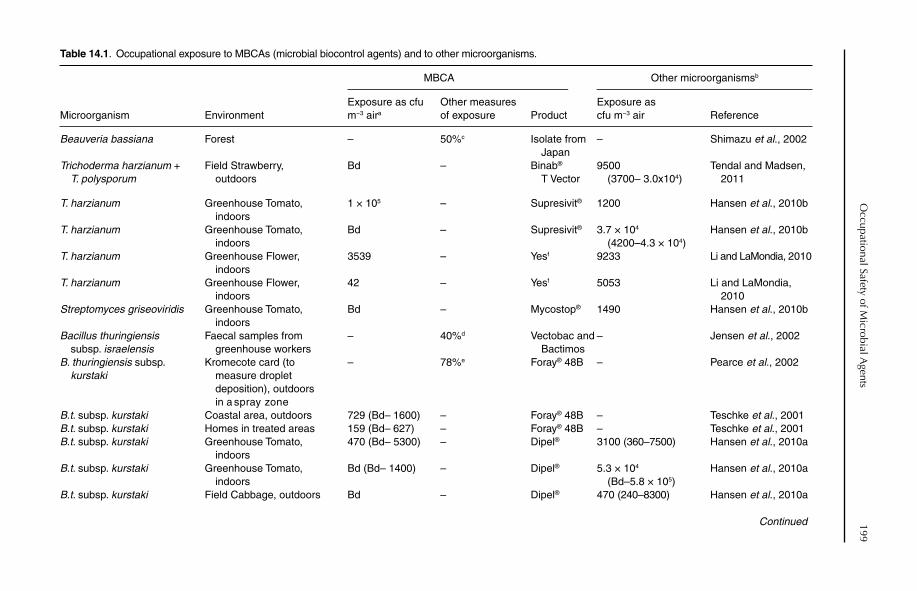
Occupational Safety of M
icrobial Agents
199
Continued
Table 14.1. Occupational exposure to MBCAs (microbial biocontrol agents) and to other microorganisms.
MBCA Other microorganismsb
Microorganism EnvironmentExposure as cfu m−3 aira
Other measures of exposure Product
Exposure as cfu m−3 air Reference
Beauveria bassiana Forest – 50%c Isolate from Japan
– Shimazu et al., 2002
Trichoderma harzianum + T. polysporum
Field Strawberry, outdoors
Bd – Binab®
T Vector9500
(3700– 3.0x10⁴)Tendal and Madsen,
2011
T. harzianum Greenhouse Tomato, indoors
1 × 10⁵ – Supresivit® 1200 Hansen et al., 2010b
T. harzianum Greenhouse Tomato, indoors
Bd – Supresivit® 3.7 × 10⁴
(4200–4.3 × 10⁴)Hansen et al., 2010b
T. harzianum Greenhouse Flower, indoors
3539 – Yesf 9233 Li and LaMondia, 2010
T. harzianum Greenhouse Flower, indoors
42 – Yesf 5053 Li and LaMondia, 2010
Streptomyces griseoviridis Greenhouse Tomato, indoors
Bd – Mycostop® 1490 Hansen et al., 2010b
Bacillus thuringiensissubsp. israelensis
Faecal samples from greenhouse workers
– 40%d Vectobac and Bactimos
– Jensen et al., 2002
B. thuringiensis subsp. kurstaki
Kromecote card (to measure droplet deposition), outdoors in a spray zone
– 78%e Foray® 48B – Pearce et al., 2002
B.t. subsp. kurstaki Coastal area, outdoors 729 (Bd– 1600) – Foray® 48B – Teschke et al., 2001B.t. subsp. kurstaki Homes in treated areas 159 (Bd– 627) – Foray® 48B – Teschke et al., 2001B.t. subsp. kurstaki Greenhouse Tomato,
indoors470 (Bd– 5300) – Dipel® 3100 (360–7500) Hansen et al., 2010a
B.t. subsp. kurstaki Greenhouse Tomato, indoors
Bd (Bd– 1400) – Dipel® 5.3 × 10⁴
(Bd–5.8 × 10⁵)Hansen et al., 2010a
B.t. subsp. kurstaki Field Cabbage, outdoors Bd – Dipel® 470 (240–8300) Hansen et al., 2010a
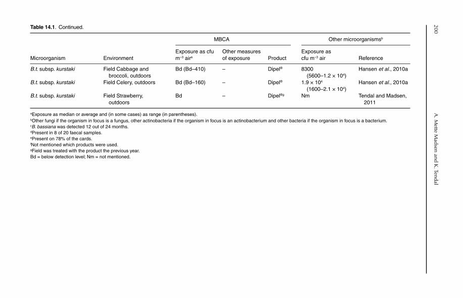
200A
. Mette M
adsen and K. Tendal
Table 14.1. Continued.
MBCA Other microorganismsb
Microorganism EnvironmentExposure as cfu m−3 aira
Other measures of exposure Product
Exposure as cfu m−3 air Reference
B.t. subsp. kurstaki Field Cabbage and broccoli, outdoors
Bd (Bd–410) – Dipel® 8300(5600–1.2 × 10⁴)
Hansen et al., 2010a
B.t. subsp. kurstaki Field Celery, outdoors Bd (Bd–160) – Dipel® 1.9 × 10⁴
(1600–2.1 × 10⁴)Hansen et al., 2010a
B.t. subsp. kurstaki Field Strawberry, outdoors
Bd – Dipel®g Nm Tendal and Madsen, 2011
aExposure as median or average and (in some cases) as range (in parentheses).bOther fungi if the organism in focus is a fungus, other actinobacteria if the organism in focus is an actinobacterium and other bacteria if the organism in focus is a bacterium.cB. bassiana was detected 12 out of 24 months.dPresent in 8 of 20 faecal samples.ePresent on 78% of the cards.fNot mentioned which products were used.gField was treated with the product the previous year.Bd = below detection level; Nm = not mentioned.
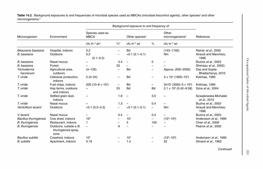
Occupational Safety of M
icrobial Agents
201Table 14.2. Background exposures to and frequencies of microbial species used as MBCAs (microbial biocontrol agents), other speciesa and other microorganisms.b
Background exposure to and frequency of:
Microorganism EnvironmentSpecies used as MBCA Other speciesa
Othermicroorganismsb Reference
cfu m−3 airc %d cfu m−3 air % cfu m−3 air
Beauveria bassiana Hospital, indoors 0.2 – Bd (143–1192) Rainer et al., 2000B. bassiana Outdoors 0.2
(0.1–0.5)– <0.1 (0.1–0.1) Nm Airaudi and Marchisio,
1996B. bassiana Nasal mucus – 3.4 – 0 – Buzina et al., 2003B. bassiana Forest – 33 – – – Shimazu et al., 2002)Trichoderma
harzianumAgricultural area,
outdoors(4–135) – Bd – Approx. (500–2000) Das and Gupta-
Bhattacharya, 2010T. viride Cellulose production,
indoors5 (0–24) – Bd – 3 × 10⁴ (1900–105) Kotimaa, 1990
T. viride Fuel chips, indoors 200 (10–6 × 10⁴) – Bd – 3x10⁴ (3000–3 × 10⁶) Kotimaa, 1990T. viride Hop farms, outdoors
and indoors– 53 Bd Bd 2.1 × 10³ (0.42–9.58) Góra et al., 2004
T. viride Settled grain dust, indoors
– 1.8 – 3.0 – Szwajkowska-Michalek et al., 2010
T. viride Nasal mucus – 1.3 – 0.4 – Buzina et al., 2003Verticillium lecanii Outdoors <0.1 (0.2–0.3) – <0.1 (0.1–0.1) – Nm Airaudi and Marchisio,
1996V. lecanii Nasal mucus – 0.4 – 0.4 – Buzina et al., 2003Bacillus thuringiensis Cow shed, indoors 106 – 107 – (105–109) Andersson et al., 1999B. thuringiensis Restaurant, indoors 1 – 5 100 – Chan et al., 2009B. thuringiensis Outdoors, outside a B.
thuringiensis spray zone
– 9 – – – Pearce et al., 2002
Bacillus subtilis Cowshed, indoors 107 – 107 – (105–109) Andersson et al., 1999B. subtilis Apartment, indoors 0.19 – 1.4 – 22 Simard et al., 1983
Continued
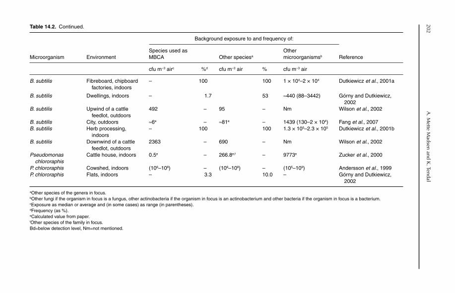
202A
. Mette M
adsen and K. Tendal
Table 14.2. Continued.
Background exposure to and frequency of:
Microorganism EnvironmentSpecies used as MBCA Other speciesa
Othermicroorganismsb Reference
cfu m−3 airc %d cfu m−3 air % cfu m−3 air
B. subtilis Fibreboard, chipboard factories, indoors
– 100 100 1 × 10⁴–2 × 10⁴ Dutkiewicz et al., 2001a
B. subtilis Dwellings, indoors – 1.7 53 ≈440 (88–3442) Górny and Dutkiewicz, 2002
B. subtilis Upwind of a cattle feedlot, outdoors
492 – 95 – Nm Wilson et al., 2002
B. subtilis City, outdoors ≈6e – ≈81e – 1439 (130–2 × 10⁴) Fang et al., 2007B. subtilis Herb processing,
indoors– 100 100 1.3 × 10⁵–2.3 × 10⁵ Dutkiewicz et al., 2001b
B. subtilis Downwind of a cattle feedlot, outdoors
2363 – 690 – Nm Wilson et al., 2002
Pseudomonaschlororaphis
Cattle house, indoors 0.5e – 266.8e,f – 9773e Zucker et al., 2000
P. chlororaphis Cowshed, indoors (106–108) – (106–108) – (105–109) Andersson et al., 1999P. chlororaphis Flats, indoors – 3.3 10.0 – Górny and Dutkiewicz,
2002
aOther species of the genera in focus.bOther fungi if the organism in focus is a fungus, other actinobacteria if the organism in focus is an actinobacterium and other bacteria if the organism in focus is a bacterium.cExposure as median or average and (in some cases) as range (in parentheses).dFrequency (as %).eCalculated value from paper.fOther species of the family in focus.Bd=below detection level, Nm=not mentioned.
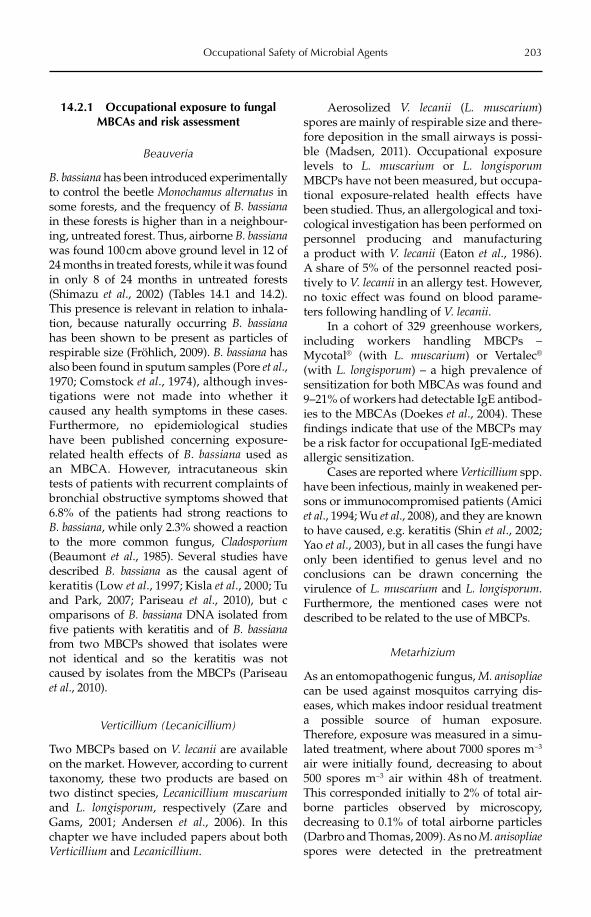
Occupational Safety of Microbial Agents 203
14.2.1 Occupational exposure to fungal MBCAs and risk assessment
Beauveria
B. bassiana has been introduced experimentally to control the beetle Monochamus alternatus in some forests, and the frequency of B. bassianain these forests is higher than in a neighbour-ing, untreated forest. Thus, airborne B. bassianawas found 100 cm above ground level in 12 of 24 months in treated forests, while it was found in only 8 of 24 months in untreated forests (Shimazu et al., 2002) (Tables 14.1 and 14.2). This presence is relevant in relation to inhala-tion, because naturally occurring B. bassianahas been shown to be present as particles of respirable size (Fröhlich, 2009). B. bassiana has also been found in sputum samples (Pore et al., 1970; Comstock et al., 1974), although inves-tigations were not made into whether it caused any health symptoms in these cases. Furthermore, no epidemiological studies have been published concerning exposure-related health effects of B. bassiana used as an MBCA. However, intracutaneous skin tests of patients with recurrent complaints of bronchial obstructive symptoms showed that 6.8% of the patients had strong reactions to B. bassiana, while only 2.3% showed a reaction to the more common fungus, Cladosporium(Beaumont et al., 1985). Several studies have described B. bassiana as the causal agent of keratitis (Low et al., 1997; Kisla et al., 2000; Tu and Park, 2007; Pariseau et al., 2010), but c omparisons of B. bassiana DNA isolated from five patients with keratitis and of B. bassianafrom two MBCPs showed that isolates were not identical and so the keratitis was not caused by isolates from the MBCPs (Pariseau et al., 2010).
Verticillium (Lecanicillium)
Two MBCPs based on V. lecanii are available on the market. However, according to current taxonomy, these two products are based on two distinct species, Lecanicillium muscariumand L. longisporum, respectively (Zare and Gams, 2001; Andersen et al., 2006). In this chapter we have included papers about both Verticillium and Lecanicillium.
Aerosolized V. lecanii (L. muscarium)spores are mainly of respirable size and there-fore deposition in the small airways is possi-ble (Madsen, 2011). Occupational exposure levels to L. muscarium or L. longisporumMBCPs have not been measured, but occupa-tional exposure-related health effects have been studied. Thus, an allergological and toxi- cological investigation has been performed on personnel producing and manufacturing a product with V. lecanii (Eaton et al., 1986). A share of 5% of the personnel reacted posi-tively to V. lecanii in an allergy test. However, no toxic effect was found on blood parame-ters following handling of V. lecanii.
In a cohort of 329 greenhouse workers, including workers handling MBCPs – Mycotal® (with L. muscarium) or Vertalec®
(with L. longisporum) – a high prevalence of sensitization for both MBCAs was found and 9–21% of workers had detectable IgE antibod-ies to the MBCAs (Doekes et al., 2004). These findings indicate that use of the MBCPs may be a risk factor for occupational IgE-mediated allergic sensitization.
Cases are reported where Verticillium spp. have been infectious, mainly in weakened per-sons or immunocompromised patients (Amici et al., 1994; Wu et al., 2008), and they are known to have caused, e.g. keratitis (Shin et al., 2002; Yao et al., 2003), but in all cases the fungi have only been identified to genus level and no conclusions can be drawn concerning the virulence of L. muscarium and L. longisporum.Furthermore, the mentioned cases were not described to be related to the use of MBCPs.
Metarhizium
As an entomopathogenic fungus, M. anisopliaecan be used against mosquitos carrying dis-eases, which makes indoor residual treatment a possible source of human exposure. Therefore, exposure was measured in a simu-lated treatment, where about 7000 spores m−3
air were initially found, decreasing to about 500 spores m−3 air within 48 h of treatment. This corresponded initially to 2% of total air-borne particles observed by microscopy, decreasing to 0.1% of total airborne particles (Darbro and Thomas, 2009). As no M. anisopliaespores were detected in the pretreatment
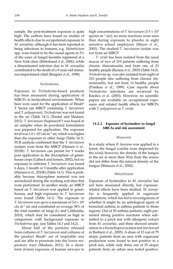
204 A. Mette Madsen and K. Tendal
sample, the post-treatment exposure is quite high. The authors have found no studies of health effects due to occupational exposure to M. anisopliae, although it has been reported as being infectious in humans, e.g. Metarhiziumspp. were found to be the causal agents in 2% of the cases of fungal keratitis registered at a New York clinic (Ritterband et al., 2006), while a disseminated infection due to M. anisopliaecontributed to the death of a 9-year-old immu-nocompromised child (Burgner et al., 1998).
Trichoderma
Exposure to Trichoderma-based products has been measured during application of MBCPs in horticultural environments. When bees were used for the application of Binab®
T Vector (an MBCP containing T. harzianumand T. polysporum), Trichoderma was not found in the air (Table 14.1) (Tendal and Madsen, 2011). T. harzianum (Supresivit®) was found in air samples when its powdered formulation was prepared for application. The exposure level was 1.0 × 10⁵ cfu m−3 air, which was higher than the exposure to other fungi (Table 14.1). PCR analysis confirmed that the T. harzianumisolates were from the MBCP (Hansen et al.,2010b). T. harzianum can persist for 9 weeks post application in the rhizosphere of green-house crops (Lübeck and Jensen, 2002), but no exposure to airborne T. harzianum was found 6 days, 1 month or 3 months after application (Hansen et al., 2010b) (Table 14.1). This is prob-ably because rhizosphere material was not aerosolized during the working activities that were performed. In another study, an MBCP based on T. harzianum was applied in green-houses, and high exposures to T. harzianumwere found (Table 14.1). The exposure to T. harzianum was up to a maximum of 3.6 × 10⁴
cfu m−3 air and constituted on average 38% of the total number of fungi (Li and LaMondia, 2010), which may be considered as high in comparison with background exposure to Trichoderma spp. (see Tables 14.1 and 14.2).
About half of the particles released from cultures of T. harzianum and cultures of the product Binab® are of respirable size and are able to penetrate into the lower res-piratory tract (Madsen, 2011). In a short-term (6 min) exposure of human airways to
high concentrations of T. harzianum (3.5 × 10⁵
spores m−3 air), no more reactions were seen than during exposure to placebo in eight sensitive school employees (Meyer et al., 2005). The studied T. harzianum isolate was not from an MBCP.
T. viride has been isolated from the nasal mucus of two of 210 patients suffering from chronic rhinosinusitis and from one of 23 healthy people (Buzina et al., 2003) (Table 14.2). Trichoderma sp. was also isolated from eight of 210 people also suffering from chronic rhi-nosinusitis, but not from 14 healthy people (Ponikau et al., 1999). Case reports about Trichoderma infections are reviewed by Kredics et al. (2003). However, no scientific papers are available on occupational expo-sures and related health effects for MBCPs with T. polysporum or T. viride.
14.2.2 Exposure of bystanders to fungal MBCAs and risk assessment
Beauveria
In a study where B. bassiana was applied to a forest, the fungal conidia were dispersed by the wind; however, the density of the fungus in the air at more than 50 m from the source did not differ from the natural density of the fungus (Shimazu et al., 2002).
Metarhizium
Exposure of bystanders to M. anisopliae has not been measured directly, but exposure-related effects have been studied. M. anisop-liae is frequently applied in sugarcane plantations, which has led to investigations of whether it might be an aetiological agent of bronchial asthma in asthma patients in those regions. Out of 50 asthma patients, eight pre-sented strong positive reactions when sub-mitted to a prick test with allergenic extract from M. anisopliae, and three showed sensiti-zation in a bronchoprovocation test (reviewed in Barbieri et al., 2005). A share of 23 out of 79 atopic patients from an area with sugarcane production were found to test positive to a prick test, while only three out of 35 atopic patients from an urban area tested positive.
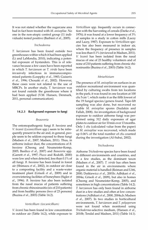
Occupational Safety of Microbial Agents 205
It was not stated whether the sugarcane area had in fact been treated with M. anisopliae. No one in the non-atopic control group (11 indi-viduals) tested positive (Barbieri et al., 2005).
Trichoderma
T. harzianum has been found outside two greenhouses within which it had been applied (Li and LaMondia, 2010), indicating a poten-tial exposure of bystanders. This is of rele-vance because a few cases have been reported in which T. harzianum or T. viride have been invasively infectious in immunocompro-mised patients (Loeppky et al., 1983; Guiserix et al., 1996; Chouaki et al., 2002). However, these cases were not related to the use of MBCPs. In another study, T. harzianum was not found outside the greenhouse where it had been applied (V.M. Hansen, Denmark, 2011, personal communication).
14.2.3 Background exposure to fungi
Beauveria
The entomopathogenic fungi B. bassiana and V. lecanii (Lecanicillium spp.) seem to be infre-quently present in the air and, in general, peo-ple seem to be seldom exposed to these fungi (Madsen et al., 2007; Madsen, 2011). Thus, in airborne indoor dust, the concentrations of B.bassiana (Cheong and Neumeister-Kemp, 2005; Basilico et al., 2007) and Beauveria spp. (Garrett et al., 1997; Picco and Rodolfi, 2000) were low and when detected, less than 0.1% of all fungi. B. bassiana has been found in forest air (Shimazu et al., 2002), in outdoor air close to a composting facility and a waste-water treatment plant (Grisoli et al., 2009) and in overwintering facilities of honeybees (Sigler etal., 1996). B. bassiana has also been isolated from the nasal mucus of patients suffering from chronic rhinosinusitis (six of 210 patients) and from healthy persons (two of 23 persons) (Buzina et al., 2003) (Table 14.2).
Verticillium (Lecanicillium)
V. lecanii has been found in low concentration in outdoor air (Table 14.2), while exposure to
Verticillium spp. frequently occurs in connec-tion with the harvesting of cereals (Darke et al., 1976); it was found at a lower frequency of 3% of samples in a study in cotton mills (Lacey and Lacey, 1987). Exposure to Verticillium spe-cies has also been measured in indoor air, where the frequency of presence in samples was less than 0.1% (reviewed in Madsen, 2011). V. lecanii has been isolated from the nasal mucus of one of 23 healthy volunteers and of none of 210 patients suffering from chronic rhi-nosinusitis (Buzina et al., 2003) (Table 14.2).
Metarhizium
The presence of M. anisopliae on surfaces in an outdoor recreational environment was quan-tified by culturing swabs from ten locations in the park; it was found in one location at 100 cfu 5 cm−2, which ranks it as the fifth lowest of the 19 fungal species/genera found. Tape-lift sampling was also done, but recovered no viable M. anisopliae spores (Sudakin and Fallah, 2008). An investigation of background exposure to outdoor airborne fungi was per-formed using 312 daily exposures of agar plates to outdoor air for 10 min over 13 months. Only 1 cfu (calculated value from the paper) of M. anisopliae was recovered, which made up 0.06% of the total number of cfu counted during the investigation (Al-Subai, 2002).
Trichoderma
Airborne Trichoderma species have been found in different environments and countries, and in a few studies, as the dominant taxon (Madsen et al., 2007). T. viride has often been found in the air in environments where organic material is handled (Reiman and Uitti, 2000; Dutkiewicz et al., 2001b; Adhikari et al.,2004a; Grisoli et al., 2009), but also in homes (Cheong and Neumeister-Kemp, 2005), and sometimes in high concentrations (Table 14.2). T. harzianum has only been found in airborne dust in a few studies and often at low concen-trations (Adhikari et al., 2000, 2004a,b; Madsen et al., 2007). In two studies in horticultural environments, T. harzianum and T. polysporumwere not found when monitored using Trichoderma-selective medium, (Hansen et al.,2010b; Tendal and Madsen, 2011) (Table 14.1).
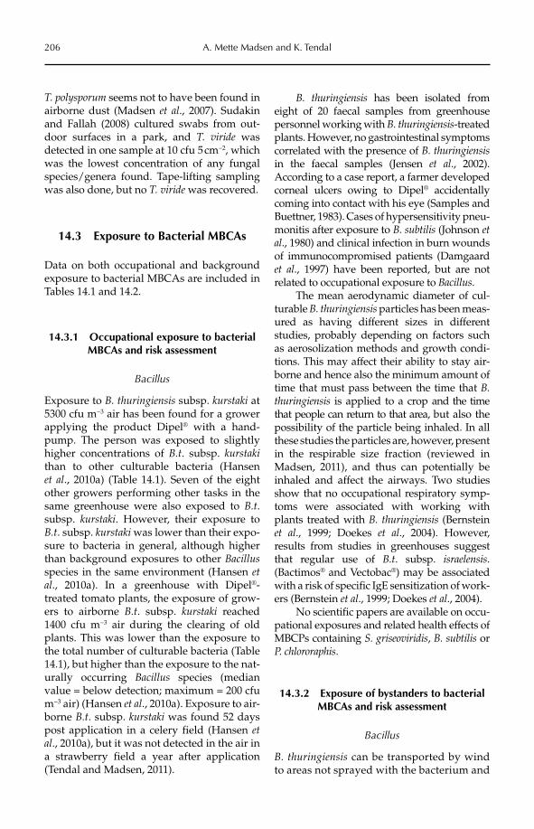
206 A. Mette Madsen and K. Tendal
T. polysporum seems not to have been found in airborne dust (Madsen et al., 2007). Sudakin and Fallah (2008) cultured swabs from out-door surfaces in a park, and T. viride was detected in one sample at 10 cfu 5 cm−2, which was the lowest concentration of any fungal species/genera found. Tape-lifting sampling was also done, but no T. viride was recovered.
14.3 Exposure to Bacterial MBCAs
Data on both occupational and background exposure to bacterial MBCAs are included in Tables 14.1 and 14.2.
14.3.1 Occupational exposure to bacterial MBCAs and risk assessment
Bacillus
Exposure to B. thuringiensis subsp. kurstaki at 5300 cfu m−3 air has been found for a grower applying the product Dipel® with a hand-pump. The person was exposed to slightly higher concentrations of B.t. subsp. kurstakithan to other culturable bacteria (Hansen et al., 2010a) (Table 14.1). Seven of the eight other growers performing other tasks in the same greenhouse were also exposed to B.t.subsp. kurstaki. However, their exposure to B.t. subsp. kurstaki was lower than their expo-sure to bacteria in general, although higher than background exposures to other Bacillusspecies in the same environment (Hansen etal., 2010a). In a greenhouse with Dipel®-treated tomato plants, the exposure of grow-ers to airborne B.t. subsp. kurstaki reached 1400 cfu m−3 air during the clearing of old plants. This was lower than the exposure to the total number of culturable bacteria (Table 14.1), but higher than the exposure to the nat-urally occurring Bacillus species (median value = below detection; maximum = 200 cfu m−3 air) (Hansen et al., 2010a). Exposure to air-borne B.t. subsp. kurstaki was found 52 days post application in a celery field (Hansen etal., 2010a), but it was not detected in the air in a strawberry field a year after application (Tendal and Madsen, 2011).
B. thuringiensis has been isolated from eight of 20 faecal samples from greenhouse personnel working with B. thuringiensis-treated plants. However, no gastrointestinal symptoms correlated with the presence of B. thuringiensisin the faecal samples (Jensen et al., 2002). According to a case report, a farmer developed corneal ulcers owing to Dipel® accidentally coming into contact with his eye (Samples and Buettner, 1983). Cases of hypersensitivity pneu-monitis after exposure to B. subtilis (Johnson et al., 1980) and clinical infection in burn wounds of immunocompromised patients (Damgaard et al., 1997) have been reported, but are not related to occupational exposure to Bacillus.
The mean aerodynamic diameter of cul-turable B. thuringiensis particles has been meas-ured as having different sizes in different studies, probably depending on factors such as aerosolization methods and growth condi-tions. This may affect their ability to stay air-borne and hence also the minimum amount of time that must pass between the time that B. thuringiensis is applied to a crop and the time that people can return to that area, but also the possibility of the particle being inhaled. In all these studies the particles are, however, present in the respirable size fraction (reviewed in Madsen, 2011), and thus can potentially be inhaled and affect the airways. Two studies show that no occupational respiratory symp-toms were associated with working with plants treated with B. thuringiensis (Bernstein et al., 1999; Doekes et al., 2004). However, results from studies in greenhouses suggest that regular use of B.t. subsp. israelensis.(Bactimos® and Vectobac®) may be associated with a risk of specific IgE sensitization of work-ers (Bernstein et al., 1999; Doekes et al., 2004).
No scientific papers are available on occu-pational exposures and related health effects of MBCPs containing S. griseoviridis, B. subtilis or P. chlororaphis.
14.3.2 Exposure of bystanders to bacterial MBCAs and risk assessment
Bacillus
B. thuringiensis can be transported by wind to areas not sprayed with the bacterium and
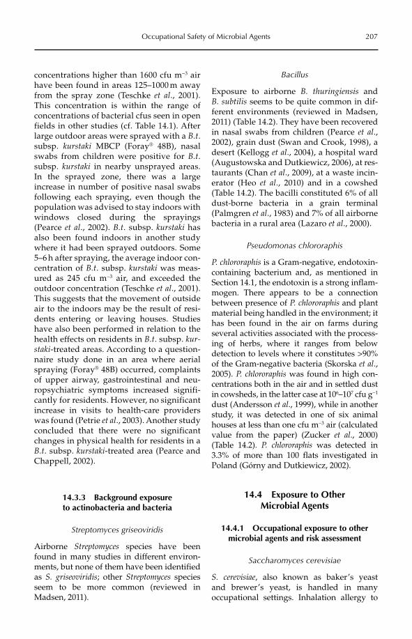
Occupational Safety of Microbial Agents 207
concentrations higher than 1600 cfu m−3 air have been found in areas 125–1000 m away from the spray zone (Teschke et al., 2001). This concentration is within the range of concentrations of bacterial cfus seen in open fields in other studies (cf. Table 14.1). After large outdoor areas were sprayed with a B.t.subsp. kurstaki MBCP (Foray® 48B), nasal swabs from children were positive for B.t.subsp. kurstaki in nearby unsprayed areas. In the sprayed zone, there was a large increase in number of positive nasal swabs following each spraying, even though the population was advised to stay indoors with windows closed during the sprayings (Pearce et al., 2002). B.t. subsp. kurstaki has also been found indoors in another study where it had been sprayed outdoors. Some 5–6 h after spraying, the average indoor con-centration of B.t. subsp. kurstaki was meas-ured as 245 cfu m−3 air, and exceeded the outdoor concentration (Teschke et al., 2001). This suggests that the movement of outside air to the indoors may be the result of resi-dents entering or leaving houses. Studies have also been performed in relation to the health effects on residents in B.t. subsp. kur-staki-treated areas. According to a question-naire study done in an area where aerial spraying (Foray® 48B) occurred, complaints of upper airway, gastrointestinal and neu-ropsychiatric symptoms increased signifi-cantly for residents. However, no significant increase in visits to health-care providers was found (Petrie et al., 2003). Another study concluded that there were no significant changes in physical health for residents in a B.t. subsp. kurstaki-treated area (Pearce and Chappell, 2002).
14.3.3 Background exposure to actinobacteria and bacteria
Streptomyces griseoviridis
Airborne Streptomyces species have been found in many studies in different environ-ments, but none of them have been identified as S. griseoviridis; other Streptomyces species seem to be more common (reviewed in Madsen, 2011).
Bacillus
Exposure to airborne B. thuringiensis and B. subtilis seems to be quite common in dif-ferent environments (reviewed in Madsen, 2011) (Table 14.2). They have been recovered in nasal swabs from children (Pearce et al., 2002), grain dust (Swan and Crook, 1998), a desert (Kellogg et al., 2004), a hospital ward (Augustowska and Dutkiewicz, 2006), at res-taurants (Chan et al., 2009), at a waste incin-erator (Heo et al., 2010) and in a cowshed (Table 14.2). The bacilli constituted 6% of all dust-borne bacteria in a grain terminal (Palmgren et al., 1983) and 7% of all airborne bacteria in a rural area (Lazaro et al., 2000).
Pseudomonas chlororaphis
P. chlororaphis is a Gram-negative, endotoxin-containing bacterium and, as mentioned in Section 14.1, the endotoxin is a strong inflam-mogen. There appears to be a connection between presence of P. chlororaphis and plant material being handled in the environment; it has been found in the air on farms during several activities associated with the process-ing of herbs, where it ranges from below detection to levels where it constitutes >90% of the Gram-negative bacteria (Skorska et al.,2005). P. chlororaphis was found in high con-centrations both in the air and in settled dust in cowsheds, in the latter case at 106–107 cfu g−1
dust (Andersson et al., 1999), while in another study, it was detected in one of six animal houses at less than one cfu m−3 air (calculated value from the paper) (Zucker et al., 2000) (Table 14.2). P. chlororaphis was detected in 3.3% of more than 100 flats investigated in Poland (Górny and Dutkiewicz, 2002).
14.4 Exposure to Other Microbial Agents
14.4.1 Occupational exposure to other microbial agents and risk assessment
Saccharomyces cerevisiae
S. cerevisiae, also known as baker’s yeast and brewer’s yeast, is handled in many occupational settings. Inhalation allergy to
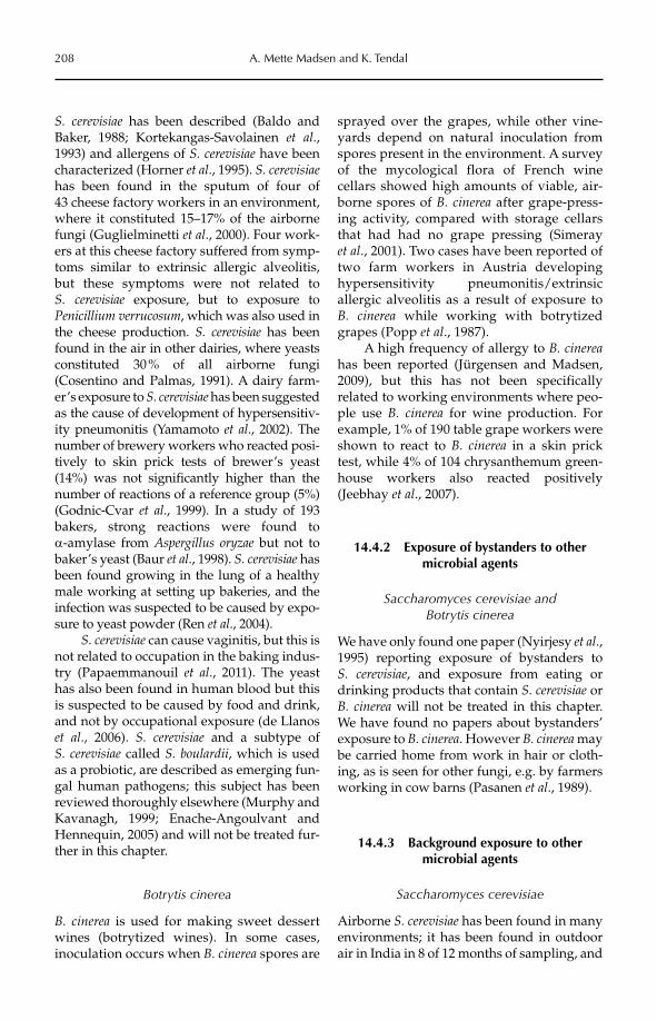
208 A. Mette Madsen and K. Tendal
S. cerevisiae has been described (Baldo and Baker, 1988; Kortekangas-Savolainen et al.,1993) and allergens of S. cerevisiae have been characterized (Horner et al., 1995). S. cerevisiaehas been found in the sputum of four of 43 cheese factory workers in an environment, where it constituted 15–17% of the airborne fungi (Guglielminetti et al., 2000). Four work-ers at this cheese factory suffered from symp-toms similar to extrinsic allergic alveolitis, but these symptoms were not related to S. cerevisiae exposure, but to exposure to Penicillium verrucosum, which was also used in the cheese production. S. cerevisiae has been found in the air in other dairies, where yeasts constituted 30 % of all airborne fungi (Cosentino and Palmas, 1991). A dairy farm-er’s exposure to S. cerevisiae has been suggested as the cause of development of hypersensitiv-ity pneumonitis (Yamamoto et al., 2002). The number of brewery workers who reacted posi-tively to skin prick tests of brewer’s yeast (14%) was not significantly higher than the number of reactions of a reference group (5%) (Godnic-Cvar et al., 1999). In a study of 193 bakers, strong reactions were found to a-amylase from Aspergillus oryzae but not to baker’s yeast (Baur et al., 1998). S. cerevisiae has been found growing in the lung of a healthy male working at setting up bakeries, and the infection was suspected to be caused by expo-sure to yeast powder (Ren et al., 2004).
S. cerevisiae can cause vaginitis, but this is not related to occupation in the baking indus-try (Papaemmanouil et al., 2011). The yeast has also been found in human blood but this is suspected to be caused by food and drink, and not by occupational exposure (de Llanos et al., 2006). S. cerevisiae and a subtype of S. cerevisiae called S. boulardii, which is used as a probiotic, are described as emerging fun-gal human pathogens; this subject has been reviewed thoroughly elsewhere (Murphy and Kavanagh, 1999; Enache-Angoulvant and Hennequin, 2005) and will not be treated fur-ther in this chapter.
Botrytis cinerea
B. cinerea is used for making sweet dessert wines (botrytized wines). In some cases, inoculation occurs when B. cinerea spores are
sprayed over the grapes, while other vine-yards depend on natural inoculation from spores present in the environment. A survey of the mycological flora of French wine cellars showed high amounts of viable, air-borne spores of B. cinerea after grape-press-ing activity, compared with storage cellars that had had no grape pressing (Simeray et al., 2001). Two cases have been reported of two farm workers in Austria developing hypersensitivity pneumonitis/extrinsic allergic alveolitis as a result of exposure to B. cinerea while working with botrytized grapes (Popp et al., 1987).
A high frequency of allergy to B. cinereahas been reported (Jürgensen and Madsen, 2009), but this has not been specifically related to working environments where peo-ple use B. cinerea for wine production. For example, 1% of 190 table grape workers were shown to react to B. cinerea in a skin prick test, while 4% of 104 chrysanthemum green-house workers also reacted positively (Jeebhay et al., 2007).
14.4.2 Exposure of bystanders to other microbial agents
Saccharomyces cerevisiae and Botrytis cinerea
We have only found one paper (Nyirjesy et al.,1995) reporting exposure of bystanders to S. cerevisiae, and exposure from eating or drinking products that contain S. cerevisiae or B. cinerea will not be treated in this chapter. We have found no papers about bystanders’ exposure to B. cinerea. However B. cinerea may be carried home from work in hair or cloth-ing, as is seen for other fungi, e.g. by farmers working in cow barns (Pasanen et al., 1989).
14.4.3 Background exposure to other microbial agents
Saccharomyces cerevisiae
Airborne S. cerevisiae has been found in many environments; it has been found in outdoor air in India in 8 of 12 months of sampling, and
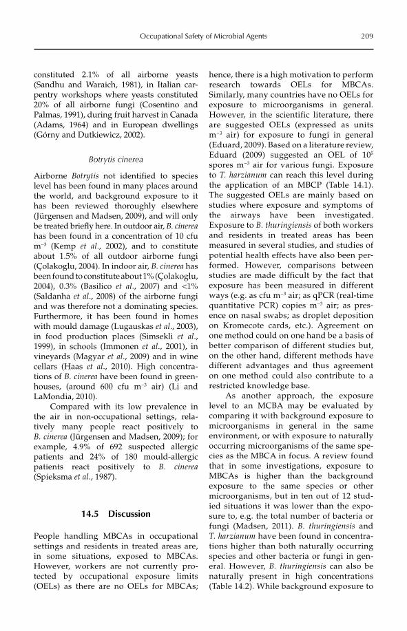
Occupational Safety of Microbial Agents 209
constituted 2.1% of all airborne yeasts (Sandhu and Waraich, 1981), in Italian car-pentry workshops where yeasts constituted 20% of all airborne fungi (Cosentino and Palmas, 1991), during fruit harvest in Canada (Adams, 1964) and in European dwellings (Górny and Dutkiewicz, 2002).
Botrytis cinerea
Airborne Botrytis not identified to species level has been found in many places around the world, and background exposure to it has been reviewed thoroughly elsewhere (Jürgensen and Madsen, 2009), and will only be treated briefly here. In outdoor air, B. cinereahas been found in a concentration of 10 cfu m−3 (Kemp et al., 2002), and to constitute about 1.5% of all outdoor airborne fungi (Çolakoglu, 2004). In indoor air, B. cinerea has been found to constitute about 1% (Çolakoglu, 2004), 0.3% (Basilico et al., 2007) and <1% (Saldanha et al., 2008) of the airborne fungi and was therefore not a dominating species. Furthermore, it has been found in homes with mould damage (Lugauskas et al., 2003), in food production places (Simsekli et al.,1999), in schools (Immonen et al., 2001), in vineyards (Magyar et al., 2009) and in wine cellars (Haas et al., 2010). High concentra-tions of B. cinerea have been found in green-houses, (around 600 cfu m−3 air) (Li and LaMondia, 2010).
Compared with its low prevalence in the air in non-occupational settings, rela-tively many people react positively to B. cinerea (Jürgensen and Madsen, 2009); for example, 4.9% of 692 suspected allergic patients and 24% of 180 mould-allergic patients react positively to B. cinerea(Spieksma et al., 1987).
14.5 Discussion
People handling MBCAs in occupational settings and residents in treated areas are, in some situations, exposed to MBCAs. However, workers are not currently pro-tected by occupational exposure limits (OELs) as there are no OELs for MBCAs;
hence, there is a high motivation to perform research towards OELs for MBCAs. Similarly, many countries have no OELs for exposure to microorganisms in general. However, in the scientific literature, there are suggested OELs (expressed as units m−3 air) for exposure to fungi in general (Eduard, 2009). Based on a literature review, Eduard (2009) suggested an OEL of 105
spores m−3 air for various fungi. Exposure to T. harzianum can reach this level during the application of an MBCP (Table 14.1). The suggested OELs are mainly based on studies where exposure and symptoms of the airways have been investigated. Exposure to B. thuringiensis of both workers and residents in treated areas has been measured in several studies, and studies of potential health effects have also been per-formed. However, comparisons between studies are made difficult by the fact that exposure has been measured in different ways (e.g. as cfu m−3 air; as qPCR (real-time quantitative PCR) copies m−3 air; as pres-ence on nasal swabs; as droplet deposition on Kromecote cards, etc.). Agreement on one method could on one hand be a basis of better comparison of different studies but, on the other hand, different methods have different advantages and thus agreement on one method could also contribute to a restricted knowledge base.
As another approach, the exposure level to an MCBA may be evaluated by comparing it with background exposure to microorganisms in general in the same environment, or with exposure to naturally occurring microorganisms of the same spe-cies as the MBCA in focus. A review found that in some investigations, exposure to MBCAs is higher than the background exposure to the same species or other microorganisms, but in ten out of 12 stud-ied situations it was lower than the expo-sure to, e.g. the total number of bacteria or fungi (Madsen, 2011). B. thuringiensis and T. harzianum have been found in concentra-tions higher than both naturally occurring species and other bacteria or fungi in gen-eral. However, B. thuringiensis can also be naturally present in high concentrations (Table 14.2). While background exposure to
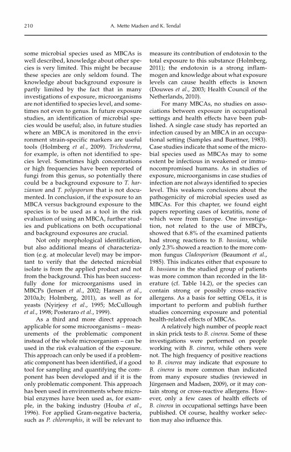
210 A. Mette Madsen and K. Tendal
some microbial species used as MBCAs is well described, knowledge about other spe-cies is very limited. This might be because these species are only seldom found. The knowledge about background exposure is partly limited by the fact that in many investigations of exposure, microorganisms are not identified to species level, and some-times not even to genus. In future exposure studies, an identification of microbial spe-cies would be useful; also, in future studies where an MBCA is monitored in the envi-ronment strain-specific markers are useful tools (Holmberg et al., 2009). Trichoderma,for example, is often not identified to spe-cies level. Sometimes high concentrations or high frequencies have been reported of fungi from this genus, so potentially there could be a background exposure to T. har-zianum and T. polysporum that is not docu-mented. In conclusion, if the exposure to an MBCA versus background exposure to the species is to be used as a tool in the risk evaluation of using an MBCA, further stud-ies and publications on both occupational and background exposures are crucial.
Not only morphological identification, but also additional means of characteriza-tion (e.g. at molecular level) may be impor-tant to verify that the detected microbial isolate is from the applied product and not from the background. This has been success-fully done for microorganisms used in MBCPs (Jensen et al., 2002; Hansen et al., 2010a,b; Holmberg, 2011), as well as for yeasts (Nyirjesy et al., 1995; McCullough et al., 1998; Posteraro et al., 1999).
As a third and more direct approach applicable for some microorganisms – meas-urements of the problematic component instead of the whole microorganism – can be used in the risk evaluation of the exposure. This approach can only be used if a problem-atic component has been identified, if a good tool for sampling and quantifying the com-ponent has been developed and if it is the only problematic component. This approach has been used in environments where micro-bial enzymes have been used as, for exam-ple, in the baking industry (Houba et al., 1996). For applied Gram-negative bacteria, such as P. chlororaphis, it will be relevant to
measure its contribution of endotoxin to the total exposure to this substance (Holmberg, 2011); the endotoxin is a strong inflam-mogen and knowledge about what exposure levels can cause health effects is known (Douwes et al., 2003; Health Council of the Netherlands, 2010).
For many MBCAs, no studies on asso-ciations between exposure in occupational settings and health effects have been pub-lished. A single case study has reported an infection caused by an MBCA in an occupa-tional setting (Samples and Buettner, 1983). Case studies indicate that some of the micro-bial species used as MBCAs may to some extent be infectious in weakened or immu-nocompromised humans. As in studies of exposure, microorganisms in case studies of infection are not always identified to species level. This weakens conclusions about the pathogenicity of microbial species used as MBCAs. For this chapter, we found eight papers reporting cases of keratitis, none of which were from Europe. One investiga-tion, not related to the use of MBCPs, showed that 6.8% of the examined patients had strong reactions to B. bassiana, while only 2.3% showed a reaction to the more com-mon fungus Cladosporium (Beaumont et al.,1985). This indicates either that exposure to B. bassiana in the studied group of patients was more common than recorded in the lit-erature (cf. Table 14.2), or the species can contain strong or possibly cross-reactive allergens. As a basis for setting OELs, it is important to perform and publish further studies concerning exposure and potential health-related effects of MBCAs.
A relatively high number of people react in skin prick tests to B. cinerea. Some of these investigations were performed on people working with B. cinerea, while others were not. The high frequency of positive reactions to B. cinerea may indicate that exposure to B. cinerea is more common than indicated from many exposure studies (reviewed in Jürgensen and Madsen, 2009), or it may con-tain strong or cross-reactive allergens. How-ever, only a few cases of health effects of B. cinerea in occupational settings have been published. Of course, healthy worker selec-tion may also influence this.
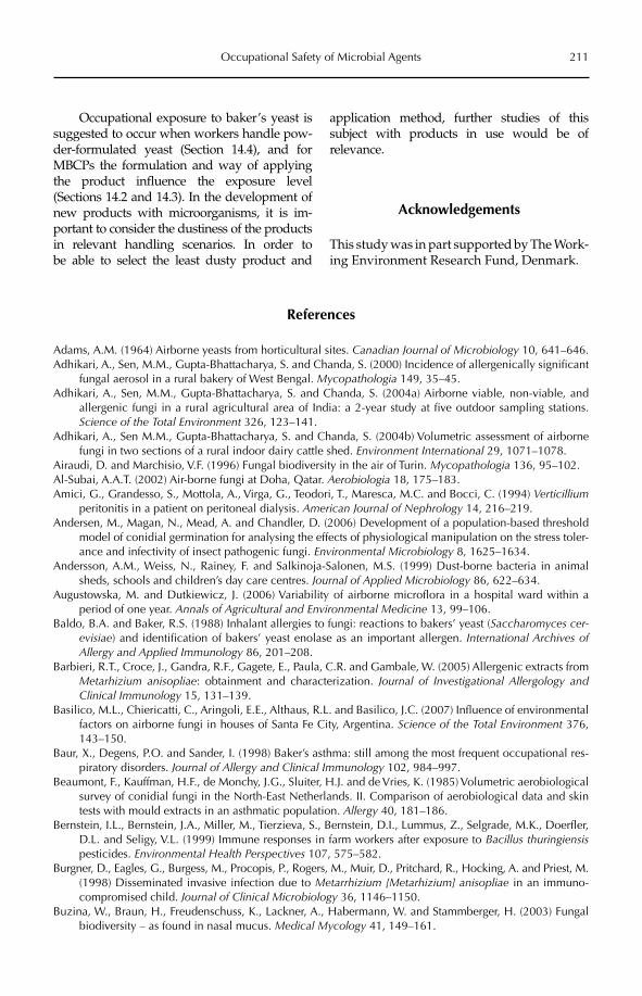
Occupational Safety of Microbial Agents 211
Occupational exposure to baker’s yeast is suggested to occur when workers handle pow-der-formulated yeast (Section 14.4), and for MBCPs the formulation and way of applying the product influence the exposure level (Sections 14.2 and 14.3). In the development of new products with microorganisms, it is im -portant to consider the dustiness of the products in relevant handling scenarios. In order to be able to select the least dusty product and
application method, further studies of this subject with products in use would be of relevance.
Acknowledgements
This study was in part supported by The Work-ing Environment Research Fund, Denmark.
References
Adams, A.M. (1964) Airborne yeasts from horticultural sites. Canadian Journal of Microbiology 10, 641–646.Adhikari, A., Sen, M.M., Gupta-Bhattacharya, S. and Chanda, S. (2000) Incidence of allergenically significant
fungal aerosol in a rural bakery of West Bengal. Mycopathologia 149, 35–45.Adhikari, A., Sen, M.M., Gupta-Bhattacharya, S. and Chanda, S. (2004a) Airborne viable, non-viable, and
allergenic fungi in a rural agricultural area of India: a 2-year study at five outdoor sampling stations. Science of the Total Environment 326, 123–141.
Adhikari, A., Sen M.M., Gupta-Bhattacharya, S. and Chanda, S. (2004b) Volumetric assessment of airborne fungi in two sections of a rural indoor dairy cattle shed. Environment International 29, 1071–1078.
Airaudi, D. and Marchisio, V.F. (1996) Fungal biodiversity in the air of Turin. Mycopathologia 136, 95–102.Al-Subai, A.A.T. (2002) Air-borne fungi at Doha, Qatar. Aerobiologia 18, 175–183.Amici, G., Grandesso, S., Mottola, A., Virga, G., Teodori, T., Maresca, M.C. and Bocci, C. (1994) Verticillium
peritonitis in a patient on peritoneal dialysis. American Journal of Nephrology 14, 216–219.Andersen, M., Magan, N., Mead, A. and Chandler, D. (2006) Development of a population-based threshold
model of conidial germination for analysing the effects of physiological manipulation on the stress toler-ance and infectivity of insect pathogenic fungi. Environmental Microbiology 8, 1625–1634.
Andersson, A.M., Weiss, N., Rainey, F. and Salkinoja-Salonen, M.S. (1999) Dust-borne bacteria in animal sheds, schools and children’s day care centres. Journal of Applied Microbiology 86, 622–634.
Augustowska, M. and Dutkiewicz, J. (2006) Variability of airborne microflora in a hospital ward within a period of one year. Annals of Agricultural and Environmental Medicine 13, 99–106.
Baldo, B.A. and Baker, R.S. (1988) Inhalant allergies to fungi: reactions to bakers’ yeast (Saccharomyces cer-evisiae) and identification of bakers’ yeast enolase as an important allergen. International Archives of Allergy and Applied Immunology 86, 201–208.
Barbieri, R.T., Croce, J., Gandra, R.F., Gagete, E., Paula, C.R. and Gambale, W. (2005) Allergenic extracts from Metarhizium anisopliae: obtainment and characterization. Journal of Investigational Allergology and Clinical Immunology 15, 131–139.
Basilico, M.L., Chiericatti, C., Aringoli, E.E., Althaus, R.L. and Basilico, J.C. (2007) Influence of environmental factors on airborne fungi in houses of Santa Fe City, Argentina. Science of the Total Environment 376, 143–150.
Baur, X., Degens, P.O. and Sander, I. (1998) Baker’s asthma: still among the most frequent occupational res-piratory disorders. Journal of Allergy and Clinical Immunology 102, 984–997.
Beaumont, F., Kauffman, H.F., de Monchy, J.G., Sluiter, H.J. and de Vries, K. (1985) Volumetric aerobiological survey of conidial fungi in the North-East Netherlands. II. Comparison of aerobiological data and skin tests with mould extracts in an asthmatic population. Allergy 40, 181–186.
Bernstein, I.L., Bernstein, J.A., Miller, M., Tierzieva, S., Bernstein, D.I., Lummus, Z., Selgrade, M.K., Doerfler, D.L. and Seligy, V.L. (1999) Immune responses in farm workers after exposure to Bacillus thuringiensispesticides. Environmental Health Perspectives 107, 575–582.
Burgner, D., Eagles, G., Burgess, M., Procopis, P., Rogers, M., Muir, D., Pritchard, R., Hocking, A. and Priest, M. (1998) Disseminated invasive infection due to Metarrhizium [Metarhizium] anisopliae in an immuno-compromised child. Journal of Clinical Microbiology 36, 1146–1150.
Buzina, W., Braun, H., Freudenschuss, K., Lackner, A., Habermann, W. and Stammberger, H. (2003) Fungal biodiversity – as found in nasal mucus. Medical Mycology 41, 149–161.
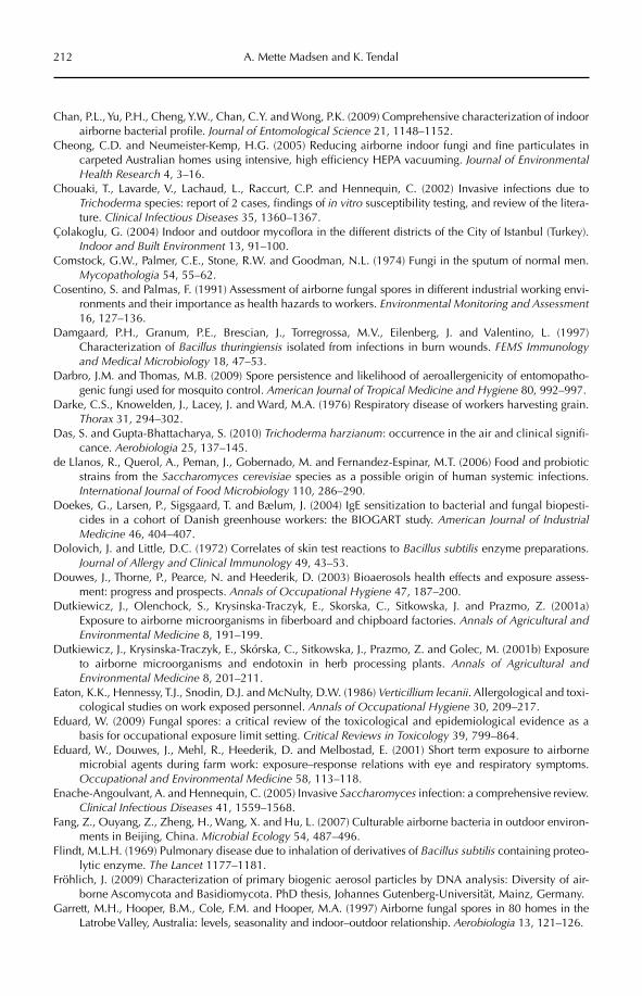
212 A. Mette Madsen and K. Tendal
Chan, P.L., Yu, P.H., Cheng, Y.W., Chan, C.Y. and Wong, P.K. (2009) Comprehensive characterization of indoor airborne bacterial profile. Journal of Entomological Science 21, 1148–1152.
Cheong, C.D. and Neumeister-Kemp, H.G. (2005) Reducing airborne indoor fungi and fine particulates in carpeted Australian homes using intensive, high efficiency HEPA vacuuming. Journal of Environmental Health Research 4, 3–16.
Chouaki, T., Lavarde, V., Lachaud, L., Raccurt, C.P. and Hennequin, C. (2002) Invasive infections due to Trichoderma species: report of 2 cases, findings of in vitro susceptibility testing, and review of the litera-ture. Clinical Infectious Diseases 35, 1360–1367.
Çolakoglu, G. (2004) Indoor and outdoor mycoflora in the different districts of the City of Istanbul (Turkey). Indoor and Built Environment 13, 91–100.
Comstock, G.W., Palmer, C.E., Stone, R.W. and Goodman, N.L. (1974) Fungi in the sputum of normal men. Mycopathologia 54, 55–62.
Cosentino, S. and Palmas, F. (1991) Assessment of airborne fungal spores in different industrial working envi-ronments and their importance as health hazards to workers. Environmental Monitoring and Assessment16, 127–136.
Damgaard, P.H., Granum, P.E., Brescian, J., Torregrossa, M.V., Eilenberg, J. and Valentino, L. (1997) Characterization of Bacillus thuringiensis isolated from infections in burn wounds. FEMS Immunology and Medical Microbiology 18, 47–53.
Darbro, J.M. and Thomas, M.B. (2009) Spore persistence and likelihood of aeroallergenicity of entomopatho-genic fungi used for mosquito control. American Journal of Tropical Medicine and Hygiene 80, 992–997.
Darke, C.S., Knowelden, J., Lacey, J. and Ward, M.A. (1976) Respiratory disease of workers harvesting grain. Thorax 31, 294–302.
Das, S. and Gupta-Bhattacharya, S. (2010) Trichoderma harzianum: occurrence in the air and clinical signifi-cance. Aerobiologia 25, 137–145.
de Llanos, R., Querol, A., Peman, J., Gobernado, M. and Fernandez-Espinar, M.T. (2006) Food and probiotic strains from the Saccharomyces cerevisiae species as a possible origin of human systemic infections. International Journal of Food Microbiology 110, 286–290.
Doekes, G., Larsen, P., Sigsgaard, T. and Bælum, J. (2004) IgE sensitization to bacterial and fungal biopesti-cides in a cohort of Danish greenhouse workers: the BIOGART study. American Journal of Industrial Medicine 46, 404–407.
Dolovich, J. and Little, D.C. (1972) Correlates of skin test reactions to Bacillus subtilis enzyme preparations. Journal of Allergy and Clinical Immunology 49, 43–53.
Douwes, J., Thorne, P., Pearce, N. and Heederik, D. (2003) Bioaerosols health effects and exposure assess-ment: progress and prospects. Annals of Occupational Hygiene 47, 187–200.
Dutkiewicz, J., Olenchock, S., Krysinska-Traczyk, E., Skorska, C., Sitkowska, J. and Prazmo, Z. (2001a) Exposure to airborne microorganisms in fiberboard and chipboard factories. Annals of Agricultural and Environmental Medicine 8, 191–199.
Dutkiewicz, J., Krysinska-Traczyk, E., Skórska, C., Sitkowska, J., Prazmo, Z. and Golec, M. (2001b) Exposure to airborne microorganisms and endotoxin in herb processing plants. Annals of Agricultural and Environmental Medicine 8, 201–211.
Eaton, K.K., Hennessy, T.J., Snodin, D.J. and McNulty, D.W. (1986) Verticillium lecanii. Allergological and toxi-cological studies on work exposed personnel. Annals of Occupational Hygiene 30, 209–217.
Eduard, W. (2009) Fungal spores: a critical review of the toxicological and epidemiological evidence as a basis for occupational exposure limit setting. Critical Reviews in Toxicology 39, 799–864.
Eduard, W., Douwes, J., Mehl, R., Heederik, D. and Melbostad, E. (2001) Short term exposure to airborne microbial agents during farm work: exposure–response relations with eye and respiratory symptoms. Occupational and Environmental Medicine 58, 113–118.
Enache-Angoulvant, A. and Hennequin, C. (2005) Invasive Saccharomyces infection: a comprehensive review. Clinical Infectious Diseases 41, 1559–1568.
Fang, Z., Ouyang, Z., Zheng, H., Wang, X. and Hu, L. (2007) Culturable airborne bacteria in outdoor environ-ments in Beijing, China. Microbial Ecology 54, 487–496.
Flindt, M.L.H. (1969) Pulmonary disease due to inhalation of derivatives of Bacillus subtilis containing proteo-lytic enzyme. The Lancet 1177–1181.
Fröhlich, J. (2009) Characterization of primary biogenic aerosol particles by DNA analysis: Diversity of air-borne Ascomycota and Basidiomycota. PhD thesis, Johannes Gutenberg-Universität, Mainz, Germany.
Garrett, M.H., Hooper, B.M., Cole, F.M. and Hooper, M.A. (1997) Airborne fungal spores in 80 homes in the Latrobe Valley, Australia: levels, seasonality and indoor–outdoor relationship. Aerobiologia 13, 121–126.
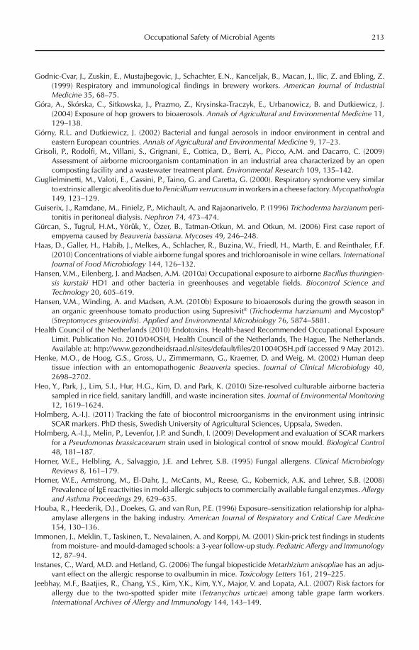
Occupational Safety of Microbial Agents 213
Godnic-Cvar, J., Zuskin, E., Mustajbegovic, J., Schachter, E.N., Kanceljak, B., Macan, J., Ilic, Z. and Ebling, Z. (1999) Respiratory and immunological findings in brewery workers. American Journal of Industrial Medicine 35, 68–75.
Góra, A., Skórska, C., Sitkowska, J., Prazmo, Z., Krysinska-Traczyk, E., Urbanowicz, B. and Dutkiewicz, J. (2004) Exposure of hop growers to bioaerosols. Annals of Agricultural and Environmental Medicine 11, 129–138.
Górny, R.L. and Dutkiewicz, J. (2002) Bacterial and fungal aerosols in indoor environment in central and eastern European countries. Annals of Agricultural and Environmental Medicine 9, 17–23.
Grisoli, P., Rodolfi, M., Villani, S., Grignani, E., Cottica, D., Berri, A., Picco, A.M. and Dacarro, C. (2009) Assessment of airborne microorganism contamination in an industrial area characterized by an open composting facility and a wastewater treatment plant. Environmental Research 109, 135–142.
Guglielminetti, M., Valoti, E., Cassini, P., Taino, G. and Caretta, G. (2000). Respiratory syndrome very similar to extrinsic allergic alveolitis due to Penicillium verrucosum in workers in a cheese factory. Mycopathologia149, 123–129.
Guiserix, J., Ramdane, M., Finielz, P., Michault, A. and Rajaonarivelo, P. (1996) Trichoderma harzianum peri-tonitis in peritoneal dialysis. Nephron 74, 473–474.
Gürcan, S., Tugrul, H.M., Yörûk, Y., Özer, B., Tatman-Otkun, M. and Otkun, M. (2006) First case report of empyema caused by Beauveria bassiana. Mycoses 49, 246–248.
Haas, D., Galler, H., Habib, J., Melkes, A., Schlacher, R., Buzina, W., Friedl, H., Marth, E. and Reinthaler, F.F. (2010) Concentrations of viable airborne fungal spores and trichloroanisole in wine cellars. InternationalJournal of Food Microbiology 144, 126–132.
Hansen, V.M., Eilenberg, J. and Madsen, A.M. (2010a) Occupational exposure to airborne Bacillus thuringien-sis kurstaki HD1 and other bacteria in greenhouses and vegetable fields. Biocontrol Science and Technology 20, 605–619.
Hansen, V.M., Winding, A. and Madsen, A.M. (2010b) Exposure to bioaerosols during the growth season in an organic greenhouse tomato production using Supresivit® (Trichoderma harzianum) and Mycostop®
(Streptomyces griseoviridis). Applied and Environmental Microbiology 76, 5874–5881.Health Council of the Netherlands (2010) Endotoxins. Health-based Recommended Occupational Exposure
Limit. Publication No. 2010/04OSH, Health Council of the Netherlands, The Hague, The Netherlands. Available at: http://www.gezondheidsraad.nl/sites/default/files/201004OSH.pdf (accessed 9 May 2012).
Henke, M.O., de Hoog, G.S., Gross, U., Zimmermann, G., Kraemer, D. and Weig, M. (2002) Human deep tissue infection with an entomopathogenic Beauveria species. Journal of Clinical Microbiology 40, 2698–2702.
Heo, Y., Park, J., Lim, S.I., Hur, H.G., Kim, D. and Park, K. (2010) Size-resolved culturable airborne bacteria sampled in rice field, sanitary landfill, and waste incineration sites. Journal of Environmental Monitoring12, 1619–1624.
Holmberg, A.-I.J. (2011) Tracking the fate of biocontrol microorganisms in the environment using intrinsic SCAR markers. PhD thesis, Swedish University of Agricultural Sciences, Uppsala, Sweden.
Holmberg, A.-I.J., Melin, P., Levenfor, J.P. and Sundh, I. (2009) Development and evaluation of SCAR markers for a Pseudomonas brassicacearum strain used in biological control of snow mould. Biological Control48, 181–187.
Horner, W.E., Helbling, A., Salvaggio, J.E. and Lehrer, S.B. (1995) Fungal allergens. Clinical Microbiology Reviews 8, 161–179.
Horner, W.E., Armstrong, M., El-Dahr, J., McCants, M., Reese, G., Kobernick, A.K. and Lehrer, S.B. (2008) Prevalence of IgE reactivities in mold-allergic subjects to commercially available fungal enzymes. Allergy and Asthma Proceedings 29, 629–635.
Houba, R., Heederik, D.J., Doekes, G. and van Run, P.E. (1996) Exposure–sensitization relationship for alpha-amylase allergens in the baking industry. American Journal of Respiratory and Critical Care Medicine154, 130–136.
Immonen, J., Meklin, T., Taskinen, T., Nevalainen, A. and Korppi, M. (2001) Skin-prick test findings in students from moisture- and mould-damaged schools: a 3-year follow-up study. Pediatric Allergy and Immunology12, 87–94.
Instanes, C., Ward, M.D. and Hetland, G. (2006) The fungal biopesticide Metarhizium anisopliae has an adju-vant effect on the allergic response to ovalbumin in mice. Toxicology Letters 161, 219–225.
Jeebhay, M.F., Baatjies, R., Chang, Y.S., Kim, Y.K., Kim, Y.Y., Major, V. and Lopata, A.L. (2007) Risk factors for allergy due to the two-spotted spider mite (Tetranychus urticae) among table grape farm workers. International Archives of Allergy and Immunology 144, 143–149.
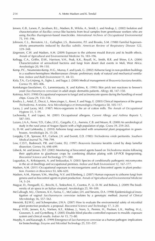
214 A. Mette Madsen and K. Tendal
Jensen, G.B., Larsen, P., Jacobsen, B.L., Madsen, B., Wilcks, A., Smidt, L. and Andrup, L. (2002) Isolation and characterization of Bacillus cereus-like bacteria from fecal samples from greenhouse workers who are using Bacillus thuringiensis-based insecticides. International Archives of Occupational Environmental75, 191–196.
Johnson, C.L., Bernstein, I.L., Gallagher, J.S., Bonventre, P.F. and Brooks, S.M. (1980) Familial hypersen-sitivity pneumonitis induced by Bacillus subtilis. American Review of Respiratory Disease 122, 339–348.
Jürgensen, C.W. and Madsen, A.M. (2009) Exposure to the airborne mould Botrytis and its health effects. Annals of Agricultural and Environmental Medicine 16, 183–196.
Kellogg, C.A., Griffin, D.W., Harrison, V.H., Peak, K.K., Royall, N., Smith, R.R. and Shinn, E.A. (2004) Characterization of aerosolized bacteria and fungi from desert dust events in Mali, West Africa. Aerobiologia 20, 99–110.
Kemp, P.C., Neumeister-Kemp, H.G., Murray, F. and Lysek, G. (2002) Airborne fungi in non-problem buildings in a southern-hemisphere Mediterranean climate: preliminary study of natural and mechanical ventila-tion. Indoor and Built Environment 11, 44–53.
Kisla, T.A., Cu-Unjieng, A., Sigler, L. and Sugar, J. (2000) Medical management of Beauveria bassiana keratitis. Cornea 19, 405–406.
Kortekangas-Savolainen, O., Lammintausta, K. and Kalimo, K. (1993) Skin prick test reactions to brewer’s yeast (Saccharomyces cerevisiae) in adult atopic dermatitis patients. Allergy 48, 147–150.
Kotimaa, M.H. (1990) Occupational exposure to fungal and actinomycete spores during the handling of wood chips. Grana 29, 153–156.
Kredics, L., Antal, Z., Doczi, I., Manczinger, L., Kevei, F. and Nagy, E. (2003) Clinical importance of the genus Trichoderma. A review. Acta Microbiologica et Immunologica Hungarica 50, 105–117.
Lacey, J. and Lacey, M.E. (1987) Micro-organisms in the air of cotton mills. The Annals of Occupational Hygiene 31, 1–19.
Lachowsky, F. and Lopez, M. (2001) Occupational allergens. Current Allergy and Asthma Reports 1, 587–593.
Lazaro, J.A.A., Ferrer, T.D., Cabo, J.F.G., Gargallo, C.L., Asensio, C.B. and Moure, R. (2000) An aerobiological study in the rural areas of Aragon (Spain) with a high population of pigs. Grana 39, 259–265.
Li, D.-W. and LaMondia, J. (2010) Airborne fungi associated with ornamental plant propagation in green-houses. Aerobiologia 26, 15–28.
Loeppky, C.B., Sprouse, R.F., Carlson, J.V. and Everett, E.D. (1983) Trichoderma viride peritonitis. SouthernMedical Journal 76, 798–799.
Low, C.D.T., Badenoch, P.R. and Coster, D.J. (1997) Beauveria bassiana keratitis cured by deep lamellar dissection. Cornea 16, 698–699.
Lübeck, M. and Jensen, D.F. (2002) Monitoring of biocontrol agents based on Trichoderma strains following their application to glasshouse crops by combining dilution plating with UP-PCR fingerprinting. Biocontrol Science and Technology 371–380.
Lugauskas, A., Krikstaponis, A. and Seskauskas, V. (2003) Species of conditionally pathogenic micromycetes in the air of dwellings and occupational premises. Indoor and Built Environment 12, 167–177.
Madsen, A.M. (2011) Occupational exposure to microorganisms used as biocontrol agents in plant produc-tion. Frontiers in Bioscience S3, 606–620.
Madsen, A.M., Hansen, V.M., Meyling, N.V. and Eilenberg, J. (2007) Human exposure to airborne fungi from genera used as biocontrol agents in plant production. Annals of Agricultural and Environmental Medicine14, 5–24.
Magyar, D., Frenguelli, G., Bricchi, E., Tedeschini, E., Csontos, P., Li, D.-W. and Bobvos, J. (2009) The biodi-versity of air spora in an Italian vineyard. Aerobiologia 25, 99–109.
McCullough, M.J., Clemons, K.V., Farina, C., McCusker, J.H. and Stevens, D.A. (1998) Epidemiological inves-tigation of vaginal Saccharomyces cerevisiae isolates by a genotypic method. Journal of Clinical Microbiology 36, 557–562.
Mensink, B.J.W.G. and Scheepmaker, J.W.A. (2007) How to evaluate the environmental safety of microbial plant protection products: a proposal. Biocontrol Science and Technology 17, 3–20.
Meyer, H.W., Jensen, K.A., Nielsen, K.F., Kildesø, J., Norn, S., Permin, H., Poulsen, L.K., Malling, H.-J., Gravesen, S. and Gyntelberg, F. (2005) Double blind placebo controlled exposure to moulds: exposure system and clinical results. Indoor Air 15, 73–80.
Murphy, A. and Kavanagh, K. (1999) Emergence of Saccharomyces cerevisiae as a human pathogen: implications for biotechnology. Enzyme and Microbial Technology 25, 551–557.
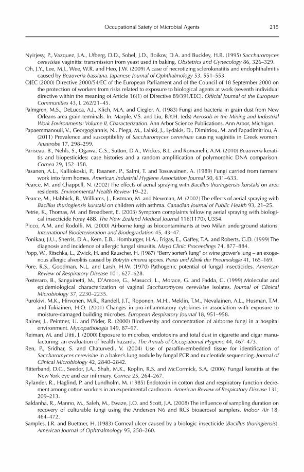
Occupational Safety of Microbial Agents 215
Nyirjesy, P., Vazquez, J.A., Ufberg, D.D., Sobel, J.D., Boikov, D.A. and Buckley, H.R. (1995) Saccharomyces cerevisiae vaginitis: transmission from yeast used in baking. Obstetrics and Gynecology 86, 326–329.
Oh, J.Y., Lee, M.J., Wee, W.R. and Heo, J.W. (2009) A case of necrotizing sclerokeratitis and endophthalmitis caused by Beauveria bassiana. Japanese Journal of Ophthalmology 53, 551–553.
OJEC (2000) Directive 2000/54/EC of the European Parliament and of the Council of 18 September 2000 on the protection of workers from risks related to exposure to biological agents at work (seventh individual directive within the meaning of Article 16(1) of Directive 89/391/EEC). Official Journal of the European Communities 43, L 262/21–45.
Palmgren, M.S., DeLucca, A.J., Klich, M.A. and Ciegler, A. (1983) Fungi and bacteria in grain dust from New Orleans area grain terminals. In: Marple, V.S. and Liu, B.Y.H. (eds) Aerosols in the Mining and Industrial Work Environments: Volume II, Characterization. Ann Arbor Science Publications, Ann Arbor, Michigan.
Papaemmanouil, V., Georgogiannis, N., Plega, M., Lalaki, J., Lydakis, D., Dimitriou, M. and Papadimitriou, A. (2011) Prevalence and susceptibility of Saccharomyces cerevisiae causing vaginitis in Greek women. Anaerobe 17, 298–299.
Pariseau, B., Nehls, S., Ogawa, G.S., Sutton, D.A., Wickes, B.L. and Romanelli, A.M. (2010) Beauveria kerati-tis and biopesticides: case histories and a random amplification of polymorphic DNA comparison. Cornea 29, 152–158.
Pasanen, A.L., Kalliokoski, P., Pasanen, P., Salmi, T. and Tossavainen, A. (1989) Fungi carried from farmers’ work into farm homes. American Industrial Hygiene Association Journal 50, 631–633.
Pearce, M. and Chappell, N. (2002) The effects of aerial spraying with Bacillus thuringiensis kurstaki on area residents. Environmental Health Review 19–22.
Pearce, M., Habbick, B., Williams, J., Eastman, M. and Newman, M. (2002) The effects of aerial spraying with Bacillus thuringiensis kurstaki on children with asthma. Canadian Journal of Public Health 93, 21–25.
Petrie, K., Thomas, M. and Broadbent, E. (2003) Symptom complaints following aerial spraying with biologi-cal insecticide Foray 48B. The New Zealand Medical Journal 116(1170), U354.
Picco, A.M. and Rodolfi, M. (2000) Airborne fungi as biocontaminants at two Milan underground stations. International Biodeterioration and Biodegradation 45, 43–47.
Ponikau, J.U., Sherris, D.A., Kern, E.B., Homburger, H.A., Frigas, E., Gaffey, T.A. and Roberts, G.D. (1999) The diagnosis and incidence of allergic fungal sinusitis. Mayo Clinic Proceedings 74, 877–884.
Popp, W., Ritschka, L., Zwick, H. and Rauscher, H. (1987) “Berry sorter’s lung” or wine grower’s lung – an exoge-nous allergic alveolitis caused by Botrytis cinerea spores. Praxis und Klinik der Pneumologie 41, 165–169.
Pore, R.S., Goodman, N.L. and Larsh, H.W. (1970) Pathogenic potential of fungal insecticides. AmericanReview of Respiratory Disease 101, 627–628.
Posteraro, B., Sanguinetti, M., D’Amore, G., Masucci, L., Morace, G. and Fadda, G. (1999) Molecular and epidemiological characterization of vaginal Saccharomyces cerevisiae isolates. Journal of Clinical Microbiology 37, 2230–2235.
Purokivi, M.K., Hirvonen, M.R., Randell, J.T., Roponen, M.H., Meklin, T.M., Nevalainen, A.L., Husman, T.M. and Tukiainen, H.O. (2001) Changes in pro-inflammatory cytokines in association with exposure to moisture-damaged building microbes. European Respiratory Journal 18, 951–958.
Rainer, J., Peintner, U. and Pöder, R. (2000) Biodiversity and concentration of airborne fungi in a hospital environment. Mycopathologia 149, 87–97.
Reiman, M. and Uitti, J. (2000) Exposure to microbes, endotoxins and total dust in cigarette and cigar manu-facturing: an evaluation of health hazards. The Annals of Occupational Hygiene 44, 467–473.
Ren, P., Sridhar, S. and Chaturvedi, V. (2004) Use of paraffin-embedded tissue for identification of Saccharomyces cerevisiae in a baker’s lung nodule by fungal PCR and nucleotide sequencing. Journal of Clinical Microbiology 42, 2840–2842.
Ritterband, D.C., Seedor, J.A., Shah, M.K., Koplin, R.S. and McCormick, S.A. (2006) Fungal keratitis at the New York eye and ear infirmary. Cornea 25, 264–267.
Rylander, R., Haglind, P. and Lundholm, M. (1985) Endotoxin in cotton dust and respiratory function decre-ment among cotton workers in an experimental cardroom. American Review of Respiratory Disease 131, 209–213.
Saldanha, R., Manno, M., Saleh, M., Ewaze, J.O. and Scott, J.A. (2008) The influence of sampling duration on recovery of culturable fungi using the Andersen N6 and RCS bioaerosol samplers. Indoor Air 18, 464–472.
Samples, J.R. and Buettner, H. (1983) Corneal ulcer caused by a biologic insecticide (Bacillus thuringiensis).American Journal of Ophthalmology 95, 258–260.
216 A. Mette Madsen and K. Tendal
Sandhu, D.K. and Waraich, M.K. (1981) Airborne yeasts in Amritsar (India). Antonie van Leeuwenhoek 47, 571–576.
Shimazu, M., Sato, H. and Maehara, N. (2002) Density of the entomopathogenic fungus, Beauveria bassianaVuillemin (Deutoromycotina: Hyphomycetes) in forest air and soil. Applied Entomology and Zoology37, 19–26.
Shin, J.Y., Kim, H.-M. and Hong, J.W. (2002) Keratitis caused by Verticillium species. Cornea 21, 240–242.Sigler, L., Abbott, S.P. and Gauvreau, H. (1996) Assessment of worker exposure to airborne moulds in honey-
bee overwintering facilities. American Industrial Hygiene Association 57, 484–490.Simard, C., Trudel, M., Paquette, G. and Payment, P. (1983) Microbial investigation of the air in an apartment
building. Journal of Hygiene 91, 277–286.Simeray, J., Mandin, D., Mercier, M. and Chaumont, J-P (2001) Survey of viable airborne fungal propagules in
French wine cellars. Aerobiologia 17, 19–24.Simsekli, Y., Gücin, F. and Asan, A. (1999) Isolation and identification of indoor airborne fungal contaminants
of food production facilities and warehouses in Bursa, Turkey. Aerobiologia 15, 225–231.Skorska, C., Sitkowska, J., Krysinska-Traczyk, E., Cholewa, G. and Dutkiewicz, J. (2005) Exposure to airborne
microorganisms, dust and endotoxin during processing of peppermint and chamomile herbs on farms. Annals of Agricultural and Environmental Medicine 12, 281–288.
Spieksma, F.Th.M., Nolard, N., Beaumont, F. and Vooren, P.H. (1987) Concentrations of airborne Botrytisconidia, and frequency of allergic sensitization to Botrytis extract. In: Boehm, G. and Leuschner, R.M. (eds) Proceedings of the 3rd International Conference on Aeromycology, August 6–9, 1986, Basel, Switzerland. Advances in Aerobiology 51, pp. 165–167.
Sudakin, D. and Fallah, P. (2008) Toxigenic fungi and mycotoxins in outdoor, recreational environments. Clinical Toxicology 46, 738–744.
Swan, J.M. and Crook, B. (1998) Airborne microorganisms associated with grain handling. Annals of Agricultural and Environmental Medicine 5, 7–15.
Szwajkowska-Michalek, L., Stuper, K., Lakomy, P., Matysiak, A. and Perkowski, J. (2010) Contents of micro-scopic fungi in dusts coming from cereal analysis laboratories. Annals of Agricultural and Environmental Medicine 17, 101–106.
Tendal, K. and Madsen, A.M. (2011) Exposure to airborne microorganisms, hyphal fragments, and pollen in a field of organically grown strawberries. Aerobiologia 27, 13–23.
Teschke, K., Chow, Y., Bartlett, K., Ross, A. and van Netten, C. (2001) Spatial and temporal distribution of airborne Bacillus thuringiensis var. kurstaki during an aerial spray program for gypsy moth eradication. Environmental Health Perspectives 109, 47–54.
Tlaskalova-Hogenova, H., Tuckova, L., Mestecky, J., Kolinska, J., Rossmann, P., Stepankova, R., Kozakova, H., Hudcovic, T., Hrncir, T., Frolova, L. and Kverka, M. (2005) Interaction of mucosal microbiota with the innate immune system. Scandinavian Journal of Immunology 62(Suppl. 1), 106–113.
Tu, E.Y. and Park, A.J. (2007) Recalcitrant Beauveria bassiana keratitis: confocal microscopy findings and treat-ment with posaconazole (Noxafil). Cornea 26, 1008–1010.
Tucker, D.L., Beresford, C.H., Sigler, L. and Rogers, K. (2004) Disseminated Beauveria bassiana infection in a patient with acute lymphoblastic leukemia. Journal of Clinical Microbiology 42, 5412–5414.
Wilson, S.C., Morrow-Tesch, J., Straus, D.C., Cooley, J.D., Wong, W.C., Mitlöhner, F.M. and McGlone, J.J. (2002) Airborne microbial flora in a cattle feedlot. Applied and Environmental Microbiology 68, 3238–3242.
Wu, C.J., Chang, T.C., Lee, H.C., Chen, T.Y., Lee, N.Y., Chang, C.M., Chen, P.L., Shih, M.J., Peng, S.L. and Ko, W.C. (2008) Verticillium infection a rare cause of hepatosplenic abscesses. Diagnostic Microbiology and Infectious Disease 62, 427–429.
Yamamoto, Y., Osanai, S., Fujiuchi, S., Akiba, Y., Honda, H. and Nakano, H. (2002) Saccharomyces-inducedhypersensitivity pneumonitis in a dairy farmer: a case report. Journal of the Japanese Respiratory Society40, 484–488.
Yao, Y.F., Zhang, Y.M., Zhou, P., Zhang, B., Qiu, W.Y. and Tseng, S.C. (2003) Therapeutic penetrating kerato-plasty in severe fungal keratitis using cryopreserved donor corneas. British Journal of Ophthalmology 87, 543–547.
Zare, R. and Gams, W. (2001) A revision of Verticillium sect. Prostrata, IV. The genera Lecanicillium and Simplicillium gen. nov. Nova Hedwigia 73, 1–50.
Zucker, B.A., Trojan, S. and Muller, W. (2000) Airborne Gram-negative bacterial flora in animal houses. Journal of Veterinary Medicine, B, Infectious Diseases and Veterinary Public Health 47, 37–46.
©CAB International 2012. Beneficial Microorganisms in Agriculture, Food and the Environment: Safety Assessment and Regulation (eds I. Sundh et al.) 217
15.1 Introduction 21715.2 Legislation and Guidelines for Testing Microorganisms 21815.3 Types of Test Models Available 219
15.3.1 General pathogenicity and infectivity 219 15.3.2 Acute toxicity and genotoxicity 220 15.3.3 Irritation/sensitization 220
15.4 Conclusion 221
15 Model Systems for Testing Microbial Pathogenicity, Virulence and Toxicity –
Introduction and Overview
Andrea Wilcks,1 Mark S. Goettel,2 Ingvar Sundh3
1CBRN-institute, Danish Emergency Management Agency, Denmark; 2Formerly of Lethbridge Research Centre, Agriculture and Agri-Food Canada, Canada;
3Department of Microbiology, Uppsala BioCenter, Swedish University of Agricultural Sciences, Sweden
15.1 Introduction
In response to a growing interest in the introduction of new microorganisms for use in beneficial applications in agriculture, food/feed and the environment, there is a parallel demand for documentation that the organ-isms and products are not toxigenic or infec-tious to humans, or have unacceptable effects on non-target organisms in the environment. However, the testing of beneficial microorga-nisms for potential toxicity or infectivity is not straightforward, especially vis-à-vis toxicity, in which field the development of new methodology generally has chemical compounds in mind, and currently available methodology is poorly adapted to microor-ganisms. Another confounding factor when testing beneficial microbes is that it is much more difficult (and in theory impossible) to
unequivocally prove the complete absence of infectivity or toxicity in a specific microbial isolate (compared with proving the presenceof infectivity or toxicity). This is because it is very difficult to define which potential hazards need to be investigated, especially for newly described strains or species with no known close pathogenic relatives.
Besides the general lack of specific meth-odology for testing the hazards of microorga-nisms, the long-standing aim to replace assays using mammals (Krewski et al., 2009; Hartung, 2010; Liebsch et al., 2011) gives further impetus to the development of new, non-mammalian methodology. While it can be difficult to trans-late the results from an in vitro test to the complex in vivo reality, in vivo models using inverte-brates may partly overcome this problem; in some cases, in vitro tests may actually havethe advantage of answering a very specific
218 A. Wilcks et al.
question and may reveal the mechanism of a pathogenic mode of action.
Bioassays utilizing various types of bio-sensor organisms thus have a role to play in the determination of the toxicity, infectivity and sensitizing properties of microorganisms. Moreover, new approaches with inverte-brates, cell lines or other microorganisms can potentially replace studies using mammals. This chapter presents an overview of some of the advantages and limitations of the availa-ble types of biosensor models for determin-ing the toxicity or pathogenicity of microbes, and to what extent they could fulfil the reg-ulatory requirements for new microbial products. More detailed descriptions of dif-ferent types of models are given in the fol-lowing chapters. Chapter 16 (Kurz and Leulier) gives an overview of the use of the nematode worm Caenorhabditis elegans and the fruit fly Drosophila melanogaster. Chapters 17–19 cover models for assessing acute cyto-toxicity (Altomare et al.), genotoxicity (Typas and Kouvelis) and sensitization/irritation (Loprieno), respectively.
The implementation of new methods for testing microorganisms would require the establishment of common test guidelines, and so would involve international under-takings and agreements. For microbial bio-control agents, this could be achieved within the OECD (Organisation for Economic Co- operation and Development) (see Meeussen Chapter 21, this volume), where the Bio-Pesticides Steering Group could have a role to play in adopting new and alternative methods to replace some of the animal studies currently required for testing microorganisms used in plant protection products. Also, the European Centre for the Validation of Alter native Testing Methods (ECVAM) under the EU Joint Research Centre (JRC) Institute for Health and Consumer Pro tection (IHCP) could be an impor-tant player.
15.2 Legislation and Guidelines for Testing Microorganisms
The growing interest in exploiting the huge potential of microorganisms has triggered the
introduction of legislation on and regulatory oversight of new microbial products within, for example, plant protection products and biocides in the EU (‘biopesticides’ in other OECD countries), and feed additives or genetically modified microorganisms (see Bergmans, Chapter 12, this volume). The mostobvious example where current legislation places strong demands for data on potential toxicity and pathogenicity is for new micro-bial biological control agents (MBCAs) (Hauschild et al., 2011). Risk assessment is mainly based on whole-animal test systems developed for chemical pesticides and not validated for testing microorganisms. For example, the human health risk assessment of plant protection products is based on a tiered approach in the US, Canadian and European Union (EU) systems. The Tier I studies encom-pass a series of tests which aim at determin-ing whether high concentrations of the active ingredients cause harm. Acute oral, pulmonary and intraperitoneal pathogenicity/toxicity is assessed using mice or rats. Acute dermal, skin and eye irritation are often assessed in rabbits, whereas guinea pigs are the animal of choice for predictive sensitization tests. If the Tier I studies result in any uncertainty, Tier II studies must be performed. These are basi-cally the same as Tier I studies, but require multiple consecutive exposures, and these may require the use of non-rodent animal species. One of the drawbacks of the existing methods is that no microorganisms that fulfil the function of positive controls are available when testing microbes, so it is not known which real pathogenic species would give a response in the model used. For microorgan-isms that have already been in use as plant protection agents, much of the risk assess-ment of a new product using the same strain can be based on ‘long history of safe use’. However, this approach cannot be used for new strains or species, so it is important to find new methods capable of predicting the potential mammalian pathogenicity of a strain of a given species.
The EU specific support action REBECA (Regulation of Biological Control Agents) recently pointed out that the availability of high-throughput and low-cost alternatives to animal test systems would encourage
Model Systems 219
the development and marketing of a new generation of MBCAs (Strauch et al., 2011). Moreover, REBECA concluded that better adaptation of the test systems for microor-ganisms would improve the safety assess-ment of microbial plant protection products and biocides.
15.3 Types of Test Models Available
15.3.1 General pathogenicity and infectivity
A drawback of safety assays using animals such as rodents is that these assays are often unable to predict the pathogenicity and viru-lence of microorganisms to humans. This is due to the fact that some of the factors that determine whether a microorganism can cause disease or symptoms are host specific. For example, the pathogenicity of Listeria monocytogenes – which causes listeriosis in humans after the ingestion of contaminated food – is due to internalin A produced by the bacterium, which binds to the receptor E-cadherin present in the small intestinal tis-sue of humans, but not to the E-cadherin found in mice and rats (Lecuit et al., 1999). Therefore, when using these rodents for assessing human pathogenicity, a false nega-tive answer may be the result. In contrast, guinea pigs have the same E-cadherin as humans and, as a result, can be used as mod-els for studying L. monocytogenes virulence (Lecuit et al., 1999). This shows that it can be important to know the virulence mecha-nisms in order to use the right model for assessing human pathogenicity and, once again, illustrates the difficulties that can be encountered when evaluating new microbial strains or species.
Another example is the bacterium Bacillusthuringiensis, an MBCA widely used against, e.g. mosquito larvae. B. thuringiensis is closely related to B. cereus, a food poisoning bacte-rium. B. cereus causes diarrhoea in humans as a result of the production of enterotoxins, and it is known that many B. thuringiensis strains, including those used commercially, produce the same enterotoxins (Damgaard, 1995). How-ever, it is difficult to assess this end point in
rodent models. A study in which rats were given a B. cereus strain known previously to cause diarrhoea in humans, did not give rise to any adverse symptoms in the animals (Wilcks et al., 2006).
Yet it is difficult to replace animal stud-ies with other models when studying the infectivity and persistence of a microorgan-ism in organs and tissues, although for microorganisms under evaluation as new MBCAs, these tests can be waived if certain prerequisites are met, e.g. if the strain is una-ble to grow at human body temperature (approx. 37°C), and is sensitive to conditions in the stomach (low pH, pepsin) and/or small intestine (bile acids, pancreatic enzymes).
Insects can also be used to identify path-ogens, to determine important virulence factors and to understand the infection pro cesses of pathogens and parasites as dif-ferent as viruses, fungi and nematodes. A detailed analysis of the use of the fruit fly, D. melanogaster, is given by Kurz and Leulier (Chapter 16, this volume). Another example is the use of larvae of the moth Galleria mel-lonella, which has not only been employed for testing insect pathogens, but also bac-teria and fungi pathogenic to mammals (Kavanagh and Reeves, 2004). Most of these studies have been conducted by injection of the bacteria into the larval haemocoel (which can be considered similar to the blood/lymph of higher animals), but for B. cereus it has been possible to use G. mellonellalarvae as a model for oral infections (Fedhila et al., 2006). G. mellonella is also of interest for use as a model to compare the virulence of B. cereus and B. thuringiensis strains, particu-larly when questions are related to their interaction with the intestinal barriers. Other advantages of Galleria are that it tolerates human body temperatures and is easy to maintain; the larvae also have physiological and immunity features and barriers that are similar to those of vertebrates. Thus, although this insect is not a vertebrate model, it is expected that some of the factors that permit the bacteria to overcome the host resistance immunity might be similar in an insect and in higher animal models or man (Vallet-Gely et al., 2008).
220 A. Wilcks et al.
The nematode C. elegans is a small eukar-yotic organism which, during recent years, has been increasingly used for research pur-poses; this is also given a detailed treatment by Kurz and Leulier (Chapter 16, this vol-ume). Its genomic DNA sequence is known, and numerous mutants are available. This worm has been used to study microorganism–animal interactions, and has increasingly been developed and used as a model to study host–pathogen interactions relevant for humans (Mellies and Lawrence-Pine, 2010).
Functional mammalian cell models use mammalian cell lines (human or animal) that can easily be cultured in the laboratory, are of relatively low cost and have no ethical issues. These are, consequently, interesting alternatives to animal models (which are expensive and have ethical issues, and demand specially trained personnel). Many mammalian cell lines are available and they all have their advantages and disadvantages. The choice of cell line is dependent on the purpose of the studies. One of the most widely used is the Caco-2 cell line derived from a human colon cancer patient, which is used as an in vitro model of the intestinal barrier (Sambuy et al., 2005). In a recent review, this cell line and others are dis-cussed in relation to using them as models for studying pathogen–host interactions and toxicity (Cencic and Langerholc, 2010).
15.3.2 Acute toxicity and genotoxicity
When using fungi as the active agent in plant protection products, one of the major con-cerns is their production of secondary metab-olites that are potentially harmful to humans or animals. However, no widely accepted guidelines exist on how to evaluate the risk from the potential production of toxic metab-olites. For the moment, the extent of investi-gations on metabolites of an MBCA depends on what is already known in the published scientific literature or becomes apparent during product development. OECD is in the process of writing an issue paper on this subject, andhere the general understanding is that the requirement for metabolite testing can be waived if the toxic metabolite is only produced
in the target organism and no residues are left. In specific cases though, the submission of experimental data for certain metabo-lites can be justified (see Meeussen, Chapter 21, this volume). In another article (Altomare et al., Chapter 17, this volume), fourcheap and rapid invertebrate model systems are presented that could be used for the screening of new strains or culture extracts for cytotoxicity at an early stage of development. As conveyed in that chapter, these models have been used both with pure metabolites and with crude extracts containing a mixture of metabolites, and they were shown to be predictive for toxicity when compared with, e.g. cell line studies or rodent models.
Testing for the genotoxicity of microor-ganisms has been a matter of discussion, as there are several questions that are still not properly answered, e.g. under what conditions should the microorganisms be tested, which fraction of the microbial formulation should be tested and which test systems would be most appropriate (see Typas and Kouvelis, Chapter 18, this volume). Typas and Kouvelis give an overview of the various in vitro and in vivo models that have been developed to test chemicals for genotoxicity, and a subset of these model systems that have been eval-uated for testing metabolites of microorgan-isms (and occasionally crude extracts). A special concern in the case of genotoxicity is – as for general cytotoxicity – the wide array of secondary metabolites that are produced by fungi.
Waivers for genotoxicity studies of bacteria used in plant protection products are often requested by the industry with reference to the fact that no suitable methods for testing are available; an argument that is often accep ted by the regulatory agencies. OECD is also aware of this problem, and internationally accepted guidelines and app-ropriate testing methodology and protocols should be developed (Meeussen, Chapter 21, this volume).
15.3.3 Irritation/sensitization
As previously mentioned, rabbits and guinea pigs are often the animals of choice when
Model Systems 221
testing for irritation and sensitization, respec-tively (see Loprieno, Chapter 19, this volume). Rabbits have been used for many years to assess the irritating effects of chemical com-pounds on the skin and the eye, and have also been included in testing guidelines for evalu-ating microorganisms. Recently, new in vitromethods, such as the murine local lymph node assay (LLNA) for skin sensitization or the reconstructed human epidermis (RhE) test method for irritation, have been adopted for the testing of chemicals. However, like the tests mentioned above for genotoxicity, and the in vivo test for irritation/sensitivity testing,they have not been validated and adopted for microorganisms.
15.4 Conclusion
We have presented an overview of available test systems that can be useful in evalua-tions of pathogenicity and toxicity in new microbial strains, and discussed the general issues and concerns that need to be consid-ered. One conclusion is that no validated and widely accepted methodology exists for the toxicity testing of microorganisms, although this may also be a very difficult task owing to the wide variety of microor-ganisms. In future, the familiarity approach of ‘long history of safe use’ implemented in the European QPS (Qualified Presump-tion of Safety) and the US GRAS (Generally Recognized As Safe) systems (see von Wright,
Chapter 3, this volume) will probably gain in importance. The QPS system was intro-duced by EFSA (European Food Safety Authority) to simplify the safety evalua-tions of new microorganisms used in the food and feed chain. If a new strain under evaluation belongs to a species that has already been given QPS status, some spe-cific questions regarding the strain have to be answered, but a whole battery of toxicity testing does not necessarily have to be per-formed. When a new organism belonging to a species that has not previously been used in food, feed or environmental applications is being evaluated, a more thorough case-by-case evaluation will be required. Still, it should be emphasized that some ‘common sense’ has to be used, instead of blindly performing a lot of required tests that may not even be valid for microorganisms. Besides the question of whether specific assays are possible to implement for testing microbes, attention must also be given to the critical step of extrapolating the test results to actual risk for humans or the environment.
In any case, the validation of alterna-tive test systems for microorganisms and the development of standard protocols and guidelines should be considered as important tasks. For that purpose, industry, regulatory authorities, ECVAM (European Centre for Validation of Alternative Testing Methods) and the OECD biopesticide steering group should be involved.
References
Altomare, C., Pernfuss, B. and Strasser, H. (2012) Assessing potential cytotoxicity of biocontrol microorgan-isms using invertebrate assays. In: Sundh, I., Wilcks, A. and Goettel, M.S. (eds) Beneficial Microorganisms in Agriculture, Food and the Environment: Safety Assessment and Regulation. CAB International, Wallingford, UK, pp. 240–255.
Bergmans, H.E.N. (2012) Determining the safety of microorganisms – introduction and overview. In: Sundh, I., Wilcks, A. and Goettel, M.S. (eds) Beneficial Microorganisms in Agriculture, Food and the Environment: Safety Assessment and Regulation. CAB International, Wallingford, UK, pp. 167–179.
Cencic, A. and Langerholc, T. (2010) Functional cell models of the gut and their applications in food micro-biology – a review. International Journal of Food Microbiology 141, S4–S14.
Damgaard, P. H. (1995) Diarrhoeal enterotoxin production by strains of Bacillus thuringiensis isolated from commercial Bacillus thuringiensis-based insecticides. FEMS Immunology and Medical Microbiology 12, 245–250.
222 A. Wilcks et al.
Fedhila, S., Daou, N., Lereclus, D. and Nielsen-LeRoux, C. (2006) Identification of Bacillus cereus internalin and other candidate virulence genes specifically induced during oral infection in insects. MolecularMicrobiology 62, 339–355.
Hartung, T. (2010) Comparative analysis of the revised Directive 2010/63/EU for the protection of laboratory animals with its predecessor 86/609/EEC – a t4 report. ALTEX-Alternatives to Animal Experimentation 27, 285–303.
Hauschild, R., Speiser, B. and Tamm, L. (2011) Regulation according to EU Directive 91/414: data require-ments and procedure compared with regulation practice in other OECD countries. In: Ehlers, R.-U. (ed.) Regulation of Biological Control Agents. Springer, Dordrecht, The Netherlands, pp. 25–77.
Kavanagh, K. and Reeves, E.P. (2004) Exploiting the potential of insects for in vivo pathogenicity testing of microbial pathogens. FEMS Microbiology Review 28, 101–112.
Krewski, D., Andersen, M.E., Mantus, E. and Zeise, L. (2009) Toxicity testing in the 21st century: Implications for human health risk assessment. Risk Analysis 29, 1539–6924.
Kurz, C.L. and Leulier, F. (2012) Nematode and insect models to assay microbial infectivity, virulence and cytotoxicity. In: Sundh, I., Wilcks, A. and Goettel, M.S. (eds) Beneficial Microorganisms in Agriculture, Food and the Environment: Safety Assessment and Regulation. CAB International, Wallingford,UK, pp. 223–239.
Lecuit, M., Dramsi, S., Gottardi, C., Fedor-Chaiken, M., Gumbiner, B. and Cossart, P. (1999) A single amino acid in E-cadherin responsible for host specificity towards the human pathogen Listeria monocytogenes. The EMBO Journal 18, 3956–3963.
Liebsch, M., Grune, B., Seiler, A., Butzke, D., Oelgeschläger, M., Pirow, R., Adler, S., Riebeling, C. and Luch, A. (2011) Alternatives to animal testing: current status and future perspectives. Archives of Toxicology 85, 841–858.
Loprieno, G. (2012) Assessing the sensitization and irritation properties of microorganisms. In: Sundh, I., Wilcks, A. and Goettel, M.S. (eds) Beneficial Microorganisms in Agriculture, Food and the Environment: Safety Assessment and Regulation. CAB International, Wallingford, UK, pp. 275–292.
Meeussen, J. (2012) OECD guidelines and harmonization for microbial control agents. In: Sundh, I., Wilcks, A. and Goettel, M.S. (eds) Beneficial Microorganisms in Agriculture, Food and the Environment: Safety Assessment and Regulation. CAB International, Wallingford, UK, pp. 308–321.
Mellies, J.L. and Lawrence-Pine, E.R. (2010) Interkingdom signalling between pathogenic bacteria and Caenorhabditis elegans. Trends in Microbiology 18, 448–452.
Sambuy, Y., De Angelis, I., Ranaldi, G., Scarino, M.L., Stammati, A. and Zucco, F. (2005) The Caco-2 cell line as a model of the intestinal barrier: influence of cell and culture-related factors on Caco-2 cell functional characteristics. Cell Biology and Toxicology 21, 1–26.
Strauch, O., Strasser, H., Hauschild, R. and Ehlers, R.-U. (2011) Proposals for bacterial and fungal biocontrol agents. In: Ehlers, R.-U. (ed.) Regulation of Biological Control Agents. Springer, Dordrecht, The Netherlands, pp. 267–288.
Typas, M.A. and Kouvelis, V.N. (2012) Assessing genotoxic effects of microbial products. In: Sundh, I., Wilcks, A. and Goettel, M.S. (eds) Beneficial Microorganisms in Agriculture, Food and the Environment: Safety Assessment and Regulation. CAB International, Wallingford, UK, pp. 256–274.
Vallet-Gely, I., Lemaitre, B. and Boccard, F. (2008) Bacterial strategies to overcome insect defences. NatureReviews Microbiology 6, 302–313.
von Wright, A. (2012) Microbes for human and animal consumption. In: Sundh, I., Wilcks, A. and Goettel, M.S. (eds) Beneficial Microorganisms in Agriculture, Food and the Environment: Safety Assessment and Regulation. CAB International, Wallingford, UK, pp. 27–40.
Wilcks, A., Hansen, B.M., Hendriksen, N.B. and Licht, T.R. (2006) Fate and effect of ingested Bacillus cereusspores and vegetative cells in the intestinal tract of human-flora-associated rats. FEMS Immunology and Medical Microbiology 46, 70–77.
©CAB International 2012. Beneficial Microorganisms in Agriculture, Food and the Environment: Safety Assessment and Regulation (eds I. Sundh et al.) 223
16.1 Introduction 22316.2 Genetically Tractable Invertebrates for Studying Biological Interactions 224
16.2.1 Physiology, life cycles and growth conditions in the laboratory 224 16.2.2 Why are the worm and the fly Relevant to mammals? 225
16.3 The fly and the worm to study host–pathogen interactions 22616.3.1 Invertebrates can be used to decipher the virulence of mammalian
pathogens 226 16.3.2 The response to insults such as chemical exposure or microbial infection
is specific 228 16.3.3 In vivo platforms to assess cytotoxicity and pathogenicity 229 16.3.4 The interactions between invertebrates and microbes are not limited
to infections 23216.4 Conclusions 233
16 Nematode and Insect Models to Assay Microbial Infectivity, Virulence
and Cytotoxicity
C. Léopold Kurz1,2,3 and François Leulier4,5
1Centre d’Immunologie de Marseille-Luminy (CIML), Université de la Méditerranée, France; 2INSERM (Institut National de la Santé et de la Recherche Médicale),
U631, Marseille, France; 3CNRS (Centre Nationale de la Recherche Scientifique) UMR6102, Marseille, France; 4Institut de Biologie du Développement
de Marseille-Luminy, Université de la Méditerranée, France; 5CNRS, UMR6112, Marseille, France
16.1 Introduction
In this chapter, we will present and com-ment on the use of two model invertebrates as potential alternatives to mammalian models for assaying the pathogenicity or toxicity of a given microorganism or molecule. The two models will be intro-duced to highlight their respective advan-tages and drawbacks, while specific works related to microbial infections, cytotoxicity
and immunity will be detailed to illustrate the topics. Our aim is to demonstrate the potential of these model animals as alterna-tives to mammals by giving an objective overview of the published and unpublished work concerning microbial virulence evalu-ation, toxicity assays or drug screenings. We will then propose perspectives and improvements to the actual systems in order to better correspond with the require-ments of safety assessment.
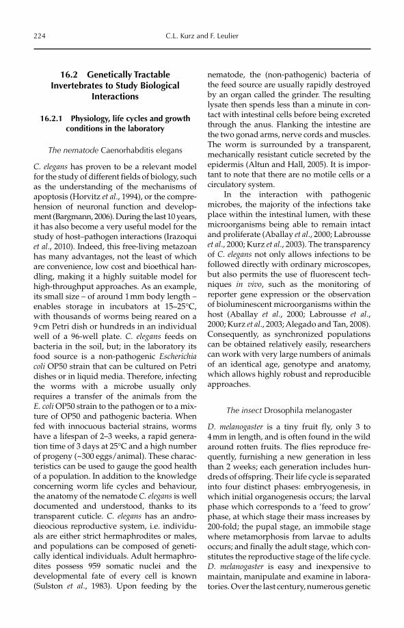
224 C.L. Kurz and F. Leulier
16.2 Genetically Tractable Invertebrates to Study Biological
Interactions
16.2.1 Physiology, life cycles and growth conditions in the laboratory
The nematode Caenorhabditis elegans
C. elegans has proven to be a relevant model for the study of different fields of biology, such as the understanding of the mechanisms of apoptosis (Horvitz et al., 1994), or the compre-hension of neuronal function and develop-ment (Bargmann, 2006). During the last 10 years, it has also become a very useful model for the study of host–pathogen interactions (Irazoqui et al., 2010). Indeed, this free-living metazoan has many advantages, not the least of which are convenience, low cost and bioethical han-dling, making it a highly suitable model for high-throughput approaches. As an example, its small size – of around 1 mm body length – enables storage in incubators at 15–25°C, with thousands of worms being reared on a 9 cm Petri dish or hundreds in an individual well of a 96-well plate. C. elegans feeds on bacteria in the soil, but; in the laboratory its food source is a non-pathogenic Escherichiacoli OP50 strain that can be cultured on Petri dishes or in liquid media. Therefore, infecting the worms with a microbe usually only requires a transfer of the animals from the E. coli OP50 strain to the pathogen or to a mix-ture of OP50 and pathogenic bacteria. When fed with innocuous bacterial strains, worms have a lifespan of 2–3 weeks, a rapid genera-tion time of 3 days at 25°C and a high number of progeny (~300 eggs/animal). These charac-teristics can be used to gauge the good health of a population. In addition to the knowledge concerning worm life cycles and behaviour, the anatomy of the nematode C. elegans is well documented and understood, thanks to its transparent cuticle. C. elegans has an andro-dieocious reproductive system, i.e. individu-als are either strict hermaphrodites or males, and populations can be composed of geneti-cally identical individuals. Adult hermaphro-dites possess 959 somatic nuclei and the developmental fate of every cell is known (Sulston et al., 1983). Upon feeding by the
nematode, the (non-pathogenic) bacteria of the feed source are usually rapidly destroyed by an organ called the grinder. The resulting lysate then spends less than a minute in con-tact with intestinal cells before being excreted through the anus. Flanking the intestine are the two gonad arms, nerve cords and muscles. The worm is surrounded by a transparent, mechanically resistant cuticle secreted by the epidermis (Altun and Hall, 2005). It is impor-tant to note that there are no motile cells or a circulatory system.
In the interaction with pathogenic microbes, the majority of the infections take place within the intestinal lumen, with these microorganisms being able to remain intact and proliferate (Aballay et al., 2000; Labrousse et al., 2000; Kurz et al., 2003). The transparency of C. elegans not only allows infections to be followed directly with ordinary microscopes, but also permits the use of fluorescent tech-niques in vivo, such as the monitoring of reporter gene expression or the observation of bioluminescent microorganisms within the host (Aballay et al., 2000; Labrousse et al.,2000; Kurz et al., 2003; Alegado and Tan, 2008). Consequently, as synchronized populations can be obtained relatively easily, researchers can work with very large numbers of animals of an identical age, genotype and anatomy, which allows highly robust and reproducible approaches.
The insect Drosophila melanogaster
D. melanogaster is a tiny fruit fly, only 3 to 4 mm in length, and is often found in the wild around rotten fruits. The flies reproduce fre-quently, furnishing a new generation in less than 2 weeks; each generation includes hun-dreds of offspring. Their life cycle is separated into four distinct phases: embryogenesis, in which initial organogenesis occurs; the larval phase which corresponds to a ‘feed to grow’ phase, at which stage their mass increases by 200-fold; the pupal stage, an immobile stage where metamorphosis from larvae to adults occurs; and finally the adult stage, which con-stitutes the reproductive stage of the life cycle. D. melanogaster is easy and inexpensive to maintain, manipulate and examine in labora-tories. Over the last century, numerous genetic
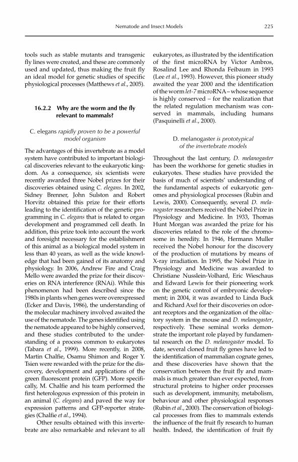
Nematode and Insect Models 225
tools such as stable mutants and transgenic fly lines were created, and these are commonly used and updated, thus making the fruit fly an ideal model for genetic studies of specific physiological processes (Matthews et al., 2005).
16.2.2 Why are the worm and the fly relevant to mammals?
C. elegans rapidly proven to be a powerful model organism
The advantages of this invertebrate as a model system have contributed to important biologi-cal discoveries relevant to the eukaryotic king-dom. As a consequence, six scientists were recently awarded three Nobel prizes for their discoveries obtained using C. elegans. In 2002, Sidney Brenner, John Sulston and Robert Horvitz obtained this prize for their efforts leading to the identification of the genetic pro-gramming in C. elegans that is related to organ development and programmed cell death. In addition, this prize took into account the work and foresight necessary for the establishment of this animal as a biological model system in less than 40 years, as well as the wide knowl-edge that had been gained of its anatomy and physiology. In 2006, Andrew Fire and Craig Mello were awarded the prize for their discov-eries on RNA interference (RNAi). While this phenomenon had been described since the 1980s in plants when genes were overexpressed (Ecker and Davis, 1986), the understanding of the molecular machinery involved awaited the use of the nematode. The genes identified using the nematode appeared to be highly conserved, and these studies contributed to the under-standing of a process common to eukaryotes (Tabara et al., 1999). More recently, in 2008, Martin Chalfie, Osamu Shimon and Roger Y. Tsien were rewarded with the prize for the dis-covery, development and applications of the green fluorescent protein (GFP). More specifi-cally, M. Chalfie and his team performed the first heterologous expression of this protein in an animal (C. elegans) and paved the way for expression patterns and GFP-reporter strate-gies (Chalfie et al., 1994).
Other results obtained with this inverte-brate are also remarkable and relevant to all
eukaryotes, as illustrated by the identification of the first microRNA by Victor Ambros, Rosalind Lee and Rhonda Feibaum in 1993 (Lee et al., 1993). However, this pioneer study awaited the year 2000 and the identification of the worm let-7 microRNA – whose sequence is highly conserved – for the realization that the related regulation mechanism was con-served in mammals, including humans (Pasquinelli et al., 2000).
D. melanogaster is prototypical of the invertebrate models
Throughout the last century, D. melanogasterhas been the workhorse for genetic studies in eukaryotes. These studies have provided the basis of much of scientists’ understanding of the fundamental aspects of eukaryotic gen-omes and physiological processes (Rubin and Lewis, 2000). Consequently, several D. mela-nogaster researchers received the Nobel Prize in Physiology and Medicine. In 1933, Thomas Hunt Morgan was awarded the prize for his discoveries related to the role of the chromo-some in heredity. In 1946, Hermann Muller received the Nobel honour for the discovery of the production of mutations by means of X-ray irradiation. In 1995, the Nobel Prize in Physiology and Medicine was awarded to Christiane Nusslein-Volhard, Eric Wieschaus and Edward Lewis for their pioneering work on the genetic control of embryonic develop-ment; in 2004, it was awarded to Linda Buck and Richard Axel for their discoveries on odor-ant receptors and the organization of the olfac-tory system in the mouse and D. melanogaster,respectively. These seminal works demon-strate the important role played by fundamen-tal research on the D. melanogaster model. To date, several cloned fruit fly genes have led to the identification of mammalian cognate genes, and these discoveries have shown that the conservation between the fruit fly and mam-mals is much greater than ever expected, from structural proteins to higher order processes such as development, immunity, metabolism, behaviour and other physiological responses (Rubin et al., 2000). The conservation of biologi-cal processes from flies to mammals extends the influence of the fruit fly research to human health. Indeed, the identification of fruit fly
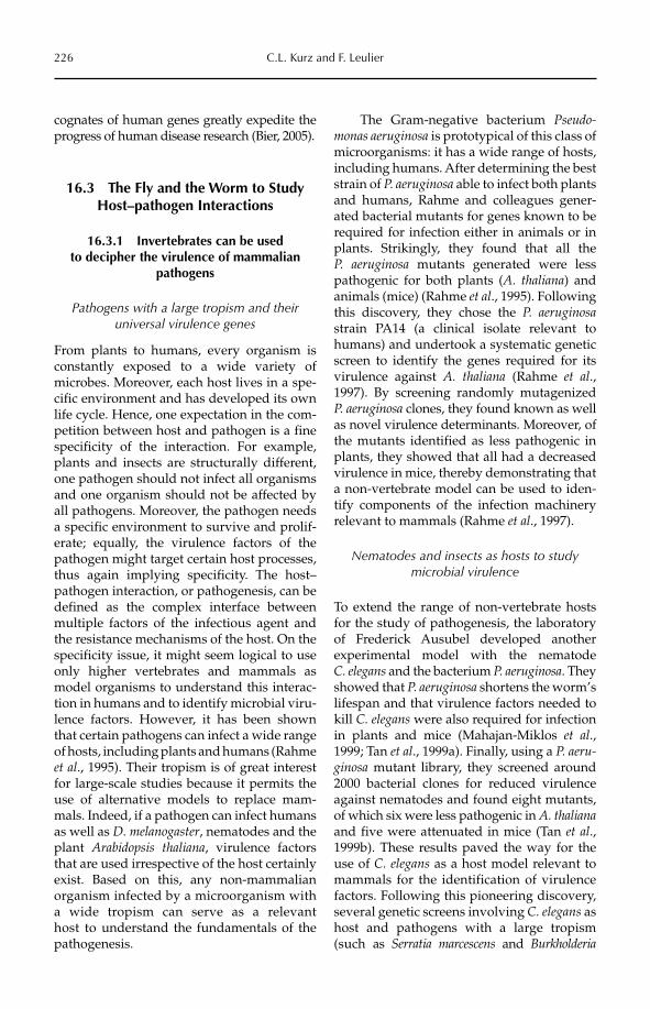
226 C.L. Kurz and F. Leulier
cognates of human genes greatly expedite the progress of human disease research (Bier, 2005).
16.3 The Fly and the Worm to Study Host–pathogen Interactions
16.3.1 Invertebrates can be used to decipher the virulence of mammalian
pathogens
Pathogens with a large tropism and their universal virulence genes
From plants to humans, every organism is constantly exposed to a wide variety of microbes. Moreover, each host lives in a spe-cific environment and has developed its own life cycle. Hence, one expectation in the com-petition between host and pathogen is a fine specificity of the interaction. For example, plants and insects are structurally different, one pathogen should not infect all organisms and one organism should not be affected by all pathogens. Moreover, the pathogen needs a specific environment to survive and prolif-erate; equally, the virulence factors of the pathogen might target certain host processes, thus again implying specificity. The host–pathogen interaction, or pathogenesis, can be defined as the complex interface between multiple factors of the infectious agent and the resistance mechanisms of the host. On the specificity issue, it might seem logical to use only higher vertebrates and mammals as model organisms to understand this interac-tion in humans and to identify microbial viru-lence factors. However, it has been shown that certain pathogens can infect a wide range of hosts, including plants and humans (Rahme et al., 1995). Their tropism is of great interest for large-scale studies because it permits the use of alternative models to replace mam-mals. Indeed, if a pathogen can infect humans as well as D. melanogaster, nematodes and the plant Arabidopsis thaliana, virulence factors that are used irrespective of the host certainly exist. Based on this, any non-mammalian organism infected by a microorganism with a wide tropism can serve as a relevant host to understand the fundamentals of the pathogenesis.
The Gram-negative bacterium Pseudo-monas aeruginosa is prototypical of this class of microorganisms: it has a wide range of hosts, including humans. After determining the best strain of P. aeruginosa able to infect both plants and humans, Rahme and colleagues gener-ated bacterial mutants for genes known to be required for infection either in animals or in plants. Strikingly, they found that all the P. aeruginosa mutants generated were less pathogenic for both plants (A. thaliana) and animals (mice) (Rahme et al., 1995). Following this discovery, they chose the P. aeruginosastrain PA14 (a clinical isolate relevant to humans) and undertook a systematic genetic screen to identify the genes required for its virulence against A. thaliana (Rahme et al.,1997). By screening randomly mutagenized P. aeruginosa clones, they found known as well as novel virulence determinants. Moreover, of the mutants identified as less pathogenic in plants, they showed that all had a decreased virulence in mice, thereby demonstrating that a non-vertebrate model can be used to iden-tify components of the infection machinery relevant to mammals (Rahme et al., 1997).
Nematodes and insects as hosts to study microbial virulence
To extend the range of non-vertebrate hosts for the study of pathogenesis, the laboratory of Frederick Ausubel developed another experimental model with the nematode C. elegans and the bacterium P. aeruginosa. They showed that P. aeruginosa shortens the worm’s lifespan and that virulence factors needed to kill C. elegans were also required for infection in plants and mice (Mahajan-Miklos et al.,1999; Tan et al., 1999a). Finally, using a P. aeru-ginosa mutant library, they screened around 2000 bacterial clones for reduced virulence against nematodes and found eight mutants, of which six were less pathogenic in A. thalianaand five were attenuated in mice (Tan et al.,1999b). These results paved the way for the use of C. elegans as a host model relevant to mammals for the identification of virulence factors. Following this pioneering discovery, several genetic screens involving C. elegans as host and pathogens with a large tropism (such as Serratia marcescens and Burkholderia
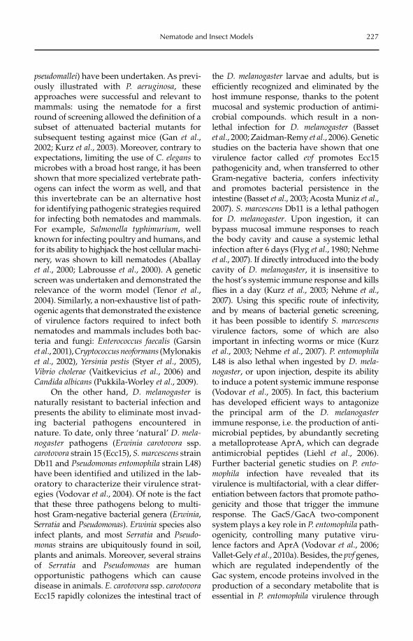
Nematode and Insect Models 227
pseudomallei) have been undertaken. As previ-ously illustrated with P. aeruginosa, these approaches were successful and relevant to mammals: using the nematode for a first round of screening allowed the definition of a subset of attenuated bacterial mutants for subsequent testing against mice (Gan et al.,2002; Kurz et al., 2003). Moreover, contrary to expectations, limiting the use of C. elegans to microbes with a broad host range, it has been shown that more specialized vertebrate path-ogens can infect the worm as well, and that this invertebrate can be an alternative host for identifying pathogenic strategies required for infecting both nematodes and mammals. For example, Salmonella typhimurium, well known for infecting poultry and humans, and for its ability to highjack the host cellular machi-nery, was shown to kill nematodes (Aballay et al., 2000; Labrousse et al., 2000). A genetic screen was undertaken and demonstrated the relevance of the worm model (Tenor et al.,2004). Similarly, a non-exhaustive list of path-ogenic agents that demonstrated the existence of virulence factors required to infect both nematodes and mammals includes both bac-teria and fungi: Enterococcus faecalis (Garsin et al., 2001), Cryptococcus neoformans (Mylonakis et al., 2002), Yersinia pestis (Styer et al., 2005), Vibrio cholerae (Vaitkevicius et al., 2006) and Candida albicans (Pukkila-Worley et al., 2009).
On the other hand, D. melanogaster is naturally resistant to bacterial infection and presents the ability to eliminate most invad-ing bacterial pathogens encountered in nature. To date, only three ‘natural’ D. mela-nogaster pathogens (Erwinia carotovora ssp. carotovora strain 15 (Ecc15), S. marcescens strain Db11 and Pseudomonas entomophila strain L48) have been identified and utilized in the lab-oratory to characterize their virulence strat-egies (Vodovar et al., 2004). Of note is the fact that these three pathogens belong to multi-host Gram-negative bacterial genera (Erwinia,Serratia and Pseudomonas). Erwinia species also infect plants, and most Serratia and Pseudo-monas strains are ubiquitously found in soil, plants and animals. Moreover, several strains of Serratia and Pseudomonas are human opportunistic pathogens which can cause disease in animals. E. carotovora ssp. carotovoraEcc15 rapidly colonizes the intestinal tract of
the D. melanogaster larvae and adults, but is efficiently recognized and eliminated by the host immune response, thanks to the potent mucosal and systemic production of antimi-crobial compounds. which result in a non-lethal infection for D. melanogaster (Basset et al., 2000; Zaidman-Remy et al., 2006). Genetic studies on the bacteria have shown that one virulence factor called evf promotes Ecc15 pathogenicity and, when transferred to other Gram-negative bacteria, confers infectivity and promotes bacterial persistence in the intestine (Basset et al., 2003; Acosta Muniz et al., 2007). S. marcescens Db11 is a lethal pathogen for D. melanogaster. Upon ingestion, it can bypass mucosal immune responses to reach the body cavity and cause a systemic lethal infection after 6 days (Flyg et al., 1980; Nehme et al., 2007). If directly introduced into the body cavity of D. melanogaster, it is insensitive to the host’s systemic immune response and kills flies in a day (Kurz et al., 2003; Nehme et al., 2007). Using this specific route of infectivity, and by means of bacterial genetic screening,it has been possible to identify S. marcescensvirulence factors, some of which are also important in infecting worms or mice (Kurz et al., 2003; Nehme et al., 2007). P. entomophilaL48 is also lethal when ingested by D. mela-nogaster, or upon injection, despite its ability to induce a potent systemic immune response (Vodovar et al., 2005). In fact, this bacterium has developed efficient ways to antagonize the principal arm of the D. melanogasterimmune response, i.e. the production of anti-microbial peptides, by abundantly secreting a metalloprotease AprA, which can degrade anti microbial peptides (Liehl et al., 2006). Further bacterial genetic studies on P. ento-mophila infection have revealed that its virulence is multifactorial, with a clear differ-entiation between factors that promote patho-genicity and those that trigger the immune response. The GacS/GacA two-component system plays a key role in P. entomophila path-ogenicity, controlling many putative viru-lence factors and AprA (Vodovar et al., 2006; Vallet-Gely et al., 2010a). Besides, the pvf genes, which are regulated independently of the Gac system, encode proteins involved in the production of a secondary metabolite that is essential in P. entomophila virulence through
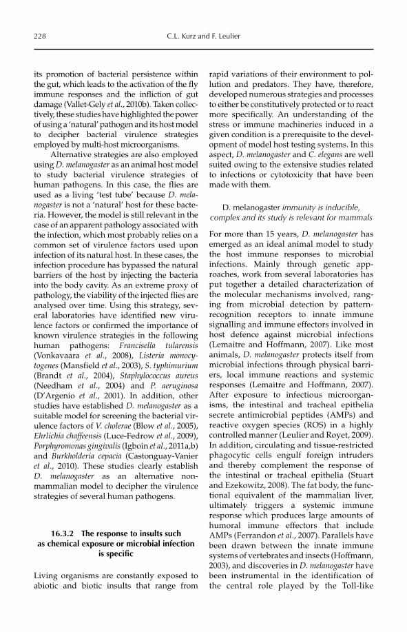
228 C.L. Kurz and F. Leulier
its promotion of bacterial persistence within the gut, which leads to the activation of the fly immune responses and the infliction of gut damage (Vallet-Gely et al., 2010b). Taken collec-tively, these studies have highlighted the power of using a ‘natural’ pathogen and its host model to decipher bacterial virulence strategies employed by multi-host microorganisms.
Alternative strategies are also employed using D. melanogaster as an animal host model to study bacterial virulence strategies of human pathogens. In this case, the flies are used as a living ‘test tube’ because D. mela-nogaster is not a ‘natural’ host for these bacte-ria. However, the model is still relevant in the case of an apparent pathology associated with the infection, which most probably relies on a common set of virulence factors used upon infection of its natural host. In these cases, the infection procedure has bypassed the natural barriers of the host by injecting the bacteria into the body cavity. As an extreme proxy of pathology, the viability of the injected flies are analysed over time. Using this strategy, sev-eral laboratories have identified new viru-lence factors or confirmed the importance of known virulence strategies in the following human pathogens: Francisella tularensis(Vonkavaara et al., 2008), Listeria monocy-togenes (Mansfield et al., 2003), S. typhimurium(Brandt et al., 2004), Staphylococcus aureus(Needham et al., 2004) and P. aeruginosa(D’Argenio et al., 2001). In addition, other studies have established D. melanogaster as a suitable model for screening the bacterial vir-ulence factors of V. cholerae (Blow et al., 2005), Ehrlichia chaffeensis (Luce-Fedrow et al., 2009), Porphyromonas gingivalis (Igboin et al., 2011a,b) and Burkholderia cepacia (Castonguay-Vanier et al., 2010). These studies clearly establish D. melanogaster as an alternative non- mammalian model to decipher the virulence strategies of several human pathogens.
16.3.2 The response to insults such as chemical exposure or microbial infection
is specific
Living organisms are constantly exposed to abiotic and biotic insults that range from
rapid variations of their environment to pol-lution and predators. They have, therefore, developed numerous strategies and processes to either be constitutively protected or to react more specifically. An understanding of the stress or immune machineries induced in a given condition is a prerequisite to the devel-opment of model host testing systems. In this aspect, D. melanogaster and C. elegans are well suited owing to the extensive studies related to infections or cytotoxicity that have been made with them.
D. melanogaster immunity is inducible, complex and its study is relevant for mammals
For more than 15 years, D. melanogaster has emerged as an ideal animal model to study the host immune responses to microbial infections. Mainly through genetic app-roaches, work from several laboratories has put together a detailed characterization of the molecular mechanisms involved, rang-ing from microbial detection by pattern- recognition receptors to innate immune signalling and immune effectors involved in host defence against microbial infections (Lemaitre and Hoffmann, 2007). Like most animals, D. melanogaster protects itself from microbial infections through physical barri-ers, local immune reactions and systemic responses (Lemaitre and Hoffmann, 2007). After exposure to infectious microorgan-isms, the intestinal and tracheal epithelia secrete antimicrobial peptides (AMPs) and reactive oxygen species (ROS) in a highly controlled manner (Leulier and Royet, 2009). In addition, circulating and tissue-restricted phagocytic cells engulf foreign intruders and thereby complement the response of the intestinal or tracheal epithelia (Stuart and Ezekowitz, 2008). The fat body, the func-tional equivalent of the mammalian liver, ultimately triggers a systemic immune response which produces large amounts of humoral immune effectors that include AMPs (Ferrandon et al., 2007). Parallels have been drawn between the innate immune systems of vertebrates and insects (Hoffmann, 2003), and discoveries in D. melanogaster have been instrumental in the identification of the central role played by the Toll-like
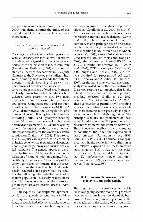
Nematode and Insect Models 229
receptors in mammalian immunity (Lemaitre, 2004), thus demonstrating the utility of this animal model for studying host–microbe interactions.
Worms do possess inducible and specific defence mechanism
The elegant studies that have been performed with D. melanogaster (see above) illustrated the relevance of genetically tractable inverte-brates for the elucidation of innate immunity (Lemaitre and Hoffmann, 2007) and prompted studies of defence mechanisms in C. elegans.Contrary to the D. melanogaster studies, which were primarily host oriented, the different infection models involving C. elegans that have already been described in Section 16.3.1 were a prerequisite and offered a ready means to clearly demonstrate whether inducible host defences were present or not. Two main approaches were undertaken, transcriptomic and genetic. Using microarrays and the infec-tion of nematodes by S. marcescens, Mallo et al.(2002) demonstrated the upregulation of a number of genes following bacterial infection, including lectin- and lysozyme-encoding genes. Moreover, mechanistic insights were obtained and elements of a TGF (transforming growth factor)-beta pathway were demon-strated as necessary for the worm’s resistance to infection (Mallo et al., 2002). This proved that C. elegans can respond to infection by inducing genes whose regulation is dependent upon signalling pathways required to achieve full resistance. The genetic approach devel-oped by Kim et al. (2002) was based upon the isolation of mutants with an enhanced sus-ceptibility to pathogens. The subtlety of this assay was to identify mutants that die preco-ciously from the infection, but that (fortu-nately) retained some eggs within the body, thereby allowing the establishment of a mutant population. This study resulted in the identification of elements of the conserved p38 mitogen-activated protein kinase (MAPK) pathway.
Subsequently transcriptomic approach- es, forward genetic screens and candidate gene app roaches, combined with the wide range of established infection models, allowed the identification of several host signalling
pathways required for the direct response to infection (Couillault et al., 2004; Estes et al.,2010), as well as the mechanisms necessary for repairing immune-related damages (Garsin et al., 2003). The current view of nematode immunity is of a pathogen-specific response to infection involving a network of pathways with signalling modules such as p38 MAPK (Kim et al., 2002), extracellular signal-regu-lated kinase (ERK) (Nicholas and Hodgkin, 2004), c-Jun N-terminal kinase (JNK) (Kim etal., 2004), insulin-like receptor (ILR) (Garsin et al., 2003), TGF-beta receptor (Mallo et al.,2002; Zugasti and Ewbank, 2009) and pro-teins required for programmed cell death (PCD) (Aballay and Ausubel, 2001; Jia et al.,2009). At the same time, various microarray analyses revealed one of the main features of C. elegans response to infection, that is, the robust transcriptional induction of peptide-encoding effectors (Mallo et al., 2002; Couillault et al., 2004; Troemel et al., 2006). These genes, such as putative AMP-encoding genes, are becoming precious molecular tools for characterizing more precisely the path-ways responsible for their regulation. The principle is to use the promoters of these genes fused to gfp (the GFP gene) to obtain biosensors for nematode immune activation. Thus, it becomes possible to identify mutants or conditions that alter the expression of these effectors (Ferrandon et al., 1998; Couillault et al., 2004). The animal’s transpar-ency permits the convenient visualization of the relative expression of these immune reporter genes. Such a strategy was origi-nally successfully exploi ted for deciphering the D. melanogaster innate immunity (Ferrandon et al., 1998) and was adapted to C.elegans (Pujol et al., 2008).
16.3.3 In vivo platforms to assess cytotoxicity and pathogenicity
The importance of invertebrates as models for investigating specific biological processes relevant to mammals has now definitely been proven. Concerning more specifically the issues related to the toxicity of a given mole-cule or the pathogenicity of a microorganism,
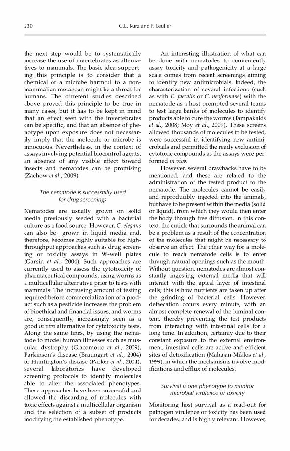
230 C.L. Kurz and F. Leulier
the next step would be to systematically increase the use of invertebrates as alterna-tives to mammals. The basic idea support-ing this principle is to consider that a chemical or a microbe harmful to a non-mammalian metazoan might be a threat for humans. The different studies described above proved this principle to be true in many cases, but it has to be kept in mind that an effect seen with the invertebrates can be specific, and that an absence of phe-notype upon exposure does not necessar-ily imply that the molecule or microbe is innocuous. Nevertheless, in the context of assays involving potential biocontrol agents, an absence of any visible effect toward insects and nematodes can be promising (Zachow et al., 2009).
The nematode is successfully used for drug screenings
Nematodes are usually grown on solid media previously seeded with a bacterial culture as a food source. However, C. eleganscan also be grown in liquid media and, therefore, becomes highly suitable for high-throughput approaches such as drug screen-ing or toxicity assays in 96-well plates (Garsin et al., 2004). Such approaches are currently used to assess the cytotoxicity of pharmaceutical compounds, using worms as a multicellular alternative prior to tests with mammals. The increasing amount of testing required before commercialization of a prod-uct such as a pesticide increases the problem of bioethical and financial issues, and worms are, consequently, increasingly seen as a good in vivo alternative for cytotoxicity tests. Along the same lines, by using the nema-tode to model human illnesses such as mus-cular dystrophy (Giacomotto et al., 2009), Parkinson’s disease (Braungart et al., 2004) or Huntington’s disease (Parker et al., 2004), several laboratories have developed screening protocols to identify molecules able to alter the associated phenotypes. These approaches have been successful and allowed the discarding of molecules with toxic effects against a multicellular organism and the selection of a subset of products modifying the established phenotype.
An interesting illustration of what can be done with nematodes to conveniently assay toxicity and pathogenicity at a large scale comes from recent screenings aiming to identify new antimicrobials. Indeed, the characterization of several infections (such as with E. faecalis or C. neoformans) with the nematode as a host prompted several teams to test large banks of molecules to identify products able to cure the worms (Tampakakis et al., 2008; Moy et al., 2009). These screens allowed thousands of molecules to be tested, were successful in identifying new antimi-crobials and permitted the ready exclusion of cytotoxic compounds as the assays were per-formed in vivo.
However, several drawbacks have to be mentioned, and these are related to the administration of the tested product to the nematode. The molecules cannot be easily and reproducibly injected into the animals, but have to be present within the media (solid or liquid), from which they would then enter the body through free diffusion. In this con-text, the cuticle that surrounds the animal can be a problem as a result of the concentration of the molecules that might be necessary to observe an effect. The other way for a mole-cule to reach nematode cells is to enter through natural openings such as the mouth. Without question, nematodes are almost con-stantly ingesting external media that will interact with the apical layer of intestinal cells; this is how nutrients are taken up after the grinding of bacterial cells. However, defaecation occurs every minute, with an almost complete renewal of the luminal con-tent, thereby preventing the test products from interacting with intestinal cells for a long time. In addition, certainly due to their constant exposure to the external environ-ment, intestinal cells are active and efficient sites of detoxification (Mahajan-Miklos et al.,1999), in which the mechanisms involve mod-ifications and efflux of molecules.
Survival is one phenotype to monitor microbial virulence or toxicity
Monitoring host survival as a read-out for pathogen virulence or toxicity has been used for decades, and is highly relevant. However,
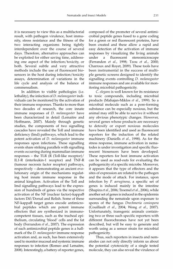
Nematode and Insect Models 231
it is necessary to view this as a multifactorial result, with pathogen virulence, host immu-nity, stress resistance and the fitness of the two interacting organisms being tightly interdependent over the course of several days. Therefore, alternative approaches can be exploited for either saving time, address-ing one aspect of the infection/toxicity, or both. Several subtle and very attractive methods include the use of fluorescent bio-sensors in the host during infection/toxicity assays, determination of variations in the life cycle and analysis of the balance of commensalism.
In addition to visible pathologies (i.e. lethality), the infection of D. melanogaster indi-viduals can be monitored by the activation of their immune responses. Thanks to more than two decades of research, the core innate immune responses of D. melanogaster have been characterized in detail (Lemaitre and Hoffmann, 2007). Mainly through genetic studies, the components of two signalling cascades have revealed the Toll and immune deficiency (Imd) pathways, which lead to the potent activation of D. melanogaster immune responses upon infections. These signalling events share striking parallels with signalling events occurring during mammalian immune responses – the TLR (R (Toll-like receptor)/IL1-R (interleukin-1 receptor) and TNF-R (tumour necrosis factor receptor) pathways, respectively – demonstrating an ancient evo-lutionary origin of the mechanisms regulat-ing host innate immune response in the animal kingdom. Activation of the Toll and Imd signalling pathways lead to the expres-sion of hundreds of genes via the respective activation of the NF (nuclear factor)-kappaB factors Dif/Dorsal and Relish. Some of these NF-kappaB target genes encode antimicro-bial peptides which are potent immune effectors that are synthesized in immuno-competent tissues, such as the tracheal epi-thelium, circulating ‘blood’ cells and the fat body (Ferrandon et al., 2007). The expression of such antimicrobial peptide genes is a hall-mark of the D. melanogaster immune response activation and, as such, has been extensively used to monitor mucosal and systemic immune responses to infection (Romeo and Lemaitre, 2008). Interestingly, a battery of reporter genes,
composed of the promoter of several antimi-crobial peptide genes fused to a gene coding for a green or red fluorescent proteins, have been created and these allow a rapid and easy detection of the activation of immune responses by visualizing the living animals under a fluorescent stereomicroscope (Ferrandon et al., 1998; Tzou et al., 2000; Charroux and Royet, 2009). These tools have been instrumental in the success of multi-ple genetic screens designed to identify the signalling events controlling D. melanogasterimmune responses and are available for mon-itoring microbial pathogenicity.
C. elegans is well known for its resistance to toxic compounds, including microbial products (Mahajan-Miklos et al., 1999). So a microbial molecule such as a pore-forming substance can be especially noxious, but the animal may still be able to survive it without any obvious phenotypic changes. However, several genes whose products are necessary to detoxify or export noxious molecules have been identified and used as fluorescent reporters for the induction of the related machinery (Daniells et al., 1998). As for the stress response, immune activation in nema-todes is under investigation and specific fluo-rescent biosensors have been developed. These reporters for host immune activation can be used as read-outs for evaluating the pathogenicity of a specific microbe. Moreover, it appears that the type of effectors and the sites of expression are related to the pathogen and the mode of attack. For instance, upon infection by P. aeruginosa, a specific set of genes is induced mainly in the intestine (Shapira et al., 2006; Troemel et al., 2006), while another set of genes is induced in the epidermis surrounding the nematode upon exposure to spores of the fungus Drechmeria coniospora(Couillault et al., 2004; Wong et al., 2007). Unfortunately, transgenic animals contain-ing two or three such specific reporters with different fluorochromes have not yet been described, but will be easy to generate and worth using as a sensor strain for microbial pathogenicity.
Thus, such reporters in insects and nem-atodes can not only directly inform us about the potential cytotoxicity of a single tested molecule, they can also reveal the virulence of
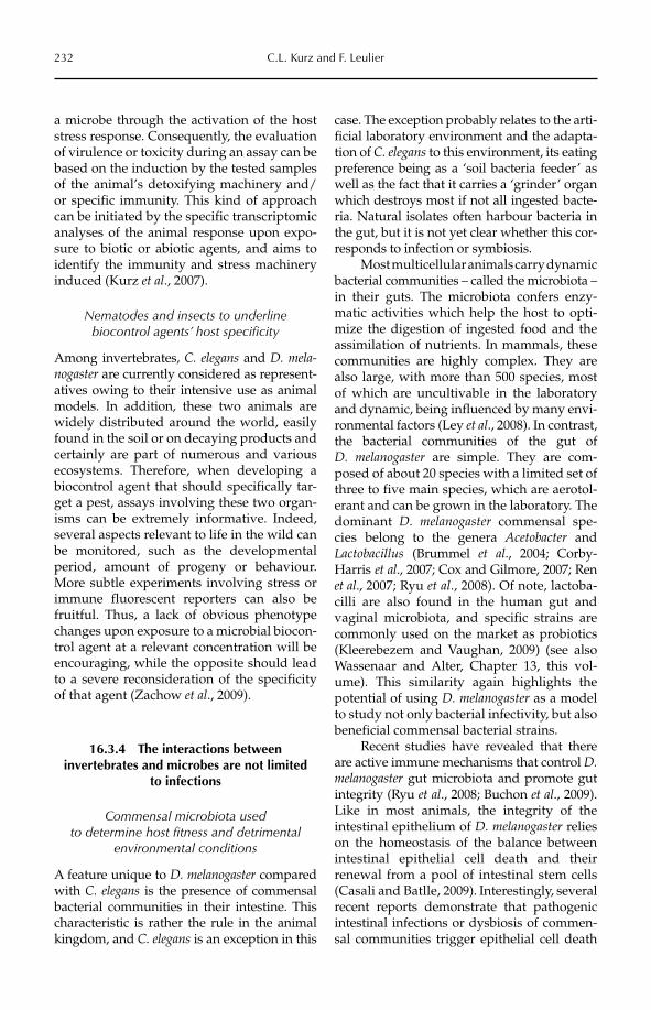
232 C.L. Kurz and F. Leulier
a microbe through the activation of the host stress response. Consequently, the evaluation of virulence or toxicity during an assay can be based on the induction by the tested samples of the animal’s detoxifying machinery and/or specific immunity. This kind of approach can be initiated by the specific transcriptomic analyses of the animal response upon expo-sure to biotic or abiotic agents, and aims to identify the immunity and stress machinery induced (Kurz et al., 2007).
Nematodes and insects to underline biocontrol agents’ host specificity
Among invertebrates, C. elegans and D. mela-nogaster are currently considered as represent-atives owing to their intensive use as animal models. In addition, these two animals are widely distributed around the world, easily found in the soil or on decaying products and certainly are part of numerous and various ecosystems. Therefore, when developing a biocontrol agent that should specifically tar-get a pest, assays involving these two organ-isms can be extremely informative. Indeed, several aspects relevant to life in the wild can be monitored, such as the developmental period, amount of progeny or behaviour. More subtle experiments involving stress or immune fluorescent reporters can also be fruitful. Thus, a lack of obvious phenotype changes upon exposure to a microbial biocon-trol agent at a relevant concentration will be encouraging, while the opposite should lead to a severe reconsideration of the specificity of that agent (Zachow et al., 2009).
16.3.4 The interactions between invertebrates and microbes are not limited
to infections
Commensal microbiota used to determine host fitness and detrimental
environmental conditions
A feature unique to D. melanogaster compared with C. elegans is the presence of commensal bacterial communities in their intestine. This characteristic is rather the rule in the animal kingdom, and C. elegans is an exception in this
case. The exception probably relates to the arti-ficial laboratory environment and the adapta-tion of C. elegans to this environment, its eating preference being as a ‘soil bacteria feeder’ as well as the fact that it carries a ‘grinder’ organ which destroys most if not all ingested bacte-ria. Natural isolates often harbour bacteria in the gut, but it is not yet clear whether this cor-responds to infection or symbiosis.
Most multicellular animals carry dynamic bacterial communities – called the microbiota –in their guts. The microbiota confers enzy-matic activities which help the host to opti-mize the digestion of ingested food and the assimilation of nutrients. In mammals, these communities are highly complex. They are also large, with more than 500 species, most of which are uncultivable in the laboratory and dynamic, being influenced by many envi-ronmental factors (Ley et al., 2008). In contrast, the bacterial communities of the gut of D. melanogaster are simple. They are com-posed of about 20 species with a limited set of three to five main species, which are aerotol-erant and can be grown in the laboratory. The dominant D. melanogaster commensal spe-cies belong to the genera Acetobacter and Lacto bacillus (Brummel et al., 2004; Corby-Harris et al., 2007; Cox and Gilmore, 2007; Ren et al., 2007; Ryu et al., 2008). Of note, lactoba-cilli are also found in the human gut and vaginal microbiota, and specific strains are commonly used on the market as probiotics (Kleerebezem and Vaughan, 2009) (see also Wassenaar and Alter, Chapter 13, this vol-ume). This similarity again highlights the potential of using D. melanogaster as a model to study not only bacterial infectivity, but also beneficial commensal bacterial strains.
Recent studies have revealed that there are active immune mechanisms that control D. melanogaster gut microbiota and promote gut integrity (Ryu et al., 2008; Buchon et al., 2009). Like in most animals, the integrity of the intestinal epithelium of D. melanogaster relies on the homeostasis of the balance betweenintestinal epithelial cell death and their renewal from a pool of intestinal stem cells (Casali and Batlle, 2009). Interestingly, several recent reports demonstrate that pathogenic intestinal infections or dysbiosis of commen-sal communities trigger epithelial cell death
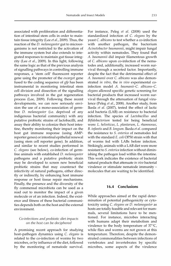
Nematode and Insect Models 233
associated with proliferation and differentia-tion of intestinal stem cells in order to main-tain tissue integrity (Lee et al., 2009). Thus, the reaction of the D. melanogaster gut to microor-ganisms is not restricted to the activation of the immune system but also extends to inte-grated responses to maintain gut tissue integ-rity (Lee et al., 2009). In this light, following the same logic as that of the previous analysis of signalling pathways in controlling immune responses, a ‘stem cell’ fluorescent reporter gene using the promoter of the escargot gene fused to the coding sequence of gfp has been instrumental in monitoring intestinal stem cell division and dissection of the signalling pathways involved in the gut regeneration process (Lee, 2009). Following these recent developments, we can now seriously envi-sion the use of a mono-association of germ-free D. melanogaster (i.e. deprived of any indigenous bacterial community) with any putative probiotic strains of lactobacilli, and assay their ability to colonize their host intes-tine, thereby monitoring their impact on the host gut immune response (using AMP-reporter genes) or intestinal epithelial renewal (using stem cell reporter gene). In addition, and similar to recent studies performed in C. elegans (see below), co-infection of germ-free animals with established D. melanogasterpathogens and a putative probiotic strain may be developed to screen new beneficial probiotic strains that may counteract the infectivity of natural pathogens, either direc-tly or indirectly, by enhancing host immune response or host tissue repair mechanisms. Finally, the presence and the diversity of the fly commensal microbiota can be used as a read out to monitor the impact of a given molecule or of an infection. Indeed, the exist-ence and fitness of these bacterial communi-ties depends both on the host and the external environment.
Co-infections and probiotic diet impacts on the host can be deciphered
A promising recent approach for studying host–pathogen dynamics using C. elegans is related to the co-infection of worms by two microbes, or by influence of the diet, followed by the monitoring of nematode survival.
For instance, Peleg et al. (2008) used the standardized infection of C. elegans by the yeast C. albicans to test whether a co-infection with another pathogen, the bacterium Acineto bacter baumannii, might impair fungal activity within nematodes. They found that A. baumannii did impair filamentous growth of C. albicans upon co-infection of the nema-todes and, additionally, increased worm sur-vival through a secreted factor. Importantly, des pite the fact that the detrimental effect of A. baumanii over C. albicans was also demon-strated in vitro, the in vivo experimental co-infection model A. baumanii–C. albicans–C. elegans allo wed specific genetic screening for bacterial products that increased worm sur-vival through the attenuation of fungal viru-lence (Peleg et al., 2008). Another study, from Ikeda et al. (2007), tested the effect of lactic acid bacteria (LAB) on resistance to bacterial infection. The species of Lactobacillus and Bifidobacterium tested for being beneficial were: L. helveticus, L. plantarum, L. rhamnosus,B. infantis and B. longum. Ikeda et al. compared the resistance to S. enterica of nematodes fed with the standard E. coli OP50 strain with that of worms fed with LABs before infection. Strikingly, animals with a LAB diet were more resistant to S. enterica infection without dimin-ishing the pathogen load within the intestine. This work indicates the existence of bacterial natural products that attenuate in vivo bacterial virulence or stimulate nematode immunity – molecules that are waiting to be identified.
16.4 Conclusions
While approaches aimed at the rapid deter-mination of potential pathogenicity or cyto-to xicity using C. elegans or D. melanogaster as hosts are totally feasible and relevant for mam-mals, several limitations have to be men-tioned. For instance, microbes interacting with humans adapt their metabolism and virulence to the body temperature of 37 °C, while flies and worms are not grown at this temperature. Therefore, despite the demon-strated commonalities between infections of vertebrates and invertebrates by specific microbes, some aspects of the virulence
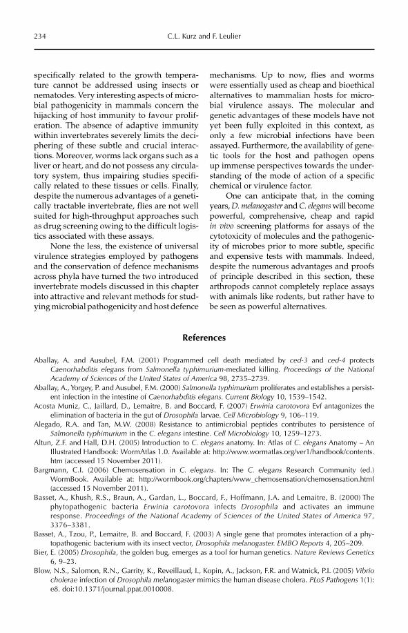
234 C.L. Kurz and F. Leulier
specifically related to the growth tempera-ture cannot be addressed using insects or nematodes. Very interesting aspects of micro-bial pathogenicity in mammals concern the hijacking of host immunity to favour prolif-eration. The absence of adaptive immunity within invertebrates severely limits the deci-phering of these subtle and crucial interac-tions. Moreover, worms lack organs such as a liver or heart, and do not possess any circula-tory system, thus impairing studies specifi-cally related to these tissues or cells. Finally, despite the numerous advantages of a geneti-cally tractable invertebrate, flies are not well suited for high-throughput approaches such as drug screening owing to the difficult logis-tics associated with these assays.
None the less, the existence of universal virulence strategies employed by pathogens and the conservation of defence mechanisms across phyla have turned the two introduced invertebrate models discussed in this chapter into attractive and relevant methods for stud-ying microbial pathogenicity and host defence
mechanisms. Up to now, flies and worms were essentially used as cheap and bioethical alternatives to mammalian hosts for micro-bial virulence assays. The molecular and genetic advantages of these models have not yet been fully exploited in this context, as only a few microbial infections have been assayed. Furthermore, the availability of gene-tic tools for the host and pathogen opens up immense perspectives towards the under-standing of the mode of action of a specific chemical or virulence factor.
One can anticipate that, in the coming years, D. melanogaster and C. elegans will become powerful, comprehensive, cheap and rapid in vivo screening platforms for assays of the cytotoxicity of molecules and the pathogenic-ity of microbes prior to more subtle, specific and expensive tests with mammals. Indeed, despite the numerous advantages and proofs of principle described in this section, these arthropods cannot completely replace assays with animals like rodents, but rather have to be seen as powerful alternatives.
References
Aballay, A. and Ausubel, F.M. (2001) Programmed cell death mediated by ced-3 and ced-4 protects Caenorhabditis elegans from Salmonella typhimurium-mediated killing. Proceedings of the National Academy of Sciences of the United States of America 98, 2735–2739.
Aballay, A., Yorgey, P. and Ausubel, F.M. (2000) Salmonella typhimurium proliferates and establishes a persist-ent infection in the intestine of Caenorhabditis elegans. Current Biology 10, 1539–1542.
Acosta Muniz, C., Jaillard, D., Lemaitre, B. and Boccard, F. (2007) Erwinia carotovora Evf antagonizes the elimination of bacteria in the gut of Drosophila larvae. Cell Microbiology 9, 106–119.
Alegado, R.A. and Tan, M.W. (2008) Resistance to antimicrobial peptides contributes to persistence of Salmonella typhimurium in the C. elegans intestine. Cell Microbiology 10, 1259–1273.
Altun, Z.F. and Hall, D.H. (2005) Introduction to C. elegans anatomy. In: Atlas of C. elegans Anatomy – An Illustrated Handbook: WormAtlas 1.0. Available at: http://www.wormatlas.org/ver1/handbook/contents.htm (accessed 15 November 2011).
Bargmann, C.I. (2006) Chemosensation in C. elegans. In: The C. elegans Research Community (ed.) WormBook. Available at: http://wormbook.org/chapters/www_chemosensation/chemosensation.html (accessed 15 November 2011).
Basset, A., Khush, R.S., Braun, A., Gardan, L., Boccard, F., Hoffmann, J.A. and Lemaitre, B. (2000) The phytopathogenic bacteria Erwinia carotovora infects Drosophila and activates an immune response. Proceedings of the National Academy of Sciences of the United States of America 97, 3376–3381.
Basset, A., Tzou, P., Lemaitre, B. and Boccard, F. (2003) A single gene that promotes interaction of a phy-topathogenic bacterium with its insect vector, Drosophila melanogaster. EMBO Reports 4, 205–209.
Bier, E. (2005) Drosophila, the golden bug, emerges as a tool for human genetics. Nature Reviews Genetics6, 9–23.
Blow, N.S., Salomon, R.N., Garrity, K., Reveillaud, I., Kopin, A., Jackson, F.R. and Watnick, P.I. (2005) Vibriocholerae infection of Drosophila melanogaster mimics the human disease cholera. PLoS Pathogens 1(1): e8. doi:10.1371/journal.ppat.0010008.
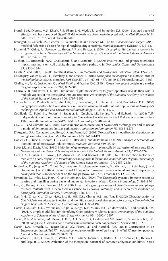
Nematode and Insect Models 235
Brandt, S.M., Dionne, M.S., Khush, R.S., Pham, L.N., Vigdal, T.J. and Schneider, D.S. (2004) Secreted bacterial effectors and host-produced Eiger/TNF drive death in a Salmonella-infected fruit fly. PLoS Biology 2(12): e418. doi:10.1371/journal.pbio.0020418.
Braungart, E., Gerlach, M., Riederer, P., Baumeister, R. and Hoener, M.C. (2004) Caenorhabditis elegans MPP+ model of Parkinson’s disease for high-throughput drug screenings. Neurodegenerative Diseases 1, 175–183.
Brummel, T., Ching, A., Seroude, L., Simon, A.F. and Benzer, S. (2004) Drosophila lifespan enhancement by exogenous bacteria. Proceedings of the National Academy of Sciences of the United States of America101, 12974–12979.
Buchon, N., Broderick, N.A., Chakrabarti, S. and Lemaitre, B. (2009) Invasive and indigenous microbiota impact intestinal stem cell activity through multiple pathways in Drosophila. Genes and Development23, 2333–2344.
Casali, A. and Batlle, E. (2009) Intestinal stem cells in mammals and Drosophila. Cell Stem Cell 4, 124–127.Castonguay-Vanier, J., Vial, L., Tremblay, J. and Deziel, E. (2010) Drosophila melanogaster as a model host for
the Burkholderia cepacia complex. PloS One 5(7), e11467. e11467. doi:10.1371/journal.pone.0011467.Chalfie, M., Tu, Y., Euskirchen, G., Ward, W.W. and Prasher, D.C. (1994) Green fluorescent protein as a marker
for gene expression. Science 263, 802–805.Charroux, B. and Royet, J. (2009) Elimination of plasmatocytes by targeted apoptosis reveals their role in
multiple aspects of the Drosophila immune response. Proceedings of the National Academy of Sciences of the United States of America 106, 9797–9802.
Corby-Harris, V., Pontaroli, A.C., Shimkets, L.J., Bennetzen, J.L., Habel, K.E. and Promislow, D.E. (2007) Geographical distribution and diversity of bacteria associated with natural populations of Drosophilamelanogaster. Applied and Environmental Microbiology 73, 3470–3479.
Couillault, C., Pujol, N., Reboul, J., Sabatier, L., Guichou, J.F., Kohara, Y. and Ewbank, J.J. (2004) TLR-independent control of innate immunity in Caenorhabditis elegans by the TIR domain adaptor protein TIR-1, an ortholog of human SARM. Nature Immunology 5, 488–494.
Cox, C.R. and Gilmore, M.S. (2007) Native microbial colonization of Drosophila melanogaster and its use as a model of Enterococcus faecalis pathogenesis. Infection and Immunity 75, 1565–1576.
D’Argenio, D.A., Gallagher, L.A., Berg, C.A. and Manoil, C. (2001) Drosophila as a model host for Pseudomonas aeruginosa infection. Journal of Bacteriology 183, 1466–1471.
Daniells, C., Duce, I., Thomas, D., Sewell, P., Tattersall, J. and de Pomerai, D. (1998) Transgenic nematodes as biomonitors of microwave-induced stress. Mutation Research 399, 55–64.
Ecker, J.R. and Davis, R.W. (1986) Inhibition of gene expression in plant cells by expression of antisense RNA. Proceedings of the National Academy of Sciences of the United States of America 83, 5372–5376.
Estes, K.A., Dunbar, T.L., Powell, J.R., Ausubel, F.M. and Troemel, E.R. (2010) bZIP transcription factor zip-2mediates an early response to Pseudomonas aeruginosa infection in Caenorhabditis elegans. Proceedings of the National Academy of Science of the United States of America 107, 2153–2158.
Ferrandon, D., Jung, A.C., Criqui, M., Lemaitre, B., Uttenweiler-Joseph, S., Michaut, L., Reichhart, J. and Hoffmann, J.A. (1998) A drosomycin-GFP reporter transgene reveals a local immune response in Drosophila that is not dependent on the Toll pathway. The EMBO Journal 17, 1217–1227.
Ferrandon, D., Imler, J.L., Hetru, C. and Hoffmann, J.A. (2007) The Drosophila systemic immune response: sensing and signalling during bacterial and fungal infections. Nature Reviews Immunology 7, 862–874.
Flyg, C., Kenne, K. and Boman, H.G. (1980) Insect pathogenic properties of Serratia marcescens: phage-resistant mutants with a decreased resistance to Cecropia immunity and a decreased virulence to Drosophila. Journal of General Microbiology 120, 173–181.
Gan, Y.H., Chua, K.L., Chua, H.H., Liu, B., Hii, C.S., Chong, H.L. and Tan, P. (2002) Characterization of Burkholderia pseudomallei infection and identification of novel virulence factors using a Caenorhabditiselegans host system. Molecular Microbiology 44, 1185–1197.
Garsin, D.A., Sifri, C.D., Mylonakis, E., Qin, X., Singh, K.V., Murray, B.E., Calderwood, S.B. and Ausubel, F.M. (2001) A simple model host for identifying Gram-positive virulence factors. Proceedings of the National Academy of Sciences of the United States of America 98, 10892–10897.
Garsin, D.A., Villanueva, J.M., Begun, J., Kim, D.H., Sifri, C.D., Calderwood, S.B., Ruvkun, G. and Ausubel, F.M. (2003) Long-lived C. elegans daf-2 mutants are resistant to bacterial pathogens. Science 300, 1921.
Garsin, D.A., Urbach, J., Huguet-Tapia, J.C., Peters, J.E. and Ausubel, F.M. (2004) Construction of an Enterococcus faecalis Tn917-mediated-gene-disruption library offers insight into Tn917 insertion patterns. Journal of Bacteriology 186, 7280–7289.
Giacomotto, J., Pertl, C., Borrel, C., Walter, M.C., Bulst, S., Johnsen, B., Baillie, D.L., Lochmuller, H., Thirion, C. and Segalat, L. (2009) Evaluation of the therapeutic potential of carbonic anhydrase inhibitors in two
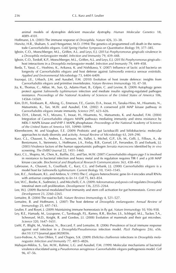
236 C.L. Kurz and F. Leulier
animal models of dystrophin deficient muscular dystrophy. Human Molecular Genetics 18, 4089–4101.
Hoffmann, J.A. (2003) The immune response of Drosophila. Nature 426, 33–38.Horvitz, H.R., Shaham, S. and Hengartner, M.O. (1994) The genetics of programmed cell death in the nema-
tode Caenorhabditis elegans. Cold Spring Harbor Symposia on Quantitative Biology 59, 377–385.Igboin, C.O., Moeschberger, M.L., Griffen, A.L. and Leys, E.J. (2011a) Porphyromonas gingivalis virulence in
a Drosophila melanogaster model. Infection and Immunity 79, 439–448.Igboin, C.O., Tordoff, K.P., Moeschberger, M.L., Griffen, A.L. and Leys, E.J. (2011b) Porphyromonas gingivalis–
host interactions in a Drosophila melanogaster model. Infection and Immunity 79, 449–458.Ikeda, T., Yasui, C., Hoshino, K., Arikawa, K. and Nishikawa, Y. (2007) Influence of lactic acid bacteria on
longevity of Caenorhabditis elegans and host defense against Salmonella enterica serovar entiritidis. Applied and Environmental Microbiology 73, 6404–6409.
Irazoqui, J.E., Urbach, J.M. and Ausubel, F.M. (2010) Evolution of host innate defence: insights from Caenorhabditis elegans and primitive invertebrates. Nature Reviews Immunology 10, 47–58.
Jia, K., Thomas, C., Akbar, M., Sun, Q., Adams-Huet, B., Gilpin, C. and Levine, B. (2009) Autophagy genes protect against Salmonella typhimurium infection and mediate insulin signaling-regulated pathogen resistance. Proceedings of the National Academy of Sciences of the United States of America 106, 14564–14569.
Kim, D.H., Feinbaum, R., Alloing, G., Emerson, F.E., Garsin, D.A., Inoue, H., Tanaka-Hino, M., Hisamoto, N., Matsumoto, K., Tan, M.W. and Ausubel, F.M. (2002) A conserved p38 MAP kinase pathway in Caenorhabditis elegans innate immunity. Science 297, 623–626.
Kim, D.H., Liberati, N.T., Mizuno, T., Inoue, H., Hisamoto, N., Matsumoto, K. and Ausubel, F.M. (2004) Integration of Caenorhabditis elegans MAPK pathways mediating immunity and stress resistance by MEK-1 MAPK kinase and VHP-1 MAPK phosphatase. Proceedings of the National Academy of Sciences of the United States of America 101, 10990–10994.
Kleerebezem, M. and Vaughan, E.E. (2009) Probiotic and gut lactobacilli and bifidobacteria: molecular approaches to study diversity and activity. Annual Review of Microbiology 63, 269–290.
Kurz, C.L., Chauvet, S., Andres, E., Aurouze, M., Vallet, I., Michel, G.P., Uh, M., Celli, J., Filloux, A., de Bentzmann, S., Steinmetz, I., Hoffmann, J.A., Finlay, B.B., Gorvel, J.P., Ferrandon, D. and Ewbank, J.J. (2003) Virulence factors of the human opportunistic pathogen Serratia marcescens identified by in vivoscreening. The EMBO Journal 22, 1451–1460.
Kurz, C.L., Shapira, M., Chen, K., Baillie, D.L. and Tan, M.W. (2007) Caenorhabditis elegans pgp-5 is involved in resistance to bacterial infection and heavy metal and its regulation requires TIR-1 and a p38 MAP kinase cascade. Biochemical and Biophysical Research Communications 363, 438–443.
Labrousse, A., Chauvet, S., Couillault, C., Kurz, C.L. and Ewbank, J.J. (2000) Caenorhabditis elegans is a model host for Salmonella typhimurium. Current Biology 10, 1543–1545.
Lee, R.C., Feinbaum, R.L. and Ambros, V. (1993) The C. elegans heterochronic gene lin-4 encodes small RNAs with antisense complementarity to lin-14. Cell 75, 843–854.
Lee, W.C., Beebe, K., Sudmeier, L. and Micchelli, C.A. (2009) Adenomatous polyposis coli regulates Drosophilaintestinal stem cell proliferation. Development 136, 2255–2264.
Lee, W.J. (2009) Bacterial-modulated host immunity and stem cell activation for gut homeostasis. Genes and Development 23, 2260–2265.
Lemaitre, B. (2004) The road to Toll. Nature Reviews Immunology 4, 521–527.Lemaitre, B. and Hoffmann, J. (2007) The host defense of Drosophila melanogaster. Annual Review of
Immunology 25, 697–743.Leulier, F. and Royet, J. (2009) Maintaining immune homeostasis in fly gut. Nature Immunology 10, 936–938.Ley, R.E., Hamady, M., Lozupone, C., Turnbaugh, P.J., Ramey, R.R., Bircher, J.S., Schlegel, M.L., Tucker, T.A.,
Schrenzel, M.D., Knight, R. and Gordon, J.I. (2008) Evolution of mammals and their gut microbes. Science 320, 1647–1651.
Liehl, P., Blight, M., Vodovar, N., Boccard, F. and Lemaitre, B. (2006) Prevalence of local immune response against oral infection in a Drosophila/Pseudomonas infection model. PLoS Pathogens 2(6), e56. doi:10.1371/journal.ppat.0020056.
Luce-Fedrow, A., Von Ohlen, T. and Chapes, S.K. (2009) Ehrlichia chaffeensis infections in Drosophila mela-nogaster. Infection and Immunity 77, 4815–4826.
Mahajan-Miklos, S., Tan, M.W., Rahme, L.G. and Ausubel, F.M. (1999) Molecular mechanisms of bacterial virulence elucidated using a Pseudomonas aeruginosa–Caenorhabditis elegans pathogenesis model. Cell96, 47–56.
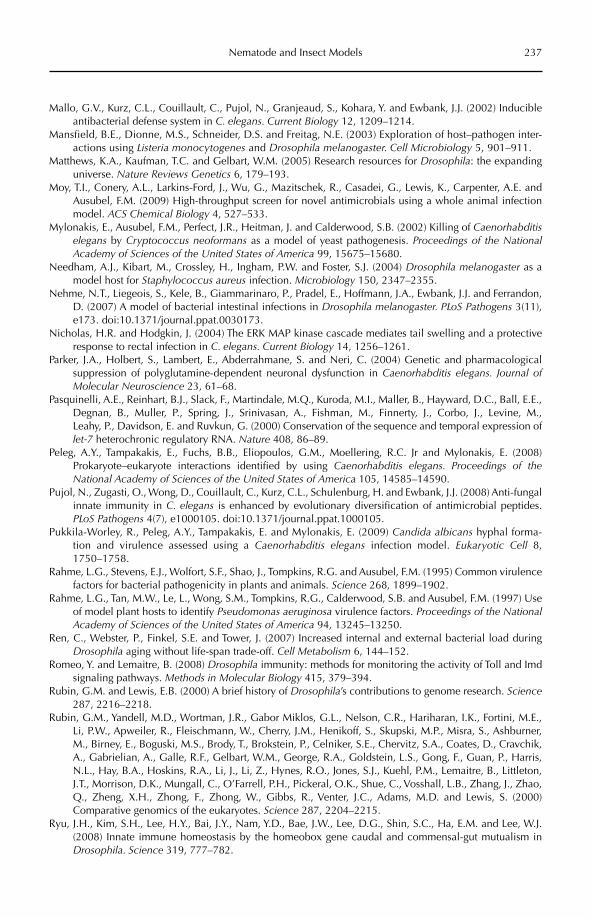
Nematode and Insect Models 237
Mallo, G.V., Kurz, C.L., Couillault, C., Pujol, N., Granjeaud, S., Kohara, Y. and Ewbank, J.J. (2002) Inducible antibacterial defense system in C. elegans. Current Biology 12, 1209–1214.
Mansfield, B.E., Dionne, M.S., Schneider, D.S. and Freitag, N.E. (2003) Exploration of host–pathogen inter-actions using Listeria monocytogenes and Drosophila melanogaster. Cell Microbiology 5, 901–911.
Matthews, K.A., Kaufman, T.C. and Gelbart, W.M. (2005) Research resources for Drosophila: the expanding universe. Nature Reviews Genetics 6, 179–193.
Moy, T.I., Conery, A.L., Larkins-Ford, J., Wu, G., Mazitschek, R., Casadei, G., Lewis, K., Carpenter, A.E. and Ausubel, F.M. (2009) High-throughput screen for novel antimicrobials using a whole animal infection model. ACS Chemical Biology 4, 527–533.
Mylonakis, E., Ausubel, F.M., Perfect, J.R., Heitman, J. and Calderwood, S.B. (2002) Killing of Caenorhabditiselegans by Cryptococcus neoformans as a model of yeast pathogenesis. Proceedings of the National Academy of Sciences of the United States of America 99, 15675–15680.
Needham, A.J., Kibart, M., Crossley, H., Ingham, P.W. and Foster, S.J. (2004) Drosophila melanogaster as a model host for Staphylococcus aureus infection. Microbiology 150, 2347–2355.
Nehme, N.T., Liegeois, S., Kele, B., Giammarinaro, P., Pradel, E., Hoffmann, J.A., Ewbank, J.J. and Ferrandon, D. (2007) A model of bacterial intestinal infections in Drosophila melanogaster. PLoS Pathogens 3(11), e173. doi:10.1371/journal.ppat.0030173.
Nicholas, H.R. and Hodgkin, J. (2004) The ERK MAP kinase cascade mediates tail swelling and a protective response to rectal infection in C. elegans. Current Biology 14, 1256–1261.
Parker, J.A., Holbert, S., Lambert, E., Abderrahmane, S. and Neri, C. (2004) Genetic and pharmacological suppression of polyglutamine-dependent neuronal dysfunction in Caenorhabditis elegans. Journal of Molecular Neuroscience 23, 61–68.
Pasquinelli, A.E., Reinhart, B.J., Slack, F., Martindale, M.Q., Kuroda, M.I., Maller, B., Hayward, D.C., Ball, E.E., Degnan, B., Muller, P., Spring, J., Srinivasan, A., Fishman, M., Finnerty, J., Corbo, J., Levine, M., Leahy, P., Davidson, E. and Ruvkun, G. (2000) Conservation of the sequence and temporal expression of let-7 heterochronic regulatory RNA. Nature 408, 86–89.
Peleg, A.Y., Tampakakis, E., Fuchs, B.B., Eliopoulos, G.M., Moellering, R.C. Jr and Mylonakis, E. (2008) Prokaryote–eukaryote interactions identified by using Caenorhabditis elegans. Proceedings of the National Academy of Sciences of the United States of America 105, 14585–14590.
Pujol, N., Zugasti, O., Wong, D., Couillault, C., Kurz, C.L., Schulenburg, H. and Ewbank, J.J. (2008) Anti-fungalinnate immunity in C. elegans is enhanced by evolutionary diversification of antimicrobial peptides. PLoS Pathogens 4(7), e1000105. doi:10.1371/journal.ppat.1000105.
Pukkila-Worley, R., Peleg, A.Y., Tampakakis, E. and Mylonakis, E. (2009) Candida albicans hyphal forma-tion and virulence assessed using a Caenorhabditis elegans infection model. Eukaryotic Cell 8, 1750–1758.
Rahme, L.G., Stevens, E.J., Wolfort, S.F., Shao, J., Tompkins, R.G. and Ausubel, F.M. (1995) Common virulence factors for bacterial pathogenicity in plants and animals. Science 268, 1899–1902.
Rahme, L.G., Tan, M.W., Le, L., Wong, S.M., Tompkins, R.G., Calderwood, S.B. and Ausubel, F.M. (1997) Use of model plant hosts to identify Pseudomonas aeruginosa virulence factors. Proceedings of the National Academy of Sciences of the United States of America 94, 13245–13250.
Ren, C., Webster, P., Finkel, S.E. and Tower, J. (2007) Increased internal and external bacterial load during Drosophila aging without life-span trade-off. Cell Metabolism 6, 144–152.
Romeo, Y. and Lemaitre, B. (2008) Drosophila immunity: methods for monitoring the activity of Toll and Imd signaling pathways. Methods in Molecular Biology 415, 379–394.
Rubin, G.M. and Lewis, E.B. (2000) A brief history of Drosophila’s contributions to genome research. Science287, 2216–2218.
Rubin, G.M., Yandell, M.D., Wortman, J.R., Gabor Miklos, G.L., Nelson, C.R., Hariharan, I.K., Fortini, M.E., Li, P.W., Apweiler, R., Fleischmann, W., Cherry, J.M., Henikoff, S., Skupski, M.P., Misra, S., Ashburner, M., Birney, E., Boguski, M.S., Brody, T., Brokstein, P., Celniker, S.E., Chervitz, S.A., Coates, D., Cravchik, A., Gabrielian, A., Galle, R.F., Gelbart, W.M., George, R.A., Goldstein, L.S., Gong, F., Guan, P., Harris, N.L., Hay, B.A., Hoskins, R.A., Li, J., Li, Z., Hynes, R.O., Jones, S.J., Kuehl, P.M., Lemaitre, B., Littleton, J.T., Morrison, D.K., Mungall, C., O’Farrell, P.H., Pickeral, O.K., Shue, C., Vosshall, L.B., Zhang, J., Zhao, Q., Zheng, X.H., Zhong, F., Zhong, W., Gibbs, R., Venter, J.C., Adams, M.D. and Lewis, S. (2000) Comparative genomics of the eukaryotes. Science 287, 2204–2215.
Ryu, J.H., Kim, S.H., Lee, H.Y., Bai, J.Y., Nam, Y.D., Bae, J.W., Lee, D.G., Shin, S.C., Ha, E.M. and Lee, W.J. (2008) Innate immune homeostasis by the homeobox gene caudal and commensal-gut mutualism in Drosophila. Science 319, 777–782.
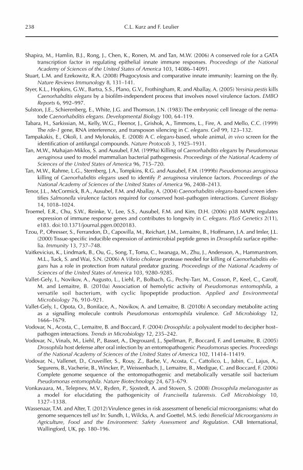
238 C.L. Kurz and F. Leulier
Shapira, M., Hamlin, B.J., Rong, J., Chen, K., Ronen, M. and Tan, M.W. (2006) A conserved role for a GATA transcription factor in regulating epithelial innate immune responses. Proceedings of the National Academy of Sciences of the United States of America 103, 14086–14091.
Stuart, L.M. and Ezekowitz, R.A. (2008) Phagocytosis and comparative innate immunity: learning on the fly. Nature Reviews Immunology 8, 131–141.
Styer, K.L., Hopkins, G.W., Bartra, S.S., Plano, G.V., Frothingham, R. and Aballay, A. (2005) Yersinia pestis kills Caenorhabditis elegans by a biofilm-independent process that involves novel virulence factors. EMBOReports 6, 992–997.
Sulston, J.E., Schierenberg, E., White, J.G. and Thomson, J.N. (1983) The embryonic cell lineage of the nema-tode Caenorhabditis elegans. Developmental Biology 100, 64–119.
Tabara, H., Sarkissian, M., Kelly, W.G., Fleenor, J., Grishok, A., Timmons, L., Fire, A. and Mello, C.C. (1999) The rde-1 gene, RNA interference, and transposon silencing in C. elegans. Cell 99, 123–132.
Tampakakis, E., Okoli, I. and Mylonakis, E. (2008) A C. elegans-based, whole animal, in vivo screen for the identification of antifungal compounds. Nature Protocols 3, 1925–1931.
Tan, M.W., Mahajan-Miklos, S. and Ausubel, F.M. (1999a) Killing of Caenorhabditis elegans by Pseudomonas aeruginosa used to model mammalian bacterial pathogenesis. Proceedings of the National Academy of Sciences of the United States of America 96, 715–720.
Tan, M.W., Rahme, L.G., Sternberg, J.A., Tompkins, R.G. and Ausubel, F.M. (1999b) Pseudomonas aeruginosakilling of Caenorhabditis elegans used to identify P. aeruginosa virulence factors. Proceedings of the National Academy of Sciences of the United States of America 96, 2408–2413.
Tenor, J.L., McCormick, B.A., Ausubel, F.M. and Aballay, A. (2004) Caenorhabditis elegans-based screen iden-tifies Salmonella virulence factors required for conserved host–pathogen interactions. Current Biology14, 1018–1024.
Troemel, E.R., Chu, S.W., Reinke, V., Lee, S.S., Ausubel, F.M. and Kim, D.H. (2006) p38 MAPK regulates expression of immune response genes and contributes to longevity in C. elegans. PLoS Genetics 2(11), e183. doi:10.1371/journal.pgen.0020183.
Tzou, P., Ohresser, S., Ferrandon, D., Capovilla, M., Reichart, J.M., Lemaitre, B., Hoffmann, J.A. and Imler, J.L. (2000) Tissue-specific inducible expression of antimicrobial peptide genes in Drosophila surface epithe-lia. Immunity 13, 737–748.
Vaitkevicius, K., Lindmark, B., Ou, G., Song, T., Toma, C., Iwanaga, M., Zhu, J., Andersson, A., Hammarstrom, M.L., Tuck, S. and Wai, S.N. (2006) A Vibrio cholerae protease needed for killing of Caenorhabditis ele-gans has a role in protection from natural predator grazing. Proceedings of the National Academy of Sciences of the United States of America 103, 9280–9285.
Vallet-Gely, I., Novikov, A., Augusto, L., Liehl, P., Bolbach, G., Pechy-Tarr, M., Cosson, P., Keel, C., Caroff, M. and Lemaitre, B. (2010a) Association of hemolytic activity of Pseudomonas entomophila, a versatile soil bacterium, with cyclic lipopeptide production. Applied and Environmental Microbiology 76, 910–921.
Vallet-Gely, I., Opota, O., Boniface, A., Novikov, A. and Lemaitre, B. (2010b) A secondary metabolite acting as a signalling molecule controls Pseudomonas entomophila virulence. Cell Microbiology 12, 1666–1679.
Vodovar, N., Acosta, C., Lemaitre, B. and Boccard, F. (2004) Drosophila: a polyvalent model to decipher host–pathogen interactions. Trends in Microbiology 12, 235–242.
Vodovar, N., Vinals, M., Liehl, P., Basset, A., Degrouard, J., Spellman, P., Boccard, F. and Lemaitre, B. (2005) Drosophila host defense after oral infection by an entomopathogenic Pseudomonas species. Proceedings of the National Academy of Sciences of the United States of America 102, 11414–11419.
Vodovar, N., Vallenet, D., Cruveiller, S., Rouy, Z., Barbe, V., Acosta, C., Cattolico, L., Jubin, C., Lajus, A., Segurens, B., Vacherie, B., Wincker, P., Weissenbach, J., Lemaitre, B., Medigue, C. and Boccard, F. (2006) Complete genome sequence of the entomopathogenic and metabolically versatile soil bacterium Pseudomonas entomophila. Nature Biotechnology 24, 673–679.
Vonkavaara, M., Telepnev, M.V., Ryden, P., Sjostedt, A. and Stoven, S. (2008) Drosophila melanogaster as a model for elucidating the pathogenicity of Francisella tularensis. Cell Microbiology 10, 1327–1338.
Wassenaar, T.M. and Alter, T. (2012) Virulence genes in risk assessment of beneficial microorganisms: what do genome sequences tell us? In: Sundh, I., Wilcks, A. and Goettel, M.S. (eds) Beneficial Microorganisms in Agriculture, Food and the Environment: Safety Assessment and Regulation. CAB International, Wallingford, UK, pp. 180–196.
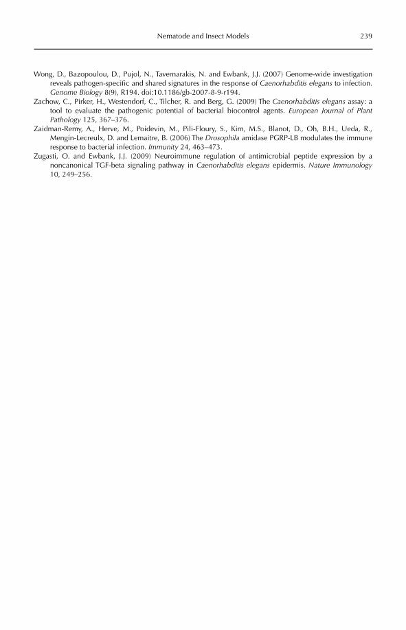
Nematode and Insect Models 239
Wong, D., Bazopoulou, D., Pujol, N., Tavernarakis, N. and Ewbank, J.J. (2007) Genome-wide investigation reveals pathogen-specific and shared signatures in the response of Caenorhabditis elegans to infection. Genome Biology 8(9), R194. doi:10.1186/gb-2007-8-9-r194.
Zachow, C., Pirker, H., Westendorf, C., Tilcher, R. and Berg, G. (2009) The Caenorhabditis elegans assay: a tool to evaluate the pathogenic potential of bacterial biocontrol agents. European Journal of Plant Pathology 125, 367–376.
Zaidman-Remy, A., Herve, M., Poidevin, M., Pili-Floury, S., Kim, M.S., Blanot, D., Oh, B.H., Ueda, R., Mengin-Lecreulx, D. and Lemaitre, B. (2006) The Drosophila amidase PGRP-LB modulates the immune response to bacterial infection. Immunity 24, 463–473.
Zugasti, O. and Ewbank, J.J. (2009) Neuroimmune regulation of antimicrobial peptide expression by a noncanonical TGF-beta signaling pathway in Caenorhabditis elegans epidermis. Nature Immunology10, 249–256.
©CAB International 2012. Beneficial Microorganisms in Agriculture, Food and the Environment: 240 Safety Assessment and Regulation (eds I. Sundh et al.)
17.1 Introduction 24017.2 Invertebrate Model Systems 242
17.2.1 Artemia salina (Crustacea: Branchiopoda) 242 17.2.2 Daphnia magna (Crustacea: Branchiopoda) 242 17.2.3 Paramecium caudatum and Tetrahymena pyriformis
(Ciliophora: Oligohymenophorea: Urocentrida and Tetrahymenida) 24217.3 Assessment of Acute Cytotoxicity of Fungal Metabolites 243
17 Assessing Potential Cytotoxicityof Biocontrol Microorganisms
Using Invertebrate Assays
Claudio Altomare,1 Barbara Pernfuss2 and Hermann Strasser2
1Institute of Sciences of Food Production, National Research Council (CNR), Italy; 2Institute of Microbiology, Leopold-Franzens University of Innsbruck, Austria
17.1 Introduction
There is increasing interest in the exploitation of microorganisms for the biological control of crop pests, weeds and diseases (Whipps and Davies, 2000; Butt et al., 2001). A number of biological pesticides (biopesticides, biolog-ical control agents or BCAs) containing fungi or bacteria as the active ingredient are already marketed in Europe, the USA and developing countries (Copping, 2009), while many other biocontrol strains or formulations are expec-ted to be placed on the world market in the next few years. However, many microorgan-isms are known to produce bioactive second-ary metabolites, including toxins that may be harmful to humans and animals (Cole and Cox, 1981; Cole and Schweikert, 2003; Strasser et al., 2011). In particular, some fungal micro-biological control agents (MBCAs) have been reported to secrete biologically active com-pounds (Vey et al., 2001), which might be
potentially toxic when present in formulated products, crops or the environment. Therefore, risk assessment procedures, including hazard characterization of MBCA metabolites, are required before the introduction and use of MBCAs (Blum et al., 2003; Strasser et al., 2008; Strauch et al., 2011). In Europe, the European Parliament and Council Regulation 1107/ 2009/EEC (OJEU, 2009), repealing Council Directives 79/117/EEC and 91/414/EEC, identifies the dossier requirements for autho-rization of the production and marketing of pesticides, including those whose active sub-stance is a MBCA. In particular, the regula-tion requires the provision of information on short-term cytotoxicity, as well as ecotoxicity, of any relevant metabolites (i.e. metabolites of toxicological, ecotoxicological and/or envi-ronmental concern) formed by the BCA.
The technical, economic and ethical issues related to the use of mammals for toxicity test-ing have prompted the search for in vitro tests
Assessing Potential Cytotoxicity 241
that can, at least at some stages of toxicologi-cal assessment, replace tests on laboratory animals (Lilienblum et al., 2008; Andersen and Krewski, 2009). Cell lines or tissue cultures are valid predictive toxicology systems that can be used in the study of bioactive compounds. These tests are particularly useful to gain information about the mechanisms of cyto-toxicity or genotoxicity of a substance, such as the destruction of cell membranes, apopto-sis, cytolysis, oxidative stress, impairment of mitochondrial function, cell proliferation, quantification of DNA damage and repair, and the analysis of micronuclei. However, in many cases such detailed information is not essential in the early stages of development of a product, when what is urgently needed is a qualitative response of the type toxic/ non-toxic in order to identify a potential toxi-cological risk and drive the decision process. In these cases, toxicity tests that are simpler and less expensive than cell line or tissue assays, but have comparable predictive value, are highly desirable (Eisenbrand et al., 2002; Lilienblum et al., 2008). Ideally, the results of these alternative cytotoxicity assays should have high correlation with those of both cell line tests and in vivo tests in order to allow predictive inference of toxic effect.
Recent years have seen the development of a number of toxicity tests in which the response has been measured in simple eukary-otic organisms, such as protozoa (Gerhardt et al., 2010) and arthropods (Calow, 1993; Lagadic and Caquet, 1998). These tests offer some advan-tages over other in vitro toxicity tests, which can be summarized in the following points:
Test organisms can be cultured under •laboratory conditions at low cost.High fecundity and short lifespan of test •animals.Animals can be hatched synchronously •from dormant eggs (arthropods).Low genetic variability among •individuals.Tests give an indication of potential •adverse effects on the environment (eco-logical relevance).Kits are often available for better stand-•ardization and higher repeatability and reproducibility of tests.
These characteristics of invertebrates account for their extensive use in toxicity tests. Many invertebrate species can be cul-tured easily under laboratory conditions because of their small size, high fecundity and short lifespan. This makes the mainte-nance of invertebrates and the simultaneous breeding of various species easier than for vertebrates. The handling of the animals is also easy and therefore the number of indi-viduals exposed to each tested concentration or dose may be increased. This improves the statistical significance of test results without a significant increase in cost.
Many invertebrate test organisms form dormant eggs (cysts) that remain viable for a long time. Cyst-based toxicity tests elimi-nate the need for stock culturing of test spe-cies. Animals can be hatched synchronously, the neonates originate from genetically defined stocks and are of the same age and in the same physiological condition. In addition, some invertebrate species (e.g. Daphnia magna)are parthenogenetic and genetic variability is thereby reduced. As a result, standardization of the test and reproducibility of the results are significantly enhanced.
Finally, invertebrates occupy key posi-tions in the food webs of both aquatic and terrestrial ecosystems. Some species (e.g. daph nids, paramecia) are present throughout a wide range of habitats. Therefore, they are ‘biosensors’ for the potential ecotoxicological risks associated with the presence of chemicals and toxicants in the environment (Lagadic and Caquet, 1998). Invertebrates are already used in tests that are required by some regu-latory authorities for the environmental risk assessment of pesticides, chemicals and pol-lutants (OJEC, 1992; US EPA, 2002).
Without the ambition to offer a compre-hensive review on the use of invertebrate systems for toxicity testing, on which topic an extensive literature and several critical reviews are already available (Calow, 1993; Lagadic and Caquet, 1998; Sauvant et al., 1999), in this chapter we will show four examples of how invertebrate test organisms, namely Artemia salina, Daphnia magna, Parameciumcaudatum and Tetrahymena pyriformis, have been successfully used for the toxicological assessment of the metabolites of fungal BCAs.
242 C. Altomare et al.
As Appendix 1 to this chapter, we include one developed standard operating proce-dure for P. caudatum to provide guidance for a modern approach to MBCA metabolite tox-icity testing.
17.2 Invertebrate model systems
17.2.1 Artemia salina (Crustacea: Branchiopoda)
Artemia salina is an anostracan crustacean, commonly known as the brine shrimp, which is widely distributed in tropical, subtropical and temperate climate regions. Brine shrimps are inhabitants of extreme saline water envi-ronments, although they have great powers of adaptation and are able to tolerate physio-logically large changes in salinity. A. salinaacute toxicity tests have often been used for ecotoxicological studies of xenobiotic sub-stances (Nunes et al., 2006), as well as for the toxicological screening of chemicals (Barahona and Sánchez-Fortún, 1999; Cleuvers, 2003) and natural compounds (Solis et al., 1993; Caldwell et al., 2003), including mycotoxins (Harwig and Scott, 1971; Schmidt, 1989; Hartl and Humpf, 2000). Acute toxicity to A. salinahas been reported to be correlated with cyto-toxicity, as the following examples indicate. McLaughlin et al. (1993) found a correlation between brine shrimp kill and the cytotoxic-ity of plant extracts to 9-KB (human nasopha-ryngeal carcinoma) cells. Solis et al. (1993) found the A. salina test predictive of KB cell cytotoxicity, except for compounds requir-ing metabolic activation, because brine shrimps lack the necessary cytochrome P-450 enzyme. Logrieco et al. (1996) reported that the median cytotoxic concentration (CC50) of the fungal toxin fusaproliferin to the human B-lymphocyte cell line IARC/LCL 171 and the insect cell line SF-9 were approximately the same as the median lethal concentration (LC50) found in the A. salina assay. Moreover, Lagarto Parra et al. (2001) found a strong correlation (r = 0.85, P < 0.05) between the results of in vivo tests on mice and in vitrotests with A. salina of various plant extracts. The authors concluded that the A. salina
bioassay was a useful tool for prediction of the oral acute toxicity of plant extracts in mammals.
17.2.2 Daphnia magna (Crustacea: Branchiopoda)
Daphnia magna (the water flea) is a freshwa-ter cladoceran crustacean. Cladocerans can reproduce asexually by parthenogenesis, without male fertilization. This is one of the main arguments that led to the widespread use of Daphnia spp. in toxicity testing. D. magnahas been used for a long time as a laboratory animal for testing ecotoxicity (Anderson, 1944). Other Daphnia species than D. magnamay occasionally be used (Lilius et al., 1995), but laboratories mostly use D. magna as the standard. D. magna is specified to be used in the OECD Guidelines for the Testing of Chemicals(OECD, 2004, 2008). The OECD Test No. 202 is a 48 h acute toxicity test in which young Daphnia are exposed to varying concentra-tions of the test substance and the (effective) concentration estimated to immobilize 50% of the daphnids (EC50) is determined. It has been reported for a number of compounds that the toxicity data of Daphnia tests correlate well with toxicity to vertebrates, as has also been shown for A. salina. For instance, correlation between toxicity to Daphnia and toxicity to fish has been shown for a group of 42 indus-trial chemicals (Walker et al., 1991). Also, a strong correlation between the acute toxicityof 54 different compounds to D. magna and rats was found by Guilhermino et al. (2000).
17.2.3 Paramecium caudatumand Tetrahymena pyriformis (Ciliophora:
Oligohymenophorea: Urocentrida and Tetrahymenida)
Paramecium caudatum and Tetrahymena pyri-formis are unicellular ciliate protozoa which are widespread in freshwater systems. They have been extensively used in the last decade as tools for toxicological assessments because they have almost the same metabolic systems as higher animals (Hauser, 1990). In addition,
Assessing Potential Cytotoxicity 243
they can be purchased from most of the prominent strain culture collections of proto-zoa, and can be easily grown in unsterile liq-uid culture at room temperature. Members of both Tetrahymena and Paramecium have been reported to be highly sensitive to a number of xenobiotic substances and have been used as sensitive eukaryotic cells to detect potential (eco)toxic substances (Komala, 1982, 1984). T. pyriformis is more commonly used in toxi-cological studies and therefore more toxicity data are currently available for this species than for P. caudatum.
Tetrahymena species have been used as model organisms in scientific and industrial research (Pauli et al., 1993; Pauli and Berger, 1997). Uma et al. (2008) demonstrated that T. pyriformis is a useful tool to assess the acute toxicity of heterocyclic compounds. Skrobek et al. (2006) reported that T. pyriformis was more sensitive to the synthetic pesticide amidosulfuron than rats. In studies on the acute toxicity of the mycotoxin patulin, com-parable results in rats and in T. pyriformiswere obtained by Escoula et al. (1977) and by McKinley and Carlton (1980).
P. caudatum has been successfully used to study the toxicity of the mycotoxin pro-duced by the basidiomycete Hebeloma edurum(Pöder, 1982) and also that of other fungal secondary metabolites (Stemer, 2004; Gierner, 2005). Mijatov (1975) reported a good corre-lation between the sensitivity of P. caudatumand that of white mice, guinea pigs and rabbit skin treated with water extracts of maize or peanut meal colonized by Fusarium gramine-arum and F. poae. Hussain et al. (1985) vali-dated P. caudatum as a biosensor by assessing the sensitivity of this organism to different pesticides. Miyoshi et al. (2003) demonstrated the high sensitivity of P. caudatum to a number of organic solvents and heavy metals, but also to mutagenic, cancerogenic and terato-genic substances.
17.3 Assessment of Acute Cytotoxicity of Fungal Metabolites
The simplification of risk assessment pro-cedures for fungal MBCAs and their rele-vant metabolites without compromising high
levels of safety was the main objective of the EU-funded project ‘Risk Assessment of Fungal Biological Control Agents’ (RAFBCA, QLKl-2001-01391) that took place from 2001 to 2004. This project was the first attempt to address scientifically the issue of risk assess-ment of MBCAs, which process has greatly hindered the registration of fungal biopesti-cides. Indeed, the risk assessment procedures requested by Directive 91/414/EEC for the registration of MBCAs and derived products had been tailored for chemicals and extended, almost unmodified, to biological agents and their metabolites. Among biocontrol stake-holders there was a general consensus that such requirements, hardly affordable for the small and medium-sized enterprises involved in biopesticide production, were a major hur-dle in the registration and subsequent com-mercialization of MBCAs. Therefore, one of the goals of the project was to identify sensi-tive biosensors that could be used for assess-ment of the cytotoxicity of metabolites of fungal MBCAs with a significant simplifica-tion of procedures and reduction of cost.
In the RAFBCA project, A. salina, D. magna,P. caudatum and T. pyriformis were used to assess the acute toxicity of seven bioactive fungal metabolites (Tables 17.1 and 17.2) with different molecular structures and modes of action (Pernfuss et. al., 2003; Stemer, 2004; Gierner, 2005; Favilla et al., 2006; Skrobek et al.,2006). Those unique mycotoxins are pro-duced by mycoparasitic (Gliocladium spp., Tricho derma spp.), entomopathogenic (Beauveria brongniartii, Metarhizium anisopliae) or phyto-pathogenic (Stagonospora spp.) fungi used for biological control of fungal plant diseases, insect pests or weeds, respectively, and had not been previously evaluated in the above invertebrate models.
For the A. salina assays, materials for aquarists, commercially available at low cost, were used. For the higher levels of standardi-zation and reproducibility of D. magna assays, a laboratory kit (Daphtoxkit F, Creasel BVBA, Deinze, Belgium) was used with some modifi-cation of the protocol to minimize the amount of the test compounds needed (Favilla et al.,2006). Both crustaceans proved to be sensi-tive to all the fungal MBCA metabolites tested, regardless of the chemical nature of the
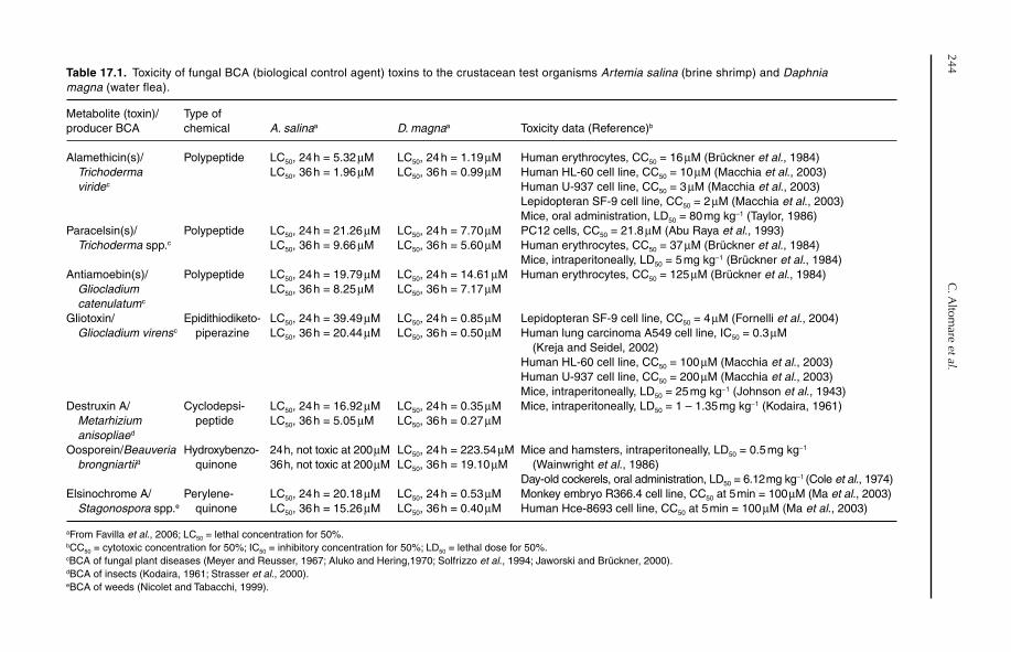
244C
. Altom
are et al.Table 17.1. Toxicity of fungal BCA (biological control agent) toxins to the crustacean test organisms Artemia salina (brine shrimp) and Daphnia magna (water flea).
Metabolite (toxin)/ producer BCA
Type of chemical A. salinaa D. magnaa Toxicity data (Reference)b
Alamethicin(s)/Trichoderma
viride c
Polypeptide LC50, 24 h = 5.32 µMLC50, 36 h = 1.96 µM
LC50, 24 h = 1.19 µMLC50, 36 h = 0.99 µM
Human erythrocytes, CC50 = 16 µM (Brückner et al., 1984)Human HL-60 cell line, CC50 = 10 µM (Macchia et al., 2003)Human U-937 cell line, CC50 = 3 µM (Macchia et al., 2003)Lepidopteran SF-9 cell line, CC50 = 2 µM (Macchia et al., 2003)Mice, oral administration, LD50 = 80 mg kg−1 (Taylor, 1986)
Paracelsin(s)/Trichoderma spp.c
Polypeptide LC50, 24 h = 21.26 µMLC50, 36 h = 9.66 µM
LC50, 24 h = 7.70 µMLC50, 36 h = 5.60 µM
PC12 cells, CC50 = 21.8 µM (Abu Raya et al., 1993)Human erythrocytes, CC50 = 37 µM (Brückner et al., 1984)Mice, intraperitoneally, LD50 = 5 mg kg−1 (Brückner et al., 1984)
Antiamoebin(s)/Gliocladium
catenulatum c
Polypeptide LC50, 24 h = 19.79 µMLC50, 36 h = 8.25 µM
LC50, 24 h = 14.61 µMLC50, 36 h = 7.17 µM
Human erythrocytes, CC50 = 125 µM (Brückner et al., 1984)
Gliotoxin/Gliocladium virens c
Epidithiodiketo- piperazine
LC50, 24 h = 39.49 µMLC50, 36 h = 20.44 µM
LC50, 24 h = 0.85 µMLC50, 36 h = 0.50 µM
Lepidopteran SF-9 cell line, CC50 = 4 µM (Fornelli et al., 2004)Human lung carcinoma A549 cell line, IC50 = 0.3 µM (Kreja and Seidel, 2002)Human HL-60 cell line, CC50 = 100 µM (Macchia et al., 2003)Human U-937 cell line, CC50 = 200 µM (Macchia et al., 2003)Mice, intraperitoneally, LD50 = 25 mg kg−1 (Johnson et al., 1943)
Destruxin A/Metarhizium
anisopliae d
Cyclodepsi- peptide
LC50, 24 h = 16.92 µMLC50, 36 h = 5.05 µM
LC50, 24 h = 0.35 µMLC50, 36 h = 0.27 µM
Mice, intraperitoneally, LD50 = 1 – 1.35 mg kg−1 (Kodaira, 1961)
Oosporein/Beauveria brongniartii d
Hydroxybenzo- quinone
24 h, not toxic at 200 µM36 h, not toxic at 200 µM
LC50, 24 h = 223.54 µMLC50, 36 h = 19.10 µM
Mice and hamsters, intraperitoneally, LD50 = 0.5 mg kg−1
(Wainwright et al., 1986)Day-old cockerels, oral administration, LD50 = 6.12 mg kg−1 (Cole et al., 1974)
Elsinochrome A/Stagonospora spp.e
Perylene- quinone
LC50, 24 h = 20.18 µMLC50, 36 h = 15.26 µM
LC50, 24 h = 0.53 µMLC50, 36 h = 0.40 µM
Monkey embryo R366.4 cell line, CC50 at 5 min = 100 µM (Ma et al., 2003)Human Hce-8693 cell line, CC50 at 5 min = 100 µM (Ma et al., 2003)
aFrom Favilla et al., 2006; LC50 = lethal concentration for 50%.bCC50 = cytotoxic concentration for 50%; IC50 = inhibitory concentration for 50%; LD50 = lethal dose for 50%.cBCA of fungal plant diseases (Meyer and Reusser, 1967; Aluko and Hering,1970; Solfrizzo et al., 1994; Jaworski and Brückner, 2000).dBCA of insects (Kodaira, 1961; Strasser et al., 2000).eBCA of weeds (Nicolet and Tabacchi, 1999).
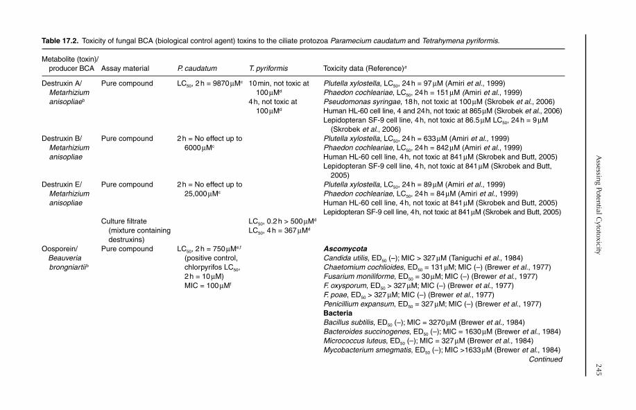
Assessing Potential C
ytotoxicity245
Table 17.2. Toxicity of fungal BCA (biological control agent) toxins to the ciliate protozoa Paramecium caudatum and Tetrahymena pyriformis.
Metabolite (toxin)/ producer BCA Assay material P. caudatum T. pyriformis Toxicity data (Reference)a
Destruxin A/Metarhizium
anisopliae b
Pure compound LC50, 2 h = 9870 µMc 10 min, not toxic at 100 µMd
4 h, not toxic at 100 µMd
Plutella xylostella, LC50, 24 h = 97 µM (Amiri et al., 1999)Phaedon cochleariae, LC50, 24 h = 151 µM (Amiri et al., 1999)Pseudomonas syringae, 18 h, not toxic at 100 µM (Skrobek et al., 2006)Human HL-60 cell line, 4 and 24 h, not toxic at 865 µM (Skrobek et al., 2006)Lepidopteran SF-9 cell line, 4 h, not toxic at 86.5 µM LC50, 24 h = 9 µM (Skrobek et al., 2006)
Destruxin B/Metarhizium
anisopliae
Pure compound 2 h = No effect up to 6000 µMc
Plutella xylostella, LC50, 24 h = 633 µM (Amiri et al., 1999)Phaedon cochleariae, LC50, 24 h = 842 µM (Amiri et al., 1999)Human HL-60 cell line, 4 h, not toxic at 841 µM (Skrobek and Butt, 2005)Lepidopteran SF-9 cell line, 4 h, not toxic at 841 µM (Skrobek and Butt, 2005)
Destruxin E/Metarhizium
anisopliae
Pure compound 2 h = No effect up to 25,000 µMc
Plutella xylostella, LC50, 24 h = 89 µM (Amiri et al., 1999)Phaedon cochleariae, LC50, 24 h = 84 µM (Amiri et al., 1999)Human HL-60 cell line, 4 h, not toxic at 841 µM (Skrobek and Butt, 2005)Lepidopteran SF-9 cell line, 4 h, not toxic at 841 µM (Skrobek and Butt, 2005)
Culture filtrate (mixture containing destruxins)
LC50, 0.2 h > 500 µMd
LC50, 4 h = 367 µMd
Oosporein/Beauveria
brongniartii b
Pure compound LC50, 2 h = 750 µMe,f
(positive control, chlorpyrifos LC50, 2 h = 10 µM) MIC = 100 µMf
AscomycotaCandida utilis, ED50 (–); MIC > 327 µM (Taniguchi et al., 1984)Chaetomium cochlioides, ED50 = 131 µM; MIC (–) (Brewer et al., 1977)Fusarium moniliforme, ED50 = 30 µM; MIC (–) (Brewer et al., 1977)F. oxysporum, ED50 > 327 µM; MIC (–) (Brewer et al., 1977)F. poae, ED50 > 327 µM; MIC (–) (Brewer et al., 1977)Penicillium expansum, ED50 = 327 µM; MIC (–) (Brewer et al., 1977)BacteriaBacillus subtilis, ED50 (–); MIC = 3270 µM (Brewer et al., 1984)Bacteroides succinogenes, ED50 (–); MIC = 1630 µM (Brewer et al., 1984)Micrococcus luteus, ED50 (–); MIC = 327 µM (Brewer et al., 1984)Mycobacterium smegmatis, ED50 (–); MIC >1633 µM (Brewer et al., 1984)
Continued
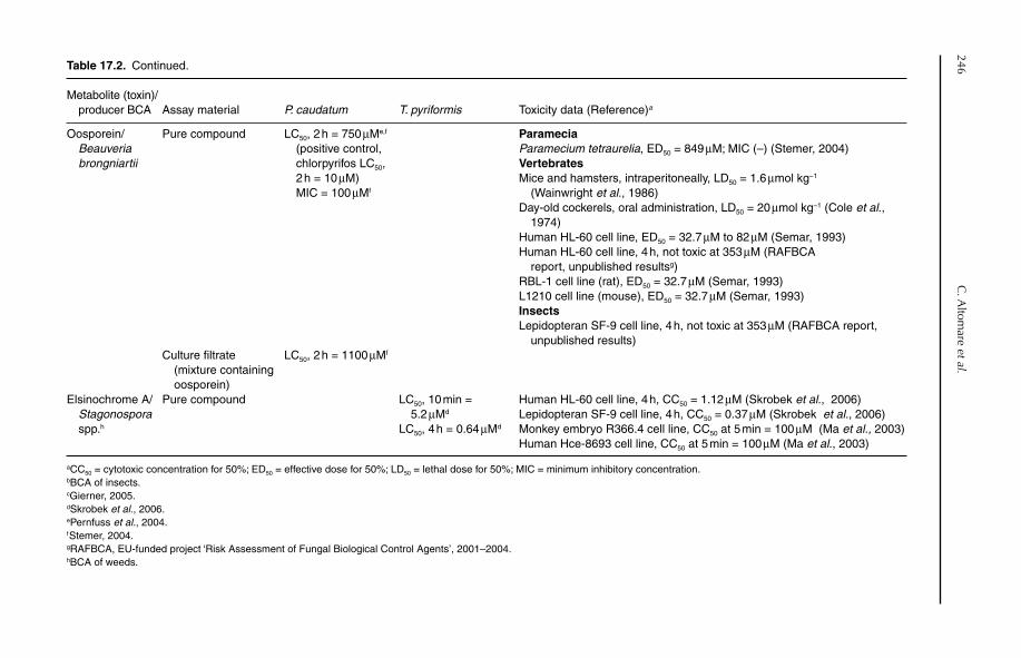
246C
. Altom
are et al.Table 17.2. Continued.
Metabolite (toxin)/ producer BCA Assay material P. caudatum T. pyriformis Toxicity data (Reference)a
Oosporein/Beauveria
brongniartii
Pure compound LC50, 2 h = 750 µMe,f
(positive control, chlorpyrifos LC50, 2 h = 10 µM) MIC = 100 µMf
ParameciaParamecium tetraurelia, ED50 = 849 µM; MIC (–) (Stemer, 2004)VertebratesMice and hamsters, intraperitoneally, LD50 = 1.6 µmol kg−1
(Wainwright et al., 1986)Day-old cockerels, oral administration, LD50 = 20 µmol kg−1 (Cole et al., 1974)Human HL-60 cell line, ED50 = 32.7 µM to 82 µM (Semar, 1993)Human HL-60 cell line, 4 h, not toxic at 353 µM (RAFBCA report, unpublished resultsg)RBL-1 cell line (rat), ED50 = 32.7 µM (Semar, 1993)L1210 cell line (mouse), ED50 = 32.7 µM (Semar, 1993)InsectsLepidopteran SF-9 cell line, 4 h, not toxic at 353 µM (RAFBCA report, unpublished results)
Culture filtrate (mixture containing oosporein)
LC50, 2 h = 1100 µMf
Elsinochrome A/Stagonospora
spp.h
Pure compound LC50, 10 min = 5.2 µMd
LC50, 4 h = 0.64 µMd
Human HL-60 cell line, 4 h, CC50 = 1.12 µM (Skrobek et al., 2006)Lepidopteran SF-9 cell line, 4 h, CC50 = 0.37 µM (Skrobek et al., 2006)Monkey embryo R366.4 cell line, CC50 at 5 min = 100 µM (Ma et al., 2003)Human Hce-8693 cell line, CC50 at 5 min = 100 µM (Ma et al., 2003)
aCC50 = cytotoxic concentration for 50%; ED50 = effective dose for 50%; LD50 = lethal dose for 50%; MIC = minimum inhibitory concentration.bBCA of insects.cGierner, 2005.dSkrobek et al., 2006.ePernfuss et al., 2004.fStemer, 2004.gRAFBCA, EU-funded project ‘Risk Assessment of Fungal Biological Control Agents’, 2001–2004.hBCA of weeds.
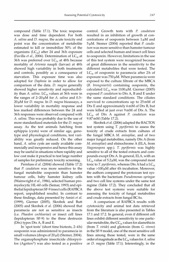
Assessing Potential Cytotoxicity 247
compound (Table 17.1). The toxic response was dose and time dependent. For both A. salina and D. magna, the acute toxicity end point was the concentration of metabolite estimated to kill or immobilize 50% of the organisms (LC50) after 24 and 36 h exposure (Favilla et al., 2006). Determination of LC50 at 36 h was preferred over LC50 at 48 h because mortality of Artemia naupli (larvae) at 48 h showed high variability in both treatments and controls, possibly as a consequence of starvation. This exposure time was also adopted for Daphnia in order to allow for comparison of the data. D. magna generally showed higher sensitivity and reproducibil-ity than A. salina. LC50 values at 36 h were in the ranges of 2–20 mM for A. salina and 0.3–20mM for D. magna. In D. magna bioassays, a lower variability in mortality response and less marked differences between the 24 and 36 h responses were observed compared with A. salina. This was probably due to the use of more standardized materials for the D. magnaassay. As the test animals hatching from ephippia (cysts) were of similar age, geno-type and physiological conditions, test vari-ability was greatly reduced. On the other hand, A. salina cysts are easily available com-mercially and inexpensive and hence this assay may be useful in situations where rapidity and low cost make it practical to test large number of samples for preliminary toxicity screening.
Pernfuss et al. (2004) showed (Table 17.2) that P. caudatum was more sensitive to the fungal metabolite oosporein than hamster tumour cells, baby hamster kidney cells (Wainwright et al., 1986), selected human pro-myelocytic HL-60 cells (Semar, 1993) and epi-thelial lepidopteran SF-9 insect cells (RAFBCA report, unpublished results). In contrast to these findings, data presented by Amiri et al.(1999), Gierner (2005), Skrobek and Butt (2005) and Skrobek et al. (2006) showed that paramecia are not as sensitive as insects (i.e. Phaedon cochleariae) or insect cell lines (lepidopteran SF-9) to the three destruxin (Dtx) types Dtx A, B and E.
In ‘spot tests’ (short time biotests, 2–4 h) oosporein was administered to paramecia in small volumes (drops of 20 ml) (Stemer, 2004). The organophosphate insecticide chlorpyri-fos (Agritox®) was also tested as a positive
control. Growth tests with P. caudatumresulted in an inhibition of growth at con-centrations of oosporein between 1 mM and 5 mM. Stemer (2004) reported that P. cauda-tum was more sensitive than hamster tumour cells and selected human and insect cell lines to oosporein. However, limitations in the use of this test system were recognized because of great differences in the sensitivity to the different metabolites that were tested. The LC50 of oosporein to paramecia after 2 h of exposure was 750 mM. When paramecia were exposed to the culture filtrate of the MBCA (B. brongniartii) containing oosporein, the calculated LC50 was 1100 mM. Gierner (2005) exposed P. caudatum to Dtx A, B and E under the same standard conditions. The ciliates survived to concentrations up to 25 mM of Dtx E and approximately 6 mM of Dtx B, but were killed at just over 9 mM of Dtx A. The LC50 of Dtx A against P. caudatum was 9.87 mM (Table 17.2).
Skrobek et al. (2006) adapted the BACTOX test system using T. pyriformis to assess the toxicity of crude extracts from cultures of the fungal MBCA M. anisopliae, and of two major fungal metabolites, namely Dtx A (from M. anisopliae) and elsinochrome A (ELA, from Stagonospora spp.). T. pyriformis was highly sensitive to all of the tested extracts and com-pounds except Dtx A. In general, ELA, with an LC50 value of 5.2 mM, was the compound most toxic to T. pyriformis, whereas Dtx A had a LC50
value >100 mM after 4 h incubation. Moreover, the authors compared the protozoan test sys-tem with the bacterium Pseudomonas syringaeand two cell line systems under the same test regime (Table 17.2). They concluded that all the above test systems were suitable for assessing the toxicity of fungal metabolites and crude extracts from fungal BCAs.
A comparison of RAFBCA results with cytotoxicity and animal test data retrieved from the literature is also presented in Tables 17.1 and 17.2. In general, even if different cell lines exhibit different sensitivity to one partic-ular metabolite, the CC50 values for alamethicin (from T. viride) and gliotoxin (from G. virens)in the SF-9 model, one of the most sensitive cell lines among those tested, were in the same order of magnitude as the LC50 values for A. salinaor D. magna (Table 17.1). Interestingly, in the
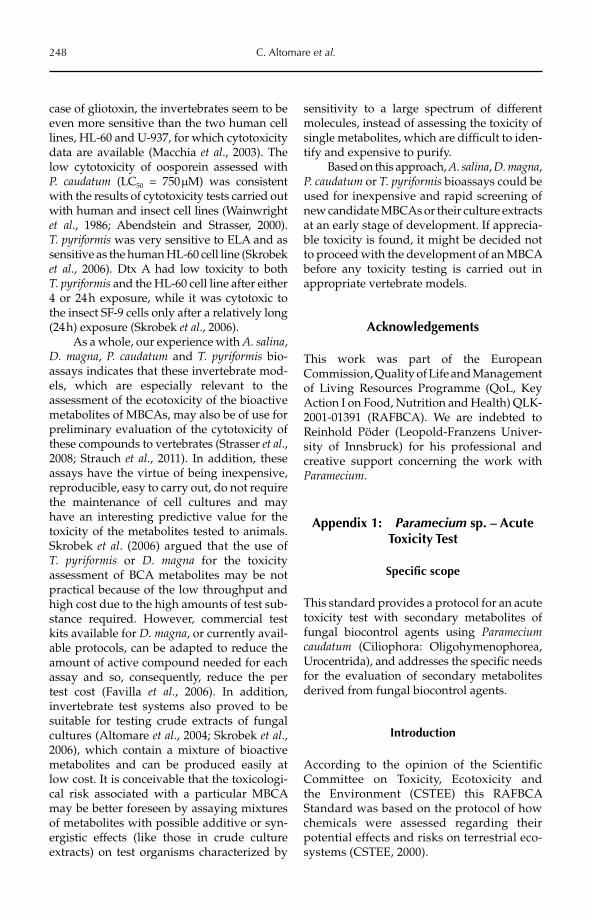
248 C. Altomare et al.
case of gliotoxin, the invertebrates seem to be even more sensitive than the two human cell lines, HL-60 and U-937, for which cytotoxicity data are available (Macchia et al., 2003). The low cytotoxicity of oosporein assessed with P. caudatum (LC50 = 750 mM) was consistent with the results of cytotoxicity tests carried out with human and insect cell lines (Wainwright et al., 1986; Abendstein and Strasser, 2000). T. pyriformis was very sensitive to ELA and as sensitive as the human HL-60 cell line (Skrobek et al., 2006). Dtx A had low toxicity to both T. pyriformis and the HL-60 cell line after either 4 or 24 h exposure, while it was cytotoxic to the insect SF-9 cells only after a relatively long (24 h) exposure (Skrobek et al., 2006).
As a whole, our experience with A. salina,D. magna, P. caudatum and T. pyriformis bio-assays indicates that these invertebrate mod-els, which are especially relevant to the assessment of the ecotoxicity of the bioactive metabolites of MBCAs, may also be of use for preliminary evaluation of the cytotoxicity of these compounds to vertebrates (Strasser et al., 2008; Strauch et al., 2011). In addition, these assays have the virtue of being inexpensive, reproducible, easy to carry out, do not require the maintenance of cell cultures and may have an interesting predictive value for the toxicity of the metabolites tested to animals. Skrobek et al. (2006) argued that the use of T. pyriformis or D. magna for the toxicity assessment of BCA metabolites may be not practical because of the low throughput and high cost due to the high amounts of test sub-stance required. However, commercial test kits available for D. magna, or currently avail-able protocols, can be adapted to reduce the amount of active compound needed for each assay and so, consequently, reduce the per test cost (Favilla et al., 2006). In addition, invertebrate test systems also proved to be suitable for testing crude extracts of fungal cultures (Altomare et al., 2004; Skrobek et al.,2006), which contain a mixture of bioactive metabolites and can be produced easily at low cost. It is conceivable that the toxicologi-cal risk associated with a particular MBCA may be better foreseen by assaying mixtures of metabolites with possible additive or syn-ergistic effects (like those in crude culture extracts) on test organisms characterized by
sensitivity to a large spectrum of different molecules, instead of assessing the toxicity of single metabolites, which are difficult to iden-tify and expensive to purify.
Based on this approach, A. salina, D. magna,P. caudatum or T. pyriformis bioassays could be used for inexpensive and rapid screening of new candidate MBCAs or their culture extracts at an early stage of development. If apprecia-ble toxicity is found, it might be decided not to proceed with the development of an MBCA before any toxicity testing is carried out in appropriate vertebrate models.
Acknowledgements
This work was part of the European Commission, Quality of Life and Management of Living Resources Programme (QoL, Key Action I on Food, Nutrition and Health) QLK-2001-01391 (RAFBCA). We are indebted to Reinhold Pöder (Leopold-Franzens Univer-sity of Innsbruck) for his professional and creative support concerning the work with Paramecium.
Appendix 1: Paramecium sp. – Acute Toxicity Test
Specific scope
This standard provides a protocol for an acute toxicity test with secondary metabolites of fungal biocontrol agents using Parameciumcaudatum (Ciliophora: Oligohymenophorea, Urocentrida), and addresses the specific needs for the evaluation of secondary metabolites derived from fungal biocontrol agents.
Introduction
According to the opinion of the Scientific Committee on Toxicity, Ecotoxicity and the Environment (CSTEE) this RAFBCA Standard was based on the protocol of how chemicals were assessed regarding their potential effects and risks on terrestrial eco-systems (CSTEE, 2000).
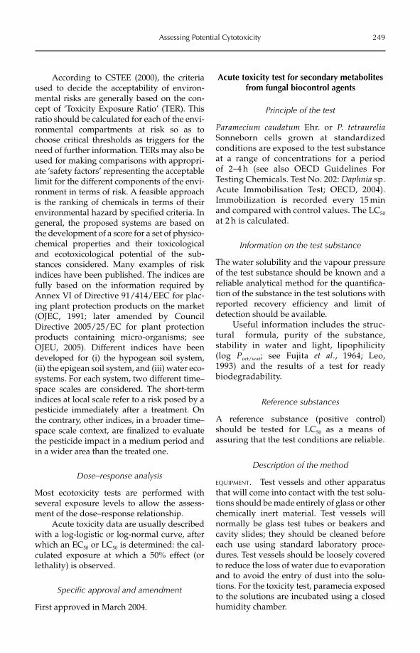
Assessing Potential Cytotoxicity 249
According to CSTEE (2000), the criteria used to decide the acceptability of environ-mental risks are generally based on the con-cept of ‘Toxicity Exposure Ratio’ (TER). This ratio should be calculated for each of the envi-ronmental compartments at risk so as to choose critical thresholds as triggers for the need of further information. TERs may also be used for making comparisons with appropri-ate ‘safety factors’ representing the acceptable limit for the different components of the envi-ronment in terms of risk. A feasible approach is the ranking of chemicals in terms of their environmental hazard by specified criteria. In general, the proposed systems are based on the development of a score for a set of physico-chemical properties and their toxicological and ecotoxicological potential of the sub-stances considered. Many examples of risk indices have been published. The indices are fully based on the information required by Annex VI of Directive 91/414/EEC for plac-ing plant protection products on the market (OJEC, 1991; later amended by Council Directive 2005/25/EC for plant protection products containing micro-organisms; see OJEU, 2005). Different indices have been developed for (i) the hypogean soil system, (ii) the epigean soil system, and (iii) water eco-systems. For each system, two different time–space scales are considered. The short-term indices at local scale refer to a risk posed by a pesticide immediately after a treatment. On the contrary, other indices, in a broader time–space scale context, are finalized to evaluate the pesticide impact in a medium period and in a wider area than the treated one.
Dose–response analysis
Most ecotoxicity tests are performed with several exposure levels to allow the assess-ment of the dose–response relationship.
Acute toxicity data are usually described with a log-logistic or log-normal curve, after which an EC50 or LC50 is determined: the cal-culated exposure at which a 50% effect (or lethality) is observed.
Specific approval and amendment
First approved in March 2004.
Acute toxicity test for secondary metabolites from fungal biocontrol agents
Principle of the test
Paramecium caudatum Ehr. or P. tetraureliaSonneborn cells grown at standardized conditions are exposed to the test substance at a range of concentrations for a period of 2–4 h (see also OECD Guidelines For Testing Chemicals. Test No. 202: Daphnia sp. Acute Immobilisation Test; OECD, 2004). Immobilization is recorded every 15 min and compared with control values. The LC50
at 2 h is calculated.
Information on the test substance
The water solubility and the vapour pressure of the test substance should be known and a reliable analytical method for the quantifica-tion of the substance in the test solutions with reported recovery efficiency and limit of detection should be available.
Useful information includes the struc-tural formula, purity of the substance, stability in water and light, lipophilicity (log Poct/wat; see Fujita et al., 1964; Leo, 1993) and the results of a test for ready biodegradability.
Reference substances
A reference substance (positive control) should be tested for LC50 as a means of assuring that the test conditions are reliable.
Description of the method
EQUIPMENT. Test vessels and other apparatus that will come into contact with the test solu-tions should be made entirely of glass or other chemically inert material. Test vessels will normally be glass test tubes or beakers and cavity slides; they should be cleaned before each use using standard laboratory proce-dures. Test vessels should be loosely covered to reduce the loss of water due to evaporation and to avoid the entry of dust into the solu-tions. For the toxicity test, paramecia exposed to the solutions are incubated using a closed humidity chamber.
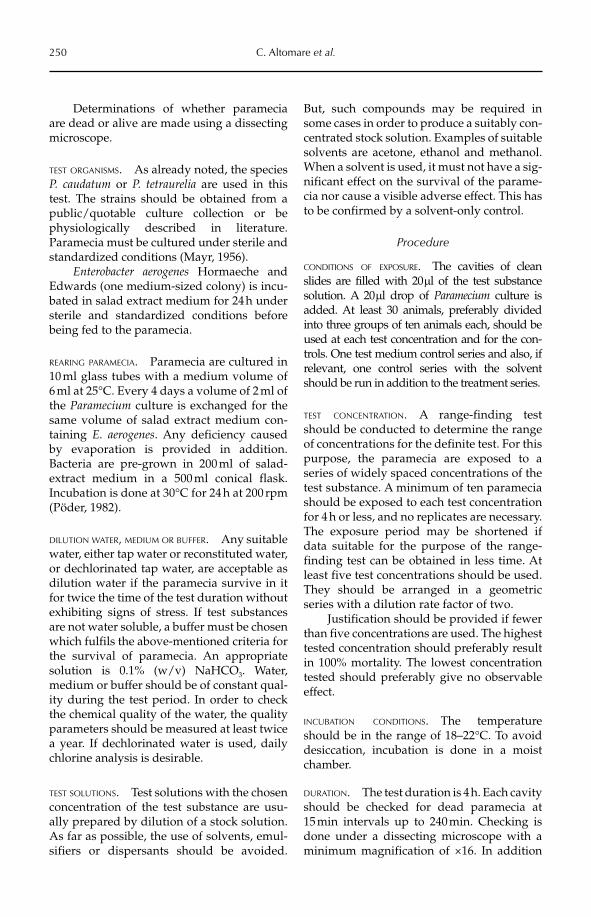
250 C. Altomare et al.
Determinations of whether paramecia are dead or alive are made using a dissecting microscope.
TEST ORGANISMS. As already noted, the species P. caudatum or P. tetraurelia are used in this test. The strains should be obtained from a public/quotable culture collection or be physiologically described in literature. Paramecia must be cultured under sterile and standardized conditions (Mayr, 1956).
Enterobacter aerogenes Hormaeche and Edwards (one medium-sized colony) is incu-bated in salad extract medium for 24 h under sterile and standardized conditions before being fed to the paramecia.
REARING PARAMECIA. Paramecia are cultured in 10 ml glass tubes with a medium volume of 6 ml at 25°C. Every 4 days a volume of 2 ml of the Paramecium culture is exchanged for the same volume of salad extract medium con-taining E. aerogenes. Any deficiency caused by evaporation is provided in addition. Bacteria are pre-grown in 200 ml of salad-extract medium in a 500 ml conical flask. Incubation is done at 30°C for 24 h at 200 rpm (Pöder, 1982).
DILUTION WATER, MEDIUM OR BUFFER. Any suitable water, either tap water or reconstituted water, or dechlorinated tap water, are acceptable as dilution water if the paramecia survive in it for twice the time of the test duration without exhibiting signs of stress. If test substances are not water soluble, a buffer must be chosen which fulfils the above-mentioned criteria for the survival of paramecia. An appropriate solution is 0.1% (w/v) NaHCO3. Water, medium or buffer should be of constant qual-ity during the test period. In order to check the chemical quality of the water, the quality parameters should be measured at least twice a year. If dechlorinated water is used, daily chlorine analysis is desirable.
TEST SOLUTIONS. Test solutions with the chosen concentration of the test substance are usu-ally prepared by dilution of a stock solution. As far as possible, the use of solvents, emul-sifiers or dispersants should be avoided.
But, such compounds may be required in some cases in order to produce a suitably con-centrated stock solution. Examples of suitable solvents are acetone, ethanol and methanol. When a solvent is used, it must not have a sig-nificant effect on the survival of the parame-cia nor cause a visible adverse effect. This has to be confirmed by a solvent-only control.
Procedure
CONDITIONS OF EXPOSURE. The cavities of clean slides are filled with 20 ml of the test substance solution. A 20 ml drop of Paramecium culture is added. At least 30 animals, preferably divided into three groups of ten animals each, should be used at each test concentration and for the con-trols. One test medium control series and also, if relevant, one control series with the solvent should be run in addition to the treatment series.
TEST CONCENTRATION. A range-finding test should be conducted to determine the range of concentrations for the definite test. For this purpose, the paramecia are exposed to a series of widely spaced concentrations of the test substance. A minimum of ten paramecia should be exposed to each test concentration for 4 h or less, and no replicates are necessary. The exposure period may be shortened if data suitable for the purpose of the range-finding test can be obtained in less time. At least five test concentrations should be used. They should be arranged in a geometric series with a dilution rate factor of two.
Justification should be provided if fewer than five concentrations are used. The highest tested concentration should preferably result in 100% mortality. The lowest concentration tested should preferably give no observable effect.
INCUBATION CONDITIONS. The temperature should be in the range of 18–22°C. To avoid desiccation, incubation is done in a moist chamber.
DURATION. The test duration is 4 h. Each cavity should be checked for dead paramecia at 15 min intervals up to 240 min. Checking is done under a dissecting microscope with a minimum magnification of ×16. In addition
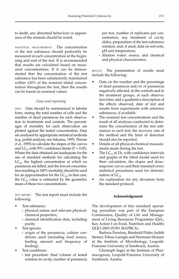
Assessing Potential Cytotoxicity 251
to death, any abnormal behaviour or appear-ance of the animals should be noted.
ANALYTICAL MEASUREMENTS. The concentration of the test substance should preferably be measured at each concentration at the begin-ning and end of the test. It is recommended that results are calculated based on meas-ured concentrations. If it can be demon-strated that the concentration of the test substance has been satisfactorily maintained within ±20% of the nominal initial concen-tration throughout the test, then the results can be based on nominal values.
Data and reporting
DATA. Data should be summarized in tabular form, stating the total number of cells and the number of dead paramecia for each observa-tion in treatments and controls. The percent-ages of mortality for each observation are plotted against the tested concentration. Data are analysed by appropriate statistical methods (e.g. probit analysis; see Sakuma, 1995; Throne et al., 1995) to calculate the slopes of the curves and LC50 with 95% confidence limits (P = 0.05). Where the data obtained are inadequate for the use of standard methods for calculating the LC50, the highest concentration at which no paramecia are killed, and the lowest concentra-tion resulting in 100% mortality, should be used for an approximation for the LC50; in that case, the LC50 value is estimated by the geometric mean of these two concentrations.
TEST REPORT. The test report must include the following:
• Test substance:° physical nature and relevant physical-
chemical properties;° chemical identification data, including
purity.Test species:•° origin of the paramecia, culture con-
ditions used (including food source, feeding amount and frequency of feeding).
Test conditions:•° test procedure: final volume of tested
solution in cavity, number of paramecia
per test, number of replicates per con-centration, any treatment of cavity slides, preparation of the test substance solution, and, if used, data on solvents, pH and temperature;
° dilution water: source, and chemical and physical characteristics.
RESULTS. The presentation of results must include the following:
Data on the number and the percentage •of dead paramecia and/or of paramecia negatively affected, in the controls and in the treatment groups, at each observa-tion time; add a qualitative description of the effects observed; date of test and results from experiments with reference substances, if available.The nominal test concentrations and the •result of all analyses conducted to deter-mine the concentration of the test sub-stances in each test; the recovery rate of the method and the limit of detection should also be reported.Details of all physical-chemical measure-•ments made during the test.The LC• 50 at 2 h, with confidence intervals and graphs of the fitted model used for their calculation, the slopes and dose–response curves and their standard error; statistical procedures used for determi-nation of LC50.
An explanation for any deviation from •the standard protocol.
Acknowledgement
The development of this standard operat-ing procedure was part of the European Commission, Quality of Life and Manage-ment of Living Resources Programme (QoL, Key Action I on Food, Nutrition and Health) QLK1-2001-01391 (RAFBCA).
Barbara Pernfuss, Reinhold Pöder, Judith Stemer, Tobias Laengle and Hermann Strasser at the Institute of Microbiology, Leopold-Franzens University of Innsbruck, Austria.
Christoph Seger at the Institute of Phar-macognosy, Leopold-Franzens Univer sity of Innsbruck, Austria
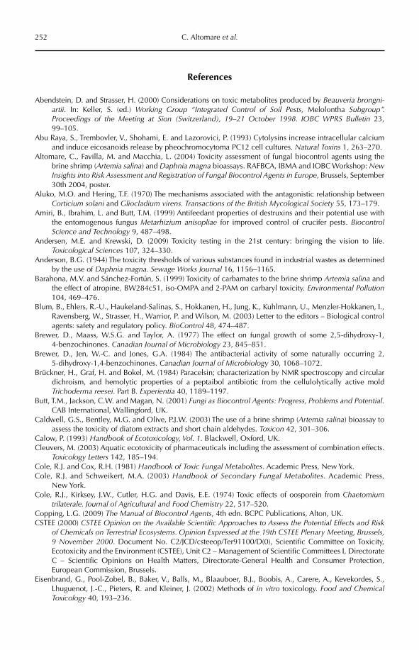
252 C. Altomare et al.
References
Abendstein, D. and Strasser, H. (2000) Considerations on toxic metabolites produced by Beauveria brongni-artii. In: Keller, S. (ed.) Working Group “Integrated Control of Soil Pests, Melolontha Subgroup”. Proceedings of the Meeting at Sion (Switzerland), 19–21 October 1998. IOBC WPRS Bulletin 23, 99–105.
Abu Raya, S., Trembovler, V., Shohami, E. and Lazorovici, P. (1993) Cytolysins increase intracellular calcium and induce eicosanoids release by pheochromocytoma PC12 cell cultures. Natural Toxins 1, 263–270.
Altomare, C., Favilla, M. and Macchia, L. (2004) Toxicity assessment of fungal biocontrol agents using the brine shrimp (Artemia salina) and Daphnia magna bioassays. RAFBCA, IBMA and IOBC Workshop: New Insights into Risk Assessment and Registration of Fungal Biocontrol Agents in Europe, Brussels, September 30th 2004, poster.
Aluko, M.O. and Hering, T.F. (1970) The mechanisms associated with the antagonistic relationship between Corticium solani and Gliocladium virens. Transactions of the British Mycological Society 55, 173–179.
Amiri, B., Ibrahim, L. and Butt, T.M. (1999) Antifeedant properties of destruxins and their potential use with the entomogenous fungus Metarhizium anisopliae for improved control of crucifer pests. BiocontrolScience and Technology 9, 487–498.
Andersen, M.E. and Krewski, D. (2009) Toxicity testing in the 21st century: bringing the vision to life. Toxicological Sciences 107, 324–330.
Anderson, B.G. (1944) The toxicity thresholds of various substances found in industrial wastes as determined by the use of Daphnia magna. Sewage Works Journal 16, 1156–1165.
Barahona, M.V. and Sánchez-Fortún, S. (1999) Toxicity of carbamates to the brine shrimp Artemia salina and the effect of atropine, BW284c51, iso-OMPA and 2-PAM on carbaryl toxicity. Environmental Pollution104, 469–476.
Blum, B., Ehlers, R.-U., Haukeland-Salinas, S., Hokkanen, H., Jung, K., Kuhlmann, U., Menzler-Hokkanen, I., Ravensberg, W., Strasser, H., Warrior, P. and Wilson, M. (2003) Letter to the editors – Biological control agents: safety and regulatory policy. BioControl 48, 474–487.
Brewer, D., Maass, W.S.G. and Taylor, A. (1977) The effect on fungal growth of some 2,5-dihydroxy-1,4-benzochinones. Canadian Journal of Microbiology 23, 845–851.
Brewer, D., Jen, W.-C. and Jones, G.A. (1984) The antibacterial activity of some naturally occurring 2,5-dihydroxy-1,4-benzochinones. Canadian Journal of Microbiology 30, 1068–1072.
Brückner, H., Graf, H. and Bokel, M. (1984) Paracelsin; characterization by NMR spectroscopy and circular dichroism, and hemolytic properties of a peptaibol antibiotic from the cellulolytically active mold Trichoderma reesei. Part B. Experientia 40, 1189–1197.
Butt, T.M., Jackson, C.W. and Magan, N. (2001) Fungi as Biocontrol Agents: Progress, Problems and Potential.CAB International, Wallingford, UK.
Caldwell, G.S., Bentley, M.G. and Olive, P.J.W. (2003) The use of a brine shrimp (Artemia salina) bioassay to assess the toxicity of diatom extracts and short chain aldehydes. Toxicon 42, 301–306.
Calow, P. (1993) Handbook of Ecotoxicology, Vol. 1. Blackwell, Oxford, UK.Cleuvers, M. (2003) Aquatic ecotoxicity of pharmaceuticals including the assessment of combination effects.
Toxicology Letters 142, 185–194.Cole, R.J. and Cox, R.H. (1981) Handbook of Toxic Fungal Metabolites. Academic Press, New York.Cole, R.J. and Schweikert, M.A. (2003) Handbook of Secondary Fungal Metabolites. Academic Press,
New York.Cole, R.J., Kirksey, J.W., Cutler, H.G. and Davis, E.E. (1974) Toxic effects of oosporein from Chaetomium
trilaterale. Journal of Agricultural and Food Chemistry 22, 517–520.Copping, L.G. (2009) The Manual of Biocontrol Agents, 4th edn. BCPC Publications, Alton, UK.CSTEE (2000) CSTEE Opinion on the Available Scientific Approaches to Assess the Potential Effects and Risk
of Chemicals on Terrestrial Ecosystems. Opinion Expressed at the 19th CSTEE Plenary Meeting, Brussels, 9 November 2000. Document No. C2/JCD/csteeop/Ter91100/D(0), Scientific Committee on Toxicity, Ecotoxicity and the Environment (CSTEE), Unit C2 – Management of Scientific Committees I, Directorate C – Scientific Opinions on Health Matters, Directorate-General Health and Consumer Protection, European Commission, Brussels.
Eisenbrand, G., Pool-Zobel, B., Baker, V., Balls, M., Blaauboer, B.J., Boobis, A., Carere, A., Kevekordes, S., Lhuguenot, J.-C., Pieters, R. and Kleiner, J. (2002) Methods of in vitro toxicology. Food and Chemical Toxicology 40, 193–236.
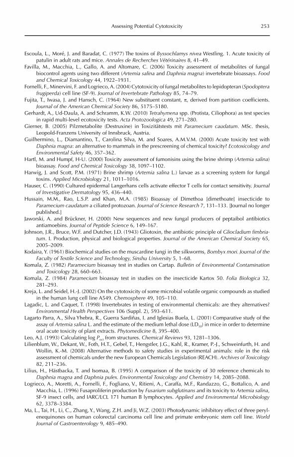
Assessing Potential Cytotoxicity 253
Escoula, L., Moré, J. and Baradat, C. (1977) The toxins of Byssochlamys nivea Westling. 1. Acute toxicity of patulin in adult rats and mice. Annales de Recherches Vétérinaires 8, 41–49.
Favilla, M., Macchia, L., Gallo, A. and Altomare, C. (2006) Toxicity assessment of metabolites of fungal biocontrol agents using two different (Artemia salina and Daphnia magna) invertebrate bioassays. Food and Chemical Toxicology 44, 1922–1931.
Fornelli, F., Minervini, F. and Logrieco, A. (2004) Cytotoxicity of fungal metabolites to lepidopteran (Spodopterafrugiperda) cell line (SF-9). Journal of Invertebrate Pathology 85, 74–79.
Fujita, T., Iwasa, J. and Hansch, C. (1964) New substituent constant, p, derived from partition coefficients. Journal of the American Chemical Society 86, 5175–5180.
Gerhardt, A., Ud-Daula, A. and Schramm, K.W. (2010) Tetrahymena spp. (Protista, Ciliophora) as test species in rapid multi-level ecotoxicity tests. Acta Protozoologica 49, 271–280.
Gierner, B. (2005) Pilzmetabolite (Destruxine) in Toxizitätstests mit Paramecium caudatum. MSc. thesis, Leopold-Franzens University of Innsbruck, Austria.
Guilhermino, L., Diamantino, T., Carolina Silva, M. and Soares, A.M.V.M. (2000) Acute toxicity test with Daphnia magna: an alternative to mammals in the prescreening of chemical toxicity? Ecotoxicology and Environmental Safety 46, 357–362.
Hartl, M. and Humpf, H-U. (2000) Toxicity assessment of fumonisins using the brine shrimp (Artemia salina)bioassay. Food and Chemical Toxicology 38, 1097–1102.
Harwig, J. and Scott, P.M. (1971) Brine shrimp (Artemia salina L.) larvae as a screening system for fungal toxins. Applied Microbiology 21, 1011–1016.
Hauser, C. (1990) Cultured epidermal Langerhans cells activate effector T cells for contact sensitivity. Journalof Investigative Dermatology 95, 436–440.
Hussain, M.M., Rao, L.S.P. and Khan, M.A. (1985) Bioassay of Dimethoa [dimethoate] insecticide to Paramecium caudatum a ciliated protozoan. Journal of Science Research 7, 131–133. [Journal no longer published.]
Jaworski, A. and Brückner, H. (2000) New sequences and new fungal producers of peptaibol antibiotics antiamoebins. Journal of Peptide Science 6, 149–167.
Johnson, J.R., Bruce, W.F. and Dutcher, J.D. (1943) Gliotoxin, the antibiotic principle of Gliocladium fimbria-tum. I. Production, physical and biological properties. Journal of the American Chemical Society 65, 2005–2009.
Kodaira, Y. (1961) Biochemical studies on the muscardine fungi in the silkworms, Bombyx mori. Journal of the Faculty of Textile Science and Technology, Sinshu University 5, 1–68.
Komala, Z. (1982) Paramecium bioassay test in studies on Cartap. Bulletin of Environmental Contamination and Toxicology 28, 660–663.
Komala, Z. (1984) Paramecium bioassay test in studies on the insecticide Kartox 50. Folia Biologica 32, 281–293.
Kreja, L. and Seidel, H.-J. (2002) On the cytotoxicity of some microbial volatile organic compounds as studied in the human lung cell line A549. Chemosphere 49, 105–110.
Lagadic, L. and Caquet, T. (1998) Invertebrates in testing of environmental chemicals: are they alternatives? Environmental Health Perspectives 106 (Suppl. 2), 593–611.
Lagarto Parra, A., Silva Yhebra, R., Guerra Sardiñas, I. and Iglesias Buela, L. (2001) Comparative study of the assay of Artemia salina L. and the estimate of the medium lethal dose (LD50) in mice in order to determine oral acute toxicity of plant extracts. Phytomedicine 8, 395–400.
Leo, A.J. (1993) Calculating log Poct from structures. Chemical Reviews 93, 1281–1306.Lilienblum, W., Dekant, W., Foth, H.T., Gebel, T., Hengstler, J.G., Kahl, R., Kramer, P.-J., Schweinfurth, H. and
Wollin, K.-M. (2008) Alternative methods to safety studies in experimental animals: role in the risk assessment of chemicals under the new European Chemicals Legislation (REACH). Archives of Toxicology82, 211–236.
Lilius, H., Hästbacka, T. and Isomaa, B. (1995) A comparison of the toxicity of 30 reference chemicals to Daphnia magna and Daphnia pulex. Environmental Toxicology and Chemistry 14, 2085–2088.
Logrieco, A., Moretti, A., Fornelli, F., Fogliano, V., Ritieni, A., Caraffa, M.F., Randazzo, G., Bottalico, A. and Macchia, L. (1996) Fusaproliferin production by Fusarium subglutinans and its toxicity to Artemia salina,SF-9 insect cells, and IARC/LCL 171 human B lymphocytes. Applied and Environmental Microbiology62, 3378–3384.
Ma, L., Tai, H., Li, C., Zhang, Y., Wang, Z.H. and Ji, W.Z. (2003) Photodynamic inhibitory effect of three peryl-enequinones on human colorectal carcinoma cell line and primate embryonic stem cell line. World Journal of Gastroenterology 9, 485–490.
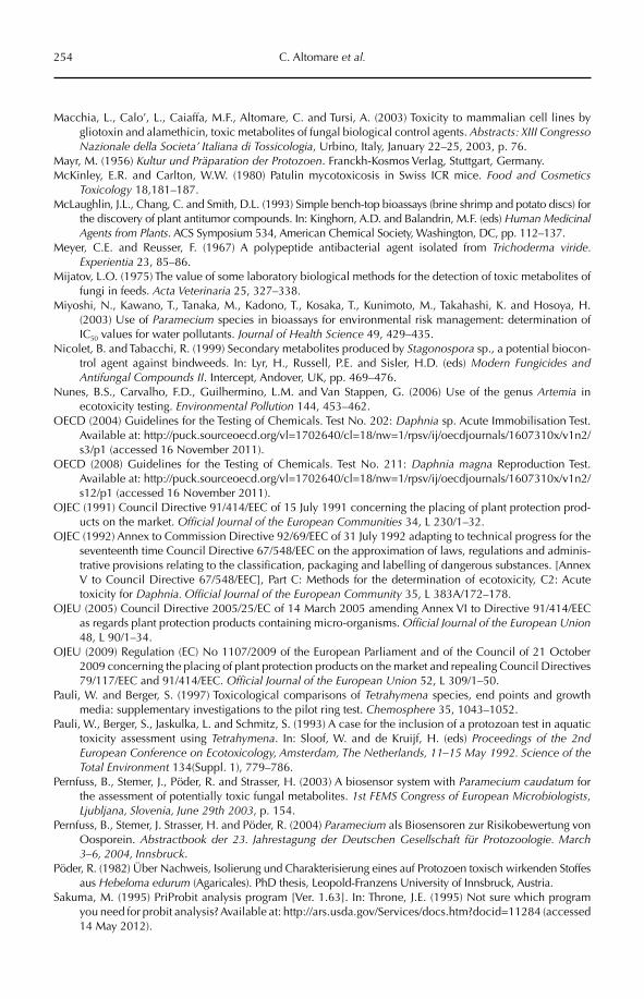
254 C. Altomare et al.
Macchia, L., Calo’, L., Caiaffa, M.F., Altomare, C. and Tursi, A. (2003) Toxicity to mammalian cell lines by gliotoxin and alamethicin, toxic metabolites of fungal biological control agents. Abstracts: XIII Congresso Nazionale della Societa’ Italiana di Tossicologia, Urbino, Italy, January 22–25, 2003, p. 76.
Mayr, M. (1956) Kultur und Präparation der Protozoen. Franckh-Kosmos Verlag, Stuttgart, Germany.McKinley, E.R. and Carlton, W.W. (1980) Patulin mycotoxicosis in Swiss ICR mice. Food and Cosmetics
Toxicology 18,181–187.McLaughlin, J.L., Chang, C. and Smith, D.L. (1993) Simple bench-top bioassays (brine shrimp and potato discs) for
the discovery of plant antitumor compounds. In: Kinghorn, A.D. and Balandrin, M.F. (eds) Human Medicinal Agents from Plants. ACS Symposium 534, American Chemical Society, Washington, DC, pp. 112–137.
Meyer, C.E. and Reusser, F. (1967) A polypeptide antibacterial agent isolated from Trichoderma viride. Experientia 23, 85–86.
Mijatov, L.O. (1975) The value of some laboratory biological methods for the detection of toxic metabolites of fungi in feeds. Acta Veterinaria 25, 327–338.
Miyoshi, N., Kawano, T., Tanaka, M., Kadono, T., Kosaka, T., Kunimoto, M., Takahashi, K. and Hosoya, H. (2003) Use of Paramecium species in bioassays for environmental risk management: determination of IC50 values for water pollutants. Journal of Health Science 49, 429–435.
Nicolet, B. and Tabacchi, R. (1999) Secondary metabolites produced by Stagonospora sp., a potential biocon-trol agent against bindweeds. In: Lyr, H., Russell, P.E. and Sisler, H.D. (eds) Modern Fungicides and Antifungal Compounds II. Intercept, Andover, UK, pp. 469–476.
Nunes, B.S., Carvalho, F.D., Guilhermino, L.M. and Van Stappen, G. (2006) Use of the genus Artemia in ecotoxicity testing. Environmental Pollution 144, 453–462.
OECD (2004) Guidelines for the Testing of Chemicals. Test No. 202: Daphnia sp. Acute Immobilisation Test. Available at: http://puck.sourceoecd.org/vl=1702640/cl=18/nw=1/rpsv/ij/oecdjournals/1607310x/v1n2/s3/p1 (accessed 16 November 2011).
OECD (2008) Guidelines for the Testing of Chemicals. Test No. 211: Daphnia magna Reproduction Test. Available at: http://puck.sourceoecd.org/vl=1702640/cl=18/nw=1/rpsv/ij/oecdjournals/1607310x/v1n2/s12/p1 (accessed 16 November 2011).
OJEC (1991) Council Directive 91/414/EEC of 15 July 1991 concerning the placing of plant protection prod-ucts on the market. Official Journal of the European Communities 34, L 230/1–32.
OJEC (1992) Annex to Commission Directive 92/69/EEC of 31 July 1992 adapting to technical progress for the seventeenth time Council Directive 67/548/EEC on the approximation of laws, regulations and adminis-trative provisions relating to the classification, packaging and labelling of dangerous substances. [Annex V to Council Directive 67/548/EEC], Part C: Methods for the determination of ecotoxicity, C2: Acute toxicity for Daphnia. Official Journal of the European Community 35, L 383A/172–178.
OJEU (2005) Council Directive 2005/25/EC of 14 March 2005 amending Annex VI to Directive 91/414/EEC as regards plant protection products containing micro-organisms. Official Journal of the European Union48, L 90/1–34.
OJEU (2009) Regulation (EC) No 1107/2009 of the European Parliament and of the Council of 21 October 2009 concerning the placing of plant protection products on the market and repealing Council Directives 79/117/EEC and 91/414/EEC. Official Journal of the European Union 52, L 309/1–50.
Pauli, W. and Berger, S. (1997) Toxicological comparisons of Tetrahymena species, end points and growth media: supplementary investigations to the pilot ring test. Chemosphere 35, 1043–1052.
Pauli, W., Berger, S., Jaskulka, L. and Schmitz, S. (1993) A case for the inclusion of a protozoan test in aquatic toxicity assessment using Tetrahymena. In: Sloof, W. and de Kruijf, H. (eds) Proceedings of the 2nd European Conference on Ecotoxicology, Amsterdam, The Netherlands, 11–15 May 1992. Science of the Total Environment 134(Suppl. 1), 779–786.
Pernfuss, B., Stemer, J., Pöder, R. and Strasser, H. (2003) A biosensor system with Paramecium caudatum for the assessment of potentially toxic fungal metabolites. 1st FEMS Congress of European Microbiologists,Ljubljana, Slovenia, June 29th 2003, p. 154.
Pernfuss, B., Stemer, J. Strasser, H. and Pöder, R. (2004) Paramecium als Biosensoren zur Risikobewertung von Oosporein. Abstractbook der 23. Jahrestagung der Deutschen Gesellschaft für Protozoologie. March 3–6, 2004, Innsbruck.
Pöder, R. (1982) Über Nachweis, Isolierung und Charakterisierung eines auf Protozoen toxisch wirkenden Stoffes aus Hebeloma edurum (Agaricales). PhD thesis, Leopold-Franzens University of Innsbruck, Austria.
Sakuma, M. (1995) PriProbit analysis program [Ver. 1.63]. In: Throne, J.E. (1995) Not sure which program you need for probit analysis? Available at: http://ars.usda.gov/Services/docs.htm?docid=11284 (accessed 14 May 2012).
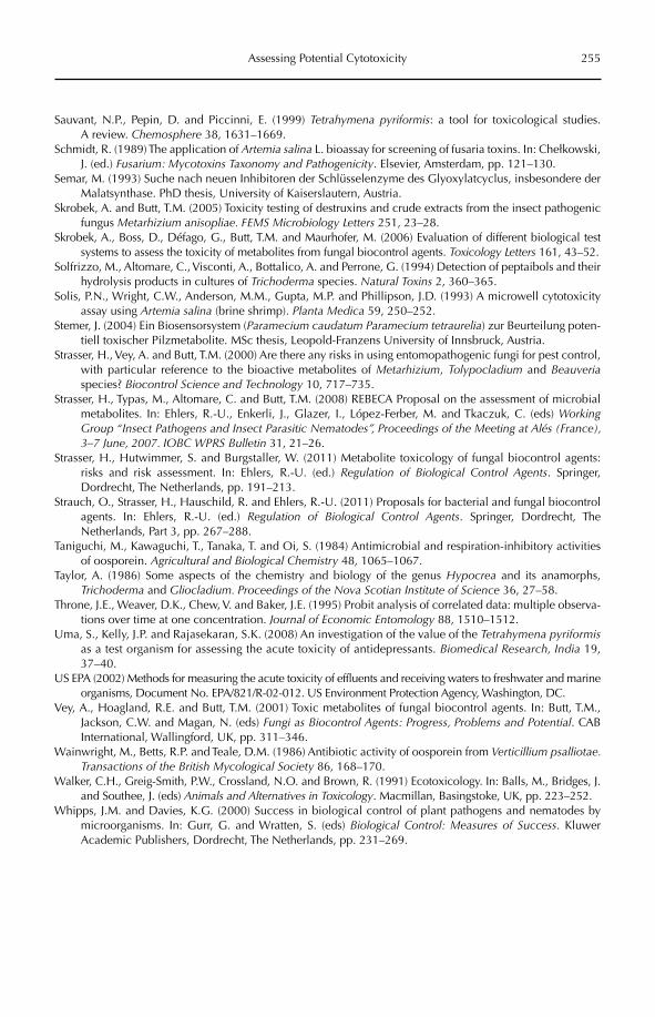
Assessing Potential Cytotoxicity 255
Sauvant, N.P., Pepin, D. and Piccinni, E. (1999) Tetrahymena pyriformis: a tool for toxicological studies. A review. Chemosphere 38, 1631–1669.
Schmidt, R. (1989) The application of Artemia salina L. bioassay for screening of fusaria toxins. In: Chełkowski, J. (ed.) Fusarium: Mycotoxins Taxonomy and Pathogenicity. Elsevier, Amsterdam, pp. 121–130.
Semar, M. (1993) Suche nach neuen Inhibitoren der Schlüsselenzyme des Glyoxylatcyclus, insbesondere der Malatsynthase. PhD thesis, University of Kaiserslautern, Austria.
Skrobek, A. and Butt, T.M. (2005) Toxicity testing of destruxins and crude extracts from the insect pathogenic fungus Metarhizium anisopliae. FEMS Microbiology Letters 251, 23–28.
Skrobek, A., Boss, D., Défago, G., Butt, T.M. and Maurhofer, M. (2006) Evaluation of different biological test systems to assess the toxicity of metabolites from fungal biocontrol agents. Toxicology Letters 161, 43–52.
Solfrizzo, M., Altomare, C., Visconti, A., Bottalico, A. and Perrone, G. (1994) Detection of peptaibols and their hydrolysis products in cultures of Trichoderma species. Natural Toxins 2, 360–365.
Solis, P.N., Wright, C.W., Anderson, M.M., Gupta, M.P. and Phillipson, J.D. (1993) A microwell cytotoxicity assay using Artemia salina (brine shrimp). Planta Medica 59, 250–252.
Stemer, J. (2004) Ein Biosensorsystem (Paramecium caudatum Paramecium tetraurelia) zur Beurteilung poten-tiell toxischer Pilzmetabolite. MSc thesis, Leopold-Franzens University of Innsbruck, Austria.
Strasser, H., Vey, A. and Butt, T.M. (2000) Are there any risks in using entomopathogenic fungi for pest control, with particular reference to the bioactive metabolites of Metarhizium, Tolypocladium and Beauveriaspecies? Biocontrol Science and Technology 10, 717–735.
Strasser, H., Typas, M., Altomare, C. and Butt, T.M. (2008) REBECA Proposal on the assessment of microbial metabolites. In: Ehlers, R.-U., Enkerli, J., Glazer, I., López-Ferber, M. and Tkaczuk, C. (eds) Working Group “Insect Pathogens and Insect Parasitic Nematodes”, Proceedings of the Meeting at Alés (France), 3–7 June, 2007. IOBC WPRS Bulletin 31, 21–26.
Strasser, H., Hutwimmer, S. and Burgstaller, W. (2011) Metabolite toxicology of fungal biocontrol agents: risks and risk assessment. In: Ehlers, R.-U. (ed.) Regulation of Biological Control Agents. Springer, Dordrecht, The Netherlands, pp. 191–213.
Strauch, O., Strasser, H., Hauschild, R. and Ehlers, R.-U. (2011) Proposals for bacterial and fungal biocontrol agents. In: Ehlers, R.-U. (ed.) Regulation of Biological Control Agents. Springer, Dordrecht, The Netherlands, Part 3, pp. 267–288.
Taniguchi, M., Kawaguchi, T., Tanaka, T. and Oi, S. (1984) Antimicrobial and respiration-inhibitory activities of oosporein. Agricultural and Biological Chemistry 48, 1065–1067.
Taylor, A. (1986) Some aspects of the chemistry and biology of the genus Hypocrea and its anamorphs, Trichoderma and Gliocladium. Proceedings of the Nova Scotian Institute of Science 36, 27–58.
Throne, J.E., Weaver, D.K., Chew, V. and Baker, J.E. (1995) Probit analysis of correlated data: multiple observa-tions over time at one concentration. Journal of Economic Entomology 88, 1510–1512.
Uma, S., Kelly, J.P. and Rajasekaran, S.K. (2008) An investigation of the value of the Tetrahymena pyriformisas a test organism for assessing the acute toxicity of antidepressants. Biomedical Research, India 19, 37–40.
US EPA (2002) Methods for measuring the acute toxicity of effluents and receiving waters to freshwater and marine organisms, Document No. EPA/821/R-02-012. US Environment Protection Agency, Washington, DC.
Vey, A., Hoagland, R.E. and Butt, T.M. (2001) Toxic metabolites of fungal biocontrol agents. In: Butt, T.M., Jackson, C.W. and Magan, N. (eds) Fungi as Biocontrol Agents: Progress, Problems and Potential. CAB International, Wallingford, UK, pp. 311–346.
Wainwright, M., Betts, R.P. and Teale, D.M. (1986) Antibiotic activity of oosporein from Verticillium psalliotae. Transactions of the British Mycological Society 86, 168–170.
Walker, C.H., Greig-Smith, P.W., Crossland, N.O. and Brown, R. (1991) Ecotoxicology. In: Balls, M., Bridges, J. and Southee, J. (eds) Animals and Alternatives in Toxicology. Macmillan, Basingstoke, UK, pp. 223–252.
Whipps, J.M. and Davies, K.G. (2000) Success in biological control of plant pathogens and nematodes by microorganisms. In: Gurr, G. and Wratten, S. (eds) Biological Control: Measures of Success. Kluwer Academic Publishers, Dordrecht, The Netherlands, pp. 231–269.
©CAB International 2012. Beneficial Microorganisms in Agriculture, Food and the Environment: 256 Safety Assessment and Regulation (eds I. Sundh et al.)
18.1 Introduction 25618.2 How Are Genotoxicity Data Used in Safety Assessment? Detection of Putative
Toxicity and Carcinogenicity of Metabolites Produced by Microbials 25818.2.1 Bioassays and biosensors 260
18.2.2 Generally approved tests for the identification of genotoxic, mutagenic and carcinogenic substances 260
18.3 Limitations of Genetic Toxicity Tests 26418.4 New Methods and Improvements in Genotoxicity Testing 264
18.4.1 Vitotox assay 265 18.4.2 Yeast DEL assay 265 18.4.3 Microarrays 266 18.4.4 Transgenic rodent model 266
18.5 Further Development of Genotoxicity Testing 26618.6 Conclusions 268
18 Assessing Genotoxic Effects of Microbial Products
Milton A. Typas and Vassili N. KouvelisDepartment of Genetics and Biotechnology, University of Athens, Greece
18.1 Introduction
In almost every aspect and activity of our life, for good or bad, microbes and their products play an important role. They make the soil fertile; they clean up the environ-ment; they produce vitamins and antibiot-ics; they improve food; they may even protect us from less desirable microbes; and they are exploited by industry for their biotech nological potential (see Sundh et al., Chapter 1, this volume). Microbes, and more particularly fungi, on which emphasis will be placed in this chapter, can be found in every habitat, parasitize any organism and decay all possible materials. So, in several cases, they are associated with undesirable side effects, such as food spoilage and human,
animal and plant diseases – reasons that make them generally regarded as nasty and unpopular.
Unfortunately, it is under the latter cir-cumstances that they mainly attract people’s attention and thereby create fears for toxicity, disease and even deadly events! For almost eight decades, chemicals were exclusively used in the protection of crops against pests and they have been been applied in most culti-vated land throughout the world. However, because chemical pesticides have repeatedly been proven to be mutagenic and carcinogenic (various European Union (EU) guidelines; Ruiz and Marzin, 1997), the need for environ-mentally friendly and safe alternatives is con-tinuously increasing. This was realized early enough by the European Commision which,
Assessing Genotoxic Effects 257
through the Common Agricultural Policy (CAP), which aimed towards a sustainable agriculture in a global environment, and introduced criteria of quality control, taking responsibility for the countryside and keep-ing consumers confident about food safety (European Commission, 2008). Through recent years, both bacteria and fungi have demon-strated several advantageous properties over chemicals when used for crop protection and are considered to be a relevant and safe replacement of (at least) part of the large quan-tities of chemicals used (Strasser et al., 2000; Hunter, 2009). Thus, the application of micro-bial biological control agents (BCAs), known as MBCAs, which are based on microorgan-isms is expanding and gaining acceptance from scientists, regulators and farmers.
The best known and most successful microbial product, which has been exploited commercially for almost 50 years, is the bacte-rium Bacillus thuringiensis (Bt), with its toxins and the genes (cry) that produce these toxins (Flexner et al., 1986). Bt and its toxins, either used solely or genetically incorporated into transgenic crops, are only effective against insects within certain families and not others (for reviews see McClintock et al., 1995; Glare and O’Callaghan, 2000; Kumar et al., 2008; and references therein). In that sense, recent reports on the develoment of insect resistance to Bt in transgenic plants, even under condi-tions that are designed to delay this resist-ance, such as the refuge strategy, strongly indicate that crop protection cannot solely depend on this bacterium and its toxins (Tabashnik et al., 2008), and other solutions are also being sought. As several species of fungi have lately been used as MBCAs, an emphasis will be placed in this chapter on fungi and their products, as they comprise a benign alternative for the protection of crops worldwide (Butt et al., 2001).
A well-established fact is that many fungi produce and secrete mycotoxins (i.e. natural secondary metabolites of diverse chemical structure) which provide them an edge of sur-vival through: (i) symbiosis and syntrophism; (ii) antagonism or competition for nutrients within an ecological niche; and (iii) pathogen-esis (i.e. the ability to weaken and/or kill their host) (Vey et al., 2001; Mayer et al., 2008).
In addition, there are several fungal metabo-lites that are highly toxic (e.g. fumonisins, ochratoxins, patulin, zearalenone) or carcino-genic (e.g. moniliformin, aflatoxins). These pose serious risks to human and animal health when foods are contaminated by the saprophytic fungi that produce them (Abramson, 1998; Strasser et al., 2000). Until now, several bodies of legislation and direc-tives have been implemented that provide details of the requirements for authorization and commercial exploitation of microbial products. These directives aim to guarantee that the products or the microorganism that produces them have no immediate or delayed harmful effects on humans, non-target organ-isms and the environment itself. In addition, they satisfy criteria which verify that there is no impact on the existing biodiversity and the ecosystem to which they will be applied, e.g. OECD (2008); and European Council Directive 1107/ 2009 (OJEU, 2009), amending Council Direc tive 91/414/CEE (OJEC, 1991). So the putative exploitation and commercialization of any MBCA raises two important questions: (i) are hazardous metabolites produced by a specific strain used as an MBCA – uniquely or in combination with other microbes; and (ii) do the microbes and their metabolites pose any risk for humans and environment?
The first question is easily answered by the numerous reports on metabolites that have been isolated from pure microbial cul-tures of MBCAs (examples are given in Table 18.1). However, it is interesting to note that even species from different ecological habitats, such as entomopathogenic and saprophytic fungi, may produce similar com-pounds (Mayer et al., 2008).
The second question hits the heart of the problem because a major obstacle in the regis-tration process and the subsequent commer-cialization and exploitation of a microbial product is the assessment of its risks and of the metabolites that it produces. So far, it is known that some of the metabolites produced by MBCAs are toxic in vitro to animal cell lines (Vey and Quiot, 1989; Dumas et al., 1996; Fornelli et al., 2004), whereas other metabo-lites have antibiotic, fungicidal, insecticidal or antiviral properties (Terry et al., 1992; Amiri et al., 1999; Kershaw et al., 1999; Bandani et al.,
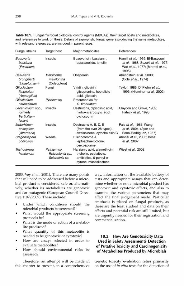
258 M.A. Typas and V.N. Kouvelis
2000; Vey et al., 2001). There are many points that still need to be addressed before a micro-bial product is considered safe or, alternati-vely, whether its metabolites are genotoxic and/or mutagenic (European Council Direc-tive 1107/2009). These include:
Under which conditions should the •microbial products be screened?What would the appropriate screening •protocols be?What is the mode of action of a metabo-•lite produced?What quantity of this metabolite is •needed to be genotoxic or cytotoxic?How are assays selected in order to • evaluate metabolites?How should environmental risks be •assessed?
Therefore, an attempt will be made in this chapter to present, in a comprehensive
way, information on the available battery of tests and appropriate assays that can deter-mine whether or not a microbial product has genotoxic and cytotoxic effects, and also to examine the various parameters that may affect the final judgement made. Particular emphasis is placed on fungal products, as these are the least studied and data on their effects and potential risk are still limited, but are urgently needed for their registration and commercialization.
18.2 How Are Genotoxicity Data Used in Safety Assessment? Detection
of Putative Toxicity and Carcinogenicity of Metabolites Produced by Microbials
Genetic toxicity evaluation relies primarily on the use of in vitro tests for the detection of
Table 18.1. Fungal microbial biological control agents (MBCAs), their target hosts and metabolites, and references to work on these. Details of saprophytic fungal genera producing the same metabolites, with relevant references, are included in parentheses.
Fungal strains Target host Major metabolites References
Beauveria bassiana (Fusarium)
Insects Beauvericin, bassianin, bassianolide, tenellin
Hamill et al., 1969; El-Basyouni et al., 1968; Suzuki et al., 1977;
Wat et al., 1977; (Moretti et al., 1995)
Beauveria brongniartii (Chaetomium)
Melolontha melolontha (Coleoptera)
Oosporein Abendstein et al., 2000; (Cole et al., 1974)
Gliocladium fimbriatum (Aspergillus)
Fungi Viridin, gliovirin, glisoprenins, heptelidic acid, gliotoxin
Taylor, 1986; Di Pietro et al., 1993; (Nieminen et al., 2002)
Gliocladium catenulatum
Pythium sp. Presumed as for G. fimbriatum
Lecanicillium spp., formerly
Verticillium lecanii
Insects Destruxins, dipicolinic acid, hydroxycarboxylic acid, cyclosporin
Claydon and Grove, 1982; Patrick et al., 1993
Metarhizium anisopliae (Alternaria)
Insects Destruxins A, B, D, E (from the over 28 types), swainsinone, cytochalasin C
Pais et al., 1981; Wang et al., 2004; (Ayer and
Pena-Rodriguez, 1987)Stagonospora convolvuli
Weeds Elsinochrome A, leptosphaerodione, cercosporine
Ahonsi et al., 2005; Boss et al., 2007
Trichoderma harzianum
Pythium sp., Rhizoctonia sp., Sclerotinia sp.
Harzianic acid, alamethicin, tricholin, peptaibols, antibiotics, 6-pentyl-α- pyrone, massoilactone
Wiest et al., 2002
Assessing Genotoxic Effects 259
agents that cause mutations and chromo-somal aberrations of the DNA and, conse-quently, affect its expression. Because in vitrotests are used as the cornerstone of genotoxic-ity testing, it is necessary to correctly interpret in vitro findings in relation to the potential in vivo risk of genetic damage in a living organism. Yet, as experience shows, the inter-pretation of results from in vitro genetic toxic-ity testing is usually very subjective, and experts often evaluate the same set of test results differently. This clearly increases the risk for making the wrong decision, especially in cases where conclusions rely on minimal additional information beyond the genetic toxicity data. As a result, knowledge of the microbe and the metabolites it produces is of crucial importance for its risk assessment. Each case is somewhat different from another and it is always important to consider all the available information for decision making (Dearfield et al., 2011). Nevertheless, if in vitroassays indicate the lack of any mutagenic or genotoxic potential of a microbial product or of its secondary metabolites, then no further action is needed. However, additional evalu-ation will be required if ambiguous or, even worse, positive results are obtained from the primary in vitro assays (Gatehouse et al., 1994).
Apart from a good understanding of a MBCA itself, particular attention must be placed on whether genotoxicity tests have been performed only with pure metabolites – putatively produced by the microbes in scrutiny – because such tests may lead to wrong conclusions, as it is often a synergistic effect that renders a substance mutagenic or genotoxic (Kouvelis et al., 2011). As shown in Table 18.1, fungi produce several different secondary metabolites which are secreted simultanously into their environment. Their mode of action, the interactions among them and with other substances in the environment, the amounts in which they are produced and secreted, and their persistence in the field, may differ a lot in nature compared with laboratory conditions and, until now, little is known about all these parameters.
Therefore, it is very important that any confounding factors are taken into consi-deration and are included in the testing and interpretation of results. For example, the
conditions applied (e.g. pH and osmolality other than those under normal conditions), the possible interactions with the other sub-stances besides the one examined (such as the medium used for the assay), the involve-ment of a metabolic activation system (such as the S9 homogenate – see Section 18.2.2, and other cofactors), and the possible geno-toxic effects of any impurities are some of the parameters that need to be considered (for reviews see Mayer et al., 2008; Dearfield et al., 2011). Crude extracts from the micro-organism may provide an important alter-native to pure metabolites, because, under natural conditions, mixtures of metabolitesare secreted by microbes and it is often unclear to what extent synergistic effects account for toxicity (of, for example, mycotoxins). In addi-tion, crude extracts provide the advantage of including in the assays metabolites that have not been properly characterized as they may be present in low concentrations or difficult to isolate owing to the domination of other major metabolites. Finally, the ease by which the crude extracts can be produced, even in large quantities, renders them accessible to many laboratories at low cost, and thus provides material for a reliable quick test for any toxico-genic effect on standard tester organisms (Kouvelis et al., 2011).
The genotoxic activity of a secondary metabolite may result from either a direct or an indirect effect on the genetic material. A direct mode of action indicates that DNA is the primary target for the substance (i.e. result- ing in DNA adducts, thymidine dimers and, in some cases, strand breaks), while an indirect mode of action has primary targets other than DNA. In the latter case, the DNA is being sec-ondarily damaged via free radicals, reactive oxygen or nitrogen species, nucleotide pool imbalance, spindle disruption, or the inhibition of DNA synthesis or topoisomerases. In bothcases, the end result is still a genotoxic insult and possible genetic alteration. Distinguish-ing the mode of action of a metabolite is very important because the type of assay used for screening of the metabolite may otherwise provide false results. For instance, the bacte-rial reverse mutation assay (Ames test) detects gene mutations (Mortelmans and Zeiger, 2000), while a chromosome aberration
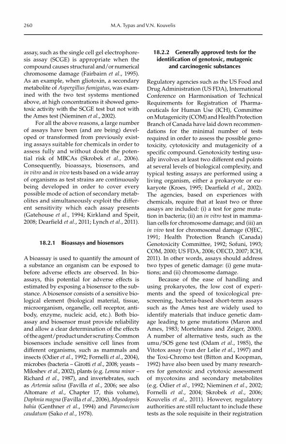
260 M.A. Typas and V.N. Kouvelis
assay, such as the single cell gel electrophore-sis assay (SCGE) is appropriate when the compound causes structural and/or numerical chromosome damage (Fairbairn et al., 1995). As an example, when gliotoxin, a secondary metabolite of Asper gillus fumigatus, was exam-ined with the two test systems mentioned above, at high concentrations it showed geno-toxic activity with the SCGE test but not withthe Ames test (Nieminen et al., 2002).
For all the above reasons, a large number of assays have been (and are being) devel-oped or transformed from previously exist-ing assays suitable for chemicals in order to assess fully and without doubt the poten-tial risk of MBCAs (Skrobek et al., 2006). Consequently, bioassays, biosensors, and in vitro and in vivo tests based on a wide array of organisms as test strains are continuously being developed in order to cover every possible mode of action of secondary metab-olites and simultaneously exploit the differ-ent sensitivity which each assay presents (Gatehouse et al., 1994; Kirkland and Speit, 2008; Dearfield et al., 2011; Lynch et al., 2011).
18.2.1 Bioassays and biosensors
A bioassay is used to quantify the amount of a substance an organism can be exposed to before adverse effects are observed. In bio-assays, this potential for adverse effects is estimated by exposing a biosensor to the sub-stance. A biosensor consists of a sensitive bio-logical element (biological material, tissue, microorganism, organelle, cell receptor, anti-body, enzyme, nucleic acid, etc.). Both bio-assay and biosensor must provide reliability and allow a clear determination of the effects of the agent/product under scrutiny. Common biosensors include sensitive cell lines from different organisms, such as mammals and insects (Odier et al., 1992; Fornelli et al., 2004), microbes (bacteria – Girotti et al., 2008; yeasts – Miloshev et al., 2002), plants (e.g. Lemna minor – Richard et al., 1987), and invertebrates, such as Artemia salina (Favilla et al., 2006; see also Altomare et al., Chapter 17, this volume), Daphnia magna (Favilla et al., 2006), Mysodopsisbahia (Genthner et al., 1994) and Parameciumcaudatum (Sako et al., 1978).
18.2.2 Generally approved tests for the identification of genotoxic, mutagenic
and carcinogenic substances
Regulatory agencies such as the US Food and Drug Administration (US FDA), International Conference on Harmonisation of Technical Requirements for Registration of Pharma-ceuticals for Human Use (ICH), Committee on Mutagenicity (COM) and Health Protection Branch of Canada have laid down recommen-dations for the minimal number of tests required in order to assess the possible geno-toxicity, cytotoxicity and mutagenicity of a specific compound. Genotoxicity testing usu-ally involves at least two different end points at several levels of biological complexity, and typical testing assays are performed using a living organism, either a prokaryote or eu -karyote (Kroes, 1995; Dearfield et al., 2002). The agencies, based on experiences with chemicals, require that at least two or three assays are included: (i) a test for gene muta-tion in bacteria; (ii) an in vitro test in mamma-lian cells for chromosome damage; and (iii) an in vivo test for chromosomal damage (OJEC, 1991; Health Protection Branch (Canada) Genotoxicity Committee, 1992; Sofuni, 1993; COM, 2000; US FDA, 2006; OECD, 2007; ICH, 2011). In other words, assays should address two types of genetic damage: (i) gene muta-tions; and (ii) chromosome damage.
Because of the ease of handling and using prokaryotes, the low cost of experi-ments and the speed of toxicological pre-screening, bacteria-based short-term assays such as the Ames test are widely used to identify materials that induce genetic dam-age leading to gene mutations (Maron and Ames, 1983; Mortelmans and Zeiger, 2000). A number of alternative tests, such as the umu/SOS gene test (Odam et al., 1985), the Vitotox assay (van der Lelie et al., 1997) and the Toxi-Chromo test (Bitton and Koopman, 1992) have also been used by many research-ers for genotoxic and cytotoxic assessment of mycotoxins and secondary metabolites (e.g. Odier et al., 1992; Nieminen et al., 2002; Fornelli et al., 2004; Skrobek et al., 2006; Kouvelis et al., 2011). However, regulatory authorities are still reluctant to include these tests as the sole requisite in their registration
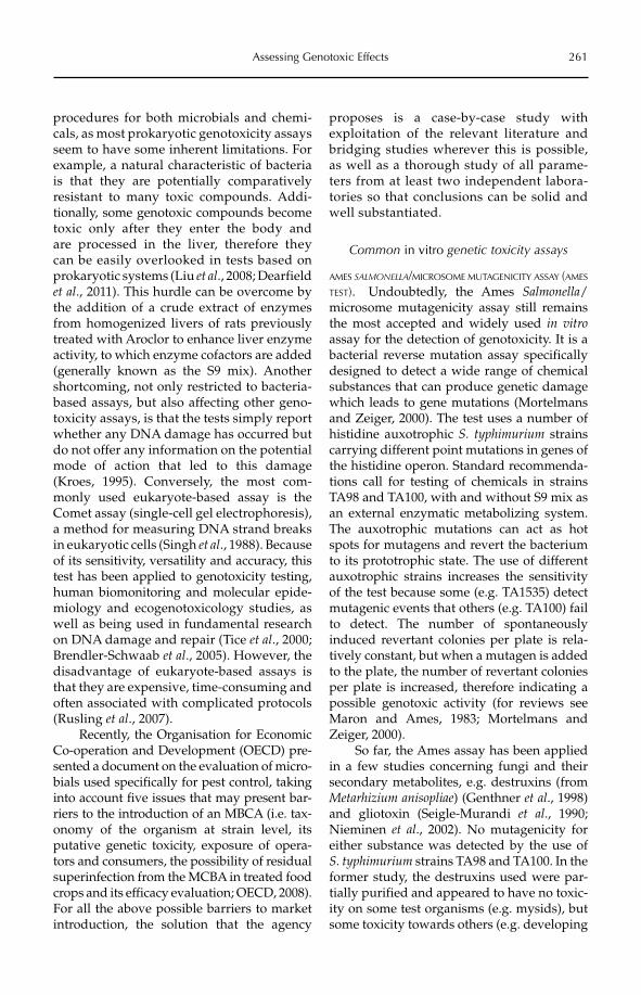
Assessing Genotoxic Effects 261
procedures for both microbials and chemi-cals, as most prokaryotic genotoxicity assays seem to have some inherent limitations. For example, a natural characteristic of bacteria is that they are potentially comparatively resistant to many toxic compounds. Addi-tionally, some genotoxic compounds becometoxic only after they enter the body and are processed in the liver, therefore they can be easily overlooked in tests based on prokar yotic systems (Liu et al., 2008; Dearfield et al., 2011). This hurdle can be overcome by the addition of a crude extract of enzymes from homogenized livers of rats previously treated with Aroclor to enhance liver enzyme activity, to which enzyme cofactors are added (generally known as the S9 mix). Another shortcoming, not only restricted to bacteria-based assays, but also affecting other geno-toxicity assays, is that the tests simply report whether any DNA damage has occurred but do not offer any information on the potential mode of action that led to this damage (Kroes, 1995). Conversely, the most com-monly used eukar yote-based assay is the Comet assay (single-cell gel electrophoresis), a method for measuring DNA strand breaks in eukaryotic cells (Singh et al., 1988). Because of its sensitivity, versatility and accuracy, this test has been applied to genotoxicity testing, human biomonitoring and molecular epide-miology and ecogenotoxicology studies, as well as being used in fundamental research on DNA damage and repair (Tice et al., 2000; Brendler-Schwaab et al., 2005). However, the disadvantage of eukaryote-based assays is that they are expensive, time-consuming and often associated with complicated protocols (Rusling et al., 2007).
Recently, the Organisation for Economic Co-operation and Development (OECD) pre-sented a document on the evaluation of micro-bials used specifically for pest control, taking into account five issues that may present bar-riers to the introduction of an MBCA (i.e. tax-onomy of the organism at strain level, its putative genetic toxicity, exposure of opera-tors and consumers, the possibility of residual superinfection from the MCBA in treated food crops and its efficacy evaluation; OECD, 2008). For all the above possible barriers to market introduction, the solution that the agency
proposes is a case-by-case study with exploitation of the relevant literature and bridging studies wherever this is possible, as well as a thorough study of all parame-ters from at least two independent labora-tories so that conclusions can be solid and well substantiated.
Common in vitro genetic toxicity assays
AMES SALMONELLA/MICROSOME MUTAGENICITY ASSAY (AMES
TEST). Undoubtedly, the Ames Salmonella/microsome mutagenicity assay still remains the most accepted and widely used in vitroassay for the detection of genotoxicity. It is a bacterial reverse mutation assay specifically designed to detect a wide range of chemical substances that can produce genetic damage which leads to gene mutations (Mortelmans and Zeiger, 2000). The test uses a number of histidine auxotrophic S. typhimurium strains carrying different point mutations in genes of the histidine operon. Standard recommenda-tions call for testing of chemicals in strains TA98 and TA100, with and without S9 mix as an external enzymatic metabolizing system. The auxotrophic mutations can act as hot spots for mutagens and revert the bacterium to its prototrophic state. The use of different auxotrophic strains increases the sensitivity of the test because some (e.g. TA1535) detect mutagenic events that others (e.g. TA100) fail to detect. The number of spontaneously induced revertant colonies per plate is rela-tively constant, but when a mutagen is added to the plate, the number of revertant colonies per plate is increased, therefore indicating a possible genotoxic activity (for reviews see Maron and Ames, 1983; Mortelmans and Zeiger, 2000).
So far, the Ames assay has been applied in a few studies concerning fungi and their secondary metabolites, e.g. destruxins (from Metarhizium anisopliae) (Genthner et al., 1998) and gliotoxin (Seigle-Murandi et al., 1990; Nieminen et al., 2002). No mutagenicity for either substance was detected by the use of S. typhimurium strains TA98 and TA100. In the former study, the destruxins used were par-tially purified and appeared to have no toxic-ity on some test organisms (e.g. mysids), but some toxicity towards others (e.g. developing
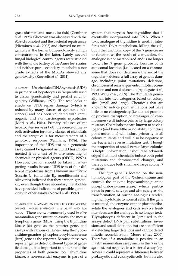
262 M.A. Typas and V.N. Kouvelis
grass shrimps and mosquito fish) (Genthner et al., 1998). Gliotoxin was also tested with the SOS-chromotest and the bacterial repair assay (Nieminen et al., 2002) and showed no muta-genicity in the former but genotoxicity at high concentrations in the latter. Lately, several fungal biological control agents were studied with the whole battery of the Ames test strains and neither pure secondary metabolites nor crude extracts of the MBCAs showed any genotoxicity (Kouvelis et al., 2011).
UDS ASSAY. Unscheduled DNA synthesis (UDS) in primary rat hepatocytes is frequently used to assess genotoxicity and predict carcino-genicity (Williams, 1976). The test looks at effects on DNA repair damage (which is reduced by many classes of genotoxic sub-stances) and has been validated with carci-nogenic and non-carcinogenic mycotoxins (Mori et al., 1984). Primary cultures of rat hepatocytes serve as both the source of meta-bolic activation for many classes of chemicals and the target cells for measurements of a genotoxic response (Williams, 1976). The importance of the UDS test as a genotoxic assay cannot be ignored as OECD has imple-mented it as a test of in vivo screening of chemicals or physical agents (OECD, 1997b). However, caution should be taken in inter-preting results because UDS tests of four dif-ferent mycotoxins from Fusarium moniliforme(fusarin C, fumonisin B1, moniliformin and bikaverin) indicated that they are non-genoto-xic, even though these secondary metabolites have provided indications of possible genoto-xicity in other assays (Norred et al., 1992).
IN VITRO TEST IN MAMMALIAN CELLS FOR CHROMOSOME
DAMAGE: MOUSE LYMPHOMA tk ± ASSAY AND hprt
ASSAY. There are two commonly used in vitromammalian gene mutation assays, the mouse lymphoma assay (MLA) using the thymidine kinase (tk) gene as the reporter gene, and assays with various cell lines using the hypox-anthine-guanine phosphoribosyl-transferase (hprt) gene as the reporter. Because these two reporter genes detect different types of gene-tic damage, it is important to understand the properties of both genetic loci. Thymidine kinase, a non-essential enzyme, is part of a
system that recycles free thymidine that is eventually incorporated into DNA. When a toxic analogue of thymidine is used, it inter-feres with DNA metabolism, killing the cell, but if the functional copy of the tk gene ceases to function as the result of a mutation, the analogue is not metabolized and is no longer toxic. The tk gene, probably because of its autosomal location (i.e. located on a chromo-some that does not determine the sex of the organism), detects a full array of genetic dam-age, including point mutations, deletions, chromosomal rearrangements, mitotic recom-bination and non-disjunction (Applegate et al., 1990; Wang et al., 2009). The tk mutants gener-ally fall into two categories based on colony size (small and large). Chemicals that are known to induce point mutations but have little or no clastogenicity (i.e. do not promote or produce disruption or breakages of chro-mosomes) will induce primarily large colony mutants. Chemicals that are known to be clas-togens (and have little or no ability to induce point mutations) will induce primarily small colony mutants and will also be negative in the bacterial reverse mutation test. Though the proportion of small versus large colonies is helpful information, it should be acknowl-edged that most chemicals induce both point mutations and chromosomal changes, and thereby induce both small and large colony tkmutants.
The hprt gene is located on the non-homologous part of the X-chromosome and controls the enzyme hypoxanthine-guanine phosphoribosyl-transferase, which partici-pates in purine salvage and also catalyses the transformation of purine analogues render-ing them cytotoxic to normal cells. If the gene is mutated, the enzyme cannot phosphoribo-sylate the analogues and cells survive treat-ment because the analogue is no longer toxic. T-lymphocytes deficient in hprt used in the assays detect DNA pair substitutions, inver-sions and small deletions, but are not efficient at detecting large deletions and cannot detect mitotic recombination (Moore et al., 2000). Therefore, if a metabolite is positive in an in vitro mammalian assay such as the tk or the hprt test, but negative in a bacterial assay (e.g. Ames), it could represent a difference between prokaryotic and eukaryotic cells, but it is also
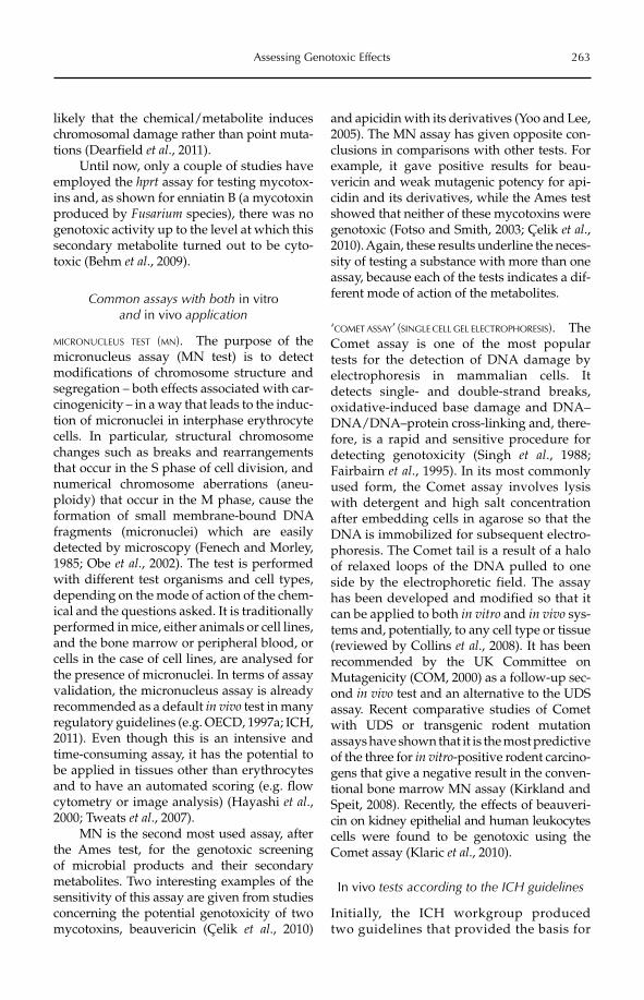
Assessing Genotoxic Effects 263
likely that the chemical/metabolite induces chromosomal damage rather than point muta-tions (Dearfield et al., 2011).
Until now, only a couple of studies have employed the hprt assay for testing mycotox-ins and, as shown for enniatin B (a mycotoxin produced by Fusarium species), there was no genotoxic activity up to the level at which this secondary metabolite turned out to be cyto-toxic (Behm et al., 2009).
Common assays with both in vitroand in vivo application
MICRONUCLEUS TEST (MN). The purpose of the micronucleus assay (MN test) is to detect modifications of chromosome structure and segregation – both effects associated with car-cinogenicity – in a way that leads to the induc-tion of micronuclei in interphase erythrocyte cells. In particular, structural chromosome changes such as breaks and rearrangements that occur in the S phase of cell division, and numerical chromosome aberrations (aneu-ploidy) that occur in the M phase, cause the formation of small membrane-bound DNA fragments (micronuclei) which are easily detected by microscopy (Fenech and Morley, 1985; Obe et al., 2002). The test is performed with different test organisms and cell types, depending on the mode of action of the chem-ical and the questions asked. It is traditionally performed in mice, either animals or cell lines, and the bone marrow or peripheral blood, or cells in the case of cell lines, are analysed for the presence of micronuclei. In terms of assay validation, the micronucleus assay is already recommended as a default in vivo test in many regulatory guidelines (e.g. OECD, 1997a; ICH, 2011). Even though this is an intensive and time-consuming assay, it has the potential to be applied in tissues other than erythrocytes and to have an automated scoring (e.g. flow cytometry or image analysis) (Hayashi et al.,2000; Tweats et al., 2007).
MN is the second most used assay, after the Ames test, for the genotoxic screening of microbial products and their secondary metabolites. Two interesting examples of the sensitivity of this assay are given from studies concerning the potential genotoxicity of two mycotoxins, beauvericin (Çelik et al., 2010)
and apicidin with its derivatives (Yoo and Lee, 2005). The MN assay has given opposite con-clusions in comparisons with other tests. For example, it gave positive results for beau-vericin and weak mutagenic potency for api-cidin and its derivatives, while the Ames test showed that neither of these mycotoxins were genotoxic (Fotso and Smith, 2003; Çelik et al.,2010). Again, these results underline the neces-sity of testing a substance with more than one assay, because each of the tests indicates a dif-ferent mode of action of the metabolites.
‘COMET ASSAY’ (SINGLE CELL GEL ELECTROPHORESIS). The Comet assay is one of the most popular tests for the detection of DNA damage by electrophoresis in mammalian cells. It detects single- and double-strand breaks, oxidative-induced base damage and DNA–DNA/DNA–protein cross-linking and, there-fore, is a rapid and sensitive procedure for detecting genotoxicity (Singh et al., 1988; Fairbairn et al., 1995). In its most commonly used form, the Comet assay involves lysis with detergent and high salt concentration after embedding cells in agarose so that the DNA is immobilized for subsequent electro-phoresis. The Comet tail is a result of a halo of relaxed loops of the DNA pulled to one side by the electrophoretic field. The assay has been developed and modified so that it can be applied to both in vitro and in vivo sys-tems and, potentially, to any cell type or tissue (reviewed by Collins et al., 2008). It has been recommended by the UK Committee on Mutagenicity (COM, 2000) as a follow-up sec-ond in vivo test and an alternative to the UDS assay. Recent comparative studies of Comet with UDS or transgenic rodent mutation assays have shown that it is the most predictive of the three for in vitro-positive rodent carcino-gens that give a negative result in the conven-tional bone marrow MN assay (Kirkland and Speit, 2008). Recently, the effects of beauveri-cin on kidney epithelial and human leukocytes cells were found to be genotoxic using the Comet assay (Klaric et al., 2010).
In vivo tests according to the ICH guidelines
Initially, the ICH workgroup produced two guidelines that provided the basis for
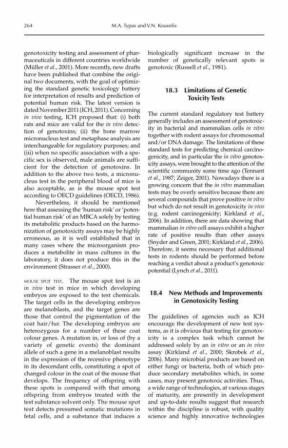
264 M.A. Typas and V.N. Kouvelis
genotoxicity testing and assessment of phar-maceuticals in different countries worldwide (Müller et al., 2001). More recently, new drafts have been published that combine the origi-nal two documents, with the goal of optimiz-ing the standard genetic toxicology battery for interpretation of results and prediction of potential human risk. The latest version is dated November 2011 (ICH, 2011). Concerning in vivo testing, ICH proposed that: (i) both rats and mice are valid for the in vivo detec-tion of genotoxins; (ii) the bone marrow micronucleus test and metaphase analysis are interchangeable for regulatory purposes; and (iii) when no specific association with a spe-cific sex is observed, male animals are suffi-cient for the detection of genotoxins. In addition to the above two tests, a micronu-cleus test in the peripheral blood of mice is also acceptable, as is the mouse spot test according to OECD guidelines (OECD, 1986).
Nevertheless, it should be mentioned here that assessing the ‘human risk’ or ‘poten-tial human risk’ of an MBCA solely by testing its metabolitic products based on the harmo-nization of genotoxicity assays may be highly erroneous, as it is well established that in many cases where the microorganism pro-duces a metabolite in mass cultures in the laboratory, it does not produce this in the environment (Strasser et al., 2000).
MOUSE SPOT TEST. The mouse spot test is an in vivo test in mice in which developing embryos are exposed to the test chemicals. The target cells in the developing embryos are melanoblasts, and the target genes are those that control the pigmentation of the coat hair/fur. The developing embryos are heterozygous for a number of these coat colour genes. A mutation in, or loss of (by a variety of genetic events) the dominant allele of such a gene in a melanoblast results in the ex pression of the recessive phenotype in its de scendant cells, constituting a spot of changed colour in the coat of the mouse that develops. The frequency of offspring with these spots is compared with that among offspring from embryos treated with the test substance solvent only. The mouse spot test detects presumed somatic mutations in fetal cells, and a substance that induces a
biologically significant increase in the number of genetically relevant spots is genotoxic (Russell et al., 1981).
18.3 Limitations of Genetic Toxicity Tests
The current standard regulatory test battery generally includes an assessment of genotoxic-ity in bacterial and mammalian cells in vitrotogether with rodent assays for chromosomal and/or DNA damage. The limitations of these standard tests for predicting chemical carcino-genicity, and in particular the in vitro genotox-icity assays, were brought to the attention of the scientific community some time ago (Tennant et al., 1987; Zeiger, 2001). Now adays there is a growing concern that the in vitro mammalian tests may be overly sensitive because there are several compounds that prove positive in vitrobut which do not result in genotoxicity in vivo(e.g. rodent carcinogenicity; Kirkland et al., 2006). In addition, there are data showing that mammalian in vitro cell assays exhibit a higher rate of positive results than other assays (Snyder and Green, 2001; Kirkland et al., 2006). Therefore, it seems necessary that additional tests in rodents should be performed before reaching a verdict about a product’s genotoxic potential (Lynch et al., 2011).
18.4 New Methods and Improvements in Genotoxicity Testing
The guidelines of agencies such as ICH encourage the development of new test sys-tems, as it is obvious that testing for genotox-icity is a complex task which cannot be addressed solely by an in vitro or an in vivoassay (Kirkland et al., 2000; Skrobek et al.,2006). Many microbial products are based on either fungi or bacteria, both of which pro-duce secondary metabolites which, in some cases, may present genotoxic activities. Thus, a wide range of technologies, at various stages of maturity, are presently in development and up-to-date results suggest that research within the discipline is robust, with quality science and highly innovative technologies
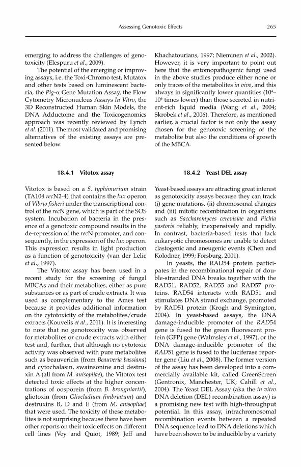
Assessing Genotoxic Effects 265
emerging to address the challenges of geno-toxicity (Elespuru et al., 2009).
The potential of the emerging or improv-ing assays, i.e. the Toxi-Chromo test, Mutatox and other tests based on luminescent bacte-ria, the Pig-a Gene Mutation Assay, the Flow Cytometry Micronucleus Assays In Vitro, the 3D Reconstructed Human Skin Models, the DNA Adductome and the Toxicogenomics approach was recently reviewed by Lynch et al. (2011). The most validated and promising alternatives of the existing assays are pre-sented below.
18.4.1 Vitotox assay
Vitotox is based on a S. typhimurium strain (TA104 recN2-4) that contains the lux operon of Vibrio fisheri under the transcriptional con-trol of the recN gene, which is part of the SOS system. Incubation of bacteria in the pres-ence of a genotoxic compound results in the de-repression of the recN promoter, and con-sequently, in the expression of the lux operon. This expression results in light production as a function of genotoxicity (van der Lelie et al., 1997).
The Vitotox assay has been used in a recent study for the screening of fungal MBCAs and their metabolites, either as pure substances or as part of crude extracts. It was used as complementary to the Ames test because it provides additional information on the cytotoxicity of the metabolites/crude extracts (Kouvelis et al., 2011). It is interesting to note that no genotoxicity was observed for metabolites or crude extracts with either test and, further, that although no cytotoxic activity was observed with pure metabolites such as beauvericin (from Beauveria bassiana)and cytochalasin, swainsonine and destru-xin A (all from M. anisopliae), the Vitotox test detected toxic effects at the higher concen-trations of oosporein (from B. brongniartii), gliotoxin (from Gliocladium fimbriatum) and destru xins B, D and E (from M. anisopliae)that were used. The toxicity of these metabo-lites is not surprising because there have been other reports on their toxic effects on different cell lines (Vey and Quiot, 1989; Jeff and
Khachatourians, 1997; Nieminen et al., 2002). However, it is very important to point out here that the entomopathogenic fungi used in the above studies produce either none or only traces of the metabolites in vivo, and this always in significantly lower quantities (104–106 times lower) than those secreted in nutri-ent-rich liquid media (Wang et al., 2004; Skrobek et al., 2006). Therefore, as mentioned earlier, a crucial factor is not only the assay chosen for the genotoxic screening of the metabolite but also the conditions of growth of the MBCA.
18.4.2 Yeast DEL assay
Yeast-based assays are attracting great interest as genotoxicity assays because they can track (i) gene mutations, (ii) chromosomal changes and (iii) mitotic recombination in organisms such as Saccharomyces cerevisiae and Pichiapastoris reliably, inexpensively and rapidly. In contrast, bacteria-based tests that lack eukaryotic chromosomes are unable to detect clastogenic and aneugenic events (Chen and Kolodner, 1999; Forsburg, 2001).
In yeasts, the RAD54 protein partici-pates in the recombinational repair of dou-ble-stranded DNA breaks together with the RAD51, RAD52, RAD55 and RAD57 pro-teins. RAD54 interacts with RAD51 and stimulates DNA strand exchange, promoted by RAD51 protein (Krogh and Symington, 2004). In yeast-based assays, the DNA damage-inducible promoter of the RAD54 gene is fused to the green fluorescent pro-tein (GFP) gene (Walmsley et al., 1997), or the DNA damage-inducible promoter of the RAD51 gene is fused to the luciferase repor-ter gene (Liu et al., 2008). The former version of the assay has been developed into a com-mercially available kit, called GreenScreen (Gentronix, Manchester, UK; Cahill et al., 2004). The Yeast DEL Assay (aka the in vitroDNA deletion (DEL) recombination assay) is a promising new test with high-throughput potential. In this assay, intrachromosomal recombination events between a repeated DNA sequence lead to DNA deletions which have been shown to be inducible by a variety
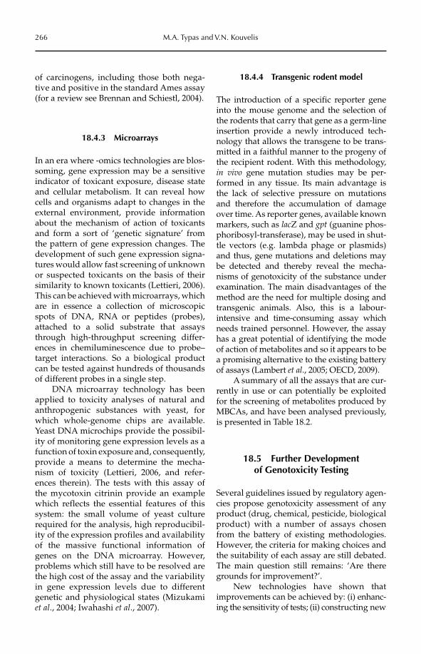
266 M.A. Typas and V.N. Kouvelis
of carcinogens, including those both nega-tive and positive in the standard Ames assay (for a review see Brennan and Schiestl, 2004).
18.4.3 Microarrays
In an era where -omics technologies are blos-soming, gene expression may be a sensitive indicator of toxicant exposure, disease state and cellular metabolism. It can reveal how cells and organisms adapt to changes in the external environment, provide infor mation about the mechanism of action of toxicants and form a sort of ‘genetic signature’ from the pattern of gene expression changes. The development of such gene expression signa-tures would allow fast screening of unknown or suspected toxicants on the basis of their similarity to known toxicants (Lettieri, 2006). This can be achieved with microarrays, which are in essence a collection of microscopic spots of DNA, RNA or peptides (probes), attached to a solid substrate that assays through high-throughput screening differ-ences in chemiluminescence due to probe–target interactions. So a biological product can be tested against hundreds of thousands of different probes in a single step.
DNA microarray technology has been applied to toxicity analyses of natural and anthropogenic substances with yeast, for which whole-genome chips are available. Yeast DNA microchips provide the possibil-ity of monitoring gene expression levels as a function of toxin exposure and, consequently, provide a means to determine the mecha-nism of toxicity (Lettieri, 2006, and refer-ences therein). The tests with this assay of the mycotoxin citrinin provide an example which reflects the essential features of this system: the small volume of yeast culture required for the analysis, high reproducibil-ity of the expression profiles and availability of the massive functional information of genes on the DNA microarray. However, problems which still have to be resolved are the high cost of the assay and the variability in gene expression levels due to different genetic and physiological states (Mizukami et al., 2004; Iwahashi et al., 2007).
18.4.4 Transgenic rodent model
The introduction of a specific reporter gene into the mouse genome and the selection of the rodents that carry that gene as a germ-line insertion provide a newly introduced tech-nology that allows the transgene to be trans-mitted in a faithful manner to the progeny of the recipient rodent. With this methodology, in vivo gene mutation studies may be per-formed in any tissue. Its main advantage is the lack of selective pressure on mutations and therefore the accumulation of damage over time. As reporter genes, available known markers, such as lacZ and gpt (guanine phos-phoribosyl-transferase), may be used in shut-tle vectors (e.g. lambda phage or plasmids) and thus, gene mutations and deletions may be detected and thereby reveal the mecha-nisms of genotoxicity of the substance under examination. The main disadvantages of the method are the need for multiple dosing and transgenic animals. Also, this is a labour-intensive and time-consuming assay which needs trained personnel. However, the assay has a great potential of identifying the mode of action of metabolites and so it appears to be a promising alternative to the existing battery of assays (Lambert et al., 2005; OECD, 2009).
A summary of all the assays that are cur-rently in use or can potentially be exploited for the screening of metabolites produced by MBCAs, and have been analysed previously, is presented in Table 18.2.
18.5 Further Development of Genotoxicity Testing
Several guidelines issued by regulatory agen-cies propose genotoxicity assessment of any product (drug, chemical, pesticide, biological product) with a number of assays chosen from the battery of existing methodologies. However, the criteria for making choices and the suitability of each assay are still debated. The main question still remains: ‘Are there grounds for improvement?’.
New technologies have shown that impro vements can be achieved by: (i) enhanc-ing the sensitivity of tests; (ii) constructing new
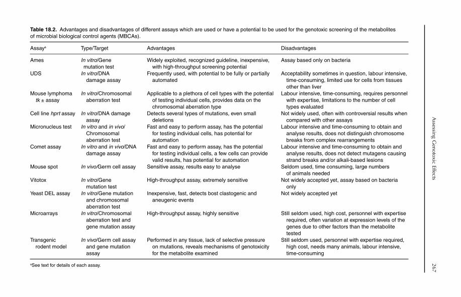
Assessing G
enotoxic Effects267
Table 18.2. Advantages and disadvantages of different assays which are used or have a potential to be used for the genotoxic screening of the metabolites of microbial biological control agents (MBCAs).
Assaya Type/Target Advantages Disadvantages
Ames In vitro/Gene mutation test
Widely exploited, recognized guideline, inexpensive, with high-throughput screening potential
Assay based only on bacteria
UDS In vitro/DNA damage assay
Frequently used, with potential to be fully or partially automated
Acceptability sometimes in question, labour intensive, time-consuming, limited use for cells from tissues other than liver
Mouse lymphoma tk ± assay
In vitro/Chromosomal aberration test
Applicable to a plethora of cell types with the potential of testing individual cells, provides data on the chromosomal aberration type
Labour intensive, time-consuming, requires personnel with expertise, limitations to the number of cell types evaluated
Cell line hprt assay In vitro/DNA damage assay
Detects several types of mutations, even small deletions
Not widely used, often with controversial results when compared with other assays
Micronucleus test In vitro and in vivo/ Chromosomal aberration test
Fast and easy to perform assay, has the potential for testing individual cells, has potential for automation
Labour intensive and time-consuming to obtain and analyse results, does not distinguish chromosome breaks from complex rearrangements
Comet assay In vitro and in vivo/DNA damage assay
Fast and easy to perform assay, has the potential for testing individual cells, a few cells can provide valid results, has potential for automation
Labour intensive and time-consuming to obtain and analyse results, does not detect mutagens causing strand breaks and/or alkali-based lesions
Mouse spot In vivo/Germ cell assay Sensitive assay, results easy to analyse Seldom used, time consuming, large numbers of animals needed
Vitotox In vitro/Gene mutation test
High-throughput assay, extremely sensitive Not widely accepted yet, assay based on bacteria only
Yeast DEL assay In vitro/Gene mutation and chromosomal aberration test
Inexpensive, fast, detects bost clastogenic and aneugenic events
Not widely accepted yet
Microarrays In vitro/Chromosomal aberration test and gene mutation assay
High-throughput assay, highly sensitive Still seldom used, high cost, personnel with expertise required, often variation at expression levels of the genes due to other factors than the metabolite tested
Transgenic rodent model
In vivo/Germ cell assay and gene mutation assay
Performed in any tissue, lack of selective pressure on mutations, reveals mechanisms of genotoxicity for the metabolite examined
Still seldom used, personnel with expertise required, high cost, needs many animals, labour intensive, time-consuming
aSee text for details of each assay.
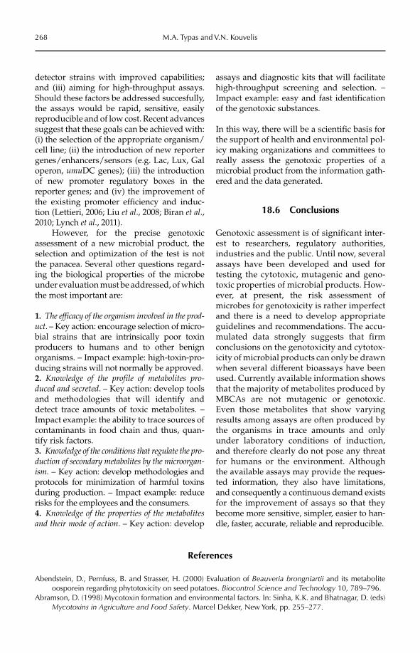
268 M.A. Typas and V.N. Kouvelis
detector strains with improved capabilities;and (iii) aiming for high-throughput assays. Should these factors be addressed succesfully, the assays would be rapid, sensitive, easily reproducible and of low cost. Recent advances suggest that these goals can be achieved with: (i) the selection of the appropriate organism/cell line; (ii) the introduction of new reporter genes/enhancers/sensors (e.g. Lac, Lux, Gal operon, umuDC genes); (iii) the introduction of new promoter regulatory boxes in the reporter genes; and (iv) the improvement of the existing promoter efficiency and induc-tion (Lettieri, 2006; Liu et al., 2008; Biran et al.,2010; Lynch et al., 2011).
However, for the precise genotoxic assessment of a new microbial product, the selection and optimization of the test is not the panacea. Several other questions regard-ing the biological properties of the microbe under evaluation must be addressed, of which the most important are:
1. The efficacy of the organism involved in the prod-uct. – Key action: encourage selection of micro-bial strains that are intrinsically poor toxin producers to humans and to other benign organisms. – Impact example: high-toxin-pro-ducing strains will not normally be approved.2. Knowledge of the profile of metabolites pro-duced and secreted. – Key action: develop tools and methodologies that will identify and detect trace amounts of toxic metabolites. – Impact example: the ability to trace sources of contaminants in food chain and thus, quan-tify risk factors.3. Knowledge of the conditions that regulate the pro-duction of secondary metabolites by the microorgan-ism. – Key action: develop methodologies and protocols for minimization of harmful toxins during production. – Impact example: reduce risks for the employees and the consumers.4. Knowledge of the properties of the metabolites and their mode of action. – Key action: develop
assays and diagnostic kits that will facilitate high-throughput screening and selection. – Impact example: easy and fast identification of the genotoxic substances.
In this way, there will be a scientific basis for the support of health and environmental pol-icy making organizations and committees to really assess the genotoxic properties of a microbial product from the information gath-ered and the data generated.
18.6 Conclusions
Genotoxic assessment is of significant inter-est to researchers, regulatory authorities, industries and the public. Until now, several assays have been developed and used for testing the cytotoxic, mutagenic and geno-toxic pro perties of microbial products. How-ever, at present, the risk assessment of microbes for genotoxicity is rather imperfect and there is a need to develop appropriate guidelines and recommendations. The accu-mulated data strongly suggests that firm conclusions on the genotoxicity and cytotox-icity of microbial products can only be drawn when several different bioassays have been used. Currently available information shows that the majority of metabolites produced by MBCAs are not mutagenic or genotoxic. Even those metabolites that show varying results among assays are often produced by the organisms in trace amounts and only under laboratory conditions of induction, and therefore clearly do not pose any threat for humans or the environment. Although the available assays may provide the reques-ted information, they also have limitations, and consequently a continuous demand exists for the improvement of assays so that they become more sensitive, simpler, easier to han-dle, faster, accurate, reliable and reproducible.
References
Abendstein, D., Pernfuss, B. and Strasser, H. (2000) Evaluation of Beauveria brongniartii and its metabolite oosporein regarding phytotoxicity on seed potatoes. Biocontrol Science and Technology 10, 789–796.
Abramson, D. (1998) Mycotoxin formation and environmental factors. In: Sinha, K.K. and Bhatnagar, D. (eds) Mycotoxins in Agriculture and Food Safety. Marcel Dekker, New York, pp. 255–277.
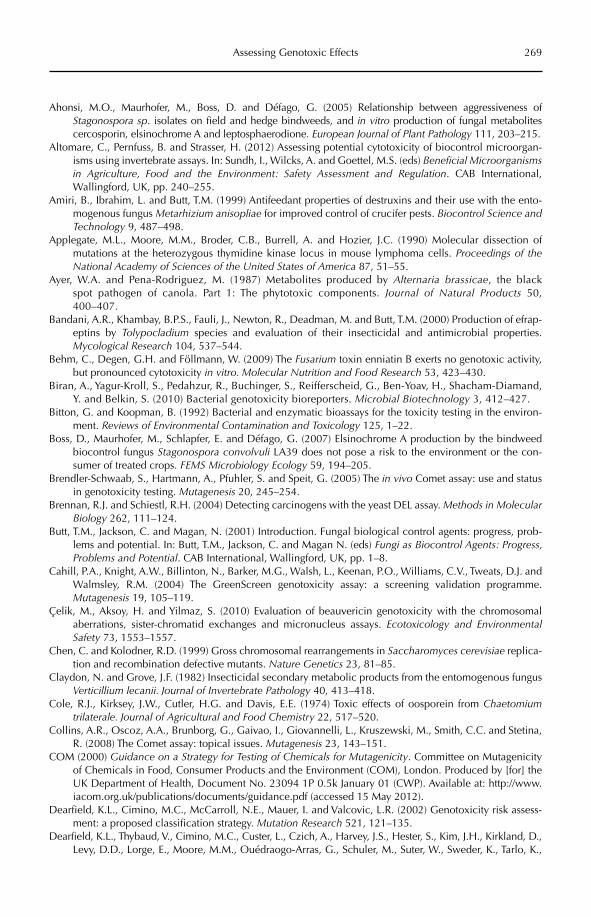
Assessing Genotoxic Effects 269
Ahonsi, M.O., Maurhofer, M., Boss, D. and Défago, G. (2005) Relationship between aggressiveness of Stagonospora sp. isolates on field and hedge bindweeds, and in vitro production of fungal metabolites cercosporin, elsinochrome A and leptosphaerodione. European Journal of Plant Pathology 111, 203–215.
Altomare, C., Pernfuss, B. and Strasser, H. (2012) Assessing potential cytotoxicity of biocontrol microorgan-isms using invertebrate assays. In: Sundh, I., Wilcks, A. and Goettel, M.S. (eds) Beneficial Microorganisms in Agriculture, Food and the Environment: Safety Assessment and Regulation. CAB International, Wallingford, UK, pp. 240–255.
Amiri, B., Ibrahim, L. and Butt, T.M. (1999) Antifeedant properties of destruxins and their use with the ento-mogenous fungus Metarhizium anisopliae for improved control of crucifer pests. Biocontrol Science and Technology 9, 487–498.
Applegate, M.L., Moore, M.M., Broder, C.B., Burrell, A. and Hozier, J.C. (1990) Molecular dissection of mutations at the heterozygous thymidine kinase locus in mouse lymphoma cells. Proceedings of the National Academy of Sciences of the United States of America 87, 51–55.
Ayer, W.A. and Pena-Rodriguez, M. (1987) Metabolites produced by Alternaria brassicae, the black spot pathogen of canola. Part 1: The phytotoxic components. Journal of Natural Products 50, 400–407.
Bandani, A.R., Khambay, B.P.S., Fauli, J., Newton, R., Deadman, M. and Butt, T.M. (2000) Production of efrap-eptins by Tolypocladium species and evaluation of their insecticidal and antimicrobial properties. Mycological Research 104, 537–544.
Behm, C., Degen, G.H. and Föllmann, W. (2009) The Fusarium toxin enniatin B exerts no genotoxic activity, but pronounced cytotoxicity in vitro. Molecular Nutrition and Food Research 53, 423–430.
Biran, A., Yagur-Kroll, S., Pedahzur, R., Buchinger, S., Reifferscheid, G., Ben-Yoav, H., Shacham-Diamand, Y. and Belkin, S. (2010) Bacterial genotoxicity bioreporters. Microbial Biotechnology 3, 412–427.
Bitton, G. and Koopman, B. (1992) Bacterial and enzymatic bioassays for the toxicity testing in the environ-ment. Reviews of Environmental Contamination and Toxicology 125, 1–22.
Boss, D., Maurhofer, M., Schlapfer, E. and Défago, G. (2007) Elsinochrome A production by the bindweed biocontrol fungus Stagonospora convolvuli LA39 does not pose a risk to the environment or the con-sumer of treated crops. FEMS Microbiology Ecology 59, 194–205.
Brendler-Schwaab, S., Hartmann, A., Pfuhler, S. and Speit, G. (2005) The in vivo Comet assay: use and status in genotoxicity testing. Mutagenesis 20, 245–254.
Brennan, R.J. and Schiestl, R.H. (2004) Detecting carcinogens with the yeast DEL assay. Methods in Molecular Biology 262, 111–124.
Butt, T.M., Jackson, C. and Magan, N. (2001) Introduction. Fungal biological control agents: progress, prob-lems and potential. In: Butt, T.M., Jackson, C. and Magan N. (eds) Fungi as Biocontrol Agents: Progress, Problems and Potential. CAB International, Wallingford, UK, pp. 1–8.
Cahill, P.A., Knight, A.W., Billinton, N., Barker, M.G., Walsh, L., Keenan, P.O., Williams, C.V., Tweats, D.J. and Walmsley, R.M. (2004) The GreenScreen genotoxicity assay: a screening validation programme. Mutagenesis 19, 105–119.
Çelik, M., Aksoy, H. and Yilmaz, S. (2010) Evaluation of beauvericin genotoxicity with the chromosomal aberrations, sister-chromatid exchanges and micronucleus assays. Ecotoxicology and Environmental Safety 73, 1553–1557.
Chen, C. and Kolodner, R.D. (1999) Gross chromosomal rearrangements in Saccharomyces cerevisiae replica-tion and recombination defective mutants. Nature Genetics 23, 81–85.
Claydon, N. and Grove, J.F. (1982) Insecticidal secondary metabolic products from the entomogenous fungus Verticillium lecanii. Journal of Invertebrate Pathology 40, 413–418.
Cole, R.J., Kirksey, J.W., Cutler, H.G. and Davis, E.E. (1974) Toxic effects of oosporein from Chaetomiumtrilaterale. Journal of Agricultural and Food Chemistry 22, 517–520.
Collins, A.R., Oscoz, A.A., Brunborg, G., Gaivao, I., Giovannelli, L., Kruszewski, M., Smith, C.C. and Stetina, R. (2008) The Comet assay: topical issues. Mutagenesis 23, 143–151.
COM (2000) Guidance on a Strategy for Testing of Chemicals for Mutagenicity. Committee on Mutagenicity of Chemicals in Food, Consumer Products and the Environment (COM), London. Produced by [for] the UK Department of Health, Document No. 23094 1P 0.5k January 01 (CWP). Available at: http://www.iacom.org.uk/publications/documents/guidance.pdf (accessed 15 May 2012).
Dearfield, K.L., Cimino, M.C., McCarroll, N.E., Mauer, I. and Valcovic, L.R. (2002) Genotoxicity risk assess-ment: a proposed classification strategy. Mutation Research 521, 121–135.
Dearfield, K.L., Thybaud, V., Cimino, M.C., Custer, L., Czich, A., Harvey, J.S., Hester, S., Kim, J.H., Kirkland, D., Levy, D.D., Lorge, E., Moore, M.M., Ouédraogo-Arras, G., Schuler, M., Suter, W., Sweder, K., Tarlo, K.,
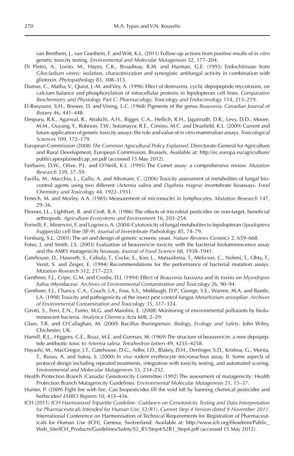
270 M.A. Typas and V.N. Kouvelis
van Benthem, J., van Goethem, F. and Witt, K.L. (2011) Follow-up actions from positive results of in vitrogenetic toxicity testing. Environmental and Molecular Mutagenesis 52, 177–204.
Di Pietro, A., Lorito, M., Hayes, C.K., Broadway, R.M. and Harman, G.E. (1993) Endochitinase from Gliocladium virens: isolation, characterization and synergistic antifungal activity in combination with gliotoxin. Phytopathology 83, 308–313.
Dumas, C., Matha, V., Quiot, J.-M. and Vey, A. (1996) Effect of destruxins, cyclic depsipeptide mycotoxins, on calcium balance and phosphorylation of intracellular proteins in lepidopteran cell lines. Comparative Biochemistry and Physiology Part C: Pharmacology, Toxicology and Endocrinology 114, 213–219.
El-Basyouni, S.H., Brewer, D. and Vining, L.C. (1968) Pigments of the genus Beauveria. Canadian Journal of Botany 46, 441–448.
Elespuru, R.K., Agarwal, R., Atrakchi, A.H., Bigger, C.A., Heflich, R.H., Jagannath, D.R., Levy, D.D., Moore, M.M., Ouyang, Y., Robison, T.W., Sotomayor, R.E., Cimino, M.C. and Dearfield, K.L. (2009) Current and future application of genetic toxicity assays: the role and value of in vitro mammalian assays. Toxicological Sciences 109, 172–179.
European Commission (2008) The Common Agricultural Policy Explained. Directorate-General for Agriculture and Rural Development, European Commission, Brussels. Available at: http://ec.europa.eu/agriculture/publi/capexplained/cap_en.pdf (accessed 15 May 2012).
Fairbairn, D.W., Olive, P.L. and O’Neill, K.L. (1995) The Comet assay: a comprehensive review. Mutation Research 339, 37–59.
Favilla, M., Macchia, L., Gallo, A. and Altomare, C. (2006) Toxicity assessment of metabolites of fungal bio-control agents using two different (Artemia salina and Daphnia magna) invertebrate bioassays. Food Chemistry and Toxicology 44, 1922–1931.
Fenech, M. and Morley, A.A. (1985) Measurement of micronuclei in lymphocytes. Mutation Research 147, 29–36.
Flexner, J.L., Lighthart, B. and Croft, B.A. (1986) The effects of microbial pesticides on non-target, beneficial arthropods. Agriculture Ecosystems and Environment 16, 203–254.
Fornelli, F., Minervini, F. and Logrieco, A. (2004) Cytotoxicity of fungal metabolites to lepidopteran (Spodopterafrugiperda) cell line (SF-9). Journal of Invertebrate Patholology 85, 74–79.
Forsburg, S.L. (2001) The art and design of genetic screens: yeast. Nature Reviews Genetics 2, 659–668.Fotso, J. and Smith, J.S. (2003) Evaluation of beauvericin toxicity with the bacterial bioluminescence assay
and the AMES mutagenicity bioassay. Journal of Food Science 68, 1938–1941.Gatehouse, D., Haworth, S., Cebula, T., Cocke, E., Kier, L., Matsushima, T., Melicion, C., Nohmi, T., Ohta, T.,
Venit, S. and Zeiger, E. (1994) Recommendations for the performance of bacterial mutation assays. Mutation Research 312, 217–223.
Genthner, F.J., Cripe, G.M. and Crosby, D.J. (1994) Effect of Beauveria bassiana and its toxins on Mysodopsis bahia (Mysidacea). Archives of Environmental Contamination and Toxicology 26, 90–94.
Genthner, F.J., Chancy, C.A., Couch, J.A., Foss, S.S., Middaugh, D.P., George, S.E., Warren, M.A. and Bantle, J.A. (1998) Toxicity and pathogenicity of the insect pest control fungus Metarhizium anisopliae. Archives of Environmental Contamination and Toxicology 35, 317–324.
Girotti, S., Ferri, E.N., Fumo, M.G. and Maiolini, E. (2008) Monitoring of environmental pollutants by biolu-minescent bacteria. Analytica Chemica Acta 608, 2–29.
Glare, T.R. and O’Callaghan, M. (2000) Bacillus thuringiensis: Biology, Ecology and Safety. John Wiley, Chichester, UK.
Hamill, R.L., Higgens, C.E., Boaz, M.E. and Gorman, M. (1969) The structure of beauvericin, a new depsipep-tide antibiotic toxic to Artemia salina. Tetrahedron Letters 49, 4255–4258.
Hayashi, M., MacGregor, J.T., Gatehouse, D.G., Adler, I.D., Blakey, D.H., Dertinger, S.D., Krishna, G., Morita, T., Russo, A. and Sutou, S. (2000) In vivo rodent erythrocyte micronucleus assay. II. Some aspects of protocol design including repeated treatments, integration with toxicity testing, and automated scoring. Environmental and Molecular Mutagenesis 35, 234–252.
Health Protection Branch (Canada) Genotoxicity Committee (1992) The assessment of mutagenicity: Health Protection Branch Mutagenicity Guidelines. Environmental Molecular Mutagenesis 21, 15–37.
Hunter, P. (2009) Fight fire with fire. Can biopesticides fill the void left by banning chemical pesticides and herbicides? EMBO Reports 10, 433–436.
ICH (2011) ICH Harmonized Tripartite Guideline: Guidance on Genotoxicity Testing and Data Interpretation for Pharmaceuticals Intended for Human Use, S2(R1), Current Step 4 Version dated 9 November 2011.International Conference on Harmonisation of Technical Requirements for Registration of Pharmaceut-icals for Human Use (ICH), Geneva, Switzerland. Available at: http://www.ich.org/fileadmin/Public_Web_Site/ICH_Products/Guidelines/Safety/S2_R1/Step4/S2R1_Step4.pdf (accessed 15 May 2012).
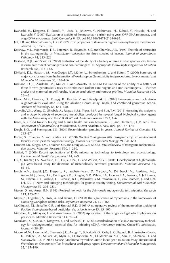
Assessing Genotoxic Effects 271
Iwahashi, H., Kitagawa, E., Suzuki, Y., Ueda, Y., Ishizawa, Y., Nobumasa, H., Kuboki, Y., Hosoda, H. and Iwahashi, Y. (2007) Evaluation of toxicity of the mycotoxin citrinin using yeast ORF DNA microarray and oligo DNA microarray. BMC Genomics 8, 95. doi:10.1186/1471-2164-8-95.
Jeff, L.B. and Khachatourians, G.G. (1997) Toxic properties of Beauveria pigments on erythrocyte membranes. Toxicon 35, 1351–1356.
Kershaw, M.J., Moorhouse, E.R., Bateman, R., Reynolds, S.E. and Charnley, A.K. (1999) The role of destruxins in the pathogenicity of Metarhizium anisopliae for three species of insects. Journal of Invertebrate Pathology 74, 213–223.
Kirkland, D.[J.] and Speit, G. (2008) Evaluation of the ability of a battery of three in vitro genotoxicity tests to discriminate rodent carcinogens and non-carcinogens. III. Appropriate follow-up testing in vivo. Mutation Research 654, 114–132.
Kirkland, D.J., Hayashi, M., MacGregor, J.T., Müller, L., Scherchtman, L. and Sofuni, T. (2000) Summary of major conclusions from the International Workshop on Genotoxicity test procedures. Environmental and Molecular Mutagenesis 35, 162–166.
Kirkland, D.[J.], Aardema, M., Muller, L. and Makoto, H. (2006) Evaluation of the ability of a battery of three in vitro genotoxicity tests to discriminate rodent carcinogens and non-carcinogens. II. Further analysis of mammalian cell results, relative predictivity and tumour profiles. Mutation Research 608, 29–42.
Klaric, M.S., Darabos, D., Rozgaj, R., Kasuba, V. and Pepeljnjak, S. (2010) Beauvericin and ochratoxin A genotoxicity evaluated using the alkaline Comet assay: single and combined genotoxic action. Archives of Toxicology 84, 641–650.
Kouvelis, V.N., Wang, C., Skrobek, A., Pappas, K.M., Typas, M.A. and Butt, T.M. (2011) Assessing the toxigenic and mutagenic effects of secondary metabolites produced by several fungal biological control agents with the Ames assay and the VITOTOX® test. Mutation Research 722, 1–6.
Kroes, R. (1995) Toxicity testing and human health. In: van Leeuwen, C.J. and Hermens, J.L.M. (eds) RiskAssessment of Chemicals: An Introduction. Kluwer Academic, New York, pp. 147–174.
Krogh, B.O. and Symington, L.S. (2004) Recombination proteins in yeasts. Annual Review of Genetics 38, 233–271.
Kumar, S., Chandra, A. and Pandey, K.C. (2008) Bacillus thuringiensis (Bt) transgenic crop: an environment friendly insect-pest management strategy. Journal of Environmental Biology 29, 641–653.
Lambert, I.B., Singer, T.M., Boucher, S.E. and Douglas, G.R. (2005) Detailed review of transgenic rodent muta-tion assays. Mutation Research 590, 1–280.
Lettieri, T. (2006) Recent applications of DNA microarray technology to toxicology and ecotoxicology. Environmental Health Perspectives 114, 4–9.
Liu, X., Kramer, J.A., Swaffield, J.C., Hu, Y., Chai, G. and Wilson, A.G.E. (2008) Development of highthrough-put yeast-based assay for detection of metabolically activated genotoxins. Mutation Research 31, 63–69.
Lynch, A.M., Sasaki, J.C., Elespuru, R., Jacobson-Kram, D., Thybaud, V., De Boeck, M., Aardema, M.J., Aubrecht, J., Benz, D.R., Dertinger, S.D., Douglas, G.R., White, P.A., Escobar, P.A., Fornace, A. Jr, Honma, M., Naven, R.T., Rusling, J.F., Schiestl, R.H., Walmsley, R.M., Yamamura, E., van Benthem, J. and Kim, J.H. (2011) New and emerging technologies for genetic toxicity testing. Environmental and Molecular Mutagenesis 52, 205–223.
Maron, D. and Ames, B.N. (1983) Revised methods for the Salmonella mutagenicity test. Mutation Research113, 173–215.
Mayer, S., Engelhart, S., Kolk, A. and Blome, H. (2008) The significance of mycotoxins in the framework of assessing workplace related risks. Mycotoxin Research 24, 151–164.
McClintock, T.J., Schaffer, C.R. and Sjoblad, R.D. (1995) A comparative review of the mammalian toxicity of Bacillus thuringiensis-based pesticides. Pesticide Science 45, 95–105.
Miloshev, G., Mihaylov, I. and Anachkova, B. (2002) Application of the single cell gel electrophoresis on yeast cells. Mutation Research 513, 69–74.
Mizukami, S., Suzuki, Y., Kitagawa, E. and Iwahashi, H. (2004) Standardization of cDNA microarray technol-ogy for toxicogenomics; essential data for initiating cDNA microarray studies. Chem-Bio Informatics Journal 4, 38–55.
Moore, M.M., Honma, M., Clements, J.C., Awogi, T., Bolcsfoldi, G., Cole, J., Gollapudi, B., Harrington-Brock, K., Mitchell, A., Muster, W., Myhr, B., O’Donovan, M., Ouldehkhim, M.C., San, R., Shimada, H. and Stankowski, L.F. Jr (2000) Mouse lymphoma thymidine kinase locus gene mutation assay: International Workshop on Genotoxicity Test Procedures workgroup report. Environmental and Molecular Mutagenesis35, 185–190.
272 M.A. Typas and V.N. Kouvelis
Moretti, A., Logrieco, A., Bottalico, A., Ritienti, A., Randazzo, G. and Corda, P. (1995) Beauvericin production by Fusarium subglutinans from different geographical areas. Mycological Research 99, 282–286.
Mori, H., Kawai, K., Ohbayashi, F., Kuniyasu, T., Yamazaki, M., Hamasaki, T. and Williams, G.M. (1984) Genotoxicity of a variety of mycotoxins in the hepatocyte primary culture/DNA repair test using rat and mouse hepatocytes. Cancer Research 44, 2918–2923.
Mortelmans, K. and Zeiger, E. (2000) The Ames Salmonella/microsome mutagenicity assay. Mutation Research455, 29–60.
Müller, L., Kasper, P. and Schechtman, L. (2001) ICH guidances on genotoxicity and carcinogenicity: scientific background and regulatory practice. In: Choy, W.N. (ed.) Genetic Toxicology and Cancer Risk Assessment.Marcel Dekker, New York, pp. 187–221.
Nieminen, S.M., Maki-Paakkanen, J., Hirvonen, M.-R., Roponen, M. and von Wright, A. (2002) Genotoxicity of gliotoxin, a secondary metabolite of Aspergillus fumigatus, in a battery of short-term test systems. Mutation Research 520, 161–170.
Norred, W.P., Plattner, R.D., Vesonder, R.F., Bacon, C.W. and Voss, K.A. (1992) Effects of selected secondary metabolites of Fusarium moniliforme on unscheduled synthesis of DNA by rat primary hepatocytes. Food and Chemical Toxicology 30, 233–237.
Obe, G., Pfeiffer, P., Savage, J.R.K., Johannes, C., Goedecke, W., Jeppesen, P., Natarajan, A.T., Martínez-López, W., Folle, G.A. and Drets, M.E. (2002) Chromosomal aberrations: formation, identification and distribution. Mutation Research 504, 17–36.
Odam, Y., Nakamura, S., Oki, I. and Kato, T. (1985) Evaluation of a new system (Umu-test) for the detection of environmental mutagens and carcinogens. Mutation Research 147, 219–229.
Odier, F., Vey, A. and Bureau, J.P. (1992) In vitro effect of fungal cyclodepsipeptides on leukemic cells: study of destruxins A, B and E. Biology of the Cell 74, 267–271.
OECD (1986) Genetic Toxicology: Mouse Spot Test. OECD Guideline for the Testing of Chemicals, No. 484. Organisation for Economic Co-operation and Development (OECD), Paris. Available at: http://www.oecd-ilibrary.org/content/book/9789264071483-en (accessed 27 October 2011).
OECD (1997a) Mammalian Erythrocyte Micronucleus Test. OECD Guideline for the Testing of Chemicals, No. 474. Organisation for Economic Co-operation and Development (OECD), Paris. Available at: http://www.oecd-ilibrary.org/content/book/9789264071285-en (accessed 27 October 2011).
OECD (1997b) Unscheduled DNA Synthesis (UDS) Test with Mammalian Liver Cells In Vivo. OECD Guideline for the Testing of Chemicals, No. 486. Organisation for Economic Co-operation and Development (OECD), Paris. Available at: http://browse.oecdbookshop.org/oecd/pdfs/free/9748601e.pdf (accessed 27 October 2011).
OECD (2007) Section 4 – Health Effects: Introduction to the OECD Guidelines on genetic toxicology testing and guidance on the selection and application of assays. In: OECD Guidelines for Testing of Chemicals: Full List of Test Guidelines, August 2007. Organisation for Economic Co-operation and Development, Paris, pp. 9–10. Available at: http://www.oecd.org/dataoecd/9/11/33663321.pdf (accessed 15 May 2012).
OECD (2008) Working Document on the Evaluation of Microbials for Pest Control. OECD Environment, Health and Safety Publications Series on Pesticides No. 43, Environment Directorate, Joint Meeting of the Chemicals Committee and the Working Party on Chemicals, Pesticides and Biotechnology, Document No. ENV/JM/MONO(2008)36. Organisation for Economic Co-operation and Development, Paris. Available at: http://www.oecd.org/dataoecd/45/46/41946259.pdf (accessed 27 October 2011).
OECD (2009) Detailed Review Paper on Transgenic Rodent Mutation Assays; Part 2: Annexes to the Detailed Review Paper on Transgenic Rodent Mutation, Assays. Series on Testing and Assessment, No. 103, Environment Directorate, Joint Meeting of the Chemicals Committee and the Working Party on Chemicals, Pesticides and Biotechnology, Document Nos ENV/JM/MONO(2009)7, ENV/JM/MONO(2009)29, Organisation for Economic Co-operation and Development, Paris. Available at: http://www.oecd.org/officialdocuments/displaydocumentpdf/?cote=env/jm/mono(2009)7&doclanguage=en; http://www.oecd.org/officialdocuments/displaydocumentpdf/?cote=env/jm/mono(2009)29&doclanguage=en (accessed 15 May 2012).
OJEC (1991) Council Directive 91/414/EEC of 15 July 1991 concerning the placing of plant protection prod-ucts on the market. Official Journal of the European Communities 34, L 230/1–32.
OJEU (2009) Regulation (EC) No 1107/2009 of the European Parliament and of the Council of 21 October 2009 concerning the placing of plant protection products on the market and repealing Council Directives 79/117/EEC and 91/414/EEC. Official Journal of the European Union 52, L 309/1–50.
Pais, M., Das, B.C. and Ferron, P. (1981) Depsipeptides from Metarhizium anisopliae. Phytochemistry 20, 715–723.
Assessing Genotoxic Effects 273
Patrick, M., Adlard, M.W. and Keshavarz, T. (1993) Production of an indolizidine alkaloid swainsonine by the filamentous fungus, Metarhizium anisopliae. Biotechnology Letters 15, 997–1000.
Richard, J.M., Ravanel, P. and Cantin, D. (1987) Phytotoxicity of orellanine, a mushroom toxin. Toxicon 25, 350–354.
Ruiz, M.J. and Marzin, D. (1997) Genotoxicity of six pesticides by Salmonella mutagenicity test and SOS chromotest. Mutation Research 390, 245–255.
Rusling, J.F., Hvastkovs, E.G. and Schenkman, J.B. (2007) Toxicity screening using biosensors that measure DNA damage. Current Opinion in Drug Discovery and Development 10, 67–73.
Russell, L.B., Selby, P.B., von Halle, E., Sheridan, W. and Valcovic, L. (1981) Use of the mouse spot test in chemical mutagenesis: interpretation of past data and recommendations for future work. Mutation Research 86, 355–379.
Sako, F., Kobayashi, N., Taniguchi, N. and Takakuwa, E. (1978) A study on the toxicity of natural food dyes – toxicity and enzyme inhibition in Paramecium caudatum. Toxicological Sciences 3, 127–136.
Seigle-Murandi, F., Krivoboc, S., Steiman, R. and Marzin, D. (1990) Production, mutagenicity and immuno-toxicity of gliotoxin. Journal of Agricultural and Food Chemistry 38, 1854–1856.
Singh, N.P., McCoy, M.T., Tice, R.R. and Schneider, E.L. (1988) A simple technique for quantitation of low levels of DNA damage in individual cells. Experimental Cell Research 175, 184–191.
Skrobek, A., Boss, D., Défago, G., Butt, T.M. and Mauerhofer, M. (2006) Evaluation of different biological test systems to assess the toxicity of metabolites from fungal biocontrol agents. Toxicology Letters 161, 43–52.
Snyder, R.D. and Green, J.W. (2001) A review of the genotoxicity of marketed pharmaceuticals. Mutation Research 488, 151–169.
Sofuni, T. (1993) Japanese guidelines for mutagenicity testing. Environmental and Molecular Mutagenesis 21, 2–7.
Strasser, H., Vey, A. and Butt, T.M. (2000) Are there any risks in using entomopathogenic fungi for pest control, with particular reference to the bioactive metabolites of Metarhizium, Tolypocladium and Beauveriaspecies? Biocontrol Science and Technology 10, 717–735.
Sundh, I., Wilcks, A. and Goettel, M.S. (2012) Microbes and the law – safety assessment and regulation of beneficial microorganisms. In: Sundh, I., Wilcks, A. and Goettel, M.S. (eds) Beneficial Microorganisms in Agriculture, Food and the Environment: Safety Assessment and Regulation. CAB International, Wallingford, UK, pp. 1–11.
Suzuki, A., Kanaoka, M., Isogai, A., Kurakoshi, S., Ichinoe, M. and Tamura, S. (1977) Bassianolide, a new insecticidal cyclodepsipeptide from Beauveria bassiana and Verticillium lecanii. Tetrahedron Letters 25, 2167–2170.
Tabashnik, B.E., Gassmann, A.J., Crowder, D.W. and Carriére, Y. (2008) Insect resistance to Bt crops: evidence versus theory. Nature Biotechnology 26, 199–202.
Taylor, A. (1986) Some aspects of the chemistry and biology of the genus Hypocrea and its anamorphs, Trichoderma and Gliocladium. Proceedings of the Nova Scotian Institute of Science 36, 27–58.
Tennant, R.W., Margolin, B.H., Shelby, M.D., Zeiger, E., Haseman, J.K., Spalding, J., Caspary, W., Resnick, M., Stasiewicz, S., Anderson, B. and Minor, R. (1987) Prediction of chemical carcinogenicity in rodents from in vitro genetic toxicity assays. Science 236, 933–941.
Terry, B.J., Liu, W.-C., Cianci, C.W., Proszynski, E., Fernandes, P. and Meyers, E. (1992) Inhibition of herpes sim-plex virus type I DNA polymerase by the natural product oosporein. Journal of Antibiotics 2, 286–288.
Tice, R.R., Agurell, E., Anderson, D., Burlinson, B., Hartmann, A., Kobayashi, H., Miyamae, Y., Rojas, E., Ryu, J.-C. and Sasaki, Y.F. (2000) The single cell gel/Comet assay: guidelines for in vitro and in vivo genetic toxicology testing. Environmental and Molecular Mutagenesis 35, 206–221.
Tweats, D.J., Blakey, D., Heflich, R.H., Jacobs, A., Jacobsen, S.D., Morita, T., Nohmi, T., O’Donovan, M.R., Sasaki, Y.F., Sofuni, T. and Tice, R. (2007) Report of the IWGT working group on strategies and interpreta-tion of regulatory in vivo tests I. Increases in micronucleated bone marrow cells in rodents that do not indicate genotoxic hazards. Mutation Research 627, 78–91.
US FDA (2006) Guidance for Industry and Review Staff: Recommended Approaches to Integration of Genetic Toxicology Study Results. Center for Drug Evaluation and Research, US Food and Drug Administration, Washington, DC. Available at: http://www.fda.gov/ohrms/dockets/98fr/04d-0493-gdl0002.pdf (accessed 27 October 2011).
van der Lelie, D., Regniers, L., Borremans, B., Provoost, A. and Verschaeve, L. (1997) The VITOTOX® test, an SOS bioluminescence Salmonella typhimurium test to measure genotoxicity kinetics. Mutation Research389, 279–290.
274 M.A. Typas and V.N. Kouvelis
Vey, A. and Quiot, J.-M. (1989) Etude in vitro et chez l’insecte hôte des destruxines, toxins cyclodepsipep-tidiques produites par le champignon entomopathogène Metarhizium anisopliae. Canadian Journal of Microbiology 35, 1000–1008.
Vey, A., Hoagland, R. and Butt, T.M. (2001) Toxic metabolites of fungal biocontrol agents. In: Butt, T.M., Jackson, C. and Magan, N. (eds) Fungi as Biocontrol Agents: Progress, Problems and Potential. CAB International, Wallingford, UK, pp. 311–346.
Walmsley, R.M., Billinton, N. and Heyer, W.D. (1997) Green fluorescent protein as a reporter for the DNA damage-induced gene RAD54 in Saccharomyces cerevisiae. Yeast 13, 1535–1545.
Wang, C., Skrobek, A. and Butt, T.M. (2004) Investigations on the destruxin production of the entomopatho-genic fungus Metarhizium anisopliae in liquid and solid media. Journal of Invertebrate Pathology 85, 168–174.
Wang, J., Sawyer, J.R., Chen, L., Chen, T., Honma, M., Mei, N. and Moore, M.M. (2009) The mouse lymphoma assay detects recombination, deletion, and aneuploidy. Toxicological Sciences 109, 96–105.
Wat, C.-K., McInnes, A.G., Smith, D.G., Wright, J.L.C. and Vining, L.C. (1977) The yellow pigments of Beauveria species. Structures of tenellin and bassianin. Canadian Journal of Chemistry 55, 4090–4098.
Wiest, A., Grzegorski, D., Xu, B.-W., Goulard, C., Rebuffat, S., Ebbole, D.J., Bodo, B. and Kenerley, C. (2002) Identification of peptaibols from Trichoderma virens and cloning of a peptaibol synthetase. Journal of Biological Chemistry 277, 20862–20868.
Williams, G.M. (1976) Carcinogen-induced DNA repair in primary rat liver cell cultures: a possible screen for chemical carcinogens. Cancer Letters 1, 231–236.
Yoo, E.J. and Lee, B.M. (2005) Comparative mutagenicity of apicidin and apicidin derivatives (SD-0203 and SD-2007), histone deacetylase inhibitors. Journal of Toxicology and Environmental Health, Part A 68, 2097–2109.
Zeiger, E. (2001) Mutagens that are not carcinogens: faulty theory or faulty tests? Mutation Research 492, 29–38; Erratum: Mutation Research 498, 219.
©CAB International 2012. Beneficial Microorganisms in Agriculture, Food and the Environment: Safety Assessment and Regulation (eds I. Sundh et al.) 275
19.1 Introduction 27519.2 Irritation and Allergic Contact Dermatitis 276
19.2.1 Irritation contact dermatitis (ICD) 276 19.2.2 Allergic contact dermatitis (ACD) 277 19.2.3 ICD versus ACD 277
19.3 Testing Methodologies 27819.3.1 Methods for skin irritation 278
19.3.2 Methods for skin sensitization 28219.4 Microorganisms and Irritation/Sensitization 284
19.4.1 Data on the exposed human population 286 19.4.2 Strategies for assessing irritation or sensitization by microorganisms 288
19.5 Conclusion 289
19 Assessing the Sensitization and Irritant Properties of Microorganisms
Gregorio LoprienoDipartimento di Prevenzione, A.S.L. 2, Lucca, Italy
19.1 Introduction
Occupational skin diseases, including irritation and sensitization, are among the most important risks related to workplace exposure and use of chemical, physical and biological agents. Approximately 4 million working days are estimated to be lost every year owing to absenteeism resulting from work-related skin diseases (English, 2004). In Europe, skin diseases account for 7.1% of all occupational diseases and skin sensitization has the highest incidence rate, at 5.5%, although similar incidence rates have been reported elsewhere in the world (Ale and Maibach, 2008; EU OSHA, 2008). In the UK, 70–90% of all cases of occupational dermatoses between 2002 and 2005 were determined to be caused by a contact dermatitis irritant and/or an allergen (Chew and Maibach, 2006; Turner
et al., 2007; Sasseville, 2008). The risk of devel-oping an occupational skin disease is present in practically all occupations, being highestin mining and quarrying, at 31.5%, and the lowest in education, at 0.5% (EU OSHA, 2008). A long list of agents could induce con-tact dermatitis in humans, including chemical substances, chemical mixtures, physical agents and also proteins of natural origins (EU OSHA, 2008; Frosch and Kügler, 2011). Therefore, there is a need to identify the hazards and risks of irritants and allergenic contact der-matitis that may be associated with exposure in the workplace and in the final use of micro-organisms. A series of in vivo animal tests which were initially designed and recognized as adequate to test the hazards of chemical irri-tants and sensitizers (Basketter, 2008; Hartung and Daston, 2009) were later included in the testing guidelines for evaluating products
276 G. Loprieno
based on microorganisms, such as microbial biocontrol agents (MBCAs) and biocides (OJEU, 2005, 2006), but without even a for-mal validation of these procedures for testing microbes. Recently, new methods, such as the murine local lymph node assay (LLNA) for skin sensitization or the Reconstructed Human Epidermis Test Method (RhE) for irritation were adopted in the OECD (Organisation for Economic Co-operation and Development) Guidelines for testing chemicals after a long and complex validation process (OECD, 2010). However, the introduction of products based on live microorganisms has posed questions regarding the relevance of using chemical-based methodologies for human hazard eval-uation of living organisms such as bacteria and viruses.
In this chapter, current methods for pre-dicting the effects of microorganisms on skin, as well as available human exposure data, are critically reviewed. Tentative strategies for interpreting experimental data obtained with animal and cellular protocols for assess-ing the irritation and sensitization hazards of microorganisms are discussed.
19.2 Irritation and Allergic Contact Dermatitis
When we refer to irritation and sensitization, we refer to two different processes that share some common pathways, but show different progressions and have different evolution and consequences. In most cases, they are clinically similar, and show non-specific signs either in the acute and/or chronic phases. As a consequence, the final diagnosis is generally established after a specific medi-cal treatment which solves the dermatitis (ex adiuvantibus), or when it is possible to clearly recognize the causative agent, i.e. by patch tests, and remove it or introduce adequate protective measures (ex nocentibus). However, caustic burns from corrosive chemicals such as chrome, or allergic hand dermatitis induced by p-phenylene diamine (PPD) in hairdress-ers can be easily recognized as irritative and allergenic, respectively (Chew and Maibach, 2008). Finally, both of these processes may coexist in the same subject, although a number
of occupational skin manifestations may mimic contact dermatitis, thus complicating the matter (Frosch and Kügler, 2011).
19.2.1 Irritation contact dermatitis (ICD)
Skin irritation could be defined as a direct insult to the skin surface which occurs quickly after exposure to chemical or physical agents (Rustemeyer et al., 2011). The result is a clini-cally evident lesion that varies from a simple transient effect in the contact area that quickly recovers, up to a severe irritation which could result in large blisters (bullae), erosion or ulcer-ation, and leave scars. The skin reaction is usually proportional to the nature of the causative agent or to the exposure condition in an apparent dose–response pattern. The complete spectra of all forms and lesions observed related to irritancy, as well as the endogenous and exogenous factors involved, are too extensive to be reported here, but more details are given by Weltfriend et al.(2006). Irritation contact dermatitis (ICD) was considered for years to be a simple monomor-phous process localized on the skin surface. Recently though, ICD has been recognized as a complex biological syndrome based on skin irritation induced by diverse external stimuli, which leads to an inflammatory response without the production of specific antibodies and/or cell clones (Chew and Maibach, 2008).
Two theoretical models have been pro-posed to better understand what happens and how skin reacts to irritancy. The first one is based on the assumption that irritancy occurs when the toxic action of the insult exceeds a specific threshold of clinical evi-dence. After the insult, the skin attempts to repair itself and eventually the irritancy dis-appears. This model includes skin responses either after a single and strong insult or a series of small ones, both exceeding the threshold. Accord ing to this model, specific irritation patterns have specific and different thresholds (Malten, 1981).
While the first model is focused on insult intensity and frequency, the second model is more related to how the skin strata involved determine the type and degree of irritation (Table 19.1). When a chemical is applied on to
Assessing the Properties of Microorganisms 277
the skin surface and acts as an irritant, it induces cellular damage in the keratinocytes in the stra-tum corneum. The damaged stratum corneum increases the permeability for the chemical in the lower strata and results in a more severe irritation. The first level of irritation limited to the surface is responsible for sensory impair-ment and dryness, while erythema is a conse-quence of dermal involvement (Simion, 2006).
When the insult is continuous and repeated, the ICD becomes chronic, repre-senting the most frequent form, especially in wet environments and in the presence of weak irritants. Severe chronic ICD leads to hyperkeratosis and eczema which persist for a long time even after removal of the causa-tive agent (Rustemeyer et al., 2011).
19.2.2 Allergic contact dermatitis (ACD)
While irritation is determined by damage to the epidermal skin barrier, allergic contact dermatitis (ACD) is a multi-step process leading to a specific immunological response. ACD could be defined as a delayed cell-mediated immunological response which arises follow-ing more than one contact with an allergenic agent. At the skin level, ACD is characterized by delayed erythema, oedema and vesicles, all signs of an immune system-mediated reac-tion. In short, ACD starts with an initial con-tact with a small (molecular weight usually less than 400) hapten that binds to the skin pro-tein and forms a complex. The dermal immune system must be reached by the allergenic com-plex in order to evoke a response, and when this happens, new exposure after days or weeks provokes dermal effects that are often more severe than the initial one. The first con-tact between the allergenic complex and the immune system is called the induction phase (Marzulli and Maibach, 2008).
In the induction phase, the allergenic complex must reach the inner layers of the
stratum corneum and come into contact with the Langerhans cells. These are dendritic cells that are able to initiate a primary T-lymphocyte response in the corresponding lymph node into which they migrate after binding to the allergic complex (Gober and Gaspari, 2008). The stimuli of the Langerhans cells promote specific T-lymphocytes sensitized against the allergenic complex, ending this phase.
After induction, whenever there is a new contact with the allergenic complex, sensitized T-lymphocytes occurring at the site of expo-sure provoke a cell-mediated inflammatory response at the skin level (type IV allergy after the Gell and Coombs classification), worsening for each exposure occasion, defined as the elici-tation phase (Marzulli and Maibach, 2008).
In the induction phase, the hapten must penetrate the stratum corneum. Whether it does so is determined by several factors including: the chemico-physical properties of the hapten itself (dimension and lipophilic-ity); skin surface area, integrity and thickness; exposure time; and, finally, the action of other factors that enhance penetration, such as sol-vents or an occlusion patch. Again, in contrast with acute ICD, the skin response during ACD elicitation is not proportional to the amount of exposure, but is more correlated with exposure frequency because the skin response is determined by the quality and the quantity of the immune system cells recruited and with cytokine release. These aspects have been well investigated both in vivo and in vitro,with results that contribute to a better under-standing of the molecular background of skin sensitization and also of some aspects of ICD (Marzulli and Maibach, 2008).
19.2.3 ICD versus ACD
A comparison of ICD and ACD is quite use-ful as it highlights some aspects that can
Table 19.1. Intensity of irritation due to skin strata involvement.
Skin strata affected Observable lesion Sensorial reaction
Skin surface No sign (subclinical irritation) Softness, dryness, tightnessStratum corneum Dryness (weak irritation) Dryness, itches, stingsDerma Erythema (irritation) Dryness, stings, pain
278 G. Loprieno
contribute to our understanding of the principles and the end points of the in vivo and in vitro test models for evaluating both effects and of how the models perform, or rather mayperform, with microorganisms. ICD is mostly a result of damage to the skin barrier which leads to an unspecific innate immune response, whereas ACD is a more complex res ponse that leads to a specific immunological cell-mediated response (Rustemeyer et al., 2011). On top of the differing bases of these different respon-ses, there is the presence of danger signals for the immune system. The subsequent series of events are similar in both processes as his-tology, immunohistochemistry and electron microscopy of ICD and ACD has failed to dem-onstrate differences, and cyto kine secretions are similar and non-specific (McFadden and Basketter, 2000) (Table 19.2).
19.3 Testing Methodologies
In order to prevent occupational skin diseases, a series of criteria are needed to enable a sci-entifically based and practical identification of a potential irritant or sensitizing agent. Additionally, the criteria must be able to quan-tify the potency of the irritancy/sensitization hazard. This information is the basis of risk assessment for workers and end users (Basketter, et al. 1999; Maurer, 2007).
For both irritancy and sensitization there were, until recently, only a limited number of in vivo tests on animals available. These meth-ods have been widely used for a long time and are part of the OECD Guidelines as well as of other international guidelines for the testing of chemicals. They are based on the observation of clinical signs on animal skin after topical and, in some cases, systemic treatment at a given dose. Animals are scored for clinical signs at time intervals, and the results are classified on a value scale from noreaction to severe reaction according to the examiner’s experience. Apart from clinical signs, no other types of observation are rou-tinely performed. These tests still offer advan-tages, such as:
1. Standardized protocols that are recognized worldwide.
2. Relevant experience with chemicals in dif-ferent fields of application.3. Availability of a consistent bibliography of data on animals and comparison with human data.4. Availability of a long list of testing labora-tories with trained personnel.
Despite these favourable aspects, there are also well-known disadvantages, such as:
1. Uncertainty in translating the results obtained in animals to humans.2. Expression of the result obtained in terms of hazard and not potency.3. Animal-based methodologies.
Recently, a series of new tests were submitted for international validation of standardiza-tion of the methods. They were developed for answering scientific questions and practical needs, such as:
1. The need to quantify rather than qualify the effect observed to improve result objectivity.2. The need to express the results in terms of potency in order to better categorize different agents.3. The need to replace, reduce or refine the use of animals in skin toxicology, a sector under strong criticism by animal welfare organizations.
19.3.1 Methods for skin irritation
Dermal irritation studies are tests that pre-dict the irritant and/or corrosive effects of chemicals that may accidentally or intention-ally contact the skin (Rauckman and Soifer, 2006). In 2002, OECD revised the protocol and introduced ‘A Sequential Testing Strategy for Dermal Irritation and Corrosion’, which included the following points (OECD, 2002, 2010):
Evaluation of existing human and ani-•mal data. If the tested material shows unequivocal positive or negative results in human and/or animal, it does not need to be tested again.Analysis of structure–activity relation-•ships (SAR). The results of testing of
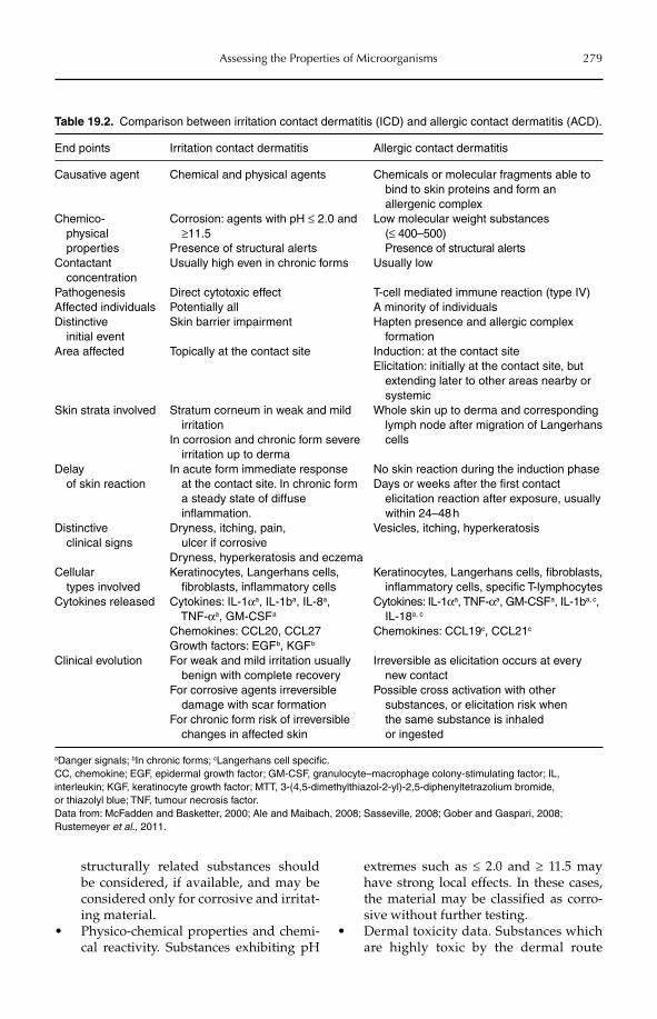
Assessing the Properties of Microorganisms 279
Table 19.2. Comparison between irritation contact dermatitis (ICD) and allergic contact dermatitis (ACD).
End points Irritation contact dermatitis Allergic contact dermatitis
Causative agent Chemical and physical agents Chemicals or molecular fragments able to bind to skin proteins and form an allergenic complex
Chemico- physical properties
Corrosion: agents with pH ≤ 2.0 and ≥11.5
Presence of structural alerts
Low molecular weight substances (≤ 400–500) Presence of structural alerts
Contactant concentration
Usually high even in chronic forms Usually low
Pathogenesis Direct cytotoxic effect T-cell mediated immune reaction (type IV)Affected individuals Potentially all A minority of individualsDistinctive initial event
Skin barrier impairment Hapten presence and allergic complex formation
Area affected Topically at the contact site Induction: at the contact siteElicitation: initially at the contact site, but extending later to other areas nearby or systemic
Skin strata involved Stratum corneum in weak and mild irritationIn corrosion and chronic form severe irritation up to derma
Whole skin up to derma and corresponding lymph node after migration of Langerhans cells
Delay of skin reaction
In acute form immediate response at the contact site. In chronic form a steady state of diffuse inflammation.
No skin reaction during the induction phaseDays or weeks after the first contact elicitation reaction after exposure, usually within 24–48 h
Distinctive clinical signs
Dryness, itching, pain, ulcer if corrosiveDryness, hyperkeratosis and eczema
Vesicles, itching, hyperkeratosis
Cellular types involved
Keratinocytes, Langerhans cells, fibroblasts, inflammatory cells
Keratinocytes, Langerhans cells, fibroblasts, inflammatory cells, specific T-lymphocytes
Cytokines released Cytokines: IL-1αa, IL-1ba, IL-8a, TNF-αa, GM-CSF a
Chemokines: CCL20, CCL27Growth factors: EGF b, KGF b
Cytokines: IL-1αa, TNF-αa, GM-CSF a, IL-1ba, c, IL-18a, c
Chemokines: CCL19c, CCL21c
Clinical evolution For weak and mild irritation usually benign with complete recoveryFor corrosive agents irreversible damage with scar formationFor chronic form risk of irreversible changes in affected skin
Irreversible as elicitation occurs at every new contactPossible cross activation with other substances, or elicitation risk when the same substance is inhaled or ingested
aDanger signals; bIn chronic forms; cLangerhans cell specific.CC, chemokine; EGF, epidermal growth factor; GM-CSF, granulocyte–macrophage colony-stimulating factor; IL, interleukin; KGF, keratinocyte growth factor; MTT, 3-(4,5-dimethylthiazol-2-yl)-2,5-diphenyltetrazolium bromide, or thiazolyl blue; TNF, tumour necrosis factor.Data from: McFadden and Basketter, 2000; Ale and Maibach, 2008; Sasseville, 2008; Gober and Gaspari, 2008; Rustemeyer et al., 2011.
structurally related substances should be considered, if available, and may be considered only for corrosive and irritat-ing material.Physico-chemical properties and chemi-•cal reactivity. Substances exhibiting pH
extremes such as £ 2.0 and ³ 11.5 may have strong local effects. In these cases, the material may be classified as corro-sive without further testing.Dermal toxicity data. Substances which •are highly toxic by the dermal route
280 G. Loprieno
cannot be tested for irritancy because they can induce death or severe suffering in animals. Negative findings are accep-ted only if the dermal toxicity study was performed on rabbits, at compara-ble doses and up to 2000 mg kg−1 body weight without signs of irritancy or corrosion.Results from • in vitro or ex vivo tests for corrosion. If the tested material shows positive results in in vitro or ex vivo cor-rosivity tests, it does not need to be tested again.In vivo• tests in rabbits. Starting with an initial test using one animal, it is possible to stop further testing if the substance is corrosive, otherwise the irritant or nega-tive response should be confirmed using up to two additional animals for an expo-sure period of 4 h, in a sequential manner, or by exposing them simultaneously.
One in vivo and one in vitro testing model for dermal irritation are adopted by OECD, namely Test Guideline Nos 404 and 439. A comparison of the advantages and disadvantages of these two methods is pre-sented in Table 19.3 and the guidelines will be described in more detail in the following sections.
In vivo animal models
The OECD Test No. 404 defines the protocol for Acute Dermal Irritation/Corrosion. This method was initially adopted in 1981, revised in 1992 and reformulated in 2002 with the inclusion of several aspects related to animal welfare and in vitro alternatives. The most common animal used is the New Zealand White rabbit because it has no dermal pigment and a large dorsal surface that is practical for observation. Rabbits have been shown to be rather sensitive to the irritant/corrosive effects of a wide range of chemicals. The test material is applied on the dorsal skin surface and a nearby untreated area is considered as the control. The effects on the rabbit skin are scored at timed intervals and the duration of the study is sufficient to see reversibility or irreversibility of the effects. A dose of 0.5 ml of liquid or 0.5 g of solid or paste is applied to
the test site, with a semi-occlusive patch for an exposure time of 4 h. After the exposure patch is removed and the skin washed, scor-ing is performed at 60 min, and then at 24, 48 and 72 h. Exposure is terminated after 14 days if there are signs of irritation to determine possible recovery, or immediately at any time if the animal shows continuing signs of severe pain or distress. After one animal treatment, additional animals are treated until an une-quivocal response is obtained in the majority of animals tested.
Dermal irritation grading is based on a scoring system originally developed by Draize (OECD, 2010). Scores for erythema and oedema are combined at each scoring time up to a maximum score of 8 and counted as the primary irritation index. Materials producing a primary irritation index of 0.00 are classified as non-irritant; scores from 0.01 to 2.00 rate a material as a slight or weak irri-tant, from 2.01 to 5.00 as a moderate or mild irritant, and above 5.01 as a severe irritant (Rauckman and Soifer, 2006).
In vitro models
For many years, no in vitro methods were available for skin irritation testing. While corrosivity as an expression of cell lethality was easily simulated in vitro, testing for irri-tancy remained a difficult task. Pre-valida-tion studies, such as those sponsored by the European Centre for Validation of Alternative Methods between 1998 and 2006, failed to reach final objectives because the proposed methods were not suitable for safety assess-ment and regulatory purposes (Basketter and Jones, 2008).
Recently, OECD adopted the Recons-tructed Human Epidermis (RhE) Test No. 439 as predictive for skin irritation under certain conditions (OECD, 2010). The RhE is based on non-transformed human-derived epider-mal keratinocytes that are cultured to form a multilayered, highly differentiated model of the human epidermis. It consists of organized basal, spinous and granular layers, and a multilayered stratum corneum containing intercellular lamellar lipid layers represent-ing the main lipid classes analogous to those found in vivo. The RhE model can be prepared
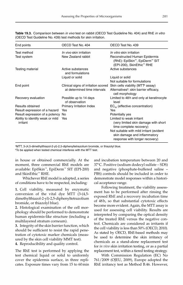
Assessing the Properties of Microorganisms 281
in house or obtained commercially. At the moment, three commercial RhE models are available: EpiSkin™, EpiDerm™ SIT (EPI-200) and SkinEthic™ RHE.
Whichever RhE model is adopted, a series of conditions have to be respected, including:
1. Cell viability, measured by enzymatic conversion of the vital dye MTT (3-(4,5- dimethylthiazol-2-yl)-2,5-diphenyltetrazolium bromide, or thiazolyl blue).2. Histological examination of the cell mor-phology should be performed to demonstrate human epidermis-like structure (including a multilayered stratum corneum).3. Integrity of the skin barrier function, which should be sufficient to resist the rapid pene-tration of cytotoxic marker chemicals (meas-ured by the skin cell viability MMT test).4. Reproducibility and quality control.
The RhE test is performed by applying the test chemical liquid or solid to uniformly cover the epidermis surface, in three repli-cates. Exposure times vary from 15 to 60 min
and incubation temperature between 20 and 37°C. Positive (sodium dodecyl sulfate – SDS) and negative (phosphate-buffered saline – PBS) controls should be included in order to demonstrate model responses within a histori-cal acceptance range.
Following treatment, the viability assess-ment has to be performed after rinsing the exposed RhE and a recovery incubation time of 48 h, so that substantial cytotoxic effects become more evident. Again, the MTT assay is used for assessing cell viability. Results are interpreted by comparing the optical density of the treated RhE versus the negative con-trol. Chemicals are considered as irritants if the cell viability is less than 50% (OECD, 2010). As stated by OECD, RhE-based methods may be used to determine the skin irritancy of chemicals as a stand-alone replacement test for in vivo skin irritation testing, or as a partial replacement test, within a tiered testing strategy.
With Commission Regulation (EC) No 761/2009 (OJEU, 2009), Europe adopted the RhE irritancy test as Method B.46. However,
Table 19.3. Comparison between in vivo test on rabbit (OECD Test Guideline No. 404) and RhE in vitro(OECD Test Guideline No. 439) test methods for skin irritation.
End points OECD Test No. 404 OECD Test No. 439
Test method In vivo skin irritation In vitro skin irritationTest system New Zealand rabbit Reconstructed Human Epidermis
(RhE): EpiSkin™, EpiDerm™ SIT (EPI-200), SkinEthic™ RHE
Testing material Active substances and formulationsLiquid or solid
Active substances
Liquid or solidNot suitable for formulations
End point Clinical signs of irritation scored at determined time intervals
Skin cells viability (MTTa assay)Alternativesb: skin barrier efficacy, cell morphology
Recovery evaluation Possible up to 14 days of observation
Limited to 48 h and only at keratinocyte level
Results obtained Primary Irritation Index EC50 (effective concentration)Result expression of a hazard Yes YesResult expression of a potency No Potentially yesAbility to identify weak or mild irritant
Yes Limited to weak irritant (very limited skin damage with short time complete recovery)Not suitable with mild irritant (evident skin damage and inflammatory response with longer recovery)
aMTT, 3-(4,5-dimethylthiazol-2-yl)-2,5-diphenyltetrazolium bromide, or thiazolyl blue.bTo be applied when tested chemical interferes with the MTT test.
282 G. Loprieno
the regulation clearly indicates that the RhE method is suitable only for classifying sub-stances as skin irritants category 2 according to the United Nations (UN) Globally Harmoni-zed System of Classification and Labeling of Chemicals (GHS). Furthermore, it is not intended for testing gases and aerosols, and mixtures have not been assessed yet in a spe-cific validation study (OJEU, 2009).
19.3.2 Methods for skin sensitization
The prediction of the allergic potential of chemicals and formulations is a common practice in regulatory and experimental toxicology. Curiously, predictive tests were defined at a time when the mechanism of sensitization was not completely known. The available testing methodologies with guinea pigs are an evolution of the first method developed by Draize in 1944 (ECETOC, 1990), which was adopted by the US Federal Drug Administration (US FDA) in 1959 (ECETOC, 1990). A series of improve-ments were necessary to better reproduce human exposure conditions and to increase sensitivity of the test, and seven methods had been developed by 1980. After a close examination by OECD in 1992, it was recog-nized that only two methods were frequently used in the USA and Europe: the Bühler test and the guinea pig maximization test (GPMT) of Magnusson and Kligman (1970), respectively. These were adopted and rec-ommended by OECD (Test Guideline No. 406), limiting the others only to special cases (ECETOC, 1990; Maurer, 2007).
Subsequently, a new method based on mice, the murine local lymph node assay (LLNA) was internationally validated and adopted by OECD in 2002 under a separate guideline (Test No. 429). In 2010, two LLNA variants were published (Test Guidelines Nos 442A and 442B) (OECD, 2010).
In contrast to irritation, sensitization is a complex process involving the skin, derma and immune system, and actually no in vitro alter-native is available. The LLNA has recognized this by reducing and refining the number of animals tested, as a result of pressure by animal
welfare agencies and the Interagency Co or dinating Committee on the Validation of Alternative Methods (ICCVAM; Kimber et al., 2003).
Guinea pig methods
Basically, the guinea pig methods consist of two experimental parts: the induction and the challenge or provocation phase. During the induction phase, animals are treated repeat-edly for 2–3 weeks to simulate the first con-tact. After a resting period, the animals are treated again with a non-irritating amount of the tested material at another site. A challenge that induces an allergenic reaction topically is scored according to a four-grade scale from erythema to oedema. The more animals that show challenge effects, the stronger is the tested material, so the final judgement is based on the total number of animals affec-ted rather than single individual responses (Magnusson and Kligman, 1970).
Both the Bühler test and the GPMT fol-low the above general scheme. However, treatment type and timing, together with the evaluation period, are the most relevant differences (Table 19.4). The scope of adju-vant use and injections in GPMT are to enhance the clinical reaction after challenge induction. Also, pretreatment with sodium lauryl sulfate (SLS) is to facilitate the dermal penetration of the tested chemical through the skin barrier impairment induced by the irritant, in order to increase the possibility of a sensitization. While the GPMT is con-ducted using two types of exposure (skin and percutaneous) during induction, in the Bühler test, animals are treated repeatedly only by the dermal route, but at a concentra-tion which can be up to 10-fold higher than the corresponding tested material in real use. It is generally recognized that the Bühler test underestimates and the GPMT overestimates the sensitization potential of chemicals. Finally, it should be emphasized that the Bühler test and the GPMT may identify the hazard, but not the potency of sensitizers, as they cannot correctly classify weak allergens that are unable to trigger a clinical response in animals (Basketter and Kimber, 2010).
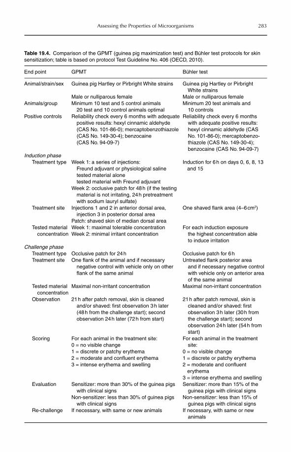
Assessing the Properties of Microorganisms 283
Table 19.4. Comparison of the GPMT (guinea pig maximization test) and Bühler test protocols for skin sensitization; table is based on protocol Test Guideline No. 406 (OECD, 2010).
End point GPMT Bühler test
Animal/strain/sex Guinea pig Hartley or Pirbright White strains
Male or nulliparous female
Guinea pig Hartley or Pirbright White strainsMale or nulliparous female
Animals/group Minimum 10 test and 5 control animals 20 test and 10 control animals optimal
Minimum 20 test animals and 10 controls
Positive controls Reliability check every 6 months with adequate positive results: hexyl cinnamic aldehyde (CAS No. 101-86-0); mercaptobenzothiazole (CAS No. 149-30-4); benzocaine (CAS No. 94-09-7)
Reliability check every 6 months with adequate positive results: hexyl cinnamic aldehyde (CAS
No. 101-86-0); mercaptobenzo-thiazole (CAS No. 149-30-4); benzocaine (CAS No. 94-09-7)
Induction phaseTreatment type Week 1: a series of injections:
Freund adjuvant or physiological saline tested material alone tested material with Freund adjuvantWeek 2: occlusive patch for 48 h (if the testing material is not irritating, 24 h pretreatment with sodium lauryl sulfate)
Induction for 6 h on days 0, 6, 8, 13 and 15
Treatment site Injections 1 and 2 in anterior dorsal area, injection 3 in posterior dorsal areaPatch: shaved skin of median dorsal area
One shaved flank area (4–6 cm2)
Tested material concentration
Week 1: maximal tolerable concentrationWeek 2: minimal irritant concentration
For each induction exposure the highest concentration able to induce irritation
Challenge phaseTreatment type Occlusive patch for 24 h Occlusive patch for 6 hTreatment site One flank of the animal and if necessary
negative control with vehicle only on other flank of the same animal
Untreated flank posterior area and if necessary negative control with vehicle only on anterior area of the same animal
Tested material concentration
Maximal non-irritant concentration Maximal non-irritant concentration
Observation 21 h after patch removal, skin is cleaned and/or shaved: first observation 3 h later (48 h from the challenge start); second observation 24 h later (72 h from start)
21 h after patch removal, skin is cleaned and/or shaved: first observation 3 h later (30 h from the challenge start); second observation 24 h later (54 h from start)
Scoring For each animal in the treatment site:0 = no visible change1 = discrete or patchy erythema2 = moderate and confluent erythema3 = intense erythema and swelling
For each animal in the treatment site:0 = no visible change1 = discrete or patchy erythema2 = moderate and confluent
erythema3 = intense erythema and swelling
Evaluation Sensitizer: more than 30% of the guinea pigs with clinical signsNon-sensitizer: less than 30% of guinea pigs with clinical signs
Sensitizer: more than 15% of the guinea pigs with clinical signsNon-sensitizer: less than 15% of guinea pigs with clinical signs
Re-challenge If necessary, with same or new animals If necessary, with same or new animals
284 G. Loprieno
Mouse methods
While guinea pigs were chosen for their simi-larity to humans in clinical signs during aller-gic responses, mice received attention owing to the good understanding of their immune system. The mouse-based LLNA method has advantages and disadvantages, but undoubt-edly represents a step forward in the predic-tion and evaluation of the sensitization hazards and potentials of chemicals. Rather than looking at the clinical changes induced after challenge on animal skin, the LLNA explores and quantifies the ability of the tested material to promote cell proliferation during the induction phase. In the original LLNA protocol, cell proliferation is detected by the incorporation of a radioactive com-pound, such as tritiated 3H-methyl thymidine or, alternatively, 125I-iododeoxyuridine and fluorodeoxyuridine, which during replication are incorporated into the DNA of the lym-phocytes. The use of radioactive compounds is substituted in the new, recently approved protocols by the detection of ATP content via bioluminescence (Test Guideline No. 442a) or non-radiolabelled 5-bromo-2-deoxyuridine, detected by ELISA (Test Guideline No. 442b). In the LLNA, proliferation is proportional to the dose and to the potency of the applied allergen and provides a simple means of obtaining a quantitative measurement of sensitization. Proliferation is measured by comparing the mean proliferation in each test group to the mean proliferation in the vehicle-treated control group to obtain the Stimulation Index (SI). At the threshold, it is assumed that a threefold increase in activity occurred compared with concurrent vehicle controls, the so-called EC3 value. An SI £ EC3 indicates that the tested material is a sensi-tizer. A summary of the main end points related to the LLNA is presented in Table 19.5; see OECD testing guidelines (OECD, 2010) for the detailed protocols.
Furthermore, the LLNA has the potential to reduce the number of animals required and offers a substantial refinement for allergic contact sensitization testing. The LLNA is limited to the induction phase of sensitization and, unlike guinea pig tests, does not require the elicitation of challenge-induced dermal
hypersensitivity reactions, thereby resulting in less animal pain and distress.
19.4 Microorganisms and Irritation/Sensitization
The presence of microorganisms in the workplace poses the question regarding their ability to induce skin irritation and/or sensitization. In order to determine this, it is necessary to first underline under which conditions these effects could be manifested. Enhanced exposure levels to microorgan-isms in the workplace can be expected in three situations:
1. In workplace activities that involve pur-poseful handling of products containing bio-logical agents, for example, in biotechnology or biopesticide production.2. Where workplace materials are contami-nated by biological agents.3. Where sources of bioaerosols are inciden-tal to the main work activity, e.g. contamina-tion of humidifiers and air conditioners.
First, opportunistic microorganisms could induce skin diseases under conditions that alter the efficiency of the skin barrier, such as wetting or exposure to detergents or surfac-tants. Secondly, in biotechnology, enzymes of bacterial and/or fungal origin can be sus-pected to cause irritation to the skin, eyes and the respiratory tract. Finally, there are cases in which the microorganism is the active ingre-dient in a formulated product for profes-sional and non-professional end users, for example biopesticides and biocides. Exam-ples of known workplace conditions related to these situations are reported in Table 19.6.
In the first case, represented by, e.g. met-alworking fluid dermatoses, environmental bacteria are responsible for ICD in connection with small traumas and a wet environment. The role of the microorganisms in the patho-genesis of dermatoses has often been ignored, owing to the lack of specificity of skin bacte-rial activity (Ueno et al., 2002; Taibjee et al.,2003). In most of the cases reported, it is dif-ficult to identify single microorganisms which may be responsible and it is reasonable to accept the possibility that microorganisms are
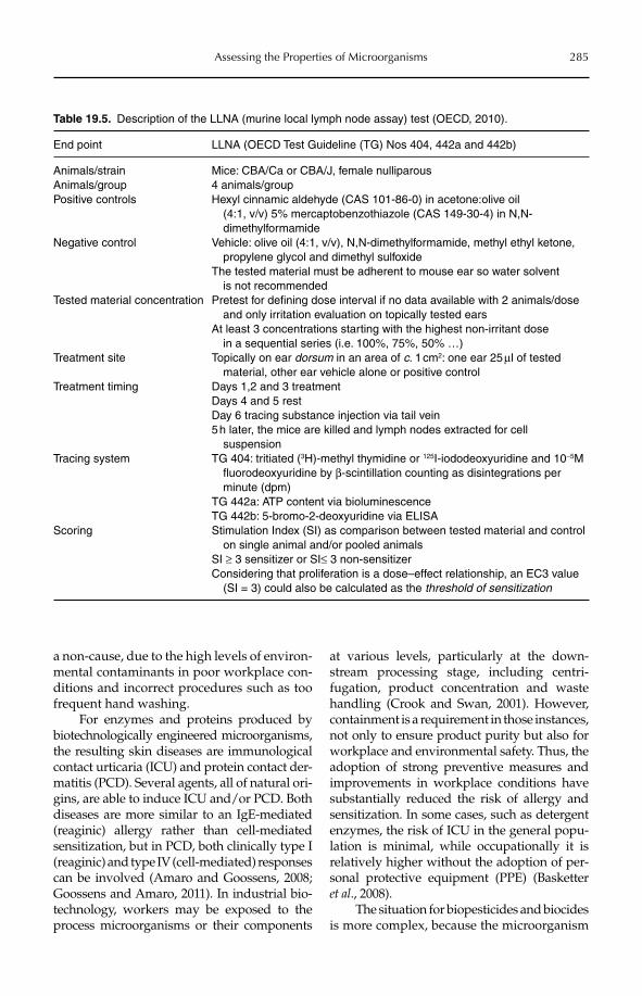
Assessing the Properties of Microorganisms 285
a non-cause, due to the high levels of environ-mental contaminants in poor workplace con-ditions and incorrect procedures such as too frequent hand washing.
For enzymes and proteins produced by biotechnologically engineered microorganisms, the resulting skin diseases are immunological contact urticaria (ICU) and protein contact der-matitis (PCD). Several agents, all of natural ori-gins, are able to induce ICU and/or PCD. Both diseases are more similar to an IgE-mediated (reaginic) allergy rather than cell-mediated sensitization, but in PCD, both clinically type I (reaginic) and type IV (cell-mediated) responses can be involved (Amaro and Goossens, 2008; Goossens and Amaro, 2011). In industrial bio-technology, workers may be exposed to the process microorganisms or their components
at various levels, particularly at the down-stream processing stage, including centri-fugation, product concentration and waste hand ling (Crook and Swan, 2001). However, containment is a requirement in those instances, not only to ensure product purity but also for workplace and environmental safety. Thus, the adoption of strong preventive measures and improvements in workplace conditions have substantially reduced the risk of allergy and sensitization. In some cases, such as detergent enzymes, the risk of ICU in the general popu-lation is minimal, while occupationally it is relatively higher without the adoption of per-sonal protective equipment (PPE) (Basketter et al., 2008).
The situation for biopesticides and biocides is more complex, because the microorganism
Table 19.5. Description of the LLNA (murine local lymph node assay) test (OECD, 2010).
End point LLNA (OECD Test Guideline (TG) Nos 404, 442a and 442b)
Animals/strain Mice: CBA/Ca or CBA/J, female nulliparousAnimals/group 4 animals/groupPositive controls Hexyl cinnamic aldehyde (CAS 101-86-0) in acetone:olive oil
(4:1, v/v) 5% mercaptobenzothiazole (CAS 149-30-4) in N,N- dimethylformamide
Negative control Vehicle: olive oil (4:1, v/v), N,N-dimethylformamide, methyl ethyl ketone, propylene glycol and dimethyl sulfoxideThe tested material must be adherent to mouse ear so water solvent is not recommended
Tested material concentration Pretest for defining dose interval if no data available with 2 animals/dose and only irritation evaluation on topically tested earsAt least 3 concentrations starting with the highest non-irritant dose in a sequential series (i.e. 100%, 75%, 50% …)
Treatment site Topically on ear dorsum in an area of c. 1 cm2: one ear 25 µl of tested material, other ear vehicle alone or positive control
Treatment timing Days 1,2 and 3 treatmentDays 4 and 5 restDay 6 tracing substance injection via tail vein5 h later, the mice are killed and lymph nodes extracted for cell suspension
Tracing system TG 404: tritiated (3H)-methyl thymidine or 125I-iododeoxyuridine and 10−5M fluorodeoxyuridine by β-scintillation counting as disintegrations per minute (dpm)TG 442a: ATP content via bioluminescenceTG 442b: 5-bromo-2-deoxyuridine via ELISA
Scoring Stimulation Index (SI) as comparison between tested material and control on single animal and/or pooled animalsSI ≥ 3 sensitizer or SI≤ 3 non-sensitizerConsidering that proliferation is a dose–effect relationship, an EC3 value (SI = 3) could also be calculated as the threshold of sensitization
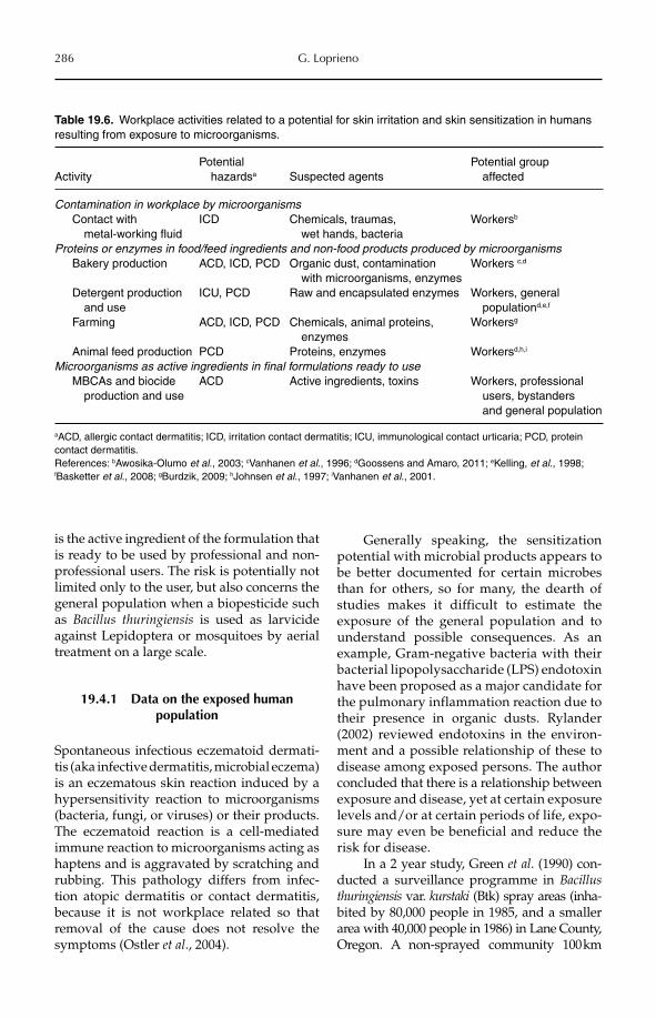
286 G. Loprieno
is the active ingredient of the formulation that is ready to be used by professional and non-professional users. The risk is potentially not limited only to the user, but also concerns the general population when a biopesticide such as Bacillus thuringiensis is used as larvicide against Lepidoptera or mosquitoes by aerial treatment on a large scale.
19.4.1 Data on the exposed human population
Spontaneous infectious eczematoid dermati-tis (aka infective dermatitis, microbial eczema) is an eczematous skin reaction induced by a hypersensitivity reaction to microorganisms (bacteria, fungi, or viruses) or their products. The eczematoid reaction is a cell-mediated immune reaction to microorganisms acting as haptens and is aggravated by scratching and rubbing. This pathology differs from infec-tion atopic dermatitis or contact dermatitis, because it is not workplace related so that removal of the cause does not resolve the symptoms (Ostler et al., 2004).
Generally speaking, the sensitization potential with microbial products appears to be better documented for certain microbes than for others, so for many, the dearth of studies makes it difficult to estimate the exposure of the general population and to understand possible consequences. As an example, Gram-negative bacteria with their bacterial lipopolysaccharide (LPS) endotoxin have been propo sed as a major candidate for the pulmonary inflammation reaction due to their presence in organic dusts. Rylander (2002) reviewed endotoxins in the environ-ment and a possible relationship of these to disease among exposed persons. The author concluded that there is a relationship between exposure and disease, yet at certain exposure levels and/or at certain periods of life, expo-sure may even be beneficial and reduce the risk for disease.
In a 2 year study, Green et al. (1990) con-ducted a surveillance programme in Bacillus thuringiensis var. kurstaki (Btk) spray areas (inha-bited by 80,000 people in 1985, and a smaller area with 40,000 people in 1986) in Lane County, Oregon. A non-sprayed community 100 km
Table 19.6. Workplace activities related to a potential for skin irritation and skin sensitization in humans resulting from exposure to microorganisms.
ActivityPotential hazardsa Suspected agents
Potential group affected
Contamination in workplace by microorganisms Contact with
metal-working fluidICD Chemicals, traumas,
wet hands, bacteriaWorkersb
Proteins or enzymes in food/feed ingredients and non-food products produced by microorganisms Bakery production ACD, ICD, PCD Organic dust, contamination
with microorganisms, enzymesWorkers c,d
Detergent production and use
ICU, PCD Raw and encapsulated enzymes Workers, general populationd,e,f
Farming ACD, ICD, PCD Chemicals, animal proteins, enzymes
Workersg
Animal feed production PCD Proteins, enzymes Workersd,h,i
Microorganisms as active ingredients in final formulations ready to use MBCAs and biocide
production and useACD Active ingredients, toxins Workers, professional
users, bystanders and general population
aACD, allergic contact dermatitis; ICD, irritation contact dermatitis; ICU, immunological contact urticaria; PCD, protein contact dermatitis.References: bAwosika-Olumo et al., 2003; cVanhanen et al., 1996; dGoossens and Amaro, 2011; eKelling, et al., 1998; fBasketter et al., 2008; gBurdzik, 2009; hJohnsen et al., 1997; iVanhanen et al., 2001.

Assessing the Properties of Microorganisms 287
away served as a control population. Human culture specimens were obtained for routine clinical purposes during, and for 1 month after, the spray period. Specimens that were positive for any Bacillus species were subcultured for B. thuringiensis and the presence of toxin crys-tals (parasporal inclusions) was investigated. Fifty-five cultures out of 105 were positive for Btk, but there was no evidence of human path-ogenicity. There was no increase in the number of telephone complaints nor was there a change in the pattern of complaints.
Another study was conducted in an area with a population of 1,400,000 in Lower Mainland, British Columbia, Canada (Nobel et al., 1992). After a spray campaign with Btk, the study examined 26,000 telephone calls, 1140 family practice patients and 3300 hospi-tal admissions, and closely monitored 120 workers with occupational exposure. There were no differences in emergency room vis-its between spray days and non-spray days, nor was there a significant change in the pat-tern of telephone calls. There was no evi-dence that Btk spraying was associated with illness or infection and, in fact, there was a lower incidence of diarrhoea in the spray zone than in the control area. However, workers showed symptoms of headache; nose, throat and eye irritation; dry skin and chapped lips, all signs of irritation and/or sensitization. The range of worker exposure was between 750 and 5.5 × 10⁶ cfu (colony forming units) m−3.
Similar conclusions of no health effects were drawn from an evaluation of the use of B.t. israelensis commissioned by the New Zealand Ministry of Health (Glare and O’Callaghan, 1998). However, an independ-ent study carried out during eradication spray programmes using Foray 48B (Btk) to control infestation by the painted apple moth that was conducted in West Auckland, New Zealand, found adverse health symp-toms in 315 of the resident population of 150,000 people exposed to the Btk spray oper-ation, although these made up only 0.21% of the exposed population (Blackmore, 2003). Another study on 292 subjects, also in West Auckland, found that there was an increase in health problems in 168 subjects; these ranged from cough to dizziness and sleep difficulties.
Skin irritation and eczema were not statisti-cally relevant (Petrie et al., 2003).
A longitudinal, follow-up investigation of 48 workers who were involved in picking Bt (Javelin®) sprayed crops (celery, parsley, cabbage, kale, spinach, strawberries) was conducted by Bernstein et al. (1999). These authors presented three exposure groups, ‘Low’, ‘Medium’ and ‘High’. There was no evidence of occupationally related respira-tory symptoms. Four types of Btk spore and two vegetative (subsp. kurstaki and aizawai)antigen extracts were prepared, of which some elicited positive skin tests, chiefly with exposed workers, at an incidence of 35%. Yet 25% of the ‘Low’ exposure group also pre-sented an atopic response. Positive skin tests were seen with water- and mercaptoethanol-sodium dodecyl sulfate extracts of Javelin, yet their incidence was not higher for Javelin extracted pro-Delta-endotoxin or proteinase K spore extracts. Specific IgE and IgG antibodies to vegetative cells were present in all groups, witnessing an immunological response.
Doekes et al. (2004) tested human sera from the BIOGART project, a longitudinal respiratory health study on more than 300 Danish greenhouse workers. This study had a 2 and 3 year follow-up. While many sera had detectable IgE to Bt (23–29%), all positive reactions showed a relatively weak IgE anti-Bt positive serum response. The authors con-cluded that even though the IgE binding components may be genuine Bt components, they could also be shared with some or many commonly found bacterial species, and in that case, similar IgE levels may be found in the general population.
A review analysis of the effect on the human population exposed to Bt spraying was redacted by Otvos et al. (2005). The authors concluded that Bt is one of the saf-est biopesticides and noted that no scientifi-cally documented case of human infection has been reported since its introduction in the 1960s, as well as no human health prob-lems as a result of spraying. However, the majority of the studies examined were focused on human risk of infection rather than respiratory and/or skin distress, which were considered transitory and less rele-vant symptoms.
288 G. Loprieno
In conclusion, studies on exposure and health effects on residents in areas treated with Bt show divergent results (Madsen, 2011). While some authors conclude that symptom complaints increased significantly, others conclude that there were no significant changes in physical health for residents in the affected area and that almost all of the studies were scientifically questionable. However, the conclusions drawn by studies on exposed workers, which showed humoral changes and skin or respiratory distress, but without clinical relevance, are more pertinent.
19.4.2 Strategies for assessing irritation or sensitization by microorganisms
As mentioned previously, after direct infec-tion, sensitization and irritation are the most relevant adverse effects for workplace expo-sure to microorganisms. There is need for a correct identification of the potential risk in order to evaluate the need for preventive measures such as gloves or, in some cases, also the limitations of their use. Madsen (2011) estimated the amount of microbial pesticides to which workers are exposed and concluded that exposure occurs in occupational settings but that residents in treated areas and neigh-bourhoods are also exposed. According to the author, exposure can also occur days or weeks after treatment, posing the need to correctly predict the irritation and sensitization poten-tial of the final product.
For chemicals, the use of predictive tests on animals is scientifically recognized as appropriate and, for many years, molecules and products were classified for human risk according to experimental animal data. If we look at microorganisms and products based on microorganisms such as MBCAs, the ques-tion of predicting sensitization and irritation potential is more complex.
Microorganisms are not chemicals and, in most cases, the living microorganism is unable to penetrate the skin barrier. However, sensitization and/or irritation could arise from the presence of other substances in microbial products, such as toxins, enzymes, proteins, fragments, contaminants and formulants.
For biological material produced by the active ingredient microbe, such as toxins and/or enzymes, the risk of irritation is related to the possibility of direct toxic effects on skin kera-tinocytes. In the same way, the sensitization risk by MBCAs could be similar to that of PCD, with a mixed pattern of IgE and IgG production and cellular response as weakly indicated by studies on population and work-ers exposed to Bt. Formulants could play a relevant role but, in practice, sensitizers and irritants are avoided in final products.
If we look at the available methodologies for testing end-use products based on micro-organisms, such as enzymes or MBCAs, there are several questions that must be answered. For irritation, the possibility of using in vitromethods is only related to the possibility of classifying the tested material as irritant or cor-rosive, i.e. as positive. In other words, these tests are useless if we have a negative result, i.e. non-irritant; they are also useless if we need to distinguish between a mild and/or a true irritant. The only way to solve the problem is to accompany the in vitro tests with a physico-chemical examination and/or a structural alert investigation (by SAR analysis). This approach, with difficulty, was proposed for chemicals but has not been tried with microbials, and could be better defined only after years of experience which, at the moment, is of course lacking. A tentative strategy is the possibility of applying RhE tests on MBCAs and, if the results are negative according to other aspects, e.g. human data, waiving animal testing.
For sensitization, the question is even more complex, because methodological improvements such as LLNA may not be suitable for microorganisms. LLNA is a test based on Langerhans cell recruitment, lymph node migration and lymphocyte cloning. As Langerhans cells are immune competent and react naturally against bacteria and exogenous intruders, it can be suspected that in the case of, e.g. MBCAs, the presence of the microbe or its fragments could elicit a response from Langerhans cells, thus overestimating the sensitization potential or shifting the sensiti-zation response into a true aspecific immune response. The only animal test method for sensitization that could be suitable for MBCAs is the Bühler test, which is more
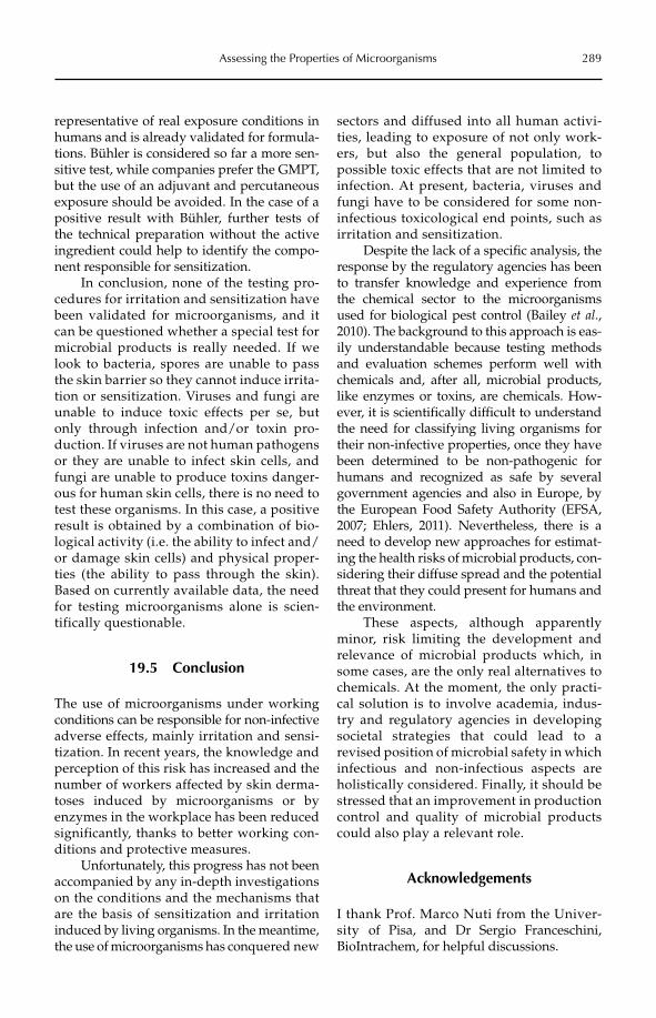
Assessing the Properties of Microorganisms 289
representative of real exposure conditions in humans and is already validated for formula-tions. Bühler is considered so far a more sen-sitive test, while companies prefer the GMPT, but the use of an adjuvant and percutaneous exposure should be avoided. In the case of a positive result with Bühler, further tests of the technical preparation without the active ingredient could help to identify the compo-nent responsible for sensitization.
In conclusion, none of the testing pro-cedures for irritation and sensitization have been validated for microorganisms, and it can be questioned whether a special test for microbial products is really needed. If we look to bacteria, spores are unable to pass the skin barrier so they cannot induce irrita-tion or sensitization. Viruses and fungi are unable to induce toxic effects per se, but only through infection and/or toxin pro-duction. If viruses are not human pathogens or they are unable to infect skin cells, and fungi are unable to produce toxins danger-ous for human skin cells, there is no need to test these organisms. In this case, a positive result is obtained by a combination of bio-logical activity (i.e. the ability to infect and/or damage skin cells) and physical proper-ties (the ability to pass through the skin). Based on currently available data, the need for testing microorganisms alone is scien-tifically questionable.
19.5 Conclusion
The use of microorganisms under working conditions can be responsible for non-infective adverse effects, mainly irritation and sensi-tization. In recent years, the knowledge and perception of this risk has increased and the number of workers affected by skin derma-toses induced by microorganisms or by enzymes in the workplace has been reduced significantly, thanks to better working con-ditions and protective measures.
Unfortunately, this progress has not been accompanied by any in-depth investigations on the conditions and the mechanisms that are the basis of sensitization and irritation induced by living organisms. In the meantime, the use of microorganisms has conquered new
sectors and diffused into all human activi-ties, leading to exposure of not only work-ers, but also the general population, to possible toxic effects that are not limited to infection. At present, bacteria, viruses and fungi have to be considered for some non-infectious toxicological end points, such as irritation and sensitization.
Despite the lack of a specific analysis, the response by the regulatory agencies has been to transfer knowledge and experience from the chemical sector to the microorganisms used for biological pest control (Bailey et al.,2010). The background to this approach is eas-ily understandable because testing methods and evaluation schemes perform well with chemicals and, after all, microbial products, like enzymes or toxins, are chemicals. How-ever, it is scientifically difficult to understand the need for classifying living organisms for their non-infective properties, once they have been determined to be non-pathogenic for humans and recognized as safe by several government agencies and also in Europe, by the European Food Safety Authority (EFSA, 2007; Ehlers, 2011). Never theless, there is a need to develop new approaches for estimat-ing the health risks of microbial products, con-sidering their diffuse spread and the potential threat that they could present for humans and the environment.
These aspects, although apparently minor, risk limiting the development and relevance of microbial products which, in some cases, are the only real alternatives to chemicals. At the moment, the only practi-cal solution is to involve academia, indus-try and regulatory agencies in developing societal strategies that could lead to a revised position of microbial safety in which infectious and non-infectious aspects are holistically considered. Finally, it should be stressed that an improvement in production control and quality of microbial products could also play a relevant role.
Acknowledgements
I thank Prof. Marco Nuti from the Univer-sity of Pisa, and Dr Sergio Franceschini, BioIntrachem, for helpful discussions.
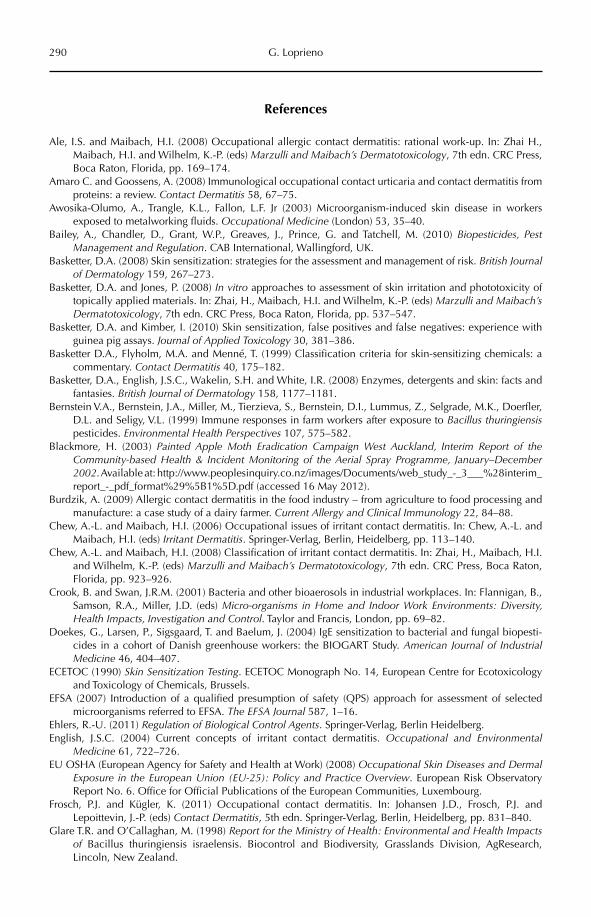
290 G. Loprieno
References
Ale, I.S. and Maibach, H.I. (2008) Occupational allergic contact dermatitis: rational work-up. In: Zhai H., Maibach, H.I. and Wilhelm, K.-P. (eds) Marzulli and Maibach’s Dermatotoxicology, 7th edn. CRC Press, Boca Raton, Florida, pp. 169–174.
Amaro C. and Goossens, A. (2008) Immunological occupational contact urticaria and contact dermatitis from proteins: a review. Contact Dermatitis 58, 67–75.
Awosika-Olumo, A., Trangle, K.L., Fallon, L.F. Jr (2003) Microorganism-induced skin disease in workers exposed to metalworking fluids. Occupational Medicine (London) 53, 35–40.
Bailey, A., Chandler, D., Grant, W.P., Greaves, J., Prince, G. and Tatchell, M. (2010) Biopesticides, Pest Management and Regulation. CAB International, Wallingford, UK.
Basketter, D.A. (2008) Skin sensitization: strategies for the assessment and management of risk. British Journal of Dermatology 159, 267–273.
Basketter, D.A. and Jones, P. (2008) In vitro approaches to assessment of skin irritation and phototoxicity of topically applied materials. In: Zhai, H., Maibach, H.I. and Wilhelm, K.-P. (eds) Marzulli and Maibach’s Dermatotoxicology, 7th edn. CRC Press, Boca Raton, Florida, pp. 537–547.
Basketter, D.A. and Kimber, I. (2010) Skin sensitization, false positives and false negatives: experience with guinea pig assays. Journal of Applied Toxicology 30, 381–386.
Basketter D.A., Flyholm, M.A. and Menné, T. (1999) Classification criteria for skin-sensitizing chemicals: a commentary. Contact Dermatitis 40, 175–182.
Basketter, D.A., English, J.S.C., Wakelin, S.H. and White, I.R. (2008) Enzymes, detergents and skin: facts and fantasies. British Journal of Dermatology 158, 1177–1181.
Bernstein V.A., Bernstein, J.A., Miller, M., Tierzieva, S., Bernstein, D.I., Lummus, Z., Selgrade, M.K., Doerfler, D.L. and Seligy, V.L. (1999) Immune responses in farm workers after exposure to Bacillus thuringiensispesticides. Environmental Health Perspectives 107, 575–582.
Blackmore, H. (2003) Painted Apple Moth Eradication Campaign West Auckland, Interim Report of the Community-based Health & Incident Monitoring of the Aerial Spray Programme, January–December 2002. Available at: http://www.peoplesinquiry.co.nz/images/Documents/web_study_-_3___%28interim_report_-_pdf_format%29%5B1%5D.pdf (accessed 16 May 2012).
Burdzik, A. (2009) Allergic contact dermatitis in the food industry – from agriculture to food processing and manufacture: a case study of a dairy farmer. Current Allergy and Clinical Immunology 22, 84–88.
Chew, A.-L. and Maibach, H.I. (2006) Occupational issues of irritant contact dermatitis. In: Chew, A.-L. and Maibach, H.I. (eds) Irritant Dermatitis. Springer-Verlag, Berlin, Heidelberg, pp. 113–140.
Chew, A.-L. and Maibach, H.I. (2008) Classification of irritant contact dermatitis. In: Zhai, H., Maibach, H.I. and Wilhelm, K.-P. (eds) Marzulli and Maibach’s Dermatotoxicology, 7th edn. CRC Press, Boca Raton, Florida, pp. 923–926.
Crook, B. and Swan, J.R.M. (2001) Bacteria and other bioaerosols in industrial workplaces. In: Flannigan, B., Samson, R.A., Miller, J.D. (eds) Micro-organisms in Home and Indoor Work Environments: Diversity, Health Impacts, Investigation and Control. Taylor and Francis, London, pp. 69–82.
Doekes, G., Larsen, P., Sigsgaard, T. and Baelum, J. (2004) IgE sensitization to bacterial and fungal biopesti-cides in a cohort of Danish greenhouse workers: the BIOGART Study. American Journal of Industrial Medicine 46, 404–407.
ECETOC (1990) Skin Sensitization Testing. ECETOC Monograph No. 14, European Centre for Ecotoxicology and Toxicology of Chemicals, Brussels.
EFSA (2007) Introduction of a qualified presumption of safety (QPS) approach for assessment of selected microorganisms referred to EFSA. The EFSA Journal 587, 1–16.
Ehlers, R.-U. (2011) Regulation of Biological Control Agents. Springer-Verlag, Berlin Heidelberg.English, J.S.C. (2004) Current concepts of irritant contact dermatitis. Occupational and Environmental
Medicine 61, 722–726.EU OSHA (European Agency for Safety and Health at Work) (2008) Occupational Skin Diseases and Dermal
Exposure in the European Union (EU-25): Policy and Practice Overview. European Risk Observatory Report No. 6. Office for Official Publications of the European Communities, Luxembourg.
Frosch, P.J. and Kügler, K. (2011) Occupational contact dermatitis. In: Johansen J.D., Frosch, P.J. and Lepoittevin, J.-P. (eds) Contact Dermatitis, 5th edn. Springer-Verlag, Berlin, Heidelberg, pp. 831–840.
Glare T.R. and O’Callaghan, M. (1998) Report for the Ministry of Health: Environmental and Health Impacts of Bacillus thuringiensis israelensis. Biocontrol and Biodiversity, Grasslands Division, AgResearch, Lincoln, New Zealand.
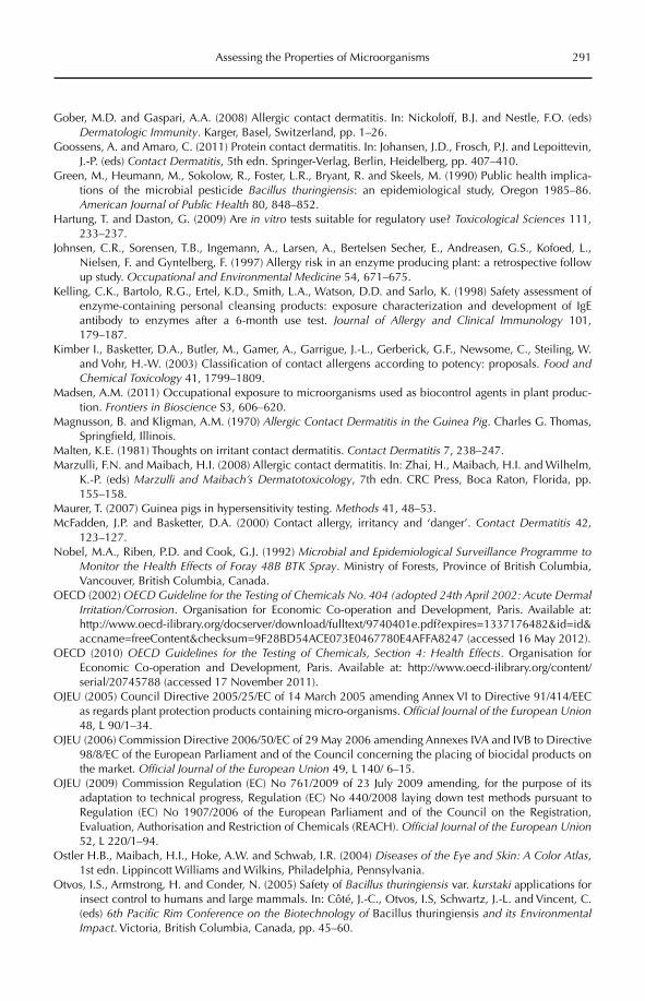
Assessing the Properties of Microorganisms 291
Gober, M.D. and Gaspari, A.A. (2008) Allergic contact dermatitis. In: Nickoloff, B.J. and Nestle, F.O. (eds) Dermatologic Immunity. Karger, Basel, Switzerland, pp. 1–26.
Goossens, A. and Amaro, C. (2011) Protein contact dermatitis. In: Johansen, J.D., Frosch, P.J. and Lepoittevin, J.-P. (eds) Contact Dermatitis, 5th edn. Springer-Verlag, Berlin, Heidelberg, pp. 407–410.
Green, M., Heumann, M., Sokolow, R., Foster, L.R., Bryant, R. and Skeels, M. (1990) Public health implica-tions of the microbial pesticide Bacillus thuringiensis: an epidemiological study, Oregon 1985–86. American Journal of Public Health 80, 848–852.
Hartung, T. and Daston, G. (2009) Are in vitro tests suitable for regulatory use? Toxicological Sciences 111, 233–237.
Johnsen, C.R., Sorensen, T.B., Ingemann, A., Larsen, A., Bertelsen Secher, E., Andreasen, G.S., Kofoed, L., Nielsen, F. and Gyntelberg, F. (1997) Allergy risk in an enzyme producing plant: a retrospective follow up study. Occupational and Environmental Medicine 54, 671–675.
Kelling, C.K., Bartolo, R.G., Ertel, K.D., Smith, L.A., Watson, D.D. and Sarlo, K. (1998) Safety assessment of enzyme-containing personal cleansing products: exposure characterization and development of IgE antibody to enzymes after a 6-month use test. Journal of Allergy and Clinical Immunology 101, 179–187.
Kimber I., Basketter, D.A., Butler, M., Gamer, A., Garrigue, J.-L., Gerberick, G.F., Newsome, C., Steiling, W. and Vohr, H.-W. (2003) Classification of contact allergens according to potency: proposals. Food and Chemical Toxicology 41, 1799–1809.
Madsen, A.M. (2011) Occupational exposure to microorganisms used as biocontrol agents in plant produc-tion. Frontiers in Bioscience S3, 606–620.
Magnusson, B. and Kligman, A.M. (1970) Allergic Contact Dermatitis in the Guinea Pig. Charles G. Thomas, Springfield, Illinois.
Malten, K.E. (1981) Thoughts on irritant contact dermatitis. Contact Dermatitis 7, 238–247.Marzulli, F.N. and Maibach, H.I. (2008) Allergic contact dermatitis. In: Zhai, H., Maibach, H.I. and Wilhelm,
K.-P. (eds) Marzulli and Maibach’s Dermatotoxicology, 7th edn. CRC Press, Boca Raton, Florida, pp. 155–158.
Maurer, T. (2007) Guinea pigs in hypersensitivity testing. Methods 41, 48–53.McFadden, J.P. and Basketter, D.A. (2000) Contact allergy, irritancy and ‘danger’. Contact Dermatitis 42,
123–127.Nobel, M.A., Riben, P.D. and Cook, G.J. (1992) Microbial and Epidemiological Surveillance Programme to
Monitor the Health Effects of Foray 48B BTK Spray. Ministry of Forests, Province of British Columbia, Vancouver, British Columbia, Canada.
OECD (2002) OECD Guideline for the Testing of Chemicals No. 404 (adopted 24th April 2002: Acute Dermal Irritation/Corrosion. Organisation for Economic Co-operation and Development, Paris. Available at: http://www.oecd-ilibrary.org/docserver/download/fulltext/9740401e.pdf?expires=1337176482&id=id&accname=freeContent&checksum=9F28BD54ACE073E0467780E4AFFA8247 (accessed 16 May 2012).
OECD (2010) OECD Guidelines for the Testing of Chemicals, Section 4: Health Effects. Organisation for Economic Co-operation and Development, Paris. Available at: http://www.oecd-ilibrary.org/content/serial/20745788 (accessed 17 November 2011).
OJEU (2005) Council Directive 2005/25/EC of 14 March 2005 amending Annex VI to Directive 91/414/EEC as regards plant protection products containing micro-organisms. Official Journal of the European Union48, L 90/1–34.
OJEU (2006) Commission Directive 2006/50/EC of 29 May 2006 amending Annexes IVA and IVB to Directive 98/8/EC of the European Parliament and of the Council concerning the placing of biocidal products on the market. Official Journal of the European Union 49, L 140/ 6–15.
OJEU (2009) Commission Regulation (EC) No 761/2009 of 23 July 2009 amending, for the purpose of its adaptation to technical progress, Regulation (EC) No 440/2008 laying down test methods pursuant to Regulation (EC) No 1907/2006 of the European Parliament and of the Council on the Registration, Evaluation, Authorisation and Restriction of Chemicals (REACH). Official Journal of the European Union52, L 220/1–94.
Ostler H.B., Maibach, H.I., Hoke, A.W. and Schwab, I.R. (2004) Diseases of the Eye and Skin: A Color Atlas,1st edn. Lippincott Williams and Wilkins, Philadelphia, Pennsylvania.
Otvos, I.S., Armstrong, H. and Conder, N. (2005) Safety of Bacillus thuringiensis var. kurstaki applications for insect control to humans and large mammals. In: Côté, J.-C., Otvos, I.S, Schwartz, J.-L. and Vincent, C. (eds) 6th Pacific Rim Conference on the Biotechnology of Bacillus thuringiensis and its Environmental Impact. Victoria, British Columbia, Canada, pp. 45–60.
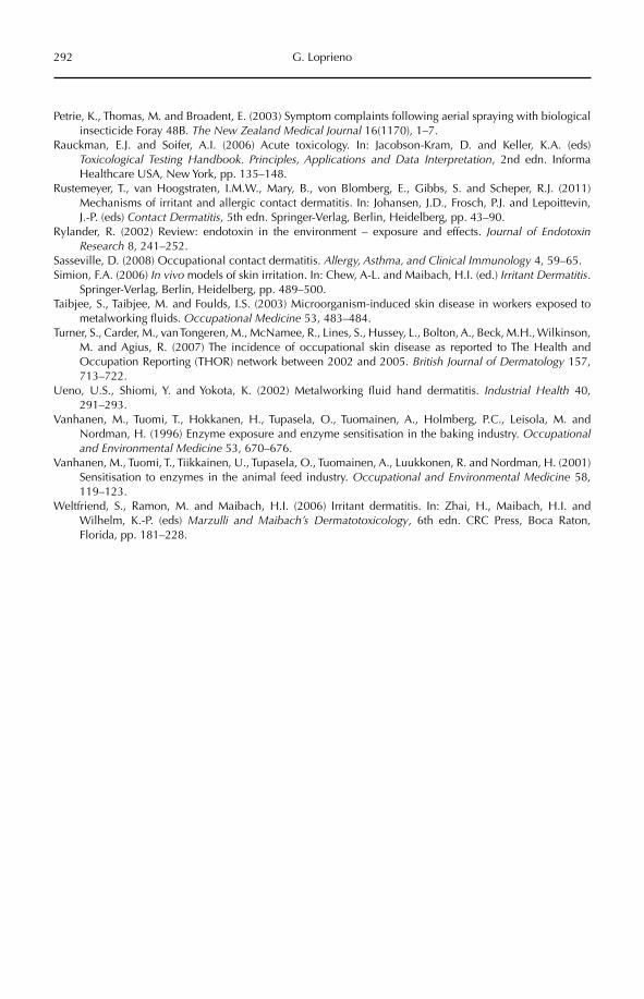
292 G. Loprieno
Petrie, K., Thomas, M. and Broadent, E. (2003) Symptom complaints following aerial spraying with biological insecticide Foray 48B. The New Zealand Medical Journal 16(1170), 1–7.
Rauckman, E.J. and Soifer, A.I. (2006) Acute toxicology. In: Jacobson-Kram, D. and Keller, K.A. (eds) Toxicological Testing Handbook. Principles, Applications and Data Interpretation, 2nd edn. Informa Healthcare USA, New York, pp. 135–148.
Rustemeyer, T., van Hoogstraten, I.M.W., Mary, B., von Blomberg, E., Gibbs, S. and Scheper, R.J. (2011) Mechanisms of irritant and allergic contact dermatitis. In: Johansen, J.D., Frosch, P.J. and Lepoittevin, J.-P. (eds) Contact Dermatitis, 5th edn. Springer-Verlag, Berlin, Heidelberg, pp. 43–90.
Rylander, R. (2002) Review: endotoxin in the environment – exposure and effects. Journal of Endotoxin Research 8, 241–252.
Sasseville, D. (2008) Occupational contact dermatitis. Allergy, Asthma, and Clinical Immunology 4, 59–65.Simion, F.A. (2006) In vivo models of skin irritation. In: Chew, A-L. and Maibach, H.I. (ed.) Irritant Dermatitis.
Springer-Verlag, Berlin, Heidelberg, pp. 489–500.Taibjee, S., Taibjee, M. and Foulds, I.S. (2003) Microorganism-induced skin disease in workers exposed to
metalworking fluids. Occupational Medicine 53, 483–484.Turner, S., Carder, M., van Tongeren, M., McNamee, R., Lines, S., Hussey, L., Bolton, A., Beck, M.H., Wilkinson,
M. and Agius, R. (2007) The incidence of occupational skin disease as reported to The Health and Occupation Reporting (THOR) network between 2002 and 2005. British Journal of Dermatology 157, 713–722.
Ueno, U.S., Shiomi, Y. and Yokota, K. (2002) Metalworking fluid hand dermatitis. Industrial Health 40, 291–293.
Vanhanen, M., Tuomi, T., Hokkanen, H., Tupasela, O., Tuomainen, A., Holmberg, P.C., Leisola, M. and Nordman, H. (1996) Enzyme exposure and enzyme sensitisation in the baking industry. Occupationaland Environmental Medicine 53, 670–676.
Vanhanen, M., Tuomi, T., Tiikkainen, U., Tupasela, O., Tuomainen, A., Luukkonen, R. and Nordman, H. (2001) Sensitisation to enzymes in the animal feed industry. Occupational and Environmental Medicine 58, 119–123.
Weltfriend, S., Ramon, M. and Maibach, H.I. (2006) Irritant dermatitis. In: Zhai, H., Maibach, H.I. and Wilhelm, K.-P. (eds) Marzulli and Maibach’s Dermatotoxicology, 6th edn. CRC Press, Boca Raton, Florida, pp. 181–228.
©CAB International 2012. Beneficial Microorganisms in Agriculture, Food and the Environment: Safety Assessment and Regulation (eds I. Sundh et al.) 293
20.1 Introduction 29320.2 What is Proprietary Biological Matter? 29620.3 A Slowly Shrinking Public Science Domain? 29620.4 Moving into a Global ‘Legal Can of Worms’? 30120.5 Moving Out of the Can to Exploit Science Constructively for the Benefit
of Humankind? 30220.6 Still in the Can … for CGIAR 30320.7 … to Enter into Public–Private Partnerships (PPPs) 30320.8 With Some Additional Work to Do … 30320.9 Biological Matter and the Law 304
20 International Conventions and Agreements – Consequences for International Trade and Utilization of
Biological Matter, Including Microorganisms
Carl-Gustaf ThornströmUppsala BioCenter, Department for Plant Biology and Forest Genetics, Swedish
University of Agricultural Sciences, Sweden
20.1 Introduction
Developments in biotechnology, molecular genetics, intellectual property regimes and systems for regulating access to genetic resour-ces and related information over the last two decades are rapidly changing the conditionsfor public research on microorganisms, plants and other types of biological matter/genetic resources. Moreover, these developments also influence the possibilities for exploiting these biological resources in beneficial prod-ucts, e.g. in food production or environmental applications. While no multilateral or inter-national regulatory regimes specifically cov-ering microorganisms have so far been agreed upon, the emerging ‘proprietary science land-scape’ is comprehensive and includes several
international agreements that can strongly influence trade with and the utilization of biological matter (Table 20.1).
The CBD (Convention on Biological Diversity) is a legally binding agreement on biodiversity under the United Nations (UN). The WTO TRIPS (World Trade Organization Trade-Related Aspects of Intellectual Pro perty Rights) is a trade agreement introducing a global mini-standard on what (matter) can be protected under intellectual property rights.The FAO ITPGRFA (Food and Agricul ture Organization International Treaty on Plant Genetic Resources for Food and Agriculture) is an international treaty on multilateral access to – agreed upon – plant genetic resources for food and agriculture. UPOV (International Union for the Protection of New Varieties of
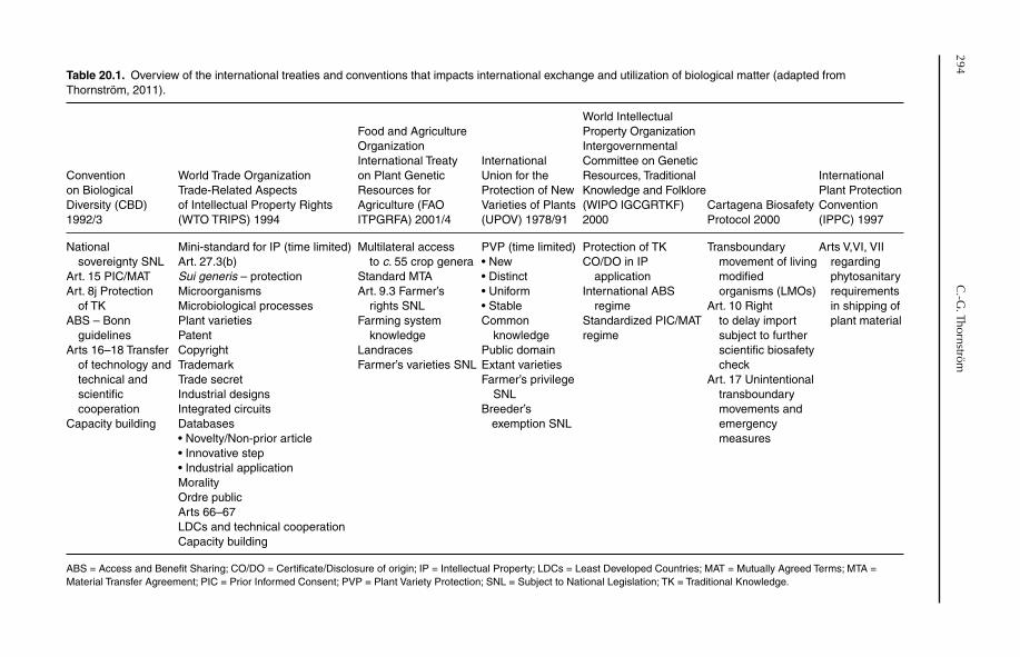
294C
.-G. Thornström
Table 20.1. Overview of the international treaties and conventions that impacts international exchange and utilization of biological matter (adapted from Thornström, 2011).
Convention on Biological Diversity (CBD) 1992/3
World Trade Organization Trade-Related Aspects of Intellectual Property Rights (WTO TRIPS) 1994
Food and Agriculture OrganizationInternational Treaty on Plant Genetic Resources for Agriculture (FAO ITPGRFA) 2001/4
International Union for the Protection of New Varieties of Plants (UPOV) 1978/91
World Intellectual Property Organization Intergovernmental Committee on Genetic Resources, Traditional Knowledge and Folklore (WIPO IGCGRTKF) 2000
Cartagena Biosafety Protocol 2000
International Plant Protection Convention (IPPC) 1997
National sovereignty SNLArt. 15 PIC/MATArt. 8j Protection of TKABS – Bonn guidelinesArts 16–18 Transfer
of technology and technical and scientificcooperation
Capacity building
Mini-standard for IP (time limited)Art. 27.3(b)Sui generis – protectionMicroorganismsMicrobiological processesPlant varietiesPatentCopyrightTrademarkTrade secretIndustrial designsIntegrated circuitsDatabases• Novelty/Non-prior article• Innovative step• Industrial applicationMoralityOrdre publicArts 66–67LDCs and technical cooperationCapacity building
Multilateral access to c. 55 crop generaStandard MTAArt. 9.3 Farmer’s rights SNLFarming system knowledgeLandracesFarmer’s varieties SNL
PVP (time limited)• New• Distinct• Uniform• StableCommon knowledgePublic domainExtant varietiesFarmer’s privilege SNLBreeder’s
exemption SNL
Protection of TKCO/DO in IP applicationInternational ABS regimeStandardized PIC/MAT regime
Transboundary movement of living modified organisms (LMOs)Art. 10 Right
to delay import subject to further scientific biosafety check
Art. 17 Unintentional transboundary movements and emergencymeasures
Arts V,VI, VII regardingphytosanitary requirementsin shipping of plant material
ABS = Access and Benefit Sharing; CO/DO = Certificate/Disclosure of origin; IP = Intellectual Property; LDCs = Least Developed Countries; MAT = Mutually Agreed Terms; MTA = Material Transfer Agreement; PIC = Prior Informed Consent; PVP = Plant Variety Protection; SNL = Subject to National Legislation; TK = Traditional Knowledge.
International Conventions and Agreements 295
Plants) is an international convention grant-ing specified rights to plant breeders and farmers as regards use of (there is a time limitation of 20 years) intellectually protec-ted seed varieties. WIPO IGCGRTKF (World Intellectual Property Organization Intergov-ernmental Com mittee on Genetic Resources, Traditional Knowledge and Folklore) is an on going negotiation to harmonize obligationsmainly between the CBD and WTO TRIPS (especially on access to and use of biological matter and traditional/indigenous knowledge). The Cartagena Biosafety Protocol deals with the regulation of transboundary movement of living modified organisms (LMOs). The IPPC (International Plant Protection Conven-tion) deals with phytosanitary requirements in the shipping of plant material across natio n- al borders.
The conditions set by these agreements comprise an evolving new global legal regime related to all biological matter. This regime will have a strong impact on the exchange and use of all biological/genetic resources, including microorganisms. Lengthy work has started in both the CBD and FAO to define and develop legal instruments for multilateral exchange of biological matter that comply with regula-tions, for example under TRIPS.
In this chapter I will refer mainly to three treaties (several others also matter) that have considerable bearing on the exchange and exploitation (for research as well as for the trade and marketing of new products) of micro-organisms in public and private sectors:
The United Nations Convention on •Biodiversity (CBD)The WTO TRIPS agreement and its •annex on trade-related intellectual property rightsThe FAO ITPGRFA on plant genetic •resources for food and agriculture.
The two latter treaties have (different) provisions for intergovernmental enforce-ment and sanctions, while the first (CBD) leaves this subject to national legislation. In short, the CBD means nationalization of genetic resources (previously seen as part of humankind’s common heritage), TRIPS sets minimum standards for what must be pro-tected as intellectual property (IP) and the
FAO treaty stipulates multilateral access and benefit sharing (ABS) rules for around 50 crop genera of high country interdependence and for global food security. In addition, starting in 2007, several sessions in FAO have been devoted to policy issues sur-roun d ing agricultural microbial genetic resources (AMiGRs), to which I will return later in this chapter.
In short, the new regulatory regimes on access and ownership impose an enclosure of the biological and genetic commons. In this zero sum game, the public domain is continuously reduced as more and more of the commons are proprietized. Thus, this transfers organisms from being free public goods to becoming private, corporate or state property. Biological common rights are thus replaced with regulated/discriminating access. The emerging new legal regimes have deep impacts on the freedom to operate (FTO) for public science (Kowalski et al., 2002; Safrin, 2004). In fact, for public (and in fact also private) sector research, this can be summarized in the equation: IP × ABS = FTO. In other words, if we marry IP with ABS, how do we create a viable offspring that grants acceptable FTO for science and scientists?
In this chapter, the focus is on treaties that have strong impact on exchange and practical implementation of biological matter/genetic resources, particularly treaties that may have impacts on the utilization of microor-ganisms. Animal genetic resources are dealt with in FAO through a Global Plan of Action, which is not treated here. Human genetic resources are subject to other legal provisions, mainly conventions and protocols under the World Health Organization (WHO), and are not treated either. Additionally, in this chap-ter I do not treat defence strategies against trade and the antagonistic use of microorgan-isms or advanced biotechnology: bioterrorism (Suffert et al., 2009).
Microorganisms are included in several of the approximately 20 categories of bio-logical matter specified in, for example, the CBD, but also in TRIPS and the ITPGRFA, and in this chapter I present those categories. A few examples are discussed (partly from the world of plants), in which the treaties
296 C.-G. Thornström
have led to direct effects on capabilities to conduct research and exploit new research findings. Finally, a few prospective ways are discussed that may lead forward towards higher FTO, and thereby could facilitate the transition of new research results into biologi-cal products that can lead to improvements of food security and environmental quality.
20.2 What is Proprietary Biological Matter?
Taking into consideration provisions under the CBD, FAO ITPGRFA, TRIPS and UPOV in the late 1990s, at least the following cate-gories of biological matter and related infor-mation were proprietary and subject to national legislation (CGIAR, 2001):
plant seeds or other propagative plant •parts collected after 1994plant and animal cell lines•plasmids•other recombinant vectors•gene promoters•gene markers•transformed bacteria/fungi•isolated plant DNA•plant cDNAs (complementary DNAs)•isolated animal DNA•bacteria and fungi (other than •transformed)isolated/purified proteins (other than •those obtained by purchase of laboratory reagents)equipment for specialized laboratory •purposesinformation regarding laboratory •methodsgenomic sequence database(s)•other nucleotide sequence database(s) •such as PCR primer databases, cDNA sequences, etc.ethnobiological information (traditional •knowledge, indigenous knowledge)farming systems information.•
As modern research moves further into the very basic architectures of biologi-cal matter (genomics/DNA), new and often
even smaller components become targets for possible proprietary claims.
20.3 A Slowly Shrinking Public Science Domain?
National legislation regarding access to genetic resources under CBD is presently in progress or in operation in some 30 coun-tries, mainly in the southern hemisphere. In those countries, access to genetic resour-ces is now subject to prior informed con-sent (PIC) under mutually agreed terms (MAT). In South America, the Andean Community, in its decision AP391/1996, agreed to apply a common regime to genetic resources with very strict and far-reaching ABS regulations (Correa, 2001). Another example of a very restricting imple mentation of the CBD is Indonesia (State Ministry of Research and Technology, 2011), where a foreign scientist must move through a comprehensive set of steps in order to obtain a research permit (Fig. 20.1). These may also include a copy of the mar-riage certificate if a foreign scientist wants to bring his/her spouse along.
As stated above, TRIPS sets a minimum standard for IP. The current trend for IP in biotechnology is that patents are granted on ever smaller pieces of biological matter/organisms. These patents are especially broad in scope in the USA (Oldham, 2004), and restrict use by third parties even if only for research purposes. The research exemption(i.e. that the biological material may be used for further research without permission of the IP holder) is in principle not valid in the USA. Additionally, the so-called farmer’s privilege, which allows reuse of seed that has been patented, is not valid either. Until quite recently, under ‘research exemption’ there was more or less a global reality that scien-tists/researchers could access most biologi-cal matter and related information free of charge or for a symbolic fee – at least if its use was for research only – the so called bona fide use formula. This, of course, also included AMiGRs. Further, based on UPOV, farmers were allo wed (subject to national legislation) to reuse protected seed without approval of
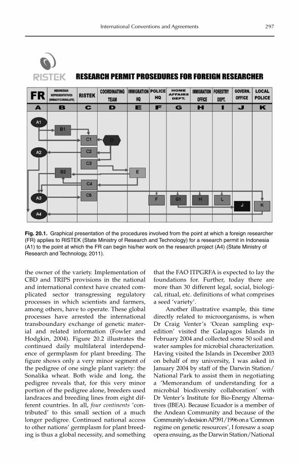
International Conventions and Agreements 297
the owner of the variety. Implementation of CBD and TRIPS provisions in the national and international context have created com-plicated sector transgressing regulatory processes in which scientists and farmers, among others, have to operate. These global processes have arrested the international transboundary exchange of genetic mater-ial and related information (Fowler and Hodgkin, 2004). Figure 20.2 illustrates the continued daily multilateral interdepend-ence of germplasm for plant breeding. The figure shows only a very minor segment of the pedigree of one single plant variety: the Sonalika wheat. Both wide and long, the pedigree reveals that, for this very minor portion of the pedigree alone, breeders used landraces and breeding lines from eight dif-ferent countries. In all, four continents ‘con-tributed’ to this small section of a much longer pedigree. Continued national access to other nations’ germplasm for plant breed-ing is thus a global necessity, and something
that the FAO ITPGRFA is expected to lay the foundations for. Further, today there are more than 30 different legal, social, biologi-cal, ritual, etc. definitions of what comprises a seed ‘variety’.
Another illustrative example, this time directly related to microorganisms, is when Dr Craig Venter’s ‘Ocean sampling exp-edition’ visited the Galapagos Islands in February 2004 and collected some 50 soil and water samples for microbial characterization. Having visited the Islands in December 2003 on behalf of my university, I was asked in January 2004 by staff of the Darwin Station/National Park to assist them in negotiating a ‘Memorandum of understanding for a microbial biodiversity collaboration’ with Dr Venter’s Institute for Bio-Energy Alterna-tives (IBEA). Because Ecuador is a member of the Andean Community and because of the Com munity’s decision AP391/1996 on a ‘Com mon regime on genetic resources’, I foresaw a soap opera ensuing, as the Darwin Station/National
Fig. 20.1. Graphical presentation of the procedures involved from the point at which a foreign researcher (FR) applies to RISTEK (State Ministry of Research and Technology) for a research permit in Indonesia (A1) to the point at which the FR can begin his/her work on the research project (A4) (State Ministry of Research and Technology, 2011).
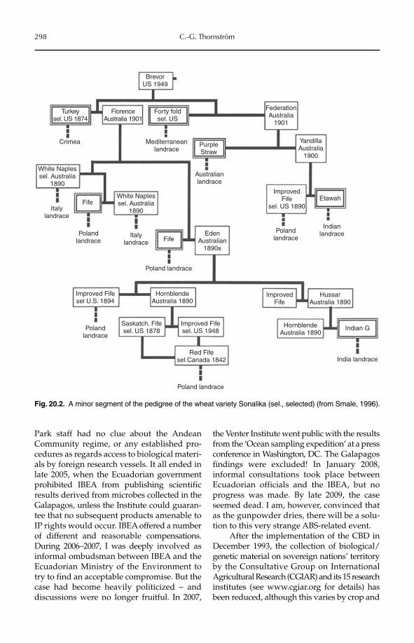
298 C.-G. Thornström
Park staff had no clue about the Andean Community regime, or any established pro-cedures as regards access to biological materi-als by foreign research vessels. It all ended in late 2005, when the Ecuadorian governmentprohibited IBEA from publishing scientificresults derived from microbes collected in the Galapagos, unless the Insti tute could guaran-tee that no subsequent products amenable to IP rights would occur. IBEA offe red a number of different and reasonable compensations. During 2006–2007, I was deeply involved as informal ombudsman between IBEA and the Ecuadorian Ministry of the Environment to try to find an acceptable compromise. But the case had become heavily politicized – and discussions were no longer fruitful. In 2007,
the Venter Institute went public with the results from the ‘Ocean sampling expedition’ at a press conference in Washington, DC. The Galapagos findings were excluded! In January 2008, informal consultations took place between Ecuadorian off icials and the IBEA, but no progress was made. By late 2009, the case seemed dead. I am, however, convinced that as the gunpowder dries, there will be a solu-tion to this very strange ABS-related event.
After the implementation of the CBD in December 1993, the collection of biological/genetic material on sovereign nations’ territory by the Consultative Group on Internatio nal Agricultural Research (CGIAR) and its 15 research institutes (see www.cgiar.org for details) has been reduced, although this varies by crop and
BrevorUS 1949
FlorenceAustralia 1901
Forty foldsel. US
PurpleStraw
Mediterraneanlandrace
Australianlandrace
EdenAustralian
1890sFife
Poland landrace
HornblendeAustralia 1890
ImprovedFife
HussarAustralia 1890
Indian GHornblendeAustralia 1890
Improved Fifesel. US 1948
Red Fifesel.Canada 1842
Improved Fifesel U.S. 1894
Saskatch. Fifesel. US 1878
India landrace
FederationAustralia
1901
YandillaAustralia
1900
ImprovedFife
sel. US 1890
Polandlandrace
Indianlandrace
Etawah
Turkeysel. US 1874
Crimea
White Naplessel. Australia
1890
White Naplessel. Australia
1890Italylandrace
Fife
Polandlandrace
Polandlandrace
Poland landrace
Italylandrace
Fig. 20.2. A minor segment of the pedigree of the wheat variety Sonalika (sel., selected) (from Smale, 1996).
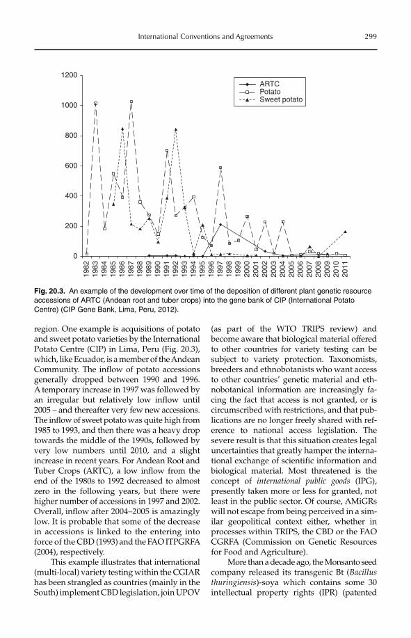
International Conventions and Agreements 299
region. One example is acquisitions of potato and sweet potato varieties by the International Potato Centre (CIP) in Lima, Peru (Fig. 20.3), which, like Ecuador, is a member of the Andean Community. The inflow of potato accessions generally dropped between 1990 and 1996. A temporary increase in 1997 was followed by an irregular but relatively low inflow until 2005 – and thereafter very few new accessions. The inflow of sweet potato was quite high from 1985 to 1993, and then there was a heavy drop towards the middle of the 1990s, followed by very low numbers until 2010, and a slight increase in recent years. For Andean Root and Tuber Crops (ARTC), a low inflow from the end of the 1980s to 1992 decrea sed to almost zero in the following years, but there were higher number of accessions in 1997 and 2002. Overall, inflow after 2004–2005 is amazingly low. It is probable that some of the decrease in accessions is linked to the entering into force of the CBD (1993) and the FAO ITPGRFA (2004), respectively.
This example illustrates that international (multi-local) variety testing within the CGIAR has been strangled as countries (mainly in the South) implement CBD legislation, join UPOV
(as part of the WTO TRIPS review) and become aware that biological material offered to other countries for variety testing can be subject to variety protection. Taxonomists, breeders and ethnobotanists who want access to other countries’ genetic material and eth-nobotanical information are increasingly fa -cing the fact that access is not granted, or is circumscribed with restrictions, and that pub-lications are no longer freely shared with ref-erence to national access legislation. The severe result is that this situation creates legal uncertainties that greatly hamper the interna-tional exchange of scientific information and biological material. Most threatened is the concept of international public goods (IPG), presently taken more or less for granted, not least in the public sector. Of course, AMiGRs will not escape from being perceived in a sim-ilar geopolitical context either, whether in processes within TRIPS, the CBD or the FAO CGRFA (Commission on Genetic Resources for Food and Agriculture).
More than a decade ago, the Monsanto seed company released its transgenic Bt (Bacillus thuringiensis)-soya which contains some 30 intellectual property rights (IPR) (patented
0
200
400
600
800
1000
120019
8219
8319
8419
8519
8619
8719
8819
8919
9019
9119
9219
9319
9419
9519
9619
9719
9819
9920
0020
0120
0220
0320
0420
0520
0620
0720
0820
0920
1020
11
ARTCPotatoSweet potato
Fig. 20.3. An example of the development over time of the deposition of different plant genetic resource accessions of ARTC (Andean root and tuber crops) into the gene bank of CIP (International Potato Centre) (CIP Gene Bank, Lima, Peru, 2012).
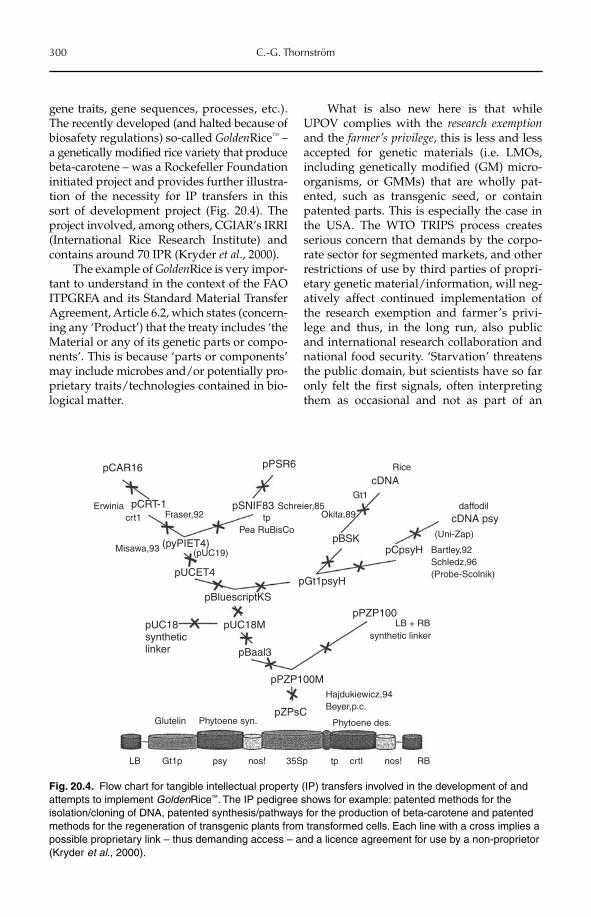
300 C.-G. Thornström
gene traits, gene sequences, processes, etc.). The recently developed (and halted because of biosafety regulations) so-called GoldenRice™ – a genetically modified rice variety that produce beta-carotene – was a Rockefeller Foundation initiated project and provides further illustra-tion of the necessity for IP transfers in this sort of development project (Fig. 20.4). The project involved, among others, CGIAR’s IRRI (International Rice Research Institute) and contains around 70 IPR (Kryder et al., 2000).
The example of GoldenRice is very impor-tant to understand in the context of the FAO ITPGRFA and its Standard Material Transfer Agreement, Article 6.2, which states (concern-ing any ‘Product’) that the treaty includes ‘the Material or any of its genetic parts or compo-nents’. This is because ‘parts or components’ may include microbes and/or potentially pro-prietary traits/technologies contained in bio-logical matter.
What is also new here is that while UPOV complies with the research exemptionand the farmer’s privilege, this is less and less accepted for genetic materials (i.e. LMOs, including genetically modified (GM) micro-organisms, or GMMs) that are wholly pat-ented, such as transgenic seed, or contain patented parts. This is especially the case in the USA. The WTO TRIPS process creates serious concern that demands by the corpo-rate sector for segmented markets, and other restrictions of use by third parties of propri-etary genetic material/information, will neg-atively affect continued implementation of the research exemption and farmer’s privi-lege and thus, in the long run, also public and international research collaboration and national food security. ‘Starva tion’ threatens the public domain, but scientists have so far only felt the first signals, often interpreting them as occasional and not as part of an
pUC18syntheticlinker
Misawa,93
Erwiniacrt1 Fraser,92
pCAR16
pCRT-1 pSNIF83
pBSK
pGt1psyH
pPZP100
pPZP100M
pZPsC
pCpsyH
daffodil
pPSR6
pUCET4
pUC18M
pBaal3
pBluescriptKS
(pyPIET4)(pUC19)
tpPea RuBisCo
Schreier,85Okita,89
Gt1
cDNA
cDNA psy(Uni-Zap)
Bartley,92Schledz,96(Probe-Scolnik)
LB + RBsynthetic linker
Hajdukiewicz,94Beyer,p.c.
Phytoene des.Phytoene syn.Glutelin
LB Gt1p psy nos! nos! RB35Sp tp crtI
Rice
Fig. 20.4. Flow chart for tangible intellectual property (IP) transfers involved in the development of and attempts to implement GoldenRice™. The IP pedigree shows for example: patented methods for the isolation/cloning of DNA, patented synthesis/pathways for the production of beta-carotene and patented methods for the regeneration of transgenic plants from transformed cells. Each line with a cross implies a possible proprietary link – thus demanding access – and a licence agreement for use by a non-proprietor (Kryder et al., 2000).
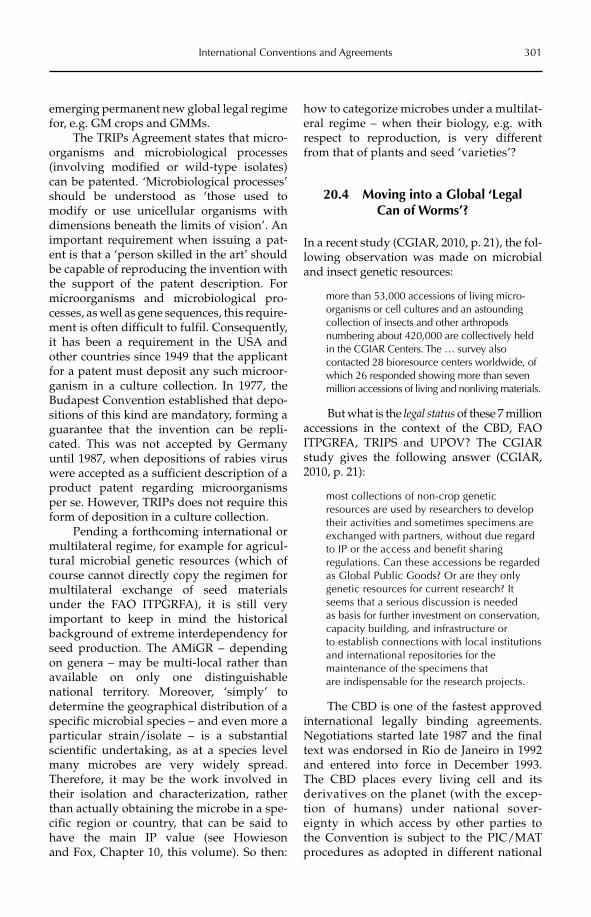
International Conventions and Agreements 301
emerging permanent new global legal regime for, e.g. GM crops and GMMs.
The TRIPs Agreement states that micro-organisms and microbiological processes (invol ving modified or wild-type isolates) can be patented. ‘Microbiological processes’ should be understood as ‘those used to modify or use unicellular organisms with dimensions beneath the limits of vision’. An important requirement when issuing a pat-ent is that a ‘person skilled in the art’ should be capable of reproducing the invention with the support of the patent description. For microorganisms and microbiological pro-cesses, as well as gene sequences, this require-ment is often difficult to fulfil. Consequently, it has been a requirement in the USA and other countries since 1949 that the applicant for a patent must deposit any such microor-ganism in a culture collection. In 1977, the Budapest Convention established that depo-sitions of this kind are mandatory, forming a guarantee that the invention can be repli-cated. This was not accepted by Germany until 1987, when depositions of rabies virus were accepted as a sufficient description of a product patent regard ing microorganisms per se. However, TRIPs does not require this form of deposition in a culture collection.
Pending a forthcoming international or multilateral regime, for example for agricul-tural microbial genetic resources (which of course cannot directly copy the regimen for multilateral exchange of seed materials under the FAO ITPGRFA), it is still very important to keep in mind the historical background of extreme interdependency for seed production. The AMiGR – depending on genera – may be multi-local rather than available on only one distinguishable national territory. More over, ‘simply’ to determine the geographical distribution of a specific microbial species – and even more a particular strain/isolate – is a substantial scientific undertaking, as at a species level many microbes are very widely spread. Therefore, it may be the work involved in their isolation and characterization, rather than actually obtaining the microbe in a spe-cific region or country, that can be said to have the main IP value (see Howieson and Fox, Chapter 10, this volume). So then:
how to categorize micro bes under a multilat-eral regime – when their biology, e.g. with respect to reproduction, is very different from that of plants and seed ‘varieties’?
20.4 Moving into a Global ‘Legal Can of Worms’?
In a recent study (CGIAR, 2010, p. 21), the fol-lowing observation was made on microbial and insect genetic resources:
more than 53,000 accessions of living micro-organisms or cell cultures and an astounding collection of insects and other arthropods numbering about 420,000 are collectively held in the CGIAR Centers. The … survey also contacted 28 bioresource centers worldwide, of which 26 responded showing more than seven million accessions of living and nonliving materials.
But what is the legal status of these 7 million accessions in the context of the CBD, FAO ITPGRFA, TRIPS and UPOV? The CGIAR study gives the following answer (CGIAR, 2010, p. 21):
most collections of non-crop genetic resources are used by researchers to develop their activities and sometimes specimens are exchanged with partners, without due regard to IP or the access and benefit sharing regulations. Can these accessions be regarded as Global Public Goods? Or are they only genetic resources for current research? It seems that a serious discussion is needed as basis for further investment on conservation, capacity building, and infrastructure or to establish connections with local institutions and international repositories for the maintenance of the specimens that are indispensable for the research projects.
The CBD is one of the fastest approved international legally binding agreements. Negotiations started late 1987 and the final text was endorsed in Rio de Janeiro in 1992 and entered into force in December 1993. The CBD places every living cell and its derivatives on the planet (with the excep-tion of humans) under national sover-eignty in which access by other parties to the Convention is subject to the PIC/MAT procedures as adopted in different national
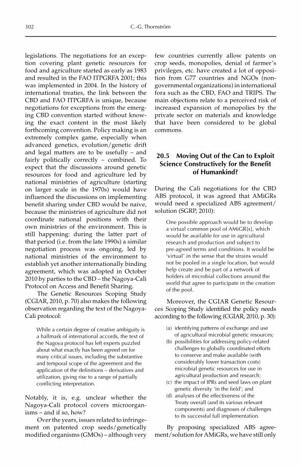
302 C.-G. Thornström
legislations. The negotiations for an excep-tion covering plant genetic resources for food and agriculture started as early as 1983 and resulted in the FAO ITPGRFA 2001; this was implemented in 2004. In the history of international treaties, the link between the CBD and FAO ITPGRFA is unique, because negotiations for exceptions from the emerg-ing CBD convention started without know-ing the exact content in the most likely forthcoming convention. Policy making is an extremely complex game, especially when advanced genetics, evolution/genetic drift and legal matters are to be usefully – and fairly politically correctly – combined. To expect that the discussions around genetic resources for food and agriculture led by national ministries of agriculture (starting on larger scale in the 1970s) would have influenced the discussions on implementing benefit sharing under CBD would be naive, because the ministries of agriculture did not coordinate national positions with their own ministries of the environment. This is still happening: during the latter part of that period (i.e. from the late 1990s) a similar negotiation process was ongoing, led by national ministries of the environment to establish yet another internationally binding agreement, which was adopted in October 2010 by parties to the CBD – the Nagoya-Cali Protocol on Access and Benefit Sharing.
The Genetic Resources Scoping Study (CGIAR, 2010, p. 70) also makes the following observation regarding the text of the Nagoya-Cali protocol:
While a certain degree of creative ambiguity is a hallmark of international accords, the text of the Nagoya protocol has left experts puzzled about what exactly has been agreed on for many critical issues, including the substantive and temporal scope of the agreement and the application of the definitions – derivatives and utilization, giving rise to a range of partially conflicting interpretation.
Notably, it is, e.g. unclear whether the Nagoya-Cali protocol covers microorgan-isms – and if so, how?
Over the years, issues related to infringe-ment on patented crop seeds/genetically modified organisms (GMOs) – although very
few countries currently allow patents on crop seeds, monopolies, denial of farmer’s privileges, etc. have created a lot of opposi-tion from G77 countries and NGOs (non-governmental organi zations) in international fora such as the CBD, FAO and TRIPS. The main objections relate to a perceived risk of increased expansion of monopolies by the private sector on materials and knowledge that have been considered to be global commons.
20.5 Moving Out of the Can to Exploit Science Constructively for the Benefit
of Humankind?
During the Cali negotiations for the CBD ABS protocol, it was agreed that AMiGRs would need a specialized ABS agreement/solution (SGRP, 2010):
One possible approach would be to develop a virtual common pool of AMiGR[s], which would be available for use in agricultural research and production and subject to pre-agreed terms and conditions. It would be ‘virtual’ in the sense that the strains would not be pooled in a single location, but would help create and be part of a network of holders of microbial collections around the world that agree to participate in the creation of the pool.
Moreover, the CGIAR Genetic Resour-ces Scoping Study identified the policy needs according to the following (CGIAR, 2010, p. 30):
(a) identifying patterns of exchange and use of agricultural microbial genetic resources;
(b) possibilities for addressing policy-related challenges to globally coordinated efforts to conserve and make available (with considerably lower transaction costs) microbial genetic resources for use in agricultural production and research;
(c) the impact of IPRs and seed laws on plant genetic diversity ‘in the field’; and
(d) analyses of the effectiveness of the Treaty overall (and its various relevant components) and diagnoses of challenges to its successful full implementation.
By proposing specialized ABS agree-ment/solution for AMiGRs, we have still only
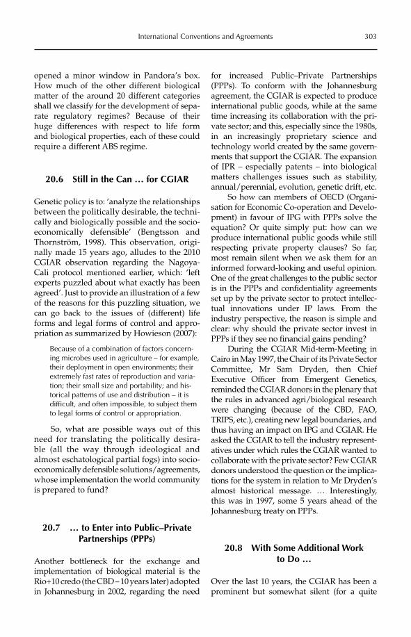
International Conventions and Agreements 303
opened a minor window in Pandora’s box. How much of the other different biological matter of the around 20 different categories shall we classify for the development of sepa-rate regulatory regimes? Because of their huge differences with respect to life form and biological properties, each of these could require a different ABS regime.
20.6 Still in the Can … for CGIAR
Genetic policy is to: ‘analyze the relationshipsbetween the politically desirable, the techni-cally and biologically possible and the socio-economically defensible’ (Bengtsson and Thornström, 1998). This observation, origi-nally made 15 years ago, alludes to the 2010 CGIAR observation regarding the Nagoya-Cali protocol mentioned earlier, which: ‘left experts puzzled about what exactly has been agreed’. Just to provide an illustration of a few of the reasons for this puzzling situation, we can go back to the issues of (different) life forms and legal forms of control and appro-priation as summarized by Howieson (2007):
Because of a combination of factors concern-ing microbes used in agriculture – for example, their deployment in open environments; their extremely fast rates of reproduction and varia-tion; their small size and portability; and his-torical patterns of use and distribution – it is difficult, and often impossible, to subject them to legal forms of control or appropriation.
So, what are possible ways out of this need for translating the politically desira-ble (all the way through ideological and almost eschatological partial fogs) into socio- economically defensible solutions/agreements, whose implementation the world community is prepared to fund?
20.7 … to Enter into Public–Private Partnerships (PPPs)
Another bottleneck for the exchange and implementation of biological material is the Rio+10 credo (the CBD – 10 years later) adopted in Johannesburg in 2002, regarding the need
for increased Public–Private Partnerships (PPPs). To conform with the Johannesburg agreement, the CGIAR is expected to produce international public goods, while at the same time increasing its collaboration with the pri-vate sector; and this, especially since the 1980s, in an increasingly proprietary science and technology world created by the same govern-ments that support the CGIAR. The expansion of IPR – especially patents – into biological matters challenges issues such as stability, annual/perennial, evolution, genetic drift, etc.
So how can members of OECD (Organi-sation for Economic Co-operation and Develo-pment) in favour of IPG with PPPs solve the equation? Or quite simply put: how can we produce international public goods while still respecting private property clauses? So far, most remain silent when we ask them for an informed forward-looking and useful opinion. One of the great challenges to the public sector is in the PPPs and confidentiality agreements set up by the private sector to protect intellec-tual innovations under IP laws. From the industry perspective, the reason is simple and clear: why should the private sector invest in PPPs if they see no financial gains pending?
During the CGIAR Mid-term-Meeting in Cairo in May 1997, the Chair of its Private Sector Committee, Mr Sam Dryden, then Chief Executive Officer from Emergent Genetics, reminded the CGIAR donors in the plenary that the rules in advanced agri/biological research were changing (because of the CBD, FAO, TRIPS, etc.), creating new legal boundaries, and thus having an impact on IPG and CGIAR. He asked the CGIAR to tell the industry represent-atives under which rules the CGIAR wanted to collaborate with the private sector? Few CGIAR donors understood the question or the implica-tions for the system in relation to Mr Dryden’s almost historical message. … Interestingly, this was in 1997, some 5 years ahead of the Johannesburg treaty on PPPs.
20.8 With Some Additional Work to Do …
Over the last 10 years, the CGIAR has been a prominent but somewhat silent (for a quite
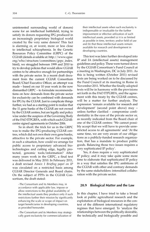
304 C.-G. Thornström
uninterested surrounding world of donors) scene for an intellectual battlefield, trying to satisfy its donors requesting IPG produced in an increasingly proprietary biological world created by the very same donors! This fact is alarming or, at worst, more or less close to intellectual schizophrenia. In the Genetic Resour ces Policy Committee (GRPC) of the CGIAR (details available at: http://www.cgiar.org/who/structure/committees/grpc_index.html), we struggled between 1999 and 2010 to try to develop policies that would allow CGIAR centres to be attractive enough for partnerships with the private sector. In a recent draft docu-ment from the current CGIAR Consortium Board/Chief Executive Officer, an attempt was made – based on our 10 year work in the now dismantled GRPC – to formulate recommenda-tions on how demands from the private sector for exclusivity can be combined with demands for IPG by the CGIAR. Just to complicate things further, we had as a starting point to realize that the 11 gene banks of the CGIAR are not owned by the CGIAR centres, but are legally and policy wise under the auspices of the Governing Body of the FAO ITPGRFA, with which each CGIAR-centre signed agreements in October 2006.
So in short: the trick for us in the GRPC was to make the IPG-producing CGIAR cen-tres, which did not own their own gene banks, attractive to the private sector. For example, in such a situation, how could we arrange for public access to proprietary advanced bio-technologies and cutting edge, legally pro-tected, genomic tools/information? After many years work in the GRPC, a final text was delivered in May 2010. In February 2011, a draft revised Annex 1 Briefing paper on IPwas circulated on a restricted basis among CGIAR Director Generals and Board chairs. On the subject of PPPs in the CGIAR Con-sortium, the draft stated:
–The Consortium and its Members may, in accordance with applicable law, impose or allow restrictions to the global availability of the intellectual assets they produce only if such restrictions further their mission by significantly enhancing the scale or scope of impact on target beneficiaries in developing countries, as provided hereunder.
–The Consortium and its Members may strategi-cally grant exclusivity for commercialization of
their intellectual assets when such exclusivity is indispensable or invaluable for the further improvement or effective utilization of such intellectual assets, provided (a) it is as limited as possible in time, territory and/or field of use and (b) that these intellectual assets remain available for research and development in developing countries.
This text was later further developed into IP and IA (intellectual assets) management guidelines and policy. These were turned down by the CGIAR Consortium’s Fund Council at a meeting in Washington, DC in July 2011. As this is being written (October 2011) revised texts are being worked on to be discussed by the Fund Council at its meeting in Rome in November 2011. Whether the finally adopted texts will be in harmony with the provisions set forth in the FAO ITPGRFA, and the agree-ment with this treaty by the CGIAR centres, will be a matter for further analysis. The expression ‘remain available for research and development’ (second point above) may – unless further clarified – compromise confi-dentiality in the eyes of the private sector or, as recently indicated from the Board chair of one of the CGIAR centres: ‘We cannot possi-bly accept that we will have to provide unre-stricted access to all agreements’ and ‘At the same time, we are very aware of our obliga-tions as a publicly-funded research organiza-tion, that has a mandate to produce public goods. Balancing those two issues requires a very sophisticated IP policy’.
Yes, it does require a very sophisticated IP policy; and it may take quite some more time to elaborate that sophisticated IP policy in a way that satisfies the IPG ambitions of the CGIAR with other and contrary demands by the same stakeholders: intensified collabo-ration with the private sector.
20.9 Biological Matter and the Law
In this chapter, I have tried to take a broad look at public agricultural research and the exploitation of biological resources in the con-text of the different international regulatory regimes that have emerged. To ‘analyze the relationships between the politically desirable, the technically and biologically possible and
International Conventions and Agreements 305
the socio-economically defensible’ (Bengtssonand Thornström, 1998) is still a challenge, not only for the CGIAR Consortium, but also for most public R&D institutions dealing with biological matter and related information. If reasonable PPPs are to be established and delivered, researchers in the public sector must stop pushing things under the carpet, and instead be realistic about the politically correct vis-à-vis accepting realities in our ever-deepening understanding of biology, genetic drift and evolution.
Thus – given current geopolitical realities – there is a need for the training of scientists in the public sector, but also of scientists in NGOs and in the private sector, on the com-plex legal issues that surround the exchange and use of biological matter. One example is the advanced training programme GRIP (Genetic Resources and Intellectual Property Rights) that I lead (from 2003 to 2013), which is supported by SIDA (the Swedish
International Development Cooperation Agency) (details available at: https://www.slu.se/grip).
We are reluctant to accept the rapidly emerging ‘proprietary science landscape’ – actually created by ourselves – which con-strains rather than encourages the securing of and clever use of biodiversity. To similar effect, TRIPS at its present stage of development may discourage intellectual innovation by introducing ‘patent carpets’ and biological monopolies that nobody may find useful to humankind in the long run – and the quite alarming challenges that we have from climate, biodiversity, ecosystems and global food security. Microorganisms are somewhere there at the base of those challenges. We must rationalize their sustainable use, as well as that of other biological matter, whether in the CGIAR, in the private sector or at public sec-tor universities and the like in the North and in the South.
Appendix: Abbreviations
ABS Access and Benefit SharingAMiGR Agricultural Microbial Genetic ResourceARTC Andean Root and Tuber CropsCBD Convention on Biological DiversityCGIAR Consultative Group on International Agricultural ResearchCGRFA Commission on Genetic Resources for Food and AgricultureCIP International Potato CentreCO/DO Certificate/Disclosure of originFAO Food and Agriculture Organization (of the United Nations)FAO ITPGRFA International Treaty on Plant Genetic Resources for Food and AgricultureFTO Freedom to OperateGMM Genetically Modified MicroorganismGMO Genetically Modified OrganismGRIPS Genetic Resources and Intellectual Property Rights (SIDA programme)GRPC Genetic Policy Resources Committee (of the CGIAR)IA Intellectual AssetsIBEA Institute for Bio-Energy AlternativesIGCGRTKF Intergovernmental Committee on Genetic Resources, Traditional Knowledge
and Folklore (of the WIPO)IP Intellectual PropertyIPG International Public GoodsIPPC International Plant Protection ConventionIPR Intellectual Property RightsIRRI International Rice Research InstituteLDC Least Developed CountryLMO Living Modified Organism
306 C.-G. Thornström
MAT Mutually Agreed TermsMTA Material Transfer AgreementNGO Non-Governmental OrganizationOECD Organisation for Economic Co-operation and DevelopmentPIC Prior Informed ConsentPPP Public–Private PartnershipsPVP Plant Variety ProtectionSGRP System-wide Genetic Resources Programme (of CGIAR)SIDA Swedish International Development Cooperation AgencySNL Subject to National LegislationTK Traditional knowledgeTRIPs Trade-Related Intellectual Property RightsUPOV International Union for the Protection of New Varieties of PlantsWIPO World Intellectual Property OrganizationWTO World Trade Organization
References
Bengtsson, B. and Thornström, C.-G. (1998) Biodiversity and Future Genetic Policy –A Study of Sweden. WB/ESDAR Special Report No 5, based on Biodiversitet och Framtida Genpolitik, UD/Ds 1996:73. Ministry of Foreign Affairs, Stockholm, Sweden.
CGIAR (2001) CGIAR Working Document on IP. Report from Mid-Term Meeting 2001, May 21–25, Durban, South Africa. Consultative Group on International Agricultural Research, Washington, DC.
CGIAR (2010) GRSS – Genetic Resources Scoping Study. Consultative Group on International Agricultural Research, Washington, DC.
Correa, C. (2001) Appendix: Legal issues relating to access to plant genetic resources. In: Petit, M., Fowler, C., Collins, W., Correa, C. and Thornström, C.-G. (eds) Why Governments Can´t Make Policy –The Case of Plant Genetic Resources in the International Arena. International Potato Centre, Lima, Peru.
Fowler, C. and Hodgkin, T. (2004) Plant genetic resources for food and agriculture: assessing global availabil-ity. Annual Review of Environment and Resources 29, 143–179.
Howieson, J.G. (2007) Technical Issues Relating to Agricultural Microbial Genetic Resources (AMiGRs), Including their Characteristics, Utilization, Preservation and Distribution. A Draft Information Paper Prepared for the Genetic Resources Policy Committee (GRPC) of the CGIAR, Commission on Genetic Resources for Food and Agriculture, Eleventh Regular Session, Rome, 11–15 June 2007. CGRFA-11/07/Circ.3, Food and Agriculture Organization, Rome. Available at: http://www.bioversityinternational.org/fileadmin/bioversityDocs/Policy/Access_and_Benefit_Sharing/AMIGRS_Howieson_CGRFA11_2007_r11c3e.pdf (accessed 10 November 2011).
Howieson, J.G. and Fox, S.L. (2012) Plant growth promotion with microorganisms. In: Sundh, I., Wilcks, A. and Goettel, M.G. (eds) Beneficial Microorganisms in Agriculture, Food and the Environment: Safety Assessment and Regulation. CAB International, Wallingford, UK. pp. 138–152.
Kowalski, S.P., Ebora, R.V., Kryder, R.D. and Potter, R.H. (2002) Transgenic crops, biotechnology and owner-ship rights: what scientists need to know. The Plant Journal 31, 407–421.
Kryder, D., Kowalski, S. and Krattiger, A. (2000) The Intellectual and Technical Property Components of Pro-Vitamin A Rice (GoldenRice™): A Preliminary Freedom-to-Operate Review. ISAAA Briefs No 20, International Service for the Acquisition of Agri-Biotech Applications, Ithaca, New York.
Oldham, P. (2004) Global Status and Trends in Intellectual Property Claims. Issue No. 2: Microorganisms. Submission to the Executive Secretary of the Convention on Biological Diversity. The ESRC Centre for Economic and Social Aspects of Genomics (CESAGen), Lancaster, UK. Available at: http://policydialogue.org/files/events/Oldham_Global_Status_and_Trends_Microorganisms.pdf (accessed 10 November 2011).
Safrin, S. (2004) Hyperownership in a time of biotechnological promise: the international conflict to control the building blocks of life. The American Journal of International Law 98, 641–685.
SGRP (2010) Leaving Room in the CBD’s ABS Protocol for the Future Development of Specialized Access and Benefit-Sharing Arrangements: The Example of Agricultural Microbial Genetic Resources. System-wide Genetic Resources Programme (SGRP) of the CGIAR, Rome, Italy. Available at: http://www.bioversityinternational.org/
International Conventions and Agreements 307
fileadmin/bioversityDocs/Policy/Access_and_Benefit_Sharing/SGRP%202010%20Policy%20Brief_microbi-als.pdf (accessed 10 November 2011).
Smale, M. (1996) Understanding Global Trends in the Use of Wheat Diversity and International Flows of Wheat Genetic Diversity. Economics Working Papers 7670, International Maize and Wheat Improvement Center (CIMMYT), Mexico, D.F., Mexico. Available at: http://ideas.repec.org/p/ags/cimmew/7670.html (accessed 10 November 2011).
State Ministry of Research and Technology (2011) Research Permit Procedures for Foreign Universities, Research Institutes, Enterprises and Individual Researchers in Indonesia. State Ministry of Research and Technology – Republic of Indonesia, Jakarta, Indonesia, p. 2. Available at: http://www.ristek.go.id/file/upload/lain_lain/frp/PANDUAN_frp_English.pdf (accessed 10 November 2011).
Suffert, F., Latxague, É. and Sache, I. (2009) Plant pathogens as agroterrorist weapons: assessment of the threat for European agriculture and forestry. Food Security 1, 221–232.
Thornström, C.-G. (2011) A Decade with Genetic Resources, Access & Benefit Sharing and Intellectual Property Rights/GRIP-10, SIDA/SLU (Sveriges Lantbruksuniversitet), Uppsala, Sweden.
©CAB International 2012. Beneficial Microorganisms in Agriculture, Food and the Environment: 308 Safety Assessment and Regulation (eds I. Sundh et al.)
21.1 Introduction 30921.2 About OECD 309
21.2.1 Working Group on Pesticides 310 21.2.2 BioPesticides Steering Group 310
21.3 OECD Guidance on Dossier and Monograph Format 31121.4 Registration Procedures 312
21.4.1 The USA 312 21.4.2 Canada 312 21.4.3 European Union 313
21.5 Data requirements for microorganisms 31421.6 REBECA: Regulation of Biological Control Agents 314
21.6.1 Baculoviruses 31421.7 Working Document on the Evaluation of Microbials for Pest Control 315
21.7.1 Taxonomic identification of microorganisms in MBCPs 315 21.7.2 Genetic toxicity assessment: Needs and recommended
approaches 315 21.7.3 Occupational, bystander and consumer exposure
and risk assessments 316 21.7.4 Microbial metabolite residues in treated food crops 316 21.7.5 Efficacy evaluation 316
21.8 Workshop on the Regulation of Biopesticides 31621.9 Seminars 31721.10 Second Working Document on the Evaluation of Microbials
for Pest Control 318 21.10.1 Microbial contaminant limits for microbial pest
control products 318 21.10.2 Pre-submission meetings 318 21.10.3 Storage stability 318 21.10.4 Evaluation of the environmental safety of MBCPs 319 21.10.5 Fungal metabolites 319
21.11 Vision for the future 31921.12 Conclusion 319
21 OECD Guidelines and Harmonization for Microbial Control Agents
Jeroen J. MeeussenChair of the OECD-BioPesticides Steering Group, European Commission,
Belgium
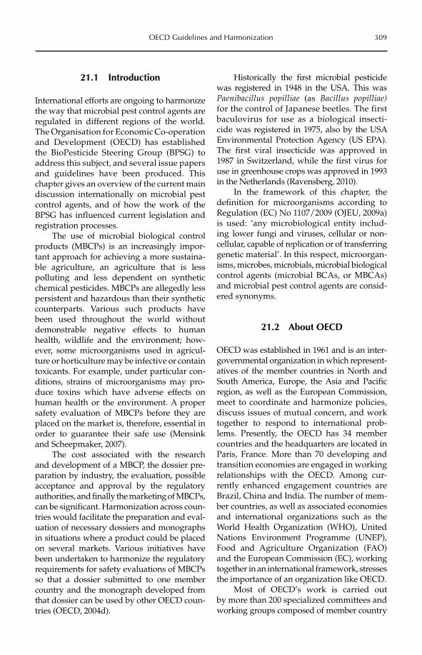
OECD Guidelines and Harmonization 309
21.1 Introduction
International efforts are ongoing to harmonize the way that microbial pest control agents are regulated in different regions of the world. The Organisation for Economic Co-operation and Development (OECD) has established the BioPesticide Steering Group (BPSG) to address this subject, and several issue papers and guidelines have been produced. This chapter gives an overview of the current main discussion internationally on microbial pest control agents, and of how the work of the BPSG has influenced current legislation and registration processes.
The use of microbial biological control products (MBCPs) is an increasingly impor-tant approach for achieving a more sustaina-ble agriculture, an agriculture that is less polluting and less dependent on synthetic chemical pesticides. MBCPs are allegedly less persistent and hazardous than their synthetic counterparts. Various such products have been used throughout the world without demonstrable negative effects to human health, wildlife and the environment; how-ever, some microorganisms used in agricul-ture or horticulture may be infective or contain toxicants. For example, under particular con-ditions, strains of microorganisms may pro-duce toxins which have adverse effects on human health or the environment. A proper safety evaluation of MBCPs before they are placed on the market is, therefore, essential in order to guarantee their safe use (Mensink and Scheepmaker, 2007).
The cost associated with the research and development of a MBCP, the dossier pre-paration by industry, the evaluation, possible acceptance and approval by the regulatory authorities, and finally the marketing of MBCPs, can be significant. Harmonization across coun-tries would facilitate the preparation and eval-uation of necessary dossiers and monographs in situations where a product could be placed on several markets. Various initiatives have been undertaken to harmonize the regulatory requirements for safety evaluations of MBCPs so that a dossier submitted to one member country and the monograph developed from that dossier can be used by other OECD coun-tries (OECD, 2004d).
Historically the first microbial pesticide was registered in 1948 in the USA. This was Paenibacillus popilliae (as Bacillus popilliae)for the control of Japanese beetles. The first baculovirus for use as a biological insecti-cide was registered in 1975, also by the USA Environ mental Protection Agency (US EPA). The first viral insecticide was approved in 1987 in Switzerland, while the first virus for use in greenhouse crops was approved in 1993 in the Netherlands (Ravensberg, 2010).
In the framework of this chapter, the definition for microorganisms according to Regulation (EC) No 1107/2009 (OJEU, 2009a) is used: ‘any microbiological entity includ-ing lower fungi and viruses, cellular or non- cellular, capable of replication or of transferring genetic material’. In this respect, microorgan-isms, microbes, microbials, microbial biological control agents (microbial BCAs, or MBCAs) and microbial pest control agents are consid-ered synonyms.
21.2 About OECD
OECD was established in 1961 and is an inter-governmental organization in which represent-atives of the member countries in North and South America, Europe, the Asia and Pacific region, as well as the European Commission, meet to coordinate and harmonize policies, discuss issues of mutual concern, and work together to respond to international prob-lems. Presently, the OECD has 34 member countries and the headquarters are located in Paris, France. More than 70 developing and transition economies are engaged in working relationships with the OECD. Among cur-rently enhanced engagement countries are Brazil, China and India. The number of mem-ber countries, as well as associated economies and international organizations such as the World Health Organization (WHO), United Nations Environment Programme (UNEP), Food and Agriculture Organization (FAO) and the European Commission (EC), working together in an international framework, stresses the impor tance of an organization like OECD.
Most of OECD’s work is carried out by more than 200 specialized committees and working groups composed of member country
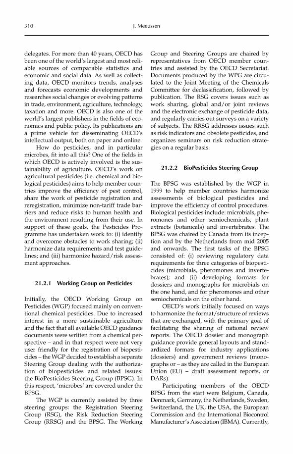
310 J. Meeussen
delegates. For more than 40 years, OECD has been one of the world’s largest and most reli-able sources of comparable statistics and economic and social data. As well as collect-ing data, OECD monitors trends, analyses and forecasts economic developments and researches social changes or evolving patterns in trade, environment, agriculture, technology, taxation and more. OECD is also one of the world’s largest publishers in the fields of eco-nomics and public policy. Its publications are a prime vehicle for disseminating OECD’s intellectual output, both on paper and online.
How do pesticides, and in particular microbes, fit into all this? One of the fields in which OECD is actively involved is the sus-tainability of agriculture. OECD’s work on agricultural pesticides (i.e. chemical and bio-logical pesticides) aims to help member coun-tries improve the efficiency of pest control, share the work of pesticide registration and reregistration, minimize non-tariff trade bar-riers and reduce risks to human health and the environment resulting from their use. In support of these goals, the Pesticides Pro-gramme has undertaken work to: (i) identify and overcome obstacles to work sharing; (ii) harmonize data requirements and test guide-lines; and (iii) harmonize hazard/risk assess-ment approaches.
21.2.1 Working Group on Pesticides
Initially, the OECD Working Group on Pesticides (WGP) focused mainly on conven-tional chemical pesticides. Due to increased interest in a more sustainable agriculture and the fact that all available OECD guidance documents were written from a chemical per-spective – and in that respect were not very user friendly for the registration of biopesti-cides – the WGP decided to establish a separate Steering Group dealing with the authoriza-tion of biopesticides and related issues: the BioPesticides Steering Group (BPSG). In this respect, ‘microbes’ are covered under the BPSG.
The WGP is currently assisted by three steering groups: the Registration Steering Group (RSG), the Risk Reduction Steering Group (RRSG) and the BPSG. The Working
Group and Steering Groups are chaired by representatives from OECD member coun-tries and assisted by the OECD Secretariat. Documents produced by the WPG are circu-lated to the Joint Meeting of the Chemicals Committee for declassification, followed by publication. The RSG covers issues such as work sharing, global and/or joint reviews and the electronic exchange of pesticide data, and regularly carries out surveys on a variety of subjects. The RRSG addresses issues such as risk indicators and obsolete pesticides, and organizes seminars on risk reduction strate-gies on a regular basis.
21.2.2 BioPesticides Steering Group
The BPSG was established by the WGP in 1999 to help member countries harmonize assessments of biological pesticides and improve the efficiency of control procedures. Biological pesticides include: microbials, phe-romones and other semiochemicals, plant extracts (botanicals) and invertebrates. The BPSG was chaired by Canada from its incep-tion and by the Netherlands from mid 2005 and onwards. The first tasks of the BPSG consisted of: (i) reviewing regulatory data requirements for three categories of biopesti-cides (microbials, pheromones and inverte-brates); and (ii) developing formats for dossiers and monographs for microbials on the one hand, and for pheromones and other semiochemicals on the other hand.
OECD’s work initially focused on ways to harmonize the format/structure of reviews that are exchanged, with the primary goal of facilitating the sharing of national review reports. The OECD dossier and monograph guidance provide general layouts and stand-ardized formats for industry applications (dossiers) and government reviews (mono-graphs or – as they are called in the European Union (EU) – draft assessment reports, or DARs).
Participating members of the OECD BPSG from the start were Belgium, Canada, Denmark, Germany, the Netherlands, Sweden, Switzerland, the UK, the USA, the European Commission and the International Biocontrol Manufacturer’s Association (IBMA). Currently,
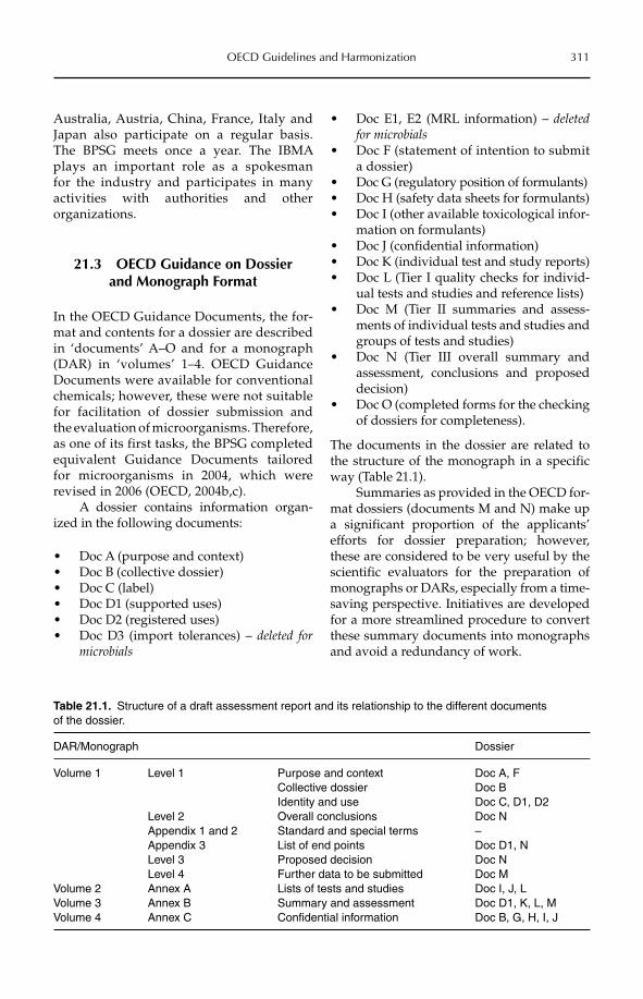
OECD Guidelines and Harmonization 311
Australia, Austria, China, France, Italy and Japan also participate on a regular basis. The BPSG meets once a year. The IBMA plays an important role as a spokesman for the industry and participates in many activities with authorities and other organizations.
21.3 OECD Guidance on Dossier and Monograph Format
In the OECD Guidance Documents, the for-mat and contents for a dossier are described in ‘documents’ A–O and for a monograph (DAR) in ‘volumes’ 1–4. OECD Guidance Documents were available for conventional chemicals; however, these were not suitable for facilitation of dossier submission and the evaluation of microorganisms. Therefore, as one of its first tasks, the BPSG completed equivalent Guidance Documents tailored for microorganisms in 2004, which were revised in 2006 (OECD, 2004b,c).
A dossier contains information organ-ized in the following documents:
Doc A (purpose and context)•Doc B (collective dossier)•Doc C (label)•Doc D1 (supported uses)•Doc D2 (registered uses)•Doc D3 (import tolerances) – • deleted for microbials
Doc E1, E2 (MRL information) – • deletedfor microbialsDoc F (statement of intention to submit •a dossier)Doc G (regulatory position of formulants)•Doc H (safety data sheets for formulants)•Doc I (other available toxicological infor-•mation on formulants)Doc J (confidential information)•Doc K (individual test and study reports)•Doc L (Tier I quality checks for individ-•ual tests and studies and reference lists)Doc M (Tier II summaries and assess-•ments of individual tests and studies and groups of tests and studies)Doc N (Tier III overall summary and •assessment, conclusions and proposed decision)Doc O (completed forms for the checking •of dossiers for completeness).
The documents in the dossier are related to the structure of the monograph in a specific way (Table 21.1).
Summaries as provided in the OECD for-mat dossiers (documents M and N) make up a significant proportion of the applicants’ efforts for dossier preparation; however, these are considered to be very useful by the scientific evaluators for the preparation of monographs or DARs, especially from a time-saving perspective. Initiatives are developed for a more streamlined procedure to convert these summary documents into monographs and avoid a redundancy of work.
Table 21.1. Structure of a draft assessment report and its relationship to the different documents of the dossier.
DAR/Monograph Dossier
Volume 1 Level 1 Purpose and context Doc A, FCollective dossier Doc BIdentity and use Doc C, D1, D2
Level 2 Overall conclusions Doc NAppendix 1 and 2 Standard and special terms –Appendix 3 List of end points Doc D1, NLevel 3 Proposed decision Doc NLevel 4 Further data to be submitted Doc M
Volume 2 Annex A Lists of tests and studies Doc I, J, LVolume 3 Annex B Summary and assessment Doc D1, K, L, MVolume 4 Annex C Confidential information Doc B, G, H, I, J
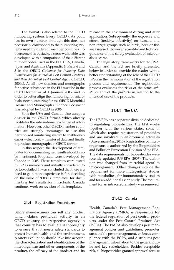
312 J. Meeussen
The format is also related to the OECD numbering system. Every OECD data point has its own number, although this does not necessarily correspond to the numbering sys-tems used by different member countries. To overcome this obstacle, a cross-walk table was developed with a comparison of the different number codes used in the EU, USA, Canada, Japan and Australia (Appendix 6, Parts 4 and 5 in the OECD Guidance for Industry Data Submissions for Microbial Pest Control Products and their Microbial Pest Control Agents; OECD, 2004c). As all new dossiers and monographs for active substances in the EU must be in the OECD format as of 1 January 2005, and in order to better align the numbering for micro-bials, new numbering for the OECD Microbial Dossier and Monograph Guidance Document was adopted by OECD in 2006.
Both the USA and Canada also accept a dossier in the OECD format, which already facilitates the international exchange of infor-mation. However, other OECD member coun-tries are strongly encouraged to use this harmonized numbering system to enable even easier – electronic – transfer of data and finally to produce monographs in OECD format.
In this respect, the development of tem-plates for documenting test results should also be mentioned. Proposals were developed by Canada in 2005. These templates were tested by BPSG members and initially considered to be too detailed. It was concluded that there is a need to gain more experience before deciding on the issue of ‘OECD templates’ for docu-menting test results for microbials. Canada continues work on revision of the templates.
21.4 Registration Procedures
Before manufacturers can sell any product which claims pesticidal activity in an OECD country, the respective agency in that country has to evaluate it thoroughly to ensure that it meets safety standards to protect human health and the environment. A safety evaluation should take into account the characterization and identification of the microorganism and other components of the product, the efficacy of the product and its
release in the environment during and after application. Subsequently, the exposure and effects (toxicity, infectivity) on humans and non-target groups such as birds, bees or fish are assessed. However, scientific and technical guidance on the safety evaluation of microbi-als is scarce.
The regulatory frameworks for the USA, Canada and the EU are briefly presented below in order to provide the reader with a better understanding of the role of the OECD BPSG in the harmonization of the registration process and requirements. The registration process evaluates the risks of the active sub-stance and of the products in relation to the intended use of the products.
21.4.1 The USA
The US EPA has a separate division dedicated to regulating biopesticides. The EPA works together with the various states, some of which also require registration of pesticides and are involved in enforcement activities (Braverman et al., 2010). Registration of micro-organisms is authorized by the Biopesticides and Pollution Prevention Division of the EPA. The data requirements for biopesticides were recently updated (US EPA, 2007). The defini-tion was changed from ‘microbial agent’ to ‘microorganism’. Other changes include the requirement for more mutagenicity studies with metabolites, for immunotoxicity studies and for an additional avian study. The require-ment for an intracerebral study was removed.
21.4.2 Canada
Health Canada’s Pest Management Reg-ulatory Agency (PMRA) is responsible for the federal regulation of pest control prod-ucts under the Pest Control Products Act (PCPA). The PMRA also develops pest man-agement policies and guidelines, promotes sustainable pest management, enforces com-pliance with the PCPA, and distributes pest management information to the general pub-lic and key stakeholders. Besides acceptable risk, all bio pesticides granted approval for use
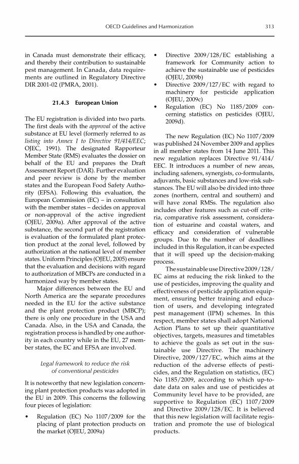
OECD Guidelines and Harmonization 313
in Canada must demonstrate their efficacy, and thereby their contribution to sustainable pest management. In Canada, data require-ments are outlined in Regulatory Directive DIR 2001-02 (PMRA, 2001).
21.4.3 European Union
The EU registration is divided into two parts. The first deals with the approval of the active substance at EU level (formerly referred to as listing into Annex I to Directive 91/414/EEC;OJEC, 1991). The designated Rapporteur Member State (RMS) evaluates the dossier on behalf of the EU and prepares the Draft Assessment Report (DAR). Further evaluation and peer review is done by the member states and the European Food Safety Autho-rity (EFSA). Following this evaluation, the European Commission (EC) – in consul tation with the member states – decides on approval or non-approval of the active ingredient (OJEU, 2009a). After approval of the active substance, the second part of the registration is evaluation of the formulated plant protec-tion product at the zonal level, followed by authorization at the national level of member states. Uniform Principles (OJEU, 2005) ensure that the evaluation and decisions with regard to authorization of MBCPs are conducted in a harmonized way by member states.
Major differences between the EU and North America are the separate procedures needed in the EU for the active substance and the plant protection product (MBCP); there is only one procedure in the USA and Canada. Also, in the USA and Canada, the registration process is handled by one author-ity in each country while in the EU, 27 mem-ber states, the EC and EFSA are involved.
Legal framework to reduce the risk of conventional pesticides
It is noteworthy that new legislation concern-ing plant protection products was adopted in the EU in 2009. This concerns the following four pieces of legislation:
Regulation (EC) No 1107/2009 for the •placing of plant protection products on the market (OJEU, 2009a)
Directive 2009/128/EC establishing a •framework for Community action to achieve the sustainable use of pesticides (OJEU, 2009b)Directive 2009/127/EC with regard to •machinery for pesticide application (OJEU, 2009c)Regulation (EC) No 1185/2009 con-•cerning statistics on pesticides (OJEU, 2009d).
The new Regulation (EC) No 1107/2009 was published 24 November 2009 and applies in all member states from 14 June 2011. This new regulation replaces Directive 91/414/EEC. It introduces a number of new areas, including safeners, synergists, co-formulants, adjuvants, basic substances and low-risk sub-stances. The EU will also be divided into three zones (northern, central and southern) and will have zonal RMSs. The regulation also includes other features such as cut-off crite-ria, comparative risk assessment, considera-tion of estuarine and coastal waters, and efficacy and consideration of vulnerable groups. Due to the number of deadlines included in this Regulation, it can be expected that it will speed up the decision-making process.
The sustainable use Directive 2009/128/EC aims at reducing the risk linked to the use of pesticides, improving the quality and effectiveness of pesticide application equip-ment, ensuring better training and educa-tion of users, and developing integrated pest management (IPM) schemes. In this respect, member states shall adopt National Action Plans to set up their quantitative objectives, targets, measures and timetables to achieve the goals as set out in the sus-tainable use Directive. The machinery Directive, 2009/127/EC, which aims at the reduction of the adverse effects of pesti-cides, and the Regulation on statistics, (EC) No 1185/2009, according to which up-to-date data on sales and use of pesticides at Community level have to be provided, are supportive to Regulation (EC) 1107/2009 and Directive 2009/128/EC. It is believed that this new legislation will facilitate regis-tration and promote the use of biological products.
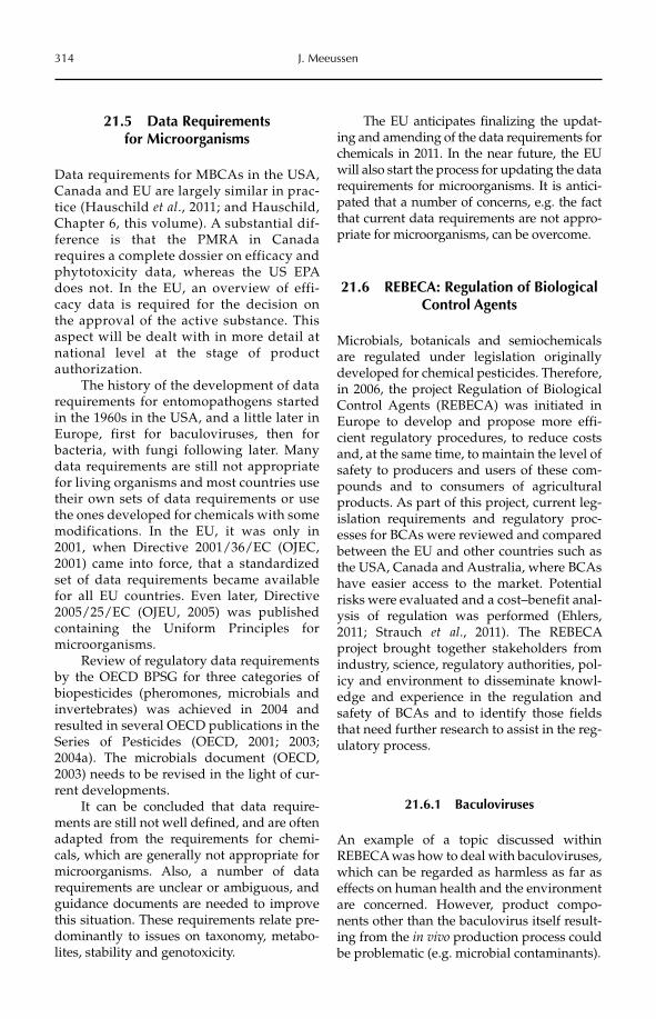
314 J. Meeussen
21.5 Data Requirements for Microorganisms
Data requirements for MBCAs in the USA, Canada and EU are largely similar in prac-tice (Hauschild et al., 2011; and Hauschild, Chapter 6, this volume). A substantial dif-ference is that the PMRA in Canada requires a complete dossier on efficacy and phytotoxicity data, whereas the US EPA does not. In the EU, an overview of effi-cacy data is required for the decision on the approval of the active substance. This aspect will be dealt with in more detail at national level at the stage of product authorization.
The history of the development of data requirements for entomopathogens started in the 1960s in the USA, and a little later in Europe, first for baculoviruses, then for bacteria, with fungi following later. Many data requirements are still not appropriate for living organisms and most countries use their own sets of data requirements or use the ones developed for chemicals with some modifications. In the EU, it was only in 2001, when Directive 2001/36/EC (OJEC, 2001) came into force, that a standardized set of data requirements became available for all EU countries. Even later, Directive 2005/25/EC (OJEU, 2005) was published containing the Uniform Principles for microorganisms.
Review of regulatory data requirements by the OECD BPSG for three categories of biopesticides (pheromones, microbials and invertebrates) was achieved in 2004 and resul ted in several OECD publications in the Series of Pesticides (OECD, 2001; 2003; 2004a). The microbials document (OECD, 2003) needs to be revised in the light of cur-rent developments.
It can be concluded that data require-ments are still not well defined, and are often adapted from the requirements for chemi-cals, which are generally not appropriate for micro organisms. Also, a number of data requirements are unclear or ambiguous, and guidance documents are needed to improve this situation. These requirements relate pre-dominantly to issues on taxonomy, metabo-lites, stability and genotoxicity.
The EU anticipates finalizing the updat-ing and amending of the data requirements for chemicals in 2011. In the near future, the EU will also start the process for updating the data requirements for microorganisms. It is antici-pated that a number of concerns, e.g. the fact that current data requirements are not appro-priate for microorganisms, can be overcome.
21.6 REBECA: Regulation of Biological Control Agents
Microbials, botanicals and semiochemicals are regulated under legislation originally developed for chemical pesticides. Therefore, in 2006, the project Regulation of Biological Control Agents (REBECA) was initiated in Europe to develop and propose more effi-cient regulatory procedures, to reduce costs and, at the same time, to maintain the level of safety to producers and users of these com-pounds and to consumers of agricultural products. As part of this project, current leg-islation requirements and regulatory proc-esses for BCAs were reviewed and compared between the EU and other countries such as the USA, Canada and Australia, where BCAs have easier access to the market. Potential risks were evaluated and a cost–benefit anal-ysis of regulation was performed (Ehlers, 2011; Strauch et al., 2011). The REBECA project brought together stakeholders from industry, science, regulatory authorities, pol-icy and environment to disseminate knowl-edge and experience in the regulation and safety of BCAs and to identify those fields that need further research to assist in the reg-ulatory process.
21.6.1 Baculoviruses
An example of a topic discussed within REBECA was how to deal with baculoviruses, which can be regarded as harmless as far as effects on human health and the environment are concerned. However, product compo-nents other than the baculovirus itself result-ing from the in vivo production process could be problematic (e.g. microbial contaminants).
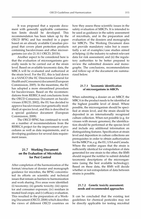
OECD Guidelines and Harmonization 315
It was proposed that a separate docu-ment with generally applicable contamina-tion limits should be developed. This recommendation has been taken up by the OECD BPSG and has resulted in a paper based on an already available Canadian pro-posal that covers plant protection products containing baculoviruses and other microor-ganisms (see also 21.10.1 OECD, 2011b).
Another aspect to be considered here is that the evaluation of microorganisms gen-erally needs to be carried out at the strain level. Based on available taxonomic data, the MBCA will be evaluated and authorized at the strain level. For the EU, this is laid down in a SANCO (the EC Directorate-General for Health and Consumers) document (European Commission, 2005). In the meantime, the EC has adopted a more streamlined procedure for baculoviruses. Based on the recommen-dations from REBECA and conclusions from the OECD Consensus Document on baculo-viruses (OECD, 2002), the EU has decided to approve baculoviruses (not genetically mod-ified) at species level, and this is described in a special guidance document (European Commission, 2008).
The OECD BPSG has continued to work on a number of recommendations from the REBECA project for the improvement of pro-cedures as well as data requirements, and is developing guidance for several data require-ment issues.
21.7 Working Document on the Evaluation of Microbials
for Pest Control
After completion of the harmonization of the format/structure of dossier and monograph guidance for microbes, the BPSG concentra-ted its efforts on scientific and technical issues that remain as barriers to harmonization and work sharing. Five areas were identified:(i) taxonomy; (ii) genetic toxicity; (iii) opera-tor and consumer exposure; (iv) residues in treated food crops; and (v) efficacy evaluation. This resulted in the preparation of a Work-ing Document (OECD, 2008) which describes the views of different OECD countries on
how they assess these scientific issues in the safety evaluation of MBCPs. It is intended to be used as guidance in the safety assessment of microbials, and in the preparation and evaluation of the dossiers and monographs for MBCPs. The Working Document does not provide mandatory rules but is essen-tially a set of examples/case studies aimed at helping: (i) the industry to submit relevant data for risk assessment; and (ii) the regula-tory authorities to be better prepared to review the submitted dossiers and mono-graphs. The conclusions, recommendations and follow-up of the document are summa-rized below.
21.7.1 Taxonomic identification of microorganisms in MBCPs
When submitting a dossier on an MBCP, the MBCA in the product needs to be identified at the highest possible level of detail. Where possible, the microorganism should be speci-fied at strain level, and the strain should be deposited in an internationally recognized culture collection. When not possible (e.g. for viruses with mosaic genomes), the identifica-tion should be performed at the species level and include any additional information on distinguishing features. Specification at strain level and deposition in culture collections are prerequisites in order to obtain authorization for the MBCP in, e.g. the EU, USA and Canada. Where the notifier argues that the strain is sufficiently identical for extrapolation of data generated for one strain to the other, the RMS should request the notifier to submit detailed taxonomic descriptions of the microorgan-isms (using the best available technology). Based on these data, the RMS will decide whether or not extrapolation of data between strains is possible.
21.7.2 Genetic toxicity assessment: needs and recommended approaches
It can be concluded that the existing test guidelines for chemical pesticides may not be directly applicable for testing microbial
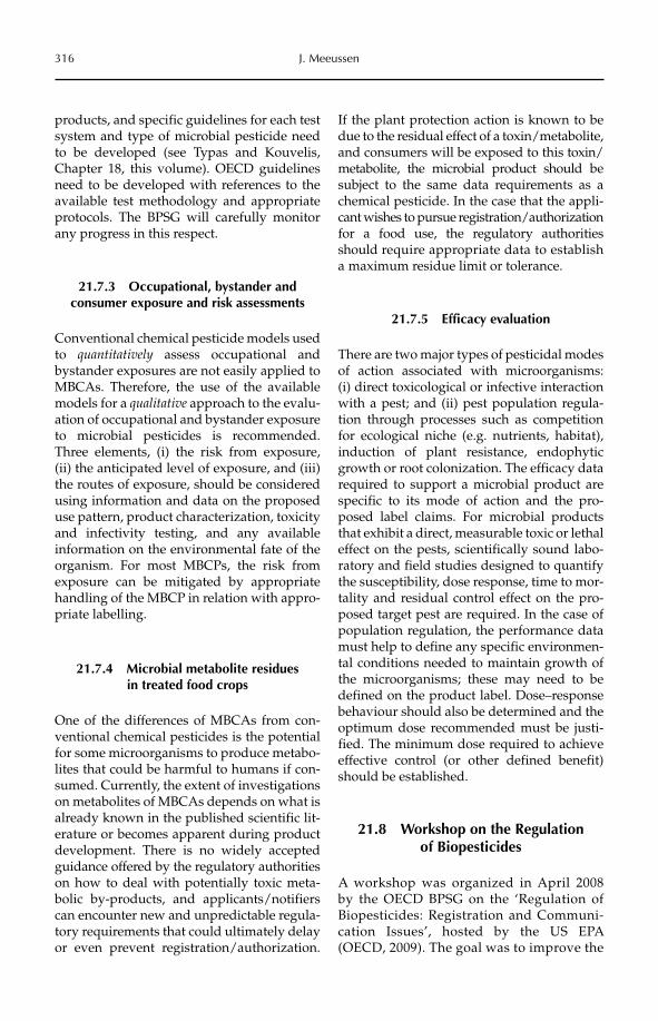
316 J. Meeussen
products, and specific guidelines for each test system and type of microbial pesticide need to be developed (see Typas and Kouvelis, Chapter 18, this volume). OECD guidelines need to be developed with references to the available test methodology and appropriate protocols. The BPSG will carefully monitor any progress in this respect.
21.7.3 Occupational, bystander and consumer exposure and risk assessments
Conventional chemical pesticide models used to quantitatively assess occupational and bystander exposures are not easily applied to MBCAs. Therefore, the use of the available models for a qualitative approach to the evalu-ation of occupational and bystander exposure to microbial pesticides is recommended. Three elements, (i) the risk from exposure, (ii) the anticipated level of exposure, and (iii) the routes of exposure, should be considered using information and data on the proposed use pattern, product characterization, toxicity and infectivity testing, and any available information on the environmental fate of the organism. For most MBCPs, the risk from exposure can be mitigated by appropriate handling of the MBCP in relation with appro-priate labelling.
21.7.4 Microbial metabolite residues in treated food crops
One of the differences of MBCAs from con-ventional chemical pesticides is the potential for some microorganisms to produce metabo-lites that could be harmful to humans if con-sumed. Currently, the extent of investigations on metabolites of MBCAs depends on what is already known in the published scientific lit-erature or becomes apparent during product development. There is no widely accepted guidance offered by the regulatory authorities on how to deal with potentially toxic meta-bolic by-products, and applicants/notifiers can encounter new and unpredictable regula-tory requirements that could ultimately delay or even prevent registration/authorization.
If the plant protection action is known to be due to the residual effect of a toxin/metabolite, and consumers will be exposed to this toxin/metabolite, the microbial product should be subject to the same data requirements as a chemical pesticide. In the case that the appli-cant wishes to pursue registration/authorization for a food use, the regulatory authorities should require appropriate data to establish a maximum residue limit or tolerance.
21.7.5 Efficacy evaluation
There are two major types of pesticidal modes of action associated with microorganisms: (i) direct toxicological or infective interaction with a pest; and (ii) pest population regula-tion through processes such as competition for ecological niche (e.g. nutrients, habitat), induction of plant resistance, endophytic growth or root colonization. The efficacy data required to support a microbial product are specific to its mode of action and the pro-posed label claims. For microbial products that exhibit a direct, measurable toxic or lethal effect on the pests, scientifically sound labo-ratory and field studies designed to quantify the susceptibility, dose response, time to mor-tality and residual control effect on the pro-posed target pest are required. In the case of population regulation, the performance data must help to define any specific environmen-tal conditions needed to maintain growth of the microorganisms; these may need to be defined on the product label. Dose–response behaviour should also be determined and the optimum dose recommended must be justi-fied. The minimum dose required to achieve effective control (or other defined benefit) should be established.
21.8 Workshop on the Regulation of Biopesticides
A workshop was organized in April 2008 by the OECD BPSG on the ‘Regulation of Biopesticides: Registration and Communi-cation Issues’, hosted by the US EPA (OECD, 2009). The goal was to improve the
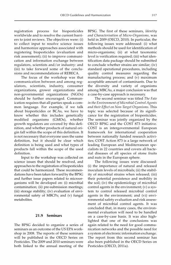
OECD Guidelines and Harmonization 317
registration process for biopesticides worldwide and to resolve the current barri-ers to joint reviews. The objectives were: (i) to collect input to resolve science issues and harmonize approaches associated with registering biopesticides (evaluation and risk assessment); (ii) to improve communi-cation and information exchange between regulators, scientists and/or industry: and (iii) to take forward some of the conclu-sions and recommendations of REBECA.
The focus of the workshop was that communication between and among reg-ulators, scientists, industry, consumer organizations, grower organizations and non-governmental organizations (NGOs)should be further encouraged. Commun-ication requires that all parties speak a com-mon language. For example, if we talk about biopesticides or BCAs, we have to know whether this includes genetically modified organisms (GMOs), whether growth regulators are covered by this defi-nition, and whether products of natural ori-gin fall within the scope of this definition. It is not necessary that everyone uses the same definition, but it should be clear which definition is being used and what types of products fall within the scope of the used definition.
Input to the workshop was collected on science issues that should be resolved, and approaches to the registration of biopesticides that could be harmonized. These recommen-dations have been taken forward by the BPSG and further issue papers related to microor-ganisms will be developed on: (i) microbial contamination; (ii) pre-submission meetings; (iii) storage stability; (iv) evaluation of envi-ronmental safety of MBCPs; and (v) fungal metabolites.
21.9 Seminars
The BPSG decided to organize a series of seminars as an outcome of the US EPA work-shop in 2008. The reports of these seminars will be published in the OECD Series on Pesticides. The 2009 and 2010 seminars were both linked to the annual meeting of the
BPSG. The first of these seminars, Identity and Characterisation of Micro-Organisms, was held in 2009 (OECD, 2010). In particular, the following issues were addressed: (i) what methods should be used for identification of micro-organisms; (ii) at what taxonomic level is verification required; (iii) what iden-tification data package should be submitted to conclude whether strains are similar; (iv) standard operational procedures, including quality control measures regarding the manufacturing process; and (v) maximum acceptable amount of contaminants. Due to the diversity and variety of organisms among MBCAs, a major conclusion was that a case-by-case approach is necessary.
The second seminar was titled The Fate in the Environment of Microbial Control Agents and their Effect on Non-Target Organisms. This topic was selected because of its signifi-cance for the registration of biopesticides. The seminar was jointly organized by the OECD BPSG and the COST 873 initiative. COST is an intergovernmental European framework for international cooperation between nationally funded research activi-ties; COST Action 873 is a large network of leading European and Mediter ranean spe-cialists in 22 countries and covers all bacte-rial diseases of all species of stone fruits and nuts in the European sphere.
The following issues were discussed: (i) the importance of natural and released inoculum levels of microbials; (ii) the stabil-ity of microbial strains when released; (iii) their potential persistence and mobility in the soil; (iv) the epidemiology of microbial control agents in the environment; (v) a sys-tem to control released microbial control agents in the environment; and (vi) envi-ronmental safety evaluation and risk assess-ment of microbial control agents. It was concluded that, in many cases, the environ-mental evaluation will need to be handled on a case-by-case basis. It was also high-lighted that one of the conclusions was again related to the need for good commu-nication networks and the possible need for a system of electronic information exchange. The report from this second seminar has also been published in the OECD Series on Pesticides (OECD, 2011a).
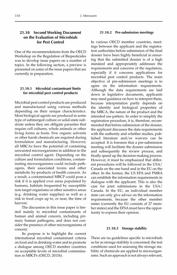
318 J. Meeussen
21.10 Second Working Document on the Evaluation of Microbials
for Pest Control
One of the recommendations from the OECD Workshop on the Regulation of Biopesticides was to develop issue papers on a number of topics. In the following section, a preview is presented on some of the issue papers that are currently in preparation.
21.10.1 Microbial contaminant limits for microbial pest control products
Microbial pest control products are produced and manufactured using various methods depending on their unique characteristics. Most biological agents are produced in some type of submerged culture or solid-state sub-strate unless they are obligate parasites that require cell cultures, whole animals or other living forms as hosts. Few organic solvents or other harsh chemicals are required during formulation and manufacturing. However, all MBCAs have the potential of containing unwanted microorganisms in addition to the microbial control agent. Depending on the culture and formulation conditions, contam-inating microorganisms could include path-ogens, their associated toxins and other metabolic by-products of health concern. As a result, a contaminated MBCP could pose a risk if it is applied over areas populated by humans, habitats frequented by susceptible non-target organisms or other sensitive areas (e.g. drinking water supplies) as well as a risk to food crops up to, or near, the time of harvest.
The discussion in this issue paper is lim-ited mainly to microbial contaminants of human and animal concern, including pri-mary human pathogens, and does not con-sider the presence of other microorganisms of concern.
Its purpose is to highlight the current international microbial contaminant criteria on food and in drinking water and to promote a dialogue among OECD member countries on acceptable levels of microbial contamina-tion in MBCPs (OECD, 2011b).
21.10.2 Pre-submission meetings
In various OECD member countries, meet-ings between the applicant and the registra-tion authorities before submission of the final dossier have been highly beneficial in ensur-ing that the submitted dossier is of a high standard and appropriately addresses the requirements and concerns of the regulators, especially if it concerns applications for microbial pest control products. The main objective of pre-submission meetings is to agree on the information requirements. Although the data requirements are laid down in legislative documents, applicants may need guidance on how to interpret them, because interpretation partly depends on the identity and biological properties of the MBCA, the nature of the product and the intended use pattern. In order to simplify the registration procedure, it is, therefore, recom-mended that before submission of the dossier, the applicant discusses the data requirements with the authority and whether studies, pub-lished literature and/or waivers can be accepted. It is foreseen that a pre-submission meeting will facilitate the dossier submission and subsequently the review process, and finally speed up the decision-making process. However, it must be emphasized that differ-ent procedures will be followed in the USA/Canada on the one hand and in the EU on the other. In the former, the US EPA and PMRA can establish the information requirements in dialogue with the applicant. This is also the case for joint submissions in the USA/Canada. In the EU, an individual member state can only give advice on the information requirements, because the other member states (currently the EU consists of 27 mem-ber states) and the EFSA must have the oppor-tunity to express their opinion.
21.10.3 Storage stability
There are no guidelines specific to microbials as far as storage stability is concerned; the test conditions used for assessing the storage sta-bility of chemicals are applied to microorgan-isms. Such an approach is not always relevant,
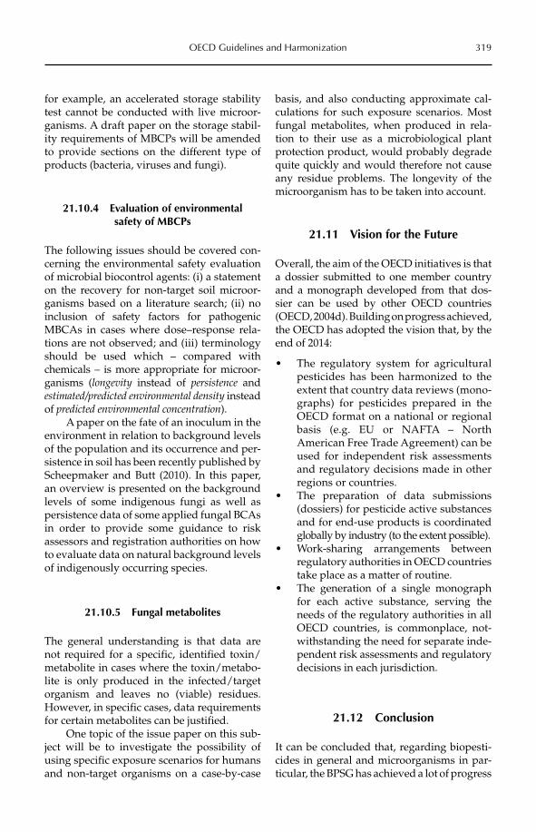
OECD Guidelines and Harmonization 319
for example, an accelerated storage stability test cannot be conducted with live microor-ganisms. A draft paper on the storage stabil-ity requirements of MBCPs will be amended to provide sections on the different type of products (bacteria, viruses and fungi).
21.10.4 Evaluation of environmental safety of MBCPs
The following issues should be covered con-cerning the environmental safety evaluation of microbial biocontrol agents: (i) a statement on the recovery for non-target soil microor-ganisms based on a literature search; (ii) no inclusion of safety factors for pathogenic MBCAs in cases where dose–response rela-tions are not observed; and (iii) terminology should be used which – compared with chemicals – is more appropriate for microor-ganisms (longevity instead of persistence and estimated/predicted environmental density instead of predicted environmental concentration).
A paper on the fate of an inoculum in the environment in relation to background levels of the population and its occurrence and per-sistence in soil has been recently published by Scheepmaker and Butt (2010). In this paper, an overview is presented on the background levels of some indigenous fungi as well as persistence data of some applied fungal BCAs in order to provide some guidance to risk assessors and registration authorities on how to evaluate data on natural background levels of indigenously occurring species.
21.10.5 Fungal metabolites
The general understanding is that data are not required for a specific, identified toxin/metabolite in cases where the toxin/metabo-lite is only produced in the infected/target organism and leaves no (viable) residues. However, in specific cases, data requirements for certain metabolites can be justified.
One topic of the issue paper on this sub-ject will be to investigate the possibility of using specific exposure scenarios for humans and non-target organisms on a case-by-case
basis, and also conducting approximate cal-culations for such exposure scenarios. Most fungal metabolites, when produced in rela-tion to their use as a microbiological plant protection product, would probably degrade quite quickly and would therefore not cause any residue problems. The longevity of the microorganism has to be taken into account.
21.11 Vision for the Future
Overall, the aim of the OECD initiatives is that a dossier submitted to one member country and a monograph developed from that dos-sier can be used by other OECD countries (OECD, 2004d). Building on progress achieved, the OECD has adopted the vision that, by the end of 2014:
The regulatory system for agricultural •pesticides has been harmonized to the extent that country data reviews (mono-graphs) for pesticides prepared in the OECD format on a national or regional basis (e.g. EU or NAFTA – North American Free Trade Agreement) can be used for independent risk assessments and regulatory decisions made in other regions or countries.The preparation of data submissions •(dossiers) for pesticide active substances and for end-use products is coordinated globally by industry (to the extent possible).Work-sharing arrangements between •regulatory authorities in OECD countries take place as a matter of routine.The generation of a single monograph •for each active substance, serving the needs of the regulatory authorities in all OECD countries, is commonplace, not-withstanding the need for separate inde-pendent risk assessments and regulatory decisions in each jurisdiction.
21.12 Conclusion
It can be concluded that, regarding biopesti-cides in general and microorganisms in par-ticular, the BPSG has achieved a lot of progress
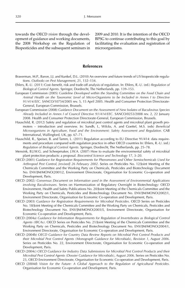
320 J. Meeussen
towards the OECD vision through the devel-opment of guidance and working documents, the 2008 Workshop on the Regulation of Biopesticides and the subsequent seminars in
2009 and 2010. It is the intention of the OECD BPSG to continue contributing to this goal by facilitating the evaluation and registration of microorganisms.
References
Braverman, M.P., Baron, J.J. and Kunkel, D.L. (2010) An overview and future trends of US biopesticide regula-tions. Outlooks on Pest Management, 21, 132–134.
Ehlers, R.-U. (2011) Cost–benefit, risk and trade-off analysis of regulation. In: Ehlers, R.-U. (ed.) Regulation of Biological Control Agents. Springer, Dordrecht, The Netherlands, pp. 139–153.
European Commission (2005) Guideline Developed within the Standing Committee on the Food Chain and Animal Health on the Taxonomic Level of Micro-Organisms to be Included in Annex I to Directive 91/414/EEC. SANCO/10754/2005 rev. 5, 15 April 2005. Health and Consumer Protection Directorate-General, European Commission, Brussels.
European Commission (2008) Guidance Document on the Assessment of New Isolates of Baculovirus Species Already Included in Annex I of Council Directive 91/414/EEC. SANCO/0253/2008 rev. 2, 22 January 2008. Health and Consumer Protection Directorate-General, European Commission, Brussels.
Hauschild, R. (2012) Safety and regulation of microbial pest control agents and microbial plant growth pro-moters – introduction and overview. In: Sundh, I., Wilcks, A. and Goettel, M.S. (eds) BeneficialMicroorganisms in Agriculture, Food and the Environment: Safety Assessment and Regulation. CAB International, Wallingford, UK, pp. 67–71.
Hauschild, R., Speiser, B. and Tamm, L. (2011) Regulation according to EU Directive 91/414: data require-ments and procedure compared with regulation practice in other OECD countries In: Ehlers, R.-U. (ed.) Regulation of Biological Control Agents. Springer, Dordrecht, The Netherlands, pp. 25–78.
Mensink, B.J.W.G. and Scheepmaker, J.W.A. (2007) How to evaluate the environmental safety of microbial plant protection products: a proposal. Biocontrol Science and Technology 17, 3–20.
OECD (2001) Guidance for Registration Requirements for Pheromones and Other Semiochemicals Used for Arthropod Pest Control, [revised] 26 February 2002. Series on Pesticides No. 12/Joint Meeting of the Chemicals Committee and the Working Party on Chemicals, Pesticides and Biotechnology Document No. ENV/JM/MONO(2001)2, Environment Directorate, Organisation for Economic Co-operation and Development, Paris.
OECD (2002) Consensus Document on Information used in the Assessment of Environmental Applications involving Baculoviruses. Series on Harmonization of Regulatory Oversight in Biotechnology: OECD Environment, Health and Safety Publications No. 20/Joint Meeting of the Chemicals Committee and the Working Party on Chemicals, Pesticides and Biotechnology Document No. ENV/JM/MONO(2002)1, Environment Directorate, Organisation for Economic Co-operation and Development, Paris.
OECD (2003) Guidance for Registration Requirements for Microbial Pesticides. OECD Series on Pesticides No. 18/Joint Meeting of the Chemicals Committee and the Working Party on Chemicals, Pesticides and Biotechnology Document No. ENV/JM/MONO(2003)5, Environment Directorate, Organisation for Economic Co-operation and Development, Paris.
OECD (2004a) Guidance for Information Requirements for Regulation of Invertebrates as Biological Control Agents (IBCAs). OECD Series on Pesticides No. 21/Joint Meeting of the Chemicals Committee and the Working Party on Chemicals, Pesticides and Biotechnology Document No. ENV/JM/MONO(2004)1, Environment Directorate, Organisation for Economic Co-operation and Development, Paris.
OECD (2004b) OECD Guidance for Country Data Review Reports on Microbial Pest Control Products and their Microbial Pest Control Agents (Monograph Guidance for Microbials), Revision 1, August 2006.Series on Pesticides No. 22, Environment Directorate, Organisation for Economic Co-operation and Development, Paris.
OECD (2004c) OECD Guidance for Industry Data Submissions for Microbial Pest Control Products and their Microbial Pest Control Agents (Dossier Guidance for Microbials), August 2006. Series on Pesticides No. 23, OECD Environment Directorate, Organisation for Economic Co-operation and Development, Paris.
OECD (2004d) Vision for the Future: A Global Approach to the Regulation of Agricultural Pesticides.Organisation for Economic Co-operation and Development, Paris.
OECD Guidelines and Harmonization 321
OECD (2008) Working Document on the Evaluation of Microbials for Pest Control. Series on Pesticides No. 43/Joint Meeting of the Chemicals Committee and the Working Party on Chemicals, Pesticides and Biotechnology Document No. ENV/JM/MONO(2008)36, Environment Directorate, Organisation for Economic Co-operation and Development, Paris.
OECD (2009) Report of the Workshop on the Regulation of Biopesticides: Registration and Communication issues, 15–17 April 2008, EPA, Arlington, USA. Series on Pesticides No. 44/Joint Meeting of the Chemicals Committee and the Working Party on Chemicals, Pesticides and Biotechnology Document No. ENV/JM/MONO(2009)19, Environment Directorate, Organisation for Economic Co-operation and Development, Paris.
OECD (2010) Report of the 1st OECD Biopesticides Steering Group Seminar on Identity and Characterisation of Micro-Organisms, 1st July 2009, OECD, Paris. Series on Pesticides No. 53/Joint Meeting of the Chemicals Committee and the Working Party on Chemicals, Pesticides and Biotechnology Document No. ENV/JM/MONO(2010)43, Environment Directorate, Organisation for Economic Co-operation and Development, Paris.
OECD (2011a) Report of the Second OECD BioPesticides Steering Group Seminar on the Fate in the Environment of Microbial Control Agents and their Effects on Non-Target Organisms. Series on Pesticides No. 64/Joint Meeting of the Chemicals Committee and the Working Party on Chemicals, Pesticides and Biotechnology Document No. ENV/JM/MONO(2011)42, Environment Directorate, Organisation for Economic Co-operation and Development, Paris.
OECD (2011b) OECD Issue Paper on Microbial Contaminant Limits for Microbial Pest Control Products. Series on Pesticides No. 65/Joint Meeting of the Chemicals Committee and the Working Party on Chemicals, Pesticides and Biotechnology Document No. ENV/JM/MONO(2011)43, Environment Directorate, Organisation for Economic Co-operation and Development, Paris.
OJEC (1991) Council Directive 91/414/EEC of 15 July 1991 concerning the placing of plant protection prod-ucts on the market. Official Journal of the European Communities 34, L 230/1–32.
OJEC (2001) Commission Directive 2001/36/EC of 16 May 2001, amending Council Directive 91/414/EEC concerning the placing of plant protection products on the market. Official Journal of the European Communities 44, L 164/1–38.
OJEU (2005) Council Directive 2005/25/EC of 14 March 2005 amending Annex VI to Directive 91/414/EEC as regards plant protection products containing micro-organisms. Official Journal of the European Union48, L 90/1–34.
OJEU (2009a) Regulation (EC) No 1107/2009 of the European Parliament and of the Council of 21 October 2009 concerning the placing of plant protection products on the market and repealing Council Directives 79/117/EEC and 91/414/EEC. Official Journal of the European Union 52, L 309, 1–50.
OJEU (2009b) Directive 2009/128/EC of the European Parliament and of the Council of 21 October 2009 establishing a framework for Community action to achieve the sustainable use of pesticides. OfficialJournal of the European Union 52, L 309, 71–86.
OJEU (2009c) Directive 2009/127/EC of the European Parliament and of the Council of 21 October 2009 amending Directive 2006/42/EC with regard to machinery for pesticide application. Official Journal of the European Union 52, L 310/29–33.
OJEU (2009d) Regulation (EC) No 1185/2009 of the European Parliament and of the Council of 25 November 2009 concerning statistics on pesticides. Official Journal of the European Union 52, L 324, 1–22.
PMRA (2001) Guidelines for the Registration of Microbial Pest Control Agents and Products, March 30, 2001.Pest Management Regulatory Agency, Ottawa, Ontario, Canada.
Ravensberg, W.J. (2010) The development of microbial pest control products for control of arthropods: a criti-cal evaluation and a roadmap to success. PhD thesis, Wageningen University, The Netherlands.
Scheepmaker, J.W.A. and Butt, T.M. (2010) Natural and released inoculum levels of entomopathogenic fungal biocontrol agents in soil in relation to risk assessment and in accordance with EU regulations. BiocontrolScience and Technology 20, 503–552.
Strauch, O., Strasser, H., Hauschild, R. and Ehlers, R.-U. (2011) Proposals for bacterial and fungal biocontrol agents. In: Ehlers, R.-U. (ed.) Regulation of Biological Control Agents. Springer, Dordrecht, The Netherlands, pp. 267–288.
Typas, M.A. and Kouvelis, V.N. (2012) Assessing genotoxic effects of microbial products. In: Sundh, I., Wilcks, A. and Goettel, M.S. (eds) Beneficial Microorganisms in Agriculture, Food and the Environment: Safety Assessment and Regulation. CAB International, Wallingford, UK, pp. 256–274.
US EPA (2007) Data requirements for biochemical and microbial pesticides. EPA 40 CFR Part 158, Final rule. Federal Register 72(207), 60988–61025.
©CAB International 2012. Beneficial Microorganisms in Agriculture, Food and the Environment: 322 Safety Assessment and Regulation (eds I. Sundh et al.)
22.1 Introduction: Divergent Understandings of Risk and the Public 32222.2 How Does the Public ‘Understand’ Risk? 32422.3 How Do Scientists Understand the Public? 32622.4 From Risk Perception to Participatory Public Engagement 32822.5 Conclusion: Constructing Shared Understandings of Risks and Benefits 331
22 Understanding Public Risk Perception for the Use of Beneficial Microorganisms
Keith D. WarnerReligious Studies Department and the Center for Science, Technology, and Society,
Santa Clara University, California, USA
22.1 Introduction: Divergent Understandings of Risk and the Public
Realizing the potential benefits that microorganisms could provide society hinges, in part, on scientists understand-ing how society at large perceives risk. Scientific understanding of microbiology has expan ded remarkably, but so too has public suspicion of scientists and of (public and private) institutions that use science. Billions of dollars have been spent to create, assess and communicate technical infor-mation about the risks of technologies, yet social science surveys have consistently shown that the public has become more, not less, concerned about the risks of modern life (Slovic, 1987, 2001). Those who develop new scientific applications and novel tech-nologies build up significant specialized scientific knowledge and a familiarity with them. Members of the public do not gen-erally have this same knowledge, nor a favourable disposition towards using sci-ence to manage risk. Scientific risk commu-nication across this gap in understanding is a
major challenge to deriving benefit from the application of new science in mod-ern society, from microorganisms to nanotechnology.
Effective public communication across gaps in understanding requires all parties to understand themselves as simultaneously senders and receivers of messages. This com-munication should foster shared understand-ings of scientific knowledge, the relationship of risks to benefits, and social values such as democratic decision making. Communicating across gaps in risk perception and under-standing depends heavily upon scientific experts listening to feedback from a cluster of diverse public audiences in order to under-stand the assumptions that shape the inter-pretation of messages, while simultaneously improving the quality of communication. In short, scientists and their institutions must listen to the public to understand public risk perception, and devise communication strat-egies to overcome this gap in risk perception and understanding. The academic disciplines of social psychology and science communica-tion can help scientists and practitioners
Understanding Public Risk Perception 323
understand how to communicate across this risk perception divide.
Those trained in scientific expertise are predisposed toward discounting lay risk per-ception as subjective and emotional, in con-trast to scientific risk analysis, which experts consider to be rational and value free. Critical social scientists, however, perceive the stages of risk analysis (i.e. risk assessment, manage-ment and communication) to be influenced by social values and assumptions, to varying degrees. If scientists (and institutions that use science) misunderstand how the public per-ceives their knowledge, technologies and institutions, well-intended policies are likely to be ineffective (Slovic, 1987). Popular risk concerns have, in some cases, overridden expert recommendations for risk manage-ment. For example, public concerns about hazardous waste management resulted in directing the bulk of the budget of the US Environmental Protection Agency (US EPA) to that issue, when in fact hazards from indoor air pollution are considered by scientists to be more serious (Slovic, 1999).
Most lay audiences bring the following beliefs to their intuitive risk judgements about novel microorganisms, which shape their risk perception: microbes are invisible, largely unknowable and probably dangerous (because many lay audiences presume all microbes nec-essarily cause human disease). Microorganisms are particularly challenging for engaging pub-lic risk perception because they are generally invisible to ordinary people. Two biological control scientists summarize these challenges in the following way: ‘Despite the lack of doc-umented serious conflicts, there is an air of pathophobia that has brought to a virtual standstill the application of the classical approach in the use of plant pathogens for weed control’ (Freeman and Charudattan, 1985). The term ‘phobia’ is apt in this context, for a chief obstacle to the beneficial use of microorganisms may indeed be unfounded fears on the part of the public or public agen-cies. Risk fears can serve as obstacles to appro-priate regulation as well as to public support (Waage, 1995; Evans, 2000; Sheppard et al.,2003; Delfosse, 2005). Transforming public phobia into appropriate public support depends not only on carefully crafted scientific
communication but also on social deliberative processes grounded in democratic values. Both are critical to realizing the beneficial potential of microorganisms.
No opinion surveys of public percep-tion of the risks and benefits of microorgan-isms as a broad taxonomic category exist. However, many salient lessons relevant to the use of microorganisms can be drawn from social science research investigating public views of other biological or novel technologies. From a social science perspec-tive, these other technologies function as proxies for understanding public risk per-ception of microorganisms. Pioneering work in the ‘social psychology of risk’ addressed human perception of nuclear power and toxic chemicals (Slovic, 2001). Those who developed communication strategies for crop biotechnologies did not consult social psychologists or risk perception experts, and they committed many fundamental mistakes in science communication (Wynne, 2001). These errors imposed significant costs to industry, government credibility and society at large.
To head off the polarization that accom-panied the introduction of transgenic biotech-nologies, funders of nanotechnology have enhanced their support for social science work (Barben et al., 2008). This recent research has further characterized how members of the public perceive risks of novel technolo-gies (Kahan et al., 2007). From the perspective of critical social science, public perception of nanotechnology risks is functionally equiva-lent to public perception of microorganism risks. There are many significant biological and ecological differences between nanotech-nology, genetically modified microbes, and ‘wild-type’ – not genetically modified – microbes. However, few members of the lay public are able to distinguish meaningfully between these in their composition and potential risk. For example, McNeil et al.(2010) surveyed Canadians about their per-ception of biocontrol pest strategies; the find-ings suggested that those surveyed could not distinguish between a ‘beneficial microbe’ and biocontrol agent, and a food contami-nant. The field of science communication has investigated strategies for facilitating more
324 K.D. Warner
constructive public engagement with novel technologies (Burri, 2009), and these contain lessons relevant to effective and appropriate risk communication.
There is no singular, homogenous public audience, any more than there is one world-view held by all scientists in all places at all times. Thus, we must speak of many scientific perspectives and a diversity of lay audiences, both in the plural, to remind us of the many perspectives, and the error of conceiving of the public in a homogenous, anonymous way. The word ‘public’ necessarily bundles together people holding a wide range of sci-entific expertise, value predispositions and social power. For example, some opinion leaders in business, universities and non- governmental organizations, and govern-ment regulatory officials, may be considered a form of ‘public’ in the sense that they may be outside a specialist research community, but they are essential to mediating under-standings of risk to a more general, less scien-tifically informed general public. Hence, understanding the diversity of views held by public audiences is essential to understand-ing public risk perception (Wynne, 1992; Bucchi and Neresini, 2008).
Critical social science research addresses how scientific experts and lay publics perceive, analyse, communicate and evaluate risk, and can propose exam-ples of social processes to overcome gaps in understandings of risk. Examples of this work can be found in the fields of social psychology of risk (Beck, 1992; Slovic, 2001) and science communication (Gregory and Miller, 1998). These fields of social science incorporate natural science data into how human beings develop, use, perceive and communicate knowledge and risk within society. Therefore, this chapter does not specifically evaluate safety assessment and regulation of beneficial microorganisms, but rather how scientists and diverse pub-lic audiences under stand, communicate and deliberate risks and benefits. The implications will apply broadly to any use of microorganisms for societal benefit, whether for food or feed preservation, or for agricultural, environmental or health purposes.
This chapter begins by describing the ways in which diverse members of the pub-lic perceive risk, drawing heavily on social psychology of risk literature. It then examines the ways in which scientific institutions under-stand the public, and describes some errors in public risk communication. The chapter con-cludes by outlining new, more constructive approaches to fostering public engagement with novel technologies that could help real-ize their potential for public benefit. Greater efforts to conduct ‘upstream engagement’ with nanotechnology through anticipatory public dialogues (Macnaghten et al., 2005; Burri, 2009) are developing models for delib-erating and negotiating risk perception, eval-uation and judgement. These can inform scientists’ understandings of public risk per-ception and improve the effectiveness of risk communication efforts.
22.2 How Does the Public ‘Understand’ Risk?
The Risk Society (Beck, 1992) was one of the most influential books in European social sci-ence in the late 20th century. Beck outlined the fundamental shift across industrial socie-ties over the past five decades: from a primary concern about resource scarcity to the man-agement and distribution of risk. He argues that the scientific and technological forces that created industrial development are them-selves now evaluated by the public with their lay understanding of risk. Beck argued that debates about risk will be central in society for the indefinite future, but that diverse con-ceptualizations of what constitutes ‘risk’ are determined chiefly by social, not scientific, factors. The astonishing growth of scientific expertise (among some sectors) to create our technological society has necessarily led to divergent understandings of risk. In Beck’s risk society, lay versus expert understandings of risk substantively frame public judgement on the application of science and technology. Thus, most controversies about appropriate regulation are actually predictable expres-sions of broader social concern about risk. Politically charged disputes over regulatory
Understanding Public Risk Perception 325
safety criteria become an expected, even rou-tine, in the application of science and technol-ogy in the risk society (Slovic, 1999).
The standard expert conceptualization of risk is the statistical probability of an adverse event that can be objectively quantified by a risk assessment process (National Research Council, 1996). It is usually expressed in prob-abilistic terms, such as ‘risk = hazard × expo-sure’ (Delfosse, 2005). This approach fulfils the criteria of consistency and quantification. Critical social scientists reject conceptualiza-tions of risk as pre-existing in nature, awaiting human discovery and measurement (Slovic, 1999). Rather, risk is an abstract concept invented by human beings to help society manage uncertainties. Risk is a mental model constructed by humans. Harms, hazards and danger are real, but risk is a conceptual frame-work for evaluating and managing these. Critical social scientists have demonstrated both complexity of the concept of ‘risk’, and the inadequacies of conventional risk commu-nication to the public in terms that are nar-rowly quantitative and probabilistic (Slovic, 2001).
The scientific method, as an abstraction, may be considered value free; however, the application of science in society through risk analysis necessarily incorporates social or cultural values (Douglas and Wildavsky, 1982). These values may be explicit or tacit. Risk analysis has scientific components, but inevitably it has elements that are subjective and value laden, meaning that the cultural values are assumed and incorporated into the process. Value judgements are embedded in the risk model in the decisions made using it, e.g. which theory is to guide the construction of models, what context is to be considered, what elements are to be considered, what possible consequences are to be considered, and what time frame is to be considered. Social values shape the assumptions made about all of these factors, which are woven into scientific risk assessment processes. How these assumptions are communicated to the public is also value laden, and they reflect the experts’ perception of the public’s under-standing of risk.
Recent research in cognitive psychology and neuroscience has demonstrated that
human beings conceptualize risk in two fun-damentally different ways (Slovic et al., 2004). The ‘analytical’ system uses formal logic, probabilistic reasoning and scientific delibera-tion. The ‘experiential’ system is an intuitive, largely automatic response to perceived dan-ger, and often inaccessible to subjective aware-ness. The former is slow, but the latter is much more rapid. The experiential system has resulted from human evolutionary processes that selected against those who failed to per-ceive environmental risks (e.g. larger preda-tors, foul water), and may be considered the default approach to human risk perception (Slovic et al., 2004). This second ‘system’ of risk perception is instinctual to human beings, and scientific training develops the skills and disposition to deploy the analytical system in its place. Social science research has consist-ently found that the public has a broader con-ceptualization of risk than experts, consistent with their perspective of the world. This pub-lic perception of risk is both qualitative and complex (Slovic, 1999).
Non-scientists perceive risk through the lens of their own life experience and the deci-sions about uncertainties that they negotiate in daily life (Wynne, 1992). Members of the pub-lic evaluate technological risks in light of the following types of social factors: dread, cata-strophic potential, equity in outcome, degree of certainty, reversibility and the potential to personally choose the risk (or not). Examples of how these criteria might be manifest in pub-lic risk perception include: death from cancer is dreaded, but death from automobile acci-dents is less so; exposure to environmental tobacco smoke is perceived as riskier than cig-arette smoking; hazardous industrial waste is perceived as more risky because it is not cho-sen, as opposed to toxic household products which are purchased (Slovic, 1999).
The public develops opinions about the risks of new technology based on factors that are not included in expert risk models. For example, research into public perception of the risks of nanotechnology has demonstrated that the public holds greater concerns about personal privacy issues and equity of benefits than do scientists (Priest et al., 2010). Although some scientists may perceive this as irrational or unfair, this is in fact how non-experts
326 K.D. Warner
develop their opinions. Research has consist-ently shown that trust is the chief criterion that most lay publics use to evaluate novel technology. A scientist may ask: does the pro-posed introduction of the technology repre-sent acceptable risk? But lay publics ask: is the scientific claim trustworthy? They answer by evaluating the trustworthiness of scientists and sponsoring institutions, and their per-ceived motivations. There is both wisdom and error in public perceptions of risk (Slovic, 1987). Understanding that lay members of the public develop opinions about acceptable risk based on their level of trust in scientists and their institutions is fundamental to under-standing public risk perception and, thus, indirectly, is essential to successfully intro-ducing a novel microorganism.
Early social psychology explained how social factors such as gender, race, class, political views and individual psychology shape public risk perception (Slovic, 1999). Recent work has demonstrated the impor-tance of world views – and the social values embedded in them – in shaping risk percep-tion of nanotechnology (Kahan et al., 2007). An individualistic world view can be defined as one that prizes the autonomy of individu-als and markets to operate freely from per-ceived collective interference. When more information about nanotechnology is pro-vided to those with an individualistic world view, they are more likely to see it as benefi-cial. An egalitarian world view can be defi-ned as one that is highly concerned with the equitable distribution of benefits (and risks) across a society. When the same risk informa-tion is provided to those with an egalitarian cultural outlook as to those with an individu-alistic world view, they are more likely to perceive nanotechnology as having more risks than benefits (Kahan et al., 2007). A key implication of this finding is that providing more information about a novel technology prompts different responses, from support to fear. More information reinforces the favour-able views of those with a general risk-accepting approach to life, but for those who are more likely to be risk averse, more infor-mation can augment their concerns (Kahan et al., 2007). By understanding the diversity of world views held by the public, one can
craft more appropriate and effective risk communication strategies.
The tone or affect of a scientific risk mes-sage for a public audience generally plays a greater role in shaping public response than scientific data. For example, if a risk commu-nication bears tacit meanings of the inevita-bility of a government action, members of the public may react negatively to the perceived exercise of government power, not to the scientific assessment of the risk. This finding poses a fundamental challenge to public risk communication, and points to the need for strategies that are sensitive to broadly held social values. It also points to the critical importance of understanding science com-munication processes from the perspective of lay audiences, lest miscommunication and confusion occur. The rise of the digital media environment poses genuine challenges to public agencies gathering public comment on the proposed use of microorganisms. Novel communication strategies are required – more than simply posting information on a web page.
In sum, social science has demonstrated that lay public understandings of risk are more complex and instinctual – and poten-tially volatile – than the statistical probability of an undesirable event. Members of the public use an ‘intuitive system’ of perceiving and evaluating risk that differs from that of experts and scientists. Lay public risk percep-tion is strongly shaped by social factors such as class, gender, affect and world view. These factors strongly influence differential predis-positions towards the risks of novel technolo-gies. It is inevitable that these social factors and cultural values will shape public percep-tions of risk, but it is not inevitable that the debate becomes polarized or negative, or undermines the introduction of new technol-ogies (Kahan and Rejeski, 2009).
22.3 How Do Scientists Understand the Public?
Expert risk communication to the public car-ries the potential of a perverse outcome. Expert efforts to communicate ‘acceptable’ risk to lay publics can backfire. Providing
Understanding Public Risk Perception 327
more scientific information about risk may increase risk fears, at least among some pub-lics, and undermine the intended communi-cation effort (Douglas and Wildavsky, 1982). Effective risk communication between scien-tists and the public depends upon the public learning about science, but also upon scien-tists adopting a realistic approach to the pub-lic and its risk perception. A recent survey of American scientists found that they perceive the public to have an understanding of sci-ence that is deficient: ‘While the public holds scientists in high regard, many scientists offer unfavorable, if not critical, assessments of the public’s knowledge and expectations. Fully 85% see the public’s lack of scientific knowl-edge as a major problem for science, and nearly half (49%) fault the public for having unrealistic expectations about the speed of scientific achievements’ (The Pew Research Center, 2009).
Research into science communication has taken up the question of how scientists’ perception of the public shapes the communi-cation process. Critical social scientists have developed conceptual models to describe the rather constrained ways in which scientists perceive the public.
1. The cognitive deficit model. This assumes that if only the lay public knew more about science and ceased to be in a state of knowl-edge deficit, a better relationship between sci-ence and the public would emerge (Gregory and Miller, 1998; Sturgis and Allum, 2004). In this model, the shortcoming is in the public itself, and this is the reason why the potential of science is thwarted.2. Injection of science model. Scientific knowl-edge is developed by experts and implanted into the bloodstream of society. Here the delivery system constrains the application of science for society (Mooney, 2010).3. The loading dock model of science and policy.The task of scientists is to develop and deliver scientific knowledge to policy makers, and their job, in turn, is to explain what it means –and how it should be supplied – to the public. This model assumes that if policy makers did their job properly, there would be less of a regulatory bottleneck and greater public sup-port (Cash et al., 2006).
These models do not criticize individual sci-entists, laboratories, discoveries or institu-tions. Instead, they critique the underlying assumptions that guide the actions of some scientific, political and commercial leaders and institutions who use science.
Institutions charged with advancing tech-nological innovation can readily slip into simplistic thinking about the public and its views. Scientists’ concerns about public understanding of science have, at times, been rendered as ‘public appreciation for the technological products of science’. This would be based on the assumption that ‘once a lay person learns about science and technology, she or he will automatically appreciate it’. The term ‘public acceptance’ of novel technologies carries with it the tacit message that an expert has determined that the risks are acceptable and that a choice has already been made ‘for’ the public (Barben, 2009). When scientific, industrial or government leaders use the term ‘public acceptance’, they assume that a tech-nology has been proven (to their satisfaction) to be safe; therefore, the chief task is persua-sion. Use of this term suggests that the public cannot rationally decide to reject a technol-ogy, or express the desire for restrictions upon it. These assumptions undermine effec-tive communication.
These assumptions, which are embed-ded in the term ‘public acceptance of science and technology’, are also most pernicious. Efforts to mitigate, manage and communicate risk to the public – uninformed by how risk is perceived by the public – exacerbate pub-lic fears and mistrust. The failure of scientific regulatory institutions to understand that the public renders judgement based more on its perception of scientists’ trustworthiness than on knowledge of science or risk, unwit-tingly creates public alienation from science, and this fundamental error is repeated (Irwin and Wynne, 1996). Avoiding this error requires scientists and their institutions to revisit their assumptions about the public (Yafee, 1997; Wynne, 2001). The position of regulatory scientists and their public agencies is key to effectively managing the communi-cation between researchers and the public, for they are charged (in democracies) with representing the public’s interest. Regulatory
328 K.D. Warner
institutions are given tremendous responsi-bilities, but are highly constrained in their resources for conducting the kind of research necessary to weigh risks versus benefits regarding the proposed use of a novel micro-organism. They are also highly constrained by statute, regulations and admini strative rules, both in making their decisions and in communicating to the public (Irwin et al., 1997). Ideally, regulatory agencies should function as a bridge to foster mutual under-standing by scientific researchers and soci-ety but, in practice, they often comply with the interests of elected officials or their industrial clients, or at least are perceived as acting that way by some (Wynne, 2001).
To address public risk perception in ways that are meaningful to the lay public requires addressing the issue of trust in and trustwor-thiness of scientists and their institutions (Gregory and Miller, 1998; Warner et al., 2008). Evaluating the trustworthiness of others is something everyone can do. This poses two challenges. First, few scientific institutions think of themselves as needing to foster pub-lic trust; many resist doing so. Secondly, trust is hard to create but very easy to destroy. This is known as the Asymmetry Principle (Slovic, 1993). The ‘new media environment’, with the rise of the Internet, social media and other novel communication technologies (Press and Williams, 2010), when combined with the Asymmetry Principle, can exacerbate public mistrust of official decision making about risk, unless new communicative and deliber-ative strategies are implemented. In this social context, many typical communication practices used by the scientists of regulatory agency may unintentionally undermine public trust. This has the potential to block the introduc-tion of a novel microorganism with more potential benefits than risks, but also, more broadly, to erode public confidence in regula-tory agencies and their decision making on behalf of the public’s interest.
Studies of public responses to nanotech-nology risk communication have consist-ently found that public attitudes are contingent upon three elements: issue fram-ing, evaluation of risks versus benefits and the perceived trustworthiness of the messen-ger (Priest, 2006; Kahan et al., 2007; Kahan
and Rejeski, 2009). These generally apply to public risk communication regarding benefi-cial microorganisms. Understanding that the public holds a range of pre-existing attitudes towards novel technologies logically supports the need for a well-conceived communica-tion strategy that presents microorganisms in the context of the benefits they are antici-pated to supply, and the importance of developing messages for these diverse audi-ences. Risks should never be communicated to the public apart from the intended social benefits; as simple as this principle may sound, it is repeatedly disregarded by research scientists and regulatory scientists. The commonsensical recommendation to always communicate anticipated benefits with risks may be beyond the control of sci-entists and regulatory agencies. For exam-ple, under current rules in the USA, the benefits of a proposed biocontrol agent intro-duction cannot be considered by regulatory scientists; the administrative decision to award a permit for introducing a biocontrol agent can only be based on the potential risks.
22.4 From Risk Perception to Participatory Public Engagement
Many critical social scientists understand these ‘risk controversies’ as less about the uncertainties of natural science, and instead as challenges to democratic decision mak-ing in highly technological societies (Beck, 1992; Kleinman, 2000; Hackett et al., 2008). As divergent understandings of risk among the public, scientific experts, regulatory agencies and policy makers have become more apparent, a host of initiatives have sprung up to try to bridge these gaps: enhan-ced public communication, public outreach, public consultation and public participa-tion. In practice, these terms are often used interchangeably or without distinct mean-ings (Rowe and Frewer, 2005). The initia-tives generally share the assumption that the public should be engaged not as a pas-sive audience but as responsible citizens (Whiteside, 2006).
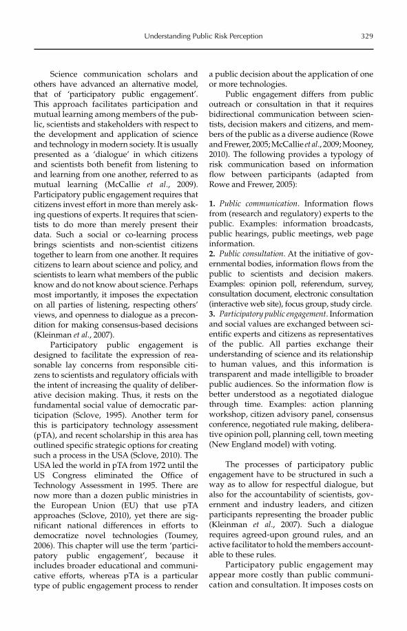
Understanding Public Risk Perception 329
Science communication scholars and others have advanced an alternative model, that of ‘participatory public engagement’. This approach facilitates participation and mutual learning among members of the pub-lic, scientists and stakeholders with respect to the development and application of science and technology in modern society. It is usually presented as a ‘dialogue’ in which citizens and scientists both benefit from listening to and learning from one another, referred to as mutual learning (McCallie et al., 2009). Participatory public engagement requires that citizens invest effort in more than merely ask-ing questions of experts. It requires that scien-tists to do more than merely present their data. Such a social or co-learning process brings scientists and non-scientist citizens together to learn from one another. It requires citizens to learn about science and policy, and scientists to learn what members of the public know and do not know about science. Perhaps most importantly, it imposes the expectation on all parties of listening, respecting others’ views, and openness to dialogue as a precon-dition for making consensus-based decisions (Kleinman et al., 2007).
Participatory public engagement is designed to facilitate the expression of rea-sonable lay concerns from responsible citi-zens to scientists and regulatory officials with the intent of increasing the quality of deliber-ative decision making. Thus, it rests on the fundamental social value of democratic par-ticipation (Sclove, 1995). Another term for this is participatory technology assessment (pTA), and recent scholarship in this area has outlined specific strategic options for creating such a process in the USA (Sclove, 2010). The USA led the world in pTA from 1972 until the US Congress eliminated the Office of Technology Assessment in 1995. There are now more than a dozen public ministries in the European Union (EU) that use pTA approaches (Sclove, 2010), yet there are sig-nificant national differences in efforts to democratize novel technologies (Toumey, 2006). This chapter will use the term ‘partici-patory public engagement’, because it includes broader educational and communi-cative efforts, whereas pTA is a particular type of public engagement process to render
a public decision about the application of one or more technologies.
Public engagement differs from public outreach or consultation in that it requires bidirectional communication between scien-tists, decision makers and citizens, and mem-bers of the public as a diverse audience (Rowe and Frewer, 2005; McCallie et al., 2009; Mooney, 2010). The following provides a typology of risk communication based on information flow between participants (adapted from Rowe and Frewer, 2005):
1. Public communication. Information flows from (research and regulatory) experts to the public. Examples: information broadcasts, public hearings, public meetings, web page information.2. Public consultation. At the initiative of gov-ernmental bodies, information flows from the public to scientists and decision makers. Examples: opinion poll, referendum, survey, consultation document, electronic consultation (interactive web site), focus group, study circle.3. Participatory public engagement. Infor mation and social values are exchanged between sci-entific experts and citizens as representatives of the public. All parties exchange their understanding of science and its relationship to human values, and this information is transparent and made intelligible to broader public audiences. So the information flow is better understood as a negotiated dialogue through time. Examples: action planning workshop, citizen advisory panel, consensus conference, negotiated rule making, delibera-tive opinion poll, planning cell, town meeting (New England model) with voting.
The processes of participatory public engagement have to be structured in such a way as to allow for respectful dialogue, but also for the accountability of scientists, gov-ernment and industry leaders, and citizen participants representing the broader public (Kleinman et al., 2007). Such a dialogue requires agreed-upon ground rules, and an active facilitator to hold the members account-able to these rules.
Participatory public engagement may appear more costly than public communi-cation and consultation. It imposes costs on
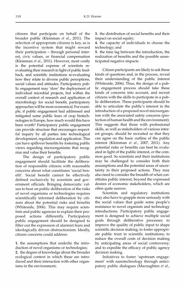
330 K.D. Warner
citizens that participate on behalf of the broader public (Kleinman et al., 2011). The selection of appropriate citizens is key, as is the incentive system that might reward their participation – through personal inter-est, civic values, or financial compensation (Kleinman et al., 2011). However, most costly is the potential expense of scientists re- evaluating their research in light of public feed-back, and scientific institutions re-evaluating how they relate to diverse public perceptions, social values and attitudes. Participatory pub-lic engagement may ‘slow’ the deployment of individual microbial projects, but within the overall context of research and application of microbiology for social benefit, participatory approaches will be more economical. For exam-ple, if public engagement had addressed and mitigated some public fears of crop biotech-nologies in Europe, how much would this have been worth? Participatory public engagement can provide structure that encourages respect-ful inquiry by all parties into technological development, regulation and application. This can have spillover benefits by fostering public views regarding microorganisms that recog-nize and value their benefits.
The design of participatory public engagement should facilitate the delibera-tion of responsible citizens with reasonable concerns about what constitutes ‘social ben-efit’. Social benefit cannot be effectively defined exclusively by scientists and gov-ernment officials. Bringing democratic val-ues to bear on public deliberation of the risks of novel organisms or technologies requires scientifically informed deliberation by citi-zens about the potential risks and benefits (Whiteside, 2006). This may require scien-tists and public agencies to explain their pro-posed actions differently. Participatory public engagement should be designed to filter out the expression of alarmist fears and ideologically driven obstructionism. Ideally, citizen concerns could address:
1. the assumptions that underlie the intro-duction of novel organisms or technologies;2. the degree of knowledge about the broader ecological context in which these are intro-duced and their interaction with other organ-isms in the environment;
3. the distribution of social benefits and their impact on social equity;4. the capacity of individuals to choose the technology; and5. the time lag between the introduction, the realization of benefits and the possible unan-ticipated negative impacts.
Citizen participants are likely to ask these kinds of questions and, in the process, reveal their understanding of the public interest (Whiteside, 2006). Thus, the design of a pub-lic engagement process should take these kinds of concerns into account, and recruit citizens with the skills to participate in a pub-lic deliberation. These participants should be able to articulate the public’s interest in the introduction of a proposed novel microorgan-ism with the associated safety concerns (pro-tection of human health and the environment). This suggests that those with professional skills, as well as stakeholders of various inter-est groups, should be recruited so that they can agree on the basic outline of the public interest (Kleinman et al., 2007, 2011). Any potential risks or benefits can best be evalu-ated in light of the public interest, or the com-mon good. So scientists and their institutions may be challenged to consider both their assumptions and the potential areas of uncer-tainty in their proposed actions. They may also need to consider the breadth of what con-stitutes public interest, beyond the expressed desires of economic stakeholders, which are often quite narrow.
Scientists and regulatory institutions may also have to grapple more seriously with the social values that guide some people’s resistance to novel organism and technology introductions. Participatory public engage-ment is designed to achieve multiple social goals through deliberative processes: to improve the quality of public input to shape scientific decision making; to foster appropri-ate public trust in scientific institutions; to reduce the overall costs of decision making by anticipating areas of social controversy; and to expedite the efficacy of public agency decision making.
Initiatives to foster ‘upstream engage-ment’ with nanotechnology through antici-patory public dialogues (Macnaghten et al.,
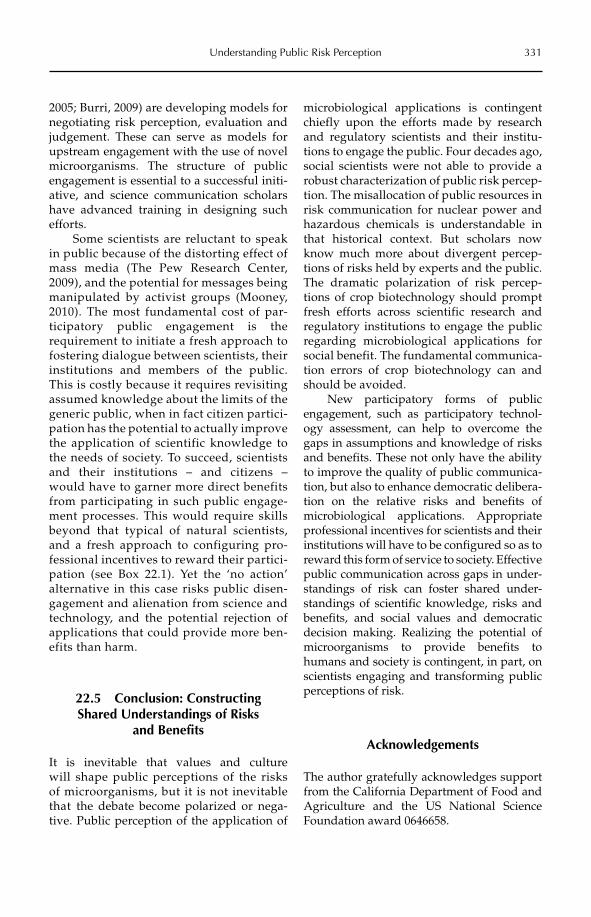
Understanding Public Risk Perception 331
2005; Burri, 2009) are developing models for negotiating risk perception, evaluation and judgement. These can serve as models for upstream engagement with the use of novel microorganisms. The structure of public engagement is essential to a successful initi-ative, and science communication scholars have advanced training in designing such efforts.
Some scientists are reluctant to speak in public because of the distorting effect of mass media (The Pew Research Center, 2009), and the potential for messages being manipulated by activist groups (Mooney, 2010). The most fundamental cost of par-ticipatory public engagement is the requirement to initiate a fresh approach to fostering dialogue between scientists, their institutions and members of the public. This is costly because it requires revisiting assumed knowledge about the limits of the generic public, when in fact citizen partici-pation has the potential to actually improve the application of scientific knowledge to the needs of society. To succeed, scientists and their institutions – and citizens – would have to garner more direct benefits from participating in such public engage-ment processes. This would require skills beyond that typical of natural scientists, and a fresh approach to configuring pro-fessional incentives to reward their partici-pation (see Box 22.1). Yet the ‘no action’ alternative in this case risks public disen-gagement and alienation from science and technology, and the potential rejection of applications that could provide more ben-efits than harm.
22.5 Conclusion: Constructing Shared Understandings of Risks
and Benefits
It is inevitable that values and culture will shape public perceptions of the risks of microorganisms, but it is not inevitable that the debate become polarized or nega-tive. Public perception of the application of
microbiological applications is contingent chiefly upon the efforts made by research and regulatory scientists and their institu-tions to engage the public. Four decades ago, social scientists were not able to provide a robust characterization of public risk percep-tion. The misallocation of public resources in risk communication for nuclear power and hazardous chemicals is understandable in that historical context. But scholars now know much more about divergent percep-tions of risks held by experts and the public. The dramatic polarization of risk percep-tions of crop biotechnology should prompt fresh efforts across scientific research and regulatory institutions to engage the public regarding microbiological applications for social benefit. The fundamental communica-tion errors of crop biotechnology can and should be avoided.
New participatory forms of public engagement, such as participatory technol-ogy assessment, can help to overcome the gaps in assumptions and knowledge of risks and benefits. These not only have the ability to improve the quality of public communica-tion, but also to enhance democratic delibera-tion on the relative risks and benefits of microbiological applications. Appropriate professional incentives for scientists and their institutions will have to be configured so as to reward this form of service to society. Effective public communication across gaps in under-standings of risk can foster shared under-standings of scientific knowledge, risks and benefits, and social values and democratic decision making. Realizing the potential of microorganisms to provide benefits to humans and society is contingent, in part, on scientists engaging and transforming public perceptions of risk.
Acknowledgements
The author gratefully acknowledges support from the California Department of Food and Agriculture and the US National Science Foundation award 0646658.
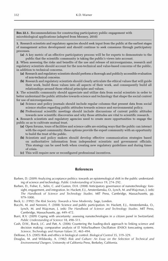
332 K.D. Warner
References
Barben, D. (2009) Analyzing acceptance politics: towards an epistemological shift in the public understand-ing of science and technology. Public Understanding of Science 19, 274–292.
Barben, D., Fisher, E., Selin, C. and Guston, D.H. (2008) Anticipatory governance of nanotechnology: fore-sight, engagement, and integration. In: Hackett, E.J., Amsterdamska, O., Lynch, M. and Wajcman, J. (eds) The Handbook of Science and Technology Studies. MIT Press, Cambridge, Massachusetts, pp. 979–1000.
Beck, U. (1992) The Risk Society: Towards a New Modernity. Sage, London.Bucchi, M. and Neresini, F. (2008) Science and public participation. In: Hackett, E.J., Amsterdamska, O.,
Lynch, M. and Wajcman, J. (eds) The Handbook of Science and Technology Studies. MIT Press, Cambridge, Massachusetts, pp. 449–472.
Burri, R.V. (2009) Coping with uncertainty: assessing nanotechnologies in a citizen panel in Switzerland. Public Understanding of Science 18, 498–511.
Cash, D.W., Borck, J.C. and Patt, A. (2006) Countering the loading-dock approach to linking science and decision making: comparative analysis of El Niño/Southern Oscillation (ENSO) forecasting systems. Science, Technology and Human Values 31, 465–494.
Delfosse, E.S. (2005) Risk and ethics in biological control. Biological Control 35, 319–329.Douglas, M. and Wildavsky, A. (1982) Risk and Culture: An Essay on the Selection of Technical and
Environmental Dangers. University of California Press, Berkeley, California.
Box 22.1. Recommendations for constructing participatory public engagement with microbiological applications (adapted from Mooney, 2010)
1. Research scientists and regulatory agencies should seek input from the public at the earliest stages of management action development and should continue to seek consensus through participatory processes.
(a) A key metric of an effective participatory process will be for experts to demonstrate to the public that the scientific community is taking the public’s views into account.
2. When assessing the risks and benefits of the use and release of microorganisms, research and regulatory scientists should account for the non-technical and value-based concerns of the public, in addition to technical concerns.
(a) Research and regulatory scientists should perform a thorough and publicly accessible evaluation of non-technical concerns.
(b) Research and regulatory scientists should clearly articulate the ethical values that will guide their work, build those values into all aspects of their work, and consequently build all relationships around those ethical principles and values.
3. The scientific community should appreciate and utilize data from social scientists in order to better understand the public attitudes towards science and technology that shape the social contextfor use of microorganisms.
(a) Science and policy journals should include regular columns that present data from social science studies regarding public attitudes towards science and environmental policy.
(b) Professional scientific meetings should include discussions of current public attitudes towards new scientific discoveries and why those attitudes are vital to scientific research.
4. Research scientists and regulatory agencies need to create more opportunities to engage the public so as to cultivate mutual trust.
(a) Open forums, tours of facilities and science cafés are existing ways that the public can interact with the expert community; these options provide the expert community with an opportunity to build the trust of the public.
(b) Scientists and policy makers should develop effective communication strategies based on authoritative information from independent scientists and government officials. This strategy can be used both when creating new regulatory guidelines and during times of crisis.
(c) This will require new or reconfigured professional incentives.
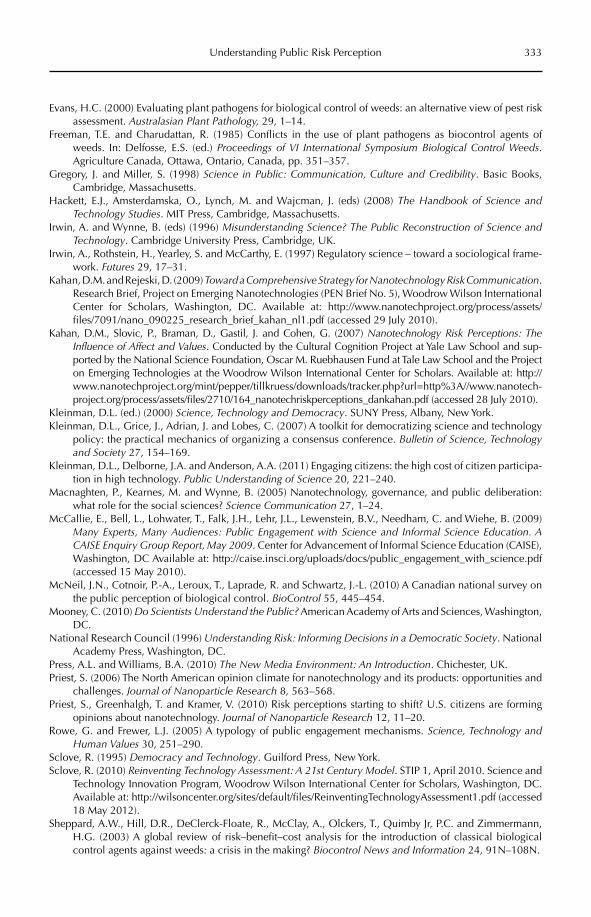
Understanding Public Risk Perception 333
Evans, H.C. (2000) Evaluating plant pathogens for biological control of weeds: an alternative view of pest risk assessment. Australasian Plant Pathology, 29, 1–14.
Freeman, T.E. and Charudattan, R. (1985) Conflicts in the use of plant pathogens as biocontrol agents of weeds. In: Delfosse, E.S. (ed.) Proceedings of VI International Symposium Biological Control Weeds.Agriculture Canada, Ottawa, Ontario, Canada, pp. 351–357.
Gregory, J. and Miller, S. (1998) Science in Public: Communication, Culture and Credibility. Basic Books, Cambridge, Massachusetts.
Hackett, E.J., Amsterdamska, O., Lynch, M. and Wajcman, J. (eds) (2008) The Handbook of Science and Technology Studies. MIT Press, Cambridge, Massachusetts.
Irwin, A. and Wynne, B. (eds) (1996) Misunderstanding Science? The Public Reconstruction of Science and Technology. Cambridge University Press, Cambridge, UK.
Irwin, A., Rothstein, H., Yearley, S. and McCarthy, E. (1997) Regulatory science – toward a sociological frame-work. Futures 29, 17–31.
Kahan, D.M. and Rejeski, D. (2009) Toward a Comprehensive Strategy for Nanotechnology Risk Communication.Research Brief, Project on Emerging Nanotechnologies (PEN Brief No. 5), Woodrow Wilson International Center for Scholars, Washington, DC. Available at: http://www.nanotechproject.org/process/assets/files/7091/nano_090225_research_brief_kahan_nl1.pdf (accessed 29 July 2010).
Kahan, D.M., Slovic, P., Braman, D., Gastil, J. and Cohen, G. (2007) Nanotechnology Risk Perceptions: The Influence of Affect and Values. Conducted by the Cultural Cognition Project at Yale Law School and sup-ported by the National Science Foundation, Oscar M. Ruebhausen Fund at Tale Law School and the Project on Emerging Technologies at the Woodrow Wilson International Center for Scholars. Available at: http://www.nanotechproject.org/mint/pepper/tillkruess/downloads/tracker.php?url=http%3A//www.nanotech-project.org/process/assets/files/2710/164_nanotechriskperceptions_dankahan.pdf (accessed 28 July 2010).
Kleinman, D.L. (ed.) (2000) Science, Technology and Democracy. SUNY Press, Albany, New York.Kleinman, D.L., Grice, J., Adrian, J. and Lobes, C. (2007) A toolkit for democratizing science and technology
policy: the practical mechanics of organizing a consensus conference. Bulletin of Science, Technology and Society 27, 154–169.
Kleinman, D.L., Delborne, J.A. and Anderson, A.A. (2011) Engaging citizens: the high cost of citizen participa-tion in high technology. Public Understanding of Science 20, 221–240.
Macnaghten, P., Kearnes, M. and Wynne, B. (2005) Nanotechnology, governance, and public deliberation: what role for the social sciences? Science Communication 27, 1–24.
McCallie, E., Bell, L., Lohwater, T., Falk, J.H., Lehr, J.L., Lewenstein, B.V., Needham, C. and Wiehe, B. (2009) Many Experts, Many Audiences: Public Engagement with Science and Informal Science Education. A CAISE Enquiry Group Report, May 2009. Center for Advancement of Informal Science Education (CAISE), Washington, DC Available at: http://caise.insci.org/uploads/docs/public_engagement_with_science.pdf (accessed 15 May 2010).
McNeil, J.N., Cotnoir, P.-A., Leroux, T., Laprade, R. and Schwartz, J.-L. (2010) A Canadian national survey on the public perception of biological control. BioControl 55, 445–454.
Mooney, C. (2010) Do Scientists Understand the Public? American Academy of Arts and Sciences, Washington, DC.
National Research Council (1996) Understanding Risk: Informing Decisions in a Democratic Society. National Academy Press, Washington, DC.
Press, A.L. and Williams, B.A. (2010) The New Media Environment: An Introduction. Chichester, UK.Priest, S. (2006) The North American opinion climate for nanotechnology and its products: opportunities and
challenges. Journal of Nanoparticle Research 8, 563–568.Priest, S., Greenhalgh, T. and Kramer, V. (2010) Risk perceptions starting to shift? U.S. citizens are forming
opinions about nanotechnology. Journal of Nanoparticle Research 12, 11–20.Rowe, G. and Frewer, L.J. (2005) A typology of public engagement mechanisms. Science, Technology and
Human Values 30, 251–290.Sclove, R. (1995) Democracy and Technology. Guilford Press, New York.Sclove, R. (2010) Reinventing Technology Assessment: A 21st Century Model. STIP 1, April 2010. Science and
Technology Innovation Program, Woodrow Wilson International Center for Scholars, Washington, DC. Available at: http://wilsoncenter.org/sites/default/files/ReinventingTechnologyAssessment1.pdf (accessed 18 May 2012).
Sheppard, A.W., Hill, D.R., DeClerck-Floate, R., McClay, A., Olckers, T., Quimby Jr, P.C. and Zimmermann, H.G. (2003) A global review of risk–benefit–cost analysis for the introduction of classical biological control agents against weeds: a crisis in the making? Biocontrol News and Information 24, 91N–108N.
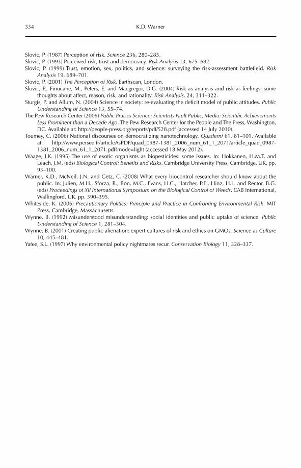
334 K.D. Warner
Slovic, P. (1987) Perception of risk. Science 236, 280–285.Slovic, P. (1993) Perceived risk, trust and democracy. Risk Analysis 13, 675–682.Slovic, P. (1999) Trust, emotion, sex, politics, and science: surveying the risk-assessment battlefield. Risk
Analysis 19, 689–701.Slovic, P. (2001) The Perception of Risk. Earthscan, London.Slovic, P., Finucane, M., Peters, E. and Macgregor, D.G. (2004) Risk as analysis and risk as feelings: some
thoughts about affect, reason, risk, and rationality. Risk Analysis, 24, 311–322.Sturgis, P. and Allum, N. (2004) Science in society: re-evaluating the deficit model of public attitudes. Public
Understanding of Science 13, 55–74.The Pew Research Center (2009) Public Praises Science; Scientists Fault Public, Media: Scientific Achievements
Less Prominent than a Decade Ago. The Pew Research Center for the People and The Press, Washington, DC. Available at: http://people-press.org/reports/pdf/528.pdf (accessed 14 July 2010).
Toumey, C. (2006) National discourses on democratizing nanotechnology. Quaderni 61, 81–101. Available at: http://www.persee.fr/articleAsPDF/quad_0987-1381_2006_num_61_1_2071/article_quad_0987-1381_2006_num_61_1_2071.pdf?mode=light (accessed 18 May 2012).
Waage, J.K. (1995) The use of exotic organisms as biopesticides: some issues. In: Hokkanen, H.M.T. and Leach, J.M. (eds) Biological Control: Benefits and Risks. Cambridge University Press, Cambridge, UK, pp. 93–100.
Warner, K.D., McNeil, J.N. and Getz, C. (2008) What every biocontrol researcher should know about the public. In: Julien, M.H., Sforza, R., Bon, M.C., Evans, H.C., Hatcher, P.E., Hinz, H.L. and Rector, B.G. (eds) Proceedings of XII International Symposium on the Biological Control of Weeds. CAB International, Wallingford, UK. pp. 390–395.
Whiteside, K. (2006) Precautionary Politics: Principle and Practice in Confronting Environmental Risk. MIT Press, Cambridge, Massachusetts.
Wynne, B. (1992) Misunderstood misunderstanding: social identities and public uptake of science. Public Understanding of Science 1, 281–304.
Wynne, B. (2001) Creating public alienation: expert cultures of risk and ethics on GMOs. Science as Culture10, 445–481.
Yafee, S.L. (1997) Why environmental policy nightmares recur. Conservation Biology 11, 328–337.
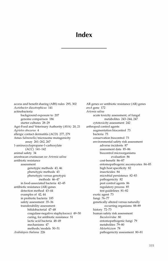
335
access and benefit sharing (ABS) rules 295, 302Acetobacter diazotrophicus 141actinobacteria
background exposure to 207genome comparison 186starter cultures 28–29
Agri-Food and Veterinary Authority (AVA) 20, 21Agriotes obscurus 4allergic contact dermatitis (ACD) 277, 279Ames Salmonella/microsome mutagenicity
assay 261–262, 2671-aminocyclopropane-1-carboxylate
(ACC) 141–142animal safety 34anostracan crustacean see Artemia salinaantibiotic resistance
assessmentgenotypic methods 43, 46phenotypic methods 43phenotypic versus genotypic
methods 46–47in food-associated bacteria 42–45
antibiotic resistance (AR) genesdetection method 43–44examples of 42, 44in probiotic bacteria 185safety assessment 35–36transferability assessment
bifidobacterial 47–48coagulase-negative staphylococci 49–50curing, for antibiotic resistance 51lactic acid bacteria 48–49mechanisms 47methods/models 50–51
Arabidopsis thaliana 226
AR genes see antibiotic resistance (AR) genesaroA gene 172Artemia salina
acute toxicity assessment, of fungal metabolites 243–244, 247
cytotoxicity assessment 242arthropod control agents
augmentation biocontrol 73bacteria 75conservation biocontrol 73environmental safety risk assessment
adverse incidents 87assessment data 85–86biocontrol microorganisms
evaluation 86cost-benefit 86–87entomopathogenic ascomycetes 84–85high host specificity 82insecticides 84microbial persistence 82–83pathogenicity 82pest control agents 86regulatory process 85test guidelines 81–82
exotic agent 73fungi 76–77genetically altered versus naturally
occurring organisms 88–89history 72–73human safety risk assessment
Baculoviridae 80entomopathogenic fungi 79metabolites 79–80Metarhizium 78pathogenicity assessment 80–81
Index
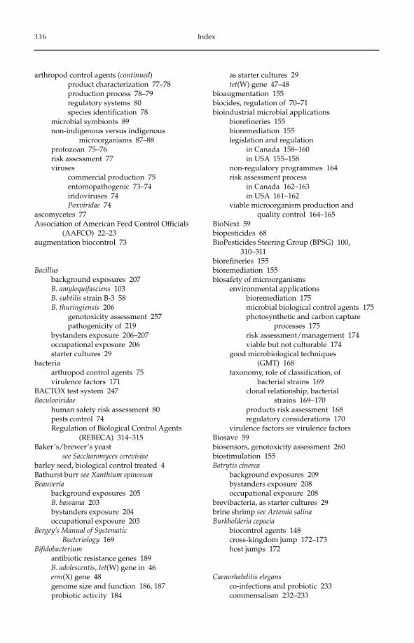
336 Index
arthropod control agents (continued)product characterization 77–78production process 78–79regulatory systems 80species identification 78
microbial symbionts 89non-indigenous versus indigenous
microorganisms 87–88protozoan 75–76risk assessment 77viruses
commercial production 75entomopathogenic 73–74iridoviruses 74Poxviridae 74
ascomycetes 77Association of American Feed Control Officials
(AAFCO) 22–23augmentation biocontrol 73
Bacillusbackground exposures 207B. amyloquifasciens 103B. subtilis strain B-3 58B. thuringiensis 206
genotoxicity assessment 257pathogenicity of 219
bystanders exposure 206–207occupational exposure 206starter cultures 29
bacteriaarthropod control agents 75virulence factors 171
BACTOX test system 247Baculoviridae
human safety risk assessment 80pests control 74Regulation of Biological Control Agents
(REBECA) 314–315Baker’s/brewer’s yeast
see Saccharomyces cerevisiaebarley seed, biological control treated 4Bathurst burr see Xanthium spinosumBeauveria
background exposures 205B. bassiana 203bystanders exposure 204occupational exposure 203
Bergey’s Manual of Systematic Bacteriology 169
Bifidobacteriumantibiotic resistance genes 189B. adolescentis, tet(W) gene in 46erm(X) gene 48genome size and function 186, 187probiotic activity 184
as starter cultures 29tet(W) gene 47–48
bioaugmentation 155biocides, regulation of 70–71bioindustrial microbial applications
biorefineries 155bioremediation 155legislation and regulation
in Canada 158–160in USA 155–158
non-regulatory programmes 164risk assessment process
in Canada 162–163in USA 161–162
viable microorganism production and quality control 164–165
BioNext 59biopesticides 68BioPesticides Steering Group (BPSG) 100,
310–311biorefineries 155bioremediation 155biosafety of microorganisms
environmental applicationsbioremediation 175microbial biological control agents 175photosynthetic and carbon capture
processes 175risk assessment/management 174viable but not culturable 174
good microbiological techniques (GMT) 168
taxonomy, role of classification, of bacterial strains 169
clonal relationship, bacterial strains 169–170
products risk assessment 168regulatory considerations 170
virulence factors see virulence factorsBiosave 59biosensors, genotoxicity assessment 260biostimulation 155Botrytis cinerea
background exposures 209bystanders exposure 208occupational exposure 208
brevibacteria, as starter cultures 29brine shrimp see Artemia salinaBurkholderia cepacia
biocontrol agents 148cross-kingdom jump 172–173host jumps 172
Caenorhabditis elegansco-infections and probiotic 233commensalism 232–233
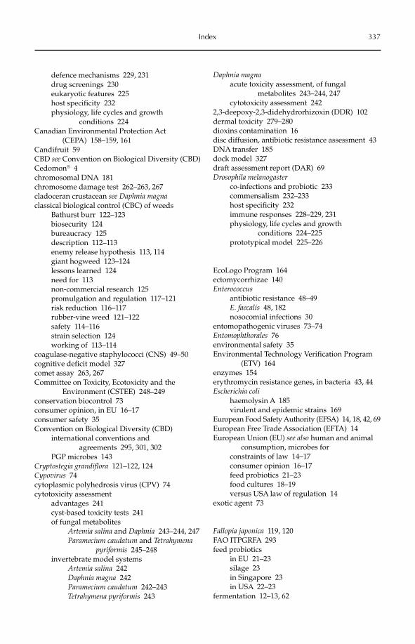
Index 337
defence mechanisms 229, 231drug screenings 230eukaryotic features 225host specificity 232physiology, life cycles and growth
conditions 224Canadian Environmental Protection Act
(CEPA) 158–159, 161Candifruit 59CBD see Convention on Biological Diversity (CBD)Cedomon® 4chromosomal DNA 181chromosome damage test 262–263, 267cladoceran crustacean see Daphnia magnaclassical biological control (CBC) of weeds
Bathurst burr 122–123biosecurity 124bureaucracy 125description 112–113enemy release hypothesis 113, 114giant hogweed 123–124lessons learned 124need for 113non-commercial research 125promulgation and regulation 117–121risk reduction 116–117rubber-vine weed 121–122safety 114–116strain selection 124working of 113–114
coagulase-negative staphylococci (CNS) 49–50cognitive deficit model 327comet assay 263, 267Committee on Toxicity, Ecotoxicity and the
Environment (CSTEE) 248–249conservation biocontrol 73consumer opinion, in EU 16–17consumer safety 35Convention on Biological Diversity (CBD)
international conventions and agreements 295, 301, 302
PGP microbes 143Cryptostegia grandiflora 121–122, 124Cypovirus 74cytoplasmic polyhedrosis virus (CPV) 74cytotoxicity assessment
advantages 241cyst-based toxicity tests 241of fungal metabolites
Artemia salina and Daphnia 243–244, 247Paramecium caudatum and Tetrahymena
pyriformis 245–248invertebrate model systems
Artemia salina 242Daphnia magna 242Paramecium caudatum 242–243Tetrahymena pyriformis 243
Daphnia magnaacute toxicity assessment, of fungal
metabolites 243–244, 247cytotoxicity assessment 242
2,3-deepoxy-2,3-didehydrorhizoxin (DDR) 102dermal toxicity 279–280dioxins contamination 16disc diffusion, antibiotic resistance assessment 43DNA transfer 185dock model 327draft assessment report (DAR) 69Drosophila melanogaster
co-infections and probiotic 233commensalism 232–233host specificity 232immune responses 228–229, 231physiology, life cycles and growth
conditions 224–225prototypical model 225–226
EcoLogo Program 164ectomycorrhizae 140Enterococcus
antibiotic resistance 48–49E. faecalis 48, 182nosocomial infections 30
entomopathogenic viruses 73–74Entomophthorales 76environmental safety 35Environmental Technology Verification Program
(ETV) 164enzymes 154erythromycin resistance genes, in bacteria 43, 44Escherichia coli
haemolysin A 185virulent and epidemic strains 169
European Food Safety Authority (EFSA) 14, 18, 42, 69European Free Trade Association (EFTA) 14European Union (EU) see also human and animal
consumption, microbes forconstraints of law 14–17consumer opinion 16–17feed probiotics 21–23food cultures 18–19versus USA law of regulation 14
exotic agent 73
Fallopia japonica 119, 120FAO ITPGRFA 293feed probiotics
in EU 21–23silage 23in Singapore 23in USA 22–23
fermentation 12–13, 62
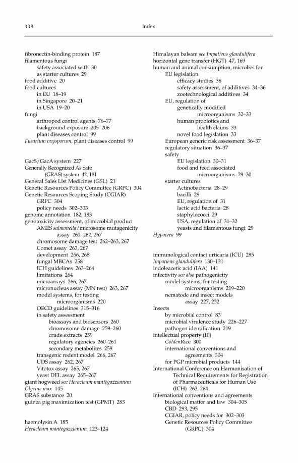
338 Index
fibronectin-binding protein 187filamentous fungi
safety associated with 30as starter cultures 29
food additive 20food cultures
in EU 18–19in Singapore 20–21in USA 19–20
fungiarthropod control agents 76–77background exposure 205–206plant diseases control 99
Fusarium oxysporum, plant diseases control 99
GacS/GacA system 227Generally Recognized As Safe
(GRAS) system 42, 181General Sales List Medicines (GSL) 21Genetic Resources Policy Committee (GRPC) 304Genetic Resources Scoping Study (CGIAR)
GRPC 304policy needs 302–303
genome annotation 182, 183genotoxicity assessment, of microbial product
AMES salmonella/microsome mutagenicity assay 261–262, 267
chromosome damage test 262–263, 267Comet assay 263, 267development 266, 268fungal MBCAs 258ICH guidelines 263–264limitations 264microarrays 266, 267micronucleus assay (MN test) 263, 267model systems, for testing
microorganisms 220OECD guidelines 315–316in safety assessment
bioassays and biosensors 260chromosome damage 259–260crude extracts 259regulatory agencies 260–261secondary metabolites 259
transgenic rodent model 266, 267UDS assay 262, 267Vitotox assay 265, 267yeast DEL assay 265–267
giant hogweed see Heracleum mantegazzianumGlycine max 145GRAS substance 20guinea pig maximization test (GPMT) 283
haemolysin A 185Heracleum mantegazzianum 123–124
Himalayan balsam see Impatiens glanduliferahorizontal gene transfer (HGT) 47, 169human and animal consumption, microbes for
EU legislationefficacy studies 36safety assessment, of additives 34–36zootechnological additives 34
EU, regulation ofgenetically modified
microorganisms 32–33human probiotics and
health claims 33novel food legislation 33
European generic risk assessment 36–37regulatory situation 36–37safety
EU legislation 30–31food and feed associated
microorganisms 29–30starter cultures
Actinobacteria 28–29bacilli 29EU, regulation of 31lactic acid bacteria 28staphylococci 29USA, regulation of 31–32yeasts and filamentous fungi 29
Hypocrea 99
immunological contact urticaria (ICU) 285Impatiens glandulifera 130–131indoleacetic acid (IAA) 141infectivity see also pathogenicity
model systems, for testing microorganisms 219–220
nematode and insect models assay 227, 232
Insectsby microbial control 83microbial virulence study 226–227pathogen identification 219
intellectual property (IP)GoldenRice 300international conventions and
agreements 304for PGP microbial products 144
International Conference on Harmonisation of Technical Requirements for Registration of Pharmaceuticals for Human Use (ICH) 263–264
international conventions and agreementsbiological matter and law 304–305CBD 293, 295CGIAR, policy needs for 302–303Genetic Resources Policy Committee
(GRPC) 304
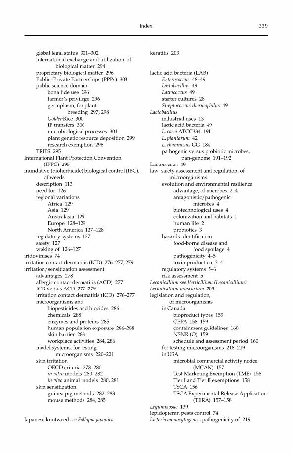
Index 339
global legal status 301–302international exchange and utilization, of
biological matter 294proprietary biological matter 296Public–Private Partnerships (PPPs) 303public science domain
bona fide use 296farmer’s privilege 296germplasm, for plant
breeding 297, 298GoldenRice 300IP transfers 300microbiological processes 301plant genetic resource deposition 299research exemption 296
TRIPS 295International Plant Protection Convention
(IPPC) 295inundative (bioherbicide) biological control (IBC),
of weedsdescription 113need for 126regional variations
Africa 129Asia 129Australasia 129Europe 128–129North America 127–128
regulatory systems 127safety 127woking of 126–127
iridoviruses 74irritation contact dermatitis (ICD) 276–277, 279irritation/sensitization assessment
advantages 278allergic contact dermatitis (ACD) 277ICD versus ACD 277–279irritation contact dermatitis (ICD) 276–277microorganisms and
biopesticides and biocides 286chemicals 288enzymes and proteins 285human population exposure 286–288skin barrier 288workplace activities 284, 286
model systems, for testing microorganisms 220–221
skin irritationOECD criteria 278–280in vitro models 280–282in vivo animal models 280, 281
skin sensitizationguinea pig methods 282–283mouse methods 284, 285
Japanese knotweed see Fallopia japonica
keratitis 203
lactic acid bacteria (LAB)Enterococcus 48–49Lactobacillus 49Lactococcus 49starter cultures 28Streptococcus thermophilus 49
Lactobacillusindustrial uses 13lactic acid bacteria 49L. casei ATCC334 191L. plantarum 42L. rhamnosus GG 184pathogenic versus probiotic microbes,
pan-genome 191–192Lactococcus 49law–safety assessment and regulation, of
microorganismsevolution and environmental resilience
advantage, of microbes 2, 4antagonistic/pathogenic
microbes 4biotechnological uses 4colonization and habitats 1human life 2probiotics 3
hazards identificationfood-borne disease and
food spoilage 4pathogenicity 4–5toxin production 3–4
regulatory systems 5–6risk assessment 5
Lecanicillium see Verticillium (Lecanicillium)Lecanicillium muscarium 203legislation and regulation,
of microorganismsin Canada
bioproduct types 159CEPA 158–159containment guidelines 160NSNR (O) 159schedule and assessment period 160
for testing microorganisms 218–219in USA
microbial commercial activity notice (MCAN) 157
Test Marketing Exemption (TME) 158Tier I and Tier II exemptions 158TSCA 156TSCA Experimental Release Application
(TERA) 157–158Leguminosae 139lepidopteran pests control 74Listeria monocytogenes, pathogenicity of 219
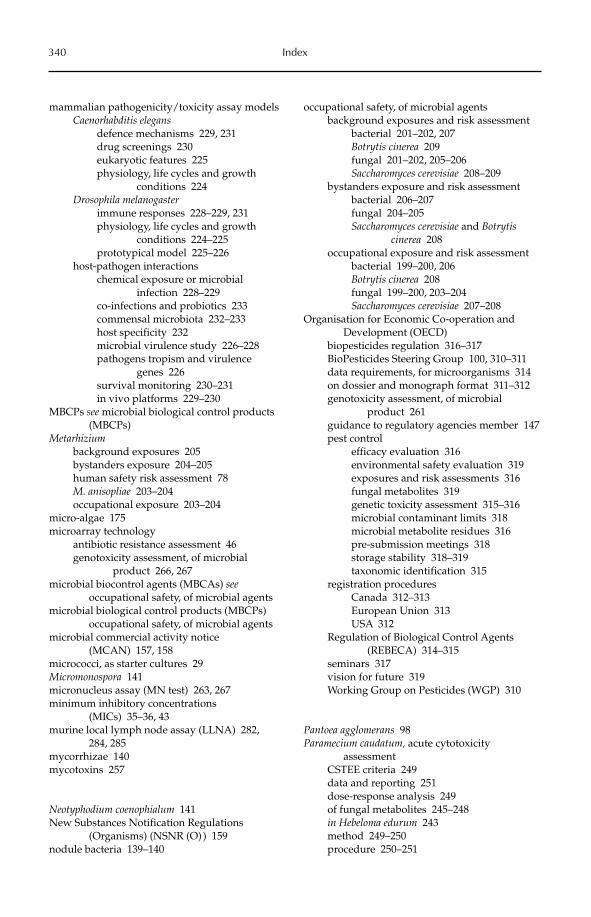
340 Index
mammalian pathogenicity/toxicity assay modelsCaenorhabditis elegans
defence mechanisms 229, 231drug screenings 230eukaryotic features 225physiology, life cycles and growth
conditions 224Drosophila melanogaster
immune responses 228–229, 231physiology, life cycles and growth
conditions 224–225prototypical model 225–226
host-pathogen interactionschemical exposure or microbial
infection 228–229co-infections and probiotics 233commensal microbiota 232–233host specificity 232microbial virulence study 226–228pathogens tropism and virulence
genes 226survival monitoring 230–231in vivo platforms 229–230
MBCPs see microbial biological control products (MBCPs)
Metarhiziumbackground exposures 205bystanders exposure 204–205human safety risk assessment 78M. anisopliae 203–204occupational exposure 203–204
micro-algae 175microarray technology
antibiotic resistance assessment 46genotoxicity assessment, of microbial
product 266, 267microbial biocontrol agents (MBCAs) see
occupational safety, of microbial agentsmicrobial biological control products (MBCPs)
occupational safety, of microbial agentsmicrobial commercial activity notice
(MCAN) 157, 158micrococci, as starter cultures 29Micromonospora 141micronucleus assay (MN test) 263, 267minimum inhibitory concentrations
(MICs) 35–36, 43murine local lymph node assay (LLNA) 282,
284, 285mycorrhizae 140mycotoxins 257
Neotyphodium coenophialum 141New Substances Notification Regulations
(Organisms) (NSNR (O) ) 159nodule bacteria 139–140
occupational safety, of microbial agentsbackground exposures and risk assessment
bacterial 201–202, 207Botrytis cinerea 209fungal 201–202, 205–206Saccharomyces cerevisiae 208–209
bystanders exposure and risk assessmentbacterial 206–207fungal 204–205Saccharomyces cerevisiae and Botrytis
cinerea 208occupational exposure and risk assessment
bacterial 199–200, 206Botrytis cinerea 208fungal 199–200, 203–204Saccharomyces cerevisiae 207–208
Organisation for Economic Co-operation and Development (OECD)
biopesticides regulation 316–317BioPesticides Steering Group 100, 310–311data requirements, for microorganisms 314on dossier and monograph format 311–312genotoxicity assessment, of microbial
product 261guidance to regulatory agencies member 147pest control
efficacy evaluation 316environmental safety evaluation 319exposures and risk assessments 316fungal metabolites 319genetic toxicity assessment 315–316microbial contaminant limits 318microbial metabolite residues 316pre-submission meetings 318storage stability 318–319taxonomic identification 315
registration proceduresCanada 312–313European Union 313USA 312
Regulation of Biological Control Agents (REBECA) 314–315
seminars 317vision for future 319Working Group on Pesticides (WGP) 310
Pantoea agglomerans 98Paramecium caudatum, acute cytotoxicity
assessmentCSTEE criteria 249data and reporting 251dose-response analysis 249of fungal metabolites 245–248in Hebeloma edurum 243method 249–250procedure 250–251
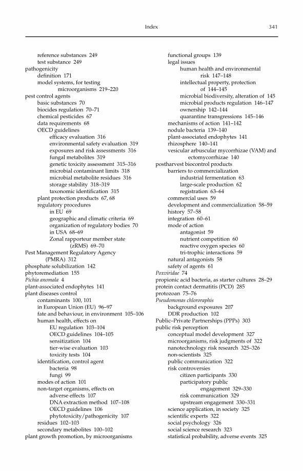
Index 341
reference substances 249test substance 249
pathogenicitydefinition 171model systems, for testing
microorganisms 219–220pest control agents
basic substances 70biocides regulation 70–71chemical pesticides 67data requirements 68OECD guidelines
efficacy evaluation 316environmental safety evaluation 319exposures and risk assessments 316fungal metabolites 319genetic toxicity assessment 315–316microbial contaminant limits 318microbial metabolite residues 316storage stability 318–319taxonomic identification 315
plant protection products 67, 68regulatory procedures
in EU 69geographic and climatic criteria 69organization of regulatory bodies 70in USA 68–69Zonal rapporteur member state
(zRMS) 69–70Pest Management Regulatory Agency
(PMRA) 312phosphate solubilization 142phytoremediation 155Pichia anomala 4plant-associated endophytes 141plant diseases control
contaminants 100, 101in European Union (EU) 96–97fate and behaviour, in environment 105–106human health, effects on
EU regulation 103–104OECD guidelines 104–105sensitization 104tier-wise evaluation 103toxicity tests 104
identification, control agentbacteria 98fungi 99
modes of action 101non-target organisms, effects on
adverse effects 107DNA extraction method 107–108OECD guidelines 106phytotoxicity/pathogenicity 107
residues 102–103secondary metabolites 100–102
plant growth promotion, by microorganisms
functional groups 139legal issues
human health and environmental risk 147–148
intellectual property, protection of 144–145
microbial biodiversity, alteration of 145microbial products regulation 146–147ownership 142–144quarantine transgressions 145–146
mechanisms of action 141–142nodule bacteria 139–140plant-associated endophytes 141rhizosphere 140–141vesicular arbuscular mycorrhizae (VAM) and
ectomycorrhizae 140postharvest biocontrol products
barriers to commercializationindustrial fermentation 63large-scale production 62registration 63–64
commercial uses 59development and commercialization 58–59history 57–58integration 60–61mode of action
antagonist 59nutrient competition 60reactive oxygen species 60tri-trophic interactions 59
natural antagonists 58safety of agents 61
Poxviridae 74propionic acid bacteria, as starter cultures 28–29protein contact dermatitis (PCD) 285protozoan 75–76Pseudomonas chlororaphis
background exposures 207DDR production 102
Public–Private Partnerships (PPPs) 303public risk perception
conceptual model development 327microorganisms, risk judgments of 322nanotechnology risk research 325–326non-scientists 325public communication 322risk controversies
citizen participants 330participatory public
engagement 329–330risk communication 329upstream engagement 330–331
science application, in society 325scientific experts 322social psychology 326social science research 323statistical probability, adverse events 325
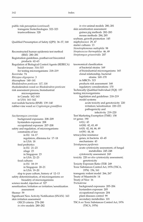
342 Index
public risk perception (continued)transgenic biotechnologies 322–323trustworthiness 328
Qualified Presumption of Safety (QPS) 36–37, 168
Reconstructed human epidermis test method (RhE) 280–281
Registration guidelines, postharvest biocontrol products 62–63
Regulation of Biological Control Agents (REBECA)baculoviruses 314–315for testing microorganisms 218–219
Reoviridae 74Rhizopus oligosporus 3rhizosphere 140–141Rhododendron ponticum 117, 118rhododendron weed see Rhododendron ponticumrisk assessment process, bioindustrial
microorganismsin Canada 162–163in USA 161–162
root nodule bacteria (RNB) 139–140rubber-vine weed see Cryptostegia grandiflora
Saccharomyces cerevisiaebackground exposures 208–209bystanders exposure 208occupational exposure 207–208
safety and regulation, of microorganismsconstraints of law
in EU 14–17regulators, dilemma for 17–18in USA 18
feed probioticsin EU 21–23silage 23in Singapore 23in USA 22–23
food culturesin EU 18–19in Singapore 20–21in USA 19–20
slop to pure culture, history of 12–13safety determinination, of microorganisms see
biosafety of microorganismsscience model, injection of 327sensitization/irritation see irritation/sensitization
assessmentShemer 59Significant New Activity Notification (SNAN) 163skin irritation assessment
OECD criteria 278–280in vitro models 280–282
in vivo animal models 280, 281skin sensitization assessment
guinea pig methods 282–283mouse methods 284, 285
soybean, growth promotion 145staphylococci 29, 47starter cultures 13Stenotrophomonas maltophila 98Streptococcus thermophilus 46, 49Streptomyces griseoviridis 207
taxonomical classificationof bacterial strains 169of bioindustrial microorganisms 165clonal relationship, bacterial
strains 169–170in MBCPs 315products risk assessment 168regulatory considerations 170
Technically Qualified Individual (TQI) 157testing microorganisms
legislation and guidelines 218–219model systems
acute toxicity and genotoxicity 220irritation/sensitization 220–221pathogenicity and
infectivity 219–220Test Marketing Exemption (TME) 158tet genes 190
tet(L) 43tet(M) 42, 43, 49tet(S) 42, 44, 46, 49tet(W) 44, 46
tetracycline resistancegenes, in bacteria 43–45mechanisms 43
Tetrahymena pyriformisacute cytotoxicity assessment, of fungal
metabolites 245–248cytotoxicity assessment 243
toxicity 220 see also cytotoxicity assessment; genotoxicity
Toxicity Exposure Ratio (TER 249Toxic Substances Control Act, 1976 (TSCA,
1976) 156, 161transgenic rodent model 266, 267Treaty of Maastricht 14Treaty of Nice 16Trichoderma
background exposures 205–206bystanders exposure 205occupational exposure 204plant diseases control 99secondary metabolites 101
TSCA see Toxic Substances Control Act, 1976 (TSCA, 1976)
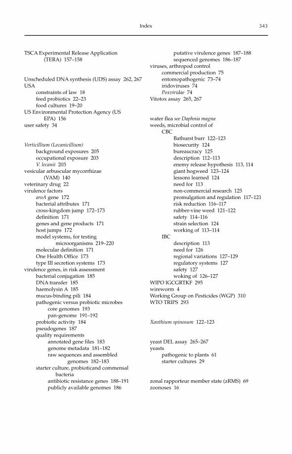
Index 343
TSCA Experimental Release Application (TERA) 157–158
Unscheduled DNA synthesis (UDS) assay 262, 267USA
constraints of law 18feed probiotics 22–23food cultures 19–20
US Environmental Protection Agency (US EPA) 156
user safety 34
Verticillium (Lecanicillium)background exposures 205occupational exposure 203V. lecanii 203
vesicular arbuscular mycorrhizae (VAM) 140
veterinary drug 22virulence factors
aroA gene 172bacterial attributes 171cross-kingdom jump 172–173definition 171genes and gene products 171host jumps 172model systems, for testing
microorganisms 219–220molecular definition 171One Health Office 173type III secretion systems 173
virulence genes, in risk assessmentbacterial conjugation 185DNA transfer 185haemolysin A 185mucus-binding pili 184pathogenic versus probiotic microbes
core genomes 193pan-genome 191–192
probiotic activity 184pseudogenes 187quality requirements
annotated gene files 183genome metadata 181–182raw sequences and assembled
genomes 182–183starter culture, probioticand commensal
bacteriaantibiotic resistance genes 188–191publicly available genomes 186
putative virulence genes 187–188sequenced genomes 186–187
viruses, arthropod controlcommercial production 75entomopathogenic 73–74iridoviruses 74Poxviridae 74
Vitotox assay 265, 267
water flea see Daphnia magnaweeds, microbial control of
CBCBathurst burr 122–123biosecurity 124bureaucracy 125description 112–113enemy release hypothesis 113, 114giant hogweed 123–124lessons learned 124need for 113non-commercial research 125promulgation and regulation 117–121risk reduction 116–117rubber-vine weed 121–122safety 114–116strain selection 124working of 113–114
IBCdescription 113need for 126regional variations 127–129regulatory systems 127safety 127woking of 126–127
WIPO IGCGRTKF 295wireworm 4Working Group on Pesticides (WGP) 310WTO TRIPS 293
Xanthium spinosum 122–123
yeast DEL assay 265–267yeasts
pathogenic to plants 61starter cultures 29
zonal rapporteur member state (zRMS) 69zoonoses 16


