Embed Size (px)
Citation preview
102 www.journals.viamedica.pl/medical_research_journal
CASE REPORT
Philip E. Idaewor1, Abdalla Saad Abdalla Al-Zawi2, Peter Ozua1
, Nazar Alsanjari1,
Samir Shah3
1Department of Pathomorphology, Basildon & Thurroch University Hospital, Basildon, ESSEX, United Kingdom 2Breast Unit, Department of Surgery, Basildon & Thurrock University Hospital, Basildon, ESSEX, United Kingdom 3Department of Cardiothoracic Surgery, Basildon and Thurrock University Hospital, Basildon, Essex, United Kingdom
Benign paravertebral cyst of Hattori: A case report and review of literature
ABstrActHideo Hattori reported the first case of a mediastinal cyst with features characteristic of a Mullerian cyst in 2005.
He presented a case of an 18-year-old woman who presented with an abnormal shadow on chest X-ray
film during medical examination and who had no other anatomic abnormalities or clinical problems.
We present a case of benign cyst within the mediastinum in the left paravertebral space at the level of the aortic
arch in a 56-year-old woman. This patient was being investigated for unexplained shortness of breath and
dry cough. As at the time writing this report, to our knowledge, this is the first case report of Hattori cyst
in the United Kingdom (UK) following literature search.
Key words: Hattori cyst, Mullerian cyst, Mediastinal cyst, Paravertebral space
Med Res J 2018; 3 (2): 102–107
Corresponding author:
Abdalla Saad Abdalla Al-Zawi, Breast Unit, Department of Surgery, Basildon & Thurrock University Hospital, Nethermayne, SS16 5NL Basildon, ESSEX, United Kingdom e-mail: [email protected]
Medical Research Journal 2018;Volume 3, Number 2, 102–10710.5603/MRJ.2018.0017Copyright © 2018 Via MedicaISSN 2451–2591
Introduction
Presence of primary congenital mediastinal cysts is reported to be a rare condition, and it is found only in 10–12% of mediastinal masses [1]. When Hattori reported for the first time, a case of a mediastinal cyst with features characteristic of a Mullerian origin, he con-firmed that the cyst was lined by ciliated non-stratified cuboidal to columnar epithelium with no cytological atypia [2]. These epithelia cells were positive for estro-gen receptors (ER) and progesterone receptors (PR) [1]. Those features signaled towards the possibility of Mullerian origin of the cysts. Following his first report, Hattori further undertook a study of the prevalence of estrogen and progesterone receptor expression in mediastinal cysts [3]. Extra genito-urinary tract or pelvic cysts of Mullerian origin are rare and have been reported mostly in the skin and the retroperitoneal cavity of female patients [4–6].
Case Presentation:
A 56-year-old female ex-smoker with a 20 pack-year history. She has a past medical history of asthma. In July 2010, she developed dry cough with occasional production of green sputum and associated mild shortness of breath. However, she revealed that this
shortness of breath has been ongoing for the previous 5 years. A course of antibiotics was prescribed in August 2010 by her GP and a chest x-ray was requested. The possibility of pleural thickening at the base of the left lung was described on the chest x-ray. Following the course of antibiotics, her symptoms improved signifi-cantly. However, she re-presented in October 2010 at the respiratory clinic with pain between her shoulder blades which responds to ibuprofen. General physical examination was unremarkable. A CT chest and abdo-men was then ordered.
Imaging
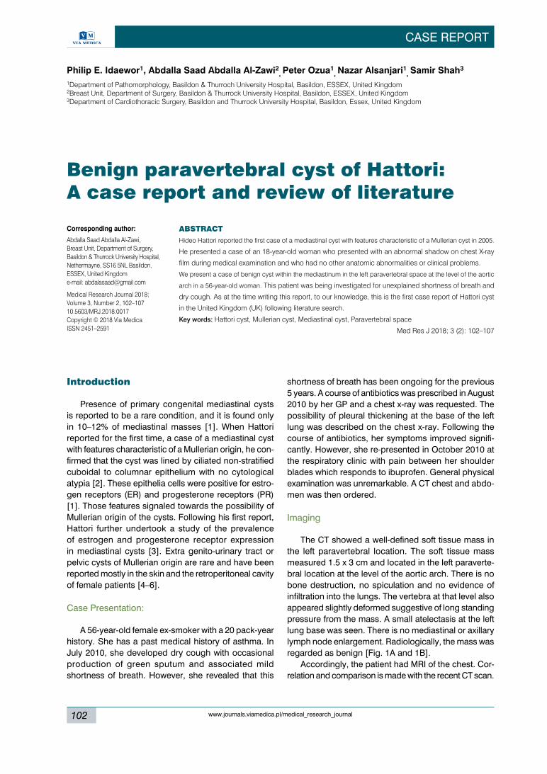
The CT showed a well-defined soft tissue mass in the left paravertebral location. The soft tissue mass measured 1.5 x 3 cm and located in the left paraverte-bral location at the level of the aortic arch. There is no bone destruction, no spiculation and no evidence of infiltration into the lungs. The vertebra at that level also appeared slightly deformed suggestive of long standing pressure from the mass. A small atelectasis at the left lung base was seen. There is no mediastinal or axillary lymph node enlargement. Radiologically, the mass was regarded as benign [Fig. 1A and 1B].
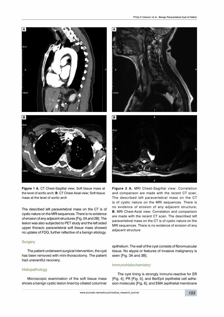
Accordingly, the patient had MRI of the chest. Cor-relation and comparison is made with the recent CT scan.
Philip E Idaewor et al., Benign Paravertebral Cyst of Hattori
103www.journals.viamedica.pl/medical_research_journal
A A
B B
Figure 1 A. CT Chest-Sagittal view; Soft tissue mass at the level of aortic arch; B. CT Chest-Axial view; Soft tissue mass at the level of aortic arch
Figure 2 A. MRI Chest-Sagittal view; Correlation and comparison are made with the recent CT scan. The described left paravertebral mass on the CT is of cystic nature on the MRI sequences. There is no evidence of erosion of any adjacent structure; B. MRI Chest-Axial view; Correlation and comparison are made with the recent CT scan. The described left paravertebral mass on the CT is of cystic nature on the MRI sequences. There is no evidence of erosion of any adjacent structure
The described left paravertebral mass on the CT is of cystic nature on the MRI sequences. There is no evidence of erosion of any adjacent structures [Fig. 2A and 2B]. The lesion was also subjected to PET study and the left sided upper thoracic paravertebral soft tissue mass showed no uptake of FDG, further reflective of a benign etiology.
Surgery
The patient underwent surgical intervention, the cyst has been removed with mini-thoracotomy. The patient had uneventful recovery.
Histopathology
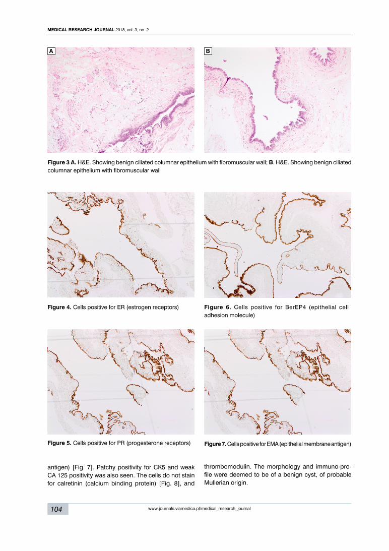
Microscopic examination of the soft tissue mass shows a benign cystic lesion lined by ciliated columnar
epithelium. The wall of the cyst consists of fibromuscular tissue. No atypia or features of invasive malignancy is seen [Fig. 3A and 3B].
Immunohistochemistry:
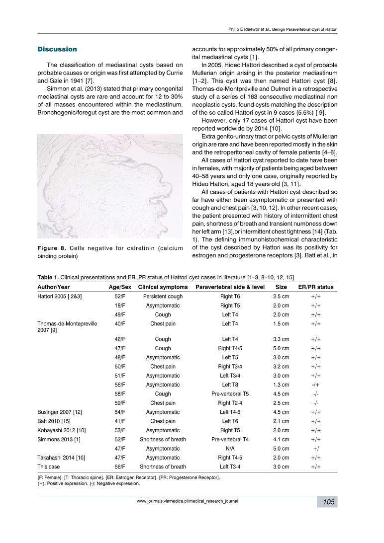
The cyst lining is strongly immuno-reactive for ER [Fig. 4], PR [Fig. 5], and BerEp4 (epithelial cell adhe-sion molecule) [Fig. 6], and EMA (epithelial membrane
104
Medical research journal 2018, vol. 3, no. 2
www.journals.viamedica.pl/medical_research_journal
A B
Figure 3 A. H&E. Showing benign ciliated columnar epithelium with fibromuscular wall; B. H&E. Showing benign ciliated columnar epithelium with fibromuscular wall
Figure 4. Cells positive for ER (estrogen receptors)
Figure 5. Cells positive for PR (progesterone receptors)
Figure 6. Cells positive for BerEP4 (epithelial cell adhesion molecule)
antigen) [Fig. 7]. Patchy positivity for CK5 and weak CA 125 positivity was also seen. The cells do not stain for calretinin (calcium binding protein) [Fig. 8], and
thrombomodulin. The morphology and immuno-pro-file were deemed to be of a benign cyst, of probable Mullerian origin.
Figure 7. Cells positive for EMA (epithelial membrane antigen)
Philip E Idaewor et al., Benign Paravertebral Cyst of Hattori
105www.journals.viamedica.pl/medical_research_journal
Figure 8. Cells negative for calretinin (calcium binding protein)
Table 1. Clinical presentations and ER ,PR status of Hattori cyst cases in literature [1–3, 8–10, 12, 15]
Author/Year Age/Sex Clinical symptoms Paravertebral side & level Size ER/PR status
Hattori 2005 [ 2&3] 52/F Persistent cough Right T6 2.5 cm +/+
18/F Asymptomatic Right T5 2.0 cm +/+
49/F Cough Left T4 2.0 cm +/+
Thomas-de-Montepreville 2007 [9]
40/F Chest pain Left T4 1.5 cm +/+
46/F Cough Left T4 3.3 cm +/+
47/F Cough Right T4/5 5.0 cm +/+
48/F Asymptomatic Left T5 3.0 cm +/+
50/F Chest pain Right T3/4 3.2 cm +/+
51/F Asymptomatic Left T3/4 3.0 cm +/+
56/F Asymptomatic Left T8 1.3 cm -/+
58/F Cough Pre-vertebral T5 4.5 cm -/-
59/F Chest pain Right T2-4 2.5 cm -/-
Businger 2007 [12] 54/F Asymptomatic Left T4-6 4.5 cm +/+
Batt 2010 [15] 41/F Chest pain Left T6 2.1 cm +/+
Kobayashi 2012 [10] 53/F Asymptomatic Right T5 2.0 cm +/+
Simmons 2013 [1] 52/F Shortness of breath Pre-vertebral T4 4.1 cm +/+
47/F Asymptomatic N/A 5.0 cm +/
Takahashi 2014 [10] 47/F Asymptomatic Right T4-5 2.0 cm +/+
This case 56/F Shortness of breath Left T3-4 3.0 cm +/+
[F: Female]. [T: Thoracic spine]. [ER: Estrogen Receptor]. [PR: Progesterone Receptor]. (+): Positive expression. (-): Negative expression.
Discussion
The classification of mediastinal cysts based on probable causes or origin was first attempted by Currie and Gale in 1941 [7].
Simmon et al. (2013) stated that primary congenital mediastinal cysts are rare and account for 12 to 30% of all masses encountered within the mediastinum. Bronchogenic/foregut cyst are the most common and
accounts for approximately 50% of all primary congen-ital mediastinal cysts [1].
In 2005, Hideo Hattori described a cyst of probable Mullerian origin arising in the posterior mediastinum [1–2]. This cyst was then named Hattori cyst [8]. Thomas-de-Montpréville and Dulmet in a retrospective study of a series of 163 consecutive mediastinal non neoplastic cysts, found cysts matching the description of the so called Hattori cyst in 9 cases (5.5%) [ 9].
However, only 17 cases of Hattori cyst have been reported worldwide by 2014 [10].
Extra genito-urinary tract or pelvic cysts of Mullerian origin are rare and have been reported mostly in the skin and the retroperitoneal cavity of female patients [4–6].
All cases of Hattori cyst reported to date have been in females, with majority of patients being aged between 40–58 years and only one case, originally reported by Hideo Hattori, aged 18 years old [3, 11].
All cases of patients with Hattori cyst described so far have either been asymptomatic or presented with cough and chest pain [3, 10, 12]. In other recent cases, the patient presented with history of intermittent chest pain, shortness of breath and transient numbness down her left arm [13],or intermittent chest tightness [14] (Tab. 1). The defining immunohistochemical characteristic of the cyst described by Hattori was its positivity for estrogen and progesterone receptors [3]. Batt et al., in
106
Medical research journal 2018, vol. 3, no. 2
www.journals.viamedica.pl/medical_research_journal
describing the first case of mediastinal cyst with Mulle-rian differentiation from the United States, noted that on the basis of histology, immunohistochemical staining characteristics, and the presence of estrogen and pro-gesterone receptors, they conclude that the patient had a benign mediastinal paravertebral endosalpingiotic Mullerian cyst, identical to that described by Hattori [15]. Simmon et al. in addition to also demonstrating nuclear positivity for ER and PR in their cases also showed these to express the novel pan-Mullerian marker PAX8 (paired box gene 8) and WT1 (Wilms’ tumor protein 1), features that support Mullerian differentiation [8] (Tab. 1).
The origin of Hattori cyst is unknown and remains a subject of speculation. Mullerian cysts in the female pelvis can develop from Mullerian duct remnants or coelomic lining undergoing metaplasia. However, no embryonic Mullerian structure has been identified in the mediastinum, pleura or pericardium [15]. Moreover, mediastinal structures are not regarded as part of the secondary Mullerian system and it is rare for a Mullerian cyst to be located outside the female pelvis [8]. The skin and retroperitoneum are some of the reported locations outside of the pelvis [4]. This peculiar unexplained loca-tion therefore makes Hattori cyst not only an interesting but also an intriguing finding [16]. They may be consid-ered part of the Mullerian Choristoma group described by Batt et al. relying on Ludwig’s theory to explain this phenomenon [17]. Batt et al. in specific reference to Hattori cyst has suggested that the lesion is derived from the primary Mullerian tissue similar to postulated pathogenesis of Mayer- Rokistansky-Kuster-Hauser syndrome (MRKH) [15]. Hattori and other authors also argued that the apparent rarity of this cyst of Mullerian origin in the literature can be attributed to misdiagnosis as bronchogenic or other commoner cystic lesions in the mediastinum [1, 3], or misplaced mesothelium and mesenchyme with characteristic of the secondary Mul-lerian system of the mediastinum [2]. This suggestion if true will imply metaplastic changes of the mesothelium into a ciliated epithelium type.
Hence, Hattori is of the opinion that all cysts diag-nosed as bronchogenic or neurogenic cyst should be stained with hormone receptors (ER and PR) [3]. This is important in defining the lineage of this cyst.
Associated clinical factors noted in some of the patients presented previously include hormone replace-ment therapy, obesity, previous gynecologic diseases and abnormal hormonal profile with high serum estra-diol level [5, 9, 14].
None of the above factors were noted in our patient. Our case clearly demonstrates a cyst in which the radiological, histological, and immunohistochemical profile is consistent with a Hattori cyst. As with all pre-vious case reports, the cyst in our case was present in the paravertebral space and the patient aged 56 years
within the 40–60 years bracket aside from the first case described by Hattori who was 18 years old.
conclusion:
It is recommended to include the Hattoriʼs cyst in the differential diagnosis of posterior mediastinal cysts. Their nature is benign; however, surgical re-moval with thoracotomy or thoracoscopy is advisable to alleviate patient symptoms and obtain the proper tissue diagnosis.
Acknowledgement:
Authors would like to thank Ms Jessica Eades from the Breast Unit Radiology team at Basildon University Hospital for her expert technical assistance in image se-lection.
references:
1. Simmons M, Duckworth LV, Scherer K, et al. Mullerian cysts of the posterior mediastinum: report of two cases and review of the litera-ture. J Thorac Dis. 2013; 5(1): E8–EE10, doi: 10.3978/j.issn.2072-1439.2012.07.10, indexed in Pubmed: 23372963.
2. Hattori H. Ciliated cyst of probable mullerian origin arising in the posterior mediastinum. Virchows Arch. 2005; 446(1): 82–84, doi: 10.1007/s00428-004-1087-0, indexed in Pubmed: 15480767.
3. Hattori H. High prevalence of estrogen and progesterone receptor expression in mediastinal cysts situated in the posterior mediastinum. Chest. 2005; 128(5): 3388–3390, doi: 10.1378/chest.128.5.3388, indexed in Pubmed: 16304289.
4. Farmer ER, Helwig EB. Cutaneous ciliated cysts. Arch Dermatol. 1978; 114(1): 70–73, indexed in Pubmed: 619786.
5. Konishi E, Nakashima Y, Iwasaki T. Immunohistochemical analysis of retroperitoneal Müllerian cyst. Hum Pathol. 2003; 34(2): 194–198, doi: 10.1053/hupa.2003.12, indexed in Pubmed: 12612890.
6. Fabien-Dupuis C, Cooper B, Upperman J, et al. Mullerian-Type Cilia-ted Cyst of the Thigh with PAX-8 and WT1 Positivity: A Case Report and Review of the Literature. Case Rep Med. 2016; 2016: 2487820, doi: 10.1155/2016/2487820, indexed in Pubmed: 28070193.
7. Curreri AR, Gale JW. MEDIASTINAL CYSTS. Ann Surg. 1941; 113(6): 1086–1087, indexed in Pubmed: 17857814.
8. Lee S, Hwang C, Park D, et al. A Ciliated Cyst with Müllerian Dif-ferentiation Arising in the Posterior Mediastinum. The Korean Jo-urnal of Pathology. 2014; 48(5): 401–404, doi: 10.4132/koreanjpa-thol.2014.48.5.401.
9. Thomas-de-Montpréville V, Dulmet E. Cysts of the posterior mediasti-num showing müllerian differentiation (Hattori’s cysts). Ann Diagn Pa-thol. 2007; 11(6): 417–420, doi: 10.1016/j.anndiagpath.2006.12.011, indexed in Pubmed: 18022126.
10. Takahashi Y, Omori H, Yoshifuku S, et al. A Case of Müllerian Cyst Arising in the Posterior Mediastinum. Nihon Gekakei Rengo Gakkaishi (Journal of Japanese College of Surgeons). 2014; 39(4): 804–807, doi: 10.4030/jjcs.39.804.
11. Satoru K, Takashi I, Yoko K, et al. Case of Mullerian Cyst Arising in Po-sterior Mediastinum. Ann Thorac Cardiovasc Surg. 2012; 18: 39–41.
12. Businger AP, Frick H, Sailer M, et al. A ciliated cyst in the posterior me-diastinum compatible with a paravertebral Mullerian cyst. Eur J Car-diothorac Surg. 2008; 33(1): 133–136, doi: 10.1016/j.ejcts.2007.09.036, indexed in Pubmed: 17977003.
13. Chandra N, Seevanayagam S. A Case of Posterior Mediastinal Cyst of Mullerian Type (Hattori’s Cysts). Heart, Lung and Circulation. 2017; 26: S360–S361, doi: 10.1016/j.hlc.2017.03.024.
14. Tsan-Chieh L, Fu-Pang C, Teh-Ying C, et al. Progressive Enlarging Symptomatic Mediastinal Mullerian Cyst in a Female Patient with High Estradiol Level. J Radiol Sci. 2012; 37: 117–121.
Philip E Idaewor et al., Benign Paravertebral Cyst of Hattori
107www.journals.viamedica.pl/medical_research_journal
15. Batt RE, Mhawech-Fauceglia P, Odunsi K, et al. Pathogenesis of mediastinal paravertebral müllerian cysts of Hattori: developmental endosalpingiosis-müllerianosis. Int J Gynecol Pathol. 2010; 29(6): 546–551, doi: 10.1097/PGP.0b013e3181e3640a, indexed in Pubmed: 20881858.
16. Chon SH, Im UiJ, Song DS. Paravertebral mediastinal Mullerian cyst resected by video assisted thoracoscopic surgery. J Thorac Dis. 2015; 7(3): E47–E49, doi: 10.3978/j.issn.2072-1439.2014.12.05, indexed in Pubmed: 25922749.
17. Batt RE, Smith RA, Buck Lo, et al. Mullerianosis. Histo Histopathol. 2007; 22: 1161–1166.


