Embed Size (px)
Citation preview

0270-91 39/84/0405-038S$02.00/0 HEPATOLOGY Copyright 8 1984 by the American Association for the Study of Liver Diseases
Vol. 4, No. 5 , pp. 38S-45S, 1984 Printed in U.S.A.
Bilirubin Chemistry, Ionization and Solubilization by Bile Salts
J. DONALD OSTROW AND LILLIAN CELIC
Gastroenterology Section, Department of Medicine, Veterans Administration Lakeside Medical Center and Northwestern University Medical School, Chicago, Illinois 6061 1
Bilirubin is a linear tetrapyrrole whose conformation is affected by internal hydrogen bonds formed between the carboxyl side chains and dipyrromethenone rings. Structural variations include: constitutional isomerism of the vinyl or carboxyethyl side chains, geometric isomerism of the methene bridges, tautomerism of the lactam groups, conformational rotations about the central methylene bridge and ionization of one or both carboxyl groups. Aggregation of the dianion into dimers and multimers may occur. The pK,’ values of the two carboxyl groups are affected greatly by the environment and may differ widely in micellar solutions like bile. Solubility of bilirubin in water is less than 1 nM at pH = 7 and about 0.1 at pH = 8. Nonetheless, it dissolves poorly in most lipid solvents, except for asymmetrical chloroalkanes. Hydrogen bond-breaking solvents, especially dimethyl sulfoxide, are most effective in solubilizing bilirubin. In bile salt solutions, solubility of bilirubin is well above the concentrations of unconjugated bilirubin found in normal human gallbladder bile, and is impaired by lecithin but unaffected by cholesterol. At physiological pH in bile salt solutions, bilirubin is predominantly in its monoanion form that binds readily to the micelles. In such solutions, addition of physiological concentrations of calcium precipitates calcium bilirubinate, leaving residual bilirubin concentrations of up to 15 rc.M in 50 mM taurocholate or close to the maximum bilirubin concentrations in normal bile. Studies in which disodium bilirubi- nate is dissolved in bile salt solutions and pH is adjusted to the physiological range reveal that metastable supersaturation with bilirubin may occur and that a mesophase may also form in the presence of lecithin, akin to that seen with cholesterol. These results form a basis for understanding the precipitation of calcium bilirubinate from bile during pigment gallstone formation.
Bilirubin is a linear tetrapyrrole of the biladiene-a,c type, consisting of two asymmetrical dipyrromethenones connected by a methylene bridge (1) (Figure 1). In the natural 1x0-Z,Z isomer that predominates in uiuo, each dipyrrolic half-molecule carries a carboxyethyl side chain that might be expected to render the pigment a highly polar, ionized and water-soluble molecule a t physiologi- cal pH values. However, in its fully protonated (diacid) form, each of the two carboxyl groups forms internal hydrogen bonds with the -C=O and two >NH in the dipyrromethenone rings in the opposite half of the mol- ecule (2). Collectively, these two trios of hydrogen bonds stabilize the bilirubin molecule in a rigid conformation that resembles a ridge tile or partially folded book (2), in
This study was supported by Research Grant RO-1-AM-32130 from the National Institute of Arthritis, Diabetes, Digestive and Kidney Diseases-National Institutes of Health, United States Public Health Service, by a Merit Review Research Award from the United States Veterans Administration and by the Otho S.A. Sprague Foundation a t Northwestern University.
Address reprint requests to: J. Donald Ostrow, M.D. (111G), Vet- erans Administration Lakeside Medical Center, 333 East Huron Street, Chicago, Ilinois 60611,
which each planar dipyrromethenone constitutes one leaf of the book. In this conformation, which exists both in the crystalline state (2) and in certain solutions (3), these internal hydrogen bonds suppress the ionization of the carboxyl groups and monopolize all of the polar groups in the bilirubin molecule, limiting their interaction with water. As a consequence, crystalline bilirubin-IXa-Z,Z is virtually insoluble in buffered aqueous solutions at phys- iological pH values and has an apparent pKa’ value in the weakly alkaline range (4). In such solutions, there is a linear relationship between the log of bilirubin solubil- ity and pH values, with a maximum solubility of 0.1 p M at pH = 8.0. These values are more than two orders of magnitude less than the maximum concentration of un- conjugated bilirubin found in normal human gallbladder bile ( 5 ) . Consequently, the changes in structure and physicochemical interactions of bilirubin that affect its solubility and permit its solubilization in bile are of utmost importance in understanding the pathogenesis of pigment gallstones that contain bilirubin.
This paper will review current concepts of structural variations in bilirubin, its association with other com- ponents of bile and the effects of these factors on its solubility in aqueous solutions and bile.
38s

Vol. 4, No. 5, Suppl. 1984 BILIRUBIN CHEMISTRY AND SOLUBILITY 39s
STRUCTURAL VARIATIONS IN BILIRUBIN IXa-Z,Z
In bilirubin IXa-Z,Z, the molecule is slightly asym- metrical because the vinyl side chain on the A ring is in the endo- position (on carbon 3), but the vinyl side chain on the D ring is in the exo- position (on carbon 18) (Figure 1). Moreover, the A 4,5 and A15,16 double bonds, in the methene bridges of the two dipyrromethenone fragments, are both in the Z configuration (2). Five types of structural variations can occur in this molecule, many of which affect the ability to form the internal hydrogen bonds described above:
(i) Constitutional isomerism can affect the positions of the vinyl and/or carboxyethyl side chains. By a process of dipyrrole exchange, in which the dipyrromethenone fragments cleave from each other and randomly reasso- ciate covalently, isomers are formed in which both vinyl groups are in the exo- positions (Type I11 isomer) or both in the endo positions (Type XIII) (6). Minor amounts of these isomers are found in bile, but, aside from a modest shift in the absorption maximum in the visible spectrum and slight variation in polarity as revealed by chroma- tography, the properties of these isomers differ little from the normal Type IX.
Changes in the position of the carboxyethyl side chains can result from nonenzymatic cleavage of heme at other than the a-methene bridge, yielding p, y and 6 isomers (7). In these isomers, one or both of the carboxyethyl groups are sterically unable to form hydrogen bonds with the dipyrromethenone rings, greatly altering their ioni-. zation, polarity and ability to interact with water (7). In mammals, such isomers have been found only in congen- itally jaundiced (Gunn) rats (8) and from oxidation of certain unstable mutant hemoglobins (9).
(ii) Configurational (geometric) isomerism can occur a t one or both of the methene bridges, forming 4E, 152; 42, 15E; and 4E, 15E isomers. The E,Z and Z,E isomers are the major products formed during anaerobic irradiation of bilirubin IXa-Z,Z with blue light in uitro (10) and are known also as photobilirubins I-A and B (11, 12). The E,Z isomer can undergo further photocatalyzed cycliza- tion via the endo vinyl group on the A ring, forming the
I CH3 CH3
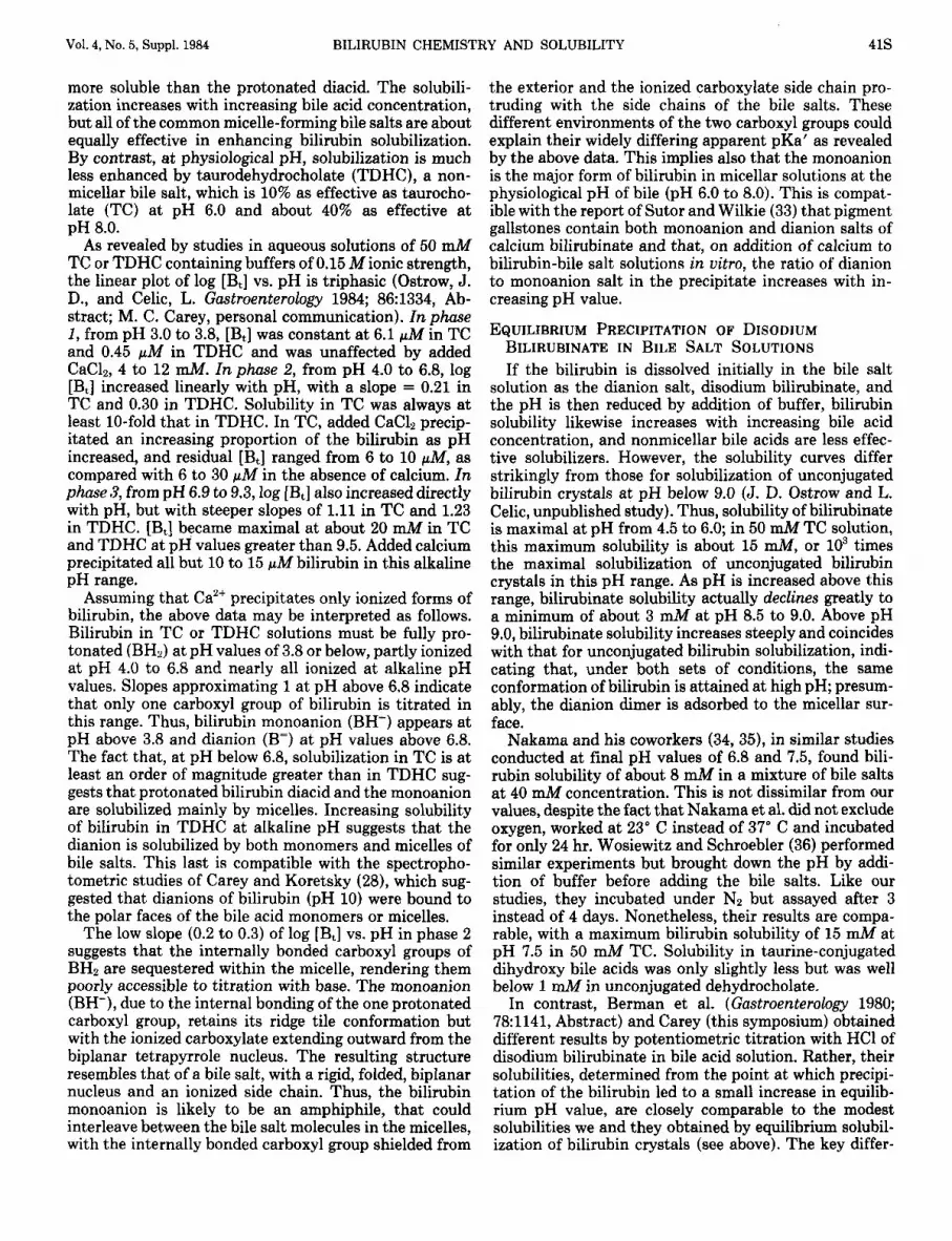
FIG. 1. Molecular structure of diacid bilirubin, showing internal bonding of each fully protonated carboxyl group to the opposite dipyr- romethenone. The two trios of hydrogen bonds are represented by the dashed lines. In three dimensions, the molecule would have a “ridge tile” or folded-book conformation, with the central methylene group on the ridge (arrows). The A and B rings lie in one plane and the C and D rings in another plane, with an interplanar angle of about 98”. (Courtesy of Dr. Rolf Brodersen.)
stable photobilirubin I1 (11-13). In all of these photoiso- mers, the exo- ring of the dipyrromethenone in the E configuration becomes sterically incapable of forming hydrogen bonds with the opposite carboxyethyl side chain, so that the carboxyl group is exposed to solvent and ionizes a t the expected pKa value near 4.5. These photoisomers are, therefore, more polar and water solu- ble than the Z,Z isomer and are capable of being excreted in the bile without conjugation (12, 14, 15). In the bile or in uitro, especially in acid media, the photobilirubins I (E,Z or Z,E) revert spontaneously to the Z,Z, form, even in the absence of light (10-12). The more stable photo- bilirubin I1 does not revert to the Z,Z form in the dark but does undergo oxidation and polymerization to give purple and black pigments (13, 14).
(iii) Tautomerism involves lactam-lactim interconver- sions of the two pyrrolenone end-rings. Under all normal circumstances, both in the crystalline state and in solution, bilirubin exhibits a dominance of the lactam form by four orders of magnitude over the lactim (16- 18). Stabilization of the lactim, by formation of an ether, precludes internal hydrogen bonding by this group and changes the conformation and polarity of the molecule by releasing the opposite carboxyethyl group (19).
(iv) Conformers result from rotations of the pyrrole rings about the single bonds in the central methylene bridge, since, under almost all conditions, the exo- and endo- ring in each dipyrromethenone are almost coplanar (synperiplanar) (2). In the internally bonded (ridge tile) arrangement, the two planar dipyrromethenone halves are in a syn-syn conformation about the central meth- ylene bridge, with an interplanar angle of about 100” (2). If the internal bonding is broken, as in the dimethylester (3) or lactim-ether (19), the molecule assumes a “perpen- dicular” conformation, with torsional angles of +24” and -103” for the dipyrromethenones in relation to the cen- tral methylene bridge.
(v) State of ionization of the carboxyl and lactam groups can affect the properties of bilirubin. The lactam group characteristically ionizes at pH between 12 and 14, depending upon the solvent (20, 21), leading to a change in color from yellow-orange to deep reddish- brown (e.g., in 0.1 N NaOH). This ionization is of no relevance at physiological pH values. By contrast, the carboxyl groups are titratable at physiological pH values and, therefore, their state of ionization profoundly affects the conformation and properties of the pigment. It is not clear whether the ionized carboxylate group is capable of internally bonding with the contralateral dipyrrometh- enone (22), and the pKa values of the two carboxyls are disputed also. This is discussed in the next section.
IONIZATION AND pKa VALUES OF THE TWO CARBOXYL GROUPS
Theoretically, bilirubin can exist as the fully proton- ated diacid (BH,), as monoanions (BH-) or as a dianion (BE). However, different investigators, using different conditions, have not been able to agree as to the ioniza- tion constants of the carboxyl groups or even whether the two groups have identical or different pKa values.
Potentiometric titration with base of the diacid (BH2),

40s OSTROW AND CELIC HEPATOLOGY
in solvents that break the internal hydrogen bonds, has yielded conflicting results. Titrating with NaOH, Hansen et al. (20) found BH2 in dimethyl sulfoxide to have identical pKa values of 4.4 for both carboxyl groups. By contrast, using quarternary amine bases to titrate BH2 in dimethyl formamide, both Lee et al. (21), and Van Norman and Szentirmay (23) noted breaks in the titra- tion curves after one as well as two base equivalents were added. Calculated pKa values for the two carboxyl groups were 4.3 and 5.3.
Potentiometric titration with HC1 of bilirubin dianion salt (B=) in simple aqueous solution leads to progressive precipitation of insoluble BH2 as the pH falls below about 8.0 (20, 24). It has been noted by these workers, as well as others (21), that the aqueous titration curves could be reconstructed by using the pKa obtained in dimethyl formamide or dimethyl sulfoxide (v.s.) and applying a mathematical model that assumes that bilirubin is solu- bilized only by prior ionization of the solid BH2. This model predicts that the apparent pKa’ values, read off a graph of solubility vs. pH, will be higher than the true or intrinsic pKa when a solid phase is formed by precipi- tation of the protonated species. Similar conclusions have been reached concerning titration of fatty acids (25) and bile acids (26).
Both potentiometric (Berman, M. D. et al., Gastroen- terology 1980; 781141, Abstract) and spectrophotometric (J. D. Ostrow and L. Celic, unpublished data) titrations with HC1, of B= dissolved in aqueous taurocholate solu- tions, have yielded apparent pKa’ values of 6.2 to 6.5. Ostrow and Celic found also that the pKa‘ is the same on back titration with alkali if the solution is sufficiently dilute to avoid precipitation of the BH2. Berman et al. also found that the pKa’ rises to 6.9 if lecithin is added. Neither found evidence for dual pKa’ values for the two carboxyl groups. The concordance of results-even though Berman et al. worked at bilirubin concentrations that led to precipitation of BH2 as the pH was lowered, whereas Ostrow and Celic did not-suggests that solu- bilization in micelles may be akin to precipitation in a solid phase; in either case, the protonated species (BH2) is sequestered in a separate phase. Micellar phase bili- rubin might not easily release H+ ions, due to the sur- rounding cloud of cationic counterions. A similar phe- nomenon is observed with titration of oleic acid in bile salt solution (25, 27).
BILIRUBIN AGGREGATION In aqueous or bile salt solutions, as indicated by shifts
in visible spectral peaks, bilirubin dianions form dimers at concentrations of 1 to 10 pM and larger aggregates at higher concentrations (28, 29). The effects of added solvents suggest that both hydrophobic and electrostatic (?r-orbital) interactions are involved in stabilizing the dimers, whereas hydrophobic interactions play a major role in the multimers (28). In the aggregates, it is likely that the bilirubin molecules stack in parallel, like a deck of playing cards. McDonagh (National Institutes of Health Workshop on Pigment Gallstones, Philadelphia, 1981) has suggested that the dimer may precipitate as a monohydrate when the dianion salts are neutralized rap-
idly with acid. It has not been studied whether bilirubin monoanion or diacid forms aggregates.
Brodersen et al. (30) have shown that bilirubin, when bound to serum albumin, can serve as a nidus for aggre- gation of one or more pigment molecules, forming huge soluble complexes. Binding of bilirubin to albumin may disrupt the intramolecular hydrogen bonds; thus, biliru- bin bound to albumin-agarose beads may be eluted with ethanol (31), in which the pigment is normally virtually insoluble. Moreover, titration of bilirubin bound to al- bumin reveals a common pKa near 5.3 for both carboxyl groups (4), so that the pigment is fully in the dianion form over the physiological pH range (6.0 to 8.0).
BILIRUBIN SOLUBILITY The solubility of bilirubin in various organic solvents
(4) does not favor the conventional wisdom that bilirubin diacid is “lipid soluble,” although it is virtually insoluble in water. Thus, at 25” C, BH2 is virtually insoluble in linear and cyclic alkanes (e.g., n-hexane) and in alcohols containing one to five carbon atoms, as well as in ether and long chain fatty acids. Solubility in a variety of ketones, in ethyl acetate and in simple cyclic hydrocar- bons (e.g., benzene) ranges from 8 to 50 pM. Solubility is comparable in the symmetrical solvent, carbon tetra- chloride, but is 1.8 and 2.5 mM in the asymmetrical chlorinated hydrocarbons, dichloromethane and chloro- form, respectively. Solvents that break hydrogen bonds convey similar solubility: 0.8 mM in N,N-dimethyl form- amide and 2.3 mM in formamide. However, the most potent solvent, dimethyl sulfoxide, dissolves well over 10 mM bilirubin.
I n buffered aqueous solutions, equilibrium solubiliza- tion of crystalline bilirubin is less than 1 nM at pH values less than 7.0 but rises logarithmically with in- creasing pH values to a concentration of about 1 pM at pH 8.5 (4). This linear relationship between log solubility and pH has a slope of 2. Based on a model that assumes that the pigment dissolves as the diacid, BH2, and then ionizes in solution to a degree determined by pH, this indicates that two protons are titrated simultaneously (4). It indicates also that, unlike the diacid, the dianion form is quite soluble, and may not have internal hydrogen bonding. The maximum aqueous solubility of the dianion in 0.15 M NaCl at pH 10.0 is about 36 mM (28, 32), which is much greater than that predicted from extrap- olation of the solubility curve observed between pH 7.5 to 8.5. This difference is likely due to aggregation of bilirubin dianions, forming “micelles” of enhanced solu- bility (28).
SOLUBILIZATION OF CRYSTALLINE BH2 BY BILE SALT SOLUTIONS Bile acids, which have some capacity to interfere with
hydrogen bond formation, increase by four orders of magnitude (to about 1 mM at pH 8.0) the dissolution of bilirubin in aqueous solutions (Ostrow, J. D. et al., Gas- troenterology 1980; 781315, Abstract, and 1984; 86:1334, Abstract). As in simple aqueous solutions, the logarithm of the solubility of bilirubin, log I&], increases linearly with pH value, indicating that the ionized form(s) are
Vol. 4, No. 5, Suppl. 1984 BILIRUBIN CHEMISTRY AND SOLUBILITY 41s
more soluble than the protonated diacid. The solubili- zation increases with increasing bile acid concentration, but all of the common micelle-forming bile salts are about equally effective in enhancing bilirubin solubilization. By contrast, at physiological pH, solubilization is much less enhanced by taurodehydrocholate (TDHC), a non- micellar bile salt, which is 10% as effective as taurocho- late (TC) at pH 6.0 and about 40% as effective at pH 8.0.
As revealed by studies in aqueous solutions of 50 d 4 TC or TDHC containing buffers of 0.15 M ionic strength, the linear plot of log [B,] vs. pH is triphasic (Ostrow, J. D., and Celic, L. Gastroenterology 1984; 86:1334, Ab- stract; M. C. Carey, personal communication). In phase 1 , from pH 3.0 to 3.8, [B,] was constant at 6.1 pM in TC and 0.45 pM in TDHC and was unaffected by added CaC12, 4 to 12 mM. In phase 2, from pH 4.0 to 6.8, log [B,] increased linearly with pH, with a slope = 0.21 in TC and 0.30 in TDHC. Solubility in TC was always at least 10-fold that in TDHC. In TC, added CaC12 precip- itated an increasing proportion of the bilirubin as pH increased, and residual [Bt] ranged from 6 to 10 pM, as compared with 6 to 30 pM in the absence of calcium. In phase 3, from pH 6.9 to 9.3, log [Bt] also increased directly with pH, but with steeper slopes of 1.11 in TC and 1.23 in TDHC. [B,] became maximal at about 20 mM in TC and TDHC at pH values greater than 9.5. Added calcium precipitated all but 10 to 15 pM bilirubin in this alkaline pH range.
Assuming that Caz+ precipitates only ionized forms of bilirubin, the above data may be interpreted as follows. Bilirubin in TC or TDHC solutions must be fully pro- tonated (BH2) at pH values of 3.8 or below, partly ionized at pH 4.0 to 6.8 and nearly all ionized at alkaline pH values. Slopes approximating 1 at pH above 6.8 indicate that only one carboxyl group of bilirubin is titrated in this range. Thus, bilirubin monoanion (BH-) appears at pH above 3.8 and dianion (B=) at pH values above 6.8. The fact that, at pH below 6.8, solubilization in TC is at least an order of magnitude greater than in TDHC sug- gests that protonated bilirubin diacid and the monoanion are solubilized mainly by micelles. Increasing solubility of bilirubin in TDHC at alkaline pH suggests that the dianion is solubilized by both monomers and micelles of bile salts. This last is compatible with the spectropho- tometric studies of Carey and Koretsky (28), which sug- gested that dianions of bilirubin (pH 10) were bound to the polar faces of the bile acid monomers or micelles.
The low slope (0.2 to 0.3) of log [Bt] vs. pH in phase 2 suggests that the internally bonded carboxyl groups of BH, are sequestered within the micelle, rendering them poorly accessible to titration with base. The monoanion (BH-), due to the internal bonding of the one protonated carboxyl group, retains its ridge tile conformation but with the ionized carboxylate extending outward from the biplanar tetrapyrrole nucleus. The resulting structure resembles that of a bile salt, with a rigid, folded, biplanar nucleus and an ionized side chain. Thus, the bilirubin monoanion is likely to be an amphiphile, that could interleave between the bile salt molecules in the micelles, with the internally bonded carboxyl group shielded from
the exterior and the ionized carboxylate side chain pro- truding with the side chains of the bile salts. These different environments of the two carboxyl groups could explain their widely differing apparent pKa' as revealed by the above data. This implies also that the monoanion is the major form of bilirubin in micellar solutions at the physiological pH of bile (pH 6.0 to 8.0). This is compat- ible with the report of Sutor and Wilkie (33) that pigment gallstones contain both monoanion and dianion salts of calcium bilirubinate and that, on addition of calcium to bilirubin-bile salt solutions in uitro, the ratio of dianion to monoanion salt in the precipitate increases with in- creasing pH value.
EQUILIBRIUM PRECIPITATION OF DISODIUM BILIRUBINATE IN BILE SALT SOLUTIONS
If the bilirubin is dissolved initially in the bile salt solution as the dianion salt, disodium bilirubinate, and the pH is then reduced by addition of buffer, bilirubin solubility likewise increases with increasing bile acid concentration, and nonmicellar bile acids are less effec- tive solubilizers. However, the solubility curves differ strikingly from those for solubilization of unconjugated bilirubin crystals at pH below 9.0 (J. D. Ostrow and L. Celic, unpublished study). Thus, solubility of bilirubinate is maximal at pH from 4.5 to 6.0; in 50 mM TC solution, this maximum solubility is about 15 mM, or lo3 times the maximal solubilization of unconjugated bilirubin crystals in this pH range. As pH is increased above this range, bilirubinate solubility actually declines greatly to a minimum of about 3 mM at pH 8.5 to 9.0. Above pH 9.0, bilirubinate solubility increases steeply and coincides with that for unconjugated bilirubin solubilization, indi- cating that, under both sets of conditions, the same conformation of bilirubin is attained at high pH; presum- ably, the dianion dimer is adsorbed to the micellar sur- face.
Nakama and his coworkers (34,35), in similar studies conducted at final pH values of 6.8 and 7.5, found bili- rubin solubility of about 8 mM in a mixture of bile salts at 40 mM concentration. This is not dissimilar from our values, despite the fact that Nakama et al. did not exclude oxygen, worked at 23" C instead of 37" C and incubated for only 24 hr. Wosiewitz and Schroebler (36) performed similar experiments but brought down the pH by addi- tion of buffer before adding the bile salts. Like our studies, they incubated under Nz but assayed after 3 instead of 4 days. Nonetheless, their results are compa- rable, with a maximum bilirubin solubility of 15 mh4 at pH 7.5 in 50 mM TC. Solubility in taurine-conjugated dihydroxy bile acids was only slightly less but was well below 1 mM in unconjugated dehydrocholate.
In contrast, Berman et al. (Gastroenterology 1980; 78:1141, Abstract) and Carey (this symposium) obtained different results by potentiometric titration with HC1 of disodium bilirubinate in bile acid solution. Rather, their solubilities, determined from the point at which precipi- tation of the bilirubin led to a small increase in equilib- rium pH value, are closely comparable to the modest solubilities we and they obtained by equilibrium solubil- ization of bilirubin crystals (see above). The key differ-

425 OSTROW AND CELIC HEPATOLOGY
ence in their studies is that the pH is decreased very slowly by dropwise addition of HC1, allowing re-equili- bration after each aliquot of acid. It is likely that the much higher solubilities obtained by us, Nakama and Wosiewitz and Schroebler represent metastable supersat- uration. With the very rapid neutralization obtained in those studies, the internal hydrogen bonds of bilirubin may not have reformed as the carboxylate group(s) was protonated. Thus, the bilirubin might have remained in a more soluble, metastable, open conformation, possibly still associated in dimers that were stabilized in part by the interaction of the ionized bile salt side chains with the dipyrromethenone nuclei of the pigment. Such inter- action might interfere competitively with the binding of bilirubin carboxyl groups to the lactam and pyrrole -NH groups.
In the metastable systems, the four phases of the solubility vs. pH curve might involve the following. From pH 10 to 8.5, one of the four carboxylate groups in the bilirubinate dimer is protonated, forming the less soluble acid salt, Na3HB2. From pH 8 to 6, protonation of the second carboxyl group leads to formation of two mono- meric monoanions, with dissociation of the dimer. The monoanion, however, is in the fully open conformation, enhancing its solubility on the micelle. From pH 6 to 5, there is no further protonation of the monoanion, since the remaining carboxylate group is in the open confor- mation, with its intrinsic pKa of 4.3 to 4.4. Below pH 5, this second carboxylate group is protonated, precipitat- ing the bilirubin diacid. Further work is needed to eval- uate this hypothesis.
EFFECTS OF ADDED LECITHIN AND CHOLESTEROL In the true equilibrium systems in bile salt solutions,
either by solubilization of bilirubin crystals (Ostrow, J. D., and Celic, L. Gastroenterology 1984; 86:1334, Ab- stract) or from slow titration of disodium bilirubinate (Berman, M. D., et al., Gastroenterology 1980; 78:1141, Abstract), added lecithin inhibits bilirubin solubility, whereas addition of cholesterol is without effect. The inhibition by lecithin is marked (about 50%), up to 1ecithin:bile salt molar ratios of about 15 , but it changes relatively little as more lecithin is added above this level. This inhibitory effect of lecithin is exactly opposite to its enhancement of the solubility of cholesterol in bile salt micelles and underscores the differences in the in- teractions of bilirubin and cholesterol with bile acid micelles. Both Nakama et al. (34, 35) and Wosiewitz and Schroebler (36) reported completely opposite results. They found added lecithin to be without effect and added cholesterol to be inhibitory, to what was likely metastable bilirubin solubility. Ostrow and Celic (unpublished data) have found that added lecithin moderately enhances metastable bilirubin solubility but that passage of the “equilibrated” solution through a Millipore filter leads to disequilibration, with separation of a bilirubin-lecithin mesophase that floats on the surface when the cloudy filtrate is centrifuged. The clear infranatant then con- tains bilirubin in concentrations that are identical to those obtained on equilibrium solubilization of crystal- line bilirubin.
SUMMARY
It seems, from the above, that bilirubin in micellar bile salt solutions behaves differently than in any other sol- vents, probably existing mainly as a monoanion at phys- iological pH in bile. The monoanion structure resembles that of a bile salt, being a rigid, folded, planar amphiphile that can interact with bile salt micelles, greatly enhanc- ing its solubility. This also conveys different environ- ments to the two carboxyl side chains of bilirubin, yield- ing widely different pKa’ values for each. Although the solubilization of bilirubin by bile salts is inhibited by lecithin, the equilibrium solubility in model bile systems is vastly greater than the concentrations of unconjugated bilirubin found in normal human bile. However, added calcium at physiological concentrations precipitates the insoluble monoanion salt, leaving residual bilirubin sol- ubilities of 10 to 15 pA4 that are close to the maximum concentrations found in bile. As discussed elsewhere in this symposium (by E. W. Moore), the interaction of calcium with bile salts buffers the concentration of free calcium ion and affords partial protection against precip- itation of the calcium bilirubinate(s).
Acknowledgment: We are indebted to Celesta M. Harvey for her expert typing of the manuscript.
REFERENCES 1. Schmid R. Bilirubin metabolism; state-of-the-art. Gastroenterol-
2. Bonnett R, Davies JE, Hursthouse MB. Structure of bilirubin. Nature (Lond) 1976; 262:326-328.
3. Kaplan D, Navon G. Nuclear magnetic resonance studies of the conformation of bilirubin and its derivatives in solution. J Chem Soc, Perkin 1981; 2:1374-1383.
4. Brodersen R. Bilirubin solubility and interaction with albumin and phospholipid. J Biol Chem 1979; 254:2364-2369.
5. Boonyapisit ST, Trotman BW, Ostrow JD. Unconjugated bilirubin, and the hydrolysis of conjugated bilirubin, in gallbladder bile of patients with cholelithiasis. Gastroenterology 1978; 74:70-74.
6. McDonagh AF. Thermal and photochemical reactions of bilirubin IX-a . Ann NY Acad Sci 1975; 244553-569.
7. Blanckaert N, Heirwegh KPM, Compernolle F. Synthesis and separation by thin-layer chromatography of bilirubin IX isomers. Biochem J 1976; 155:405-417.
8. Blanckaert N, Fevery J, Heirwegh KPM, et al. Characterization of the major diazo-positive pigments in bile of homozygous Gunn rats. Biochem J 1977; 164:237-249.
9. Brown SB, Docherty JC. Haem degradation in abnormal hemoglo- bins. Biochem J 1978; 173:985-987.
10. Falk H, Miiller N, Ratzenhofer M, et al. The structure of “photo- bilirubin.” Monatsh Chem 1982; 113:1421-1432.
11. Stoll MS, Zenone EA, Ostrow JD, et al. Preparation and properties of bilirubin photoisomers. Biochem J 1979; 183:139-146.
12. Onishi S, Kawade N, Itoh S, et al. High pressure liquid chromato- graphic analysis of anaerobic photoproducts of bilirubin I X a in uitro, and its comparison with photoproducts in uiuo. Biochem J 1980; 190527432.
13. Stoll MS, Vicker N, Gray CH, et al. Concerning the structure of photobilirubin 11. Biochem J 1982; 201:179-188.
14. Stoll MS, Zenone EA, Ostrow JD. Excretion of administered and endogenous photobilirubins in the bile of the jaundiced Gunn rat. J Clin Invest 1981; 68134-141.
15. McDonagh AF, Palma LA, Lightner DA. Blue light and bilirubin excretion. Science 1980; 208145-151.
16. Falk H, Grumbayr K, Thirring K, et al. Contributions to the chemistry of pyrrole pigments 21. X-ray photoelectron spectro- metric investigations of the N1. level of bile pigments. Monatsh Chem 1978; 109:1183-1189.
O ~ Y 1978 74:1307-1312.

Vol. 4, No. 5, Suppl. 1984 BILIRUBIN CHEMISTRY AND SOLUBILITY 43s
17. Falk H, Gergely S, Grubmayr K, et al. Chemistry of pyrrole pigments XV. The lactam-lactim tautomerism of bile pigments. Liebig’s Ann Chem 1977; (4):565-581.
18. Severini-Ricca G, Manitto P, Monti D, et al. The carbon-13 nuclear magnetic resonance spectra of bilirubin and its derivatives. Gazz Chem Itai 1975; 105:1273-1278.
19. Sheldrick WS, Becker W. Bile pigment structures. 111. Crystal and molecular structure of diethoxybilirubin diethyl ester. Z Natur- forsch 1979; 34B:1542-1546.
20. Hansen PE, Thiessen H, Brodersen R. Bilirubin acidity; titrimetric and ”C-NMR studies. Acta Chem Scand B 1979; 33:281-293.
21. Lee JJ , Daly LH, Cowger ML. Bilirubin ionic equilibria; their effects on spectra and conformation. Res Commun Chem Pathol Pharmacoll974; 9763-770.
22. Mugnoli AA, Manitto P, Monti D. Structure of di-isopropylam- monium bilirubinate. Nature (Lond) 1978; 273:568-569.
23. Van Norman JD, Szentirmay R. Chemistry of bilirubin and bili- verdin in N,N dimethyl formamide. Anal Chem 1974; 461456- 1464.
24. Overbeek JTG, Vink CLJ, Deenstra H. The solubility of bilirubin. Rec Trav Chim Pays-Bas 1955; 74:81-84.
25. Lucassen J. Hydrolysis and precipitates in carboxylate soap solu- tions. J Phys Chem 1966; 701824-1830.
26. Igimi H, Carey MC. pH-solubility relations of chenodeoxycholic and ursodeoxycholic acids: physical-chemical basis for dissimilar solution and membrane phenomena. J Lipid Res 1980; 21:72-89.
27. Hofman AF. Molecular association in fat digestion. Interaction in bulk of monoolein, oleic acid, and sodium oleate with dilute micellar bile salt solutions. Adv Chem 1968; 8453-66.
28. Carey MC, Koretsky AP. Self-association of unconjugated bilirubin IXa in aqueous solution at pH 10.0, and physical-chemical inter- actions with bile salt monomers and micelles. Biochem J 1979; 179:
29. Brodersen R. Dimerization of bilirubin anion in aqueous solution. Acta Chem Scand 1966; 202895-2896.
30. Brodersen R, Funding L, Pedersen AO, et al. Binding of bilirubin to low-affinity sites of human serum albumin in uitro, followed by co-crystallization. Scand J Clin Lab Invest 1972; 29:433-446.
31. Plotz PH, Berk PD, Scharschmidt BF, et al. Removing substances from blood by affinity chromatography. I. Removing bilirubin and other albumin-bound substances from plasma and blood with al- bumin-conjugated agarose beads. J Clin Invest 1974; 53:778-785.
32. Kolosov IV, Shapovalenko EP. I. Acid-base equilibriums in biliru- bin solutions. 11. Study of acid-base equilibriums in aqueous solu- tions of bilirubin by the method of solubility. J Gen Chem, USSR (English translation) 1977; 47:2149-2151, 2155-2156.
33. Sutor DJ, Wilkie SI. The crystalline salts of calcium bilirubinate in human gallstones. Clin Sci Molec Med 1977; 53:lOl-103.
34. Nakama T. Significance of biliary cholesterol and bilirubin in gallstone formation. Fukuoka Acta Med 1976; 67413-441.
35. Nakama T, Furusawa T, Itoh H, et al. Correlation of cholesterol and bilirubin solubilization in bile salt solution. Gastroenterol Jpn
36. Wosiewitz U, Schroebler S. Solubilization of unconjugated bilirubin
675-689.
1979; 6~565--572.
by bile salts. Experientia 1979; 35:717-718.
DISCUSSION
Hofmann: Could we go back to the question that was asked earlier? The question is: If you have two proprionic groups next to each other on bilirubin, can they have the same pKa, or does one ionize and then the other?
Mukerjee: I will have to quote from memory on the dicarboxylic acids. The pK, and pK2 may differ by as much as three or four units. There is a theory on that by Kirkwood (1). My memory is that in an acid, when one has eight carbon atoms separating two groups, the ApK has gone down to about 0.5. I would like to suggest something, however. I published a paper some time ago on pKa determination (2). This method is an isoextrac-
tion method in which the pKa can be determined by using analytical techniques alone. We have done it for bile acids (unpublished data using analytical techniques) at M because I was sort of suspicious that associa- tion might change pKa values. I would strongly recom- mend it because it is quite precise.
Carey: How do you know it is precise? Mukerjee: We tested it out on p-nitrophenol and ben-
zoic acid. That has been published (3). Ostrow: I read your paper, and we tried your method.
The problem is we do not have a solvent that solubilizes bilirubin at the concentrations we require.
Mukerjee: You mean you cannot choose a good organic solvent?
Ostrow: We have tried many of them; either the bili- rubin is too soluble, the bilirubin is too insoluble or the solvent messes up the micelle.
Hofmann: Dr. Moore and I do not agree with Dr. Ostrow. We think he can use that method if he consults with you.
Ostrow: So far I have not been successful, so I would be happy to work with Dr. Mukerjee on that approach.
Carey: There is no question that in our present igno- rance one cannot get at the two carboxylate pKas of bilirubin directly in water. We tried to circumvent this problem some years ago by acidometrically titrating pur- ified biliverdin. Because of the central methine bridge, instead of the methylene bridge in bilirubin, this tetra- pyrrole will not internally hydrogen bond when proton- ated (4). Its aqueous solubility is several orders of mag- nitude greater than bilirubin at neutral pH. In dilute solution, we were clearly able to obtain a two equivalent titration of two carboxylic acids with widely different pKa values.
Ostrow: And the pKas were? Carey: Of the order of 7 and 4. Ostrow: Good. And this fits with spectral titration data
published by Gray et al. in 1961 (5). Carey: But I do not think one can cheat “mother
nature” by doing the experiments with biliverdin. Inter- nal hydrogen bonding usually strengthens the acidic properties of one carboxylate group while weakening those of the other.
Ostrow: But that is not bilirubin. Let me raise a point. Let us suppose the monoanion is formed; it has an amphiphilic-looking pseudo-bile acid structure. It is very easy to envision that, once one makes the molecule asymmetric, the second carboxyl group is now in a dif- ferent environment than when the first one was titrated. That accounts for the difference. My data suggest that the pKa of one carboxyl group is actually around 6.5, and the other one may be close to 9. I cannot be certain of that.
Mysels: With respect to the question of whether the second type of acid has to be different, in an acid- hydrogen equilibrium you have the negative ion and the hydrogen ion. There are billions of them, and they inter- act. If it has two groups, and one group reacts and the other does not, that gives a different particle. Again, as you proceed with titration, you get billions of them. They are different acids, and one has two separate ionization

44s OSTROW AND CELIC HEPATOLOGY
constants because the one that has two negative groups is different from the one that has only one negative group.
But if those molecules are somehow attached to an- other entity-maybe big micelles or a droplet of emulsion or something-then as titration proceeds, the charge gradually changes from a very large number to a slightly smaller number to a slightly smaller number to a still slightly smaller number, and you have a continuum. Suddenly, it appears as if there was an acid with one dissociation constant and two equivalents.
So the differences are that we are dealing with individ- ual particles, all identical, having very distinct properties, i.e., one charge or two charges, or that we are dealing with a smaller number of aggregates in which the charge changes gradually from a large number to a small num- ber. One gets one or the other, especially if that stuff also dissolves a t the same time. One can get that very regular curve you pointed out by calculation. What I am trying to emphasize is what that calculation involves.
Balaram: I would just like to make a point that if you compare biliverdin with bilirubin, it is sort of cheating.
Ostrow: I agree. Balaram: You have a ridge tile-like conformation in
the case of biliverdin and the bilirubin conformations that you illustrated (6). The open kind of conformation in which you have hydrogen bonds is a different confor- mation and puts the two carboxylic acid groups very far away from one another. You might expect that they would both have similar pKas. But if you twist around the central methylene group and generate the other bilirubin conformation, you might have a bilirubin struc- ture with two different pKas.
We have unpublished evidence that the conforma- tional equilibrium in bilirubin depends upon the solvent used.
Ostrow: I agree. Balaram: I have one other point concerning the differ-
ent titration curves you obtained, depending upon whether you used potassium hydroxide or amines. This might be because amines have a tendency to complex bilirubin and affect this conformational equilibrium. Cal- cium ions also could, in fact, affect conformational equi- librium by chelating between the two carboxylic acids.
Ostrow: I agree 100% with everything you said. As far as I know, nobody has ever pointed out the differences, in what was used to titrate, between the studies in which one and two pKa values were obtained.
Naneollas: When you change the type of base, there is a very good case for sodium ion forming ion pairs with these anions. Potassium species will be a little stable. To return to Dr. Mysels’ comments, one of the problems is that, when we measure pH, we measure bulk pH and not pH at the surface of these charged molecules. One can calculate the pH at the surface of a molecule simply by using a Boltzmann distribution expression, introducing the surface potential. It has been done. If you then extrapolate to zero extent of exchange, one can calculate an intrinsic pKa. The problem is one cannot talk about an intrinsic pKa of each site because it changes with
extent of neutralization. You can either analyze your data in terms of rigid molecules in solution or as gel-like molecules which can imbibe electrolyte.
Ostrow: I am not a physical chemist. I just made these observations and know that something is wrong with the interpretations of many of the published papers. Some of my interpretations may be naive, but I feel that the key point is whether bilirubin is in a dianion state, and, therefore, whether the sort of studies you did are valid, or whether it is in a monoanion state or even fully protonated. Nobody has studied the monoanion, which is possibly the predominant form at physiological pH values in bile acid solutions.
Carey: In my judgment, the studies that I have done are valid. My major reservation is that we do not know the physical states, i.e., conformations, of bilirubin mon- omers in aqueous solution, either when fully ionized or partially ionized. We need to have that information before we can go forward.
Ostrow: Right, but I do not think it is fully ionized. My guess, from our data, is that, under physiological conditions, it is mostly in that monoanion form. The studies in the literature are either done in the fully protonated form or in the dianion form.
Carey: As a baseline, it is necessary to determine the solution configuration of the dianion. The crystal struc- ture of the isopropyl ammonium salt-chloroform solvate was analyzed by Mugnoli et al. (7) and indicates that four of the six internal hydrogen bonds of bilirubin are still intact. By extrapolation, this suggests that this configuration may possibly be the case in aqueous solu- tion at high pH.
Ostrow: That may be a wild extrapolation. You are assuming a stabilized side chain, so that the resonance between the two oxygens on the carboxyl groups is no longer possible. I don’t think that is valid in solution. I would disagree with that interpretation.
Mukrjee: With respect to the pKa at an interface vs. pKa of a single molecule in the bulk, I would like to point out that we have done quite a bit of work in testing the Gouy-Chapman type theories (for discussion of this the- ory, see reference 8), and they have some problems. There are also ion-specific effects that come in. For a nonionic micellar system, i.e., with no charge, for two different dyes of the same skeletal structure, one would change pKa by about 1.3, i.e., the dissociation constant by a factor of 20. This is strictly a medium effect involving the electrostatic image interaction. The other one may change by only a factor of 5. So you cannot even depend on the same skeletal structure.
The microenvironmental characteristics, the effective dielectric constant a t an interface, the electrical image interactions-they all come in and are very important.
Ostrow: That means we have to study it in bile. That is really what it boils down to.
Muherjee: It means it is a lot of fun. It means we can do a lot of things.
Small: I would submit that things like surface pH and pH are all derived terms, and what we would really like to know is the state of ionization of each group.

Vol. 4, No. 5, Suppl. 1984 BILIRUBIN CHEMISTRY AND SOLUBILITY 45s
Ostrow: Absolutely. Hofmann: But there is no way the biologists here can
ever do anything with the Gouy-Chapman theory. Small: In my presentation, I will show you a way of
measuring the state of ionization in all these different states (see page 77s).
Mysels: It might be better to ask a good organic chemist what he thinks the ionization constant of that carboxyl group in that position with that environment is than to try to make complicated measurements which are then impossible to interpret.
Ostrow: I asked Heinz Falk, and he said he did not know.
REFERENCES 1. Kirkwood JG, Riseman J. Intrinsic viscosities and diffusion con-
stants of flexible macromolecules in solution. J Chem Phys 1948;
60564-573. 2. Mukerjee P, Ghosh AK. The “iso-extraction” method and the study
of the self-association of methylene blue in aqueous solutions. J Am Chem SOC 1970; 92:6403-6407.
3. Mukerjee P, Moroi Y. Cell for isoextraction studies and determi- nation of some acid dissociation constants. Anal Chem 1978
4. Carey MC, Koretsky AP. Self-association of unconjugated bilirubin IX in aqueous solution at pH 10.0 and physical-chemical interac- tions with bile salt-monomers and micelles. Biochem J 1979;
5. Gray CH, Kulczycka A, Nicholson DC. The chemistry of the bile pigments; Part IV. Spectrophotometric titration of the bile pig- ments. J Chem SOC 1961; 2:2276-2285.
6. Bonnett R, Davies JE, Hursthouse MB. Structure of bilirubin. Nature 1976; 262:326-328.
7. Mugnoli A, Manitto P, Monti D. Structure of diisopropylammo- nium bilirubinate. Nature (Lond) 1978; 273568-569.
8. Adamson AW: Physical chemistry of surfaces. New York: John Wiley & Sons, 1976 196-201.
50~1589-1591.
179:675-698.


