Embed Size (px)
Citation preview
Journal of Bioenergetics and Biomembranes, Vol. 30, No. 3, 1998
Binding of Rat Brain Hexokinase to Recombinant YeastMitochondria: Effect of Environmental Factors and theSource of Porin1
Claude Aflalo2 and Heftsiba Azoulay2
Received October 14, 1997; accepted February 9, 1998
Heterologous binding of rat brain hexokinase to wild type, porinless, and recombinant yeastmitochondria expressing human porin was assessed, partially characterized, and compared tothat in the homologous system (rat liver mitochondria). With porin-containing yeast mitochon-dria it is shown that (i) a significant, saturatable association occurs; (ii) its extent and apparentaffinity, correlated with the origin of porin, are enhanced in the presence of dextran; (iii) thebinding requires Mg ions and apparently follows a complex cooperative mechanism. Thisheterologous association does not seem to differ fundamentally from that in the homologoussystem and represents a good basis for molecular studies in yeast. With porinless yeastmitochondria, binding occurs at much lower affinity, but to many more sites per mitochondrion.The results indicating a major but not exclusive role for porin in the binding are discussed interms of (i) the mode and mechanism of binding, and (ii) the suitability of the rat hexokinase-yeast mitochondria couple for the study of heterogeneous catalysis in reconstituted cellularmodel systems.
KEY WORDS: Mitochondria; rat brain hexokinase; porin; VDAC; yeast; cooperative binding.
INTRODUCTION
Structural organization plays an important role inthe control of metabolism. Biological catalytic systemsevolved to either overcome—or take advantage of—physical limitations imposed by the intricate and con-centrated cellular environment. The binding ofhexokinase to mitochondria in mammalian brain repre-sents a classical example for dynamic cellular organi-zation and rearrangement according to metabolic needs(Brdiczka, 1994; Bustamente et al., 1981; Inui andIshibashi, 1979; McCabe, 1994). It is commonlyaccepted that bound hexokinase has exclusive access tomitochondrial ATP, and may in turn efficiently recyclelimiting ADP to oxidative phosphorylation in the
1 Dedicated to the memory of Prof. Noun Shavit (deceased June19. 1997).
2 Department of Life Sciences, The Ben Gurion University of theNegev, P.O. Box 653, Beer Sheva 84105, Israel.
matrix (Arora and Pedersen, 1988; Beltran del Rio andWilson, 1992;Laterveer et al., 1994; Saks et al., 1993).The reversible association between homologous puri-fied components has been demonstrated and character-ized in vitro (Gellerich et al., 1993; Kabir and Wilson,1993; Polakis and Wilson, 1985; White and Wilson,1989; Wicker et al., 1993; Xie and Wilson, 1988).
Rat brain hexokinase (type I, 110 kDa) is com-posed of two distinct homologous domains, both struc-turally similar to yeast hexokinase (55 kDa), with thecatalytic activity confined to the C-terminal domain(White and Wilson, 1989). Unlike in yeast, the mam-malian enzyme is sensitive to allosteric inhibition byglucose-6-phosphate, and binds to mitochondria. Thecurrent model (Wilson, 1995) for its association withthe outer membrane involves primarily an N-terminalhydrophobic peptide inserted in the membrane (Xieand Wilson, 1988), oligomerization of the enzyme (Xieand Wilson, 1990), and stabilization through Mgbridges between the N-terminal domain and the nega-
2450145-479X/98/0600-0245$15.00/0 C 1998 Plenum Publishing Corporation
246 Aflalo and Azoulay
tive surface of mitochondria (Feigner and Wilson,1977). The principal outer membrane protein counter-part in the association has been identified as the mito-chondrial pore or porin (Feigner et al., 1979;Nakashima et al., 1986).
Porins (or VDAC3) are found in all eukaryoticcells where they form large hydrophilic pores in mem-branes (Benz, 1994). Despite the lack of significantsequence homology, their function and properties as apore have been strikingly conserved (Forte et al.,1987). It is commonly accepted that they share thesame general tertiary structure (12-16 strands trans-membrane B-barrel) (Mannella et al., 1992; Rauch andMoran, 1994), although the exact arrangement of thepore in the membrane is yet a matter of controversy(De Pinto et al., 1991; Forte et al., 1987).
A dynamic structure has been proposed for mito-chondrial topology, in which localized contact betweeninner and outer membranes (Kottke et al., 1988) pro-vides a microcompartment involved in nucleotideexchange between the matrix and cytoplasm (Brdiczka,1994; Nicolay et al., 1990). Isolated mitochondria con-tain fewer contact sites, but their frequency may berestored to the native state in the presence of macro-molecules (Laterveer et al., 1995; Wicker et al., 1993).Contact sites are enriched in porin and exhibit rela-tively high hexokinase binding ability (Kottke et al.,1988; Wicker et al., 1993).
Genetic alteration of recombinant rHK (Arora etal., 1993; Tsai and Wilson, 1995) have providedimportant information in structure-function studies ofthe mammalian enzyme. Similar studies of clonedVDAC in yeast (Blachly-Dyson et al., 1990, 1993)helped in the molecular characterization of the assem-bly and function of the mitochondrial pore. The latterapproach may in principle be used to clarify the modeof interaction between hexokinase and porin. However,the recombinant porin being expressed in yeast, bind-ing studies to altered porin are limited to heterologoussystems only (mammalian hexokinase and yeast mito-chondria). In view of the probable contribution of addi-
3 Abbreviations: BSA: bovine serum albumin; DTT: dithiothreitol;EDTA: ethylene diamine tetraacetic acid; FL: firefly luciferase;G6P: glucose-6-phosphate; G6PDH: lucose-6-phosphate dehydro-genase; Hepes: 2-[4-(2-hydroxyethyl-1-piperazine)] ethanesul-fonic acid; PMSF: phenylmethylsulfonyl fluoride; rHK: rat brainhexokinase; R + : rat liver mitochondria: U: unit of enzyme activity(= 1 Umol product/min); VDAC: voltage-dependent anion chan-nel; Y + : wild type yeast mitochondria: Y-: porinless yeast mito-chondria; Yh': yeast mitochondria expressing recombinant taggedhuman porin; yHK: yeast hexokinase.
tional, species-specific (Kabir and Wilson, 1993)microenvironmental factors in the association, the rele-vance of such studies in heterologous systems has beenquestioned (Wilson, 1997). In search of a versatilemodel system suited to genetic manipulation, we init i-ated the characterization of hexokinase binding to yeastmitochondria harboring porin from variable source.Experimental evidence obtained in heterologous sys-tems (a preliminary report of which has been presentedelsewhere, see Azoulay and Aflalo, 1996) shows noqualitative difference with the homologous one, indi-cating a similar condition for rat brain hexokinasebinding to rat or yeast mitochondria-porin pairs.
MATERIALS AND METHODS
Baker's yeast hexokinase (yHK), bovine pancreastrypsin, soybean trypsin inhibitor, and Leuconostocglucose-6-phosphate dehydrogenase (G6PDH), as wellas ATP, ADP, NAD+, dextran (average M, = 40,000),and cibacron blue-agarose were from commercialsources.
Biological Materials
Adult white rats (var. Sprague-Dawley) were thesource of brains and livers for hexokinase (rHK) andmitochondria (R + ) preparations, respectively. Yeast(S. cereviciae) strains DL1 (Hase et al., 1984) and themutant M22-2 (Blachly-Dyson et al., 1990) were usedfor wild type (Y + ) and porinless (Y –) mitochondriapreparations, respectively. Yeast mitochondria harbor-ing tagged human porin (Yh') were isolated from theM22-2 strain expressing the recombinant human poringene HVDAC1-HA (with a short hemagglutinin tagat the C-terminus) (Yu et al., 1995).
Preparation of Mitochondria
Rat liver mitochondria were isolated accordingto published procedures (Hovious et al., 1990). Afterhomogenization of rat liver, the mitochondria wereisolated in isosmotic buffer (0.3 M sorbitol, 50 mMK-Hepes pH 7.8, 1 mM PMSF, and 1 mg/ml BSA) bythree cycles of differential centrifugation, and purifiedon Percoll (30% in buffer above) density gradient cen-trifugation (90,000g, 60 min). The final mitochondrialpellet was resuspended in 0.3 M sorbitol to give a
Binding of Rat Brain Hexokinase to Yeast Mitochondria 247
protein concentration of 20 mg/ml, and stored at-70°C.
Yeast mitochondria were isolated from exponen-tial cultures growing aerobically on selective mediumcontaining lactate as described (Aflalo, 1990; Hase etal., 1984). Briefly, yeast spheroplasts were preparedand homogenized, and mitochondria isolated by threecycles of differential centrifugation in an isosmoticbuffer (Buffer A: 0.6 M sorbitol, 50 mM K-HepespH 7.8, 20 mM KC1) supplemented with protectivereagents (1 mM DTT, 1 mM PMSF, 1 mM K-EDTA,and 1 mg/ml BSA). The final mitochondrial pellet wasresuspended in Buffer A at a protein concentration of20 mg/ml and used immediately or rapidly frozen andstored at -70°C for up to one month with some lossof phosphorylative activity (20-30%). Protein wasdetermined by the biuret method using BSA as a stan-dard. This preparation yields rather pure mitochondriasubstantially free of microsomal contamination, asassessed by electron microscopy. Further purificationon Percoll gradient did not affect phosphorylative orhexokinase binding activities.
Rat Brain Hexokinase Preparation
Rat brain hexokinase was prepared according toa procedure developed by Wilson (1989), involvingthe isolation of crude brain mitochondria including thetightly bound enzyme. Incubation with 1.2 mM G6Pand 0.5 mM K-EDTA releases the enzyme to the solu-ble fraction. Further purification of the enzyme in solu-tion is achieved by affinity chromatography oncibacron blue-agarose (Pharmacia), from which hexo-kinase is specifically eluted by G6P. The purifiedenzyme was concentrated and washed in 10 mM K-Hepes pH 7.8, 0.5 mM K-EDTA, 10 mM glucose, and1 mM DTT, using an Amicon device and a 50,000molecular weight cut-off membrane (Spectrum, typeC). Protein was determined according to Bradford(1976) using ovalbumin as a standard. The concen-trated enzyme (up to 1 mg/ml, 30-60 U/mg) was frozenin liquid nitrogen and stored at –70 D C until use (upto 6 months) with no apparent loss of catalytic nor ratliver mitochondria-binding activity (75-90%bindable).
Spectrophotometric Assay of HexokinaseActivity
Hexokinase activity was measured spectrophoto-metrically at room temperature, by coupling NADH
formation by G6PDH to G6P production by hexoki-nase. The reaction mix (1 ml) contained 20 mM K-Hepes pH 7.8, 4 mM Mg-Hepes, 1 mM K-EDTA, 0.6mM NAD+, 10 mM glucose, 1 mM ATP, 1 mg/mlBSA, and 0.5 U of G6PDH. Aliquots containing hexo-kinase in the range 0.5-5 mU were added to the mix,and the steady-state rate of absorbance increase at 340nm was recorded and used to calculate hexokinaseactivity. Under these conditions a linear dose responsewas obtained in the assay.
Standard Protocol for Binding of Hexokinaseto Mitochondria
Yeast or rat brain hexokinase (250 mU/ml) wasincubated with yeast or rat liver mitochondria (2 mg/ml) in a reaction mix (0.1 ml) containing 5 mM Mg-Hepes pH 7.8, 1 mg/ml BSA, and sorbitol at isosmoticconcentration (0.6 M or 0.3 M, respectively). Whereindicated, dextran (routinely 25% w/v, equivalent to 6mOsm) was added to the incubations. After reachingequilibrium (one hour on ice), the mixture was centri-fuged (13,000g for 10 min) to separate between free(supernatant) and bound (pellet) hexokinase fractions.After solubilization of the pellet (0.1 ml of 2% TritonX-100, 2 mM K-EDTA, and 10 mM glucose), aliquotsof bound and free fractions were analyzed for hexoki-nase activity. Control experiments indicated that theconditions for measurement of the activity in the pellet(presence of mitochondria solubilized in detergent) orin the supernatant (presence of dextran) had no effecton the determination. Moreover, no hexokinase-likeactivity was detectable in mitochondrial supernatantsor solubilized pellets after incubation without addedhexokinase.
The fraction of bound hexokinase is routinelyexpressed as the percentage of the activity of the boundfraction, relative to the sum of these found in the boundand free fractions. The latter was commonly found inthe range of 95-105% of the activity added at thebeginning of the experiment.
RESULTS
In order to characterize the optimal binding condi-tions for the heterologous system composed of brainhexokinase (rHK) and yeast mitochondria, we startedwith the basic conditions described in Materials andMethods including minor modifications to the proce-
248 Aflalo and Azoulay
dure initially developed for binding rHK to rat mito-chondria (R+) (Rose and Warms, 1967; Xie andWilson, 1990), in which Mg2+ and a large excess ofbrain or liver mitochondria over hexokinase was usedin the binding mix. Yeast hexokinase (yHK), a non-bindable species (Kovac et al., 1986; Wilson, 1997),was used as a negative control to detect nonspecificadsorption to mitochondria. Finally as a positive con-trol, we used rat liver mitochondria (R+) which effi-ciently bind purified rat brain hexokinase in theseconditions.
Effect of Macromolecules
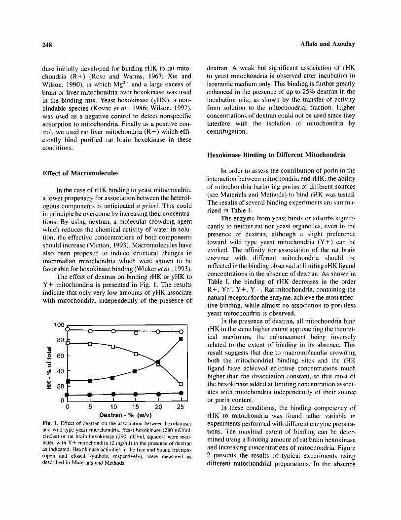
In the case of rHK binding to yeast mitochondria,a lower propensity for association between the heterol-ogous components is anticipated a priori. This couldin principle be overcome by increasing their concentra-tions. By using dextran, a molecular crowding agentwhich reduces the chemical activity of water in solu-tion, the effective concentrations of both componentsshould increase (Minton, 1993). Macromolecules havealso been proposed to induce structural changes inmammalian mitochondria which were shown to befavorable for hexokinase binding (Wicker et al., 1993).
The effect of dextran on binding rHK or yHK toY+ mitochondria is presented in Fig. 1. The resultsindicate that only very low amounts of yHK associatewith mitochondria, independently of the presence of
Fig. 1. Effect of dextran on the association between hexokinasesand wild type yeast mitochondria. Yeast hexokinase (280 mU/ml,circles) or rat brain hexokinase (290 mU/ml, squares) were incu-bated with Y+ mitochondria (2 mg/ml) in the presence of dextranas indicated. Hexokinase activities in the free and bound fractions(open and closed symbols, respectively), were measured asdescribed in Materials and Methods.
dextran. A weak but significant association of rHKto yeast mitochondria is observed after incubation inisosmotic medium only. This binding is further greatlyenhanced in the presence of up to 25% dextran in theincubation mix, as shown by the transfer of activityfrom solution to the mitochondrial fraction. Higherconcentrations of dextran could not be used since theyinterfere with the isolation of mitochondria bycentrifugation.
Hexokinase Binding to Different Mitochondria
In order to assess the contribution of porin in theinteraction between mitochondria and rHK, the abilityof mitochondria harboring porins of different sources(see Materials and Methods) to bind rHK was tested.The results of several binding experiments are summa-rized in Table I.
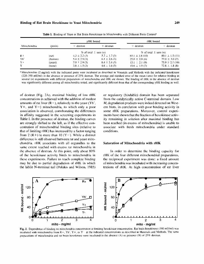
The enzyme from yeast binds or adsorbs signifi-cantly to neither rat nor yeast organelles, even in thepresence of dextran, although a slight preferencetoward wild type yeast mitochondria (Y + ) can beevoked. The affinity for association of the rat brainenzyme with different mitochondria should bereflected in the binding observed at limiting rHK ligandconcentrations in the absence of dextran. As shown inTable I, the binding of rHK decreases in the orderR+, Yh', Y+, Y-. Rat mitochondria, containing thenatural receptor for the enzyme, achieve the most effec-tive binding, while almost no association to porinlessyeast mitochondria is observed.
In the presence of dextran, all mitochondria bindrHK to the same higher extent approaching the theoret-ical maximum, the enhancement being inverselyrelated to the extent of binding in its absence. Thisresult suggests that due to macromolecular crowdingboth the mitochondrial binding sites and the rHKligand have achieved effective concentrations muchhigher than the dissociation constant, so that most ofthe hexokinase added at limiting concentration associ-ates with mitochondria independently of their sourceor porin content.
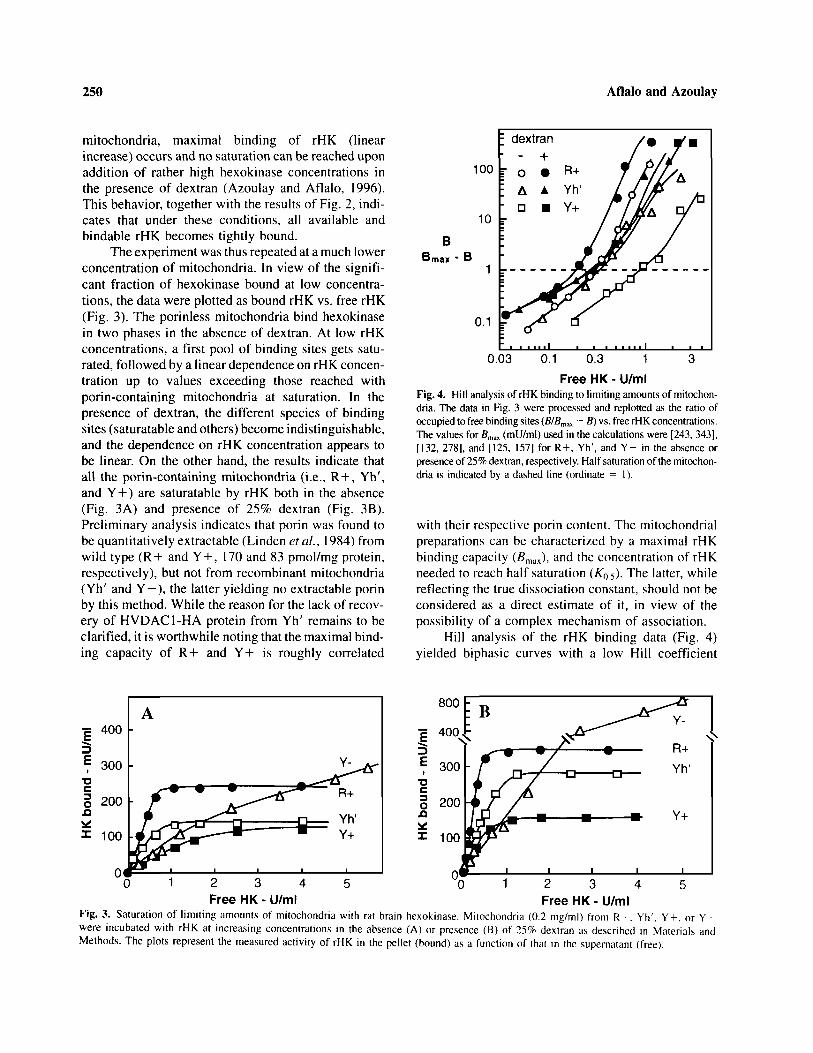
In these conditions, the binding competency ofrHK to mitochondria was found rather variable inexperiments performed with different enzyme prepara-tions. The maximal extent of binding can be deter-mined using a limiting amount of rat brain hexokinaseand increasing concentrations of mitochondria. Figure2 presents the results of typical experiments usingdifferent mitochondrial preparations. In the absence
Binding of Rat Brain Hexokinase to Yeast Mitochondria 249
of dextran (Fig. 2A), maximal binding of low rHKconcentrations is achieved with the addition of modestamounts of rat liver (R + ), relatively to the yeast (Yh',Y + , and Y—) mitochondria, to which only a poorassociation is observed, corroborating the differencesin affinity suggested in the screening experiments inTable I. In the presence of dextran, the binding curvesare strongly shifted to the left, as if the effective con-centration of mitochondrial binding sites (relative tothat of limiting rHK) has increased by a factor rangingfrom 2 (R + ) to more than 10 (Y-). While a distinctdifference is still observed between rat and yeast mito-chondria, rHK associates with all organelles to thesame extent reached with excess rat mitochondria inthe absence of dextran. At this point, only about 80%of the hexokinase activity binds to mitochondria inthese experiments. Failure to reach complete bindingmay be due to partial degradation of rHK in whichthe labile N-terminal tail (Polakis and Wilson, 1985)
or regulatory (bindable) domain has been separatedfrom the catalytically active C-terminal domain. LowMr degradation products were indeed detected on West-ern blots, in correlation with poor binding activity insome rHK preparations. Moreover, control experi-ments have shown that the fraction of hexokinase activ-ity remaining in solution after maximal binding hasbeen reached (in excess of mitochondria) is unable toassociate with fresh mitochondria under standardconditions.
Saturation of Mitochondria with rHK
In order to determine the binding capacity forrHK of the four different mitochondrial preparations,the reciprocal experiment was done: a fixed amountof mitochondria was incubated with increasing concen-trations of rHK. At high concentration of rat liver
Fig. 2. Dependence of binding on mitochondria concentration at limiting hexokinase concentration. Rat brain hexokinase (190 mU/ml) wasincubated with mitochondria from R + , Yh', Y + , or Y- at the indicated concentrations as described in Materials and Methods. The samepreparations of mitochondria and rat brain hexokinase were incubated in the absence (A) or presence (B) of 25% dextran.
Table I. Binding of Yeast or Rat Brain Hexokinase to Mitochondria with Different Porin Contenta
Mitochondria
R +Yh'Y +Y–
(porin)
(rat)(human)(yeast)(none)
yHK bound
– dextran + dextran
% of total ± sem (n)4.3 ± 2.2 (3)5.4 ± 2.9 (3)7.0 ± 2.9 (3)3.8 ± 1.5 (3)
5.7 ± 1.7 (3)6.1 ± 2.6 (3)8.4 ± 2.4 (3)4.6 ± 1 .6 (3)
rHK bound
- dextran + dextran
% of total ± sem (n)65.1 ± 3.8 (10)25.0 ± 2.9 (6)15.1 ± 2.1 (9)10.6 ± 1.9 (7)
80.1 ± 1.5 (11)77.0 ± 3.0 (7)75.9 ± 2.1 (10)72.8 ± 1.8 (8)
a Mitochondria (2 mg/ml) with the indicated porin were incubated as described in Materials and Methods with the indicated hexokinase(220-350 mU/ml) in the absence or presence of 25% dextran. The average and standard error of the mean (sem) for relative binding inseveral (n) experiments with different preparations of mitochondria and rHK are shown. The binding of rHK in the absence of dextranwas significantly different among all mitochondria tested, and significantly different from that of the corresponding yHK binding as well.
250 Aflalo and Azoulay
mitochondria, maximal binding of rHK (linearincrease) occurs and no saturation can be reached uponaddition of rather high hexokinase concentrations inthe presence of dextran (Azoulay and Aflalo, 1996).This behavior, together with the results of Fig. 2, indi-cates that under these conditions, all available andbindable rHK becomes tightly bound.
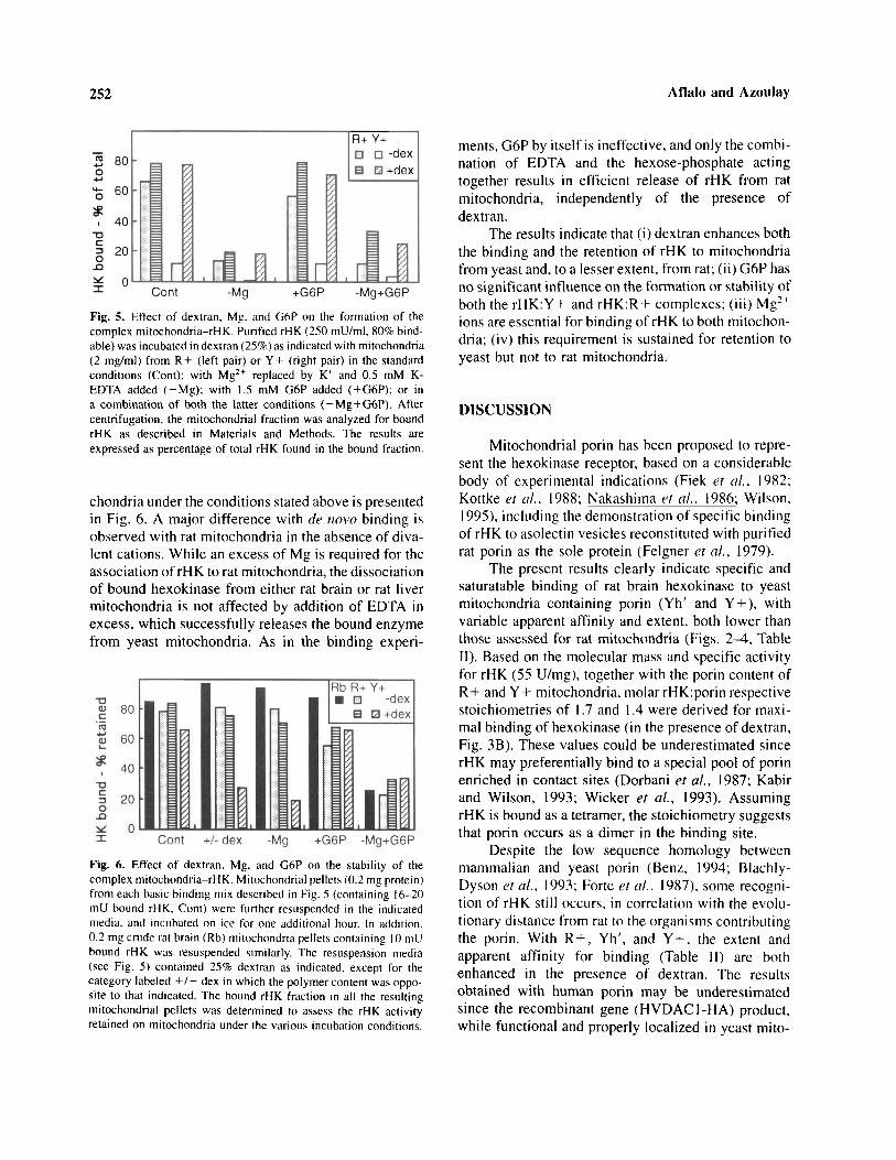
The experiment was thus repeated at a much lowerconcentration of mitochondria. In view of the signifi-cant fraction of hexokinase bound at low concentra-tions, the data were plotted as bound rHK vs. free rHK(Fig. 3). The porinless mitochondria bind hexokinasein two phases in the absence of dextran. At low rHKconcentrations, a first pool of binding sites gets satu-rated, followed by a linear dependence on rHK concen-tration up to values exceeding those reached withporin-containing mitochondria at saturation. In thepresence of dextran, the different species of bindingsites (saturatable and others) become indistinguishable,and the dependence on rHK concentration appears tobe linear. On the other hand, the results indicate thatall the porin-containing mitochondria (i.e., R+, Yh',and Y+) are saturatable by rHK both in the absence(Fig. 3A) and presence of 25% dextran (Fig. 3B).Preliminary analysis indicates that porin was found tobe quantitatively extractable (Linden et al., 1984) fromwild type (R+ and Y + , 170 and 83 pmol/mg protein,respectively), but not from recombinant mitochondria(Yh' and Y–) , the latter yielding no extractable porinby this method. While the reason for the lack of recov-ery of HVDAC1-HA protein from Yh' remains to beclarified, it is worthwhile noting that the maximal bind-ing capacity of R+ and Y+ is roughly correlated
Bmax - B
Fig. 4. Hill analysis of rHK binding to limiting amounts of mitochon-dria. The data in Fig. 3 were processed and replotted as the ratio ofoccupied to free binding sites (B/Bmax — B) vs. free rHK concentrations.The values for Bmax (mU/ml) used in the calculations were [243, 343],[132, 278], and [125, 157] for R + , Yh', and Y+ in the absence orpresence of 25% dextran, respectively. Half saturation of the mitochon-dria is indicated by a dashed line (ordinate = 1).
with their respective porin content. The mitochondrialpreparations can be characterized by a maximal rHKbinding capacity (Bmax), and the concentration of rHKneeded to reach half saturation (K 0 . 5 ) . The latter, whilereflecting the true dissociation constant, should not beconsidered as a direct estimate of it, in view of thepossibility of a complex mechanism of association.
Hill analysis of the rHK binding data (Fig. 4)yielded biphasic curves with a low Hill coefficient
Fig. 3. Saturation of l imit ing amounts of mitochondria with rat brain hexokinase. Mitochondria (0.2 mg/ml) from R + . Yh'. Y + , or Y–were incubated with rHK at increasing concentrations in the absence (A) or presence (B) of 25% dextran as described in Materials andMethods. The plots represent the measured activity of rHK in the pellet (bound) as a function of that in the supernatant (free).

Binding of Rat Brain Hexokinase to Yeast Mitochondria 251
(1.0-1.5) in the low rHK range, and higher (2.5–4) inthe high rHK range for all mitochondria. This behaviorindicates that the mechanism of association is morecomplex than simple cooperative binding (yielding alinear Hill plot). At limiting added rHK (up to 1-3-fold in excess of Bmax), the low apparent Hill coefficientis further reduced (from 1.5 to 1) in the presenceof dextran, suggesting that in these conditions, tightbinding of rHK occurs noncooperatively to indepen-dent binding sites. However, in the presence of a rela-tive excess of rHK ligand, the high Hill coefficient(positive cooperativity in binding) is increased in thepresence of dextran (see Table II). This result supportsthe notion of rHK being bound in an oligomeric formby a cooperative mechanism (Wicker et al., 1993), aspreviously suggested for rat liver mitochondria incross-linking studies (Xie and Wilson, 1990).
The results for the various porin-containing mito-chondrial preparations are summarized in Table II. Inthe absence of dextran, K0.5 increases in the order R + ,Yh', Y+, while the corresponding binding capacity(Bmax) decreases in the same order. In the presence ofdextran, the apparent affinity to rHK increases to reacha similar value (low K0.5), close to that observed inthe homologous system (rHK:R + ) without dextran.This trend is expected, independently of the mecha-nism for binding, as a general effect of macromolecularcrowding (Minton, 1993). Theoretical studies (basedon empirical measurements) predict at 25% dextranan increase in the effective concentration of a largeprotein by about one order of magnitude (Minton,1983). However, while a large increase (ca. 3-fold)in apparent affinity is observed with wild type yeastmitochondria (Y+), it is only moderate (1.3-fold) withmitochondria bearing mammalian porin (R+ and Yh').Moreover, the neutral polymer enhances the bindingcapacity (Bm a x) of all mitochondria, more significantly
for the latter (R+ and Yh') as compared to wild typeyeast (Y+). The apparent Hill coefficient (nH)observed at high [rHK] in the absence of dextran issubstantially lower with yeast mitochondria (Y+ andYh'). In this respect, a distinct effect of dextran isobserved in tightening the cooperativity of binding toyeast mitochondria, with the value of nH determinedfor the heterologous couples rHK:Y+ and rHK:Yh'raised to that observed for the homologous couplerHK:R+.
Formation and Stability of theMitochondria:rHK Complexes
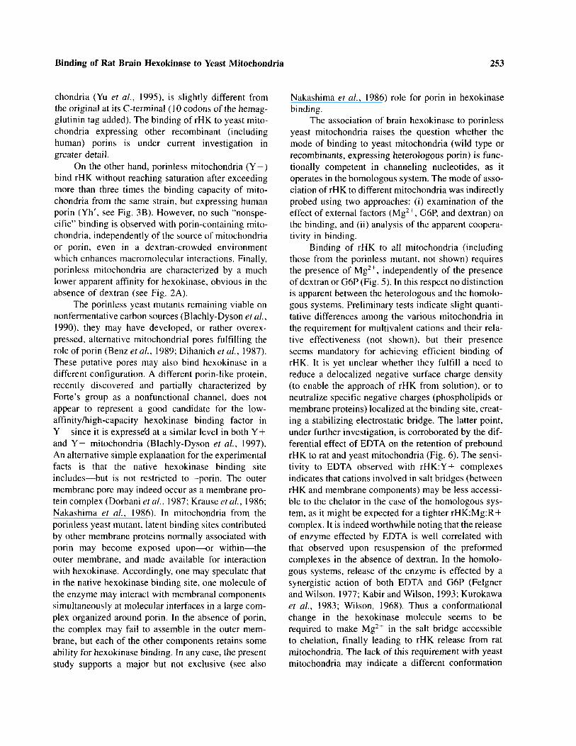
The effect of G6P and EDTA on the binding ofrHK to mitochondria, as well as their influence on thestability of a preformed complex were tested in theabsence and presence of dextran for both yeast andrat mitochondria.
A straightforward comparison of the requirementsfor binding rHK to rat liver (R + ) vs. wild type yeast(Y+) mitochondria is presented in Fig. 5. Besides theselective enhancement by dextran observed with Y+,the presence of divalent cations (either endogenous,removable by EDTA, or added) appears to be a com-mon and absolute requisite for binding under all condi-tions tested. On the other hand, no significant effectof G6P on binding to any mitochondria is detected.Moreover, the addition of the hexose-phosphate didnot strengthen the requirement for Mg in binding (insome cases the effect of EDTA was somewhat attenu-ated). Thus the three factors tested (dextran, Mg, andG6P) appear to operate independently of the bindingprocess with both yeast and rat mitochondria.
A similar comparison for the stability of pre-formed complexes between rHK and different mito-
Table II. Binding Parameters for the Association of Hexokinase to Different Mitochondriad
Mitochondria
R +Yh'Y +
– dextran
Bmaxa (mU/ml)
192 ± 35138 ± 3123 ± 2
K0.5b/ (mU/ml)
261 ± 25341 + 2940 ± 83
nHc
4.0 ± 0.22.6 ± 0.32.7 ± 0.2
+ dextran
Bmaxa (mU/ml)
328 ± 18227 ± 36163 ± 4
K0.5b (mU/ml)
206 ± 29268 ± 21288 ± 28
nHc
4.0 ± 0.24.0 ± 0.33.5 ± 0.2
aBmax is the value for the concentration of mitochondria] binding sites in a 0.2 mg/ml suspension expressed in bound rHK equivalents (mU/ml).b K0.5 is the concentration of free rHK (mU/ml) at which half saturation of the mitochondria is observed.CnH represents the apparent Hi l l coefficient, calculated as the slope of Hill plots in the high rHK concentration range.dThe values, derived graphically from independent data sets (2-3 for each type of mitochondria) similar to those in Fig. 3 (Bmax, K0.5) or
Fig. 4 (nH), are presented as averages with standard errors of the mean.
252 Aflalo and Azoulay
Fig. 5. Effect of dextran, Mg. and G6P on the formation of thecomplex mitochondria-rHK. Purified rHK (250 mU/ml, 80% bind-able) was incubated in dextran (25%) as indicated with mitochondria(2 mg/ml) from R+ (left pair) or Y+ (right pair) in the standardconditions (Cont); with Mg2+ replaced by K+ and 0.5 mM K-EDTA added (-Mg); with 1.5 mM G6P added ( + G6P); or ina combination of both the latter conditions ( –Mg + G6P). Aftercentrifugation, the mitochondrial fraction was analyzed for boundrHK as described in Materials and Methods. The results areexpressed as percentage of total rHK found in the bound fraction.
chondria under the conditions stated above is presentedin Fig. 6. A major difference with de novo binding isobserved with rat mitochondria in the absence of diva-lent cations. While an excess of Mg is required for theassociation of rHK to rat mitochondria, the dissociationof bound hexokinase from either rat brain or rat livermitochondria is not affected by addition of EDTA inexcess, which successfully releases the bound enzymefrom yeast mitochondria. As in the binding experi-
Fig. 6. Effect of dextran. Mg, and G6P on the stability of thecomplex mitochondria-rHK. Mitochondrial pellets (0.2 mg protein)from each basic binding mix described in Fig. 5 (containing 16-20mU bound rHK. Cont) were further resuspended in the indicatedmedia, and incubated on ice for one additional hour. In addition.0.2 mg crude rat brain (Rb) mitochondria pellets containing 10 mUbound rHK was resuspended similarly. The resuspension media(see Fig. 5) contained 25% dextran as indicated, except for thecategory labeled +/– dex in which the polymer content was oppo-site to that indicated. The bound rHK fraction in all the resultingmitochondrial pellets was determined to assess the rHK activityretained on mitochondria under the various incubation conditions.
ments, G6P by itself is ineffective, and only the combi-nation of EDTA and the hexose-phosphate actingtogether results in efficient release of rHK from ratmitochondria, independently of the presence ofdextran.
The results indicate that (i) dextran enhances boththe binding and the retention of rHK to mitochondriafrom yeast and, to a lesser extent, from rat; ( i i ) G6P hasno significant influence on the formation or stability ofboth the rHK:Y+ and rHK:R+ complexes; ( i i i ) Mg2 +
ions are essential for binding of rHK to both mitochon-dria; (iv) this requirement is sustained for retention toyeast but not to rat mitochondria.
DISCUSSION
Mitochondrial porin has been proposed to repre-sent the hexokinase receptor, based on a considerablebody of experimental indications (Fiek et al., 1982;Kottke et al., 1988; Nakashima et al., 1986; Wilson,1995), including the demonstration of specific bindingof rHK to asolectin vesicles reconstituted with purifiedrat porin as the sole protein (Feigner et al., 1979).
The present results clearly indicate specific andsaturatable binding of rat brain hexokinase to yeastmitochondria containing porin (Yh' and Y+), withvariable apparent affinity and extent, both lower thanthose assessed for rat mitochondria (Figs. 2–4, TableII). Based on the molecular mass and specific activityfor rHK (55 U/mg), together with the porin content ofR+ and Y+ mitochondria, molar rHK:porin respectivestoichiometries of 1.7 and 1.4 were derived for maxi-mal binding of hexokinase (in the presence of dextran.Fig. 3B). These values could be underestimated sincerHK may preferentially bind to a special pool of porinenriched in contact sites (Dorbani et al., 1987; Kabirand Wilson, 1993; Wicker et al., 1993). AssumingrHK is bound as a tetramer, the stoichiometry suggeststhat porin occurs as a dimer in the binding site.
Despite the low sequence homology betweenmammalian and yeast porin (Benz, 1994; Blachly-Dyson et al., 1993; Forte et al., 1987), some recogni-tion of rHK still occurs, in correlation with the evolu-tionary distance from rat to the organisms contributingthe porin. With R + , Yh', and Y + , the extent andapparent affinity for binding (Table II) are bothenhanced in the presence of dextran. The resultsobtained with human porin may be underestimatedsince the recombinant gene (HVDAC1-HA) product,while functional and properly localized in yeast mito-
Binding of Rat Brain Hexokinase to Yeast Mitochondria 253
chondria (Yu et al., 1995), is slightly different fromthe original at its C-terminal (10 codons of the hemag-glutinin tag added). The binding of rHK to yeast mito-chondria expressing other recombinant (includinghuman) porins is under current investigation ingreater detail.
On the other hand, porinless mitochondria (Y–)bind rHK without reaching saturation after exceedingmore than three times the binding capacity of mito-chondria from the same strain, but expressing humanporin (Yh', see Fig. 3B). However, no such "nonspe-cific" binding is observed with porin-containing mito-chondria, independently of the source of mitochondriaor porin, even in a dextran-crowded environmentwhich enhances macromolecular interactions. Finally,porinless mitochondria are characterized by a muchlower apparent affinity for hexokinase, obvious in theabsence of dextran (see Fig. 2A).
The porinless yeast mutants remaining viable onnonfermentative carbon sources (Blachly-Dyson et al.,1990), they may have developed, or rather overex-pressed, alternative mitochondrial pores fulfilling therole of porin (Benz et al., 1989; Dihanich et al., 1987).These putative pores may also bind hexokinase in adifferent configuration. A different porin-like protein,recently discovered and partially characterized byForte's group as a nonfunctional channel, does notappear to represent a good candidate for the low-affinity/high-capacity hexokinase binding factor inY- since it is expressed at a similar level in both Y+and Y- mitochondria (Blachly-Dyson et al., 1997).An alternative simple explanation for the experimentalfacts is that the native hexokinase binding siteincludes—but is not restricted to—porin. The outermembrane pore may indeed occur as a membrane pro-tein complex (Dorbani et al., 1987; Krause et al., 1986;Nakashima et al., 1986). In mitochondria from theporinless yeast mutant, latent binding sites contributedby other membrane proteins normally associated withporin may become exposed upon—or within—theouter membrane, and made available for interactionwith hexokinase. Accordingly, one may speculate thatin the native hexokinase binding site, one molecule ofthe enzyme may interact with membranal componentssimultaneously at molecular interfaces in a large com-plex organized around porin. In the absence of porin,the complex may fail to assemble in the outer mem-brane, but each of the other components retains someability for hexokinase binding. In any case, the presentstudy supports a major but not exclusive (see also
Nakashima et al., 1986) role for porin in hexokinasebinding.
The association of brain hexokinase to porinlessyeast mitochondria raises the question whether themode of binding to yeast mitochondria (wild type orrecombinants, expressing heterologous porin) is func-tionally competent in channeling nucleotides, as itoperates in the homologous system. The mode of asso-ciation of rHK to different mitochondria was indirectlyprobed using two approaches: (i) examination of theeffect of external factors (Mg2+, G6P, and dextran) onthe binding, and (ii) analysis of the apparent coopera-tivity in binding.
Binding of rHK to all mitochondria (includingthose from the porinless mutant, not shown) requiresthe presence of Mg2+, independently of the presenceof dextran or G6P (Fig. 5). In this respect no distinctionis apparent between the heterologous and the homolo-gous systems. Preliminary tests indicate slight quanti-tative differences among the various mitochondria inthe requirement for multivalent cations and their rela-tive effectiveness (not shown), but their presenceseems mandatory for achieving efficient binding ofrHK. It is yet unclear whether they fulfill a need toreduce a delocalized negative surface charge density(to enable the approach of rHK from solution), or toneutralize specific negative charges (phospholipids ormembrane proteins) localized at the binding site, creat-ing a stabilizing electrostatic bridge. The latter point,under further investigation, is corroborated by the dif-ferential effect of EDTA on the retention of preboundrHK to rat and yeast mitochondria (Fig. 6). The sensi-tivity to EDTA observed with rHK:Y+ complexesindicates that cations involved in salt bridges (betweenrHK and membrane components) may be less accessi-ble to the chelator in the case of the homologous sys-tem, as it might be expected for a tighter rHK:Mg:R+complex. It is indeed worthwhile noting that the releaseof enzyme effected by EDTA is well correlated withthat observed upon resuspension of the preformedcomplexes in the absence of dextran. In the homolo-gous systems, release of the enzyme is effected by asynergistic action of both EDTA and G6P (Feignerand Wilson, 1977; Kabir and Wilson, 1993; Kurokawaet al., 1983; Wilson, 1968). Thus a conformationalchange in the hexokinase molecule seems to berequired to make Mg2+ in the salt bridge accessibleto chelation, finally leading to rHK release from ratmitochondria. The lack of this requirement with yeastmitochondria may indicate a different conformation
254 Aflalo and Azoulay
for the bound rHK, which should be reflected in thebinding process.
Titration of porin-containing mitochondria withrHK occurs as a biphasic process with respect to theapparent cooperativity (Fig. 4). With all mitochondria,a transition from low to high Hill coefficient isobserved at limiting and excess rHK, respectively. Thehomologous system (rHK:R+) is characterized by aHill coefficient of 4, concurring with the proposal thatrHK may be bound in a tetrameric form (Xie andWilson, 1990). However, lower apparent cooperativity(nH = 2.5) is observed for binding to yeast mitochon-dria (heterologous systems), which occurs at lowerapparent affinity, suggesting that the mode of bindingmay vary with the source of the organelles. Neverthe-less, with porin-containing yeast mitochondria in thepresence of dextran, both the apparent affinity to rHKand the cooperativity of its binding reach values closeto those obtained in the homologous system (Table II).Similar effects for dextran were observed with bindingto intact rat mitochondria, but not to reconstituted outermembrane vesicles nor to uncoupled mitochondria(Wicker et al., 1993). Taken together with our results,these facts point toward an important role for a definitestructural organization of the mitochondrial compo-nents in binding rHK. Indeed, the transition from non-cooperative to cooperative binding (curved Hill plots)does not support a static conformation for the mito-chondrial binding sites, nor a mechanism involvingoligomerization of rHK in solution as a required stepbefore binding can take place. In the latter case onewould expect macromolecular crowding to result inan increase of the apparent nH in the low [rHK] range,rather than the observed decrease. We propose thatbinding of the first rHK molecule effects a chain ofstructural changes in which (i) the formation of a com-plex between mitochondrial membrane proteins is ini-tiated; (ii) the resulting complexes bind cooperativelyadditional rHK molecules. The structural changes mayalso involve the fusion between the inner and outermembranes, as well as rearrangement of lipids (cardio-lipin and cholesterol) around the binding site. The latterconsiderations represent possible sources for the subtledifference in binding rHK to yeast vs. rat mitochondria.
Diffusional restrictions on nucleotides have beenimplicated in many cellular processes (Aflalo, 1991;Aflalo and Shavit, 1983; Aw and Jones, 1985; Saks etal., 1993), among which oxidative phosphorylationin yeast mitochondria coupled to yeast hexokinase insolution has been modeled (Aflalo and Segel, 1992)and experimentally tested (Aflalo, 1997) using recom-binant firefly luciferase localized at the outer mito-
chondrial membrane in yeast (Aflalo, 1990). The studyof nucleotides channeling between the matrix and rHKat the outer membrane of yeast mitochondria by anengineered local probe represents a powerful experi-mental model system to reexamine cellular processesin situ.
CONCLUSIONS
We show that rat brain hexokinase binds in signif-icant amounts to yeast mitochondria. The affinity andextent of binding depend on the origin of porin, andboth are enhanced in the presence of dextran. Porinlessmitochondria also bind hexokinase, but this occurs by adifferent mechanism. These preliminary data reveal arather complex nature for the interaction between mam-malian brain hexokinase and mitochondria. Many physi-cochemical factors are involved in the associationbetween mammalian hexokinase and mitochondria,among which porin plays a major but not exclusive rolewhich can be addressed by molecular studies conductedin yeast.
ACKNOWLEDGMENTS
We wish to thank Michael Forte for the gift of theM22-2 yeast strain and the yeast expression plasmidcontaining the HVDAC1-HA gene, as well as John EWilson for his helpful advice and discussion of theresults. This research was supported by grant No. 95-110 from the US-Israel Binational Science Founda-tion (BSF).
REFERENCES
Aflalo, C. (1990). Biochemistry 29, 4758–4766.Aflalo, C. (1991). Int. Rev. Cytol. 130, 269-323.Aflalo, C. (1997). J. Bioenerg. Biomembr. 29, 549-559.Aflalo, C., and Segel, L. A. (1992). J. Theor. Biol. 158, 67-108.Aflalo, C., and Shavit, N. (1983). FEBS Lett. 154, 175–179.Arora, K. K., and Pedersen, P. L. (1988). J. Biol. Chem. 263,
17422–17428.Arora, K. K., Filburn. C. R., and Pedersen. P. L. (1993). J. Biol.
Chem. 268, 18259–18266.Aw, T. Y., and Jones, D. P. (1985). Am. J. Physiol. 249, C385-C392.Azoulay, H.. and Aflalo, C. (1996). In BioThermoKinetics of the
Living Cell (Westerhoff, H. V., Snoep, J. L., Sluse, F. E., Wijker,J. E., and Kholodenko, B. N., eds.), BioThermoKinetics Press,Amsterdam, pp 289–294.
Beltran del Rio, H., and Wilson, J. E. (1992). Arch. Biochem.Biophys. 296, 667-677.
Benz, R. (1994). Biochim. Biophys. Ada 1197, 167–196.
Binding of Rat Brain Hexokinase to Yeast Mitochondria 255
Benz. R., Schmid, A., and Dihanich, M. (1989). J. Bioenerg. Bio-membr. 21, 349–450.
Blachly-Dyson, E., Peng, S., Colombini, M., and Forte, M. (1990).Science 247, 1233-1236.
Blachly-Dyson, E., Zambronicz, E. B., Yu, W. H., Adams, V.,McCabe, E. R., Adelman. J., Colombini, M., and Forte, M.(1993). J. Biol. Chem. 268, 1835-1841.
Blachly-Dyson, E., Song, J., Wolfgang, W. J., Colombini, M., andForte, M. (1997). Mol. Cell. Biol. 17, 5727-5738.
Bradford, M. (1976). Anal. Biochem. 72, 248-254.Brdiczka, D. (1994). Biochim. Biophys. Acta 1187, 264–269.Bustamate, E., Morris, H. P., and Pedersen, P. L. (1981). J. Biol.
Chem. 256, 8699-8707.De Pinto, V, Prezioso, G., Thinnes, F, Link, T. A., and Palmieri,
F. (1991). Biochemistry 30, 10191-10200.Dihanich, M., Suda, K., and Schatz, G. (1987). EMBO J. 6, 723-728.Dorbani, L., Jancsik, V., Linden, M., Leterrier, J., Nelson, B., and
Rendon. A. (1987). Arch. Biochem. Biophys. 252, 188–196.Feigner, P., and Wilson, J. (1977). Arch. Biochem. Biophys. 182.
282-294.Feigner, P. L., Messer, J. L., and Wilson, J. E. (1979). J. Biol.
Chem. 254, 4946–4949.Fiek. C., Benz, R., Roos. N., and Brdiczka, D. (1982). Biochim.
Biophys. Acta 688, 429–440.Forte, M., Guy, H. R., and Mannella, C. A. (1987). J. Bioenerg.
Biomembr. 19, 341–350.Gellerich, F. N., Wagner, M., Kapischke, M., Wicker, U., and Brdic-
zka, D. (1993). Biochim. Biophys. Ada 1142, 217-227.Hase, T., Mullen U., Riezman, H., and Schatz, G. (1984). EMBO
J. 3, 3157-3164.Hovious, R., Lambrechts, H.. Nicolay, K., and De Kruijff, B. (1990).
Biochim. Biophys. Acta 1021, 217-226.Inui. M., and Ishibashi. S. (1979). ./. Biochem. (Tokyo) 85.
1151–1156 .Kabir. F., and Wilson, J. E. (1993). Arch. Biochem. Biophys.
300, 641–650.Kottke. M.,3 Adam, V., Riesinger. I., Bremm, G., Bosch, W., Brdic-
zka. D., Sandri. G., and Panfili, E. (1988). Biochim. Biophys.Acta 935, 87-102.
Kovac, L., Nelson, B. D., and Ernster, L. (1986). Biochem. Biophys.Res. Commun. 134, 285–291.
Krause, J., Hay, R., Kowollik, C., and Brdiczka, D. (1986). Biochim.Biophys. Acta 860, 690–698.
Kurokawa, M., Yokoyama, K., and Ishibashi, S. (1983). Biochim.Biophys. Acta 759, 92–98.
Laterveer, F. D., Van Der Heijden, R., Toonen, M., and Nicolay,K. (1994). Biochim. Biophys. Acta 1188, 251–259.
Laterveer, F. D., Gellerich. F. N., and Nicolay, K. (1995). Eur. J.Biochem. 232, 569-577.
Linden, M., Andersson, G., Gellerfors, P., and Nelson, B. D. (1984).Biochim. Biophys. Acta 770, 93-96.
Mannella, C. A., Forte, M., and Colombini, M. (1992). J. Bioenerg.Biomembr. 24, 7–19.
McCabe, E. R. (1994). J. Bioenerg. Biomembr. 26, 317-325.Minton, A. (1983). Mol. Cell. Biochem. 55, 119-140.Minton, A. (1993). J. Mol. Recogn. 6, 211-214.Nakashima, R. A., Mangan, P. S., Colombini, M., and Pedersen,
P. L. (1986). Biochemistry 25, 1015-1021.Nicolay, K., Rojo, M., Wallimann, T., Demel, R., and Hovius, R.
(1990). Biochim. Biophys. Acta 1018, 229-233.Polakis, P. G., and Wilson, J. E. (1985). Arch. Biochem. Biophys.
236, 328-337.Rauch. G., and Moran, O. (1994). Biochem. Biophys. Res. Commun.
200, 908–915.Rose, I., and Warms, J. (1967). J. Biol. Chem. 242, 1635-1645.Saks, V. A., Vasil'eva, E., Belikova, Y. O., Kuznetsov, A. V., Lya-
pina, S., Petrova, L., and Perov, N. A. (1993). Biochim. Bio-phys. Acta 1144, 134-148.
Tsai, H. J., and Wilson, J. E. (1995). Arch. Biochem. Biophys.316, 206–214.
White, T. K., and Wilson, J. E. (1989). Arch. Biochem. Biophys.274, 375-393.
Wicker, U., Bucheler, K., Gellerich, F. N., Wagner, M., Kapischke,M., and Brdiczka, D. (1993). Biochim. Biophys. Acta 1142,228-239.
Wilson, J. (1968). J. Biol. Chem. 243, 3640–3647.Wilson, J. E. (1989). Prep. Biochem. 19, 13–21.Wilson. J. E. (1995). Rev. Physiol. Biochem. Pharmacol. 126,
65–198.Wilson. J. E. (1997). J. Bioenerg. Biomembr. 29, 97-102.Xie, G. C., and Wilson, J. E. (1988). Arch. Biochem. Biophys.
267, 803-810.Xie, G., and Wilson, J. E. (1990). Arch. Biochem. Biophys. 276,
285-293.Yu, W. H., Wolfgang, W., and Forte, M. (1995). J. Biol. Chem.
270, 13998-14006.


