Embed Size (px)
Citation preview
Henriques et al. Parasites & Vectors 2012, 5:214http://www.parasitesandvectors.com/content/5/1/214
RESEARCH Open Access
Bioluminescent imaging of Trypanosoma cruziinfection in Rhodnius prolixusCristina Henriques1, Daniele P Castro2, Leonardo HF Gomes3, Eloi S Garcia2 and Wanderley de Souza1,4*
Abstract
Background: Usually the analysis of the various developmental stages of Trypanosoma cruzi in the experimentallyinfected vertebrate and invertebrate hosts is based on the morphological observations of tissue fragments fromanimals and insects. The development of techniques that allow the imaging of animals infected with parasitesexpressing luciferase open up possibilities to follow the fate of bioluminescent parasites in infected vectors.
Methods: D-luciferin (60 μg) was injected into the hemocoel of the whole insect before bioluminescenceacquisition. In dissected insects, the whole gut was incubated with D-luciferin in PBS (300 μg/ml) for ex vivobioluminescence acquisition in the IVISW Imaging System, Xenogen.
Results: Herein, we describe the results obtained with the luciferase gene integrated into the genome of theDm28c clone of T. cruzi, and the use of these parasites to follow, in real time, the infection of the insect vectorRhodnius prolixus, by a non- invasive method. The insects were evaluated by in vivo bioluminescent imaging on thefeeding day, and on the 7 th, 14 th, 21 st and 28 th days after feeding. To corroborate the bioluminescent imagingmade in vivo, and investigate the digestive tract region, the insects were dissected. The bioluminescence emittedwas proportional to the number of protozoans in regions of the gut. The same digestive tracts were also maceratedto count the parasites in distinct morphological stages with an optical microscope, and for bioluminescenceacquisition in a microplate using the IVISW Imaging System. A positive correlation of parasite numbers andbioluminescence in the microplate was obtained.
Conclusions: This is the first report of bioluminescent imaging in Rhodnius prolixus infected with trypomastigotesof the Dm28c-luc stable strain, expressing firefly luciferase. In spite of the distribution limitations of the substrate(D-luciferin) in the insect body, longitudinal evaluation of infected insects by bioluminescent imaging is a valuabletool. Bioluminescent imaging of the digestive tract infected with Dm28c-luc is highly sensitive and accuratemethod to track the fate of the parasite in the vector, in the crop, intestine and rectum. This methodology is usefulto gain a better understanding of the parasite – insect vector interactions.
Keywords: Trypanosoma cruzi, Rhodnius prolixus, Chagas diseases, Bioluminescent imaging, Bioluminescence,Luciferase
BackgroundChagas disease is an endemic parasitic disease that isranked as one of the most important in several areas ofLatin America. Triatomine bugs are vectors of the para-site Trypanosoma cruzi, the causative agent of Chagasdisease. The gut of the vector Rhodnius prolixus is
* Correspondence: [email protected]ório de Ultraestrutura Celular Hertha Meyer, UFRJ, Rio de Janeiro, RJ,Brazil4Instituto Nacional de Metrologia, Qualidade e Tecnologia, Inmetro, Duquede Caxias Xerém, RJ, BrazilFull list of author information is available at the end of the article
© 2012 Henriques et al.; licensee BioMed CenCreative Commons Attribution License (http:/distribution, and reproduction in any medium
basically comprised of three major regions, the foregut(stomach or crop), the midgut (intestine), and the hind-gut (rectum); however more elaborate divisions havebeen proposed [1,2]. These different regions are import-ant for the development of the parasite [3,4]. The vectorhost digestion starts when the insect ingests blood fromthe vertebrate host that is then stored in the crop whereit is concentrated and hemolyzed. Subsequently, it passesinto the intestine for digestion and absorption, followedby storage of the non absorbed products in the rectumand subsequent release. The remaining compounds are
tral Ltd. This is an Open Access article distributed under the terms of the/creativecommons.org/licenses/by/2.0), which permits unrestricted use,, provided the original work is properly cited.
Henriques et al. Parasites & Vectors 2012, 5:214 Page 2 of 15http://www.parasitesandvectors.com/content/5/1/214
swept by the urine and feces that are secreted rapidlyafter new blood ingestion [2,5].T. cruzi development begins inside the insect vector as
soon as the insect has fed on the blood of an infectedhost. In the crop, most of the trypomastigotes differenti-ate into epimastigotes, the main replicative stage. A sig-nificant proportion of parasites are lysed in part due tothe interaction with bacteria found in the crop micro-biota [6]. Subsequently the remaining epimastigotes mi-grate to the intestine where they proliferate and adhereto the perimicrovillar membranes. Then the epimasti-gotes transform into the non-replicative, but infectivemetacyclic trypomastigotes, which are released in theurine and feces [2,5,7,8].Until now the study of the distribution of T. cruzi in-
side the insect vector has been based on the microscopicexamination of the digestive tract following dissection ofthe insects. However, recently the use of bioluminescentimaging of the whole animal has shown that it is pos-sible to follow the parasite in vivo within their mamma-lian hosts if the parasites are labeled with luciferase[9,10]. In the present work we describe the resultsobtained with the integration of the luciferase gene inthe genome of the Dm28c clone of T. cruzi, and the useof the labeled parasites to follow, in real time, the infec-tion of the invertebrate vector R. prolixus. We observedthe infected insects for one month after feeding/infec-tion, and the light emitted could be traced continuouslywithin the insect. We also analyzed insects that were dis-sected to expose the digestive tract. This methodology willbe useful for further studies to acquire a better under-standing of the parasite – insect vector interactions.
MethodsExpression of firefly luciferase (Fluc) in Trypanosoma cruziThe luciferase gene was amplified by PCR using specificprimers. The forward primer contains an XbaI site andthe Kozak sequence (underlined), upstream of the startcodon, 50- GCTCTAGA GCCACC ATGGAAGACGCCAAAAACATAAAG – 30 (F-luc), and the reverse primercontains an XhoI site (underlined) 5
0- CCGCTCGAG
CGGTTACACGGCGATCTTTCC- 30 (R-luc). Amplifica-tion was carried out using the Tli DNA Polymerase (Pro-mega) and the following PCR conditions: 94oC, 5 min;94oC, 30 sec; 60oC, 30 sec; 72oC, 2 min, 30 cycles; 72oC,10 min. The PCR product was cloned into the Zero BluntTOPO PCR Cloning Kit (Invitrogen), digested from theTOPO vector and subcloned into the integrative pTREXvector at the XbaI and XhoI restriction sites [11]. Theconstruction was sequenced on an ABI 3730 GeneticAnalyzer (Applied Biosystems), using the sequencing plat-form form PDTIS/FIOCRUZ.T. cruzi epimastigotes of Dm28c clone were suspended
at 1 x 10⁸ cells/mL in electroporation buffer (EPB)
containing 137 mM NaCl; 5 mM KCl; 0.7 mMNa2HPO4; 6 mM glucose; 21 mM HEPES, pH 7.3. Thecellular suspension (400 μl) was mixed with 50 μg ofplasmid, digested with NheI, placed in a 0.2 cm cuvetteand subjected to a pulse of 0.45 kV, 500 μF at roomtemperature in a Gene Pulser apparatus (BioRad Labora-tories) [12]. The parasites were re-suspended in LITmedium and stable transformants were selected with200 to 500 μg/mL of G418. Thereafter, the high expres-sing epimastigotes were selected by serial dilution in a96 well plate, and selected by bioluminescent emissionwith Steady-Glo reagent (Promega) in a microplatereader SpectraMax2.
Genomic southern blotTo examine the integration of the Fluc gene and thepTREX construction in the T. cruzi genome we per-formed Southern blot of the epimastigote genomicDNA. Epimastigotes (108) were lysed with 1 ml of buffer(10 mM Tris–HCl, pH7.5, 100 mM NaCl, 0.5% SDS 25mM EDTA and 0.1 mg/ml of proteinase K). The DNAwas isolated by phenol:chloroform extraction and recov-ered by ethanol precipitation. Genomic DNA from thewild type Dm28c and from the genetically modifiedDm28c-luc strain were digested with EcoRI restrictionenzyme, using the standard protocol, then the restrictionfragments were separated by electrophoresis in agarosegel and transferred to a positively charged nylon mem-brane with a high salt buffer.To produce the probe, pTREX-luc plasmid was
digested with NheI and XhoI. A fragment of 2.2 kb, con-taining the neo gene and the gapdh intergenic region,was agarose gel purified and 1 μg was used as templateto produce the probe with ready-to-go DNA labelingbeads (GE Healthcare). Unlabeled dCTP was added tothe reaction mixture according to the manufacturer’sprotocol, and incubated for 24 hours. The length of theprobe ranged from 200 to 1000 bp; unincorporatednucleotides and reaction buffer were removed by theWizardW SV Gel and PCR Clean-Up System (Promega).The probe was labeled with alkaline phosphatase by theGene Images AlkPhos Direct Labeling and DetectionSystem (GE Healthcare) and 50 ng of labeled probe wasadded to 30 ml of hybridization buffer in a bottle con-taining the nylon membrane with immobilized DNA,and was then incubated in a hybridization oven for 48hours at 55oC. After hybridization the blots were washedfollowing the manufacturer recommendations. Thewashed blots were placed directly into the detection sys-tem protocol, using the CDP-Star chemiluminescent de-tection reagent, which uses the probe-bound alkalinephosphatase to catalyze the decomposition of a stabi-lized dioxetane substrate (GE Healthcare). Autoradiog-raphy films were exposed from 10 minutes to 2 hours.
Henriques et al. Parasites & Vectors 2012, 5:214 Page 3 of 15http://www.parasitesandvectors.com/content/5/1/214
PCR and sequencing to identify the pTREX-luc integrationTo identify the integration of the pTREX-luc construc-tion in the genome, a couple of primers were designedto align the transcription start point, tsp1, of the riboso-mal promoter from pTREX, F-TSP1- 50TCATGGAGCGGTATTCTC-30 and R-TSP1 50GAGAATACCGCTCCATGA-3,and another pair of primers to align the ribosomal locusrecombination site [13], F-RS pTREX- 50-GTCCGAACGCGGAAATGT-30 and R-RS pTREX- 50-ACATTTCCGCGTTCGGAC-30. PCR amplifications were performedusing genomic DNA from Dm28c-luc as a template and aset of primers: 1- F-TSP1/R-LUC; 2- F-RS pTREX/R-LUCand; F-LUC/R-LUC using the following settings: 94oC, 2min; 94oC, 30 sec; 60oC, 30 sec; 68oC, 2 min, 30 cycles;72oC, 10 min and the Platinum Taq DNA PolymeraseHigh Fidelity (Invitrogen). The PCR fragments wereresolved by 1% agarose gel in Tris-Acetate- EDTA (TAE)buffer, stained with ethidium bromide and gel purified bythe WizardW SV Gel and PCR Clean-Up System (Pro-mega). The PCR products were sequenced with primersspecific for the ribosomal promoter region: F-TSP1; R-TSP1; F-RS pTREX; R-RS pTREX, on an ABI 3730 Gen-etic Analyzer (Applied Biosystems), using the sequencingplatform from PDTIS/FIOCRUZ.
Parasite cultivationGenetically modified epimastigotes of T. cruzi (Dm28c-luc), expressing luciferase, and wild type (Dm28c clone)were cultivated in liver infusion tryptose (LIT) mediumwith 10% fetal calf serum, at 28oC until the logarithmicstage of growth [14]. Epimastigotes of Dm28c-luc werecultivated in the same medium, but supplemented withG418 200 μg/ml. The non-infective and replicative epi-mastigotes were transformed into non-dividing and in-fective metacyclic trypomastigotes. This is a processknown as metacyclogenesis, which is carried out by sub-jecting T. cruzi epimastigotes from the late exponentialgrowth phase at a cell density of 3 x 10⁷ cells/ml to nu-tritional stress in a triatomine artificial urine (TAU)medium (190 mM NaCl; 8 mM phosphate buffer, pH6.0; 17 mM KCl; 2 mM MgCl2; 2mM CaCl2) for 2 hours.Then, a further incubation in TAU supplementedwith amino acids and glucose (TAU3AAG) (TAUsupplemented with 0.035% sodium bicarbonate,10 mM L-proline, 50 mM sodium glutamate, 2 mMsodium L-aspartate, and 10 mM glucose) [15]. Metacyclicparasites were used to infect the host cell LLCMK2, andtrypomastigotes from the cell culture were used to infectthe insects.
Insect infectionFifth-instar Rhodnius prolixus larvae obtained from ourcolony were used throughout these studies. After molt-ing, insects that had been starved for 15-20 days and
weighed 35.2 ± 3.4 mg, were randomly chosen and thenallowed to feed on defibrinated rabbit blood through amembrane feeding apparatus [4]. Defibrinated rabbitblood used for feeding the insects was provided by theLaboratory Animals Creation Center of Fiocruz (Cecal).All research programs using Cecal respect the guidelinesof the Ethics Committee on Animal Use (Ceua) com-posed of Fiocruz researchers and external consultants. Acontrol group was fed with blood alone and infectedgroups on blood containing ~1 x 10⁷ tissue culture-derived trypomastigotes of T. cruzi Dm28c clone per mlof blood meal. The experimental group was fed withblood infected with genetically modified trypomasti-gotes, Dm28 luc expressing luciferase, at 1.7 x 10⁷ trypo-mastigotes per ml of blood. Only fully gorged insectswere used (180.5 ± 22.1 mg) and partially fed insectswere discarded. All insects were raised and maintainedas previously described [4]. To analyze the insect infec-tion on the feeding day, D-luciferin substrate was givenat 1 mg/mL by oral treatment together with the blood-meal containing the wild type Dm28c or the geneticallymodified Dm28c-luc trypomastigotes and imaged in theIVISW Lumina Imaging System at the Bioimaging Cen-tral Unit/National Institute of Science and the NationalInstitute of Science and Technology for Structural Biol-ogy and Bioimaging INBEB/CENABIOII/UFRJ.
Substrate treatmentFor bioluminescent imaging on days 7, 14, 21 and 28after feeding, the insects were inoculated laterally in thethorax with 2 μl of D-luciferin solution (30 mg/ml) usinga 30 gauge hypodermic needle (BD Precision Glide)adapted to a 10-μl Hamilton syringe. Afterwards, theinsects were immobilized by attaching the dorsal regionwith a double-sided adhesive tape and after 5 min theywere put into the IVISW Lumina Xenogen equipment toacquire bioluminescence. To avoid mortality throughsuccessive inoculations we tested the acquisition of bio-luminescent imaging with D-luciferin topical applica-tions. The insects were immobilized as described aboveand 5 μl of D-luciferin solution (30 mg/ml) was appliedover the insect ventral region. The insects were keptimmobilized for 15 min to let the compound penetratethrough the cuticle before capturing the biolumines-cence. To analyze the parasite migration into the digest-ive tract, the insects were dissected and the organ wasincubated with D-luciferin, 300 μg/mL in PBS, for 5 minin Petri dishes. The digestive tracts were then analyzedwith the bioluminescence equipment, IVISW LuminaXenogen, at the Bioimaging Central Unit of the NationalInstitute of Science and the National Institute of Scienceand Technology for Structural Biology and BioimagingINBEB/CENABIOII/UFRJ .
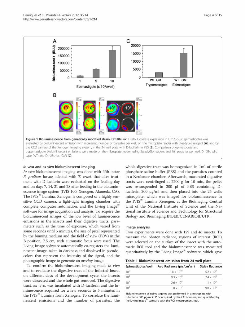
Table 1 Bioluminescent emission from 24 well plate
Epimastigotes/well Avg Radiance (p/s/cm2/sr) Stdev Radiance
105 1.8 x 10¹⁰ 5.2 x 10⁹
104 9.3 x 10⁸ 2.4 x 10⁸
103 2.6 x 10⁸ 1.1 x 10⁸
102 1.8 x 10⁷ 9.8 x 10⁶
Bioluminescence of epimastigotes was performed in a microplate withD-luciferin 300 μg/ml in PBS, acquired by the CCD camera, and quantified bythe Living ImageW software with the ROI measurement tool.
Figure 1 Bioluminescence from genetically modified strain, Dm28c-luc. Firefly luciferase expression in Dm28c-luc epimastigotes wasevaluated by bioluminescent emission with increasing number of parasites per well, on the microplate reader with SteadyGlo reagent (A), and bythe CCD camera of the Xenogen imaging system, in the 24 well plate with D-luciferin in PBS (B). Comparison of epimastigote andtrypomastigote bioluminescent emissions were made on the microplate reader, using SteadyGlo reagent and 106 parasites per well, Dm28c wildtype (WT) and Dm28c-luc (GM) (C).
Henriques et al. Parasites & Vectors 2012, 5:214 Page 4 of 15http://www.parasitesandvectors.com/content/5/1/214
In vivo and ex vivo bioluminescent imagingIn vivo bioluminescent imaging was done with fifth-instarR. prolixus larvae infected with T. cruzi, that after treat-ment with D-luciferin were evaluated on the feeding dayand on days 7, 14, 21 and 28 after feeding in the biolumin-escence image system (IVIS 100; Xenogen, Alameda, CA).The IVISW Lumina, Xenogen is composed of a highly sen-sitive CCD camera, a light-tight imaging chamber withcomplete computer automation, and the Living ImageW
software for image acquisition and analysis. To acquire thebioluminescent images of the low level of luminescenceemissions in the insects and their digestive tracts, para-meters such as the time of exposure, which varied fromsome seconds until 5 minutes, the size of pixel representedby the binning medium and the field of view (FOV) in theB position, 7.5 cm, with automatic focus were used. TheLiving Image software automatically co-registers the lumi-nescent image, taken in darkness and displayed in pseudo-colors that represent the intensity of the signal, and thephotographic image to generate an overlay image.To confirm the bioluminescent imaging made in vivo
and to evaluate the digestive tract of the infected insecton different days of the development cycle, the insectswere dissected and the whole gut removed. The digestivetract, ex vivo, was incubated with D-luciferin and the lu-minescence acquired for a few seconds to 5 minutes inthe IVISW Lumina from Xenogen. To correlate the lumi-nescent emissions and the number of parasites, the
whole digestive tract was homogenized in 1ml of sterilephosphate saline buffer (PBS) and the parasites countedin a Neubauer chamber. Afterwards, macerated digestivetracts were centrifuged at 2200 g for 10 min, the pelletwas re-suspended in 200 μl of PBS containing D-luciferin 300 μg/ml and then placed into the 24 wellsmicroplate, which was imaged for bioluminescence inthe IVISW Lumina Xenogen, at the Bioimaging CentralUnit of the National Institute of Science and the Na-tional Institute of Science and Technology for StructuralBiology and Bioimaging INBEB/CENABIOII/UFRJ.
Image analysisTwo experiments were done with 129 and 46 insects. Tomeasure the photon radiance, regions of interest (ROI)were selected on the surface of the insect with the auto-matic ROI tool and the bioluminescence was measuredquantitatively by the Living ImageW software, which gave
Henriques et al. Parasites & Vectors 2012, 5:214 Page 5 of 15http://www.parasitesandvectors.com/content/5/1/214
the total flux of photons or radiance (photons/second fromthe surface) in each pixel summed or integrated over theROI area, in a square centimeter (cm2) of the tissue, multi-plied by one steradian (sr). The photon radiance is dis-played as the average radiance, which is the sum of theradiance from each pixel inside the ROI/number of pixelsor super pixels (photons/sec/cm2/sr) and the standard de-viation of the pixel radiance inside the ROI. The correl-ation coefficient was calculated by linear regression of theaverage radiance versus the number of parasites counted inthe Neubauer chamber. The results are presented as theaverage and standard error of photon radiance.
Results and discussionExpression of luciferase in Trypanosoma cruziStable transfectant epimastigotes of the Dm28 strainexpressing luciferase were produced by electroporation of
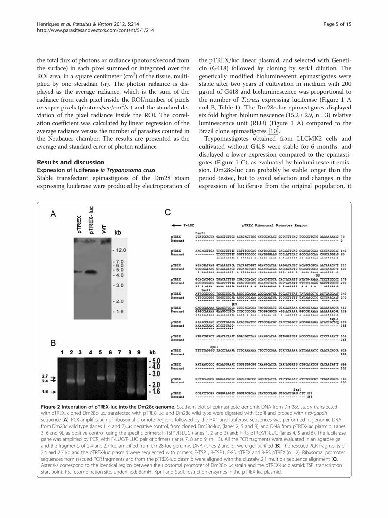
Figure 2 Integration of pTREX-luc into the Dm28c genome. Southernwith pTREX, cloned Dm28c-luc, transfected with pTREX-luc, and Dm28c wilsequence (A). PCR amplification of ribosomal promoter regions followed bfrom Dm28c wild type (lanes 1, 4 and 7), as negative control; from cloned3, 6 and 9), as positive control, using the specific primers: F-TSP1/R-LUC (langene was amplified by PCR, with F-LUC/R-LUC pair of primers (lanes 7, 8 anand the fragments of 2.4 and 2.7 kb, amplified from Dm28-luc genomic DN2.4 and 2.7 kb and the pTREX-luc plasmid were sequenced with primers: F-sequences from rescued PCR fragments and from the pTREX-luc plasmid wAsterisks correspond to the identical region between the ribosomal promostart point; RS, recombination site, underlined; BamHI, KpnI and SacII, restric
the pTREX/luc linear plasmid, and selected with Geneti-cin (G418) followed by cloning by serial dilution. Thegenetically modified bioluminescent epimastigotes werestable after two years of cultivation in medium with 200μg/ml of G418 and bioluminescence was proportional tothe number of T.cruzi expressing luciferase (Figure 1 Aand B, Table 1). The Dm28c-luc epimastigotes displayedsix fold higher bioluminescence (15.2 ± 2.9, n = 3) relativeluminescence unit (RLU) (Figure 1 A) compared to theBrazil clone epimastigotes [10].Trypomastigotes obtained from LLCMK2 cells and
cultivated without G418 were stable for 6 months, anddisplayed a lower expression compared to the epimasti-gotes (Figure 1 C), as evaluated by bioluminescent emis-sion. Dm28c-luc can probably be stable longer than theperiod tested, but to avoid selection and changes in theexpression of luciferase from the original population, it
blot of epimastigote genomic DNA from Dm28c stably transfectedd type were digested with EcoRI and probed with neo/gapdhy the HX1 and luciferase sequences was performed in genomic DNADm28c-luc, (lanes 2, 5 and 8); and DNA from pTREX-luc plasmid, (laneses 1, 2 and 3) and; F-RS pTREX/R-LUC (lanes 4, 5 and 6). The luciferased 9) (n = 3). All the PCR fragments were evaluated in an agarose gelA (lanes 2 and 5), were gel purified (B). The rescued PCR fragments ofTSP1; R-TSP1; F-RS pTREX and R-RS pTREX (n = 2). Ribosomal promoterere aligned with the clustalw 2.1 multiple sequence alignment (C).ter of Dm28c-luc strain and the pTREX-luc plasmid; TSP, transcriptiontion enzymes in the pTREX-luc plasmid.
Henriques et al. Parasites & Vectors 2012, 5:214 Page 6 of 15http://www.parasitesandvectors.com/content/5/1/214
is recommended to start new cultures from frozentrypomastigotes periodically or metacyclogenesis fromfrozen stocks of epimastigotes. To investigate thepTREX-luc plasmid integration, Southern blots of gen-omic DNA from Dm28c transfected with pTREX, gen-omic DNA of cloned Dm28c-luc and of wild type Dm28cwere digested with EcoRI and probed with neo/gapdhintergenic region. A faint band was observed in thepTREX uncloned transfectants when compared topTREX-luc cloned transfectants, which displayed a bandof approximately 5 kb (Figure 2 A), correspondent to afragment of the genome cleaved by EcoRI enzyme, a re-striction site inexistent in pTREX-luc plasmid.To evaluate the integration of pTEX-luc, PCR of gen-
omic DNA was performed using specific primers toamplify regions of the ribosomal promoter, followed bythe luciferase gene. Single PCR products were amplifiedfrom genomic DNA of cloned Dm28c-luc, generatingfragments of expected size as compared with PCR amp-lified fragments of pTREX-luc plasmid (Figure 2 B). ThePCR of wild type Dm28c genomic DNA was negative forthe pairs of primers tested (Figure 2 B). Fragments of 2.4
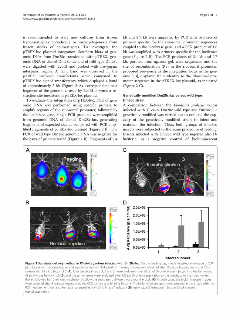
Figure 3 Substrate delivery method in Rhodnius prolixus infected withμl of blood with trypomastigotes and supplemented with D-luciferin (1 mgcamera with binning factor of 2 (A). After feeding, insects (1, 2 and 3) weredirectly in the hemolymph (B); and the same insects were evaluated after 1thorax, followed by 15 minutes incubation to allow the substrate to diffusewere acquired after 5 minutes exposure by the CCD camera and binning fROI measurement tool and the radiance quantified by Living ImageW softwtopical application.
kb and 2.7 kb were amplified by PCR with two sets ofprimers specific for the ribosomal promoter sequencescoupled to the luciferase gene, and a PCR product of 1.6kb was amplified with primers specific for the luciferasegene (Figure 2 B). The PCR products of 2.4 kb and 2.7kb, purified from agarose gel, were sequenced and thesite of recombination (RS) in the ribosomal promoter,proposed previously as the integration locus in the gen-ome [13], displayed 87 % identity to the ribosomal pro-moter sequence in the pTREX-luc plasmid, as indicated(Figure 2 C).
Genetically modified Dm28c-luc versus wild typeDm28c strainA comparison between the Rhodnius prolixus vectorinfected with T. cruzi Dm28c wild type and Dm28c-lucgenetically modified was carried out to evaluate the cap-acity of the genetically modified strain to infect andmaintain the infection. Thus, both groups of infectedinsects were subjected to the same procedure of feeding.Insects infected with Dm28c wild type ingested also D-luciferin, as a negative control of bioluminescent
Dm28c-luc. On the feeding day, insects ingested an average of 250/ml), images were obtained after 10 seconds exposure by the CCDevaluated after 60 μg of D-luciferin was injected into the hemocoel,50 μg D-luciferin application to the cuticle, onto the insect ventralthroughout the body (C). In both cases, the bioluminescent images
actor 4. The bioluminescent spots were selected in the image with theare (D), (gray square) hemocoel injection (black square)
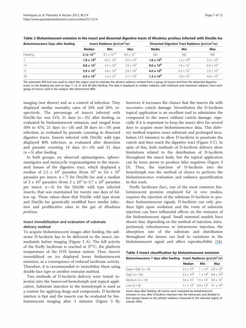
Table 2 Bioluminescent emission in the insect and dissected digestive tracts of Rhodnius prolixus infected with Dm28c-luc
Bioluminescence Days after feeding Insect Radiance (p/s/cm2/sr) Dissected Digestive Tract Radiance (p/s/cm2/sr)
Median Min Max Media Min Max
Feeding 2.1x 10¹¹ 9.1 x 10¹⁰ 4.8 x 10¹¹ ND ND ND
7 1.8 x 10⁸ 6.9 x 10⁵ 4.9 x 10⁹ 1.6 x 10⁹ 1.3 x 10⁸ 3.3 x 10⁹
14 8.8 x 10⁷ 4.2 x 10⁶ 7.9 x 10⁹ 9.0 x 10⁸ 1.8 x 10⁷ 6.9 x 10⁹
21 3.9 x 10⁷ 3.8 x 10⁶ 2.8 x 10⁹ 4.0 x 10⁸ 6.3 x 10⁷ 2.9 x 10⁹
28 6.9 x 10⁷ 1.4 x 10⁷ 2.7 x 10⁸ 1.3 x 10⁹ 1.8 x 10⁷ 4.8 x 10⁹
The automatic ROI tool was used to select the region used to estimate the photon radiance emitted from a group of insects and from the dissected digestivetracts on the feeding day and on days 7, 14, 21 and 28 after feeding. The data is displayed as median radiance, with minimum and maximum radiance, from eachgroup of insects used in the analysis. Not determined (ND).
Table 3 Insect classification by bioluminescent emission
Bioluminescence 7 days after feeding Insect Radiance (p/s/cm2/sr)
Median Min Max
Super High (n = 8) 2.5 x 10⁹ 1 x 10⁹ 4.9 x 10⁹
High (n = 30) 3.2 x 10⁸ 1 x 10⁸ 8.8 x 10⁸
Medium (n = 18) 3.6 x 10⁷ 1.4 x 10⁷ 9.8 x 10⁷
Low (n = 8) 5.1 x 10⁶ 6.9 x 10⁵ 9.1 x 10⁶
Seven days after feeding, all insects were evaluated by bioluminescentemission in vivo, after D-luciferin injection into the hemocoel, and divided infour groups based on the photon radiance measured in the selected region ofinterest (ROI).
Henriques et al. Parasites & Vectors 2012, 5:214 Page 7 of 15http://www.parasitesandvectors.com/content/5/1/214
imaging (not shown) and as a control of infection. Theydisplayed similar mortality rates of 24% and 26%, re-spectively. The percentage of insects infected withDm28c-luc was 51%, 21 days (n = 35) after feeding, asevaluated by bioluminescent emission, and ranged from50% to 47%, 21 days (n = 10) and 28 days (n = 19) postinfection, as evaluated by parasite counting in dissecteddigestive tracts. Insects infected with Dm28c wild typedisplayed 40% infection, as evaluated after dissectionand parasite counting 14 days (n = 10) and 21 days(n = 5) after feeding.In both groups, we observed epimastigotes, sphero-
mastigotes and metacyclic trypomastigotes in the macer-ated tissues of the digestive tract, which displayed amedian of 2.5 x 10⁴ parasites (from 103 to 3.6 x 10⁵
parasites per insect, n = 7) for Dm28c-luc and a medianof 3 x 10⁴ parasites (from 2 x 10³ to 3.7 x 10⁵ parasitesper insect, n = 4) for the Dm28c wild type infectedinsects, that was maintained for twenty one days of fol-low up. These values show that Dm28c wild type strainand Dm28c-luc genetically modified have similar infec-tion and proliferative rates in the gut of Rhodniusprolixus.
Insect immobilization and evaluation of substratedelivery methodTo acquire bioluminescent images after feeding, the sub-strate D-luciferin has to be delivered to the insect, im-mediately before imaging (Figure 3 A). The full activityof the firefly luciferase is reached at 37oC, the platformtemperature of the IVIS lumina system. Thus, insectsimmobilized on ice displayed lower bioluminescentemission, as a consequence of reduced luciferase activity.Therefore, it is recommended to immobilize them usingdouble face tape or another restraint method.Two methods of D-luciferin delivery were tested: in-
jection into the hemocoel/hemolymph and topical appli-cation. Substrate injection in the hemolymph is used asa routine for applying drugs and compounds. D-luciferininjetion is fast and the insects can be evaluated by bio-luminescent imaging after 5 minutes (Figure 3 B),
however, it increases the chance that the insects die withsuccessive cuticle damage. Nevertheless the D-luciferintopical application is an excellent tool to administer thecompound to the insect without cuticle damage, espe-cially if it is important to keep the insect alive for severaldays to acquire more bioluminescence data. This deliv-ery method requires more substrate and prolonged incu-bation (15 minutes) to allow D-luciferin to penetrate thecuticle and then reach the digestive tract (Figure 3 C). Inspite of this, both methods of D-luciferin delivery showlimitations related to the distribution of D-luciferinthroughout the insect body, but the topical applicationcan be more prone to produce false negatives (Figure 3D). Thus, the injection of D-luciferin into thehemolymph was the method of choice to perform thebioluminescence evaluation and radiance quantificationin this work.Firefly luciferase (luc), one of the most common bio-
luminescent proteins employed for in vivo studies,requires the injection of the substrate D-luciferin to pro-duce bioluminescent signals. D-luciferin can only pro-duce light upon oxidation and the route of substrateinjection can have influential effects on the emission ofthe bioluminescent signal. Small mammal models haveshown that, depending on the method of injection, intra-peritoneal, subcutaneous or intravenous injection, theabsorption rate of the substrate and distributionthroughout the tissues can lead to variations in thebioluminescent signal and affect reproducibility [16].
Henriques et al. Parasites & Vectors 2012, 5:214 Page 8 of 15http://www.parasitesandvectors.com/content/5/1/214
Studies using radio-labeled D-luciferin injected intra-venously demonstrated that the uptake rate of the sub-strate is actually slower in gastrointestinal organs,pancreas, and spleen than would be achieved usingintraperitoneal injection [17]. However, the intravenousinjection of the substrate generates a faster biolumines-cent signal but with a shorter duration than the intra-peritoneal route [18,19]. Thus, the injection methodshould be considered in light of the proposed objectivesof any study.
Bioluminescent imaging after feedingA bioluminescent image of Rhodnius prolixus infectedwith T. cruzi expressing luciferase acquired in an IVISW
Imaging System is a diffuse projection on the surface ofthe insect from the trypanosome inside the digestive
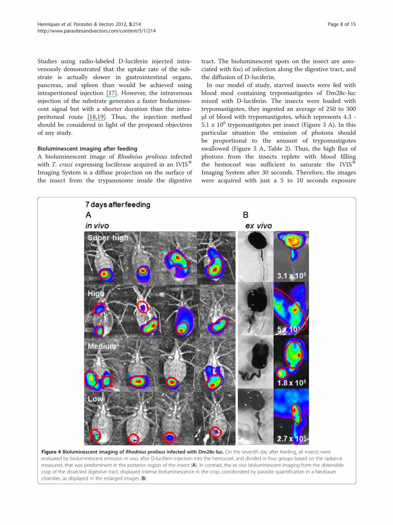
Figure 4 Bioluminescent imaging of Rhodnius prolixus infected with Devaluated by bioluminescent emission in vivo, after D-luciferin injection intomeasured, that was predominant in the posterior region of the insect (A). Icrop of the dissected digestive tract, displayed intense bioluminescence inchamber, as displayed in the enlarged images (B).
tract. The bioluminescent spots on the insect are asso-ciated with foci of infection along the digestive tract, andthe diffusion of D-luciferin.In our model of study, starved insects were fed with
blood meal containing trypomastigotes of Dm28c-lucmixed with D-luciferin. The insects were loaded withtrypomastigotes, they ingested an average of 250 to 300μl of blood with trypomastigotes, which represents 4.3 -5.1 x 10⁶ trypomastigotes per insect (Figure 3 A). In thisparticular situation the emission of photons shouldbe proportional to the amount of trypomastigotesswallowed (Figure 3 A, Table 2). Thus, the high flux ofphotons from the insects replete with blood fillingthe hemocoel was sufficient to saturate the IVISW
Imaging System after 30 seconds. Therefore, the imageswere acquired with just a 5 to 10 seconds exposure
m28c-luc. On the seventh day after feeding, all insects werethe hemocoel, and divided in four groups based on the radiance
n contrast, the ex vivo bioluminescent imaging from the distensiblethe crop, corroborated by parasite quantification in a Neubauer
Henriques et al. Parasites & Vectors 2012, 5:214 Page 9 of 15http://www.parasitesandvectors.com/content/5/1/214
(Figure 3 A). The images were taken immediately afterthe blood meal, which lasted approximately 30 minutes.In the control insects, which were fed with bloodcontaining wild type Dm28c strain and the substrate D-luciferin, bioluminescence was absent even after 5 minexposure by the CCD camera (not shown).
Bioluminescent imagingIn experiments using several insects, it is worth notingthat insects with high photon emission can mask the spotsof insects with low photos emission, obtained by the CCDcamera. Bioluminescent imaging in Living Image softwarehas a minimum threshold, the image is an overlay of a lu-minescent image over a grayscale photographic image, theupper (Max Bar) and lower limits (Min Bar) are in thecolor table display. All photon emissions below the MinBar setting of relatively lower bioluminescent emission arenot displayed in the pseudocolor image, and are transpar-ent, to avoid saturation of the image.The pseudocolor scheme makes it easy to quantify spot
regions of bright light emission, but because the range ofradiance in our model of study varied from 5 x 10⁹ to6.9 x 105 photons/sec/cm2/sr (Table 3), it is highly recom-mended to inspect negative insects, by removing theinsects which display higher measurements of radianceand make a new bioluminescence acquisition, until bio-luminescence is not detected. The sensitivity of lower-photon emitter insects is dependent on the relationship of
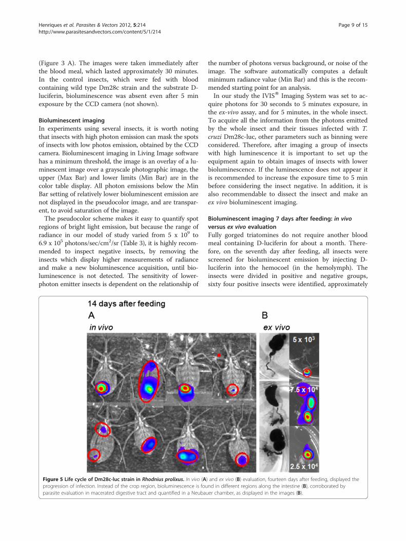
Figure 5 Life cycle of Dm28c-luc strain in Rhodnius prolixus. In vivo (Aprogression of infection. Instead of the crop region, bioluminescence is fouparasite evaluation in macerated digestive tract and quantified in a Neubau
the number of photons versus background, or noise of theimage. The software automatically computes a defaultminimum radiance value (Min Bar) and this is the recom-mended starting point for an analysis.In our study the IVISW Imaging System was set to ac-
quire photons for 30 seconds to 5 minutes exposure, inthe ex-vivo assay, and for 5 minutes, in the whole insect.To acquire all the information from the photons emittedby the whole insect and their tissues infected with T.cruzi Dm28c-luc, other parameters such as binning wereconsidered. Therefore, after imaging a group of insectswith high luminescence it is important to set up theequipment again to obtain images of insects with lowerbioluminescence. If the luminescence does not appear itis recommended to increase the exposure time to 5 minbefore considering the insect negative. In addition, it isalso recommendable to dissect the insect and make anex vivo bioluminescent imaging.
Bioluminescent imaging 7 days after feeding: in vivoversus ex vivo evaluationFully gorged triatomines do not require another bloodmeal containing D-luciferin for about a month. There-fore, on the seventh day after feeding, all insects werescreened for bioluminescent emission by injecting D-luciferin into the hemocoel (in the hemolymph). Theinsects were divided in positive and negative groups,sixty four positive insects were identified, approximately
) and ex vivo (B) evaluation, fourteen days after feeding, displayed thend in different regions along the intestine (B), corroborated byer chamber, as displayed in the images (B).
Henriques et al. Parasites & Vectors 2012, 5:214 Page 10 of 15http://www.parasitesandvectors.com/content/5/1/214
55%, which displayed different degrees of biolumines-cence/photon emission. However, the bioluminescentemission of the whole insect was predominant in theposterior region of the abdomen. In contrast, in dis-sected insects the digestive tract displayed intense bio-luminescence in the crop and not in the posterior regionof the digestive tract (Figures 4 A and B). Epimastigoteswere predominant in a group of insects dissected. Theparasites were counted in the macerated digestive tract,which displayed a median of 2.1 x 10⁵ parasites (from104 to 5 x 10⁵ parasites per insect, n = 10) (Figure 4 B).This number of parasites represents 4.5 % (min 0.2% –max 10%) of parasites ingested after feeding, which sug-gest that most trypomastigotes died and only a smallpercentage could survive the hostile environment of thecrop and transform into epimastigotes.
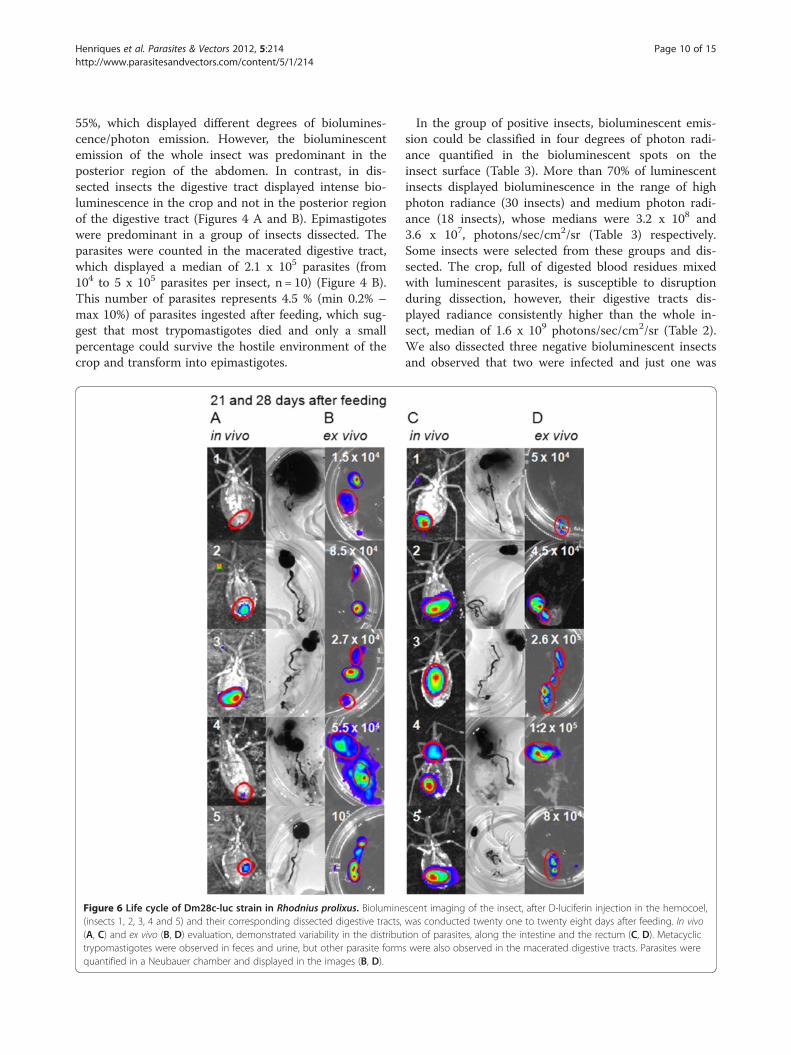
Figure 6 Life cycle of Dm28c-luc strain in Rhodnius prolixus. Biolumine(insects 1, 2, 3, 4 and 5) and their corresponding dissected digestive tracts,(A, C) and ex vivo (B, D) evaluation, demonstrated variability in the distributrypomastigotes were observed in feces and urine, but other parasite formquantified in a Neubauer chamber and displayed in the images (B, D).
In the group of positive insects, bioluminescent emis-sion could be classified in four degrees of photon radi-ance quantified in the bioluminescent spots on theinsect surface (Table 3). More than 70% of luminescentinsects displayed bioluminescence in the range of highphoton radiance (30 insects) and medium photon radi-ance (18 insects), whose medians were 3.2 x 10⁸ and3.6 x 107, photons/sec/cm2/sr (Table 3) respectively.Some insects were selected from these groups and dis-sected. The crop, full of digested blood residues mixedwith luminescent parasites, is susceptible to disruptionduring dissection, however, their digestive tracts dis-played radiance consistently higher than the whole in-sect, median of 1.6 x 10⁹ photons/sec/cm2/sr (Table 2).We also dissected three negative bioluminescent insectsand observed that two were infected and just one was
scent imaging of the insect, after D-luciferin injection in the hemocoel,was conducted twenty one to twenty eight days after feeding. In vivotion of parasites, along the intestine and the rectum (C, D). Metacyclics were also observed in the macerated digestive tracts. Parasites were
Henriques et al. Parasites & Vectors 2012, 5:214 Page 11 of 15http://www.parasitesandvectors.com/content/5/1/214
not. Therefore, from the 45 % negative bioluminescentinsects a percentage is probably infected but due to D-luciferin diffusion and distribution throughout the insectbody, the infection rate evaluated by bioluminescent im-aging could be under detected.
Bioluminescent imaging 14 days after feeding, in vivoversus ex vivo evaluationTwo weeks after feeding, the digestion of the blood mealis close to completion, the crop is no longer biolumines-cent for the majority of insects (Figures 5 A and B), epi-mastigotes and spheromastigotes were found by lightmicroscopy in macerated digestive tracts, some werefound attached to portions of the gut epithelium. Meta-cyclic trypomastigotes were also found in maceratedguts and in feces and urine of some insects, in agree-ment with the bioluminescent imaging of the rectal re-gion of the dissected digestive tract (Figure 5 B).Photon emissions acquired by the CCD camera did
not have enough spatial resolution to show the regionsof the intestine and the coiled digestive tract inside theinsect. In the insect the bioluminescent image is just aspot that represents part of the infection site. However,the dissected guts can show the precise localization ofthe parasites along the tract (Figures 5 A and B). In the
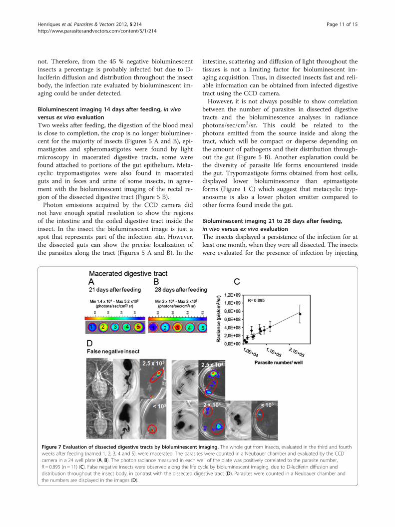
Figure 7 Evaluation of dissected digestive tracts by bioluminescent imweeks after feeding (named 1, 2, 3, 4 and 5), were macerated. The parasitecamera in a 24 well plate (A, B). The photon radiance measured in each wR= 0.895 (n = 11) (C). False negative insects were observed along the life cydistribution throughout the insect body, in contrast with the dissected digthe numbers are displayed in the images (D).
intestine, scattering and diffusion of light throughout thetissues is not a limiting factor for bioluminescent im-aging acquisition. Thus, in dissected insects fast and reli-able information can be obtained from infected digestivetract using the CCD camera.However, it is not always possible to show correlation
between the number of parasites in dissected digestivetracts and the bioluminescence analyses in radiancephotons/sec/cm2/sr. This could be related to thephotons emitted from the source inside and along thetract, which will be compact or disperse depending onthe amount of pathogens and their distribution through-out the gut (Figure 5 B). Another explanation could bethe diversity of parasite life forms encountered insidethe gut. Trypomastigote forms obtained from host cells,displayed lower bioluminescence than epimastigoteforms (Figure 1 C) which suggest that metacyclic tryp-anosome is also a lower photon emitter compared toother forms found inside the gut.
Bioluminescent imaging 21 to 28 days after feeding,in vivo versus ex vivo evaluationThe insects displayed a persistence of the infection for atleast one month, when they were all dissected. The insectswere evaluated for the presence of infection by injecting
aging. The whole gut from insects, evaluated in the third and fourths were counted in a Neubauer chamber and evaluated by the CCDell of the plate was positively correlated to the parasite number,cle by bioluminescent imaging, due to D-luciferin diffusion andestive tract (D). Parasites were counted in a Neubauer chamber and
Henriques et al. Parasites & Vectors 2012, 5:214 Page 12 of 15http://www.parasitesandvectors.com/content/5/1/214
D-luciferin in the hemocoel, which produced images ofthe terminal region of the insects, corresponding to the in-testine and rectum (Figures 6 A and C). To corroboratethe bioluminescent imaging with the whole insect andobserve the parasite mobility throughout the gut, theinsects were carefully dissected to avoid disruption ofthe digestive tract and to maintain the parasites insidethe gut region (Figures 6 B and D). Consistent with theassessments made previously, the digestive tract of dis-sected insects displayed higher radiance than live insects(Table 2).On the 21st and 28th days after feeding, ten and nineteen
insects infected with Dm28c-luc were dissected. Metacyclictrypomastigotes, spheromastigotes and epimastigotes werefound in the macerated tissues of the digestive tract. In thethird week after feeding, the infection rate was maintained,50% of insects were infected and the median was 4.1 x 10⁴
parasites (from 1.5 x 10⁴ to 105 parasites per insect, n=5).After 4 weeks, nineteen insects were evaluated, 47 % of theinsects remained infected, the parasites counted in the
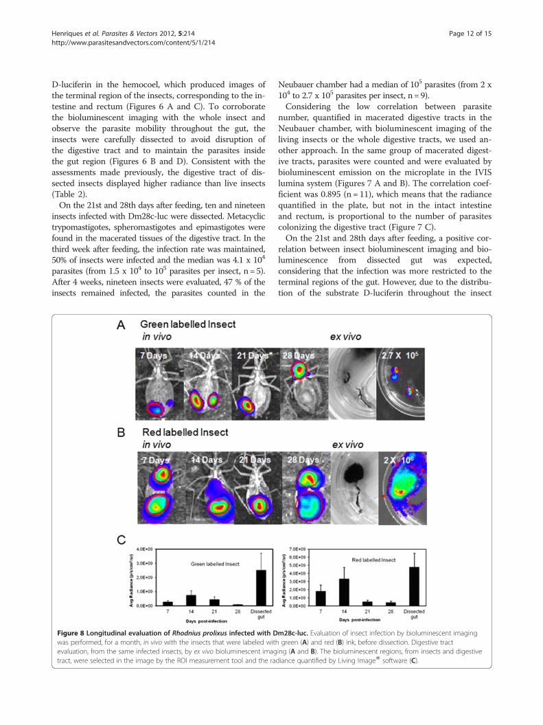
Figure 8 Longitudinal evaluation of Rhodnius prolixus infected with Dwas performed, for a month, in vivo with the insects that were labeled withevaluation, from the same infected insects, by ex vivo bioluminescent imagtract, were selected in the image by the ROI measurement tool and the rad
Neubauer chamber had a median of 105 parasites (from 2 x10⁴ to 2.7 x 10⁵ parasites per insect, n= 9).Considering the low correlation between parasite
number, quantified in macerated digestive tracts in theNeubauer chamber, with bioluminescent imaging of theliving insects or the whole digestive tracts, we used an-other approach. In the same group of macerated digest-ive tracts, parasites were counted and were evaluated bybioluminescent emission on the microplate in the IVISlumina system (Figures 7 A and B). The correlation coef-ficient was 0.895 (n = 11), which means that the radiancequantified in the plate, but not in the intact intestineand rectum, is proportional to the number of parasitescolonizing the digestive tract (Figure 7 C).On the 21st and 28th days after feeding, a positive cor-
relation between insect bioluminescent imaging and bio-luminescence from dissected gut was expected,considering that the infection was more restricted to theterminal regions of the gut. However, due to the distribu-tion of the substrate D-luciferin throughout the insect
m28c-luc. Evaluation of insect infection by bioluminescent imaginggreen (A) and red (B) ink, before dissection. Digestive tract
ing (A and B). The bioluminescent regions, from insects and digestiveiance quantified by Living ImageW software (C).
Henriques et al. Parasites & Vectors 2012, 5:214 Page 13 of 15http://www.parasitesandvectors.com/content/5/1/214
body, distribution of parasites inside the gut and the con-formation of the intestine inside the hemocoel, the coeffi-cient of correlation was low and some insects were falsenegatives. We observed occasionally that when the insect
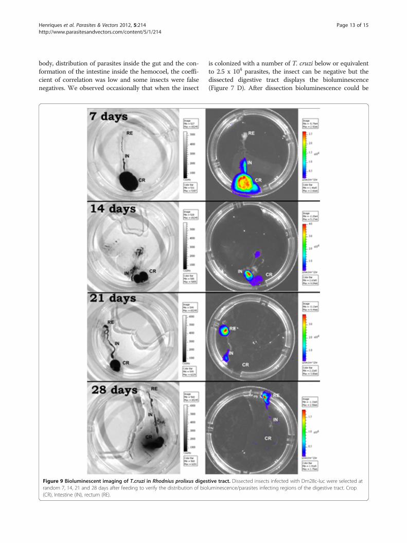
Figure 9 Bioluminescent imaging of T.cruzi in Rhodnius prolixus digesrandom 7, 14, 21 and 28 days after feeding to verify the distribution of bio(CR), Intestine (IN), rectum (RE).
is colonized with a number of T. cruzi below or equivalentto 2.5 x 10⁴ parasites, the insect can be negative but thedissected digestive tract displays the bioluminescence(Figure 7 D). After dissection bioluminescence could be
tive tract. Dissected insects infected with Dm28c-luc were selected atluminescence/parasites infecting regions of the digestive tract. Crop
Henriques et al. Parasites & Vectors 2012, 5:214 Page 14 of 15http://www.parasitesandvectors.com/content/5/1/214
quantified precisely in false negative insects, the medianwas 2.5 x 10³ parasites (from 103 to 2.5 x 10⁴ parasites perinsect, n = 5) (Figure 7 D). Thus it is recommended towork with highly infected insects that can be followed upfor a month, overcoming the limitations encountered withlow parasite load, in infected insects.
Longitudinal evaluation of labeled insectsIn spite of the substrate diffusion limitation, the sameinsects can be followed up over several days and the in-fection can be tracked for a month. Thus, a group ofinsects were selected according to bioluminescence in-tensity on the seventh day after feeding, for longitudinalevaluation. These insects were marked with a spot of dif-ferent colors of water paint (gouache ink), on the dorsalregion. Two insects were evaluated for a month by bio-luminescence acquisition, before dissection and micro-scopic counting of parasites in the digestive tract(Figures 8 A and B). However, as discussed before, thedigestive tracts displayed higher bioluminescence thanthe whole insect (Figure 8 C).To observe the parasite cycle and the progression of
infection in the digestive tract, the insects were carefullydissected and the infection could be tracked accuratelythrough the crop, intestine and rectum by biolumines-cent imaging (Figure 9). The infection also displayedsome variations among the groups of insects.Previous studies, reported the use of parasites expres-
sing fluorescent proteins to track the fate of the parasiteinside the vector. Rhodnius prolixus infected withDsRed-labeled T.cruzi was used as a marker to follow theparasite life cycle and investigate co-infection with T.rangeli expressing GFP [20]. Plasmodium falciparumgenetically modified lines that stably express gametocyte-specific GFP-luciferase reporters were used as a tool toevaluate the dose- and time-dependent drug action ongametocyte maturation and transmission, evaluated bybioluminescent emission in a plate reader and by lightfluorescence microscopy. Mature gametocytes treatedwith drugs, were formulated as artificial mosquito bloodmeals and fed to A. stephensi mosquitoes. Mosquito mid-guts were dissected 6 or 7 days after gametocyte inges-tion to ascertain the percentage of infected mosquitoesand quantify oocyst production. However, the positive ef-fect of drugs on the inhibition of mature gametocyte trans-mission to Anopheles mosquitoes was not evaluated byin vivo or ex vivo bioluminescent imaging but by oocystquantification under a phase-contrast microscope [21].The fact that dissected digestive tracts display higher
bioluminescent emission than the intact insect is prob-ably related to the diffusion of D-luciferin throughoutthe hemolymph and tissue regions. We can speculatethat the substrate D-luciferin (a) is not achieving an ad-equate concentration in the anterior region of the insect,
crop, (b) may be degraded in lipidic bodies, or (c)excreted through Malpighian tubules, involved in elimin-ation of products of digestion, and in ionic and osmoticregulation through the action of channels, exchangersand transporters [22,23]. D-luciferin is transportedthrough the ABCG2/BRCP transporter [24], thus specifictransporters and exchangers in the insect organs couldincrease the turnover of D-luciferin in the hemolymph,and reduce bioluminescent emission in the insect.
ConclusionsTo our knowledge this is the first report of biolumines-cent imaging in Rhodnius prolixus infected with Dm28c-luc trypomastigotes, expressing firefly luciferase. Thestrain displayed stable expression, and reproducible datacould be acquired, as evaluated by bioluminescent im-aging of intact as well as of dissected digestive tracts ofRhodnius prolixus, and confirmed by light microscopyobservation of the parasites, in the macerated intestines.We can conclude that, bioluminescent imaging analysisoffers a considerable quantity of information regardingthe parasites movements in the digestive tract as evalu-ated by dissected guts.
Competing interestsThe authors declare that they have no competing interests.
Authors’ contributionsThe authors ESG and WS idealized the project, contributed to theexperimental design and manuscript. CH produced the strain, Dm28-luc, andLDFG made the southern blot and the evaluation of pTREX-luc integration inthe genome. DPC contributed significantly with insect physiology and insecthandling information. CH and DPC executed bioluminescent imaging, dataanalysis and manuscript elaboration. All authors read and approved the finalversion of the manuscript.
AcknowledgementsThe authors thank the Program for Technological Development in Tools forHealth-PDTIS/FIOCRUZ and the National Institute of Science and the NationalInstitute of Science and Technology for Structural Biology and Bioimaging-INBEB/CENABIOII/UFRJ, for use of its facilities. Cristina Henriques has apostdoctoral fellowship- PNPD 150209/2009-6/project 558945/2008-2 fromCNPq. This project was supported by CNPq and FAPERJ -APQ1 E-26/171.172.2006.
Author details1Laboratório de Ultraestrutura Celular Hertha Meyer, UFRJ, Rio de Janeiro, RJ,Brazil. 2Laboratório de Bioquímica e Fisiologia de Insetos, Instituto OswaldoCruz (Fiocruz), Rio de Janeiro, RJ, Brazil. 3Laboratório de Genômica Funcionale Bioinformática, Instituto Oswaldo Cruz (Fiocruz), Rio de Janeiro, RJ, Brazil.4Instituto Nacional de Metrologia, Qualidade e Tecnologia, Inmetro, Duquede Caxias Xerém, RJ, Brazil.
Received: 16 May 2012 Accepted: 19 September 2012Published: 26 September 2012
References1. Billingsley PF, Downe AE: The effects of artificial diets on the anterior
intestinal cell ultrastructure of Rhodnius prolixus (Hemiptera:Reduviidae).Int J Parasitol 1989, 19(3):291–299.
2. Kollien AH, Schaub GA: The development of Trypanosoma cruzi inTriatominae. Parasitol Today 2000, 16:381–387.
3. Garcia ES, Azambuja P: Development and interaction of Trypanosomacruzi within the insect vector. Parasitol Today 1991, 7:240–244.
Henriques et al. Parasites & Vectors 2012, 5:214 Page 15 of 15http://www.parasitesandvectors.com/content/5/1/214
4. Garcia ES, Azambuja P: Infection of triatomines with Trypanosoma cruzi. InMolecular Biology of Insect Disease Vectors: A Methods Manual. Edited byCrampton JM, Beard CB, Louis C. London, UK: Chapton and Hall;1997:146–155.
5. Garcia ES, Ratcliffe NA, Whitten MM, Gonzalez MS, Azambuja P: Exploringthe role of insect host factors in the dynamics of Trypanosomacruzi – Rhodnius prolixus interactions. J Insect Physiol 2007, 53:11–21.
6. Azambuja P, Ratcliffe NA, Garcia ES: Towards an understanding of theinteraction of Trypanosoma cruzi and Trypanosoma rangeli within thereduviid insect host, Rhodnius prolixus. An Acad Brasil Cienc 2005,77:397–404.
7. Garcia ES, Genta FA, Azambuja P, Schaub GA: Interactions betweenintestinal compounds of triatomines andTrypanosoma cruzi. TrendsParasitol 2010, 26:499–505.
8. Schaub GA: Interactions if trypanosomatids and triatomines. In Adv InsectPhysiol 2009, 37:177–242.
9. Canavaci AMC, Bustamante JM, Padilla AM, Perez Brandan CM, Simpson LJ,Xu D, Boehlke CL, Tarleton RL: In Vitro and In Vivo High-ThroughputAssays for the Testing of Anti-Trypanosoma cruzi Compounds.PLoS Neglected Trop Dis 2010, 4:e740.
10. Hyland KV, Asfaw SH, Olson CL, Daniels MD, Engman DM: Bioluminescentimaging of Trypanosoma cruzi infection. Int J Parasitol 2008, 38(12):1391–1400.
11. Vazquez MP, Levin MJ: Functional analysis of the intergenic region ofTcP2β gene loci allowed the construction of a improved Trypanosomacruzi expression vector. Gene 1999, 239:217–225.
12. Cruz A, Beverley SM: Gene replacement in parasitic protozoa. Nature 1990,348:171–173.
13. Lorenzi HA, Vazquez MP, Levin MJ: Integration of expression vectors intothe ribosomal locus of Trypanosoma cruzi. Gene 2003, 310:91–99.
14. Camargo EP: Growth and differentiation in Trypanosoma cruzi. Rev InstMed São Paulo 1964, 6:93–100.
15. Contreras VT, Salles JM, Thomas N, Morel CM, Goldenberg S: In vitrodifferentiation of Trypanosoma cruzi under chemically definedconditions. Mol Biochem Parasitol 1985, 16:315–327.
16. Close DM, Xu T, Sayler GS, Ripp S: In Vivo Bioluminescent Imaging (BLI):Noninvasive Visualization and Interrogation of Biological Processes inLiving Animals. Sensors 2011, 11:180–206.
17. Lee KH, Byun SS, Paik JY, Lee SY, Song SH, Choe YS, Kim BT: Cell uptake andtissue distribution of radioiodine labelled D-D-luciferin: Implications forluciferase based gene imaging. Nucl Med Commun 2003, 24:1003–1009.
18. Keyaerts M, Verschueren J, Bos TJ, Tchouate-Gainkam LO, Peleman C,Breckpot K, Vanhove C, Caveliers V, Bossuyt A, Lahoutte T: Dynamicbioluminescence imaging for quantitative tumour burden assessmentusing IV or IP administration of D-D-luciferin: Effect on intensity, timekinetics and repeatability of photon emission. Eur J Nucl Med Mol Imaging2008, 35:999–1007.
19. Inoue Y, Kiryu S, Izawa K, Watanabe M, Tojo A, Ohtomo K: Comparison ofsubcutaneous and intraperitoneal injection of D-D-luciferin for in vivobioluminescence imaging. Eur J Nucl Med Mol Imaging 2009, 36:771–779.
20. Guevara P, Dias M, Rojas A, Crisante G, Abreu-Blanco MT, Umezawa E,Vazquez M, Levin M, Añez N, Ramirez JL: Expression of fluorescent genesin Trypanosoma cruzi and Trypanosoma rangeli (Kinetoplastida:Trypanosomatidae): its application to parasite-vector biology.J Med Entomol 2005, 42(1):48–56.
21. Adjalley SH, Johnston GL, Li T, Eastman RT, Ekland EH, Eappen AG, RichmanA, Sim BK, Lee MC, Hoffman SL, Fidock DA: Quantitative assessment ofPlasmodium falciparum sexual development reveals potenttransmission-blocking activity by methylene blue. Proc Natl Acad Sci USA2011, 108(47):E1214–E1223.
22. O’Donnell MJ: Too much of a good thing: how insects cope with excessions or toxins in the diet. J Exp Biol 2009, 212(3):363–372.
23. Toh SQ, Glanfield A, Gobert GN, Jones KM: Heme and blood-feedingparasites: friends or foes? Parasit Vectors 2010, 3:108.
24. Zhang Y, Bressler JP, Neal J, Lal B, Bhang HC, Laterra J, Pomper MG: ABCG2/BCRP Expression Modulates D-Luciferin–Based Bioluminescence Imaging.Cancer Res 2007, 67:9389–9397.
doi:10.1186/1756-3305-5-214Cite this article as: Henriques et al.: Bioluminescent imaging ofTrypanosoma cruzi infection in Rhodnius prolixus. Parasites & Vectors 20125:214.
Submit your next manuscript to BioMed Centraland take full advantage of:
• Convenient online submission
• Thorough peer review
• No space constraints or color figure charges
• Immediate publication on acceptance
• Inclusion in PubMed, CAS, Scopus and Google Scholar
• Research which is freely available for redistribution
Submit your manuscript at www.biomedcentral.com/submit


