Embed Size (px)
Citation preview
Biomolecules for development of biosensors and their applications
Sandeep K. Sharma a, Neeta Sehgal b, Ashok Kumar a,*
a Institute of genomics and Integrative Biology, Mall Road, Delhi 110007, Indiab Department of Zoology, Delhi University, Delhi 110007, India
Abstract
Biosensors are analytical devices incorporating biological materials such as enzymes, tissues, microorganisms, antibodies, cell
receptors or biologically derived materials or a biomimic component intimately associated with or integrated within a physico-
chemical transducer or transducing microsystem which may be either optical, electrochemical, thermometric, piezoelectric or
magnetic. The electronic signals produced are proportional to the concentration of specific analyte. A biomaterial may be any
material, natural or man-made, that comprises whole or part of a living structure or biomedical device, which performs natural
function. An essential component of molecular sensor is reagent layers. Creation of these layers require the immobilization of
recognition elements for the detection method. The recognition elements are biomolecules. Laboratory methods of immobilization
are numerous, but may not always appropriate for manufacture of biosensors. In the present article, we describe the use of various
biomaterials for biosensors as well as their availability.
� 2002 Elsevier Science B.V. All rights reserved.
PACS: 87; 87.14.)g; 87.14.Ee
Keywords: Biomaterials; Biomolecules; Biosensors; Enzymes; Immunosensors
1. Introduction
Biosensors are moving from the laboratory level to
field testing and commercialization in US, Europe and
Japan. Biosensors have potential for continuous andin situ application in fields such as medical diagnostics,
genetics and environmental monitoring. DNA micro-
chip technology is emerging on the horizon and being
designed to analyze gene expression patterns, genome
mapping and detect genetic mutations etc. Biosensors
such as nerve cells grown on a microprocessor, can
generate electrical signals in response to stimuli of toxins
present in the environment. Such type of technologiesare creating strategic, innovative links between elec-
tronic engineering and biological sciences [1].
Biosensors are analytical devices incorporating a bi-
ological material in an intimate contact with a suitable
transducer device that converts the biochemical signal
into quantifiable electric signals. A biologically derived
material or a biomimic intimately associated with or
integrated within a physicochemical transducer or
transducing microsystem, which may be electrochemi-
cal, thermometric, optical, piezoelectric or magnetic.
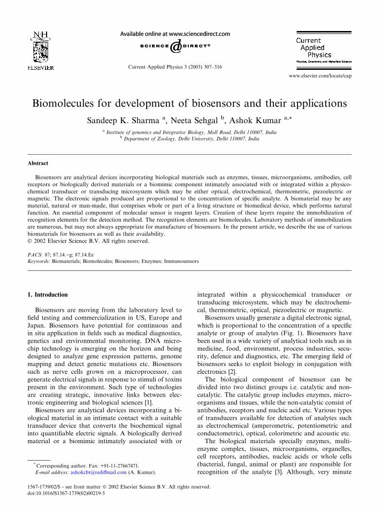
Biosensors usually generate a digital electronic signal,
which is proportional to the concentration of a specificanalyte or group of analytes (Fig. 1). Biosensors have
been used in a wide variety of analytical tools such as in
medicine, food, environment, process industries, secu-
rity, defence and diagnostics, etc. The emerging field of
biosensors seeks to exploit biology in conjugation with
electronics [2].
The biological component of biosensor can be
divided into two distinct groups i.e. catalytic and non-catalytic. The catalytic group includes enzymes, micro-
organisms and tissues, while the non-catalytic consist of
antibodies, receptors and nucleic acid etc. Various types
of transducers available for detection of analytes such
as electrochemical (amperometric, potentiometric and
conductometric), optical, colorimetric and acoustic etc.
The biological materials specially enzymes, multi-
enzyme complex, tissues, microorganisms, organelles,cell receptors, antibodies, nucleic acids or whole cells
(bacterial, fungal, animal or plant) are responsible for
recognition of the analyte [3]. Although, very minute
* Corresponding author. Fax: +91-11-27667471.
E-mail address: [email protected] (A. Kumar).
1567-1739/02/$ - see front matter � 2002 Elsevier Science B.V. All rights reserved.
doi:10.1016/S1567-1739(02)00219-5
Current Applied Physics 3 (2003) 307–316
www.elsevier.com/locate/cap
quantities of the biomaterials are required, but their
purity may play a vital role in reliability. The study ofnatural biopolymer is fundamental for understanding
how animal, plant cells and animal tissues function
and respond under different conditions. Knowledge of
three-dimensional structure of protein is necessary for
understanding and consequently controlling the modi-
fication of their biological activity, as might be required
for the proper design of a biosensor [4].
There is often a need for electrons to pass fromenzyme-based biological components to the amplifier or
microprocessor. Ferrocene represents the potential ways
of solving the problem. The cells in natural state provide
many examples of such transmission for example cyto-
chromes which are hemoprotein, whose main biological
function is electron or hydrogen transport by valency
change of their heme iron [5]. An essential component of
a molecular sensor is the reagent layer. Creation ofthese layers require the immobilization of the recogni-
tion elements for the detection method. In the case of
biosensors, this involve the biomolecules such as en-
zymes, antibodies, microorganisms, etc. (Fig. 2).
Various methods are available for immobilization of
biomolecules, but not always appropriate for manu-
facture of biosensors. The most commonly used bio-
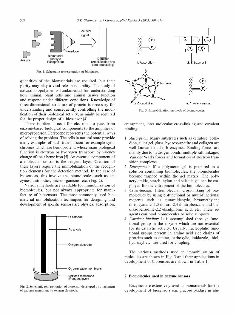
material immobilization techniques for designing anddevelopment of specific sensors are physical adsorption,
entrapment, inter molecular cross-linking and covalent
binding:
1. Adsorption: Many substrates such as cellulose, collo-dion, silica gel, glass, hydroxyapatite and collagen are
well known to adsorb enzymes. Binding forces are
mainly due to hydrogen bonds, multiple salt linkages,
Van der Wall�s forces and formation of electron tran-
sition complexes.
2. Entrapment: If a polymeric gel is prepared in a
solution containing biomolecules, the biomolecules
become trapped within the gel matrix. The poly-acrylamide, starch, nylon and siliastic gel can be em-
ployed for the entrapment of the biomolecules.
3. Cross-linking: Intermolecular cross-linking of bio-
molecules by using bi-functional or multi-functional
reagents such as glutaraldehyde, hexamethylene
di-isocyanate, 1,5-difluro 2,4-dinitrobenzene and bis-
diazobenzidine-2,20-disulphonic acid, etc. These re-
agents can bind biomolecules to solid supports.4. Covalent binding: It is accomplished through func-
tional group in the enzyme which are not essential
for its catalytic activity. Usually, nucleophilic func-
tional groups present in amino acid side chains of
proteins such as amino, carboxylic, imidazole, thiol,
hydroxyl etc. are used for coupling.
The various methods used in immobilization ofmolecules are shown in Fig. 3 and their applications in
development of biosensors are shown in Table 1.
2. Biomolecules used in enzyme sensors
Enzymes are extensively used as biomaterials for the
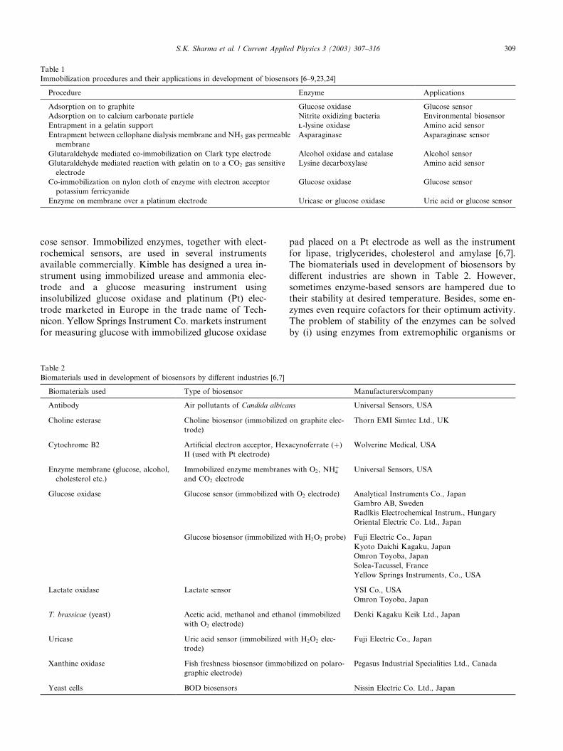
development of biosensors e.g. glucose oxidase in glu-Fig. 2. Schematic representation of biosensor developed by attachment
of enzyme membrane to oxygen electrode.
Fig. 3. Immobilization methods of biomolecules.
Fig. 1. Schematic representation of biosensor.
308 S.K. Sharma et al. / Current Applied Physics 3 (2003) 307–316
cose sensor. Immobilized enzymes, together with elect-
rochemical sensors, are used in several instruments
available commercially. Kimble has designed a urea in-
strument using immobilized urease and ammonia elec-trode and a glucose measuring instrument using
insolubilized glucose oxidase and platinum (Pt) elec-
trode marketed in Europe in the trade name of Tech-
nicon. Yellow Springs Instrument Co. markets instrument
for measuring glucose with immobilized glucose oxidase
pad placed on a Pt electrode as well as the instrument
for lipase, triglycerides, cholesterol and amylase [6,7].
The biomaterials used in development of biosensors by
different industries are shown in Table 2. However,sometimes enzyme-based sensors are hampered due to
their stability at desired temperature. Besides, some en-
zymes even require cofactors for their optimum activity.
The problem of stability of the enzymes can be solved
by (i) using enzymes from extremophilic organisms or
Table 1
Immobilization procedures and their applications in development of biosensors [6–9,23,24]
Procedure Enzyme Applications
Adsorption on to graphite Glucose oxidase Glucose sensor
Adsorption on to calcium carbonate particle Nitrite oxidizing bacteria Environmental biosensor
Entrapment in a gelatin support LL-lysine oxidase Amino acid sensor
Entrapment between cellophane dialysis membrane and NH3 gas permeable
membrane
Asparaginase Asparaginase sensor
Glutaraldehyde mediated co-immobilization on Clark type electrode Alcohol oxidase and catalase Alcohol sensor
Glutaraldehyde mediated reaction with gelatin on to a CO2 gas sensitive
electrode
Lysine decarboxylase Amino acid sensor
Co-immobilization on nylon cloth of enzyme with electron acceptor
potassium ferricyanide
Glucose oxidase Glucose sensor
Enzyme on membrane over a platinum electrode Uricase or glucose oxidase Uric acid or glucose sensor
Table 2
Biomaterials used in development of biosensors by different industries [6,7]
Biomaterials used Type of biosensor Manufacturers/company
Antibody Air pollutants of Candida albicans Universal Sensors, USA
Choline esterase Choline biosensor (immobilized on graphite elec-
trode)
Thorn EMI Simtec Ltd., UK
Cytochrome B2 Artificial electron acceptor, Hexacynoferrate (þ)
II (used with Pt electrode)
Wolverine Medical, USA
Enzyme membrane (glucose, alcohol,
cholesterol etc.)
Immobilized enzyme membranes with O2, NHþ4
and CO2 electrode
Universal Sensors, USA
Glucose oxidase Glucose sensor (immobilized with O2 electrode) Analytical Instruments Co., Japan
Gambro AB, Sweden
Radlkis Electrochemical Instrum., Hungary
Oriental Electric Co. Ltd., Japan
Glucose biosensor (immobilized with H2O2 probe) Fuji Electric Co., Japan
Kyoto Daichi Kagaku, Japan
Omron Toyoba, Japan
Solea-Tacussel, France
Yellow Springs Instruments, Co., USA
Lactate oxidase Lactate sensor YSI Co., USA
Omron Toyoba, Japan
T. brassicae (yeast) Acetic acid, methanol and ethanol (immobilized
with O2 electrode)
Denki Kagaku Keik Ltd., Japan
Uricase Uric acid sensor (immobilized with H2O2 elec-
trode)
Fuji Electric Co., Japan
Xanthine oxidase Fish freshness biosensor (immobilized on polaro-
graphic electrode)
Pegasus Industrial Specialities Ltd., Canada
Yeast cells BOD biosensors Nissin Electric Co. Ltd., Japan
S.K. Sharma et al. / Current Applied Physics 3 (2003) 307–316 309
(ii) by �tailoring� enzymes. Microorganisms or wholecells are more active and stable as they remain in their
natural environment. The first successful subcellular
component based sensor has been developed for gluta-
mine measurements in which mitochondrial fraction
containing glutaminase is immobilized at ammonia gas
electrode [10].
The first enzyme electrode, an amperometric type
biosensor, was developed by Clark and Lyons [11]. Theyused a soluble biomaterial glucose oxidase held between
membranes. The oxygen uptake was measured with
oxygen electrode
Glucose þ O2 þ H2O �!Glucose oxidaseH2O2 þ Gluconic acid
Cass et al. reported the use of ferrocene (dicyclo pen-
tadienyl iron) and its derivatives as a mediator in glu-
cose sensor as it transfer the electrons not only
quantitatively but also at faster rates [12]. Marthew et al.
have tested a pen sized glucose meter which uses dis-
posable strips for blood spot application where the fer-rocene technology is employed [13].
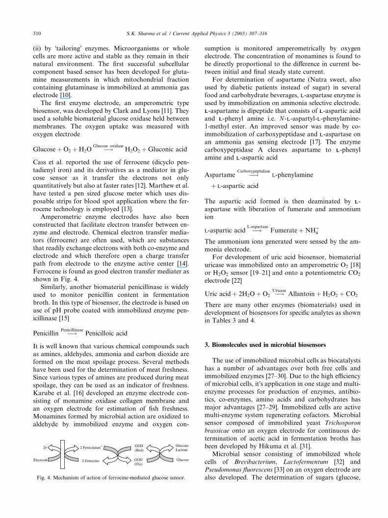
Amperometric enzyme electrodes have also been
constructed that facilitate electron transfer between en-
zyme and electrode. Chemical electron transfer media-
tors (ferrocene) are often used, which are substances
that readily exchange electrons with both co-enzyme and
electrode and which therefore open a charge transfer
path from electrode to the enzyme active center [14].Ferrocene is found as good electron transfer mediater as
shown in Fig. 4.
Similarly, another biomaterial penicillinase is widely
used to monitor penicillin content in fermentation
broth. In this type of biosensor, the electrode is based on
use of pH probe coated with immobilized enzyme pen-
icillinase [15]
Penicillin �!PenicillinasePenicilloic acid
It is well known that various chemical compounds suchas amines, aldehydes, ammonia and carbon dioxide are
formed on the meat spoilage process. Several methods
have been used for the determination of meat freshness.
Since various types of amines are produced during meat
spoilage, they can be used as an indicator of freshness.
Karube et al. [16] developed an enzyme electrode con-
sisting of monamine oxidase collagen membrane and
an oxygen electrode for estimation of fish freshness.Monamines formed by microbial action are oxidized to
aldehyde by immobilized enzyme and oxygen con-
sumption is monitored amperometrically by oxygenelectrode. The concentration of monamines is found to
be directly proportional to the difference in current be-
tween initial and final steady state current.
For determination of aspartame (Nutra sweet, also
used by diabetic patients instead of sugar) in several
food and carbohydrate beverages, LL-aspartase enzyme is
used by immobilization on ammonia selective electrode.
LL-aspartame is dipeptide that consists of LL-aspartic acidand LL-phenyl amine i.e. N -LL-aspartyl-LL-phenylamine-
1-methyl ester. An improved sensor was made by co-
immobilization of carboxypeptidase and LL-aspartase on
an ammonia gas sensing electrode [17]. The enzyme
carboxypeptidase A cleaves aspartame to LL-phenyl
amine and LL-aspartic acid
Aspartame �!Carboxypeptidasel-phenylamine
þ l-aspartic acid
The aspartic acid formed is then deaminated by LL-
aspartase with liberation of fumerate and ammonium
ion
l-aspartic acid �!L-aspartaseFumerate þ NHþ
4
The ammonium ions generated were sensed by the am-
monia electrode.
For development of uric acid biosensor, biomaterial
uricase was immobilized onto an amperometric O2 [18]
or H2O2 sensor [19–21] and onto a potentiometric CO2
electrode [22]
Uric acid þ 2H2O þ O2 �!UricaseAllantoin þ H2O2 þ CO2
There are many other enzymes (biomaterials) used in
development of biosensors for specific analytes as shownin Tables 3 and 4.
3. Biomolecules used in microbial biosensors
The use of immobilized microbial cells as biocatalysts
has a number of advantages over both free cells and
immobilized enzymes [27–30]. Due to the high efficiencyof microbial cells, it�s application in one stage and multi-
enzyme processes for production of enzymes, antibio-
tics, co-enzymes, amino acids and carbohydrates has
major advantages [27–29]. Immobilized cells are active
multi-enzyme system regenerating cofactors. Microbial
sensor composed of immobilized yeast Trichosporon
brassicae onto an oxygen electrode for continuous de-
termination of acetic acid in fermentation broths hasbeen developed by Hikuma et al. [31].
Microbial sensor consisting of immobilized whole
cells of Brevibacterium, Lactofermentum [32] and
Pseudomonas fluorescens [33] on an oxygen electrode are
also developed. The determination of sugars (glucose,
2e-
Electrode 2 Ferrocine
GOD(Red)
GOD(Oxi)
2 Ferricinium+ GluconoLactone
Glucose
Fig. 4. Mechanism of action of ferrocine-mediated glucose sensor.
310 S.K. Sharma et al. / Current Applied Physics 3 (2003) 307–316
fructose and sucrose) in a fermentation broth for glu-
tamic acid production, total sugars were determined for
the extent of oxygen consumption by the immobilized
microorganisms [32]. Similarly, sensor of glucose, lac-
tate, pyruvate, sucrose and ethanol based on the cells
from the yeast Hansenula anomala are also developed
and the rate of conversion of metabolites determined by
an O2 electrode [34].
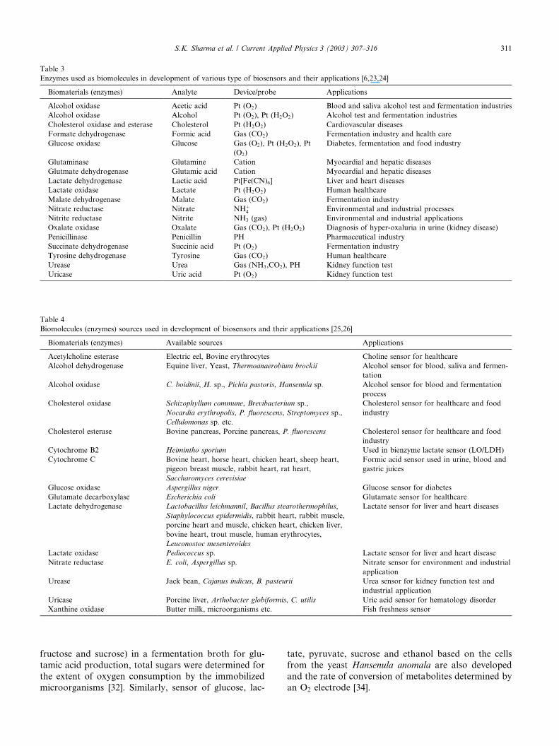
Table 3
Enzymes used as biomolecules in development of various type of biosensors and their applications [6,23,24]
Biomaterials (enzymes) Analyte Device/probe Applications
Alcohol oxidase Acetic acid Pt (O2) Blood and saliva alcohol test and fermentation industries
Alcohol oxidase Alcohol Pt (O2), Pt (H2O2) Alcohol test and fermentation industries
Cholesterol oxidase and esterase Cholesterol Pt (H2O2) Cardiovascular diseases
Formate dehydrogenase Formic acid Gas (CO2) Fermentation industry and health care
Glucose oxidase Glucose Gas (O2), Pt (H2O2), Pt
(O2)
Diabetes, fermentation and food industry
Glutaminase Glutamine Cation Myocardial and hepatic diseases
Glutmate dehydrogenase Glutamic acid Cation Myocardial and hepatic diseases
Lactate dehydrogenase Lactic acid Pt[Fe(CN)6] Liver and heart diseases
Lactate oxidase Lactate Pt (H2O2) Human healthcare
Malate dehydrogenase Malate Gas (CO2) Fermentation industry
Nitrate reductase Nitrate NHþ4 Environmental and industrial processes
Nitrite reductase Nitrite NH3 (gas) Environmental and industrial applications
Oxalate oxidase Oxalate Gas (CO2), Pt (H2O2) Diagnosis of hyper-oxaluria in urine (kidney disease)
Penicillinase Penicillin PH Pharmaceutical industry
Succinate dehydrogenase Succinic acid Pt (O2) Fermentation industry
Tyrosine dehydrogenase Tyrosine Gas (CO2) Human healthcare
Urease Urea Gas (NH3,CO2), PH Kidney function test
Uricase Uric acid Pt (O2) Kidney function test
Table 4
Biomolecules (enzymes) sources used in development of biosensors and their applications [25,26]
Biomaterials (enzymes) Available sources Applications
Acetylcholine esterase Electric eel, Bovine erythrocytes Choline sensor for healthcare
Alcohol dehydrogenase Equine liver, Yeast, Thermoanaerobium brockii Alcohol sensor for blood, saliva and fermen-
tation
Alcohol oxidase C. boidinii, H. sp., Pichia pastoris, Hansenula sp. Alcohol sensor for blood and fermentation
process
Cholesterol oxidase Schizophyllum commune, Brevibacterium sp.,
Nocardia erythropolis, P. fluorescens, Streptomyces sp.,
Cellulomonas sp. etc.
Cholesterol sensor for healthcare and food
industry
Cholesterol esterase Bovine pancreas, Porcine pancreas, P. fluorescens Cholesterol sensor for healthcare and food
industry
Cytochrome B2 Heimintho sporium Used in bienzyme lactate sensor (LO/LDH)
Cytochrome C Bovine heart, horse heart, chicken heart, sheep heart,
pigeon breast muscle, rabbit heart, rat heart,
Saccharomyces cerevisiae
Formic acid sensor used in urine, blood and
gastric juices
Glucose oxidase Aspergillus niger Glucose sensor for diabetes
Glutamate decarboxylase Escherichia coli Glutamate sensor for healthcare
Lactate dehydrogenase Lactobacillus leichmannil, Bacillus stearothermophilus,
Staphylococcus epidermidis, rabbit heart, rabbit muscle,
porcine heart and muscle, chicken heart, chicken liver,
bovine heart, trout muscle, human erythrocytes,
Leuconostoc mesenteroides
Lactate sensor for liver and heart diseases
Lactate oxidase Pediococcus sp. Lactate sensor for liver and heart disease
Nitrate reductase E. coli, Aspergillus sp. Nitrate sensor for environment and industrial
application
Urease Jack bean, Cajanus indicus, B. pasteurii Urea sensor for kidney function test and
industrial application
Uricase Porcine liver, Arthobacter globiformis, C. utilis Uric acid sensor for hematology disorder
Xanthine oxidase Butter milk, microorganisms etc. Fish freshness sensor
S.K. Sharma et al. / Current Applied Physics 3 (2003) 307–316 311
3.1. Ammonia sensor
Nitrifying bacteria has two genera of bacteria i.e.
Nitrosomonas sp. and Nitrobacter sp. Nitrosomonas sp.
utilizes ammonia as the sole source of energy:
NH3 þ 1=2O2 �!Nitrosomonas sp:NO�
2 þ H2O þ Hþ
Nitrobacter sp. of bacteria oxidize nitrate to nitrite as
follows:
NO�2 þ 1=2O2 �!Nitrobacter sp:
NO�3
The oxidation of both substrates proceeds at high rate
and oxygen consumption by bacteria can be determined
directly by oxygen electrode attached to the immobilized
bacteria. Therefore, ammonia is determined ampero-
metrically by microbial sensor using immobilized bio-
material (nitrifying bacteria) and an oxygen electrode
[35].
3.2. BOD sensor
Biological oxygen demand (BOD) is one of the most
widely used and important indication of organic pollu-
tion. The conventional test method requires 5 days in-
cubation period and includes complicated procedures.
Therefore, rapid and reproducible method was devel-
oped using immobilized yeast (T. cutaneum), sand-wiched between oxygen permitted teflon membrane and
a porous membrane, attached to the oxygen probe�splatinum cathode. When sample solutions were injected
into the system, organic compound was assimilated by
immobilized microorganisms. Consumption of oxygen
by microorganisms caused a decrease in dissolved oxy-
gen using the membrane. As a result, the current of the
sensor decreased with time until a steady state wasreached. The microbial sensor can be used for a long
time for the estimation of BOD [31].
3.3. Fish freshness sensor
Within the period of time between the death of a fish
and its consumption, a large number of biochemical and
physicochemical changes take place. Accurate and rapiddetermination of freshness is essential for the marine
food industry. Various chemical indicators such as vol-
atile basic nitrogen [36], ammonia [37,38], amines [39],
volatile acids [40,41], pH [42–45], adenosine triphos-
phate (ATP) and related compounds [46–58] have been
identified for fish freshness. These all procedures are very
tedious and time consuming. Therefore, it is difficult to
determine fish freshness accuracy by simple indicators.Later, scientists discovered that phosphorylation in an-
imal muscle [46–57], the decomposition of ATP in fish
meat sets in after the death of the fish and ADP, AMP
and related compounds are formed. For determination
of fish freshness, enzyme sensors specific for hypoxan-
thine [59], inosine [60], IMP [61] and AMP [62], have
been developed using immobilized enzyme membrane
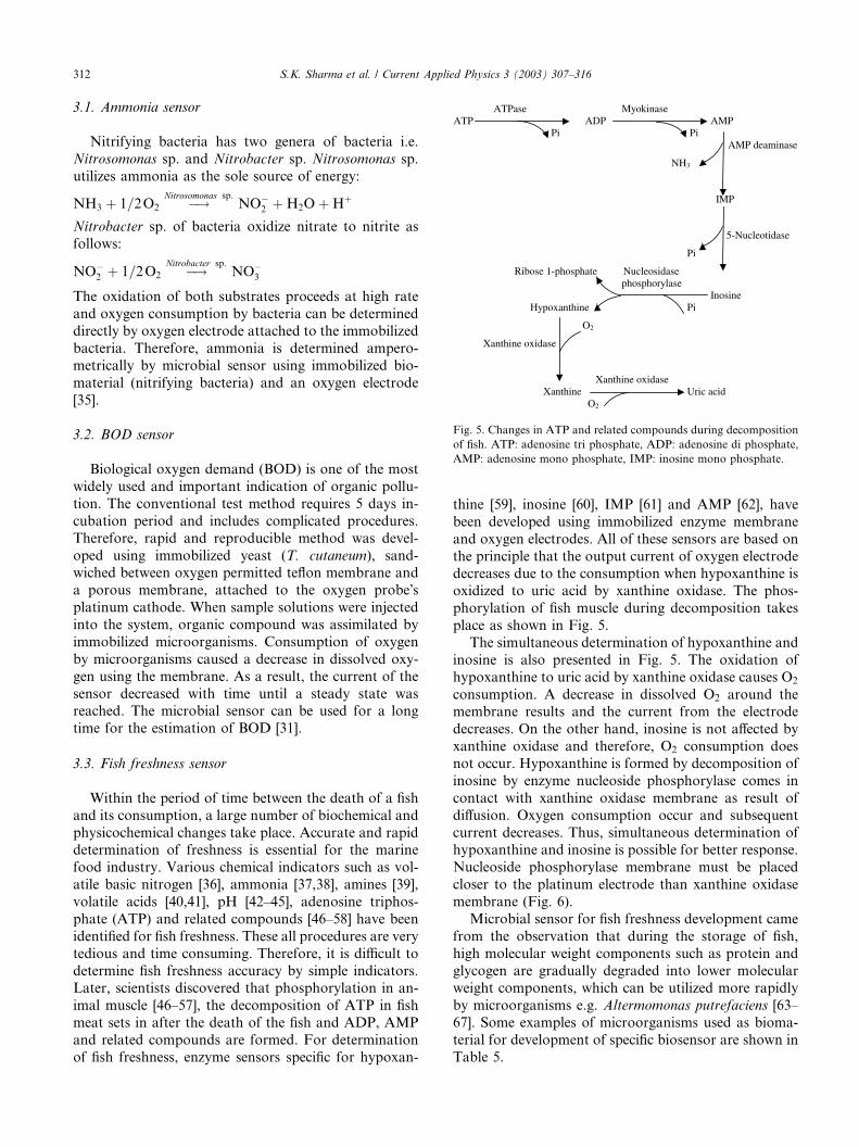
and oxygen electrodes. All of these sensors are based on
the principle that the output current of oxygen electrodedecreases due to the consumption when hypoxanthine is
oxidized to uric acid by xanthine oxidase. The phos-
phorylation of fish muscle during decomposition takes
place as shown in Fig. 5.
The simultaneous determination of hypoxanthine and
inosine is also presented in Fig. 5. The oxidation of
hypoxanthine to uric acid by xanthine oxidase causes O2
consumption. A decrease in dissolved O2 around themembrane results and the current from the electrode
decreases. On the other hand, inosine is not affected by
xanthine oxidase and therefore, O2 consumption does
not occur. Hypoxanthine is formed by decomposition of
inosine by enzyme nucleoside phosphorylase comes in
contact with xanthine oxidase membrane as result of
diffusion. Oxygen consumption occur and subsequent
current decreases. Thus, simultaneous determination ofhypoxanthine and inosine is possible for better response.
Nucleoside phosphorylase membrane must be placed
closer to the platinum electrode than xanthine oxidase
membrane (Fig. 6).
Microbial sensor for fish freshness development came
from the observation that during the storage of fish,
high molecular weight components such as protein and
glycogen are gradually degraded into lower molecularweight components, which can be utilized more rapidly
by microorganisms e.g. Altermomonas putrefaciens [63–
67]. Some examples of microorganisms used as bioma-
terial for development of specific biosensor are shown in
Table 5.
ATPase MyokinaseATP ADP AMP
Pi PiAMP deaminase
NH3
IMP
5-Nucleotidase
Pi
Ribose 1-phosphate Nucleosidasephosphorylase
InosineHypoxanthine Pi
O2
Xanthine oxidase
Xanthine oxidaseXanthine Uric acid
O2
Fig. 5. Changes in ATP and related compounds during decomposition
of fish. ATP: adenosine tri phosphate, ADP: adenosine di phosphate,
AMP: adenosine mono phosphate, IMP: inosine mono phosphate.
312 S.K. Sharma et al. / Current Applied Physics 3 (2003) 307–316
Cell-based biosensors [68] uses cells as biomaterial for
chemical detection and are composed of two compo-
nents: (1) a cell that expresses a particular receptor
protein immobilized on a prepared substrate and (2) a
transducer setup that records the cell response. Cells aremonitored either by using extracellular electrodes in the
substrate or by using registered photodiode arrays todetect changes in voltage-sensitive dyes in the cell
membrane.
4. Biomolecules used in development of immunosensors
Another possible application of biomaterial is the
construction of immunosensors using antigens or anti-bodies. Immobilized creatine kinase M (CK-M) anti-
body is used as pretreatment for detection of the
cardiospecific CK-MB isoenzyme. Goat antihuman CK-
M Ig G was immobilized on a electrode and that can be
used for several assays and is regenerable [31]. Such type
of sensors have excellent selectivity because of high an-
tibody–antigen specificity. Enzymes are extremely useful
as labels in immunoassays as their catalytic propertiesallow the detection and quantitation of low levels of
immune reactants. The enzymes most commonly used
are alkaline phosphatase, horseradish peroxidase, glu-
cose oxidase and b-galactosidase.
Immunologically based sensors have a three-pronged
approach for the development of immunologically based
optical sensors for environmental, clinical, and defence
applications [73,74]. The first element of the approachfocuses on performing a localized immunoassay on the
+ -
Oxygen electrode
Teflon membrane
Nucleoside phosphorylase membrane
Cellulose membrane
Xanthine oxidase membrane
Fig. 6. Schematic representation of biosensor for fish freshness.
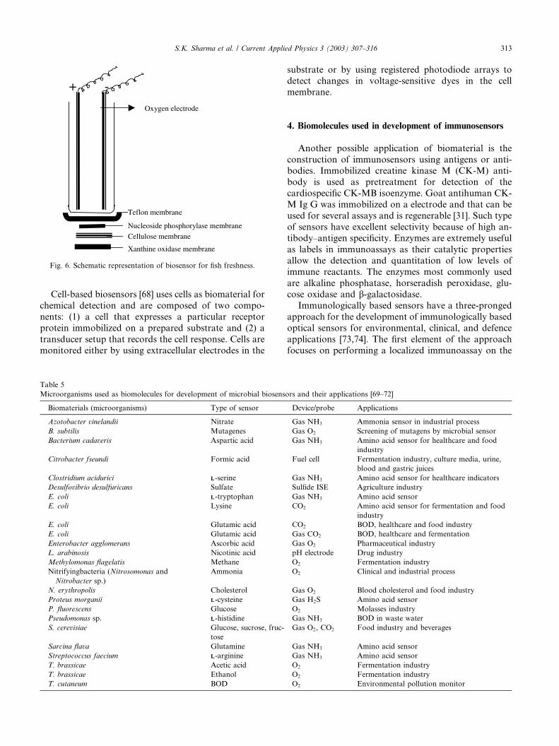
Table 5
Microorganisms used as biomolecules for development of microbial biosensors and their applications [69–72]
Biomaterials (microorganisms) Type of sensor Device/probe Applications
Azotobacter vinelandii Nitrate Gas NH3 Ammonia sensor in industrial process
B. subtilis Mutagenes Gas O2 Screening of mutagens by microbial sensor
Bacterium cadaveris Aspartic acid Gas NH3 Amino acid sensor for healthcare and food
industry
Citrobacter fseundi Formic acid Fuel cell Fermentation industry, culture media, urine,
blood and gastric juices
Clostridium acidurici LL-serine Gas NH3 Amino acid sensor for healthcare indicators
Desulfovibrio desulfuricans Sulfate Sulfide ISE Agriculture industry
E. coli LL-tryptophan Gas NH3 Amino acid sensor
E. coli Lysine CO2 Amino acid sensor for fermentation and food
industry
E. coli Glutamic acid CO2 BOD, healthcare and food industry
E. coli Glutamic acid Gas CO2 BOD, healthcare and fermentation
Enterobacter agglomerans Ascorbic acid Gas O2 Pharmaceutical industry
L. arabinosis Nicotinic acid pH electrode Drug industry
Methylomonas flagelatis Methane O2 Fermentation industry
Nitrifyingbacteria (Nitrosomonas and
Nitrobacter sp.)
Ammonia O2 Clinical and industrial process
N. erythropolis Cholesterol Gas O2 Blood cholesterol and food industry
Proteus morganii LL-cysteine Gas H2S Amino acid sensor
P. fluorescens Glucose O2 Molasses industry
Pseudomonas sp. LL-histidine Gas NH3 BOD in waste water
S. cerevisiae Glucose, sucrose, fruc-
tose
Gas O2, CO2 Food industry and beverages
Sarcina flava Glutamine Gas NH3 Amino acid sensor
Streptococcus faecium LL-arginine Gas NH3 Amino acid sensor
T. brassicae Acetic acid O2 Fermentation industry
T. brassicae Ethanol O2 Fermentation industry
T. cutaneum BOD O2 Environmental pollution monitor
S.K. Sharma et al. / Current Applied Physics 3 (2003) 307–316 313
surface of an optical fiber. Changes in fluorescence orchemiluminescence are detected if the analyte is present.
This approach implements advances in fluorescence,
optical, and biochemical technologies to improve the
speed, sensitivity and utility of such optical sensors. The
second approach involves fabricating a continuous flow
immunosensor, which relies on displacing labeled anti-
gen from immobilized antibody in the presence of the
species to be detected. As little as 5 pg TNT can bedetected in less than 1 min. The third approach involves
fabricating arrays of antibodies organized on a dispos-
able chip. The array-based sensor can be interrogated
optically using fluorescence or infrared detectors.
4.1. Ultrasensitive fiber-optic biosensors
Fiber-optic biosensor has been developed for therapid analysis of clinical and environmental samples [75–
77]. Real-time fluoroimmunoassays for multiple agents
require adaptation to the miniaturized instrumentation
and new fluorescent labels. Fiber-optic fluorometers are
used to monitor the binding of antigen to antibody
immobilized near the distal end of a long optical fiber.
Since the measurement occurs at the fiber�s surface in the
evanescent wave, only detection of bound fluorophoresis possible. This improves the speed, sensitivity and
utility of immunoassays [78].
Two basic techniques are used in optic fibre biosen-
sors, total internal reflection spectroscopy (IRS) and
fluorescence spectroscopy via the transmission of both
fluorophore excitation and emission light along one
more fibres and filtering the signal being required to
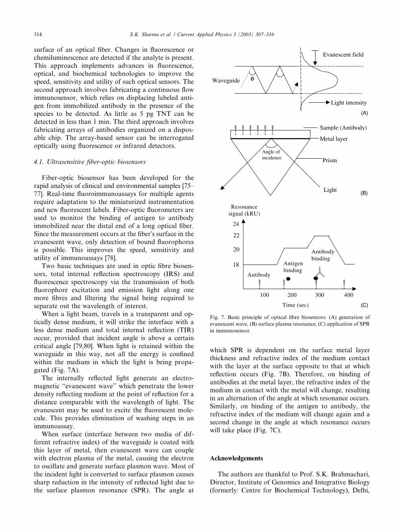
separate out the wavelength of interest.When a light beam, travels in a transparent and op-
tically dense medium, it will strike the interface with a
less dense medium and total internal reflection (TIR)
occur, provided that incident angle is above a certain
critical angle [79,80]. When light is retained within the
waveguide in this way, not all the energy is confined
within the medium in which the light is being propa-
gated (Fig. 7A).The internally reflected light generate an electro-
magnetic ‘‘evanescent wave’’ which penetrate the lower
density reflecting medium at the point of reflection for a
distance comparable with the wavelength of light. The
evanescent may be used to excite the fluorescent mole-
cule. This provides elimination of washing steps in an
immunoassay.
When surface (interface between two media of dif-ferent refractive index) of the waveguide is coated with
this layer of metal, then evanescent wave can couple
with electron plasma of the metal, causing the electron
to oscillate and generate surface plasmon wave. Most of
the incident light is converted to surface plasmon causes
sharp reduction in the intensity of reflected light due to
the surface plasmon resonance (SPR). The angle at
which SPR is dependent on the surface metal layer
thickness and refractive index of the medium contact
with the layer at the surface opposite to that at which
reflection occurs (Fig. 7B). Therefore, on binding of
antibodies at the metal layer, the refractive index of themedium in contact with the metal will change, resulting
in an alternation of the angle at which resonance occurs.
Similarly, on binding of the antigen to antibody, the
refractive index of the medium will change again and a
second change in the angle at which resonance occurs
will take place (Fig. 7C).
Acknowledgements
The authors are thankful to Prof. S.K. Brahmachari,
Director, Institute of Genomics and Integrative Biology
(formerly: Centre for Biochemical Technology), Delhi,
! ! ! ! ! !
Light
Prism
Metal layer
Sample (Antibody)
Angle ofincidence
! ! ! !100 200 300 400
Time (sec)
Antigenbinding
Antibodybinding
Antibody
Resonancesignal (kRU)
18
22
24
20
θ
Evanescent field
Light intensity
Waveguide
(A)
(B)
(C)
Fig. 7. Basic principle of optical fibre biosensors: (A) generation of
evanescent wave, (B) surface plasma resonance, (C) application of SPR
in immunosensor.
314 S.K. Sharma et al. / Current Applied Physics 3 (2003) 307–316
for encouragement in writing this manuscript. SandeepK. Sharma thanks CSIR, New Delhi for providing fel-
lowship.
References
[1] F.S. Ligler, Lecture on Biosensors, June 5, 1997, Biosensor and
Biomedical Laboratory, Naval Research Laboratory, ASEEE-
1818 N Street, NW, Washington DC, 200369 (2000). Available
from <www.asee.org>.
[2] C. Reid, Biosens. Bioelectron., June 5 (2000). Available from
[3] J.H.T. Luong, A. Mulchandani, G.G. Guilbault, Trends Biotech-
nol. 6 (1988) 310.
[4] J.A. Kas, R.M.J. Brown, C.E. Schmidt, Natural Polym. (2000).
Available from <www.asee.org>.
[5] J. Pickup, Trends Biotechnol. 1 (1993) 285.
[6] G.G. Guilbault, G. de Olivera Neto, Immobilized enzyme
electrode, in: J. Woodward (Ed.), Immobilized Cells and Enzymes:
A Practical Approach, IRL Press, Oxford, 1985, p. 56.
[7] F.W. Scheller, D. Pffiffer, F. Schubert, R. Renneberg, D. Kirstein,
Application of enzyme based amperometric biosensors to the
analysis of real samples, in: A.P.F. Turner, I. Karube, G.S. Wilson
(Eds.), Biosensor––Fundamental and Applications, Oxford Uni-
versity Press, New York, 1987, p. 319.
[8] D.P. Nikolelis, Anal. Chim. Acta 161 (1984) 343.
[9] S.A. Barker, Immobilization od biological components of biosen-
sor, in: A.P.F. Turner, I. Karube, G.S. Wilson (Eds.), Biosensor––
Fundamental and Application, Oxford University Press, New
York, 1987, p. 85.
[10] M.A. Arnold, G.A. Rechnitz, Biosensors based on plant and
animal tissues, in: A.P.F. Turner, I. Karube, G. Wilson (Eds.),
Biosensors––Fundamentals and Applications, Oxford University
Press, New York, 1987, p. 31.
[11] L. Clark, C. Lyons, Ann. N.Y. Acad. Sci. 102 (1962) 29.
[12] A.E.G. Cass, G. Davis, G.D. Francis, Anal. Chem. 56 (1984) 667.
[13] D.R. Marthews, R.R. Holman, E. Brown, Lancet 1 (1987) 778.
[14] J.C. Pickup, Biosensors for diabetic mellitus, in: D.L. Wise (Ed.),
Applied Biosensors, Butterworth Publishers, Stometham, MA,
USA, 1989, p. 234.
[15] G. Guilbault, Handbook of Immobilized Enzymes, Marcel
Dekker, New York, 1984, p. 87.
[16] I. Karube, I. Saitoh, Y. Araki, S. Suzuki, Enzyme Microb.
Technol. 2 (1980) 117.
[17] G.G. Guilbault, G.J. Lubrano, J.M. Kaufimann, G.J. Patriarche,
Anal. Chim. Acta 206 (1988) 369.
[18] G.G. Guilbault, M. Nianjo, Anal. Chem. 46 (1974) 1769.
[19] J. Kulis, M. Pesiekiene, V.S. Laurinavicius, S. Tatikyan, A.
Simonyan, Zh. Anal. Khim. 40 (1985) 2077.
[20] M. Jaenchen, G. Gruenig, K. Berterman, Anal. Lett. 18 (1985)
1799.
[21] H. Iwai, S. Akihama, Chem. Pharm. Bull. 34 (1986) 3471.
[22] T. Kawashima, A. Arima, N. Hatakeyma, N. Tominaga, M.
Ando, Nippon Kaqahu Kaishi (1980) 1542.
[23] S.S. Kuan, G.G. Guilbault, Ion selective electrodes and biosensors
based on ISEs, in: A.P.F. Turner, I. Karube, G.S. Wilson (Eds.),
Biosensor––Fundamental and Applications, Oxford University
Press, New York, 1987, p. 44.
[24] R.R. Coulet, G. Bardeletti, F. Schaud, in: D.L. Wise (Ed.),
Amperometric Enzyme Membrane Electrode, Marcel Dekker,
Bioinstrumentation and Biosensor Inc., New York, 1991, p. 758.
[25] B.J. Gould, B.F. Rocks, Enzyme in clinical analysis––data, in: A.
Wiseman (Ed.), Hand Book of Enzyme Biotechnology, Haisted
Press, New York, 1986, p. 434.
[26] Sigma Biochemicals and reagents for life science research
catalogue, Sigma-Aldrich Co., USA, 2001.
[27] K.A. Koshcheyenko, Prikil. Biochim. Microbiol. 27 (1981) 477.
[28] K.A. Koshcheyenko, Microbiologia 11 (1981) 55.
[29] S. Fukui, A. Tanaka, Annu. Rev. Microbiol. 36 (1982) 45.
[30] F.B. Kolor, Process Biochem. 17 (1982) 12.
[31] M. Hikuma, H. Suzuki, Y. Yasuda, I. Karube, S. Suzuki, Eur.
J. Appl. Microbiol. Biotechnol. 8 (1979) 289.
[32] M. Hikuma, H. Obamna, T. Yasuda, I. Karube, S. Suzuki,
Enzyme Microb. Technol. 2 (1980) 237.
[33] I. Karube, S. Mitsuda, S. Suzuki, Eur. J. Appl. Microbiol.
Biotechnol. 7 (1980) 343.
[34] J. Kulys, K. Kadziauskiene, Biotechnol. Bioeng. 22 (1980) 221.
[35] I. Karube, T. Okada, S. Suzuki, Anal. Chem. 53 (1981) 1852.
[36] A. Takase, Nippon Suisan Gakkaishi 19 (1953) 71.
[37] F. Ota, Z. Oshiro, Nippon Suisan Gakkaishi 19 (1954) 1150.
[38] Y. Yamamura, Nippon Suisan Gakkaishi 2 (1933) 118.
[39] T. Tokunaga, H. Iida, K. Miwa, Nippon Suisan Gakkaishi 43
(1977) 219.
[40] T. Suzuki, Nippon Suisan Gakkaishi 19 (1953) 102.
[41] S. Asakawa, Nippon Suisan Gakkaishi 24 (1959) 714.
[42] T. Kawabata, M. Fujimaki, K. Amano, F. Tomiya, Nippon
Suisan Gakkaishi 18 (1952) 124.
[43] M. Mikaye, K. Hayashi, Nippon Suisan Gakkaishi 21 (1955) 123.
[44] Y. Yamamura, Nippon Suisan Gakkaishi 5 (1936) 98.
[45] M. Yamamoto, M. Sonehara, Nippon Suisan Gakkaishi 19 (1953)
761.
[46] S. Ehira, H. Uchiyama, Nippon Suisan Gakkaishi 35 (1969) 1080.
[47] T. Saito, Nippon Suisan Gakkaishi 27 (1961) 461.
[48] Y. Fujii, H. Uchiyama, S. Ehira, E. Noguchi, Nippon Suisan
Gakkaishi 32 (1966) 410.
[49] S. Ehira, M. Anekawa, Nippon Suisan Gakkaishi 32 (1966) 716.
[50] T. Saito, K. Arai, M. Matsuyoshi, Nippon Suisan Gakkaishi 24
(1959) 749.
[51] H. Uchiyama, S. Ehira, Nippon Suisan Gakkaishi 36 (1970) 977.
[52] K. Yamada, S. Higashino, T. Kawahara, R. Ito, Nippon Suisan
Gakkaishi 47 (1981) 631.
[53] K. Numata, H. Suzuki, K. Usui, Nippon Shokuhin Kogyo
Gakkaishi 28 (1981) 542.
[54] N.R. Jones, J. Murrey, E.I. Livingston, C.K. Murrey, J. Sci.
Food. Agric. 15 (1964) 763.
[55] N.R. Jones, J. Murrey, J.R. Burt, J. Food Sci. 30 (1965) 791.
[56] F.D. Jahns, J.L. Howe, R.J. Coduri, A.G. Rand, Food Technol.
30 (1976) 27.
[57] J.R. Burt, J. Murrey, G.D. Stroud, J. Food Technol. 31 (1968)
165.
[58] E.H. Lee, T. Oshima, C. Koizumi, Nippon Suisan Gakkaishi 48
(1982) 255.
[59] E. Watanbe, K. Ando, I. Karube, H. Matsuoka, S. Suzuki, J.
Food Sci. 48 (1983) 496.
[60] E. Watanbe, K. Toyoma, I. Karube, H. Matsuoka, S. Suzuki,
Appl. Microbiol. Biotechnol. 19 (1984) 18.
[61] E. Watanbe, K. Toyoma, I. Karube, H. Matsuoka, S. Suzuki, J.
Food Sci. 49 (1984) 114.
[62] E. Watanbe, T. Ogura, K. Toyoma, I. Karube, H. Matsuoka, S.
Suzuki, Enzyme Microb. Technol. 6 (1984) 207.
[63] T. Saito, K. Arai, M.A. Matsuyoshi, Nippon Suisan Gakkaishi 24
(1959) 749.
[64] S. Ehira, H. Uchiyana, Nippon Suisan Gakkaishi 35 (1969) 1080.
[65] M. Hikuma, T. Kubo, T. Yasudo, I. Karube, S. Suzuki,
Biotechnol. Bioeng. 21 (1979) 1845.
[66] M. Hikuma, T. Kubo, T. Yasudo, I. Karube, S. Suzuki, Anal.
Chim. Acta 109 (1979) 33.
[67] E. Watanabe, A. Nagumo, M. Hoshi, S. Konagaya, M. Tanaka, J.
Food Sci. 52 (1987) 592.
[68] F.S. Ligler, D.A. Stenger, Fabrication of cell based biosensors,
Centre for Biomedical Science and Engg., Naval Research
S.K. Sharma et al. / Current Applied Physics 3 (2003) 307–316 315
Laboratory, ASEEE-1818 N Street, NW, Washington DC, 200369
(2000). Available from <www.asee.org>.
[69] I. Karube, Micro-organism based sensor, in: A.P.F. Turner, I.
Karube, G.S. Wilson (Eds.), Biosensor––Fundamental and Ap-
plications, Oxford University Press, New York, 1987, p. 13.
[70] I. Karube, Micro-organisms based sensors, in: A.P.F. Turner, I.
Karube, G.S. Wilson (Eds.), Biosensors: Fundamental and
Applications, Oxford University Press, Oxford, 1989, p. 28.
[71] L. Macholan, in: D.L. Wise (Ed.), Biocatalytic Membrane
Electrode, Marcel Dekker, Bioinstrumentation and Biosensor
Inc., New York, 1991, p. 349.
[72] I. Karube, S. Suzuki (Eds.), Microbial Biosensors, Biosensors––A
Practical Approach, IRL Press at Oxford University Press, New
York, 1996, p. 165.
[73] F.S. Ligler, G.P. Anderson, D.W. Conard, A.W. Kusterheek,
Immunologically based sensors, Centre for Biomedical Science
and Engg., Naval Research Laboratory, ASEEE-1818 N
Street, NW, Washington DC, 200369 (2000). Available from
<www.asee.org>.
[74] H.J. Kwon, H.I. Balcer, K.A. Kang, Comp. Biochem. Physiol. A.
Mol. Integr. Physiol. 132 (2002) 231.
[75] M.A. Cooper, Nat. Rev. Drug. Discov. 1 (2002) 515.
[76] N.M. Velasco-Garcia, M. Toby, Trends Biotechnol. 19 (2001)
433.
[77] S. Dong, X. Chen, J. Biotechnol. 82 (2002) 303.
[78] G.P. Anderson, F.S. Ligler, L.C. Shriver-lake, Ultrasensitive fiber-
optic biosensors, Centre for Biomedical Science and Engg., Naval
Research Laboratory, ASEEE-1818 N Street, NW, Washington
DC, 200369 (2000). Available from <www.asee.org>.
[79] D. Griffiths, G. Hall, Trends Biotechnol. 1 (1993) 122.
[80] G.A. Robinson, Optical immunosensors, in: A.P.F. Turner (Ed.),
Advances in Biosensors, Jai Press, London, 1991, p. 229.
316 S.K. Sharma et al. / Current Applied Physics 3 (2003) 307–316


