Embed Size (px)
Citation preview
�����������������
Citation: Garavaglia, M.L.;
Giustarini, D.; Colombo, G.; Reggiani,
F.; Finazzi, S.; Calatroni, M.; Landoni,
L.; Portinaro, N.M.; Milzani, A.;
Badalamenti, S.; et al. Blood Thiol
Redox State in Chronic Kidney
Disease. Int. J. Mol. Sci. 2022, 23, 2853.
https://doi.org/10.3390/ijms23052853
Academic Editor: Stefania Marzocco
Received: 31 December 2021
Accepted: 3 March 2022
Published: 5 March 2022
Publisher’s Note: MDPI stays neutral
with regard to jurisdictional claims in
published maps and institutional affil-
iations.
Copyright: © 2022 by the authors.
Licensee MDPI, Basel, Switzerland.
This article is an open access article
distributed under the terms and
conditions of the Creative Commons
Attribution (CC BY) license (https://
creativecommons.org/licenses/by/
4.0/).
International Journal of
Molecular Sciences
Review
Blood Thiol Redox State in Chronic Kidney DiseaseMaria Lisa Garavaglia 1 , Daniela Giustarini 2 , Graziano Colombo 1 , Francesco Reggiani 3,4, Silvia Finazzi 3,Marta Calatroni 3,4, Lucia Landoni 1, Nicola Marcello Portinaro 5, Aldo Milzani 1, Salvatore Badalamenti 3,Ranieri Rossi 2,*,† and Isabella Dalle-Donne 1,*,†
1 Department of Biosciences (Department of Excellence 2018–2022), Università degli Studi di Milano,Via Celoria 26, 20133 Milan, Italy; [email protected] (M.L.G.); [email protected] (G.C.);[email protected] (L.L.); [email protected] (A.M.)
2 Department of Biotechnology, Chemistry and Pharmacy (Department of Excellence 2018–2022),University of Siena, Via A. Moro 2, 53100 Siena, Italy; [email protected]
3 Nephrology and Dialysis Unit, IRCCS Humanitas Research Hospital, Via Manzoni 56, Rozzano,20089 Milan, Italy; [email protected] or [email protected] (F.R.);[email protected] (S.F.); [email protected] or [email protected] (M.C.);[email protected] (S.B.)
4 Department of Biomedical Sciences, Humanitas University, Pieve Emanuele, 20090 Milan, Italy5 Department of Medical Biotechnologies and Translational Medicine, Università degli Studi di Milano,
20133 Milan, Italy; [email protected]* Correspondence: [email protected] (R.R.); [email protected] (I.D.-D.)† These authors contributed equally to this work.
Abstract: Thiols (sulfhydryl groups) are effective antioxidants that can preserve the correct structureof proteins, and can protect cells and tissues from damage induced by oxidative stress. Abnormallevels of thiols have been measured in the blood of patients with moderate-to-severe chronic kidneydisease (CKD) compared to healthy subjects, as well as in end-stage renal disease (ESRD) patients onhaemodialysis or peritoneal dialysis. The levels of protein thiols (a measure of the endogenous antiox-idant capacity inversely related to protein oxidation) and S-thiolated proteins (mixed disulphides ofprotein thiols and low molecular mass thiols), and the protein thiolation index (the molar ratio of theS-thiolated proteins to free protein thiols in plasma) have been investigated in the plasma or red bloodcells of CKD and ESRD patients as possible biomarkers of oxidative stress. This type of minimallyinvasive analysis provides valuable information on the redox status of the less-easily accessibletissues and organs, and of the whole organism. This review provides an overview of reversiblemodifications in protein thiols in the setting of CKD and renal replacement therapy. The evidencesuggests that protein thiols, S-thiolated proteins, and the protein thiolation index are promisingbiomarkers of reversible oxidative stress that could be included in the routine monitoring of CKDand ESRD patients.
Keywords: chronic kidney disease; end-stage renal disease; haemodialysis; peritoneal dialysis;protein thiols; S-thiolated proteins; protein thiolation index
1. Introduction
Chronic kidney disease (CKD) affects approximately 10% of the adult populationworldwide [1]. In 2016, it was the ninth most common cause of death in high-incomecountries, and epidemiological projections—considering the demographic trends andlifestyles of this population—suggest that the mortality rates due to this pathology willmore than double globally by 2040 [2]. Therefore, the development of interventionalstrategies to support the prevention and management of CKD is of primary importancefor the health system. According to the Kidney Disease Improving Global Outcomes [3],CKD is classified into five stages based on the glomerular filtration rate (GFR), whichrepresents the best index of renal function, and three stages based on albuminuria levels.
Int. J. Mol. Sci. 2022, 23, 2853. https://doi.org/10.3390/ijms23052853 https://www.mdpi.com/journal/ijms
Int. J. Mol. Sci. 2022, 23, 2853 2 of 22
The rationale of this classification is the strong correlation between the GFR and albuminuriavalues and the prognosis of CKD [3]. CKD—regardless of the underlying aetiology—is slowly progressive, and several factors (i.e., uncontrolled hypertension and diabetes,proteinuria, inflammation, obesity) may accelerate the progression [4]. In a small percentageof patients, the disease can progress to end-stage renal disease (ESRD, CKD stage 5), inwhich renal replacement therapy (i.e., haemodialysis, peritoneal dialysis, and/or kidneytransplantation) is necessary to ensure survival. Haemodialysis (HD), peritoneal dialysis(PD), and kidney transplantation have distinct impacts on patients’ quality of life, and areassociated with different clinical outcomes and adverse events [5].
CKD is associated with a higher risk for cardiovascular mortality, as well as all-causemortality. Many experimental and clinical studies, of which a complete overview wasprovided in a recent review [6], have shown that the amplification of oxidative stressoccupies a central position in the intricate and intertwined thread of the mechanismsinvolved in the pathogenesis of CKD, and that the disease progression is associated witha significant increase in reactive oxygen species (ROS) generation. It is well establishedthat patients with CKD show a significantly increased oxidative stress compared to healthysubjects, which is present from the early stages of the disease and during its progression,which can result from a series of causal factors that influence and exacerbate each other ina holistic vicious circle [7].
The kidney is a metabolically very active organ, the epithelial cells of which are richin mitochondria in which redox reactions occur. Furthermore, ROS play an importantrole in the physiological regulation of renal function. This makes the kidney particularlyvulnerable to damage caused by oxidative stress, which can accelerate the progression ofkidney disease. Oxidative stress can be induced and enhanced in the context of the uremicmilieu [8–15], and of CKD-associated chronic low-grade inflammation [16–19]. Oxidativestress can be further exacerbated by other pro-oxidant conditions which are frequentlypresent in moderate-to-severe stages of CKD, and are even more in ESRD (e.g., advancedage, diabetes, dyslipidemia, hypertension, obesity, and infectious complications) [20–22],and by the lower levels of water-soluble antioxidant vitamins measured in CKD patientsdue to the dietary restriction of fresh fruits and vegetables required of them in order to avoidhyperkalaemia [23,24]. Furthermore, HD or PD can lead to increased oxidative stress [17,25],which is instead mitigated by restoring renal function after kidney transplantation [26,27].
In the last two decades or so, oxidative stress has been increasingly related to the onsetand/or progression of a growing number of human diseases. For this reason, there is aconsiderable research interest in the identification of oxidative stress biomarkers that canhave diagnostic, prognostic, and therapeutic value, for an ever-earlier prediction of thedisease onset and/or for the monitoring of disease progression. According to the BESTresource provided by the FDA-NIH Biomarker Working Group, [28], a clinically usefulbiomarker should be able to be measured reliably and reproducibly (it must be reasonablystable, present in accessible tissues or fluids, and cost-effective). Furthermore, it mustshow specificity for the disease, have prognostic value, or be correlated with the activity(progression, regression, and outcome after the intervention) of the disease. Regardingbiomarkers of oxidative stress, the increase in oxidative damage can be detected by theidentification of stable oxidation end products produced by different reaction pathways,because the in vivo detection of circulating ROS is extremely difficult due to their highbiological reactivity and short half-life.
Carbonylated proteins, nitrated protein, protein-bound di-tyrosines, advanced ox-idation protein products [29–35], and protein thiols (P-SH) [36] have been investigatedas reliable biomarkers of oxidative stress in correlation with diseases. Other oxidativestress biomarkers have been discussed in a recent comprehensive review, to which refer-ence should be made for an exhaustive description that goes beyond the objectives of thistext [37].
In the presence of oxidative stress, the nucleophilic functional groups (such as -OH,-NH2 and -SH) of proteins, polysaccharides and nucleic acids can be oxidized. Due to
Int. J. Mol. Sci. 2022, 23, 2853 3 of 22
its chemical characteristics, -SH is more reactive than -OH and -NH2; consequently, itis more prone to oxidation or conjugation. As far as proteins are concerned, P-SH canoxidize to sulfenic acid (P-SOH). The latter can be irreversibly oxidized either to sulphinic(P-SO2H) or to sulphonic (P-SO3H) acids [29]. Alternatively, P-SOH can be convertedback to a disulphide through reactions with low molecular mass thiols (LMM-SH), e.g.,homocysteine, cysteinylglycine, cysteine and glutathione, leading to the formation ofprotein/LMM-SH mixed disulphides, also known as S-thiolated proteins. These processeseventually allow the return to the P-SH reduced form [29].
Thiol-based redox systems protect cells and organisms against ROS, maintain redoxhomeostasis in various cellular compartments, and contribute to redox signalling andredox regulation [38]. Here, it is interesting to note that protein S-thiolation has a dualphysiological role: the storage of LMM-thiols and the regulation of protein activity [39,40].Moreover, S-thiolation is considered an adaptive response to protect P-SH from the lossof biological activity due to irreversible oxidation (i.e., further oxidation to P-SO2H andP-SO3H) [29,39,41]. Although protein S-thiolation is considered a defensive mechanism,it can also have pathological consequences in particular conditions. For instance, theproteins S-homocysteinylation and protein S-glutathionylation are recognized risk factorsfor vascular diseases [42,43].
The levels of P-SH and S-thiolated proteins in plasma or in isolated red blood cells(RBCs) have been suggested as biomarkers of oxidative stress in some human diseases, suchas chronic obstructive pulmonary disease and asthma [44], alkaptonuria [45], cardiovasculardiseases [43], and physiological ageing [46]. Furthermore, over the past two decades,numerous studies have highlighted the change in the levels of several oxidative damagebiomarkers, such as plasma P-SH and S-thiolated proteins in CKD stages 1–4 (i.e., patientswith different degrees of CKD but with residual renal function), and in ESRD patients.Recent reviews summarize the experimental evidence demonstrating the involvement ofROS in the progression of chronic renal failure, the oxidative damage produced at the levelof RBCs in chronic renal failure, and the ways in which these factors may be related to theonset of major systemic comorbidities such as cardiovascular disease (CVD) [47–49].
The aim of this review is to provide an update of the most relevant observationalstudies that have investigated P-SH and S-thiolated proteins in the plasma and RBCs ofCKD patients, and the ways in which their levels change in the progression from moderateto severe stages of CKD, and in patients with ESRD on HD and PD. Because the search forscreening markers that enable the early diagnosis and more effective monitoring of CKDis still ongoing, a complete review on this topic could allow us to create a wider vision toovercome the technical and economic difficulties that preclude the use of oxidative stressbiomarkers in clinical practice for CKD. Success in this area would allow the identificationof individuals who are at risk, as well as the prediction of the short- and long-term resultsof any therapeutic approach.
2. Protein Thiols and S-Thiolated Proteins in Plasma and RBCs
Multiple systems are involved in plasma redox signalling. These systems do not havethe same sensitivity to oxidants or respond the same way to antioxidants. Therefore, forcompleteness and clarity, we present here a brief overview of the thiols and S-thiolatedproteins present in plasma and RBCs, before addressing the review of the known alterationsof P-SH and S-thiolated proteins in patients with CKD.
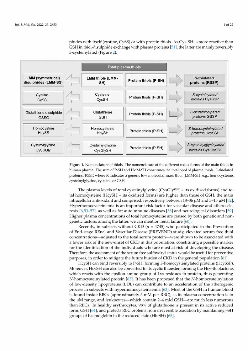
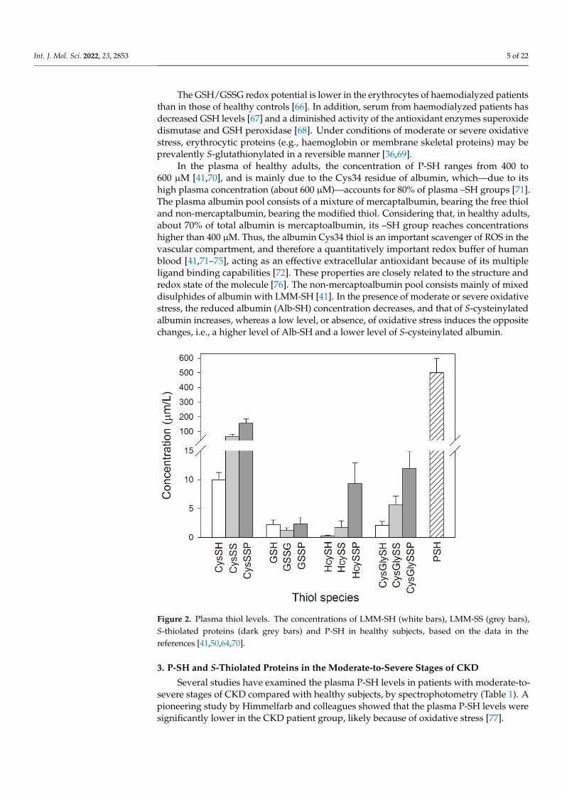
Plasma thiols can be categorized into P-SH and LMM-SH which are not bound to pro-tein; the latter can generate both low molecular mass symmetrical disulphides (LMM-SS)and mixed disulphides with proteins (or S-thiolated proteins, RSSP) (Figure 1). LMM-SH—i.e., homocysteine, cysteinylglycine, cysteine and glutathione (GSH)—are present in theplasma of healthy subjects at a low concentration range of 0.1–12 µM, and all togetherconstitute only 12–20 µM [41,50]. The major thiol/disulphide redox pool in mammalianplasma is the cysteine/cystine couple. Free cysteine (CysSH) has a physiological plasmaconcentration of approximately 10 µM [50]. It is susceptible to oxidation, forming disul-
Int. J. Mol. Sci. 2022, 23, 2853 4 of 22
phides with itself (cystine, CySS) or with protein thiols. As Cys-SH is more reactive thanGSH in thiol-disulphide exchange with plasma proteins [51], the latter are mainly reversiblyS-cysteinylated (Figure 2).
Int. J. Mol. Sci. 2022, 23, x FOR PEER REVIEW 4 of 24
constitute only 12–20 μM [41,50]. The major thiol/disulphide redox pool in mammalian
plasma is the cysteine/cystine couple. Free cysteine (CysSH) has a physiological plasma
concentration of approximately 10 µM [50]. It is susceptible to oxidation, forming disul-
phides with itself (cystine, CySS) or with protein thiols. As Cys-SH is more reactive than
GSH in thiol-disulphide exchange with plasma proteins [51], the latter are mainly revers-
ibly S-cysteinylated (Figure 2).
Figure 1. Nomenclature of thiols. The nomenclature of the different redox forms of the main thiols
in human plasma. The sum of P-SH and LMM-SH constitutes the total pool of plasma thiols. S-
thiolated proteins: RSSP, where R indicates a generic low molecular mass thiol (LMM-SH, e.g., ho-
mocysteine, cysteinylglycine, cysteine or GSH.
The plasma levels of total cysteinylglycine (CysGlySH + its oxidised forms) and total
homocysteine (HcySH + its oxidised forms) are higher than those of GSH, the main intra-
cellular antioxidant and comprised, respectively, between 18–36 μM and 5–15 μM [52].
Hyperhomocysteinemia is an important risk factor for vascular disease and atherosclero-
sis [6,53–57], as well as for autoimmune diseases [58] and neurological disorders [59].
Higher plasma concentrations of total homocysteine are caused by both genetic and non-
genetic factors: among the latter, we can mention renal failure [60].
Recently, in subjects without CKD (n = 4745) who participated in the Prevention of
End-stage REnal and Vascular Disease (PREVEND) study, elevated serum free thiol con-
centrations—adjusted to the total serum protein—were shown to be associated with a
lower risk of the new-onset of CKD in this population, constituting a possible marker for
the identification of the individuals who are most at risk of developing the disease. There-
fore, the assessment of the serum free sulfhydryl status would be useful for preventive
purposes, in order to mitigate the future burden of CKD in the general population [61].
HcySH can bind reversibly to P-SH, forming S-homocysteinylated proteins
(HcySSP). Moreover, HcySH can also be converted to its cyclic thioester, forming the Hcy-
thiolactone, which reacts with the epsilon-amino group of Lys residues in protein, thus
generating N-homocysteinylated protein [62]. It has been proposed that the N-homocys-
teinylation of low-density lipoproteins (LDL) can contribute to an acceleration of the ath-
erogenic process in subjects with hyperhomocysteinaemia [63]. Most of the GSH in human
blood is found inside RBCs (approximately 3 mM per RBC), as its plasma concentration
is in the µM range, and leukocytes—which contain 2–4 mM GSH—are much less numer-
ous than RBCs. In healthy erythrocytes, 98% of glutathione is present in its active reduced
Figure 1. Nomenclature of thiols. The nomenclature of the different redox forms of the main thiols inhuman plasma. The sum of P-SH and LMM-SH constitutes the total pool of plasma thiols. S-thiolatedproteins: RSSP, where R indicates a generic low molecular mass thiol (LMM-SH, e.g., homocysteine,cysteinylglycine, cysteine or GSH.
The plasma levels of total cysteinylglycine (CysGlySH + its oxidised forms) and to-tal homocysteine (HcySH + its oxidised forms) are higher than those of GSH, the mainintracellular antioxidant and comprised, respectively, between 18–36 µM and 5–15 µM [52].Hyperhomocysteinemia is an important risk factor for vascular disease and atheroscle-rosis [6,53–57], as well as for autoimmune diseases [58] and neurological disorders [59].Higher plasma concentrations of total homocysteine are caused by both genetic and non-genetic factors: among the latter, we can mention renal failure [60].
Recently, in subjects without CKD (n = 4745) who participated in the Preventionof End-stage REnal and Vascular Disease (PREVEND) study, elevated serum free thiolconcentrations—adjusted to the total serum protein—were shown to be associated witha lower risk of the new-onset of CKD in this population, constituting a possible markerfor the identification of the individuals who are most at risk of developing the disease.Therefore, the assessment of the serum free sulfhydryl status would be useful for preventivepurposes, in order to mitigate the future burden of CKD in the general population [61].
HcySH can bind reversibly to P-SH, forming S-homocysteinylated proteins (HcySSP).Moreover, HcySH can also be converted to its cyclic thioester, forming the Hcy-thiolactone,which reacts with the epsilon-amino group of Lys residues in protein, thus generatingN-homocysteinylated protein [62]. It has been proposed that the N-homocysteinylationof low-density lipoproteins (LDL) can contribute to an acceleration of the atherogenicprocess in subjects with hyperhomocysteinaemia [63]. Most of the GSH in human bloodis found inside RBCs (approximately 3 mM per RBC), as its plasma concentration is inthe µM range, and leukocytes—which contain 2–4 mM GSH—are much less numerousthan RBCs. In healthy erythrocytes, 98% of glutathione is present in its active reducedform, GSH [64], and protects RBC proteins from irreversible oxidation by maintaining –SHgroups of haemoglobin in the reduced state (Hb-SH) [65].
Int. J. Mol. Sci. 2022, 23, 2853 5 of 22
The GSH/GSSG redox potential is lower in the erythrocytes of haemodialyzed patientsthan in those of healthy controls [66]. In addition, serum from haemodialyzed patients hasdecreased GSH levels [67] and a diminished activity of the antioxidant enzymes superoxidedismutase and GSH peroxidase [68]. Under conditions of moderate or severe oxidativestress, erythrocytic proteins (e.g., haemoglobin or membrane skeletal proteins) may beprevalently S-glutathionylated in a reversible manner [36,69].
In the plasma of healthy adults, the concentration of P-SH ranges from 400 to600 µM [41,70], and is mainly due to the Cys34 residue of albumin, which—due to itshigh plasma concentration (about 600 µM)—accounts for 80% of plasma –SH groups [71].The plasma albumin pool consists of a mixture of mercaptalbumin, bearing the free thioland non-mercaptalbumin, bearing the modified thiol. Considering that, in healthy adults,about 70% of total albumin is mercaptoalbumin, its –SH group reaches concentrationshigher than 400 µM. Thus, the albumin Cys34 thiol is an important scavenger of ROS in thevascular compartment, and therefore a quantitatively important redox buffer of humanblood [41,71–75], acting as an effective extracellular antioxidant because of its multipleligand binding capabilities [72]. These properties are closely related to the structure andredox state of the molecule [76]. The non-mercaptoalbumin pool consists mainly of mixeddisulphides of albumin with LMM-SH [41]. In the presence of moderate or severe oxidativestress, the reduced albumin (Alb-SH) concentration decreases, and that of S-cysteinylatedalbumin increases, whereas a low level, or absence, of oxidative stress induces the oppositechanges, i.e., a higher level of Alb-SH and a lower level of S-cysteinylated albumin.
Int. J. Mol. Sci. 2022, 23, x FOR PEER REVIEW 5 of 24
form, GSH [64], and protects RBC proteins from irreversible oxidation by maintaining –
SH groups of haemoglobin in the reduced state (Hb-SH) [65].
The GSH/GSSG redox potential is lower in the erythrocytes of haemodialyzed pa-
tients than in those of healthy controls [66]. In addition, serum from haemodialyzed pa-
tients has decreased GSH levels [67] and a diminished activity of the antioxidant enzymes
superoxide dismutase and GSH peroxidase [68]. Under conditions of moderate or severe
oxidative stress, erythrocytic proteins (e.g., haemoglobin or membrane skeletal proteins)
may be prevalently S-glutathionylated in a reversible manner [36,69].
In the plasma of healthy adults, the concentration of P-SH ranges from 400 to 600 μM
[41,70], and is mainly due to the Cys34 residue of albumin, which—due to its high plasma
concentration (about 600 μM)—accounts for 80% of plasma –SH groups [71]. The plasma
albumin pool consists of a mixture of mercaptalbumin, bearing the free thiol and non-
mercaptalbumin, bearing the modified thiol. Considering that, in healthy adults, about
70% of total albumin is mercaptoalbumin, its –SH group reaches concentrations higher
than 400 μM. Thus, the albumin Cys34 thiol is an important scavenger of ROS in the vas-
cular compartment, and therefore a quantitatively important redox buffer of human blood
[41,71–75], acting as an effective extracellular antioxidant because of its multiple ligand
binding capabilities [72]. These properties are closely related to the structure and redox
state of the molecule [76]. The non-mercaptoalbumin pool consists mainly of mixed disul-
phides of albumin with LMM-SH [41]. In the presence of moderate or severe oxidative
stress, the reduced albumin (Alb-SH) concentration decreases, and that of S-cysteinylated
albumin increases, whereas a low level, or absence, of oxidative stress induces the oppo-
site changes, i.e., a higher level of Alb-SH and a lower level of S-cysteinylated albumin.
Figure 2. Plasma thiol levels. The concentrations of LMM-SH (white bars), LMM-SS (grey bars), S-
thiolated proteins (dark grey bars) and P-SH in healthy subjects, based on the data in the references
[41,50,64,70].
3. P-SH and S-Thiolated Proteins in the Moderate-to-Severe Stages of CKD
Several studies have examined the plasma P-SH levels in patients with moderate-to-
severe stages of CKD compared with healthy subjects, by spectrophotometry (Table 1). A
pioneering study by Himmelfarb and colleagues showed that the plasma P-SH levels were
significantly lower in the CKD patient group, likely because of oxidative stress [77].
Figure 2. Plasma thiol levels. The concentrations of LMM-SH (white bars), LMM-SS (grey bars),S-thiolated proteins (dark grey bars) and P-SH in healthy subjects, based on the data in thereferences [41,50,64,70].
3. P-SH and S-Thiolated Proteins in the Moderate-to-Severe Stages of CKD
Several studies have examined the plasma P-SH levels in patients with moderate-to-severe stages of CKD compared with healthy subjects, by spectrophotometry (Table 1). Apioneering study by Himmelfarb and colleagues showed that the plasma P-SH levels weresignificantly lower in the CKD patient group, likely because of oxidative stress [77].
Int. J. Mol. Sci. 2022, 23, 2853 6 of 22
Table 1. P-SH and S-thiolated proteins in the plasma or serum of patients with moderate-to-severestages of CKD. All of the data are expressed as means ± SD, unless otherwise indicated. (a) Median,with interquartile range in parentheses. The data of S-thiolated proteins are in grey cells. *** p < 0.001,** p < 0.01, * p < 0.05.
Concentration Albumin Concentration mg/dLAdditional Information Reference
CKD Patients Control Group CKD Patients Control Group
202 ± 20 µM (n = 10) 279 ± 12 µM (n = 10) - - CKD patients not receivingrenal replacement therapy [77]
182.3 ± 15.9 µg/L(n = 24) ***
286.4 ± 21.8 µg/L(n = 20) - -
albuminsignificantly lower in CKD
patients compared tohealthy controls (p < 0.0001)
Analyses performedin serum
[78]
304.0 ± 55.2 µM(n = 184) ***
328.4 ± 33.3 µM(n = 43) 4.3 ± 0.38 4.4 ± 0.22 CKD patients at stages 3, 4 [79]
191.26 ± 16.75 µM(n = 41) **
342.34 ± 43.43 µM(n = 41) 2.1 ± 1.0 4.4 ± 1.3
CKD patients onconservative treatment
Analyses performedin serum
[80]
6.3 ± 0.9 µmol/gprotein
(n = 16) *
7.3 ± 0.8 µmol/gprotein(n = 13)
- - Non dialysis CKD patients [81]
3.59 (3.31–4.80)µmol/g protein (a)
(n = 24)- - - CKD hypertensive patients
stages 3, 4 [82]
stage 2:9.8 ± 3.5 nmol/mg
albuminstage 3:
9.9 ± 2.6 nmol/mgalbuminstages 5:
7.9 ± 2.5 nmol/mgalbumin(n = 68)
- - - - [83]
69.44 ± 7.26%(n = 55) - 4.1 ± 0.4 - Predialysis patients
with CKD [84]28.59 ± 6.93% S-thiolated albumin
stages 1, 277.2 ± 3.4% (n = 7)
stage 3a75.5 ± 3.8%
(n = 7)stage 3b
71.5 ± 3.3% (n = 6)stages 4, 5
66.2 ± 4.1% (n = 12)
-
stages 1, 24.0 ± 0.1stage 3a4.0 ± 0.1stage 3b4.1 ± 0.3
stages 4, 54.1 ± 0.3
-
[85]stages 1, 2
21.0 ± 3.4% (n = 7)stage 3a
22.4 ± 4.1% (n = 7)stage 3b
26.2 ± 3.1% (n = 6)stages 4, 5
31.1 ± 4.1% (n = 12)
S-thiolated albumin
Int. J. Mol. Sci. 2022, 23, 2853 7 of 22
Table 1. Cont.
Concentration Albumin Concentration mg/dLAdditional Information Reference
CKD Patients Control Group CKD Patients Control Group
633 ± 248 nM 430 ± 153 nM
S-cysteinylated +S-homocysteinylated LDL
Analyses carried out bycapillary electrophoresis
laser-inducedfluorescence detection
[86]
Successively, lower plasma P-SH levels in CKD patients were correlated to lowerplasma albumin levels (hypoalbuminaemia) [78], suggesting a causal relationship betweenhypoalbuminaemia and lower plasma P-SH levels. A positive association between P-SHand the estimated glomerular filtration rate (eGFR), and a negative correlation betweenP-SH and creatinine was also demonstrated in adult CKD patients in a recent study, whichalso detected a significant reduction in P-SH in stage 5 CKD patients [87]. However,the albumin levels were not evaluated, and the control group subjects were significantlyyounger than CKD patients [87].
The levels of total plasma thiols (P-SH + LMM-SH) were measured in a group ofpatients with CKD stages 1–5, most of whom were stage 3 (46%) and stage 4 (24%): the levelsof plasma thiols, adjusted to the plasma albumin concentrations, decreased dependentlyon the CKD stage [83]. Moreover, a positive correlation between the eGFR and thiolconcentration was observed [83]. A decrease in Alb-SH accompanied by a decrease in renalfunction was confirmed in non-diabetic CKD patients [88]. Decreased thiol and albuminlevels have also been reported in a more recent study conducted on a small group of childrenwith stage 3–5 CKD [89]. This study included and grouped data from both haemodialyzedand non-haemodialyzed patients, making it difficult to understand the real burden of theresults differentiated for the two groups. However, a positive correlation between thealbumin concentration and levels of thiols, (mixed and symmetrical) disulphides, and totalthiols was found in the plasma of CKD children. When the thiol, disulphide, and total thiollevels were normalized to the albumin concentration, only the levels of total thiols andthiols were significantly lower in CKD children than in age-matched control subjects. Inthe CKD group, the levels of thiols, total thiols, and disulphides correlated positively withthe albumin concentration, and negatively with the urea concentration, whereas the thioland total thiol levels were positively correlated with the eGFR and negatively with thecreatinine level [89]. Two other recent studies on adult CKD patients not on HD determinedthat total plasma thiol, and the (“native”) thiol levels were significantly lower in CKDpatients than in healthy controls [90,91]. One study also found a significant reduction in thelevel of disulphides but no significant difference in the disulphide/thiol, disulphide/totalthiol, and thiol/total thiol ratios between the CKD and healthy control groups [91]. Theother study found a significant variation in disulphide/thiol and disulphide/total thiolratios only [90]. This discrepancy could be explained by the fact that the control group inthe latter study had a significantly higher BMI than the CKD group, causing an increase inoxidative stress in the control group with increased levels of disulphides [92].
Terawaki and colleagues first adopted the redox state of albumin Cys34 measured byan HPLC method as a marker of oxidative stress in pre-dialysis patients with CKD. Theirfindings indicated that both S-cysteinylated albumin and irreversibly oxidized albumin(Alb-SO2H and Alb-SO3H) increased as renal function, measured as creatinine clearance,decreased [84]. The authors confirmed their findings in a subsequent study, where theyalso showed that albumin-SH levels progressively decreased with decreasing kidney func-tion [85]. Conversely, the levels of S-thiolated albumin increased progressively as kidneyfunction decreased, suggesting that the redox state of albumin progressively changes withthe worsening of kidney function. Moreover, they found a significant positive correlationbetween S-thiolated albumin and blood urea nitrogen, and between S-thiolated albumin
Int. J. Mol. Sci. 2022, 23, 2853 8 of 22
and serum creatinine. Another recent study analysed the serum fraction of oxidized albu-min normalized to total albumin (i.e., oxidized and native albumin) in pre-dialysis CKDpatients [93]. The correlation between the oxidized serum fraction of albumin, f(HNA), andnumerous clinical parameters related to CKD was examined. The study revealed a positivecorrelation between f(HNA), kidney function and urea nitrogen in the blood, and also withthe degree of anaemia of the CKD patients, independent of age; f(HNA) was also signifi-cantly and independently associated with the plasma levels of haemoglobin and ferritin. Anegative correlation was demonstrated between the levels of xanthine oxidase—an isoformof xanthine oxidoreductase which is thought to increase the cardiovascular burden amongCKD patients via oxidative radical production—and the plasma fraction of Alb-SH andreversibly oxidized albumin [94]. These findings suggest that the analysis of the redox stateof albumin Cys34 thiol can be a potentially useful method for the assessment of oxidativestress and the degree of kidney dysfunction in CKD patients, although some analytical andinstrumental limitations must be overcome.
Elevated levels of plasma HcySH are commonly present in patients with CKD. Ina recent study involving 62 patients in different stages of CKD, a negative correlationwas found between the plasma HcySH and eGFR in patients with CKD stages III to V,although a significant increase in HcySH levels was present only in subjects with stageV compared to patients with stage II CKD. The plasma Cys/Hcy ratio turned out to bea more sensitive indicator because it was already significantly decreased at stage III ofCKD [95]. Zinellu and colleagues reported that HcySH and CysSH bound to LDL weresignificantly increased in CKD patients, and the plasma levels of S-homocysteinylated LDLand creatinine were positively correlated with each other [86]. Based on these results, theauthors suggested that the increase in S-homocysteinylated LDL levels might account, atleast in part, for the excess of cardiovascular risk in CKD patients: S-homocysteinylatedLDL can therefore be considered a key risk marker of cardiovascular disease (CVD) inCKD patients [86]. Another study detected a significant increase in S-cysteinylated andS-homocysteinylated plasma proteins in CKD patients compared to healthy subjects [96].More recently, Zinellu and colleagues evaluated the plasma levels of P-SH and LMM-SHin a group of hypertensive CKD patients at stages 3–4, more than 60% of whom werehyperhomocysteinemic (HcySH > 15 µmol/L), reporting that the thickness of the carotidintima-media was inversely related to plasma levels of P-SH [82].
4. P-SH and S-Thiolated Proteins in HD Patients Compared to Healthy Subjects
Several studies have examined the plasma levels of P-SH and S-thiolated proteinsin ESRD patients on maintenance HD compared to healthy subjects; others detected theconcentration of S-thiolated proteins in the RBCs of HD patients (Table 2).
Table 2. P-SH and S-thiolated proteins in the plasma or serum of ESRD patients on HD. All of thedata are expressed as means ± SD, unless otherwise indicated. Data regarding S-thiolated proteinsare in the grey cells. (b) Median with interquartile range in parentheses; (c) median with the 25–75th percentile in parentheses. HbSSCy, cysteinylated haemoglobin; HbSSG, S-glutathionylatedhaemoglobin; PTI, protein thiolation index.
Concentration Albumin Concentration g/dLAdditional Information Reference
HD Patients Control Group HD Patients Control Group
178 ± 18 µM(n = 10) *** 279 ± 12 µM (n = 10) - - - [77]
Int. J. Mol. Sci. 2022, 23, 2853 9 of 22
Table 2. Cont.
Concentration Albumin Concentration g/dLAdditional Information Reference
HD Patients Control Group HD Patients Control Group
normoalbuminemicpatients
299.28 ± 8.64 µM(n = 18) ***
hypoalbuminemicpatients
229.00 ± 9.43 µM(n = 18) ***
428.39 ± 8.61 µM(n = 18)
normoalbuminemicpatients
4.2 ± 0.02hypoalbuminemic
patients2.9 ± 0.03
- - [97]
174.9 ± 10.5 µg/L(n = 32) ***
286.4 ± 21.8 µg/L(n = 20)
albuminsignificantly
lower in CKDpatients
compared tohealthy controls
(p < 0.0001)
-
Dialysis vintage16 ± 4 months
Analyses performedin serum
[78]
280 ± 11 µM(n = 28)
416 ± 6 µM(n = 49) 3.78 ± 0.6 - - [98]
170.79 ± 12.31 µM(n = 22) ***
286.4 ± 21.8 µM(n = 20) - -
Dialysis vintage16 ± 4 months
Analyses performedin serum
[99]
164.12 ± 13.54 µM(n = 48) **
342.34 ± 43.43 µM(n = 41) 2.8 ± 0.2 4.4 ± 1.3
Dialysis vintage16 ± 4 months
Analyses performedin serum
[80]
Figure 3(n = §) ***
Figure 3(n = 24) - - - [100]
3.72 ± 0.66 µmol/gprotein
(n = 20) ***
4.73 ± 0.71 µmol/gprotein (n = 20) 7.37 ± 0.49 (a) 7.61 ± 0.386 Dialysis vintage
4.4 ± 1.9 months [101]
36.0 ± 6.03%(n = 22)
64.6 ± 0.4%(n = 11) - - Dialysis vintage 1–9 years
[102]29.7 ± 0.5% 29.7 ± 0.5% S-cysteinylated albumin
40.4 ± 8.7%(n = 20) **
53.6 ± 6.4%(n = 10) - - Dialysis vintage
1–10 years [103]49.7 ± 8.0%) ** 38.7 ± 6.3% - - S-cysteinylated albumin
825.53 ± 121.08 ***pmol/mg
(n = 28)
139.52 ± 15.43pmol/mg
(n = 14)- -
Dialysis vintage> 6 months
S-homocysteinylatedproteins
[104]
mean 0.76 (min–max0.61–0.88) (b)(n = 71) ***
mean0.43 (min–max0.40–0.54) (b)
(n = 24)3.49 ± 0.38 4.23 ± 0.31 PTI [105]
19.3 ± 4.80 pmol/mgHb
(n = 33) ***
13.2 ± 2.79 pmol/mg(n = 21) HbSSG [66]
11.5 (9.6–17.2)pmol/mg Hb (c) ***
38.3 (29.0–63.3)pmol/mg Hb (c) HbSSCy [66]
*** p ≤ 0.001; ** p ≤ 0.01. (a) Total plasma protein. § The patients were divided into four groups according to theHD duration: group 1, 0–2 years of treatment (n = 31); group 2, 3–5 years of treatment (n = 40); group 3, 6–8 yearsof treatment (n = 27); group 4, 9–11 years of treatment (n = 13). There was no significant difference in the mean agebetween the groups. The plasma –SH levels were significantly lower in all of the patient groups than in controls(all groups p < 0.001).
Int. J. Mol. Sci. 2022, 23, 2853 10 of 22Int. J. Mol. Sci. 2022, 23, x FOR PEER REVIEW 11 of 24
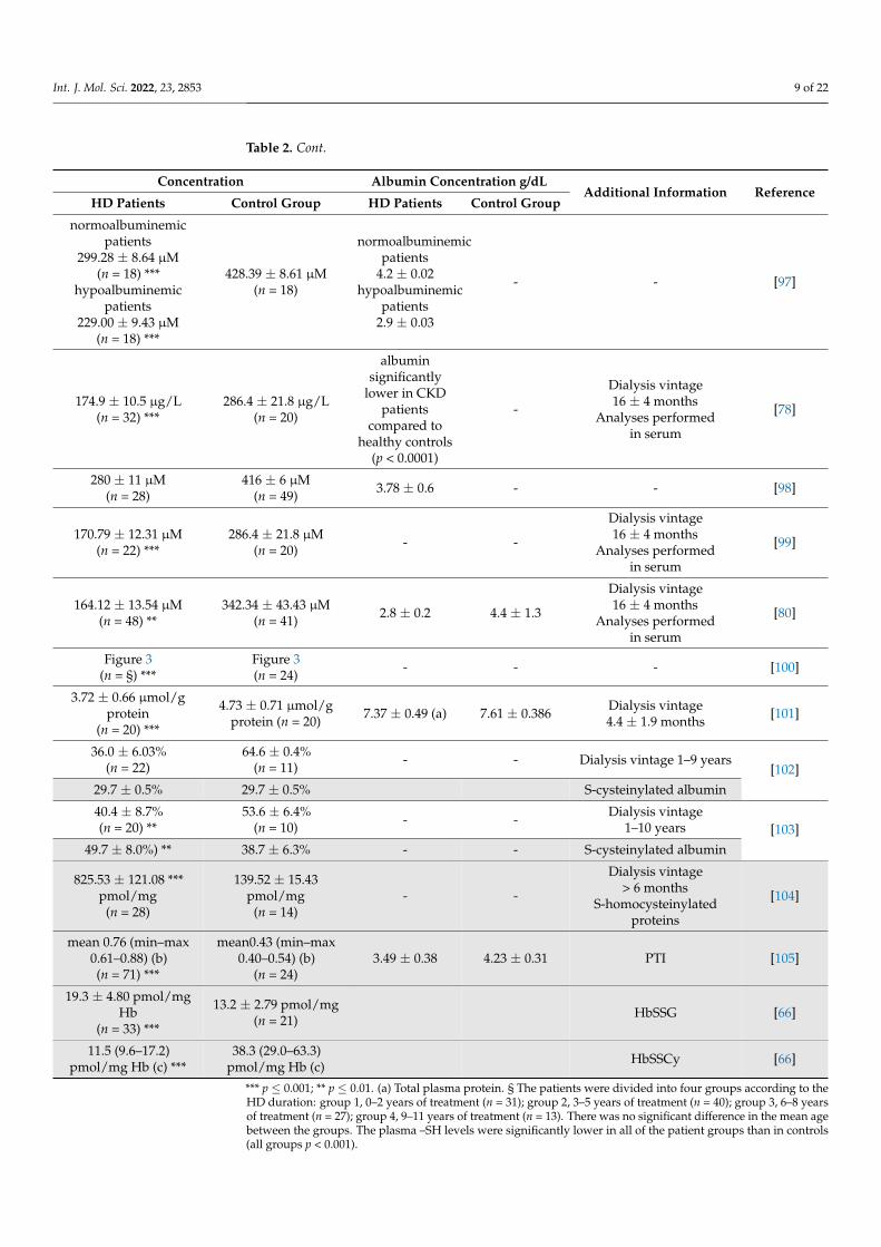
Figure 3. S-thiolated plasma proteins and PTI in ESRD patients compared to healthy subjects. (A)
The plasma proteins of ESRD patients (n = 20) and age-matched healthy subjects (n = 20) were ana-
lysed for their total S-thiolated protein content by colorimetric reaction with ninhydrin. The data
are expressed as the mean ± SD. The differences between the means of the two groups were evalu-
ated using Student’s t-test (** = p < 0.001). (B) The plasma of both ESRD patients (n = 20) and age-
matched healthy subjects (n = 20) was analysed for PTI. The data are expressed as the mean ± SD.
The differences between means of the two groups were evaluated using Student’s t-test (** = p <
0.001). Reprinted with permission from Colombo et al. [101].
Several studies examined in HD patients the levels of plasma S-thiolated proteins
[102–105] and S-thiolated haemoglobin in RBCs [66] (Table 2). The plasma levels of Alb-
SH and S-cysteinylated albumin were determined in HD patients subjected to intravenous
iron administration by the HPLC method [84]. HD patients had increased levels of S-cys-
teinylated albumin and irreversibly oxidized albumin (Alb-SO2H and Alb-SO3H), and de-
creased levels of Alb-SH [102]. On the other hand, the plasma levels of S-homocysteinyl-
ated proteins were significantly higher in HD patients than in healthy subjects [104]. This
finding could have functional consequences; indeed, the site of interaction with diazepam
in homocysteinylated albumin is altered due to the presence of the sulphur amino acid,
and the interaction between the drug and the protein is reduced, partly explaining why
the albumin-binding capacity is reduced in uraemia [104]. Other investigations deter-
mined that ESRD patients show increased levels of HcySH, which have been related to
carotid artery atherosclerosis and increased rates of cardiovascular events [109,110]. Other
studies have shown that S-glutathionylated haemoglobin (HbSSG) was 46% higher in HD
patients than in healthy subjects, and that S-cysteinylated Hb (HbSSCy) was >3-fold
higher in HD patients than in controls [66]. In addition, both HbSSCy and HbSSG were
positively correlated with blood urea nitrogen, creatinine, and normalized protein cata-
bolic rate, which are parameters related to both uraemia and nutrition [66].
5. P-SH and S-Thiolated Proteins in HD Patients Before and After an HD Session
Several studies have measured the levels of P-SH and S-thiolated proteins in HD pa-
tients before (pre-HD) and after an HD session (post-HD) (Table 3). Some authors re-
ported that the levels of total plasma thiols (P-SH + LMM-SH) significantly decreased
post-HD, returning to the range measured in healthy subjects [111–113]. The plasma P-SH
levels in ESRD patients on HD were significantly lower than those in healthy subjects.
However, the HD session was associated with an increase in the plasma P-SH levels, such
that after HD the concentration of P-SH did not differ from that of healthy subjects. Similar
results were also obtained when plasma P-SH were expressed as nanomoles per milligram
of protein, calculated using total plasma protein concentrations [112]. This result was also
confirmed when considering possible dialysis-induced changes in the plasma protein con-
centration [113], which can dramatically affect the measured values of total plasma thiols
Figure 3. S-thiolated plasma proteins and PTI in ESRD patients compared to healthy subjects. (A) Theplasma proteins of ESRD patients (n = 20) and age-matched healthy subjects (n = 20) were analysedfor their total S-thiolated protein content by colorimetric reaction with ninhydrin. The data areexpressed as the mean ± SD. The differences between the means of the two groups were evaluatedusing Student’s t-test (** = p < 0.001). (B) The plasma of both ESRD patients (n = 20) and age-matchedhealthy subjects (n = 20) was analysed for PTI. The data are expressed as the mean ± SD. Thedifferences between means of the two groups were evaluated using Student’s t-test (** = p < 0.001).Reprinted with permission from Colombo et al. [101].
Himmelfarb and colleagues reported a significant decrease in plasma P-SH in HD pa-tients [77], in line with what has also been observed in other studies (Table 2). For example,the plasma P-SH levels (measured in µmol/L) decreased significantly in hypoalbuminemicHD patients, both compared to healthy subjects and to normoalbuminemic HD patients,as well as between the latter and healthy subjects [97]. The levels of inflammation andoxidative stress biomarkers increased in both groups of HD patients compared to healthysubjects, and further selective increases in levels of inflammation and oxidative stressbiomarkers occurred in hypoalbuminemic patients, suggesting that hypoalbuminemia,acute-phase inflammation, and oxidative stress could act synergistically to increase cardio-vascular morbidity and mortality risk in HD patients [97]. Significantly lower plasma P-SHlevels in hypoalbuminemic HD patients could be due, at least in part, to hypoalbuminemia.
Plasma P-SH has also been investigated in HD patients not receiving iron or re-combinant human erythropoietin [99]. Iron deficiency is common in HD patients [106]:intravenous iron administration is one of the cornerstones of anaemia’s treatment, but itcould further aggravate oxidative stress [107]. The plasma P-SH levels were found to belower in HD patients not receiving intravenous iron or recombinant human erythropoietinvs. healthy controls [99]; however, this study did not specify whether the HD patients werenormoalbuminemic or hypoalbuminemic.
Koca and colleagues divided HD patients into four groups according to their HD dura-tion (dialysis vintage) [100], reporting that the total plasma thiols were significantly lowerin all of the groups of HD patients than in healthy subjects, but no significant differencewas found between the groups of HD patients with different dialysis vintages [100].
The protein thiolation index (PTI, a new biomarker of oxidative stress that is definedas the molar ratio of the sum of all LMM-SH bound to S-thiolated plasma proteins to freeP-SH [46,108]) is higher in HD patients than in controls, whereas the concentration of P-SHand that of total plasma thiols are lower in HD patients than in healthy subjects [90,101,105](Figure 3). P-SH and PTI provide different but complementary information. A low P-SHconcentration indicates an absolute loss of protein reducing power but does not provideinformation on concomitant changes in disulphide forms, while a high PTI indicates arelative excess of oxidized vs. reduced proteins in patients; however, being a ratio, it offersno indication about the absolute amounts of disulphide and reduced forms.
Int. J. Mol. Sci. 2022, 23, 2853 11 of 22
Several studies examined in HD patients the levels of plasma S-thiolated proteins [102–105]and S-thiolated haemoglobin in RBCs [66] (Table 2). The plasma levels of Alb-SH andS-cysteinylated albumin were determined in HD patients subjected to intravenous iron ad-ministration by the HPLC method [84]. HD patients had increased levels of S-cysteinylatedalbumin and irreversibly oxidized albumin (Alb-SO2H and Alb-SO3H), and decreasedlevels of Alb-SH [102]. On the other hand, the plasma levels of S-homocysteinylated pro-teins were significantly higher in HD patients than in healthy subjects [104]. This findingcould have functional consequences; indeed, the site of interaction with diazepam in ho-mocysteinylated albumin is altered due to the presence of the sulphur amino acid, andthe interaction between the drug and the protein is reduced, partly explaining why thealbumin-binding capacity is reduced in uraemia [104]. Other investigations determinedthat ESRD patients show increased levels of HcySH, which have been related to carotidartery atherosclerosis and increased rates of cardiovascular events [109,110]. Other studieshave shown that S-glutathionylated haemoglobin (HbSSG) was 46% higher in HD patientsthan in healthy subjects, and that S-cysteinylated Hb (HbSSCy) was >3-fold higher inHD patients than in controls [66]. In addition, both HbSSCy and HbSSG were positivelycorrelated with blood urea nitrogen, creatinine, and normalized protein catabolic rate,which are parameters related to both uraemia and nutrition [66].
5. P-SH and S-Thiolated Proteins in HD Patients before and after an HD Session
Several studies have measured the levels of P-SH and S-thiolated proteins in HDpatients before (pre-HD) and after an HD session (post-HD) (Table 3). Some authorsreported that the levels of total plasma thiols (P-SH + LMM-SH) significantly decreasedpost-HD, returning to the range measured in healthy subjects [111–113]. The plasma P-SHlevels in ESRD patients on HD were significantly lower than those in healthy subjects.However, the HD session was associated with an increase in the plasma P-SH levels, suchthat after HD the concentration of P-SH did not differ from that of healthy subjects. Similarresults were also obtained when plasma P-SH were expressed as nanomoles per milligramof protein, calculated using total plasma protein concentrations [112]. This result wasalso confirmed when considering possible dialysis-induced changes in the plasma proteinconcentration [113], which can dramatically affect the measured values of total plasmathiols [105]. Moreover, the total plasma thiol levels decreased significantly even afterhaemodiafiltration [113].
Table 3. P-SH and S-thiolated proteins in the plasma of HD patients before and after HD. All ofthe data are expressed as means ± SD, unless otherwise indicated. The data regarding S-thiolatedproteins are in grey cells. (b) Median with interquartile range in parentheses. Pre-HD, before HD;post-HD, after HD; PTI, protein thiolation index; M, male; F, female.
Concentration Albumin Concentration g/dLAdditional Information Reference
HD Patients Control Group HD Patients Control Group
pre-HD268 ± 22 µM
post-HD425 ± 15 µM
(n = 11)
438 ± 16 µM(n = 17) - -
Polysulfone membraneDialysis vintage49 ± 11 months
[112]
pre-HD265 ± 19 µM
post-HD408 ± 23 µM
(n = 11)
438 ± 16 µM(n = 17) - -
Cellulose triacetatemembrane
Dialysis vintage49 ± 11 months
[112]
Int. J. Mol. Sci. 2022, 23, 2853 12 of 22
Table 3. Cont.
Concentration Albumin Concentration g/dLAdditional Information Reference
HD Patients Control Group HD Patients Control Group
pre-HD312.4 ± 60.5 µM
post-HD435.6 ± 78.9 µM
(n = 55) ***
451.1 ± 90.1 µM(n = 25) 7.1 ± 0.4 g/L (a) 6.9 ± 0.6 (a) Dialysis vintage
86.6 ± 75.1 months [111]
pre-HD339 (54) µM (b)
post-HD493 (94) µM (b)
(n = 19)
512 (58) µM (a)(n = 17) - - standard bicarbonate
hemodialysis [113]
pre-HDF343 (48) µM (b)
post-HDF529 (110) µM (b)
(n = 25)
512 (58) µM (a)(n = 17) - - hemodiafiltration [113]
pre-HD9.56 ± 2.5 mol/mg
proteinpost-HD
11.17 ± 2.2 mol/mgprotein
(n = 10) *
8.59 ± 0.7 mol/mgprotein(n = 9)
- - - [114]
pre-HD254.62 ± 62.1 µM
post-HD258.32 ± 68.2 µM
(n = 21)
213.21 ± 42.0 µM(n = 15) - - S-cysteinylated albumin
Dialysis vintage 1–9 years [115]
pre-HD 58 ± 7%post-HD 37 ± 7%
(n = 8) **
27 ± 4%(n = 10) 3.77 ± 0.36 4.12 ± 0.57
S-thiolated albuminDialysis vintage
38.6 ± 71.8 months[116]
pre-HD0.79 ± 0.21after-HD
from 23.0 to 61.5%decrease
(mean 48.17 ± 9.17)(n = 20)
0.52 ± 0.17 (n = 20)
pre-HD7.37 ± 0.49 (a)
post-HD7.89 ± 0.72 (a)
7.61 ± 0.386 (a)PTI
Dialysis vintage4.4 ± 19 months
[101]
*** p ≤ 0.001; ** p ≤ 0.01; * p ≤ 0.05. (a) Total plasma protein.
A study reported that the concentration of S-cysteinylated proteins in pre-HD ESRDpatients was higher than that in controls, and did not change after HD [115]. This resultcontrasts with that of other studies showing a decrease in S-thiolated protein levels post-HD. The concentration of Alb-SH and S-cysteinylated albumin were measured in ESRDnon-diabetic patients pre-HD and post-HD, compared to healthy subjects, by direct infusionelectrospray mass spectrometry [116]. The total amount of albumin isolated from ESRDpatients pre-HD was characterized by a significant decrease in the Alb-SH fraction, and bya concomitant increase in the S-cysteinylated albumin fraction; HD significantly decreasedthe S-cysteinylated albumin and restored Alb-SH in all ESRD patients [116]. A studyof 20 patients under dialytic treatment measured 59 ± 3% and 42 ± 2% of S-thiolatedalbumin before and after dialysis, while 39 ± 3% S-thiolated albumin was found in healthysubjects [117].
Both the albumin thiol content and the conformational status of albumin were eval-uated in CKD patients before and after HD, and were compared with those measured in
Int. J. Mol. Sci. 2022, 23, 2853 13 of 22
healthy subjects [81]. The results were consistent with those already observed previously,with a reduction in the albumin thiol level, which was further significantly reduced afterHD. The susceptibility to oxidation of plasma albumin isolated from controls and CKD pa-tients before and after HD was assessed by paramagnetic resonance electron spectroscopyas a marker of the conformational modification of the molecule. The results show thatalbumin in CKD patients is highly modified in vivo, and is not vulnerable to oxidationin the same way as normal albumin, suggesting a possible conformational variation inHD subjects.
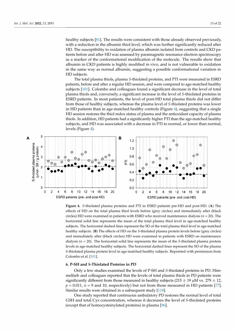
The total plasma thiols, plasma S-thiolated proteins, and PTI were measured in ESRDpatients, before and after a regular HD session, and were compared to age-matched healthysubjects [101]. Colombo and colleagues found a significant decrease in the level of totalplasma thiols and, conversely, a significant increase in the level of S-thiolated proteins inESRD patients. In most patients, the level of post-HD total plasma thiols did not differfrom those of healthy subjects, whereas the plasma level of S-thiolated proteins was lowerin HD patients than in age-matched healthy controls (Figure 4), suggesting that a singleHD session restores the thiol redox status of plasma and the antioxidant capacity of plasmathiols. In addition, HD patients had a significantly higher PTI than the age-matched healthysubjects, and HD was associated with a decrease in PTI to normal, or lower than normal,levels (Figure 4).
Int. J. Mol. Sci. 2022, 23, x FOR PEER REVIEW 14 of 24
Figure 4. S-thiolated plasma proteins and PTI in ESRD patients pre-HD and post-HD. (A) The effects
of HD on the total plasma thiol levels before (grey circles) and immediately after (black circles) HD
were examined in patients with ESRD who received maintenance dialysis (n = 20). The horizontal
solid line represents the mean of the total plasma thiol level in age-matched healthy subjects. The
horizontal dashed lines represent the SD of the total plasma thiol level in age-matched healthy sub-
jects. (B) The effects of HD on the S-thiolated plasma protein levels before (grey circles) and imme-
diately after (black circles) HD were examined in patients with ESRD on maintenance dialysis (n =
20). The horizontal solid line represents the mean of the S-thiolated plasma protein levels in age-
matched healthy subjects. The horizontal dashed lines represent the SD of the plasma S-thiolated
plasma protein level in age-matched healthy subjects. Reprinted with permission from Colombo et
al. [101].
6. P-SH and S-Thiolated Proteins in PD
Only a few studies examined the levels of P-SH and S-thiolated proteins in PD. Him-
melfarb and colleagues reported that the levels of total plasma thiols in PD patients were
significantly different from those measured in healthy subjects (215 ± 19 μM vs. 279 ± 12,
p = 0.011, n = 9 and 10, respectively) but not from those measured in HD patients [77].
Similar results were obtained in a subsequent study [118].
One study reported that continuous ambulatory PD restores the normal level of total
GSH and total Cys concentration, whereas it decreases the level of S-thiolated proteins
(except that of homocysteinylated proteins) in plasma [96].
7. Conclusions and Perspectives
All of the articles examined in this review provide evidence of a generalized thiol/ox-
idative stress in patients with CKD, which is reflected in the alterations of the levels of P-
SH and S-thiolated proteins compared to healthy control subjects. Analyses of the levels
of P-SH and S-thiolated proteins suggest that oxidative stress is present even in the early
stages of CKD; it is augmented in parallel with the progression of the disease to ESRD,
and is further exacerbated in HD patients. Oxidative stress is involved in the pathogenesis
of CKD progression and complications, particularly cardiovascular diseases. Thus, it rep-
resents a non-traditional risk factor for all causes of mortality in patients with CKD and
ESRD. For this reason, oxidative stress must become an important diagnostic and prog-
nostic factor, and a target for CKD prevention and treatment [119]. Unfortunately, oxida-
tive stress assessment is still not standardized, and this fact represents an obstacle for its
use in everyday clinical practice.
Some studies have shown that the levels of total plasma thiols, measured with
Ellman’s DTNB method, diminish in moderate-severe CKD (Table1) in ESRD patients on
HD (Tables 2 and 3) or on PD. However, the spectrophotometric analysis of the total
plasma thiols with Ellman’s method mainly measures P-SH, as they represent the most
Figure 4. S-thiolated plasma proteins and PTI in ESRD patients pre-HD and post-HD. (A) Theeffects of HD on the total plasma thiol levels before (grey circles) and immediately after (blackcircles) HD were examined in patients with ESRD who received maintenance dialysis (n = 20). Thehorizontal solid line represents the mean of the total plasma thiol level in age-matched healthysubjects. The horizontal dashed lines represent the SD of the total plasma thiol level in age-matchedhealthy subjects. (B) The effects of HD on the S-thiolated plasma protein levels before (grey circles)and immediately after (black circles) HD were examined in patients with ESRD on maintenancedialysis (n = 20). The horizontal solid line represents the mean of the S-thiolated plasma proteinlevels in age-matched healthy subjects. The horizontal dashed lines represent the SD of the plasmaS-thiolated plasma protein level in age-matched healthy subjects. Reprinted with permission fromColombo et al. [101].
6. P-SH and S-Thiolated Proteins in PD
Only a few studies examined the levels of P-SH and S-thiolated proteins in PD. Him-melfarb and colleagues reported that the levels of total plasma thiols in PD patients weresignificantly different from those measured in healthy subjects (215 ± 19 µM vs. 279 ± 12,p = 0.011, n = 9 and 10, respectively) but not from those measured in HD patients [77].Similar results were obtained in a subsequent study [118].
One study reported that continuous ambulatory PD restores the normal level of totalGSH and total Cys concentration, whereas it decreases the level of S-thiolated proteins(except that of homocysteinylated proteins) in plasma [96].
Int. J. Mol. Sci. 2022, 23, 2853 14 of 22
7. Conclusions and Perspectives
All of the articles examined in this review provide evidence of a generalized thiol/oxidativestress in patients with CKD, which is reflected in the alterations of the levels of P-SH andS-thiolated proteins compared to healthy control subjects. Analyses of the levels of P-SHand S-thiolated proteins suggest that oxidative stress is present even in the early stagesof CKD; it is augmented in parallel with the progression of the disease to ESRD, and isfurther exacerbated in HD patients. Oxidative stress is involved in the pathogenesis of CKDprogression and complications, particularly cardiovascular diseases. Thus, it representsa non-traditional risk factor for all causes of mortality in patients with CKD and ESRD.For this reason, oxidative stress must become an important diagnostic and prognosticfactor, and a target for CKD prevention and treatment [119]. Unfortunately, oxidativestress assessment is still not standardized, and this fact represents an obstacle for its use ineveryday clinical practice.
Some studies have shown that the levels of total plasma thiols, measured with Ellman’sDTNB method, diminish in moderate-severe CKD (Table 1) in ESRD patients on HD(Tables 2 and 3) or on PD. However, the spectrophotometric analysis of the total plasmathiols with Ellman’s method mainly measures P-SH, as they represent the most abundantthiol pool in human plasma, being in the 400–600 µM range, whereas LMM-SH altogetherconstitute only 12–20 µM [41,50,70]. Therefore, the contribution of LMM-SH to the amountof plasma thiols measured by Ellman’s method with DTNB can be considered negligible.Hence, these findings suggest a decrease in the plasma P-SH levels in patients with CKD,and in ESRD patients on HD or PD (Tables 1–3).
Unfortunately, the values of the plasma P-SH levels in CKD (Table 1) and ESRDpatients on renal replacement therapy (Tables 2 and 3), as well as in healthy controlsubjects, are often shown in different units, e.g., µmol/L, µg/L, and µmol/g protein,making it very difficult to compare the results of various studies conducted in differentlaboratories. Furthermore, in some studies, the CKD and ESRD patients (Tables 1–3) werehypoalbuminemic (with serum albumin levels < 3.8 g/dL), making it particularly difficultto compare P-SH levels, even if they are shown in the same units, between CKD patientsand healthy normoalbuminemic subjects. In order to compare the levels of plasma P-SHbetween CKD patients or ESRD patients on renal replacement therapy and healthy subjects,it is very important to normalize the P-SH concentrations against the total plasma proteinor plasma albumin concentrations, e.g., to measure the concentration of the plasma P-SHas µmol -SH/g plasma proteins [44,83,101,114,118]. In this way, the P-SH values wouldbecome easily comparable between different studies and, within each individual study,between CKD patients and healthy controls.
Unfortunately, there is no gold standard method for the measurement of the albuminconcentration in plasma. Albumin is generally measured using a colorimetric methodbased on bromocresol-green (BCG) or bromocresol-purple (BCP). Other approaches, e.g.,immunonephelometry (INP) or immunoturbidimetry, are used less frequently, as they aremore time-consuming and expensive. In addition, there are significant discrepancies whenmeasuring the albumin concentration by different methods (BCG, BCP and INP), especiallywhen determining it in ESRD patients on HD [120]. The albumin concentration measuredby BCG is consistently 4–10 g/L higher than that determined by BCP or INP.
Another important point concerning the normalization of the P-SH concentrationin relation to the plasma albumin concentration should be mentioned here. In fact, inCKD, albumin can undergo several irreversible post-translational modifications, suchas carbonylation [32,121,122] and carbamylation [123–125]. Due to the increased ureaand isocyanate concentrations in ESRD patients, protein carbamylation (i.e., the covalentbinding of isocyanate to the ε-amino group of lysine) increases significantly [126]. ForHD patients, the carbamylation of albumin is the main cause of the discrepancy found forvalues measured by BCP; this leads to an underestimation of 0–6 g/L. Several researchershave proposed that INP, or immunoturbidimetry, would be the preferred method for themeasurement albumin in HD patients [120,127,128]. Some have also suggested that the
Int. J. Mol. Sci. 2022, 23, 2853 15 of 22
choice of method for the measurement of the plasma albumin concentration (BCP, BCG,INP) could have an important impact on the classification of CKD patients [127].
Based on the results presented in this article, we can conclude that CKD and ESRDpatients on HD or PD show lower plasma thiol levels than healthy controls, suggest-ing that the uremic condition per se plays a key role in oxidative/thiol stress (Table 3).S-cysteinylated Cys34 accounts for most of the oxidized forms of albumin in CKD [84,85,101](Table 1) and in ESRD patients on renal replacement therapy [102,103] (Tables 2 and 3).CKD patients are among those with the highest risk for CVD [129,130]. CKD and ESRDpatients have a 5- to 10-fold higher risk of developing CVD than age-matched healthycontrols [131,132]. The Cys34 thiol of albumin represents by far the largest fraction of allplasma thiols; thus, it can have a protective effect on the vascular endothelium, as wasalready stated above. In this regard, some researchers have assessed whether changesin the redox state of the Cys34 thiol of albumin influence the incidence of CVD in HDpatients [133,134]. These authors reported that a lower concentration of Alb-SH was indica-tive of an increased CV mortality, demonstrating that the redox state of the Cys34 thiol isclosely related to CVD in both HD [133,134] and PD patients [135]. The Alb-SH levels weresignificantly higher in PD patients with high peritoneal membrane transport propertiesthan in those with low peritoneal membrane transport properties [136].
All of these studies suggest that, together with albumin carbamylation [124,125], theredox state of albumin Cys34 thiol is related to the overall risk factor for CVD among ESRDpatients. However, care should be taken when interpreting the outcome of these small-scalestudies to define the association of the redox status of albumin Cys34 thiol with CVD, asfurther large-scale studies are required.
Regarding S-thiolated plasma protein levels in pre- and post-HD ESRD patients(Table 3), following the HD session, the plasma P-SH levels transiently increase because ofthe de-thiolation of S-thiolated plasma proteins, as there is a decrease in oxidative stressduring the HD session. This is very important in the case of albumin, the Cys34-SH ofwhich represents the dominant antioxidant in plasma. However, in the period betweentwo consecutive HD sessions, oxidative plasma conditions induce P-SH to bind LMM-SH,determining the reversible formation of S-thiolated plasma proteins. Therefore, S-thiolatedplasma proteins represent a useful marker of thiol-specific reversible oxidative stress inESRD patients on HD. Therefore, HD has only a limited and transient beneficial effect onthe redox state of plasma P-SH, i.e., HD patients are exposed to periodical peaks of severeoxidative stress.
Individual HD sessions have different effects on different types of biomarkers ofprotein oxidation. For example, the post-HD levels of plasma protein carbonyls are signifi-cantly higher than the pre-HD levels, suggesting an increase in oxidative stress during theHD session [32,137–139]. The apparent discrepancy between the low levels of S-thiolatedproteins (and, consequently, high plasma P-SH levels) post-HD and the high plasma levelsof protein carbonyls post-HD is probably because, while protein S-thiolation is a reversibleprocess, protein carbonylation is, generally, irreversible [29].
In CKD, RBCs are exposed to oxidative stress and uremic toxins released into theblood. This is particularly evident in ESRD patients, where Hb has been found to beS-glutathionylated (Tables 2 and 3). This fact has clinical relevance because the exposureto oxidative stress and uremic toxins may have a role in the development of anaemia inpatients with CKD. In fact, it has been widely demonstrated that the RBCs of CKD/ESRDpatients have a reduced lifespan, which is caused by a hastened eryptosis, or RBC rupture,compared to healthy subjects. It was observed that, when blood from normal donors wastransfused into uremic patients, reductions in the RBC lifespan occurred [140]. Severalstudies examined RBCs’ oxidative stress in renal failure. HbSSG has been suggested tobe a clinical marker of oxidative stress in ESRD patients undergoing HD [66,141,142] orPD [142]. Moreover, the structure of Hb in ESRD patients is impaired, and these alterationsare enhanced during HD [143]. Regarding RBC thiol stress in HD and PD, LMM-SH (i.e.,GSH) in the RBCs of patients on continuous ambulatory PD remain at a physiological
Int. J. Mol. Sci. 2022, 23, 2853 16 of 22
level [144], whereas the GSH level in RBCs decreases before HD, and increases afterHD [144,145]. However, in HD patients the increase in LMM-SH level is short-lastingand limited to a brief post-HD period, while the increase in the level of LMM-SH in theRBCs of PD patients is continuous and persistent [144]. Thus, both PD and HD therapiesincrease the antioxidant power of RBC, but PD does so continuously, while the effect ofHD is short-lasting and intermittent. These results suggest that, as far as thiol stress isconcerned, toxic compounds do not accumulate in PD patients, thanks to the continuousdialytic procedure and the fact that PD patients more often maintain a residual diuresis,lowering the severity of oxidative stress [144].
In conclusion, this review provides evidence indicating a decrease in the plasma P-SHlevels and an increase in S-thiolated plasma proteins and S-thiolated RBC proteins inpatients with CKD (Tables 1–3). The levels of P-SH and S-thiolated proteins, and the PTIare promising biomarkers of reversible thiol/oxidative stress in moderate-to-severe CKD,and in HD and PD. The successful transposition of these biomarkers into a clinical testrequires the availability of a simple, artefact-free assay that should be selective, specific,and sensitive. The measurement of these biomarkers is complicated by the complexity ofthe methodologies that must be applied in order to avoid procedural artifacts, and thatrequire special care in sample handling and preparation. Nevertheless, several methodsare available that minimize artifacts and reproducibly measure the amounts of P-SH, S-thiolated proteins, PTI, and LMM-SH in cells, tissues, and body fluids [146–151]. Thesepromising biomarkers, which are serious candidates for the evaluation of oxidative stress inCKD patients, deserve further exploration in large and appropriately designed prospectivestudies. When this will be done, the levels of blood P-SH, S-thiolated proteins, LMM-SH, and the PTI could be included in the routine monitoring of CKD patients. This willdetermine important clinical implications, because—at the moment—an assessment ofoxidative stress in patients with CKD is not feasible in everyday clinical practice, in spite ofits relevance in CKD progression and complications development.
Funding: This research received no external funding.
Conflicts of Interest: The authors declare no conflict of interest. We further confirm that themanuscript has been read and approved by all of the named authors, and that the order of theauthors listed in the manuscript has been approved by all of us.
Abbreviations
Alb-SH reduced albuminCKD chronic kidney diseaseCVD cardiovascular diseaseCysGlySH cysteinylglycineCySS cystineDTNB 5,5-dithiobis(2-nitrobenzoic acid)eGFR estimated glomerular filtration rateESRD end-stage renal diseaseGSH glutathioneHbSSCy cysteinylated haemoglobinHbSSG glutathionylated haemoglobinHcySH homocysteineHD haemodialysisLDL low-density lipoproteinLMM-SH low molecular mass thiolsPD peritoneal dialysisPSH protein thiolsPTI protein thiolation indexRBCs red blood cellsROS reactive oxygen species
Int. J. Mol. Sci. 2022, 23, 2853 17 of 22
References1. Bello, A.K.; Levin, A.; Tonelli, M.; Okpechi, I.G.; Feehally, J.; Harris, D.; Jindal, K.; Salako, B.L.; Rateb, A.; Osman, M.A.; et al.
Assessment of global kidney health care status. JAMA 2017, 317, 1864–1881. [CrossRef] [PubMed]2. Luyckx, V.A.; Cherney, D.Z.I.; Bello, A.K. Preventing CKD in developed countries. Kidney Int. Rep. 2019, 5, 263–277. [CrossRef]
[PubMed]3. KDIGO CKD Workgrop. KDIGO 2012 Clinical practice guideline for the evaluation and management of chronic kidney disease.
Kidney Int. Suppl. 2013, 3, 1–150.4. Ruiz-Ortega, M.; Rayego-Mateos, S.; Lamas, S.; Ortiz, A.; Rodrigues-Diez, R.R. Targeting the progression of chronic kidney
disease. Nat. Rev. Nephrol. 2020, 16, 16269–16288. [CrossRef]5. Purnell, T.S.; Auguste, P.; Crews, D.C.; Lamprea-Montealegre, J.; Olufade, T.; Greer, R.; Ephraim, P.; Sheu, J.; Kostecki, D.; Powe,
N.R.; et al. Comparison of life participation activities among adults treated by hemodialysis, peritoneal dialysis, and kidneytransplantation: A systematic review. Am. J. Kidney Dis. 2013, 62, 953–973. [CrossRef] [PubMed]
6. Duni, A.; Liakopoulos, V.; Roumeliotis, S.; Peschos, D.; Dounousi, E. Oxidative stress in the pathogenesis and evolution of chronickidney disease: Untangling Ariadne’s thread. Int. J. Mol. Sci. 2019, 20, 3711. [CrossRef] [PubMed]
7. Gyurászová, M.; Gurecká, R.; Bábícková, J.; Tóthová, L’. Oxidative stress in the pathophysiology of kidney disease: Implicationsfor noninvasive monitoring and identification of biomarkers. Oxid. Med. Cell. Longev. 2020, 2020, 5478708. [CrossRef]
8. Vanholder, R.; Gryp, T.; Glorieux, G. Urea and chronic kidney disease: The comeback of the century? (in uraemia research).Nephrol. Dial. Transpl. 2018, 33, 4–12. [CrossRef]
9. van den Brand, J.A.; Mutsaers, H.A.; van Zuilen, A.D.; Blankestijn, P.J.; van den Broek, P.H.; Russel, F.G.; Masereeuw, R.; Wetzels,J.F. Uremic solutes in chronic kidney disease and their role in progression. PLoS ONE 2016, 11, e0168117. [CrossRef]
10. Poulianiti, K.P.; Kaltsatou, A.; Mitrou, G.I.; Jamurtas, A.Z.; Koutedakis, Y.; Maridaki, M.; Stefanidis, I.; Sakkas, G.K.; Karatzaferi, C.Systemic redox imbalance in chronic kidney disease: A systematic review. Oxid. Med. Cell. Longev. 2016, 2016, 8598253. [CrossRef]
11. Oberg, B.P.; McMenamin, E.; Lucas, F.L.; McMonagle, E.; Morrow, J.; Ikizler, T.A.; Himmelfarb, J. Increased prevalence of oxidantstress and inflammation in patients with moderate to severe chronic kidney disease. Kidney Int. 2004, 65, 1009–1016. [CrossRef][PubMed]
12. Cachofeiro, V.; Goicochea, M.; de Vinuesa, S.G.; Oubiña, P.; Lahera, V.; Luño, J. Oxidative stress and inflammation, a link betweenchronic kidney disease and cardiovascular disease. Kidney Int. Suppl. 2008, 74, S4–S9. [CrossRef] [PubMed]
13. Lim, Y.J.; Sidor, N.A.; Tonial, N.C.; Che, A.; Urquhart, B.L. Uremic toxins in the progression of chronic kidney disease andcardiovascular disease: Mechanisms and therapeutic targets. Toxins 2021, 13, 142. [CrossRef] [PubMed]
14. Mihajlovic, M.; Krebber, M.M.; Yang, Y.; Ahmed, S.; Lozovanu, V.; Andreeva, D.; Verhaar, M.C.; Masereeuw, R. Protein-bounduremic toxins induce reactive oxygen species-dependent and inflammasome-mediated IL-1β production in kidney proximaltubule cells. Biomedicines 2021, 9, 1326. [CrossRef]
15. Pieniazek, A.; Bernasinska-Slomczewska, J.; Gwozdzinski, L. Uremic toxins and their relation with oxidative stress induced inpatients with CKD. Int. J. Mol. Sci. 2021, 22, 6196. [CrossRef]
16. Borges Bonan, N.; Schepers, E.; Pecoits-Filho, R.; Dhondt, A.; Pletinck, A.; De Somer, F.; Vanholder, R.; Van Biesen, W.; Moreno-Amaral, A.; Glorieux, G. Contribution of the uremic milieu to an increased pro-inflammatory monocytic phenotype in chronickidney disease. Sci. Rep. 2019, 9, 10236. [CrossRef]
17. Mihai, S.; Codrici, E.; Popescu, I.D.; Enciu, A.M.; Albulescu, L.; Necula, L.G.; Mambet, C.; Anton, G.; Tanase, C. Inflammation-related mechanisms in chronic kidney disease prediction, progression, and outcome. J. Immunol. Res. 2018, 2018, 2180373.[CrossRef]
18. Carrero, J.J.; Stenvinkel, P. Inflammation in end-stage renal disease—What have we learned in 10 years? Semin. Dial. 2010, 23,498–509. [CrossRef]
19. Kao, M.P.; Ang, D.S.; Pall, A.; Struthers, A.D. Oxidative stress in renal dysfunction: Mechanisms, clinical sequelae and therapeuticoptions. J. Hum. Hypertens. 2010, 24, 1–8. [CrossRef]
20. Foley, R.N. Infectious complications in chronic dialysis patients. Perit. Dial. Int. 2008, 28, S167–S171. [CrossRef] [PubMed]21. Sharma, K. Obesity, oxidative stress, and fibrosis in chronic kidney disease. Kidney Int. Suppl. 2011, 2014, 4113–4117. [CrossRef]
[PubMed]22. Pellegrino, D.; La Russa, D.; Marrone, A. Oxidative imbalance and kidney damage: New study perspectives from animal models
to hospitalized patients. Antioxidants 2019, 8, 594. [CrossRef] [PubMed]23. Takahashi, N.; Morimoto, S.; Okigaki, M.; Seo, M.; Someya, K.; Morita, T.; Matsubara, H.; Sugiura, T.; Iwasaka, T. Decreased
plasma level of vitamin C in chronic kidney disease: Comparison between diabetic and non-diabetic patients. Nephrol. Dial.Transpl. 2011, 26, 1252–1257. [CrossRef]
24. Clase, C.M.; Ki, V.; Holden, R.M. Water-soluble vitamins in people with low glomerular filtration rate or on dialysis: A review.Semin. Dial. 2013, 26, 546–567. [CrossRef] [PubMed]
25. Stepniewska, J.; Dołegowska, B.; Popinska, M.; Sałata, D.; Budkowska, M.; Gołembiewska, E.; Myslak, M.; Domanski, M.;Marchelek-Mysliwiec, M.; Ciechanowski, K. Prooxidative-antioxidative balance of cells in different types of renal replacementtherapy. Blood Purif. 2014, 37, 4–11. [CrossRef] [PubMed]
26. Vostálová, J.; Galandáková, A.; Svobodová, A.R.; Orolinová, E.; Kajabová, M.; Schneiderka, P.; Zapletalová, J.; Strebl, P.; Zadražil, J.Time-course evaluation of oxidative stress-related biomarkers after renal transplantation. Ren. Fail. 2012, 34, 413–419. [CrossRef]
Int. J. Mol. Sci. 2022, 23, 2853 18 of 22
27. Navarro-García, J.A.; Rodríguez-Sánchez, E.; Aceves-Ripoll, J.; Abarca-Zabalía, J.; Susmozas-Sánchez, A.; González Lafuente, L.;Bada-Bosch, T.; Hernández, E.; Mérida-Herrero, E.; Praga, M.; et al. Oxidative status before and after renal replacement therapy:Differences between conventional high flux hemodialysis and on-line hemodiafiltration. Nutrients 2019, 11, 2809. [CrossRef]
28. FDA-NIH Biomarker Working Group. 2021. Available online: https://www.ncbi.nlm.nih.gov/books/NBK326791/ (accessed on30 December 2021).
29. Bachi, A.; Dalle-Donne, I.; Scaloni, A. Redox proteomics: Chemical principles, methodological approaches and biologi-cal/biomedical promises. Chem. Rev. 2013, 113, 596–698. [CrossRef]
30. Colombo, G.; Reggiani, F.; Cucchiari, D.; Portinaro, N.M.; Giustarini, D.; Rossi, R.; Garavaglia, M.L.; Saino, N.; Milzani, A.;Badalamenti, S.; et al. Plasma protein-bound di-tyrosines as biomarkers of oxidative stress in end stage renal disease patients onmaintenance haemodialysis. BBA Clin. 2017, 7, 55–63. [CrossRef]
31. Colombo, G.; Clerici, M.; Altomare, A.; Rusconi, F.; Giustarini, D.; Portinaro, N.; Garavaglia, M.L.; Rossi, R.; Dalle-Donne, I.;Milzani, A. Thiol oxidation and di-tyrosine formation in human plasma proteins induced by inflammatory concentrations ofhypochlorous acid. J. Proteom. 2017, 152, 22–32. [CrossRef]
32. Colombo, G.; Reggiani, F.; Cucchiari, D.; Astori, E.; Garavaglia, M.L.; Portinaro, N.M.; Saino, N.; Finazzi, S.; Milzani, A.;Badalamenti, S.; et al. Plasma protein carbonylation in haemodialysed patients: Focus on diabetes and gender. Oxid. Med. Cell.Longev. 2018, 2018, 4149681. [CrossRef] [PubMed]
33. Colombo, G.; Reggiani, F.; Astori, E.; Finazzi, S.; Garavaglia, M.L.; Angelini, C.; Milzani, A.; Badalamenti, S.; Dalle-Donne, I.Advanced oxidation protein products in non-diabetic end stage renal disease patients on maintenance haemodialysis. Free Radic.Res. 2019, 53, 1114–1124. [CrossRef] [PubMed]
34. Colombo, G.; Reggiani, F.; Angelini, C.; Finazzi, S.; Astori, E.; Garavaglia, M.L.; Landoni, L.; Portinaro, N.M.; Giustarini, D.; Rossi,R.; et al. Plasma protein carbonyls as biomarkers of oxidative stress in chronic kidney disease, dialysis, and transplantation. Oxid.Med. Cell. Longev. 2020, 2020, 2975256. [CrossRef] [PubMed]
35. Zavadskiy, S.; Sologova, S.; Moldogazieva, N. Oxidative distress in aging and age-related diseases: Spatiotemporal dysregulationof protein oxidation and degradation. Biochimie 2021. online ahead of print, S0300-9084(21)00273-X. [CrossRef]
36. Giustarini, D.; Dalle-Donne, I.; Milzani, A.; Braconi, D.; Santucci, A.; Rossi, R. Membrane skeletal protein S-glutathionylation inhuman red blood cells as index of oxidative stress. Chem. Res. Toxicol. 2019, 32, 1096–1102. [CrossRef] [PubMed]
37. Dhama, K.; Latheef, S.K.; Dadar, M.; Samad, H.; Munjal, A.; Khandia, R.; Karthik, K.; Tiwari, R.; Yatoo, M.I.; Bhatt, P.; et al.Biomarkers in stress related diseases/disorders: Diagnostic, prognostic, and therapeutic values. Front. Mol. Biosci. 2019, 6, 91.[CrossRef]
38. Ulrich, K.; Jakob, U. The role of thiols in antioxidant systems. Free Radic. Biol. Med. 2019, 140, 14–27. [CrossRef]39. Dalle-Donne, I.; Milzani, A.; Gagliano, N.; Colombo, R.; Giustarini, D.; Rossi, R. Molecular mechanisms and potential clinical
significance of S-glutathionylation. Antioxid. Redox Signal. 2008, 10, 446–473. [CrossRef]40. Dalle-Donne, I.; Rossi, R.; Colombo, G.; Giustarini, D.; Milzani, A. Protein S-glutathionylation: A regulatory device from bacteria
to humans. Trends Biochem. Sci. 2009, 34, 85–96. [CrossRef]41. Turell, L.; Radi, R.; Alvarez, B. The thiol pool in human plasma: The central contribution of albumin to redox processes. Free
Radic. Biol. Med. 2013, 65, 244–253. [CrossRef]42. Glushchenko, A.V.; Jacobsen, D.W. Molecular targeting of proteins by L-homocysteine: Mechanistic implications for vascular
disease. Antioxid. Redox Signal. 2007, 9, 1883–1898. [CrossRef]43. Pastore, A.; Piemonte, F. Protein glutathionylation in cardiovascular diseases. Int. J. Mol. Sci. 2013, 14, 20845–20876. [CrossRef]
[PubMed]44. Zinellu, A.; Fois, A.G.; Sotgia, S.; Zinellu, E.; Bifulco, F.; Pintus, G.; Mangoni, A.A.; Carru, C.; Pirina, P. Plasma protein thiols: An
early marker of oxidative stress in asthma and chronic obstructive pulmonary disease. Eur. J. Clin. Investig. 2016, 46, 181–188.[CrossRef] [PubMed]
45. Braconi, D.; Giustarini, D.; Marzocchi, B.; Peruzzi, L.; Margollicci, M.; Rossi, R.; Bernardini, G.; Millucci, L.; Gallagher, J.A.; LeQuan Sang, K.H.; et al. Inflammatory and oxidative stress biomarkers in alkaptonuria: Data from the DevelopAKUre project.Osteoarthr. Cartil. 2018, 26, 1078–1086. [CrossRef] [PubMed]
46. Giustarini, D.; Dalle-Donne, I.; Lorenzini, S.; Selvi, E.; Colombo, G.; Milzani, A.; Fanti, P.; Rossi, R. Protein thiolation index (PTI)as a biomarker of oxidative stress. Free Radic. Biol. Med. 2012, 53, 907–915. [CrossRef] [PubMed]
47. Gwozdzinski, K.; Pieniazek, A.; Gwozdzinski, L. Reactive oxygen species and their involvement in red blood cell damage inchronic kidney disease. Oxid. Med. Cell. Longev. 2021, 2021, 6639199. [CrossRef]
48. Ravarotto, V.; Bertoldi, G.; Innico, G.; Gobbi, L.; Calò, L.A. The pivotal role of oxidative stress in the pathophysiology ofcardiovascular-renal remodeling in kidney disease. Antioxidants 2021, 10, 1041. [CrossRef]
49. Ebert, T.; Neytchev, O.; Witasp, A.; Kublickiene, K.; Stenvinkel, P.; Shiels, P.G. Inflammation and oxidative stress in chronic kidneydisease and dialysis patients. Antioxid. Redox Signal. 2021, 35, 1426–1448. [CrossRef]
50. Rossi, R.; Giustarini, D.; Milzani, A.; Dalle-Donne, I. Cysteinylation and homocysteinylation of plasma protein thiols duringageing of healthy human beings. J. Cell. Mol. Med. 2009, 13, 3131–3140. [CrossRef]
51. Aebi, S.; Assereto, R.; Lauterburg, B.H. High-dose intravenous glutathione in man. Pharmacokinetics and effects on cyst(e)ine inplasma and urine. Eur. J. Clin. Investig. 1991, 21, 103–110. [CrossRef]
52. Oliveira, P.V.S.; Laurindo, F.R.M. Implications of plasma thiol redox in disease. Clin. Sci. 2018, 132, 1257–1280. [CrossRef]
Int. J. Mol. Sci. 2022, 23, 2853 19 of 22
53. Di Minno, M.N.; Tremoli, E.; Coppola, A.; Lupoli, R.; Di Minno, G. Homocysteine and arterial thrombosis: Challenge andopportunity. Thromb. Haemost. 2010, 103, 942–961. [CrossRef] [PubMed]
54. McCully, K.S. Homocysteine and the pathogenesis of atherosclerosis. Expert Rev. Clin. Pharmacol. 2015, 8, 211–219. [CrossRef][PubMed]
55. Nigwekar, S.U.; Kang, A.; Zoungas, S.; Cass, A.; Gallagher, M.P.; Kulshrestha, S.; Navaneethan, S.D.; Perkovic, V.; Strippoli,G.F.; Jardine, M.J. Interventions for lowering plasma homocysteine levels in dialysis patients. Cochrane Database Syst. Rev. 2016,2016, CD004683. [CrossRef] [PubMed]
56. Balint, B.; Jepchumba, V.K.; Guéant, J.L.; Guéant-Rodriguez, R.M. Mechanisms of homocysteine-induced damage to the endothe-lial, medial and adventitial layers of the arterial wall. Biochimie 2020, 173, 100–106. [CrossRef]
57. Jiang, Q.; Wang, L.; Si, X.; Tian, J.-L.; Zhang, Y.; Gui, H.-L.; Li, B.; Tan, D.-H. Current progress on the mechanisms ofhyperhomocysteinemia-induced vascular injury and use of natural polyphenol compounds. Eur. J. Pharmacol. 2021, 905, 174168.[CrossRef]
58. Lazzerini, P.E.; Capecchi, P.L.; Selvi, E.; Lorenzini, S.; Bisogno, S.; Galeazzi, M.; Laghi Pasini, F. Hyperhomocysteinemia,inflammation and autoimmunity. Autoimmun. Rev. 2007, 6, 503–509. [CrossRef]
59. Cordaro, M.; Siracusa, R.; Fusco, R.; Cuzzocrea, S.; Di Paola, R.; Impellizzeri, D. Involvements of hyperhomocysteinemia inneurological disorders. Metabolites 2021, 11, 37. [CrossRef]
60. Park, J.H.; Song, J.S.; Choi, S.T. Increased carotid intima-media thickness (IMT) in hyperuricemic individuals may be explained byhyperhomocysteinemia associated with renal dysfunction: A cross-sectional study. J. Korean Med. Sci. 2019, 34, e237. [CrossRef]
61. Bourgonje, A.R.; Abdulle, A.E.; Bourgonje, M.F.; Binnenmars, S.H.; Gordijn, S.J.; Bulthuis, M.L.C.; la Bastide-van Gemert, S.;Kieneker, L.M.; Gansevoort, R.T.; Bakker, S.J.L.; et al. Serum free sulfhydryl status associates with new-onset chronic kidneydisease in the general population. Redox Biol. 2021, 48, 102211. [CrossRef]
62. Ostrakhovitch, E.A.; Tabibzadeh, S. Homocysteine in chronic kidney disease. In Advances in Clinical Chemistry; Makowski, G.S.,Ed.; Academic Press: Burlington, MA, USA, 2015; Volume 72, pp. 77–106.
63. Jakubowski, H. The pathophysiological hypothesis of homocysteine thiolactone-mediated vascular disease. J. Physiol. Pharmacol.2008, 59, 155–167.
64. Giustarini, D.; Milzani, A.; Dalle-Donne, I.; Rossi, R. Red blood cells as a physiological source of glutathione for extracellularfluids. Blood Cells Mol. Dis. 2008, 40, 174–179. [CrossRef] [PubMed]
65. Baynes, J.W. Oxygen and life. In Medical Biochemistry; Baynes, J.W., Domoniczak, M.H., Eds.; Elsevier: Philadelphia, PA, USA,2005; pp. 497–506.
66. Khazim, K.; Giustarini, D.; Rossi, R.; Verkaik, D.; Cornell, J.E.; Cunningham, S.E.; Mohammad, M.; Trochta, K.; Lorenzo, C.; Folli,F.; et al. Glutathione redox potential is low and glutathionylated and cysteinylated hemoglobin levels are elevated in maintenancehemodialysis patients. Transl. Res. 2013, 162, 16–25. [CrossRef] [PubMed]
67. Vaziri, N.D. Oxidative stress in uremia: Nature, mechanisms, and potential consequences. Semin. Nephrol. 2004, 24, 469–473.[CrossRef] [PubMed]
68. Ceballos-Picot, I.; Witko-Sarsat, V.; Merad-Boudia, M.; Nguyen, A.T.; Thévenin, M.; Jaudon, M.C.; Zingraff, J.; Verger, C.; Jungers,P.; Descamps-Latscha, B. Glutathione antioxidant system as a marker of oxidative stress in chronic renal failure. Free Radic. Biol.Med. 1996, 21, 845–853. [CrossRef]
69. Colombo, G.; Dalle-Donne, I.; Giustarini, D.; Gagliano, N.; Portinaro, N.; Colombo, R.; Rossi, R.; Milzani, A. Cellular redoxpotential and hemoglobin S-glutathionylation in human and rat erythrocytes: A comparative study. Blood Cells Mol. Dis. 2010, 44,133–139. [CrossRef]
70. Giustarini, D.; Dalle-Donne, I.; Lorenzini, S.; Milzani, A.; Rossi, R. Age-related influence on thiol, disulfide, and protein-mixeddisulfide levels in human plasma. J. Gerontol. A Biol. Sci. Med. Sci. 2006, 61, 1030–1038. [CrossRef]
71. Colombo, G.; Clerici, M.; Giustarini, D.; Rossi, R.; Milzani, A.; Dalle-Donne, I. Redox albuminomics: Oxidized albumin in humandiseases. Antioxid. Redox Signal. 2012, 17, 1515–1527. [CrossRef]
72. Taverna, M.; Marie, A.L.; Mira, J.P.; Guidet, B. Specific antioxidant properties of human serum albumin. Ann. Intensive Care 2013,3, 4. [CrossRef]
73. Watanabe, H.; Imafuku, T.; Otagiri, M.; Maruyama, T. Clinical implications associated with the posttranslational modification-induced functional impairment of albumin in oxidative stress-related diseases. J. Pharm. Sci. 2017, 106, 2195–2203. [CrossRef]
74. Bonanata, J.; Turell, L.; Antmann, L.; Ferrer-Sueta, G.; Botasini, S.; Méndez, E.; Alvarez, B.; Coitiño, E.L. The thiol of human serumalbumin: Acidity, microenvironment and mechanistic insights on its oxidation to sulfenic acid. Free Radic. Biol. Med. 2017, 108,952–962. [CrossRef]
75. Belinskaia, D.A.; Voronina, P.A.; Shmurak, V.I.; Jenkins, R.O.; Goncharov, N.V. Serum albumin in health and disease: Esterase,antioxidant, transporting and signaling properties. Int. J. Mol. Sci. 2021, 22, 10318. [CrossRef] [PubMed]
76. Oettl, K.; Stauber, R.E. Physiological and pathological changes in the redox state of human serum albumin critically influence itsbinding properties. Br. J. Pharmacol. 2007, 151, 580–590. [CrossRef] [PubMed]
77. Himmelfarb, J.; McMonagle, E.; McMenamin, E. Plasma protein thiol oxidation and carbonyl formation in chronic renal failure.Kidney Int. 2000, 58, 2571–2578. [CrossRef] [PubMed]
78. Prakash, M.; Upadhya, S.; Prabhu, R. Protein thiol oxidation and lipid peroxidation in patients with uraemia. Scand. J. Clin. Lab.Investig. 2004, 64, 599–604. [CrossRef] [PubMed]
Int. J. Mol. Sci. 2022, 23, 2853 20 of 22
79. Ramos, L.F.; Shintani, A.; Ikizler, T.A.; Himmelfarb, J. Oxidative stress and inflammation are associated with adiposity in moderateto severe CKD. J. Am. Soc. Nephrol. 2008, 19, 593–599. [CrossRef] [PubMed]
80. Prakash, M.; Shetty, J.K.; Rao, L.; Sharma, S.; Rodrigues, A.; Prabhu, R. Serum paraoxonase activity and protein thiols in chronicrenal failure patients. Indian J. Nephrol. 2008, 18, 13–16. [CrossRef]
81. Pieniazek, A.; Gwozdzinski, L.; Zbrog, Z.; Gwozdzinski, K. Alterations in conformational state of albumin in plasma in chronichemodialyzed patients. PLoS ONE 2018, 13, e0192268. [CrossRef]
82. Zinellu, A.; Sotgia, S.; Mangoni, A.A.; Sotgiu, E.; Ena, S.; Arru, D.; Assaretti, S.; Baralla, A.; Satta, A.E.; Carru, C. Effects of ramipriland telmisartan on plasma concentrations of low molecular weight and protein thiols and carotid intima media thickness inpatients with chronic kidney disease. Dis. Markers 2016, 2016, 1821596. [CrossRef]
83. Aveles, P.R.; Criminácio, C.R.; Gonçalves, S.; Bignelli, A.T.; Claro, L.M.; Siqueira, S.S.; Nakao, L.S.; Pecoits-Filho, R. Associationbetween biomarkers of carbonyl stress with increased systemic inflammatory response in different stages of chronic kidneydisease and after renal transplantation. Nephron Clin. Pract. 2010, 116, c294–c299. [CrossRef]
84. Terawaki, H.; Yoshimura, K.; Hasegawa, T.; Matsuyama, Y.; Negawa, T.; Yamada, K.; Matsushima, M.; Nakayama, M.; Hosoya, T.;Era, S. Oxidative stress is enhanced in correlation with renal dysfunction: Examination with the redox state of albumin. KidneyInt. 2004, 66, 1988–1993. [CrossRef]
85. Matsuyama, Y.; Terawaki, H.; Terada, T.; Era, S. Albumin thiol oxidation and serum protein carbonyl formation are progressivelyenhanced with advancing stages of chronic kidney disease. Clin. Exp. Nephrol. 2009, 13, 308–315. [CrossRef] [PubMed]
86. Zinellu, A.; Loriga, G.; Scanu, B.; Pisanu, E.; Sanna, M.; Deiana, L.; Satta, A.E.; Carru, C. Increased low-density lipoproteinS-homocysteinylation in chronic kidney disease. Am. J. Nephrol. 2010, 32, 242–248. [CrossRef] [PubMed]
87. Sridhar, A.V.; Srinivasa Rao, P.V.; Sivakumar, V.; Satish, P.; Shalini, P.; Suchitra, M.M.; Kiranmayi, V.S. Study of oxidant andanti-oxidant status in patients with chronic kidney disease. J. Clin. Sci. Res. 2018, 7, 124–130. [CrossRef]
88. Suzuki, Y.; Suda, K.; Matsuyama, Y.; Era, S.; Soejima, A. Close relationship between redox state of human serum albumin andserum cysteine levels in non-diabetic CKD patients with various degrees of renal function. Clin. Nephrol. 2014, 82, 320–325.[CrossRef] [PubMed]
89. Cakirca, C.; Nas, C.; Yilmaz, K.; Guzelcicek, A.; Erel, O. Evaluation of dynamic thiol-disulfide balance in children with stage 3–5chronic kidney disease. Ann. Med. Res. 2019, 26, 1565–1568. [CrossRef]
90. Solak, I.; Guney, I.; Mercan, S.; Erel, O.; Neselioglu, S.; Cetinkaya, C.; Eryılmaz, M. Changes in thiol/disulfide homeostasis inpatients with chronic kidney disease. Med. Sci. 2020, 9, 201–204. [CrossRef]
91. Ates, I.; Özkayar, N.; Meriç Yılmaz, F.; Bayrakçı, N.; Neselioglu, S.; Erel, O.; Coskun Yenigün, E.; Dede, F. Oxidative stress level inpatients with chronic kidney disease. Ortadogu Med. J. 2018, 10, 45–50. [CrossRef]
92. Sogut, I.; Aydın, A.S.; Gokmen, E.S.; Atak, P.G.; Erel, O.; Degrigo, U.G. Evaluation of oxidative stress and thiol/ disulfideparameters according to the body mass index in adult individuals. Erciyes Med. J. 2018, 40, 155–161. [CrossRef]
93. Nakatani, S.; Yasukawa, K.; Ishimura, E.; Nakatani, A.; Toi, N.; Uedono, H.; Tsuda, A.; Yamada, S.; Ikeda, H.; Mori, K.; et al.Non-mercaptalbumin, oxidized form of serum albumin, significantly associated with renal function and anemia in chronic kidneydisease patients. Sci. Rep. 2018, 8, 16796. [CrossRef]
94. Terawaki, H.; Hayashi, T.; Murase, T.; Iijima, R.; Waki, K.; Tani, Y.; Nakamura, T.; Yoshimura, K.; Uchida, S.; Kazama, J.J.Relationship between xanthine oxidoreductase redox and oxidative stress among chronic kidney disease patients. Oxid. Med. Cell.Longev. 2018, 2018, 9714710. [CrossRef]
95. Kruglova, M.P.; Ivanov, A.V.; Virus, E.D.; Bulgakova, P.O.; Samokhin, A.S.; Fedoseev, A.N.; Grachev, S.V.; Kubatiev, A.A. UrineS-adenosylmethionine are related to degree of renal insufficiency in patients with chronic kidney disease. Lab. Med. 2021, 52,47–56. [CrossRef] [PubMed]
96. Przemysław, W.; Piotr, K.; Grazyna, C.; Danuta, K.P.; Małgorzata, I.; Bernadeta, M.; Małgorzata, S.; Witold, S. Total, free, andprotein-bound thiols in plasma of peritoneal dialysis and predialysis patients. Int. Urol. Nephrol. 2011, 43, 1201–1209. [CrossRef][PubMed]
97. Danielski, M.; Ikizler, T.A.; McMonagle, E.; Kane, J.C.; Pupim, L.; Morrow, J.; Himmelfarb, J. Linkage of hypoalbuminemia,inflammation, and oxidative stress in patients receiving maintenance hemodialysis therapy. Am. J. Kidney Dis. 2003, 42, 286–294.[CrossRef]
98. Himmelfarb, J.; McMonagle, E.; Freedman, S.; Klenzak, J.; McMenamin, E.; Le, P.; Pupim, L.B.; Ikizler, T.A.; The PICARD Group.Oxidative stress is increased in critically ill patients with acute renal failure. J. Am. Soc. Nephrol. 2004, 15, 2449–2456. [CrossRef]
99. Prakash, M.; Upadhya, S.; Prabhu, R. Serum non-transferrin bound iron in hemodialysis patients not receiving intravenous iron.Clin. Chim. Acta 2005, 360, 194–198. [CrossRef]
100. Koca, T.; Berber, A.; Koca, H.B.; Demir, T.A.; Koken, T. Effects of hemodialysis period on levels of blood trace elements andoxidative stress. Clin. Exp. Nephrol. 2010, 14, 463–468. [CrossRef]
101. Colombo, G.; Reggiani, F.; Podestà, M.A.; Garavaglia, M.L.; Portinaro, N.M.; Milzani, A.; Badalamenti, S.; Dalle-Donne, I. Plasmaprotein thiolation index (PTI) as a biomarker of thiol-specific oxidative stress in haemodialyzed patients. Free Radic. Biol. Med.2015, 89, 443–451. [CrossRef]
102. Anraku, M.; Kitamura, K.; Shinohara, A.; Adachi, M.; Suenga, A.; Maruyama, T.; Miyanaka, K.; Miyoshi, T.; Shiraishi, N.;Nonoguchi, H.; et al. Intravenous iron administration induces oxidation of serum albumin in hemodialysis patients. Kidney Int.2004, 66, 841–848. [CrossRef]
Int. J. Mol. Sci. 2022, 23, 2853 21 of 22
103. Mera, K.; Anraku, M.; Kitamura, K.; Nakajou, K.; Maruyama, T.; Otagiri, M. The structure and function of oxidized albuminin hemodialysis patients: Its role in elevated oxidative stress via neutrophil burst. Biochem. Biophys. Res. Commun. 2005, 334,1322–1328. [CrossRef]
104. Perna, A.F.; Satta, E.; Acanfora, F.; Lombardi, C.; Ingrosso, D.; De Santo, N.G. Increased plasma protein homocysteinylation inhemodialysis patients. Kidney Int. 2006, 69, 869–876. [CrossRef]
105. Fanti, P.; Giustarini, D.; Rossi, R.; Cunningham, S.E.; Folli, F.; Khazim, K.; Cornell, J.; Matteucci, E.; Bansal, S. Dietary intake ofproteins and calories is inversely associated with the oxidation state of plasma thiols in end-stage renal disease patients. J. Ren.Nutr. 2015, 25, 494–503. [CrossRef] [PubMed]
106. Zumbrennen-Bullough, K.; Babitt, J.L. The iron cycle in chronic kidney disease (CKD): From genetics and experimental models toCKD patients. Nephrol. Dial. Transpl. 2014, 29, 263–273. [CrossRef] [PubMed]
107. Del Vecchio, L.; Longhi, S.; Locatelli, F. Safety concerns about intravenous iron therapy in patients with chronic kidney disease.Clin. Kidney J. 2016, 9, 260–267. [CrossRef] [PubMed]
108. Giustarini, D.; Galvagni, F.; Colombo, G.; Dalle-Donne, I.; Milzani, A.; Aloisi, A.M.; Rossi, R. Measurement of Protein ThiolationIndex (PTI) as a biomarker of oxidative stress in serum: Effect of delayed sample separation/stabilization. Anal. Biochem. 2017,538, 38–41. [CrossRef] [PubMed]
109. Grekas, D.; Economou, H.; Makedou, A.; Destanis, E.; Theodoridou, A.; Avdelidou, A.; Demitriadis, A.; Tourkantonis, A. Associa-tion between hyperhomocysteinemia and ultrasonographic atherosclerotic indices of carotid arteries in chronic hemodialysispatients. Nephron Clin. Pract. 2005, 101, c180–c186. [CrossRef]
110. Zoccali, C. Traditional and emerging cardiovascular and renal risk factors: An epidemiologic perspective. Kidney Int. 2006, 70,26–33. [CrossRef] [PubMed]
111. Gerardi, G.; Usberti, M.; Martini, G.; Albertini, A.; Sugherini, L.; Pompella, A.; Di, L.D. Plasma total antioxidant capacity inhemodialyzed patients and its relationships to other biomarkers of oxidative stress and lipid peroxidation. Clin. Chem. Lab. Med.2002, 40, 104–110. [CrossRef]
112. Ward, R.A.; Ouseph, R.; McLeish, K.R. Effects of high-flux hemodialysis on oxidant stress. Kidney Int. 2003, 63, 353–359. [CrossRef]113. Palleschi, S.; De Angelis, S.; Rossi, B.; Diana, L.; Papa, V.; Severini, G.; Splendiani, G. Homocysteinemia correlates with plasma
thiol redox status in patients with end-stage renal disease. Nephron Clin. Pract. 2008, 108, c106–c112. [CrossRef]114. Pieniazek, A.; Brzeszczynska, J.; Kruszynska, I.; Gwozdzinski, K. Investigation of albumin properties in patients with chronic
renal failure. Free Radic. Res. 2009, 43, 1008–1018. [CrossRef]115. Wlodek, P.J.; Iciek, M.B.; Milkowski, A.; Smolenski, O.B. Various forms of plasma cysteine and its metabolites in patients
undergoing hemodialysis. Clin. Chim. Acta 2001, 304, 9–18. [CrossRef]116. Regazzoni, L.; Del Vecchio, L.; Altomare, A.; Yeum, K.J.; Cusi, D.; Locatelli, F.; Carini, M.; Aldini, G. Human serum albumin
cysteinylation is increased in end stage renal disease patients and reduced by hemodialysis: Mass spectrometry studies. FreeRadic. Res. 2013, 47, 172–180. [CrossRef] [PubMed]
117. Bocedi, A.; Cattani, G.; Stella, L.; Massoud, R.; Ricci, G. Thiol disulfide exchange reactions in human serum albumin: The apparentparadox of the redox transitions of Cys34. FEBS J. 2018, 285, 3225–3237. [CrossRef] [PubMed]
118. Capusa, C.; Stoian, I.; Rus, E.; Lixandru, D.; Barbulescu, C.; Mircescu, G. Does dialysis modality influence the oxidative stress ofuremic patients? Kidney Blood Press. Res. 2012, 35, 220–225. [CrossRef]
119. Rapa, S.F.; Di Iorio, B.R.; Campiglia, P.; Heidland, A.; Marzocco, S. Inflammation and oxidative stress in chronic kidneydisease-potential therapeutic role of minerals, vitamins and plant-derived metabolites. Int. J. Mol. Sci. 2019, 21, 263. [CrossRef]
120. Kok, M.B.; Tegelaers, F.P.W.; van Dam, B.; van Rijn, J.L.M.L.; van Pelt, J. Carbamylation of albumin is a cause for discrepanciesbetween albumin assays. Clin. Chim. Acta 2014, 434, 6–10. [CrossRef]
121. Mitrogianni, Z.; Barbouti, A.; Galaris, D.; Siamopoulos, K.C. Oxidative modification of albumin in predialysis, hemodialysis, andperitoneal dialysis patients. Nephron Clin. Pract. 2009, 113, c234–c240. [CrossRef]
122. Pavone, B.; Sirolli, V.; Bucci, S.; Libardi, F.; Felaco, P.; Amoroso, L.; Sacchetta, P.; Urbani, A.; Bonomini, M. Adsorption andcarbonylation of plasma proteins by dialyser membrane material: In vitro and in vivo proteomics investigations. Blood Transfus.2010, 8, s113–s119. [CrossRef]
123. Kalim, S.; Tamez, H.; Wenger, J.; Ankers, E.; Trottier, C.A.; Deferio, J.J.; Berg, A.H.; Karumanchi, S.A.; Thadhani, R.I. Carbamylationof serum albumin and erythropoietin resistance in end stage kidney disease. Clin. J. Am. Soc. Nephrol. 2013, 8, 1927–1934.[CrossRef]
124. Drechsler, C.; Kalim, S.; Wenger, J.B.; Suntharalingam, P.; Hod, T.; Thadhani, R.I.; Karumanchi, S.A.; Wanner, C.; Berg, A.H.Protein carbamylation is associated with heart failure and mortality in diabetic patients with end-stage renal disease. Kidney Int.2015, 87, 1201–1208. [CrossRef]
125. Kalim, S.; Trottier, C.A.; Wenger, J.B.; Wibecan, J.; Ahmed, R.; Ankers, E.; Karumanchi, S.A.; Thadhani, R.; Berg, A.H. Longitudinalchanges in protein carbamylation and mortality risk after initiation of hemodialysis. Clin. J. Am. Soc. Nephrol. 2016, 11, 1809–1816.[CrossRef] [PubMed]
126. Gajjala, P.R.; Fliser, D.; Speer, T.; Jankowski, V.; Jankowski, J. Emerging role of post-translational modifications in chronic kidneydisease and cardiovascular disease. Nephrol. Dial. Transpl. 2015, 30, 1814–1824. [CrossRef] [PubMed]
127. Labriola, L.; Wallemacq, P.; Gulbis, B.; Jadoul, M. The impact of the assay for measuring albumin on corrected (‘adjusted’) calciumconcentrations. Nephrol. Dial. Transpl. 2009, 24, 1834–1838. [CrossRef] [PubMed]
Int. J. Mol. Sci. 2022, 23, 2853 22 of 22
128. Kato, A.; Takita, T.; Furuhashi, M.; Fujimoto, T.; Suzuki, H.; Hakamada, M.; Maruyama, Y. Influence of the assay for measuringserum albumin on corrected total calcium in chronic hemodialysis patients. Ther. Apher. Dial. 2011, 15, 540–546. [CrossRef][PubMed]
129. Bossola, M.; Tazza, L. Wishful thinking: The surprisingly sparse evidence for a relationship between oxidative stress andcardiovascular disease in hemodialysis patients. Semin. Dial. 2015, 28, 224–230. [CrossRef] [PubMed]
130. Duni, A.; Liakopoulos, V.; Rapsomanikis, K.P.; Dounousi, E. Chronic kidney disease and disproportionally increased car-diovascular damage: Does oxidative stress explain the burden? Oxid. Med. Cell. Longev. 2017, 2017, 9036450. [CrossRef][PubMed]
131. Jager, K.J.; Lindholm, B.; Goldsmith, D.; Fliser, D.; Wiecek, A.; Suleymanlar, G.; Ortiz, A.; Massy, Z.; Martinez-Castelao, A.;Agarwal, R.; et al. Cardiovascular and non-cardiovascular mortality in dialysis patients: Where is the link? Kidney Int. Suppl.2011, 1, 21–23. [CrossRef]
132. van der Velde, M.; Matsushita, K.; Coresh, J.; Astor, B.C.; Woodward, M.; Levey, A.; de Jong, P.; Gansevoort, R.T.; Chronic KidneyDisease Prognosis Consortium; van der Velde, M.; et al. Lower estimated glomerular filtration rate and higher albuminuria areassociated with all-cause and cardiovascular mortality. A collaborative meta-analysis of high-risk population cohorts. Kidney Int.2011, 79, 1341–1352. [CrossRef]
133. Terawaki, H.; Takada, Y.; Era, S.; Funakoshi, Y.; Nakayama, K.; Nakayama, M.; Ogura, M.; Ito, S.; Hosoya, T. The redox state ofalbumin and serious cardiovascular incidence in hemodialysis patients. Ther. Apher. Dial. 2010, 14, 465–471. [CrossRef]
134. Terawaki, H.; Era, S.; Nakayama, M.; Hosoya, T. Decrease in reduced-form albumin among chronic kidney disease patients: Newinsights in cardiovascular complications. Ther. Apher. Dial. 2011, 15, 156–160. [CrossRef]
135. Terawaki, H.; Matsuyama, Y.; Matsuo, N.; Ogura, M.; Mitome, J.; Hamaguchi, A.; Terada, T.; Era, S.; Hosoya, T. A lower levelof reduced albumin induces serious cardiovascular incidence among peritoneal dialysis patients. Clin. Exp. Nephrol. 2012, 16,629–635. [CrossRef] [PubMed]
136. Lim, P.S.; Chen, H.P.; Chen, C.H.; Wu, M.Y.; Wu, C.Y.; Wu, T.K. Association between redox status of serum albumin and peritonealmembrane transport properties in patients on peritoneal dialysis. Blood Purif. 2015, 40, 243–249. [CrossRef] [PubMed]
137. Pavone, B.; Sirolli, V.; Giardinelli, A.; Bucci, S.; Forlì, F.; Di Cesare, M.; Sacchetta, P.; Di Pietro, N.; Pandolfi, A.; Urbani, A.; et al.Plasma protein carbonylation in chronic uremia. J. Nephrol. 2011, 24, 453–464. [CrossRef] [PubMed]
138. Albarello, K.; dos Santos, G.A.; Bochi, G.V.; Sangoi, M.B.; Almeida, T.C.; Paz da Silva, J.E.; Garcia, S.C.; Moresco, R.N. Ischemiamodified albumin and carbonyl protein as potential biomarkers of protein oxidation in hemodialysis. Clin. Biochem. 2012, 45,450–454. [CrossRef]
139. Caimi, G.; Carollo, C.; Hopps, E.; Montana, M.; Lo Presti, R. Protein oxidation in chronic kidney disease. Clin. Hemorheol. Microcirc.2013, 54, 409–413. [CrossRef]
140. Li, J.H.; Luo, J.F.; Jiang, Y.; Ma, Y.J.; Ji, Y.Q.; Zhu, G.L.; Zhou, C.; Chu, H.W.; Zhang, H.D. Red blood cell lifespan shortening inpatients with early-stage chronic kidney disease. Kidney Blood Press. Res. 2019, 44, 1158–1165. [CrossRef]
141. Naito, C.; Kajita, M.; Niwa, T. Determination of glutathionyl hemoglobin in hemodialysis patients using electrospray ionizationliquid chromatography-mass spectrometry. J. Chromatogr. B Biomed. Sci. Appl. 1999, 731, 121–124. [CrossRef]
142. Takayama, F.; Tsutsui, S.; Horie, M.; Shimokata, K.; Niwa, T. Glutathionyl hemoglobin in uremic patients undergoing hemodialysisand continuous ambulatory peritoneal dialysis. Kidney Int. Suppl. 2001, 78, S155–S158. [CrossRef]
143. Pieniazek, A.; Gwozdzinski, K. Changes in the conformational state of hemoglobin in hemodialysed patients with chronic renalfailure. Oxid. Med. Cell. Longev. 2015, 2015, 783073. [CrossRef]
144. Włodek, P.; Marcykiewicz, B.; Iciek, M.; Suliga, M.; Smolenski, O.; Kowalczyk-Pachel, D. Thiol levels, protein carbonylationand anaerobic sulfur metabolism in erythrocytes of peritoneal dialysis and predialysis patients. Nephrology 2010, 15, 755–761.[CrossRef]
145. Ozden, M.; Maral, H.; Akaydin, D.; Cetinalp, P.; Kalender, B. Erythrocyte glutathione peroxidase activity, plasma malondialdehydeand erythrocyte glutathione levels in hemodialysis and CAPD patients. Clin. Biochem. 2002, 35, 269–273. [CrossRef]
146. Giustarini, D.; Dalle-Donne, I.; Milzani, A.; Rossi, R. Detection of glutathione in whole blood after stabilization with N-ethylmaleimide. Anal. Biochem. 2011, 415, 81–83. [CrossRef] [PubMed]
147. Giustarini, D.; Dalle-Donne, I.; Milzani, A.; Fanti, P.; Rossi, R. Analysis of GSH and GSSG after derivatization with N-ethylmaleimide. Nat. Protoc. 2013, 8, 1660–1669. [CrossRef] [PubMed]
148. Rudyk, O.; Eaton, P. Biochemical methods for monitoring protein thiol redox states in biological systems. Redox Biol. 2014, 2,803–813. [CrossRef] [PubMed]
149. Giustarini, D.; Galvagni, F.; Tesei, A.; Farolfi, A.; Zanoni, M.; Pignatta, S.; Milzani, A.; Marone, I.M.; Dalle-Donne, I.; Nassini, R.;et al. Glutathione, glutathione disulfide, and S-glutathionylated proteins in cell cultures. Free Radic. Biol. Med. 2015, 89, 972–981.[CrossRef] [PubMed]
150. Giustarini, D.; Galvagni, F.; Orlandini, M.; Fanti, P.; Rossi, R. Immediate stabilization of human blood for delayed quantificationof endogenous thiols and disulfides. J. Chromatogr. B Anal. Technol. Biomed. Life Sci. 2016, 1019, 51–58. [CrossRef]
151. Giustarini, D.; Colombo, G.; Garavaglia, M.L.; Astori, E.; Portinaro, N.M.; Reggiani, F.; Badalamenti, S.; Aloisi, A.M.; Santucci, A.;Rossi, R.; et al. Assessment of glutathione/glutathione disulphide ratio and S-glutathionylated proteins in human blood, solidtissues, and cultured cells. Free Radic. Biol. Med. 2017, 112, 360–375. [CrossRef]


![Redox-Controllable Amphiphilic[2]Rotaxanes](https://img.pdfslide.net/doc/110x75/6351b8c456dfd8130803340c/redox-controllable-amphiphilic2rotaxanes.jpg)
