Embed Size (px)
Citation preview

REVIEW PAPER
Bone Marrow Microenvironment and Tumor Progression
Christophe F. Chantrain & Olivier Feron &
Etienne Marbaix & Yves A. DeClerck
Received: 11 January 2008 /Accepted: 8 March 2008 /Published online: 7 May 2008# The Author(s) 2008
Abstract The bone marrow constitutes an unique micro-environment for cancer cells in three specific aspects. First,the bone marrow actively recruits circulating tumor cellswhere they find a sanctuary rich in growth factors andcytokines that promote their proliferation and survival.When in the bone marrow, tumor cells profoundly affect thehomeostasis of the bone and the balance between osteo-genesis and osteolysis. As a consequence, growth andsurvival factors normally sequestered into the bone matrixare released, further fueling cancer progression. Second,tumor cells actively recruit bone marrow-derived precursor
cells into their own microenvironment. When in the tumors,these bone marrow-derived cells contribute to an inflam-matory reaction and to the formation of the tumorvasculature. Third, bone marrow-derived cells can homein distant organs, where they form niches that attractcirculating tumor cells. Our understanding of the contribu-tion of the bone marrow microenvironment to cancerprogression has therefore dramatically improved over thelast few years. The importance of this new knowledgecannot be underestimated considering that the vast majorityof cancer treatments such as cytotoxic and myeloablativechemotherapy, bone marrow transplantation and radiationtherapy inflict a trauma to the bone marrow microenviron-ment. How such trauma affects the influence that the bonemarrow microenvironment exerts on cancer is still poorlyunderstood. In this article, the reciprocal relationshipbetween the bone marrow microenvironment and tumorcells is reviewed, and its potential impact on cancer therapyis discussed.
Keywords Tumor microenvironment . Angiogenesis .
Vasculogenesis . Metastasis . Bone marrowmicroenvironment . Bone marrow stem cells
AbbreviationsEC endothelial cellsBMDC bone marrow-derived cellsBMDSC bone marrow-derived stem cellsGFP green fluorescent proteinHSC hematopoietic stem cellsMSC mesenchymal stem cellsVEGF vascular endothelial cell growth factorVEGFR VEGF receptorCEC circulating ECEPC endothelial progenitor cells
Cancer Microenvironment (2008) 1:23–35DOI 10.1007/s12307-008-0010-7
C. F. ChantrainDivision of Hematology-Oncology, Department of Pediatrics,Universite Catholique de Louvain,Brussels, Belgium
C. F. Chantrain : E. MarbaixCell Biology Unit, Christian de Duve Institute of CellularPathology, Universite Catholique de Louvain,Brussels, Belgium
O. FeronUnit of Pharmacology and Therapeutics,Universite Catholique de Louvain,Brussels, Belgium
E. MarbaixDepartment of Pathology, School of Medicine,Universite Catholique de Louvain,Brussels, Belgium
Y. A. DeClerck (*)Division of Hematology-Oncology, Departments of Pediatrics andBiochemistry and Molecular Biology, University of SouthernCalifornia Keck School of Medicine and The Saban ResearchInstitute of Childrens Hospital Los Angeles,4650 Sunset Boulevard, MS #54,Los Angeles, CA, USAe-mail: [email protected]
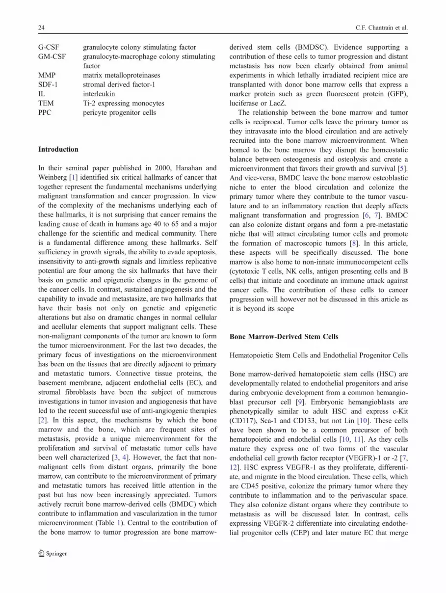
G-CSF granulocyte colony stimulating factorGM-CSF granulocyte-macrophage colony stimulating
factorMMP matrix metalloproteinasesSDF-1 stromal derived factor-1IL interleukinTEM Ti-2 expressing monocytesPPC pericyte progenitor cells
Introduction
In their seminal paper published in 2000, Hanahan andWeinberg [1] identified six critical hallmarks of cancer thattogether represent the fundamental mechanisms underlyingmalignant transformation and cancer progression. In viewof the complexity of the mechanisms underlying each ofthese hallmarks, it is not surprising that cancer remains theleading cause of death in humans age 40 to 65 and a majorchallenge for the scientific and medical community. Thereis a fundamental difference among these hallmarks. Selfsufficiency in growth signals, the ability to evade apoptosis,insensitivity to anti-growth signals and limitless replicativepotential are four among the six hallmarks that have theirbasis on genetic and epigenetic changes in the genome ofthe cancer cells. In contrast, sustained angiogenesis and thecapability to invade and metastasize, are two hallmarks thathave their basis not only on genetic and epigeneticalterations but also on dramatic changes in normal cellularand acellular elements that support malignant cells. Thesenon-malignant components of the tumor are known to formthe tumor microenvironment. For the last two decades, theprimary focus of investigations on the microenvironmenthas been on the tissues that are directly adjacent to primaryand metastatic tumors. Connective tissue proteins, thebasement membrane, adjacent endothelial cells (EC), andstromal fibroblasts have been the subject of numerousinvestigations in tumor invasion and angiogenesis that haveled to the recent successful use of anti-angiogenic therapies[2]. In this aspect, the mechanisms by which the bonemarrow and the bone, which are frequent sites ofmetastasis, provide a unique microenvironment for theproliferation and survival of metastatic tumor cells havebeen well characterized [3, 4]. However, the fact that non-malignant cells from distant organs, primarily the bonemarrow, can contribute to the microenvironment of primaryand metastatic tumors has received little attention in thepast but has now been increasingly appreciated. Tumorsactively recruit bone marrow-derived cells (BMDC) whichcontribute to inflammation and vascularization in the tumormicroenvironment (Table 1). Central to the contribution ofthe bone marrow to tumor progression are bone marrow-
derived stem cells (BMDSC). Evidence supporting acontribution of these cells to tumor progression and distantmetastasis has now been clearly obtained from animalexperiments in which lethally irradiated recipient mice aretransplanted with donor bone marrow cells that express amarker protein such as green fluorescent protein (GFP),luciferase or LacZ.
The relationship between the bone marrow and tumorcells is reciprocal. Tumor cells leave the primary tumor asthey intravasate into the blood circulation and are activelyrecruited into the bone marrow microenvironment. Whenhomed to the bone marrow they disrupt the homeostaticbalance between osteogenesis and osteolysis and create amicroenvironment that favors their growth and survival [5].And vice-versa, BMDC leave the bone marrow osteoblasticniche to enter the blood circulation and colonize theprimary tumor where they contribute to the tumor vascu-lature and to an inflammatory reaction that deeply affectsmalignant transformation and progression [6, 7]. BMDCcan also colonize distant organs and form a pre-metastaticniche that will attract circulating tumor cells and promotethe formation of macroscopic tumors [8]. In this article,these aspects will be specifically discussed. The bonemarrow is also home to non-innate immunocompetent cells(cytotoxic T cells, NK cells, antigen presenting cells and Bcells) that initiate and coordinate an immune attack againstcancer cells. The contribution of these cells to cancerprogression will however not be discussed in this article asit is beyond its scope
Bone Marrow-Derived Stem Cells
Hematopoietic Stem Cells and Endothelial Progenitor Cells
Bone marrow-derived hematopoietic stem cells (HSC) aredevelopmentally related to endothelial progenitors and ariseduring embryonic development from a common hemangio-blast precursor cell [9]. Embryonic hemangioblasts arephenotypically similar to adult HSC and express c-Kit(CD117), Sca-1 and CD133, but not Lin [10]. These cellshave been shown to be a common precursor of bothhematopoietic and endothelial cells [10, 11]. As they cellsmature they express one of two forms of the vascularendothelial cell growth factor receptor (VEGFR)-1 or -2 [7,12]. HSC express VEGFR-1 as they proliferate, differenti-ate, and migrate in the blood circulation. These cells, whichare CD45 positive, colonize the primary tumor where theycontribute to inflammation and to the perivascular space.They also colonize distant organs where they contribute tometastasis as will be discussed later. In contrast, cellsexpressing VEGFR-2 differentiate into circulating endothe-lial progenitor cells (CEP) and later mature EC that merge
24 C.F. Chantrain et al.
with EC originating from adjacent blood vessels to form thetumor vasculature [13, 14]. CEP in the peripheral blood,which derive from hemangioblasts, are phenotypicallydefined as negative for CD45 and positive for CD13/aminopeptidase N, VEGFR-2, CD117/Kit and CD34 [15].Since CD34 is also expressed on mature circulating EC(CEC), the expression of the marker CD133 allows thedistinction between CEP which are negative for CD133 andCEC that express CD133 [16]. However, some studies haveshown that HSC are not the only source of bone marrowderived EC [17], suggesting therefore that endotheliallineage probably derives from hemangioblasts and non-hematopoietic stem cells in adults.
Mesenchymal Stem Cells
The second type of stem cells in the bone marrow is thepool of mesenchymal stem cells (MSC) [18]. These cells donot express the HSC marker CD34, but express multipleother markers like Stro-1, CD105/endoglin, CD44/hyaluronicreceptor and CD90 [19, 20]. When cultured, they typicallyadhere to plastic or ECM proteins, and can differentiate intoa large variety of mature mesenchymal cells includingosteoblasts/osteocytes, chondrocytes, cardiomyocytes, myo-cytes, adipocytes and even neuronal cells [21, 22]. Theirpool in the bone marrow is enhanced by Wnt-3a, a memberof the Wnt family of growth factors. [23]. These cells are thesubject of a growing attention because of their potentialtherapeutic value in wound repair and tissue regeneration.Their contribution to tumor progression is less wellunderstood than HSC but they play an important role inbone metastasis, as will be discussed later.
Bone Marrow Niches
The fate of BMDSC, in particular HSC, is controlled by themicroenvironment in the bone marrow. This microenviron-ment is made of specific niches that provide support for theproliferation and maintenance of stem cells [24]. Interac-tions between stem cells and their microenvironmentregulate their maintenance, proliferation, differentiationand migration into the blood circulation (Fig. 1). Distinctniches have been anatomically and physiologically definedwithin the bone marrow [25, 26]. In the endosteal region,osteoblasts and other mesenchymal-derived stromal cellssuch as reticular cells, fibroblasts and adipocytes constitutethe osteoblastic niche that supports the maintenance ofHSC in a quiescent and undifferentiated state. Alterationof normal osteoblastic function results in a decrease ofhematopoiesis and vice versa expansion of osteoblasts isassociated with an increased number of HSC in the bonemarrow [27, 28]. In the central region of the bone marrow,EC in sinusoids contribute to the formation of a secondcompartment defined as the vascular niche. BMDSC arerecruited in this niche where they proliferate, differentiateand migrate through the sinusoidal wall [26]. Thus whereasthe osteoblastic niche promotes the maintenance of a poolof undifferentiated stem cells, the vascular niche promotesthe release of differentiated and mature hematopoietic cells.
The mechanisms by which the microenvironment of theniche interacts with stem cells and controls their fateinclude adhesion and humoral factors [29]. Among theseis the angiopoietin-1/Tie-2 axis. HSC express the receptortyrosine kinase Tie2 and adhere to osteoblasts that expressangiopoietin-1 in the bone marrow niche. The interaction of
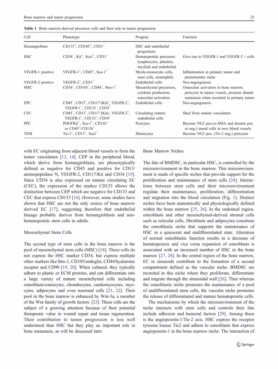
Table 1 Bone marrow-derived precursor cells and their role in tumor progression
Cell Phenotype Progeny Function
Hemangioblast CD133+, CD105+, CD31+ HSC and endothelialprogenitors
HSC CD34+, Kit+, Sca1+, CD31− Hematopoietic precursor:lymphocytes, platelets,myeloid and endothelial
Give rise to VEGFR-1 and VEGFR-2 + cells
VEGFR-1 positive VEGFR-1+, CD45+, Sca-1+ Myelo-monocytic cells,mast cells, neutrophils
Inflammation in primary tumor andpremetastatic niche
VEGFR-2 positive VEGFR-2+, CD31+ Endothelial cells Neo-angiogenesisMSC CD34−, CD105+, CD44+, Stro-1+ Mesenchymal precursors,
cytokine production,osteoclast activation.
Osteoclast activation in bone marrow,pericytes in tumor vessels, promote distantmetastasis when recruited in primary tumor
EPC CD45−, CD13+, CD117 (Kit)+, VEGFR-2+,VEGFR-1−, CD133−, CD34+
Endothelial cells Neo-angiogenesis
CEC CD45−, CD13+, CD117 (Kit)-, VEGFR-2+,VEGFR-1+, CD133+, CD34+
Circulating matureendothelial cells
Shed from mature vasculature
PPC PDGFRβ+, Sca-1+, CD11b+
or CD45+/CD11b+Pericytes Become NG2 pos.(α-SMA and desmin pos.
or neg.) mural cells in new blood vesselsTEM Tie-2+, CD13+, Sca1+ Monocytes Become NG2 pos. (Tie-2 neg.) pericytes
Bone marrow and tumor progression 25

Tie-2 with its ligand angiopoietin-1 maintains in vivo thelong-term repopulating activity of HSC. Angiopoietin-1promotes in HSC a state of quiescence and their adhesion tothe bone, protecting the HSC compartment from myelo-suppressive stress [30]. Adhesion of HSC to osteopontinpresent in endosteal bone via β1 integrin, is anotheradhesion mechanism that promotes the maintenance ofHSC in the endosteal niche [31]. Among the humoralfactors that control the fate of HSC is the chemokine,stromal-derived factor-1 (SDF-1), also called CXCL12.SDF-1 is produced by bone marrow stromal cells andosteoblasts, and binds to its receptor CXCR-4 expressed atthe cell surface of BMDSC. It thus acts as a retention factor,promoting the maintenance of BMDSC in the osteoblasticniche [32]. The balance between the osteoblastic and thevascular niche as well as the mobilization of BMDSC tothe peripheral circulation is a dynamic process that can bealtered by various traumatic conditions such as chemother-apy, radiation therapy, bone marrow transplantation, hyp-oxia, or tumor invasion. This balance is modulated bymultiple cytokines, chemokines, proteolytic enzymes andadhesion molecules [33]. Among the proteases involved,matrix metalloproteinase-9 (MMP-9) should be discussedin particular. MMP-9 is expressed by stromal cells andHSC in the bone marrow and its production is increasedupon treatment with cytotoxic drugs like 5 fluorouracil(5FU) that also stimulates the release of BMDSC from the
osteoblastic niche toward the vascular niche. MMP-9 exertsits proteolytic activity on several specific proteins thatcontrol the proliferation and fate of HSC in the bonemarrow. First, it cleaves membrane-associated Kit-Ligand(mKit-L) expressed by stromal cells in the osteoblasticniche. As a result, a soluble form of Kit-L (sKit-L) isreleased which promotes the proliferation of HSC [34].Second, MMP-9 degrades anchorage proteins such asosteopontin, enabling the migration of HSC from theosteoblastic to the vascular niche [35, 36]. Third, MMP-9cleaves SDF-1 at a Ser4–Lys5 peptide bond that inactivatesthe protein [37]. SDF-1 itself enhances the expression ofMMP-9 by BMDSC, thus promoting its own degradation[38]. The HSC mobilizing agent, granulocyte-colonystimulating factor (G-CSF), promotes HSC migration fromthe osteoblastic niche toward the vascular niche in a similarway by inducing neutrophil elastase degradation of SDF-1[39]. By proteolytically degrading type IV collagen presentin the basement membrane. MMP-9 also promotes theintravasation of BMSC into blood vessels. Thus, low levelsof MMP-9 result in high levels of mKit-L, poor release ofsKit-L and high levels of SDF-1 that retain HSC in theosteoblastic niche. In contrast, high levels of MMP-9 resultin abundant release of sKit-L, inactivation of SDF-1 andmigration of HSC toward the vascular niche and HSCdifferentiation [40] and intravasation into the peripheralblood circulation. This therefore explains the higher
VEFGRVEFGR--11VEFGRVEFGR--22 MMPMMP--99HSCHSC
SDFSDF --11KitKit --LL
osteopontinosteopontinosteoblastosteoblast
AngioAngio --1/Tie21/Tie2E
nd
ost
eal
En
do
stea
l Nic
he
Nic
he
Vas
cul a
r N
ich
eV
ascu
l ar
Nic
he
1
2
3
4
Fig. 1 VEGFR-1 and 2 positiveprecursor cells are released fromthe bone marrow. HSC areretained in the endosteal nicheby a combination of cell–cell(Tie-2/angio-1), cell-matrix(α4β1/osteopontin) andreceptor–ligand (CXCR-4/SDF-1) interactions. The mobilizationof these cells toward the vascu-lar niche and their release in theperipheral blood is in part con-trolled by MMP-9 which (1)degrades SDF-1, (2) degradesosteopontin, (3) solubilizes cKit-L and (4) degrades the basementmembrane
26 C.F. Chantrain et al.

mortality and impaired bone marrow recovery observed inMMP-9 deficient mice post chemotherapy with 5FU [34].
Tumors Recruit Bone Marrow Derived Cellswhich Contribute to Carcinogenesis and Progression
BMD-HSC Contribute to Inflammation in the Primary Tumor
Tumors recruit inflammatory cells into their stroma thoughthe expression of chemokines (Fig. 2). Most tumor cellsproduce chemokines of the two major groups CXC and CC.Typically, CXC chemokines are active on neutrophils andlymphocytes whereas CC chemokines target monocytes,eosinophils, dendritic cells, lymphocytes and NK cells [41].Tumor cells and stromal cells also produce many othergrowth factors such as VEGF, placental derived growthfactor (PlGF), a ligand that binds exclusively to VEGFR-1,transforming growth factor-β (TGFβ) and macrophagecolony stimulating factor (M-CSF) that contribute to therecruitment of monocytes and macrophages and the pro-motion of their survival and differentiation into the tumor[42, 43].
These inflammatory cells derive from VEGFR-1 positiveHSC [44][6, 45]. Although it was originally considered thatthese cells incarnate the immune defense against develop-ing tumors, there is accumulating evidence suggesting thatwhereas full activation of adaptive immune cells at thestage of tumor growth may result in eradication ofmalignant cells, chronic activation of innate immune cellsat sites of premalignant growth may actually enhance tumordevelopment [46]. Bone marrow-derived inflammatorycells contribute indeed to the transformation and prolifer-ation of tumor cells, to the modulation of the immuneresponse and to the development of tumor vascularization,and this process requires B lymphocytes [47].
Chronic inflammation has long been known as apredisposing factor in various human cancers such asbreast, liver, colon, bladder, prostate, stomach, ovary andskin carcinoma [6, 41]. In addition to potentially mutagenicinfectious agents that sustain chronic inflammation, inflam-matory cells create a microenvironment favorable for thetransformation of normal proliferating cells. Inflammatorycells, in particular phagocytic cells, generate reactiveoxygen and nitrogen species that contribute to the forma-tion of the mutagenic agent peroxynitrite [48, 49]. Macro-
En
do
ste
al
Nic
he
En
do
ste
al
Nic
he
Va
sc
ula
r N
ich
eV
as
cu
lar
Nic
he
VEGFVEGF
Pri
ma
ry T
um
or
Pri
ma
ry T
um
or
VEFGRVEFGR--11
VEFGRVEFGR--22
MSCMSC
Tumor cellTumor cell
SDFSDF--11
VEGFVEGF
PlGFPlGF
MM--CSFCSF
CCLCCL--5 (RANTES)5 (RANTES)
Fig. 2 The primary tumor actively recruits BMDC. Tumor cellsrelease SDF-1, VEGF, PlGF, M-CSF and other cytokines that attractHSC and MSC from the bone marrow. In the tumor tissue, VEGFR-1pos. cells give rise to CD45 pos. myelo-monocytic cells, macrophagesand mast cells that contribute to an inflammatory reaction and to theformation of the perivasculature. By producing MMP-9 these cells
further contribute to the solubilization of VEGF and other growthfactors. VEGFR-2 pos. cells give rise to endothelial cells andcontribute to the formation of a de novo vasculature. MSC arerecruited by primary tumors and give rise to pericytes and contribute tothe formation of distant metastasis by expressing CCL-5/RANTES thatstimulates tumor cell migration
Bone marrow and tumor progression 27

phages express macrophage migration inhibitory factor(MIF), a potent cytokine that bypasses p53 regulatoryfunctions and thus enhances the occurrence of oncogenicmutations [50]. Inflammatory cells also contribute to tumorcell proliferation by releasing growth factors like epidermalgrowth factor (EGF), and cytokines like interleukin-6 (IL-6)or tumor necrosis factor (TNF), as well as chemokinessuch as SDF-1 that enhance leukocyte recruitment [45].Tumor associated macrophages express immunosuppressivemolecules such as IL-10 and prostaglandin E-2 (PGE-2) thatallow tumor cells to evade the immune system [45]. Macro-phages also contribute to tumor progression by assistingtumor cells in intravasating [51, 52]. Mast cells contribute tothe early stages of tumorigenesis by stimulating proliferationand malignant conversion but at the same time promoting amore differentiated and less aggressive phenotype [53, 54].Mice deficient in mast cells are also less sensitive to thedevelopment of intestinal tumors when exposed to thecarcinogen 1,2-dimethylhydrazine [54] and exhibit a de-creased rate of tumor angiogenesis [55]. A similar functionfor tumor infiltrating neutrophils has been reported in anothermodel of multistage carcinogenesis [56]. Through the ex-pression of MMP-9, mast cells, macrophages and neutrophilsplay a critical role for the initiation of an angiogenic switchthat supports the growth of transformed cells into tumors[57].
BMD-MSC Contribute to Metastasis
Bone marrow-derived mesenchymal stem cells (BMD-MSC) have also been recently shown to be recruited inlarge numbers in the stroma of developing tumors.Although the role of these cells in tumor progression isnot fully understood, it has been recently suggested thatthey may promote metastasis in a breast carcinoma model.Dr. Weinberg’s laboratory has shown for example thattumor cells stimulate the expression of chemokine CCL5(RANTES) in mesenchymal stem cells, which then acts in aparacrine manner to enhance tumor cell motility, invasionand the formation of distant metastasis [58].
BMDC Contribute to Tumor Vascularization
Both VEGFR-1 and VEGFR-2 BMDC participate in thedevelopment of tumor vascularization. Bone marrow-derived inflammatory VEGFR-1 positive cells are recruitedto neo-angiogenic niches and support new blood vesselformation [59]. Among these cells, macrophages infiltratehypoxic tumor areas and cooperate with tumor cells topromote angiogenesis. Hypoxia enhances the expressionof several proangiogenic molecules by inducing the ex-pression of the transcription factor hypoxia-induciblefactors (HIF) in macrophages. As a result, inflammatory
cells produce TGFβ, VEGF, PDGF, basic fibroblastgrowth factor (bFGF) and specific chemokines thatstimulate tumor vascularization. These angiogenic chemo-kines include CXCL1 through CXCL8 with the exceptionof CXCL4 [45]. Several contain a Glu–Leu–Arg motif(ELR motif) at the NH2-terminus, and stimulate ECproliferation and chemotaxis by interacting with theirreceptor CXCR-2. The contribution of ELR-containingchemokines to tumor angiogenesis has been reported invarious human cancers [60]. SDF-1 which is produced bytumor cells, macrophages and many stromal cells alsopromotes tumor angiogenesis by recruiting CXCR-4positive BMDC and VEGF and bFGF induce CXCR-4expression in EC [61, 62]. Proteases produced byVEGFR-1 HSC, and in particular MMP-9, play a criticalregulatory role in tumor angiogenesis. MMP-9 producedby macrophages and mast cells releases ECM-boundVEGF, and therefore increases its bioavailability andbiological activity [56, 63, 64]. MMP-9 also stimulatesthe recruitment of pericytes along EC [65, 66]. Experi-ments in many laboratories have shown that in mice eithergenetically lacking MMP-9, treated with an inhibitor ofMMP or depleted in neutrophils, tumor development ischaracterized by reduced frequency, decreased tumorgrowth and reduced tumor vascularization [53, 56, 63–66]. Occasionally, VEGFR-1 positive monocytes and imma-ture myeloid cells acquire an EC-like phenotype and can beidentified in the tumor vascular endothelium [67, 68].
VEGFR-2 positive BMDC are also recruited into thetumor and contribute to the endothelium and the formationof a mature vascular network, a process called vasculo-genesis or neo-angiogenesis [15, 59, 69, 70]. The level ofcontribution of vasculogenesis to tumor vascularization ishowever highly variable and depends on the tumor type, theorgan site, the development stage and the mouse strain inanimal models. Whereas in murine bone marrow transplan-tation models the precise quantification of these cells ispossible using specific markers like EGFP, the quantifica-tion of these cells in human tumors is hampered bydifficulties of marking and tracking BMDC [59]. As aresult, reports on the percentage of these cells in tumorshave been highly variable [71]. The bone marrow is also areservoir of pericyte progenitor cells (PPC) [72]. Twosubsets of PPC have been reported to be released by thebone marrow, platelet derived growth factor receptor β(PDGFRβ) positive, Sca-1+ and CD11b+ cells and CD45+,CD11b+ cells [73]. Although they express Sca-1, whetherPDGFRβ+ cells originate from HSC or MSC in the marrowis unclear. Rajentie et al. [74] reported that a subpopulationof BMDC expressing the NG2 pericyte-specific proteogly-can, and the hematopoietic markers CD45 and CD11bcould be detected in the perivascular space of tumors wherethey contribute to mural cells.
28 C.F. Chantrain et al.

Finally, a few other BMDC that express neither VEFGR-1 or VEGFR-2 can contribute to tumor vascularization.Tie2 expressing CD13+ monocytes (TEM) identified inseveral mouse tumor models are recruited to tumor sitesand promote angiogenesis in a paracrine manner aftersurrounding angiogenic tumor vessels. TEM home to peri-endothelial sites similar to pericytes, and can differentiateinto α-SMA+ pericytes which then become negative forTie-2 [75]. Precursors of dendritic cells (DC) termed“vascular leukocytes” because they express VE-cadherinin addition to CD45, CD11c, and MHC-II, also participatein tumor vasculogenesis. These cells migrate toward tumorvessels and contribute to the assembly of the tumorneovasculature [76].
Contribution of BMDC to Cancer Metastasis
The Bone Marrow Attracts Tumor Cells
In many cancer types, circulating tumor cells can be easilyidentified in the peripheral blood. Although the presence ofthese cells is often a predictor of poor clinical outcome, itdoes not necessarily mean that metastatic disease is presentor will occur [77, 78]. However these cells can be activelyrecruited by the bone marrow. The mechanism by which thebone marrow recruits these circulating tumor cells in itsmicroenvironment has been extensively studied (Fig. 3).Central to this process is the production of chemokines by
bone marrow stromal cells that act as chemoattractants fortumor cells. Among those, SDF-1 seems again to play themajor role. Many tumor cells express the SDF-1 receptorCXCR-4 and thus, like HSC precursor cells, will be attractedinto the osteoblastic niche [79–82]. Animal experimentshave demonstrated that the migration of CXCR-4 positivebreast cancer cells toward the bone marrow can be blockedwhen cells are pretreated with the CXCR-4 inhibitor AMD3100 or a blocking antibody against CXCR-4. Down-regulation of CXCR-4 in breast cancer cells also inhibitstheir ability to colonize the bone, and CXCR-4 over-expressing tumor cells have a higher propensity to formbone marrow metastasis when injected intravenously inmice [83, 84]. SDF-1 is however not the only chemokineinvolved and other chemokines like the ELR-containingCXCL10 have been shown to attract tumor cells in the bonemarrow [85]. When tumor cells reach the osteoblasticniche, they do not remain quiescent like HSC andsignificantly affect the homeostasis of the bone.
Tumor Cells Affect the Bone Microenvironment
The bone is a tissue primarily composed of type I collagenand minerals in the form of hydroxyapatite that is normallyhighly resistant to degradation. Under physiological con-ditions, it is nevertheless one of the most active tissuesundergoing constant remodeling through a delicate balanceof new bone formation by osteoblasts and bone degradationby osteoclasts. Osteoblasts are cells of mesenchymal origin
RANKLRANKLRANKL
SDFSDF--11
ILIL--66
PTHrPPTHrP
osteoclastsosteoclasts
En
do
stea
lE
nd
ost
eal N
ich
eN
ich
eV
ascu
lar
Nic
he
Vas
cula
r N
ich
e
osteoblastsosteoblasts
Dkk1Dkk1
IGFIGF--1, IGF1, IGF--2, 2, TGFTGFPDGF, BMPPDGF, BMP
OPGOPG
stop
ILIL--11ILIL--66M-CSFMIPMIP--11M-CSF
Fig. 3 Tumor cells are recruited by the bone marrow where they alterthe osteoblasts and osteoclast balance. SDF-1, which is abundantlypresent in the bone marrow, attracts CXCR-4 expressing tumor cells.When in the bone marrow tumor cells interact with osteoblasts andosteoclast precursor cells through the production of PTHrP, RANKL
and other osteoclast activating factors. Alternatively, tumor cellsstimulate MSC to make IL-6. As a result, osteoclasts are activated.Tumor cells also inhibit osteoblast activity and new bone formation byblocking the Wnt pathway via the production of Dkk1. This leads toan increase in bone degradation and release of growth factors
Bone marrow and tumor progression 29
that reside in the endosteal niche at the inner surface of thebone where they interact with hematopoietic progenitorcells [86]. Osteoclasts are cells derived from hematopoieticprogenitor cells of the monocyte-macrophage lineage andare highly specialized and equipped to degrade the bonematrix upon activation. Under pathological conditions likeosteoarthritis, osteoporosis or cancer metastasis, the remod-eling of the bone is significantly increased and the balancedisrupted [81]. The presence of tumor cells in theosteoblastic niche can shift bone remodeling toward anexcess in bone degradation, which will create osteolyticbone metastases typically seen in breast cancer, multiplemyeloma, thyroid cancer and neuroblastoma or toward anexcess in bone synthesis and the formation of osteoblasticmetastases typically observed in prostate cancer. In mostcancer metastases, however, there is not pure osteolysis orosteogenesis but a combination of both processes [3, 4].The disruption of bone remodeling seen in cancer metas-tasis is the result of a combination of humoral and adhesiveinteractions between tumor cells and stromal cells in thebone marrow microenvironment. A typical mechanism seenin breast cancer bone metastasis involves the secretion ofparathyroid hormone-related peptide (PTHrP) by tumorcells [87]. In response to PTHrP, osteoblasts express themembrane bound receptor activator of NFkB ligand(RANKL), a member of the TNF-α family of cytokinesand a key regulator of osteoclastogenesis [88]. RANKLbinds to its receptor RANK, present at the cell surface ofosteoclast precursor cells, and via NFkB activation stim-ulates the transcriptional expression of genes like integrinαvβ3, cathepsin K, MMP-9 and H+-ATPase necessary forosteoclast adhesion to the bone, the formation of anosteolytic lacuna and bone degradation [89]. The interac-tion between RANK and RANKL is regulated by osteo-protegerin (OPG), a member of the TNF receptor familyproduced by osteoblasts and bone marrow stromal cells thatacts as a decoy receptor for RANKL, preventing itsinteraction with osteoclast-associated RANK [90]. Thisinteraction leads to inhibition of osteoclast differentiation,maturation and activation. The degradation of the bonematrix by osteoclasts has another important consequence onthe bone microenvironment that fuels the proliferation oftumor cells. Many growth factors that are immobilized inthe bone matrix like TGF-β, IGF-I and II, PDGF, and bonemorphogenic proteins (BMP) are released in a soluble formwhen the bone is degraded. These factors stimulate theproliferation of osteoblasts but also of tumor cells, creatinga vicious circle [4]. Tumor cells can also directly activateosteoclasts without interacting with osteoblasts by secretinga variety of osteoclast activating factors (OAF) like IL-1,IL-6, MIP1-α, M-CSF or RANKL which will stimulatebone degradation. In other situations, tumor cells do not
produce OAF but will stimulate the expression of OAF byMSC [91, 92]. This is, for example, the case in neuroblas-toma and myeloma. Our laboratory has shown that inneuroblastoma, tumor cells do not make OAF but stimulatethrough a soluble factor, the expression of IL-6 by MSC,which then acts as a potent activator of osteoclasts [93].Neuroblastoma cells express the IL-6 receptor and thusrespond to IL-6 produced by MSC. The production of IL-6by MSC not only stimulates osteoclasts but provides tumorcells with a growth and survival advantage. For example wehave shown that neuroblastoma cells proliferate faster andare resistant to drug-induced apoptosis when cultured in thepresence of MSC [94]. In myeloma, contact betweenmyeloma cells and bone marrow stromal cells alsostimulates the production of IL-6 by MSC and stromalcells [95]. Tumor cells not only stimulate osteoclasts butcan also inhibit osteoblasts and osteogenesis. Osteoblastactivation and maturation is stimulated by the Wnt pathway[96]. This pathway is negatively regulated by Dickkopf-1(Dkk1), a soluble protein that when binding to Kremen andLRP-5/6, two membrane associated receptors, prevents theinteraction of Frizzled-bound Wnt with LRP5/6 that isrequired for activation of the canonical Wnt pathway [97].Breast cancer cells and myeloma cells express Dkk1 andexpression of Dkk1 in these cells promotes osteolyticmetastasis by inhibiting bone synthesis [98–100]. In othercases, tumor cells will promote osteoblast proliferation andactivity. This is in particular the case of prostate cancer cellsthat express members of the bone morphogenic proteinfamily that stimulate osteoblasts and osteogenesis [101].Thus tumor cells that have been recruited into the bonemarrow can dramatically modify the bone microenviron-ment into a fertile soil that provides them with a substantialproliferative and survival advantage. This explains whywhen a cancer has metastasized to the bone, it is generallyrapidly progressive and resistant to chemotherapy.
BMDC Contribute to the Establishment of DistantMetastasis
Until recently, the accepted dogma in metastasis has beenthat distant organs are first colonized by a sub-population oftumor cells from the primary tumor and that inflammatorycells and EC are then recruited at these sites, where theycontribute to angiogenesis and stimulate the proliferation oftumor cells. Recent provocative data however have sug-gested that the order of these events may well be reversedand that inflammatory cells may be the first ones to reachmetastatic sites, forming pre-metastatic niches that attractcirculating tumor cells [102]. Using irradiated mice trans-planted with EGFP-expressing bone marrow cells andinjected subcutaneously with B16 or Lewis Lung tumor
30 C.F. Chantrain et al.

cells, Kaplan et al. demonstrated that VEGFR-1 positiveBMDC colonize the lung or liver at least a week beforetumor cells reach these sites. They also demonstrated thattumor cells only establish metastasis at sites that areprecolonized by VEGFR-1+ BMDC and that metastasiscould be inhibited by treating the mice with an anti-VEGFR-1 antibody or by removal of VEGFR-1 positivecells from the marrow. The deposition of fibronectin atthese pre-metastatic sites seems to be critical in attractingthe VEGFR-1 positive cells that express the fibronectin-binding integrin α4β1 (VLA4). It is postulated (Fig. 4) thatthese cells could then alter the microenvironment leading tothe release of chemokines attracting tumor cells like SDF-1.Soon after implantation of tumor cells at these sites,VEGFR-2+ cells are recruited from the bone marrow topromote neo-angiogenesis. Production of VEGF and PlGFby metastatic tumor cells stimulates angiogenesis and therecruitment of additional VEGFR-1+ BMDC. In support ofthe possibility of a similar mechanism in human cancermetastasis, the presence of VEGFR-1 positive cells inhistological sections of human metastatic tumors has beendemonstrated. Altogether the data suggest that circulatingtumor cells need specific niches to establish themselves andform successful metastatic tumors. The bone marrowtherefore not only is a source of local osteoblastic nichesthat promote the establishment of bone metastases but alsoof distant pre-metastatic niches that attract circulating tumor
cells in distant organs where they find a favorable soil formetastasis [26, 103].
Conclusion
It is now evident that the bone marrow plays a very uniquemicroenvironmental role in carcinogenesis, tumorigenesis,angiogenesis and metastasis. Most of the experimentalevidence discussed in this article suggests that it has apositive—and thus undesirable—effect on cancer progres-sion. A major mechanism by which the bone marrowcontributes to cancer progression is by the release ofVEGFR-1 and VEGFR-2 precursor cells that are a sourceof mature inflammatory and vascular cells. The vastmajority of the treatments currently used in human cancer,including high-dose cytotoxic and myeloablative chemo-therapy, bone marrow transplantation and radiation therapy,cause an injury to the bone marrow that is followed by aphase of recovery. The effect of such injuries to the bonemarrow microenvironment and in particular to the releaseof VEGFR-1 and -2 positive cells is so far poorlyunderstood, but there is evidence that it may promote therelease of these BMDC. For example, in animal models oflimb injury the revascularization of the limb is acceleratedif the mice receive low-dose irradiation, because irradiationof the bone marrow promotes the colonization of the
osteoblastsosteoblasts
Prim
ary Tu
mo
rP
rimary T
um
or
11
22
33
SDFSDF--11
FibronectinFibronectin
VEFGRVEFGR--11
VEFGRVEFGR--22
TumorTumor cellcell
PlGFPlGFVEGFVEGF
En
do
stea
lE
nd
ost
eal N
ich
eN
ich
eV
ascu
lar
Nic
he
Vas
cula
r N
ich
eP
reP
re-- m
etas
tati
c N
ich
em
etas
tati
c N
ich
e
Fig. 4 The bone marrowreleases VEGFR-1 and 2 pos.precursor cells that promote thedevelopment of distant metasta-ses. VEGFR-1 BMDC cells cancolonize distant organs via ad-hesion to fibronectin formingpre-metastatic niches (1). Thesecells release SDF-1 that attractsCXCR-4 circulating tumor cells(2). When established in thesepre-metastatic niches, tumorcells secrete VEGF, PlGF andother cytokines that attractVEGFR-2 pos. BMDC that willcontribute to neo-angiogenesis (3)
Bone marrow and tumor progression 31

injured limb by BMDC. Not surprisingly, this process isdependent on MMP-9 which increases the solubilizationof Kit-L and the release of VEGF which both contributeto revascularization of the limb [104]. In mice treated withcyclophosphamide there is an increased release of EPC inthe peripheral blood between 1 and 3 weeks after theadministration of the cytotoxic agent. This release howeveris only observed if a bolus maximal tolerable dose isadministered and not if low-dose metronomic doses aregiven [105, 106]. This thus supports the concept that highdose pulse chemotherapy may stimulate the release of EPCand ultimately tumor revascularization. In patients withcancer, studies have also shown an increase in EPC posthigh-dose chemotherapy [107]. However whether there isactually an increase in tumor vasculogenesis post adminis-tration of high-dose chemotherapy in human cancer hasnot been demonstrated yet. The data nevertheless suggestthat pulses of high-dose chemotherapy may have anunanticipated and detrimental side effect by promoting therelease of VEGFR-1 and -2 BMDC from the bone marrowosteoblastic niche into the peripheral blood circulation [108].This may have several important and negative conse-quences on patient survival as it may not only promoteinflammation and vasculogenesis in the primary tumor butalso the formation of pre-metastatic niches and thusmetastatic disease. Should this concept be correct, a betterunderstanding of the mechanisms involved in the release ofBMDC and how it is affected by injury to the bone marrow,will allow the design of new therapeutic protocols that
could prevent the increased release of BMDC from thebone marrow niche (Fig. 5). For example, adding drugs thattarget VEGF, PlGF, SDF-1 or MMP-9 immediately after apulse of high dose chemotherapy may prevent the release ofVEGFR-1 and R2 positive cells from the bone marrow andtheir recruitment by the primary tumor or by pre-metastaticniches. A decrease in the levels of circulating EPC inpatients with elevated VEGF levels in the peripheral blood(POEMS syndrome) treated with Bevasizumab (Avastin)has been recently shown [109]. MMP inhibitors have failedin clinical trials in the past but were tested in chronicadministration in patients with end stage disease and witha reduction in the primary tumor as the therapeutic goal[110]. However when used for a short period of timebetween courses of intensive chemotherapy, they may beeffective without unacceptable toxicity. Alternatively, inter-vening with SDF-1/CXCR-4 signaling with small inhibitorslike AMD 3100 [111] may have a similar effect by pre-venting the colonization of the primary tumor by BMDCreleased into the peripheral blood post high-dose chemo-therapy. There is clearly a fertile ground for investigation inthis area that has the potential to significantly influence theway we administer chemotherapy to patients with cancer.
Acknowledgments Publication of this article was supported in partby NIH/NCI grants CA042919 and CA81403 to YDC and a grantfrom the Salus Sanguinis Foundation and the St-Luc Foundationto CFC and YDC. We thank Mrs. J. Rosenberg for typing themanuscript.
Pulse chemo
Inhibitors of VEGFR-1 Inhibitors of VEGFR-1and R2 pos. BMDC releaseand R2 pos. BMDC release
Pulse chemo
0
1,000
2,000
3,000
4,000
5,000
6,000
7,000
1 7 14 21 280
5
10
15
20
25
30
WB
C
CE
P/c
u.m
m
WB
C
Pulse chemo Pulse chemo
0
1,000
2,000
3,000
4,000
5,000
6,000
7,000
1 7 14 21 280
5
10
15
20
25
30
CE
P/c
u.m
m
BA
syaDsyaDFig. 5 Revisiting the administration of high dose chemotherapy. aRecent observations suggest that there is an increase in the level ofEPC after pulse high dose chemotherapy. This increase may favortumor progression and the establishment of distant metastases if thesecells are recruited by the primary tumor and by pre-metastatic niches.
b The administration of agents blocking the recruitment of VEGFR-1and 2 positive BMDC between courses of high dose chemotherapymay prevent a stimulation of neo-angiogenesis and the formation ofpre-metastatic niche post chemotherapy
32 C.F. Chantrain et al.

Open Access This article is distributed under the terms of theCreative Commons Attribution Noncommercial License which permitsany noncommercial use, distribution, and reproduction in any medium,provided the original author(s) and source are credited.
References
1. Hanahan D, Weinberg RA (2000) The hallmarks of cancer. Cell100:57–70
2. Hurwitz H, Fehrenbacher L, Novotny W, Cartwright T,Hainsworth J, Heim W, Berlin J, Baron A, Griffing S, Holmgren E,Ferrara N, Fyfe G, Rogers B, Ross R, Kabbinavar F (2004)Bevacizumab plus irinotecan, fluorouracil, and leucovorin formetastatic colorectal cancer. N Engl J Med 350:2335–2342
3. Roodman GD (2004) Mechanisms of bone metastasis. N Engl JMed 350:1655–1664
4. Guise TA, Kozlow WM, Heras-Herzig A, Padalecki SS, Yin JJ,Chirgwin JM (2005) Molecular mechanisms of breast cancermetastases to bone. Clin Breast Cancer 5(Suppl):S46–S53
5. Mitsiades CS, McMillin DW, Klippel S, Hideshima T, Chauhan D,Richardson PG, Munshi NC, Anderson KC (2007) The role of thebone marrow microenvironment in the pathophysiology of myelo-ma and its significance in the development of more effectivetherapies. Hematol Oncol Clin North Am 21:1007–1034
6. Coussens LM, Werb Z (2002) Inflammation and cancer. Nature420:860–867
7. Rafii S, Lyden D, Benezra R, Hattori K, Heissig B (2002) Vascularand haematopoietic stem cells: novel targets for anti-angiogenesistherapy? Nat Rev Cancer 2:826–835
8. Kaplan RN, Rafii S, Lyden D (2006) Preparing the “soil”: thepremetastatic niche. Cancer Res 66:11089–11093
9. Schatteman GC, Dunnwald M, Jiao C (2007) Biology of bonemarrow-derived endothelial cell precursors. Am J Physiol HeartCirc Physiol 292:H1–18
10. Loges S, Fehse B, Brockmann MA, Lamszus K, Butzal M,Guckenbiehl M, Schuch G, Ergun S, Fischer U, Zander AR,Hossfeld DK, Fiedler W, Gehling UM (2004) Identification ofthe adult human hemangioblast. Stem Cells Dev 13:229–242
11. Choi K, Kennedy M, Kazarov A, Papadimitriou JC, Keller G(1998) A common precursor for hematopoietic and endothelialcells. Development 125:725–732
12. Eichmann A, Marcelle C, Breant C, Le Douarin NM (1993) Twomolecules related to the VEGF receptor are expressed in earlyendothelial cells during avian embryonic development. MechDev 42:33–48
13. Bailey AS, Jiang S, Afentoulis M, Baumann CI, Schroeder DA,Olson SB, Wong MH, Fleming WH (2004) Transplanted adulthematopoietic stems cells differentiate into functional endothelialcells. Blood 103:13–19
14. Rabbany SY, Heissig B, Hattori K, Rafii S (2003) Molecularpathways regulating mobilization of marrow-derived stem cellsfor tissue revascularization. Trends Mol Med 9:109–117
15. Bertolini F, Shaked Y, Mancuso P, Kerbel RS (2006) Themultifaceted circulating endothelial cell in cancer: towardsmarker and target identification. Nat Rev Cancer 6:835–845
16. Bertolini F, Mancuso P, Kerbel RS (2005) Circulating endothe-lial progenitor cells. N Engl J Med 353:2613–2616
17. Urbich C, Dimmeler S (2004) Endothelial progenitor cells:characterization and role in vascular biology. Circ Res 95:343–353
18. Dennis JE, Merriam A, Awadallah A, Yoo JU, Johnstone B,Caplan AI (1999) A quadripotential mesenchymal progenitor cellisolated from the marrow of an adult mouse. J Bone Miner Res14:700–709
19. Gronthos S, Zannettino AC, Hay SJ, Shi S, Graves SE,Kortesidis A, Simmons PJ (2003) Molecular and cellularcharacterisation of highly purified stromal stem cells derivedfrom human bone marrow. J Cell Sci 116:1827–1835
20. Kemp KC, Hows J, Donaldson C (2005) Bone marrow-derivedmesenchymal stem cells. Leuk Lymphoma 46:1531–1544
21. Le Blanc K (2006) Mesenchymal stromal cells: tissue repair andimmune modulation. Cytotherapy 8:559–561
22. Prockop DJ (1997) Marrow stromal cells as stem cells fornonhematopoietic tissues. Science 276:71–74
23. Etheridge SL, Spencer GJ, Heath DJ, Genever PG (2004)Expression profiling and functional analysis of wnt signalingmechanisms in mesenchymal stem cells. Stem Cells 22:849–860
24. Heissig B, Ohki Y, Sato Y, Rafii S, Werb Z, Hattori K (2005) Arole for niches in hematopoietic cell development. Hematology10:247–253
25. Yaniv I, Stein J, Farkas DL, Askenasy N (2006) The tale of earlyhematopoietic cell seeding in the bone marrow niche. Stem CellsDev 15:4–16
26. Kaplan RN, Psaila B, Lyden D (2007) Niche-to-niche migrationof bone-marrow-derived cells. Trends Mol Med 13:72–81
27. Taichman RS, Reilly MJ, Emerson SG (1996) Human osteo-blasts support human hematopoietic progenitor cells in vitrobone marrow cultures. Blood 87:518–524
28. Visnjic D, Kalajzic Z, Rowe DW, Katavic V, Lorenzo J, Aguila HL(2004) Hematopoiesis is severely altered in mice with an inducedosteoblast deficiency. Blood 103:3258–3264
29. Taichman RS (2005) Blood and bone: two tissues whose fatesare intertwined to create the hematopoietic stem-cell niche. Blood105:2631–2639
30. Arai F, Hirao A, Ohmura M, Sato H, Matsuoka S, Takubo K, Ito K,Koh GY, Suda T (2004) Tie2/angiopoietin-1 signaling regulateshematopoietic stem cell quiescence in the bone marrow niche. Cell118:149–161
31. Nilsson SK, Johnston HM, Whitty GA, Williams B, Webb RJ,Denhardt DT, Bertoncello I, Bendall LJ, Simmons PJ, HaylockDN (2005) Osteopontin, a key component of the hematopoieticstem cell niche and regulator of primitive hematopoieticprogenitor cells. Blood 106:1232–1239
32. Hattori K, Heissig B, Rafii S (2003) The regulation of hematopoi-etic stem cell and progenitor mobilization by chemokine SDF-1.Leuk Lymphoma 44:575–582
33. Arai F, Suda T (2007) Maintenance of quiescent hematopoieticstem cells in the osteoblastic niche. Ann NYAcad Sci 1106:41–53
34. Heissig B, Hattori K, Dias S, Friedrich M, Ferris B, Hackett NR,Crystal RG, Besmer P, Lyden D, Moore MA, Werb Z, Rafii S(2002) Recruitment of stem and progenitor cells from the bonemarrow niche requires MMP-9 mediated release of kit-ligand.Cell 109:625–637
35. Takafuji V, Forgues M, Unsworth E, Goldsmith P, Wang XW(2007) An osteopontin fragment is essential for tumor cellinvasion in hepatocellular carcinoma. Oncogene 26:6361–6371
36. Kollet O, Dar A, Shivtiel S, Kalinkovich A, Lapid K, Sztainberg Y,Tesio M, Samstein RM, Goichberg P, Spiegel A, Elson A, Lapidot T(2006) Osteoclasts degrade endosteal components and promotemobilization of hematopoietic progenitor cells. Nat Med 12:657–664
37. McQuibban GA, Butler GS, Gong JH, Bendall L, Power C,Clark-Lewis I, Overall CM (2001) Matrix metalloproteinaseactivity inactivates the CXC chemokine stromal cell-derivedfactor-1. J Biol Chem 276:43503–43508
38. Yu X, Collin-Osdoby P, Osdoby P (2003) SDF-1 increasesrecruitment of osteoclast precursors by upregulation of matrixmetalloproteinase-9 activity. Connect Tissue Res 44 Suppl 1:79–84
39. Petit I, Szyper-Kravitz M, Nagler A, Lahav M, Peled A, Habler L,Ponomaryov T, Taichman RS, Arenzana-Seisdedos F, Fujii N,
Bone marrow and tumor progression 33

Sandbank J, Zipori D, Lapidot T (2002) G-CSF induces stem cellmobilization by decreasing bone marrow SDF-1 and up-regulatingCXCR4. Nat Immunol 3:687–694
40. Kopp HG, Avecilla ST, Hooper AT, Rafii S (2005) The bonemarrow vascular niche: home of HSC differentiation andmobilization. Physiology (Bethesda) 20:349–356
41. Balkwill F, Mantovani A (2001) Inflammation and cancer: backto Virchow? Lancet 357:539–545
42. Hattori K, Heissig B, Wu Y, Dias S, Tejada R, Ferris B,Hicklin DJ, Zhu Z, Bohlen P, Witte L, Hendrikx J, Hackett NR,Crystal RG, Moore MA,Werb Z, Lyden D, Rafii S (2002) Placentalgrowth factor reconstitutes hematopoiesis by recruiting VEGFR1(+) stem cells from bone-marrow microenvironment. Nat Med8:841–849
43. Hattori K, Dias S, Heissig B, Hackett NR, Lyden D, Tateno M,Hicklin DJ, Zhu Z, Witte L, Crystal RG, Moore MA, Rafii S(2001) Vascular endothelial growth factor and angiopoietin-1stimulate postnatal hematopoiesis by recruitment of vasculogenicand hematopoietic stem cells. J Exp Med 193:1005–1014
44. Gerber HP, Malik AK, Solar GP, Sherman D, Liang XH, MengG, Hong K, Marsters JC, Ferrara N (2002) VEGF regulateshaematopoietic stem cell survival by an internal autocrine loopmechanism. Nature 417:954–958
45. Allavena P, Sica A, Solinas G, Porta C, Mantovani A (2008) Theinflammatory micro-environment in tumor progression: the role oftumor-associated macrophages. Crit Rev Oncol Hematol 66:1–9
46. de Visser KE, Coussens LM (2006) The inflammatory tumormicroenvironment and its impact on cancer development.Contrib Microbiol 13:118–137
47. de Visser KE, Korets LV, Coussens LM (2005) De novocarcinogenesis promoted by chronic inflammation is B lympho-cyte dependent. Cancer Cell 7:411–423
48. Maeda H, Akaike T (1998) Nitric oxide and oxygen radicals ininfection, inflammation, and cancer. Biochemistry (Mosc) 63:854–865
49. Pollard JW (2004) Tumour-educated macrophages promotetumour progression and metastasis. Nat Rev Cancer 4:71–78
50. Hudson JD, Shoaibi MA, Maestro R, Carnero A, Hannon GJ,Beach DH (1999) A proinflammatory cytokine inhibits p53tumor suppressor activity. J Exp Med 190:1375–1382
51. Wyckoff JB, Wang Y, Lin EY, Li JF, Goswami S, Stanley ER,Segall JE, Pollard JW, Condeelis J (2007) Direct visualization ofmacrophage-assisted tumor cell intravasation in mammarytumors. Cancer Res 67:2649–2656
52. Condeelis J, Pollard JW (2006) Macrophages: obligate partners fortumor cell migration, invasion, and metastasis. Cell 124:263–266
53. Coussens LM, Tinkle CL, Hanahan D, Werb Z (2000) MMP-9supplied by bone marrow-derived cells contributes to skincarcinogenesis. Cell 103:481–490
54. Wedemeyer J, Galli SJ (2005) Decreased susceptibility of mastcell-deficient Kit(W)/Kit(W-v) mice to the development of 1, 2-dimethylhydrazine-induced intestinal tumors. Lab Invest 85:388–396
55. Starkey JR, Crowle PK, Taubenberger S (1988) Mast-cell-deficientW/Wvmice exhibit a decreased rate of tumor angiogenesis.Int J Cancer 42:48–52
56. Nozawa H, Chiu C, Hanahan D (2006) Infiltrating neutrophilsmediate the initial angiogenic switch in a mouse model ofmultistage carcinogenesis. Proc Natl Acad Sci U S A103:12493–12498
57. Coussens LM, Raymond WW, Bergers G, Laig-Webster M,Behrendtsen O, Werb Z, Caughey GH, Hanahan D (1999)Inflammatory mast cells up-regulate angiogenesis during squa-mous epithelial carcinogenesis. Genes Dev 13:1382–1397
58. Karnoub AE, Dash AB, Vo AP, Sullivan A, Brooks MW, BellGW, Richardson AL, Polyak K, Tubo R, Weinberg RA (2007)
Mesenchymal stem cells within tumour stroma promote breastcancer metastasis. Nature 449:557–563
59. Kopp HG, Ramos CA, Rafii S (2006) Contribution of endothe-lial progenitors and proangiogenic hematopoietic cells tovascularization of tumor and ischemic tissue. Curr Opin Hematol13:175–181
60. Balkwill F, Coussens LM (2004) Cancer: an inflammatory link.Nature 431:405–406
61. Salcedo R, Wasserman K, Young HA, Grimm MC, Howard OM,Anver MR, Kleinman HK, Murphy WJ, Oppenheim JJ (1999)Vascular endothelial growth factor and basic fibroblast growthfactor induce expression of CXCR4 on human endothelial cells:in vivo neovascularization induced by stromal-derived factor-1alpha. Am J Pathol 154:1125–1135
62. Jin DK, Shido K, Kopp HG, Petit I, Shmelkov SV, Young LM,Hooper AT, Amano H, Avecilla ST, Heissig B, Hattori K, ZhangF, Hicklin DJ, Wu Y, Zhu Z, Dunn A, Salari H, Werb Z, HackettNR, Crystal RG, Lyden D, Rafii S (2006) Cytokine-mediateddeployment of SDF-1 induces revascularization through recruit-ment of CXCR4+ hemangiocytes. Nat Med 12:557–567
63. Bergers G, Brekken R, McMahon G, Vu TH, Itoh T, Tamaki K,Tanzawa K, Thorpe P, Itohara S, Werb Z, Hanahan D (2000)Matrix metalloproteinase-9 triggers the angiogenic switch duringcarcinogenesis. Nat Cell Biol 2:737–744
64. Huang S, Van Arsdall M, Tedjarati S, McCarty M, Wu W,Langley R, Fidler IJ (2002) Contributions of stromal metal-loproteinase-9 to angiogenesis and growth of human ovariancarcinoma in mice. J Natl Cancer Inst 94:1134–1142
65. Chantrain CF, Shimada H, Jodele S, Groshen S, Ye W, ShalinskyDR, Werb Z, Coussens LM, DeClerck YA (2004) Stromal matrixmetalloproteinase-9 regulates the vascular architecture in neuro-blastoma by promoting pericyte recruitment. Cancer Res 64:1675–1686
66. Jodele S, Chantrain CF, Blavier L, Lutzko C, Crooks GM,Shimada H, Coussens LM, DeClerck YA (2005) The contribu-tion of bone marrow-derived cells to the tumor vasculature inneuroblastoma is matrix metalloproteinase-9 dependent. CancerRes 65:3200–3208
67. Urbich C, Heeschen C, Aicher A, Dernbach E, Zeiher AM,Dimmeler S (2003) Relevance of monocytic features for neo-vascularization capacity of circulating endothelial progenitorcells. Circulation 108:2511–2516
68. Yang L, DeBusk LM, Fukuda K, Fingleton B, Green-Jarvis B,Shyr Y, Matrisian LM, Carbone DP, Lin PC (2004) Expansion ofmyeloid immune suppressor Gr+ CD11b+ cells in tumor-bearinghost directly promotes tumor angiogenesis. Cancer Cell 6:409–421
69. De Palma M, Naldini L (2006) Role of haematopoietic cells andendothelial progenitors in tumour angiogenesis. Biochim Bio-phys Acta 1766:159–166
70. Li B, Sharpe EE, Maupin AB, Teleron AA, Pyle AL, Carmeliet P,Young PP (2006) VEGF and PlGF promote adult vasculogenesisby enhancing EPC recruitment and vessel formation at the site oftumor neovascularization. FASEB J 20:1495–1497
71. Goon PK, Lip GY, Boos CJ, Stonelake PS, Blann AD (2006)Circulating endothelial cells, endothelial progenitor cells, andendothelial microparticles in cancer. Neoplasia 8:79–88
72. Lamagna C, Bergers G (2006) The bone marrow constitutes areservoir of pericyte progenitors. J Leukoc Biol 80:677–681
73. Song S, Ewald AJ, Stallcup W, Werb Z, Bergers G (2005)PDGFRbeta + perivascular progenitor cells in tumours regulatepericyte differentiation and vascular survival. Nat Cell Biol 7:870–879
74. Rajantie I, Ilmonen M, Alminaite A, Ozerdem U, Alitalo K,Salven P (2004) Adult bone marrow-derived cells recruitedduring angiogenesis comprise precursors for periendothelialvascular mural cells. Blood 104:2084–2086
34 C.F. Chantrain et al.

75. De Palma M, Venneri MA, Galli R, Sergi SL, Politi LS,Sampaolesi M, Naldini L (2005) Tie2 identifies a hematopoieticlineage of proangiogenic monocytes required for tumor vesselformation and a mesenchymal population of pericyte progeni-tors. Cancer Cell 8:211–226
76. Coukos G, Benencia F, Buckanovich RJ, Conejo-Garcia JR(2005) The role of dendritic cell precursors in tumour vasculo-genesis. Br J Cancer 92:1182–1187
77. Muller V, Hayes DF, Pantel K (2006) Recent translationalresearch: circulating tumor cells in breast cancer patients. BreastCancer Res 8:110
78. Elshimali YI, Grody WW (2006) The clinical significance ofcirculating tumor cells in the peripheral blood. Diagn Mol Pathol15:187–194
79. Sun YX, Wang JC, Shelburne CE, Lopatin DE, Chinnaiyan AM,Rubin MA, Pienta KJ, Taichman RS (2003) Expression ofCXCR4, CXCL12 (SDF-1) in human prostate cancers (PCa) invivo. J Cell Biochem 89:462–473
80. Geminder H, Sagi-Assif O, Goldberg L, Meshel T, Rechavi G,Witz IP, Ben Baruch A (2001) A possible role for CXCR4, itsligand, the CXC chemokine stromal cell-derived factor-1, inthe development of bone marrow metastases in neuroblastoma.J Immunol 167:4747–4757
81. Kozlow W, Guise TA (2005) Breast cancer metastasis to bone:mechanisms of osteolysis and implications for therapy. JMammary Gland Biol Neoplasia 10:169–180
82. Strahm B, Durbin AD, Sexsmith E, Malkin D (2008) TheCXCR4-SDF1alpha axis is a critical mediator of rhabdomyosar-coma metastatic signaling induced by bone marrow stroma. ClinExp Metastasis 25:1–10
83. Zhang L, Yeger H, Das B, Irwin MS, Baruchel S (2007) Tissuemicroenvironment modulates CXCR4 expression and tumormetastasis in neuroblastoma. Neoplasia 9:36–46
84. Muller A, Homey B, Soto H, Ge N, Catron D, Buchanan ME,McClanahan T, Murphy E, Yuan W, Wagner SN, Barrera JL,Mohar A, Verastegui E, Zlotnik A (2001) Involvement of che-mokine receptors in breast cancer metastasis. Nature 410:50–56
85. Goldberg-Bittman L, Sagi-Assif O, Meshel T, Nevo I, Levy-Nissenbaum O, Yron I, Witz IP, Ben Baruch A (2005) Cellularcharacteristics of neuroblastoma cells: regulation by the ELR-CXC chemokine CXCL10 and expression of a CXCR3-likereceptor. Cytokine 29:105–117
86. Yin T, Li L (2006) The stem cell niches in bone. J Clin Invest116:1195–1201
87. Guise TA, Yin JJ, Taylor SD, Kumagai Y, Dallas M, Boyce BF,Yoneda T, Mundy GR (1996) Evidence for a causal role ofparathyroid hormone-related protein in the pathogenesis of humanbreast cancer-mediated osteolysis. J Clin Invest 98:1544–1549
88. Kitazawa S, Kitazawa R (2002) RANK ligand is a prerequisitefor cancer-associated osteolytic lesions. J Pathol 198:228–236
89. Teitelbaum SL (2000) Bone resorption by osteoclasts. Science289:1504–1508
90. Kostenuik PJ, Shalhoub V (2001) Osteoprotegerin: a physiolog-ical and pharmacological inhibitor of bone resorption. CurrPharm Des 7:613–635
91. Sohara Y, Shimada H, DeClerck YA (2005) Mechanisms of boneinvasion and metastasis in human neuroblastoma. Cancer Lett228:203–209
92. Callander NS, Roodman GD (2001) Myeloma bone disease.Semin Hematol 38:276–285
93. Sohara Y, Shimada H, Minkin C, Erdreich-Epstein A, Nolta JA,DeClerck YA (2005) Bone marrow mesenchymal stem cellsprovide an alternate pathway of osteoclast activation and bonedestruction by cancer cells. Cancer Res 65:1129–1135
94. Ara T, Shimada H, Keshelava N, Metelitsa LS, Song LP,Groshen SG, Seeger RC, DeClerck YA (2008) IL-6 promotes
the growth and survival of neuroblastoma cells. Cancer Res (inpress)
95. Anderson KC, Kyle RA, Dalton WS, Landowski T, Shain K,Jove R, Hazlehurst L, Berenson J (2000) Multiple myeloma: newinsights and therapeutic approaches. Hematology (Am SocHematol Educ Program) 72:147–165
96. Kim JB, Leucht P, Lam K, Luppen C, Ten Berge D, Nusse R,Helms JA (2007) Bone regeneration is regulated by wntsignaling. J Bone Miner Res 22:1913–1923
97. Niehrs C (2006) Function and biological roles of the Dickkopffamily of Wnt modulators. Oncogene 25:7469–7481
98. Voorzanger-Rousselot N, Goehrig D, Journe F, Doriath V, Body JJ,Clezardin P, Garnero P (2007) Increased Dickkopf-1 expression inbreast cancer bone metastases. Br J Cancer 97: 964–970
99. Qian J, Xie J, Hong S, Yang J, Zhang L, Han X, Wang M, Zhan F,Shaughnessy JD Jr., Epstein J, Kwak LW, Yi Q (2007) Dickkopf-1(DKK1) is a widely expressed and potent tumor-associatedantigen in multiple myeloma. Blood 110:1587–1594
100. Giuliani N, Morandi F, Tagliaferri S, Lazzaretti M, Donofrio G,Bonomini S, Sala R, Mangoni M, Rizzoli V (2007) Production ofWnt inhibitors bymyeloma cells: potential effects on canonicalWntpathway in the bone microenvironment. Cancer Res 67:7665–7674
101. Feeley BT, Gamradt SC, Hsu WK, Liu N, Krenek L, Robbins P,Huard J, Lieberman JR (2005) Influence of BMPs on theformation of osteoblastic lesions in metastatic prostate cancer.J Bone Miner Res 20:2189–2199
102. Kaplan RN, Riba RD, Zacharoulis S, Bramley AH, Vincent L,Costa C, MacDonald DD, Jin DK, Shido K, Kerns SA, Zhu Z,Hicklin D, Wu Y, Port JL, Altorki N, Port ER, Ruggero D,Shmelkov SV, Jensen KK, Rafii S, Lyden D (2005) VEGFR1-positive haematopoietic bone marrow progenitors initiate the pre-metastatic niche. Nature 438:820–827
103. Psaila B, Kaplan RN, Port ER, Lyden D (2006) Priming the‘soil’ for breast cancer metastasis: the pre-metastatic niche.Breast Dis 26:65–74
104. Heissig B, Rafii S, Akiyama H, Ohki Y, Sato Y, Rafael T, Zhu Z,Hicklin DJ, Okumura K, Ogawa H, Werb Z, Hattori K (2005)Low-dose irradiation promotes tissue revascularization throughVEGF release from mast cells and MMP-9-mediated progenitorcell mobilization. J Exp Med 202:739–750
105. Shaked Y, Ciarrocchi A, Franco M, Lee CR, Man S, CheungAM, Hicklin DJ, Chaplin D, Foster FS, Benezra R, Kerbel RS(2006) Therapy-induced acute recruitment of circulating endo-thelial progenitor cells to tumors. Science 313:1785–1787
106. Bertolini F, Paul S, Mancuso P, Monestiroli S, Gobbi A, ShakedY, Kerbel RS (2003) Maximum tolerable dose and low-dosemetronomic chemotherapy have opposite effects on the mobili-zation and viability of circulating endothelial progenitor cells.Cancer Res 63:4342–4346
107. Furstenberger G, von Moos R, Lucas R, Thurlimann B, Senn HJ,Hamacher J, Boneberg EM (2006) Circulating endothelial cellsand angiogenic serum factors during neoadjuvant chemotherapyof primary breast cancer. Br J Cancer 94:524–531
108. Shaked Y, Kerbel RS (2007) Antiangiogenic strategies ondefense: on the possibility of blocking rebounds by the tumorvasculature after chemotherapy. Cancer Res 67:7055–7058
109. Rosti V, Massa M, Campanelli R, De Amici M, Piccolo G, PerfettiV (2007) Vascular endothelial growth factor promoted endothelialprogenitor cell mobilization into the peripheral blood of a patientwith POEMS syndrome. Haematologica 92:1291–1292
110. Coussens LM, Fingleton B, Matrisian LM (2002) Matrix metal-loproteinase inhibitors and cancer: trials and tribulations. Science295:2387–2392
111. Hatse S, Princen K, Bridger G, De Clercq E, Schols D (2002)Chemokine receptor inhibition by AMD3100 is strictly confinedto CXCR4. FEBS Lett 527:255–262
Bone marrow and tumor progression 35


