Embed Size (px)
Citation preview

BOOK: BIOLOGY II LABORATORY MANUAL (LUMEN)

Book: Biology II Laboratory Manual (Lumen)
This text is disseminated via the Open Education Resource (OER) LibreTexts Project (https://LibreTexts.org) and like the hundredsof other texts available within this powerful platform, it is freely available for reading, printing and "consuming." Most, but not all,pages in the library have licenses that may allow individuals to make changes, save, and print this book. Carefullyconsult the applicable license(s) before pursuing such effects.
Instructors can adopt existing LibreTexts texts or Remix them to quickly build course-specific resources to meet the needs of theirstudents. Unlike traditional textbooks, LibreTexts’ web based origins allow powerful integration of advanced features and newtechnologies to support learning.
The LibreTexts mission is to unite students, faculty and scholars in a cooperative effort to develop an easy-to-use online platformfor the construction, customization, and dissemination of OER content to reduce the burdens of unreasonable textbook costs to ourstudents and society. The LibreTexts project is a multi-institutional collaborative venture to develop the next generation of open-access texts to improve postsecondary education at all levels of higher learning by developing an Open Access Resourceenvironment. The project currently consists of 14 independently operating and interconnected libraries that are constantly beingoptimized by students, faculty, and outside experts to supplant conventional paper-based books. These free textbook alternatives areorganized within a central environment that is both vertically (from advance to basic level) and horizontally (across different fields)integrated.
The LibreTexts libraries are Powered by MindTouch and are supported by the Department of Education Open Textbook PilotProject, the UC Davis Office of the Provost, the UC Davis Library, the California State University Affordable Learning SolutionsProgram, and Merlot. This material is based upon work supported by the National Science Foundation under Grant No. 1246120,1525057, and 1413739. Unless otherwise noted, LibreTexts content is licensed by CC BY-NC-SA 3.0.
Any opinions, findings, and conclusions or recommendations expressed in this material are those of the author(s) and do notnecessarily reflect the views of the National Science Foundation nor the US Department of Education.
Have questions or comments? For information about adoptions or adaptions contact [email protected]. More information on ouractivities can be found via Facebook (https://facebook.com/Libretexts), Twitter (https://twitter.com/libretexts), or our blog(http://Blog.Libretexts.org).
This text was compiled on 08/15/2022
®

1
TABLE OF CONTENTS
1: Main Body
1.1: Chapter 1
2: Course Contents
2.1: About This Course
3: Faculty Resources
3.1: Faculty Resources Overview3.2: Lab Files Download3.3: PDF3.4: I Need Help
4: Module 1- Safety and Viruses
4.1: Lab Safety Contract4.2: Viruses Lab4.3: Viruses Lab (Instructor Materials Preparation)4.4: Dolphin Case Study4.5: Dolphin Case Study (Instructor Materials Preparation)
5: Module 2- Microbiology
5.1: Microbiology and Protista Lab5.2: Microbiology and Protista Lab (Instructor Materials Preparation)5.3: Reading- Prokaryotes5.4: Reading- Protists
6: Module 3- Fungi
6.1: Fungi Lab6.2: Fungi Lab (Instructor Materials Preparation)6.3: Reading- Fungi6.4: Case Study- Amphibian Die Off, Chytrid Fungi
7: Module 4- Seedless Plants
7.1: Seedless Plant Lab7.2: Seedless Plant Lab (Instructor Materials Preparation)7.3: Reading- Seedless Plants
8: Module 5- Seed Plants
8.1: Seed Plants Lab8.2: Seed Plants Lab (Instructor Materials Preparation)8.3: Reading- Seed Plants
2
9: Module 6- Plant Organization
9.1: Plant Organization Lab9.2: Plant Organization Lab (Instructor Materials Preparation)
10: Module 7- Invertebrate I
10.1: Invertebrate Lab I10.2: Invertebrate Lab I (Instructor Materials Preparation)10.3: Reading- Sponges10.4: Reading- Cnidarians10.5: Reading- Roundworms10.6: Reading- Arthropods
11: Module 8- Invertebrate II
11.1: Invertebrate Lab II11.2: Invertebrate Lab II (Instructor Materials Preparation)11.3: Reading- Flatworms11.4: Reading- Mollusks11.5: Reading- Annelids11.6: Natal Bean Discrimination by Bean Beetles11.7: Natal Bean Discrimination by Bean Beetles (Instructor Materials Preparation)*
12: Module 9- Deuterostomes
12.1: Deuterostome Lab12.2: Deuterostome Lab (Instructor Materials Preparation)12.3: Reading- Echinoderms12.4: Reading- Chordates
13: Module 10- Cardiovascular
13.1: Respiratory System and Pig Dissection
13.1.1: Fetal Pig Dissection Lab13.1.2: Fetal Pig Dissection Lab (Instructor Materials Preparation)13.1.3: Reading- Fetal Pig Dissection13.1.4: Cardiovascular and Respiratory Systems Lab13.1.5: Cardiovascular and Respiratory Systems Lab (Instructor Materials Preparation)
14: Module 11- Homeostasis
14.1: Homeostasis Lab14.2: Homeostasis Lab (Instructor Materials Preparation)
15: Module 12- Food Choice
15.1: Food Choice Lab15.2: Food Choice Lab (Instructor Materials Preparation)*
16: Module 13- Succession Lab
16.1: Community Ecology Lab16.2: Community Ecology Lab (Instructor Materials Preparation)*

3
17: Module 14- Ecosystem Lab
17.1: Ecosystem and Eutrophication Lab17.2: Ecosystem and Eutrophication Lab (Instructor Materials Preparation)
18: Module 15- Sample Lab Report
18.1: Sample Lab Report- Sugar Size and Diffusion Through a Mock-Cell Membrane18.2: Rubric
Index
Glossary
Book: Biology II Laboratory Manual (Lumen) is shared under a not declared license and was authored, remixed, and/or curated by LibreTexts.

1.1 https://bio.libretexts.org/@go/page/41752
CHAPTER OVERVIEW
1: Main Body1.1: Chapter 1
1: Main Body is shared under a not declared license and was authored, remixed, and/or curated by LibreTexts.

1.1.1 https://bio.libretexts.org/@go/page/41774
1.1: Chapter 1This is the first chapter in the main body of the text. You can change the text, rename the chapter, add new chapters, and add newparts.
1.1: Chapter 1 is shared under a not declared license and was authored, remixed, and/or curated by LibreTexts.

2.1 https://bio.libretexts.org/@go/page/41753
CHAPTER OVERVIEW
2: Course Contents2.1: About This Course
2: Course Contents is shared under a not declared license and was authored, remixed, and/or curated by LibreTexts.

2.1.1 https://bio.libretexts.org/@go/page/41775
2.1: About This CourseThis set of Biology II lab assignments ensures students have the opportunity to apply the concepts and information they learn asthey work through Biology II course content. Content includes lab assignments for students, as well as Instructor MaterialsPreparation for each lab with detailed lists of what faculty members need for each lab. The materials required are broken down bystudent (or groups of students).
These lab materials were developed by faculty at Tidewater Community College.
About LumenLumen Learning’s mission is to make great learning opportunities available to all students, regardless of socioeconomicbackground.
We do this by using open educational resources (OER) to create well-designed and low-cost course materials that replace expensivetextbooks. Because learning is about more than affordability and access, we also apply learning science insights and efficacyresearch to develop learning activities that are engineered to improve subject mastery, course completion and retention.
If you’d like to connect with us to learn more about adopting this course, please Contact Us.
You can also make an appointment for OER Office Hours to connect virtually with a live Lumen expert about any question youmay have.
Contributors and AttributionsCC licensed content, Original
About This Course. Provided by: Lumen Learning. License: CC BY: Attribution
2.1: About This Course is shared under a not declared license and was authored, remixed, and/or curated by LibreTexts.

1
CHAPTER OVERVIEW
3: Faculty Resources3.1: Faculty Resources Overview3.2: Lab Files Download3.3: PDF3.4: I Need Help
3: Faculty Resources is shared under a not declared license and was authored, remixed, and/or curated by LibreTexts.

3.1.1 https://bio.libretexts.org/@go/page/41776
3.1: Faculty Resources Overview
We’ve seen overwhelming demand for high quality, openly-licensed course materials, including supplemental resources to enrichteaching and learning and to make life easier for instructors. To support this need, we’ve developed and curated faculty resources touse with this course.
Free and Open Supplemental MaterialsOn the following pages, you will find supplemental resources that are freely available to use with the interactive learning materialsfor this course. Since these resources are openly licensed, you may use them as is or adapt them to your needs.
Additional Faculty Resources
Additional supplemental resources, learning tools, and support services are available to faculty who adopt Waymaker, OHM, orCandela courses with paid support from Lumen Learning. For many courses, these include some combination of summativeassessments, answer keys, solutions manuals, or other materials shared only with authorized instructors in order to protectacademic integrity.
Click here to learn more about additional instructor tools and resources available to faculty who adopt Lumen-supportedcourseware. Information about pricing and payment options is available on this page. Lumen’s low-cost support fees replace thecost of expensive textbooks and may be paid by students or by the institution directly.
Continuously Improving Learning Materials
Are you interested in collaborating with us to make these course materials better? We use learning data to identify where contentimprovements are needed, and then we invite faculty and subject matter experts to work with us developing continuousimprovements aimed at increasing learning.
Learn more from this blog post, or sign up here to join our continuous improvement mailing list and stay up to date aboutupcoming OER hackathons and other continuous improvement activities.
Contributors and AttributionsCC licensed content, Original
Faculty Resources Overview. Provided by: Lumen Learning. License: CC BY: Attribution
CC licensed content, Shared previouslyBooks Icon. Authored by: Ilham Fitrotul Hayat. Provided by: Noun Project. Located at:https://thenounproject.com/term/books/2792694/. License: CC BY: Attribution
3.1: Faculty Resources Overview is shared under a not declared license and was authored, remixed, and/or curated by LibreTexts.

3.2.1 https://bio.libretexts.org/@go/page/41777
3.2: Lab Files DownloadYou can download word documents of the lab exercises here:
Virus LabDolphin Stranding Case StudyMicrobiology and Protista LabFungi LabFungi Case StudySeedless Plant LabSeed Plant LabPlant Organization LabBean Beetles LabInvertebrates Lab IInvertebrates Lab IIDeuterostome LabFetal Pig DissectionCardiovascular and Respiratory Systems LabHomeostasis LabFood Choice LabCommunity Ecology SupplementSilver Springs LabSilver Springs Data (spreadsheet)
3.2: Lab Files Download is shared under a not declared license and was authored, remixed, and/or curated by LibreTexts.

3.3.1 https://bio.libretexts.org/@go/page/41778
3.3: PDF
Lumen makes significant investments to ensure our digital courseware is accessible, allowing students to learn using superiorinteractivity, multimedia, and a variety of accommodations for individuals with varying abilities.
PDFs offer an inferior learning experience compared to the richness and interactivity in our digital courseware. A PDF version ofthe textbook is available as a print alternative. The PDF does not include interactive content such as simulations, videos, andquizzes and is not vetted for accessibility. For these reasons, we do not recommend using the textbook in the PDF form. Theoffline version should be used as a print backup rather than as the primary textbook.
You can download the PDF using the following link:
Biology II Labs PDF
To share these files with your students, copy and paste the text and download link above into a page or announcement in yourlearning management system (Blackboard, Canvas, etc.).
Contributors and AttributionsCC licensed content, Original
PDF. Provided by: Lumen Learning. License: CC BY: Attribution
CC licensed content, Shared previouslyKindle Oasis. Authored by: Richard Slater. Provided by: Noun Project. Located at: https://thenounproject.com/term/kindle-oasis/611350/. License: CC BY: Attribution
3.3: PDF is shared under a not declared license and was authored, remixed, and/or curated by LibreTexts.

3.4.1 https://bio.libretexts.org/@go/page/41779
3.4: I Need Help
Need more information about this course? Have questions about faculty resources? Can’t find what you’re looking for?Experiencing technical difficulties?
We’re here to help! Take advantage of the following Lumen customer-support resources:
Check out one of Lumen’s Faculty User Guides here.Submit a support ticket here and tell us what you need.Talk and screen-share with a live human during Lumen’s OER office hours. See available times here.
Contributors and AttributionsCC licensed content, Original
I Need Help. Provided by: Lumen Learning. License: CC BY: Attribution
CC licensed content, Shared previouslyHelp Me!!. Provided by: Richard Elzey. Located at: https://www.flickr.com/photos/elzey/5344782612/. License: CC BY:Attribution
3.4: I Need Help is shared under a not declared license and was authored, remixed, and/or curated by LibreTexts.

1
CHAPTER OVERVIEW
4: Module 1- Safety and Viruses4.1: Lab Safety Contract4.2: Viruses Lab4.3: Viruses Lab (Instructor Materials Preparation)4.4: Dolphin Case Study4.5: Dolphin Case Study (Instructor Materials Preparation)
4: Module 1- Safety and Viruses is shared under a not declared license and was authored, remixed, and/or curated by LibreTexts.

4.1.1 https://bio.libretexts.org/@go/page/41780
4.1: Lab Safety ContractRead over and sign the Laboratory Safety Contract. This document goes over essential practices for lab safety.
Contributors and AttributionsCC licensed content, Original
Biology 102 Labs. Authored by: Lynette Hauser. Provided by: Tidewater Community College . Located at:http://www.tcc.edu/. License: CC BY: Attribution
4.1: Lab Safety Contract is shared under a not declared license and was authored, remixed, and/or curated by LibreTexts.

4.2.1 https://bio.libretexts.org/@go/page/41781
4.2: Viruses Lab
Lab Objectives
At the conclusion of the lab, the student should be able to:
Understand the properties of a virusExplain how viruses are spread through a population by sharing bodily fluids
A SlideShare element has been excluded from this version of the text. You can view it online here: pb.libretexts.org/bio2lm/?p=38
A virus is not considered a living organism as it only contains DNA surrounded by a protein coat. However, viruses are seriousinfectious agents causes conditions such as AIDS, chicken pox, and herpes. Viruses must have a live host cell to reproduce. Thevirus takes over the protein building machinery of the cell to create new viruses to spread the infection. Viruses can infect manydifferent types of organisms, both prokaryotic and eukaryotic. One way a virus can be spread through a population is by sharing bodily fluids such as saliva, blood, or semen. We willdemonstrate how quickly a virus can spread through today’s simulation activity.
Sharing Bodily FluidsDuring this lab you will share “bodily fluids” with other students in the lab to simulate the spread of an infectious disease through apopulation.
Procedure1. Obtain a numbered vial of solution and a plastic pipet from your instructor.2. Record your student name and vial number on the class data sheet.3. Share bodily fluids with another person in lab. Use the plastic pipet to withdraw solution from your vial and place 5 drops of
your solution in another classmate’s vial. Your classmate will also share fluids with you in the same way. Return the cap to thevial and invert to mix.
4. Record the name of the person you shared bodily fluids with in the table below.5. Exchange bodily fluids with another person following the directions above. Record the name of the person with whom you
exchanged fluids.6. Exchange fluids with another student (different than the first two) and record his/her name below. You should complete three
total fluid exchanges.
My vial #:_______________________
Record of Bodily Fluid Exchanges
Exchange 1: ______________________________ Exchange 2: ______________________________ Exchange 3: ______________________________
Your lab instructor will add a drop of the test reagent to determine if you are infected with the disease. If your sample turns pinkthen you are infected. If it turns yellow you are not infected. If you are positive for the disease you may have originally had thedisease or you may have contracted the disease in lab today from sharing bodily fluids.
Are you infected?Is it possible to determine if you were originally infected or did you contract the disease from someone during today’s lab?
As a class you will fill in Table 1 below. Include each person’s name. If your test result is positive put a plus sign (+) next to yourname. If your result is negative put a negative sign (−) by your name. For those individuals that are positive, record whom theyexchange fluids with and whether that person was positive or negative.

4.2.2 https://bio.libretexts.org/@go/page/41781
Questions1. How many people in the class are infected?2. Can you determine who was originally infected?3. If you can, whom do you think was originally infected?4. What do the class results show about the spread of disease through activities in which bodily fluids are shared?
Fill out a table similar to Table 1 for all members of your class. Be sure to add as many tables as there are students!
Table 1. Class results for bodily fluid exchange activity
Student’s Name Test result (+/−) Exchange #1 (+/−) Exchange #2 (+/−) Exchange #3 (+/−)
1.
2.
3.
4.
5.
After you’ve completed Table 1, discuss with your lab group how this experiment simulates a real life infection through apopulation and answer the following questions.
1. What are some ways that viruses are spread?2. What are some diseases that are spread by contact with bodily fluids?3. What are ways to prevent the spread of these diseases?
Contributors and AttributionsCC licensed content, Original
Biology 102 Labs. Authored by: Lynette Hauser. Provided by: Tidewater Community College . Located at:http://www.tcc.edu/. License: CC BY: Attribution
CC licensed content, Shared previouslyImmunology lab, Biology 102. Provided by: Piedmont Virginia Community College. Located at: http://www.pvcc.edu/.License: CC BY: Attribution
4.2: Viruses Lab is shared under a not declared license and was authored, remixed, and/or curated by LibreTexts.

4.3.1 https://bio.libretexts.org/@go/page/41782
4.3: Viruses Lab (Instructor Materials Preparation)
Lab Materials
This is the prep for one section of 24 students.
Sharing Bodily Fluids
Students will do this part in table teams (groups of 4).
Materials Quantity Notes
Vials labeled 1-24 24
randomly choose 1 vial to be infected. Thisshould be a number less than 10 in case there isa small # of students in the class. Let instructorknow which vial is infected in advance
Plastic pipets
Reagent
Contributors and AttributionsCC licensed content, Original
Instructor Materials Preparation. Authored by: Lynette Hauser. Provided by: Tidewater Community College. Located at:http://www.tcc.edu/. License: CC BY: Attribution
4.3: Viruses Lab (Instructor Materials Preparation) is shared under a not declared license and was authored, remixed, and/or curated byLibreTexts.

4.4.1 https://bio.libretexts.org/@go/page/41783
4.4: Dolphin Case Study
Learning Objectives
After completing the activities, you should be able to:
Define unusual mass stranding eventsExplain the difference between mass strandings and an unusual mass stranding eventState one symptom of cetacean morbillivirus and explain in general how the virus is spreadExplain in general how a virus can spread through a population of organismsExplain in general how a vaccine worksState two different ways a vaccine can be created
Introduction
In July 2013, stranding teams (such as the Virginia Aquarium’s Stranding Response team) from New York to North Carolina startednoticing a large amount of dolphin strandings.
Some data that was collected on how many dolphins were being found dead or dying on the beaches:
430 from July to September553 total from July to November6 in one day in Virginia Beach
Using your computer and the Internet, research dolphin strandings. Here is one website to get you started: NOAA Strandings.Answer the questions below based on your research. Please put your answers in your own words. Copying directly from a websiteis considered plagiarism.
1. Give two examples of marine mammals that can strand themselves.2. Give two specific reasons why marine mammals strand themselves.
Note: Data are dolphin strandings that have been confirmed and responded to by Stranding Network Members. Florida data isthrough Brevard County. Current UME Data are considered preliminary and may be subject to change as more informationbecomes available. From NOAA.gov data
3. Using the data above, how many dolphins were found a year on average in the Mid Atlantic states in the years 2007–2012?4. How much larger were the number of strandings in 2013? Calculate how many times larger.
These data lead researchers to believe that this event in 2013 would be classified as an Unusual Mortality Event (UME). Using

4.4.2 https://bio.libretexts.org/@go/page/41783
your computer and the Internet, research UMEs. Here is one website to get you started: Marine Mammal Unusual MortalityEvents (www.nmfs.noaa.gov/pr/health/mmume/). Answer the questions below based on your research. Please put your answersin your own words. Copying directly from a website is considered plagiarism.
5. Explain the difference between mass strandings, group strandings, and an unusual mass stranding event.6. The dolphins that washed ashore exhibited skin lesions and weight loss that was abnormal for bottle nose dolphins.
1. What kinds of data do you think would be useful in determining the cause of this UME event based on the condition of thedolphins? Name three things you think researchers should record about these dolphins.
2. Some scientists remembered that there was a similar case in 1987–1988, as described in Morbilliviral Disease in AtlanticBottlenose Dolphins and in Dophin Strandings.
7. What happened to bottlenose dolphins in 1987–1988?8. Does this UME seem similar to that situation? Why or why not?
In the 2013 strandings, dolphins were tested for cetacean morbillivirus, and approximately 85% of them were positive(NOAA.gov data). Using your computer and the Internet, research cetacean morbillivirus. Here is one website to get you started:Morbilliviris Infection. Answer the questions below based on your research. Please put your answers in your own words.Copying directly from a website is considered plagiarism.
9. What is cetacean morbillivirus? What symptoms does it cause?10. How is cetacean morbillivirus spread?
One of the things that researchers keep track of is the different populations of marine mammals. Scientists are able to trackdolphins based on photo IDs and genetics. Different populations can have different coloration and caudal fins (tail fins). Thepopulations of dolphins that have been monitored are shown on the next page. Researchers noticed that dolphins that were being found stranded were not only from coastal resident stocks of dolphins, butincluded migratory populations.
11. Thumbnail for the embedded element "How A Virus Changes The World (Nominated 2012 Webby Award)""title="How A VirusChanges The World (Nominated 2012 Webby Award)"src="/@api/deki/files/29087/hqdefault.jpg"/>
12. How long do dolphins live?13. Would dolphins have immunity to this virus?
Humans receive vaccines for many viruses like smallpox and measles. Using your computer and the Internet, research how avaccine works. Use this website to get you started: How Vaccines Work. Answer the questions below based on your research.Please put your answers in your own words.
14. What substances from the immune system naturally fight against a virus?15. How does a vaccine work?16. List two different ways vaccines can be created.17. Based on your research, do you think vaccination a possibility for cetacean morbillivirus? Discuss the practicality of vaccinating
a mobile, wild population.
Data and information from NOAA Fisheries and Susan Barco, Virginia Aquarium Stranding Response Team.
Contributors and AttributionsCC licensed content, Original
Biology 102 Labs. Authored by: Lynette Hauser. Provided by: Tidewater Community College. Located at:http://www.tcc.edu/. License: CC BY: Attribution
CC licensed content, Shared previouslyAuthored by: Staci Forgey. Provided by: Tidewater Community College. Located at: http://www.tcc.edu/. License: CC BY:AttributionNOAA data and Virginia Aquarium Data. Authored by: Susan Barco. Provided by: Virginia Aquarium Stranding ResponseTeam. License: CC BY: Attribution
4.4: Dolphin Case Study is shared under a not declared license and was authored, remixed, and/or curated by LibreTexts.

4.5.1 https://bio.libretexts.org/@go/page/41784
4.5: Dolphin Case Study (Instructor Materials Preparation)
Lab Materials
This is the prep for one section of 24 students.
Dolphin Stranding Case Study
Students will do this part in table teams (groups of 4).
Materials Quantity Notes
computers 1 per 2-3 students can be laptops or tablets
Contributors and AttributionsCC licensed content, Original
Instructor Materials Preparation. Authored by: Lynette Hauser. Provided by: Tidewater Community College. Located at:http://www.tcc.edu/. License: CC BY: Attribution
4.5: Dolphin Case Study (Instructor Materials Preparation) is shared under a not declared license and was authored, remixed, and/or curated byLibreTexts.

1
CHAPTER OVERVIEW
5: Module 2- Microbiology5.1: Microbiology and Protista Lab5.2: Microbiology and Protista Lab (Instructor Materials Preparation)5.3: Reading- Prokaryotes5.4: Reading- Protists
5: Module 2- Microbiology is shared under a not declared license and was authored, remixed, and/or curated by LibreTexts.

5.1.1 https://bio.libretexts.org/@go/page/41785
5.1: Microbiology and Protista Lab
Learning Objectives
After completing this lab, you student should be able to:
Describe the basic structures of a bacterial cell.State the three domains of lifeName the shape of a given bacteria specimenState the domain of cyanobacteriaBe able to identify the cyanobacteria examples viewed in labState the domain of the protistaBe able to identify the green algae examples viewed in lab and know if they are colonial or filamentousBe able to recognize the protista specimen viewed in labIdentify protista as photosynthetic or heterotrophic
A SlideShare element has been excluded from this version of the text. You can view it online here: pb.libretexts.org/bio2lm/?p=48
Download a PDF of the lab to print.
Part 1: Prokaryotes
Procedure1. Access the page “Reading: Prokaryotes.”2. We will not be using any live bacteria specimens. Instead, watch this video about aseptic technique.This technique is important
to avoid microorganism contamination.
A YouTube element has been excluded from this version of the text. You can view it online here: pb.libretexts.org/bio2lm/?p=48
Questions1. Answer the questions below based on the video.
1. What two tools are most commonly used to transfer bacteria?2. With the Bunsen burner, what color is the hottest flame?3. How are the inoculation tools sterilized?4. When transferring bacteria from a liquid culture to a Petri plate, why do you turn the plate while spreading the bacteria?5. When transferring bacteria from a Petri plate to a stab culture, how many times should you stab the needle?

5.1.2 https://bio.libretexts.org/@go/page/41785
6. When transferring bacteria into a liquid tube do you flame the mouth of the tube before inoculation, after inoculation, orboth?
2. Skip to the end of the lab activity where it says “Prepared slides of typical bacteria” and view the prepared slides of bacterialshapes available in the laboratory.1. Draw a picture of the coccus shaped bacteria.2. Draw a picture of the bacillus shaped bacteria.3. Draw a picture of the spirillum shaped bacteria.
3. View the prepared slides of cyanobacteria available in the laboratory. Although they are single celled note how they formcolonies and attach to one another1. What is the function of the heterocycst in the Anabaena?2. If the Oscillatoria is moving, describe the movement quality below.3. Which cyanobacteria species form chains? Which cyanobacteria species form clumps?
Part 2: Protista
Procedure1. Access the page “Reading: Protists.”2. Watch this video.
A YouTube element has been excluded from this version of the text. You can view it online here: pb.libretexts.org/bio2lm/?p=48
Questions1. View the Euglenozoans specimens available.
1. What color is the euglena?2. What structure does the euglena use to move?3. Can you see any internal chloroplasts?4. Can you see the red eyespot? It does not give the organism vision, rather allows it to sense the presence of light.5. Trypanosoma sp. cause African sleeping sickness. (This disease was discussed in the video.)6. What part of the human body does the Trypanosoma invade?7. What structure does the Trypanosoma use to move?8. How does the Trypanosoma avoid being killed by the white blood cells?9. Can African sleeping sickness cause death?
2. View the diatom specimens available.
1. What material is found in the cell wall of the diatoms?2. Are the organisms single or multi cellular?
3. View the brown algae specimens available.1. What pigment does brown algae use for photosynthesis?2. Name and describe the characteristics of one brown algae specimen below.
4. View the dinoflagellate specimens available.

5.1.3 https://bio.libretexts.org/@go/page/41785
1. What structure does the dinoflagellate use for movement? How many of these structures does it have?2. Are the organisms single or multi cellular?
5. View the ciliate specimens available.1. What structure does Paramecium use to move? Does it have only one or many of these structures?2. Paramecium contains two nucli, a macronucleus (large) and a micronucleus (small). Can you find both of them on your
specimen?3. Paramecium also contains contractile vacuoles that help maintain water balance through osmosis. Can you locate any on
your specimen?
6. View the red algae specimens available.1. What pigment does red algae use for photosynthesis?2. Name and describe the characteristics of one red algae specimen below.
7. View the green algae specimens available.1. What pigment does green algae use for photosynthesis?2. Name and describe the characteristics of one green algae specimen below.
8. View the Tubulinid specimens available.
1. What structure does Amoeba use to move?2. Is the Amoeba single or multi celled?3. The Amoeba contains contractile vacuoles that help maintain water balance through osmosis. Can you locate any on your
specimen?
Summary Questions1. Answer the questions below to summarize the lab activity:
1. What type of cell is considered more primitive or basic?2. State one difference between a prokaryotic and a eukaryotic cell.3. What two domains contain prokaryotic celled organisms?4. Identify structures 1, 3, 4, 5, 6, 7, 8, and 9 on the generalized prokaryotic cell pictured below
5. Are the cyanobacteria autotrophic or heterotrophic?6. Which cyanobacteria species secretes a gelatinous sheath?7. Which protista are most similar to green plants? Why?8. You viewed several protista that exhibited movement. Give an example of a protista that used each of the following
movement structures:1. Flagella:2. Cilia:3. Pseudopod:
9. Give two examples of photosynthetic protista you viewed in lab and state what pigment each uses for photosynthesis.
Contributors and AttributionsCC licensed content, Original
Biology 102 Labs. Authored by: Lynette Hauser. Provided by: Tidewater Community College . Located at:http://www.tcc.edu/. License: CC BY: Attribution
CC licensed content, Shared previously

5.1.4 https://bio.libretexts.org/@go/page/41785
Prokaryotes Lab (Biology 102). Authored by: Michael J. Gregory, Ph.D.. Located at:b51ab7d9e5e1e7063dcb70cee5c33cf7f4b7bad8.googledrive.com/host/0Bx6hk6AUBHxDc2d4TDJZTFIyMGs/default.htm.License: CC BY-NC-SA: Attribution-NonCommercial-ShareAlike
5.1: Microbiology and Protista Lab is shared under a not declared license and was authored, remixed, and/or curated by LibreTexts.

5.2.1 https://bio.libretexts.org/@go/page/41786
5.2: Microbiology and Protista Lab (Instructor Materials Preparation)
Lab Materials
This is the prep for one section of 24 students.
Part 1: Prokaryotes
Students will do this part in table teams (groups of 4).
Materials Quantity Notes
bacteria types slide 1 per table contains coccus, bacillus and spirilla shaped,can also be set up on side bench as demo
anabaena live organisms are ideal but prepared slides willwork
oscillatoria live organisms are ideal but prepared slides willwork
gloeocapsa live organisms are ideal but prepared slides willwork
Part 2: Protista
Students will do this part in table teams (groups of 4).
Materials Quantity Notes
Trypanosoma slide 1 per table
Dinoflagellate slide 1 per table
mixed diatom slide 1 per table
euglena slide 1 per table
brown algae variable can be plastimounts or preserved specimens
paramecium slide 1 per table live organisms can be used if available
red algae variable can be plastimounts or preserved specimens
green algae variablecan be plastimounts or preserved specimens.Live organisms can be used like volvox andspirogyra
amoeba slide 1 per table live organisms can be used if available
Contributors and AttributionsCC licensed content, Original
Instructor Materials Preparation. Authored by: Lynette Hauser. Provided by: Tidewater Community College. Located at:http://www.tcc.edu/. License: CC BY: Attribution
5.2: Microbiology and Protista Lab (Instructor Materials Preparation) is shared under a not declared license and was authored, remixed, and/orcurated by LibreTexts.

5.3.1 https://bio.libretexts.org/@go/page/41787
5.3: Reading- Prokaryotes
Introduction
Prokaryotes include the domains Bacteria and Archaea. All of the organisms that we study in this lab will be in the domainBacteria.
This exercise is designed to familiarize students with some basic equipment and techniques used in the study of microorganisms. Inaddition, students will learn some basic techniques used in identifying prokaryotes and make and view microscope slides of somecommon prokaryotes.
Microbiology Laboratory Equipment
Sterilization
It is important that all instruments and media discussed below be sterile, that is, free of any living organisms. The use of sterileequipment, media, and techniques prevents unwanted microorganisms from contaminating your cultures.
Media
Culture media containing the necessary nutrients are used to grow microorganisms in a laboratory. Four kinds of commonly-usedculture media are shown below.
Figure 1. Common culture media
Broth
Broth is a liquid that contains nutrients for bacteria to grow. It is kept in glass tubes and capped with a metal or plastic sleeve.
Agar
Agar is solid or semisolid. It liquefies at 100º C and solidifies at 40º C.
Agar plates are Petri dishes that contain agar for growing microorganisms. They have a large surface area and are useful forisolating and studying microorganisms. After they are inoculated, they are incubated in an inverted position. This preventscondensation from dripping from the cover onto the agar.
Agar slants are useful for maintaining cultures. Microorganisms grow on the surface of agar plates and slants.
Transfer Instruments
Subculturing refers to transferring microorganisms from one medium to another. For example, bacteria growing in broth may betransferred to an agar plate.
Wire loops are used to transfer microorganisms from liquid media to liquid or solid media.
Figure 2. Wire loop

5.3.2 https://bio.libretexts.org/@go/page/41787
Pipettes are used to transfer liquids. A mechanical device must be used with pipettes to create a vacuum.
Incubation
Bacterial cells on the agar or in the broth will reproduce rapidly if other environmental conditions such as temperature arefavorable. A single cell on the agar will shortly produce a colony of cells that is easily visible to the naked eye. Such a colony is apure culture because it is a single species.
An incubator is a chamber that maintains a constant temperature. After microorganisms are transferred to broth or agar, they areplaced in an incubator (incubated) for a period of time while the cells reproduce.
Refrigerators are useful for maintaining stock cultures for long periods of time because microorganisms grow (reproduce) veryslowly at low temperatures. They can also be used to store subcultures after they have been incubated.
Culture Transfer Techniques
Figure 3. Bacti-cinerator
The procedure discussed below can be used to transfer microorganisms from a tube of broth to another culture tube.
Figure 4. Bunsen burner
Microorganisms are often transferred from one medium to another with a wire loop. Before the loop is used to remove a sample ofmicroorganisms, it must first be sterilized. A bacti-cinerator or bunsen burner can be used to heat the loop. Figure 3 shows a bacti-cinerator. Figure 4 shows a bunsen burner being used to sterilize the loop. The wire should be heated in a bacti-cinerator or abunsen burner flame until it glows red. The loop should be cooled in the air for 10 to 20 sec. Care should be taken not to put itdown in order to avoid contamination.
Hold the source tube and also the tube to be inoculated in one hand as shown in figure 5. The loop is held in the other hand.
The two tubes are uncapped by using the hand that holds the loop. The likelihood of contamination can be minimized by keepingthe caps in your hand as shown below.

5.3.3 https://bio.libretexts.org/@go/page/41787
Figure 5. Uncapping test tubes
Pass the mouths of the tubes through the flame. Skip this step if you are using a bacti-cinerator.
Figure 6. Sterilize tubes
Remove a sample from a broth culture by using a sterile wire loop.
Figure 7. Transfer sample
Touch the colony to be subcultured with the wire but do not break the surface of the agar.

5.3.4 https://bio.libretexts.org/@go/page/41787
Figure 8. Transfer is completed
Reflame the mouths of the tubes and replace the caps.
Figure 9. Reflaming tubes
Sterilize the loop in the flame or the bacti-cinerator before putting it down.
Notes on Transferring SamplesTransferring to broth—Put the loop in the broth and then swirl it.Agar slant or plate—When inoculating an agar slant or plate, draw the loop very lightly over the surface while being carefulnot to break the surface. A straight or a zig-zag motion can be used.
Laboratory Procedure1. Transfer S. marcescens: from broth to a sterile agar slant using a wire loop.2. Transfer S. marcescens: from broth to a sterile broth using a wire loop.3. Transfer S. marcescens: from a slant to a sterile slant.4. Transfer S.marcescens: from a broth to an agar plate.
Put your name on each tube or plate and place them in a 37 degree incubator for 48 hours.
Sampling the Environment
The procedure below will demonstrate that bacteria are commonly found throughout our environment.
Use a cotton swab to sample bacteria on a surface such as a desktop, the floor, or a stair handrail. After rubbing the swab on thesurface, rub it lightly on the surface of an agar plate.
Your instructor will place the plates in an incubator for 48 hours. They can be examined during the next lab period.

5.3.5 https://bio.libretexts.org/@go/page/41787
Staining
Procaryotes are typically stained to make them easier for viewing. We will use a basic staining procedure called gram staining.This staining method separates bacteria into two groups based on the thickness of their cell wall. Gram positive bacteria have athick cell wall and will appear dark purple after a gram stain. Gram negative bacteria have a thinner cell wall and will appearlighter in color.
Preparing a Smear
The gram staining technique involves making a smear of bacteria on a slide and then adding the stain.
Use a wire loop to take a sample Staphlococcus epidermidis from a slant and place it on the center of a slide. Take a sample ofEscherichia coli from a slant and place it on the center of a second slide.
Use a wire loop to add a very small amount of distilled water to the sample and use a wire loop to spread the culture evenly over anarea the size of a dime or smaller. Be careful not to use too much water so that it will not take too long to dry.
Allow the slides to air-dry. If you used too much water, it can be spread over the surface of the slide so that it dries faster.
After the slides are air dried, the bacteria must be fixed (attached) to the surface of the slides so that they do not wash off during thestaining process. The bacteria can be fixed by holding the slide above the opening of the bacti-cinerator for about 30 seconds. If abunsen burner is used, pass the slide over the flame two or three times. A continuous, nonstop motion should be used as the slidepasses over the flame. Each pass should take approximately 1 second.
Gram Staining
Figure 10. Gram Staining1. Place a wire test tube rack in a plastic tray and place the slides on the test tube rack as shown in figure 10.2. Flood the slides with crystal violet for 1 minute. This should be done over the sink or a tray to prevent stain from spilling on the
laboratory bench top.3. Wash the slides with tap water.4. Flood the slides with Gram’s iodine (a mordant) for 1 minute.5. Wash again with tap water.6. Flood the slides with 95% ethyl alcohol. This decolorizes bacteria that have thin cell walls.7. Wash with tap water.8. Counterstain with safranin for 45 seconds.9. Wash with tap water.
10. Blot dry. The slide is ready for viewing; cover slips are not necessary. View the slide using high power. You may wish to alsoview the slide using the oil immersion lens.
1. Draw and describe each slide. Note the gram positive cocci (Staphlococcus epidermidis) and the gram negative bacilli(Escherichia coli).
2. Observe and draw a prepared slide of typical spirilla.11. After you are finished with the slides, clean the immersion oil from the microscope lens.
ShapeThe shape of a cell is used to help classify bacteria. Round cells are called cocci (sing. coccus), rod-shaped cells are bacilli(bacillus), and rigid, spiral-shaped cells are spirilla (spirillum). Flexible, spiral-shaped bacteria are spirochetes.

5.3.6 https://bio.libretexts.org/@go/page/41787
Figure 11. Cocci x 400
Figure 12. Bacilli X 1000.
5.3.7 https://bio.libretexts.org/@go/page/41787
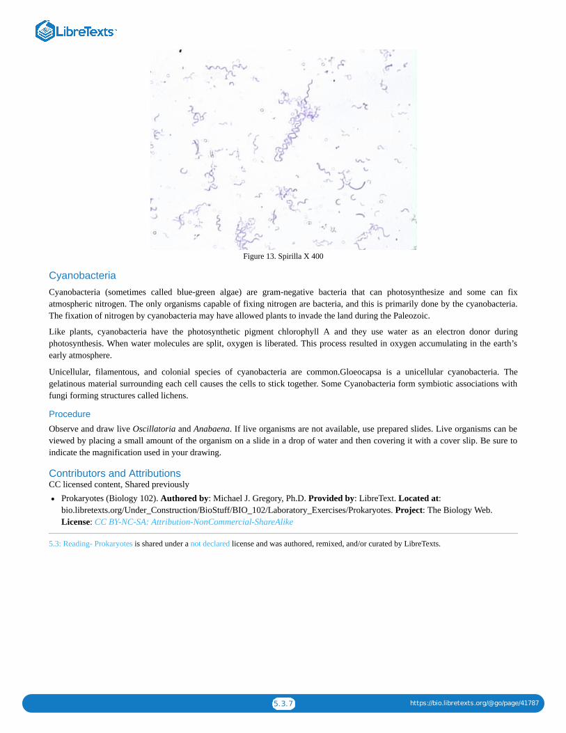
Figure 13. Spirilla X 400
Cyanobacteria
Cyanobacteria (sometimes called blue-green algae) are gram-negative bacteria that can photosynthesize and some can fixatmospheric nitrogen. The only organisms capable of fixing nitrogen are bacteria, and this is primarily done by the cyanobacteria.The fixation of nitrogen by cyanobacteria may have allowed plants to invade the land during the Paleozoic.
Like plants, cyanobacteria have the photosynthetic pigment chlorophyll A and they use water as an electron donor duringphotosynthesis. When water molecules are split, oxygen is liberated. This process resulted in oxygen accumulating in the earth’searly atmosphere.
Unicellular, filamentous, and colonial species of cyanobacteria are common.Gloeocapsa is a unicellular cyanobacteria. Thegelatinous material surrounding each cell causes the cells to stick together. Some Cyanobacteria form symbiotic associations withfungi forming structures called lichens.
Procedure
Observe and draw live Oscillatoria and Anabaena. If live organisms are not available, use prepared slides. Live organisms can beviewed by placing a small amount of the organism on a slide in a drop of water and then covering it with a cover slip. Be sure toindicate the magnification used in your drawing.
Contributors and AttributionsCC licensed content, Shared previously
Prokaryotes (Biology 102). Authored by: Michael J. Gregory, Ph.D. Provided by: LibreText. Located at:bio.libretexts.org/Under_Construction/BioStuff/BIO_102/Laboratory_Exercises/Prokaryotes. Project: The Biology Web.License: CC BY-NC-SA: Attribution-NonCommercial-ShareAlike
5.3: Reading- Prokaryotes is shared under a not declared license and was authored, remixed, and/or curated by LibreTexts.

5.4.1 https://bio.libretexts.org/@go/page/41788
5.4: Reading- ProtistsThe goal of this exercise is to learn about the protists. We will study major groups and for most of the groups, we will studyrepresentative genera.
ProcedureFor each specimen:
1. Read the information thoroughly.2. Create notes. Your notes will be most helpful if they include a drawing, a description, and significant information such as life
cycle, commercial value, ecological significance, and unusual characteristics.
Euglenozoans
Kinetoplastids
Kinetoplastids are flagellated and unicellular. They have a dark staining region of mitochondria called a kinetoplast.
Some kinetoplastids are symbiotic (close) relationships with other organisms. Trypanosomes are Kinetoplastids that cause Africansleeping sickness. They are transmitted to their human hosts by the bite of a tsetse fly. Trypanosoma causes African sleepingsickness.
Euglena
Euglena are unicellular. Many Euglenids feed by phagocytosis. Many species of Euglenids are photosynthetic but can becomeheterotrophic when sunlight is unavailable (mixotrophs).
Euglena use flagella for moving. The outer covering called a pellicle, is flexible and assists in moving. Some have an eyespot witha photoreceptor is capable of detecting the presence of light. Reproduction is asexual.
DiatomsDiatoms are the most numerous unicellular algae in the oceans and as such are an important source of food and oxygen. They arealso important in freshwater environments. They capture 20 to 25% of solar energy captured by living organisms. The cell walls ofdiatoms contain silica (a component of glass) and are formed in 2 halves like a pillbox. Their remains form diatomaceous earth. Itis used for filtering agents, and abrasives such as scouring powders. Diatoms are a major component of phytoplankton infreshwater and marine environments.
Brown AlgaeBrown algae are autotrophs (photosynthetic). Their characteristic brown color is due to carotenoid pigments. They are multicellularand range in size from small to very large. Some are 50 m to 100 m long. They are often found along rocky shores in temperateclimates. The body (thallus) contains holdfasts for attachment, blades, and a stem-like structure that holds the blades is called astipe. Many species have floats that function in floatation. Some have gas-filled floats. Mucilaginous (slimy) material in the cellwalls retards drying in exposed individuals when the tide goes out. Most species have a life cycle with alternation of generations.
Fucus
Fucus is a common “seaweed” found along the rocky coast. Some species of Fucus have diploid adults.

5.4.2 https://bio.libretexts.org/@go/page/41788
Figure 1. Gametes are produced in the receptacles.
Macrocystis and Nereocystis
Macrocystis and nereocystis are deep-water kelps.
Sargassam
Sargassam sometimes breaks off to form floating masses. Other marine organisms congregate around these masses.
Laminaria
Laminaria is a brown alga that is usually found attached just below the intertidal zone. It has a life cycle with alternation ofgenerations.
Figure 2. Alternation of Generations
DinoflagellatesProtective cellulose plates cover dinoflagellates and two flagella enable them to move. One of the flagella lies in a transversegroove that causes cell to spin as it moves.
Most are found in marine or freshwater environments and many are photosynthetic. They are important components ofphytoplankton and thus are important in aquatic food chains. This group also includes many heterotrophic and many mixotrophicspecies.
Some species are responsible for red tides that kill fish and shellfish (Gymnodinium, Gonyaulax, Pfiesteria). Some live assymbiants within some invertebrates. For example, some corals grow faster with dinoflagellates living within their cells. Somespecies are capable of bioluminescence (they produce light).
Both sexual and asexual reproduction occur. Sexual reproduction produces cysts which are resistant to unfavorable environmentalconditions. Cysts are dormant and become active when environmental conditions improve.
CiliatesThe genus Vorticella belongs in this group.
Paramecium

5.4.3 https://bio.libretexts.org/@go/page/41788
Figure 3. Paramecium caudatum X 100
The pellicle (outer covering) of paramecium is covered with hundreds of cilia. They have numerous organelles including a gullet(oral groove) and an anal pore. Ciliates have a large macronucleus and a smaller micronucleus.
The micronucleus is involved in sexual and asexual reproduction. Other nuclear activities are handled by the macronucleus. Themacronucleus is polyploid (approximately 860 N in Paramecium aurelia) and the micronucleus is diploid.
Figure 4. Paramecium X 200
During reproduction, the macronucleus disintegrates. Later, a micronucleus will develop into a macronucleus. Most reproduction isasexual (mitosis). Sexual reproduction is by conjugation.
The micronucleus will divide by meiosis; 3 of the 4 resulting nuclei will disintegrate as will the macronucleus. The remaininghaploid nucleus will divide by mitosis producing an individual with two haploid nuclei. Two conjugating individuals will eachexchange one of the nuclei. The two haploid nuclei will then fuse producing a diploid nucleus.

5.4.4 https://bio.libretexts.org/@go/page/41788
Red AlgaeRed algae are mostly multicellular and are found mainly in warmer, tropical oceans. Their red color is due to an accessoryphotosynthetic pigment called phycoerythrin. The accessory pigments of red algae are able to absorb blue and green light. Thisallows some species to survive in deep waters where blue and green light predominates.
Some species are filamentous but most have a complex pattern of branching. Some coralline forms deposit calcium carbonate intheir cell walls, which contributes to the development of coral reefs.
Green AlgaeFour common forms of green algae are single-celled, colonial, filamentous, and multicellular. Green algae are thought to beancestors of the first plants. Both kinds of organisms have the following characteristics in common:
1. They have a cell wall that contains cellulose.2. They have chlorophyll a and b.3. They store their food as starch inside the chloroplast.
Most species are freshwater but there are many marine species. Some live in damp soil.
Chlamydomonas
Chlamydomonas is a single-celled organism with two flagella. Although this organism is a single cell, the life cycle is similar tothat with haploid adults.
Figure 5. Chlamydomonas’ life cycle
It reproduces asexually (by mitosis) when conditions are favorable. Sexual reproduction occurs when conditions becomeunfavorable. The zygote forms a thick-walled zygospore that is resistant to environmental extremes and divides by meiosis whenenvironmental conditions become favorable.
Most species of Chlamydomonas are isogamous (both gametes are the same size; they are isogametes), some are oogamous(gametes are two sizes; the larger gametes are eggs, the smaller ones are sperm).
Volvox
Volvox is a colonial green algae. The cells are arranged in a gelatinous sphere with two flagella directed to the outside. They divideasexually to produce a daughter colony.
Some cells are specialized to produce sperm and eggs for sexual reproduction. Specialization of cells as seen in the reproductivecells is a characteristic of multicellular organisms. Volvox is considered to be a colony because it appears to be intermediatebetween a group of individual cells and a multicellular organism.
Spirogyra
Spirogyra is a filamentous form. It has a ribbonlike spiral-shaped chloroplast. The life cycle has haploid adults.
Sexual reproduction occurs by conjugation. Conjugation refers to the process where gametes are transferred from one individual toanother by a connection between the two.
The zygote is resistant and overwinters. In the spring, it divides by meiosis to produce haploid filaments.

5.4.5 https://bio.libretexts.org/@go/page/41788
Ulva
Ulva is multicellular with a leaflike body that is two cells thick but up to one meter long. The life cycle is alternation ofgenerations. Both the haploid and the diploid generations look alike (isomorphic).
Tubulinids
Figure 7. Amoeba proteus X 100
Tubulinids move by cytoplasmic extensions called pseudopodia. They feed by phagocytizing (engulfing) their prey. Tubulinidsare found in soil, marine, and freshwater environments. Amoeba proteus (figure 7) is found in freshwater.
Amoeba movement and phagocytosis (video)
Contributors and AttributionsCC licensed content, Shared previously
Protists (Biology 102). Authored by: Michael J. Gregory, Ph.D.. Provided by: LibreTexts. Located at:bio.libretexts.org/Under_Construction/BioStuff/BIO_102/Laboratory_Exercises/Protists. Project: The Biology Web. License:CC BY-NC-SA: Attribution-NonCommercial-ShareAlike
5.4: Reading- Protists is shared under a not declared license and was authored, remixed, and/or curated by LibreTexts.

1
CHAPTER OVERVIEW
6: Module 3- Fungi6.1: Fungi Lab6.2: Fungi Lab (Instructor Materials Preparation)6.3: Reading- Fungi6.4: Case Study- Amphibian Die Off, Chytrid Fungi
6: Module 3- Fungi is shared under a not declared license and was authored, remixed, and/or curated by LibreTexts.

6.1.1 https://bio.libretexts.org/@go/page/41789
6.1: Fungi Lab
Learning ObjectivesThumbnail for the embedded element "Budding Yeast - Time-lapse""title="Budding Yeast - Time-lapse"src="/@api/deki/files/29119/hqdefault-3.jpg"/>
A YouTube element has been excluded from this version of the text. You can view it online here: pb.libretexts.org/bio2lm/?p=58
Questions1. Zygomycota (bread mold): View the prepared slides of the zygospores and sporangia.
1. What kind of reproduction is used by the zygomycota?2. Is the zygospore diploid or haploid?3. Draw a picture of the zygospores you viewed under the microscope.
2. Ascomycota (sac fungi).1. We do not have a slide of the Peziza-please view the pictures on the website.2. Aspergillus: View the slides available of Aspergillus.
1. Can you find any conidiospores?2. Are conidiospores used in sexual or asexual reproduction?3. Use the space below to draw a picture of the conidiospores as you viewed under the microscope.
3. Yeast: create a wet mount slide of the yeast (as assisted by your instructor) to view under the microscope.1. Are yeast single or multi celled?2. Do yeast reproduce asexually or sexually?3. Are you able to view budding, the asexual reproductive process of yeast? Review the Yeast Budding video to help
visualize budding.4. When yeast reproduces sexually, what is the name of the diploid cell that is formed?
3. We do not have a slide of the Schizosaccharomyces octosporus—please view the pictures on the website.4. Skip over the Morels.5. View the Penicillium slides only, no live specimens.
1. Name the specialized stalks that the asexual spores attach to.2. Use the space below to draw a picture of the Penicillium specimen as you viewed it under the microscope.
Basidiomycota (club fungi)1. View the mushroom specimens available in the lab. Do not dissect them. See if you can find the gills on the underside of the
basidiocarp.
1. Name the specific spores formed by the mushroom in the gills.2. View the cross section slide of the Coprinusmushroom.
1. Can you locate the basiodispores?2. Name the specific stalk that the basidiospores attach to.3. Use the space below to draw a picture of the Coprinus basidiospores and basidia as you viewed under the microscope.
Lichens1. There may or may not be live specimens of the lichens to view in the classroom. If live specimens are present, please look at
them. And, access this website to learn more.
1. What type of lichen has the algae dispersed throughout?2. What type of lichen exhibits the fastest growth?3. What type of lichen grows in a circular pattern forming lobes?
2. View the lichen thallus slide under the microscope.1. What two organisms create the lichen?2. Use the space below to draw a picture of the lichen thallus as you viewed it under the microscope. On your picture try to
label both the fungi and the algae.

6.1.2 https://bio.libretexts.org/@go/page/41789
Answer the questions below to summarize the lab activity:1. What is the domain of the fungi?2. How do fungi obtain energy?3. What is the reproductive structure of the fungi? It’s not sperm and egg!4. In the lab activity, which groups of fungi prefer to reproduce asexually? Which groups of fungi tend to exhibit sexual
reproduction?5. A lichen is a mutualisic relationship between what two organisms?
Contributors and AttributionsCC licensed content, Original
Biology 102 Labs. Authored by: Lynette Hauser. Provided by: Tidewater Community College. Located at:http://www.tcc.edu/. License: CC BY: Attribution
6.1: Fungi Lab is shared under a not declared license and was authored, remixed, and/or curated by LibreTexts.
6.2.1 https://bio.libretexts.org/@go/page/41790
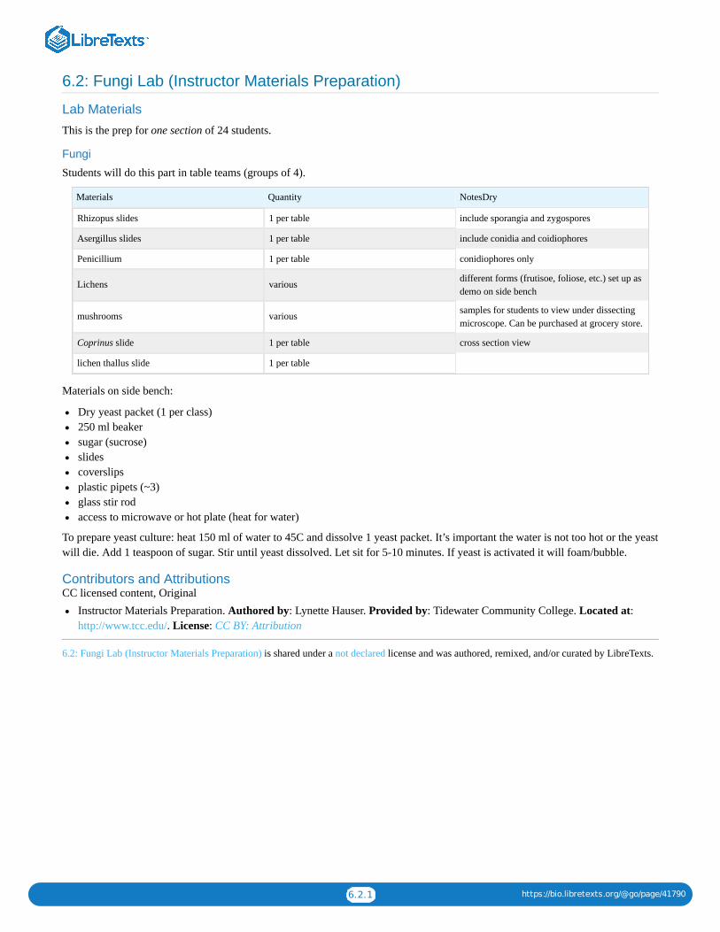
6.2: Fungi Lab (Instructor Materials Preparation)
Lab Materials
This is the prep for one section of 24 students.
Fungi
Students will do this part in table teams (groups of 4).
Materials Quantity NotesDry
Rhizopus slides 1 per table include sporangia and zygospores
Asergillus slides 1 per table include conidia and coidiophores
Penicillium 1 per table conidiophores only
Lichens various different forms (frutisoe, foliose, etc.) set up asdemo on side bench
mushrooms various samples for students to view under dissectingmicroscope. Can be purchased at grocery store.
Coprinus slide 1 per table cross section view
lichen thallus slide 1 per table
Materials on side bench:
Dry yeast packet (1 per class)250 ml beakersugar (sucrose)slidescoverslipsplastic pipets (~3)glass stir rodaccess to microwave or hot plate (heat for water)
To prepare yeast culture: heat 150 ml of water to 45C and dissolve 1 yeast packet. It’s important the water is not too hot or the yeastwill die. Add 1 teaspoon of sugar. Stir until yeast dissolved. Let sit for 5-10 minutes. If yeast is activated it will foam/bubble.
Contributors and AttributionsCC licensed content, Original
Instructor Materials Preparation. Authored by: Lynette Hauser. Provided by: Tidewater Community College. Located at:http://www.tcc.edu/. License: CC BY: Attribution
6.2: Fungi Lab (Instructor Materials Preparation) is shared under a not declared license and was authored, remixed, and/or curated by LibreTexts.

6.3.1 https://bio.libretexts.org/@go/page/41791
6.3: Reading- Fungi
Zygomycota
Fungi in the phylum Zygomycota are called zygomycetes. The zygomycetes are terrestrial. They are usually saprotrophs but thereare some parasites. The hyphae are coenocytic (theyn lack septa). Septa are found only in the reproductive structures.
Reproduction in Zygomycota
Fusion of two hyphae leads to the formation of a zygosporangium, a thick-walled structure that is capable of survivingenvironmental extremes. Before karyogamy, the zygosporangium contains many haploid nuclei. after karyogamy, it contains manydiploid nuclei.
Rhizopus (Bread Mold)
Figure 2. Rhizopus* sporangia
Asexual reproduction involves mycelia producing sporangia that produce haploid spores by mitosis. The spores produce newmycelia.
Figure 3. Rhizopus* zygotes
When environmental conditions deteriorate, sexual reproduction may occur. Hyphae from opposite mating types produce structuresthat contain several haploid nuclei. Fusion of two of these structures from opposite mating types results in a heterokaryoticzygosporangium. A thick wall develops that functions to protect the zygospore until environmental conditions become favorable.When conditions are favorable, nuclear fusion (karyogamy) occurs within the zygosporangium producing diploid nuclei. This isfollowed by meiosis. The zygosporangium then germinates to produce a sporangium which releases haploid spores.
Observe Rhizopus (bread mold) growing on a culture dish. Use a dissecting microscope to see details of the hyphae and sporangia.Is there any evidence of sexual reproduction?
6.3.2 https://bio.libretexts.org/@go/page/41791
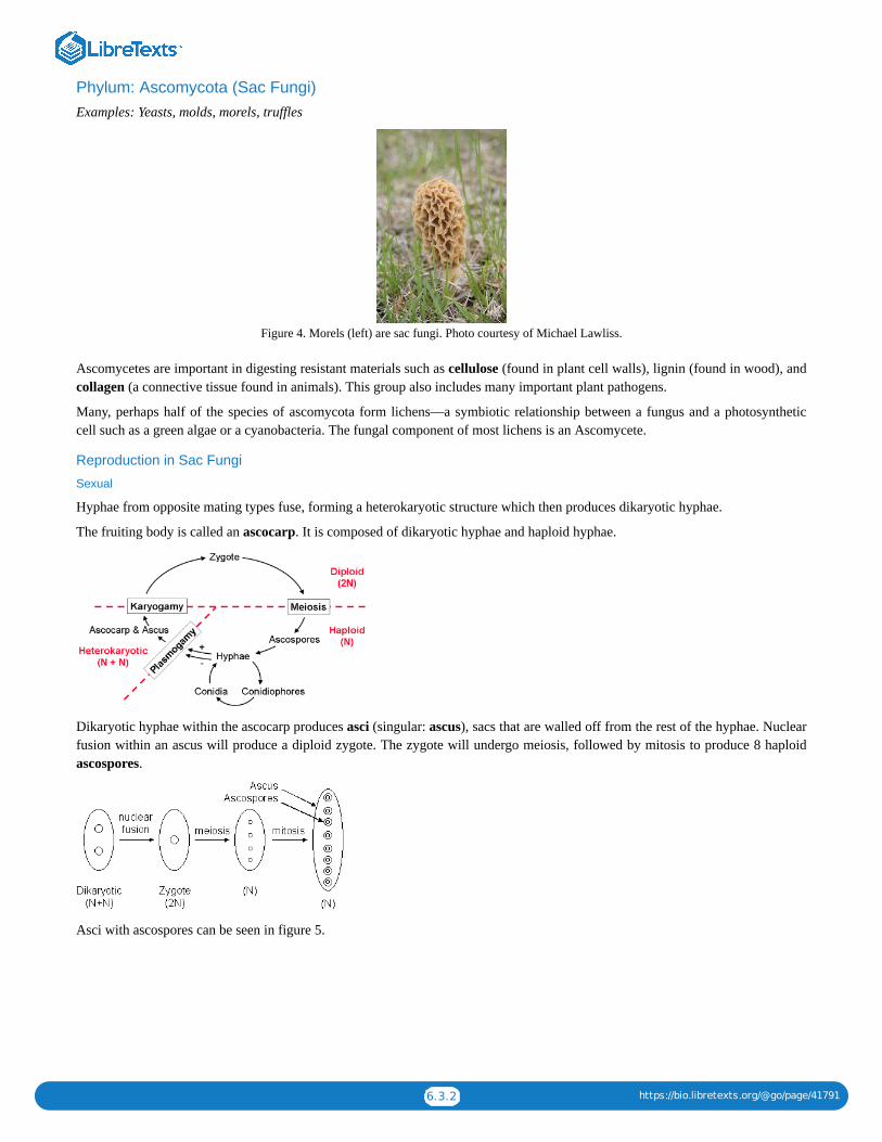
Phylum: Ascomycota (Sac Fungi)Examples: Yeasts, molds, morels, truffles
Figure 4. Morels (left) are sac fungi. Photo courtesy of Michael Lawliss.
Ascomycetes are important in digesting resistant materials such as cellulose (found in plant cell walls), lignin (found in wood), andcollagen (a connective tissue found in animals). This group also includes many important plant pathogens.
Many, perhaps half of the species of ascomycota form lichens—a symbiotic relationship between a fungus and a photosyntheticcell such as a green algae or a cyanobacteria. The fungal component of most lichens is an Ascomycete.
Reproduction in Sac Fungi
Sexual
Hyphae from opposite mating types fuse, forming a heterokaryotic structure which then produces dikaryotic hyphae.
The fruiting body is called an ascocarp. It is composed of dikaryotic hyphae and haploid hyphae.
Dikaryotic hyphae within the ascocarp produces asci (singular: ascus), sacs that are walled off from the rest of the hyphae. Nuclearfusion within an ascus will produce a diploid zygote. The zygote will undergo meiosis, followed by mitosis to produce 8 haploidascospores.
Asci with ascospores can be seen in figure 5.

6.3.3 https://bio.libretexts.org/@go/page/41791
Figure 5. Peziza cross section X 200.
Asexual
Most reproduction is by asexual spores called conidia. Unlike the Zygomycetes which produce asexual spores within sporangia,conidia are produced on the ends of specialized hyphae called conidiophores.
Examples of Sac Fungi
Morels and truffles are gourmet delicacies. This group includes many important plant parasites such as Dutch elm disease, chestnutblight, leaf curl fungi, and Claviceps.
An ergot is the hard, purple-black fungus Claviceps purpurea. It contains toxic alkaloids, including LSD. When infected rye ismade into bread, the toxins are ingested and cause vomiting, muscle pain, feeling hot or cold, hand and foot lesions, hysteria andhallucinations. Historians believe that those that accused their neighbors of witchcraft in Salem may have been suffering fromergotism. Claviceps is used to stimulate uterine contractions and to treat migraine headaches.
Peziza (Cup Fungi)
Observe preserved Peziza (cup fungus) using a dissecting microscope.
Observe a slide of Peziza at scanning, low, and high power magnification. Find an ascus and ascospores on the upper surface(inside the cup).
Aspergillus
Observe the conidiophores and conidia (asexual spores) of Aspergillus.
Yeast
Yeast are single-celled members of the sac fungi. Most reproduction is asexual; a small cell pinches off from a larger cell. This typeof mitosis where a smaller individual grows from a larger individual is called budding.
Make a wet mount of live yeast and see if you can observe budding under high power. If you cannot see yeast budding, view aprepared slide of yeast budding under high power.
Yeast also reproduce sexually by forming an ascus and eight ascospores. View a slide of Schizosaccharomyces octosporus underhigh power or oil immersion and find an ascus with ascospores.
Figure 6. Yeast (Saccharomyces) budding X 1000.
During sexual reproduction, the fusion of two cells results in the formation of an ascus.

6.3.4 https://bio.libretexts.org/@go/page/41791
Figure 7. Schizosaccharomyces octosporus X 1000
The elongated cell in the upper left part of figure 7 contains ascospores.
Figure 8. Schizosaccharomyces octosporus X 1000
Cells in the lower left part of the figure 8 contain ascospores.
Yeast is important in leavening bread by CO production and in producing ethanol for alcoholic beverages.
Penicillium
Observe Penicillium growing on a culture dish.
Figure 9. Penicillium growing on an agar plate
Penicillium reproduces asexually. Observe a slide of Penicilliumconidiophores under high power. The spores are called conidia.
Figure 10. Penicillium Conidiophores and conidia X 400.
Phylum: Basidiomycota (Club Fungi)
Reproduction
Asexual reproduction in club fungi is rare. Their fruiting bodies are called basidiocarps. This is the visible mushroom.
2

6.3.5 https://bio.libretexts.org/@go/page/41791
Figure 11. Mushrooms showing gills
Spores, called basidiospores are produced on basidia within the basidiocarps. In mushrooms, the basidia are located along the gillson the underside of the cap. In figure 6, a portion of the cap of this mushroom has been broken away to reveal the gills.
Figure 12. Basidia and basidiospores X 1000
In ascomycota (sac fungi), the ascospores were enclosed in an ascus. In basidiomycota, the basidiospores are not enclosed.Compare the diagrams of a basidium with basidiospores above with that of an ascus with ascospores seen earlier.
Basidiospores germinate to produce monokaryotic (haploid, one nucleus per cell) hyphae. Mushrooms are composed of dikaryotichyphae which are formed when hyphae fuse. Dikaryotic nuclei within the basidium fuse to produce a zygote and meiosis thenproduces basidiospores.
Observe some representative club fungi on display including mushrooms, puffballs, and bracket fungi.
Bracket Fungi

6.3.6 https://bio.libretexts.org/@go/page/41791
Figure 13. Bracket fungi
Bracket Fungi and Lichens
Figure 14. Bracket fungi and lichens
Mushrooms
Figure 15. Mushrooms
Cut a mushroom to reveal the gills as shown in figure 16. Basidia and basidiospores form on the gills.
Figure 16. Mushroom cut to reveal the gills
View a cross section of the cap of a mushroom (Coprinus) showing the gills. Find a basidium and basidiospores.

6.3.7 https://bio.libretexts.org/@go/page/41791
Figure 17. Coprinus X 400
Figure 18. Coprinus X 1000 showing basidia and basidiospores
Symbiotic Associations of Fungi and Other Organisms
Lichens
Lichens are structures made up of two different species:
1. a fungus2. either a cyanobacterium or a green algae
The photosynthetic cells are contained within the middle layer.
The photosynthetic cells provide photosynthesis for the lichen. It was thought that the relationship was mutualistic because thefungus prevented the algal cells from desiccation. Recent evidence indicates that the photosynthetic cells may grow faster whenseparated from the fungus. Perhaps the fungus is parasitizing the photosynthetic cells.
Reproduction is asexual. Fragments are produced that contain fungal hyphae and photosynthetic cells.
Lichens derive most of their water and minerals from rainwater and air. This allows them to survive on bare rock, tree trunks,inhospitable places.
Observe the lichens on display. Some lichens have a crust-like appearance (crustose). Others have a shrublike (fruticose) or leaflike(foliose) appearance.

6.3.8 https://bio.libretexts.org/@go/page/41791
Figure 19. Lichens growing on a rock
Figure 20. Lichens growing on a tree
Figure 21. Lichens growing on a tree
Figure 22. Lichen thallus (cross-section X 200)

6.3.9 https://bio.libretexts.org/@go/page/41791
Figure 23. Lichen thallus X 400
Contributors and AttributionsCC licensed content, Shared previously
Kingdom: Fungi, Biology 102. Authored by: Michael J. Gregory, Ph.D.. Provided by: LibreTexts. Located at:bio.libretexts.org/Under_Construction/BioStuff/BIO_102/Laboratory_Exercises/Fungi. Project: The Biology Web. License:CC BY-NC-SA: Attribution-NonCommercial-ShareAlike
6.3: Reading- Fungi is shared under a not declared license and was authored, remixed, and/or curated by LibreTexts.

6.4.1 https://bio.libretexts.org/@go/page/41792
6.4: Case Study- Amphibian Die Off, Chytrid FungiReview this slideshow then answer the questions below.
A SlideShare element has been excluded from this version of the text. You can view it online here: pb.libretexts.org/bio2lm/?p=64
Questions1. What phylum of fungi is responsible for chytridiomycosis?2. Why is this fungi successful at killing amphibians?3. Why be concerned about the world’s amphibian populations? What role do amphibians play in ecosystems?4. How might Bd be spreading through the environment?5. Propose a hypothesis that would explain why Bd is spreading like a new disease throughout the world.6. Some scientists are attempting to prevent the extinction of amphibians by capturing amphibians in the wild, breeding them in
chytrid free labs, then releasing some of the amphibians into the wild. Discuss how such a strategy could save some species ofamphibians.
7. How else might we prevent the spread of Bd throughout the world?
Contributors and AttributionsCC licensed content, Shared previously
Amphibian Die Off. Authored by: Lynette Hauser. Provided by: Tidewater Community College. Located at:www.slideshare.net/CandelaContent/amphibian-die-off. License: CC BY: Attribution
6.4: Case Study- Amphibian Die Off, Chytrid Fungi is shared under a not declared license and was authored, remixed, and/or curated byLibreTexts.

1
CHAPTER OVERVIEW
7: Module 4- Seedless Plants7.1: Seedless Plant Lab7.2: Seedless Plant Lab (Instructor Materials Preparation)7.3: Reading- Seedless Plants
7: Module 4- Seedless Plants is shared under a not declared license and was authored, remixed, and/or curated by LibreTexts.

7.1.1 https://bio.libretexts.org/@go/page/41793
7.1: Seedless Plant Lab
Lab Objectives
At the conclusion of the lab, the student should be able to:
Explain what is meant by “alteration of generations”Explain the difference between the sporophyte and gametophyte generation in plants. State which generation is haploid andwhich is diploidName the process that makes spores and state if spores are haploid or diploidName the process that creates sperm and egg from spores and state if sperm and egg are haploid or diploidName the phyla discussed in the lab and give an example of a plant from eachIdentify and know the function of the archegonium and the antheridumIdentify the fern structures discussedUnderstand the basic moss and fern life cycle
A SlideShare element has been excluded from this version of the text. You can view it online here: pb.libretexts.org/bio2lm/?p=68
Download a PDF of the lab to print.
Procedure1. Access the page “Reading: Seedless Plants.”2. Phylum Bryophyta (Mosses)
1. View the live moss specimens available in the lab.1. Is the green “leaf like” tissue gametophyte or sporophyte?2. Is the stalk that emerges from the green “leaf like” tissue gametophyte or sporophyte?
2. As indicated in #3 of the website use the space below to draw a simple life cycle of the moss. Include in the life cycle 2N, N,sporophyte, gametophyte, meiosis, spores, egg, sperm, antheridium, archigonium, fertilization. If you need help inconstructing your life cycle picture check out this website.
3. View the prepared slide of the archigonium and the antheridum (there should be a slide with both).
1. Is the archegonium male or female?2. What cell is produced in the archegonium?3. Is this cell haploid or diploid?4. Is the antheridium male or female?5. What cell is produced in the antheridium?6. Is this cell haploid or diploid?
4. View the prepared slide of the moss capsule.1. Is the capsule sporophyte or gametophyte tissue?2. What cell is produced in the capsule?3. Is this cell haploid or diploid?4. How are moss spores dispersed to new locations?
3. Skip the liverworts section (Phylum Hepatophyta)4. Seedless Vascular Plants5. Phylum Pterophyta (Ferns)
1. As indicated in #1 of the website use the space below to draw a simple life cycle of the fern. Include in the life cycle 2N, N,sporophyte, gametophyte, meiosis, spores, egg, sperm, antheridium, archigonium, fertilization, sorus. If you need help inconstructing your life cycle picture check out this website.
2. Observe the preserved fern frond. Locate the sori on the underside.
7.1.2 https://bio.libretexts.org/@go/page/41793
1. Is the frond sporophyte or gametophyte?2. What cell is produced in the sori?3. Is this cell diploid or haploid?
3. View the prepared slide of the fern prothallus under the microscope.1. What shape is the prothallus?2. Is the prothallus sporophyte or gametophyte?3. Can you find the archegonium and the antheridium?4. What cell is made in the archegonium?5. What cell is made in the antheridium?
6. Skip the horsetails7. Skip the spikemosses and club mosses (Phylum Kycophyta)8. Answer the review questions below.
1. Is gametophyte tissue haploid or diploid?2. Is sporophyte tissue haploid or diploid?3. Is the moss life cycle gametophyte or sporophyte dominant?4. Is the fern life cycle gametophyte or sporophyte dominant?5. In the life cycle of the primitive plant, the process of meiosis produces what cell?6. Does the gametophyte or sporophyte generation produce spores?7. What process do spores undergo to create sperm and egg?8. State one reason why moss and fern are considered primitive plants.9. What is meant by the idea of “alteration of generations?”
Contributors and AttributionsCC licensed content, Shared previously
Biology 102 Labs. Authored by: Lynette Hauser. Provided by: Tidewater Community College. Located at:http://www.tcc.edu/. License: CC BY: Attribution
7.1: Seedless Plant Lab is shared under a not declared license and was authored, remixed, and/or curated by LibreTexts.

7.2.1 https://bio.libretexts.org/@go/page/41794
7.2: Seedless Plant Lab (Instructor Materials Preparation)
Lab Materials
This is the prep for one section of 24 students.
Seedless Plants
Students will do this part in table teams (groups of 4).
Materials Quantity Notes
live moss speciman with gametophye andsporophyte tissue
1-2 put on side bench as demo, helpful to viewunder dissecting scope
moss antheridium slide 1 per table
moss archigonium slide 1 per table
moss capsule with spores slide 1 per table
fern leaf with sori 1-2 put on side bench as demo, helpful to viewunder dissecting scope
fern prothallus slide 1 per table should contain both antheridia and archegonia
Contributors and AttributionsCC licensed content, Original
Instructor Materials Preparation. Authored by: Lynette Hauser. Provided by: Tidewater Community College. Located at:http://www.tcc.edu/. License: CC BY: Attribution
7.2: Seedless Plant Lab (Instructor Materials Preparation) is shared under a not declared license and was authored, remixed, and/or curated byLibreTexts.

7.3.1 https://bio.libretexts.org/@go/page/41795
7.3: Reading- Seedless Plants
Introduction
Plants (kingdom Plantae) are autotrophs; they make their own organic nutrients. The term “organic” refers to compounds thatcontain carbon. Organic nutrients such as sugars are made by photosynthesis.
Plants are adapted to living on land. For example, the above-ground parts of most plants are covered by a waxy layer called acuticle to prevent water loss. Aquatic plants are secondarily adapted to living in water.
Some evidence that suggests that plants evolved from the green algae is:
they both use chlorophyll a, chlorophyll b, and carotenoid pigments during photosynthesis.the primary food reserve of both is starch.they both have cellulose cell walls.
Genetic and morphological evidence indicates that plants evolved from a group of green algae called charophyceans. Manycharophyceans inhabit shallow freshwater environments. Natural selection may have favored individuals capable of survivingoccasional drying in these environments and this gave rise to land plants.
These traits occur in plants but not charophyceans. Some evolved independently in other algae.
Apical meristemsAlternation of generationsSpores with protective wallsSpores produced in sporangiaGametes are produced in multicellular structures called gametangia; Antheridia produce sperm; Archegonia produce eggsMulticellular dependent embryosMany have a cuticle that waterproofs and offers some protection
Alternation of Generations
The basic alternation of generations life cycle is illustrated below.
The diploid plant that produces spores is called a sporophyte. The haploid plant that produces gametes is called a gametophyte.
Some protists also have an alternation of generations life cycle but the structures that produce gametes in protists are usually singlecells. Plants produce gametes in multicullar structures that have an outer protective layer. Sperm are produced in structures calledantheridia (sing. antheridium), eggs are produced in archegonia (sing. archegonium),. As in protists and fungi, spores of plantsare produced insporangia (sing. sporangium).
A dependent sporophyte is a sporophyte that is small and grows attached to the gametophyte. It obtains nutrients from thegametophyte. An independent sporophyte grows separately from the gametophyte. Similarly, a dependent gametophyte is smalland grows attached to the sporophyte while an independent gametophyte grows separately from the sporophyte.
The evolutionary trend in plants has been from plants with a dominant gametophyte and reduced, dependent sporophyte (ex.Mosses) to plants with a dominant, independent sporophyte and a reduced, dependent gametophyte (ex. Seed plants).

7.3.2 https://bio.libretexts.org/@go/page/41795
Classification
Evolutionary relationships among the plants are shown below.
We will study the following phyla of plants.
Characteristics Classification
Bryophytes (no vascular tissue)Liverworts (Phylum Hepatophyta)Mosses (Phylum Bryophyta)Hornworts (Phylum Anthocerophyta
Seedless vascular plants Club mosses, Spike Mosses, Quillworts (Phylum Lycophyta)Horsetails,Whisk Ferns, Ferns (Phylum Pterophyta)
Gymnosperms (vascular, naked seeds)
Conifers (Phylum Coniferophyta)Cycads (Phylum Cycadophyta)Ginkgos (Phylum Ginkgophyta)Gnetophytes (Phylum Gnetophyta)
Angiosperms (vascular, protected seeds) Flowering Plants (Phylum Anthophyta)MonocotsEudicots
Bryophytes
Phylum: Bryophyta (Mosses)1. Observe different kinds of moss on display and note the body form of the gametophyte.
Figure 1. Moss growing on a rock.
2. Obtain live sporulating moss and identify the sporophyte and gametophyte generations.
Figure 2. Moss gametophytes and sporophytes

7.3.3 https://bio.libretexts.org/@go/page/41795
3. Draw the life cycle of a typical bryophyte such as moss. Your drawing should contain the following terms:1. 2N2. N3. sporophyte4. sporangium5. meiosis6. spores7. protonema8. gametophyte9. antheridium
10. sperm11. archegonium12. egg13. fertilization
4. Observe a slide showing the antheridial head of Mnium (a moss). Begin using the scanning (4X) objective and then switch tothe low power objective (10X).
5. What is produced in this structure (the antheridium)?6. Show where the antheridium occurs on the live moss plant. Indicate where this structure occurs in the life cycle diagram that
you prepared (above).
Figure 3. Mnium (a moss) antheridial head
Figure 4. Mnium (a moss) antheridial head x40
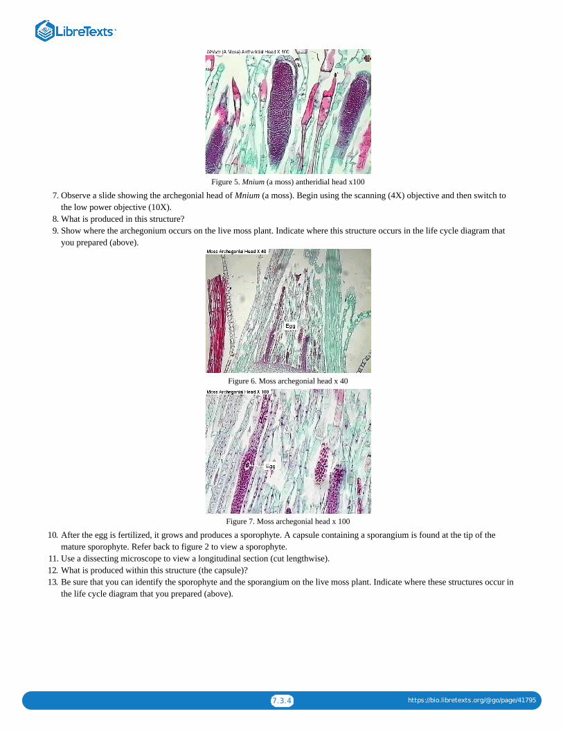
7.3.4 https://bio.libretexts.org/@go/page/41795
Figure 5. Mnium (a moss) antheridial head x100
7. Observe a slide showing the archegonial head of Mnium (a moss). Begin using the scanning (4X) objective and then switch tothe low power objective (10X).
8. What is produced in this structure?9. Show where the archegonium occurs on the live moss plant. Indicate where this structure occurs in the life cycle diagram that
you prepared (above).
Figure 6. Moss archegonial head x 40
Figure 7. Moss archegonial head x 100
10. After the egg is fertilized, it grows and produces a sporophyte. A capsule containing a sporangium is found at the tip of themature sporophyte. Refer back to figure 2 to view a sporophyte.
11. Use a dissecting microscope to view a longitudinal section (cut lengthwise).12. What is produced within this structure (the capsule)?13. Be sure that you can identify the sporophyte and the sporangium on the live moss plant. Indicate where these structures occur in
the life cycle diagram that you prepared (above).

7.3.5 https://bio.libretexts.org/@go/page/41795
Figure 8. Moss capsule containing spores
Figure 9. Moss capsule x40
14. How are moss spores dispersed to new locations?
Phylum: Hepatophyta (Liverworts)
Observe live Marchantia (a liverwort). Do the plants contain gemma cups? What is the function of gemma cups?
Figure 10. Marchantia (live)
Seedless Vascular Plants
Phylum: Pterophyta
Ferns
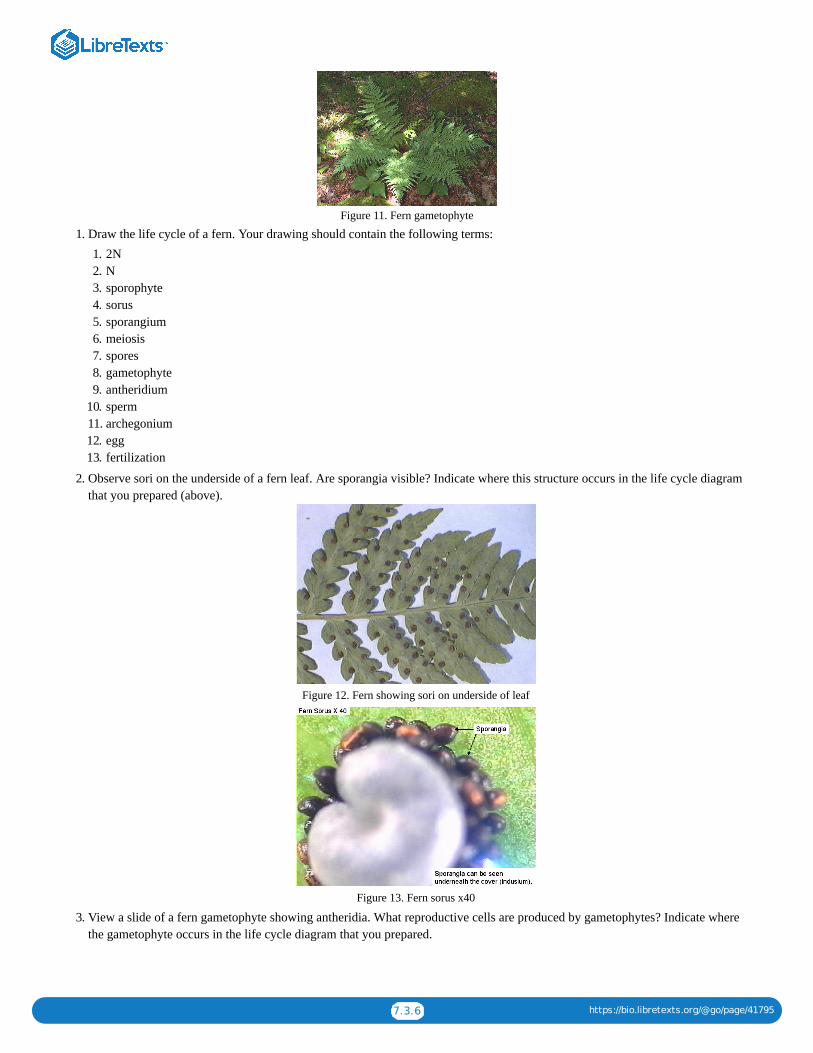
7.3.6 https://bio.libretexts.org/@go/page/41795
Figure 11. Fern gametophyte
1. Draw the life cycle of a fern. Your drawing should contain the following terms:1. 2N2. N3. sporophyte4. sorus5. sporangium6. meiosis7. spores8. gametophyte9. antheridium
10. sperm11. archegonium12. egg13. fertilization
2. Observe sori on the underside of a fern leaf. Are sporangia visible? Indicate where this structure occurs in the life cycle diagramthat you prepared (above).
Figure 12. Fern showing sori on underside of leaf
Figure 13. Fern sorus x40
3. View a slide of a fern gametophyte showing antheridia. What reproductive cells are produced by gametophytes? Indicate wherethe gametophyte occurs in the life cycle diagram that you prepared.
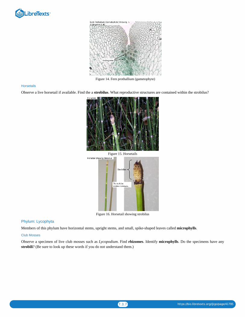
7.3.7 https://bio.libretexts.org/@go/page/41795
Figure 14. Fern prothallium (gametophyte)
Horsetails
Observe a live horsetail if available. Find the a strobilus. What reproductive structures are contained within the strobilus?
Figure 15. Horsetails
Figure 16. Horsetail showing strobilus
Phylum: Lycophyta
Members of this phylum have horizontal stems, upright stems, and small, spike-shaped leaves called microphylls.
Club Mosses
Observe a specimen of live club mosses such as Lycopodium. Find rhizomes. Identify microphylls. Do the specimens have anystrobili? (Be sure to look up these words if you do not understand them.)

7.3.8 https://bio.libretexts.org/@go/page/41795
Figure 17. Club moss (lycopodium)
Spike Mosses
Observe a specimen of a spike moss such as Selaginella. Note the structure of the microphylls.
Figure 18. Spike moss (selaginella)
Contributors and AttributionsCC licensed content, Original
Seedless Plants (Kingdom: Plantae), Biology 102. Authored by: Michael J. Gregory, Ph.D.. Provided by: LibreTexts. Locatedat: bio.libretexts.org/Under_Construction/BioStuff/BIO_102/Laboratory_Exercises/Seedless_Plants. Project: The BiologyWeb. License: CC BY-NC-SA: Attribution-NonCommercial-ShareAlike
7.3: Reading- Seedless Plants is shared under a not declared license and was authored, remixed, and/or curated by LibreTexts.

1
CHAPTER OVERVIEW
8: Module 5- Seed Plants8.1: Seed Plants Lab8.2: Seed Plants Lab (Instructor Materials Preparation)8.3: Reading- Seed Plants
8: Module 5- Seed Plants is shared under a not declared license and was authored, remixed, and/or curated by LibreTexts.

8.1.1 https://bio.libretexts.org/@go/page/41796
8.1: Seed Plants Lab
Learning ObjectivesDefine the terms (meanings of the names) angiosperm and gymnospermState what type of cells create eggs and what type of cells create sperm in gymnosperms and angiospermsDescribe the general characteristics seed plants.Name the phyla discussed in the lab and give an example of a plant from eachRecognize and identify plant specimens viewed in the lab, both slides and live samplesUnderstand the basic gymnosperm and angiosperm life cycleRecognize the difference between a male and female pine coneExplain the seed plant alteration of generations including sporophyte and gametophyte structuresIdentify and know the function of the microscopes and the megasporesIdentify the flower parts and which structures and male and femaleExplain and recognize the difference between a monocot and a eudicot flowerDistinguish between the different types of fruits and give an example of each type
A SlideShare element has been excluded from this version of the text. You can view it online here: pb.libretexts.org/bio2lm/?p=76
Download a PDF of the lab to print.
Procedure1. Access the Seed plant lab.2. Gymnosperms
1. Name the four subgroups within gymnosperms.2. We will focus on conifers. Observe the conifer leaf samples available.
1. How does the needle-like leaf benefit the conifer?3. Reproduction in Pines: As indicated on the website, draw a simple pine life cycle in the space on the next page. Be sure to
include the terms egg, embryo, fertilization, megaspore, microscope, gametophyte, sporophyte, meiosis, mitosis, and pollen.Use this website to get you started.
4. Observe the pine cones on display. Are pine cones haploid or diploid?5. Are male or female pine cones larger?6. View the slide of the pollen (male) pine cone cross section.
1. Can you find the microscopes on the slide?2. Are microscopes haploid or diploid?3. What process do microscopes undergo to form pollen grains?4. Can you find pollen grains on the slide?5. Use the space on the next page to draw what you observed under the microscope.
7. View the slide of the seed (female) pine cone cross section.1. Can you find the megaspores on the slide?2. Are megaspores haploid or diploid?3. What process do megaspores undergo to form the egg or ovule?4. Can you find the egg on the slide?5. Is the tissue surrounding the egg haploid or diploid?6. Once the egg is fertilized what structure will form?7. Use the space below to draw what you observed under the microscope.
8. View the pine seeds on display

8.1.2 https://bio.libretexts.org/@go/page/41796
1. Are the seeds haploid or diploid?2. How will the seeds be dispersed through the environment?3. What cell division process will the seeds undergo to create a new pine tree?
3. Angiosperms1. As indicated on the website, draw a simple angiosperm life cycle in the space below. Be sure to include the terms egg,
embryo, fertilization, megaspore, microscope, gametophyte, sporophyte, meiosis, mitosis, and pollen. Use this website to getyou started: http://www.sumanasinc.com/webcontent/animations/content/angiosperm.html
2. Use the flower model to identify the different structures.
1. Collectively, the male flower parts are called the ___________________.2. Collectively, the female flower parts are called the __________________.3. Use the table below to describe the function of each flower part and if it is male, female, or neither.
Flower structure Function Male/Female/Neither
Anther
Filament
Stigma
Style
Ovary
Petal
Sepals
3. Dissect the live flower. Start with the outside (sepals, petals) and work your way in. Identify each structure as you dissectthe flower.1. How many petals does the flower have?2. Is the flower a monocot or a eudicot? What characteristic did you use to determine?3. Once you dissect the flower, dispose of the flower parts. You do not have to cut into the ovary or the anther as indicated
on the website.4. View the slide of the lily mature female gametophyte.
1. Can you locate the egg?2. Is the egg haploid or diploid?3. What type of cell underwent mitosis to create the egg?4. Can you locate the polar nuclei?5. Is the ovary (tissue surrounding the egg) haploid or diploid?6. Use the space below to draw what you observed under the microscope.
5. View the slide of the lily anther cross section.1. Can you locate the pollen grains?2. Is the pollen haploid or diploid?3. What cell type underwent mitosis to create the pollen?4. Is the anther (tissue surrounding the pollen) haploid or diploid?5. Use the space below to draw what you observed under the microscope.
6. View the slide of the lily pollen grains.1. How many cells are held within a single pollen grain?2. Use the space below to draw what you observed under the microscope.
7. Skip the slide of germinated pollen. That slide is not available in the lab.8. Skip the slide of the lily developing embryo. It is not available in the lab.9. Skip the slide of the Capsella embryo, both early and mature embryo. These slides are not available in the lab.
10. Although there is not a live bean seed available, please view the preserved bean seed.
1. Is the seed haploid or diploid?

8.1.3 https://bio.libretexts.org/@go/page/41796
2. How many cotyledons does the bean seed have?3. Is the bean a monocot or a eudicot?
11. Although there is not a live corn seed available, please view the preserved corn seed.1. How many cotyledons does the corn seed have?2. Is the corn a monocot or a eudicot?3. What is the function of the endosperm tissue?
12. Skip over the recently germinated bean and corn plants.13. Fruits. There will be several fruit examples available in the lab. They may be different than the ones described on the
website. Use the table below to discuss the fruits you view.
Name of fruit Simple, Aggregate, or Multiple Dry or fleshy
Review Questions
Answer the review questions below.
1. What does gymnosperm mean?2. What group of gymnosperm plants is the largest?3. What type of spore is used for male reproduction in seed plants?4. Through mitosis, the male spore develops into what structure?5. What type of spore is used for female reproduction in seed plants?6. Through mitosis, the female spore develops into what structure?7. What does angiosperm mean?8. What structures of the flower are female?9. What structures of the flower are male?
10. What is the function of the petals of the flower?11. The process of ___________________ occurs in the flower anther to create haploid ________________ followed by mitosis to
create __________________.12. The process of __________________ occurs in the flower ovary to create the haploid ___________________ followed by
mitosis to create the ______________ and the n+n ______________________.13. State one difference between monocots and eudicots.14. Explain how angiosperms undergo a double fertilization.15. What part of the flower develops into a fruit?16. How is a simple fruit different from a complex fruit?17. Give an example of a fleshy fruit18. Give an example of a dry fruit.
Contributors and AttributionsCC licensed content, Shared previously
Biology 102 Labs. Authored by: Lynette Hauser. Provided by: Tidewater Community College. Located at:http://www.tcc.edu/. License: CC BY: Attribution
8.1: Seed Plants Lab is shared under a not declared license and was authored, remixed, and/or curated by LibreTexts.

8.2.1 https://bio.libretexts.org/@go/page/41797
8.2: Seed Plants Lab (Instructor Materials Preparation)
Lab Materials
This is the prep for one section of 24 students.
Seed Plants
Conifers
Students will do this part in table teams (groups of 4).
Materials Quantity Notes
pine seeds various on side bench as demo
pine cone various both pollen and seed cone examples on sidebench as demo
pine pollen cone cross section slide 1 per table
pine seed cone cross section slide 1 per table
flower model 1 per table
live flower 1 per table for dissection, Alstroemeria works well
lily gametophyte slide 1 per table
lily anther cross section slide 1 per table
lily pollen grain slide 1 per table
corn seed model 2-3 per class demo on side bench
bean seed model 2-3 per class demo on side bench
fruit examples variousexamples include apples, beans, raspberries,walnuts, acorns. Try to have several simple andmultiple fruits, both fruity and dry
Contributors and AttributionsCC licensed content, Original
Instructor Materials Preparation. Authored by: Lynette Hauser. Provided by: Tidewater Community College. Located at:http://www.tcc.edu/. License: CC BY: Attribution
8.2: Seed Plants Lab (Instructor Materials Preparation) is shared under a not declared license and was authored, remixed, and/or curated byLibreTexts.

8.3.1 https://bio.libretexts.org/@go/page/41798
8.3: Reading- Seed Plants
Gymnosperms
The four phyla of gymnosperms are cycads, ginkgo, gnetophytes, and conifers.
Gymnosperms have naked seeds. The seeds of angiosperms are contained within a fruit.
Gymnosperm Diversity
We will examine conifers in some detail during this lab class but will use photographs on the Internet to study the other threedivisions. Click on the links below to view photographs of them.
Cycads
Cycads re cone-bearing palmlike plants found mainly in tropical and subtropical regions today. They were very numerous in theMesozoic Era.
Ginkgo
There is only one species of Ginkgo left. It survived due to Chinese planting them along roadsides. Click here for information andphotographs.
Gnetophytes
Welwitschia and Ephedra (information and photographs)
Conifers
Conifers are the largest group of gymnosperms. They include evergreen trees such as pine, cedar, spruce, fir, and redwood trees.Examine the leaves of pine on display.
The leaves of conifers are needle-like and are adapted for dry conditions such as hot summers or freezing winters. Needles losewater slower than broad, flat leaves and therefore do not need to be shed during seasons when water is scarce, so most conifers areevergreen.
Reproduction in Pine
Life Cycle of Seed Plants
Seed plants are heterosporous—they have two different spore sizes: megaspores and microspores.
The generalized life cycle of plants has been modified (below) to illustrate plants which have separate male and femalegametophytes (megagametophyte and microgametophyte) produced by different sized spores (megaspores and microspores).
The evolutionary trend from nonvascular plants to seedless vascular plants to seed plants has been a reduction in the size of thegametophyte. In seed plants, the gametophyte is usually microscopic and is retained within the tissues of the sporophyte.
The megasporangium is surrounded by layers of sporophyte tissue called the integument. The integument and structures within(megasporangium, megaspore) are the ovule.
Microspores germinate within the sporophyte tissue and become pollen grains. The microgametophyte is contained within thetough, protective coat of the pollen grain.

8.3.2 https://bio.libretexts.org/@go/page/41798
The entire microgametophyte (pollen grain) is transferred to the vicinity of the megagametophyte by a process of pollination. Windor animals usually accomplish this transfer.
When pollen reaches the female gametophyte, it produces an elongate structure (pollen tube) that grows to the egg cell. Sperm aretransferred directly through this tube to the egg. The advantage of this process is that sperm do not have to swim long distances asthey do in seedless plants.
Seeds
Seeds contain the sporophyte embryo, food for the embryo, and a protective coat.
The embryo within the seed is dormant; it can survive for long periods without additional food or water. When conditions becomefavorable, the embryo resumes growth as the seed germinates.
1. Draw the life cycle of pine and include the following terms: eggs, embryo, fertilization, megagametophyte, megasporangium,megaspore, meiosis, microgametophyte, microsporangium, microspores, and zygote.
2. Observe the pine pollen cones on display. Is this structure haploid or diploid?
3. View a slide showing a section (l.s.) of a pine pollen cone. Identify the microsporangium. Identify the microgametophytes.What is another name for microgametophyte?

8.3.3 https://bio.libretexts.org/@go/page/41798
4. View a pine seed cone on display. Are there any seeds within the cone?
5. View a slide showing a longitudinal section of a pine seed cone. Identify the integument, ovule, megasporangium, andmegagametophyte. Which of these structures is part of the sporophyte? Which are haploid? Which are diploid?

8.3.4 https://bio.libretexts.org/@go/page/41798

8.3.5 https://bio.libretexts.org/@go/page/41798
6. View the pine seeds on display. From your drawing of the life cycle of pine, identify the structures that are part of the seed.
AngiospermsCreate another diagram of the life cycle of seed plants that includes the following terms: eggs, embryo, fertilization,megagametophyte, megasporangium, megaspore, meiosis, microgametophyte, microsporangium, microspores, and zygote. Thisdiagram will be used as a reference when viewing the reproductive structures of angiosperms.
Flower Parts1. Obtain a monocot flower such as lily and identify the following structures: anther, filament, stamen, stigma, style, ovary, pistil,
petals, sepals. State the function of each of these structures.

8.3.6 https://bio.libretexts.org/@go/page/41798
2. Remove the petals, stamens and pistil.
3. How many petals are present? How many sepals? Is lily a monocot or a eudicot? List three characteristics that can be used todistinguish between monocots and eudicots.
Within the Ovary
1. Use a scalpel to cut a thin cross section slice from the ovary. This can be done by cutting across the ovary and then slicing a thinsection next to the first cut. Use a dissecting microscope to determine the number of carpels within the ovary. Identify theovules. Which structures on the life cycle diagram are found within the ovules?
2. View a prepared slide of a lily mature female gametophyte. Identify the megagametophyte, Find the megagametophyte on thelife cycle diagram. Try to find an egg and polar nuclei.

8.3.7 https://bio.libretexts.org/@go/page/41798
The photograph below shows a megaspore mother cell. It will divide by meiosis to produce megaspores.
Within the Anther
1. Use a scalpel to cut a thin cross-section of a lily anther and view it under a dissecting microscope. Identify themicrosporangium. Are pollen grains visible? What structures on the life cycle diagram are contained within the anther? Meiosis occurs within the anther to produce microspores. Microspores undergo mitosis to produce microgametophytes (pollengrains).
2. If you were unable to get a good view of a lily anther in the dissection above, view a prepared slide of a lily anther c.s. andidentify the microsporangium and pollen grains. Find where these two structures are located on your life cycle diagram.

8.3.8 https://bio.libretexts.org/@go/page/41798
3. View a slide of lily pollen. identify the two nuclei.
4. View slides of germinated pollen. Note the three nuclei within the pollen tubes. One is a tube nucleus. It directs the growth ofthe pollen tube. The other two are sperm.
After Fertilization: Embryonic Development1. View a slide of a Capsella early embryo. Identify the suspensor and cotyledons. Is Capsella a monocot or a eudicot?
2. View a slide of a Capsella mature embryo. Identify the cotyledons, the tip of the growing root (root apical meristem) and the tipof the growing shoot (shoot apical meristem).

8.3.9 https://bio.libretexts.org/@go/page/41798
3. Obtain a bean seed that has been soaking in water. Cut the seed in half so that each cotyledon is visible and examine it using adissecting microscope. Identify the embryo.
4. Obtain a corn that has been soaking in water and cut it lengthwise. Use a dissecting microscope to identify the embryo, thecotyledon, and the endosperm.

8.3.10 https://bio.libretexts.org/@go/page/41798
5. Observe beans and corn on display that have been recently germinated. Identify the cotyledons on the beans. Can you seecotyledons on the corn? Can you identify the coleoptile?
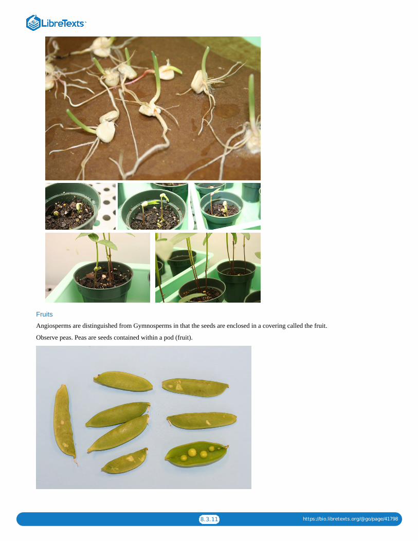
8.3.11 https://bio.libretexts.org/@go/page/41798
Fruits
Angiosperms are distinguished from Gymnosperms in that the seeds are enclosed in a covering called the fruit.
Observe peas. Peas are seeds contained within a pod (fruit).

8.3.12 https://bio.libretexts.org/@go/page/41798
Observe the sliced tomato. It is produced from several fused carpels. Can you see the carpels? How many are there?
Observe a strawberry or a blackberry. These fruits are formed from a single flower that contained many pistals.
Observe a pineapple. This fruit is produced by the fusion of many flowers. Can you see each individual fruit?

8.3.13 https://bio.libretexts.org/@go/page/41798
Contributors and AttributionsCC licensed content, Shared previously
Seed Plants (Kingdom: Plantae), Biology 102. Authored by: Michael J. Gregory, Ph.D.. Provided by: LibreTexts. Located at:bio.libretexts.org/Under_Construction/BioStuff/BIO_102/Laboratory_Exercises/Seed_Plants. Project: The Biology Web.License: CC BY-NC-SA: Attribution-NonCommercial-ShareAlike
8.3: Reading- Seed Plants is shared under a not declared license and was authored, remixed, and/or curated by LibreTexts.

1
CHAPTER OVERVIEW
9: Module 6- Plant Organization9.1: Plant Organization Lab9.2: Plant Organization Lab (Instructor Materials Preparation)
9: Module 6- Plant Organization is shared under a not declared license and was authored, remixed, and/or curated by LibreTexts.

9.1.1 https://bio.libretexts.org/@go/page/41799
9.1: Plant Organization Lab
Lab Objectives
At the conclusion of the lab, the student should be able to:
List and give the major function of the three main types of plant tissuesIdentify a monocot verses a eudicot plant by observing either root, stem, leaf, or flowersName and describe the various components and tissues within the root, both monocot and eudicotName and describe the various components and tissues within the stem, both monocot and eudicotName the describe the various components and tissues within the leaf, both monocot and eudicotIdentify various examples of stem diversityDistinguish between simple and compound leaves
A SlideShare element has been excluded from this version of the text. You can view it online here: pb.libretexts.org/bio2lm/?p=84
Download a PDF of the lab to print.
Angiosperms, or flowering plants are incredibly diverse. This group of plants contains between 250,000 and 400,000 differentspecies. Although flowering plants are diverse in size, shape, color, and habitat, all angiosperms have the following four structures:the root, the stem, the leaf, and the flower. Each structures in comprised of one or more different types of tissues. In this lab activitywe will learn more about angiosperm structures and tissues.
Angiosperm TissuesFlowering plants contain three main types of tissues in the roots, stems, and leaves. Each tissue has a specific function as describedbelow:
1. Dermal tissue: protects the outside of the plant.2. Ground tissue: used for photosynthesis and storage. An internal plant tissue.3. Vascular tissue: used for transport of water and sugars. Xylemtransports water and phloem transports sugars.
Unlike animals, angiosperms increase in size their entire life because of meristematic tissue. Meristematic tissue continues todivide and create new cells through photosynthesis increasing the height and width of flowering plants. Plants have two types ofmeristems, as described below:
1. Apical meristem: located at the tip of the shoot and the tip of the root. The apical meristem lengthens up and down. It isresponsible for primary growth.
2. Lateral meristem: located at branches to increase plant girth. It is responsible for secondary growth.
Angiosperm Body PlanView the representative plants on display in the lab. Notice that the plants have aboveground and below-ground portions. The shootsystem is aboveground. What components of the plan comprise the shoot system?
The root system is below ground. State two functions of plant roots.
Angiosperms are divided into two different groups, monocots and eudicots. These groups differ based on tissue organization in theseed, root, stem, leaf and flowers. For example, monocots have leaf veins that form a parallel patter and flower parts in multiples ofthrees. Eudicots have leaf veins in a net pattern and flower parts in multiples of fours or fives.
Using this information, identify the live plants on display as either monocots or eudicots using a table similar to the one below.
Monocots Eudicots

9.1.2 https://bio.libretexts.org/@go/page/41799
Root
There are two main root arrangements. Most monocots have a fibrous root system where all of the roots are about the same size.Many eudicots have a taproot system with one very large main root and smaller roots branching off. A carrot is an example of ataproot.
View the monocot and eudicot roots models on display and the cross section slides of the monocot and eudicot root. Identify thefollowing structures:
Root capRoot hairsZone of cell divisionZone of elongationZone of maturationXylemCortexEndodermisPithPericycleEpidermisPhloem
Questions
1. Draw and label the cross section of the monocot and eudicot root slides.2. Why is the root cap necessary?3. How does the arrangement of xylem and phloem differ in the monocot verses the eudicot root?
StemThe stem provides aboveground support for flowers and leaves. Some stems are herbaceous or nonwoody while others are woody.Herbaceous stems increase in length via the apical meristem but they do not increase in girth through secondary growth.
View the cross section slides of the eudicot herbaceous stem and the monocot herbaceous stem. Identify the following tissues inboth slides and notice that they have different arrangements of the xylem and phloem.
CortexXylemPhloemPithEpidermis
Questions
1. In the eudicot stem, which vascular tissue is more external, the xylem or the phloem?2. The vascular bundles in a monocot herbaceous stem are said to have a scattered arrangement. Explain why in your own words.3. Draw and label the cross section of the monocot and eudicot stem slides.
Many plants have modified stems to assist with food storage or for vegetative reproduction. Some examples include:
1. Rhizome: horizontal underground stem2. Corm: underground fleshy stem, used for storage3. Stolon: underground horizontal stem4. Runner: aboveground horizontal stem5. Tuber: underground storage stem6. Bulb: underground storage stem with fleshy leaves
View the stems on display and identify which ones below to the categories described above. You may see more than one exampleof some and no examples of others.

9.1.3 https://bio.libretexts.org/@go/page/41799
LeafThe main function of the leaf is photosynthesis. Therefore, it contains many chloroplasts and is thin to facilitate gas and watertransport. View the leaf model and the leaf cross section slide. Make sure you can identify the following components:
Epidermis (upper and lower)CuticleSpongy MesophyllPalisade MesophyllXylemPhloemStomata (stoma singular)Guard Cells
Question
1. Draw and label the leaf with all of the components listed above.
The main, flat portion of the leaf is called the blade and it attaches to the stem via the petiole stalk. There are two mainarrangements for the leaf blade. Leaves can either be simple or compound. A simple leaf has a single blade. A compound leaf hasa blade divded into leaflets. All of the leaflets share the same auxiliary bud which is the source of new growth. The auxiliary budis located at the base of the petiole.
There are two arrangements of compound leaves. Palmately compound leaves have all leaflets attached at the same point at theend of the petiole. Pinnately compound leaves have leaflets attached at intervals along the peitole.
View the preserved leaf specimens. Choose six to identify as simple or compound. If the leaf is compound, state if it is palmately orpinnately compound. Record your findings in a table similar to the one below.
Name of Leaf Simple or Compound Palmate or Pinnate
Questions
1. What cellular division process occurs in the apical meristem region?2. The plant has two apical meristems. Identify the location of both.3. If a plant had parallel leaf veins would you identify it as a monocot or a eudicot?4. The tissue responsible for water transport is ________ and the tissue responsible for sugar transport is________. 5. You look at a stem cross section and notice there is a ring of vascular bundles. Would you identify it as monocot or eudicot?6. A potato is an example of what type of modified stem?7. What is the function of the root hairs?8. Vascular bundles contain what two tissues?9. Are fibrous roots more common in eudicots or monocots?
10. State one structure that is part of the plant shoot system.
Contributors and AttributionsCC licensed content, Original
Biology 102 Labs. Authored by: Lynette Hauser. Provided by: Tidewater Community College. Located at:http://www.tcc.edu/. License: CC BY: Attribution
9.1: Plant Organization Lab is shared under a not declared license and was authored, remixed, and/or curated by LibreTexts.

9.2.1 https://bio.libretexts.org/@go/page/41800
9.2: Plant Organization Lab (Instructor Materials Preparation)
Lab Materials
This is the prep for one section of 24 students.
Angiosperm Tissues
Students will do this part in table teams (groups of 4).
Materials Quantity Notes
root cross section slides 1 per table monocot and eudicot
stem cross section slides 1 per table monocot and eudicot
Angiosperm Body Plan
Students will do this part in table teams (groups of 4).
Materials Quantity Notes
live stem examples varioussome options include ginger root for rhizome,potato for tuber, carrot for tap root, onion forbulb, set up as demo on side bench
leaf examples varioustry to have several monocot, several eudicot,and both simple and compound leaves, set upas demo on side bench
leaf model 1 per table
live plant specimens various try to have several monocot and eudicot, set upon side bench as demo
root model 2-3 per class
Contributors and AttributionsCC licensed content, Original
Instructor Materials Preparation. Authored by: Lynette Hauser. Provided by: Tidewater Community College. Located at:http://www.tcc.edu/. License: CC BY: Attribution
9.2: Plant Organization Lab (Instructor Materials Preparation) is shared under a not declared license and was authored, remixed, and/or curated byLibreTexts.

1
CHAPTER OVERVIEW
10: Module 7- Invertebrate I10.1: Invertebrate Lab I10.2: Invertebrate Lab I (Instructor Materials Preparation)10.3: Reading- Sponges10.4: Reading- Cnidarians10.5: Reading- Roundworms10.6: Reading- Arthropods
10: Module 7- Invertebrate I is shared under a not declared license and was authored, remixed, and/or curated by LibreTexts.

10.1.1 https://bio.libretexts.org/@go/page/41801
10.1: Invertebrate Lab I
Learning ObjectivesState the phyla of the organisms discussed in the lab activitiesUse the characteristics of symmetry, coelom, embryo tissue layers, and patterns of development to differentiate between thedifferent invertebrate groupsRecognize and identify the sponge specimens viewed in the labExplain the purpose of the different sponge cellsRecognize and identify the cnidaria specimens viewed in lab and if the specimens are polyp or medusa formRecognize and identify the nematoda examples viewed in the labRecognize and identify the arthropoda examples viewed in the lab
A SlideShare element has been excluded from this version of the text. You can view it online here: pb.libretexts.org/bio2lm/?p=90
Download a PDF of the lab to print.
Sponges
Procedure
Access the page “Reading: Sponges.”
Questions1. The preserved sponge specimens will be on display, but may differ from the ones directly mentioned in the lab handout. Please
make observations on the available specimens and fill in the chart below.
Name of Specimen Physical DescriptionSponge Structures Visible (osculum, otherspores, spicules)
1. What type of symmetry is displayed in the sponge specimens?2. View the Grantia slides. There will not be slides available of the spicules but view the pictures in the lab materials.
1. What is the function of spicules?2. Do sponges contain true tissues?3. Can you find any collar cells in the slide?4. What is the function of the collar cells?5. Can you find any epidermal cells in the sponge slide?6. What is the function of the epidermal cells?
Cnidarians
Procedure1. Access the page: “Reading: Cnidarians.”2. View the hydra specimens (live and slides).3. Watch these videos:

10.1.2 https://bio.libretexts.org/@go/page/41801
A YouTube element has been excluded from this version of the text. You can view it online here: pb.libretexts.org/bio2lm/?p=90
A YouTube element has been excluded from this version of the text. You can view it online here: pb.libretexts.org/bio2lm/?p=90
Questions1. Does the hydra illustrate the polyp or the medusa stage?2. How many germ layers does the hydra contain?3. What type of symmetry is seen in the hydra?4. Can you find the hydra tentacles? How many tentacles does your hydra specimen contain?5. Name the stinging cells present on the tentacles that are unique to cnidarians.6. Explain the movement of the hydra if live specimens are available. If they are not, review the video on hydra movement.7. If there are live hydra specimens do not add the vinegar as indicated on the lab website. Vinegar causes them to expel the
cnidocytes. Review the nematocysts video, which shows a jellyfish discharging the nematocyst cells.8. View the preserved cnidarian specimens will be on display. They may differ from the ones directly mentioned in the lab website
but there should be some medusa cnidarians as well as corals and sea anemones. Please make observations on the availablespecimens. And fill in the chart below.
Name of Specimen Physical Description Polyp or Medusa Stage?
Roundworms (phylum nematoda)
Procedure
Access the page “Reading: Roundworms”
Watch this video:

10.1.3 https://bio.libretexts.org/@go/page/41801
A YouTube element has been excluded from this version of the text. You can view it online here: pb.libretexts.org/bio2lm/?p=90
Questions1. We will not be dissecting the Ascaris as indicated in the website. Instead view the preserved slide.
1. What type of symmetry is seen in the roundworm?2. Does it exhibit cephalization?3. Ascaris is a parasite that swims constantly in human intestines. What structure protects the nematode from being digested?
2. View the Trichinellaslide. Trichinella is also a parasite. It can infect humans as well as other mammals like pigs, bears, androdents. If untreated, it can lead to death.1. What mammal tissue does this roundworm infect?2. Draw a picture of the Trichinella as viewed under the microscope.
3. If there are live vinegar eel specimens available, view them. If there are no live specimens available, review the vinegar eelsvideo.
1. Describe the movement of the vinegar eels.2. Do they have a complete or incomplete digestive system?
Arthropods
Procedure
Access the page “Reading: Arthropods.”
Questions1. View the preserved arthropod specimens available. There will be at least one example of each lineage group discussed on the
website but not all of the specimens may be available. Use the table below to organize your observations.
Name of Specimen Lineage Exoskeleton? Jointed Appendages? Specialized Segments
2. What type of symmetry is displayed in the arthropods?3. Do the arthropods exhibit cephalization?
Crayfish Dissection
Procedure
Complete the crayfish dissection using the directions available on the lab website.

10.1.4 https://bio.libretexts.org/@go/page/41801
Questions1. Make sure you can identify the following external structures: antenna, chiliped, cephalothorax, abdomen, and walking legs2. Do you have a male or female crayfish?3. How many swimmerets does your crayfish have?4. How many rows of gills does the crayfish have?5. Where do the gills attach?6. Can you find the stomach and the digestive glands?7. What does the stomach attach to directly?8. Try to locate the green glands. What is the function of this structure?
Review QuestionsAnswer the review questions below. The phyla we viewed today were the porifera, the cnidaria, the nematoda and the arthropoda.
1. Which phyla exhibited bilateral symmetry?2. Which phyla had no true tissues?3. Which phyla contained parasitic organisms?4. Which phyla were coelomates?5. Which phyla exhibited cephaliziation?6. Which phyla that you viewed today contained specialized appendages?7. Which phyla exhibited radial symmetry?8. Which phyla were pseudocoelomates?9. Which phyla had a complete digestive system?
10. Which phyla were multicellular?11. Which phyla were asymmetrical?12. Which phyla were acoelomates?
Contributors and AttributionsCC licensed content, Original
Biology 102 Labs. Authored by: Lynette Hauser. Provided by: Tidewater Community College. Located at:http://www.tcc.edu/. License: CC BY: Attribution
10.1: Invertebrate Lab I is shared under a not declared license and was authored, remixed, and/or curated by LibreTexts.

10.2.1 https://bio.libretexts.org/@go/page/41802
10.2: Invertebrate Lab I (Instructor Materials Preparation)
Lab Materials
This is the prep for one section of 24 students.
Sponges
Students will do this part in table teams (groups of 4).
Materials Quantity Notes
sponge preserved specimens various on side bench as demo
grantia slide 1 per table
Cnidarians
Students will do this part in table teams (groups of 4).
Materials Quantity Notes
hydra slide 1 per table
hydra live specimens 1 container per class view under dissecting scope as demo on sidebench
cnidaria preserved specimens various on side bench as demo
hydra model 1-2 per class on side bench as demo
Roundworms (phylum nematoda)
Students will do this part in table teams (groups of 4).
Materials Quantity Notes
Ascaris slide 1 per table
Trichinella slide 1 per table
live vinegar eels 1 vial per class view under dissecting scope as demo on sidebench
Arthropods
Students will do this part in table teams (groups of 4).
Materials Quantity Notes
arthropod preserved specimens various on side bench as demo, helpful to haverepresentatives from different subgroups
Crayfish Dissection
Students will do this part in table teams (groups of 4).
Materials Quantity Notes
dissection kit 1 per table should include scalpel, scissors, forceps,dissection needles, dissection pins
dissection tray 1 per table
crayfish 1 per table
gloves 1 pair per student latex and non latex options in various sizes
goggles 1 pair per student

10.2.2 https://bio.libretexts.org/@go/page/41802
Materials Quantity Notes
lab coat 1 per student
dissection waste container 1 per class dissection waste should not go in regular trash
Contributors and AttributionsCC licensed content, Original
Instructor Materials Preparation. Authored by: Lynette Hauser. Provided by: Tidewater Community College. Located at:http://www.tcc.edu/. License: CC BY: Attribution
10.2: Invertebrate Lab I (Instructor Materials Preparation) is shared under a not declared license and was authored, remixed, and/or curated byLibreTexts.

10.3.1 https://bio.libretexts.org/@go/page/41803
10.3: Reading- SpongesThere are two phyla of sponges: Calcarea and Silicea.
Preserved SpecimensExamine preserved sponges on display. Identify the osculum. Can you see pores? Some of these specimens are shown below.
Figure 1. Chalina
Figure 2. Spongila
Figure 3. Commercial Sponge

10.3.2 https://bio.libretexts.org/@go/page/41803
Structure of SpongesThe photographs below are of Grantia. The body of this species is highly folded producing many chambers. In the last twophotographs, the living cells have been removed to reveal the spicules.
Examine the following prepared slides: Grantia c.s.and Grantia l.s. Find collar cells, epidermal cells, and pores. What is thefunction of the collar cells? What is the function of the pores?
Figure 4. Grantia c.s. X 400
Figure 5. Grantia l.s. X 40
Examine a slide of Grantia spicules. What is the function of the spicules?

10.3.3 https://bio.libretexts.org/@go/page/41803
Figure 6. Grantia spicules X 40
Figure 7. Grantia spicules X 100
Spicules are needle-like structures composed of either calcium carbonate or silica and offer support and protection.
Contributors and AttributionsCC licensed content, Shared previously
Sponges, Biology 102. Authored by: Michael J. Gregory, Ph.D.. Provided by: LibreTexts. Located at:bio.libretexts.org/Under_Construction/BioStuff/BIO_102/Laboratory_Exercises/Sponges. Project: The Biology Project.License: CC BY-NC-SA: Attribution-NonCommercial-ShareAlike
10.3: Reading- Sponges is shared under a not declared license and was authored, remixed, and/or curated by LibreTexts.

10.4.1 https://bio.libretexts.org/@go/page/41804
10.4: Reading- CnidariansThis laboratory exercise covers the following animals. You should learn this classification scheme and be able to classify theanimals into these categories.
Phylum: CnidariaClass: Hydrozoa (Hydra and relatives)Class: Anthozoa (Sea Anemones and Corals)Class: Scyphozoa (Jellyfishes)
Some examples of Cnidarians are hydra, jellyfishes, corals, sea anemones, and Portuguese man-of-wars.
Characteristics
Radial Symmetry
The body parts of a radially symmetrical animal are arranged around a central axis so that each part extends from the center. Theanimal can be cut along the axis in more than one plane to produce identical halves. Animals that exhibit radial symmetry tend tobe sessile (immobile). Radial symmetry allows them to reach out in all directions.
Cnidarians have two tissue layers. The outer layer is the epidermis. It is formed from ectoderm. The inner layer, the gastrodermis,secretes digestive juices into the inner space called the gastrovascular cavity. The gastrodermis is formed from endoderm.
Cnidarians do not have mesoderm and therefore do not have organs.
A nonliving gelatinous material called mesoglea separates the two tissue layers. A nerve net is located between the epidermis andmesoglea. The body contains long structures called tentacles that can be moved to capture prey. The tentacles contain stinging cellscalled cnidocytes and within each one is a capsule called a nematocyst, which discharges to either trap or sting the prey. Contractile(muscle-like) fibers are found in both the epidermis and the gastrodermis. Their movements are not complex because they do nothave a brain.
Cnidarians have a hydrostatic skeleton. The contractile fibers act against the fluid-filled gastrovascular cavity. The movements arelike a balloon; the animal can be short and thick or long and thin. Cnidarians have a saclike gut and extracellular digestion.
Two body forms are found among the Cnidarians, a polyp and a medusa. A polyp is attached and has the tentacles and mouthdirected upward. A medusa is free-floating and has the mouth and tentacles on the ventral surface. It resembles an upside-downpolyp. Some species have both a polyp and a medusa in their life cycle, others have one or the other form dominant.
Hydrozoa
Hydra1. Use a dropper to place a live Hydra on a slide. Examine the Hydra using a dissection microscope.

10.4.2 https://bio.libretexts.org/@go/page/41804
Figure 1. Hydra (live)
2. Hydra reproduce both sexually and asexually by budding. Try to find a live Hydra with buds. If you cannot find a live Hydrabudding, look for budding in a prepared slide of Hydra.
Figure 2, Hydra budding. This is a form of asexual reproduction.
3. Add a drop of vinegar to the slide containing Hydra. Describe what happened to the cnidocytes.
Figure 3. Left: Hydra (Live) Exposed to 5% Vinegar Solution X 100. Right: Hydra (Live) Exposed to 5% Vinegar Solution X200
4. Examine microscope slides of hydra l.s. and hydra c.s. Look for the presence of two tissue layers. Identify stinging cells(Cnidocytes) in a slide of the whole animal.
Figure 4. Left: Hydra l.s. X 100. Middle: Hydra c.s. X 100. Right: Hydra c.s. X 200

10.4.3 https://bio.libretexts.org/@go/page/41804
Figure 5. Portion of a Hydra tentacle showing cnidocytes
Figure 5. Left: Hydra l.s. X 40. Right: Hydra l.s. with ingested food X 40
Other Hydrozoans
Examine preserved specimens of Gonionemius, Polyorchis, and Physalia.
Figure 6. Left: Gonionemus, preserved. Middle: Polyorchis, preserved. Right: Portuguese Man-Of-War

10.4.4 https://bio.libretexts.org/@go/page/41804
Sea Anemones and Coral (Class Anthozoa)Examine a sea anemone and coral.
Figure 7. Sea anemone, preserved
Figure 8. Left: Astrangia (Northern Coral) Skeleton. Middle: Coral Skeleton. Right: Coral Skeleton.
Jellyfish (Class Schyphozoa)
Examine preserved jellyfish on display.
Figure 9. Aurelia
Contributors and AttributionsCC licensed content, Shared previously
Cnidarians, Biology 102. Authored by: Michael J. Gregory, Ph.D.. Provided by: LibreTexts. Located at:bio.libretexts.org/Under_Construction/BioStuff/BIO_102/Laboratory_Exercises/Cnidarians. Project: The Biology Web.License: CC BY-NC-SA: Attribution-NonCommercial-ShareAlike
10.4: Reading- Cnidarians is shared under a not declared license and was authored, remixed, and/or curated by LibreTexts.

10.5.1 https://bio.libretexts.org/@go/page/41805
10.5: Reading- Roundworms
Ascaris Dissection
Procedure1. Obtain a female Ascaris and place it on a dissecting pan. The posterior end is curved in males and straight in females.2. Make a shallow longitudinal cut the length of the worm and pin it open as illustrated below.3. Identify the gut, ovary, oviduct, uterus, and vagina.
TrichinellaView a prepared slide of Trichinella in the muscle tissue of a host organism.

10.5.2 https://bio.libretexts.org/@go/page/41805
Figure 1. Trichinella spiralis encysted in muscle tissue of a host X 40
Contributors and AttributionsCC licensed content, Shared previously
Roundworms, Biology 102. Authored by: Michael J. Gregory, Ph.D.. Provided by: LibreTexts. Located at:bio.libretexts.org/Under_Construction/BioStuff/BIO_102/Laboratory_Exercises/Roundworms. Project: The Biology Web.License: CC BY-NC-SA: Attribution-NonCommercial-ShareAlike
10.5: Reading- Roundworms is shared under a not declared license and was authored, remixed, and/or curated by LibreTexts.

10.6.1 https://bio.libretexts.org/@go/page/41806
10.6: Reading- ArthropodsThree clades of arthropods are listed below.
CheliceratesArachnids—(Spiders, Scorpions, Harvestmen, Ticks and Mites)Horseshoe Crabs
Myriapods
MillipedesCentipedes
PancrustaceansCrustaceans
DecpodsIsopodsKrillCopepodsBarnacle
Insects
Arthropods have an exoskeleton that cannot enlarge as the animal grows. As a result, it must be shed from time to time to allowgrowth. The exoskeleton from a tarantula (an arachnid) is on display.
Life CycleThe young of some arthropods look like the adults. The change from young to adult that these species undergo is called incompletemetamorphosis.
In many species, the egg hatches to produce a larva (pl. larvae) that does not look like the adult. At some point in the maturationprocess, the larva will produce a pupa (pl. pupae). In this phase its tissues will become reorganized into the adult form. This typeof development is called complete metamorphosis.
Observe a culture of fruit flies (Drosophila) and observe the eggs, larvae, pupae, and adults.
CheliceratesThe first pair of appendages of the members of this phylum are pincerlike or fanglike mouthparts called chelicerae. Observe thesemouthparts in the tarantula on display.
Arachnids (Spiders, Scorpions, Harvestmen, Ticks, Mites)
Figure 1. Left: Scorpion. Middle: Tick X 40. Right: Mite X 40.
Horseshoe Crabs

10.6.2 https://bio.libretexts.org/@go/page/41806
Figure 2. Left: Horseshoe crab dorsal surface. Right: Horseshoe crab ventral surface
Crustaceans—Lobsters and Relatives
Decapods
Figure 3. Crab
Crayfish Dissection
Obtain a crayfish and identify the structures shown in the photograph below. You should familiarize yourself with the names of theexternal structures shown in the photograph below.
Figure 4
The photographs below show the ventral surface of male and female crayfish. Click on the photographs to view them. Identify thesex of your crayfish. Find somebody in class that has the opposite sex and view the ventral surface of their crayfish. Notice thatcrayfish have five pairs of swimmerets. In males, the anterior two pairs are large and less flexible than those of females. They areused to transfer sperm to the female.

10.6.3 https://bio.libretexts.org/@go/page/41806
Figure 5
Find the seminal receptacles located between the third and fourth walking legs. These structures function to receive sperm from themale during mating.
Observe the sperm duct in males located at the base of the fifth walking leg.
Begin your dissection by cutting along the midline of the carapace from the posterior edge to an area just behind the two eyes (seethe photographs below). Next, cut laterally just behind the eye until you reach the ventral edge. Carefully remove the piece ofcarapace that you have cut to expose the gills underneath. As you peel this piece of carapace away, it may be necessary to reachunderneath with the point of a scissors or needle to brush away and detach any tissue from the interior of the animal that is attachedto the carapace.
Figure 6
Your crayfish should look like the first photograph below. Next, remove the carapace on the other side.
Using a needle, carefully separate the first row of gills and notice that there is another row underneath. Remove one of the legs andobserve how the gills are attached to the walking legs.
The first two photographs below show the gills on the left side of a crayfish. The third photograph is a crayfish with the entirecarapace removed. The first row of gills on each side has been moved aside to expose a row underneath it.

10.6.4 https://bio.libretexts.org/@go/page/41806
Figure 7.
Figure 8. Crayfish leg showing gill attachment
Brush all of the gills to the side and then carefully cut the membrane (epidermis) that covers the internal organs.
The heart is a small diamond-shaped structure located below where the posterior edge of the carapace was. It may be difficult to seethe blood vessels attached to the heart. Openings (ostia) in the side of the heart should be visible. These allow blood to enter thecirculatory system.
Large white digestive glands can be seen on each side of the stomach. They produce digestive enzymes.

10.6.5 https://bio.libretexts.org/@go/page/41806
Figure 9.
Remove the digestive glands and then carefully remove the stomach. Notice how the stomach is attached to the mouth. Cut openthe stomach and observe the tooth-like structures at the anterior end for grinding food. This is called the gastric mill.
The green glands are positioned ventrally near the anterior end of the body cavity. They are spherical and will appear to beembedded within the surface. Their function is excretion. Find the openings for the green glands on the outer surface near the baseof each antenna.
Find the brain near the anterior end and the two ventral nerve cords attached to the brain.
Figure 10. Left: Crayfish ventral surface. Middle: Crayfish viewed from above. The stomach has been removed revealing the nervecords and green glands underneath. Right: Crayfish viewed from above looking into the head region. The stomach has beenremoved. The brain can be seen.
Copepods and Krill

10.6.6 https://bio.libretexts.org/@go/page/41806
Figure 11. Left: A copepod (Cyclops, male). Right: Krill (Gammarus).
Barnacles
Figure 12. Left: Balanus—a barnacle. Right: A stalked barnacle
Isopods
Figure 13
Myriopods
Millipedes

10.6.7 https://bio.libretexts.org/@go/page/41806
Figure 14
Centipedes
Figure 15. A centipede
Insects
Figure 16. Fly life history showing complete metamorphosis
Contributors and AttributionsCC licensed content, Shared previously
Arthropods, Biology 102. Authored by: Michael J. Gregory, Ph.D.. Provided by: LibreTexts. Located at:bio.libretexts.org/Under_Construction/BioStuff/BIO_102/Laboratory_Exercises/Arthropods. Project: The Biology Web.License: CC BY-NC-SA: Attribution-NonCommercial-ShareAlike
10.6: Reading- Arthropods is shared under a not declared license and was authored, remixed, and/or curated by LibreTexts.

1
CHAPTER OVERVIEW
11: Module 8- Invertebrate II11.1: Invertebrate Lab II11.2: Invertebrate Lab II (Instructor Materials Preparation)11.3: Reading- Flatworms11.4: Reading- Mollusks11.5: Reading- Annelids11.6: Natal Bean Discrimination by Bean Beetles11.7: Natal Bean Discrimination by Bean Beetles (Instructor Materials Preparation)*
11: Module 8- Invertebrate II is shared under a not declared license and was authored, remixed, and/or curated by LibreTexts.
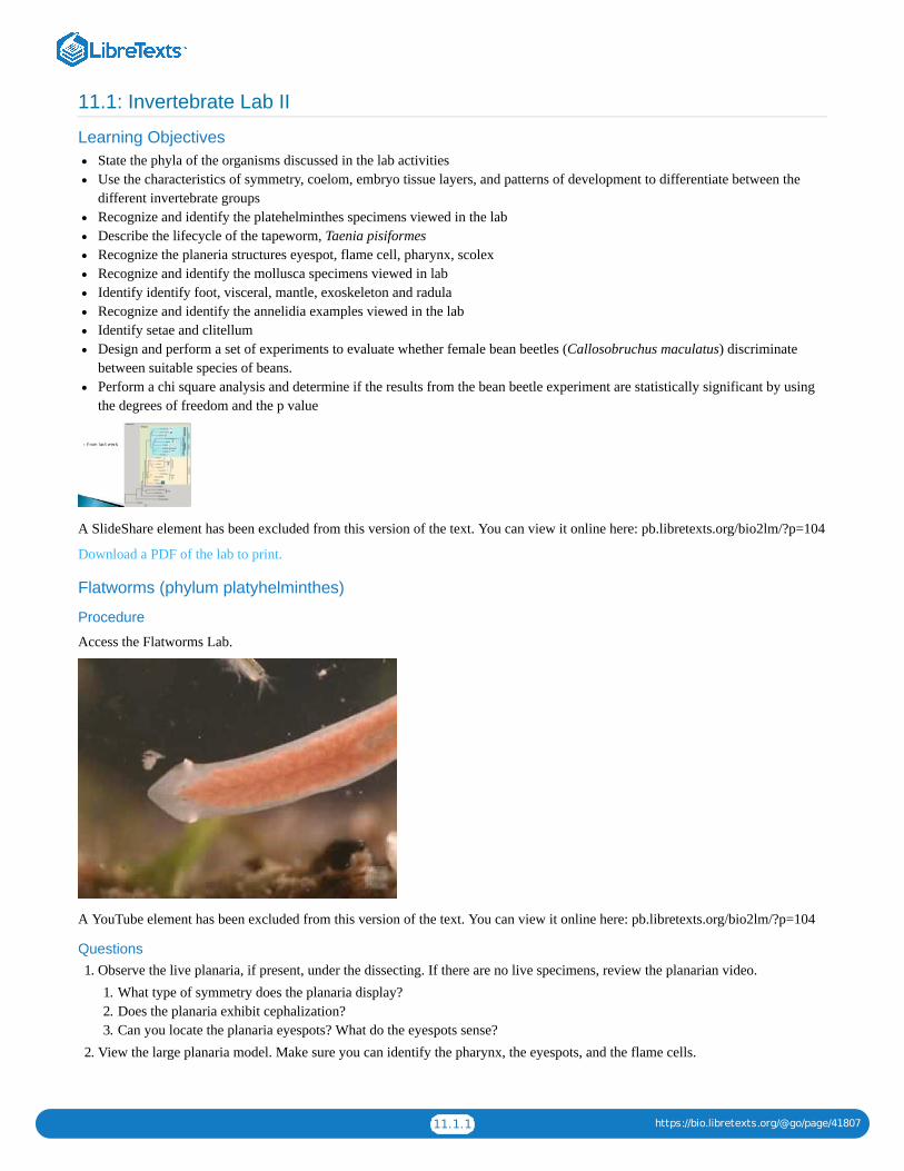
11.1.1 https://bio.libretexts.org/@go/page/41807
11.1: Invertebrate Lab II
Learning ObjectivesState the phyla of the organisms discussed in the lab activitiesUse the characteristics of symmetry, coelom, embryo tissue layers, and patterns of development to differentiate between thedifferent invertebrate groupsRecognize and identify the platehelminthes specimens viewed in the labDescribe the lifecycle of the tapeworm, Taenia pisiformesRecognize the planeria structures eyespot, flame cell, pharynx, scolexRecognize and identify the mollusca specimens viewed in labIdentify identify foot, visceral, mantle, exoskeleton and radulaRecognize and identify the annelidia examples viewed in the labIdentify setae and clitellumDesign and perform a set of experiments to evaluate whether female bean beetles (Callosobruchus maculatus) discriminatebetween suitable species of beans.Perform a chi square analysis and determine if the results from the bean beetle experiment are statistically significant by usingthe degrees of freedom and the p value
A SlideShare element has been excluded from this version of the text. You can view it online here: pb.libretexts.org/bio2lm/?p=104
Download a PDF of the lab to print.
Flatworms (phylum platyhelminthes)
Procedure
Access the Flatworms Lab.
A YouTube element has been excluded from this version of the text. You can view it online here: pb.libretexts.org/bio2lm/?p=104
Questions1. Observe the live planaria, if present, under the dissecting. If there are no live specimens, review the planarian video.
1. What type of symmetry does the planaria display?2. Does the planaria exhibit cephalization?3. Can you locate the planaria eyespots? What do the eyespots sense?
2. View the large planaria model. Make sure you can identify the pharynx, the eyespots, and the flame cells.
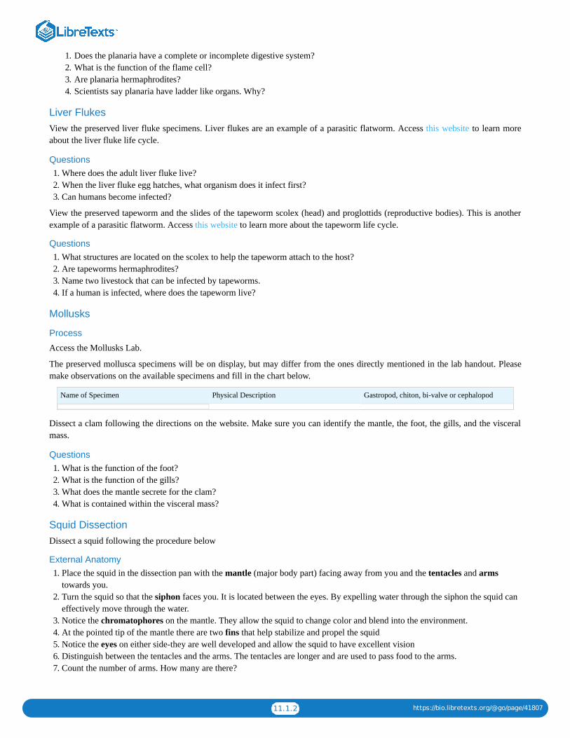
11.1.2 https://bio.libretexts.org/@go/page/41807
1. Does the planaria have a complete or incomplete digestive system?2. What is the function of the flame cell?3. Are planaria hermaphrodites?4. Scientists say planaria have ladder like organs. Why?
Liver FlukesView the preserved liver fluke specimens. Liver flukes are an example of a parasitic flatworm. Access this website to learn moreabout the liver fluke life cycle.
Questions1. Where does the adult liver fluke live?2. When the liver fluke egg hatches, what organism does it infect first?3. Can humans become infected?
View the preserved tapeworm and the slides of the tapeworm scolex (head) and proglottids (reproductive bodies). This is anotherexample of a parasitic flatworm. Access this website to learn more about the tapeworm life cycle.
Questions1. What structures are located on the scolex to help the tapeworm attach to the host?2. Are tapeworms hermaphrodites?3. Name two livestock that can be infected by tapeworms.4. If a human is infected, where does the tapeworm live?
Mollusks
Process
Access the Mollusks Lab.
The preserved mollusca specimens will be on display, but may differ from the ones directly mentioned in the lab handout. Pleasemake observations on the available specimens and fill in the chart below.
Name of Specimen Physical Description Gastropod, chiton, bi-valve or cephalopod
Dissect a clam following the directions on the website. Make sure you can identify the mantle, the foot, the gills, and the visceralmass.
Questions1. What is the function of the foot?2. What is the function of the gills?3. What does the mantle secrete for the clam?4. What is contained within the visceral mass?
Squid DissectionDissect a squid following the procedure below
External Anatomy1. Place the squid in the dissection pan with the mantle (major body part) facing away from you and the tentacles and arms
towards you.2. Turn the squid so that the siphon faces you. It is located between the eyes. By expelling water through the siphon the squid can
effectively move through the water.3. Notice the chromatophores on the mantle. They allow the squid to change color and blend into the environment.4. At the pointed tip of the mantle there are two fins that help stabilize and propel the squid5. Notice the eyes on either side-they are well developed and allow the squid to have excellent vision6. Distinguish between the tentacles and the arms. The tentacles are longer and are used to pass food to the arms.7. Count the number of arms. How many are there?

11.1.3 https://bio.libretexts.org/@go/page/41807
8. Notice the suction cups on both the tentacles and the arms. How does the distribution of the suction cups differ between thesetwo structures?
9. Pull back the arms and locate the beak or mouth in the middle.
Internal Anatomy1. Use a pair of scissors and starting at the bottom of the mantle above the siphon cut one long incision up to the tip of the mantle.
Be careful to lift up with the scissors while cutting to avoid cutting the internal organs.2. Spread the mantle open and try to indentify the following internal structures
1. Feathery gills2. Heart, located at the base of each gill. Squid actually have three hearts!3. Liver, probably yellowish in color and long in shape running down the middle4. Ink sack, which looks like a small silver fish. If you find it, cut it out at both ends and you can extract some of the ink and
try to write with it!5. The pen, which is all that remains of the shell. To try and find the pen, lift the head of the squid and place it down over the
organs. You should notice a pointy area along the midline of the body, the tip of the pen. If you grasp the tip and pull the penwill release from the mantle. It resembles a transparent feather.
Annelidia
Process
Access the page “Reading: Annelidia.”
Skip the earthworm dissection indicated on the lab website. Rather, view the earthworm model. Make sure you can identify thesetae and the clitellum.
Questions1. What type of symmetry does the earthworm display?2. Does the earthworm exhibit cephalization?3. Does the earthworm exhibit segmentation?
The preserved annelida specimens will be on display, but may differ from the ones directly mentioned in the lab handout. Pleasemake observations on the available specimens and fill in the table below.
Name of Specimen Physical Description Leech, Earthworm, or Marine Worm
Review QuestionsAnswer the review questions below. The phyla we viewed today were the platyhelminthes, mollusca, and annelida.
1. Which phyla exhibited bilateral symmetry?2. Which phyla contained parasitic organisms?3. Which phyla were coelomates?4. Which phyla exhibited cephaliziation?5. Which phyla that you viewed today contained specialized appendages?6. Which phyla were aceolomates?7. Which phyla had an incomplete digestive system?8. Which phyla contained a shell?9. Which phyla contained hermaphrodite organisms?
Contributors and AttributionsCC licensed content, Shared previously
Biology 102 Labs. Authored by: Lynette Hauser. Provided by: Tidewater Community College. Located at:http://www.tcc.edu/. License: CC BY: Attribution
11.1: Invertebrate Lab II is shared under a not declared license and was authored, remixed, and/or curated by LibreTexts.

11.2.1 https://bio.libretexts.org/@go/page/41808
11.2: Invertebrate Lab II (Instructor Materials Preparation)
Lab Materials
This is the prep for one section of 24 students.
Flatworms (phylum platyhelminthes)
Students will do this part in table teams (groups of 4).
Materials Quantity Notes
live planaria 1 vial can be shared by all lab sections set up on side bench under dissectionmicroscope as a demo
flatworm model 1 per bench
planaria slides 1 per bench can alternatively be set as a demo
Taneia slides 1 per bench can alternatively be set as a demo
Liver Flukes
Students will do this part in table teams (groups of 4).
Materials Quantity Notes
live flukes 1 vial can be shared by all sections set up on side bench under dissectionmicroscope as a demo
fluke slides 1 per bench can alternatively be set as a demo
Mollusks
Students will do this part in table teams (groups of 4).
Materials Quantity Notes
mollusk examples (preserved) various set up on side bench as a demo including squid,octopus, chitin, clams, snails, etc.
Squid Dissection
Students will do this part in table teams (groups of 4).
Materials Quantity Notes
dissection kit 1 per bench should include scalpel, scissors, forceps,dissection needles, dissection pins
dissection tray 1 per bench
squid 1 per bench
gloves 1 pair per student latex and non latex options in various sizes
lab coat 1 per student
goggles 1 pair per student
dissection waste container 1 per class dissection waste should not go in regular trash
Clam Dissection
Students will do this part in table teams (groups of 4).
Materials Quantity Notes
dissection kit 1 per bench

11.2.2 https://bio.libretexts.org/@go/page/41808
Materials Quantity Notes
dissection tray 1 per bench
clam 1 per bench
gloves 1 pair per student latex and non latex options in various sizes
lab coat 1 per student
goggles 1 pair per student
dissection waste container 1 per class dissection waste should not go in regular trash
Annelidia
Students will do this part in table teams (groups of 4).
Materials Quantity Notes
earthworm model 1 per table
preserved annelid examples various examples include earthworms, leeches,polychete worms
Contributors and AttributionsCC licensed content, Original
Instructor Materials Preparation. Authored by: Lynette Hauser. Provided by: Tidewater Community College. Located at:http://www.tcc.edu/. License: CC BY: Attribution
11.2: Invertebrate Lab II (Instructor Materials Preparation) is shared under a not declared license and was authored, remixed, and/or curated byLibreTexts.

11.3.1 https://bio.libretexts.org/@go/page/41809
11.3: Reading- FlatwormsThis laboratory exercise covers the following animals. You should learn this classification scheme and be able to classify theanimals into these categories.
Phylum: Platyhelminthes (Flatworms)Class: Turbellaria (planarians)Class: Trematoda (Flukes)Class: Cestoda (Tapeworms)
Characteristics
Flatworms are flattened and have bilateral symmetry.
They are triploblastic (have 3 embryonic tissue layers: ectoderm, mesoderm, and endoderm) and therefore have organ-level oforganization. There is no body cavity, so they are acoelomate.
Flatworms have a gastrovascularcavity with one opening (a sac-like gut).
Free-living Species
Example: Dugesia—a freshwater planarian
Planarians have a branching sac-like gut (one opening).
The main function of the excretory system is for water regulation. It consists of two structures called protonephridia. Eachprotonephridium contains flame cells that move excess water into tubes that open to the outside.
Planarians have a head region with sense organs. The nervous system of Dugesia is somewhat more complex than the nerve net ofCnidarians. It consists of a brain and nerve cords arranged in a ladder-like configuration.
Planarians have ocelli (eyespots) allow the presence and intensity of light to be determined. These structures are covered but havean opening to one side and forward. They can tell the direction of light because shadows fall on some of the receptor cells whileothers are illuminated. They move away from light.
Planarians are hermaphroditic, that is, they contain both male and female sex organs. They can reproduce asexually simply bypinching in half; each half grows a new half.
Movement is accomplished by the use of cilia and also by muscular contractions.
TrematodesMembers of this group are primarily parasites (feed on a host species).
Parasitic forms lack cephalization.
The reproductive cycle typically involves two host species, a primary host and a secondary (or intermediate) host. Adults live inthe primary host and larvae develop in the secondary host. The life cycle often alternates between sexual and asexual reproduction.
Liver flukes are found in vertebrate livers.
Nearly half of people in the tropics have blood flukes. Schistosomiasis is a blood fluke that afflicts 200 million people in theworld. The secondary host is a snail.

11.3.2 https://bio.libretexts.org/@go/page/41809
Planarians1. Place a living planarian on a watch glass and observe its movements under a dissecting microscope. Look for the eyespots,
auricles, gastrovascular cavity, and pharynx.2. Planarians cannot see images but they can tell the direction of light with their eyespots. Cover 1/2 of the watch glass with
aluminum foil. Does the planarian favor the light area or the dark area?3. View a slide of a preserved planarian and note the eyespots, auricles, gastrovascular cavity, and pharynx.
Figure 1. Left: Planarian anterior end X 40. Middle: Planarian digestive tract mid section X 40. Right: Planarian c.s. X 40.
Liver FlukesObserve either a preserved liver fluke or a slide of a liver fluke using a dissecting microscope.
Figure 2. Left: Sheep liver fluke (Fasciola hepatica) stained. Right: Liver fluke (preserved)
Tapeworms1. View a preserved tapeworm (Taenia).2. View slides of Taenia. Locate the scolex. View a gravid (filled with eggs) proglottid.
Tapeworms live in the intestines of vertebrates.
They may reach 10 m in length (>30 feet). They have no digestive or nervous tissue. Attachment to the intestinal wall is by ascolex, a structure that contains hooks and suckers.

11.3.3 https://bio.libretexts.org/@go/page/41809
Figure 3. Taenia scolex X 40
Figure 4. Taenia (preserved)
The segments (proglottids) each contain male and female reproductive organs. Eggs are fertilized from sperm, which often comefrom other proglottids of the same individual. After fertilization, other organs within the proglottid disintegrate and the proglottidbecomes filled with eggs.
The intermediate hosts are usually pigs or cattle. They can become infected by drinking water contaminated with human feces.
Tapeworms can be passed to humans in undercooked meat, especially pork.
The photographs below show the scolex and proglottids at increasing distances from the scolex. Those segments closest to thescolex (on the left) are the smallest. Those furthest away (on the right) become filled with zygotes, break away, and pass out withthe feces.

11.3.4 https://bio.libretexts.org/@go/page/41809
Figure 5. Left: Taenia pisiformis anterior end. Middle: Taenia pisiformis mid region. Right: Taenia pisiformis posterior end
Contributors and AttributionsCC licensed content, Shared previously
Flatworms, Biology 102. Authored by: Michael J. Gregory, Ph.D.. Provided by: LibreTexts. Located at:bio.libretexts.org/Under_Construction/BioStuff/BIO_102/Laboratory_Exercises/Flatworms. Project: The Biology Web.License: CC BY-NC-SA: Attribution-NonCommercial-ShareAlike
11.3: Reading- Flatworms is shared under a not declared license and was authored, remixed, and/or curated by LibreTexts.

11.4.1 https://bio.libretexts.org/@go/page/41810
11.4: Reading- MollusksThis laboratory exercise covers the following animals. You should learn this classification scheme and be able to classify theanimals into these categories.
Phylum: Mollusca (Mollusks)Class: Polyplacophora (Chitons)Class: Gastropoda (snails)Class: Bivalvia (Clams)Class: Cephalopoda (Nautilus, Squid, Octopus)
All mollusks have a visceral mass, a mantle, and a foot. The visceral mass contains the digestive, excretory, and reproductiveorgans. The mantle is a covering. It may secrete a shell. The foot is muscular and is used for locomotion, attachment, and/or foodcapture.
The mantle and foot can be seen in the figure 1. The visceral mass is underneath the gill.
Figure 1.
There may be a radula, a structure that resembles a tongue but contains hard plates and is often used for scraping food. The coelomis reduced and limited to the region near the heart.
Most mollusks have an open circulatory system but cephalopods (squids, octopus) have a closed circulatory system. The bloodpigment of mollusks is hemocyanin, not hemoglobin. The heart of a clam can be seen in the photograph below. Bivalves have threepairs of ganglia but do not have a brain.
Most mollusks have separate sexes but most snails (gastropods) are hermaphrodites. Some marine mollusks have a ciliated larvalform called a trochophore.
Chitons (Class: Polyplacophora)
Chitons have a dorsal shell composed of 8 plates. A ventral foot is used for locomotion and for attachment to rocks. It pulls itselfclose to rocks for protection. Observe the chiton on display.

11.4.2 https://bio.libretexts.org/@go/page/41810
Figure 2. Left: chiton, dorsal surface. Right: ventral surface
Snails—Class GastropodaGastropods have an elongated, flattened foot and usually a head and shell although nudibranchs (sea slugs) and terrestrial slugs lacka shell.
Most are marine but there are also numerous freshwater and terrestrial species. Herbivorous gastropods use a radula to scrape foodfrom surfaces. Carnivores may use a radula to bore a hole through surfaces such as bivalve (clam) shells. Some gastropods such asthe slug (below) do not have a shell.
The larvae undergo torsion during development. It is a twisting that positions the visceral mass so that the anus is above the head.It is due to one side of the visceral mass growing faster than the other. The advantage (or function) of torsion is uncertain but it maybe to balance the animal or it may be to allow the head to be withdrawn into the shell first when predators approach.
Observe the snails and slugs on display.
Figure 3. A slug. Slugs do not have shells.
Gas Exchange (Respiration)
Some gastropod species are terrestrial and have lungs for gas exchange, others are aquatic and use gills.
The space near the entrance to the shell that is bound by the mantle is the mantle cavity. Aquatic gastropods have gills located inthe mantle cavity. The mantle of terrestrial gastropods functions as a lung.
Bivalves—Clams and relatives (Class: Bivalvia)
Bivalves have two shells (valves) held closed by powerful muscles. The shell is produced by the underlying mantle; it grows alongthe outer margins. They use their foot for burrowing. Mussels use their foot for the production of threads for attachment.

11.4.3 https://bio.libretexts.org/@go/page/41810
The gills are large because they are used for filter-feeding as well as respiration. Food is trapped by mucus on the gills and movedby cilia. Water enters and exits through siphons.
Obtain a preserved clam for dissection and place it on a dissecting tray. Remove one of the valves (shells) by inserting a scalpel andcutting the adductor muscles on each side of the hinge. See the diagram below for the location of the adductor muscles.
Figure 4. Adductor muscles of a clam.
Figure 5. The anterior and posterior adductor muscles being cut so that the valves can be pulled apart.
The mantle is a membrane that surrounds the internal structures and is characteristic of all mollusks. The portion of the mantlefrom the exposed surface may have remained attached to the valve that was removed or it may be covering the internal structures.Find the mantle and if necessary, remove it to expose the internal structures of the clam.
Identify the foot, visceral mass, gills, and labial palps. The gills are large because they are used for filter-feeding as well asrespiration. Food is trapped by mucus on the gills and moved by cilia to the mouth. Cilia on the labial palps also direct food andmucus to the mouth.
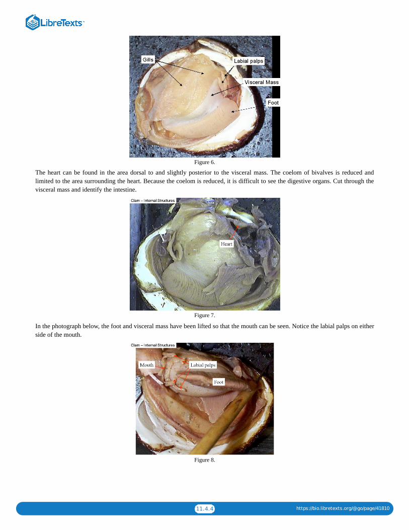
11.4.4 https://bio.libretexts.org/@go/page/41810
Figure 6.
The heart can be found in the area dorsal to and slightly posterior to the visceral mass. The coelom of bivalves is reduced andlimited to the area surrounding the heart. Because the coelom is reduced, it is difficult to see the digestive organs. Cut through thevisceral mass and identify the intestine.
Figure 7.
In the photograph below, the foot and visceral mass have been lifted so that the mouth can be seen. Notice the labial palps on eitherside of the mouth.
Figure 8.

11.4.5 https://bio.libretexts.org/@go/page/41810
Squid, Octopus, Nautilus, and Relatives (Class: Cephalopoda)Cephalopods are predators and live in a marine environment. A closed circulatory system allows them to move rapidly in pursuit ofprey. They move by jet propulsion; water in the mantle cavity is squirted rapidly through a siphon. The foot has evolved intotentacles around head. Cephalopods have a powerful beak-like structure to tear apart prey. The sense organs of cephalopods arewell developed.
Mollusks are the simplest animals with eyes. Some mollusks have lenses and therefore are capable of forming clear images. Thecamera-type eyes of some cephalopods (squid, octopus) are capable of focusing and forming clear images. Cephalopods are fast-moving predators and well-developed camera-type eyes help them catch prey.
Well-developed brains (especially in octopuses) give them a high learning capacity. Cephalopods can hide from enemies byreleasing a dark colored fluid from ink sacs.
Shells
The shell of a nautilus encloses the animal. A squid’s shell is small and internal. Octopuses do not have shells. Examinerepresentative cephalopods on display.
Figure 9. A Squid
Contributors and AttributionsCC licensed content, Shared previously
Mollusks, Biology 102. Authored by: Michael J. Gregory, Ph.D.. Provided by: LibreTexts. Located at:bio.libretexts.org/Under_Construction/BioStuff/BIO_102/Laboratory_Exercises/Mollusks. Project: The Biology Web. License:CC BY-NC-SA: Attribution-NonCommercial-ShareAlike
11.4: Reading- Mollusks is shared under a not declared license and was authored, remixed, and/or curated by LibreTexts.

11.5.1 https://bio.libretexts.org/@go/page/41811
11.5: Reading- AnnelidsThis laboratory exercise covers the following animals. You should learn this classification scheme and be able to classify theanimals into these categories.
Phylum: Annelida (Annelids)Class: Oligochaeta (Earthworms, Leeches)Class: Polychaeta (Marine Annelids)
Oligochaeta (Earthworms, Leeches)
Earthworms
Segmentation
Segmented animals have repeating units. This has led to specialization of parts over evolutionary time because the differentsegments could become specialized for different purposes. Notice the variety of specialized structures near the anterior end of theclam worm in the photograph below.
Molecular data suggests that segmentation evolved independently in the annelids, the arthropods, and the chordates becauseancestors to each of these three groups were not segmented.
Segmented Worms (Phylum Annelida)
Annelids are bilateral, coelomate protostomes. The coelom is partitioned by septa (crosswalls).
The fluid-filled coelom acts as a hydrostatic skeleton. When the circular muscles that surround each segment contract, thesegment becomes thinner and longer. When the longitudinal muscles that extend from one end of the segment to the othercontract, the segment becomes shorter but thicker.
Because muscles can only contract and cannot lengthen, other muscles are used to lengthen them. In annelids, when circularmuscles contract to lengthen the segment, the longitudinal muscles are lengthened. When the longitudinal muscles contract to makethe segment shorter and thicker, the circular muscles become lengthened.
Setae are bristles on the skin that anchor or help move the animal. Movement occurs when waves of contraction of longitudinalmuscles cause a “bulge” to progress from the anterior end to the posterior end.
Annelids exhibit specialization of the digestive tract. Some of these structures are the pharynx, crop, gizzard, intestine, andaccessory glands. Annelids have a closed circulatory system.
A pair of cerebral ganglia function as a simple brain. A ventral nerve cord extends the length of the animal and connects to a pair offused ganglia (mass of nervous tissue) in each segment. The ganglia within each segment function to coordinate musclecontractions.
Examine the exterior of an earthworm and find the ventral surface. Place the worm in a dissecting pan with the ventral surfacedown. Identify the clitellum. This structure produces mucous needed for reproduction. Find rows of setae along either side of theventral surface. These help provide traction as the animal moves through the soil.
Find the dorsal blood vessel. This structure should be visible through the body wall and will appear slightly darker than the rest ofthe body. It extends the length of the animal. With a scalpel, make a shallow cut along the dorsal surface beginning at a pointapproximately half way between the clitellum (see photograph below) and the posterior end and ending at the anterior end. Try toavoid cutting the dorsal blood vessel by keeping your cut to one side of the vessel. Try to cut only the body wall but not the internalstructures.

11.5.2 https://bio.libretexts.org/@go/page/41811
Figure 1.
The septa between each segment will prevent you from spreading the walls open to view the interior structures. Use a needle orscalpel to carefully cut the septa while spreading the body open. Pins can be used to hold the body open.
Find the structures shown in the photographs below. Be sure that you understand the function of each of these structures.
Figure 2. The anterior end of this earthworm is on the right.
Figure 3. Left: The anterior end of the worm. Middle: The brain. Right: The mid-section
Use a needle to cut the septa in the region of the intestine and roll it aside to expose the nerve cord underneath.
Examine a labeled earthworm composite. This slide shows three different cross sections. The cross-section on the right side of theslide is from the intestinal region.
Identify the intestine, typhlosole, coelom, nephridium, longitudinal muscles, circular muscles, and dorsal blood vessel. Are anysetae present in the cross section?
Leeches
Most leeches are freshwater predators or parasites. Parasites have oral and posterior suckers used to attach to the host.Bloodsucking leeches produce hirudin, a powerful anticoagulant, in their saliva.
When surgeons reattach severed human fingers, they occasionally use laboratory-raised leeches during the patient’s recovery. Theleeches remove blood from the tissues around the reattachment sites, release anesthetics and anticoagulants, and thereby relievepressure and decrease pain. Removal of fluids from the area by the leech enables fresh fluids to move into the area.

11.5.3 https://bio.libretexts.org/@go/page/41811
Figure 4. Leech
Marine Annelids (class: Polychaeta)
The largest class of annelids is polychaeta. Polychaetes have parapodia, fleshy, paddlelike lobes on each segment. Parapodiafunction in locomotion and gas exchange. Setae are located on the parapodia.
Feeding
Many Polychaetes are predators. Tubeworms are sessile filter feeders that live in tubes constructed from sediment from the oceanfloor. Cilia on their tentacles create water currents that enable them to filter food from the water.
Figure 5. Left: Clam worm (Nereis). Right: Clam worm (Nereis) anterior end
Contributors and AttributionsCC licensed content, Shared previously
Annelids (Biology 2). Authored by: Michael J. Gregory, Ph.D.. Provided by: LibreTexts. Located at:bio.libretexts.org/Under_Construction/BioStuff/BIO_102/Laboratory_Exercises/Annelids. Project: The Biology Web. License:CC BY-NC-SA: Attribution-NonCommercial-ShareAlike
11.5: Reading- Annelids is shared under a not declared license and was authored, remixed, and/or curated by LibreTexts.

11.6.1 https://bio.libretexts.org/@go/page/41812
11.6: Natal Bean Discrimination by Bean BeetlesAdapted from C. Beck and L. Blumer by Staci Forgey, TCC Biology Faculty
ObjectiveDesign and perform a set of experiments to evaluate whether female bean beetles (Callosobruchus maculatus) discriminatebetween suitable species of beans.
IntroductionBean beetles, Callosobruchus maculatus, are herbivorous pest insects that are found in Africa and Asia. Females lay their eggs onthe surface of beans. Eggs are layed singly, and hatch into larvae (maggots) several days later. The larva then burrows into the beanand will form a pupa 21–30 days after the egg was deposited. They mature 24–36 hours after emergence from the pupa and do notneed to feed.
Adults typically live for 1–2 weeks. Mating and oviposition occur during this time period. Females will choose the best substrate(bean) to lay their offspring on, since the larvae cannot move. By choosing a substrate for oviposition, the female chooses the foodresource available to her offspring (Brown and Downhower 1988). This is a critical choice for the female, as it influences thegrowth, survival and future reproductive success of her offspring (Mitchell, 1975, Wasserman and Futuyma, 1981). Females canlay eggs on a wide range of bean species, but very few bean species will result in normal development and the successfulemergence of adults. Some species of beans are toxic to larvae (Janzen 1977).
MaterialsIn class, you will be provided with live cultures of bean beetles containing adults that have been raised on mung beans (Vignaradiata).
Female beetles are easily identified in the live cultures because they have two dark stripes on the posterior of the abdomen, whereasthe posterior abdomen of males is uniformly light in color.
You will also have access to petri dishes and several types of beans.
Experimental DesignSince the oviposition choices of females influences the survival and future success of their offspring, females may be very sensitiveto the species and condition of the beans on which they are depositing eggs.
Each group should design a set of experiments to address whether female bean beetles discriminate between suitable species ofbeans. We will re-visit the experiment next week and collect our data set. We will choose one experiment to perform as a class.
As a group, list several ways we might examine female bean choice for oviposition. We will pick one experiment from the class toperform. Write your ideas below.
**Stop here. We will go over ideas for our experiment as a class and choose one to perform**
Outline the agreed on experiment using the criteria below.
1. Formulate a hypothesis for this week’s experiment. Be specific!2. We will re-visit the experiment in a week to collect data on the number of eggs laid. Formulate a hypothesis for this experiment.3. Identify the independent variable(s).4. Identify the dependent variable(s).5. What variables will you keep standard?6. What is your control?7. Design data collection tables for both of your experiments on a separate sheet of paper.
For this data, it is most useful to perform a Chi Square analysis. Chi Square statistical test evaluate whether this is a significantdifference between groups of data. The null hypothesis (H ) is the hypothesis that states that there is no difference between thegroups of data. The alternative hypothesis, (H ) is the hypothesis you outlined above.
1. Restate your H hypothesis.2. H hypothesis.
0
a
0
a

11.6.2 https://bio.libretexts.org/@go/page/41812
3. If the null hypothesis is correct, what would we expect to see?4. If our alternative hypothesis is correct, what would we expect to see?
To do this statistical test, we need to calculate observed and expected values for the bean species and for our control. Our expectedvalues are based on our null hypothesis (that there is no difference).
For example, if we had 40 eggs and four different treatment groups, we would expect there to be 10 eggs on each group.
Treatment Observed number of eggs Expected number of eggs
Mung beans 5 10
Pinto beans 25 10
Chick peas 10 10
Control 0 10
Fill in the data table below with our observed (what we actually found) and expected values.
Treatment Observed number of eggs Expected number of eggs
To calculate the chi-square value, or χ , we simply add the square differences, divided by the expected, of all the observed andexpected. In mathematical terms:
\displaystyle\chi^2=\Sigma\frac{(O-E)^2}{E}
So for our example from the previous sample table, the first \displaystyle\frac{(O-E)^2}{E} would be \displaystyle\frac{(5-10)^2}{10}=2.5
We would then add to this value all of the other \displaystyle\frac{(O-E)^2}{E} in the table and then add to get the \displaystyle\chi^2 value.
TreatmentObserved number ofeggs
Expected number ofeggs Observed-Expected (Observed-Expected)
(Observed-Expected) /Expected
Compute the χ value for your data by adding all of the (Observed-Expected)2/Expected values.
In order to find something to compare this number with, we need to calculate the degrees of freedom, or the number of differentcomparisons that can be made within the table. The degrees of freedom are the number of columns (M) minus one times thenumber of rows (N) minus one.
Degrees of freedom = (M − 1)(N − 1)
A B
C D
How many ways can this two by two table be broken into individual comparisons? Hint: Use the formula above.
Calculate the degrees of freedom in your experiment.
Now we can compare against the Chi distribution for the likelihood that our data is generated by chance. Remember, we arelooking at the column labeled 0.05 for our p value. This means that at this level, we are 95% sure our results are real. The Chidistribution table gives us a critical value to compare our test value to.
If test > critical value @ p level, reject null hypothesisIf test < critical value @ alpha level, fail to reject null hypothesis
Compare to the Chi-distribution table. Did your results come about by chance?
DF/P 0.995 .990 0.975 .950 .900 .750 .500 .250 .100 .050 .025 .010 .005
2
22
2

11.6.3 https://bio.libretexts.org/@go/page/41812
1 0.00004
.00016 0.001 0.004 0.016 0.102 0.455 1.323 2.706 3.841 5.024 6.635 7.879
2 0.010 0.020 0.0506 0.103 0.211 0.575 1.386 2.773 4.605 5.991 7.378 9.210 10.597
3 0.072 0.115 0.216 0.351 0.584 1.213 2.366 4.108 6.251 7.815 9.348 11.345 12.838
4 0.207 0.297 0.484 0.711 1.064 1.923 3.357 5.385 7.779 9.488 11.143 13.277 14.860
5 0.412 0.554 0.831 1.145 1.610 2.675 4.351 6.626 9.236 11.070 12.833 15.086 16.750
6 0.676 0.872 1.237 1.635 2.204 3.455 5.348 7.841 10.645 12.592 14.449 16.812 18.548
7 0.989 1.239 1.690 2.167 2.833 4.255 6.346 9.037 12.017 14.067 16.013 18.475 20.278
8 1.344 1.647 2.180 2.733 3.490 5.071 7.344 10.219 13.362 15.507 17.535 20.090 21.955
9 1.735 2.088 2.700 3.325 4.168 5.899 8.343 11.389 14.684 16.919 19.023 21.666 23.589
10 2.156 2.558 3.247 3.940 4.865 6.737 9.342 12.549 15.987 18.307 20.483 23.209 25.188
11 2.603 3.053 3.816 4.575 5.578 7.584 10.341 13.701 17.275 19.675 21.920 24.725 26.757
12 3.074 3.571 4.404 5.226 6.304 8.438 11.340 14.845 18.549 21.026 23.337 26.217 28.300
13 3.565 4.107 5.009 5.892 7.042 9.299 12.340 15.984 19.812 22.362 24.736 27.688 29.819
14 4.075 4.660 5.629 6.571 7.790 10.165 13.339 14.114 21.064 23.685 26.119 29.141 31.319
15 4.601 5.229 6.262 7.261 8.547 11.037 14.339 18.245 22.307 24.996 27.488 30.578 32.801
16 5.142 5.812 6.908 7.962 9.312 11.912 15.339 19.369 23.542 26.296 28.845 32.000 34.267
17 5.697 6.408 7.564 8.672 10.085 12.792 16.338 20.489 24.769 27.587 30.191 33.409 35.718
18 6.265 7.015 8.231 9.390 10.865 13.675 17.338 21.605 25.989 28.869 31.526 34.805 37.156
19 6.844 7.633 8.907 10.117 11.657 14.562 18.338 22.18 27.204 30.144 32.852 36.191 38.582
20 7.434 8.260 9.591 10.851 12.443 15.452 19.337 23.848 28.412 31.410 34.170 37.566 39.997
21 8.034 8.897 10.283 11.591 13.240 16.344 20.337 24.935 29.615 32.671 35.479 38.932 41.401
22 8.643 9.542 10.982 12.338 14.041 17.240 21.337 26.039 30.813 33.924 36.781 40.289 42.796
23 9.260 10.196 11.689 13.091 14.848 18.137 22.337 27.141 32.007 35.172 38.076 41.638 44.181
24 9.886 10.856 12.401 13.848 15.659 19.037 23.337 28.241 33.196 36.415 39.364 42.980 45.559
25 10.520 11.524 13.120 14.611 16.473 19.939 24.337 29.339 34.382 37.652 40.646 44.314 46.928
26 11.160 12.198 13.844 15.379 17.292 20.843 25.336 30.435 35.563 38.885 41.923 45.642 48.290
27 11.808 12.879 14.573 16.151 18.114 21.749 26.336 31.528 36.741 40.113 43.195 46.963 49.645
28 12.461 13.565 15.308 16.928 18.939 22.657 27.336 32.620 37.916 41.337 44.461 48.278 50.993
29 13.121 14.256 16.047 17.708 19.768 23.567 28.336 33.711 39.087 42.557 45.722 49.588 52.336
30 13.787 14.953 16.791 18.493 20.599 24.478 29.336 34.800 40.256 43.773 46.979 50.892 53.672
We will now use this data and information to do a lab write up.
Lab Report TemplateThe report should be typed and single spaced. See grading rubric for clarity on formatting.
Title PageShould include Title (a brief, concise, yet descriptive title), your name, lab instructor’s name, and lab section.
Note: this is a separate sheet

11.6.4 https://bio.libretexts.org/@go/page/41812
Body of Report
Identify the different sections of the body of the report with headings.
The report should begin with a brief paragraph (complete sentences) that includes a statement of the problem and your hypothesis.This should be under the heading of Introduction.
Statement of the problem:What question are you trying to answer? Include any preliminary observations or background information about the subject (in this case the bean beetles) such asreproductive cycle, life span etc. Be sure to cite any sources.
Hypothesis:Write a possible explanation/prediction for the problem/question you are asking. Make sure this possible explanation/prediction is a complete sentence and not a question. Make sure the statement is testable. In other words, can you perform an experiment that will either support or refute yourprediction. If you cannot not think of a way to test your prediction, then it is not testable.
Next heading should be Materials:
Make a list (this does not need to be in paragraph form) of ALL items used in the experiment and their quantities. Of thematerials used, identify which are dependent and independent variables, constants (standardized variable) and controlgroup (you will lose points if you do not identify ALL dependent and independent variables, constants and controls).
Next heading should be Procedure :
Write a paragraph (complete sentences) which explains what you did in the experiment. Your procedure should be written so that anyone else could repeat the experiment. For instance, what beetle did you use? Howwas your petri dish set up? What beans did you use? That means that even some of the most obvious steps need to be statedso there is no ambiguity.When designing the procedure, be sure to include replicating the experiment to ensure data is reproducible and valid.
Next heading should be Results :
Write a paragraph (complete sentences) describing the results and observations of your experiment. Here you will compareresults for controls and variables and not simply list the numbers.This section also includes data tables, graphs or charts to illustrate the results of you experiment. Be sure to include calculatedaverages of t.All tables, graphs and charts should be labeled appropriately (a title, labels for x & y axis, legend etc.) so the reader will be ableto understand.
Next heading should be Conclusions :
Write a paragraph restating your hypothesis and whether you accept or reject your hypothesis In this paragraph, explain why you accepted or rejected your hypothesis using data from the experiment. Include a briefsummary of the data—averages, highest, lowest, etc., to help the reader understand your results and why you have come toparticular conclusions.Discuss your thoughts about the possible reasons for your results (for example, if you chose salt water as a variable, give apossible reason why salt water, in particular, may have generated your results). Discuss possible errors that could have occurred in the collection of the data (experimental errors) and describe how these errorsmay have impacted the data.
External HelpsThese websites have helpful hints related to scientific writing that will help you with your lab report:
The Writing Center: Scientific ReportsWriting a Biology Lab Report
How to write citations:
APA Citation Style

11.6.5 https://bio.libretexts.org/@go/page/41812
The following rubric will be used to grade these reports:
Excellent 5 points
Satisfactory 2.5 points
Unsatisfactory 0 points
Title Page Contains title, student name,instructor name and section
Missing either instructor name orsection
No title page
Formatting: typed, spacing Typed and single spaced Typed, but not single spaced Not typed
Grammar and Spelling No errors; contains completesentences and no misspellings
A few minor errors in grammarand spelling
Several major errors in grammarand spelling
Formatting: headings Each section has a heading asdescribed in template
Some sections lack headings No headings
Hypothesis Predictions are clearly stated andwritten as a testable statement
Predictions/expected outcomes arenot clearly stated
Not written as a testable statement
MaterialsAll equipment and materialsdescribed; identify variables,controls and constants
Materials incompletely describedNo identification of variables,controls and constants
Procedure Clear step-by step descriptionDescription missing detailsmaking it difficult for anotherscientist to repeat experiment
Description missing so muchdetail it would be impossible torepeat
ResultsClearly written description ofresults comparing controls andvariables
Results are presented but nocomparison between controls andvariables are made
No written description of results
Data tables, graphs or chartsEasy to interpret, clear labels, alldata, including calculatedaverages, included
Disorganized (not easy tounderstand, missing labels) but alldata included
Disorganized and or data clearlymissing
Conclusion
Clearly explains acceptance orrejection of hypothesis using datato support conclusion; identifiessources of error
Accepts or rejects hypothesis butdoes not use data to explain why;or does not identify sources oferror
Does not explain conclusion anddoes not identify sources of error
Total ______out of 50 points
Literature Cited
Brown, L. and J.F. Downhower. 1988. Analyses in Behavioral Ecology: A Manual for Lab and Field. Sinauer Associates, 194pages.
Janzen, D.H. 1977. How southern cowpea weevil larvae (Bruchidae Callosobruchus maculatus) die on non-host seeds. Ecology58:921–927.
Mitchell, R. 1975. The evolution of oviposition tactics in the bean weevil, Callosobruchus maculatus F. Ecology 56:696–702.
Wasserman, S.S. and D.J. Futuyma. 1981. Evolution of host plant utilization in laboratory populations of the southern cowpeaweevil, Callosobruchus maculatus Fabricius (Coleoptera: Bruchidae). Evolution 35:605–617.
Contributors and AttributionsCC licensed content, Shared previously
Biology 102 Labs. Authored by: Lynette Hauser. Provided by: Tidewater Community College. Located at:http://www.tcc.edu/. License: CC BY: Attribution
11.6: Natal Bean Discrimination by Bean Beetles is shared under a not declared license and was authored, remixed, and/or curated by LibreTexts.

11.7.1 https://bio.libretexts.org/@go/page/41813
11.7: Natal Bean Discrimination by Bean Beetles (Instructor Materials Preparation)*
Lab Materials
This is the prep for one section of 24 students.
Bean Beetles
Side bench: containers of assorted beans: garbanzo bean, black beans, mung beans, baby lima beans, split green peas, black eyedpeas
To share between students: cultures of bean beetles, male and female adults. Bean beetles can be purchased from CarolinaBiological. Information for care of bean beetles to maintain your own culture can be found here: http://beanbeetles.org/handbook/
Students will do this part in table teams (groups of 4).
Materials Quantity Notes
large Petri dish (experimental chamber) 1 per table
soft forceps 1-2 per table
small paint brush 1-2 per table
hand held lens 1-2 per table
Contributors and AttributionsCC licensed content, Original
Instructor Materials Preparation. Authored by: Lynette Hauser. Provided by: Tidewater Community College. Located at:http://www.tcc.edu/. License: CC BY: Attribution
11.7: Natal Bean Discrimination by Bean Beetles (Instructor Materials Preparation)* is shared under a not declared license and was authored,remixed, and/or curated by LibreTexts.

1
CHAPTER OVERVIEW
12: Module 9- Deuterostomes12.1: Deuterostome Lab12.2: Deuterostome Lab (Instructor Materials Preparation)12.3: Reading- Echinoderms12.4: Reading- Chordates
12: Module 9- Deuterostomes is shared under a not declared license and was authored, remixed, and/or curated by LibreTexts.

12.1.1 https://bio.libretexts.org/@go/page/41814
12.1: Deuterostome Lab
Learning ObjectivesState the phyla of the organisms discussed in the lab activitiesUse the characteristics of symmetry, coelom, embryo tissue layers, and patterns of development to differentiate between thedifferent organismsDescribe the general characteristics of echinodermsIdentify and locate external and internal structures of a starfishState the common characteristics of all chordatesBe able to identify the chordate characteristics on a model or pictureIdentify and locate external and internal structures of a frog
A SlideShare element has been excluded from this version of the text. You can view it online here: pb.libretexts.org/bio2lm/?p=120
Download a PDF of the lab to print.
Echinoderms
Procedure
Access the page “Reading: Echinoderms.”
Questions1. Skip viewing the slide of the different developmental phases of the sea stars2. Dissect the starfish following the directions on the website. Remember the oral end (with mouth) is actually on the underside of
the starfish.1. On the oral side make sure you find the mouth.2. Also on the oral side in the center region of each leg look for the tube feet. Tube feet are used for locomotion powered by
the water vascular system. How many rows of tube feet does your starfish have?3. Try to differentiate between the spines and the skin gills. The spines are longer are used for protection. The skin gills are
smaller and used for gas exchange.4. Find the sieve plate/madreporite on the aboral side. This is the water entrance point for the water vascular system used for
movement.5. The starfish has plates located underneath the skin for protection and support. What material comprises these plates?6. The starfish has a two part stomach, the upper pyloric stomach and the lower cardiac stomach. Can you differentiate
between the two stomachs on your specimen?7. In the starfish arms you should find both digestive glands and gonads. The digestive glands are brown and typically on top
of the off white gonads. Make sure you can identify both structures.3. The preserved echinoderm specimens will be on display, but may differ from the ones directly mentioned in the lab handout.
Please make observations on the available specimens and fill in the chart below.
Name of Specimen Physical Description
Chordates
Procedure
Access the page “Reading: Chordates.”

12.1.2 https://bio.libretexts.org/@go/page/41814
Questions1. There are two groups of invertebrate chordates, the cephalochordates and the urochordates. We don’t have any urochordate
examples in the lab.1. View the lancelet slide. The lancelet is an example of a cephalochordate. It contains all four chordate characteristics. List the
four chordate characteristics below.2. View the lancelet model. Make sure you can identify all four chordate characteristics on the model.
DissectionOur vertebrate chordate example of today’s lab is the frog. Dissect a frog following the procedure below.
1. External anatomy1. Place the frog in the dissection pan legs down.2. Identify the eyes, covered by a nicitating membrane, the external nares (nostrils), and the tympanum located behind each
eye.1. What is the function of the tympanum?
3. Examine the front and back limbs. How many phalanges are on the hindfeet? The forefeet? Which pair of limbs is thelongest? How does this assist the frog in its movement?
2. Mouth1. Turn the frog over and open the mouth as wide as you can. You can cut the hinges of the jaw if necessary. Identify the
following structures:1. Two vomerine teeth located in the middle of the roof of the mouth2. Maxillary teeth (smaller) located on the sides of the upper jaw3. Tongue4. Pharynx (located behind the tongue)5. Esophagus, the opening leading to the stomach6. Glottis, slit where air passes through to enter the trachea, which leads to the lungs7. Eustacian tubes (2) openings that lead to the ears. They are located in the angle of the jaw.
3. Body Dissection1. Place the frog belly side up in the dissecting tray. You can pin down the limbs if necessary.2. Lift up the skin with forceps midway between the hind legs of the frog. Use scissors to cut the skin along the midline of the
frog starting between the hind legs and ending at the neck. Be careful not too cut too deeply.3. Cut the skin horizontally above the hind legs and below the front legs creating skin flaps.4. Pick up a skin flap with forceps and use a scalpel to separate the skin from the muscle below.5. Pin the skin flaps to the dissection tray.6. Repeat the same procedure to cut through the muscles. Create one long incision along the midline of the frog from between
the hind legs to the neck. Be careful not to cut too deeply and damage the internal organs. When you reach the area justbelow the front legs of the frog, turn your scissors sideways to cut through the chest bones and avoid damaging the heart andlungs . Then make horizontal incisions above the rear legs and between the front legs. Use forceps and a scalpel to separatethe muscle from the tissue below. Then pin the muscle to the dissection tray.
4. Internal Organs
1. The most prominent organ is the liver, dark brown in color, and taking up most of the abdominal cavity2. Identify the lungs, two small pouches on opposite sides of the frog midline. They may be partially hidden by the liver.3. Lift up the liver and underneath locate the gallbladder.4. Identify the heart covered by the protective pericardium. Frogs have a three chambered heart with two atria and one
ventricle. Try to locate these different areas of the frog heart.1. How is it a disadvantage to have a 3 chambered heart?
5. The stomach is a j-shaped organ located underneath the left lobe of the liver. It connects to the esophagus bringing foodfrom the mouth and the small intestine used for nutrient absorption.
6. The small intestine connect to the large intestine which carry any undigested material to the cloaca. Frogs have oneopening to the outside environment and the cloaca receives materials from the intestine, the urinary system and the

12.1.3 https://bio.libretexts.org/@go/page/41814
reproductive system.7. Find the pancreas, a yellow ribbon located between the stomach and the small intestine.8. Locate the spleen, shaped similarly to a pea and located near the stomach.9. You will be able to see the yellow, finger like, fat bodies, which the frog uses to store fat.
10. The kidneys of the frog are long and narrow and located along the back body wall.11. Try to find the mesonephric ducts, thin white tubes that carry urine from the kidney to the cloaca.12. If your frog is female, the abdominal cavity will be filled with black and white eggs. The eggs are stored in the ovaries.13. If you have a male frog, locate the testes. The testes are shaped like a bean and located at the top of the kidneys. They are
yellow/tan in color.1. Do you have a male or female frog?
Review Questions
Answer the review questions below. The phyla we viewed today were the echinodermata and the chordata.
1. Which phyla observed today were deuterostomes?2. Which phyla exhibited cephalization?3. Which phyla were coelomates?4. What does the name “echinodermata” mean?5. What type of symmetry does the echinoderm larva display?6. Give an example of an echinoderm example other than a starfish.7. What unique system does the starfish use for movement?8. Give an example of a chordate that is not a vertebrate.9. State the four common characteristics shared by all chordates.
10. Name the two types of teeth found in frogs.11. Frogs have small lungs that are inefficient. What other structure do frogs use for gas exchange?12. Frogs have one opening to the outside environment, the cloaca. What three areas transfer material outside through the cloaca?
Contributors and AttributionsCC licensed content, Shared previously
Biology 102 Labs. Authored by: Lynette Hauser. Provided by: Tidewater Community College. Located at:http://www.tcc.edu/. License: CC BY: Attribution
12.1: Deuterostome Lab is shared under a not declared license and was authored, remixed, and/or curated by LibreTexts.
12.2.1 https://bio.libretexts.org/@go/page/41815
12.2: Deuterostome Lab (Instructor Materials Preparation)
Lab Materials
This is the prep for one section of 24 students.
Echinoderms
Students will do this part in table teams (groups of 4).
Materials Quantity Notes
starfish model 1 per table
example echinoderm organisms (preserved) various these are put on the side bench and can includesea stars, sea fans, sea urchins, sea cucumbers
Chordates
Students will do this part in table teams (groups of 4).
Materials Quantity Notes
lancelet slides 1 per table can also be set out on side bench as a demo
lancelet model 1 per table
frog model 1 per table
Dissection
Students will do this part in table teams (groups of 4).
Materials Quantity Notes
dissection kit 1 per table should include scalpel, scissors, forceps,dissection needles, dissection pins
dissection tray 1 per table
waste disposal container 1 per class dissected organisms should not go in regulartrash
gloves 1 pair per student latex and nonlatex (for allergies) in varioussizes
lab coat 1 per student
goggles 1 pair per student
preserved starfish 1 per table
preserved frog 1 per table
Contributors and AttributionsCC licensed content, Original
Instructor Materials Preparation. Authored by: Lynette Hauser. Provided by: Tidewater Community College. Located at:http://www.tcc.edu/. License: CC BY: Attribution
12.2: Deuterostome Lab (Instructor Materials Preparation) is shared under a not declared license and was authored, remixed, and/or curated byLibreTexts.

12.3.1 https://bio.libretexts.org/@go/page/41816
12.3: Reading- Echinoderms
Phylum: Echinodermata
Reading
Echinoderms are coelomate, and deuterostomes.
Echinoderms include sea stars (starfishes), sea urchins, sand dollars, sea cucumbers, and sea lilies. There are 6,000 species ofechinoderms; they are all marine.
Although echinoderm adults have radial symmetry, they evolved from ancestors that were bilaterally symmetrical. They have free-swimming, bilateral larvae that metamorphose (change as they mature) into adults with radial symmetry.
The adult body usually has five-part organization.
They possess an internal skeleton (endoskeleton) composed of calcium carbonate plates just beneath the surface of the skin. Theplates often bear spines that protrude through the skin.
Echinoderms have numerous tube feet underneath each arm. The tube feet are connected to a system of pipes referred to as thewater vascular system. Water enters the system by a sieve plate on the aboral surface. Each tube foot has a fleshy bulb orampulla attached so that the entire structure looks like an medicine dropper or pipette. When muscles surrounding the ampullacontract, fluid inside the bulb moves down into the tube foot, extending it.
Large digestive glands produce enzymes necessary for digestion. Sexes are separate and gametes are shed into the water. Thegonads are large due to the necessity of releasing large numbers of gametes into the marine environment. Coelomic fluid circulatessubstances and carries amoeboid cells that clean up particulate wastes. Gas exchange is done with numerous tiny gills that extendfrom the surface of the skin.
The nervous system consists of a central nerve ring with nerve branches extending into the arms. They do not have a brain.
Identify the blastopore. What structure in the adult does this give rise to? Notice that the embryo shows bilateral symmetry yet theadult is radial. The ancestral echinoderms were bilateral and bilateral symmetry is maintained in the larval stage.
Figure 1. Development (various stages)

12.3.2 https://bio.libretexts.org/@go/page/41816
Sea Star (Class Asteroidea)Sea stars and their relatives are composed of a central disk, usually with five or a multiple of five arms (rays) extending outward.
They feed on bivalves (clams) by pulling apart the shell and lowering their stomach into the mollusk, releasing enzymes anddigesting the mollusk, then absorbing the digested material into the body of the starfish, where digestive glands in each armcontinue the process of food breakdown.
Obtain a preserved sea star for dissection. Identify the oral and aboral surfaces. On the aboral surface, find the sieve plate(madreoporite), a structure that allows water to enter the water vascular system. Notice the spines that protrude from the skin. Theword “echinoderm” means spiny-skinned.”
On the oral surface, identify the tube feet and mouth.
Figure 2. Left: Aboral surface. Right: Close-up of the skin of a sea star.
Examine the oral surface of a sea star under a dissecting microscope and observe the spines. Most of the fleshy projections are skingills and are used to absorb oxygen. Claw-like projections called pedicellariae function to capture small animals and protect theskin gills. A sieve plate (madreporite) located on the aboral surface allows water to enter the water vascular system.
To examine the internal structures, you will remove the skin from the aboral surface (see the diagram below). With scissors or ascalpel, cut along the side of one arm the entire length of the arm from the tip of the arm to the point where the arm attaches to thecentral disk. As you cut you will notice that there is a layer of calcium carbonate plates just below the surface of the skin. It willbe necessary to cut through these plates.
Figure 3.
Cut around the tip and then cut along the opposite side of the arm. When you reach the base of the arm, continue cutting to the nextarm and repeat this cutting procedure for this arm and the rest of the arms.

12.3.3 https://bio.libretexts.org/@go/page/41816
Figure 4.
At this point, you have cut the animal’s skin into two halves, an upper surface and a lower surface.
Carefully, peel the skin back from the tip of one arm. It will be necessary to use a dissecting needle or a blunt probe to help you liftthe skin while keeping the internal structures from being lifted away and damaged. Repeat this procedure for the remaining 4 arms.Use extra care to peel the skin away from the central disk of the animal. If your initial cuts along the sides of the arms were notsufficient, you may need to cut the side as you peel the skin back.
The stomach of the animal has two parts. The upper part is called the pyloric stomach and the lower part is called the cardiacstomach. When feeding, it extends the cardiac stomach out through the mouth and into the shell of a bivalve mollusk. It secretesdigestive enzymes into the mantle cavity of the bivalve. The enzymes digest the bivalve’s tissue. Digestion is completed within thepyloric stomach and the digestive glands in the arms (discussed below).
Identify the stomach and try to discern the cardiac and pyloric portions.
The digestive gland is the soft, brown material that fills most of the space in the arms. Remove or lift the digestive gland from oneof the arms. The gonads are lighter in color and located underneath the digestive gland. They may be much smaller than thedigestive gland. The size of the gonads depends on the stage of the reproductive cycle of the animal and may not be evident insome animals.
Observe the rows of ampullae along each side of the ambulacral ridge. The muscle surrounding these bulb-like structures cancontract and extend the tube feet underneath.
Figure 5. Left: Internal structures. Middle: Tube feet. Right: Ampullae.
Other EchinodermsSea Urchins, Sand Dollars—Class EchinoideaBrittle Stars—Class OphiuroideaSea Cucumbers—Class HolothuroideaSea Lilies—Class Crinoidea
Examine a slide showing different stages of development in sea stars. Find the following stages: single cell, blastula, gastrula, and alater embryonic stage. Name and describe the type of cleavage exhibited by deuterostome embryos.

12.3.4 https://bio.libretexts.org/@go/page/41816
Figure 6. Sea Urchin (Preserved) Oral Surface
Figure 7. Sea Urchin Shell, Oral Surface
Figure 8. Sea Urchin Shell, Aboral Surface

12.3.5 https://bio.libretexts.org/@go/page/41816
Figure 9. Sea Cucumber, Preserved
Figure 10. Brittle Star—aboral surface
Figure 10. Brittle Star—oral surface
Contributors and AttributionsCC licensed content, Shared previously
Echinoderms, Biology 102. Authored by: Michael J. Gregory, Ph.D.. Provided by: LibreTexts. Located at:bio.libretexts.org/Under_Construction/BioStuff/BIO_102/Laboratory_Exercises/Echinoderms. Project: The Biology Web.License: CC BY-NC-SA: Attribution-NonCommercial-ShareAlike
12.3: Reading- Echinoderms is shared under a not declared license and was authored, remixed, and/or curated by LibreTexts.

12.4.1 https://bio.libretexts.org/@go/page/41817
12.4: Reading- Chordates
Chordates
The diagram below shows evolutionary relationships among the major clades of chordates but avoids classification into subphylumand classes. For example, the close relationship between Myxini and the rest of the vertebrates can be seen even though there isuncertainty in the classification of Myxini.
Chordates exhibit bilateral symmetry.
Chordates have the following characteristics at some point in their life history:
1. a dorsal, hollow nerve cord.2. a dorsal supporting rod called a notochord. This is replaced by a vertebral column in vertebrates.3. pharyngeal clefts (pouches). These develop into openings to the exterior (gill slits) in some chordates. Gill slits functioned as a
mechanism for filter-feeding in primitive vertebrates. The gills of fish function in gas exchange.4. a postanal tail. In most other kinds of animals, the digestive tract extends the entire length of the animal.
Subphylum Urochordata (Tunicates)
Larvae
The larvae of tunicates resemble the ancestral chordate. It has chordate characteristics and looks like a tadpole. The free-swimminglarva develops into a sessile, filter-feeding adult.
Adult
The adult has a thick-walled body sac and an incurrent siphon and an excurrent siphon. Gill slits are the only chordate featureretained by the adult form. In some tunicates, the adult form may have been lost. These animals retained the larval form as adults.
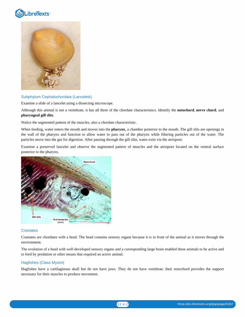
12.4.2 https://bio.libretexts.org/@go/page/41817
Subphylum Cephalochordata (Lancelets)
Examine a slide of a lancelet using a dissecting microscope.
Although this animal is not a vertebrate, it has all three of the chordate characteristics. Identify the notochord, nerve chord, andpharyngeal gill slits.
Notice the segmented pattern of the muscles, also a chordate characteristic.
When feeding, water enters the mouth and moves into the pharynx, a chamber posterior to the mouth. The gill slits are openings inthe wall of the pharynx and function to allow water to pass out of the pharynx while filtering particles out of the water. Theparticles move into the gut for digestion. After passing through the gill slits, water exits via the atriopore.
Examine a preserved lancelet and observe the segmented pattern of muscles and the atriopore located on the ventral surfaceposterior to the pharynx.
Craniates
Craniates are chordates with a head. The head contains sensory organs because it is in front of the animal as it moves through theenvironment.
The evolution of a head with well-developed sensory organs and a corresponding large brain enabled these animals to be active andto feed by predation or other means that required an active animal.
Hagfishes (Class Myxini)
Hagfishes have a cartilaginous skull but do not have jaws. They do not have vertebrae; their notochord provides the supportnecessary for their muscles to produce movement.

12.4.3 https://bio.libretexts.org/@go/page/41817
Subphylum Vertebrata (Vertebrates)The notochord of vertebrates is generally replaced by a vertebral column composed of numerous small bones called vertebrae thatare joined together to form a flexible supporting structure. In most vertebrates, the vertebrae surround the spinal cord.
The vertebral column allows the body to flex and provides attachment sites for muscles. In addition, it surrounds and protects thenerve cord.
They exhibit extreme cephalization and possess complex sense organs (ex: eyes, ears).
Lampreys (Class: Petromyzontida)
Lampreys do not have jaws. Most species of lampreys are parasites. They attach to host fish and feed on the blood of the host.
The larvae are filter feeders that live in freshwater streams. As they mature, they move downstream to the ocean (or lakes) andbegin a parasitic life style.
The skeleton is cartilage and the notocord persists in the adult.
Gnathostomes
Gnathostomes are vertebrates with jaws. The evolution of jaws promoted the switch from filter-feeding to predation and thuspromoted an active life style. Jaws evolved from the forward gill supports in fish.
The appearance of jaws transformed the worlds ecology due to improved predation and herbivory. As a result, complex food chainsevolved.
Filter feeding (gill slits) became less important with the evolution of jaws because jaws allowed the animal to chew larger fooditems and to capture prey. Gills became more important in gas exchange.
Aquatic gnathostomes have a lateral line system. This system is composed of a line of sensory organs on each side of the body. It isable to detect vibrations in the water.
Cartilaginous Fish (Class: Chondrichthyes)
Chondrichthyes include the sharks, and rays. The cartilage skeleton of sharks is partially hardened with calcium. This type ofskeleton is strong and is more flexible and lighter than a bony skeleton. The bodies of cartilaginous fish are covered with smalltoothlike scales. Their teeth are larger versions of these scales.
They do not have a swim bladder but the oil-storing capacity of their livers improves their buoyancy. The shape of the head andcaudal fin also lift the animal as it swims. Sharks must swim to keep from sinking.
Some sharks are fast-swimming predators; others are filter feeders.
Reproduction
Fertilization is internal. The pelvic fin is used to transfer sperm to the female.
Some species are oviparous—they lay eggs that hatch outside the mother’s body; some are ovoviviparous—the eggs are retainedwithin the body and young are born alive; and a few are viviparous—they receive some nourishment via a placenta that developsfrom the yolk sac of the egg.
The reproductive, digestive and excretory system exit the body through a common opening called a cloaca.
Osteichthyans
Osteichthyans have an ossified (bony) skeleton. The skeleton is hardened with calcium phosphate.
Characteristics of Aquatic Osteichthyans (Bony Fish)
The gills are covered by an operculum so that the gill chamber is enclosed and protected. A swim bladder and is used for buoyancy.It evolved from lungs. The gas content, and thus buoyancy, can be regulated by transfer to and from the blood.
Bony fish have broad, flat scales. The skin contains mucous secreting glands that reduce friction as the fish swims through thewater. A lateral line system detects vibrations in the water. Most species are oviparous. Bony fish are the largest group ofvertebrates. Approximately 49,000 species have been identified.

12.4.4 https://bio.libretexts.org/@go/page/41817
Ray-finned Fishs (Class Actinopterygii)
The fins are supported by spinelike rays. In ray-finned fishes, the lungs gave rise to the swim bladder which gives the fishbuoyancy.
Lobe-fins
Lobe-finned fish have fins located on fleshy appendages. This group includes coelacanths (class Actinistia), Lungfishes (classDipnoi) and tetrapods.
Coelacanths were thought to have been extinct for 75 million years until one was captured in 1938 off the southeastern coast ofAfrica. The first vertebrate animals to develop lungs were fish. The lungs developed from a sac-like pocket of tissue that formed inthe pharynx.
Lungfishes are a group of lobe-finned fish that inhabit stagnant fresh water ponds that dry up. Their lungs allow them to gulpoxygen from the air when it has been depleted from the water. Their lobe fins enable them to walk under water.
The ancestors of lungfish gave rise to amphibians.
Contributors and AttributionsCC licensed content, Shared previously
Chordates, Biology 102. Authored by: Michael J. Gregory, Ph.D.. Provided by: LibreTexts. Located at:bio.libretexts.org/Under_Construction/BioStuff/BIO_102/Laboratory_Exercises/Chordates. Project: The Biology Web.License: CC BY-NC-SA: Attribution-NonCommercial-ShareAlike
12.4: Reading- Chordates is shared under a not declared license and was authored, remixed, and/or curated by LibreTexts.

13.1 https://bio.libretexts.org/@go/page/41764
13: Module 10- CardiovascularThis page was auto-generated because a user created a sub-page to this page.
13: Module 10- Cardiovascular is shared under a not declared license and was authored, remixed, and/or curated by LibreTexts.

13.1.1 https://bio.libretexts.org/@go/page/41818
SECTION OVERVIEW
13.1: Respiratory System and Pig Dissection
13.1.1: Fetal Pig Dissection Lab
13.1.2: Fetal Pig Dissection Lab (Instructor Materials Preparation)
13.1.3: Reading- Fetal Pig Dissection
13.1.4: Cardiovascular and Respiratory Systems Lab
13.1.5: Cardiovascular and Respiratory Systems Lab (Instructor Materials Preparation)
13.1: Respiratory System and Pig Dissection is shared under a not declared license and was authored, remixed, and/or curated by LibreTexts.

13.1.1.1 https://bio.libretexts.org/@go/page/41831
13.1.1: Fetal Pig Dissection Lab
Learning ObjectivesLearn about the anatomy of the pig as an example of a vertebrate mammalIdentify structures on the pig and know their functionsIdentify structures that are a part of the digestive system, respiratory system, circulatory system, reproductive system, andexcretory systemCompare pig anatomy to human anatomy
A SlideShare element has been excluded from this version of the text. You can view it online here: pb.libretexts.org/bio2lm/?p=130
Download a PDF of the lab to print.
ProcedureAccess the page “Reading: Fetal Pig Dissection.” The pig may or may not be injected with dye. Follow the steps in the handout toview the external pig anatomy.
Questions1. Based on the external anatomy is your pig male or female? How can you tell?2. Can you locate any hair on your pig, a common characteristic of mammals? Where is there the most hair?
Follow the steps in the handout to cut open the pig beginning with the mouth. Make sure you can locate the following structures inthe mouth region:
GlottisEpiglottisHard and soft palatePharynxTrachea
Question1. What do the hard and soft palate separate?
Cut into the neck region. Make sure you can locate the following structures:
1. Trachea2. Thymus3. Thyroid4. Esophagus
Question1. Is the trachea in front of or behind the esophagus?
Cut into the thoracic cavity beneath the rib cage. Make sure you can locate the following structures:
1. Heart2. Lungs3. Bronchi4. Diaphragm

13.1.1.2 https://bio.libretexts.org/@go/page/41831
Questions1. How many chambers does the pig heart have?2. How does the size of the pig lungs compare to the size of the frog lungs you dissected previously?3. What role does the diaphragm play in respiration?4. What cavity contains the lungs?5. What cavity contains the heart?
Focus next on the abdominal cavity. First look at the digestive system organs. Make sure you can locate the following organs:
1. Stomach2. Spleen3. Liver4. Gallbladder5. Small intestine6. Large intestine7. Pancreas
Questions1. What is the function of the liver?2. What is the function of the gallbladder?3. What type of digestion occurs in the stomach?4. Name the three sections of the small intestines in order.5. Name one process that occurs in the large intestine.6. Which digestive organs located in the abdominal cavity are considered to be accessory organs?
Also in the abdominal cavity you will find the excretory system organs. Make sure you can locate:
KidneysBladder
Finally in the abdominal cavity are the reproductive organs. If you have a female pig look at another group’s male pig and viceversa. You should be able to find:
Testes (male)Uterus with horn (female)Ovary (female)
The arteries and veins are challenging to identify, especially if the pig is not injected with dye. Arteries carry blood away from theheart. Veins return blood to the heart. Try to identify the following:
1. Aorta2. Pulmonary artery3. Coronary arteries4. Jugular vein5. Carotid artery6. Renal artery7. Renal vein
Questions1. Where does the renal vein transport blood?2. Where does the pulmonary artery transport blood?
View the human torso model on your bench. Locate the same organs you found above on the fetal pig.
Questions1. List three similarities between the pig internal anatomy and human internal anatomy.2. List three differences between the pig internal anatomy and human internal anatomy.

13.1.1.3 https://bio.libretexts.org/@go/page/41831
Contributors and AttributionsCC licensed content, Shared previously
Biology 102 Labs. Authored by: Lynette Hauser. Provided by: Tidewater Community College. Located at:http://www.tcc.edu/. License: CC BY-NC-SA: Attribution-NonCommercial-ShareAlike
13.1.1: Fetal Pig Dissection Lab is shared under a not declared license and was authored, remixed, and/or curated by LibreTexts.

13.1.2.1 https://bio.libretexts.org/@go/page/41832
13.1.2: Fetal Pig Dissection Lab (Instructor Materials Preparation)
Lab Materials
This is the prep for one section of 24 students.
Pig Dissection
Students will do this part in table teams (groups of 4).
Materials Quantity Notes
preserved pig 1 per table helpful to have a few male and a few femalepigs
dissection kit 1 per table should include scalpel, scissors, forceps,dissection needles, dissection pins
dissection tray 1 per table
gloves 1 pair per student latex and non latex options in various sizes
lab coat 1 per student
goggles 1 pair per student
dissection waste container 1 per class dissection waste should not go in regular trash
Contributors and AttributionsCC licensed content, Original
Instructor Materials Preparation. Authored by: Lynette Hauser. Provided by: Tidewater Community College. Located at:http://www.tcc.edu/. License: CC BY: Attribution
13.1.2: Fetal Pig Dissection Lab (Instructor Materials Preparation) is shared under a not declared license and was authored, remixed, and/orcurated by LibreTexts.

13.1.3.1 https://bio.libretexts.org/@go/page/41833
13.1.3: Reading- Fetal Pig DissectionThe fetal pig that you will dissect has been injected with a colored latex (rubber) compound. The arteries have been filled with red latex and the veins with blue. An incisionwas made on the side of the neck to enable the injections. The incision can be seen in the first photograph below.
Several different pig dissections were used to obtain the photographs below. As a result, a structure shown in one photograph may look different than the same structureshown in another photograph.
Click on any of the photographs to view enlargements. Links to high-resolution, unlabeled photographs are also provided for many of the photographs.
Orientation
The following words will be used to help identify the location of structures.
Anterior refers to the head end. If a structure is anterior to another, then it is closer to the head.Posterior refers to the tail end.Dorsal refers to the back side. The pig in figure 1 is lying on its dorsal side.Ventral is the belly side. It is opposite the dorsal side. The pig in figure 1 below has its ventral side up.
External Structures
Obtain a fetal pig and identify the structures listed in figure 1. Use figures 1–4 below to identify its sex.
Use your pig and also a pig of the opposite sex to identify the structures in the photographs below. The word “urogenital” refers to an opening that serves both the urinary(excretory) and the reproductive systems.
Figure 1. Female: injection site, nipples, umbilical cord

13.1.3.2 https://bio.libretexts.org/@go/page/41833
Figure 2. Female: genital papilla, urogenital opening, anus
Figure 3. Male: scrotum

13.1.3.3 https://bio.libretexts.org/@go/page/41833
Figure 4. Male: urogenital opening, penis, anus
Preparation and Initial Cuts
Tie one front leg of the animal with a string that passes underneath the dissecting pan to the other leg. Repeat this with the back leg.
Figure 5.
Insert one blade of scissors through the body wall on one side of the umbilical cord and cut posteriorly to the base of the leg as shown in figure 6. Continue cutting from theanterior end of this cut so that it resembles an upside-down U. Your finished cut will be anterior to the navel and along each side of the navel. The flap of body wall thatcontains the navel can be folded posteriorly to reveal the internal organs of the abdomen.

13.1.3.4 https://bio.libretexts.org/@go/page/41833
Figure 6.
Figure 7.

13.1.3.5 https://bio.libretexts.org/@go/page/41833
Figure 8.
Extend a single cut along the midline of the ventral surface of the animal to about 2 cm. from the chin. Cut completely through the body wall in the abdominal area but keepthe cut shallow in the neck region.
Figure 9.
A cut is made on the side of the animal from the point just posterior to the diaphragm dorsally. A similar cut is made on the other side. These two cuts will enable you tospread open the abdominal cavity.

13.1.3.6 https://bio.libretexts.org/@go/page/41833
Figure 10.
Mouth and Neck Region
Use a scalpel to cut the sides of the mouth so that the bottom jaw can be opened for easier viewing (see figure 11). You will need to cut through the musculature and the jointthat holds the lower jaw to the skull.
Figure 11.
Open the jaw wide enough so that the glottis and epiglottis are exposed. The epiglottis projects up through the soft palate into a region called the nasopharynx. The hardpalate and soft palate separate the nasal and oral cavities. When breathing, air passes through the nasal passages to the pharynx. The pharynx is the space in the posteriorportion of the mouth that both food and air pass through. From the pharynx, it passes through the glottis to the trachea.

13.1.3.7 https://bio.libretexts.org/@go/page/41833
Figure 12. Hard palate, soft palate, glottis, epiglottis, and tongue.
Carefully, peel the skin away from the incision in the neck region using a blunt probe (a needle or the point of scissors will do if a blunt probe is not available). Use the probeto peel away muscle tissue until the thymus gland on each side of the trachea is exposed.
Use a probe to separate the two lobes of the thymus gland and to further separate the musculature over the trachea. The thyroid gland is darker and lies between the posteriorends of the two lobes of the thymus gland.
Figure 13. Thymus

13.1.3.8 https://bio.libretexts.org/@go/page/41833
Figure 14. The surrounding tissues have been separated to reveal the thyroid gland.
Continue separating the tissue with a probe until the trachea and esophagus are exposed. The esophagus is dorsal to the trachea. The large hard structure attached to thetrachea is the larynx. It contains the vocal chords.
In the photograph below, the heart and blood vessels of the neck region have been removed so that the trachea can be seen more clearly. You should not remove thesestructures yet because you will need to identify the blood vessels later in the dissection.
Figure 15. Esophagus, larynx, trachea, bronchus, and lungs.
Respiratory SystemObserve how the diaphragm attaches to the body wall and separates the abdominal cavity from the lung (pleural) and heart (pericardial) cavities (figure 16 and 18 below).Contraction of the diaphragm forces air into the lungs.
You have already seen the nasopharynx, hard palate, soft palate, epiglottis, glottis, trachea, and larynx. Follow the trachea to where it branches into two bronchi and observethat each bronchus leads to a lung. The left lung contains three lobes and the right lung contains four. Each lung is located in a body cavity called a pleural cavity.

13.1.3.9 https://bio.libretexts.org/@go/page/41833
Figure 16. Diaphragm.
Figure 17. Lungs

13.1.3.10 https://bio.libretexts.org/@go/page/41833
18. Lungs, diaphragm.
Figure 19. Lungs, diaphragm (cut)

13.1.3.11 https://bio.libretexts.org/@go/page/41833
Figure 20. Esophagus, larynx, trachea, bronchus, and lung.
Digestive System
You have already seen how the esophagus leads from the pharynx through the neck region. Using a probe, trace follow the esophagus to the stomach. Identify the smallintestine and large intestine. Find the posterior part of the large intestine called the rectum and observe that it leads to the anus. Locate the cecum, a blind pouch where thesmall intestine joins the large intestine.
Identify the liver. Lift the right lobe and find the gallbladder. This structure stores bile produced by the liver. Find the bile duct that leads to the small intestine. Thepancreas is located dorsal and posterior to the stomach. It extends along the length of the stomach from the left side of the body (your right) to the point where the stomachjoins the small intestine. Lift the stomach and identify this light-colored organ.
The spleen is an elongate, flattened, brownish organ that extends along the posterior part of the stomach ventral to (above) the pancreas.
The cecum is a blind pouch where the small intestine joins the large intestine. It houses bacteria used to digest plant materials such as cellulose. The cecum is large inherbivores but much of it has been lost during evolution in humans. The appendix in humans is the evolutionary remains of a larger cecum in human ancestors.
Figure 21. Duodenum, gallbladder, liver, lungs, large intestine, pancreas, small intestine, stomach. The liver has been lifted to reveal the gallbladder.

13.1.3.12 https://bio.libretexts.org/@go/page/41833
Figure 22. bile duct, gallbladder, large intestine, liver, and small intestine. The liver has been lifted to reveal the gallbladder.
Figure 23. Large intestine, liver, small intestine, spleen, and stomach.

13.1.3.13 https://bio.libretexts.org/@go/page/41833
Figure 24. small intestine, large intestine, pancreas, spleen, stomach – The spleen has been moved aside to reveal the pancreas.
Figure 25. The stomach and liver are lifted to show the pancreas.

13.1.3.14 https://bio.libretexts.org/@go/page/41833
Figure 26. Large intestine, pancreas, small intestine, spleen, and stomach.
Figure 27. cecum, large intestine, liver, small intestine, spleen. The cecum is found at the point where the small intestine joins the large intestine.

13.1.3.15 https://bio.libretexts.org/@go/page/41833
Figure 28. Large intestine, liver, small intestine, spleen, and stomach.
Circulatory System
Figures 29 and 30 summarize the circulatory system of a mammal.
Figure 29. Circulatory system
Figure 30. Circulatory system
The drawing below shows some of the major arteries that carry blood to the body. Blood vessels that branch from the aorta carry blood to most of the body.
The pulmonary artery is capable of delivering a large amount of blood to the lungs but the lungs are not needed to oxygenate the blood of a fetus, so most of the blood isdiverted to the aorta. This diagram shows that the ductus arteriosus connects the pulmonary artery to the aorta and diverts blood that would otherwise go to the lungs.
Shortly after birth, the ductus arteriosus closes and blood in the pulmonary artery goes to the lungs instead of the body.

13.1.3.16 https://bio.libretexts.org/@go/page/41833
Blood passes from the left ventricle through the aortic arch and aorta to the body. The first branch of the aorta is the brachiocephalic artery. The second branch is the leftsubclavian artery which goes to the left front leg. The right subvclavian carries blood to the right front leg and the carotids carry blood to the head.
Figure 31. Major arteries
The pericardium is a membrane that surrounds the heart and lines the pericardial cavity. It contains a lubricating fluid and isolates the heart from body movements such asthe expansion and contraction of the nearby pleural (lung) cavity.
To view details of the aortic arch, ductus arteriosus, and pulmonary artery, it will be helpful to remove the left lung. With the left lung removed, the heart can be pushed tothe right side to reveal the aorta and other blood vessels shown in figures 33–42.
Figure 32. Diaphragm, heart, lungs, and pericardium

13.1.3.17 https://bio.libretexts.org/@go/page/41833
Figure 33. Aortic arch, coronary artery, left atrium, left ventricle, pulmonary artery, right atrium, right ventricle.
Figure 34. Aorta, aortic arch, left atrium, brachiocephalic artery, ductus arteriosus, lung, pulmonary artery, pulmonary trunk, left subclavian artery,
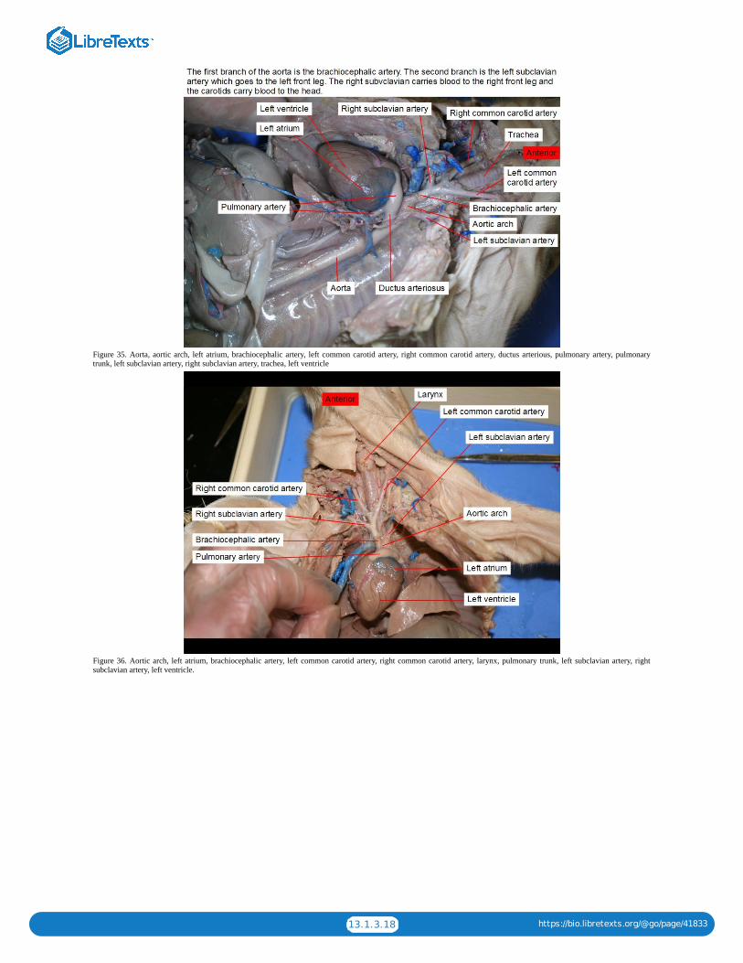
13.1.3.18 https://bio.libretexts.org/@go/page/41833
Figure 35. Aorta, aortic arch, left atrium, brachiocephalic artery, left common carotid artery, right common carotid artery, ductus arterious, pulmonary artery, pulmonarytrunk, left subclavian artery, right subclavian artery, trachea, left ventricle
Figure 36. Aortic arch, left atrium, brachiocephalic artery, left common carotid artery, right common carotid artery, larynx, pulmonary trunk, left subclavian artery, rightsubclavian artery, left ventricle.

13.1.3.19 https://bio.libretexts.org/@go/page/41833
Figure 37. Anterior vena cava, coronary artery, larynx, posterior vena cava, right subclavian vein, trachea.
Figure 38. Anterior vena cava, coronary artery, right external jugular vein, right internal jugular vein, larynx, lungs, right subclavian vein, trachea

13.1.3.20 https://bio.libretexts.org/@go/page/41833
Figure 39. Anterior vena cava, posterior vena cava.
Figure 40. Heart, liver, lung, posterior vena cava, thymus, thyroid

13.1.3.21 https://bio.libretexts.org/@go/page/41833
Figure 41. Aorta, colon, kidney, posterior vena cava, renal artery, renal vein, testicular artery, testis, umbilical artery, ureter, urinary bladder, vas deferens. The renal arterypasses blood from the aorta to the kidney. The renal vein returns blood from the kidney to the posterior vena cava.
Figure 42. Aorta, colon (large intestine), diaphragm, heart, kidney, lung, renal artery, posterior vena cava, renal vein, small intestine, spleen, stomach, ureter.

13.1.3.22 https://bio.libretexts.org/@go/page/41833
Figure 43. External iliac artery, kidney, large intestine, posterior vena cava, renal vein, small intestine, testis, umbilical artery, ureter, urinary bladder.
Figure 44. Left atrium, brachiocephalic artery, left common carotid artery, right common carotid artery, coronary artery, external iliac artery, external iliac vein, posteriorvena cava, pulmonary trunk, renal artery, renal vein, left subclavian artery, right subclavian artery, umbilical artery, left ventricle.
Excretory System

13.1.3.23 https://bio.libretexts.org/@go/page/41833
Figure 45. Aorta, colon (large intestine), diaphragm, heart, kidney, lung, renal artery, posterior vena cava, renal vein, small intestine, spleen, stomach, ureter.
Figure 46. Aorta, kidney, liver, posterior vena cava, renal artery, renal vein, spleen, ureter, urinary bladder.

13.1.3.24 https://bio.libretexts.org/@go/page/41833
Figure 47. Kidney, liver, posterior vena cava, renal vein, spleen, ureter, urinary bladder
Reproductive System (Female)
Figure 48. Urogenital papilla, anus

13.1.3.25 https://bio.libretexts.org/@go/page/41833
Figure 49. Colon, horn of uterus, ovary, urinary bladder
Figure 50. Colon, body of uterus, horn of uterus, ovaries, urethra, urinary bladder, urogenital sinus
Reproductive System (Male)

13.1.3.26 https://bio.libretexts.org/@go/page/41833
Figure 51. Penis, scrotum, urogenital opening
Figure 52. Seminal vesicle, testis, ureter, urinary bladder, vas deferens

13.1.3.27 https://bio.libretexts.org/@go/page/41833
Figure 53. Bulbourethral gland, spermatic cord, testis, urethra, urinary bladder, vas deferens
Figure 54. Path of urine flow
Contributors and AttributionsCC licensed content, Shared previously
Fetal Pig Dissection, Biology 102. Authored by: Michael J. Gregory, Ph.D.. Provided by: LibreTexts. Located at:bio.libretexts.org/Under_Construction/BioStuff/BIO_102/Laboratory_Exercises/Fetal_Pig_Dissection. Project: The Biology Web. License: CC BY-NC-SA: Attribution-NonCommercial-ShareAlike
13.1.3: Reading- Fetal Pig Dissection is shared under a not declared license and was authored, remixed, and/or curated by LibreTexts.

13.1.4.1 https://bio.libretexts.org/@go/page/41834
13.1.4: Cardiovascular and Respiratory Systems Lab
State the parts of the cardiovascular and respiratory systems and give the functions of each part.Identify the parts of the cardiovascular and respiratory systems on the human torso model and other models.Explain how the respiratory and cardiovascular systems are interrelated.Describe the path of the blood through the heartTest the effects of various factors on heart rate and breathing ratesMeasure various respiratory volumes using a spirometer.Define the following terms:
nostrils trachea spirometer nasopharynx
bronchi pleura pharynx larynx
forced vital capacity epiglottis lung vital capacity
diaphragm glottis forced expiratory volume intercostal muscles
aorta brachial artery and vein superior vena cava ventricle
hepatic artery and vein inferior vena cava atrium renal artery and vein
iliac artery and vein pulmonary artery and vein jugular vein systole
diastole coronary artery and vein carotid artery systole
A SlideShare element has been excluded from this version of the text. You can view it online here: pb.libretexts.org/bio2lm/?p=136
Download a PDF of the lab to print.
Activity I: Identifying StructuresFind the following structures on the human torso model and other models.
NostrilsTracheaNasal cavityOral cavityBronchiDiaphragmPharynxPleuraLarynxLungEpiglottisIntercostal musclesGlottisIliac artery and veinCarotid arteriesJugular veinsRight and left ventricleRight and left atria
Learning Objectives

13.1.4.2 https://bio.libretexts.org/@go/page/41834
Pulmonary artery and veinBrachial artery and veinCoronary artery and veinRenal artery and veinHepatic artery and veinFemoral artery and veinSuperior and inferior vena cava
Use the chart on the next page to help organize your understanding of the different arteries and veins. For each one listed, statewhere the blood is traveling to and from.
Artery or Vein Where blood is traveling to and from
Pulmonary Artery
Pulmonary Vein
Coronary Artery
Coronary Vein
Hepatic Artery
Hepatic Vein
Iliac Artery
Iliac Vein
Jugular Vein
Carotid Artery
Brachial Artery
Brachial Vein
Femoral Artery
Femoral Vein
Renal Artery
Renal Vein
Use the heart picture below and label the different atria and ventricles. Also include the aorta, superior vena cava and inferior venacava.
Activity II: Respiratory Volumes and Lung CapacitiesSpirometry is the classic pulmonary function test. A spirometer is an instrument used to measure how much air and how quicklyair is expelled after a deep inhalation. The measurements can be used to calculate how efficiently and how quickly the lungs can be

13.1.4.3 https://bio.libretexts.org/@go/page/41834
filled upon inspiration and emptied during expiration. The most common measurements obtained from a spirometer are listed in thetable below.
Abbreviation Measurement Description
VC Vital Capacity Maximal amount of air exhaled steadily fromfull inspiration to maximal expiration
FVC Forced vital capacity The total amount of air that you blow out inone breath
FEV1 Forced expiratory volume in one second
The amount of air you can blow out in onesecond. An individual with normal lungs andairways can blow out most of the air from thelungs in the first second
FER Forced expiratory ratio (FEV1/FVC)x100 Percentage of the FVC expelled in the firstsecond of a forced expiration
PEF Peak expiratory flow Peak expiratory flow in liters per minute
The spirometer we will use measures peak expiratory flow, a useful indicator of lung function to assess conditions such as asthma.Peak flow is achieved by blowing out as fast as possible after taking in as much air as possible.
Directions to Operate Spirometer1. For best results, stand. If you are unable to stand, sit in a straight and upright position.2. Move the peak indicator (red internal piece) to the start position (all the way on the left).3. Hold the spirometer in your hand with your thumb and forefinger on the grips and the mouthpiece facing toward you. Avoid
blocking the vent holes as much as possible and do not allow the fingers to interfere with the red peak indicator.4. Take as deep a breath as possible filing your lungs completely with air5. Place your mouth on the mouthpiece, past your teeth, and form a tight seal with your lips. Place your tongue below the
mouthpiece to make sure it is not blocking the opening at any time.6. Blow out as hard and fast as you can. The red indicator will move indicating your peak flow.7. Do not reset the peak indicator. Repeat steps 4-6 two more times for a total of three. The indicator will automatically point to
the best of the three efforts.8. Record your results in the table on the next page.9. Clean off the spirometer using a paper towel and rubbing alcohol.
10. Have your partner complete the same process.
PEF
My value
My partner
Results
Comparing Your Results to Expected Values
Use the normal predicted average peak expiratory flow tables in the spirometer box and any additional handouts provided by yourinstructor to compare your readings to expected values based on an individuals gender, age, ethnicity, and height.
How does your average PEF compare with the value for a person your age and height?
What do These Values Mean in the Real World?
Individuals suffering from obstructive pulmonary disease (narrowed airways) have a low FEV1 but a normal FVC. Since theairways are narrowed less air can be blown out in one second. Individuals with obstructive lung disease also have a FER less than70% of the predicted value. Asthma is one condition which causes narrowing of the airways. Spirometry is used to diagnoseasthma and asses the efficiency of treatments.
Individuals can also suffer from restrictive pulmonary disease. These individuals have a normal FEV1 since the airways areunobstructed but a lower FVC. The lower FVC is caused by various conditions that affect the lung tissue or the capacity of the

13.1.4.4 https://bio.libretexts.org/@go/page/41834
lungs to expand.
There are some conditions that involve both lung obstruction and restriction, such as cystic fibrous. Individuals with cystic fibroussecrete excess mucus which narrows the airways and damages the lung tissue.
Activity III: InvestigationHow do different everyday activities affect your circulation and respiration? You and a partner will work together to come up withand implement a procedure to test the following question:
How does body position (laying down verses sitting verses standing) affect your heart rate and your breathing?
In the space below, write a hypothesis and null hypothesis for the question.
Hypothesis:
Null Hypothesis:
You have the following tools available to test these questions:
1. Spirometer2. Measuring pulse3. Measuring breathing rate
You need to determine how you can test both respiration and circulation. Some points to consider:
How long your experiment should run?How many trials you should doHow often should you take measurements?How could you create a control?
Use the space below to write out a procedure to test each question. Make sure that your instructor approves your procedures beforeyou begin your experiments.
Procedure for Question 1:
Now begin your experiments. Use the space below to record your results. Feel free to make your own tables to organize your data.
Use your results to answer the following questions:
1. How did your heart rate change with body position? When was it the lowest? Highest?2. How did your breathing rate change with body position? When was it the lowest? Highest?3. Why might your heart and breathing rate differ from your partner?
As modified from Piedmont Virginia Community College’s Biology 102 Circulatory and Respiratory Systems Lab
Contributors and AttributionsCC licensed content, Original
Biology 102 Labs. Authored by: Lynette Hauser. Provided by: Tidewater Community College. Located at:http://www.tcc.edu/. License: CC BY: Attribution
13.1.4: Cardiovascular and Respiratory Systems Lab is shared under a not declared license and was authored, remixed, and/or curated byLibreTexts.
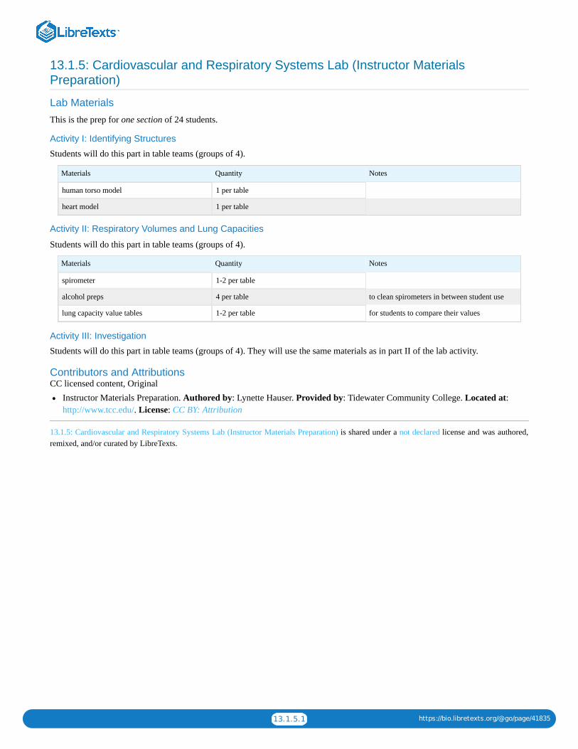
13.1.5.1 https://bio.libretexts.org/@go/page/41835
13.1.5: Cardiovascular and Respiratory Systems Lab (Instructor MaterialsPreparation)
Lab Materials
This is the prep for one section of 24 students.
Activity I: Identifying Structures
Students will do this part in table teams (groups of 4).
Materials Quantity Notes
human torso model 1 per table
heart model 1 per table
Activity II: Respiratory Volumes and Lung Capacities
Students will do this part in table teams (groups of 4).
Materials Quantity Notes
spirometer 1-2 per table
alcohol preps 4 per table to clean spirometers in between student use
lung capacity value tables 1-2 per table for students to compare their values
Activity III: Investigation
Students will do this part in table teams (groups of 4). They will use the same materials as in part II of the lab activity.
Contributors and AttributionsCC licensed content, Original
Instructor Materials Preparation. Authored by: Lynette Hauser. Provided by: Tidewater Community College. Located at:http://www.tcc.edu/. License: CC BY: Attribution
13.1.5: Cardiovascular and Respiratory Systems Lab (Instructor Materials Preparation) is shared under a not declared license and was authored,remixed, and/or curated by LibreTexts.

1
CHAPTER OVERVIEW
14: Module 11- Homeostasis14.1: Homeostasis Lab14.2: Homeostasis Lab (Instructor Materials Preparation)
14: Module 11- Homeostasis is shared under a not declared license and was authored, remixed, and/or curated by LibreTexts.

14.1.1 https://bio.libretexts.org/@go/page/41819
14.1: Homeostasis Lab
Learning ObjectivesDescribe the anatomy of the liver including the path of blood flow from the intestines, through the liver, and to the heartCompare glucose levels in different veins and arteries before and after eatingDescribe the anatomy of the kidneys and a nephron including circulation of the bloodExplain the three steps of urine formation and where each stem occurs in the nephronPredict whether substances will be found in the filtrate or the urine after kidney functionExplain how the kidneys and the liver contribute to homeostasis
A SlideShare element has been excluded from this version of the text. You can view it online here: pb.libretexts.org/bio2lm/?p=142
Download a PDF of the lab to print.
Homeostasis describes the dynamic balance of the body’s internal environment and the effort to maintain a constant, stable inside.There are many body components that contribute to homeostasis. This lab activity will focus on the liver and the kidneys.
Part I: Liver
The liver is an organ within the digestive system and is responsible for maintaining sugar levels in the blood as part of homeostasis.After a large meal, the liver converts extra glucose into glycogen, a polysaccharide that stores glucose. A hormone called insulin isproduced by the pancreas stimulates glycogen production. When levels of glucose in the blood drop, the liver breaks downglycogen back into glucose for the blood to circulate throughout the body. A hormone called glucagon produced by the pancreasstimulates this process. All cells of the body require glucose for cellular respiration to make energy.
The liver receives blood from the small intestines through the hepatic portal vein. After a large meal, the hepatic vein wouldtransport glucose rich blood from the small intestines to the liver. Blood leaves the liver and returns to the heart through the hepaticvein. We will conduct a simulation to learn more about the liver’s role in maintaining blood glucose levels in relationship tohomeostasis.
The table below describes the blood serums you will test:
Serum Location
A Mesenteric artery (takes blood from aorta to small intestine)
B Hepatic portal vein (transports blood between intestines and liver)
C Hepatic vein (takes blood form liver to heart)
We will use a test called the Benedict’s testto determine the amount of glucose in each location. The benedicts test ranges in colorfrom blue (no glucose) to orange/red (lots of glucose). Follow the directions below.

14.1.2 https://bio.libretexts.org/@go/page/41819
Procedure
Glucose levels after eating:
1. Fill the large beaker ½ full with tap water. Place the beaker on top of the hot plate. Turn the hot plate on to create a hot waterbath.
2. Label three test tubes A1, B1, and C1 with a wax pencil3. Use the small plastic ruler to mark on the test tube at 1 cm and 2 cm.4. Fill test tube A1 to the 1 cm mark with serum A1 and to the 2 cm mark with Benedict’s reagent.5. Fill test tube B1 to the 1 cm mark with serum B1 and to the 2 cm mark with Benedict’s reagent.6. Fill test tube C1 to the 1 cm mark with serum C1 and to the 2 cm mark with Benedict’s reagent.7. Place all three test tubes into the hot water bath at the same time.8. Heat the tubes for 5 minutes. Observe and record any color changes.9. Record your results in the table below. Remember that blue indicates no glucose and red/orange indicates the most glucose. A
green color signifies some glucose.
Results
Table 1 Glucose levels after eating
Test tubes in order of color change Source of the serum
Questions
1. Which blood vessel, the mesentery artery, the hepatic portal vein, or the hepatic vein contains the most glucose after eating?2. Explain why the hepatic portal vein contains more glucose than the hepatic vein after eating.
Procedure
Glucose levels before eating:
1. Keep your hot water bath from the first procedure2. Label three test tubes A2, B2, and C2 with a wax pencil3. Use the small plastic ruler to mark on the test tube at 1 cm and 2 cm.4. Fill test tube A2 to the 1 cm mark with serum A2 and to the 2 cm mark with Benedict’s reagent.5. Fill test tube B2 to the 1 cm mark with serum B2 and to the 2 cm mark with Benedict’s reagent.6. Fill test tube C2 to the 1 cm mark with serum C2 and to the 2 cm mark with Benedict’s reagent.7. Place all three test tubes into the hot water bath at the same time.8. Heat the tubes for 5 minutes. Observe and record any color changes.9. Record your results in the table below. Remember that blue indicates no glucose and red/organge indicates the most glucose. A
green color signifies some glucose.
Results
Table 2: Glucose levels before eating
Test tubes in order of color change Source of the serum
Questions
1. Which blood vessel, the mesentery artery, the hepatic portal vein, or the hepatic vein contains the most glucose before eating?2. Explain why the hepatic portal vein contains less glucose than the hepatic vein before eating.
Once you have recorded your results, please clean up the materials. Make sure to:
Turn off and unplug the hot water bathDump the contents of the test tubes down the sinkWash out the test tubes and place them in the test tube rack to dry

14.1.3 https://bio.libretexts.org/@go/page/41819
Part II: KidneysThe kidneys are part of the urinary system. As they produce urine to release nitrogenous wastes from the body the kidneys alsomaintain homeostasis through pH balance and water-salt balance in osmoregulation. These bean shaped organs are located alongthe dorsal wall of the abdominal cavity.
Observe the kidney models available in the lab. Locate the outer renal cortex tissue and the more internal renal medulla. The renalpelvis is the area that collects the urine. Find the renal artery and the renal vein.
The functioning unit of the kidney is called the nephron. Part of the nephron is located in the cortex and part in the medulla. Usethe picture below and the models in the lab to identify the following components of the nephron.
GlomerulusBowman’s capsuleProximal tubuleDistal tubuleLoop of Henle (descending limb and ascending limb)Collecting ductPeritubular capillaries
Questions1. Which parts of the nephron are located in the renal cortex?2. Which components of the nephron are located in the medulla?
Urine production in the kidney involves four main steps:
1. Filtration: molecules move out of the glomerulus into Bowman’s capsule. Large molecules like proteins and blood cells are toobig to be filtered and remain in the blood.
2. Reabsorption: glucose and amino acids move from the proximal tubule back into the blood stream through peritubularcapillaries.
3. Secretion: Substances like histamines, H+, and ammonia get secreted into the nephron from the peritubular capillaries4. Water reabsorption: both the Loop of Henle and the collecting duct reabsorb water to maintain the blood volume
Label the parts of the nephron on the diagram below and indicate where the different urine production steps occur.

14.1.4 https://bio.libretexts.org/@go/page/41819
Focus on FiltrationBlood entering the glomerulus contains cells, proteins, glucose, amino acids, salts, urea, and water. Fill in the table belowindicating which molecules will leave the glomerulus and enter the Bowman’s capsule. Write yes or no for each and state whybased on size (small or big).
Substance Enter Bowman’s Capsule? Why?
Cells
Proteins
Glucose
Amino acids
Salts
Urea
Water
Focus on ReabsorptionWhen the filtrate enters the proximal tubule it contains the following molecules: glucose, water, urea, amino acids, and salts. Waterand salts are passively reabsorbed to maintain blood volume and pH as part of homeostasis.
Questions1. What would happen to the blood volume over time if water were not reabsorbed?2. How would this lack of water reabsorption affect blood pressure?3. Fill in the table below indicating which molecules will be reabsorbed into the blood.
Substance Reabsorbed? Why?
Glucose
Amino acids
Salts
Urea
Water
Kidneys are also important in osmoregulation , maintaing an internal salt/water balance. The kidney can produce large amounts ofdilute urine or small amounts of concentrated urine depending on the needs of the body. The pituitary glad produces antidiuretichormone (ADH) which controls the concentration of urine output. ADH specifically acts on the collecting duct making it more orless permeable to water.

14.1.5 https://bio.libretexts.org/@go/page/41819
The table below has several different events that would impact osmoregulation. Fill in the chart with either “increase” or“decrease” to explain how the kidney would help maintain homeostasis.
Event Change in bloodconcentration
ADH output Water Reabsorption Type of Urine produced
Dehydration due tosunbathing in theafternoon and forgettingyour water bottle
Scant, concentrated
Drinking large amounts ofwater throughout the day
Copious, dilute
Going to the move theaterand eating a large bucketof salty popcorn withoutwater to wash it down
Scant, concentrated
Kidneys also play a role in pH balance.
Questions1. If the blood is more basic than normal, what pH do you think the urine will be?2. If the blood is more acidic than normal, what pH do you think the urine will be?
Contributors and AttributionsCC licensed content, Shared previously
Biology 102 Labs. Authored by: Lynette Hauser. Provided by: Tidewater Community College. Located at:http://www.tcc.edu/. License: CC BY: Attribution
14.1: Homeostasis Lab is shared under a not declared license and was authored, remixed, and/or curated by LibreTexts.

14.2.1 https://bio.libretexts.org/@go/page/41820
14.2: Homeostasis Lab (Instructor Materials Preparation)
Lab Materials
This is the prep for one section of 24 students.
Part I: Liver
Students will do this part in table teams (groups of 4).
Materials Quantity Notes
human torso models 1 per bench
liver models 1 per bench
Benedicts reagent 1 bottle per bench
test tubes 6 per bench
goggles 1 pair per student
gloves 1 pair per student latex and non latex options in various sizes
Solution A1 1 bottle per bench keep refrigerated
Solution B1 1 bottle per bench keep refrigerated
Solution C1 1 bottle per bench keep refrigerated
Solution A2 1 bottle per bench keep refrigerated
Solution B2 1 bottle per bench keep refrigerated
Solution C2 1 bottle per bench keep refrigerated
hot plate 1 per bench
goggles 1 pair per student
Recipe for Solution A1 (0.25% glucose): Dissolve 1.25g glucose/500ml DI water
Recipe for Solution A2 (0.25% glucose): Dissolve 1.25g glucose/500ml DI water
Recipe for Solution B1 (3% glucose): Dissolve 15g glucose/500ml DI water
Recipe for Solution B2 (0% glucose): 500 ml DI water
Recipe for Solution C1 (0.5% glucose): Dissolve 2.5g glucose/500ml DI water
Recipe for Solution C2 (0.5% glucose): Dissolve 2.5g glucose/500ml DI water
Part II: Kidneys
Students will do this part in table teams (groups of 4).
Materials Quantity Notes
Kidney model 1 per bench
nephron model 3 per class
Contributors and AttributionsCC licensed content, Original
Instructor Materials Preparation. Authored by: Lynette Hauser. Provided by: Tidewater Community College. Located at:http://www.tcc.edu/. License: CC BY: Attribution
14.2: Homeostasis Lab (Instructor Materials Preparation) is shared under a not declared license and was authored, remixed, and/or curated byLibreTexts.

1
CHAPTER OVERVIEW
15: Module 12- Food Choice15.1: Food Choice Lab15.2: Food Choice Lab (Instructor Materials Preparation)*
15: Module 12- Food Choice is shared under a not declared license and was authored, remixed, and/or curated by LibreTexts.

15.1.1 https://bio.libretexts.org/@go/page/41821
15.1: Food Choice LabAdapted by Staci Forgey, Tidewater Community College biology faculty, with permission from Niagara University’s Dr. WilliamEdwards Food Choice Lab.
Learning ObjectiveBy the end of this section, you will be able to:
Identify the basic tenets of Ecology and how they apply to population success.
A SlideShare element has been excluded from this version of the text. You can view it online here: pb.libretexts.org/bio2lm/?p=148
Foraging is the act of an animal searching for food. An animal’s choice of food should reflect energetic considerations such asmaximizing net energy gain per unit time or net gain per cost expended in foraging. Animals want the most energy return for theircaloric investment. They seek out food sources that will give them the most energy reward for the least amount of energyexpended. We will be examining the foraging behavior of birds in our quad.
Question1. What factors might influence seed selection in the birds we will be watching? Name three factors and their effects.
Given these factors, we will now form hypotheses and predictions that we will test by observing birds in the courtyard area. After,we will perform a Chi-Square statistical test on our data, and revise our hypotheses and thought processes to fit the new data.
Many birds will visit our feeders and collect seeds. Our feeders are stocked with three types of seeds. The Feathered Friend blackoil seed is very soft hulled. The Lyric Sunflower gray striped sunflower seeds are large and thick hulled. Gray safflower seeds arealso small and soft hulled.
Question1. Look at the three seed types. Feel them and try to break them open. Note the differences you observe between the seed types.
The table below summarizes measurements for the 3 types of seeds. This shows the average kernel (food part of the seed), hull(outside covering of the seed) and entire seed weight. The kernel/hull ratio for each seed type, and the average caloric (energy)content of the seeds. Use these values and your observations to make predictions about optimal foraging behavior for various birdtypes that visit the feeders.
Table 1: Mass and caloric content of sunflower seed components.
Seed (mg) Kernel (mg) Hull (mg) Entire seed (mg) Kernel/Hull Cal/g Cal/seed
Black oil 28.8 ± 3.8 12.2 ± 1.6 41.0 ± 0.5 2.37 ± 0.3 5400 ± 240 150
Safflower 26.8 ± 3.9 10.4 ± 3.3 38.3 ± 6.9 2.603 ± 0.1 6200 ± 240 161
Striped 61.7 ± 4.7 59.1 ± 2.7 120.8 ± 6.0 1.045 ± 0.1 5600 ± 240 350
Now, take a look at the birds that are commonly found in our area during this time period. You will have a list of birds on your labbench.
Your lab group will propose two experimental hypotheses about avian seed choice and discuss these with your instructor. Provide arationale for your testable hypotheses. Clearly state each of your ideas in the form of an “If . . . then . . .” hypothesis. If yourhypothesis is correct, which type of seed do you expect the birds to choose? Why?

15.1.2 https://bio.libretexts.org/@go/page/41821
Questions1. Clearly state a testable hypothesis explaining why birds will choose or not choose the different seeds.2. Would this hypothesis change with different birds? Why or Why not?3. What is the alternative(s) to your hypothesis? (i.e., if you are wrong)4. If your hypothesis is right, what would you predict the birds will do at the feeders?5. If your alternative is right, what would you predict?
Check in with your instructor before continuing after this point!
In order to test your hypothesis, we need to think a bit about experimental design. Variables are important to consider because theywill help us evaluate our hypothesis. For example, if we were interested in the height at which giraffes eat their food from, wemight propose a hypothesis that giraffes will eat food from high areas in a tree. Each time the giraffe ate, the height at which thefood was taken from would need to be recorded. This gives us two variables: the height at which the mouthful of vegetation camefrom and the mouthful height. This height is measurable. We will look at what is called “categorical” data. Each of our data pointswill fit into a category. We will have a bird taking a seed (making a food choice), and the type of seed chosen.
Questions1. What is the independent variable (what we are manipulating)?2. What is the dependent variable (what we are measuring)?3. Design a data table to record the species of bird and type of seed each bird selects during our lab time. You can use hash marks
to record visits. We will be visiting our feeders for a 20 minute time span.
Discuss your data sheet with your instructor before you begin collecting data!
When we return, you’ll need to transfer the data from your table into the following table as totals.
Bird Type Safflower Striped Sunflower Black Oil
TOTALS
Bird Observation
Stay well away from the feeders at all times and keep as quiet as possible. One noisy or clumsy move can scare all the birds away,leaving you without any data!
Question1. Describe the behavior of the birds with the seeds.2. How long does it take an individual bird (record the species of bird and the type of seed eaten) to eat a seed and return to the
feeder for another? Use your binoculars to follow individual birds and record the time from when a bird takes a seed to when itreturns for another. Do the birds do anything unusual with the seeds?
3. Compare and contrast the seed handling behavior of the birds visiting the feeders. How do the different birds crack each of theseeds? Do they have difficulty opening any of them?
Statistical AnalysisUsing your feeder choice data, perform a chi-square Goodness of Fit test to determine if the birds show a preference for any givenseed.
A Chi-Square test involves testing the probability that your categorical data differs from random enough to have confidence thatthe data does not come from chance alone. Typically, we accept that significance is 0.05 (called alpha). This means that there isonly a 1 in 20 chance of the data arising from chance alone. When doing a chi-square, we typically call the boring hypothesis (thatall the birds would randomly select seeds and your hypothesis is wrong) the “null” hypothesis, abbreviated H . The hypothesiswhere the data support your idea is called the alternate hypothesis, abbreviated H . Note: this is a different type of hypothesis, usedto fit specifically into statistical tests. This may not match perfectly with your description of hypotheses above. A chi-square also
O
a

15.1.3 https://bio.libretexts.org/@go/page/41821
doesn’t tell you exactly where the differences causing significant deviation from the expected. You would need more involvedstatistics for that. We will visually pick out our differences in data.
First, transfer your choice data into a table like the following table. We will calculate the expected values from the data.
Add the data for each bird horizontally, then for each seed vertically. The total row and total column should now be filled. Theserepresent how many birds and seeds by type were actually eaten and recorded. Now we will calculate the expected values based onhow these would be distributed by random chance. We will take the total for each species, multiply it by the total number of seedsfor that seed type, and divide by the total number of seeds
For example, \displaystyle\frac{111\times{178}}{532}=37.13
Put the answer into the table. This is how many safflower seeds would be expected to be eaten by the chickadees through chancealone. Complete your own table.
SAMPLE TABLE
Safflower Striped Black Oil Total
Black-capped chickadee 13 23 75 111
(Expected) 37.13
Tufted titmouse 21 32 23 76
(Expected)
House finch 45 23 43 111
(Expected)
American goldfinch 0 0 0 0
(Expected)
Nuthatches 0 0 0 0
(Expected)
Junco 76 32 32 140
(Expected)
House sparrows 23 27 44 94
(Expected)
TOTALS 178 137 217 532
Bird Type Safflower Striped Black Oil Total
(Expected)
(Expected)
(Expected)
(Expected)
(Expected)
(Expected)
(Expected)
(Expected)
TOTALS

15.1.4 https://bio.libretexts.org/@go/page/41821
To calculate the chi-square value, or \displaystyle\chi^2 , we simply add the square differences, divided by the expected, of all theobserved and expected. In mathematical terms:
\displaystyle\chi^2=\Sigma\frac{(O-E)^2}{E}
So for our example from the previous sample table, the first \displaystyle\frac{(O-E)^2}{E} would be
\displaystyle\frac{(13-37.13)^2}{37.13}=15.7
We would then add to this value all of the other \displaystyle\frac{(O-E)^2}{E} in the table to get the \displaystyle\chi^2 value.
Question1. Compute the \displaystyle\chi^2 value for your data. (Use an extra sheet of paper if necessary)
In order to find something to compare this number with, we need to calculate the degrees of freedom, or the number of differentcomparisons that can be made within the table. The degrees of freedom are the number of columns (M) minus one times thenumber of rows (N) minus one. Degrees of freedom = (M–1) (N–1)
Questions1. How many ways can this two by two table be broken into individual comparisons? Hint: Use the formula above.
A B
C D
2. Calculate the degrees of freedom in your experiment.
Now we can compare against the Chi distribution for the likelihood that our data is generated by chance. Remember, we want a0.05 or less value to say that it’s not chance, but our hypothesis that’s causing the choices.
Questions1. Compare to the Chi-distribution table at the end of the lab with your instructor’s assistance. What is the p value or probability
that your data came out by chance? Is this less than 0.05? Did your results come about by chance?2. Describe and summarize what you observed in the field. Are some parameters more difficult to measure than others? If so,
why? Which predictions did your data support? Interpret you results as they relate to your hypotheses and discuss yourinterpretation.
3. How could you re design your experiment to better measure energy gains, handling time, and energetic costs of foraging, andthus more accurately test the predictions of optimal foraging theory? Think of other hypotheses regarding seed choice by birds?Propose a follow up study that would allow you to test a related idea about avian foraging behavior. Make clear the ways inwhich your proposed study is an extension of or improvement upon the study on which you report here.
Chi Square Distribution Table:
DF/P 0.995 .990 0.975 .950 .900 .750 .500 .250 .100 .050 .025 .010 .005
1 0.00004
.00016 0.001 0.004 0.016 0.102 0.455 1.323 2.706 3.841 5.024 6.635 7.879
2 0.010 0.020 0.0506 0.103 0.211 0.575 1.386 2.773 4.605 5.991 7.378 9.210 10.597
3 0.072 0.115 0.216 0.351 0.584 1.213 2.366 4.108 6.251 7.815 9.348 11.345 12.838
4 0.207 0.297 0.484 0.711 1.064 1.923 3.357 5.385 7.779 9.488 11.143 13.277 14.860
5 0.412 0.554 0.831 1.145 1.610 2.675 4.351 6.626 9.236 11.070 12.833 15.086 16.750
6 0.676 0.872 1.237 1.635 2.204 3.455 5.348 7.841 10.645 12.592 14.449 16.812 18.548
7 0.989 1.239 1.690 2.167 2.833 4.255 6.346 9.037 12.017 14.067 16.013 18.475 20.278
8 1.344 1.647 2.180 2.733 3.490 5.071 7.344 10.219 13.362 15.507 17.535 20.090 21.955
9 1.735 2.088 2.700 3.325 4.168 5.899 8.343 11.389 14.684 16.919 19.023 21.666 23.589
10 2.156 2.558 3.247 3.940 4.865 6.737 9.342 12.549 15.987 18.307 20.483 23.209 25.188

15.1.5 https://bio.libretexts.org/@go/page/41821
11 2.603 3.053 3.816 4.575 5.578 7.584 10.341 13.701 17.275 19.675 21.920 24.725 26.757
12 3.074 3.571 4.404 5.226 6.304 8.438 11.340 14.845 18.549 21.026 23.337 26.217 28.300
13 3.565 4.107 5.009 5.892 7.042 9.299 12.340 15.984 19.812 22.362 24.736 27.688 29.819
14 4.075 4.660 5.629 6.571 7.790 10.165 13.339 14.114 21.064 23.685 26.119 29.141 31.319
15 4.601 5.229 6.262 7.261 8.547 11.037 14.339 18.245 22.307 24.996 27.488 30.578 32.801
16 5.142 5.812 6.908 7.962 9.312 11.912 15.339 19.369 23.542 26.296 28.845 32.000 34.267
17 5.697 6.408 7.564 8.672 10.085 12.792 16.338 20.489 24.769 27.587 30.191 33.409 35.718
18 6.265 7.015 8.231 9.390 10.865 13.675 17.338 21.605 25.989 28.869 31.526 34.805 37.156
19 6.844 7.633 8.907 10.117 11.657 14.562 18.338 22.18 27.204 30.144 32.852 36.191 38.582
20 7.434 8.260 9.591 10.851 12.443 15.452 19.337 23.848 28.412 31.410 34.170 37.566 39.997
21 8.034 8.897 10.283 11.591 13.240 16.344 20.337 24.935 29.615 32.671 35.479 38.932 41.401
22 8.643 9.542 10.982 12.338 14.041 17.240 21.337 26.039 30.813 33.924 36.781 40.289 42.796
23 9.260 10.196 11.689 13.091 14.848 18.137 22.337 27.141 32.007 35.172 38.076 41.638 44.181
24 9.886 10.856 12.401 13.848 15.659 19.037 23.337 28.241 33.196 36.415 39.364 42.980 45.559
25 10.520 11.524 13.120 14.611 16.473 19.939 24.337 29.339 34.382 37.652 40.646 44.314 46.928
26 11.160 12.198 13.844 15.379 17.292 20.843 25.336 30.435 35.563 38.885 41.923 45.642 48.290
27 11.808 12.879 14.573 16.151 18.114 21.749 26.336 31.528 36.741 40.113 43.195 46.963 49.645
28 12.461 13.565 15.308 16.928 18.939 22.657 27.336 32.620 37.916 41.337 44.461 48.278 50.993
29 13.121 14.256 16.047 17.708 19.768 23.567 28.336 33.711 39.087 42.557 45.722 49.588 52.336
30 13.787 14.953 16.791 18.493 20.599 24.478 29.336 34.800 40.256 43.773 46.979 50.892 53.672
Contributors and AttributionsCC licensed content, Original
Biology 102 Labs. Authored by: Lynette Hauser. Provided by: Tidewater Community College. Located at:http://www.tcc.edu/. License: CC BY: AttributionFood Choice Lab. Authored by: Dr. William Edwards. Provided by: Niagara University. Located at: http://www.niagara.edu.License: CC BY: Attribution
15.1: Food Choice Lab is shared under a not declared license and was authored, remixed, and/or curated by LibreTexts.

15.2.1 https://bio.libretexts.org/@go/page/41822
15.2: Food Choice Lab (Instructor Materials Preparation)*
Lab Materials
This is the prep for one section of 24 students.
One week in advance fill three separate bird feeders, one with black oil seeds, one with sunflower seeds, and one with safflowerseeds. Place the bird feeders in the same tree in close proximity to one another. Leave feeders until experiment begins.
Bird Observation
Students will do this part in table teams (groups of 4).
Materials Quantity Notes
metal clipboards 1 per pair of students
binoculars 1 per pair of students
bird book or identification key for local birds 1 per pair of students
Black oil, sunflower, and safflower seedsamples in Petri dishes
1 sample of each seed type per table
Statistical Analysis
Students will do this part in table teams (groups of 4).
Materials Quantity Notes
calculators 1 per pair of students
Contributors and AttributionsCC licensed content, Original
Instructor Materials Preparation. Authored by: Lynette Hauser. Provided by: Tidewater Community College. Located at:http://www.tcc.edu/. License: CC BY: AttributionFood Choice Lab. Authored by: Dr. William Edwards. Provided by: Niagara University. Located at:https://www.niagara.edu/. License: CC BY: Attribution
15.2: Food Choice Lab (Instructor Materials Preparation)* is shared under a not declared license and was authored, remixed, and/or curated byLibreTexts.

1
CHAPTER OVERVIEW
16: Module 13- Succession Lab16.1: Community Ecology Lab16.2: Community Ecology Lab (Instructor Materials Preparation)*
16: Module 13- Succession Lab is shared under a not declared license and was authored, remixed, and/or curated by LibreTexts.

16.1.1 https://bio.libretexts.org/@go/page/41823
16.1: Community Ecology LabLab adapted by Staci Forgey and Dr. James Holden, Tidewater Community College biology faculty, with permission from Dr.William Edwards, biology faculty at Niagara University.
Learning ObjectivesDescribe the processes of primary succession and secondary successionExplain what makes a community and an ecosystem differentDescribe the differences between abiotic and biotic factorsExplain why disturbances play an important role in the progression of successionDefine a climax communityDescribe why most areas will not make it to a climax communityDescribe the plant communities present after glacial succession and how they change the environmentExplain the stages of succession of milkDescribe how pH changes as milk goes through successional stagesExplain the difference between gram negative and positive bacteriaDraw and describe the shapes of bacteriaFormulate a hypothesis based on background data
A SlideShare element has been excluded from this version of the text. You can view it online here: pb.libretexts.org/bio2lm/?p=154
Ecological Succession of Bacteria in MilkThe communities within ecosystems develop over time, from very simple species assemblages, to complex, rich ecosystems. In thisprocess, called succession, each succeeding species facilitates changes in environment which allow new species to come into theecosystem. As the community becomes more and more complex, the biodiversity of the ecosystem also increases. Both biotic andabiotic processes can reset the succession process. That is, events cause by both the community itself, and outside events can returnthe community to an earlier succession state. The gradual changes in the community are both orderly and predictable in manyecosystems. The peak or most complex, advanced community that can develop in any abiotic environment is called the climaxcommunity. The picture below describes the developing communities as a series of steps, each of which can be driven against thesuccession process by disturbances:
Question1. What types of events could “reset” a succession process? Name at least one biotic and one abiotic disturbance.
One example of a severe disturbance, reducing the land to bare ground, is the passage of a glacier. Though glaciers have notcovered this part of North America for ten thousand years, there are parts of the continent that are even now becoming uncoveredby receding glaciers. One area is the pacific northwest. From Juneau to Glacier Bay, many glaciers which have previously fallendirectly into the ocean are now leaving bare soil which has not been exposed for more than fifty thousand years. Because theglaciers retreat very slowly, we can watch communities change across time in a single snap shot. Take the glaciers of Glacier BayNational Park, midway between Juneau and Anchorage, Alaska. The glaciers there have been retreating since the explorer

16.1.2 https://bio.libretexts.org/@go/page/41823
Vancouver’s first expedition in 1794. Since then, the retreat has covered over 100 km, including new coastline, meadows andmountains:
Question1. Explain how this retreat will result in different communities along the glacier’s retreat, though in similar environments. Hint:
what is the difference between exposed soil at point A and point B.
As the glacier retreats, it leaves nutrient poor soil which can only support simple plants such as liverworts, lichens, and otherprimitive plants. As they photosynthesize and die, we see them enter the decomposer pathway and increase the quality of the soilfor later plants. However, this slow glacier retreat is a unique situation. To set up an experiment to test our understanding ofsuccession would require hundreds of years, longer than a scientists lifetime. However, some organisms and communities proceedat a much faster rate, within your own refrigerators. The process of milk decomposition from a community of bacteria can test thesame processes and theories in a much more reasonable time frame. This substitution of a simpler and faster community forexperimental purposes is called a ‘model’ system.
Milk is a highly nutritious food containing carbohydrates (lactose, or milk sugar), proteins (casein, or curd), and lipids (butterfat).This high level of nutrition makes milk an excellent medium for the growth of bacteria. Pasteurizing milk does not sterilize it(sterilizing kills all bacteria) but merely destroys pathogenic bacteria, leaving many bacteria that can multiply and these bacteriawill begin to grow and bring about milk spoilage. Biologists have discovered that as milk ages, changing conditions in the milkbring about a predictable, orderly succession of microorganism communities.
In this laboratory exercise, you will observe successional patterns in several types of milk. You will record changes in theenvironmental conditions of the types of milk as they age. These changes are a result of changes in the bacterial communities. Hereare some of the major bacteria found at various stages.
Stages of Milk Succession1. Psuedomonas and Achromobacter (gram-negative rods) digest butterfat and give milk a putrid smell2. Lactobacillus (gram-positive rod) and Streptococcus (gram-positive coccus) ferment lactose to lactic acid and acetic acid.

16.1.3 https://bio.libretexts.org/@go/page/41823
3. Acidity sours milk and converts casein to curd.4. Yeast (fungi) thrive in acidic conditions and metabolize the acids into non-acidic compounds.5. Bacillus metabolize proteins into ammonia products and raise the milk’s pH. Spoiled milk odor is very noticeable at this stage.
Questions1. What are some advantages to using bacteria as opposed to plants in this experiment?2. What factors might speed or delay a successional process? Apply your example to succession in milk.
Locate the milk samples available in the lab. Take a look at the samples and form two conditions you would like to study. Developa short design for studying the first condition, state the dependent and independent variable, control and experimental group, thehypothesis and any variables which have been controlled. Then develop a short design for studying the second condition, state thedependent and independent variable, control and experimental group, the hypothesis and any variables which have been controlled.
Just as in any of our experiments, you must use effective scientific method. Develop hypotheses (at least 2—one with differentmilk types and one with either temperature or time as an independent variable) that you can test in the process of the milkcommunity succession.
We will be performing a gram stain on our milk samples. Remember from our microbiology section that bacteria can be eithergram negative (pink) or gram positive (purple). We will also look at these bacteria under a microscope to identify their shapes.Recall that bacteria can be cocci, bacilli, or spirillum.
Questions1. What hypotheses are you testing? List both here.2. What information led you to ask these hypotheses?3. Make predictions about your hypotheses. i.e. How will you know if the data supports or refutes your hypotheses?4. Be sure to identify the variables you will test, and those you will control for each experiment.5. Prepare a table for data collection. You will be recording the pH, smell, consistency, and bacteria shapes and colors present in
your milk samples.
On each lab bench are several small beakers. You will obtain a sample of the milk samples you need for your experimental designand test the pH, color, consistency, smell and other characteristics of each sample. For each milk sample:
1. Using the Vernier, take the pH of each flask. Record your results.2. Record the color, odor (sour, putrid), and consistency (coagulation slight, moderate, chunky) for the milk in each flask.3. Perform Gram Staining as outlined below. Note: We will be using chemicals and open flames. Exercise caution when
performing this portion of the lab.
MaterialsMicroscope slideBunsen burner and tubingCrystal violet (primary stain)Iodine solution/Gram’s Iodine (mordant that fixes crystal violet to cell wall)Decolorizer (e.g. ethanol)Safranin (secondary stain)Water (preferably in a squirt bottle)
Procedure1. Make a slide with your milk sample to be stained. Heat fix the sample to the slide by carefully passing the slide with a drop or
small piece of sample on it through a Bunsen burner three times.2. Add the primary stain (crystal violet) to the sample/slide and let sit for 15 seconds. Rinse slide with a gentle stream of water for
a maximum of 5 seconds to remove unbound crystal violet.3. Add Gram’s iodine for 15 seconds – this is an agent that fixes the crystal violet to the bacterial cell wall.4. Rinse sample/slide with acetone or alcohol for ~3 seconds and rinse with a gentle stream of water. The alcohol will
decolorize the sample if it is Gram negative, removing the crystal violet. However, if the alcohol remains on the sample fortoo long, it may also decolorize Gram positive cells.
5. Add the secondary stain, safranin, to the slide and incubate for 15 seconds. Wash with a gentle stream of water for a maximumof 5 seconds. If the bacteria is Gram positive, it will retain the primary stain (crystal violet) and not take the secondary stain

16.1.4 https://bio.libretexts.org/@go/page/41823
(safranin), causing it to look violet/purple under a microscope. If the bacteria is Gram negative, it will lose the primary stain andtake the secondary stain, causing it to appear red when viewed under a microscope.
Questions1. Describe the changing sequence of organisms and corresponding environmental changes during succession in the milk samples.
Which bacteria are in each of your milk samples?2. Describe the changing sequence of organisms and corresponding environmental changes during succession in chocolate milk.
Do the results of your investigation match your hypothesis?3. Compare succession in one or more types milk. Propose reasons for differences.4. Propose another experiment to test the environmental factors and/or organisms changing in your proposed scenario for milk
succession.5. How could you improve your test of the hypotheses? Be specific!6. Identify what happened to the pH of the milk as time passed.7. Infer what the change in pH means about the populations of microorganisms in the milk.
Contributors and AttributionsCC licensed content, Original
Community Ecology Lab. Authored by: Dr. William Edwards. Provided by: Niagara University. Located at:https://www.niagara.edu/. License: CC BY: Attribution
CC licensed content, Shared previouslyBiology 102 Labs. Authored by: Lynette Hauser & Dr. James Holden. Provided by: Tidewater Community Colleg. Locatedat: http://www.tcc.edu/. License: Public Domain: No Known Copyright
16.1: Community Ecology Lab is shared under a not declared license and was authored, remixed, and/or curated by LibreTexts.

16.2.1 https://bio.libretexts.org/@go/page/41824
16.2: Community Ecology Lab (Instructor Materials Preparation)*
Lab Materials
This is the prep for one section of 24 students.
Ecological Succession of Bacteria in Milk
Students will do this part in table teams (groups of 4).
Materials Quantity Notes
clean microscope slides 1 box
coverslips 1 box
Bunsen Burner 1 per table
tubing for Bunsen Burners 1 per table
wire loop 1 per table
50 ml beaker 3 per table
pH probe 1 per table can use pH paper as an alternative
compound light microscope 1-2 per table
clothespins 2 per table to hold slides for gram staining procedure
gram staining kit 1 per table includes Safranin, Crystal violet, Iodine andethanol
Bunsen striker 1 per table
squirt bottle of DI water 1 per table
Side bench for students to share: empty milk containers with nutritional information: whole milk, 2%, 1%, skim, chocolate, wholebuttermilk and reduce fat buttermilk
Side bench for students to share: 1L beaker of 10% bleach solution (to disinfect slides are staining)
1 week prior to lab purchase the following:
Whole milk: 1 gallon (red tape)2% milk: 1/2 gallon (white tape)1% milk: 1/2 gallon (green tape)Skim milk: 1/2 gallon (blue tape)Chocolate milk; 1/2 gallon (brown tape)Whole buttermilk: 1/2 gallon (yellow tape)Reduced fat buttermilk: 1/2 gallon (orange tape)
Milk set up:
1. Label seven 300 ml clear bottles with colors listed above.2. Pour 100 ml of each milk type intwo their respective bottles3. Place bottles on a cart in the prep area until the first lab4. Repeat steps 1-3 each day until the last day of the lab5. At the end of the two week there should be a total of 70 labeld bottles
Milk in incubator (should be set at 37 C)
1. Obtain one 300 ml clear glass bottl2. Pour 100 ml of whole milk into the bottl and place in the incubator (whole milk is the only type to go in the incubator)3. Repeat steps 1-2 each day until the last day of this lab4. At the end of the two week period there should be a total of 10 labeled bottles

16.2.2 https://bio.libretexts.org/@go/page/41824
Contributors and AttributionsCC licensed content, Original
Instructor Materials Preparation. Authored by: Lynette Hauser. Provided by: Tidewater Community College. Located at:http://www.tcc.edu/. License: CC BY: AttributionCommunity Ecology Lab. Authored by: Dr. William Edwards. Provided by: Niagara University. Located at:https://www.niagara.edu/. License: CC BY: Attribution
16.2: Community Ecology Lab (Instructor Materials Preparation)* is shared under a not declared license and was authored, remixed, and/orcurated by LibreTexts.

1
CHAPTER OVERVIEW
17: Module 14- Ecosystem Lab17.1: Ecosystem and Eutrophication Lab17.2: Ecosystem and Eutrophication Lab (Instructor Materials Preparation)
17: Module 14- Ecosystem Lab is shared under a not declared license and was authored, remixed, and/or curated by LibreTexts.

17.1.1 https://bio.libretexts.org/@go/page/41825
17.1: Ecosystem and Eutrophication Lab
Learning ObjectivesDefine eutrophication and explain how human behaviors lead to eutrophication.Explain how algae blooms are detrimental to aquatic ecosystems.Explain how zooplankton might impact algae blooms caused by cultural eutrophication.Predict the effects of changes in an aquatic ecosystem using a computer model
A SlideShare element has been excluded from this version of the text. You can view it online here: pb.libretexts.org/bio2lm/?p=160
Download a PDF of the lab to print.
An ecosystem is defined as an association of life and the physical environment. Ecosystems take into account both livingorganisms and the nonliving components like water, soil, light, etc. Ecosystems can be either terrestrial (dessert, forest, grassland)or aquatic (coral reef, pond, estuary).
Within ecosystems scientists study energy transformations. Energy typically enters the ecosystem from the sun, is transferred tophotosynthetic organisms (primary producers) and then to organisms that need to eat others for energy, the consumers orheterotrophs. An ecosystem will typically have several levels of consumers. The transfer of energy from organism to organism canbe illustrated through trophic levels. A trophic level includes all organisms the same number of transfer steps away from theenergy input into an ecosystem. A simple trophic level diagram for a forest ecosystem is illustrated below. Notice thephotosynthetic organisms like plants are on the first trophic level, one step away from the energy source. The squirrel is a primaryconsumer (eating plants directly) but on the second tropic level two steps away from the primary energy source. At each step alongthe pathway some energy is lost as heat. Therefore at higher tropic levels there are fewer organisms (as noted by the pyramidshape) and most ecosystems support no more than four or five total trophic levels.
Ecosystems are changing due to human behaviors. Humans negatively impact natural ecosystems through activities such asdeforestation, hunting, and pollution. Today’s lab focuses on the impact of eutrophication on aquatic ecosystems. Through

17.1.2 https://bio.libretexts.org/@go/page/41825
eutrophication bodies of water acquire extremely high concentrations of nutrients. The source of these nutrients can be natural orartificial. Humans cause cultural eutrophication through behaviors like run off from agricultural fields, wastewater from sewagetreatment plants, and excel detergents running into bodies of water. The excess nutrients fuel photosynthesis causing an increasedgrowth in algae, a photosynthetic primary producer protist, and an algae bloom. During the bloom the algae cover the surface of thewater. When the algae die, decomposers in the ecosystem break down the protists using up the oxygen available in the aquaticenvironment through respiration. As oxygen levels decrease (hypoxia), fish die, and the balance of the ecosystem is destroyed.
In an effort to understand ecosystems and human impact on ecosystems scientists often use models. Modeling of ecosystems servestwo main functions. First, the model represents scientist’s best understanding of the relationships/functions that define theecosystem. Second, models allow scientists to investigate questions that would be impossible to pursue in reality.
Ecosystem models fall into two categories, analytical models and simulation models. Analytical models use math to explain simplelinear relationships. Simulation models, more widely used, are used to illustrate complex non linear relationships in ecosystems.Since the natural ecosystem has numerous interactions between living and non living components ecosystem models must simplifythis real world situation. Models incorporate only the most important components or group similar components in an effort toeffectively represent the ecosystem in a straightforward fashion.
Silver SpringsThis lab will utilize the Silver Spring model, an analytical model, developed by H. T. Odum in 1957. Silver Springs is a real aquaticecosystem located in central Florida. Odum developed his model to illustrate energy flow through the different trophic levels. Themain organisms in Sliver Sping organized by trophic level are:
First trophic level: Eelgrass and algae are the main photosynthetic organisms.Second trophic level: Invertebrates, turtles, and fish are the herbivores.Third tropic level: Both fish and invertebrates are carnivores and prey upon the herbivores.Fourth tropic level: The top carnivores consist of gar and bass, which eat the other fish species at lower trophic levels.Decomposers: Bacteria and crayfish are the main decomposers.
The Silver Springs model is on Blackboard as an Excel file. The general model structure is pictured on the next page. Odum’smodel contains five state variables representing energy inputs into the four tropic levels and the decomposers. Flux rates describethe addition/removal of energy to/from the various parts of the system and the rate of transfer of energy between the components ofthe system. In the Excel file you can change the initial values (X) of any or all of the state variables and coefficients (P), andcontrol the length of time simulated. Following a simulation “run” you will be easily able to view graphs of the state variables orcommunity respiration versus time. To change the initial values, simply type over the current value and press return. Use the modelto investigate the scenarios below:
We will use the model in two different scenarios in today’s lab, to understand eutrophication and to determine the impact of breadon the ecosystem.

17.1.3 https://bio.libretexts.org/@go/page/41825
Figure 2. Odum’s Silver Spring Model
Activity ITo understand more about eutrophication, you will conduct a laboratory experiment (Part 1) and use the Silver Springs computermodel (Part 2)
Part 1
One potential way to decrease cultural eutrophication is by having higher trophic level organisms consume the algae. We willinvestigate this possibility using Daphnia, a zooplankton that feeds on green algae, and an alga species called Chorella. Althoughthe information we gain from this activity is useful, it is an over simplification as to what might really occur in the aquaticenvironment.
We will use a colorimeter to measure the algae population. A colorimeter measures absorbance, the amount of light that isabsorbed by a solution rather than the amount of light that can pass through. As the algae population increases (number of algaecells), the absorbance will also increase. It is a direct relationship.
Procedure
1. Obtain two cuvettes and a transfer pipette.2. Fill one cuvette with distilled water. This cuvette is the blank3. Place the blank into the colorimeter and zero it. This provides a baseline measure for the experiment. Save the blank cuvette for
later use.4. Using the disposable pipette, fill the second cuvette with the Chorella algae. Use the pipette to try and disperse the algae evenly
throughout the cuvette.5. Add 10 daphnia to the cuvette and immediately measure the absorbance using the colorimeter. Record your reading in the table
below.6. Remove the cuvette from the colorimeter and allow it to sit, undisturbed for 30 minutes.7. After 30 minutes, place the blank cuvette (with distilled water) back into the colorimeter. Use the blank to re-zero the machine.8. Remove the blank cuvette and put the algae/Daphnia cuvette into the colorimeter. Measure the absorbance and record your
reading in the table below.
Table 1: Colorimeter data of Daphnia feeding on Chorella
Absorbance value
Before feeding (time zero)

17.1.4 https://bio.libretexts.org/@go/page/41825
Questions
1. How did the absorbance change from time 0 to time 30 minutes? Did the absorbance increase, decrease, or stay the same?2. What does the absorbance value change tell you about the concentration of the Chorella? Did it increase, decrease, or stay the
same?3. What does the absorbance value chance tell you about the behavior of the Daphnia? Did they consume Chorella? How do you
know?4. In a real aquatic ecosystem, do you think zooplankton could decrease the impact of cultural eutrophication and algae blooms?
Explain why or why not.
Part 2
Now that you have observed trophic interactions and cultural eutrophication in a lab experiment, apply that knowledge to thecomputer model. Use the model to analyze the sensitivity of the Silver Springs ecosystem to cultural eutrophication. Rememberthat alga is a photosynthetic producer on the first trophic level. The Daphnia would be one of the herbivores in the environment onthe second trophic level. Change the levels in the model to simulate a eutrophication situation. View the graphs to see how thedifferent trophic level populations change through the simulation.
Questions
Based on your findings with the model, answer the following questions:
1. What is the maximum number of producers the ecosystem can support before higher trophic levels begin to decline?2. What happens to community respiration in the simulated algae bloom? Does it increase, decrease, or stay the same?3. What happens to the decomposer population in the simulated algae bloom? Does it increase, decrease, or stay the same? Why?4. Which group of carnivores (which trophic level) is more greatly impacted by the algae bloom? Why do you think this is the
case?
Activity II
Eutrophication is not the only way that human activities affect aquatic ecosystems. Tourism in the Silver Spring area has grownlately as a result of more of vacationers visiting Central Florida. When visiting Silver Spring, tourists enjoy feeding the ducks. But,the ducks are gaining weight and becoming dependent on the tourists as a food source disrupting the normal food chain in theenvironment. Some environmental organizations have voiced displeasure regarding the dependence of ducks on bread handoutsfrom visitors to the lake. Is it feasible to stop feeding the ducks or have they become too reliant on the tourists and the breadhandouts?
Task
Use the model to analyze the sensitivity of long-term duck populations to an increase or decrease in bread input. There is a separatevariable for bread and the ducks would be on the second trophic level as herbivores. View the graphs to see how the differenttrophic level populations change through the simulation.
Questions
Based on your findings with the model, answer the following questions:
1. Is an elimination of bread feasible? Why or why not?2. How would the system respond to increasing levels of tourism, assuming bread levels also increased? What trophic levels are
most impacted by increased levels of bread?3. What is the role of bread in the system, and how does its presence or absence impact the ecology of Silver Springs?4. What would be your suggestion to the managing board of Silver Springs? Should they completely eliminate the tourists feeding
the ducks? Why or why not?
Contributors and AttributionsCC licensed content, Original
Biology 102 Labs. Authored by: Lynette Hauser. Provided by: Tidewater Community College. Located at:http://www.tcc.edu/. License: CC BY: Attribution
CC licensed content, Shared previously

17.1.5 https://bio.libretexts.org/@go/page/41825
Trophic Levels. Authored by: Thompsma. Located at: https://commons.wikimedia.org/wiki/File:Trophiclevels.jpg. License:CC BY-SA: Attribution-ShareAlike
Public domain contentSilver Spring Model. Authored by: Sholto Maud. Provided by: Wikipedia. Located at:https://commons.wikimedia.org/wiki/File:Silver_Spring_Model.jpg. License: Public Domain: No Known Copyright
17.1: Ecosystem and Eutrophication Lab is shared under a not declared license and was authored, remixed, and/or curated by LibreTexts.
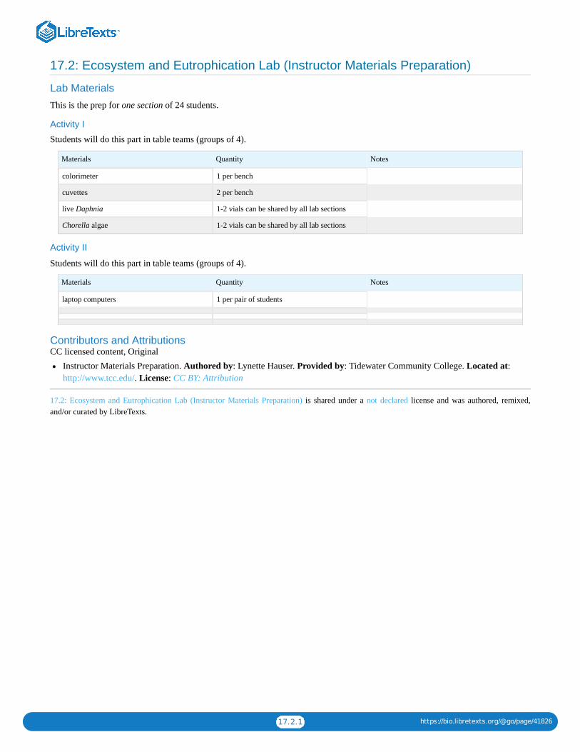
17.2.1 https://bio.libretexts.org/@go/page/41826
17.2: Ecosystem and Eutrophication Lab (Instructor Materials Preparation)
Lab Materials
This is the prep for one section of 24 students.
Activity I
Students will do this part in table teams (groups of 4).
Materials Quantity Notes
colorimeter 1 per bench
cuvettes 2 per bench
live Daphnia 1-2 vials can be shared by all lab sections
Chorella algae 1-2 vials can be shared by all lab sections
Activity II
Students will do this part in table teams (groups of 4).
Materials Quantity Notes
laptop computers 1 per pair of students
Contributors and AttributionsCC licensed content, Original
Instructor Materials Preparation. Authored by: Lynette Hauser. Provided by: Tidewater Community College. Located at:http://www.tcc.edu/. License: CC BY: Attribution
17.2: Ecosystem and Eutrophication Lab (Instructor Materials Preparation) is shared under a not declared license and was authored, remixed,and/or curated by LibreTexts.

1
CHAPTER OVERVIEW
18: Module 15- Sample Lab Report18.1: Sample Lab Report- Sugar Size and Diffusion Through a Mock-Cell Membrane18.2: Rubric
18: Module 15- Sample Lab Report is shared under a not declared license and was authored, remixed, and/or curated by LibreTexts.

18.1.1 https://bio.libretexts.org/@go/page/41827
18.1: Sample Lab Report- Sugar Size and Diffusion Through a Mock-Cell MembraneBIO 101L
Instructor: L. Hauser
IntroductionDiffusion is the process in which a substance moves from an area of high concentration to an area of lower concentration. It isimportant for membranes to be semi-permeable. If membranes were universally permeable, they would not legitimately serve theirpurpose as membranes; certain substances need to be kept out of a cell, and others kept in. If membranes were not at all permeable,there would be no interface between the cell and its environment – effectively starving the cell. Membranes, being selectivelypermeable, allow in nutrients and other necessary substances, and also provide for the purging of cell waste.
This experiment investigates the permeability of cell membranes to various types of sugars: polysaccharides, disaccharides, andpolysaccharides. Dialysis tubing is used to simulate a cell membrane; it is permeable to small molecules and water, but not to largermolecules.
Given the generally larger size of polysaccharides, it is hypothesized that starch will not pass through the dialysis tubing, and thatiodine will pass through the membrane due to the small size of its molecules. Based on the trouble that some people have digestinglactose, it is predicted that it is a polysaccharide or disaccharide and will yield diffusion results similar to starch.
Methods
I) Permeability of cell model to starchThe 100ml graduated cylinder was used to measure out 50ml of tap water; the water was poured into a 250ml beaker. One teaspoonof corn starch was added to the beaker and stirred with a spoon. A 50ml beaker was filled with 50ml of tap water. A piece ofdialysis tubing was placed into the beaker of water until it became soft and pliable. The tubing was then extracted from the beaker;one end was tied closed with dental floss, using a double-knot. The other end of the dialysis tubing was opened; a pipette was usedto fill the dialysis tubing with the starch solution. Another piece of dental floss was used to tie the end of the dialysis tubing closed.A second 250ml beaker was filled halfway with tap water. Another pipette was used to add 15 drops of iodine to the beaker; thesolution was mixed with a spoon. The filled dialysis tube was placed into the 250ml beaker so that the cornstarch mixture wassubmerged in the iodine water mixture. After 15 minutes had passed, results were observed and recorded.
II) Permeability of cell model to lactose
Part I: Determining the type of sugar being tested
The 100ml graduated cylinder was used to measure out 100ml of tap water; the water was poured into a 250ml beaker. Twoteaspoons of the lactose was added to the water, and the solution was stirred with a spoon for thorough mixing. 50ml of theresultant solution was measured out using the 100ml graduated cylinder, and was reserved for Part II.
Twenty drops of Benedict’s reagent were placed in a clean, empty test tube. Twenty drops of the lactose solution were added to thesame test tube, and the solution was heated in a boiling water bath for 2 minutes. The results were then interpreted.
Twenty drops of Barfoed’s reagent were placed in a clean, empty test tube. Twenty drops of lactose solution were added to thesame test tube. The resultant solution was headed in a boiling water bath for 3.5 minutes. The results were then interpreted.
Part II: Permeability of cell model
This method is based on the premise of the unknown sugar being a disaccharide.
A 50ml beaker was filled with 50 ml tap water. A piece of dialysis tubing was placed in the beaker of water and left to soak until itbecame soft and pliable. The dialysis tubing was then removed from the beaker, and one end tied closed with a double-knottedpiece of dental floss. The other end of the dialysis tubing was opened. The tubing was filled with lactose solution (set aside fromPart I); a pipette was used to transfer the solution from the graduated cylinder to the tubing. A second piece of dental floss was usedto tie the other end of the dialysis tubing closed. A second 250ml beaker was filled halfway full with tap water.
15 drops of iodine were added to the tap water in the beaker. The resulting solution was swirled with a spoon to mix it; the colors ofthe baggie solution and the beaker solution were noted. The dialysis tubing baggie was placed in the 250ml beaker so that the

18.1.2 https://bio.libretexts.org/@go/page/41827
lactose solution was submerged in the beaker solution, and left to sit undisturbed for 15 minutes. The color of the baggie solutionwas noted.
The baggie was removed from the beaker and samples of the beaker solution were transferred to separate, appropriately marked testtubes. 20 drops of Bendict’s reagent were added to one test tube, and the tube heated for 2 minutes. The color of the resultingsolution was noted. 20 drops of Barfoed’s reagent were added to the second test tube, and the tube heated for 3.5 minutes. Thecolor of the resulting solution was noted.
ResultsTable 1: Starch experiment results
Solution in baggie Solution in Beaker
Starting color Murky white Clear yellow
Color after 15 minutes Dark purple yellow
In the starch experiment as seen in Table 1, the starch solution inside of the dialysis baggie was initially a murky white color. Thesolution in the beaker, external to the baggie was a clear yellow color. After 15 minutes of submersion in the beaker solution, thebaggie had turned a dark purple color. The beaker solution remained clear and yellow.
In Part I of the lactose experiment, the lactose solution was initially a dark brown color. Benedict’s reagent is pale blue in color.Lactose, mixed and heated with the Benedict’s reagent, yielded a solution of a murky yellow-brown color. Barfoed’s reagent, likeBenedict’s reagent, is pale blue in color. Lactose, mixed and heated with the Barfoed’s reagent, yielded a pale blue solution.
Table 2: Lactose experiment results
Solution in baggie Solution in Beaker
Starting color brown yellow
Color after 15 minutes yellow (? Not given)
In Part II of the lactose experiment, as seen in Table 2, the lactose solution inside of the dialysis baggie was initially dark brown incoloration. The iodine and water solution in the beaker was a clear yellow color. A Benedict’s test on the beaker solution after theexperiment yielded a dark brown liquid; a Barfoed’s test on the beaker solution after the experiment resulted in a clear blue liquid.
Discussion
Permeability of cell model to starch
The starch solution inside of the dialysis baggie went from a murky white color to dark purple; iodine from the beaker solutionmust have diffused into the dialysis baggie, reacting with the starch solution and producing the “positive” dark-purple result,confirming the presence of a polysaccharide inside of the baggie. The beaker solution remained a clear yellow color throughout theexperiment; it can hence be inferred that no polysaccharide was present in the beaker solution at the end of the experiment, and inturn, that no starch diffused out of the baggie and into the beaker solution during the 15-minute soaking.
The experimental hypothesis for this section was correct; starch was unable to diffuse through the cell model, however, iodine wasable to diffuse through the cell model. The discrepancy in permeability is due to the difference in the sizes of iodine and starchmolecules.
Permeability of cell model to lactoseThe Benedict’s test control on lactose yielded a solution that was a murky yellow-brown color; this indicated the presence of amono- or di- saccharide. The Barfoed’s test control on lactose yielded a solution that was pale blue in color, without any redprecipitate; this indicates that no monosaccharaides were present, and in turn, that lactose is a disaccharide. The solution inside ofthe dialysis tubing changed color in the course of the experiment; this implies that iodine diffused into the dialysis tubing andreacted with the lactose solution. The resulting clear yellow color indicates that there were no polysaccharides present inside of thedialysis tubing.

18.1.3 https://bio.libretexts.org/@go/page/41827
A negative Benedict’s test is of blue coloration; a test on the beaker solution after the experiment is a very dark red-orange-browncolor that looks similar to the original lactose in the tubing. A Barfoed’s test on the post-experiment beaker solution was a clearlight blue; no monosaccharaides diffused out into the beaker solution, but this result was irrelevant. The Benedict’s test revealedthat lactose was able to diffuse out of the dialysis baggie, into the beaker solution. If the cell model is reliable, it appears thatlactose is able to diffuse in and out of cells.
The experimental hypothesis for this section appears to have been wrong; the cell model was permeable to lactose.
Overall, the cell model has demonstrated impermeability to large molecules such as polysaccharides, and permeability to smallermolecules such as disaccharides and iodine molecules. Since the model was permeable to a disaccharide, it would be reasonable toinfer that the model will be permeable to monosaccharaides, as they are even smaller in size than disaccharides. Further testingwith a variety of disaccharides should be done, to determine whether lactose is unique or whether the cell model is permeable to alldisaccharides.
Contributors and AttributionsCC licensed content, Original
Biology 102 Labs. Authored by: Lynette Hauser. Provided by: Tidewater Community College. Located at:http://www.tcc.edu/. License: CC BY: Attribution
18.1: Sample Lab Report- Sugar Size and Diffusion Through a Mock-Cell Membrane is shared under a not declared license and was authored,remixed, and/or curated by LibreTexts.

18.2.1 https://bio.libretexts.org/@go/page/41828
18.2: RubricThe following rubric will be used to grade the lab reports:
Excellent 5 points
Satisfactory 2.5 points
Unsatisfactory 0 points
Title Page Contains title, student name,instructor name and section
Missing either instructor name orsection
No title page
Formatting: typed, spacing Typed and single spaced Typed, but not single spaced Not typed
Grammar and Spelling No errors; contains completesentences and no misspellings
A few minor errors in grammarand spelling
Several major errors in grammarand spelling
Formatting: headings Each section has a heading asdescribed in template
Some sections lack headings No headings
Hypothesis Predictions are clearly stated andwritten as a testable statement
Predictions/expected outcomes arenot clearly stated
Not written as a testable statement
MaterialsAll equipment and materialsdescribed; identify variables,controls and constants
Materials incompletely describedNo identification of variables,controls and constants
Procedure Clear step-by step descriptionDescription missing detailsmaking it difficult for anotherscientist to repeat experiment
Description missing so muchdetail it would be impossible torepeat
ResultsClearly written description ofresults comparing controls andvariables
Results are presented but nocomparison between controls andvariables are made
No written description of results
Data tables, graphs or chartsEasy to interpret, clear labels, alldata, including calculatedaverages, included
Disorganized (not easy tounderstand, missing labels) but alldata included
Disorganized and or data clearlymissing
Conclusion
Clearly explains acceptance orrejection of hypothesis using datato support conclusion; identifiessources of error
Accepts or rejects hypothesis butdoes not use data to explain why;or does not identify sources oferror
Does not explain conclusion anddoes not identify sources of error
Total ______out of 50 points
Contributors and AttributionsCC licensed content, Original
Lab Report Grading Rubric. Authored by: Lynette Hauser. Provided by: Tidewater Community College. Located at:http://www.tcc.edu/. License: CC BY: Attribution
18.2: Rubric is shared under a not declared license and was authored, remixed, and/or curated by LibreTexts.

1 https://bio.libretexts.org/@go/page/41829
IndexSspirometer
13.1.4: Cardiovascular and Respiratory Systems Lab

1 https://bio.libretexts.org/@go/page/41830
GlossarySample Word 1 | Sample Definition 1


