Embed Size (px)
Citation preview
Original Article
Bracket base remnants after orthodontic debonding
Matteo Zanarinia; Antonio Graccob; Monica Lattucac; Silvia Marchionnid; Maria Rosaria Gattoe;Giulio Alessandri Bonettif
ABSTRACTObjective: To evaluate whether the debonding procedure leads to restitutio ad integrum of theenamel surface by investigating the presence of enamel within the bracket base remnants afterdebonding.Materials and Methods: Sixty patients who completed orthodontic treatment with fixed applianceswere included. A total of 1068 brackets were microphotographed; the brackets presenting someremnants on the base (n 5 818) were selected and analyzed with ImageJ software to measure theremnant area. From this population a statistically significant sample (n 5 100) was observed undera scanning electron microscope to check for the presence of enamel within the remnants. Energydispersive x-ray spectrometry was also performed to obtain quantitative data.Results: Statistically significant differences in the remnant percentage between arches wereobserved for incisor and canine brackets (P , .0001 and P 5 .022, respectively). From amorphologic analysis of the scanning electron micrographs the bracket bases were categorized in3 groups: group A, bases presenting a thin enamel coat (83%); group B, bases showing sizableenamel fragments (7%); group C, bases with no morphologic evidence of enamel presence (10%).Calcium presence was noted on all evaluated brackets under energy dispersive x-rayspectrometry. No significant difference was observed in the Ca/Si ratio between group A(16.21%) and group B (18.77%), whereas the Ca/Si ratio in group C (5.40%) was significantly lowerthan that of the other groups (P , .323 and P 5 .0001, respectively).Conclusion: The objective of an atraumatic debonding is not achieved yet; in some cases thedamage could be clinically relevant. (Angle Orthod. 2013;83:885–891.)
INTRODUCTION
The main objective of orthodontic debonding is toremove brackets and adhesive resin while restoringthe enamel surface to its pretreatment condition.1–3
Nevertheless, damage to the enamel due to debondinghas been, and still is, a concern to clinicians because itis reported to occur in vitro4–6 and in vivo.7,8
During bracket removal, bond failure can occur atthe adhesive-enamel or at the adhesive-bracketinterface (adhesive failure), or within the adhesive(cohesive failure); generally, a combination of adhe-sive and cohesive failure (mixed failure) takes place.9
A greater risk of damage to the tooth surface occurs incases of adhesive failure between resin and enamel.9–11
This happens especially with the use of ceramicbrackets,12–14 but enamel fracture may also occur withmetal brackets.6,7
There are several important differences between invitro and in vivo studies dealing with modes of bracketfailure. The in vivo debonding load is a combination ofshear, tensile, and torsion force,15,16 whereas in vitrostudies are conducted by means of single tests (shear,tensile, or torsion). Furthermore, the complex oralenvironment involves continually changing tempera-ture, stresses, humidity, acidity, and variability in theamount and composition of plaque. These conditionscannot be reproduced in the laboratory17,18; therefore,
a Visiting Professor, Department of Orthodontics, University ofBologna, Bologna, Italy.
b Assistant Professor, Department of Orthodontics, Universityof Padova, Padova, Italy.
c Postgraduate student, Department of Orthodontics, Univer-sity of Ferrara, Ferrara, Italy.
d Manager of Research, Laboratory of Microscopy, Depart-ment of Oral Sciences, University of Bologna, Bologna, Italy.
e Assistant Professor, Medical Statistics, Department ofOrthodontics, University of Bologna, Bologna, Italy.
f Assistant Professor, Department of Orthodontics, Universityof Bologna, Bologna, Italy.
Corresponding author: Dr Giulio Alessandri Bonetti, Depart-ment of Orthodontics, Alma Mater Studiorum, University ofBologna, Via San Vitale 59, 40125 Bologna, Italy(e-mail: [email protected])
Accepted: February 2013. Submitted: December 2012.Published Online: March 26, 2013G 2013 by The EH Angle Education and Research Foundation,Inc.
DOI: 10.2319/121112-930.1 885 Angle Orthodontist, Vol 83, No 5, 2013
in vitro studies could not be highly relevant from thestandpoint of scientific and clinical evidence.
On the basis that the debonding procedure shouldideally lead to restitutio ad integrum of the enamelsurface, the present ex vivo study was conducted toevaluate whether this assumption is true by investi-gating the presence of enamel within the bracket baseremnants after debonding.
MATERIALS AND METHODS
Sixty consecutive patients (39 females and 21males; mean age, 13 6 2 years) who completedorthodontic treatment with fixed appliances (meanduration, 1.7 6 0.5 years) at the Department ofOrthodontics, University of Bologna, Bologna, Italy,were enrolled in the study. The study protocol wasapproved by the local Institutional Review Board. Thecriteria for patient selection included intact permanentdentition and lack of any decalcification on teeth. Allthe patients underwent orthodontic treatment in botharches using metal brackets (MBT, Victory Series, 3MUnitek, Monrovia, Calif). The labial enamel surfaces ofthe teeth were cleaned and polished with a slow-speedhandpiece using a slurry made of nonfluoride pumiceand water. They were then rinsed and dried with amoisture-free air spray. Subsequently, the enamel wasetched with 35% orthophosphoric acid gel (Scotch-bond, 3M Unitek) for 30 seconds, rinsed with water,air sprayed for 30 seconds, and then dried until theetched enamel surfaces exhibited a frosty-whiteappearance.19,20
The brackets were bonded to the teeth surface usingan adhesive system (Transbond XT Light Cureadhesive primer and Transbond XT adhesive resin,3M Unitek), applied in strict accordance with themanufacturer’s instructions. The brackets were placedon the tooth surface, adjusted to their final position,and pressed firmly in place. The excess resin was thenremoved from the periphery of the bracket base usinga dental probe. Light curing was performed for20 seconds (10 seconds on the mesial side and10 seconds on the distal side) using a light-emittingdiode unit light (Ortholux, 3M Unitek). At the end oforthodontic treatment, a total of 1168 brackets wereremoved using the wings method,21 which involvesgently squeezing the mesial and distal wings withbracket-removal pliers (Ormco Corporation, Glendora,Calif). Because debonding is an operator-dependentprocedure, the results may vary between operators; toavoid this limitation, all clinical procedures were carriedout by only one operator.
A pilot study was conducted on 100 teeth todetermine the sample size, as reported in Table 1.Patient and tooth selection were performed with
pseudo-random numbers in blocks of four digits: thefirst two digits individuate the patient and the last onesindividuate the tooth (incisor, canine, premolar, firstmolar). A single expert examiner trained in micromor-phologic evaluations measured the bracket basesurface and the remnant surface using ImageJ open-source image-analysis software (Version 1.44o forMacintosh, National Institutes of Health, Bethesda,Md) and calculated the latter area as a percentage ofthe former. The outcome resulting from the pilot studywas the remnant percentage. As no significantdifferences were found between the upper and lowerarches, the two arches were unified. The meanpercentage of remnants for each tooth type was usedto compute the final sample size with a permissibleerror of 5% and a power of 95% (Table 1). Becausethe pilot study revealed that the sample size wasstatistically adequate, all 1068 brackets were observedunder a digital stereomicroscope (Meade InstrumentsEurope, GmbH & Co KG, Rhede, Germany) at a 203
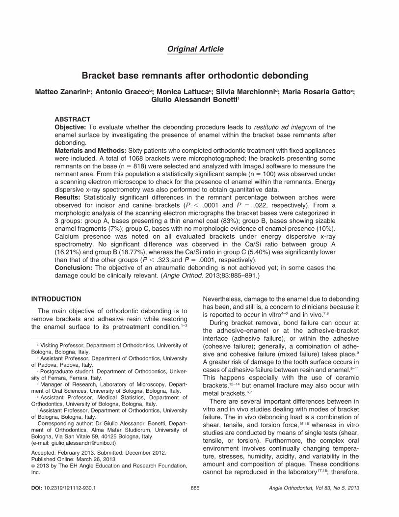
magnification. This procedure was carried out toexclude bases free of resin remnants and to selectonly those with some resin remnants, for scanningelectron microscopy (SEM) analysis. Therefore, 818brackets (283 incisor, 121 canine, 311 premolar, and103 molar) were included in the study. The sameexaminer who analyzed the pilot-study sample mea-sured the bracket base surface and the remnantsurface using ImageJ open-source image-analysissoftware and calculated the latter area as a percentageof the former (Figure 1A,B).
From the 818-bracket population, considering an a-level of 0.01 with a power of 95% and an estimatedpercentage of enamel fractures of 77% (as claimed byStratmann et al.7) it has been computed that at least 60specimens are needed as statistically adequatesample size for SEM analysis.
Using the same randomized allocation procedure asin the initial pilot study, 100 brackets (25 brackets foreach tooth type) were then selected, mounted onaluminum stubs with their bases facing up, sputter-coated with a 300-A layer of gold and palladium(Sputter Coater SC7620, Polaron, East Grinstead,UK), and analyzed using a high-vacuum SEM (JSM-5200, JEOL, Tokyo, Japan) that captured secondary
Table 1. Results of the Pilot Study
Pilot
Sample (n)
Mean
Bracket-Base
Remnants (%)
Sample Size
(n 5 Needed
At Least)
Available
Sample (n)
Incisor 20 28% 310 410
Canine 20 32% 84 184
I Premolar 20 26% 112 187
II Premolar 20 50% 146 177
First Molar 20 49% 96 110
886 ZANARINI, GRACCO, LATTUCA, MARCHIONNI, GATTO, ALESSANDRI BONETTI
Angle Orthodontist, Vol 83, No 5, 2013
electron images at 10–15 kV using a 20-mm workingdistance.
Microphotographs of the bracket base were taken atincreasing magnifications (35, 100, 200, 500, 1000, and2000 times). The 353 magnification allowed examina-tion of the bracket base on the whole; the surface wasthen progressively inspected in detail at higher magni-fication to detect any presence of enamel. Evaluationswere carried out at the same time by two calibratedexaminers. On the basis of SEM analysis, the bracketswere categorized according to the presence on the resinremnants of a thin coat of enamel (group A; n 5 83),sizable enamel fragments (group B; n 5 7), and nomorphologic evidence of enamel (group C; n 5 10).
Because the SEM analysis provides only qualitativeevaluation, the bracket sample was also examinedthrough energy dispersive x-ray spectrometry (EDX) toobtain quantitative data. The EDX analysis (StereoScan 360, Cambridge Instrument Ltd, Cambridge, UK;
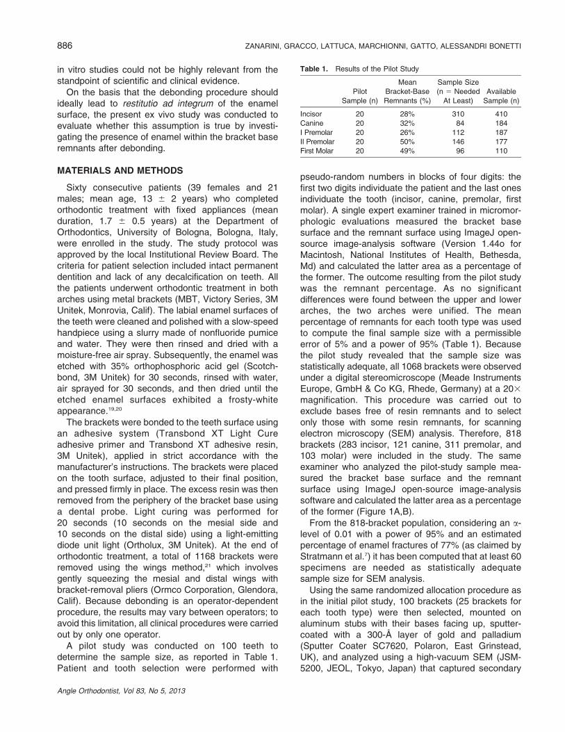
using accelerating voltage, 20kV; type of detector,Si(Li)-liquid N2 cooled; detector dead time 17%;spectra acquisition time, 70 seconds; resolution,133 eV; magnification, between 20 and 303; scanmode, area) was performed on 21 brackets from groupA, 7 brackets from group B, and 10 brackets fromgroup C. The 7 bracket bases from group B werematched to 21 bracket bases from group A (ratio 1:3),which represented the clinical controls. The 10 bracketbases from group C were also analyzed. Thepercentage of calcium was calculated from adhesivein relation to that of silicon. Because the bondingmaterials used are calcium free, the presence of thiselement could only be attributed to enamel. The studydesign is summarized in Figure 2.
Statistical Analysis
The Shapiro-Wilk test was used to test the normalityof the distribution of the primary outcome, that is,remnant percentage, for each tooth in both arches.Because of the significance of the deviation fromnormality (P 5 .0001), median and interquartile rangeswere presented to describe the data. The Mann-Whitney U-test was applied to compare the percent-ages of remnants for each tooth type between thearches. The a-level was set at 0.05.
As regards EDX examination, the Shapiro-Wilk testevidenced no significant deviation from normality of theCa/Si ratio in each of the three groups (A, B, and C).Consequently, the comparison between them wascarried out by using a t-test for independent samples,after having controlled the homoschedasticity of thevariances by means of the Levene test.
RESULTS
The remnant areas of the 818 bracket basesanalyzed with the use of ImageJ are reported inTable 2 and expressed as a percentage of the bracketbase surface. The maxillary incisor and maxillarycanine brackets exhibited the lowest median values(4.48% and 3.57%, respectively); first molar brackets,either maxillary or mandibular, displayed the highestmedian values (95.57% and 91.64%, respectively).The Mann-Whitney U-test did not show a statisticallysignificant difference between upper and lower archesfor premolar (P 5 .767) or molar brackets (P 5 .220),whereas statistically significant differences betweenarches were observed for incisor (P 5 .0001) andcanine brackets (P 5 .022).
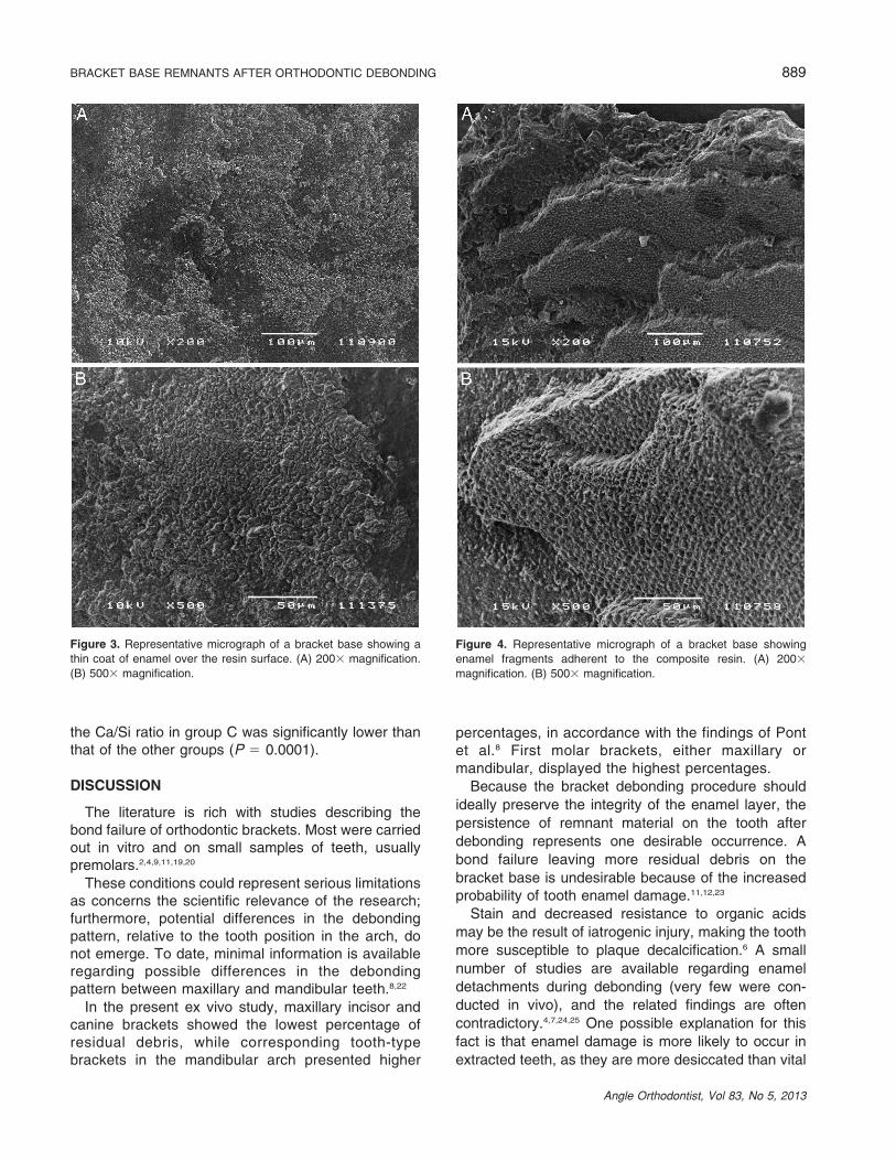
As regards the 100-bracket sample, from a morpho-logic analysis of the SEM micrographs, two modes ofenamel presence emerged: (1) a thin coat of enamelon the resin surface was found in 83 specimens (83%)(group A) (Figure 3A,B); and (2) sizable enamel
Figure 1. Representative example of the bracket-base remnant-area
measurement. (A) A bracket base showing some remnants. (B) The
remnant area detected with the use of Image J.
BRACKET BASE REMNANTS AFTER ORTHODONTIC DEBONDING 887
Angle Orthodontist, Vol 83, No 5, 2013
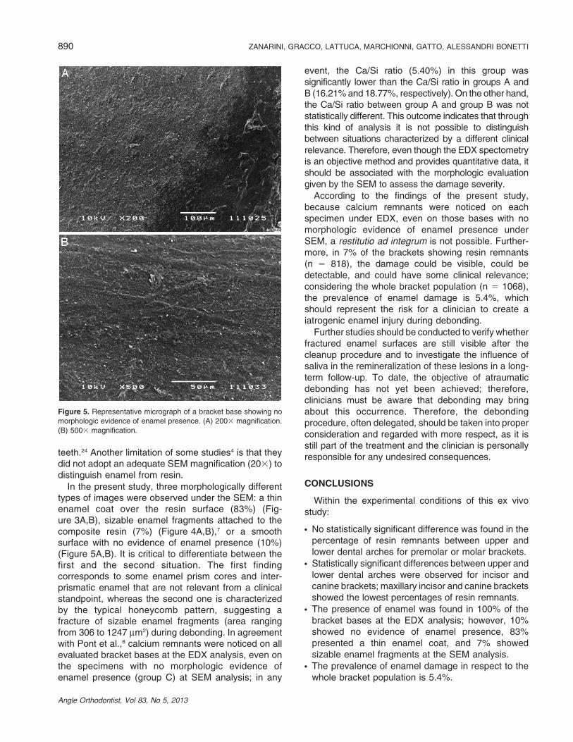
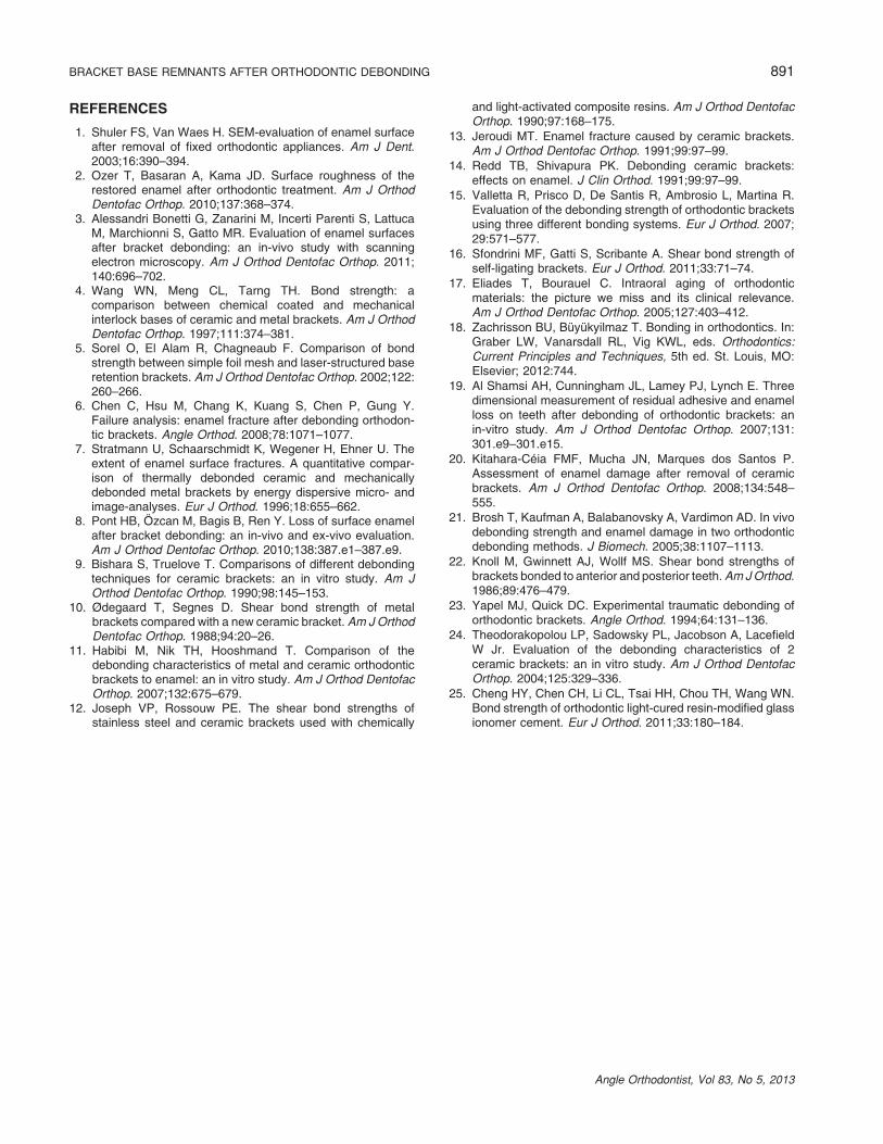
fragments (area ranging from 306 to 1247 mm2) on theresin were detected in 7 samples (7%) (group B)(Figure 4A,B), located on canine (n 5 2), premolar(n 5 3), and molar brackets (n 5 2). No evidence ofenamel was visible on 10 samples (10%) (group C)(Figure 5A,B), whereas EDX analysis showed the
presence of calcium on all specimens. The meanCa/Si ratio was 16.21 6 5.81% for group A, 18.77 6
5.79% for group B, and 5.40 6 2.02% for group C.Based on the statistical analysis (t-test for independentsamples), no significant difference was observed in theCa/Si ratio between group A and group B (P 5 0.323);
Figure 2. Graphic representation of the study design; bkts indicates brackets.
Table 2. Percentages of Bracket-base Remnants by Tooth Type
Median (%) Interquartile Range (%) Significance Between Arches (P)
Maxillary incisor 4.48 0–59.53 .0001
Mandibular incisor 48.96 8.36–87.49
Maxillary canine 3.57 0–52.19 .022
Mandibular canine 28.87 0–75.03
Maxillary premolar 41.14 6.32–85.70 .767
Mandibular premolar 46.51 3.65–82.98
Maxillary first molar 95.57 67.00–98.35 .220
Mandibular first molar 91.64 57.04–97.29
888 ZANARINI, GRACCO, LATTUCA, MARCHIONNI, GATTO, ALESSANDRI BONETTI
Angle Orthodontist, Vol 83, No 5, 2013
the Ca/Si ratio in group C was significantly lower thanthat of the other groups (P 5 0.0001).
DISCUSSION
The literature is rich with studies describing thebond failure of orthodontic brackets. Most were carriedout in vitro and on small samples of teeth, usuallypremolars.2,4,9,11,19,20
These conditions could represent serious limitationsas concerns the scientific relevance of the research;furthermore, potential differences in the debondingpattern, relative to the tooth position in the arch, donot emerge. To date, minimal information is availableregarding possible differences in the debondingpattern between maxillary and mandibular teeth.8,22
In the present ex vivo study, maxillary incisor andcanine brackets showed the lowest percentage ofresidual debris, while corresponding tooth-typebrackets in the mandibular arch presented higher
percentages, in accordance with the findings of Pontet al.8 First molar brackets, either maxillary ormandibular, displayed the highest percentages.
Because the bracket debonding procedure should
ideally preserve the integrity of the enamel layer, the
persistence of remnant material on the tooth after
debonding represents one desirable occurrence. A
bond failure leaving more residual debris on the
bracket base is undesirable because of the increasedprobability of tooth enamel damage.11,12,23
Stain and decreased resistance to organic acids
may be the result of iatrogenic injury, making the tooth
more susceptible to plaque decalcification.6 A small
number of studies are available regarding enamel
detachments during debonding (very few were con-
ducted in vivo), and the related findings are often
contradictory.4,7,24,25 One possible explanation for this
fact is that enamel damage is more likely to occur in
extracted teeth, as they are more desiccated than vital
Figure 3. Representative micrograph of a bracket base showing a
thin coat of enamel over the resin surface. (A) 2003 magnification.
(B) 5003 magnification.
Figure 4. Representative micrograph of a bracket base showing
enamel fragments adherent to the composite resin. (A) 2003
magnification. (B) 5003 magnification.
BRACKET BASE REMNANTS AFTER ORTHODONTIC DEBONDING 889
Angle Orthodontist, Vol 83, No 5, 2013
teeth.24 Another limitation of some studies4 is that theydid not adopt an adequate SEM magnification (203) todistinguish enamel from resin.
In the present study, three morphologically differenttypes of images were observed under the SEM: a thinenamel coat over the resin surface (83%) (Fig-ure 3A,B), sizable enamel fragments attached to thecomposite resin (7%) (Figure 4A,B),7 or a smoothsurface with no evidence of enamel presence (10%)(Figure 5A,B). It is critical to differentiate between thefirst and the second situation. The first findingcorresponds to some enamel prism cores and inter-prismatic enamel that are not relevant from a clinicalstandpoint, whereas the second one is characterizedby the typical honeycomb pattern, suggesting afracture of sizable enamel fragments (area rangingfrom 306 to 1247 mm2) during debonding. In agreementwith Pont et al.,8 calcium remnants were noticed on allevaluated bracket bases at the EDX analysis, even onthe specimens with no morphologic evidence ofenamel presence (group C) at SEM analysis; in any
event, the Ca/Si ratio (5.40%) in this group wassignificantly lower than the Ca/Si ratio in groups A andB (16.21% and 18.77%, respectively). On the other hand,the Ca/Si ratio between group A and group B was notstatistically different. This outcome indicates that throughthis kind of analysis it is not possible to distinguishbetween situations characterized by a different clinicalrelevance. Therefore, even though the EDX spectometryis an objective method and provides quantitative data, itshould be associated with the morphologic evaluationgiven by the SEM to assess the damage severity.
According to the findings of the present study,because calcium remnants were noticed on eachspecimen under EDX, even on those bases with nomorphologic evidence of enamel presence underSEM, a restitutio ad integrum is not possible. Further-more, in 7% of the brackets showing resin remnants(n 5 818), the damage could be visible, could bedetectable, and could have some clinical relevance;considering the whole bracket population (n 5 1068),the prevalence of enamel damage is 5.4%, whichshould represent the risk for a clinician to create aiatrogenic enamel injury during debonding.
Further studies should be conducted to verify whetherfractured enamel surfaces are still visible after thecleanup procedure and to investigate the influence ofsaliva in the remineralization of these lesions in a long-term follow-up. To date, the objective of atraumaticdebonding has not yet been achieved; therefore,clinicians must be aware that debonding may bringabout this occurrence. Therefore, the debondingprocedure, often delegated, should be taken into properconsideration and regarded with more respect, as it isstill part of the treatment and the clinician is personallyresponsible for any undesired consequences.
CONCLUSIONS
Within the experimental conditions of this ex vivostudy:
N No statistically significant difference was found in thepercentage of resin remnants between upper andlower dental arches for premolar or molar brackets.
N Statistically significant differences between upper andlower dental arches were observed for incisor andcanine brackets; maxillary incisor and canine bracketsshowed the lowest percentages of resin remnants.
N The presence of enamel was found in 100% of thebracket bases at the EDX analysis; however, 10%showed no evidence of enamel presence, 83%presented a thin enamel coat, and 7% showedsizable enamel fragments at the SEM analysis.
N The prevalence of enamel damage in respect to thewhole bracket population is 5.4%.
Figure 5. Representative micrograph of a bracket base showing no
morphologic evidence of enamel presence. (A) 2003 magnification.
(B) 5003 magnification.
890 ZANARINI, GRACCO, LATTUCA, MARCHIONNI, GATTO, ALESSANDRI BONETTI
Angle Orthodontist, Vol 83, No 5, 2013
REFERENCES
1. Shuler FS, Van Waes H. SEM-evaluation of enamel surfaceafter removal of fixed orthodontic appliances. Am J Dent.2003;16:390–394.
2. Ozer T, Basaran A, Kama JD. Surface roughness of therestored enamel after orthodontic treatment. Am J OrthodDentofac Orthop. 2010;137:368–374.
3. Alessandri Bonetti G, Zanarini M, Incerti Parenti S, LattucaM, Marchionni S, Gatto MR. Evaluation of enamel surfacesafter bracket debonding: an in-vivo study with scanningelectron microscopy. Am J Orthod Dentofac Orthop. 2011;140:696–702.
4. Wang WN, Meng CL, Tarng TH. Bond strength: acomparison between chemical coated and mechanicalinterlock bases of ceramic and metal brackets. Am J OrthodDentofac Orthop. 1997;111:374–381.
5. Sorel O, El Alam R, Chagneaub F. Comparison of bondstrength between simple foil mesh and laser-structured baseretention brackets. Am J Orthod Dentofac Orthop. 2002;122:260–266.
6. Chen C, Hsu M, Chang K, Kuang S, Chen P, Gung Y.Failure analysis: enamel fracture after debonding orthodon-tic brackets. Angle Orthod. 2008;78:1071–1077.
7. Stratmann U, Schaarschmidt K, Wegener H, Ehner U. Theextent of enamel surface fractures. A quantitative compar-ison of thermally debonded ceramic and mechanicallydebonded metal brackets by energy dispersive micro- andimage-analyses. Eur J Orthod. 1996;18:655–662.
8. Pont HB, Ozcan M, Bagis B, Ren Y. Loss of surface enamelafter bracket debonding: an in-vivo and ex-vivo evaluation.Am J Orthod Dentofac Orthop. 2010;138:387.e1–387.e9.
9. Bishara S, Truelove T. Comparisons of different debondingtechniques for ceramic brackets: an in vitro study. Am JOrthod Dentofac Orthop. 1990;98:145–153.
10. Ødegaard T, Segnes D. Shear bond strength of metalbrackets compared with a new ceramic bracket. Am J OrthodDentofac Orthop. 1988;94:20–26.
11. Habibi M, Nik TH, Hooshmand T. Comparison of thedebonding characteristics of metal and ceramic orthodonticbrackets to enamel: an in vitro study. Am J Orthod DentofacOrthop. 2007;132:675–679.
12. Joseph VP, Rossouw PE. The shear bond strengths ofstainless steel and ceramic brackets used with chemically
and light-activated composite resins. Am J Orthod DentofacOrthop. 1990;97:168–175.
13. Jeroudi MT. Enamel fracture caused by ceramic brackets.Am J Orthod Dentofac Orthop. 1991;99:97–99.
14. Redd TB, Shivapura PK. Debonding ceramic brackets:effects on enamel. J Clin Orthod. 1991;99:97–99.
15. Valletta R, Prisco D, De Santis R, Ambrosio L, Martina R.Evaluation of the debonding strength of orthodontic bracketsusing three different bonding systems. Eur J Orthod. 2007;29:571–577.
16. Sfondrini MF, Gatti S, Scribante A. Shear bond strength ofself-ligating brackets. Eur J Orthod. 2011;33:71–74.
17. Eliades T, Bourauel C. Intraoral aging of orthodonticmaterials: the picture we miss and its clinical relevance.Am J Orthod Dentofac Orthop. 2005;127:403–412.
18. Zachrisson BU, Buyukyilmaz T. Bonding in orthodontics. In:Graber LW, Vanarsdall RL, Vig KWL, eds. Orthodontics:Current Principles and Techniques, 5th ed. St. Louis, MO:Elsevier; 2012:744.
19. Al Shamsi AH, Cunningham JL, Lamey PJ, Lynch E. Threedimensional measurement of residual adhesive and enamelloss on teeth after debonding of orthodontic brackets: anin-vitro study. Am J Orthod Dentofac Orthop. 2007;131:301.e9–301.e15.
20. Kitahara-Ceia FMF, Mucha JN, Marques dos Santos P.Assessment of enamel damage after removal of ceramicbrackets. Am J Orthod Dentofac Orthop. 2008;134:548–555.
21. Brosh T, Kaufman A, Balabanovsky A, Vardimon AD. In vivodebonding strength and enamel damage in two orthodonticdebonding methods. J Biomech. 2005;38:1107–1113.
22. Knoll M, Gwinnett AJ, Wollf MS. Shear bond strengths ofbrackets bonded to anterior and posterior teeth. Am J Orthod.1986;89:476–479.
23. Yapel MJ, Quick DC. Experimental traumatic debonding oforthodontic brackets. Angle Orthod. 1994;64:131–136.
24. Theodorakopolou LP, Sadowsky PL, Jacobson A, LacefieldW Jr. Evaluation of the debonding characteristics of 2ceramic brackets: an in vitro study. Am J Orthod DentofacOrthop. 2004;125:329–336.
25. Cheng HY, Chen CH, Li CL, Tsai HH, Chou TH, Wang WN.Bond strength of orthodontic light-cured resin-modified glassionomer cement. Eur J Orthod. 2011;33:180–184.
BRACKET BASE REMNANTS AFTER ORTHODONTIC DEBONDING 891
Angle Orthodontist, Vol 83, No 5, 2013


![Supernova remnants and [gamma]-ray sources](https://img.pdfslide.net/doc/110x75/633b210257069baea508f7c3/supernova-remnants-and-gamma-ray-sources.jpg)


