Embed Size (px)
Citation preview
REVIEW
Brain changes in early-onset bipolar and unipolar depressivedisorders: a systematic review in children and adolescents
Gianluca Serafini • Maurizio Pompili • Stefan Borgwardt •
Josselin Houenou • Pierre Alexis Geoffroy • Renaud Jardri •
Paolo Girardi • Mario Amore
Received: 23 December 2013 / Accepted: 29 August 2014 / Published online: 12 September 2014
� Springer-Verlag Berlin Heidelberg 2014
Abstract Pediatric bipolar disorder (BD) and unipolar
disorder (UD) share common symptomatic and functional
impairments. Various brain imaging techniques have been
used to investigate the integrity of brain white matter (WM)
and gray matter (GM) in these disorders. Despite promising
preliminary findings, it is still unclear whether these altera-
tions may be considered as common trait markers or may be
used to distinguish BD from UD. A systematic literature
search of studies between 1980 and September 2013 which
reported WM/GM changes in pediatric and adolescent BD/
UD, as detected by diffusion tensor imaging and voxel-based
analysis was conducted. Of the 34 articles judged as eligible,
17 fulfilled our inclusion criteria and were finally retained in
this review. More abnormalities have been documented in
the brains of children and adolescents with BD than UD.
Reductions in the volume of basal ganglia and the hippo-
campus appeared more specific for pediatric UD, whereas
reduced corpus callosum volume and increased rates of deep
WM hyperintensities were more specific for pediatric BD.
Seminal papers failed to address the possibility that the
differences between unipolar and bipolar samples might be
related to illness severity, medication status, comorbidity or
diagnosis. UD and BD present both shared and distinctive
impairments in the WM and GM compartments. More WM
abnormalities have been reported in children and adolescents
with bipolar disease than in those with unipolar disease,
maybe as a result of a low number of DTI studies in pediatric
UD. Future longitudinal studies should investigate whether
neurodevelopmental changes are diagnosis-specific.
Keywords VBM/DTI analyses � Children/adolescents �Pediatric bipolar disorder � Unipolar depressive disorder �White matter/gray matter abnormalities
Introduction
Bipolar disorder (BD) is a chronic and disabling disorder
characterized by relevant impairments in social, emotional,
and academic functioning in childhood and early
G. Serafini (&) � M. Amore
Department of Neuroscience, Rehabilitation, Ophthalmology,
Genetics, Maternal and Child Health (DINOGMI),
Section of Psychiatry, University of Genoa,
IRCCS San Martino, Largo Rosanna Benzi 10,
16100 Genoa, Italy
e-mail: [email protected]
M. Pompili � P. Girardi
Department of Neurosciences, Mental Health and Sensory
Organs, Sant’Andrea Hospital, Sapienza University of Rome,
Rome, Italy
S. Borgwardt
Department of Psychiatry, University of Basel, Basel,
Switzerland
J. Houenou
Inserm, U955, Equipe 15 « Psychiatrie Genetique », Fondation
Fondamental, AP-HP, Hopitaux Universitaires Henri Mondor,
Pole de psychiatrie, 94000 Creteil, France
J. Houenou
Neurospin, Uniact Lab, CEA Saclay, Gif-Sur-Yvette, France
P. A. Geoffroy � R. Jardri
Laboratoire de Sciences Cognitives & Affectives (SCA-Lab),
PSYChiC team, CNRS, Universite Lille Nord de France,
59000 Lille, France
R. Jardri
Lille University Medical Centre (CHU Lille), 59000 Lille,
France
123
Eur Child Adolesc Psychiatry (2014) 23:1023–1041
DOI 10.1007/s00787-014-0614-z
adolescence [1, 2]. The prevalence rate of bipolar spectrum
disorder has been estimated to be 2 % among children and
adolescents in the community [3]. Early-onset BD is usu-
ally associated with poorer performance, comorbidity with
substance abuse, suicide attempts, hospitalization, and
legal difficulties [4–7]. Similarly, major depressive disor-
der (MDD) in children and adolescents is associated with a
fourfold increased risk of recurrence in adulthood, long-
term functional impairment and adult disability [8, 9].This
poor outcome in children and adolescents with unipolar
disorder (UD) or BD emphasizes the need for adequate
understanding of the mechanisms underlying these affec-
tive conditions [10–14].
Although many studies attempted to elucidate the
neurobiological basis of affective disorders in children and
adolescents, their pathogenesis remains unclear. During the
last decades, brain imaging techniques provided new
approaches to detect in vivo structural and functional brain
changes. Magnetic resonance imaging studies reported a
wide range of morphometric alterations in both pediatric
BD [15–20] and UD [21–27].
Diffused GM abnormalities have been found in patients
with pediatric BD, specifically volumetric changes in the
anterior limbic network (ALN), including the prefrontal
regions, thalamus, striatum, amygdala, hippocampal com-
plex, and the midline cerebellum [28].
In their meta-analysis, Arnone et al. [29] found that
reduced prefrontal cortex and increased globus pallidus
volumes are some of the most consistent findings in chil-
dren and young adults with BD. There is also evidence that
adolescents with a recent single manic episode exhibited
smaller subgenual cingulate cortex volumes than healthy
controls (HC) [30], whereas drug-naıve adult individuals
with a first-episode depression showed reduced GM vol-
ume in the right pre-supplementary motor area [31].
Reduced GM has also been reported in the prefrontal
cortex, amygdala, and hippocampi of depressed children.
However, these results were inconsistent, due to hetero-
geneous samples and differences in terms of gender, illness
duration, severity of symptoms, medication, or episode
states [32–34].
A second line of studies explored white matter (WM)
integrity in these disorders using diffusion tensor imaging
(DTI) [35]. Studies using a region-of-interest (ROI)
approach reported abnormalities in prefrontal–limbic cir-
cuits that have been associated with affective dysregulation
in BD [33]. Specifically, lower fractional anisotropy (FA)
has been found in the anterior corona radiata (ACR) [36]
and superior frontal WM in pediatric BD [37]. Recently,
studies using either voxel-based morphometry (VBM)
analysis or tract-based spatial statistics (TBSS) reported
WM abnormalities and lower FA in several brain regions
[38–40].
Similarly, DTI studies conducted in pediatric UD
reported microstructural WM abnormalities during the first
episode of depression in both depressed young adults [41]
and adolescents [42].
There are also some neuroimaging studies that were
conducted among unaffected but high risk of BD/MDD
individuals aimed to reveal WM changes [43–45].
Few studies in the current literature examined whether
some VBM/DTI abnormalities may help in distinguishing
individuals with UD or BD. For example, Cardoso de
Almeida and Phillips [46] suggested more widespread
abnormalities in WM connectivity and more WM hyper-
intensities in BD than UD, more habenula volume reduc-
tions in BD but not UD, and differential patterns of
functional abnormalities in emotion regulation and atten-
tional control neural circuitry in both BD and UD. There is,
however, the pressing need for more neuroimaging studies
using larger samples sizes, and comparing BD and UD
depressed subjects.
In this context, we aimed to systematically review the
current literature to determine whether pediatric/adolescent
BD is associated with greater or more consistent WM or
GM alterations than pediatric/adolescent UD.
Methods
Information sources, search strategy, and study
selection
A detailed search strategy was used to identify relevant
studies. In order to provide a new and timely critical review
of VBM/DTI abnormalities and their possible involvement
in children/adolescents with UD or BD, we performed a
systematic PubMed/Medline, Scopus, and Science Direct
search to identify all papers and book chapters in the
English language during the period between 1980 and
January 2014.
The search used first the following terms: ‘‘Voxel-based
morphometry analysis’’, OR ‘‘VBM analysis’’, AND
‘‘Diffusion tensor imaging’’ OR ‘‘DTI’’ AND ‘‘White
matter hyperintensities’’ OR ‘‘White matter lesions’’ OR
‘‘White matter abnormalities’’ OR ‘‘White matter changes
signals’’ AND ‘‘Grey matter hyperintensities’’ OR ‘‘Grey
matter lesions’’ OR ‘‘Grey matter abnormalities’’ to
investigate ‘‘pediatric and adolescent samples with bipolar
disorder’’ OR ‘‘PBD’’ OR ‘‘Bipolar disorder in children
and adolescents’’ AND subsequently the same terms to
investigate ‘‘Pediatric and adolescent samples of unipolar
disorders’’ OR ‘‘Unipolar disorders in children and ado-
lescents’’. When a title or abstract seemed to describe a
study eligible for inclusion, the full article was examined to
assess its relevance based on the inclusion criteria. Two
1024 Eur Child Adolesc Psychiatry (2014) 23:1023–1041
123
blinded, independent researchers (GS and MP) conducted a
two-step literature search. Any discrepancies between the
two reviewers were resolved by consultations with the
senior authors (JH, MA, RJ, PG). The reference lists of the
articles were also manually checked for relevant studies
while other publications were cross-referenced for any
additional published articles. Only English language full-
text articles reporting original data about the main topic
were included.
Study design and eligibility criteria
We followed the Preferred Reporting Items for Systematic
Reviews and Meta-Analyses’ (PRISMA) guidelines [47].
Studies were included according to the following criteria:
(a) being an original paper in a peer-reviewed journal and
(b) containing an analysis of VBM/DTI abnormalities in
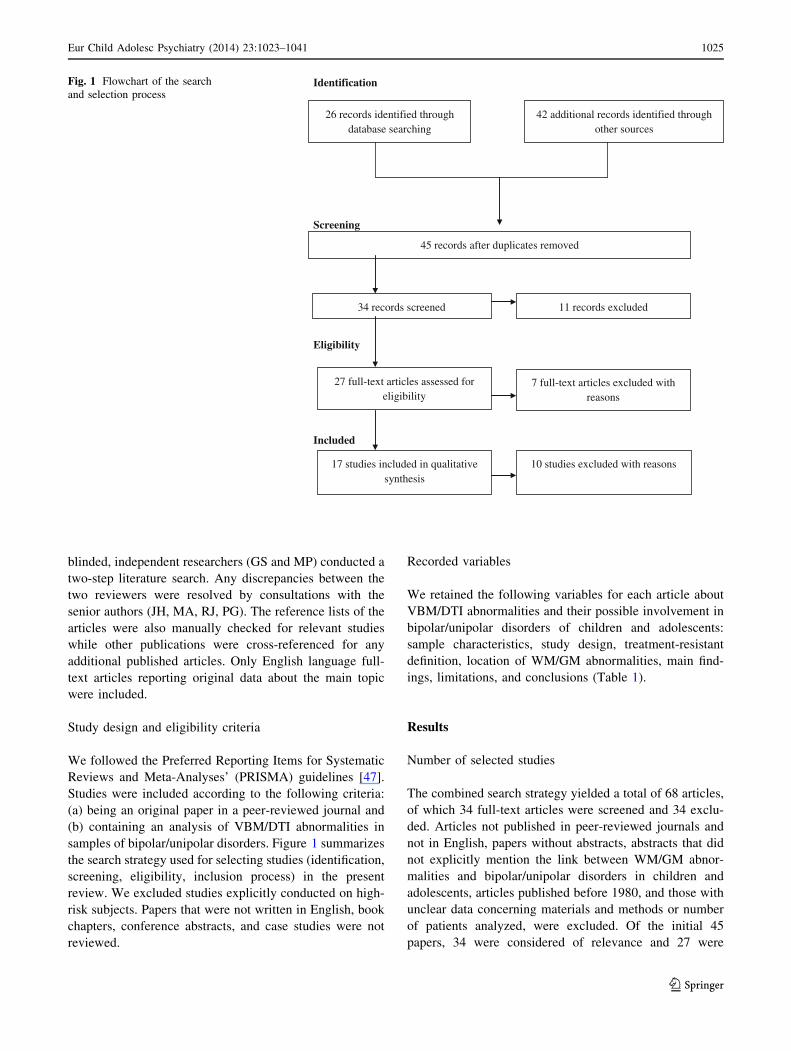
samples of bipolar/unipolar disorders. Figure 1 summarizes
the search strategy used for selecting studies (identification,
screening, eligibility, inclusion process) in the present
review. We excluded studies explicitly conducted on high-
risk subjects. Papers that were not written in English, book
chapters, conference abstracts, and case studies were not
reviewed.
Recorded variables
We retained the following variables for each article about
VBM/DTI abnormalities and their possible involvement in
bipolar/unipolar disorders of children and adolescents:
sample characteristics, study design, treatment-resistant
definition, location of WM/GM abnormalities, main find-
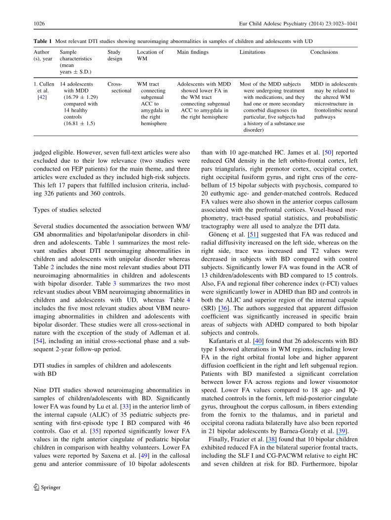
ings, limitations, and conclusions (Table 1).
Results
Number of selected studies
The combined search strategy yielded a total of 68 articles,
of which 34 full-text articles were screened and 34 exclu-
ded. Articles not published in peer-reviewed journals and
not in English, papers without abstracts, abstracts that did
not explicitly mention the link between WM/GM abnor-
malities and bipolar/unipolar disorders in children and
adolescents, articles published before 1980, and those with
unclear data concerning materials and methods or number
of patients analyzed, were excluded. Of the initial 45
papers, 34 were considered of relevance and 27 were
Identification
Screening
Eligibility
Included
26 records identified through database searching
42 additional records identified through other sources
45 records after duplicates removed
34 records screened 11 records excluded
27 full-text articles assessed for eligibility
17 studies included in qualitative synthesis
7 full-text articles excluded with reasons
10 studies excluded with reasons
Fig. 1 Flowchart of the search
and selection process
Eur Child Adolesc Psychiatry (2014) 23:1023–1041 1025
123
judged eligible. However, seven full-text articles were also
excluded due to their low relevance (two studies were
conducted on FEP patients) for the main theme, and three
articles were excluded as they included high-risk subjects.
This left 17 papers that fulfilled inclusion criteria, includ-
ing 326 patients and 360 controls.
Types of studies selected
Several studies documented the association between WM/
GM abnormalities and bipolar/unipolar disorders in chil-
dren and adolescents. Table 1 summarizes the most rele-
vant studies about DTI neuroimaging abnormalities in
children and adolescents with unipolar disorder whereas
Table 2 includes the nine most relevant studies about DTI
neuroimaging abnormalities in children and adolescents
with bipolar disorder. Table 3 summarizes the two most
relevant studies about VBM neuroimaging abnormalities in
children and adolescents with UD, whereas Table 4
includes the five most relevant studies about VBM neuro-
imaging abnormalities in children and adolescents with
bipolar disorder. These studies were all cross-sectional in
nature with the exception of the study of Adleman et al.
[54], including an initial cross-sectional phase and a sub-
sequent 2-year follow-up period.
DTI studies in samples of children and adolescents
with BD
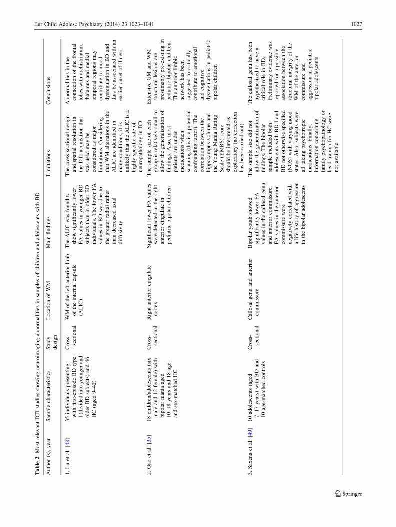
Nine DTI studies showed neuroimaging abnormalities in
samples of children/adolescents with BD. Significantly
lower FA was found by Lu et al. [33] in the anterior limb of
the internal capsule (ALIC) of 35 pediatric subjects pre-
senting with first-episode type I BD compared with 46
controls. Gao et al. [35] reported significantly lower FA
values in the right anterior cingulate of pediatric bipolar
children in comparison with healthy volunteers. Lower FA
values were reported by Saxena et al. [49] in the callosal
genu and anterior commissure of 10 bipolar adolescents
than with 10 age-matched HC. James et al. [50] reported
reduced GM density in the left orbito-frontal cortex, left
pars triangularis, right premotor cortex, occipital cortex,
right occipital fusiform gyrus, and right crus of the cere-
bellum of 15 bipolar subjects with psychosis, compared to
20 euthymic age- and gender-matched controls. Reduced
FA values were also shown in the anterior corpus callosum
associated with the prefrontal cortices. Voxel-based mor-
phometry, tract-based spatial statistics, and probabilistic
tractography were all used to analyze the DTI data.
Gonenc et al. [51] suggested that FA was reduced and
radial diffusivity increased on the left side, whereas on the
right side, trace was increased and T2 values were
decreased in subjects with BD compared with control
subjects. Significantly lower FA was found in the ACR of
13 children/adolescents with BD compared to 15 controls.
Also, FA and regional fiber coherence index (r-FCI) values
were significantly lower in ADHD than BD and controls in
both the ALIC and superior region of the internal capsule
(SRI) [36]. The authors suggested that apparent diffusion
coefficient was significantly increased in specific brain
areas of subjects with ADHD compared to both bipolar
subjects and controls.
Kafantaris et al. [40] found that 26 adolescents with BD
type I showed alterations in WM regions, including lower
FA in the right orbital frontal lobe and higher apparent
diffusion coefficient in the right and left subgenual region.
Patients with BD manifested a significant correlation
between lower FA across regions and lower visuomotor
speed. Lower FA values compared to 18 age- and IQ-
matched controls in the fornix, left mid-posterior cingulate
gyrus, throughout the corpus callosum, in fibers extending
from the fornix to the thalamus, and in parietal and
occipital corona radiata bilaterally have also been reported
in 21 bipolar adolescents by Barnea-Goraly et al. [39].
Finally, Frazier et al. [38] found that 10 bipolar children
exhibited reduced FA in the bilateral superior frontal tracts,
including the SLF I and CG-PACWM relative to eight HC
and seven children at risk for BD. Furthermore, bipolar
Table 1 Most relevant DTI studies showing neuroimaging abnormalities in samples of children and adolescents with UD
Author
(s), year
Sample
characteristics
(mean
years ± S.D.)
Study
design
Location of
WM
Main findings Limitations Conclusions
1. Cullen
et al.
[42]
14 adolescents
with MDD
(16.79 ± 1.29)
compared with
14 healthy
controls
(16.81 ± 1.5)
Cross-
sectional
WM tract
connecting
subgenual
ACC to
amygdala in
the right
hemisphere
Adolescents with MDD
showed lower FA in
the WM tract
connecting subgenual
ACC to amygdala in
the right hemisphere
Most of the MDD subjects
were undergoing treatment
with medications, and they
had one or more secondary
comorbid diagnoses (in
particular, five subjects had
a history of a substance use
disorder)
MDD in adolescents
may be related to
the altered WM
microstructure in
frontolimbic neural
pathways
1026 Eur Child Adolesc Psychiatry (2014) 23:1023–1041
123
Ta
ble
2M
ost
rele
van
tD
TI
stu
die
ssh
ow
ing
neu
roim
agin
gab
no
rmal
itie
sin
sam
ple
so
fch
ild
ren
and
ado
lesc
ents
wit
hB
D
Au
tho
r(s
),y
ear
Sam
ple
char
acte
rist
ics
Stu
dy
des
ign
Lo
cati
on
of
WM
Mai
nfi
nd
ing
sL
imit
atio
ns
Co
ncl
usi
on
s
1.
Lu
etal
.[4
8]
35
ind
ivid
ual
sp
rese
nti
ng
wit
hfi
rst-
epis
od
eB
Dty
pe
I(d
ivid
edin
toy
ou
ng
eran
d
old
erB
Dsu
bje
cts)
and
46
HC
(ag
ed9
–4
2)
Cro
ss-
sect
ion
al
WM
of
the
left
ante
rio
rli
mb
of
the
inte
rnal
cap
sule
(AL
IC)
Th
eA
LIC
was
fou
nd
to
sho
wsi
gn
ifica
ntl
ylo
wer
FA
val
ues
iny
ou
ng
erB
D
sub
ject
sth
anin
old
erB
D
ind
ivid
ual
s.T
he
low
erF
A
val
ues
inB
Dw
asd
ue
to
the
gre
ater
rad
ial
rath
er
than
dec
reas
edax
ial
dif
fusi
vit
y
Th
ecr
oss
-sec
tio
nal
des
ign
and
spat
ial
reso
luti
on
in
the
DT
Iac
qu
isit
ion
that
was
use
dm
ayb
e
con
sid
ered
asm
ajo
r
lim
itat
ion
s.C
on
sid
erin
g
that
WM
alte
rati
on
sin
the
AL
ICar
eid
enti
fied
in
man
yco
nd
itio
ns,
itis
un
lik
ely
that
the
AL
ICis
a
hig
hly
spec
ific
site
of
neu
rop
ath
olo
gy
inB
D
Ab
no
rmal
itie
sin
the
con
nec
tio
no
fth
efr
on
tal
lob
esw
ith
arch
istr
iatu
m,
thal
amu
san
dm
edia
l
tem
po
ral
reg
ion
sm
ay
con
trib
ute
tom
oo
d
dy
sreg
ula
tio
nin
BD
and
thu
sb
eas
soci
ated
wit
han
earl
ier
on
set
of
illn
ess
2.
Gao
etal
.[3
5]
18
chil
dre
n/a
do
lesc
ents
(six
mal
ean
d1
2fe
mal
e)w
ith
bip
ola
rm
ania
aged
10
–1
8y
ears
and
18
age-
and
sex
-mat
ched
HC
Cro
ss-
sect
ion
al
Rig
ht
ante
rio
rci
ng
ula
te
cort
ex
Sig
nifi
can
tlo
wer
FA
val
ues
wer
ed
etec
ted
inth
eri
gh
t
ante
rio
rci
ng
ula
tein
ped
iatr
icb
ipo
lar
chil
dre
n
Th
esa
mp
lesi
zeo
fea
ch
gro
up
isre
lati
vel
ysm
all
to
allo
wth
eg
ener
aliz
atio
no
f
fin
din
gs.
Als
o,
mo
st
pat
ien
tsar
eu
nd
er
med
icat
ion
sw
hen
scan
nin
g(t
his
isa
po
ten
tial
con
fou
nd
ing
fact
or)
.T
he
corr
elat
ion
bet
wee
nth
e
hip
po
cam
pu
sv
olu
me
and
the
Yo
un
gM
ania
Rat
ing
Sca
le(Y
MR
S)
sco
re
sho
uld
be
inte
rpre
ted
as
exp
lora
tory
(no
corr
ecti
on
has
bee
nca
rrie
do
ut)
Ex
ten
siv
eG
Man
dW
M
stru
ctu
ral
lesi
on
sar
e
pre
sum
ably
pre
-ex
isti
ng
in
ped
iatr
icb
ipo
lar
chil
dre
n.
Th
ean
teri
or
lim
bic
net
wo
rkh
asb
een
sug
ges
ted
tocr
itic
ally
con
trib
ute
toem
oti
on
al
and
cog
nit
ive
dy
sreg
ula
tio
ns
inp
edia
tric
bip
ola
rch
ild
ren
3.
Sax
ena
etal
.[4
9]
10
ado
lesc
ents
(ag
ed
7–
17
yea
rs)
wit
hB
Dan
d
10
age-
mat
ched
con
tro
ls
Cro
ss-
sect
ion
al
Cal
losa
lg
enu
and
ante
rio
r
com
mis
sure
Bip
ola
ry
ou
thsh
ow
ed
sig
nifi
can
tly
low
erF
A
val
ues
inth
eca
llo
sal
gen
u
and
ante
rio
rco
mm
issu
re.
FA
val
ues
inth
ean
teri
or
com
mis
sure
wer
e
neg
ativ
ely
corr
elat
edw
ith
ali
feh
isto
ryo
fag
gre
ssio
n
inth
eb
ipo
lar
ado
lesc
ents
Th
esa
mp
lesi
zed
idn
ot
allo
wth
eg
ener
aliz
atio
no
f
fin
din
gs.
Th
eb
ipo
lar
sam
ple
incl
ud
edb
oth
ado
lesc
ents
wit
hB
D-I
and
BD
no
to
ther
wis
esp
ecifi
ed
(NO
S)
wit
hv
ary
ing
mo
od
stat
es.
Als
o,
sub
ject
sw
ere
all
tak
ing
psy
cho
tro
pic
med
icat
ion
s.F
inal
ly,
info
rmat
ion
con
cern
ing
fam
ily
psy
cho
pat
ho
log
yo
r
hea
dtr
aum
afo
rH
Cw
ere
no
tav
aila
ble
Th
eca
llo
sal
gen
uh
asb
een
hy
po
thes
ized
toh
ave
a
crit
ical
role
inB
D.
Pre
lim
inar
yev
iden
cew
as
rep
ort
edfo
ra
po
ssib
le
asso
ciat
ion
bet
wee
nth
e
stru
ctu
ral
inte
gri
tyo
fth
e
WM
of
the
ante
rio
r
com
mis
sure
and
agg
ress
ion
inp
edia
tric
bip
ola
rad
ole
scen
ts
Eur Child Adolesc Psychiatry (2014) 23:1023–1041 1027
123
Ta
ble
2co
nti
nu
ed
Au
tho
r(s
),y
ear
Sam
ple
char
acte
rist
ics
Stu
dy
des
ign
Lo
cati
on
of
WM
Mai
nfi
nd
ing
sL
imit
atio
ns
Co
ncl
usi
on
s
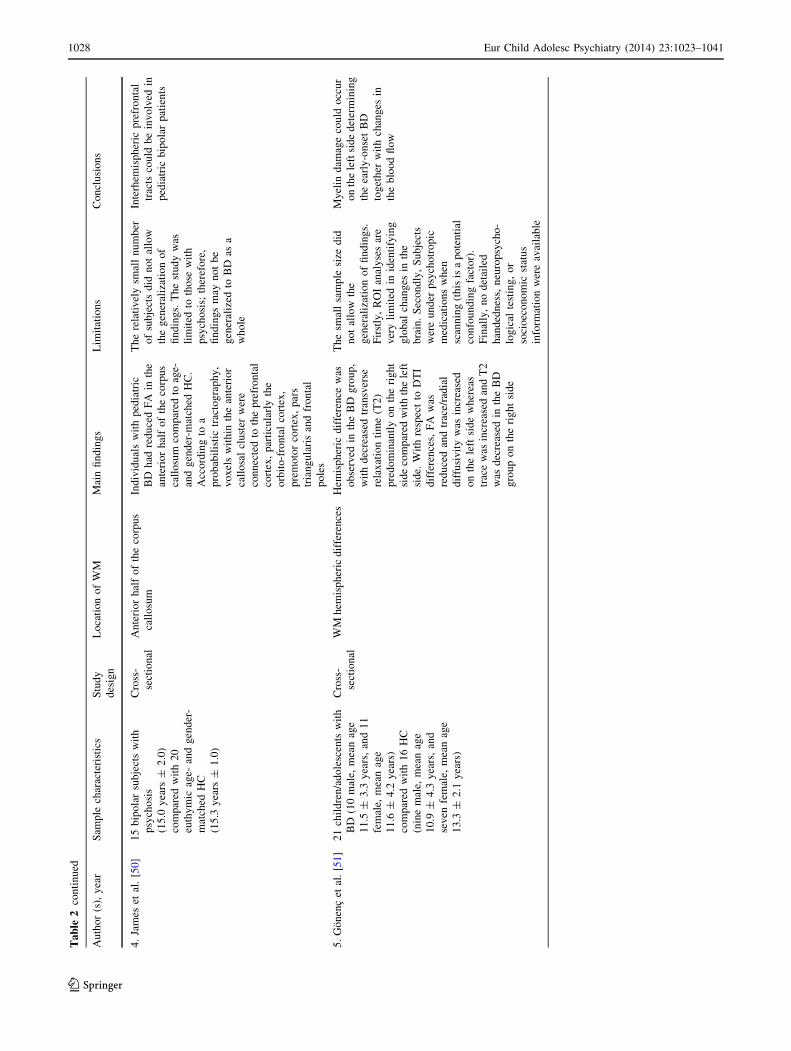
4.
Jam
eset
al.
[50]
15
bip
ola
rsu
bje
cts
wit
h
psy
cho
sis
(15
.0y
ears
±2
.0)
com
par
edw
ith
20
euth
ym
icag
e-an
dg
end
er-
mat
ched
HC
(15
.3y
ears
±1
.0)
Cro
ss-
sect
ion
al
An
teri
or
hal
fo
fth
eco
rpu
s
call
osu
m
Ind
ivid
ual
sw
ith
ped
iatr
ic
BD
had
red
uce
dF
Ain
the
ante
rio
rh
alf
of
the
corp
us
call
osu
mco
mp
ared
toag
e-
and
gen
der
-mat
ched
HC
.
Acc
ord
ing
toa
pro
bab
ilis
tic
trac
tog
rap
hy
,
vo
xel
sw
ith
inth
ean
teri
or
call
osa
lcl
ust
erw
ere
con
nec
ted
toth
ep
refr
on
tal
cort
ex,
par
ticu
larl
yth
e
orb
ito
-fro
nta
lco
rtex
,
pre
mo
tor
cort
ex,
par
s
tria
ng
ula
ris
and
fro
nta
l
po
les
Th
ere
lati
vel
ysm
all
nu
mb
er
of
sub
ject
sd
idn
ot
allo
w
the
gen
eral
izat
ion
of
fin
din
gs.
Th
est
ud
yw
as
lim
ited
toth
ose
wit
h
psy
cho
sis;
ther
efo
re,
fin
din
gs
may
no
tb
e
gen
eral
ized
toB
Das
a
wh
ole
Inte
rhem
isp
her
icp
refr
on
tal
trac
tsco
uld
be
inv
olv
edin
ped
iatr
icb
ipo
lar
pat
ien
ts
5.
Go
nen
cet
al.
[51]
21
chil
dre
n/a
do
lesc
ents
wit
h
BD
(10
mal
e,m
ean
age
11
.5±
3.3
yea
rs,
and
11
fem
ale,
mea
nag
e
11
.6±
4.2
yea
rs)
com
par
edw
ith
16
HC
(nin
em
ale,
mea
nag
e
10
.9±
4.3
yea
rs,
and
sev
enfe
mal
e,m
ean
age
13
.3±
2.1
yea
rs)
Cro
ss-
sect
ion
al
WM
hem
isp
her
icd
iffe
ren
ces
Hem
isp
her
icd
iffe
ren
cew
as
ob
serv
edin
the
BD
gro
up
,
wit
hd
ecre
ased
tran
sver
se
rela
xat
ion
tim
e(T
2)
pre
do
min
antl
yo
nth
eri
gh
t
sid
eco
mp
ared
wit
hth
ele
ft
sid
e.W
ith
resp
ect
toD
TI
dif
fere
nce
s,F
Aw
as
red
uce
dan
dtr
ace/
rad
ial
dif
fusi
vit
yw
asin
crea
sed
on
the
left
sid
ew
her
eas
trac
ew
asin
crea
sed
and
T2
was
dec
reas
edin
the
BD
gro
up
on
the
rig
ht
sid
e
Th
esm
all
sam
ple
size
did
no
tal
low
the
gen
eral
izat
ion
of
fin
din
gs.
Fir
stly
,R
OI
anal
yse
sar
e
ver
yli
mit
edin
iden
tify
ing
glo
bal
chan
ges
inth
e
bra
in.
Sec
on
dly
,S
ub
ject
s
wer
eu
nd
erp
sych
otr
op
ic
med
icat
ion
sw
hen
scan
nin
g(t
his
isa
po
ten
tial
con
fou
nd
ing
fact
or)
.
Fin
ally
,n
od
etai
led
han
ded
nes
s,n
euro
psy
cho
-
log
ical
test
ing
,o
r
soci
oec
on
om
icst
atu
s
info
rmat
ion
wer
eav
aila
ble
My
elin
dam
age
cou
ldo
ccu
r
on
the
left
sid
ed
eter
min
ing
the
earl
y-o
nse
tB
D
tog
eth
erw
ith
chan
ges
in
the
blo
od
flo
w
1028 Eur Child Adolesc Psychiatry (2014) 23:1023–1041
123
Ta
ble
2co
nti
nu
ed
Au
tho
r(s
),y
ear
Sam
ple
char
acte
rist
ics
Stu
dy
des
ign
Lo
cati
on
of
WM
Mai
nfi
nd
ing
sL
imit
atio
ns
Co
ncl
usi
on
s
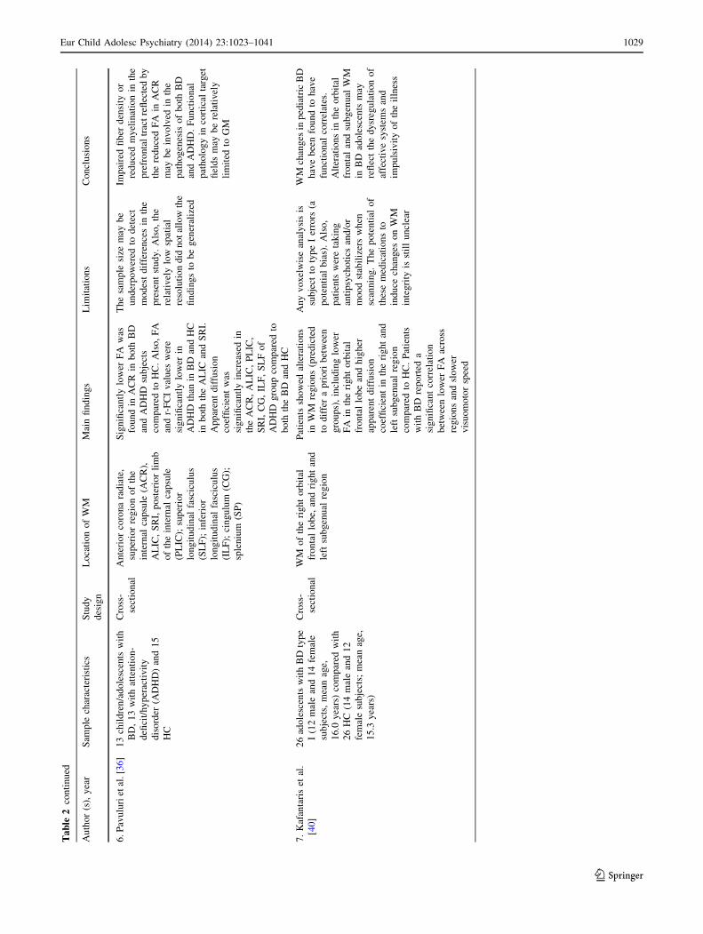
6.P
avu
luri
etal
.[3
6]
13
chil
dre
n/a
do
lesc
ents
wit
h
BD
,1
3w
ith
atte
nti
on
-
defi
cit/
hy
per
acti
vit
y
dis
ord
er(A
DH
D)
and
15
HC
Cro
ss-
sect
ion
al
An
teri
or
coro
na
rad
iate
,
sup
erio
rre
gio
no
fth
e
inte
rnal
cap
sule
(AC
R),
AL
IC,
SR
I,p
ost
erio
rli
mb
of
the
inte
rnal
cap
sule
(PL
IC);
sup
erio
r
lon
git
ud
inal
fasc
icu
lus
(SL
F);
infe
rio
r
lon
git
ud
inal
fasc
icu
lus
(IL
F);
cin
gu
lum
(CG
);
sple
niu
m(S
P)
Sig
nifi
can
tly
low
erF
Aw
as
fou
nd
inA
CR
inb
oth
BD
and
AD
HD
sub
ject
s
com
par
edto
HC
.A
lso
,F
A
and
r-F
CI
val
ues
wer
e
sig
nifi
can
tly
low
erin
AD
HD
than
inB
Dan
dH
C
inb
oth
the
AL
ICan
dS
RI.
Ap
par
ent
dif
fusi
on
coef
fici
ent
was
sig
nifi
can
tly
incr
ease
din
the
AC
R,
AL
IC,
PL
IC,
SR
I,C
G,
ILF
,S
LF
of
AD
HD
gro
up
com
par
edto
bo
thth
eB
Dan
dH
C
Th
esa
mp
lesi
zem
ayb
e
un
der
po
wer
edto
det
ect
mo
des
td
iffe
ren
ces
inth
e
pre
sen
tst
ud
y.
Als
o,
the
rela
tiv
ely
low
spat
ial
reso
luti
on
did
no
tal
low
the
fin
din
gs
tob
eg
ener
aliz
ed
Imp
aire
dfi
ber
den
sity
or
red
uce
dm
yel
inat
ion
inth
e
pre
fro
nta
ltr
act
refl
ecte
db
y
the
red
uce
dF
Ain
AC
R
may
be
inv
olv
edin
the
pat
ho
gen
esis
of
bo
thB
D
and
AD
HD
.F
un
ctio
nal
pat
ho
log
yin
cort
ical
targ
et
fiel
ds
may
be
rela
tiv
ely
lim
ited
toG
M
7.
Kaf
anta
ris
etal
.
[40
]
26
ado
lesc
ents
wit
hB
Dty
pe
I(1
2m
ale
and
14
fem
ale
sub
ject
s,m
ean
age,
16
.0y
ears
)co
mp
ared
wit
h
26
HC
(14
mal
ean
d1
2
fem
ale
sub
ject
s;m
ean
age,
15
.3y
ears
)
Cro
ss-
sect
ion
al
WM
of
the
rig
ht
orb
ital
fro
nta
llo
be,
and
rig
ht
and
left
sub
gen
ual
reg
ion
Pat
ien
tssh
ow
edal
tera
tio
ns
inW
Mre
gio
ns
(pre
dic
ted
tod
iffe
ra
pri
ori
bet
wee
n
gro
up
s),
incl
ud
ing
low
er
FA
inth
eri
gh
to
rbit
al
fro
nta
llo
be
and
hig
her
app
aren
td
iffu
sio
n
coef
fici
ent
inth
eri
gh
tan
d
left
sub
gen
ual
reg
ion
com
par
edto
HC
.P
atie
nts
wit
hB
Dre
po
rted
a
sig
nifi
can
tco
rrel
atio
n
bet
wee
nlo
wer
FA
acro
ss
reg
ion
san
dsl
ow
er
vis
uo
mo
tor
spee
d
An
yv
ox
elw
ise
anal
ysi
sis
sub
ject
toty
pe
Ier
rors
(a
po
ten
tial
bia
s).
Als
o,
pat
ien
tsw
ere
tak
ing
anti
psy
cho
tics
and
/or
mo
od
stab
iliz
ers
wh
en
scan
nin
g.
Th
ep
ote
nti
alo
f
thes
em
edic
atio
ns
to
ind
uce
chan
ges
on
WM
inte
gri
tyis
stil
lu
ncl
ear
WM
chan
ges
inp
edia
tric
BD
hav
eb
een
fou
nd
toh
ave
fun
ctio
nal
corr
elat
es.
Alt
erat
ion
sin
the
orb
ital
fro
nta
lan
dsu
bg
enu
alW
M
inB
Dad
ole
scen
tsm
ay
refl
ect
the
dy
sreg
ula
tio
no
f
affe
ctiv
esy
stem
san
d
imp
uls
ivit
yo
fth
eil
lnes
s
Eur Child Adolesc Psychiatry (2014) 23:1023–1041 1029
123
Ta
ble
2co
nti
nu
ed
Au
tho
r(s
),y
ear
Sam
ple
char
acte
rist
ics
Stu
dy
des
ign
Lo
cati
on
of
WM
Mai
nfi
nd
ing
sL
imit
atio
ns
Co
ncl
usi
on
s
8.
Bar
nea
-Go
raly
etal
.[3
9]
21
BD
ado
lesc
ents
(mea
n
age,
16
.1±
2.7
)w
ho
wer
e
chil
dre
no
fat
leas
to
ne
BD
par
ent,
and
18
age-
and
IQ-
mat
ched
HC
(mea
nag
e,
14
.5±
2.7
)
Cro
ss-
sect
ion
al
WM
of
the
forn
ix,
left
mid
-
po
ster
ior
cin
gu
late
gy
rus,
thro
ug
ho
ut
the
corp
us
call
osu
m,
fib
ers
fro
mth
e
forn
ixto
the
thal
amu
s,an
d
par
ieta
l/o
ccip
ital
coro
na
rad
iate
bil
ater
ally
Bip
ola
rad
ole
scen
tssh
ow
ed
low
erF
Av
alu
esco
mp
ared
toH
Cin
the
forn
ix,
left
mid
-po
ster
ior
cin
gu
late
gy
rus,
thro
ug
ho
ut
the
corp
us
call
osu
m,
infi
ber
s
fro
mth
efo
rnix
toth
e
thal
amu
s,an
dp
arie
tal/
occ
ipit
alco
ron
ara
dia
ta
bil
ater
ally
Mo
stp
arti
cip
ants
wer
e
tak
ing
med
icat
ion
sw
hen
scan
nin
gan
d/o
rh
ad
psy
cho
tro
pic
med
icat
ion
exp
osu
reh
isto
ry.
Als
o,
sub
ject
sh
adsi
gn
ifica
nt
AD
HD
sym
pto
ms
(a
po
ten
tial
con
fou
nd
ing
fact
or)
.P
ost
ho
c
beh
avio
ral
anal
yse
s
incl
ud
edo
nly
asu
bse
to
f
the
wh
ole
sam
ple
;
ther
efo
re,
neg
ativ
ere
sult
s
may
hav
eb
een
ob
tain
ed
du
eto
lack
of
po
wer
Ab
no
rmal
itie
sin
WM
are
pre
sen
tea
rly
inth
eco
urs
e
of
dis
ease
infa
mil
ial
BD
.
Sig
nifi
can
tW
Mtr
act
chan
ges
inb
ipo
lar
ado
lesc
ents
may
be
fou
nd
inth
ose
bra
inre
gio
ns
inv
olv
edin
emo
tio
nal
,
beh
avio
ral
and
cog
nit
ive
reg
ula
tio
n
9.
Fra
zier
etal
.[3
8]
10
bip
ola
rch
ild
ren
(mea
n
age,
9.2
±3
.0),
8H
C
(mea
nag
e,9
.2±
2.4
),an
d
7ag
e-m
atch
edch
ild
ren
at
risk
for
BD
(AR
-BP
D)
(mea
nag
e,8
.9±
3.0
)
Cro
ss-
sect
ion
al
WM
of
the
SL
FI
and
the
cin
gu
late
-par
acin
gu
late
WM
(CG
-PA
CW
M),
left
orb
ital
fro
nta
lan
dth
eri
gh
t
corp
us
call
osu
mb
od
y
Bip
ola
rch
ild
ren
rep
ort
ed
red
uce
dF
Ain
the
rig
ht
and
left
sup
erio
rfr
on
tal
trac
ts,
incl
ud
ing
the
SL
FI
and
CG
-PA
CW
Mco
mp
ared
to
HC
.F
urt
her
mo
re,
bip
ola
r
chil
dre
nsh
ow
edre
du
ced
FA
inle
fto
rbit
alfr
on
tal
WM
and
the
rig
ht
corp
us
call
osu
mb
od
y.
Bip
ola
r
chil
dre
nal
sore
po
rted
red
uce
dF
Ain
the
rig
ht
and
left
CG
-PA
CW
Mth
anA
R-
BP
D.
Fin
ally
,re
du
ced
FA
was
fou
nd
inb
ilat
eral
SL
F
Iin
eith
erth
eB
Dan
dA
R-
BP
Dg
rou
ps
com
par
edto
HC
Th
em
od
est
nu
mb
ero
f
sub
ject
san
dth
ecr
oss
-
sect
ion
aln
atu
reo
fth
e
stu
dy
did
no
tal
low
the
fin
din
gs
tob
eg
ener
aliz
ed.
Als
o,
all
sub
ject
sh
ad
atte
nti
on
-defi
cit/
hy
per
acti
vit
yd
iso
rder
(AD
HD
)an
d/o
ran
xie
ty
dis
ord
er.
Mo
reo
ver
,th
e
effe
cts
of
mo
od
stat
ean
d
com
orb
idit
ies
on
the
DT
-
MR
Ifi
nd
ing
sh
ave
no
t
bee
nev
alu
ated
.A
lso
,
ind
ivid
ual
sw
ere
tak
ing
psy
cho
tro
pic
med
icat
ion
s
wh
ensc
ann
ing
(th
isis
a
po
ten
tial
con
fou
nd
ing
fact
or)
Dec
reas
edF
Ain
the
rig
ht
and
left
CG
-PA
CW
Min
bip
ola
rch
ild
ren
com
par
ed
toth
eo
ther
gro
up
sco
uld
rep
rese
nt
ad
isea
se-s
tate
-
rela
ted
fin
din
g.
Th
ese
abn
orm
alit
ies
cou
ld
rep
rese
nt
an
end
op
hen
oty
pe
of
the
bip
ola
ril
lnes
s
1030 Eur Child Adolesc Psychiatry (2014) 23:1023–1041
123
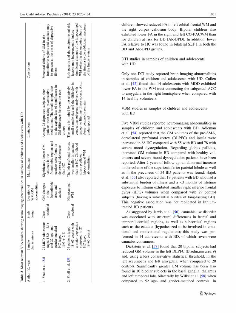
children showed reduced FA in left orbital frontal WM and
the right corpus callosum body. Bipolar children also
exhibited lower FA in the right and left CG-PACWM than
for children at risk for BD (AR-BPD). In addition, lower
FA relative to HC was found in bilateral SLF I in both the
BD and AR-BPD groups.
DTI studies in samples of children and adolescents
with UD
Only one DTI study reported brain imaging abnormalities
in samples of children and adolescents with UD. Cullen
et al. [42] found that 14 adolescents with MDD exhibited
lower FA in the WM tract connecting the subgenual ACC
to amygdala in the right hemisphere when compared with
14 healthy volunteers.
VBM studies in samples of children and adolescents
with BD
Five VBM studies reported neuroimaging abnormalities in
samples of children and adolescents with BD. Adleman
et al. [54] reported that the GM volumes of the pre-SMA,
dorsolateral prefrontal cortex (DLPFC) and insula were
increased in 68 HC compared with 55 with BD and 78 with
severe mood dysregulation. Regarding globus pallidus,
increased GM volume in BD compared with healthy vol-
unteers and severe mood dysregulation patients have been
reported. After 2 years of follow-up, an abnormal increase
in the volume of the superior/inferior parietal lobule as well
as in the precuneus of 34 BD patients was found. Hajek
et al. [55] also reported that 19 patients with BD who had a
substantial burden of illness and a \3 months of lifetime
exposure to lithium exhibited smaller right inferior frontal
gyrus (rIFG) volumes when compared with 29 control
subjects (having a substantial burden of long-lasting BD).
This negative association was not replicated in lithium-
treated BD patients.
As suggested by Jarvis et al. [56], cannabis use disorder
was associated with structural differences in frontal and
temporal cortical regions, as well as subcortical regions
such as the caudate (hypothesized to be involved in emo-
tional and motivational regulation); this study was per-
formed in 14 adolescents with BD, of which seven were
cannabis consumers.
Dickstein et al. [57] found that 20 bipolar subjects had
reduced GM volume in the left DLPFC (Brodmann area 9)
and, using a less conservative statistical threshold, in the
left accumbens and left amygdala, when compared to 20
controls. Significantly greater GM volume has been also
found in 10 bipolar subjects in the basal ganglia, thalamus
and left temporal lobe bilaterally by Wilke et al. [58] when
compared to 52 age- and gender-matched controls. InTa
ble
3M
ost
rele
van
tV
BA
stu
die
ssh
ow
ing
neu
roim
agin
gab
no
rmal
itie
sin
sam
ple
so
fch
ild
ren
and
ado
lesc
ents
wit
hU
D
Au
tho
r(s
),y
ear
Sam
ple
char
acte
rist
ics
Stu
dy
des
ign
Lo
cati
on
of
WM
/GM
abn
orm
alit
ies
Mai
nfi
nd
ing
sL
imit
atio
ns
Co
ncl
usi
on
s
1.
Sh
adet
al.
[52
]2
2M
DD
ado
lesc
ents
(ag
ed1
5.0
?2
.1)
and
22
age-
and
gen
der
-mat
ched
HC
(ag
ed
16
.0?
2.1
)
Cro
ss-
sect
ion
al
GM
chan
ges
inth
e
fro
nto
lim
bic
area
san
d
cau
dat
e
Sig
nifi
can
tly
low
erG
M
vo
lum
esin
the
fro
nto
lim
bic
reg
ion
san
d
cau
dat
ew
ere
fou
nd
in
dep
ress
edad
ole
scen
ts
than
HC
Am
on
g2
2d
epre
ssed
sub
ject
s,fo
ur
pat
ien
tsw
ere
tak
ing
psy
cho
acti
ve
med
icat
ion
s.T
he
smal
lsa
mp
lesi
ze
mig
ht
exp
lain
loss
of
sig
nifi
can
cein
GM
dif
fere
nce
sb
etw
een
the
two
gro
up
s
Str
uct
ura
ld
efici
tso
fG
Min
the
fro
nto
lim
bic
reg
ion
san
dca
ud
ate
may
be
pre
sen
tat
the
on
set
of
dep
ress
ive
illn
ess
2.
Fro
dl
etal
.[5
3]
24
pat
ien
ts(a
ged
18
–6
5y
ears
)w
ith
am
ajo
rd
epre
ssio
n
com
par
edto
27
HC
(ag
ed
18
–6
5y
ears
)
Cro
ss-
sect
ion
al
Hip
po
cam
pal
WM
Th
ele
ftp
refr
on
tal
cort
ex
was
smal
ler
inp
atie
nts
than
inH
C.
Ch
ild
ho
od
stre
ssp
red
icte
d
hip
po
cam
pal
WM
abn
orm
alit
ies
Th
est
ud
yis
lim
ited
by
the
rela
tiv
ely
smal
lsa
mp
lesi
zean
dth
ed
iffi
cult
yin
con
du
ctin
gsu
bg
rou
pan
aly
ses
wit
h
resp
ect
toli
feti
me
illn
ess
cou
rse.
Als
o,
abu
seco
mm
on
lyre
mai
ns
un
der
rep
ort
ed
Bo
thg
enet
ican
dth
een
vir
on
men
tal
risk
fact
ors
may
ind
epen
den
tly
ind
uce
sig
nifi
can
tch
ang
eso
fth
eh
ipp
oca
mp
al
WM
refl
ecti
ng
the
ou
tgo
ing
fib
ers
of
the
fim
bri
ato
the
add
itio
nal
stru
ctu
res
of
the
lim
bic
syst
em
Eur Child Adolesc Psychiatry (2014) 23:1023–1041 1031
123
Ta
ble
4M
ost
rele
van
tV
BA
stu
die
ssh
ow
ing
neu
roim
agin
gab
no
rmal
itie
sin
sam
ple
so
fch
ild
ren
and
ado
lesc
ents
wit
hB
D
Au
tho
r(s
),y
ear
Sam
ple
char
acte
rist
ics
Stu
dy
des
ign
Lo
cati
on
of
WM
/GM
abn
orm
alit
ies
Mai
nfi
nd
ing
sL
imit
atio
ns
Co
ncl
usi
on
s
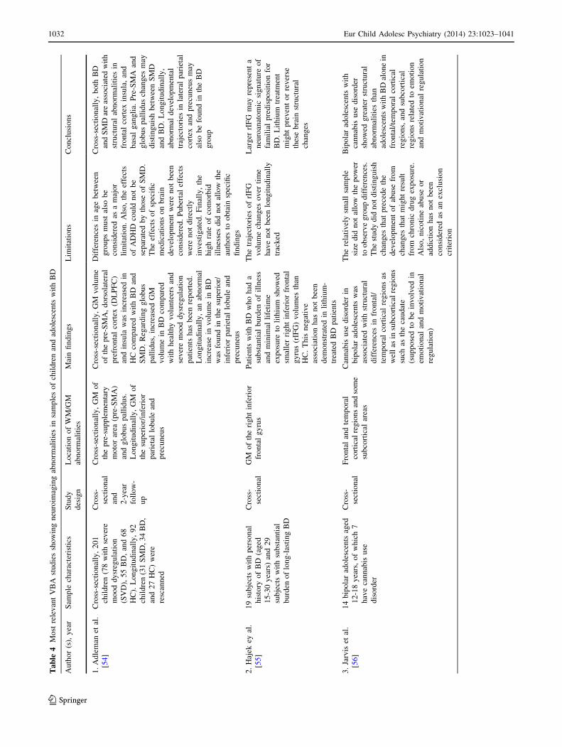
1.
Ad
lem
anet
al.
[54
]
Cro
ss-s
ecti
on
ally
,2
01
chil
dre
n(7
8w
ith
sev
ere
mo
od
dy
sreg
ula
tio
n
(SV
D),
55
BD
,an
d6
8
HC
).L
on
git
ud
inal
ly,
92
chil
dre
n(3
1S
MD
,3
4B
D,
and
27
HC
)w
ere
resc
ann
ed
Cro
ss-
sect
ion
al
and
2-y
ear
foll
ow
-
up
Cro
ss-s
ecti
on
ally
,G
Mo
f
the
pre
-su
pp
lem
enta
ry
mo
tor
area
(pre
-SM
A)
and
glo
bu
sp
alli
du
s.
Lo
ng
itu
din
ally
,G
Mo
f
the
sup
erio
r/in
feri
or
par
ieta
llo
bu
lean
d
pre
cun
eus
Cro
ss-s
ecti
on
ally
,G
Mv
olu
me
of
the
pre
-SM
A,
do
rso
late
ral
pre
fro
nta
lco
rtex
(DL
PF
C)
and
insu
law
asin
crea
sed
in
HC
com
par
edw
ith
BD
and
SM
D.
Reg
ard
ing
glo
bu
s
pal
lid
us,
incr
ease
dG
M
vo
lum
ein
BD
com
par
ed
wit
hh
ealt
hy
vo
lun
teer
san
d
sev
ere
mo
od
dy
sreg
ula
tio
n
pat
ien
tsh
asb
een
rep
ort
ed.
Lo
ng
itu
din
ally
,an
abn
orm
al
incr
ease
inv
olu
me
inB
D
was
fou
nd
inth
esu
per
ior/
infe
rio
rp
arie
tal
lob
ule
and
pre
cun
eus
Dif
fere
nce
sin
age
bet
wee
n
gro
up
sm
ust
also
be
con
sid
ered
asa
maj
or
lim
itat
ion
.A
lso
,th
eef
fect
s
of
AD
HD
cou
ldn
ot
be
sep
arat
edb
yth
ose
of
SM
D.
Th
eef
fect
so
fsp
ecifi
c
med
icat
ion
so
nb
rain
dev
elo
pm
ent
wer
en
ot
bee
n
con
sid
ered
.P
ub
erta
lef
fect
s
wer
en
ot
dir
ectl
y
inv
esti
gat
ed.
Fin
ally
,th
e
hig
hra
teo
fco
mo
rbid
illn
esse
sd
idn
ot
allo
wth
e
auth
ors
too
bta
insp
ecifi
c
fin
din
gs
Cro
ss-s
ecti
on
ally
,b
oth
BD
and
SM
Dar
eas
soci
ated
wit
h
stru
ctu
ral
abn
orm
alit
ies
in
fro
nta
lco
rtex
insu
la,
and
bas
alg
ang
lia.
Pre
-SM
Aan
d
glo
bu
sp
alli
du
sch
ang
esm
ay
dis
tin
gu
ish
bet
wee
nS
MD
and
BD
.L
on
git
ud
inal
ly,
abn
orm
ald
evel
op
men
tal
traj
ecto
ries
inla
tera
lp
arie
tal
cort
exan
dp
recu
neu
sm
ay
also
be
fou
nd
inth
eB
D
gro
up
2.
Haj
ekey
al.
[55
]
19
sub
ject
sw
ith
per
son
al
his
tory
of
BD
(ag
ed
15
-30
yea
rs)
and
29
sub
ject
sw
ith
sub
stan
tial
bu
rden
of
lon
g-l
asti
ng
BD
Cro
ss-
sect
ion
al
GM
of
the
rig
ht
infe
rio
r
fro
nta
lg
yru
s
Pat
ien
tsw
ith
BD
wh
oh
ada
sub
stan
tial
bu
rden
of
illn
ess
and
min
imal
life
tim
e
exp
osu
reto
lith
ium
sho
wed
smal
ler
rig
ht
infe
rio
rfr
on
tal
gy
rus
(rIF
G)
vo
lum
esth
an
HC
.T
his
neg
ativ
e
asso
ciat
ion
has
no
tb
een
dem
on
stra
ted
inli
thiu
m-
trea
ted
BD
pat
ien
ts
Th
etr
ajec
tori
eso
frI
FG
vo
lum
ech
ang
eso
ver
tim
e
hav
en
ot
bee
nlo
ng
itu
din
ally
trac
ked
Lar
ger
rIF
Gm
ayre
pre
sen
ta
neu
roan
ato
mic
sig
nat
ure
of
fam
ilia
lp
red
isp
osi
tio
nfo
r
BD
.L
ith
ium
trea
tmen
t
mig
ht
pre
ven
to
rre
ver
se
thes
eb
rain
stru
ctu
ral
chan
ges
3.
Jarv
iset
al.
[56
]
14
bip
ola
rad
ole
scen
tsag
ed
12
-18
yea
rs,
of
wh
ich
7
hav
eca
nn
abis
use
dis
ord
er
Cro
ss-
sect
ion
al
Fro
nta
lan
dte
mp
ora
l
cort
ical
reg
ion
san
dso
me
sub
cort
ical
area
s
Can
nab
isu
sed
iso
rder
in
bip
ola
rad
ole
scen
tsw
as
asso
ciat
edw
ith
stru
ctu
ral
dif
fere
nce
sin
fro
nta
l/
tem
po
ral
cort
ical
reg
ion
sas
wel
las
insu
bco
rtic
alre
gio
ns
such
asth
eca
ud
ate
(su
pp
ose
dto
be
inv
olv
edin
emo
tio
nal
and
mo
tiv
atio
nal
reg
ula
tio
n)
Th
ere
lati
vel
ysm
all
sam
ple
size
did
no
tal
low
the
po
wer
too
bse
rve
gro
up
dif
fere
nce
s.
Th
est
ud
yd
idn
ot
dis
tin
gu
ish
chan
ges
that
pre
ced
eth
e
dev
elo
pm
ent
of
abu
sefr
om
chan
ges
that
mig
ht
resu
lt
fro
mch
ron
icd
rug
exp
osu
re.
Als
o,
nic
oti
ne
abu
seo
r
add
icti
on
has
no
tb
een
con
sid
ered
asan
excl
usi
on
crit
erio
n
Bip
ola
rad
ole
scen
tsw
ith
can
nab
isu
sed
iso
rder
sho
wed
gre
ater
stru
ctu
ral
abn
orm
alit
ies
than
ado
lesc
ents
wit
hB
Dal
on
ein
fro
nta
l/te
mp
ora
lco
rtic
al
reg
ion
s,an
dsu
bco
rtic
al
reg
ion
sre
late
dto
emo
tio
n
and
mo
tiv
atio
nal
reg
ula
tio
n
1032 Eur Child Adolesc Psychiatry (2014) 23:1023–1041
123
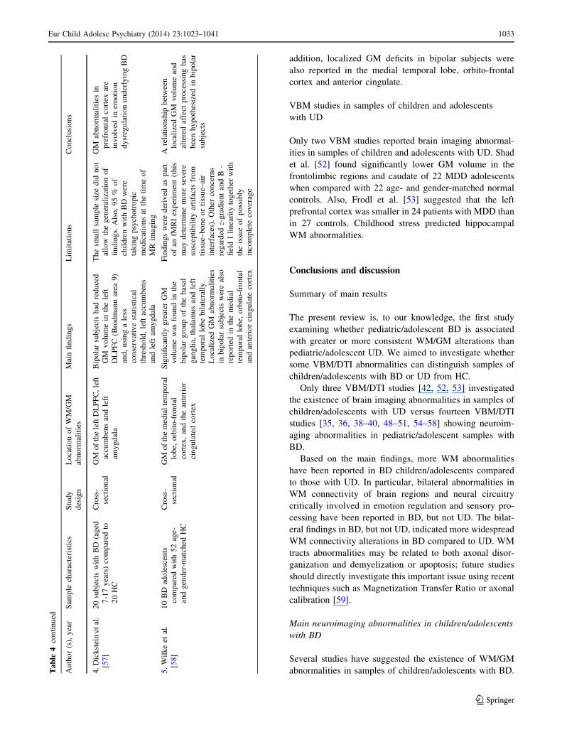
addition, localized GM deficits in bipolar subjects were
also reported in the medial temporal lobe, orbito-frontal
cortex and anterior cingulate.
VBM studies in samples of children and adolescents
with UD
Only two VBM studies reported brain imaging abnormal-
ities in samples of children and adolescents with UD. Shad
et al. [52] found significantly lower GM volume in the
frontolimbic regions and caudate of 22 MDD adolescents
when compared with 22 age- and gender-matched normal
controls. Also, Frodl et al. [53] suggested that the left
prefrontal cortex was smaller in 24 patients with MDD than
in 27 controls. Childhood stress predicted hippocampal
WM abnormalities.
Conclusions and discussion
Summary of main results
The present review is, to our knowledge, the first study
examining whether pediatric/adolescent BD is associated
with greater or more consistent WM/GM alterations than
pediatric/adolescent UD. We aimed to investigate whether
some VBM/DTI abnormalities can distinguish samples of
children/adolescents with BD or UD from HC.
Only three VBM/DTI studies [42, 52, 53] investigated
the existence of brain imaging abnormalities in samples of
children/adolescents with UD versus fourteen VBM/DTI
studies [35, 36, 38–40, 48–51, 54–58] showing neuroim-
aging abnormalities in pediatric/adolescent samples with
BD.
Based on the main findings, more WM abnormalities
have been reported in BD children/adolescents compared
to those with UD. In particular, bilateral abnormalities in
WM connectivity of brain regions and neural circuitry
critically involved in emotion regulation and sensory pro-
cessing have been reported in BD, but not UD. The bilat-
eral findings in BD, but not UD, indicated more widespread
WM connectivity alterations in BD compared to UD. WM
tracts abnormalities may be related to both axonal disor-
ganization and demyelization or apoptosis; future studies
should directly investigate this important issue using recent
techniques such as Magnetization Transfer Ratio or axonal
calibration [59].
Main neuroimaging abnormalities in children/adolescents
with BD
Several studies have suggested the existence of WM/GM
abnormalities in samples of children/adolescents with BD.Ta
ble
4co
nti
nu
ed
Au
tho
r(s
),y
ear
Sam
ple
char
acte
rist
ics
Stu
dy
des
ign
Lo
cati
on
of
WM
/GM
abn
orm
alit
ies
Mai
nfi
nd
ing
sL
imit
atio
ns
Co
ncl
usi
on
s
4.
Dic
kst
ein
etal
.
[57
]
20
sub
ject
sw
ith
BD
(ag
ed
7-1
7y
ears
)co
mp
ared
to
20
HC
Cro
ss-
sect
ion
al
GM
of
the
left
DL
PF
C,
left
accu
mb
ens
and
left
amy
gd
ala
Bip
ola
rsu
bje
cts
had
red
uce
d
GM
vo
lum
ein
the
left
DL
PF
C(B
rod
man
nar
ea9
)
and
,u
sin
ga
less
con
serv
ativ
est
atis
tica
l
thre
sho
ld,
left
accu
mb
ens
and
left
amy
gd
ala
Th
esm
all
sam
ple
size
did
no
t
allo
wth
eg
ener
aliz
atio
no
f
fin
din
gs.
Als
o,
95
%o
f
chil
dre
nw
ith
BD
wer
e
tak
ing
psy
cho
tro
pic
med
icat
ion
sat
the
tim
eo
f
MR
imag
ing
GM
abn
orm
alit
ies
in
pre
fro
nta
lco
rtex
are
inv
olv
edin
emo
tio
n
dy
sreg
ula
tio
nu
nd
erly
ing
BD
5.
Wil
ke
etal
.
[58
]
10
BD
ado
lesc
ents
com
par
edw
ith
52
age-
and
gen
der
-mat
ched
HC
Cro
ss-
sect
ion
al
GM
of
the
med
ial
tem
po
ral
lob
e,o
rbit
o-f
ron
tal
cort
ex,
and
the
ante
rio
r
cin
gu
late
dco
rtex
Sig
nifi
can
tly
gre
ater
GM
vo
lum
ew
asfo
un
din
the
bip
ola
rg
rou
po
fth
eb
asal
gan
gli
a,th
alam
us
and
left
tem
po
ral
lob
eb
ilat
eral
ly.
Lo
cali
zed
GM
abn
orm
alit
ies
inb
ipo
lar
sub
ject
sw
ere
also
rep
ort
edin
the
med
ial
tem
po
ral
lob
e,o
rbit
o-f
ron
tal
and
ante
rio
rci
ng
ula
teco
rtex
Fin
din
gs
wer
ed
eriv
edas
par
t
of
anfM
RI
exp
erim
ent
(th
is
may
det
erm
ine
mo
rese
ver
e
susc
epti
bil
ity
arti
fact
sfr
om
tiss
ue–
bo
ne
or
tiss
ue–
air
inte
rfac
es).
Oth
erco
nce
rns
reg
ard
edz-
gra
die
nt
and
B-
fiel
d1
lin
eari
tyto
get
her
wit
h
the
issu
eo
fp
oss
ibly
inco
mp
lete
cov
erag
e
Are
lati
on
ship
bet
wee
n
loca
lize
dG
Mv
olu
me
and
alte
red
affe
ctp
roce
ssin
gh
as
bee
nh
yp
oth
esiz
edin
bip
ola
r
sub
ject
s
Eur Child Adolesc Psychiatry (2014) 23:1023–1041 1033
123
In their meta-analytic study on adults, Kempton and col-
leagues [60] suggested that basal ganglia and hippocampal
volume reductions seem to be more specific for MDD than
BD, whereas reduced corpus callosum cross-sectional area
and increased rates of deep WM hyperintensities seem to
be more common in BD than MDD. As argued below,
these findings have only partially been confirmed in sam-
ples of children and adolescents. However, alterations
throughout the amygdala and basal ganglia appear poorly
specific, since they have also been documented in BD [42,
54, 58] and many other neuropsychiatric disorders [42].
The presence of subcortical alterations within the amygdala
and basal ganglia in childhood [61] could subsequently
extend to prefrontal cortical regions continuing to develop
into adulthood. These frontolimbic abnormalities occurring
during childhood and early adolescence would suggest the
existence of neurodevelopmental conditions [62]. Abnor-
malities throughout the anterior limbic system and associ-
ated prefrontal regions have been hypothesized to be
involved in affective, cognitive and vegetative symptoms
in BD [63–65]. In line with results in adult samples, one of
the most common brain region in which abnormalities have
been documented in pediatric/adolescent bipolar samples is
the corpus callosum [38, 39, 50]. The corpus callosum
represents the major interhemispheric commissure con-
necting most of the neocortical brain regions and including
fundamental brain networks related to attention, memory,
language and emotional states [66–72]. The corpus callo-
sum develops during childhood/adolescence as demon-
strated by the increased size [73], reduced signal intensity
[72] and increased FA values [74].
Some authors [75, 76] suggested that, although the
signal intensity of the corpus callosum is reduced and its
shape altered in pediatric/adolescent bipolar samples, no
differences in size have been reported between bipolar
children/adolescents and healthy volunteers.
Barnea-Goraly et al. [39] suggested an abnormal matu-
ration process in the corpus callosum of bipolar subjects, as
reflected by the lower FA in adolescence—representing
reduced coherence or aberrant myelination and increasing
FA with age. This might be due to changes in the extra-
cellular compartment related to abnormal perivascular
structures [73], but might also be related to the different
measurements of FA in the corpus callosum of adults and
adolescents [77].
Abnormalities in the CC3-motor area of the corpus
callosum interconnecting right and left paracingulate,
anterior cingulate, supplementary motor areas and lateral
premotor regions in the frontal lobes could be related to
alterations in activity and motivation—that are frequently
observed in children with BD [38].
As hypothesized, WM integrity in prefrontal limbic
network has been found to be significantly altered as
demonstrated by the lower FA in children with BD. Two
studies suggested that there are changes in the WM of the
ALIC [33, 36]. Greater alterations have been reported in
the WM microstructure of the left ALIC with pediatric
bipolar onset compared to adult bipolar onset [36].
Disorganized integration of prefrontal–limbic circuitry
associated with affective dysregulation has been
hypothesized in BD [33, 78]. The prefrontal structures
appear to show dysfunctions in modulating subcortical
structures. The alterations in the WM microstructure of
the ALIC have also been demonstrated in another DTI
tractography study [79]. This may increase the vulnera-
bility to develop bipolar illness, by altering the pro-
cessing of emotional information throughout the limbic
system.
Main neuroimaging abnormalities in children/adolescents
with UD
Only three VBM/DTI studies [42, 52, 53] showed the
existence of brain imaging abnormalities in samples of
children and adolescents with UD. Shad et al. [52] showed
lower GM volumes in the frontolimbic areas and caudate of
depressed adolescents than in healthy volunteers. These
authors also suggested that GM changes in the striatum,
especially in the caudate nucleus, may relate to the initial
presentation of MDD. The involvement of frontolimbic
regions in the pathogenesis of adult MDD has been
extensively reported [79–83]. Dorsolateral and orbito-
frontal regions of the prefrontal cortex (PFC) play a critical
role in self-monitoring and executive control of the medial
PFC and limbic system [84, 85].
As suggested by Frodl et al. [53], even subtle changes of
the hippocampal WM may be relevant in the pathogenesis
of adolescent depression. Hippocampal abnormalities have
been reported in both unipolar depressed children and
adolescents [53], whereas other authors suggested the
existence of alterations in the WM tract connecting sub-
genual ACC to amygdala in the right hemisphere [42].
Consistent evidence [86–88] suggested that the subgenual
ACC is a critical brain structure functionally connected
with the activity of frontolimbic regions. An alteration in
the ACC, including disorganized integrity of WM micro-
structure of the uncinate fasciculus would provide an
abnormal regulatory input to the amygdala [89]. These
WM changes may reflect a critical axonal reduction or cell
loss in the outgoing fibers of the fimbria to the structures of
the limbic system and may be responsible for the cognitive/
emotional disturbances of depression. There is evidence
that childhood trauma (e.g., physical or sexual abuse) may
be associated with reduced hippocampal volume in sub-
jects who subsequently develop depressive disorders in
adulthood [90–92].
1034 Eur Child Adolesc Psychiatry (2014) 23:1023–1041
123
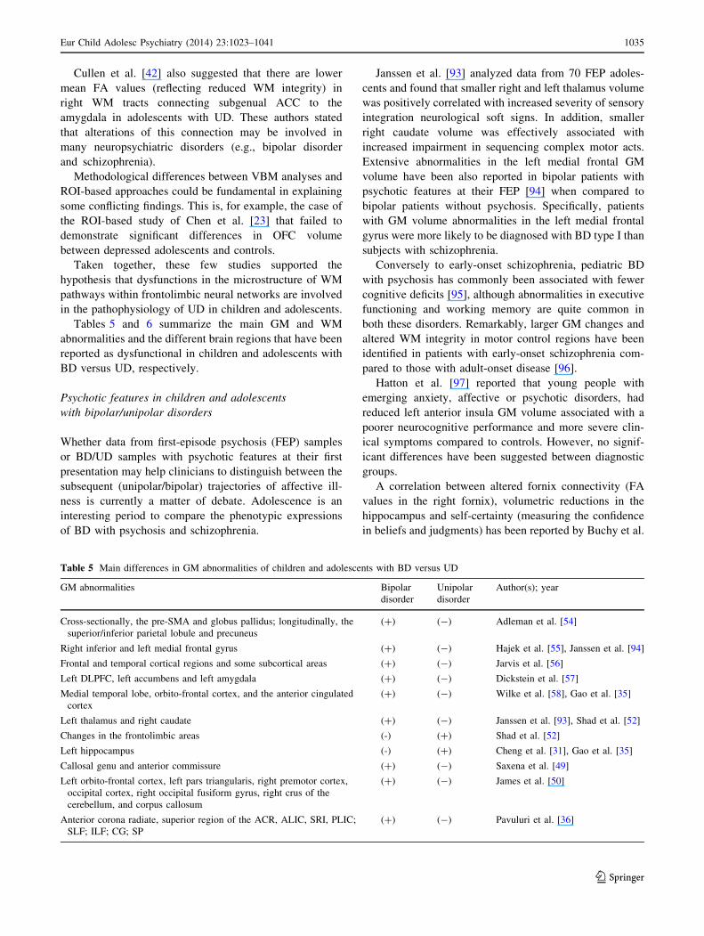
Cullen et al. [42] also suggested that there are lower
mean FA values (reflecting reduced WM integrity) in
right WM tracts connecting subgenual ACC to the
amygdala in adolescents with UD. These authors stated
that alterations of this connection may be involved in
many neuropsychiatric disorders (e.g., bipolar disorder
and schizophrenia).
Methodological differences between VBM analyses and
ROI-based approaches could be fundamental in explaining
some conflicting findings. This is, for example, the case of
the ROI-based study of Chen et al. [23] that failed to
demonstrate significant differences in OFC volume
between depressed adolescents and controls.
Taken together, these few studies supported the
hypothesis that dysfunctions in the microstructure of WM
pathways within frontolimbic neural networks are involved
in the pathophysiology of UD in children and adolescents.
Tables 5 and 6 summarize the main GM and WM
abnormalities and the different brain regions that have been
reported as dysfunctional in children and adolescents with
BD versus UD, respectively.
Psychotic features in children and adolescents
with bipolar/unipolar disorders
Whether data from first-episode psychosis (FEP) samples
or BD/UD samples with psychotic features at their first
presentation may help clinicians to distinguish between the
subsequent (unipolar/bipolar) trajectories of affective ill-
ness is currently a matter of debate. Adolescence is an
interesting period to compare the phenotypic expressions
of BD with psychosis and schizophrenia.
Janssen et al. [93] analyzed data from 70 FEP adoles-
cents and found that smaller right and left thalamus volume
was positively correlated with increased severity of sensory
integration neurological soft signs. In addition, smaller
right caudate volume was effectively associated with
increased impairment in sequencing complex motor acts.
Extensive abnormalities in the left medial frontal GM
volume have been also reported in bipolar patients with
psychotic features at their FEP [94] when compared to
bipolar patients without psychosis. Specifically, patients
with GM volume abnormalities in the left medial frontal
gyrus were more likely to be diagnosed with BD type I than
subjects with schizophrenia.
Conversely to early-onset schizophrenia, pediatric BD
with psychosis has commonly been associated with fewer
cognitive deficits [95], although abnormalities in executive
functioning and working memory are quite common in
both these disorders. Remarkably, larger GM changes and
altered WM integrity in motor control regions have been
identified in patients with early-onset schizophrenia com-
pared to those with adult-onset disease [96].
Hatton et al. [97] reported that young people with
emerging anxiety, affective or psychotic disorders, had
reduced left anterior insula GM volume associated with a
poorer neurocognitive performance and more severe clin-
ical symptoms compared to controls. However, no signif-
icant differences have been suggested between diagnostic
groups.
A correlation between altered fornix connectivity (FA
values in the right fornix), volumetric reductions in the
hippocampus and self-certainty (measuring the confidence
in beliefs and judgments) has been reported by Buchy et al.
Table 5 Main differences in GM abnormalities of children and adolescents with BD versus UD
GM abnormalities Bipolar
disorder
Unipolar
disorder
Author(s); year
Cross-sectionally, the pre-SMA and globus pallidus; longitudinally, the
superior/inferior parietal lobule and precuneus
(?) (-) Adleman et al. [54]
Right inferior and left medial frontal gyrus (?) (-) Hajek et al. [55], Janssen et al. [94]
Frontal and temporal cortical regions and some subcortical areas (?) (-) Jarvis et al. [56]
Left DLPFC, left accumbens and left amygdala (?) (-) Dickstein et al. [57]
Medial temporal lobe, orbito-frontal cortex, and the anterior cingulated
cortex
(?) (-) Wilke et al. [58], Gao et al. [35]
Left thalamus and right caudate (?) (-) Janssen et al. [93], Shad et al. [52]
Changes in the frontolimbic areas (-) (?) Shad et al. [52]
Left hippocampus (-) (?) Cheng et al. [31], Gao et al. [35]
Callosal genu and anterior commissure (?) (-) Saxena et al. [49]
Left orbito-frontal cortex, left pars triangularis, right premotor cortex,
occipital cortex, right occipital fusiform gyrus, right crus of the
cerebellum, and corpus callosum
(?) (-) James et al. [50]
Anterior corona radiate, superior region of the ACR, ALIC, SRI, PLIC;
SLF; ILF; CG; SP
(?) (-) Pavuluri et al. [36]
Eur Child Adolesc Psychiatry (2014) 23:1023–1041 1035
123
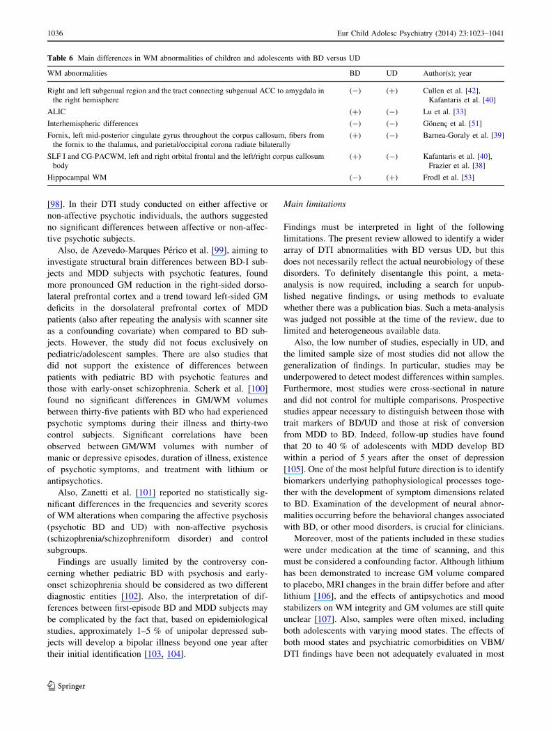
[98]. In their DTI study conducted on either affective or
non-affective psychotic individuals, the authors suggested
no significant differences between affective or non-affec-
tive psychotic subjects.
Also, de Azevedo-Marques Perico et al. [99], aiming to
investigate structural brain differences between BD-I sub-
jects and MDD subjects with psychotic features, found
more pronounced GM reduction in the right-sided dorso-
lateral prefrontal cortex and a trend toward left-sided GM
deficits in the dorsolateral prefrontal cortex of MDD
patients (also after repeating the analysis with scanner site
as a confounding covariate) when compared to BD sub-
jects. However, the study did not focus exclusively on
pediatric/adolescent samples. There are also studies that
did not support the existence of differences between
patients with pediatric BD with psychotic features and
those with early-onset schizophrenia. Scherk et al. [100]
found no significant differences in GM/WM volumes
between thirty-five patients with BD who had experienced
psychotic symptoms during their illness and thirty-two
control subjects. Significant correlations have been
observed between GM/WM volumes with number of
manic or depressive episodes, duration of illness, existence
of psychotic symptoms, and treatment with lithium or
antipsychotics.
Also, Zanetti et al. [101] reported no statistically sig-
nificant differences in the frequencies and severity scores
of WM alterations when comparing the affective psychosis
(psychotic BD and UD) with non-affective psychosis
(schizophrenia/schizophreniform disorder) and control
subgroups.
Findings are usually limited by the controversy con-
cerning whether pediatric BD with psychosis and early-
onset schizophrenia should be considered as two different
diagnostic entities [102]. Also, the interpretation of dif-
ferences between first-episode BD and MDD subjects may
be complicated by the fact that, based on epidemiological
studies, approximately 1–5 % of unipolar depressed sub-
jects will develop a bipolar illness beyond one year after
their initial identification [103, 104].
Main limitations
Findings must be interpreted in light of the following
limitations. The present review allowed to identify a wider
array of DTI abnormalities with BD versus UD, but this
does not necessarily reflect the actual neurobiology of these
disorders. To definitely disentangle this point, a meta-
analysis is now required, including a search for unpub-
lished negative findings, or using methods to evaluate
whether there was a publication bias. Such a meta-analysis
was judged not possible at the time of the review, due to
limited and heterogeneous available data.
Also, the low number of studies, especially in UD, and
the limited sample size of most studies did not allow the
generalization of findings. In particular, studies may be
underpowered to detect modest differences within samples.
Furthermore, most studies were cross-sectional in nature
and did not control for multiple comparisons. Prospective
studies appear necessary to distinguish between those with
trait markers of BD/UD and those at risk of conversion
from MDD to BD. Indeed, follow-up studies have found
that 20 to 40 % of adolescents with MDD develop BD
within a period of 5 years after the onset of depression
[105]. One of the most helpful future direction is to identify
biomarkers underlying pathophysiological processes toge-
ther with the development of symptom dimensions related
to BD. Examination of the development of neural abnor-
malities occurring before the behavioral changes associated
with BD, or other mood disorders, is crucial for clinicians.
Moreover, most of the patients included in these studies
were under medication at the time of scanning, and this
must be considered a confounding factor. Although lithium
has been demonstrated to increase GM volume compared
to placebo, MRI changes in the brain differ before and after
lithium [106], and the effects of antipsychotics and mood
stabilizers on WM integrity and GM volumes are still quite
unclear [107]. Also, samples were often mixed, including
both adolescents with varying mood states. The effects of
both mood states and psychiatric comorbidities on VBM/
DTI findings have been not adequately evaluated in most
Table 6 Main differences in WM abnormalities of children and adolescents with BD versus UD
WM abnormalities BD UD Author(s); year
Right and left subgenual region and the tract connecting subgenual ACC to amygdala in
the right hemisphere
(-) (?) Cullen et al. [42],
Kafantaris et al. [40]
ALIC (?) (-) Lu et al. [33]
Interhemispheric differences (-) (-) Gonenc et al. [51]
Fornix, left mid-posterior cingulate gyrus throughout the corpus callosum, fibers from
the fornix to the thalamus, and parietal/occipital corona radiate bilaterally
(?) (-) Barnea-Goraly et al. [39]
SLF I and CG-PACWM, left and right orbital frontal and the left/right corpus callosum
body
(?) (-) Kafantaris et al. [40],
Frazier et al. [38]
Hippocampal WM (-) (?) Frodl et al. [53]
1036 Eur Child Adolesc Psychiatry (2014) 23:1023–1041
123
studies. The presence of ADHD comorbidity in bipolar
children may be interpreted as a potential confounding
factor. ADHD symptoms are very frequent in samples of
bipolar children—ranging to 80 % [38]; this high comor-
bidity induced some authors [108] to suggest that ADHD
may represent a phenotypic manifestation of early-onset
BD.
Also, psychosis occurs in 16 % of BD cases and is
usually a marker of severity and poorer prognosis [105];
some studies analyzed bipolar subjects with psychotic
features, so that the findings may not be generalized to BD
as a whole.
Importantly, interpretation of WM abnormalities
explored with DTI and anatomical measurement are not
straightforward. Voxelwise analysis is subject to type I
errors, whereas the spatial resolution of the studies using
DTI acquisition may be excessively low.
As suggested by Frazier et al. [38], most DTI studies
suffer from low sensitivity and could be quite sensitive
only to FA differences in subsections of the most densely
packed fibers.
The observation of lower FA measurement in specific
brain areas could be erroneously interpreted as lower
connectivity [42], leading to imprecise anatomical mea-
surement of WM. Recent advancements in this field using
meta-analytic and multimodal neuroimaging approaches
should help to clarify how FA differences may reflect
connectivity dysfunctions. Moreover, processing pipelines
of DTI data are diverse and no clear consensus exists on
how to handle DTI data.
Although ROI studies provide relevant information on
regional differences, they should be similarly considered
cautiously, especially in the identification of more global
changes in the brain. Reliable tractography studies are also
missing.
Considering that the main findings derived by the
included studies are highly heterogeneous, it has not been
possible to analyze some relevant issues (e.g., differences
in the instruments/criteria used to diagnose BD in children;
BD subtypes and bipolar polarity; duration of the illness;
childhood versus adolescent onset; effects of psychoactive
medications; eventual comorbidity; family history of BD;
socio-demographic factors; sample size; different scans;
methods to evaluate the used scans and statistics) that could
have influenced the main results of the present review. All
these issues may explain, at least partially, the large dis-
parity and disorganization of the main results.
Finally, most of the correlations that were reported
should be interpreted as exploratory (see Tables), since no
correction for multiple comparisons has been performed or
because post hoc behavioral analyses included only a
subset of the entire samples. Future neuroimaging studies
should be useful in elucidating pathophysiological
processes underlying the differential development of BD
from UD supporting to clarify the relationship between
structural and functional neuroimaging findings. They will
require large multicentric samples.
Implications and future directions
Overall, based on the limited studies available, more WM
abnormalities have been reported in BD children/adoles-
cents than in those with UD suggesting the possible exis-
tence of vulnerability markers for BD. More studies about
WM abnormalities in youths with UD are necessary.
Future longitudinal studies should: (1) directly compare
groups of children with UD and BD; (2) use multimodal
imaging for cross-validation of the results and easier
interpretation; and (3) combine follow-up imaging assess-
ments to investigate whether neurodevelopmental changes
may be considered specifically related to diagnostic cate-
gories. In addition, future studies should focus on dimen-
sional approaches able to redefine ‘‘bipolarity’’ in terms of
underlying pathophysiological processes in genetically and
symptomatically at risk subjects. These studies should also
be conducted using more reliable neuroimaging techniques
such as tractography to provide biomarkers as well as
biological targets for more selective personalized treat-
ments. Longitudinal follow-up studies should also allow
identifying differential developmental trajectories between
bipolar and unipolar subjects. Finally, the combination of
different neuroimaging techniques and the development of
novel algorithms based on machine learning, a branch of
artificial intelligence, should warrant a more detailed
understanding of neural circuitry abnormalities underlying
major affective disorders [109].
Conflict of interest None.
References
1. Washburn JJ, West AE, Heil JA (2011) Treatment of pediatric
bipolar disorder: a review. Minerva Psichiatr 52:21–35
2. Faedda GL, Baldessarini RJ, Suppes T, Tondo L, Becker I,
Lipschitz DS (1995) Pediatric-onset bipolar disorder: a neglec-
ted clinical and public health problem. Harv Rev Psychiatry
3:171–195
3. Van Meter AR, Moreira AL, Youngstrom EA (2011) Meta-
analysis of epidemiologic studies of pediatric bipolar disorder.
J Clin Psychiatry 72:1250–1256
4. Geoffroy PA, Etain B, Scott J, Henry C, Jamain S, Leboyer M,
Bellivier F (2013) Reconsideration of bipolar disorder as a
developmental disorder: importance of the time of onset.
J Physiol Paris 107:278–285
5. Lewinsohn PM, Klein DN, Seeley JR (1995) Bipolar disorders
in a community sample of older adolescents: prevalence, phe-
nomenology, comorbidity, and course. J Am Acad Child Ado-
lesc Psychiatry 34:454–463
Eur Child Adolesc Psychiatry (2014) 23:1023–1041 1037
123
6. Lewinsohn PM, Klein DN, Seeley JR (2000) Bipolar disorder
during adolescence and young adulthood in a community sam-
ple. Bipolar Disord 2:281–293
7. [No authors listed] (2001) National Institute of Mental Health
Research roundtable on prepubertal bipolar disorder. J Am Acad
Child Adolesc Psychiatry 40:871–878
8. Williams SB, O’Connor EA, Eder M, Whitlock EP (2009)
Screening for child and adolescent depression in primary care
settings: a systematic evidence review for the US Preventive
Services Task Force. Pediatrics 123:e716–e735
9. Pine DS, Cohen P, Gurley D, Brook J, Ma Y (1998) The risk for
early-adulthood anxiety and depressive disorders in adolescents
with anxiety and depressive disorders. Arch Gen Psychiatry
55:56–64
10. Pompili M, Rihmer Z, Akiskal H, Amore M, Gonda X, Inna-
morati M, Lester D, Perugi G, Serafini G, Telesforo L, Tatarelli
R, Girardi P (2012) Temperaments mediate suicide risk and
psychopathology among patients with bipolar disorders. Compr
Psychiatry 53:280–285
11. Serafini G, Pompili M, Innamorati M, Dwivedi Y, Brahmachari
G, Girardi P (2013) Pharmacological properties of glutamatergic
drugs targeting NMDA receptors and their application in major
depression. Curr Pharm Des 19:1898–1922
12. Serafini G, Pompili M, Innamorati M, Giordano G, Montebovi
F, Sher L, Dwivedi Y, Girardi P (2012) The role of microRNAs
in synaptic plasticity, major affective disorders and suicidal
behavior. Neurosci Res 73:179–190
13. Goldstein BI, Bukstein OG (2010) Comorbid substance use dis-
orders among youth with bipolar disorder: opportunities for early
identification and prevention. J Clin Psychiatry 71:348–358
14. Yoon YH, Chen CM, Yi HY, Moss HB (2011) Effect of
comorbid alcohol and drug use disorders on premature death
among unipolar and bipolar disorder decedents in the United
States, 1999 to 2006. Compr Psychiatry 52:453–464
15. Mitsunaga MM, Garrett A, Howe M, Karchemskiy A, Reiss A,
Chang K (2011) Increased subgenual cingulate cortex volume
in pediatric bipolar disorder associated with mood stabilizer
exposure. J Child Adolesc Psychopharmacol 21:149–155
16. Mahon K, Burdick KE, Szeszko PR (2010) A role for white
matter abnormalities in the pathophysiology of bipolar disorder.
Neurosci Biobehav Rev 34:533–554
17. Biederman J, Makris N, Valera EM, Monuteaux MC, Goldstein
JM, Buka S, Boriel DL, Bandyopadhyay S, Kennedy DN, Ca-
viness VS, Bush G, Aleardi M, Hammerness P, Faraone SV,
Seidman LJ (2008) Towards further understanding of the co-
morbidity between attention deficit hyperactivity disorder
and bipolar disorder: a MRI study of brain volumes. Psychol
Med 38:1045–1056
18. Adleman NE, Barnea-Goraly N, Chang KD (2004) Review of
magnetic resonance imaging and spectroscopy studies in chil-
dren with bipolar disorder. Exp Rev Neurother 4:69–77
19. Frazier JA, Ahn MS, DeJong S, Bent EK, Breeze JL, Giuliano
AJ (2005) Magnetic resonance imaging studies in early-onset
bipolar disorder: a critical review. Harv Rev Psychiatry
13:125–140
20. Fusar-Poli P, Howes O, Bechdolf A, Borgwardt S (2012)
Mapping vulnerability to bipolar disorder: a systematic review
and meta-analysis of neuroimaging studies. J Psychiatry Neu-
rosci 37:170–184
21. Chantiluke K, Halari R, Simic M, Pariante CM, Papadopoulos
A, Giampietro V, Rubia K (2012) Fronto-striato-cerebellar
dysregulation in adolescents with depression during motivated
attention. Biol Psychiatry 71:59–67
22. Halari R, Simic M, Pariante CM, Papadopoulos A, Cleare A,
Brammer M, Fombonne E, Rubia K (2009) Reduced activation
in lateral prefrontal cortex and anterior cingulate during
attention and cognitive control functions in medication-
naıve adolescents with depression compared to controls. J Child
Psychol Psychiatry 50:307–316
23. Chen HH, Rosenberg DR, MacMaster FP, Easter PC, Caetano
SC, Nicoletti M, Hatch JP, Nery FG, Soares JC (2008) Orbito-
frontal cortex volumes in medication naıve children with major
depressive disorder: a magnetic resonance imaging study.
J Child Adolesc Psychopharmacol 18:551–556
24. Matsuo K, Rosenberg DR, Easter PC, MacMaster FP, Chen HH,
Nicoletti M, Caetano SC, Hatch JP, Soares JC (2008) Striatal
volume abnormalities in treatment-naıve patients diagnosed
with pediatric major depressive disorder. J Child Adolesc Psy-
chopharmacol 18:121–131
25. Caetano SC, Fonseca M, Hatch JP, Olvera RL, Nicoletti M,
Hunter K, Lafer B, Pliszka SR, Soares JC (2007) Medial tem-
poral lobe abnormalities in pediatric unipolar depression. Neu-
rosci Lett 427:142–147
26. Rosso IM, Cintron CM, Steingard RJ, Renshaw PF, Young AD,
Yurgelun-Todd DA (2005) Amygdala and hippocampus vol-
umes in pediatric major depression. Biol Psychiatry 57:21–26
27. Nolan CL, Moore GJ, Madden R, Farchione T, Bartoi M, Lorch
E, Stewart CM, Rosenberg DR (2002) Prefrontal cortical vol-
ume in childhood-onset major depression: preliminary findings.
Arch Gen Psychiatry 59:173–179
28. Adler CM, DelBello MP, Strakowski SM (2006a) Brain network
dysfunction in bipolar disorder. CNS Spectr 11:312–320, quiz
323–324
29. Arnone D, Cavanagh J, Gerber D, Lawrie SM, Ebmeier KP,
McIntosh AM (2009) Magnetic resonance imaging studies in
bipolar disorder and schizophrenia: meta-analysis. Br J Psychi-
atry 195:194–201
30. Singh MK, Chang KD, Chen MC, Kelley RG, Garrett A,
Mitsunaga MM, Bararpour L, Howe M, Reiss AL, Gotlib IH
(2012) Volumetric reductions in the subgenual anterior cingulate
cortex in adolescents with bipolar I disorder. Bipolar Disord
14:585–596
31. Cheng YQ, Xu J, Chai P, Li HJ, Luo CR, Yang T, Li L, Shan
BC, Xu XF, Xu L (2010) Brain volume alteration and the cor-
relations with the clinical characteristics in drug-naıve first-
episode MDD patients: a voxel-based morphometry study.
Neurosci Lett 480:30–34
32. Foland-Ross LC, Brooks JO 3rd, Mintz J, Bartzokis G, Town-
send J, Thompson PM, Altshuler LL (2012) Mood-state effects
on amygdala volume in bipolar disorder. J Affect Disord
139:298–301
33. Lu LH, Zhou XJ, Fitzgerald J, Keedy SK, Reilly JL, Passarotti
AM, Sweeney JA, Pavuluri M (2012) Microstructural abnor-
malities of white matter differentiate pediatric and adult-onset
bipolar disorder. Bipol Disord 14:597–606
34. Najt P, Nicoletti M, Chen HH, Hatch JP, Caetano SC, Sassi RB,
Axelson D, Brambilla P, Keshavan MS, Ryan ND, Birmaher B,
Soares JC (2007) Anatomical measurements of the orbitofrontal
cortex in child and adolescent patients with bipolar disorder.
Neurosci Lett 413:183–186
35. Gao W, Jiao Q, Qi R, Zhong Y, Lu D, Xiao Q, Lu S, Xu C,
Zhang Y, Liu X, Yang F, Lu G, Su L (2013) Combined analyses
of gray matter voxel-based morphometry and white matter tract-
based spatial statistics in pediatric bipolar mania. J Affect Dis-
ord 150:70–76
36. Pavuluri MN, Yang S, Kamineni K, Passarotti AM, Srinivasan
G, Harral EM, Sweeney JA, Zhou XJ (2009) Diffusion tensor
imaging study of white matter fiber tracts in pediatric bipolar
disorder and attention-deficit/hyperactivity disorder. Biol Psy-
chiatry 65:586–593
37. Adler CM, Adams J, DelBello MP, Holland SK, Schmithorst V,
Levine A, Jarvis K, Strakowski SM (2006) Evidence of white
1038 Eur Child Adolesc Psychiatry (2014) 23:1023–1041
123
matter pathology in bipolar disorder adolescents experiencing
their first episode of mania: a diffusion tensor imaging study.
Am J Psychiatry 163:322–324
38. Frazier JA, Breeze JL, Papadimitriou G, Kennedy DN, Hodge
SM, Moore CM, Howard JD, Rohan MP, Caviness VS, Makris
N (2007) White matter abnormalities in children with and at risk
for bipolar disorder. Bipolar Disord 9:799–809
39. Barnea-Goraly N, Chang KD, Karchemskiy A, Howe ME, Reiss
AL (2009) Limbic and corpus callosum aberrations in adoles-
cents with bipolar disorder: a tract-based spatial statistics ana-
lysis. Biol Psychiatry 66:238–244
40. Kafantaris V, Kingsley P, Ardekani B, Saito E, Lencz T, Lim K,
Szeszko P (2009) Lower orbital frontal white matter integrity in
adolescents with bipolar I disorder. J Am Acad Child Adolesc
Psychiatry 48:79–86
41. Ma N, Li L, Shu N, Liu J, Gong G, He Z, Li Z, Tan L, Stone
WS, Zhang Z, Xu L, Jiang T (2007) White matter abnormalities
in first-episode, treatment-naive young adults with major
depressive disorder. Am J Psychiatry 164:823–826
42. Cullen KR, Klimes-Dougan B, Muetzel R, Mueller BA, Cam-
chong J, Houri A, Kurma S, Lim KO (2010) Altered white
matter microstructure in adolescents with major depression: a
preliminary study. J Am Acad Child Adolesc Psychiatry
49:173–183
43. Whalley HC, Sprooten E, Hackett S, Hall L, Blackwood DH,
Glahn DC, Bastin M, Hall J, Lawrie SM, Sussmann JE, McIn-
tosh AM (2013) Polygenic risk and white matter integrity in
individuals at high risk of mood disorder. Biol Psychiatry
74:280–286
44. Sprooten E, Sussmann JE, Clugston A, Peel A, McKirdy J,
Moorhead TW, Anderson S, Shand AJ, Giles S, Bastin ME, Hall
J, Johnstone EC, Lawrie SM, McIntosh AM (2011) White matter
integrity in individuals at high genetic risk of bipolar disorder.
Biol Psychiatry 70:350–356
45. Linke J, King AV, Poupon C, Hennerici MG, Gass A, Wessa M
(2013) Impaired anatomical connectivity and related executive
functions: differentiating vulnerability and disease marker in
bipolar disorder. Biol Psychiatry 74:908–916
46. Cardoso de Almeida JR, Phillips ML (2013) Distinguishing
between unipolar depression and bipolar depression: current and
future clinical and neuroimaging perspectives. Biol Psychiatry
73:111–118
47. Moher D, Liberati A, Tetzlaff J, Altman DG, PRISMA Group
(2009) Preferred reporting items for systematic reviews and
meta-analyses: the PRISMA statement. BMJ 339:b2535
48. Lu LH, Zhou XJ, Keedy SK, Reilly JL, Sweeney JA (2011)
White matter microstructure in untreated first episode bipolar
disorder with psychosis: comparison with schizophrenia. Bipol
Disord 13:604–613
49. Saxena K, Tamm L, Walley A, Simmons A, Rollins N, Chia J,
Soares JC, Emslie GJ, Fan X, Huang H (2012) A preliminary
investigation of corpus callosum and anterior commissure
aberrations in aggressive youth with bipolar disorders. J Child
Adolesc Psychopharmacol 22:112–119
50. James A, Hough M, James S, Winmill L, Burge L, Nijhawan S,
Matthews PM, Zarei M (2011) Greater white and grey mat-
ter changes associated with early cannabis use in adolescent-
onset schizophrenia (AOS). Schizophr Res 128:91–97
51. Gonenc A, Frazier JA, Crowley DJ, Moore CM (2010) Com-
bined diffusion tensor imaging and transverse relaxometry in
early-onset bipolar disorder. J Am Acad Child Adolesc Psy-
chiatry 49:1260–1268
52. Shad MU, Muddasani S, Rao U (2012) Gray matter differences
between healthy and depressed adolescents: a voxel-based
morphometry study. J Child Adolesc Psychopharmacol
22:190–197
53. Frodl T, Reinhold E, Koutsouleris N, Donohoe G, Bondy B,
Reiser M, Moller HJ, Meisenzahl EM (2010) Childhood stress,
serotonin transporter gene and brain structures in major
depression. Neuropsychopharmacology 35:1383–1390
54. Adleman NE, Fromm SJ, Razdan V, Kayser R, Dickstein DP,
Brotman MA, Pine DS, Leibenluft E (2012) Cross-sectional and
longitudinal abnormalities in brain structure in children with
severe mood dysregulation or bipolar disorder. J Child Psychol
Psychiatry 53:1149–1156
55. Hajek T, Cullis J, Novak T, Kopecek M, Blagdon R, Propper L,
Stopkova P, Duffy A, Hoschl C, Uher R, Paus T, Young LT,
Alda M (2013) Brain structural signature of familial predispo-
sition for bipolar disorder: replicable evidence for involvement
of the right inferior frontal gyrus. Biol Psychiatry 73:144–152
56. Jarvis K, DelBello MP, Mills N, Elman I, Strakowski SM, Adler
CM (2008) Neuroanatomic comparison of bipolar adolescents
with and without cannabis use disorders. J Child Adolesc Psy-
chopharmacol 18:557–563
57. Dickstein DP, Milham MP, Nugent AC, Drevets WC, Charney
DS, Pine DS, Leibenluft E (2005) Frontotemporal alterations in
pediatric bipolar disorder: results of a voxel-based morphometry
study. Arch Gen Psychiatry 62:734–741
58. Wilke M, Kowatch RA, DelBello MP, Mills NP, Holland SK
(2004) Voxel-based morphometry in adolescents with bipolar
disorder: first results. Psychiatry Res 131:57–69
59. Alexander DC, Hubbard PL, Hall MG, Moore EA, Ptito M,
Parker GJ, Dyrby TB (2010) Orientationally invariant indices of
axon diameter and density from diffusion MRI. Neuroimage
52:1374–1389
60. Kempton MJ, Salvador Z, Munafo MR, Geddes JR, Simmons A,
Frangou S, Williams SC (2011) Structural neuroimaging studies
in major depressive disorder. Meta-analysis and comparison
with bipolar disorder. Arch Gen Psychiatry 68:675–690
61. Caetano SC, Olvera RL, Glahn D, Fonseca M, Pliszka S, Soares
JC (2005) Fronto-limbic brain abnormalities in juvenile onset
bipolar disorder. Biol Psychiatry 58:525–531
62. Blumberg HP, Kaufman J, Martin A, Charney DS, Krystal JH,
Peterson BS (2004) Significance of adolescent neurodevelop-
ment for the neural circuitry of bipolar disorder. Ann N Y Acad
Sci 1021:376–383
63. Strakowski SM, Delbello MP, Adler CM (2005) The functional
neuroanatomy of bipolar disorder: a review of neuroimaging
findings. Mol Psychiatry 10:105–116
64. Chang K, Adleman NE, Dienes K, Simeonova DI, Menon V,
Reiss A (2004) Anomalous prefrontal-subcortical activation in
familial pediatric bipolar disorder: a functional magnetic reso-
nance imaging investigation. Arch Gen Psychiatry 61:781–792
65. Yurgelun-Todd DA, Gruber SA, Kanayama G, Killgore WD,
Baird AA, Young AD (2000) fMRI during affect discrimination
in bipolar affective disorder. Bipol Disord 2:237–248
66. Plas J, Brion F, Jeanneau A, Chevalier JF, Dussaux P, Brion S
(1999) Contradictions in an original case. Contradictory psy-
chiatric behavior after traumatic injury of the anterior part of the
corpus callosum. Rev Neurol (Paris) 155:569–574
67. Paul LK, Lautzenhiser A, Brown WS, Hart A, Neumann D,
Spezio M, Adolphs R (2006) Emotional arousal in agenesis of
the corpus callosum. Int J Psychophysiol 61:47–56
68. Tamietto M, Adenzato M, Geminiani G, de Gelder B (2007)
Fast recognition of social emotions takes the whole brain:
interhemispheric cooperation in the absence of cerebral asym-
metry. Neuropsychologia 45:836–843
69. Gazzaniga MS (2000) Cerebral specialization and interhemi-
spheric communication: does the corpus callosum enable the
human condition? Brain 123:1293–1326
70. Giedd JN, Rumsey JM, Castellanos FX, Rajapakse JC, Kaysen
D, Vaituzis AC, Vauss YC, Hamburger SD, Rapoport JL (1996)
Eur Child Adolesc Psychiatry (2014) 23:1023–1041 1039
123
A quantitative MRI study of the corpus callosum in children and
adolescents. Brain Res Dev Brain Res 91:274–280
71. Giedd JN, Blumenthal J, Jeffries NO, Rajapakse JC, Vaituzis
AC, Liu H, Berry YC, Tobin M, Nelson J, Castellanos FX
(1999) Development of the human corpus callosum during
childhood and adolescence: a longitudinal MRI study. Prog
Neuropsychopharmacol Biol Psychiatry 23:571–588
72. Keshavan MS, Diwadkar VA, DeBellis M, Dick E, Kotwal R,
Rosenberg DR, Sweeney JA, Minshew N, Pettegrew JW (2002)
Development of the corpus callosum in childhood, adolescence
and early adulthood. Life Sci 70:1909–1922
73. Chepuri NB, Yen YF, Burdette JH, Li H, Moody DM, Maldjian
JA (2002) Diffusion anisotropy in the corpus callosum. AJN-
RAmJ Neuroradiol 23:803–808
74. Barnea-Goraly N, Menon V, Eckert M, Tamm L, Bammer R,
Karchemskiy A, Dant CC, Reiss AL (2005) White matter devel-
opment during childhood and adolescence: A cross sectional
diffusion tensor imaging study. Cereb Cortex 15:1848–1854
75. Caetano SC, Silveira CM, Kaur S, Nicoletti M, Hatch JP,
Brambilla P, Sassi R, Axelson D, Keshavan MS, Ryan ND,
Birmaher B, Soares JC (2008) Abnormal corpus callosum
myelination in pediatric bipolar patients. J Affect Disord
108:297–301
76. Yasar AS, Monkul ES, Sassi RB, Axelson D, Brambilla P,
Nicoletti MA, Hatch JP, Keshavan M, Ryan N, Birmaher B,
Soares JC (2006) MRI study of corpus callosum in children and
adolescents with bipolar disorder. Psychiatry Res 146:83–85
77. Yurgelun-Todd DA, Silveri MM, Gruber SA, Rohan ML, Pi-
mentel PJ (2007) White matter abnormalities observed in
bipolar disorder: a diffusion tensor imaging study. Bipolar
Disord 9:504–512
78. Phillips ML, Ladouceur CD, Drevets WC (2008) A neural
model of voluntary and automatic emotion regulation: implica-
tions for understanding the pathophysiology and neurodevelop-
ment of bipolar disorder. Mol Psychiatry 13:833–857
79. Lin F, Weng S, Xie B, Wu G, Lei H (2011) Abnormal frontal
cortex white matter connections in bipolar disorder: a DTI
tractography study. J Affect Disord 131:299–306
80. Merriam EP, Thase ME, Haas GL, Keshavan MS, Sweeney JA
(1999) Prefrontal Cortical Dysfunction in Depression Deter-
mined by Wisconsin Card Sorting Test Performance. Am J
Psychiatry 156:780–782
81. Drevets WC, Price JL, Furey ML (2008) Brain structural and
functional abnormalities in mood disorders: Implications for
neurocircuitry models of depression. Brain Struct Funct
213:93–118
82. Steele JD, Currie J, Lawrie SM, Reid I (2007) Prefrontal cortical
functional abnormality in major depressive disorder: a stereo-
tactic metaanalysis. J Affect Disord 101:1–11
83. Peng J, Liu J, Nie B, Li Y, Shan B, Wang G, Li K (2011)
Cerebral and cerebellar gray matter reduction in first-episode
patients with major depressive disorder: a voxel-based mor-
phometry study. Eur J Radiol 80:395–399
84. Zou K, Deng W, Li T, Zhang B, Jiang L, Huang C, Sun X, Sun
X (2010) Changes of brain morphometry in first-episode, drug-
nai‹ ve, non-late-life adult patients with major depression: An
optimized voxel-based morphometry study. Biol Psychiatry
67:186–188
85. Ridderinkhof KR, Ullsperger M, Crone EA, Nieuwenhuis S
(2004) The role of the medial frontal cortex in cognitive control.
Science 306:443–447
86. Johansen-Berg H, Gutman DA, Behrens TE, Matthews PM,
Rushworth MF, Katz E, Lozano AM, Mayberg HS (2008)
Anatomical connectivity of the subgenual cingulate region tar-
geted with deep brain stimulation for treatment-resistant
depression. Cereb Cortex 18:1374–1383
87. Mayberg HS, Lozano AM, Voon V, McNeely HE, Seminowicz
D, Hamani C, Schwalb JM, Kennedy SH (2005) Deep brain
stimulation for treatment-resistant depression. Neuron
45:651–660
88. Drevets WC, Bogers W, Raichle ME (2002) Functional ana-
tomical correlates of antidepressant drug treatment assessed
using PET measures of regional glucose metabolism. Eur Neu-
ropsychopharmacol 12:527–544
89. Drevets WC (1999) Prefrontal cortical-amygdalar metabolism in
major depression. Ann N Y Acad Sci 877:614–637
90. Bremner JD, Randall P, Vermetten E, Staib L, Bronen RA,
Mazure C, Capelli S, McCarthy G, Innis RB, Charney DS
(1997) Magnetic resonance imaging-based measurement of
hippocampal volume in posttraumatic stress disorder related to
childhood physical and sexual abuse—A preliminary report.
Biol Psychiatry 41:23–32
91. Stein MB, Koverola C, Hanna C, Torchia MG, McClarty B
(1997) Hippocampal volume in women victimized by childhood
sexual abuse. Psychol Med 27:951–959
92. Vythilingam M, Heim C, Newport J, Miller AH, Anderson E,
Bronen R, Brummer M, Staib L, Vermetten E, Charney DS,
Nemeroff CB, Bremner JD (2002) Childhood trauma associated
with smaller hippocampal volume in women with major
depression. Am J Psychiatry 159:2072–2080
93. Janssen J, Diaz-Caneja A, Reig S, Bombın I, Mayoral M, Par-
ellada M, Graell M, Moreno D, Zabala A, Vazquez VG, Desco
M, Arango C (2009) Brain morphology and neurological soft
signs in adolescents with first-episode psychosis. Br J Psychiatry
195:227–233
94. Janssen J, Reig S, Parellada M, Moreno D, Graell M, Fraguas D,
Zabala A, Garcia Vazquez V, Desco M, Arango C (2008)
Regional gray matter volume deficits in adolescents with first-
episode psychosis. J Am Acad Child Adolesc Psychiatry
47:1311–1320
95. White T, Ho BC, Ward J, O’Leary D, Andreasen NC (2006)
Neuropsychological performance in first-episode adolescents
with schizophrenia: a comparison with first-episode adults and
adolescent control subjects. Biol Psychiatry 60:463–471
96. Douaud G, Smith S, Jenkinson M, Behrens T, Johansen-Berg H,
Vickers J, James S, Voets N, Watkins K, Matthews PM, James A
(2007) Anatomically related grey and white matter abnormalities
in adolescent onset schizophrenia. Brain 130:2375–2386
97. Hatton SN, Lagopoulos J, Hermens DF, Naismith SL, Bennett
MR, Hickie IB (2012) Correlating anterior insula gray matter
volume changes in young people with clinical and neurocogni-
tive outcomes: an MRI study. BMC Psychiatry 12:4
98. Buchy L, Luck D, Czechowska Y, Malla A, Joober R, Lepage M
(2012) Diffusion tensor imaging tractography of the fornix and
belief confidence in first-episode psychosis. Schizophr Res
137:80–84
99. de Azevedo-Marques Perico C, Duran FL, Zanetti MV, Santos
LC, Murray RM, Scazufca M, Menezes PR, Busatto GF, Schau-
felberger MS (2011) A population-based morphometric MRI
study in patients with first-episode psychotic bipolar disorder:
comparison with geographically matched healthy controls
and major depressive disorder subjects. Bipolar Disord 13:28–40
100. Scherk H, Kemmer C, Usher J, Reith W, Falkai P, Gruber O
(2008) No change to grey and white matter volumes in bipo-
lar I disorder patients. Eur Arch Psychiatry Clin Neurosci
258:345–349
101. Zanetti MV, Schaufelberger MS, de Castro CC, Menezes PR,
Scazufca M, McGuire PK, Murray RM, Busatto GF (2008)
White-matter hyperintensities in first-episode psychosis. Br J
Psychiatry 193:25–30
102. Craddock N, Owen MJ (2010) The Kraepelinian dichotomy–
going, going… but still not gone. Br J Psychiatry 196:92–95
1040 Eur Child Adolesc Psychiatry (2014) 23:1023–1041
123
103. Schaffer A, Cairney J, Veldhuizen S, Kurdyak P, Cheung A,
Levitt A (2010) A population-based analysis of distinguishers of
bipolar disorder from major depressive disorder. J Affect Disord
125:350–354
104. Strakowski SM, Tsai SY, Delbello MP, Chen CC, Fleck DE,
Adler CM, Arndt S, Amicone J (2007) Outcome following a first
manic episode: cross-national US and Taiwan comparison.
Bipolar Disord 9:820–827
105. Birmaher B, Axelson D, Strober M, Gill MK, Yang M, Ryan N,
Goldstein B, Hunt J, Esposito-Smythers C, Iyengar S, Goldstein
T, Chiapetta L, Keller M, Leonard H (2009) Comparison of
manic and depressive symptoms between children and adoles-
cents with bipolar spectrum disorders. Bipol Disord 11:52–62
106. Cousins DA, Aribisala B, Nicol Ferrier I, Blamire AM (2013)
Lithium, gray matter, and magnetic resonance imaging signal.
Biol Psychiatry 73:652–657
107. Hafeman DM, Chang KD, Garrett AS, Sanders EM, Phillips ML
(2012) Effects of medication on neuroimaging findings in
bipolar disorder: an updated review. Bipolar Disord 14:375–410
108. Singh MK, DelBello MP, Kowatch RA, Strakowski SM (2006)
Co-occurrence of bipolar and attention-deficit hyperactivity
disorders in children. Bipol Disord 8:710–720
109. Phillips ML, Swartz HA (2014) A critical appraisal of neuro-
imaging studies of bipolar disorder: toward a new conceptuali-
zation of underlying neural circuitry and a road map for future
research. Am J Psychiatry 171:829–843
Eur Child Adolesc Psychiatry (2014) 23:1023–1041 1041
123


