Embed Size (px)
Citation preview
Intelligence 38 (2010) 293–303
Contents lists available at ScienceDirect
Intelligence
Brain networks for working memory and factors of intelligence assessed inmales and females with fMRI and DTI
C.Y. Tang a,b,⁎, E.L. Eaves a, J.C. Ng a, D.M. Carpenter a, X. Mai a, D.H. Schroeder c, C.A. Condon c,R. Colom d, R.J. Haier e
a Department of Radiology, Mt. Sinai School of Medicine, NY, NY, USAb Department of Psychiatry, Mt. Sinai School of Medicine, NY, NY, USAc Johnson O'Connor Research Foundation, Chicago, IL, USAd Universidad Autonoma de Madrid, Spaine University of California, Irvine, CA, USA (Emeritus)
a r t i c l e i n f o
⁎ Corresponding author. Department of RadiologyMedicine, NY, NY, USA.
E-mail address: [email protected] (C.Y. Tang
0160-2896/$ – see front matter © 2010 Elsevier Inc.doi:10.1016/j.intell.2010.03.003
a b s t r a c t
Article history:Received 24 September 2009Received in revised form 21 March 2010Accepted 22 March 2010Available online 28 April 2010
Neuro-imaging studies of intelligence implicate the importance of a parietal–frontal network. Oneunresolved issue is whether this network underlies a general factor of intelligence (g) or otherspecific cognitive factors. A second unresolved issue is whether males and females use differentparts of this network. Here we obtained intelligence factors (general, speed of reasoning, spatial,memory, andnumerical) froma large set of tests completedby6929 young adults, 40ofwhom(21males, 19 females) also completed DTI and fMRI during a working memory n-back task. Withinbrain areas activatedduring this task, correlationswere computed betweenpercent activation andscores on the intelligence factors. Themain findings were: (1) individual differences in activationduring the n-back task were correlated to the general intelligence factor (g), as well as to distilledestimates (removing g) of speed of reasoning, numerical ability, and spatial ability, but not tomemory, (2) the correlations were mainly bilateral for females and unilateral for males, and (3)differences in the integrity of the axonal connections were also related to the functional findingsshowing that integrity of interhemispheric connections was positively correlated to someintelligence factors in females but negatively correlated in males. This study illustrates thepotential for identifyingaspects of the neural basis of intelligenceusing a combinationof structuraland functional imaging.
© 2010 Elsevier Inc. All rights reserved.
Keywords:IntelligenceWorking memoryfMRIDTISex differences
1. Introduction
Neuro-imaging studies of the underlying structural andfunctional anatomy of intelligence implicate areas through-out the brain, irrespective of the intelligence tests used. Jungand Haier (2007) characterized these findings as mostly, butnot exclusively, in frontal and parietal areas. They proposed amodel of how these areas may form overlapping networksunderlying individual differences in intelligence: the Parieto-Frontal Integration Theory—P-FIT. The P-FIT areas represent
, Mt. Sinai School of
).
All rights reserved.
stages of information processing from posterior sensoryperception to abstraction in parietal areas to anteriorhypothesis testing and decision-making. Integration ofinformation among the areas is key. Based on functionalimaging studies that found inverse correlations betweenregional brain activation and performance on intelligencetests (Haier et al., 1988; Neubauer & Fink, 2009) the P-FITincludes the hypothesis that efficient flow of informationaround these networks is related to intelligence. Similarnetworks have been identified for performance on funda-mental cognitive tasks including aspects of attention andmemory (Cabeza & Nyberg, 2000; Naghavi & Nyberg, 2007;Wager, Jonides, & Reading, 2004; Wager & Smith, 2003),although inverse correlations with such tasks are notreported, possibly because fundamental cognitive tasks used

294 C.Y. Tang et al. / Intelligence 38 (2010) 293–303
in imaging studies usually are chosen to minimize individualdifferences in performance.
Since intelligence tests tap more than one cognitivedomain, it remains to be seen if the P-FIT or other models ofbrain networks represent a general factor of intelligence (g),common among all tests, or more specific group factors likememory or spatial ability. So far, only two structural imagingstudies have extracted a g-factor score from a battery of testsand then correlated these and other more specific group factorscores (with g removed) to gray matter (Colom et al., 2009;Haier et al., 2009). Both studies showed similar results for aspatial factor, but not for a g-factor, suggesting that there maynot be a single neural basis for g (Haier et al., 2009). Johnsonet al. (2008) also, for example, extracted other cognitive factorswith g removed in a small sample and showed some graymatter correlates different than those associated with g.
Functional imaging studies of networks related to intelli-gence have not yet used g-factor scores as dependent variables,instead relying on single tests like the Raven ProgressiveMatrices Test (Gray, Chabris, & Braver, 2003; Haier et al., 1988;Lee et al., 2006; Prabhakaran, Smith, Desmond, Glover, &Gabrieli, 1997). The interpretation of functional imaging data,moreover, is constrained by the task performed during theimaging, unlike structural imaging (Toga & Thompson, 2005),so task selection is a key element of research design. Twofunctional imaging studies with fMRI (Gray et al., 2003;Waiteret al., 2009) have used the working memory n-back taskbecause working memory is highly related to intelligence(Colom, Abad, Quiroga, Shih, & Flores-Mendoza, 2008; Colomet al., 2005; Colom, Rebollo, Palacios, Juan-Espinosa, &Kyllonen, 2004; Engle, 2002; Grabner, Fink, Stipacek, Neuper,& Neubauer, 2004; Kane, Hambrick, & Conway, 2005;Oberauer, Schulze, Wilhelm, & Suss, 2005). These two fMRIstudies of intelligence using the n-back test report thatactivation in frontal and parietal areas is correlated to singleintelligence test scores. Inverse correlations are not reported.All the subjects were males.
In addition to issues about the use of single test scoresrather than factor scores, another unresolved issue concernssex differences. A number of imaging studies show male/female differences related to intelligence (Haier, Jung, Yeo,Head, & Alkire, 2005; Luders et al., 2008; Schmithorst &Holland, 2007; Sowell et al., 2007) and other cognitiveabilities (Haier & Benbow, 1995; Jausovec & Jausovec,2008). Findings regarding brain efficiency also show strongsex differences (Neubauer, Fink, & Schrausser, 2002) thatmaybe related to any number of brain differences between malesand females (e.g. (Luders et al., 2004; Rabinowicz, Dean,Petetot, & de Courten-Myers, 1999). Due to cost, mostimaging studies focus on one sex (usually males) or partialout sex when male and female samples are combined toincrease statistical power at the cost of obscuring any actualsex differences. In our view, separate analyses for males andfemales are required to explore any differences that may beunique to the study sample or to a more general finding.
Here we extend the two previous n-back studies todetermine fMRI correlates of intelligence using factors derivedfrom a battery of tests rather than a single test score; thesefactors are independent of the g-factor. Based on the structuralstudies of (Colom et al., 2009) and (Haier et al., 2009), wehypothesize that individual factors will have functional
correlates different from the g-factor. Further, we presentanalyses separately for males and females to test whether theP-FIT areas differ in activation during the non-verbal n-backmemory task andwhether inverse correlations consistent withbrain efficiency may be stronger in males, as suggested byNeubauer and Fink (2009). The P-FIT also noted the potentialimportance of individual differences in white matter connec-tivity, especially the arcuate fasciculus; and there is somesuggestion that white matter may be more important for in-telligence in women than in men (Haier et al., 2005). There-fore, we added a second imaging method, Diffusion TensorImaging (DTI), to characterize white matter tracts among anyareas identified with functional imaging as related to n-backperformance. DTI provides information on the integrity of theaxonal connections in the brain (Basser, 1997). By combiningDTI with fMRI it is possible to provide a more comprehensivepicture of structural and functional integration during cogni-tive performance (Fjell et al., 2008). For example Schmithorstand Holland (2007) found differences in activated brain areasbetween boys and girls during a verbal task as well asdifferences in white matter pathways (Schmithorst, Holland,& Dardzinski, 2008). Older girls showed greater inter-hemi-spheric connectivity. Yu et al (2008) computed correlationsbetween the integrity of several tracts (corpus callosum,cingulum, uncinate fasciculus, optic radiation, and corticosp-inal tract) and intelligence. The 79 participants (men andwomen; mean age 23.8) were divided in two groups:average and high intelligence. White matter integrity wasassessed by fractional anisotropy (FA). The results, control-ling for age and sex, showed that high intelligenceparticipants display more white matter integrity thanaverage intelligence participants only in the right uncinatefasciculus. The authors concluded that the right uncinatefasciculus is an important neural basis of intelligencedifferences. There were no separate analyses by sex, but asample of 15 participants with mental retardation was alsostudied. These participants were compared with the 79healthy controls and they showed extensive damage in theintegrity of the brain white matter tracts: corpus callosum,uncinate fasciculus, optic radiation, and corticospinal tract.
A recent paper (Chiang et al., 2009) reported the firststudy combining a genetically informative design and a DTIapproach for analyzing the relationships between the whitematter integrity and human intelligence. Intelligence wasassessed by the Multidimensional Aptitude Battery, whichprovides measures of general intelligence, verbal (informa-tion, vocabulary, and arithmetic), and non-verbal intelligence(spatial and object assembly). The sample included 23 pairsof identical twins and 23 pairs of fraternal twins (males andfemales but all pairs were same sex; mean age 25 years).White matter integrity, quantified using fractional anisotropy(FA), was used to fit structural equationmodels (SEM) at eachpoint in the brain. They then generated three-dimensionalmaps of heritability. White matter integrity was found to beunder strong genetic control in bilateral frontal, bilateralparietal, and left occipital lobes. FA measures were correlatedwith the estimate of general intelligence and with non-verbalintelligence in the cingulum, optic radiations, superior fronto-occipital fasciculus, internal capsule, callosal isthmus, andthe corona radiate. Further, common genetic factorsmediatedthe correlation between intelligence and white matter

Table 1Descriptive statistics for samples.
Test Large sample Small sample
M SD M SD
Speed of reasoningInductive speed 143.14 23.10 140.38 24.51Analytical reasoning 53.80 14.49 60.33 14.17
NumericalNumber series 23.58 4.55 24.48 4.98Number facility 94.36 17.06 100.43 19.97
SpatialWiggly block 261.03 98.55 320.35 88.60Paper folding 22.38 14.08 28.73 14.68
MemoryVerbal-assoc. memory 20.41 9.64 24.40 10.14Number memory 80.55 28.89 91.55 28.17
Note. For the large sample, Ns ranged from 6778 to 6889; for the small sample,Nwas 40 for all tests. The reported values are for raw scores unpartialled for sexor age. The small sample scored significantly higher than the large sample on allthe tests except for Inductive speed and Number series (pb .05).
295C.Y. Tang et al. / Intelligence 38 (2010) 293–303
integrity. This latter finding suggested a common physiolog-ical mechanism and common genetic determination.
DTI studies of intelligence are relatively new and there arenot yet data in adult samples, so our analyses are exploratory.Since integrity of white matter could relate to efficient flow ofinformation, we generally expect positive correlations withintelligence factors.
2. Method
2.1. Participants
The sample was the same as reported in a previous studyfocused on structural MRI assessments of gray matter only(Haier et al., 2009). During 2002–2003, 6889 individualssought consultation from the Johnson O'Connor ResearchFoundation (JOCRF), a non-profit organization dedicated tousing psychometric assessments for vocational guidance.Each completed the same battery of eight cognitive testslisted below in one of 11 testing centers in major citiesthroughout the United States. The mean age was 25.4 years(SD=10.6); there were 3722 males (mean age=25.0,SD=10.2, and there were 3207 females (mean age=25.9,SD=11.0). In addition, participants who completed the sametest battery in 2006 and 2007 in the New York City centerwere invited to return for structural and functional MRI, andDiffusion Tensor Imaging (DTI) at Mt. Sinai Medical Center.All who volunteered were screened using our own structuredinterview to exclude anyone with a major medical orpsychiatric illness including a history of head injury andsubstance abuse. After informed consent was obtained, thefinal 40 participants in this study completing imagingincluded 21 males (mean age=26.62, SD=4.60) and 19females (mean age=26.63, SD=4.90). JOCRF clients aregenerally college-bound or college-educated, and they tendto be spread across the ability range of that population. This isa somewhat narrower range than for the general population,and so any correlations found are likely to be somewhatattenuated relative to the larger population. In a recent study(Condon & Schroeder, 2005), it was found that for under-graduate education, 39.0% of JOCRF examinees attend “most”or “very difficult” schools, per Peterson's guide to four-yearcolleges, 41.5% “moderately difficult” schools, and 16.2%“minimally difficult” or two-year schools, while 3.3% do notattend college.
2.2. Intelligence testing and factors
The eight tests in the JOCRF battery were: Inductive Speed(IS), Analytical Reasoning (AR), Number Series (NS), NumberFacility (NF), Wiggly Block (WB), Paper Folding (PF), Verbal-Associative Memory (VAM), and Number Memory (NM). Adescription of these tests, including the constructs theymeasure and their reliabilities can be found in Haier et al.(Haier, 2009) along with confirmation that this battery loadson four factors—Speed of Reasoning (IS and AR), Numerical(NS and NF), Spatial (WB and PF), and Memory (VAM andNM) in addition to a g-factor. The means and standarddeviation for all tests are shown in Table 1. The correlationsamong the tests and factors are shown in Table 2. In terms ofgender differences, the eight JOCRF tests show mean-level
differences that are more-or-less in line with those reportedin the literature (Halpern, 2000).The largest difference infavor of males in the JOCRF population is forWiggly Block (.51standard deviation units), a test of spatial visualization, whilethe largest difference in favor of females is for Verbal-Associative Memory (.45 SD units). In terms of variability,although there have been reports of greater variability amongmales (Halpern, 2000), for the JOCRF population on thesetests, the SDs for males and females are very similar, with theexception of Paper Folding, for which the male SD is 18.4%larger than the female SD. Finally, in terms of correlationsamong the tests, they are quite similar for the two genders.An SEM analysis showed no significant differences in thefactor structures for males and females for these measures(Condon & Schroeder, 2003).
We computed standardized scores (z-scores) for the eighttests and computed average z-scores for each factor. Testscores were partialled for sex and age in order to eliminatenuisance variance. The general intelligence g-score for eachsubject was the average of their z-scores on the eight tests(see (Haier et al., 2009) for additional details) with an alphareliability of .80. The g and residualized (that is, g-partialled)z-scores for each factor for these 40 subjects were used todetermine the correlations to fMRI activations and DTI, asdescribed below. Note that residualized scores for speed ofreasoning, numerical, spatial, and memory represent partici-pants' performance not shared with the general factor score(g). This distinction is often neglected in neuroimagingstudies of intelligence and cognitive abilities (Colom, 2007).
2.3. fMRI task
Based on a single letter n-back paradigm, a multi-backparadigm was developed using E-Prime (PST Inc., Pittsburgh,PA). Four levels of memory load were presented using Nsvarying between 0, 1 , 2 and 3. Each n was presented 3 timeswith different randomizations. All letter sequences were

Table 2Correlations among the tests and factors for the combined sample.
Measure 1 2 3 4 5 6 7 8 9 10 11 12 13
1. Inductive speed – 0.42 0.19 0.30 0.30 0.23 0.16 0.14 −0.20 −0.21 −0.09 0.73 0.472. Analytical reasoning – 0.40 0.44 0.41 0.43 0.27 0.27 −0.12 −0.28 0.16 0.35 0.673. Number series – 0.50 0.34 0.44 0.35 0.39 −0.18 −0.09 0.49 −0.29 0.694. Number facility – 0.29 0.30 0.28 0.36 −0.40 −0.16 0.54 0.08 0.625. Wiggly block – 0.59 0.17 0.24 0.38 −0.31 −0.20 0.10 0.666. Paper Folding – 0.26 0.31 0.56 −0.26 −0.21 −0.23 0.777. Verbal-associative memory – 0.48 −0.36 0.49 0.05 −0.17 0.538. Number memory – −0.35 0.55 0.07 −0.27 0.619. Spatial factor – −0.45 −0.53 −0.22 0.0010. Memory factor – −0.27 −0.25 0.0011. Numerical factor – −0.22 0.0012. Speed of reas. factor – 0.0013. G factor –
Note. Ns range from 6712 to 6929. All correlations significant at alpha .01 except for the correlations between g and the group factors, which were zero.
296 C.Y. Tang et al. / Intelligence 38 (2010) 293–303
randomized and counterbalanced. Each trial was preceded bya 2 s instruction screen indicating which n-back was tofollow. Each trial lasted for 30 s. In between the trials was a20 s rest period where the subjects were presented with afixation screen. All participants received instructions on thetask before the imaging session. Each BOLD (Blood Oxygen-ation Level Dependent) scan consisted of twelve trials ofdifferent n-back stimuli.
2.4. Imaging
All imaging was performed on a 3T Allegra MRI scanner(Siemens, Ehrlangen, Germany). For fMRI, EPI (Echo PlanarImaging) BOLD scans were acquired using a GE-EPI sequencewith the parameters: TR=2 s, TE=27 ms, FOV=21 cm,2.5 mm thick, skip=0.5 mm, Matrix size=64×64, 34 slices,246 measurements with a total scan time of about 8 min. DTIused a pulsed-gradient spin-echo sequence with EPI-acquisition(TR=4100ms, TE=80ms, FOV=21 cm, matrix=128×128,34 slices, thickness=3mm skip 1 mm, b-factor=1250 s/mm2,12 gradient directions, 5 averages). For incidental pathologyscreening we also acquireT2-weighted anatomical scans of thewhole brain using a turbo spin-echo (TSE) pulse sequence (34axial slices, repetition time [TR]=5380 ms, echo time [TE]=99ms, flip angle=170°, field of view [FOV]=210mm, ma-trix=512×336, voxel size=0.41×0.41×4mm). For coregis-tration and normalization purposes a high resolution T1-weighted structural imagewith good gray/whitematter contrastwas acquired using an 3D-MP-RAGE (Magnetization PreparedRapid Gradient Echo) sequence with the following protocol:matrix size=256×256×208, FOV=21CM, TR=2500 ms,TE=4.38 ms, TI=1100 ms and an 8° flip angle FLASH acquisi-tion giving a total imaging time of about 10 min.
3. Analyses
3.1. fMRI
Prior to analyses, the anatomical scans were read by staffradiologists to screen for any incidental clinical findings; nonewere found. BOLDdatawere processedusing SPM5 (WellcomeDepartment of Cognitive Neurology, Institute of Neurology,University College London, London, UK). Functional data wasslice-time corrected by interpolation to the middle slice prior
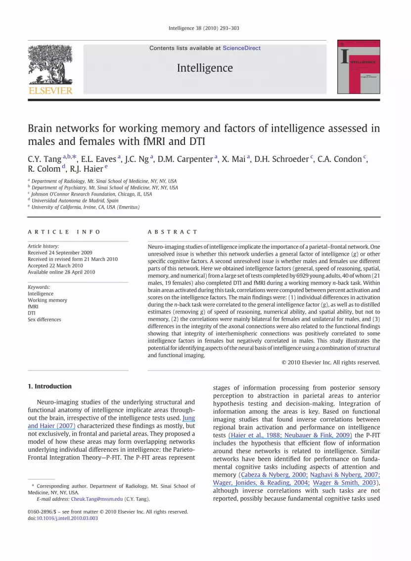
to motion correction. Functional images were co-registered toeach subject's anatomical scan. Co-registered anatomicalimages were then segmented to produce the parametersused for normalization into a standard anatomical brainreference template developed by the Montreal NeurologicalInstitute (MNI). Imageswere spatially smoothed using a 6 mmisotropic Gaussian smoothing kernel. Individual contrastimages were produced in the context of the general linearmodel using a boxcar function [0-back versus 1,2,3-back]convolved with a canonical hemodynamic response function.Next, a one-sample t-test was performed using each subject'scontrast images to determine areas of increased activity duringthe active (1,2,3-back) state compared to baseline (0-back)(Fig. 1). Significant clusters were identified (pb .001, uncor-rected; separate analyses of the 1 vs. 0, 2 vs. 0, and 3 vs. 0conditions did not yield appreciably different results; therewere no sex differences in n-back performance) and coordi-nates for spherical Regions-of-Interest (ROI) with 3 mm radiiwere obtained from the localmaximaof these clusters. Percentactivation values for these ROIswere extracted from individualsubjects' normalized scans and transferred to Statistica V7(Statsoft Inc., Tulsa, OK) for further analysis.
3.2. DTI
Raw DTI data were transferred to an off-line workstationfor post-processing. The Diffusion data set was eddy-currentandmotion corrected using an adaptation of the Camino/SPMpackage (Cook, 2006). In-house software was developed inMatlab v2007a (The Mathworks Inc., Natick, MA) for furtherprocessing of the DTI and tractography, a method to extractand visualize fiber tract bundles based on the geometricinformation contained in adjacent voxels obtained by DTIscans. Fractional Anisotropy (FA) and directionally color-coded FA maps were also computed. Color coded FA mapsimprove inter-rater reliabilities for ROI-based analysis of FAdata by segmenting the different adjacent fiber bundles intodifferent colors based on their orientation. For fiber tracking acommonly used “multiple region brute-force” fiber trackingmethod was used (Huang, Zhang, van Zijl, & Mori, 2004).First, fibers were traced using a streamline tractographyalgorithm from every voxel throughout the entire volumethat exceeded a minimum fractional anisotropy (Basser,Pajevic, Pierpaoli, Duda, & Aldroubi, 2000; Conturo et al.,

Fig. 1. fMRI-SPM group activation clusters superimposed on anatomical MRI (radiological convention), 1 Anterior Cingulate Cortex (ACC), 2 Prefrontal Cortex(PC) , 3 Parietal Cortex (PC), 4 Internal Capsule (IC), 5 Visual Cortex (VC).
297C.Y. Tang et al. / Intelligence 38 (2010) 293–303
1999; Mori, Crain, Chacko, & van Zijl, 1999). Tracking wasterminated when FA fell below a 0.1 or when the algorithmencountered a sharp angle change in the principal diffusiondirection between sequential voxels (45°). Second, FA mapswere analyzed using a region of interest (ROI) approach. ROIswere defined on color-coded tract directional maps by twoindependent raters and then averaged. Portions of the fol-lowing white matter tracts were surveyed using ROIs:Cingulum Bundle, Internal Capsule, Corpus Callosum, ForcepsMinor, Forceps Major, Inferior- and Superior LongitudinalFasciculus and corona radiata (Fig. 2). The ROI voxel locationswere used to extract the FA values, which were then trans-ferred to Statistica V7.1 (Statsoft Inc., Tulsa, OK) and mergedwith intelligence factor scores for correlation analysis.
4. Results
4.1. fMRI
As shown in Fig. 1, clusters of activation during the n-backtask were identified (1,2,3 back vs. 0 back; pb .001, uncor-rected) in: Anterior Cingulate (ACC), bilateral Prefrontal
Fig. 2. DTI–ROI positions superimposed on directional color-coded fractional anisotrCingulum, 4: Posterior Cingulum, 5: Superior Longitudinal Fasciculus, 6: Body of CForceps Minor, 10: Anterior Horn Internal Capsule, 11: Forceps Minor.
Cortex (PFC), bilateral Parietal cortex (PC), bilateral InsularCortex (IC) and bilateral visual cortex (VC). As noted, we usedthese areas to correlate percent activation with intelligencefactor scores; two regions, although activated during the n-back task, did not produce any significant correlations:namely, Insular Cortex and Visual Cortex.
Significant correlations between percent activation in theACC, PFC areas, and PC areas and intelligence factor scores areshown in Table 3. For the whole sample, activation in thecluster in the right parietal cortex (PC) was inverselycorrelated with general intelligence (r=−.34, pb .03) andspatial intelligence (r=−.39, pb .01). Spatial intelligence wasalso inversely correlated with activation at the right prefron-tal cortex (r=−.35, pb .03). For males, activation in the rightprefrontal and right parietal cortices was inversely correlatedwith spatial intelligence (r=−.58 and −.45, pb .01 and .04,respectively). Activation at the right prefrontal cortex wasalso inversely correlated with general intelligence in males(r=−.45, pb .04). Finally, for females, general intelligencewas inversely correlated with activation in left (r=−0.52,pb0.02) and right parietal cortex (r=−.50, pb .03). In thefemales, numerical intelligence was also inversely related to
opy maps. 1: Superior Corona Radiata, 2: Body Cingulum Bundle, 3: Anteriororpus Callosum, 7: Genu Corpus Callosum, 8: Splenium Corpus Callosum, 9:

Table 3Significant correlations of intelligence factor scores with % fMRI activation.
fMRI Region†
Subjects Ant. Cing. L-PFC R-PFC L-PC R-PC
All Spatial −0.16 −0.20 −0.35⁎ −0.25 −0.39⁎
Memory −0.10 0.96 −0.03 −0.21 0.20Numerical −0.27 −0.00 −0.24 −0.24 −0.25Reasoning 0.14 −0.16 −0.08 −0.02 0.05g −0.24 −0.09 −0.28 −0.27 −0.34⁎
Males Spatial −0.17 −0.42 −0.58⁎ −0.42 −0.45⁎
Memory −0.38 −0.07 −0.20 −0.07 −0.08Numerical −0.07 0.07 −0.19 0.07 −0.04Reasoning 0.04 −0.24 −0.16 −0.24 0.03g −0.23 −0.26 −0.45⁎ −0.26 −0.26
Females Spatial −0.24 0.16 −0.27 −0.36 −0.33Memory 0.06 0.34 −0.15 −0.20 −0.41Numerical −0.50⁎ −0.01 −0.35 −0.55⁎ −0.50⁎
Reasoning −0.19 −0.14 −0.00 −0.48⁎ −0.19g −0.33 0.13 −0.30 −0.52⁎ −0.50⁎
†Ant. Cing. is Anterior Cingulate Gyrus, L-PFC: Left, Right Prefrontal Cortex; L,R PC: Left, Right Parietal Cortex. All significances are at pb0.05.
298 C.Y. Tang et al. / Intelligence 38 (2010) 293–303
activation in the anterior cingulate (r=−.50, pb .03), left(r=−0.55, pb0.02) and right parietal cortex (r=−.50,pb .03). Speed of reasoning was inversely correlated withactivation in the left parietal cortex (r=− .48, pb .04). It isnoteworthy that all the correlations were negative (higherintelligence scores were associated with less brain activa-tion); no significant correlations were positive. Only thememory factor showed no correlations with activation. Noneof the correlations survived Bonferroni correction formultiplecomparisons, although most imaging studies have samplestoo small for this correction given the number of possiblecomparisons.
4.2. DTI
We computed brute-force streamline tractography (Car-penter et al., 2008; Huang et al., 2004) to identify theconnection networks between the selected areas shown inFig. 1. Significant activated clusters in the prefrontal cortex andparietal cortex were used as seed regions for fiber trackingthrough the genu and splenium of the corpus callosum; theseformed the forceps minor and forceps major respectivelySegments of the anterior cingulated where the cingulum wasclearly visible were used as a seed region for tracking of thecingulum bundle. All fMRI activation clusters were dilated byone voxel in all directions before being used as seed regionmaking them closer to nearby white matter tracts for bettertracking. We have integrated the fMRI activated regionstogether with the interconnecting white matter tracts inFig. 3. This figure illustrates the relationship of the functionaland structural anatomy and the correlations that we havedetected.
We then investigated FA correlates of the intelligencefactors. FA in ROIs in interhemispheric fibers showed correla-tions with intelligence factors as detailed in Table 4 and shownin Fig. 3. FA in the genu of the corpus callosum was positivelycorrelatedwith thememory factor scores for thewhole sample(r=.34, pb .03) aswell as for females (r=.56, pb .01). FA in thegenu of the corpus callosum was also inversely related tospatial intelligence in males (r=−.51, pb .02). FA in the leftForceps Major was inversely correlated with general intelli-
gence in males (r=− .53, pb .01) and positively in females(r=.48, pb .04). FA in the right Forceps Major was alsoinversely related to general intelligence in males (r=− .45,pb .04). Finally, FA in the Splenium of Corpus Callosum wasinversely related to numerical intelligence in males (r=− .46,pb .04). None of these correlations survived Bonferronicorrection.
ROIs in ipsilateral fibers also showed correlations with in-telligence factors as detailed in Table 5 and shown in Fig. 3. FA inthe left Cingulum Bundle was positively related to speedof reasoning for the whole sample (r=.36, pb .02) and formales (r=.58, pb .01). FA in the right Cingulum Bundle was alsocorrelated with speed of reasoning in males (r=.53, pb .01). FAin the right Superior Corona Radiata was positively correlatedwith spatial intelligence in the whole sample (r=.32, pb .04).Finally, FA in the left Superior Longitudinal Fasciculus wasinversely related to memory in males (r=−.47, pb .03). Noneof these correlations survived Bonferroni correction.
5. Discussion
This study examined intelligence factors with g removedto determine if the parieto-frontal integration theory of in-telligence (P-FIT) characterizes specific cognitive abilitiesbeyond the pervasive influence of the g-factor representinggeneral intelligence. This was examined separately in malesand females and uniquely combined fMRI and DTI imaging tostudy the neuroanatomy of intelligence. Since the sample sizesare relatively small and none of the significant findings sur-vived correction for multiple comparisons, all results should beregarded as illustrative and interpreted with caution. Mostneuroimaging studies have small samples and limited statis-tical power, so reporting uncorrected results allows for poten-tial replication across studies.
5.1. Relationship to P-FIT and other n-back studies
Functional correlations between the BOLD signal obtainedfrom the working memory task (n-back) and intelligencefactors were mostly detected in the right prefrontal andbilateral parietal cortices. All these correlations were negative

Fig. 3. 3D illustration of the significantly correlated fMRI activation clusters* and their interconnecting fiber tracts° for females (top) and males (bottom).Significant fMRI correlations are labeled with yellow legends, all fMRI correlations are negative with performance scores; Significant white matter tractscorrelations are labeled with white legends where + indicate a positive correlation with better connectivity and − indicates a negative correlation with betterconnectivity for the indicated performance scores. (*R,L-PC: right,left parietal cortex; R,L-PFC: right, left prefrontal cortex; ACC: anterior cingulate cortex; ° F-Maj:forceps major; F-min: forceps minor; GCC: genu of corpus callosum; SCC: splenium of corpus callosum; CB: cingulum bundle, L-SLF: Superior LongitudinalFasciculus, C-Rad: Corona Radiata; Only regions or tracts for which significances were found are rendered).
299C.Y. Tang et al. / Intelligence 38 (2010) 293–303
indicating that subjects with high intelligence factor scoreshad less activation during the n-backmemory task, in supportof the efficiency model of brain function (Haier, 1993; Haieret al., 1988; Neubauer & Fink, 2009). Gray et al (2003)reported that, on more demanding n-back conditions, partic-
ipants with higher intelligence scores were more accurateand showed greater activity in several frontal and parietalregions. It should be noted that the focus of their analysis wasbased on an event related design but their report alsoincluded the results using a block design which showed a

Table 4DTI correlations with intelligence factor scores for interhemispheric fibers.
DTI White matter tracts†
Subjects L Fcp Maj R Fcp Maj GCC SCC L Fcp Min R Fcp Min
All Spatial −0.11 0.01 −0.28 −0.07 0.06 −0.06Memory 0.12 0.19 0.34⁎ 0.14 −10 0.01Numerical 0.20 −0.03 −0.21 −0.31 0.07 −0.26Reasoning −0.20 −0.15 0.11 0.22 0.11 −0.21g 0.10 0.03 −0.05 −0.15 −0.14 0.14
Males Spatial −0.08 −0.05 −0.51⁎ −0.00 0.28 0.03Memory −0.10 −0.09 0.20 0.14 −0.10 0.07Numerical 0.16 −0.14 −0.13 −0.46⁎ r−0.01 0.30Reasoning 0.04 0.22 0.33 0.19 −0.12 −0.30g −0.53⁎ −0.45⁎ −0.17 −0.11 0.11 0.10
Females Spatial −0.24 0.00 0.05 −0.15 −0.15 −0.26Memory 0.24 0.38 0.56⁎ 0.15 −0.14 −0.18Numerical 0.19 −0.04 −0.26 −0.21 −0.11 0.21Reasoning −0.30 −0.36 0.30 0.27 0.45 0.16g 0.49⁎ 0.14 0.08 −0.17 −0.32 0.13
†L,R Fcp Maj: Left, Right Forceps Major; GCC: Genu of Corpus Callosum; SCC: Splenium of Corpus Callosum; L,R FcpMin: Left, Right Forceps Minor. All significancesare at pb0.05.
300 C.Y. Tang et al. / Intelligence 38 (2010) 293–303
trend of lower activity with higher scores on a single test offluid intelligence. Waiter et al. (2009) did not find significantcorrelations between individual differences in brain activityduring an n-back task and intelligence scores. Activationlevels are known to fluctuate across workingmemory loads inan inverted-U shape response (Callicott et al., 1999). Theposition of the inverted-U can shift depending on theworkingmemory capacity of the group or individual (Callicott et al.,2003). Waiter et al. (2009) used a simple version of the n-back task with only 0- and 2-back levels . The limited levelsand range of task difficulties in their study may have createdconfounds related to shifts in the inverted-U curve. In addi-tion, their study focused on elderly (mid to late 60-years-old)subjects, a population that shows a decrease in workingmemory capacity and related neurophysiology (Mattay et al.,2006) as well as a wide variability among individuals in theextent, rate and pattern of age-related changes that are ex-hibited at both neural and behavioral levels (Hedden &Gabrieli, 2004). For the current study, a greater range ofworking memory task was used on a younger population,which could help avoid inconsistencies due to shifts of the
Table 5DTI FA correlations with intelligence factor scores for ipsilateral fibers.
DTI White matter tracts†
Subjects L Cing. R Cing.
All Spatial −0.08 −0.21Memory −0.25 −0.11Numerical −0.05 −0.07Reasoning 0.36⁎ 0.34g −0.23 −0.09
Males Spatial −0.22 −0.38Memory −0.36 −0.30Numerical −0.11 0.07Reasoning 0.58⁎ 0.53⁎
g −0.22 −0.03Females Spatial 0.12 0.04
Memory −0.17 0.11Numerical −0.04 −0.30Reasoning 0.13 0.25g −0.32 −0.26
†L,R Cing: Left, Right Cingulum Bundle; L, R Cor R; Left, Right Superior Corona Radiataat pb0.05.
inverted-U. This method does not completely avoid thepotential anomalies as equal sampling of both sides of theinverted-U is not guaranteed but is less risky than samplingonly one level of working memory difficulty. The optimalmethod would include enough levels and range in the taskdifficulty to determine the point of peak activation for eachsubject.
Our n-back results are in agreement with the singlepublished study aimed at quantifying the neuro-anatomicaloverlap between the general factor of intelligence (g) andworking memory capacity (Colom, Jung, & Haier, 2007). Thatstudy showed that a common neuro-anatomic framework forthese constructs implicates mainly frontal gray matter regionsbelonging to the right superior frontal gyrus, the left middlefrontal gyrus, and the right inferior parietal lobule. Thesefindings (a) were thought to support the role of a discreteparieto-frontal network, as proposed by the P-FITmodel, and (b)were consistent with Cowan's (2005) theory which distin-guished a capacity limit (related to parietal regions) and thecontrol of attention (related to frontal areas). It was suggestedthat capacity limits and attention control relate to the
LS Cor R RS Cor R L Sup L F R Sup L F
0.08 0.32⁎ −0.13 −0.18−0.70 0.03 −0.09 0.19−0.06 −0.18 0.15 −0.10−0.05 −0.08 0.05 −0.06−0.00 −0.08 0.04 −0.01−0.09 0.40 −0.05 −0.17−0.07 −0.21 −0.47⁎ 0.27−0.24 −0.14 0.10 −0.16
0.31 −0.00 0.39 0.02−0.05 −0.08 −0.13 0.07
0.37 0.25 −0.28 −0.21−0.09 0.15 0.27 0.06
0.12 −0.21 0.16 −0.06−0.42 −0.11 −0.25 0.21
0.05 −0.08 0.12 −0.07
; L, R Sup L F: Left, Right Superior Longitudinal Fasciculus. All significances are

301C.Y. Tang et al. / Intelligence 38 (2010) 293–303
commonality between intelligence and working memory. Wealso note that we found no correlations between our memoryfactor and any fMRI activations, possibly because the factor wasderived as a broader assessment of memory than the morefocused processes required for the n-back task, although this isnot determined.
5.2. Sex differences
Because of the small sample sizes, the sex differencesobserved in the present study should be interpreted withparticular caution. Therefore, we do not interpret individualcorrelations, but note that all the fMRI correlations withintelligence factors were negative, consistent with the brainefficiency hypothesis for both males and females. We testedthe male/female difference of each fMRI/factor correlationusing z-transform. Only the difference for Numerical/LeftParietal Cortex was significant (pb .045, 2-tailed); there was atrend for Spatial/Left Prefrontal Cortex (pb .077).
Similarly, the DTI results show sex differences but the samecautions apply, especially because these analyses were explor-atory, so we will discuss specific correlations only to makegeneral illustrative points about each factor. Using z-transformcomparisons for DTI interhemisphere/factor correlations, themale/female difference was significant for g/Left Forceps Major(pb .001, 2-tailed), and there were trends (pb .05 to .09) forSpeed of Reasoning and for g in the Right ForcepsMajor; for theSpatial Factor and the Genu of the Corpus Callosum; and, forSpeed of Reasoning and the Left Forceps Minor. For DTIipsilateral/factor correlations, the male/female difference wassignificant (2-tailed) for Speed of Reasoning/Left SuperiorCorona Radiata (pb .026) and for Memory/Left Superior Longi-tudinal Fasciculus (pb .022); Speed of Reasoning/Left SuperiorLongitudinal Fasciculus showed a trend (pb .052). Thesedifferences, for both fMRI and DTI correlations, support theview that separate analyses by sex are justified, even whenthere is no task performance difference.
5.2.1. Spatial factorIn our study, spatial factor scores were correlated with
fMRI activation in the right prefrontal and posterior parietalcortex in the whole group. However, when analyzed sep-arately for males and females, only the males showedsignificant correlations. Although there is a general notionthat spatial ability is a right hemisphere function, recentstudies have found that sex matters. A meta-analysis (Vogel,Bowers, & Vogel, 2003) showed that females are much lesslateralized than their male counterparts in terms of brainfunction and spatial tasks. In that study females showed nohemispheric preference while males showed a right hemi-sphere advantage. It was hypothesized that women use bothverbal abilities, a left hemisphere function, as well as spatialabilities, a right hemisphere function, to accomplish spatialtasks. This might be contributing to the variances in ourdataset. The lack of any significant correlations in our datasetfor spatial factors in the female group might be additionalsupport for these findings.
Contrary to our expectation of positive correlations, the DTIresults show that the FA of the genu of the corpus callosumwasnegatively correlated with spatial factor scores (r=−51,pb0.02) in males. This is the section of the corpus callosum
that connects parts of the prefrontal cortex. FA is believed to bean indirect measure of myelination and the purpose of myelinis to allow rapid and efficient transmission of signals along theaxons (Ritchie, 1984; Yakovlev & Lecours, 1967). The negativecorrelation means that stronger connections between the twohemispheres decrease performance. Increased anisotropy(better connectivity) in the corpus callosum can be interpretedas an interference factor for brains using only one region orone hemisphere for a certain task. Enhanced interhemisphericconnectivity increases unnecessary information flow from thecontralateral side of the brain, possibly consistentwith efficientinformation flow. We also note that although both the rightprefrontal and right parietal lobes had significant fMRI signalcorrelation with spatial factor scores in males, the prefrontalcortex showed stronger correlations than the parietal (r=−0.58vs. r=−0.45). This is consistentwith theDTIfinding in thegenu, which showed decreased spatial performance scoreswith increased FA in the genu in the male group.
5.2.2. Numerical factorFemales had significant negative correlations with the
numerical factor in terms of activation in the anterior cingulateas well as both sides of the parietal cortex. Other studies(Dehaene, Spelke, Pinel, Stanescu, & Tsivkin, 1999) have shownthat numerical cognitive abilities recruit both sides of theparietal cortex. To our knowledge, no other studies have shownany gender effect for this function. There were no significantfMRI correlations with the numerical factor for themale group.
5.2.3. Speed of reasoning factorThe integrity of the cingulum bundle, a white matter tract
inside the cingulate gyrus, as quantified using FA, was posi-tively correlated with the speed of reasoning factor scores inmales. This is not an interhemispheric connection and thus itis expected that better ipsilateral connections are positivelycorrelatedwith performance. The cingulate gyrus is an area inthe brain that is involved with early learning and problemsolving, anticipation of tasks, motivation, and modulation ofemotional responses (Posner & Raichle, 1998).
5.2.4. Memory factorWe note that for the group as a whole there was a
significant correlation (r=0.34) between the memory factorand the interhemispheric white matter tracts in the genu ofthe corpus callosum (GCC), but when separated by gender wefind that the effect was mainly due to the female group(r=0.56) with the males no longer significant. This signifi-cant positive correlation is additional support for the notionthat females rely on both hemispheres and benefit frombetter interhemispheric connections. No significant correla-tions between functional activation and the memory factorwere detected. It is conceivable that the GCC connects largerand more diffuse brain regions used for many memoryprocesses and that our working memory task uses a morenarrow set of processes.
5.2.5. g-factorThe g-factor was correlated with activation in the right
prefrontal area in males, but was correlated to both left andright sides of the parietal cortices in females. While the FA ofthe forceps major was negatively correlated with g in males,

302 C.Y. Tang et al. / Intelligence 38 (2010) 293–303
the females had parts of the forceps major positivelycorrelated with g. Again, this negative correlation may be anindicator of interference from contralateral side of the brainin males who rely mostly on the right side of the brain. Thepositive FA correlations in the forceps major in females arealso compatible with the bilateral fMRI correlations in theparietal regions connected by the forceps major. Also, ourfinding that part of the FA of the forceps major (an extensionof the splenium) is positively correlated with g (r=0.49,pb0.034) in females is consistent with some evidence thatthe splenium may be larger in females (Dubb, Gur, Avants, &Gee, 2003). Overall, our DTI results are also consistent with areport on DTI/IQ correlations in a cohort of subjects aged 5–18where correlations were mainly negative in older males andpositive in older females (Schmithorst et al., 2008). There arealso other higher-order latent variable models that show sexdifferences that could be explored with imaging (Johnson &Bouchard, 2007a,b; Keith, Reynolds, Patel, & Ridley, 2008).
In summary, in this study, we have shown that: (a)individual differences in activation during the n-back task arecorrelated with the general intelligence factor (g), as well asto distilled estimates (removing g) of speed of reasoning,numerical ability, and spatial ability, but not to memory, (b)the correlations aremainly bilateral for females and unilateralfor males, and (c) differences in the integrity of the axonalconnections are also related to the functional findings:integrity of interhemispheric connections is positively relatedto several intelligence factors in females but is negativelycorrelated in males. It should be noted that none of thesecorrelations survived correction for multiple comparisons, soreplication in larger samples is necessary. Nevertheless, theseresults show encouraging trends to support further studiesregarding the relationship between working memory andintelligence factors. They support the growing recognitionthat brain structure and function underlie individual differ-ences in general intelligence and other specific cognitiveabilities. These results also underscore the importance ofanalyzing neuroimaging data separately for males and females.
Acknowledgements
Funding for imaging and for R. Haier was provided by theJohnson O'Connor Research Support Corporation. R. Colomwas funded by the grant SEJ-2006-07890 from the “Minis-terio de Educación y Cultura” (MEC) [Ministry of Educationand Culture, Spain].
References
Basser, P. J. (1997). New histological and physiological stains derived fromdiffusion-tensor MR images. Annals of the New York Academy of Sciences,820, 123−138.
Basser, P. J., Pajevic, S., Pierpaoli, C., Duda, J., & Aldroubi, A. (2000). In vivofiber tractography using DT-MRI data. Magnetic Resonance in Medicine,44(4), 625−632.
Cabeza, R., & Nyberg, L. (2000). Imaging cognition II: An empirical review of275 PET and fMRI studies. Journal of Cognitive Neuroscience, 12(1), 1−47.
Callicott, J. H., Mattay, V. S., Bertolino, A., Finn, K., Coppola, R., Frank, J. A., et al.(1999). Physiological characteristics of capacity constraints in workingmemory as revealed by functional MRI. Cerebral Cortex, 9(1), 20−26.
Callicott, J. H., Mattay, V. S., Verchinski, B. A., Marenco, S., Egan, M. F., &Weinberger, D. R. (2003). Complexity of prefrontal cortical dysfunctionin schizophrenia: More than up or down. American Journal of Psychiatry,160(12), 2209−2215.
Carpenter, D. M., Tang, C. Y., Friedman, J. I., Hof, P. R., Stewart, D. G.,Buchsbaum, M. S., et al. (2008). Temporal characteristics of tract-specificanisotropy abnormalities in schizophrenia. NeuroReport, 19(14),1369−1372.
Chiang, M. C., Barysheva, M., Shattuck, D. W., Lee, A. D., Madsen, S. K.,Avedissian, C., et al. (2009). Genetics of brain fiber architecture andintellectual performance. Journal of Neuroscience, 29(7), 2212−2224.
Colom, R. (2007). Intelligence? What intelligence? Behavioral and BrainSciences, 30, 155−156.
Colom, R., Abad, F., Quiroga, M. A., Shih, P. C., & Flores-Mendoza, C. (2008).Working memory and intelligence are highly related constructs, butwhy? Intelligence, 36, 584−606.
Colom, R., Abad, F., Rebollo, I., & Shih, P. C. (2005). Memory span and generalintelligence: A latent variable approach. Intelligence, 33, 623−642.
Colom, R., Haier, R. J., Head, K., Alvarez-Linera, J., Quiroga, M. A., Shih, P. C.,et al. (2009). Gray matter correlates of fluid, crystallized, and spatialintelligence: Testing the P-FIT model. Intelligence, 37(2), 124−135.
Colom, R., Jung, R. E., & Haier, R. J. (2007). General intelligence and memoryspan: Evidence for a common neuroanatomic framework. CognitiveNeuropsychology, 24(8), 867−878.
Colom, R., Rebollo, I., Palacios, A., Juan-Espinosa, M., & Kyllonen, P. C. (2004).Working memory is (almost) perfectly predicted by g. Intelligence, 32,277−296.
Condon, C. A., & Schroeder, D. H. (2003). Establishing the factor structure of theJohnson O'Connor Research Foundation's test battery. Paper presented atthe annual meeting of the International Society for Intelligence Research,Newport Beach, CA.
Condon, C. A., & Schroeder, D. H. (2005). Quality of school and standard testbattery performance (Statistical Bulletin 2005-3). Chicago: Johnson O'Connor Research Foundation.
Conturo, T. E., Lori, N. F., Cull, T. S., Akbudak, E., Snyder, A. Z., Shimony, J. S.,et al. (1999, August). Tracking neuronal fiber pathways in the livinghuman brain. Proceedings of the National Academy of Science, 96,10422−10427.
Cook, P. A. (2006). Camino: Open-source diffusion-MRI reconstruction andprocessing 14th Scientific Meeting of the International Magnetic Resonancein Medicine (pp. 2759). Washington: Seattle.
Cowan, N. (2005). Working memory capacity. New York: Psychology Press.Dehaene, S., Spelke, E., Pinel, P., Stanescu, R., & Tsivkin, S. (1999). Sources of
mathematical thinking: Behavioral and brain-imaging evidence. Science,284(5416), 970−974.
Dubb, A., Gur, R., Avants, B., & Gee, J. (2003). Characterization of sexualdimorphism in the human corpus callosum. Neuroimage, 20(1), 512−519.
Engle, R. W. (2002). Working memory capacity as executive attention.Current Directions in Psychological Science, 11, 19−23.
Fjell, A. M., Westlye, L. T., Greve, D. N., Fischl, B., Benner, T., van der Kouwe, A. J.,et al. (2008). The relationship between diffusion tensor imaging andvolumetry as measures of white matter properties. Neuroimage, 42(4),1654−1668.
Grabner, R. H., Fink, A., Stipacek, A., Neuper, C., & Neubauer, A. C. (2004).Intelligence and working memory systems: Evidence of neural efficiencyin alpha band ERD. Brain Research. Cognitive Brain Research, 20(2),212−225.
Gray, J. R., Chabris, C. F., & Braver, T. S. (2003). Neural mechanisms of generalfluid intelligence. Nature Neuroscience, 6(3), 316−322.
Haier, R. J. (2009). Neuro-intelligence, neuro-metrics and the next phase ofbrain imaging studies. Intelligence, 37(2), 121−123.
Haier, R. J. (Ed.). (1993). Cerebral glucose metabolism and intelligence.Norwood, NJ: Ablex. ed.
Haier, R. J., & Benbow, C. P. (1995). Sex differences and lateralization intemporal lobe glucose metabolism during mathematical reasoning.Developmental Neuropsychology, 11, 405−414.
Haier, R. J., Colom, R., Schroeder, D. H., Condon, C. A., Tang, C., Eaves, E., et al.(2009). Gray matter and intelligence factors: Is there a neuro-g?Intelligence, 37(2), 136−144.
Haier, R. J., Jung, R. E., Yeo, R. A., Head, K., & Alkire, M. T. (2005). Theneuroanatomy of general intelligence: Sex matters. Neuroimage, 25(1),320−327.
Haier, R. J., Siegel, B. V., Nuechterlein, K. H., Hazlett, E., Wu, J. C., Paek, J., et al.(1988). Cortical glucose metabolic-rate correlates of abstract reasoningand attention studied with positron emission tomography. Intelligence,12(2), 199−217.
Halpern, D. F. (2000). Sex differences in cognitive abilities, 3rd ed. Mahwah,NJ: Erlbaum.
Hedden, T., & Gabrieli, J. D. (2004). Insights into the ageing mind: Aview from cognitive neuroscience. Nature Reviews. Neuroscience, 5(2),87−96.
Huang, H., Zhang, J., van Zijl, P. C., & Mori, S. (2004). Analysis of noise effectson DTI-based tractography using the brute-force and multi-ROIapproach. Magnetic Resonance in Medicine, 52(3), 559−565.

303C.Y. Tang et al. / Intelligence 38 (2010) 293–303
Jausovec, N., & Jausovec, K. (2008). Spatial rotation and recognizingemotions: Gender related differences in brain activity. Intelligence, 36(5),383−393.
Johnson, W., & Bouchard, T. J. (2007). Sex differences in mental abilities: gmasks the dimensions on which they lie. Intelligence, 35(1), 23−39.
Johnson, W., & Bouchard, T. J. (2007). Sex differences in mental ability: Aproposedmeans to link them to brain structure and function. Intelligence,35(3), 197−209.
Johnson, W., Jung, R. E., Colom, R., & Haier, R. J. (2008). Cognitive abilitiesindependent of IQ correlate with regional brain structure. Intelligence,36(1), 18−28.
Jung, R. E., & Haier, R. J. (2007). The Parieto-Frontal Integration Theory (P-FIT) of intelligence: Converging neuroimaging evidence.Behavioral andBrain Sciences, 30(2), 135−154 discussion 154–187.
Kane, M. J., Hambrick, D. Z., & Conway, A. R. (2005). Working memorycapacity and fluid intelligence are strongly related constructs: Commenton Ackerman, Beier, and Boyle (2005).Psychological Bulletin, 131(1),66−71 author reply 72-65.
Keith, T. Z., Reynolds, M. R., Patel, P. G., & Ridley, K. P. (2008). Sex differencesin latent cognitive abilities ages 6 to 59: Evidence from the Woodcock–Johnson III tests of cognitive abilities. Intelligence, 36(6), 502−525.
Lee, K. H., Choi, Y. Y., Gray, J. R., Cho, S. H., Chae, J. H., Lee, S., et al. (2006).Neural correlates of superior intelligence: Stronger recruitment ofposterior parietal cortex. Neuroimage, 29(2), 578−586.
Luders, E., Narr, K. L., Bilder, R. M., Szeszko, P. R., Gurbani, M. N., Hamilton, L.,et al. (2008). Mapping the relationship between cortical convolution andintelligence: Effects of gender. Cerebral Cortex, 18(9), 2019−2026.
Luders, E., Narr, K. L., Thompson, P. M., Rex, D. E., Jancke, L., Steinmetz, H.,et al. (2004). Gender differences in cortical complexity. NatureNeuroscience, 7(8), 799−800.
Mattay, V. S., Fera, F., Tessitore, A., Hariri, A. R., Berman, K. F., Das, S., et al.(2006). Neurophysiological correlates of age-related changes in workingmemory capacity. Neuroscience Letters, 392(1–2), 32−37.
Mori, S., Crain, B. J., Chacko, V. P., & van Zijl, P. C. (1999). Three-dimensionaltracking of axonal projections in the brain by magnetic resonanceimaging. Annals of Neurology, 45(2), 265−269.
Naghavi, H. R., & Nyberg, L. (2007). Integrative action in the fronto-parietalnetwork: A cure for a scattered mind. Behavioral and Brain Sciences, 30,161−162.
Neubauer, A. C., & Fink, A. (2009). Intelligence and neural efficiency.Neuroscience and Biobehavioral Reviews, 33(7), 1004−1023.
Neubauer, A. C., Fink, A., & Schrausser, D. G. (2002). Intelligence and neuralefficiency: The influence of task content and sex on the brain–IQrelationship. Intelligence, 30(6), 515−536.
Oberauer, K., Schulze, R., Wilhelm, O., & Suss, H. M. (2005). Working memoryand intelligence—Their correlation and their relation: Comment onAckerman, Beier, and Boyle (2005).Psychological Bulletin, 131(1), 61−65author reply 72-65.
Posner, M. I., & Raichle, M. E. (1998, February). The neuroimaging or humanbrain function. Proceedings of the National Academy of Sciences of theUnited States of America Colloquium Paper, 95, 763−764.
Prabhakaran, V., Smith, J. A. L., Desmond, J. E., Glover, G. H., & Gabrieli, J. D. E.(1997). Neural substrates of fluid reasoning: An fMRI study ofneocortical activation during performance of the Raven's ProgressiveMatrices Test. Cognitive Psychology, 33(1), 43−63.
Rabinowicz, T., Dean, D. E., Petetot, J. M. C., & de Courten-Myers, G. M. (1999).Gender differences in the human cerebral cortex: More neurons in males;More processes in females. Journal of Child Neurology, 14(2), 98−107.
Ritchie, J. M. (1984). Physiological basis of conduction in myelinated nervefibers. myelin (pp. 117−146). New York: Plenum.
Schmithorst, V. J., & Holland, S. K. (2007). Sex differences in the development ofneuroanatomical functional connectivity underlying intelligence foundusing Bayesian connectivity analysis. Neuroimage, 35(1), 406−419.
Schmithorst, V. J., Holland, S. K., & Dardzinski, B. J. (2008). Developmentaldifferences in white matter architecture between boys and girls. HumanBrain Mapping, 29(6), 696−710.
Sowell, E. R., Peterson,B. S., Kan, E.,Woods, R. P., Yoshii, J., Bansal, R., et al. (2007).Sex differences in cortical thickness mapped in 176 healthy individualsbetween 7 and 87 years of age. Cerebral Cortex, 17(7), 1550−1560.
Toga, A. W., & Thompson, P. M. (2005). Genetics of brain structure andintelligence. Annual Review of Neuroscience, 28, 1−23.
Vogel, J. J., Bowers, C. A., & Vogel, D. S. (2003). Cerebral lateralization ofspatial abilities: A meta-analysis. Brain and Cognition, 52(2), 197−204.
Wager, T. D., Jonides, J., & Reading, S. (2004). Neuroimaging studies ofshifting attention: A meta-analysis. Neuroimage, 22(4), 1679−1693.
Wager, T. D., & Smith, E. E. (2003). Neuroimaging studies of workingmemory: A meta-analysis. Cognitive, Affective & Behavioral Neuroscience,3(4), 255−274.
Waiter, G. D., Deary, I. J., Staff, R. T., Murray, A. D., Fox, H. C., Starr, J. M., et al.(2009). Exploring possible neural mechanisms of intelligence differencesusing processing speed and working memory tasks: An fMRI study.Intelligence, 37, 199−206.
Yakovlev, P. I., & Lecours, A. R. (1967). The myelogenetic cycles of regionalmaturation of the brain. In A. Minkowski (Ed.), Regional development ofthe brain in early life (pp. 3−70). : Oxford: Blackwell Scientific.
Yu, C., Li, J., Liu, Y., Qin, W., Li, Y., Shu, N., et al. (2008). White matter tractintegrity and intelligence in patients with mental retardation andhealthy adults. Neuroimage, 40(4), 1533−1541.


