Embed Size (px)
Citation preview
Journal of Clinical Imaging Science • 2019 • 9(32) | 1
is is an open-access article distributed under the terms of the Creative Commons Attribution-Non Commercial-Share Alike 4.0 License, which allows others to remix, tweak, and build upon the work non-commercially, as long as the author is credited and the new creations are licensed under the identical terms.©2019 Published by Scientific Scholar on behalf of Journal of Clinical Imaging Science
Original Article
Brainstem Diffuse Axonal Injury and ConsciousnessSukhwinder Sandhu1, Erik Soule2, Peter Fiester3, Patrick Natter3, Daryoush Tavanaiepour4, Gazanfar Rahmathulla4, Dinesh Rao3
1Department of Neuroradiology, Mayo Clinic, Departments of 2Interventional Radiology, 3Neuroradiology, 4Neurosurgery, University of Florida Health, Jacksonville, Florida, USA.
*Corresponding author: Erik Soule, Department of Interventional Radiology, University of Florida College of Medicine - Jacksonville, 2nd Floor, Clinical Center, 655 West 8th Street, C90, Jacksonville, FL 32209, USA.
Received : 29 March 19 Accepted : 04 June 19 Published : 28 June 19
DOI 10.25259/JCIS-11-2019
Quick Response Code:INTRODUCTION
TBI is responsible for approximately 2 million Emergency Department visits per year and over 250,000 hospital admissions in the United States civilian population. While most patients with mild TBI tend toward clinic follow-up after the initial assessment, those with moderate and severe TBI are routinely admitted to intensive care units (ICU) with average lengths of stay between 9 and 17 days. e costs for immediate hospitalization are approximately 11.5 billion dollars with the overall lifetime needs of survivors estimated at greater than 64 billion dollars.[1] DAI was found in 56% of patients with moderate TBI and 90% of patients with severe TBI.[2] DAI is caused by profound acceleration-deceleration injury such as that seen in motor vehicle accidents, falls, and assault. Primary axotomy (transection of axons) at the time of injury may contribute to pathology. A greater contributor is stretch injury without primary axotomy, which disrupts axonal cytoskeletal elements. is impairs axonal transport mechanisms, disrupting
ABSTRACTBackground: Severe traumatic brain injuries (TBI), commonly due to motor vehicle accidents may cause death and long-term disability especially when the acceleration-deceleration force on the brain is massive. is may cause shearing of the axonal connections within the cerebral cortex and brainstem in a process referred to as diffuse axonal injury (DAI). Extensive DAI has been postulated to be a poor prognostic indicator for neurological recovery. In our institution, several patients with Grade 3 DAI were observed to recover and achieve neurological outcomes greater than expected given the presence of brainstem injury.
Methods: MRI studies from 100 patients admitted to a large tertiary trauma center for TBI were retrospectively analyzed by two fellowship-trained neuroradiologists. e size of DAI lesions, location of injury within the brainstem, and the number of discrete DAI lesions were measured and recorded. Glasgow Coma Scale (GCS) on arrival and at discharge was noted, as well as the presence of other neurological injuries.
Results: Of 20 patients initially noted to have DAI with lesions of the brainstem, eight of them were discharged with Glasgow Coma Scale (GCS) of 14–15. e 12 patients discharged with reduced consciousness (average GC 7.1) demonstrated a greater number of larger lesions, with a predilection for the dorsal pons.
Conclusion: ese results suggest that large, numerous pontine lesions may indicate worse neurological outcomes in patients with these findings.
Keywords: Traumatic brain injury, Diffuse axonal injury, Magnetic resonance imaging, Ascending reticular activating system, Consciousness
www.clinicalimagingscience.org
Journal of Clinical Imaging Science
Sandhu, et al.: Brainstem DAI
Journal of Clinical Imaging Science • 2019 • 9(32) | 2
neuronal homeostasis and leading to secondary axotomy. Stretch-induced depolarization may cause glutamate toxicity and subsequent sodium/calcium channelopathy, altering the electrochemical gradient and leading to mitochondrial damage. Cellular energy starvation, oxidative stress, and neuroinflammation proceed in a cascade potentially leading to devastating neurological injury peaking 1–2 days after the initial TBI.[3] Treatment options for severe TBI are currently lacking, but therapeutic hypothermia may reduce the secondary damage incurred by reducing the metabolic rate, and thus, the intracranial energy demand.[4]
Head computed tomography (CT) is routinely performed for patients with moderate and severe TBI in the emergency department; however, this can only identify hemorrhagic lesions, present in 10% of cases of DAI. e presence of hemorrhagic lesions on initial CT imaging was found to be associated with a favorable neurological outcome and improved overall survival compared to patients with DAI and no microhemorrhages seen on CT. is is contradictory to past studies which correlated hemorrhagic lesions with poor neurological outcome.[5] Magnetic resonance imaging (MRI) is more sensitive than CT and can detect DAI in the absence of microhemorrhages; however, it is frequently not feasible to perform MRI on unstable trauma patients. Pragmatically, MRI may be delayed until the patient fails to regain consciousness after their acute trauma-related medical problems have been stabilized. is may delay recognition of DAI for several days after the initial insult, during which time the damaging secondary effects of the axonal stretch injury may have run their course. MRI yields better resolution in assessing DAI with different sequences, including diffusion-weighted imaging (DWI) [Figure 1a], gradient-recalled echo (GRE) [Figure 1b], and susceptibility-weighted imaging (SWI) [Figure 2b and c].[6] DAI severity has been demonstrated to correlate with outcome in comatose patients, and thus are used to aid in clinical prognostication.[7]
DAI is conventionally graded according to location, with Grade 1 (lesions to the subcortical lobar white matter or cerebellum only), Grade 2 (lesions in the corpus callosum, w/or w/o lesions in the lobar white matter), and Grade 3 (traumatic lesions in the brainstem in areas typical of DAI (dorsolateral quadrant of the upper brainstem, superior cerebellar peduncles) with or without lesions in the lobar white matter or corpus callosum, Figure 2a). e Adams classification was originally developed in the landmark 1989 histopathological study and subsequently applied to clinical practice when MRI came into widespread use.[8] e use of this scoring system for prognostication was validated by a meta-analysis which demonstrated an increased risk of poor neurological outcome, as well as increased mortality for patients with Grade 3 DAI.[9] Grade 3 DAI patients were shown to take an average of 2 months to regain
consciousness in contrast to 2 weeks for Grade 2 and several days for Grade 1. ese patients are initially very vulnerable, however, some Grade 3 patients have been observed to make good long-term recovery.[10] Another recent meta-analysis determined that although the likelihood of unfavorable neurologic outcome increased three-fold with each successive grade of DAI, 37% of patients with Grade 3 DAI achieved a favorable neurologic outcome.[11] Neuroradiologists at a 695 bed academic hospital and level one trauma center noted repeated instances of Grade 3 DAI patients regaining consciousness before hospital discharge. erefore, this study was performed to further elucidate the prognostic value of brainstem lesions, specifically on consciousness at hospital discharge.
MATERIALS AND METHODS
An MPower search was performed at our institution for patients from 2014–2018 with keyword searches of “traumatic brain injury,” “TBI,” “diffuse axonal injury,” “DAI,” and “shear injury” based on radiological criteria. One hundred patients were found. On review of these patients, 50 were found to have DAI. Imaging studies were reviewed by two fellowship-trained neuroradiologists with certificates of additional qualification (CAQ). One neuroradiologist had 10 years of post-training experience in neuroradiology. e other had 9 years of post-training experience. Each neuroradiologist reviewed the scans independently and assigned each patient a DAI grade. Patients who did not have trauma, Grade 3 DAI, or did not undergo brain MRI were excluded from the study. e difference between the
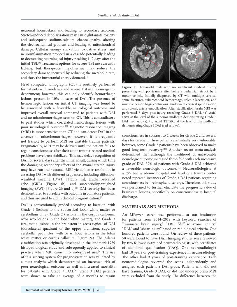
Figure 1: 53-year-old male with no significant medical history presenting with polytrauma after being a pedestrian struck by a motor vehicle. Initially diagnosed by CT with multiple cervical spine fractures, subarachnoid hemorrhage, splenic laceration, and multiple hemorrhagic contusions. Underwent cervical spine fixation and splenic artery embolization. After stabilization, brain MRI was performed 8 days post-injury revealing Grade 3 DAI. (a) Axial DWI at the level of the superior midbrain demonstrating Grade 3 DAI (red arrows). (b) Axial T2*GRE at the level of the midbrain demonstrating Grade 3 DAI (red arrows).
ba
Sandhu, et al.: Brainstem DAI
Journal of Clinical Imaging Science • 2019 • 9(32) | 3
date of admission and the date the MRI was performed was calculated and recorded. No patients were excluded based on the number of days post-injury that the MRI was performed. Lesions were determined to be secondary to DAI based on the clinical setting of injury, i.e., history of head trauma. e medical record was reviewed for signs or symptoms related to infarct or vascular injury. No patients were noted to have a clinical suspicion of the infarct. One potential confounder is patients who may have had injury secondary to increases in intracranial pressure; however, all patients had DAI in the subcortical white matter and corpus callosum in addition to brainstem injuries, excepting for one patient who had isolated Grade 3 DAI, and other intracranial traumatic injuries. Discrepancies were reviewed after independent analysis and were resolved by consensus. For patients with Grade 3 DAI, the size and location of the brainstem lesions were measured. e total number of lesions was recorded. GCS scores were documented for patients as noted by rescue personnel in the field, at the time of admission and at the time of discharge. e number of days spent in the hospital was recorded. e Ranchos Los Amigos level of cognitive functioning scale (RLAS) at discharge was also recorded.[12]
Lesion location was designated by location in the midbrain, pons, and medulla. Lesion location within the midbrain and pons were further subdivided as ventral or dorsal. Ventral versus dorsal location was assigned according to the system devised by Izzy et al., with the boundary demarcated by the dorsal border of the inferior olives for the medulla, the medial lemniscus for the pons, the posterior aspect of the decussation of the superior cerebellar peduncles for the caudal midbrain, and the posterior aspect of the red nuclei for the rostral midbrain.[13] Patients were scanned a 1.5 T Siemens Avanto. FLAIR/T2/DWI/ADC/GRE was obtained for all patients, and SWI sequences were obtained when available. Standard
T1 sequences with and without gadolinium were acquired. Slice thickness was 4 mm with a 1 mm skip, and FOV was 23 cm for all sequences. Parameters for axial DWI/ADC were TE: 102 ms, TR: 5400, with a 190 × 190 matrices. Parameters for axial T2 were TE: 90–130 ms, TR 3000–6000 ms, nex = 2, ETL: 11, flip angle: 150 with a 512 × 512 matrices. Parameters for FLAIR were TE: 90−130 ms, TR: 8000−9000 ms with inversion time of 2440 ms, nex=1, variable bandwidth with a 512−416 matrices. Parameters for GRE were TE: 20−35 ms, TR: 800−1300 ms, flip angle: 20 with a 512−416 matrices. Parameters for SWI were TE: 40−50 ms, TR: 40−50 ms, flip angle: 15 with a 320 × 260 matrices. Parameters for sagittal/axial T1 were TE min 8.7 ms, TR: 400−800 ms with a 320 × 260 matrices. e fluid-attenuated inversion recovery (FLAIR), DWI, or T2 abnormalities were measured in the largest axial dimension (anterior-posterior or transverse). e size of a hemosiderin lesion was measured on GRE or SWI imaging in the same fashion. In lesions, where both FLAIR/DWI abnormality and hemorrhage were present, the FLAIR/DWI abnormality was used. Total lesion burden per patient was calculated by summing the total size of all lesions in a subgroup, then dividing by the number of affected patients in that subgroup. is process was repeated to calculate total pontine lesion burden. e average lesion size was calculated by summing the total size of all lesions in each subgroup, then dividing by the total number of lesions in that subgroup. Due to the retrospective nature of the study, consent was not required as determined by IRB, which approved this study. No patient identifying information is included in this research article.
RESULTS
Average patient age was 35 years, with 13 males and 7 females. All patients were discharged to inpatient rehabilitation
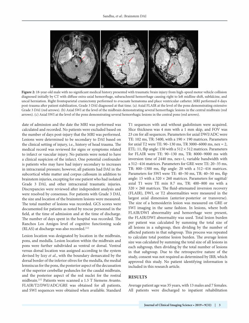
Figure 2: 18-year-old male with no significant medical history presented with traumatic brain injury from high-speed motor vehicle collision diagnosed initially by CT with diffuse extra-axial hemorrhage, subarachnoid hemorrhage causing right to left midline shift, subfalcine, and uncal herniation. Right frontoparietal craniectomy performed to evacuate hematoma and place ventricular catheter. MRI performed 6 days post-trauma after patient stabilization, Grade 3 DAI diagnosed at that time. (a) Axial FLAIR at the level of the pons demonstrating extensive Grade 3 DAI (red arrows). (b) Axial SWI at the level of the midbrain demonstrating several hemorrhagic lesions in the central midbrain (red arrows). (c) Axial SWI at the level of the pons demonstrating several hemorrhagic lesions in the central pons (red arrows).
cba
Sandhu, et al.: Brainstem DAI
Journal of Clinical Imaging Science • 2019 • 9(32) | 4
facilities, except for two who died during their hospital admission. Of 20 patients determined to have Grade 3 DAI, 8 of them regained consciousness and were discharged with a GCS of 14–15 [Table 1]. Average RLAS for these patients was 5.5. Conscious patients had an average of one brainstem lesions with an average size of 4.6 mm. Two of these patients had lesions located in the dorsal pons, three in the ventral midbrain, and three in the dorsal midbrain. e lesions in the dorsal pons for these patients were small (1 mm) and hemorrhagic. e two patients in this group with non-hemorrhagic lesions (9 mm and 3 mm) had these lesions located in the ventral midbrain.
e remaining 12 patients were discharged with an average GCS of 7.1, and an average RLAS of 2.1. ese patients had had an average of 2.6 brainstem lesions, with one patient having many diffuse hemorrhagic lesions. Of the 10 patients with measurable lesions, the average total affected area was 13.8 mm. Eight of these patients had involvement of both the pons and midbrain. ree of those eight had involvement of both the ventral and dorsal pons and midbrain. Eight of the patients with reduced consciousness had lesions located in the dorsal pons. Average pontine lesion burden was 8.8 mm for patients with reduced consciousness at discharge and pontine involvement in contrast to 1 mm for those that fully regained consciousness. Length of stay was similar between
the two subgroups of patients.
Trends were observed between prognostic factors such as number of brainstem lesions, total lesion burden, lesion burden in the pons, patient age, and outcome measures such as GCS and RLAS at discharge [Table 2]. A statistically significant correlation was demonstrated between pontine lesion burden and GCS at discharge (Spearman correlation coefficient = −0.51, P = 0.023). e correlation was stronger between pontine lesion burden and RLAS at discharge (Spearman correlation coefficient = −0.59, P = 0.006). ese associations were also observed a total number of brainstem lesions and GCS/RLAS at discharge (Spearman correlation coefficients were −0.60/−0.62, and P-values were 0.005/0.003, respectively). Age and total lesion burden did not achieve statistical significance as prognostic factors.
DISCUSSION
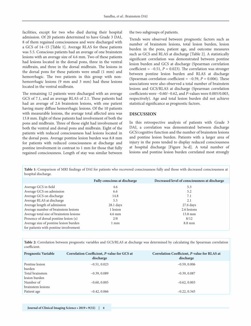
In this retrospective analysis of patients with Grade 3 DAI, a correlation was demonstrated between discharge GCS/cognitive function and the number of brainstem lesions and pontine lesion burden. Patients with a larger area of injury in the pons tended to display reduced consciousness at hospital discharge [Figure 3a-d]. A total number of lesions and pontine lesion burden correlated most strongly
Table 1: Comparison of MRI findings of DAI for patients who recovered consciousness fully and those with decreased consciousness at hospital discharge.
Fully conscious at discharge Decreased level of consciousness at discharge
Average GCS in field 4.6 5.3Average GCS on admission 6.6 5.2Average GCS on discharge 14.8 7.1Average RLAS at discharge 5.5 2.1Average length of admission 28.1 days 27.6 daysAverage number of brainstem lesions 1 lesion 2.6 lesionsAverage total size of brainstem lesions 4.6 mm 13.8 mmPresence of dorsal pontine lesion (s) 2/8 8/12Average size of pontine lesion burden for patients with pontine involvement
1 mm 8.8 mm
Table 2: Correlation between prognostic variables and GCS/RLAS at discharge was determined by calculating the Spearman correlation coefficient.
Prognostic Variable Correlation Coefficient, P-value for GCS at discharge
Correlation Coefficient, P-value for RLAS at discharge
Pontine lesion burden
–0.51, 0.023 –0.59, 0.006
Total brainstem lesion burden
–0.39, 0.089 –0.39, 0.087
Number of brainstem lesions
–0.60, 0.005 –0.62, 0.003
Patient age –0.42, 0.066 –0.22, 0.345
Sandhu, et al.: Brainstem DAI
Journal of Clinical Imaging Science • 2019 • 9(32) | 5
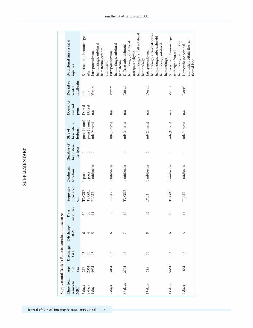
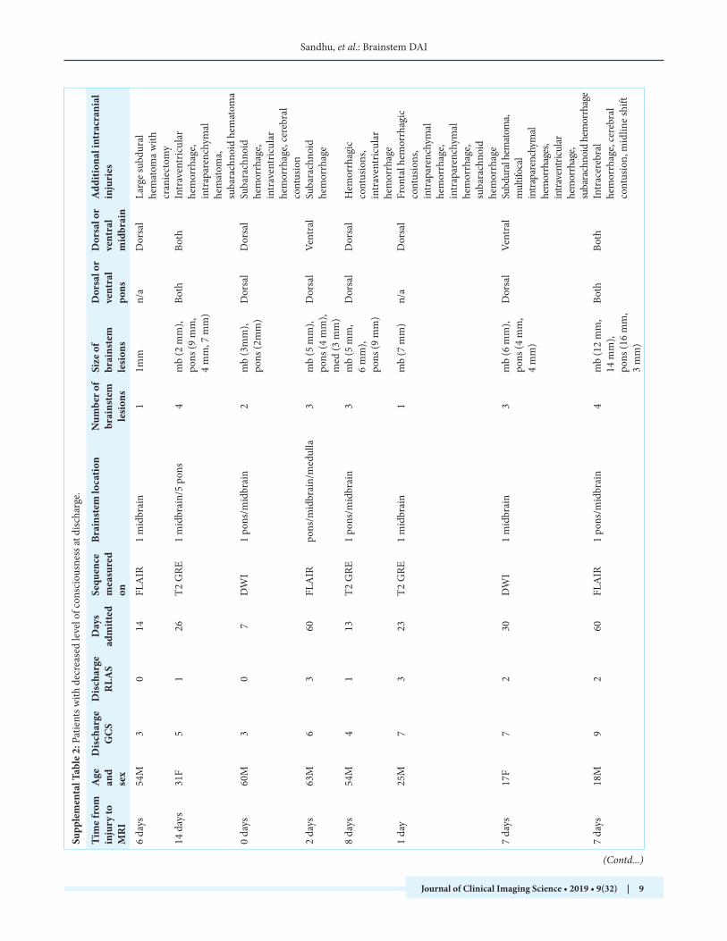
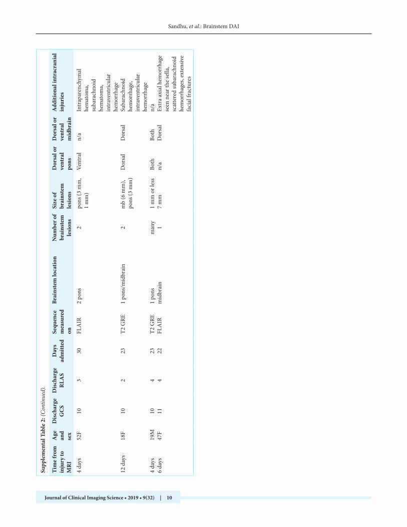
with reduced GCS/RLAS at discharge. Patients that had minimal or no pontine involvement by DAI were generally able to regain consciousness before hospital discharge [Supplemental Table 1]. In contrast, patients with large DAI lesions involving the pons tended to remain unconscious at hospital discharge [Supplemental Table 2]. It has been demonstrated that severe TBI carries a worse prognosis and elevated health-care costs.[14] Identifying patients who are at increased risk for the poor neurological outcome may allow for changes in management, which may result in socioeconomic benefits.[15,16] Clinical decision-making may be enhanced with increased sensitivity and specificity of imaging studies such as SWI for DAI with small hemorrhagic foci.[17] SWI was utilized to document an association between total DAI lesion burden and white matter atrophy at 1 year in patients with moderate to severe DAI, although these patients were able to improve their cognitive functioning with rehabilitation.[18] As imaging techniques continue to be refined and the data they generate are analyzed with regard to prognosis, MRI may play a larger role in the clinical management of comatose patients with severe TBI and DAI. e blanket description of Grade 3 for brainstem DAI lesions was of concern in this study as several patients initially assessed as Grade 3 DAI regained consciousness with GCS of 15 before hospital discharge. It is possible that Grade 3 lesions may warrant further inspection with regard to the extent of the injury and neuroanatomical location before prognostication.
e ascending reticular activating system (ARAS) contains structures vital to consciousness and travels through the pontomesencephalic tegmentum. Brainstem strokes associated with coma were shown to be clustered around this region of the rostral pons which contains the raphe nuclei, locus coeruleus, and other nuclei associated with the
ARAS.[19] Precise neuroanatomical localization of the ARAS is difficult with current imaging modalities; however, DTI was used to reconstruct the ARAS originating from the pontine reticular formation and coursing through the mesencephalic tegmentum to the thalamus.[20] Indeed, the location of DAI lesions was shown to be an independent predictor of poor prognosis when deep lesions are identified in the region of the substantia nigra and mesencephalic tegmentum on SWI. SWI was shown to detect these lesions with higher sensitivity compared to GRE. ese observations led to the proposal of an extended grading system for DAI, with an additional Grade 4 assigned for lesions involving the substantia nigra and/or mesencephalic tegmentum.[21] Microhemorrhages affecting the dorsal region of the brainstem, specifically the nuclei associated with the ARAS were found to correlate with a neurological disability at 1 year.[13] e findings from these studies reinforce the notion that MRI reports of Grade 3 DAI may vary widely in regard to a number of lesions, total affected area, and location. ese details may be relevant to prognosis in addition to the traditional Grades 1–3 classification scheme and may be pertinent to communicate between the neuroradiologist and the treating physician.
Recently, attempts have been made to correlate specific locations of injury to prognosis including brainstem involvement,[9] mesencephalic tegmental involvement,[21] and corpus callosal involvement.[11] Poor prognosis was correlated with bilateral brainstem lesions as well as dorsal brainstem lesions.[22] ere are many limitations in this field of research, including the high prevalence of confounding factors including comorbid traumatic injuries, hospital course, time to diagnosis, and initiation of therapeutic hypothermia among many others. ese limitations decrease the quality of evidence by introducing bias, and there is a need to expand the current literature before
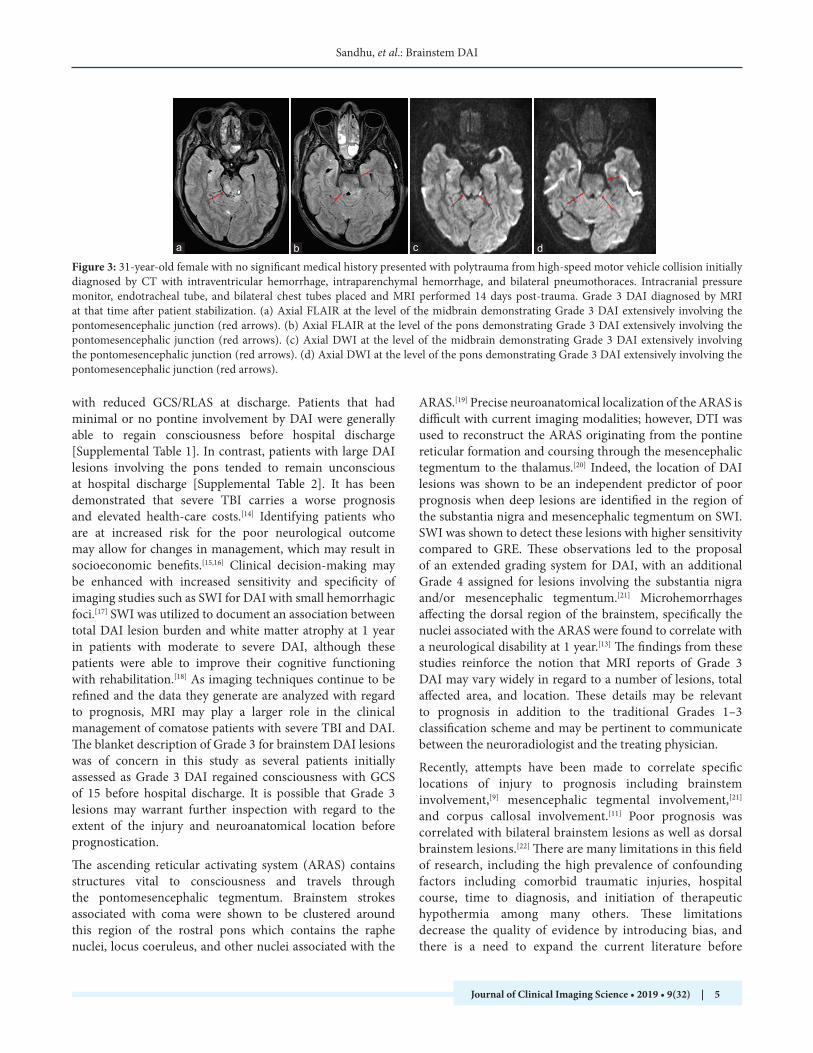
Figure 3: 31-year-old female with no significant medical history presented with polytrauma from high-speed motor vehicle collision initially diagnosed by CT with intraventricular hemorrhage, intraparenchymal hemorrhage, and bilateral pneumothoraces. Intracranial pressure monitor, endotracheal tube, and bilateral chest tubes placed and MRI performed 14 days post-trauma. Grade 3 DAI diagnosed by MRI at that time after patient stabilization. (a) Axial FLAIR at the level of the midbrain demonstrating Grade 3 DAI extensively involving the pontomesencephalic junction (red arrows). (b) Axial FLAIR at the level of the pons demonstrating Grade 3 DAI extensively involving the pontomesencephalic junction (red arrows). (c) Axial DWI at the level of the midbrain demonstrating Grade 3 DAI extensively involving the pontomesencephalic junction (red arrows). (d) Axial DWI at the level of the pons demonstrating Grade 3 DAI extensively involving the pontomesencephalic junction (red arrows).
dcba
Sandhu, et al.: Brainstem DAI
Journal of Clinical Imaging Science • 2019 • 9(32) | 6
a definitive conclusion can be drawn. Although TBI is common, severe TBI resulting in Grade 3 DAI is incurred due to severe trauma, and patients may not survive until an MRI can be performed. For comatose patients that do survive the initial trauma and are medically stabilized, accurate prognostication is vital. In this study, patients with numerous, large volume brainstem lesions, especially those affecting the dorsal pons were less likely to recover consciousness. is may indicate the involvement of structures vital to achieving consciousness such as those found in the ARAS which courses through the dorsal pons. In contrast, several patients initially determined on MRI to have Grade 3 DAI with small, solitary brainstem lesions went on to fully recover consciousness before hospital discharge. ese patients incurred fewer, smaller lesions located more anteriorly, sparing the dorsal pons and possibly, ARAS structures responsible for maintaining consciousness. Eleven patients with Grade 2 DAI, or lesions in the corpus callosum, but no brainstem lesions, were discharged with an average GCS of 13.6 after an average length of stay of 39 days, further supporting the hypothesis that lesions in the dorsal brainstem where the ARAS is located may mediate impairment of recovery of consciousness in DAI.
Limitations to this study include loss of follow-up when patients were transferred to long-term rehabilitation facilities. Due to the retrospective nature of the study, the patient’s clinical courses were unable to be followed long term. Even those patients who were unconscious at discharge may have recovered consciousness after undergoing prolonged rehabilitation. e small sample size obtained for this rare injury is a further limitation of this study. A confounding factor may be coincident traumatic intracranial injuries, which were sustained by the majority of the patients, which may affect consciousness at discharge. Two patients in the decreased consciousness at discharge group had injuries causing cerebral midline shift; one was surgically evacuated. Another patient with decreased consciousness had breast cancer metastatic to the brain at the time of injury. Numerous patients in both groups had additional intracranial injuries, most commonly subarachnoid hemorrhage, intraparenchymal hemorrhage, intraventricular hemorrhage, subdural hematoma, and cerebral contusion. Only two patients had isolated DAI as the presenting intracranial injury. In addition, prolonged time from injury to MRI in this study may diminish the imaging characteristics observed. Average days elapsed from admission to MRI was 7.5 days, however, if five outliers who received delayed MRIs at 12, 13, 14, 18, and 37 days post-admission are excluded, the average time from admission to MRI was 3.5 days. Finally, levels of serum biomarkers for TBI were not analyzed for these patients. It has recently been demonstrated that blood biomarkers may be able to help
predict unfavorable outcomes for patients who have suffered a TBI. Candidate biomarkers that may predict poor outcome after severe TBI include several markers of inflammation, coagulation, brain structural proteins, and maintenance of homeostasis.[23] e function of these proteins identified to have “good” performance for identifying poor outcome after severe TBI further supports the hypothesis that axonal shear injury disrupts neuronal homeostasis and cellular transport necessary to repair the damage done by the initial insult. e resultant cascade may lead to secondary axotomy with catastrophic damage and poor outcome. Analyzing the levels of serum biomarkers in conjunction with the radionomics of TBI may enhance understanding and allow more accurate prognostication for patients with Grade 3 DAI.
CONCLUSION
Patients with severe TBI may remain comatose for extended periods of time, especially in the presence of Grade 3 DAI. Prognostic information in these situations is valuable to clinicians and family members, in terms of the possibility of recovery of consciousness and the potential for meaningful recovery. is study shows decreased neurological recovery when patients have numerous, large lesions located in the deep structures of the pons and midbrain. It may be pertinent to consider the number, size, and location of brainstem DAI lesions before accepting the prognostic implications of Grade 3 DAI.
Financial support and sponsorship
Nil.
Conflicts of interest
ere are no conflicts of interest.
REFERENCES
1. Centers for Disease Control and Prevention: cdc.gov. Atlanta: United States; Traumatic brain injury and concussion; 2017. Available from: https://www.cdc.gov/traumaticbraininjury. [Last accessed on 2018 Aug 10].
2. Skandsen T, Kvistad KA, Solheim O, Strand IH, Folvik M, Vik A, et al. Prevalence and impact of diffuse axonal injury in patients with moderate and severe head injury: A cohort study of early magnetic resonance imaging findings and 1-year outcome. J Neurosurg 2010;113:556-63.
3. Su E, Bell M. Diffuse axonal injury. In: Laskowitz D, Grant G, editors. Translational Research in Traumatic Brain Injury. Boca Raton Florida: CRC Press/Taylor and Francis Group; 2016.
4. Andresen M, Gazmuri JT, Marín A, Regueira T, Rovegno M. erapeutic hypothermia for acute brain injuries. Scand J Trauma Resusc Emerg Med 2015;23:42.
5. Henninger N, Compton RA, Khan MW, Carandang R, Hall W, Muehlschlegel S, et al. “Don’t lose hope early”: Hemorrhagic
Sandhu, et al.: Brainstem DAI
Journal of Clinical Imaging Science • 2019 • 9(32) | 7
diffuse axonal injury on head computed tomography is not associated with poor outcome in moderate to severe traumatic brain injury patients. J Trauma Acute Care Surg 2018;84:473-82.
6. Bansal M, Sinha VD, Bansal J. Diagnostic and prognostic capability of newer magnetic resonance imaging brain sequences in diffuse axonal injury patient. Asian J Neurosurg 2018;13:348-56.
7. Chelly H, Chaari A, Daoud E, Dammak H, Medhioub F, Mnif J, et al. Diffuse axonal injury in patients with head injuries: An epidemiologic and prognosis study of 124 cases. J Trauma 2011;71:838-46.
8. Adams JH, Doyle D, Ford I, Gennarelli TA, Graham DI, McLellan DR, et al. Diffuse axonal injury in head injury: Definition, diagnosis and grading. Histopathology 1989;15:49-59.
9. Haghbayan H, Boutin A, Laflamme M, Lauzier F, Shemilt M, Moore L, et al. e prognostic value of MRI in moderate and severe traumatic brain injury: A systematic review and meta-analysis. Crit Care Med 2017;45:e1280-e1288.
10. Park SJ, Hur JW, Kwon KY, Rhee JJ, Lee JW, Lee HK, et al. Time to recover consciousness in patients with diffuse axonal injury: Assessment with reference to magnetic resonance grading. J Korean Neurosurg Soc 2009;46:205-9.
11. van Eijck MM, Schoonman GG, van der Naalt J, de Vries J, Roks G. Diffuse axonal injury after traumatic brain injury is a prognostic factor for functional outcome: A systematic review and meta-analysis. Brain Inj 2018;32:395-402.
12. Lin K, Dulebohn SC. Ranchos los amigos. In: StatPearls. Treasure Island Florida: StatPearls Publishing; 2018.
13. Izzy S, Mazwi NL, Martinez S, Spencer CA, Klein JP, Parikh G, et al. Revisiting grade 3 diffuse axonal injury: Not all brainstem microbleeds are prognostically equal. Neurocrit Care 2017;27:199-207.
14. Weiss N, Galanaud D, Carpentier A, Naccache L, Puybasset L. Clinical review: Prognostic value of magnetic resonance imaging in acute brain injury and coma. Crit Care 2007;11:230.
15. King JT Jr., Carlier PM, Marion DW. Early Glasgow outcome scale scores predict long-term functional outcome in
patients with severe traumatic brain injury. J Neurotrauma 2005;22:947-54.
16. Sinson G, Bagley LJ, Cecil KM, Torchia M, McGowan JC, Lenkinski RE, et al. Magnetization transfer imaging and proton MR spectroscopy in the evaluation of axonal injury: Correlation with clinical outcome after traumatic brain injury. AJNR Am J Neuroradiol 2001;22:143-51.
17. Tao JJ, Zhang WJ, Wang D, Jiang CJ, Wang H, Li W, et al. Susceptibility weighted imaging in the evaluation of hemorrhagic diffuse axonal injury. Neural Regen Res 2015;10:1879-81.
18. Feltrin FS, Zaninotto AL, Guirado VMP, Macruz F, Sakuno D, Dalaqua M, et al. Longitudinal changes in brain volumetry and cognitive functions after moderate and severe diffuse axonal injury. Brain Inj 2018;32:1208-17.
19. Parvizi J, Damasio AR. Neuroanatomical correlates of brainstem coma. Brain 2003;126:1524-36.
20. Yeo SS, Chang PH, Jang SH. e ascending reticular activating system from pontine reticular formation to the thalamus in the human brain. Front Hum Neurosci 2013;7:416.
21. Abu Hamdeh S, Marklund N, Lannsjö M, Howells T, Raininko R, Wikström J, et al. Extended anatomical grading in diffuse axonal injury using MRI: Hemorrhagic lesions in the substantia nigra and mesencephalic tegmentum indicate poor long-term outcome. J Neurotrauma 2017;34:341-52.
22. Hilario A, Ramos A, Millan JM, Salvador E, Gomez PA, Cicuendez M, et al. Severe traumatic head injury: Prognostic value of brain stem injuries detected at MRI. AJNR Am J Neuroradiol 2012;33:1925-31.
23. Gan ZS, Stein SC, Swanson R, Guan S, Garcia L, Mehta D, et al. Blood biomarkers for traumatic brain injury: A quantitative assessment of diagnostic and prognostic accuracy. Front Neurol 2019;10:446.
How to cite this article: Sandhu S, Soule E, Fiester P, Natter P, Tavanaiepour D, Rahmathulla G, et al. Brainstem diffuse axonal injury and consciousness. J Clin Imaging Sci 2019;9:32.
Sandhu, et al.: Brainstem DAI
Journal of Clinical Imaging Science • 2019 • 9(32) | 8
Supp
lem
enta
l Tab
le 1
: Pat
ient
s con
scio
us at
disc
harg
e.
Tim
e fr
om
inju
ry to
M
RI
Age
an
d se
x
Dis
char
ge
GC
SD
isch
arge
R
LAS
Day
s ad
mitt
edSe
quen
ce
mea
sure
d on
Brai
nste
m
loca
tion
Num
ber o
f br
ains
tem
le
sion
s
Size
of
brai
nste
m
lesi
ons
Dor
sal o
r ve
ntra
l po
ns
Dor
sal o
r ve
ntra
l m
idbr
ain
Add
ition
al in
trac
rani
al
inju
ries
3 da
ys21
F15
630
T2 G
RE1
pons
1po
ns (1
mm
)D
orsa
ln/
aSu
bara
chon
id h
emor
rhag
e2
days
23M
154
30T2
GRE
1 po
ns1
pons
(1 m
m)
Dor
sal
n/a
n/a
1 da
y49
M15
511
FLA
IR1
mid
brai
n1
mb
(9 m
m)
n/a
Vent
ral
Intr
apar
ench
ymal
he
mor
rhag
e, su
bdur
al
hem
atom
a, ce
rebr
al
cont
usio
n2
days
30M
156
30FL
AIR
1 m
idbr
ain
1m
b (3
mm
)n/
aVe
ntra
lIn
trap
aren
chym
al
hem
orrh
age,
subd
ural
he
mat
oma
37 d
ays
27M
157
30T2
GRE
1 m
idbr
ain
1m
b (5
mm
)n/
aD
orsa
lD
iffus
e su
bara
chno
id
hem
orrh
age,
mul
tifoc
al
intr
apar
ench
ymal
he
mor
rhag
es, s
mal
l sub
dura
l he
mor
rhag
e13
day
s28
F14
540
DW
I1
mid
brai
n1
mb
(3 m
m)
n/a
Dor
sal
Intr
apar
ench
ymal
he
mor
rhag
e, in
trav
entr
icul
ar
hem
orrh
age,
suba
rach
noid
he
mor
rhag
e, su
bdur
al
hem
orrh
age
18 d
ays
56M
146
40T2
GRE
1 m
idbr
ain
1m
b (8
mm
)n/
aVe
ntra
lSu
bara
chno
id h
emor
rhag
e w
ith ri
ght f
ront
al
hem
orrh
agic
cont
usio
n2
days
16M
155
14FL
AIR
1 m
idbr
ain
1m
b (7
mm
)n/
aD
orsa
lH
emor
rhag
ic co
rtic
al
cont
usio
n w
ithin
the
left
fron
tal l
obe
SUPP
LEM
ENTA
RY
Sandhu, et al.: Brainstem DAI
Journal of Clinical Imaging Science • 2019 • 9(32) | 9
Tim
e fr
om
inju
ry to
M
RI
Age
an
d se
x
Dis
char
ge
GC
SD
isch
arge
R
LAS
Day
s ad
mitt
edSe
quen
ce
mea
sure
d on
Brai
nste
m lo
catio
nN
umbe
r of
brai
nste
m
lesi
ons
Size
of
brai
nste
m
lesi
ons
Dor
sal o
r ve
ntra
l po
ns
Dor
sal o
r ve
ntra
l m
idbr
ain
Add
ition
al in
trac
rani
al
inju
ries
6 da
ys54
M3
014
FLA
IR1
mid
brai
n1
1mm
n/a
Dor
sal
Larg
e su
bdur
al
hem
atom
a w
ith
cran
iect
omy
14 d
ays
31F
51
26T2
GRE
1 m
idbr
ain/
5 po
ns4
mb
(2 m
m),
pons
(9 m
m,
4 m
m, 7
mm
)
Both
Both
Intr
aven
tric
ular
he
mor
rhag
e, in
trap
aren
chym
al
hem
atom
a,
suba
rach
noid
hem
atom
a 0
days
60M
30
7D
WI
1 po
ns/m
idbr
ain
2m
b (3
mm
), po
ns (2
mm
)D
orsa
lD
orsa
l Su
bara
chno
id
hem
orrh
age,
intr
aven
tric
ular
he
mor
rhag
e, ce
rebr
al
cont
usio
n2
days
63M
63
60FL
AIR
pons
/mid
brai
n/m
edul
la3
mb
(5 m
m),
pons
(4 m
m),
med
(3 m
m)
Dor
sal
Vent
ral
Suba
rach
noid
he
mor
rhag
e
8 da
ys54
M4
113
T2 G
RE1
pons
/mid
brai
n3
mb
(5 m
m,
6 m
m),
pons
(9 m
m)
Dor
sal
Dor
sal
Hem
orrh
agic
co
ntus
ions
, in
trav
entr
icul
ar
hem
orrh
age
1 da
y25
M7
323
T2 G
RE1
mid
brai
n1
mb
(7 m
m)
n/a
Dor
sal
Fron
tal h
emor
rhag
ic
cont
usio
ns,
intr
apar
ench
ymal
he
mor
rhag
e, in
trap
aren
chym
al
hem
orrh
age,
suba
rach
noid
he
mor
rhag
e 7
days
17F
72
30D
WI
1 m
idbr
ain
3m
b (6
mm
), po
ns (4
mm
, 4
mm
)
Dor
sal
Vent
ral
Subd
ural
hem
atom
a, m
ultif
ocal
in
trapa
renc
hym
al
hem
orrh
ages
, in
trave
ntric
ular
he
mor
rhag
e, su
bara
chno
id h
emor
rhag
e7
days
18M
92
60FL
AIR
1 po
ns/m
idbr
ain
4m
b (1
2 m
m,
14 m
m),
pons
(16
mm
, 3
mm
)
Both
Both
Intr
acer
ebra
l he
mor
rhag
e, ce
rebr
al
cont
usio
n, m
idlin
e sh
ift
Supp
lem
enta
l Tab
le 2
: Pat
ient
s with
dec
reas
ed le
vel o
f con
scio
usne
ss at
disc
harg
e.
(Contd...)
Sandhu, et al.: Brainstem DAI
Journal of Clinical Imaging Science • 2019 • 9(32) | 10
Supp
lem
enta
l Tab
le 2
: (Co
ntin
ued)
.
Tim
e fr
om
inju
ry to
M
RI
Age
an
d se
x
Dis
char
ge
GC
SD
isch
arge
R
LAS
Day
s ad
mitt
edSe
quen
ce
mea
sure
d on
Brai
nste
m lo
catio
nN
umbe
r of
brai
nste
m
lesi
ons
Size
of
brai
nste
m
lesi
ons
Dor
sal o
r ve
ntra
l po
ns
Dor
sal o
r ve
ntra
l m
idbr
ain
Add
ition
al in
trac
rani
al
inju
ries
4 da
ys52
F10
330
FLA
IR2
pons
2po
ns (3
mm
, 1
mm
)Ve
ntra
ln/
aIn
trap
aren
chym
al
hem
atom
a,
suba
rach
noid
he
mat
oma,
in
trav
entr
icul
ar
hem
orrh
age
12 d
ays
18F
102
23T2
GRE
1 po
ns/m
idbr
ain
2m
b (6
mm
), po
ns (3
mm
)D
orsa
lD
orsa
lSu
bara
chno
id
hem
orrh
age,
intr
aven
tric
ular
he
mor
rhag
e4
days
19M
104
23T2
GRE
1 po
nsm
any
1 m
m o
r les
sBo
thBo
thn/
a6
days
47F
114
22FL
AIR
mid
brai
n1
7 m
mn/
aD
orsa
lEx
tra
axia
l hem
orrh
age
seen
nea
r the
sella
, sc
atte
red
suba
rach
noid
he
mor
rhag
es, e
xten
sive
faci
al fr
actu
res


