Embed Size (px)
Citation preview

Brilliant organic nanodots: novel nano-objects for bionanophotonics Olivier Mongina, Cédric Rouxela, Anne-Claire Robina, Anna Pla-Quintanab, Tathavarathy Rama
Krishnab, Gaëlle Recherc, François Tiahoc, Anne-Marie Caminadeb, Jean-Pierre Majoralb,Mireille Blanchard-Desce*a
a Molecular Chemistry and Molecular Photonics (CNRS UMR 6510), Université de Rennes 1, Campus de Beaulieu, Bât. 10A, F-35042 Rennes Cedex, France.
b Laboratoire de Chimie de Coordination, CNRS, 205 route de Narbonne, F-31077 Toulouse Cedex 4, France.
c Equipe SCANING, CNRS UMR 6026, Université de Rennes 1, Campus de Beaulieu, F-35042 Rennes Cedex, France.
ABSTRACT
Semiconductor quantum dots are recognized to provide a particularly effective approach to bright nano-objects for bioimaging. However, these inorganic systems suffer from several drawbacks such as toxicity, dispersity, blinking … and raise a number of questions with respect to environmental issues. With this in mind, we have developed an innovative route towards purely organic nanodots showing exceptional one and two-photon brightness by confining a large number of optimized fluorophores within nano-objects of defined and controlled structure. These novel "soft" nano-objects offer major promises for bio and nanophotonics.
Keywords: biophotonics, biological imaging, nanosciences, fluorescence, two-photon absorption, dendrimers
1. INTRODUCTION
Recently semiconductor quantum dots (QDs) have been shown to provide a particularly effective approach to fluorescent nano-objects for biological imaging. Indeed, these inorganic nanoparticles exhibit large one- and two-photon absorption cross-sections, reasonable fluorescence quantum yields, broad excitation but narrow emission bands, and high photostability. These properties make them of particular interest for in vitro and in vivo imaging, and they have found applications in specific labeling of cells1-5 and tissues6,7. In addition, because their emission spectra can be tuned by playing on their size and composition, they can be used for multicolor imaging.8-10 They have also found applications in two-photon excited fluorescence (TPEF) imaging,11 which has gained widespread popularity in the biology community due to the many advantages it provides for biological microscopic imaging, including intrinsic three-dimensional resolution and increased penetration depth in tissues.12-14
However, these inorganic nanocrystals suffer from several drawbacks such as biological toxicity15 (due in particular to the presence of heavy metals such as cadmium), polydispersity16,17 and blinking.18 These nano-objects also raise a number of questions with respect to environmental issues. With this in mind, we have developed an alternative route towards all-organic nanodots showing exceptional one and two-photon brightness. Our approach is based on the confinement of a large and discrete number of optimized fluorophores within nano-objects of defined and controlled structure.19-21 Such organic nanodots (OND) can be obtained by grafting the fluorophores on the surface of a dendrimeric platform (Figure 1). This strategy is highly modular and allows adjunction of supplementary layers, decoration of the periphery with hydrosolubilizing groups, and insertion of functional groups that could be utilized for bioconjugation purposes. Moreover, it offers several potential advantages: photoluminescence (PL) characteristics can, in principle, be tuned by playing on the nature of the fluorophore, while the dendrimer scaffold can be chosen so as to minimize toxicity effects and control clearance ability. In particular, phosphorus-based dendrimers have been shown to have low toxicity and are biodegradable.22,23 However, implementing such modular approach also requires taking into account interactions
*[email protected]; phone (+33) 223236277; fax (+33) 223236955

between the various molecular building blocks including possible interactions between decorating fluorophores. The confinement of fluorophores within the OND imposes close proximity between chromophores, which favors interchromophoric interactions: (i) in the ground state with potential effects on the absorption characteristics and (ii) in the excited state with impending marked effects on PL. Such interactions can significantly affect the optical responses and photophysical properties of the OND which will then differ from the mere addition of the contributions of isolated single fluorophores. For instance formation of dimers or aggregates between adjacent fluorophores could lead to fluorescence quenching.24 Also interactions in the excited state can lead to changes of PL characteristics due to various phenomena (energy transfer, excimer formation or exciton annihilation which is an important decay process in chromophore decorated dendrimers at high excitation intensity25). Also it has been shown that interactions between chromophores within dimers26 or aggregates27,28 can lead to significant change in two-photon absorption (TPA) responses. Such effects are expected to be strongly dependent on both the nature of the chromophores and their proximity and relative orientations.
2. METHODOLOGY
2.1 Optical measurements
All photophysical measurements have been performed with freshly-prepared solutions in air-equilibrated solvents at room temperature (298 K). UV/Vis absorption spectra were recorded on a Jasco V-570 spectrophotometer. Fluorescence measurements were performed on dilute solutions (ca. 10 6 M chromophore concentration, optical density < 0.1) contained in standard 1 cm quartz cuvettes using an Edinburgh Instruments (FLS920) spectrometer in photon-counting mode. Emission spectra were obtained, for each compound, under excitation at the maximum absorption wavelength. Fluorescence quantum yields were measured according to literature procedures using fluorescein in 0.1 N NaOH as a standard (quantum yield = 0.90).29,30
2.2 TPEF measurements
Two-photon absorption cross sections ( 2) were obtained from the two-photon excited fluorescence (TPEF) cross sections ( 2 ) and the fluorescence emission quantum yield ( ). TPEF cross sections in toluene (10-4 M chromophore concentration) were determined using a Ti-sapphire laser (Coherent Mira 900 pumped by a 5 W Verdi) delivering 150 fs excitation pulses and operating between 700 and 990 nm, according to the experimental protocol established by Xu and Webb.31 This experimental protocol allows avoiding contributions from excited-state absorption that are known to result in largely overestimated TPA cross-sections. Fluorescein in 0.01 M NaOH, whose TPEF cross-sections are well-known,31 served as the reference, taking into account the necessary corrections for the refractive index of the solvents.32
The quadratic dependence of the fluorescence intensity on the excitation intensity was verified for each data point, indicating that the measurements were carried out in intensity regimes in which saturation or photodegradation do not occur. More details about the experimental setup have been previously published.32
2.3 Labeling and two-photon in vivo imaging
Stage 53 (Nieuwkoop and Faber 1956) Xenopus laevis tadpoles (national breeding facility of xenopus animals in Rennes, France) were anesthetized in MS222 (tricaine, Sigma, 0.5 mg/ml). OND (2µL of a 750µM solution, i.e. 1.5 pM) were injected in the heart with a syringe and animals were allowed to recover in Mark’s Modified Ringer (MMR) as long as necessary for the complete diffusion of the OND into blood vessels. They were then re-anesthetized and mounted in the recording chamber between two coverslips.
Two-photon imaging experiments were performed on the PIXEL platform, University of Rennes 1. A 860 nm excitation beam from a femtosecond laser (MAITAI Spectra Physics) was focused on the samples via a Leica objective HCPLAPO 20X (NA=0.7). The vessels were scanned in 3D. After collection, data were analyzed with the open source software ImageJ (http://rsb.info.nih.gov/ij/). The stacks were either projected along the z-axis or exported to be reconstructed with the freeware UCSF Chimera (http://www.cgl.ucsf.edu/chimera).

3. RESULTS AND DISCUSSION
3.1 Multichromophoric lipophilic organic nanodots
We have investigated and compared the photophysical properties of a series of multichromophoric single-layer (Figure 1) and double-layer (Figure 2) organic nanodots, built from the gathering of an exponentially increasing number of one- and two-photon active quadrupolar fluorophores F on phosphorus dendrimeric platforms.19,21
OHC[P3N3] N N P
Me SO
HC N N P
Me S
BuBu NHex
HexN
OO
2 26
G2
OHC[P3N3] N N P
Me SO
HC N N P
Me SO
HC N N P
Me S
BuBu NHex
HexN
OO
2 2 2 6G3
OHC[P3N3] N N P
Me SO
HC N N P
Me SO
HC N N P
Me SO
HC N N P
Me S
BuBu NHex
HexN
OO
2 2 2 2 6G4
BuBu NHex
HexN
OHO
OHC[P3N3] N N P
Me S
BuBu NHex
HexN
OO
26G1
F
N PN
PNP
N PN
PNP
G1
G2
F
Figure 1. Chemical structures and schematic representations of fluorophore F and single-layer organic nanodots (SL-OND) G1-G4.
NNBuBu
OOO
N
H
NP
Me
SNN
BuBu Hex
HexOO
DL
26
[P3N3]N P
NPN
P
DLFigure 2. Chemical structure and schematic representation of double-layer organic nanodot (DL-OND) DL.

One-photon absorption
All multichromophoric OND are excellent one-photon absorbers, with a strong absorption band in the near UV visible-blue region. The single-layer organic nanodots (SL-OND) G1-G4 show a nearly linear increase of the extinction coefficient with the number of chromophores, leading to giant absorption coefficients, exceeding 7 000 000 for G4(Table 1). As observed from Table 1 and illustrated in Figure 3a, the ONDs definitely show much higher absorptivities than QD480, a commercially available core-shell CdSe/ZnS quantum dot (QD) emitting in the same range, providing evidence that the nanodot route may lead to competitive nano-objects with larger brightness than QDs.
The absorption spectrum of double-layer organic nanodot (DL-OND) DL is broader and slightly blue shifted with respect to that of isolated fluorophore F (Figure 3c) and SL-OND dendrimers (Table1). Comparison of the absorption spectrum of DL with that corresponding to that expected for the additive contributions of 6 inner layer and 12 outer layer chromophores F (Figure 3c) also shows that DL exhibits a slightly lower effective maximum absorption coefficient per fluorophore along with a slight broadening observed on the blue side and red tail of the absorption band most probably in relation with inhomogeneous broadening.
Table. 1. Photophysical properties of CdSe/ZnS quantum dot QD480, fluorophore F, single-layer organic nanodots G1-G4and double-layer organic nanodot DL in toluene.
Number of fluorophores abs
max (nm) (M-1 cm-1) emmax (nm) a 2
max
(GM)b
QD480 – 460 ± 5 20 000 480 ± 5 0.3-0.5 –F 1 386 84 900 420 0.83 765
G1 12 385 1 004 000 423 0.75 8 880 G2 24 386 2 035 000 426 0.71 17 700 G3 48 386 3 785 000 441 0.62 29 800 G4 96 386 7 101 000 445 0.48 55 900 DL 6 + 12 381 1 414 000 420 0.43 8 500
a Fluorescence quantum yield determined relative to fluorescein in 0.1 N NaOH. b 1 GM = 10-50 cm4.s.photon-1.
Fluorescence
Quite interestingly, in spite of the confinement of a large number of chromophores in a very reduced volume, OND retain fluorescence and maintain reasonably high quantum yields (Table 1), even for the highest generation dendrimer G4, leading to exceptional one-photon brightness ( ). OND decorated with fluorophore F are blue emitting, and overall, their emission spectrum is only slightly modified by the generation and concomitant size increase (Figures 3b and 3d), in striking contrast with quantum dots whose emission color is directly correlated with the size. The emission color of OND depends only on the constituting chromophores. Only a slight change of the emission band shape is observed with increasing generations (Figure 3b) or layers (Figure 3d). A loss of fine vibronic structure and a broadening of the band are observed with increasing generation, revealing an increase in reorganization energy in SL-OND G1-G4as compared to the isolated fluorophore.21
In the case of double-layer dendrimer DL, the emission can stem from both inner and outer layer chromophores, corresponding to different environments felt by the emitting fluorophores. Nanodot DL exhibits an emission spectrum (Figure 3d) similar to that of G1, with a fine vibronic structure clearly visible, but with a slight blue shift of the maximum and a more pronounced red tail than G1 (and even than G2) Such behavior is consistent with a larger inhomogeneous broadening in the case of DL as already noted from of its absorption spectrum. A neat decrease of the emission quantum yield is also observed (Table 1) This might originate from more pronounced interchromophoric interactions in the excited state inside and between layers, i.e. with a closer packing in DL than in SL-OND.

(c)Figure 3. Absorption (a, c) and emission (b, d) spectra of single-layer organic nanodots G1-G4 and double-layer organic nanodot DLin toluene. Absorption and emission spectra of fluorophore F and CdSe/ZnS quantum dot QD480 are also given for comparison. Comparison (c) of experimental and theoretical (evaluated from additive contributions of 18 isolated constituting chromophores F)absorption spectra of DL in toluene.
Two-photon absorption
The TPA of the OND was studied by investigating their two-photon excited fluorescence (TPEF) in toluene. TPEF measurements allow direct measurement of the two-photon brightness (or TPEF action cross-section) , the relevant figure of merit for imaging applications. The TPA spectra of single-layer and double-layer OND in the near infra-red range (700-1000 nm) are shown in Figure 4. The TPA cross-sections in the SL-OND series increase almost linearly with the number of decorating fluorophores leading to giant TPA cross-sections, up to 56 000 GM for G4 and concomitantly to strong two-photon brightness ( ). This additive behavior indicates that the response of each individual chromophore located on the periphery is more or less unaffected (as observed in one-photon absorption), in spite of the confinement which could lead to possible TPA loss, as was shown for individual chromophores in solvents promoting aggregation27.
As a result, TPA cross-sections of OND G1-G4 are overwhelming those of most of the quantum dots, in particular of the same emission color. As a comparison, blue (2.4 nm diameter), green (2.9 nm), yellow (3.9 nm) and red (4.8 nm) emitting CdSe quantum dots exhibit TPA cross-sections at 800 nm of 780 GM, 1950 GM, 4980 GM and 10300 GM, respectively.33 As a result G4 shows unprecedented TPA cross-sections for a spherical nanoparticle of about 4 nm size and definitely superior to that of related QDs.

Figure 4. Two-photon absorption spectra of fluorophore F,single-layer organic nanodots G1-G4 and double-layer organic nanodots DL.
Figure 5. Comparison of the TPA efficiency (normalized by the number of fluorophores) of single-layer and double-layerorganic nanodots.
Double-layer OND DL with 18 chromophores exhibits TPA cross-sections almost similar to that of single-layer G1 with only 12 chromophores (Figure 4 and Table 1). We observe a noticeable difference in the TPA spectra normalized by the number of fluorophores of DL and G1 or G2 in the 700-750 nm region, whereas they nicely overlap in the 750-850 nm region (Figure 5). It appears that the higher degree of confinement in DL (in particular for the inner layer) as compared to G1 and G2 and the difference in orientation and packing between chromophores induced by the double-layer arrangement significantly affect the TPA response. However, it should be noticed that this effect is only visible on the higher-energy band (below 750 nm) which corresponds to the strongly two-photon allowed transition. On the other hand the lower-energy band which corresponds to an only slightly two-photon allowed transition (due to slight symmetry breaking)34 remains almost unaffected.
3.2 Water-soluble monochromophoric organic nanodots
The highly modular design of organic nanodots allows decoration of the periphery with hydrosolubilizing groups. In that way, we have designed a series of water-soluble monochromophoric nanodots with shielding dendrimeric layers and peripheral cationic groups.20 To ensure its water-solubility, two-photon active lipophilic fluorophores (C and F’) were incorporated in a dendrimer shells whose periphery is covered by ammonium groups (Figures 6 and 7). An alternative and simpler route consisting in grafting ammonium groups directly onto the chromophore (compound WS-C) that has been frequently used as the easier and direct way to provide solubility27,35 was also implemented for comparison with the OND approach (Figure 6).

Bu Bu
O O[P3N3][P3N3] NC
HO N
MePS H
NN CH
ONMe
PS
NEt2HH
NEt2NH
Cl ClWS-G12 2 5
Bu Bu
HO OH
Me3N NMe3
HO OH
Br Br
+ +
WS-C+
+++++
+++
++++
++ + + + +
+
++
+ + + ++
++++
+++
++++++
+++
+++
++ +
+
++ +
+++
+++
+
WS-G1
WS-G2
5
Bu Bu
O O[P3N3][P3N3] NC
HO N
MePS
NCH
O NMe
PS
N CH
ONMe
PS
N CH
ONMe
PS
HN NEt2
HHNEt2N
H
252 2Cl Cl 2
5WS-G2
C
Bu Bu
O O[P3N3] NC
HO N
MePS
NCH
O NMe
PS
NCH
O NMe
PS H
N NEt2H
[P3N3]N CH
ONMe
PS
N CH
ONMe
PS
N CH
ONMe
PSH
NEt2NH
WS-G32 2 25
2 22
5
Figure 6. Chemical structures and schematic representations of model chromophores C, WS-C and water soluble organic nanodots (WS-OND) WS-G1 – WS-G3.
BuBu NNO
HO OOH
F'
++
+++++
++
++++
++ + + + +
+
++
+ + + ++
++++
+++
++++++WS-G'2
F'
BuBu NNO
O OO[P3N3]O [P3N3] O
HC N N P
Me SO
HC N N P
Me SHN
NEt2H
Cl
HCNNP
MeSO
HCNNP
MeSHN
Et2NH
ClWS-G'2 2 2 5
2 2 5
Figure 7. Chemical structures and schematic representations of model fluorophore F’ and water soluble organic nanodot WS-G’2.
Photophysical properties
As clearly observed from Table 2 the adjunction of ammonium groups directly on the aliphatic chains of lipophilic chromophore C (to give water-soluble chromophore WS-C) does not lead to retention of the PL efficiency in water (Table 2). This fluorescence quantum yield decrease can be related to non-radiative processes mediated by water molecules and/or to molecular aggregation phenomena. Indeed, the WS-C PL in water is restored upon addition of a surfactant (sodium dodecyl sulfate). The water-soluble quadrupole WS-C also endures a marked decrease of its TPA efficiency in water as compared to C in ethanol. As a result, although being water-soluble and based on a quadrupolar chromophore that shows reasonable TPA efficiency in non-aqueous protic media, WS-C only shows very low two-photon brightness ( 2 ) in water. This clearly evidences that the "simple" water solubilization strategy is detrimental to both PL and TPA efficiency indicating that protection of the TPA chromophore from close proximity of water molecules is required Indeed, the PL efficiencies of water-soluble nanodots WS-G1, WS-G2 and WS-G3 in water are clearly much higher than that of water-soluble chromophore WS-C, and in the case of WS-G2 and WS-G3, comparable to that of C in ethanol. Interestingly, the differences in relative vibronic intensities in the emission spectra of dendrimers in ethanol and water tend to disappear with increasing the generation number, and the emission spectrum of the highest generation dendrimer WS-G3 shows similar vibronic intensities in ethanol and water indicating that the dendritic
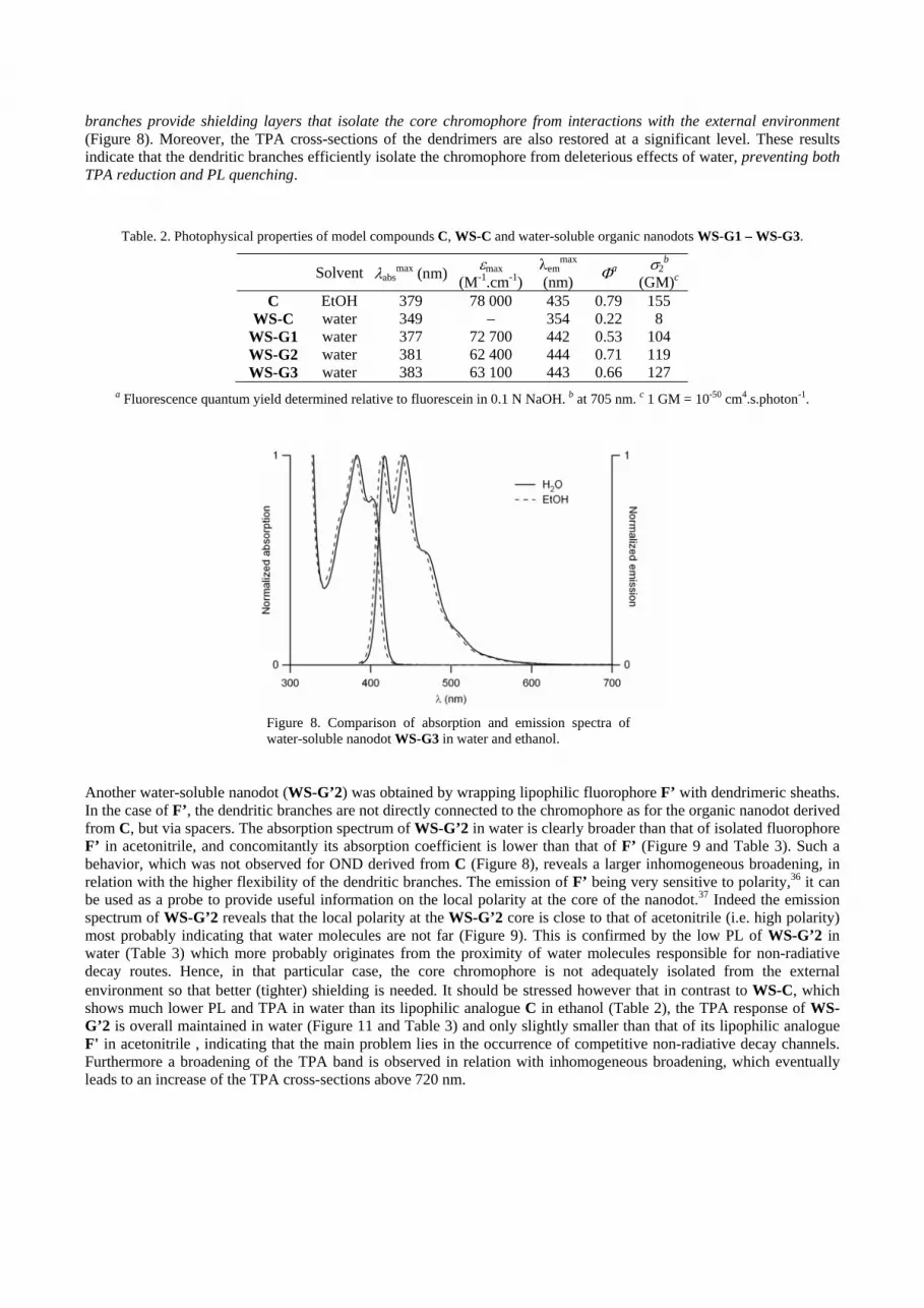
branches provide shielding layers that isolate the core chromophore from interactions with the external environment(Figure 8). Moreover, the TPA cross-sections of the dendrimers are also restored at a significant level. These results indicate that the dendritic branches efficiently isolate the chromophore from deleterious effects of water, preventing both TPA reduction and PL quenching.
Table. 2. Photophysical properties of model compounds C, WS-C and water-soluble organic nanodots WS-G1 – WS-G3.
Solvent absmax (nm) max
(M-1.cm-1)em
max
(nm) a 2
b
(GM)c
C EtOH 379 78 000 435 0.79 155 WS-C water 349 – 354 0.22 8
WS-G1 water 377 72 700 442 0.53 104 WS-G2 water 381 62 400 444 0.71 119 WS-G3 water 383 63 100 443 0.66 127
a Fluorescence quantum yield determined relative to fluorescein in 0.1 N NaOH. b at 705 nm. c 1 GM = 10-50 cm4.s.photon-1.
Figure 8. Comparison of absorption and emission spectra of water-soluble nanodot WS-G3 in water and ethanol.
Another water-soluble nanodot (WS-G’2) was obtained by wrapping lipophilic fluorophore F’ with dendrimeric sheaths. In the case of F’, the dendritic branches are not directly connected to the chromophore as for the organic nanodot derived from C, but via spacers. The absorption spectrum of WS-G’2 in water is clearly broader than that of isolated fluorophore F’ in acetonitrile, and concomitantly its absorption coefficient is lower than that of F’ (Figure 9 and Table 3). Such a behavior, which was not observed for OND derived from C (Figure 8), reveals a larger inhomogeneous broadening, in relation with the higher flexibility of the dendritic branches. The emission of F’ being very sensitive to polarity,36 it can be used as a probe to provide useful information on the local polarity at the core of the nanodot.37 Indeed the emission spectrum of WS-G’2 reveals that the local polarity at the WS-G’2 core is close to that of acetonitrile (i.e. high polarity) most probably indicating that water molecules are not far (Figure 9). This is confirmed by the low PL of WS-G’2 in water (Table 3) which more probably originates from the proximity of water molecules responsible for non-radiative decay routes. Hence, in that particular case, the core chromophore is not adequately isolated from the external environment so that better (tighter) shielding is needed It should be stressed however that in contrast to WS-C, which shows much lower PL and TPA in water than its lipophilic analogue C in ethanol (Table 2), the TPA response of WS-G’2 is overall maintained in water (Figure 11 and Table 3) and only slightly smaller than that of its lipophilic analogue F' in acetonitrile , indicating that the main problem lies in the occurrence of competitive non-radiative decay channels. Furthermore a broadening of the TPA band is observed in relation with inhomogeneous broadening, which eventually leads to an increase of the TPA cross-sections above 720 nm.

Table. 3. Photophysical properties of model fluorophore F’ and water-soluble organic nanodot WS-G’2.
Solvent abs (nm) max(M-1.cm-1)
em(nm)
a 2 (max) (GM)b
F’ CH3CN 383 85 000 498 0.69 800 WS-G’2 water 385 60 500 480 0.074 680
a Fluorescence quantum yield determined relative to fluorescein in 0.1 N NaOH. b 1 GM = 10-50 cm4.s.photon-1.
Figure 9. Absorption and emission spectra of dendrimer WS-G’2 in water and fluorophore F’ in CH3CN.
Figure 10. Two-photon absorption spectra of dendrimer WS-G’2in water and comparison with model fluorophore F’ in CH3CN.
Two-photon in vivo imaging
Water-soluble organic nanodots can be used as contrast agents for in vivo 3D TPEF imaging on living animal.20 In Figure 11 are shown as examples 2D and 3D images of bloods vessels of the tail of the Xenopus tadpole. The vascular network was labeled after intracardiac injection of an aqueous solution of WS-G2.
Figure 11. In vivo two-photon imaging of blood vessels of stage 53 Xenopus laevis tadpole, obtained after intracardiac injection of 1.5 pmol of WS-G2 (excitation at 860 nm). Left: Projection of 93 µm thick stack of a 94 x 115 µm2 image. Right: 3D representation of the same stack.

4. CONCLUSION
We have developed an innovative route towards purely organic nanodots showing exceptional one and two-photon brightness by confining a large number of optimized fluorophores within nano-objects of defined and controlled structure. Organic nanodots thus represent a promising biocompatible and eco-friendly alternative to semiconductor quantum dots as fluorescent labels for one- and two-photon fluorescence imaging applications including bioimaging as evidenced from small animal in vivo imaging thanks to the use of these organic nanodots. In addition, the highly modular design of organic nanodots allows adjunction of supplementary layers and decoration of the periphery with hydrosolubilizing groups. This modularity is of high interest for the customization of nanodots designed for specific applications, by changing the chromophore nature and thus tuning the fluorescence color, or by grafting biomolecules for targeting or diagnostic applications. These novel "soft" nano-objects offer major promises for bio and nanophotonics.
ACKNOWLEDGMENTS
We acknowledge financial support from ANR (Projects “Biodendridot” and “Moduloo”). MBD thanks Région Bretagne (ARED Project “BIONAD”) for the fellowship to CR and the Ministère de l’Enseignement Supérieur et de la Recherche (France) for the fellowship to ACR. JPM thanks the Ministère de la Recherche (France) for the post-doctoral grant to TRK. Thanks are due to the Fundación Ramón Areces for a grant to APQ. Access to multiphotonic microscopy facilities (PIXEL platform, University of Rennes 1) is acknowledged. We also aknowledge financial support (equipment grants) from Rennes Métropole and CNRS.
REFERENCES
[1] Bruchez, M., Jr., Moronne, M., Gin, P., Weiss, S. and Alivisatos, A. P., "Semiconductor nanocrystals as fluorescent biological labels", Science 281, 2013-2016 (1998).
[2] Chan, W. C. W. and Nile, S., "Quantum dot bioconjugates for ultrasensitive nonisotopic detection", Science 281, 2016-2018 (1998).
[3] Mattoussi, H., Mauro, J. M., Goldman, E. R., Anderson, G. P., Sundar, V. C., Mikulec, F. V. and Bawendi, M. G., "Self-Assembly of CdSe-ZnS Quantum Dot Bioconjugates Using an Engineered Recombinant Protein", J. Am. Chem. Soc. 122, 12142-12150 (2000).
[4] Wu, X., Liu, H., Liu, J., Haley, K. N., Treadway, J. A., Larson, J. P., Ge, N., Peale, F. and Bruchez, M. P., "Immunofluorescent labeling of cancer marker Her2 and other cellular targets with semiconductor quantum dots", Nat. Biotechnol. 21, 41-46 (2003).
[5] Dubertret, B., Skourides, P., Norris, D. J., Noireaux, V., Brivanlou, A. H. and Libchaber, A., "In vivo imaging of quantum dots encapsulated in phospholipid micelles", Science 298, 1759-1762 (2002).
[6] Akerman, M. E., Chan, W. C. W., Laakkonen, P., Bhatia, S. N. and Ruoslahti, E., "Nanocrystal targeting in vivo", Proc. Natl. Acad. Sci. U. S. A. 99, 12617-12621 (2002).
[7] Gao, X., Cui, Y., Levenson, R. M., Chung, L. W. K. and Nie, S., "In vivo cancer targeting and imaging with semiconductor quantum dots", Nat. Biotechnol. 22, 969-976 (2004).
[8] Chan, W. C. W., Maxwell, D. J., Gao, X., Bailey, R. E., Han, M. and Nie, S., "Luminescent quantum dots for multiplexed biological detection and imaging", Curr. Opin. Biotechnol. 13, 40-46 (2002).

[9] Jaiswal, J. K., Mattoussi, H., Mauro, J. M. and Simon, S. M., "Long-term multiple color imaging of live cells using quantum dot bioconjugates", Nat. Biotechnol. 21, 47-51 (2003).
[10] Mattheakis, L. C., Dias, J. M., Choi, Y.-J., Gong, J., Bruchez, M. P., Liu, J. and Wang, E., "Optical coding of mammalian cells using semiconductor quantum dots", Anal. Biochem. 327, 200-208 (2004).
[11] Larson, D. R., Zipfel, W. R., Williams, R. M., Clark, S. W., Bruchez, M. P., Wise, F. W. and Webb, W. W., "Water-Soluble Quantum Dots for Multiphoton Fluorescence Imaging in Vivo", Science 300, 1434-1437 (2003).
[12] Denk, W., Strickler, J. H. and Webb, W. W., "Two-photon laser scanning fluorescence microscopy", Science 248, 73-76 (1990).
[13] Xu, C., Zipfel, W., Shear, J. B., Williams, R. M. and Webb, W. W., "Multiphoton fluorescence excitation: new spectral windows for biological nonlinear microscopy", Proc. Natl. Acad. Sci. U. S. A. 93, 10763-10768 (1996).
[14] Zipfel, W. R., Williams, R. M., Christie, R., Nikitin, A. Y., Hyman, B. T. and Webb, W. W., "Live tissue intrinsic emission microscopy using multiphoton-excited native fluorescence and second harmonic generation", Proc. Natl. Acad. Sci. U. S. A. 100, 7075-7080 (2003).
[15] Hardman, R., "A toxicologic review of quantum dots: toxicity depends on physicochemical and environmental factors", Environ. Health Perspect. 114, 165-172 (2006).
[16] Cingolani, R., Moro, C., Manno, D., Striccoli, M., DeBlasi, C., Righini, G. C. and Ferrara, M., "Correlation between the structural and optical properties of polydispersed II-VI quantum dots in glass matrix", J. Appl. Phys. 70, 6898-6901 (1991).
[17] Pandey, P. K., Sharma, K., Nagpal, S., Bhatnagar, P. K. and Mathur, P. C., "Growth of Group II-VI semiconductor quantum dots with strong quantum confinement and low size dispersion", Phys. Status Solidi B 240, 134-138 (2003).
[18] Nirmal, M., Dabbousi, B. O., Bawendi, M. G., Macklin, J. J., Trautman, J. K., Harris, T. D. and Brus, L. E., "Fluorescence intermittency in single cadmium selenide nanocrystals", Nature 383, 802-804 (1996).
[19] Mongin, O., Krishna, T. R., Werts, M. H. V., Caminade, A.-M., Majoral, J.-P. and Blanchard-Desce, M., "A modular approach to two-photon absorbing organic nanodots: brilliant dendrimers as an alternative to semiconductor quantum dots?" Chem. Commun., 915-917 (2006).
[20] Krishna, T. R., Parent, M., Werts, M. H. V., Moreaux, L., Gmouh, S., Charpak, S., Caminade, A.-M., Majoral, J.-P. and Blanchard-Desce, M., "Water-soluble dendrimeric two-photon tracers for in vivo imaging", Angew. Chem., Int. Ed. 45, 4645-4648 (2006).
[21] Mongin, O., Pla-Quintana, A., Terenziani, F., Drouin, D., Le Droumaguet, C., Caminade, A.-M., Majoral, J.-P. and Blanchard-Desce, M., "Organic nanodots for multiphotonics: synthesis and photophysical studies", New J. Chem. 31, 1354-1367 (2007).
[22] Maszewska, M., Leclaire, J., Cieslak, M., Nawrot, B., Okruszek, A., Caminade, A.-M. and Majoral, J.-P., "Water-Soluble Polycationic Dendrimers with a Phosphoramidothioate Backbone: Preliminary Studies of Cytotoxicity and Oligonucleotide/Plasmid Delivery in Human Cell Culture", Oligonucleotides 13, 193-205 (2003).
[23] Solassol, J., Crozet, C., Perrier, V., Leclaire, J., Beranger, F., Caminade, A.-M., Meunier, B., Dormont, D., Majoral, J.-P. and Lehmann, S., "Cationic phosphorus-containing dendrimers reduce prion replication both in cell culture and in mice infected with scrapie", J. Gen. Virol. 85, 1791-1799 (2004).
[24] West, W. and Pearce, S., "The dimeric state of cyanine dyes", J. Phys. Chem. 69, 1894-1903 (1965). [25] De Belder, G., Schweitzer, G., Jordens, S., Lor, M., Mitra, S., Hofkens, J., De Feyter, S., Van der Auweraer,
M., Herrmann, A., Weil, T., Müllen, K. and De Schryver, F. C., "Singlet ± Singlet Annihilation in Multichromophoric Peryleneimide Dendrimers, Determined by Fluorescence Upconversion", ChemPhysChem 2, 49-55 (2001).
[26] Terenziani, F., Morone, M., Gmouh, S. and Blanchard-Desce, M., "Linear and Two-Photon Absorption Properties of Interacting Polar Chromophores: Standard and Unconventional Effects", ChemPhysChem 7, 685-696 (2006).
[27] Woo, H. Y., Liu, B., Kohler, B., Korystov, D., Mikhailovsky, A. and Bazan, G. C., "Solvent Effects on the Two-Photon Absorption of Distyrylbenzene Chromophores", J. Am. Chem. Soc. 127, 14721-14729 (2005).
[28] D'Avino, G., Terenziani, F. and Painelli, A., "Aggregates of Quadrupolar Dyes: Giant Two-Photon Absorption from Biexciton States", J. Phys. Chem. B 110, 25590-25592 (2006).
[29] Demas, J. N. and Crosby, G. A., "The Measurement of Photoluminescence Quantum Yields. A Review", J. Phys. Chem. 75, 991-1024 (1971).

[30] Eaton, D. F., "Reference materials for fluorescence measurement", Pure Appl. Chem. 60, 1107-1114 (1988). [31] Xu, C. and Webb, W. W., "Measurement of two-photon excitation cross sections of molecular fluorophores
with data from 690 to 1050 nm", J. Opt. Soc. Am. B 13, 481-491 (1996). [32] Werts, M. H. V., Nerambourg, N., Pélégry, D., Le Grand, Y. and Blanchard-Desce, M., "Action cross sections
of two-photon excited luminescence of some Eu(III) and Tb(III) complexes", Photochem. Photobiol. Sci. 4, 531-538 (2005).
[33] Pu, S.-C., Yang, M.-J., Hsu, C.-C., Lai, C.-W., Hsieh, C.-C., Lin, S. H., Cheng, Y.-M. and Chou, P.-T., "The empirical correlation between size and two-photon absorption cross section of CdSe and CdTe quantum dots", Small 2, 1308-1313 (2006).
[34] Katan, C., Tretiak, S., Werts, M. H. V., Bain, A. J., Marsh, R. J., Leonczek, N., Nicolaou, N., Badaeva, E., Mongin, O. and Blanchard-Desce, M., "Two-Photon Transitions in Quadrupolar and Branched Chromophores: Experiment and Theory", J. Phys. Chem. B 111, 9468-9483 (2007).
[35] Woo, H. Y., Hong, J. W., Liu, B., Mikhailovsky, A., Korystov, D. and Bazan, G. C., "Water-Soluble [2.2]Paracyclophane Chromophores with Large Two-Photon Action Cross Sections", J. Am. Chem. Soc. 127, 820-821 (2005).
[36] Mongin, O., Porrès, L., Charlot, M., Katan, C. and Blanchard-Desce, M., "Synthesis, Fluorescence, and Two-Photon Absorption of a Series of Elongated Rodlike and Banana-shaped Quadrupolar Fluorophores: A Comprehensive Study of Structure-Property Relationships", Chem. Eur. J. 13, 1481-1498 (2007).
[37] Terenziani, F., Painelli, A., Katan, C., Charlot, M. and Blanchard-Desce, M., "Charge Instability in Quadrupolar Chromophores: Symmetry Breaking and Solvatochromism", J. Am. Chem. Soc. 128, 15742-15755 (2006).