Embed Size (px)
Citation preview

Great Basin Naturalist Great Basin Naturalist
Volume 42 Number 3 Article 1
9-30-1982
Buccal floor of reptiles, a summary Buccal floor of reptiles, a summary
Wilmer W. Tanner Brigham Young University
David F. Avery Southern Connecticut State College, New Haven
Follow this and additional works at: https://scholarsarchive.byu.edu/gbn
Recommended Citation Recommended Citation Tanner, Wilmer W. and Avery, David F. (1982) "Buccal floor of reptiles, a summary," Great Basin Naturalist: Vol. 42 : No. 3 , Article 1. Available at: https://scholarsarchive.byu.edu/gbn/vol42/iss3/1
This Article is brought to you for free and open access by the Western North American Naturalist Publications at BYU ScholarsArchive. It has been accepted for inclusion in Great Basin Naturalist by an authorized editor of BYU ScholarsArchive. For more information, please contact [email protected], [email protected].

The Great Basin NaturalistPublished at Provo, Utah, by
Brigham Young University
ISSN 0017-3614
Volume 42 September 30, 1982 No. 3
BUCCAL FLOOR OF REPTILES, A SUMMARY
Wilmer W. Tanner' and David F. Avery^
Abstract.— A general survey of the information presently available on the osteology and myology of the hyobran-
chial apparatus. Included in the survey are examples of the hyobranchial skeleton of the major groups of reptiles, in-
cluding the Chelonia, Crocodilia, Rhynchocephalia, and Squamata. The myology treats the muscles directly associ-
ated with the hyoid as well as those associated with the functioning of the apparatus, but not arising or inserted
directly on or from the hyoid. The innervation of the hyobranchial apparatus is reviewed and briefly discussed based
on the information available in a few major studies. An attempt is made to cite all pertinent literature references,
and in Tables 1 and 2 the references to basic areas are indicated. Twenty-nine plates and figures are included, someof which represent original research.
I. Introduction
Few anatomical areas have been subjected
to such pronounced evolutionary changes as
have the branchial apparatus and its deriva-
tives in the vertebrate series. The hyoid ap-
paratus has responded to these numerousadaptive changes with structural and func-
tional modifications. One needs only to con-
template the change necessary in adapting
from a structure bearing gills to one associ-
ated with lungs, from an immovable to a
highly flexible tongue, or to the development
of a lamyx and archaic voice to appreciate
the anatomical importance of this area. Fur-
thermore, the class Reptilia consists of both
primitive (turtles, crocodilians, and Spheno-
don) and specialized (lizards and snakes)
forms that include organisms possessing con-
siderable structural diversification.
In reptiles the buccal floor consists of os-
seous and cartilaginous elements of the bran-
chial skeleton and the associated connective
and muscular tissues. Included among the
skeletal elements are the jaws, hyoid appa-
ratus, laryngeal cartilages, and tracheal rings.
The associated fleshy parts include the hypo-
branchial throat musculature, the tongue, and
the nerves and blood vessels associated with
them. There is also a variety of glands associ-
ated with the buccal floor; these are usually
-involved with the production of saliva that
may be poisonous.
A complete comparative anatomical treat-
ise on the buccal floor is not possible at this
time, primarily because the necessary infor-
mation is not available. Some anatomical
studies on reptiles are precise and show con-
siderable detail; however, the studies have
too often been concerned primarily with one
series of bones or one group of muscles rather
than an entire anatomical pattern. As a re-
sult, we will confine our remarks to the pres-
ent knowledge of the hyoid structure and as-
sociated muscles and nerves in the floor of
the reptilian mouth. Many studies touch on
the subject at hand in various ways. We have,
therefore, included in the bibliography many
'Life Science Museum, Brigham Young University, Provo, Utah 84602.
'Department of Biology, Southern Connecticut State College, New Haven, Cormecticut 06515.
273

274 Great Basin Naturalist Vol. 42, No. 3
studies not cited in the text. These have been
useful in our examination of the materials
available and are as follows: Adams 1919,
1925, Ashley 1955, Barrows and Smith 1947,
Beddard 1905, Bellairs 1950, Bergman 1961,
1965, Boltt and Ewer 1964, Brock 1938, Bull-
ock and Tanner 1966, Byerly 1926, Chaine
1902, Chiasson 1962, Cowan and Hick 1951,
Davis 1934, Duda 1965, Dullemeijer 1956,
1958, El Toubi 1938, 1947a, 1947b, El Toubi
and Kalil 1952, Eyal-Giladi 1964, Evans
1955, Gandolfi 1908, Gans 1961, George1948, George and Shad 1954, 1955, Haas
1952, 1960, 1968, 1973, Harris 1963, Hey-
mans 1970, lordansky 1970, Iyer 1942, 1943,
Kamal, Hammouda, and Mokhtar 1970, Kes-
teven 1944, Kingman 1932, Kluge 1962,
Kochva 1958, Liem, Marx, and Rabb 1971,
Mahendra 1949, Malam 1941, McKay 1889,
Minot 1880, Mivart 1867, Norris and Lowe1951, Oldham, Smith, and Miller 1970, Park-
er 1880, Ping 1932, Presch 1971, Rathor
1969, Reese 1923, Rice 1920, Rieppel 1981,
Rosenberg 1968, Sanders 1870, 1872, 1874,
Schumacher 1956c, Sewertzoff 1929, Shah
1963, Sidky 1967, Siebenrock 1892a, 1892b,
1893, 1894, 1895, Sinitsin 1928, and Varkey
1979.
Tables 1 and 2 provide additional informa-
tion on the material covered by these andother authors dealing with buccal floor and
associated structures.
II. Hyoid Apparatus
General
The branchial skeleton, including the vis-
ceral arches, which we have associated with
the more primitive gill-bearing vertebrates,
has been recast in the tetrapods where its
structure and function have been modified.
The branchial skeleton now appears in tetra-
pods as a part of the skull; it includes the jaw
and the hearing apparatus, as well as the la-
rynx and trachial cartilage supports. Thetetrapod has also retained the more central
part of the old visceral skeleton, which is
now known as the hyoid apparatus.
Because reptiles have lost the gill appa-
ratus in all stages of development, the hyoid
apparatus has assumed the function of a sup-
port for the tongue, glottis, and sometimes an
extended dewlap. In modern reptiles, the
hyoid is composed of several osseous and car-
tilagenous elements and exhibits a variety of
degrees of ossification. As a general rule, the
larger (or older) the animal, the more ossified
is the hyoid apparatus. In most reptiles, ex-
cept in some snakes, the hyoid apparatus is a
spreading, flexible structure that occupies
space in, and forms a support for, most of the
floor of the oropharynx.
Although the phylogenetic relationships of
the hyoid apparatus and visceral arches are
not completely understood, it is known that
the hyoid apparatus is derived from the hyoid
cartilage and the two succeeding arches. Ro-
mer (1956) believes that the hyoid of ances-
tral reptiles must have been more extensive
and that traces of a third branchial cornu can
be seen in some reptilian embryos. The third
cornu is well demonstrated in monotrememammals.
The nomenclature pertaining to the hyoid
is not uniform. Furbringer (1922) describes
the first two pairs of arches as the cornu
hyale and the cornu brachiale I, respectively;
the third arch is called the cornu branchiale
II. This latter arch is referred to by Beddard
(1907) as the branchial process and as the
basibranchial by Gnanamuthu (1937). Thethird arch is seemingly absent in several rep-
tiles, causing some workers to refer to the re-
maining two arches as the anterior and poste-
rior cornua. Unfortunately, the identity of
the third arch has not been clearly ascer-
tained. The third arch may be a degenerate
structure expressed as projections from the
basihyoid or body of the hyoid, or it may be
present as a separate arch with either the
first or second arch being lost. In the Ophidia
and some burrowing lizards such as Anniella,
Dibamus, Acontias, Acontophiops, and Typh-
losaurus, the hyoid is greatly reduced and the
identity of the posterior cornua is not posi-
tively established. (See Rieppel 1981 for a
more complete discussion.) A similar situa-
tion exists in the Testudines and Crocodilia.
The development of the hyoid apparatus has
been discussed by Rathke (1839), Kallius
(1901), Howes and Swinnerton (1901), Peyer
(1912), Edgeworth (1935), DeBeer (1937),
Pringle (1954), El Toubi and Kamal(1959a,b), El Toubi and Majid (1961), Kamaland Hammouda (1965), Langebartel (1968),
Rieppel (1981), and others (Table 1). These

September 1982 Tanner, Avery: Buccal Floor of Reptiles 275
Table 1. Publications dealing with the buccal floor of reptiles.
Genus Hyoid Tongue
Order Chelonia
Suborder Pleurodina
Musculature Nerves
Pelomedusidae
Pehtsios Poglayen-Neuwall
1953
Poglayen-Neuwall
1953
Chelidae
Batrochemys
CJielodina Furbringer 1922 Winokur 1974
Poglayen-Neuwall
1953
Graper 1932
Kesteren 1944
Poglayen-Neuwall
1953
Shah 1963
Poglayen-Neuwall
1953
Kesteren 1944
Poglayen-Neuwall
1953
Suborder Cryptodira
Dermatemydidae
Dennatemys Furbringer 1922
Chelydridae
Chelydra
Kinosternon
Sternotherus
Furbringer 1922
Edgeworth 1935
Schumacher
1973
Furbringer 1922
Schumacher 1973
Furbringer 1922
Schumacher 1973
Winokur 1974 Camp 1923
Graper 1932
Poglayen-N euwall
1953
Schumacher 1973
Poglayen-Neuwall
1953
Schumacher 1973
Poglayen-Neuwall
1953
Schumacher 1973
Poglayen-Neuwall
1953
Soliman 1964
Poglayen-Neuwall
1953
Poglayen-Neuwall
1953
Testudinidae
Chrysemys
C/em 771 1/5
Cuora
Deirochelys
Dermaiemys
Emys
Furbringer 1922
Ashley 1955
Schumacher 1973
Siebenrock 1898
Furbringer 1922
Schumacher 1973
Furbringer 1922
Furbringer 1922
Walter 1887
Furbringer 1922
Schumacher 1973
Winokur-
Pers. Comm.
Sewentzoff 1929
Poglayen-Neuwall
1953
Ashley 1955
Schumacher 1973
Graper 1932
Lubosch 1933
Schumacher 1973
Poglayen-Neuwall
1953
Shah 1963
Walter 1887
Schumacher 1973
Poglayen-Neuwall
1953
Lubosch 1933
Poglayen-Neuwall
1953
Poglayen-Neuwall
1953
Poglayen-Neuwall
1953
Gopherus Winokur 1973 George & Shad 1955

276 Great Basin Naturalist Vol. 42, No. 3
Table 1 continued.
Genus Hyoid Tongue Musculature Nerves
Graptemys Poglayen-Neuwall
1953
Poglayen-Neuwall
1953
Geochelone
(Testudo)
Bojanus 1819
Furbringer 1922
Edgeworth 1935
Hacker & Schumacher
1955
Schumacher 1973
Bojanus 1819 Bojanus 1819
Graper 1932
Edgeworth 1935
Lubosch 1933
Poglayen-Neuwall
1953
Schumacher 1973
Lubosch 1933
Poglayen-Neuwall
1953
Malachemys
Psetidemys Furbringer 1922
Schumacher 1973
Poglayen-Neuwall
1953
Ashley 1955
Poglayen-Neuwall
1953
Schumacher 1973
Poglayen-Neuwall
1953
Poglayen-Neuwall
1953
Terrapene
Trionychidae
Furbringer 1922 Poglayen-Neuwall
1953
Poglayen-Neuwall
1953
Trionyx
{Amyda)Siebenrock 1898
Sondhi 1958
Furbringer 1922
Schumacher 1973
Sondhi 1958 Graper 1932
Lubosch 1933
Poglayen-Neuwall
1953
Schumacher 1973
Poglayen-Neuwall
1953
Lissemys
Cheloniidae
Caretta
Demiachelyidae
Dermochelys
Furbringer 1922
Sondhi 1958
Schumacher 1973
Furbringer 1922
Schumacher 1973
Schumacher 1973
Gnananuthu 1937
Sondhi 1958
Order Rhynchocephalia
Sphenodontidae
Sphenodon Osawa 1898 Sewertzoff 1929
Howes & Swinnerton
1901
Furbringer 1922
Edgeworth 1931,35
Rieppel 1978
George & Shad
1954
Sondhi 1958
Schumacher 1973
Poglayen-Neuwall
1953
Schumacher 1973
Poglayen-Neuwall
1953
Poglayen-Neuwall
1953/54
Schumacher 1973
Osawa 1898
Camp 1923
Byerly 1926
Edgeworth 1931,35
Lightoller 1939
Kesteven 1944
Rieppel 1978
Poglayen-Neuwall
1953
Poglayen-Neuwall
1953
Poglayen-Neuwall
1953/54
Osawa 1898
Lubosch 1933
Kesteven 1944
Rieppel 1978

September 1982 Tanner, Avery: Buccal Floor of Reptiles 277
Table 1 continued.
Genus Hyoid Tongue Musculature Nerves
Order Squamata
Suborder Sauna
Gekkonidae
Ascolabotes
Cneniospis
Coleonyx
Eiiblepharis
Gehrydra
Gekko
Gijmnodactijlus
Hemidactijlus
Phyllodactyhts
Platydactyhis
Ptychozoon
Stenodactylus
Tarentoh
Thecodactylus
Uroplatus
Dibamidae
Dibamiis
Iguanidae
Ambryrhynchus
Anolis
Basiliscus
Brachylophiis
Callisaurus
Chalarodon
Richter 1933
Camp 1923
Kluge 1962
Cope 1892
Camp 1923
Richter 1933
Camp 1923
Richter 1933
Richter- 1933
Zavattarl 1908
Richter 1933
Edgeworth 1935
Cope 1892
Richter 1933
Richter 1933
Verslvys 1898, 1904
Camp'l923Edgeworth 1935
Rieppel 1981
Avery & Tanner
1971
Cope 1892
Zavattari 1908
Camp 1923
Avery & Tanner
1971
Cox & Tanner
1977
Avery & Tanner
1971
Sewertzoff 1929
Ping 1932
Avery & Tanner
1971
Gnanamuthu 1937
Avery & Tanner
1971
Avery & Tanner
1971
Camp 1923
Edgeworth 1935
Camp 1923
Camp 1923
Lubosch 1933
Brock 1938
Kesteven 1944
Zavattari 1909
Ping 1932
Edgeworth 1935
Gnanamuthu 1937
Sanders 1870
Poglayen-Neuwall
1954
Gnanamuthu 1937
Poglayen-Neuwall
1954
Kesteven 1944
Case 1968
Avery & Tanner
1971
Kesteven 1944
Gnanamuthu 1937
Camp 1923
Avery & Tanner
1971
Cox & Tanner
1977
Avery & Tanner
1971
Lubosch 1933
Kesteven 1944
Poglayen-Neuwall
1954
Poglayen-Neuwall
1954
Kesteven 1944
Willard 1918
Kesteven 1944
Renous-Lecuru
1972

278 Great Basin Naturalist Vol. 42, No. 3
Table 1 continued.
Genus Hyoid Tongue Musculature Nerves
Chamaeleolis

September 1982 Tanner, Avery: Buccal Floor of Reptiles 279
Table 1 continued.
Genus Hyoid Tongue Musculature Nerves
Agamidae
Agama
Amphibolurus
Calotes
Ceratophora
Chlamydosaurus
Cophotis
Draco
Hydrosaurus
Leiolepis
Lyriocephalus
Otocryptis
Phrynocephalus
Physignathus
Sitana
Uromastix
Edgeworth 1935
El-Toubi 1947
Harris 1963
Eyal-Giladi 1964
Richter 1933
Zavattari 1908
Camp 1923
Richter 1933
Edgeworth 1935
Iyer 1943
Richter 1933
Beddard 1905
Richter 1933
Richter 1933
Richter 1933
Richter 1933
Richter 1933
Richter 1933
Richter 1933
Kesteven 1944
Kesteven 1944
Islam 1955
Tilak 1964a,b
Gandolfi 1908
Gandolfi 1908
Gandolfi 1908
Sewerteoff 1929
Gnanamuthu 1937
Gnanamuthu 1937
Sewertzoff 1929
DeVis 1883
Lubosch 1933
Edgeworth 1935
Poglayen-Neuwall
1954
Harris 1963
Poglayen-Neuwall
1954
Camp 1923
Gnanamuthu 1937
Poglayen-Neuwall
1954
DeVis 1883
Gnanamuthu 1937
Sanders 1872
Poglayen-Neuwall
1954
Kesteven 1944
Kesteven 1944
Gnanamuthu 1937
Furbringer 1922
Lubosch 1933
Edgeworth 1935
George 1948
Poglayen-Neuwall
1954
Throckmorton 1978
Lubosch 1933
Poglayen-Neuwall
1954
Carpenter et al.
1977
Poglayen-Neuwall
1954
Gnanamuthu 1937
Poglayen-Neuwall
1954
Renous & Lecuru
1972
Poglayen-Neuwall
1954
Kesteven 1944
Poglayen-Neuwall
1954
Chamaeleonidae
Chamaeleo Zavattari 1908
Edgeworth 1935
Gnanamuthu 1937
Jollie 1960
Lubosch 1932
Gnanamuthu 1937
Mivart 1870
Mivart 1876
Zavattari 1908
Camp 1923
Lubosch 1933
Edgeworth 1935
Gnanamuthu 1937
Kesteven 1944
Poglayen-Neuwall
1954
Gnanamuthu 1937
Kesteven 1944
Poglayen-Neuwall
1954
Scincidae
Ablepharus Sewertzoff 1929

280 Great Basin Naturalist Vol. 42, No. 3
Table 1 continued.
Genus

September 1982 Tanner, Avery: Buccal Floor of Reptiles 281
Table 1 continued.
Genus Hyoid Tongue Musculature Nerves
Cnemidophorus
Neusticiirus
Tupinainbis
Anguinidae
Angitis
Gerrhonotus
Ophiosaurus
Xenosauridae
Shinosaitms
Cope 1892
Fisher & Tanner
1970
Richter 1933
Zavattari 1908
Reese 1932
Edgeworth 1935
Jollie 1960
Richter 1933
Walter 1887
Cope 1892
Walter 1887
Presch 1971
Sewertzoff 1929
Sewertzoff 1929
Poglayen-Neuwall
1954
Fisher & Tanner
1970
Zavattari 1908
Camp 1923
Edgeworth 1935
Poglayen-Neuwall
1954
Camp 1923
Poglayen-Neuwall
1954
Poglayen-Neuwall
1954
Poglayen-Neuwall
1954
Poglayen-Neuwall
1954
Poglayen-Neuwall
1954
Poglayen-Neuwall
1954
McDowell & Bogart McDowell & Bogart Haas 1960
1954 1954
Xenosaiirus
Helodermatidae
Helodenna
Varanidae
Varaniis
Lanthanotidae
Lanthanotus
Anniellidae
Anniella
Camp 1923 McDowell & Bogart Camp 1923
McDowell & Bogart 1954 Haas 1960
1954
Cope 1892
McDowell & Bogart
1954
Richter 1933
McDowell & Bogart
1954
Sondhi 1958
McDowell & Bogart
1954
McDowell 1972
Rieppel 1981
Cope 1892
Rieppel 1981
McDowell & Bogart
1954
Sewertzoff 1929
McDowell & Bogart
1954
Sondhi 1958
McDowell & Bogart
1954
Camp 1923
Poglayen-Neuwall
1954
Bradley 1903
Camp 1923
Edgeworth 1935
Gnanamuthu 1937
Lightoller 1939
Kesteven 1944
Poglayen-Neuwall
1954
Sondhi 1958
Poglayen-Neuwall
1954
Watkinson 1906
Lightoller 1939
Kesteven 1944
Poglayen-Neuwall
1954
Camp 1923

282

September 1982 Tanner, Avery: Buccal Floor of Reptiles 283
Table 1 continued.
Genus
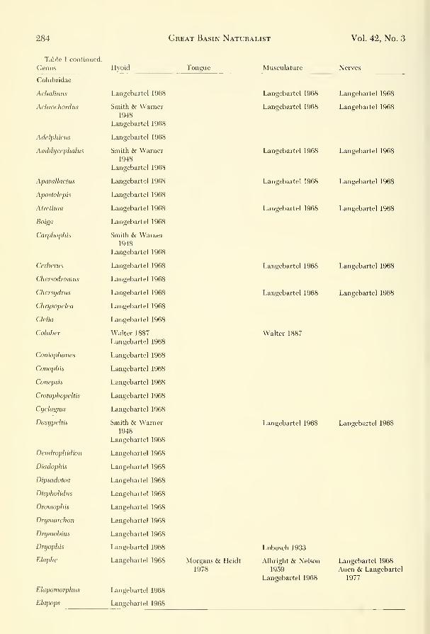
284 Great Basin Naturalist Vol. 42, No. 3
Table I continued.
Genus Hyoid Tongue Musculature Nerves
Colubridae
Achalinus
Achrochordiis
Adelphicus
Amblycephahis
Aparallactus
Apostolepis
Atrethim
Boiga
Carphophis
Cerberus
Chersodroinus
Chersydrus
Chri/sopelea
Clelia
Coluber
Coniophanes
Conophis
Conepsis
Crotaphopehis
Cyclagras
Dasypeltis
Dendrophidion
Diadophis
Dipsadotoa
Dispholidus
Droniophis
Drymarchon
Drymobitis
Dryophis
Elaphe
Elapomorphus
Ehpops
Langebartel 1968
Smith & Warner1948
Langebartel 1968
Langebartel 1968
Smith & Warner1948
Langebartel 1968
Langebartel 1968
Langebartel 1968
Langebartel 1968
Langebartel 1968
Smith & Warner1948
Langebartel 1968
Langebartel 1968
Langebartel 1968
Langebartel 1968
Langebartel 1968
Langebartel 1968
Walter 1887
Langebartel 1968
Langebartel 1968
Langebartel 1968
Langebartel 1968
Langebartel 1968
Langebartel 1968
Smith & Warner1948
Langebartel 1968
Langebartel 1968
Langebartel 1968
Langebartel 1968
Langebartel 1968
Langebartel 1968
Langebartel 1968
Langebartel 1968
Langebartel 1968
Langebartel 1968
Langebartel 1968
Langebartel 1968
Morgans & Heidt
1978
Langebartel 1968
Langebartel 1968
Langebartel 1968
Langebartel 1968
Langebartel 1968
Langebartel 1968
Langebartel 1968
Walter 1887
Langebartel 1968
Lubosch 1933
Albright & Nelson
1959
Langebartel 1968
Langebartel 1968
Langebartel 1968
Langebartel 1968
Langebartel 1968
Langebartel 1968
Langebartel 1968
Langebartel 1968
Langebartel 1968
Langebartel 1968
Auen & Langebartel
1977

September 1982 Tanner, Avery: Buccal Floor of Reptiles 285
Table 1 continued.
Genus

286 Great Basin Naturalist Vol. 42, No. 3
Table 1 continued.
Genus Hyoid Tongue Musculature Nerves
Thamnophis
Toluca
Trimorphodon
Tropidonotus
Xenodermus
Xenodon
Elapidae
Acanthophis
Aspidelaps
Bungarus
Calliophis
Demansia
Dendrospis
Denisonia
Doliophis
Elaps
Elapsoidea
Furina
Hemachatus
Hemtbungarus
Leptomicnirus
Maticora
Micruroides
Micrurus
Naja
Notechis
Ogmodon
Pseudechis
Pseudelaps
Ultocalamus
Bullock & Tanner
1966
Langebartel 1968
Oldham, Smith
& Miller 1970
Langebartel 1968
Langebartel 1968
Langebartel 1968
Langebartel 1968
Weaver 1965
Langebartel 1968
Langebartel 1968
Langebartel 1968
Langebartel 1968
Langebartel 1968
Langebartel 1968
Langebartel 1968
Langebartel 1968
Langebartel 1968
Langebartel 1968
Langebartel 1968
Langebartel 1968
Langebartel 1968
Langebartel 1968
Langebartel 1968
Langebartel 1968
Smith & Warner1948
Langebartel 1968
Langebartel 1968
Kanial, Hamouda& Mokhtar 1970
Langebartel 1968
Langebartel 1968
Langebartel 1968
Langebartel 1968
Sewertzoff 1929
Langebartel 1968
Oldham, Smith
& Miller 1970
Lubosch 1933
Langebartel 1968
Anthony & Serra
1949
Langebartel 1968
Langebartel 1968
Langebartel 1968
Lubosch 1933
Langebartel 1968
Langebartel 1968
Kesteven 1944
Langebartel 1968
Aven & Langebartel
1977
Lubosch 1933
Langebartel 1968
Langebartel 1968
Langebartel 1968
Langebartel 1968
Langebartel 1968
Langebartel 1968
Kesteven 1944

September 1982 Tanner, Avery: Buccal Floor of Reptiles 287
Table 1 continued.

288

September 1982 Tanner, Avery: Buccal Floor of Reptiles 289
three arches are present, but lack their distal
connections in some cases.
Rhynchocephalia
The hyoid of Sphenodon has been discussed
by Osawa (1898, Howes and Swinnerton
1901, Furbringer 1922, Edgeworth 1931,
1935, and Rieppel 1978.
The hyoid apparatus of Sphenodon (Fig. 1)
is simple with all elements present. The basi-
hyoid is broad with a short lingual process ex-
tending anteriorly. Laterally the basihyoid
extends as projections corresponding to the
hyoid cornua but not distinctly separate from
the basihyoid. At their distal ends, the cornua
articulate with epihyals that extend straight
posteriorly. The basihyoid also has a pair of
posterior projections, the second ceratobran-
chials, that are widely separated and curve
laterally at their distal ends. The first cerato-
branchials articulate with the basihyoid later-
al to the point of origin of the second cerato-
branchials. They curve and closely approach
the distal ends of the epihyals. Rieppel (1978)
illustrated the hyoid apparatus and its associ-
ated muscles. A taxonomic survey provides a
general overview of this order:
Chelonia
The hyoid apparatus of turtles has been de-
scribed by the following:
Chelidae
Chelodina (Furbringer 1922)
DermatemydidaeDermatemys (Furbringer (1922)
Chelydridae
Cheydra (Furbringer 1922, Edgeworth1935, Schumacher 1973), Kinosternon(Furbringer 1922, Schumacher 1973), Ster-
notherus (Furbringer 1922, Schumacher
J973), Chrysemys (Furbringer 1922, Ash-
ley 1955, Schumacher 1973), Cuora (Fur-
bringer 1922), Clemmys (Siebenrock 1898,
Furbringer 1922, Schumacher 1973), Emys(Walter 1887, Furbringer 1922, Schuma-cher 1973), Geochelone (Bojanus 1819,
Furbringer 1922, Edgeworth 1935,
Schumacher 1973), Terrapene (Furbringer
1922).
Trionychidae
Lissemys (Furbringer 1922, Sondhi 1958,
Schumacher 1973), Trionyx (Siebenrock
1898, Sondhi 1958, Furbringer 1922,
Schumacher 1973).
Cheloniidae
Caretta (Furbringer 1922, Schumacher1973).
Dermochelyidae
Dermochelys (Schumacher 1973).
Schumacher (1973) has treated the hyoids
of turtles and crocodilians extensively in this
series, so our discussion will serve as a gener-
al review.
The hyoid apparatus of turtles has been de-
scribed briefly by Bojanus (1819) and figured
by Mitchell and Morehouse (1863). More
Fig. 1. Hyoid apparatus of Sphenodon punctatum(USUN 029429): BH-body of hyoid, (basihyoid) CBl-first
ceratobranchial, CBll-second ceratobranchial, EBl-first
epibranchial, EBll-second epibranchial, EH-epihyal,
HC-hyoid comu, PL-processus lingualis.

290 Great Basin Naturalist Vol. 42, No. 3
Fig. 2.—Hyoid apparatus of A, Chelydra serpentina (Southern Connecticut State College, 598), ventral view; B,
Caiynan sclerops, ventral view; C, Caiman sclerops, lateral view (SCSC 585).
complete reports include those of Siebenrock
(1898), Furbringer (1922), Versluys (1936),
Gnanamuthu (1937), and Sondhi (1958). Thehyoid is more ossified than that of most liz-
ards and snakes.
In Trionyx and Lissemys the hyoid has a
body with a lingual process equipped with a
hypoglossum (Sondhi 1958); this is a leaflike
plate of cartilage loosely attached to its ven-
tral side. The hyoid comua are greatly re-
duced and form knoblike projections from
the body. The second ceratobranchials extend
posteriorly from the body as subcylindrical
structures.
The body of the hyoid is composed of
three pairs of serially arranged cartilaginous
blocks. The most anterior part has on its lat-
eral margins very short anterior projections.
The middle pair of plates bear the articu-
lating surfaces for the hyoid comua. The pos-
terior pair of plates are completely fused to
the middle pair and have between them and
the middle plates a diamond-shaped inter-
space. Posteriorly the last pair of plates pro-
vides facets for the articulations of the second
ceratobranchials. In Chelydra the hyoid is
more solidly constructed, consisting of boneexcept for its anterior end, the ceratohyals,
and the epihyals, which are cartilage (Fig. 2
A).
The possession of a hypoglossum by turtles
appears to be unique. The structure was first
described by Stannius (1856) as an entoglos-
sum. The term hypoglossum was first used by
Furbringer (1922), who described it as the
part not entering the tongue. Nick (1913) and
Versluys (1936) observed that in turtles, with
the exception of Dermochelys, the hypoglos-
sum is platelike, unpaired, and lies ventral to
the lingual process. Nick (1913) also suggests
that the hypoglossum is a chondrification of
connective tissue of the tendinous plate. Thehypoglossum is extensive in Trionyx, in which
it may have two slender posterior strips or be
an elongate plate, rounded at each end and
extending anteriorly from the middle com-
ponents of the body of the hyoid almost to
the symphysis of the mandible. Sondhi (1958)
suggested that the hypoglossum functions to
raise or lower the buccal floor by means of
two muscles (Mm. entoglosso-hypoglossalis
and hypoglosso-lateralis) attached to its dor-
sal surface and extending to the processus en-
toglossus and the buccal floor. In other gen-
era, Chelydra, Chrysemys, Pseudemys, and
Sternotherus, it is proportionally smaller and
varies in shape (Fig. 3). Hacker and Schu-
macher (1955) figure it for Testudo and de-
scribe the M. entoglosso-glossus that serves as
an attachment between the hypoglossum and
the processus lingualis. In Gopherus agassizi,
the hypoglossum is elongate and slender with
a median ridge ventrally and a convexity dor-
sally. It is closely associated with the process-
us lingualis. A paired muscle (M. entoglosso-
glossus) is attached to its dorsal surface on
September 1982 Tanner, Avery: Buccal Floor of Reptiles 291
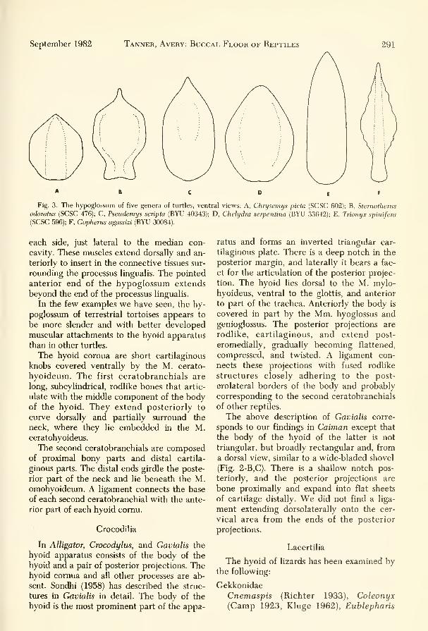
Fig. 3. The hypoglossum of five genera of turtles, ventral views: A, Chrysemys picta (SCSC 602); B, Sternotherus
odoratus (SCSC 476); C, Pseudemys scripta (BYU 40343); D, Chelydra serpentina (BYU 33642); E, Trionyx spinifera
(SCSC 596); F, Gopherus agassizi (BYU 30084).
each side, just lateral to the median con-
cavity. These muscles extend dorsally and an-
teriorly to insert in the connective tissues sur-
rounding the processus lingualis. The pointed
anterior end of the hypoglossum extends
beyond the end of the processus lingualis.
In the few examples we have seen, the hy-
poglossum of terrestrial tortoises appears to
be more slender and with better developed
muscular attachments to the hyoid apparatus
than in other turtles.
The hyoid comua are short cartilaginous
knobs covered ventrally by the M. cerato-
hyoideum. The first ceratobranchials are
long, subcylindrical, rodlike bones that artic-
ulate with the middle component of the bodyof the hyoid. They extend posteriorly to
curve dorsally and partially surround the
neck, where they lie embedded in the M.ceratohyoideus.
The second ceratobranchials are composedof proximal bony parts and distal cartila-
ginous parts. The distal ends girdle the poste-
rior part of the neck and lie beneath the M.omohyoideum. A ligament connects the base
of each second ceratobranchial with the ante-
rior part of each hyoid comu.
Crocodilia
In Alligator, Crocodylus, and Gavialis the
hyoid apparatus consists of the body of the
hyoid and a pair of posterior projections. Thehyoid comua and all other processes are ab-
sent. Sondhi (1958) has described the struc-
tures in Gavialis in detail. The body of the
hyoid is the most prominent part of the appa-
ratus and forms an inverted triangular car-
tilaginous plate. There is a deep notch in the
posterior margin, and laterally it bears a fac-
et for the articulation of the posterior projec-
tion. The hyoid lies dorsal to the M. mylo-
hyoideus, ventral to the glottis, and anterior
to part of the trachea. Anteriorly the body is
covered in part by the Mm. hyoglossus andgenioglossus. The posterior projections are
rodlike, cartilaginous, and extend post-
eromedially, gradually becoming flattened,
compressed, and twisted. A ligament con-
nects these projections with fused rodlike
structures closely adhering to the post-
erolateral borders of the body and probably
corresponding to the second ceratobranchials
of other reptiles.
The above description of Gavialis corre-
sponds to our findings in Caiman except that
the body of the hyoid of the latter is not
triangular, but broadly rectangular and, from
a dorsal view, similar to a wide-bladed shovel
(Fig. 2-B,C). There is a shallow notch pos-
teriorly, and the posterior projections are
bone proximally and expand into flat sheets
of cartilage distally. We did not find a liga-
ment extending dorsolaterally onto the cer-
vical area from the ends of the posterior
projections.
Lacertilia
The hyoid of lizards has been examined bythe following:
Gekkonidae
Cnemaspis (Richter 1933), Coleonyx(Camp 1923, Kluge 1962), Eublepharis

292 Great Basin Naturalist Vol. 42, No. 3
Table 2. Publications, not previously cited, dealing
with topics peripheral to the buccal floor.
A. Osteology
1. Chelonia
Ashley 1955, Chelydra, Chrysemys
Goppert 1903, Testudo
2. Rhynchocephalia
Goppert 1900, Sphenodon
Lakjer 1927, Sphenodon
Rieppel 1979, 1981, Sphenodon
3. Lacertila
Barrows and Smith 1947, Xenosaunis
Beddard 1905a, Ihomastix
Bellairs 1950, Anniella
Criley 1968, Barisia, Elgaria, Gerrhonotus
Duda 1965, AgamaEl Toubi 1938, Scincus
El Toubi 1947a, AgamaEl Toubi 1947b, UromasHx
El Toubi and Kamal 1959a, Chalcides
El Toubi and Kamal 1959b, Chalcides
Elyal-Giladi 1964, Agama, Chalcides
George 1954, UromasHx
Goppert 1903, Amphisbaena, Calotes, Cnemido-
phorus, Lacerta, Mabuya, Platydactylus
Iyer 1942, Calotes
Iyer 1943, Calotes
Kingman 1932, Eumeces
Lakjer 1927, Ameiva, Anguis, Amphisbaena, Ca-
lotes, Chalcides, Chamaelo, Cordylus, Eumeces,
Gekko, Hyperodapedon, Heloderma, Iguana,
Lialis, Lygosoma, Phrynosoma, Pygopus, Lacer-
ta, Tiligua, Trogonophis, Uromastix, Varanus
Mahendra 1949, Hemidactylus
Malam 1941, Gerrhosaurus
Norris and Lowe 1951, Phrynosoma
Parker 1880, Lacerta, AgamaRathor 1969, Ophiomorus
Rice 1920, EumecesSiebenroek 1892a, Uroplatus
Siebenrock 1892b, Scincus
Siebenroek 1893, Brooksesia
Siebenrock 1894, Lacerta
Siebenrock 1895, AgamaSinitsin 1928, Alopogloscus, Ameiva, Anadia, Bach-
ia, Callopistes, Cercosaura, Centropyx, Cnemido-
phorus, Dracaena, Dicrodon, Echinosaura, Ec-
leopus, Euspondylus, Gymnophthalmus, Iphisa,
Leposoma, Neusticurus, Ophiognomon, Pan-
todactylus, Prionodactylus, Pholidobolus, Pructo-
porus, Scolecosaurus, Teius, Tretioscincus,
Tupinambis
Tilak 1964a, Uromastix
Toerien 1950, Anniella
Webb 1951, Oedura, Palmatogecko
Weiner and Smith 1965, Crotaphytus
Young 1942, Xantusia
Zangerl 1944, Amphisbaena, Bipes, Geocalamus,
Leptosternon, Monopelitis, Rhineura,
Trogonophis
Table 2 continued.
4. Ophidia
Herman 1961, Echis, Vipera
Herman 1965, Calamaria
Boltt and Ewer 1954, Bitis
Dullemeijer 1956, Vipera
Dullemeijer 1959, Bitis, Crotalus, Trimeresurus,
Vipera
Kardong 1974, 1977, Agkistrodon
Liem, Mark and Rabb 1971, Azemiops
Goppert 1903, Python, Tropidonotus
McKay 1889, Acanthrophis
Rosenberg 1968, Bungarus
Varkey 1979, Nerodia
5. Crocodilia
Chiasson 1962, Alligator
Goppert 1903, Crocodylus
B. Myology
1. Chelonia
Adams 1919, Chelydra
Ashley 1955, Chelydra, Chrysemys
Schumacher 1956, Amyda, Chelodina, Chelonia,
Caretta, Clemmys, Dogania, Emydura, Emys,
Eretmochelys, Graptemys, Hardella, Macro-
chelys, Hydromedusa, Pelomedusa, Pelusios, Pla-
tysternon, Podocnemis, Testudo, Trionyx
Shah 1963, Chelodina, Deirochelys
2. Rhynchocephalia
Adams 1919, Sphenodon
Rieppel 1978, Sphenodon
3. Lacertila
Adams 1919, Iguana, Varanus
Bradley 1903, Agama, Gekko, Lacerta, Pseudopus,
Varanus
Brock 1938, Gymnodactylus
Davis 1934, Crotaphytus
George 1948, Uromastix
lordansky 1970, Agama, Cordylus, Eumeces, Gek-
ko, Lacerta, Ophiosaurus, Teratoscincus,
Varanus
Norris and Lowe 1951, Phrynosoma
Rathor 1969, Ophiomorus
Tornier 1904, Chamaeleo
4. Ophidia
Adams 1925, Natrix
Bergman 1961, Echis, Vipera
Bergman 1965, Calamaria
Boltt and Ewer 1954, Bitis
Cowan and Hick 1951, Thamnophis
Dullemeijer 1956, Vipera
Dullemeijer 1959, Bitis, Crotalus, Trimeresurus,
Vipera
Haas 1930, Amblycephalus, Calharia, Calamaria,
Cylindrophis, Eryx, Ilysia, Oxybelis, Silybura,
Xenopeltis
Haas 1931a, Acrochordus, Amblycephalus, Atrac-
taspis, Atractus, Bungarus, Calabaria, Cala-

September 1982 Tanner, Avery: Buccal Floor of Reptiles 293
Table 2 continued.
maria, Causus, Cerberus, Chersydrus, Cylin-
drophis, Dasypeltis, Dispsadomonphus, Elaps,
Eryx, Glauconia, Ilysia, Lachesis, Leptognathus,
Naja, Oxybelis, Pelamis, Python, Poly-
odontophis, Silyura, Typhlops, Xenodon,Xenopeltis
Haas 1931b, Acrodordiis, Atractaspis, Causus, Cer-
berus, Chersydrus, Cylindrophis, Dasypeltis,
Dispholidus, Leptognathus, Petalognathus, Poly-
odontophis, Scaphiophis, XenodonHaas 1952, Causus
Heymans 1970, Matrix
Heymans 1975, Aparallactus, Atractaspis,
Chilorhinophis
Kochva 1958a, Vipera
Kochva 1958b, Agkistrodon, Aspis, Atheris, Atrac-
taspis, Bitis, Bothrops, Causus, Crotalus, Echis,
Natrix, Naja, Ophiophagus, Pseudocerastes, Vi-
pera, Walterinnesia
Kardong 1974, Agkistrodon
Liem, Mark, and Rabb 1971, Azeniiops
McKey 1889, Acanthrophis
Rosenberg 1968, Bungarus
Rosenberg and Cans 1976, Elachistodon
Crocodilia
Adams 1919, Alligator
Chiasson 1962, Alligator
C. Miscellaneous
1. Chelonia
Johnson 1922, Branchial pouch derivatives, Che-
lydra, Chrysemys
Goppert 1900, Larynx, Chelonia, Dermochelys,
Emtjs, Testudo
Siebenrock 1900, Larynx, Testudo
2. Lacertila
Goppert 1900, Larynx, Amphishaena, Platydac-
tylus, Tiliqiia
Perrier 1902, Thymus and thyroid glands, Lacerta
Saint-Remy and Prenant 1904, Thymus and thy-
roid glands, Anguli, Lacerta
Sidkey 1967, Carotid Sinus, Chalcides, Scincus
3. Ophidia
Goppert 1900, Larynx, Coronella, Python,
Tropidonotus
Kroll 1973, Taste buds, Leptotyphlops
Saint-Remy and Prenant 1904, Thymus and thy-
roid glands. Coluber, Tropidonotus
Van Bourgondien and Bother 1969, Cephalic arte-
rial patterns, Agkistrodon, Crotalus. Lachesis,
Slstrurus
4. Crocodilia
Goppert 1900, Larynx, Crocodylus
Siebenrock 1899, Larynx, Crocodylus
(Cope 1892, Camp 1923), Gekko (Camp1923, Richter 1933), Gehydra (Richter
1933), Gymnodactylus (Richter 1933),
Hemidactylus (Zavattari 1908, Richter
1933, Edgeworth 1935), Phyllodactylus
(Cope 1892), Ptychozoon (Richter 1933),
Tarentola (Richter 1933), Uroplatus (Ver-
sluys 1898, 1904, Camp 1923, Edgeworth
1935).
Dibamidae
Dibamus (Rieppel 1981).
Iguanidae
Amblyrhynchus (Avery & Tanner 1971),
Anolis (Cope 1892), Basiliscus (Zavattari
1908), Brachylophus (Camp 1923, Avery
& Tanner 1971), Callisaurus (Cox & Tan-
ner 1977), Chalarodon (Avery & Tanner
1971), Chamaeleolis (Beddard 1907), Con-
olophus (Avery & Tanner 1971), Copho-
saurus (Cox & Tanner 1977), Crotaphytus
(Cope 1892, Robison & Tanner 1962),
Ctenosaura (Oelrich 1956, Avery & Tan-
ner 1971), Cyclura (Avery & Tanner
1971), Dipsosaurus (Cope 1892, Avery &Tanner 1971), Enyaliosaurus (Avery &Tanner 1971), Holbrookia (Cox & Tanner
1977), Iguana (Edgeworth 1935, Avery &Tanner 1971, Oldham & Smith 1945),
Ophirus (Avery & Tanner 1971), Phryno-
soma (Cope 1892, Camp 1923, Richter
1933, Jenkins & Tanner 1968), Polychrus
(Richter 1933), Sauromalus (Avery & Tan-
ner 1964, 1971), Sceloporus (Cope 1892),
Tropidurus (Zavattari 1908, Edgeworth
1935), Uma (Cox & Tanner 1977), Uro-
saurtis (Fanghella, Avery & Tanner 1975),
Uta (Fanghella, Avery & Tanner 1975).
AgamidaeAgama (Edgeworth 1935, El Toubi 1947,
Harris 1963, Eyal-Giladi 1964), Amphibo-luriis (Richter 1933), Calotes (Zavattari
1908, Camp 1923, Richter 1933, Edge-
worth 1935, Iyer 1943), Ceratophura
(Richter 1933), Chlamydosaurus (Beddard
1905), Cophotis (Richter 1933), Draco
(Richter 1933), Hydrosaurus (Richter
1933), Leiolepis (Richter 1933), Lyr-
iocephaltis (Richter 1933), Otocryptis
(Richter 1933), Phrynocephahis (Richter
1933, Kesteven 1944), Physignathus (Kes-

294 Great Basin Naturalist Vol. 42, No. 3
Fig. 4. Hyoid apparatus of Tarentola annularis (BYU 18123): A, ventral view; B, lateral view.
teven 1944), Uromastix (Islam 1955, Tilak
1964a,b).
Chamaeleonidae
Chamaeleo (Zavattari 1908, Edgeworth
1935, Gnanamuthu 1937, Jollie 1960).
Scincidae
Acontias (Rieppel 1981), Acontophiops(Rieppel 1981), Chalcides (Richter 1933,
El Toubi 1938, El Toubi & Kamal1959a,b), Eumeces (Cope 1892, Zavattari
1908, Richter 1933, Nash & Tanner 1970),
Lygosoma (Richter 1933), Mahuya (Rich-
ter 1933, Gnanamuthu 1937, Rao & Ra-
maswami 1952), Nessia (Richter 1933),
Riopa (Richter 1933), Tiliqua (Beddard
1907), Scincus (Richter 1933), Trachy-
saurus (Beddard 1907), Typhlosaurus(Rieppel 1981).
Cordylidae
Cordylus (Beddard 1907, Camp 1923,
Richter 1933, Edgeworth 1935), Gerrho-
saurus (Camp 1923), Zonurus (Camp1923).
Lacertidae
Acanthodactylus (Richter 1933), Lacerta
(Walter 1887, Zavattari 1908, Richter
1933, Edgeworth 1935), Ophisops (Richter
1933).
Teiidae
Ameiva (Richter 1933, Fisher & Tanner
1970), Cnemidophorus (Cope 1892, Fisher
& Tanner 1970), Neusticurus (Richter
1933), Tupinambis (Zavattari 1908, Reese
1932, Edgeworth 1935, Jollie 1960).
Anguinidae
Anguis (Richter 1933), Gerrhonotus (Wal-
ter 1887, Cope 1892), Ophiosaurus (Wal-
ter 1887).
Xenosauridae
Shinosaurus (McDowell & Bogert 1954),
Xenosaurus (McDowell & Bogert 1954,
McDowell 1972).

September 1982 Tanner, Avery: Buccal Floor of Reptiles 295
Fig. 5. Hyoid apparatus of Coleonyx variegatus (BYU 18796): A, ventral view; B, lateral view.
Helodermatidae
Heloderma (Cope 1892, McDowell & Bo-
gert 1954).
Varanidae
Varanus (Richter 1933, McDowell & Bo-
gert 1954, Sondhi 1958).
Lanthanotidae
Lanthanotus (McDowell & Bogert 1954,
Rieppel 1981).
Anniellidae
Anniella (Cope 1892, Rieppel 1981).
Anphisbaenidae
Amphisbaena (Camp 1923, Richter 1933,
Jollie 1960), Monopeltis (Richter 1933),
Rhineura (Cope 1892).
Xantusidae
Xantusia (Cope 1892, Savage 1963).
Most lizards have a hyoid consisting of a
basihyal (corpus hyoideum) with a pair, each,
of anterior and posterior comua as described
by Cope (1892), Zavattari (1908), Furbringer
(1922), Camp (1923), Versluys (1936), DeBeer
(1937), Gnanamuthu (1937), Mahendra(1947), Rao and Ramaswami (1952),
McDowell and Bogert (1954), Oelrich (1956),
Romer (1956), Sondhi (1958), Jollie (I960),
Robison and Tanner (1962), Avery and Tan-
ner (1964), Jenkins and Tanner (1968), Fisher
and Tanner (1970), Nash and Tanner (1970),
Avery and Tanner (1971), Rieppel (1981),
and others. For the remainder of this dis-
cussion we will use the hyoid nomenclature
followed by Romer (1956) as described ear-
lier. The hyoids of the geckos Coleonyx, Gek-
ko, Aristelliger, Hemidactylus, Phyllodactylus,
Thecadactylus, and Eublepharis have been
described, and we figure Tarentola (Fig. 4)
and Coleonyx (Fig. 5). In most, the body of
the hyoid is small and slender, with a long
rodlike lingual process extending anteriorly.
A pair of hyoid comua extend laterally; in

296 Great Basin Naturalist Vol. 42, No. 3
Fig. 6. Hyoid apparatus, ventral views: A, Brachylophus brevicephahts (BYU 32663); B, Sattromalus obesus (BYU
21728).
some species these form sigmoid curves, and
in others they are straight rods. Articulating
with the distal extremes of the hyoid comuaare the epihyals. Extending posteriorly from
the body of the hyoid as a pair of short or
long rods are the second ceratobranchials. Athird set of arches, the first ceratobranchials,
articulate at the point of attachment betweenthe hyoid comua and the body. The basic
pattern is retained throughout the Gekkota,
with some variation in the shape of the hyoid
comua; also, the first ceratobranchials, epi-
hyals, or both may be lost in some genera.
In the Dibamidae, Rieppel (1981) has de-
scribed the hyoid of Dibamus as having a
posteriorly bifurcated basihyal with an elon-
gated entoglossal process. The bony first ce-
ratobranchials that articulate with the post-
erolateral limbs of the basihyal are shorter in
Dibamus as compared to Anniella. He in-
dicates a major specialization exists in that
there are a pair of cartilaginous rods that
support the aditus laryngis and approach but
do not fuse to the posterolateral limbs of the
basihyal. These he considers to be hypohyals
(hyoid comua of Romer).
The hyoids of the iguanine lizards Ambly-
rhynchus, Brachylophus, Conolophus, Cteno-
saura, Cyclura, Dipsosaurus, Iguana, and
Sauromalus and Malagashe iguanids Chalaro-
don and Oplurus have been investigated by
Avery and Tanner (1971). Because these liz-
ards possess all three arches of the hyoid ap-
paratus, they are considered primitive (Fig.
6-A). The body of the hyoid (basihyal) is
triangular in all the above genera except
Oplurus and Sauromalus, in which it forms a
broad flattened sheet of cartilage. In all the
genera the hyoid comu (hypohyal) is short
and stout; it extends out from the body of the
hyoid at right angles or projects slightly ante-
rior to the body. Posterior to the body, the
second ceratobranchials extend along the
trachea and, in all genera except Oplurus and
Sauromalus, lie close together. In the latter
two genera the second ceratobranchials are
widely separated by the bulk of the trachea
(Fig. 6-B). In none of the genera are the sec-

September 1982 Tanner, Avery: Buccal Floor of Reptiles 297
Fig. 7. Hyoid apparatus, ventral views: A, Sceloportis magister (BYU 30310); B, Holbrookia maculata (BYU 15752);
C, Phrynosoina platyrbinos (BYU 22830).
end ceratobranchials attached distally to the
other arches. In some genera, particularly
Iguana, the distal extremes of these processes
attach to the skin and provide support for
movement of the dewlap.
The first ceratobranchials articulate prox-
imally w^ith the body of the hyoid between
the origins of the second ceratobranchials
and the hyoid comua. They are elongated,
thin rods that taper to points distally and
curve dorsolaterally to the sides of the neck,
where they articulate with the epihyals (cer-
tohyals). The epihyals articulate between the
hyoid comua and the first ceratobranchials
and form the most lateral extensions of the
hyoid apparatus. At their proximal ends the
epihyals are expanded into bladelike process-
es that extend medially toward the hyoid
body. These processes are not developed to
any degree in Chalarodon and Opiums.Among the other iguanids studied and de-
scribed by one of us are the hyoids of Crota-
phytus, Holbrookia, Phnjnosonia, and Uta.
We figure Sceloporus magister and Hol-
brookia maculata (Figs. 7-A & B) as represen-
tatives of the sceloporine genera. The basic
pattern described in the iguanines is main-
tained with the following exceptions. In
Phrynosoma the second ceratobranchials are
greatly reduced, and the first ceratobran-
chials and epihyals are noticeably thickened
(Fig. 7-C); the basihyoid is a laterally extend-
ed plate. Anolis has an exceptionally elon-
gated hyoid apparatus, with the second ce-
ratobranchials extending posteriorly along
the midline forming approximately two-
thirds the length of the entire hyoid appa-
ratus. This anatomical development is associ-
ated with the functional dewlap (Fig. 8).
In the agamids, the following were exam-
ined: Agama (Duda 1965, Hass 1973), and
Figure 9; Calotes, Draco, and Sitana (Gnana-

298 Great Basin Naturalist Vol. 42, No. 3
CBl
CBll
muthu 1937), Chlamydosaurus (Beddard
1905, DeVis 1883), Phrynocephalus (Haas
1973), Physignathus (Kesteven 1944), andUromastyx (Poglayen-Neuwall 1954, Versluys
1898, El Toubi 1947b, Tilak 1964b). In gen-
eral, the agamid hyoids resemble closely
those of the iguanids. In Uromastyx the basi-
hyoid is slender and laterally extended; the
hyoid comua are directed anterolaterally (Ti-
lak 1964b). The short and widely separated
second ceratobranchials extend posteriorly
from the basihyoid. The first ceratobranchials
extend posteriorly from the basihyoid. Thefirst ceratobranchials articulate at the union
of the hyoid comua and the basihyoid. Theycomprise the longest elements of the hyoid.
The epihyals attach to the distal ends of the
hyoid comua and have, at their distal ends,
epibranchials that may attach to the distal
end of the first ceratobranchials. In Agama(Fig. 9) the hyoid is similar except that the
basihyoid is more massive and the second ce-
ratobranchials are aligned more closely to-
gether. In Calotes and Draco the hyoids are
elongated and narrow. The second cerato-
branchials are exceptionally long and slender,
lying close together at the midline, whereas
CBll
Fig. 8. Hyoid apparatus, ventral view: Anolis caroli-
nensis (BYU 13768).
Fig. 9. Hyoid apparatus of Agama agama (BYU18147), ventral view.

September 1982 Tanner, Avery: Buccal Floor of Reptiles 299
Fig. 10. Hyoid apparatus: A, Chamaeleon namagyensis (USNM 161275); B, Chamaeleon brevicornis (BYU 12422),
ventral views; C, same as B, lateral view.
the epihyals are very short and not connected
by epibranchials. In Chlamydosaurus the
basihyoid is massive and bears two homUkeprojections; these extend laterally to articu-
late with the hyoid comua, which form short
tapering tips on these projections. The sec-
ond ceratobranchials appear to have been
lost unless they are represented by two very
small knobs on the posteromedial border of
the basihyoid. The first ceratobranchials are
extremely elongated, extending post-
erolaterally and composed of two pieces. The
very long proximal piece articulates distally
with the second piece, which is about one-
fifth the length of the proximal. The epihyals
are short or slender, and articulate at the
point where the hyoid comua and the lateral
projections of the basihyoid attach. In Phys-
ignathus the hyoid exhibits a normal struc-
ture except that the first ceratobranchials are
much longer than the second cerato-
branchials.
In Chamaeleo the hyoid is distinctly differ-
ent, with the basihyoid being little more than
the basal part of the lingual process. The
hyoid comua extend anterolaterally about a
third the length of the lingual process. The
first ceratobranchials extend laterally and are
short. The epihyals are small and attach to
the hyoid cornua about half the distance
from their distal ends. The second cerato-
branchials are lost (Fig. 10-A, B, and C).
Gnanamuthu (1937) described the hyoid ap-
paratus for Chamaeleo carcaratus and re-
viewed previous studies of its function.
In the Scincidae the hyoids of Scincus (El
Toubi 1938), Eumeces (Nash and Tanner
1970), Fig. 11, Mabuya (Richter 1933), and

300 Great Basin Naturalist Vol. 42, No. 3
EBI
Fig. 11. Hyoid apparatus of Eumeces gilberti (BYU 31956), dorsal view. (After Nash and Tanner 1970)
Chalcides (Furbringer 1922, Richter 1933)have been described. All three arches are
present and assume an unspecialized pattern.
In all the basihyoid is broad rather than nar-
row, and the second ceratobranchials are
very short and widely separated. The first ce-
ratobranchials are elongate and slim. Thehyoid comua are short and slim, and articu-
late distally with the epihyals, which vary in
form. They are simple rods in Eumeces andhave enlarged proximal ends in the remain-ing genera. In Scincus the enlarged ends are
simple and spoonshaped, but in Chalcidesand Mahuya the shape is complex. In bothgenera the enlarged end has a short flange ex-
tending posterolaterally from the middle of
the epihyal where the enlarged end termi-
nates. These genera have a large hooklikesecond epibranchial associated with the distal
end of the epihyal. It is attached in Chalcidesand Scincus but separate in Eumeces andMahuya. In all genera there is a short first
epibranchial attached to the terminal end ofthe first ceratobranchial (Fig. 11).
Rieppel (1981) has examined the limbless
scincoid genera Acontias, Typhlosaurus, andAcontaphiops. Acontias is described as beinglike Anniella, with the basihyal having a slen-
der entoglossal process and being bifurcated
posteriorly with its distinct posterolateral
limbs articulating with first ceratobranchials.
Hypohyal processes (hyoid comua) are pres-
ent in all species where they are T-shaped at
their distal ends. In Typhlosaurus the hyoid is
similar to Acontias, but the posterior first ce-
ratobranchials are longer and hypohyals are
absent. Rieppel calls attention to the fact
that the hyoid of Typhlosaurus is identical to
that of some Typhlopidae as described byList (1966) and Langebartel (1968). Thehyoid of Acontophiops is similar to that of
Typhlosaurus.
In the teiid Tupinambis, the lingual pro-
cess is detached from the basihyoid and em-bedded in the tongue. The second cerato-
branchials are lost, and the epihyals and first
ceratobranchials are connected byepibranchials.

September 1982 Tanner, Avery: Buccal Floor of Reptiles 301
Fig. 12. Hyoid apparatus of Cnemidophorus tigris (BYU 31925). Dorsolateral view showing the detached lingual
process (LP) and the extension of the body of the hyoid anteriorly as a spine.
The lingual process is also detached in
Cnemidophoms (Fig. 12). The hyoid extends
anteriorly as a short spine similar to that of
igiianids except for its smaller size. It is em-
bedded in connective tissue ventral to the
lingual process and the tongue. The hyoid
comua extend anterolaterally from the basi-
hyoid and articulate with the epihyals. The
latter extend anteriorly, forming bladelike
cartilages that serve as lateral supports for
the posterior half of the tongue and extend
laterally to lie adjacent to the mandible. The
posterior part of the epihyal extends posteri-
orly, curving laterally where it terminates as
cartilage in loose connective tissue on the
first ceratobranchial. Both ceratobranchials
are present; the first extends posteriorly to
terminate in the connective tissue with the
cartilagenous first epibranchial. The epihyals
and first ceratobranchials are not connected
distally, although the ends are close together
ip a common connective tissue.
Ameiva lacks the second ceratobranchials.
The first epibranchials are short, forming a
knob on the end of the ceratobranchials.
In both Ameiva and Cnemidophorus the
detached lingual process extends anteriorly to
approximately the forking of the tongue. Pos-
teriorly it is tightly enclosed in connective
tissue between the elongate M. hypoglossus.
It terminates posteriorly, ventral to the lar-
yngeal cartilages.
In Angtiis (Anguidae) the hyoid is greatly
reduced, with the second ceratobranchials
and epihyals absent. The hyoid comua are
enlarged and extend anteriorly to parallel the
lingual process for most of its length. In Ger-
rhonotus and Ophisaurus the second cerato-
branchials are also lost. The epihyals are
present, however, and articulate with the dis-
tal ends of the hyoid comua, which are more
laterally directed than in Anguis.
In Varanus (Varanidae), the hyoid comua
is complex and is composed of two articu-
lating cartilaginous rods, called by Sondhi
(1958:159-160) the portio proximalis (hyoid
cornu) and the portio distalis (epihyal):
Each has an anterior handlelike process and in life the
two hooked ends cross each other beneath the tongue-
sheath, with the handle of the portio proximahs lying
dorsal to that of the portio distalis.
According to Sondhi (1958:159-160),
the proximal end of the portio proximalis fits into a
roughly concave facet on the dorsolateral surface of the
basihyoid, near the facet at which the posterior comua
articulates. From this point the portio proximalis ex-
tends obliquely upward, outward, and forward and at its
termination curves inward to form the hook-shaped
handle that is dorsoventrally flattened. The portio dis-
talis is flattened at its proximal handlelike end, becomes
rodlike as it passes backward and upward, and gradually
302 Great Basin Naturalist Vol. 42, No. 3
tapers at its distal end. It is disposed obliquely across the
sides of the neck, its tapering end lying almost parallel
to the proximal piece of the posterior cornua of its side.
Sondhi also indicates that the portio prox-
imalis and portio distalis are attached to each
other by a cartilaginous piece, with this at-
taching piece being folded at its outer margin
like a cover of a folder so that one part of it
becomes dorsal and the other ventral. Thedorsal part is described as
narrower and is attached to the flattened, curved ante-
rior end of the portio proximalis like the blades of scis-
sors on its counterpart. The nature of attachment of the
two pieces of the anterior cornua renders them capable
of opening out to some extent like the covers of a folder.
The description of V. monitor (Sondhi
1958) and our dissection of V. indicus (BYU
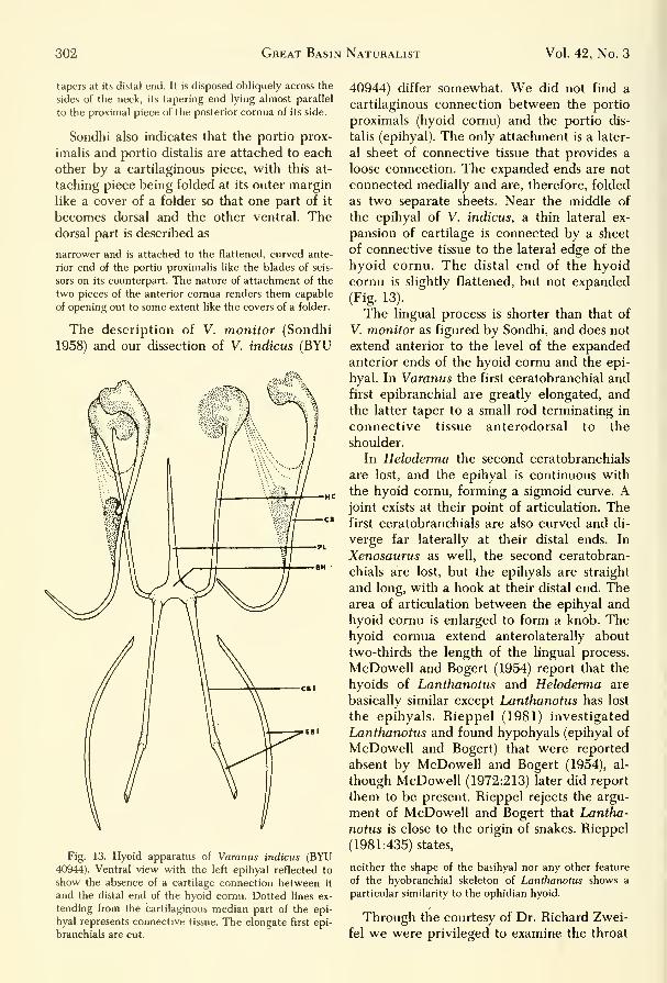
Fig. 13. Hyoid apparatus of Varanus indicus (BYU40944). Ventral view with the left epihyal reflected to
show the absence of a cartilage connection between it
and the distal end of the hyoid cornu. Dotted lines ex-
tending from the cartilaginous median part of the epi-
hyal represents connective tissue. The elongate first epi-
branch ials are cut.
40944) differ somewhat. We did not find a
cartilaginous connection between the portio
proximals (hyoid cornu) and the portio dis-
talis (epihyal). The only attachment is a later-
al sheet of connective tissue that provides a
loose connection. The expanded ends are not
connected medially and are, therefore, folded
as two separate sheets. Near the middle of
the epihyal of V. indicus, a thin lateral ex-
pansion of cartilage is connected by a sheet
of connective tissue to the lateral edge of the
hyoid cornu. The distal end of the hyoid
cornu is slightly flattened, but not expanded
(Fig. 13).
The lingual process is shorter than that of
V. monitor as figured by Sondhi, and does not
extend anterior to the level of the expandedanterior ends of the hyoid cornu and the epi-
hyal. In Varanus the first ceratobranchial and
first epibranchial are greatly elongated, andthe latter taper to a small rod terminating in
connective tissue anterodorsal to the
shoulder.
In Heloderma the second ceratobranchials
are lost, and the epihyal is continuous with
the hyoid cornu, forming a sigmoid curve. Ajoint exists at their point of articulation. Thefirst ceratobranchials are also curved and di-
verge far laterally at their distal ends. In
Xenosaurus as well, the second ceratobran-
chials are lost, but the epihyals are straight
and long, with a hook at their distal end. Thearea of articulation between the epihyal and
hyoid cornu is enlarged to form a knob. Thehyoid cornua extend anterolaterally about
two-thirds the length of the lingual process.
McDowell and Bogert (1954) report that the
hyoids of Lanthanotus and Heloderma are
basically similar except LMnthanotus has lost
the epihyals. Rieppel (1981) investigated
Lanthanotus and found hypohyals (epihyal of
McDowell and Bogert) that were reported
absent by McDowell and Bogert (1954), al-
though McDowell (1972:213) later did report
them to be present. Rieppel rejects the argu-
ment of McDowell and Bogert that Lantha-
notus is close to the origin of snakes. Rieppel
(1981:435) states,
neither the shape of the basihyal nor any other feature
of the hyobranchial skeleton of Lanthanotus shows a
particular similarity to the ophidian hyoid.
Through the courtesy of Dr. Richard Zwei-
fel we were privileged to examine the throat

September 1982 Tanner, Avery: Buccal Floor of Reptiles 303
Fig. 14. Hyoid apparatus, ventral views: A, Lanthanotus borneensis (AMNH 87375); B, Heloderma suspectum
(BYU 41436).
anatomy of iMnthanotus boreensis (AMNH87375) and found the hyoid skeleton to be
smprisingly similar to that of Heloderma (Fig.
14). Rieppel (1980, 1981) has, on the basis of
cranial anatomy, concluded that Lanthanotus
is intermediate in structure between Helo-
derma and Varanus. Branch (1982) arrived at
a similar conclusion based on hemipeneal
data. The hyoid of these genera have the
same structures; however, in Varanus there
has been considerable modification and spe-
cialization not found in the other genera.
In Gerrhosaurus (Cordylidae) the second
ceratobranchials have been lost, but the first
ceratobranchial and epihyal are retained. In
Zonurus the second ceratobranchials are
present but short. In Xantusia (Xantusidae)
the hyoid contains all the elements. Thehyoid comu extends dorsolaterally to articu-
late with the median edge of the expanded,
flattened proximal end of the epihyal. Fromthe flattened end the epihyal extends post-
erodorsally, tapering into a rod and termi-
nating as a short epibranchial immediately
posterior to the tympanum. The first cerato-
branchial extends posterodorsally and curves
to terminate in the second epibranchial and
in close association with the epibranchial of
the epihyal.
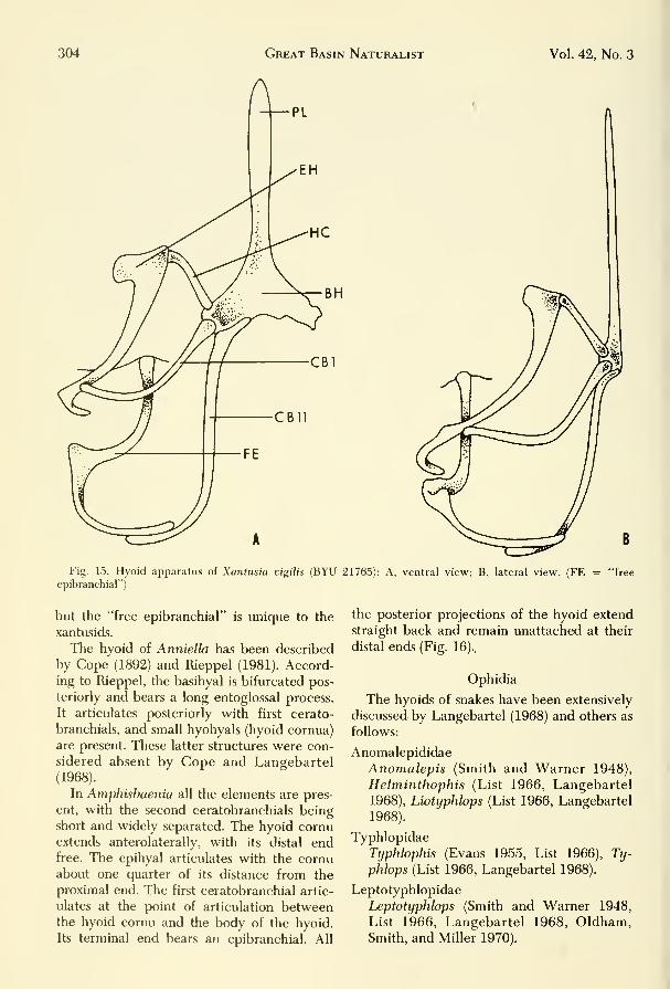
The second ceratobranchial in Xantusia
extends posterior with the distal end, curving
laterad to form an open hook. It does not ar-
ticulate with an epibranchial as in the epi-
hyal and first ceratobranchial; however, a
cartilaginous structure in close association
with the distal end of the second ceratobran-
chial extends laterally and curves anteriorly
to articulate with the basioccipital of the
skull. Cope (1900) and Savage (1963) have re-
ferred to this structure as a free epibranchial.
If this is an epibranchial, it is distinct and
differs from all others in saurians we have
seen. Its close association to the distal end of
the second ceratobranchial (Fig. 15) is not ar-
ticulated as in the other epibranchials and
leads us to believe that the entire structure
may represent fusions of other remnant gill
bars. An examination of the entire structure
(Fig. 15B) indicates to us that fusions have
occiirred. An articulation or close association
of the distal ends of the epihyal and/or the
second ceratobranchial occurs in many forms
304 Great Basin Naturalist Vol. 42, No. 3
Fig. 15. Hyoid apparatus of Xantusia vigilis (BYU 21765): A, ventral view; B, lateral view. (FE = "free
epibranchial")
but the "free epibranchial" is unique to the
xantusids.
The hyoid of Anniella has been described
by Cope (1892) and Rieppel (1981). Accord-ing to Rieppel, the basihyal is bifurcated pos-
teriorly and bears a long entoglossal process.
It articulates posteriorly with first cerato-
branchials, and small hyohyals (hyoid cornua)
are present. These latter structures were con-
sidered absent by Cope and Langebartel
(1968).
In Amphisbaenia all the elements are pres-
ent, with the second ceratobranchials being
short and widely separated. The hyoid cornuextends anterolaterally, with its distal endfree. The epihyal articulates with the cornuabout one quarter of its distance from the
proximal end. The first ceratobranchial artic-
ulates at the point of articulation betweenthe hyoid cornu and the body of the hyoid.
Its terminal end bears an epibranchial. All
the posterior projections of the hyoid extend
straight back and remain unattached at their
distal ends (Fig. 16).,
Ophidia
The hyoids of snakes have been extensively
discussed by Langebartel (1968) and others as
follows:
Anomalepididae
Anomalepis (Smith and Warner 1948),
Helminthophis (List 1966, Langebartel
1968), Liotyphlops (List 1966, Langebartel
1968).
Typhlopidae
Typhlophis (Evans 1955, List 1966), Ty-
phlops (List 1966, Langebartel 1968).
Leptotyphlopidae
Leptotyphlops (Smith and Warner 1948,
List 1966, Langebartel 1968, Oldham,Smith, and Miller 1970).

September 1982 Tanner, Avery: Buccal Floor of Reptiles 305
CBll
Fig. 16. Hyoid apparatus, ventral views: A, Amphisbaenia cornura (BYU 16127); B, Amphisbaenia kingi (BYU
16148).
Uropeltidae
Platyplactrurus (Langebartel 1968), Plec-
trurus (Rieppel 1981), Rhinophis (Smith
and Warner 1948, Langebartel 1968), Sily-
bura (Langebartel 1968).
Aniliidae
Anilius (Smith and Warner 1948, Lange-
bartel 1968, Rieppel 1981), Cylindrophis
(Smith and Warner 1948, Langebartel
1968).
Xenopeltidae
Xenopeltis (Smith and Warner 1948,
Langebartel 1968).
Boidae
Aspidites (Smith and Warner 1948, Lange-
bartel 1968), Boa (Langebartel 1968),
Calabaria (Langebartel 1968), Charina
(Langebartel 1968), Chondropython(Langebartel 1968), Constrictor (Langebar-
tel 1968), Enygrus (Langebartel 1968),
Epicrates (Langebartel 1968), Liasis
(Langebartel 1968), Lichanura (Langebar-
tel 1968), Loxocemus (Smith and Warner
1948, Langebartel 1968), Nardoana(Langebartel 1968), Python (Furbringer
1922, Langebartel 1968, Oldham, Smith,
and Miller 1970), Sanzinia (Langebartel
1968), Trachyboa (Langebartel 1968).
Colubridae
Achalinus (Langebartel 1968), Achro-
chordus (Smith and Warner 1948, Lange-
bartel 1968), Adelphicus (Langebartel
1968), Amblycephalus (Smith and Warner
1948, Langebartel 1968), Aparallactus
(Langebartel 1968), Apostolepis (Lange-
bartel 1968), Atretium (Langebartel 1968),
Boiga (Langebartel 1968), Carphophis
(Smith and Warner 1948, Langebartel
1968), Cerberus (Langebartel 1968),
Chersodromus (Langebartel 1968), Cher-
sydrus (Langebartel 1968), Chrysopelea

306 Great Basin Naturalist Vol. 42, No. 3
(Langebartel 1968), Clelia (Langebartel
1968), Coluber (Walter 1887, Langebartel
1968), Coniophanes (Langebartel 1968),
Conophis (Langebartel 1968), Conopsis
(Langebartel 1968), Crotaphopeltis (Lang-
ebartel 1968), Cyclagras (Langebartel
1968), Dasypeltis (Smith and Warner1948, Langebartel 1968), Dendrophidion
(Langebartel 1968), Diadophis (Langebar-
tel 1968), Dipsadoboa (Langebartel 1968),
Dispholidus (Langebartel 1968), Dro-
mophis (Langebartel 1968), Dry7narchon
(Langebartel 1968), Drymobius (Langebar-
tel 1968), Dryophis (Langebartel 1968),
Elaphe (Langebartel 1968), Elapomorphus
(Langebartel 1968), Elapops (Langebartel
1968), Enhydrus (Langebartel 1968),
Enulius (Langebartel 1968), Farancia
(Langebartel 1968), Ficimia (Langebartel
1968), Fimbrios (Langebartel 1968),
Geophis (Langebartel 1968), Haldea(Langebartel 1968), Haplopeltura (Lang-
ebartel 1968), Heterodon (Weaver, 1965,
Langebartel 1968), Homalopsis (Langebar-
tel 1968), Lampropeltis (Langebartel
1968), Leptodeira (Langebartel 1968), Lep-
tophis (Langebartel 1968), Manolepis(Langebartel 1968), Masticophis (Lang-
ebartel 1968), Mehelya (Langebartel
1968), Natrix (Sondhi 1958), Nerodia
(Langebartel 1968, Oldham, Smith, and
Miller 1970), Ninia (Langebartel 1968),
Nothopsis (Langebartel 1968), Opheodrys
(Langebartel 1968, Cundall 1974), Oxy-
belis (Langebartel 1968), Oxyrhabdinium
(Langebartel 1968), Fituophis (Smith and
Warner 1948, Bullock and Tanner 1966,
Langebartel 1968, Oldham, Smith, and
Miller 1970), Psamaodynastes (Langebar-
tel 1968), Rhadineae (Langebartel 1968),
Rhadinella (Langebartel 1968), Rhino-
cheilus (Langebartel 1968), Salvadora
(Langebartel 1968), Sibynomorphus(Langebartel 1968), Sibynophis (Langebar-
tel 1968), Sonora (Langebartel 1968), Tan-
tilla (Langebartel 1968), Thamnophis(Bullock and Tanner 1966, Langebartel
1968, Oldham, Smith, and Miller 1970),
Toluca (Langebartel 1968), Trimorphodon
(Langebartel 1968), Tropidonotus (Lang-
ebartel 1968), Xenodermus (Langebartel
1968), Xenodon (Weaver 1965).
Elapidae
Acanthophis (Langebartel 1968), Aspide-
laps (Langebartel 1968), Rungarus (Lange-
bartel 1968), Calliophis (Langebartel
1968), Demansia (Langebartel 1968), Den-draspis (Langebartel 1968), Denisonia(Langebartel 1968), Doliophis (Langebar-
tel 1968), Flaps (Langebartel 1968), Elap-
soidea (Langebartel 1968), Furina (Lange-
bartel 1968), Hemachatus (Langebartel
1968), Hemibungarus (Langebartel 1968),
Leptomicrurus (Langebartel 1968), Mati-
cora (Langebartel 1968), Micruroides
(Langebartel 1968), Micrurus (Smith andWarner 1968, Langebartel 1968), Naja
(Langebartel 1968, Kamal, Hamouda, andMokhtar 1970), Notechis (Langebartel
1968), Ogmodon (Langebartel 1968),
Pseiidelaps (Langebartel 1968), Ultoca-
lamus (Langebartel 1968).
Eydrophidae
Aipysurus (Langebartel 1968), Hydrophis
(Langebartel 1968), Kerilia (Langebartel
1968), Lapemis (Smith and Warner 1948,
Langebartel 1968), Laticauda (Langebar-
tel 1968), Thalasophina (Langebartel
1968).
Viperidae
Aspis (Langebartel 1968), Atheris (Lange-
bartel 1968), Atractaspis (Langebartel
1968), Ritis (Langebartel 1968), Causus(Langebartel 1968), Cerastes (Langebartel
1968), Echis (Langebartel 1968), Pseudoce-
rastes (Langebartel 1968), Vipera (Lange-
bartel 1968, Furbringer 1922).
Crotalidae
Agkistrodon (Smith and Warner 1948,
Langebartel 1968), Rothrops (Langebartel
1968), Crotalus (Langebartel 1968, Old-
ham, Smith, and Miller 1970), Lachesis
(Langebartel 1968), Sistrurus (Langebartel
1968), Trimeresurus (Langebartel 1968).
In snakes the hyoid apparatus is greatly re-
duced, with the hyoid cornua being lost and
the remainder of the processes simplified. Es-
sentially the snake hyoid consists of a body
plus a lingual process and what is thought to
be the second ceratobranchials, which are
fused to the body of the hyoid (Figs. 17A and
B, 29). The variations found in ophidian
hyoids have been discussed by Furbringer

September 1982 Tanner, Avery: Buccal Floor of Reptiles
(1922), Versluys (1936), Gnanamuthu (1937),
Smith and Warner (1948), Sondhi (1958), Al-
bright and Nelson (1959), List (1966), Under-
wood (1967), Langebartel (1968), Rieppel
(1981), and others. There are four major mor-
phological types that can be distinguished in
snakes. Tliese correspond in shape roughly to
the letters M, Y, and V, and to a parallel type
11. The most complete survey of the hyoids
of snakes is presented by Langebartel (1968),
and we have based much of our remarks on
his study.
Hyoids possessing the M shape are found
exclusively in the family Anomalepididae,
which has only four genera, Anotnalepis, Lio-
typlilops, Hehninthophis, and Tijpldopfiis. In
this group tlie hyoid has a body and the sec-
ond ceratobranchials. All other processes are
lost, including the lingual process.
A Y-shaped hyoid is foimd in the Typl-
opidae and Leptotyphiopidae. The body of
the hyoid possesses a lingual process and has
hyoid cornua (second ceratobranchials) that
project posteriorly. The possession of a ling-
ual process is variable, with it being absent
according to List (1966) in TypJiIops pusillus
and T. hanbricalis. In T. reticulatiis, T. pla-
tycephahis, and T. blandfordi lestradei the
hyoid cornua are separated from the body.
Leptoti/pJdops has a normal Y type hyoid.
Tlie V-shaped hyoid is found in the Ani-
liidae, Boidae, Uropeltidae, and Zenopel-
tidae. In this type of hyoid the lingual pro-
cess is absent and the hyoid cornua may be
attached or imattached. There is much in-
traspecific variation in the latter character.
In some specimens of Charina hottae the
cornua are attached, although they are unat-
tached in others. Langebartel (1968) consid-
ers the curving arches to be the first
ceratobranchials.
The 11 type hyoid is fomid in the colu-
brids, crotalids, elapids, hydrophids, viperids,
and some genera of the boidae {Casarea, Tra-
cJnjboa, and TropiJopJiis). The second cerato-
branchials of this type are usually long, paral-
lel rods attached to a slim hyoid body (Fig.
17). The resulting structure resembles a tim-
ing fork in appearance. A few snakes have a
hyoid body, triradiate in appearance and
with a short lingual process. Such a structure
is figured by Sondhi (1958) for Natrix (Xe-
nochrophis), in which:
307
Bh
CBI
4X
1^
B 4X
Fig. 17. Hyoid apparatus, ventral views: A, Pituophis
m. deserticola (BYU 3072); B, Crotahts viridis lutosus
(2089). Both are from adult individuals and drawn at 4X
actual size.

308 Great Basin Naturalist Vol. 42, No. 3
tilt' l)asihvoid lies ventral to the trachea and dorsal to
the posterior terminations of the oniohyoideus and ster-
nohvoidens muscles.
The processes form elongated rods that lie
ventral and extend posteriorly and parallel,
with their terminal ends enclosed in the tips
of the base of the tongue. In Pitiiophis, the
basihyoid is ventral to the tongue at about
the level of the angle of the jaws. The cerato-
branchials extend and curve posterolaterally
from the basihyoid for a short distance to a
lateral position and then extend posteriorly,
lateral to the tongue and parallel to each
other, to the posterior tip of the tongue. In
Crotalus the same obtains anteriorly with the
basihyoid and the anterior part of the cerato-
branchials; however, the posterior third of
the latter converge ventrally to becomeclosely associated along the ventromedian of
the tongue and diverge slightly near their
ends to become imbedded in muscle and con-
nective tissue (Fig. 28, Romer 1950: fig. 421-
C, Langebartel 1968:figs. 3, 4). For detailed
description of the hyoid of individual genera
of snakes, see Langebartel (1968).
III. Muscles of the Buccal Floor:
General
The buccal floor is composed of several in-
terwoven sheets of muscles. These sheets can
be separated into two major groups: the
hypobranchial musculature and the muscles
of the associated branchial arches. The hypo-
branchial muscles are derived from the myo-tomes of the occipital and cervical somites,
whereas the muscles of the branchial arches
come from tlie viceral muscle plates formedin tlie branchial region. The tongue, for the
most part, is also derived from the occipital
somites. Because of the close associations of
some of the somites with both cranial andspinal areas, some muscles are innervated byboth spinal and cranial nerves.
For the sake of convenience, we have sepa-
rated our discussion of the buccal muscula-
ture into two major divisions: (1) the muscles
associated with the hyoid apparatus and (2)
those associated with other structures. Thetongue is svifficiently important to be segre-
gated from these categories and is considered
under a separate heading.
The nomenclature of muscles of reptiles
has not been standardized; however, tables of
synonyms can be found in Edgeworth (1935),
Langebartel (1968), Haas (1973), andSchumacher (1973). Some of the more recent
short summaries of the earlier papers on the
myology of the buccal floor in reptiles can be
found in Sondhi (1958), Langebartel (1968),
Avery and Tanner (1971), Secoy (1971), andVarkey (1979). The remainder of this section
is a brief account of the musculature of the
buccal floor in selected reptiles as described
by several earlier workers such as Edgeworth
(1931), Graper (1932), Gnanamuthu (1937),
Reese (1915 and 1932), Hacker and Schumacher (1955), Oelrich (1956), Sondhi (1958),
Langebartel (1968), and others. It also should
be noted that the more advanced reptiles
have more complex muscular patterns whencompared to primitive forms. This is seem-
ingly true not only for orders, but also for
family groups. A comparison of the advanced
lizard Varanus and the primitive iguanids in
the following sections serves as an
illustration.
We refer to such forms as Gavialis, Tri-
onyx, Natrix (Xenochropliis), Varanus, and
other genera. These should be credited to
Gnanamuthu (1937) or Sondlii (1958) if not
otherwise noted.
The musculature of the following reptiles
has been studied.
Chelonia
Pelomedusidae
Pelusios (Poglayen-Neuwall 1953a).
Chelidae
Batrochemys (Poglayen-Neuwall 1953a),
Chelodina (Poglayen-Neuwall 1953a).
Chelydridae
Chelydra (Camp 1923, Graper 1932, Pog-
layen-Neuwall 1953a, Schumacher (1973),
Kinosternon (Poglayen-Neuwall 1953a,
Schumacher 1973), Sternotherus (Pog-
layen-Neuwall 1953a, Schumacher 1973).
Testudinidae
Chrysemys (Poglayen-Neuwall 1953a, Ash-
ley 1955, Schumacher 1973), Cuora (Pog-
layen-Neuwall 1953a), Clemmys (Graper
1932), Poglayen-Neuwall 1953a, Schuma-
cher 1973), Deirochelys (Shah 1963), Emys

September 1982 Tanner, Avery: Buccal Floor of Reptiles 309
(Walter 1887, Schumacher 1973), Goph-
erus (George and Shad 1954), Graptemys
(Poglayen-Neuwall 1953a), Geochelone
(Bojanus 1819, Graper 1932, Lubosch
1933, Edgeworth 1935, Poglayen-Neuwall
1953a, George and Shad 1955, Schumacher 1973), Malaclemys (Poglayen-Neu-
wall 1953a), Pseudemys (Ashley 1955,
Poglayen-Neuwall 1953a, Schumacher
1973), Terrapene (Poglayen-Neuwall
1953a).
Trionychidae
Lissevnjs (George and Shad 1954, Sondhi
1958, Schimiacher 1973), Trionyx (Graper
1932, Lubosch 1933, Poglayen-Neuwall
1953a, Sondhi 1958, Schumacher 1973).
Cheloniidae
Caretta (Poglayen-Neuwall 1953a,
Schumacher 1973).
Dermochelyidae
Dennochelys (Poglayen-Neuwall 1953a,
1953/54, Schumacher 1973).
Rhynchocephalia
Sphenodontidae
Sphenodon (Osawa 1898, Camp 1923,
Byerly 1926, Lubosch 1933, Edgeworth
1935, Lightoller 1939, Kesteven 1944,
Rieppel 1978).
Lacertilia
Gekkonidae
Coleonyx (Camp 1923), Gekko (Camp1923, Lubosch 1933), Gymnodactylus(Brock 1938, Kesteven 1944), Hemidac-
tylus (Zavattari 1908, Ping 1932, Edge-
worth 1935, Gnanamuthu 1931), Platydac-
tylus (Sanders 1870, Poglayen-Neuwall
1954), Stenodactylus (Camp 1923, Edge-
worth 1935), Tarentola (Gnanamuthu1937, Poglayen-Neuwall 1954), Thecodac-
tyhis (Kesteven 1944).
Dibamidae
Dihamua (Case 1968).
Iguanidae
AmbJrhynchus (Avery and Tanner 1971),
Anolis (Kesteven 1944), Basiliscus (Gnana-
muthu 1937), Brachylophus (Gamp 1923,
Avery and Tanner 1971), Callisaiirus (Cox
and Tanner 1977), Chalarodon (Avery and
Tanner 1971), Conolophus (Cox and Tan-
ner 1977), Crotaphytus (Davis 1934, Rob-
ison and Tanner 1968), Ctenosaura (Oel-
rich 1956, Avery and Tanner 1971),
Cyclura (Avery and Tanner 1971), Dipso-
souriis (Avery and Tanner 1971), Enyalio-
saurus (Avery and Tanner 1971), Hoibrookia (Cox and Tanner 1977), Iguana
(Mivart 1867, Edgeworth 1935, Poglayen-
Neuwall 1954, Avery and Tanner 1971,
Oldham and Smith 1975), Oplurus (Avery
and Tanner 1971), Phrynosoma (Sanders
1874, Camp 1923, Jenkins and Tanner1968), Sauromahis (Avery and Tanner1964, 1971), Scehporus ^Secoy 1971),
Tropidurus (Zavattari 1908, Edgeworth
1935), Uma (Cox and Tanner 1977), Uro-
saiirus (Fanghella, Avery and Tanner1975), Uta (Fanghella, Avery and Tanner
1975).
AgamidaeAgama (DeVis 1883, Lubosch 1933, Edge-
worth 1935, Poglayen-Neuwall 1954, Har-
ris 1963), Amphibolurus (Poglayen-Neu-
wall 1954), Calotes (Camp 1923,
Gnanamuthu 1937, Poglayen-Neuwall
1954), Chlamydosaurus (DeVis 1883),
Draco (Gnanamuthu 1937), Leiolepis
(Sanders 1872, Poglayen-Neuwall 1954),
Phrynocephalus (Kesteven 1944), Phys-
ignathus (Kesteven 1944), Sitana (Gnana-
muthu 1937), Uromastix (Furbringer 1922,
Kubosch 1933, Edgeworth 1935, George
1948, Poglayen-Neuwall 1954, Throck-
morton 1978).
Chamaeleonidae
Chamaeleo (Mivart 1870, Zavattari 1908,
Camp 1923, Lubosch 1933, Edgeworth
1935, Gnanamuthu 1937, Kesteven 1944,
Poglayen-Neuwall 1954).
Scincidae
Eumeces (Zavattari 1908, Edgeworth1935, Nash and Tanner 1970), Mabuya(Gnanamuthu 1937), Tiliqua (Lightoller
1934, Kesteven 1944, Poglayen-Neuwall
1954), Trachysaurus (Poglayen-Neuwall
1954).
Cordylidae
Cordylus (Camp 1923, Edgeworth 1935),
Gerrhosaurus (Camp 1923).

310 Great Basin Naturalist Vol. 42, No. 3
Lacertidae
Cabrita (Gnanamuthu 1937), Lacerta
(Walter 1887, Camp 1923, Edgeworth1935, Poglayen-Neuwall 1954).
Teiidae
Ameiva (Poglayen-Neuwall 1954, Fisher
and Tanner 1970), Cnemidophorus (Pog-
layen-Neuwall 1954, Fisher and Tanner
1970), Tupinambis (Zavattari 1908, Camp1923, Edgeworth 1935, Poglayen-Neuwall
1954).
Anguinidae
GerrJwnotus (Camp 1923, Poglayen-Neu-
wall 1954), Ophiosaurus (Poglayen-Neu-
wall 1954).
Xenosauridae
Shinosaurus (Haas 1960),
(Camp 1923, Haas 1960).
Xenosaurus
Helodermatidae
Heloderma (Camp 1923, Poglayen-Neu-
wall 1954).
Varanidae
Varanus (Bradley 1903, Camp 1923,
Edgeworth 1935, Gnanamuthu 1937,
Lightoller 1939, Kesteven 1944, Poglayen-
Neuwall 1954, Sondhi 1958).
Anniellidae
Anniella (Camp 1923).
Amphisbaenidae
Amphisbaena (Smalian 1885, Camp 1923),
Anopsibaena (Smalian 1885). Bipes (Sma-
lian 1885, Renous 1977), Blanus (Smalian
1885), Rhineura (Camp 1923), Trogo-
nophis (Smalian 1885).
Xantusidae
Xantusia (Camp 1923).
Ophidia
Anomalopididae
Anomalepis (Haas 1968), Hehninthophis
(Langebartel 1968), Liotyphlops (Lange-
bartel 1968).
Typhlopidae
Typhlophis (Evans 1955), Typhlops(Langebartel 1968).
Leptotyphlopidae
Leptotyphlops (Langebartel 1968, Oldham,Smith, and Miller 1970).
Uropeltidae
Plutylectrurus (Langebartel 1968), Rhi-
nophis (Langebartel 1968), Uropeltis
(Langebartel 1968).
Aniliidae
Anilius (Langebartel 1968), Cylindrophis
(Lubosch 1933, Langebartel 1968).
Xenopeltidae
Xenopeltis (Langebartel 1968).
Boidae
Boa (Gibson 1966), Calabaria (Langebartel
1968), Charina (Langebartel 1968), Con-
strictor (Langebartel 1968), Epicrates
(Langebartel 1968), Eryx (Langebartel
1968), Eunictes (Anthony and Serra 1950,
Langebartel 1968), Liasis (Langebartel
1968), Python (Lubosch 1933, Edgeworth
1935, Kesteven 1944, Frazzetta 1966,
Langebartel 1968, Oldham, Smith, and
Miller 1970), Sanzinia (Langebartel 1968),
Trachyboa (Langebartel 1968).
Colubridae
Achalinus (Langebartel 1968), Achro-
chordus (Langebartel 1968), Amblyce-phalus (Langebartel 1968), Aparallactus
(Langebartel 1968), Atretium (Langebartel
1968), Cerfoems. (Langebartel 1968), Cher-
sydrus (Langebartel 1968), Coluber (Wal-
ter 1887), Dasypeltis (Langebartel 1968),
Dryophis (Lubosch 1933), Elaphe (Albright
and Nelson 1959, Langebartel 1968), En-
hydrus (Langebartel 1968), Fimbrios
(Langebartel 1968), Haplopeltura (Lange-
bartel 1968), Heterodon (Langebartel
1968), Mehylya (Langebartel 1968), Matrix
(Sondhi 1958), Nerodia (Langebartel 1968,
Oldham, Smith, and Miller 1970, Varkey
1979), Nothopsis (Langebartel 1968), Oph-
eodrys (Cundall 1974), Pituophis (Oldham,
Smith, and Miller 1970), Sibynomorphus
(Langebartel 1968), Sibynophis (Langebar-
tel 1968), Thamnophis (Langebartel 1968,
Oldham, Smith, and Miller 1970), Tropido-
notus (Lubosch 1933), Xenodermus(Langebartel 1968), Xenodon (Langebartel
1968).

September 1982 Tanner, Avery: Buccal Floor of Reptiles 311
Elapidae
Denisonia (Langebartel 1968), Doliophis
(Langebartel 1968), Naja (Lubosch 1933,
Langebartel 1968), Notechis (Langebartel
1968), Pseudechis (Kesteven 1944).
Hydropidae
Aipysurus (Langebartel 1968), Hydrophis
(Langebartel 1968), Laticauda (Langebar-
tel 1968), Pelamis (Langebartel 1968).
Viperidae
Aspis (Langebartel 1968), Atractaspis
(Langebartel 1968), Caiisus (Haas 1952,
Langebartel 1968), Cerastes (Langebartel
1968), Echis (Langebartel 1968), Vipera
(Edgeworth 1935, Langebartel 1968).
Crotalidae
Agkistrodon (Langebartel 1968, Kardong
1973), Bothrops (Langebartel 1968), Cro-
talus (Langebartel 1968, Oldham, Smith,
and Miller 1970), Lachesis (Lubosch 1933,
Langebartel 1968).
Crocodilia
Crocodylidae
Alligator (Reese 1915, Lubosch 1933,
Edgeworth 1935, Chiasson 1962, Pog-
layen-Neuwall 1953b), Caiman (Schuma-
cher 1973), Crocodylus (Camp 1923, Edge-
worth 1935, Kesteven 1944, Sondlii 1958,
Poglayen-Neuwall 1953b).
Gavialidae
Gavialis (Sondhi 1958).
IV. Buccal Floor Muscles AssociatedWITH THE HyOID APPARATUS
1. M. geniohyoideum (genioglossus)
The M. geniohyoideus originates on the
mandible and inserts on the hyoid apparatus.
In Lissemys the M. geniohyoideus consists of
two bimdles arising from the mandible and
inserting on the second ceratobranchial. Twodistinct parts of this muscle arise from sepa-
rate although continuous sites on the man-
dible in Trionyx. Each part inserts individ-
ually on the second ceratobranchial.
According to Sondhi (1958) one of these, the
portio dorsalis, arises from the ventral surface
of the second ceratobranchial. The other, the
portio ventralis, lies ventral to the portio dor-
salis and dorsal to the Mm. mylohyoideus
posterior and constrictor colli; it inserts on
the second ceratobranchial just posterior to
the portio dorsalis.
In Deirochelys and Chelodina one part (M.
genioglossus) arises from the anterior end of
the inner border of the dentary and inserts on
the basihyoid. Another portion (M. gen-
iohyoideus) arises from the inner side of the
mandibular symphysis and passes posteriorly
to insert on the proximal end of the hyoid
comua. A similar condition exists in Lissemys
and Geochelone elegans except that the me-
dian fibers also insert on the median raphe.
The M. geniohyoideus oi Alligator is a slen-
der muscle separated into two bundles. The
medial bundle inserts onto the second cerato-
branchial, whereas the lateral attaches to the
M. sternohyoideus. The M. geniohyoideus of
Gavialis lies obliquely in the posterior part of
the buccal floor, where it originates posteri-
orly along the inner border of the mandible;
it extends posteriorly and medially to becomea tendon at its insertion near the middle of
the ventrolateral border of the ceratobran-
chial. In Crocodylus the M. geniohyoideus in-
serts on the ventrolateral aspect of the prox-
imal part of the second ceratobranchial.
In Sphenodon (Byerly 1926) and Cha-
maeleo (Gnanamuthu 1937) it is narrow,
whereas in Mahtiia, Cabrita, Anolis (Gnana-
muthu 1937), Amblyrhynchus, Brachylophus,
Chalarodon, Conolophus, Ctenosaura, Cy-
clura, Dipsosaurus, Iguana, Oplurus, Sauro-
malus (Avery and Tanner 1971), Hemidac-
tylus, Coleonyx, Tarentola (Figs. 4, 5),
Chlamydosaurus (Beddard 1906), Uromastyx,
Xenosaurus (Haas 1960), Cnemidophorus(Fisher and Tanner 1970, Presch 1971), Helo-
derma, Gerrhonotus (Camp 1923), Anniella
(Bellairs 1950), Shinisaurus (Haas 1960), and
Dibamus (Girgis 1961, Case 1968) it forms a
broad sheet arising from the posteromedial
border of the mandible and passing posteri-
orly. There it is divided into three to six slips
that may interdigitate with the M. mylo-
hoideus (Fig. 18 A, B, C, D). The superficial
lateral slips overlie the medial one posteri-
orly and insert on the first ceratobranchial
ventral to the medial muscle. A deep lateral
slip originates on the mandible dorsal to the

312 Great Basin Naturalist Vol. 42, No. 3
Fig. 18. Ventral view of the superficial supporting muscles of the throat and buccal floor: A, the gecko Tarentola
annularis (BYU 18122); B, Sceloponis magister (BYU 30310); C, Ameiva n. parva (BYU 14396); and D, Tarentola with
superficial muscles removed. The closely adhering skin in Sceloponis shows the scale impressions. Gh-Geniohyoideus;
Mha-Mylohyoideus anterior; Prh-Prearticulohvoideus; Mhp-Mylohyoideus posterior; Ptm-Pterygomandibularis; Oh-
Omohyoideus.
i

September 1982 Tanner, Avery: Buccal Floor of FIeptiles 313
Fig. 19. Ventral view of Phrynosoma platijrhinos: A, Superficial myology; B, deeper muscles. (After Jenkins and
Tanner 1968)
lateral superficial slip and inserts on the dis-
tal end of the epihyal.
The geniohyoideus in Varaniis arises from
the ventromedial border of the posterior part
of the mandibular ramus and fans out posteri-
orly to cover the buccal floor and neck. The
fibers converge posteriorly to insert on the
ventromedial border of the proximal end of
the second ceratobranchial and basihyoid,
and the more median fibers insert in the fas-
cia of the stemohyoideus and omohyoideus
muscles. In the Iguanidae the medial fibers
insert on the basihyoid or the anterior margin
of the first ceratobranchials (Fig. 19), where-
as in the gekkonids {Tarentola and Coleonyx)
fibers are loosely divided into two bundles,
the irmer one inserting on the basihyoid and
the other attached along the anterior margin
of the first ceratobranchial (Fig. 18 D).
The M. geniohyoideus (genioglossus of Av-
ery and Tanner 1971) of the iguanine lizards
consists of three parts, including the anterior
fibers that arise on the ventromedial border
of the mandible, where its fibers interdigitate
with the M. intermandibularis anterior pro-
fundus and extend posteriorly (Fig. 18).
There, the more medial fibers may insert on
the lingual process, with the remainder pass-
ing ventral to the anterior comu to insert on
the first ceratobranchial (Fig. 19). A second
division originates on the midventral raphe
and inserts on the anterior border of the first
ceratobranchial, with the third portion origi-
nating on the ventromedial border of the
mandible, and interdigitates (as does the first
part) before inserting on the lateral border of
the first ceratobranchial. A muscle deep to
the lateral slip originates on the mandible
and inserts on the posterior edge of the epi-
hyal. This muscle may easily be included as a
part of the lateral slip of the geniohyoideus.
Jenkins and Tanner (1968), following Oelrich
(1956), referred to it as the M. mandibulo-
hyoideus III (Fig. 20). We have modified
their designation to the M. mylohyoideus III,
and wonder if the muscle is not a part of the
M. geniohyoideus adapted to strengthen the
lateral part of the mandibular-hyoid-buccal
floor. We note that the same muscle is pres-
ent in Agama, but less massive than in
iguanids.
In the scincid Eumeces (Nash and Tanner
1970), the M. geniohyoideus originates from
the anteromedial fifth of the mandible and
inserts posteriorly by medial and lateral slips
onto the hypoglossus, lingual fascia, and ante-
rior margin of the first ceratobranchial. Some
fibers also insert dorsally on the oral mem-
brane and anteromedially on the cutaneous
fascia.
Fisher and Tanner (1970) describe the M.
geniohyoideus in Ameiva and Cnemidophorus
(Teiidae) as originating on the medial surface
of the dentary and inserting as five slips
along the anterior margin of the body of the
hyoid and the first ceratobranchial. Some
dorsal fibers appear to insert on the ventral
portion of the tongue. In these genera there
is considerable interdigitation of the trans-
verse and longitudinal muscles, as seen in
Figure 18 C.
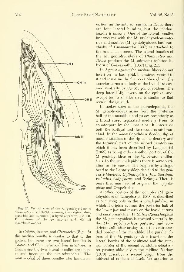
314 Great Basin Naturalist Vol. 42, No. 3
GHII
Fig. 20. Ventral view of the M. geniohyoideus of
Sauromalus (BYU 32551) showing the origins (along
mandible) and insertions (on hyoid apparatus). Gh-I-II-
III divisions of the genioglossus and Mh III
mandibulohyoideus.
In Calotes, Sitana, and Chamaeleo (Fig. 18)
the median bundle is similar to that of the
geckos, but there are two lateral bundles in
Calotes and Chamaeleo and four in Sitana. In
Chamaeleo the two lateral bundles are deep-er and insert on the ceratobranchial. Themost medial of these bundles also has an in-
sertion on the anterior cornu. In Draco there
are four lateral bundles, but the medianbundle is missing. One of the lateral bundles
interweaves with the M. mylohyoideus ante-
rior and another (M. geniohyoideus basibran-
chialis of Gnanamuthu 1937) is attached to
the branchial process. The lateral bundles of
the M. geniohyoideus of Chamaeleo andDraco produce the M. adductor inferior la-
bioris of Gnanamuthu (1937) (Fig. 21).
In Agama agama the median fibers do not
insert on the basihyoid, but extend ventral to
it and insert on the first ceratobranchial. Theanterior cornu and body of the hyoid are cov-
ered ventrally by the M. geniohyoideus. Thedeep lateral slip inserts on the epihyal and,
except for its smaller size, is similar to that
seen in the iguanids.
In snakes such as the anomalepidids, the
M. geniohyoideus arises from the posterior
half of the mandible and passes posteriorly as
a broad sheet separated medially from its
counterpart by the linea alba. It inserts on
both the basihyal and the second ceratobran-
chial. In the anomalepidids a slender slip of
muscle attaches to the tip of the dentary andthe terminal part of the second ceratobran-
chial; it has been described by Langebartel
(1968) as being either another portion of the
M. geniohyoideus or the M. ceratomandibu-
laris. In the anomalepidids there is some vari-
ation in this muscle. The origin is by a single
head in the Leptotyphlopidae and in the gen-
era Rhinophis, Cylindrophis rtifus, Sanzinia,
Enhydris, Aidpysurus, and Bothrops. There is
more than one head of origin in the Typhlo-
pidae and Uropeltidae.
Another portion of this complex (M. gen-
iohyoideus of Langebartel 1968) is described
as occurring only in the Anomalepididae, in
which it originates from the posterior half of
the lower jaw and inserts on the hyoid cornua
and ceratobranchial. In Matrix (Xenochrophis)
the M. geniohyoideus is covered ventrally bythe Mm. mylohyoideus posterior and con-
strictor colli after arising from the ventrome-
dial border of the mandible. The parallel fi-
bers of the M. geniohyoideus insert on the
lateral border of the ba.sihyoid and the ante-
rior border of the second ceratobranchial af-
ter passing obliquely to the midline. Varkey
(1979) describes a second origin from the
midventral raphe and fascia just anterior to
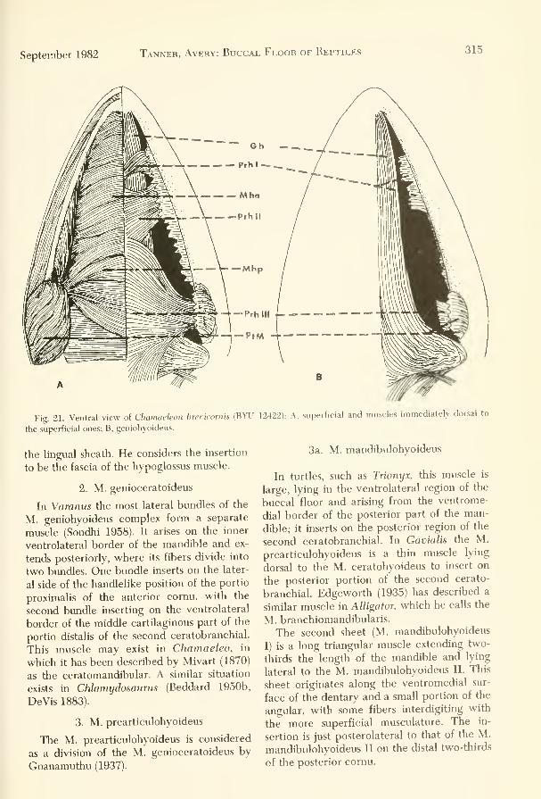
September 1982 Tanner, Avery: Buccal Floor of Reptiles 315
Fig. 2L Ventral view of Chamaeleon brecicornis (BYU 12422): A, superficial and muscles immediately dorsal to
the superficial ones; B, geniohyoideus.
the lingual sheath. He considers the insertion
to be the fascia of the hypoglossus muscle.
2. M. genioceratoideus
In Varanus the most lateral bundles of the
M. geniohyoideus complex form a separate
muscle (Sondhi 1958). It arises on the inner
ventrolateral border of the mandible and ex-
tends posteriorly, where its fibers divide into
two bundles. One bundle inserts on the later-
al side of the handlelike position of the portio
proximalis of the anterior comu, with the
second bundle inserting on the ventrolateral
border of the middle cartilaginous part of the
portio distalis of the second ceratobranchial.
This muscle may exist in Chamaeleo, in
which it has been described by Mivart (1870)
as the ceratomandibular. A similar situation
exists in Chhmydosaurus (Beddard 1950b,
DeVis 1883).
3. M. prearticulohyoideus
The M. prearticulohyoideus is considered
as a division of the M. genioceratoideus by
Gnanamuthu (1937).
3a. M. mandibulohyoideus
In turtles, such as Trionyx, this muscle is
large, lying in the ventrolateral region of the
buccal floor and arising from the ventrome-
dial border of the posterior part of the man-
dible; it inserts on the posterior region of the
second ceratobranchial. In Gavialis the M.
prearticulohyoideus is a thin muscle lying
dorsal to the M. ceratohyoideus to insert on
the posterior portion of the second cerato-
branchial. Edgeworth (1935) has described a
similar muscle in Alligator, which he calls the
M. branchiomandibularis.
The second sheet (M. mandibulohyoideus
I) is a long triangular muscle extending two-
thirds the length of the mandible and lyin^
lateral to the M. mandibulohyoideus II. This
sheet originates along the ventromedial sur-
face of the dentary and a small portion of the
angular, with some fibers interdigiting with
the more superficial musculature. The in-
sertion is just posterolateral to that of the M.
mandibulohyoideus II on the distal two-thirds
of the posterior cornu.

316 Great Basin Naturalist Vol. 42, No. 3
The M. mandibulohyoideus, as described
by Avery and Tanner (1971) for the iguaninehzards, by Robison and Tanner (1962) for
Crotaphytus, and by Jenkins and Tanner(1968) for Phrynosoma, consists of two sheets.
The most medial portion (M. mandibulo-hyoideus II) consists of a pair of small elon-
gated bundles of fibers lying medial to the M.mandibulohyoideus I and inserting togetheron the midventral raphe of the throat. It
originates as a narrow tendon from the man-dibular symphysis. Each muscle inserts on theanterior border of the proximal end of theposterior comu.
In Varanus Sondhi (1958) described it as ashort muscle lying on the ventrolateral side
of the neck, covering the posterior part of
the mandible ventrally. It arises from the
posterior and medial aspects of the mandibleand extends almost straight back along the
ventrolateral side of the neck to insert on therodlike portion of the portio distalis of theanterior comu.
4. M. mandibulopriximalis
The mandibulopriximalis has been de-scribed in Varanus by Sondhi (1958) as aslender muscle situated dorsal to the M. gen-iohyoideus and ventral to the M. gen-ioglossus. It arises from the ventrolateral bor-der of the ramus of the mandible, extendingposteriorly and obliquely to pass dorsal to thehandlehke portion of the portio distalis. Mostof this muscle inserts on the outer margin ofthe handlelike portion of the portio prox-imalis, with some of its fibers becoming sepa-rated from the remainder and inserting onthe lining of the buccal floor.
In the iguanid lizards the M. mandibulo-priximalis, if present, forms a part of the M.geniohyoideus and cannot be distinguishedfrom the latter muscle.
5. M. ceratohyoideus
The M. ceratohyoideus of Sphenodon is
short and thin, having its origin on the secondceratobranchial and its insertion on tlie ante-rior comu. Rieppel (1978) states that thepresence of the M. ceratohyoideus lying be-tween the ceratohyal and the first cerato-branchial and innervated by the M. glos-sopharyngeus is primitive. He further argues
that its failure to reach the lower jaw, as is
the case in most lizards, is also perhaps an in-
dication of its primitiveness.
In Lissemys the M. ceratohyoideus arises
from the second ceratobranchial and inserts
on the basihyoid. In Trionyx it arises from the
distal half of the second ceratobranchial, en-
closing this cartilaginous rod and extendinganteromedially on the lateral side of the buc-cal floor to insert on the middle and anterior
components of the basihyoid and on theknoblike anterior comu.
In Alligator and Crocodylus it originates onthe second ceratobranchial and inserts on the
basihyoid. In Gavialis the M. ceratohyoideuslies dorsal to the basihyoid and is not visible
in ventral view. The origin is on the dorsola-
teral border of the posterior comu, with the
muscle extending obliquely forward as a thin
sheet on the ventral side of the buccal floor
to insert on the dorsolateral side of the ante-
rior comu.The insertion in Sitana is on the basihyoid.
In Varanus it lies dorsal to the M. gen-iohyoideus on the ventrolateral side of the
middle of the neck. The origin is from the
ventrolateral border of the proximal piece of
the second ceratobranchial, from which the
muscle extends anteromedially to fan outover the ventrolateral side of the neck andinsert on the handlelike portion of the portio
distalis.
In the iguanid lizards this muscle has beendescribed as the M. branchiohyoideus by Av-ery and Tanner' (1971). In Ctenosaura the
muscle is ribbonlike and situated between the
first ceratobranchial and second ceratobran-chial of each side of the hyoid apparatus. Theorigin is along most of the anterior two-thirds
of the first ceratobranchial, with the insertion
on the posterior half of the second cerato-
branchial. This pattern is duplicated in Cha-larodon. Opiums, Crotaphytus, and all the re-
maining iguanine lizards except Sauromalus.In the latter the insertion is very narrow, bya single tendon from the proximal rim of the
anterior border of the posterior comu.In Phrynosoma this muscle covers nearly
the entire area between the anterior and pos-
terior cornua of the hyoid (Fig. 19 B). Its ori-
gin and insertion are similar to that describedabove for other iguanids. In Chamaeleo the
M. ceratohyoideus is a small thick mass aris-

September 1982 Tanner, Avery: Buccal Floor of Reptiles 317
ing from the posterolateral border of the
basihyoid to pass anterodorsally and insert on
the epihyal.
In Eumeces (Scincidae) the muscle is a nar-
row strap similar to that in the iguanid
Sauromalus.
In the teiids, Cnemidophoms and Ameiva,
this muscle has a similar origin to that of the
iguanids, but fills the entire area between the
anterior and posterior comua (Fisher and
Tanner 1970).
6. M. cornuhyoideus
The M. cornuhyoideus was described in
Varanus by Sondhi (1958) as being immedi-
ately posterior to the M. ceratohyoideus; it is
ventrally concealed by the basal branch of
the tongue and extends between the anterior
and posterior comua. It arises from the ven-
trolateral border of the proximal piece of the
second ceratobranchial and proceeds forward
to insert on the outer margin of the portio
proximalis of the anterior cornu, anterior to
the latter's articulation with the basihyoid.
This muscle has not been described in any
other reptile.
7. M. interportialis
Sondhi (1958) has reported that in Varanus
this slender muscle lies dorsal to the M. ce-
ratohyoideus and ventral to the portio prox-
imalis. The origin is on the ventrolateral side
of the anterior portion of the portio prox-
imalis, from which the muscle extends
obliquely anteriorly to insert on the medial
border of the handlelike portion of the portio
distalis. Gnanamuthu (1937) did not describe
this muscle for Varanus and probably consid-
ered it to be part of the M. ceratohyoideus. It
has not been described in other reptiles.
8. M. hypoglossolateralis
The M. hypoglossolateralis has been de-
scribed by Sondhi (1958) as a dehcate strip of
muscle lying above the hypoglossum of the
tiutle Trionyx. Its origin is on the dorsal sur-
face of that cartilaginous plate from which it
extends to tlie lining of the buccal floor on
which it inserts. This muscle is also present in
Gopherus agassizi, and we suspect its pres-
ence in association with the hypoglossal car-
tilage of other Chelonia.
9. M. entoglossohypoglossalis
The M. entoglossohypoglassalis is another
muscle described by Sondhi (1958) for Tri-
onyx. It arises from the ventrolateral border
of the anterior part of the lingual process and
inserts dorsolaterally on the posterior surface
of the hypoglossum.
10. M. omohyoideus
In turtles such as Lissemys the M. omo-
hyoideus is thick and long, and has an ante-
rior division into dorsal and ventral bundles.
The dorsal bundle inserts on the medioprox-
imal part of the first ceratobranchial, and the
ventral bundle inserts on the basihyoid along
with the M. sternohyoideus. In Trionyx the
M. omohyoideus originates on the anterior
border of the scapula and extends forward on
the ventral side of the neck to converge ante-
riorly to form two bundles, a larger medial
and small lateral, which insert on the pro-
ximal part of the second ceratobranchial. In
Chelodina the M. omohyoideus arises from
the middle of the coracoid, but in Deiro-
chelys, Lissemys, and Geochelone it origi-
nates on the ventral end of the coracoid. In
all genera the fibers pass anteriorly to insert
on the ceratobranchials.
In Alligator the M. omohyoideus is a long,
narrow, thick muscle that originates from the
upper border of the coracoid and passes for-
ward to insert on the middle of the second
ceratobranchial. In Crocodylus the origin is
from the anterior border of the scapula and
the insertion on the second ceratobranchial.
Gavialis, as described by Sondhi (1958), has a
moderately broad muscle arising from the an-
terior border of the coracoid. As it passes an-
teriorly, it divides into two parts, a portio
dorsalis and a portio ventralis. The portio
dorsalis extends obliquely anteromedially as a
narrow strap that terminates in fragile slips
that merge into the tendon of the M. ster-
nohyoideus. The portio ventralis is broad,
and its fibers parallel the trachea, finally in-
serting on the short anterior part of the sec-
ond ceratobranchial.

318 Great Basin Naturalist Vol. 42, No. 3
The M. omohyoideus is a large muscle that
usually arises on the pectoral girdle and in-
serts on the hyoid. In Sphenodon it is a large
sheet, but in Varanus it is slender and partly
covered by the M. stemohyoideus along its
medial border. We summarize from Gnana-muthu (1937:24) as follows: In Varanus it
arises on the anterior border of the scapula to
pass obliquely forward and insert on the an-
terior part of the proximal end of the second
ceratobranchial close to its articulation with
the basihyoid. A similar situation exists in
Hemidactylus. In Cabrita, Mahuia, and Cha-
maeleo the insertion of the M. omohyoideus
is on the anterior border of the basihyoid. In
Calotes it inserts not only on the basihyoid,
but also on the sides of the proximal part of
the first ceratobranchial. In Anolis and Si-
tana it inserts only on the first ceratobran-
chial, but in Draco there are three bundles,
two of which insert on the first ceratobran-
chial and the third on the second ceratobran-
chial. In Chlamydorsaurus it originates onthe clavicle and sternum and inserts on the
posterior one-third of the ceratobranchial.
In the iguanid lizards, such as Ctenosaura,
the M. omohyoideus has medial and lateral
origins. Medially the fibers originate on the
lateral tip of the transverse process of the in-
terclavicle, whereas the lateral fibers origi-
nate on the lateral half of the anterolateral
surface of the clavicle and the anterior bor-
der of the suprascapula. As the two bundles
extend anteriorly they become continuous
and insert together along the posterior edgeof the second ceratobranchial. In all the
iguanine lizards and Opiums the fibers of the
medial and lateral bundles are impossible to
separate. Chalarodon shows a slightly differ-
ent configuration, with both bundles beingseparated for their entire length.
The M. omohyoideus in the teiids Ameivaand Cnemidophorus is a thick muscleoriginating on the anterior border of the sca-
pula and then proceeding anteroventrally to
insert on the proximal end of the basihyoid
and along the second ceratobranchial. In Di-
bamus it is extremely long, originating on the
scapula and inserting on the distal two-thirds
of the ceratobranchial.
In snakes this muscle is very small andpasses anteriorly from its origin on the lateral
body muscles just posterior to the distal end
of the hyoid apparatus to insert on the poste-
rior portion of the ceratobranchials. It has
been found in the Anomalepididae Cylin-
drophis, Rhinophis, and Eryx c. colubrinus.
11. M. stemohyoideus
The M. stemohyoideus is a complex of
muscles that arises from the sternum and in-
serts on the hyoid in most reptiles (Fig. 19).
In both Lissemys and Trionyx the M. ster-
nohyoideus is large and lies adjacent to the
M. omohyoideus. It originates from the cla-
vicle and passes anteriorly and medially to
insert on the proximal part of the second ce-
ratobranchial and the middle of the basi-
hyoid. In Crocodylus the M. stemohyoideushas long tendons by which it inserts on the
second ceratobranchials. In Alligator it is flat
and broad, and originates from the ventral
surface of the episternum and forms a short
tendon that inserts on the M. geniohyoideus.
In Gavialis the M. stemohyoideus is a broadflat muscle with an origin on the ventral an-
terior half of the episternum; it passes along
the ventral side of the neck to meet its oppo-site member at the midline where it obscures
the trachea ventrally (Sondhi 1958). As it ap-
proaches the hyoid apparatus it divides into
two parts, with the outer part (portio ex-
terna) a broad band forming a large tendon
that inserts on the inner border of the man-dible. The inner bundle (portio interna) par-
allels the trachea to insert on the outer part
of the posterior border of the basihyoid.
In Sphenodon it is flat, whereas in somelizards it becomes cordlike and inserts (Riep-
pel 1978) on the caudodorsal edge and dorsal
surface of the first ceratobranchial, deep to
and lateral to the insertion of the omo-hyoideus. In Mabuia, the M. stemohyoideus
inserts on the basihyoid, whereas in Anolis,
with its small basihyoid, the insertion is onthe first ceratobranchial. In Varanus the M.stemohyoideus lies dorsal to the M. con-
strictor colli and ventral to the basihyoid andthe proximal piece of the second ceratobran-
chial. It arises from the ventrolateral border
of the clavicle and extends obliquely ante-
riorly to the ventral side of the neck, where it
parallels the M. omohyoideus and inserts onthe ventral side of the basihyoid and posteri-
or portion of the lingual process. Chamaeleo

September 1982 Tanner, Avery: Buccal Floor of Reptiles 319
has a small lateral bundle of fibers that insert
on the fascia of the lateral M. geniohyoideus.
The M. stemohyoideus of the iguanine liz-
ards (Avery and Tanner 1971), is an extensive
muscle sheet occupying a large area posterior
to the anterior comu and anterior to the ster-
num and clavicle. It originates from several
heads on the clavicle, and its oblique fibers
extend anteriorly to insert on the posterior
surface of the anterior cornu. In all the
iguanines and in Cfialarodon the muscle ap-
pears broad and sheetlike. In Opiums, it is
narrow and cordlike.
In Phrynosorna the M. stemohyoideus is
separated into three distinct muscles (Fig.
19). As described by Jenkins and Tanner
(1968), the M. stemohyoideus I originates
from the medial surface of the scapula and
the most anterior part of the clavicle and in-
serts on the distal two-thirds of the anterior
cornu. This muscle may be the M. ster-
nothyroideus of other workers. The M. ster-
lohyoideus II originates from the anterola-
teral surface of the sternum and inserts onto
the posterodorsal surface of the basihyoid.
The M. stemohyoideus III is separate for
its entire length, with an origin from the ven-
tral surface of the anterior third of the ster-
num and an insertion on the dorsal surface of
the most enlarged area of the posterior
comu.
In the agamid Chlamydorsaurus, the M.
stemohyoideus has a large origin from the
sternum immediately deep to that of the M.
omohyoideus. It expands and thins as it ex-
tends anteriorly to insert on the inner side of
the ceratobranchial ventral to the M. omo-
hyoideus. In Uromastyx the origin is from
both the sternum and the coracoid.
Nash and Tanner (1970) describe a super-
ficial and a deep layer of this muscle in the
skink Eumeces. The larger ventral or super-
ficial layer originates from the posterior and
ventral surfaces of the ceratobranchial I and
ihedial to the corpus and inserts on the inter-
clavicle with the M. stemocleidomastoideus,
trapezius, and depressor mandibularis, and
with the constrictor colli on the posterior and
ventral surfaces of the anterior comu. The
dorsal or deep layer originates on the inter-
clavicle and inserts on the posterior border of
both anterior and posterior comua.
In Dibamus, the M. stemohyoideus is a
large strap originating from the sternum and
coracoid and inserting on the distal tip of the
ceratobranchial (Gasc 1968).
In the teiids Ameiva and Cnemidophorus,
the M. stemohyoideus is broad, with an ori-
gin on the sternum and insertions on both the
posterior and anterior cornua and the
basihyoid.
In snakes the M. stemohyoideus is found as
a separate muscle only in the Typhlopidae
and Leptotyphlopidae. Its origin here is deep
to the muscles on the linea alba. The fibers
pass anteriorly to insert on the hyoid, usually
on the entire posterior edge of each comu.
12. M. stemothyroideus
In the turtle Trionyx the origin of the M.
stemothyroideus is on the anterior border of
the stemum. The muscles extend anteriorly
to insert on the ventrolateral border of the
posterior part of the basihyoid.
In lizards, the M. stemothyroideus nor-
mally has an origin on the anteromedial por-
tion of the sternum, from which it extends
anteriorly to insert along the length of the
second ceratobranchials. This situation exists
in Hemidactylus, Mabuia, Cabrita, Anolis,
Calotes, and the iguanine lizards. In Cha-
maeleo the M. stemothyroideus extends later-
ally to insert on the distal end of the cerato-
branchial. In Varanus the M. ster-
nothyroideus lies dorsal to the Mm. omo-
hyoideus and stemohyoideus. It originates as
a thin sheet from the anteromedial half of the
sternum and inserts on the anterior half of
the proximal piece of the second
ceratobranchial.
In the iguanine lizards the most medial
series of fibers of the M. stemohyoideus com-
plex, the M. stemothyroideus, may be sepa-
rated from the other members of this group
by their different origins and insertions. The
origin consists of a small area of both the in-
terclavicle and sternum. These fibers pass an-
teriorly and parallel to the trachea to insert
on the basihyoid. Along its length this muscle
is difficult to separate from the more lateral
M. stemohyoideus, except in Opiums and
Chalarodon, in which both muscles are free
and separated along their entire length.

320 Great Basin Naturalist Vol. 42, No. 3
The M. stemothyroideus of Phrynosomawas previously described by Jenkins and Tan-
ner (1968) as the M. sternohyoideus I.
13. M. costocutaneous superior
Because the shoulder girdle of snakes has
been lost, the M. omohyoideus, ster-
nohyoideus, and stemothyroideus cannot beidentified. Therefore these muscles will bediscussed here under the name M. costocu-
taneous superior.
In some snakes it is possible tentatively to
identify the homologs of these three muscles.
For example, in the Typhlopidae and the
Leptotyphlopidae, the M. sternohyoideus is a
distinct mass of fibers that arise from the ven-
tral scales and adjacent rows of lateral scales
and the ribs, extending anteriorly to the
hyoid and surroimding muscles. In Typhlops,
Leptotyphlops, Rhinopliis, Cylindrophis, andAchrochordus, the anteriormost fibers of the
complex extend to originate on the mandible
and overlay the hyoid while having no con-
nection with it. In Cylindrophis the fibers
originate on the posterior or medial edge of
the M. constrictor colli. In the anomalepididsnakes the insertion is on the posteromedial
border of the basihyoid and second cerato-
branchial. The insertion also extends to the
base of the lingual process in most specimens.
In Agkistrodon, Bothrops, and Crotalus, the
insertion is most extensive on the medianraphe and lingual process.
Sondhi (1958) describes three specific mus-cles present in Matrix (Xenochrophis) that are
probably homologous to the Mm. omo-hyoideus, sternohyoideus, and ster-
nothyroideus. The omohyoid portion arises
from the skin on the ventrolateral part of the
neck and then extends obliquely anteriorly to
insert on the ventrolateral aspect of the basi-
hyoid. In Atretium, this muscle has a cut-
aneous origin and inserts on the second ce-
ratobranchial. The second muscle, absent in
Atretium but possibly the M. sternohyoideus,
originates from the skin in the ventrolateral
region of the neck posterior to the M. omo-hyoideus and passes anteriorly to close prox-
imity with the latter to insert on the outer
border of the basihyoid. The sternothyroid
part of this complex lies in the midline of the
neck over the ventral surface of the basi-
hyoid, with its origin on the midventral por-
tion of the cervical skin. The muscle inserts
on the medial border of the basihyoid. In
Atretium the sternothyroid portion of the
complex has its origin from the second ce-
ratobranchial, with some fibers intertwining
with their opposite member at the midline.
14. M. neurocostomandibularis
According to Langebartel (1968), the M.neurocostomandibularis is present in all
snakes except the Anomalepididae. In mostsnakes it is a broad sheet forming part of the
M. neurocostomandibularis complex, but in
some it is separate and narrow. It covers a
large area of the head and in some is partially
overlain by the Mm. constrictor colli and cos-
tocutaneus superior. Its origin is on the den-
tary, from which it proceeds posteriorly to
insert variously on the hyoid apparatus.
The muscles of Python sebae (Frazzetta
1966) and Boa constrictor (Gibson 1966) that
are innervated by the hypoglossal nervesform a single muscular complex, the M.neurocostomandibularis, and correspondroughly to the M. geniohyoideus of other
reptiles. The complex extends between the
mandibles and the second ceratobranchials.
In both Boa and Python the origin is on the
lower jaw and the insertion on the posterior
part of the second ceratobranchial.
In Natrix (Xenochrophis), Sondhi (1958) de-
scribes the M. neurocostomandibularis as
probably the M. geniolateralis because the
latter muscle receives a branch from the hy-
poglossal nerve. Langebartel (1968) consid-
ered this muscle to be the M. ceratomandibu-
laris as designated by Richter (1933). Theproper identity of this muscle in the typhlo-
pids, leptotyphloids, and anomalepidids is un-
known to us. According to Langebartel
(1968), the M. ceratomandibularis in snakes
arises from the dentary and itiserts on the an-
terior part of the hyoid and the tendinous in-
scription in the M. neurocostomandibularis.
Varkey (1979) describes the M. neurocosto-
mandibularis as being very complex and hav-
ing three separate heads in Nerodia. It is a
wide flat muscle sheathing the neck and most
of the lower jaw. One origin (the vertebral
head) is on the apponeurosis of the dorsal

September 1982 Tanner, Avery: Buccal Floor of Reptiles 321
midline neck region. It passes under the con-
strictor colli to insert on the midline raphe.
The costal head originates by narrow slips
from the first seven anterior ribs and inserts
on the midline raphe with the previous slips.
The third or hyoid head has a double origin
from the midventral raphe just median to the
hyoid comua. This branch is called M. trans-
versalis branchialis by Langebartel (1968). It
inserts on the origin of the other heads on the
midline raphe.
15. M. transversalis branchialis
The M. transversalis branchialis appears
variably and erratically in the families of
snakes with the exception of the Anoma-lepididae, Typhlopidae, and Leptotyphlo-
pidae, in which it is universally absent.
When present, this muscle arises from
somewhere on the second ceratobranchial. In
Rhinophis, the origin is on the medial edge,
whereas in Cylindrophis it originates on the
anterior two-thirds. In Anilius the entire
length of the cartilage is involved.
The insertion of this muscle is usually on
the median raphe, although in some snakes it
is inserted on the fascia covering the M. cos-
tocutaneous superior.
In Nerodia, Varkey (1979) describes the M.
transversus branchialis as originating on the
midline raphe just anterior to M. inter-
mandibularis's anterior. It passes anterolate-
rally to insert broadly on the mucosa of the
angulo-splenial articulation and narrowly on
the lateral sublingual gland. Varkey indicates
his usage of this muscle name is as in Albright
and Nelson (1959), Cowan and Hick (1951),
and Weaver (1965). Langebartel (1968) calls
this muscle the dilator of the sublingual
gland, using the name M. transversalis bran-
chialis for a branch of what Varkey calls the
M. neurocostomandibularis.
16. M. hyotrachealis
In most snakes the M. hyotrachealis arises
from the second ceratobranchial, but in
Liotyphiops and the leptotyphiopids the fi-
bers are tied by connective tissue on the ven-
trolateral surface of the lining of the buccal
floor. In the typhlopids the fibers originate in
connective tissue on the hypaxial trunk mus-
cles. In other snakes the M. hyotrachealis
originates on the lateral edge of the second
ceratobranchial. In Rhinophis the origin is at
the anterior quarter of the medial edge,
while in Cylindrophis maculatus and C. rufus
the origin is from the lateral edge about half-
way down the ceratobranchial. In the boids it
originates on the posterior half of the carti-
lage. In Tropidophis the origin is deep from
the raphe of the M. neurocostomandibularis.
In colubrids, viperids, and elaphids the origin
varies extensively. In Heterodon and Pseu-
daspis the origin varies extensively. In Heter-
odon and Pseudaspis the origin is from the
rib cage, while in Agkistrodon it may be ei-
ther the rib cage or hyoid, indicating a split
origin. In Vipera aspis, Edgeworth (1935) de-
scribes one head oiF the M. hyotrachealis as
lying dorsal to the rib cage while the lateral
head attaches to the hyoid. In Cerastes the
single head originates from the ventral lining
of the buccal floor.
The insertion of the M. hypotrachealis is
normally from the trachea of the laryngeal-
tracheal area, dorsal and anterior to the in-
sertion of the M. geniotrachealis. In some
genera {Typhlops, Amblycephalus, Xeno-
peltis, and Agkistrodon piscivorus) the in-
sertion is on the ventral portion of the M.
geniolateralis. In Boa cookii, Notechis, and
others the M. hypotrachealis has a split in-
sertion with attachments on dorsal and ven-
tral sides of the geniotrachealis.
Varkey (1979) describes Nerodia' s. hyotra-
chealis as thin and narrow and of a double
origin. One head is just anterior to a trans-
verse tendinous inscription of the M. neuro-
costomandibularis. The second or median
head is from the lateral edge of the hyoid
comua. The heads join and insert on the lar-
ynx and trachea anterior to the insertion of
the geniotrachealis.
V. Buccal Floor Muscles NotAssociated with the Hyoid Apparatus
The homologies of a number of the repti-
lian throat muscles not connected with the
hyoid are unclear. We will present the most
widely used terminology and present syno-
nyms only when two or more names have
had wide usage for the same muscle. Al-
though the following muscles are not directly
attached to the hyoid apparatus, they have a

322 Great Basin Naturalist Vol. 42, No. 3
close functional relationship and are there-
fore included (Figs. 18, 19, 20).
1. M. constrictor superficialis
The M. constrictor superficialis is found in
Trionyx as a superficial muscular sheet lying
ventral to the anterior region of the neck. It
arises as a narrow slip from the skin covering
the side of the neck and broadens to insert onthe gular septum. In Gavialis it originates onthe skin overlaying the angle of the jaw, sur-
roimds the neck, and extends obliquely to in-
sert on the gular septum.
In other reptiles, such as iguanid hzards,
this muscle is probably homologous to muchof the Mm. constrictor colli and inter-
mandibularis posterior of Avery and Tanner
(1971).
2. M. constrictor colli
The M. constrictor colli is an extensive su-
perficial muscular sheet, originating on the
middorsal aponeurosis of the neck and ex-
tending ventrad to insert on the posterior
part of the midventral raphe or gular septum.
In Sphenodon it forms a broad, thin, super-
ficial sheet that completely encases the neck(Rieppel 1978, Fig. 1). It ensheathes the en-
tire neck in Chehdina and Deirochelys, butin Lissemys the neck is only partly covered.
The muscle arises from the dorsal fascia andinserts on the median raphe. It is continuous
anteriorly with the M. intermandibularis.
Sondhi (1958) lists this muscle as present in
Trionyx and Gavialis, and Gnanamuthu(1937) recognized it in Crocodylus and Tri-
onyx. In the Testudines and Crocodylia the
M. constrictor colli is not attached to the
hyoid, but has an insertion on the gularseptum.
This muscle covers most of the lateral sur-
face of the neck in Amblyrhynchus, Chalaro-
don, Cyclura, Iguana, and Saiiromalus. It is
much less extensive in Brachylophus, Con-olophus, Ctenosaura, Dipsosaunis, Opiums,Crotaphytus, and Phrynosoma.
In Chamaeleo the M. mylohyoideus poste-
rior of Mivart (1870) corresponds to the M.constrictor colli. It originates on the occipital
crest and inserts on the median raphe.
In the skink Eumeces the M. constrictor
colli is a very broad sheet originating fromthe middorsal tympanic fascial and inserting
on the median raphe. It covers most of the
neck from the angle of the jaw to the
interclavicle.
In the teiids Cnemidophorus and Ameiva,the muscle is as in Eumeces, but the anterior
border interdigitates with the posterior bor-
der of the M. cervicomandibularis. Gnana-muthu (1937) figures this muscle to be in
Hemidactylus, Mabuia, Cabrita, Anolis, Ca-lotes, and Draco.
In snakes the M. constrictor colli appears
erratically and is not constant in form within
a single genus as indicated by Python. Themuscle is normally broad and envelops the
angle of the jaw with an insertion on the
midventral raphe or hyoid. In some species of
Python it appears to be absent. The M. con-
strictor colli is found in all families of snakes
except in the Uropeltidae where it has not
been recognized.
3. M. mylohyoideus anterior
The superficial M. mylohyoideus anterior
is generally located ventrally beneath the
rami of the lower jaw anterior to the M. con-
strictor colli. It takes its origin from the ante-
rior part of the mandible and inserts on the
gular septum.
In Sphenodon the M. mylohyoideus (M. in-
termandibularis of Rieppel 1978) forms a
single large muscular sheet, but in lizards it is
differentiated into three sets; the Mm. mylo-
hyoideus anterior superficialis (= M. inter-
mandibularis anterior superficialis), mylo-
hyoideus anterior principalis (= M.
intermandibularis anterior profundus), andmylohyoideus anterior profundus (
= M. inter-
mandibularis posterior). In some forms, such
as Cabrita, Anolis, Hemidactylus, andMabuia, some fibers of the M. mylohyoideusanterior originate deep on the medial surface
of the mandible and others originate super-
ficially on the M. geniohyoideus. As the fi-
bers of the two muscles cross, they break into
numerous strips and interdigitate (Figs. 18,
19, and 20).
In turtles the M. mylohyoideus is simpler.
In Trionyx it consists of a single M. mylo-
hyoideus anterior profundus that originates

September 1982 Tanner, Avery: Buccal Floor of Reptiles 323
ventral to the M. geniohyoideus from the
ventral aspect of the mandible and passes
medially to insert on the gular septum. In
Lissemys the M. mylohyoideus anterior forms
as two muscles, with the M. mylohyoideus
anterior profundus being identical to that of
Trionyx. The M. mylohyoideus anterior prin-
cipalis is a broad sheet originating on the
mandible and inserting on the gular septum.
In Chelodina and Deirochelys the inter-
mandibular series is simple and undivided,
originating on the inner surface of the man-
dible and inserting on the median raphe. In
Chelodina the anterior quarter of the fibers
do not insert into each other as in Deiro-
chelys, but are separated by fascia.
Some age variation in this muscle can be
seen in Crocodylus: in adults the M. mylo-
hyoideus anterior is not distinguishable as a
separate muscle, but there are two sheets in
the juvenile representing the M. mylo-
hyoideus anterior and mylohyoideus posteri-
or. In adult Alligator a single transverse sheet
is present (Mm. intermaxillaris and sphincter
colli), and in Gavialis the one sheet (M.
mylohyoideus anterior principalis) is prob-
ably homologous to both the Mm. mylo-
hyoideus anterior and mylohyoideus posteri
or. In Gavialis this muscular sheet occupies
almost the entire anterior part of the ventral
inter-ramal area of the neck, originating on
the inner side of the mandible and inserting
on the gular septum.
The M. mylohyoideus anterior superficialis
exhibits several variations. In Mabuia and
Anolis the fibers extend anteriorly, over-
lapping the M. mylohyoideus anterior princi-
palis either medially or laterally. In the Cha-
maeleonidae and Agamidae the Mm.mylohyoideus anterior principalis and mylo-
hyoideus anterior profundus occur together
in the fonn of a double sheet, which we have
concluded is a variation of the M. mylo-
hyoideus anterior. In Varanus, Sondhi (1958)
indicates that the muscle extends transversely
from the mental groove to the M. gen-
ioglossus portio major. There are three sets of
fibers listed, including a broad M. mentalis
superficialis, that originate ventrally, whereas
the narrow M. mentalis profundus anterior
and the M. mentalis profimdus posterior orig
inate dorsally. All tliree bundles insert in the
lining of the buccal floor. These muscles do
not appear to be homologous to the muscular
complex we have seen in iguanids and desig-
nated the M. mylohyoideus anterior.
In Amblyrhynchus, Brachylophus, Chalaro-
don, Conolophus, Ctenosaura, Cyclura, Dip-
sosaurus. Iguana, Opiums, and Sauromalus
two distinct groups of muscle fibers are
found. The M. intermandibularis anterior are
deep fibers that originate as a tendon from
the coronoid and splenial bones and extend
medially on the ventral surface to join at the
median raphe, where they interdigitate with
about five bundles of the M. geniohyoideus.
A small bundle of fibers also extends from the
origin to insert on the connective tissue cap-
sule of the sublingual gland. In Iguana this
muscle forms the bulk of the large dewlap.
The most superficial group of fibers (M. in-
termandibularis anterior superficialis) is small
and narrow, with an origin from the oral
membrane and from the anterior part of the
M. intermandibularis anterior profundus. The
muscle fibers pass obliquely posteriorly to in-
sert on the median raphe. In Iguana and Dip-
sosaurus this superficial group is greatly re-
duced in size.
In snakes the synonymy of the throat mus-
culature is not well established. For this rea-
son we follow rather closely the studies of
Langebartel (1968) and Sondhi (1958). Theanteriormost set of transverse fibers (M. in-
termandibularis anterior) is absent in Anilius
and Xenopeltis, but is represented by a ten-
don in Rhinophis. In the anomalepidids,
typhlopids, and leptotyphlopids this muscle is
broad and may actually represent several
muscles. In the latter families one or more
muscle groups may originate on the medial
surface of the dentary. In the colubrids, vipe-
rids, and elapids a single muscle is large but
separated into two parts. The longer and
thicker anterior one originates on the medial
surface of the tip of the dentary and medially
to the fibrous inter-ramal pad. The second
(posterior) part extends obliquely from the
same origin to insert on the ventral raphe.
The M. mylohyoideus anterior in Matrix {Xe-
nochrophis) is probably represented by three
muscles: Mm. intermaxillaris, mentalis pro-
fimdus anterior, and mentalis profundus pos-
terior. The M. intermaxillaris originates from
the ventrolateral border of the dentary and
passes obliquely posteriorly to insert on the

324 Great Basin Naturalist Vol. 42, No; 3
mental groove. The remaining pair of bun-
dles originate from the mental groove and ex-
tend obliquely caudad to insert adjacent to
each other on the lining of the buccal floor.
The intermandibularis anterior of Nerodia
is described as having two separate parts.
The M. intermandibularis anterior pars
mucosalis has two portions. The first is a
small triangular anterior portion that origi-
nates on the midventral raphe of the lower
jaw and buccal membrane fascia. It inserts on
the ventromedial surface of the anterior tip
of the dentary and the ligament attached to
it. The much stouter posterior slip originates
from the midventral raphe of the lower jaw
and fascia surrounding the tongue sheath just
posterior to the insertion of the anterior slip.
The fibers pass anterolaterally to insert on
the ventromedial surface of the dentary im-
mediately posterior to the insertion of the an-
terior slip.
The second part (M. intermandibularis an-
terior pars glandularis) originates on the mid-
ventral raphe of the lower jaw of the fibrous
inter-ramal pad. The fibers pass post-
erolaterally to insert on the ventrolateral side
of the sublingual gland at its posterior end. Asmall number of fibers insert on oral mucosaposterior to the gland.
4. M. mylohyoideus posterior
The M. mylohyoideus posterior is a trans-
verse muscle situated posterior to the M.mylohyoideus anterior.
The M. mylohyoideus posterior [M. mylo-
hyoideus anterior principalis of Sondhi
(1958)] of Trionyx and Lissemys originates
from the border of the mandible, where it
forms a broad, thick sheet muscle. It extends
medially to insert on the gular septum.
In Alligator, Crocodylus, and Gavialis the
M. mylohyoideus posterior is represented bya thin sheet that combines into one muscle,
the M. mylohyoideus anterior and M. mylo-hyoideus posterior.
In some lizards {Mahuia and Cabrita) these
muscular sheets are continuous, but show a
small division between them. Sondhi (1958)reports that in Varanus they are differen-
tiated into two muscles (Mm. mylohyoideusanterior superficialis and mylohyoideus ante-
rior principalis) that are disposed one behind
the other, both originating on the lateral sur-
face of the mandible and inserting on the gu-
lar septum.
In the iguanine lizards, an anterior and a
posterior sheet of muscle fibers (M. inter-
mandibularis posterior) form the M. mylo-
hyoideus posterior. The anterior sheet is
broad and thin, with an origin from the later-
al surface of the mandible. The fibers pass
medially on each side to insert with their op-
posite members at the median raphe. Theposterior bundle of fibers (about one-quarter
of the posteriormost fibers) originate from
the lateral surface of the mandible beginning
at the midpoint of the retroarticular process
and insert on the linea alba.
In most iguanines the M. mylohyoideus
posterior exhibits a primitive condition bybeing continuous with the M. constrictor col-
li, from which it can be delineated by a natu-
ral separation along the entire border only in
Conolophus and Ctenosaura. In Cyclura and
Sauromalus this separation is present only in
the medial third of their common border. In
Amblyrhynchus, Brachylophus, Chalarodon,
Dipsosaurus, Iguana, and Opiums the twomuscles are continuous along their entire
border.
In Crotaphytus the Mm. mylohyoidei ante-
rior and posterior form one continuous sheet
with no separation between them. In Phryno-
soma the M. mylohyoideus posterior is sepa-
rated from the anterior, but is continuous
posteriorly with the M. constrictor colli, fromwhich it can be separated only with great
care.
In Eumeces the position of M. mylo-hyoideus posterior is similar to that of the
iguanid lizards, with both anterior and poste-
rior muscles being separated.
In the teiid Ameiva the M. mylohyoideus
posterior originates on the medial surface of
the dentary and immediately breaks into nine
separate divisions that interdigitate with slips
of the M. geniohyoideus. It inserts on the
midventral raphe just posterior to the M.mylohyoideus anterior (Fig. 17). In Cnemido-phortis the muscle is as above, except that
there are only five divisions instead of the
nine in Ameiva. The Mm. mylohyoideus pos-
terior and constrictor colli are continuous for
their entire border in Shinisaurus, but widely
separated in Xenosaiirus.

September 1982 Tanner, Avery: Buccal Floor of Reptiles 325
All snakes except one colubrid {Amhlyce-
phahis kuangtunensis) possess a M. inylo-
hyoideus posterior (Langebartel 1968). It lies
in the same position as the M. inter-
mandibularis posterior of Langebartel (1968),
with the former having its origins on the
mandible and insertion on the gular septum.
In the colubrid Achrochordiis it is very broad,
attaching to the middle region of the man-
dible. In Haplopeltura boa it is attached lat-
eral to the external adductor muscle of the
lower jaw. The fibers are not attached to the
mandible, but cross their opposite members
at the midventral raphe and interdigitate,
eventually attaching to the opposite man-
dible. M. mylohyoideus inserts on the lingual
process in the hydrophid Aipysumrus.
The second bundle of fibers (M. inter-
mandibularis posterior superficialis) is small
and restricted in some snakes with the paral-
lel type of hyoids. Its occurence is sporadic in
colubrids, and it is absent in most poisonous
snakes, including the hydrophids. It may be
replaced by a tendon that originates from the
posterior part of the lower jaw and inserts on
the gular septum. Sondhi (1958) states that
the M. mylohyoideus posterior is an extreme-
ly broad muscle sheet, lying immediately pos-
terior to the M. mylohyoideus anterior and
occupying the posterior region of the neck in
Matrix piscator. He further states that this
muscle originates on the dorsolateral surface
of the anterior cervical vertebrae and extends
ventrally to insert on the posterior part of the
gular septimi. Langebartel (1968) describes
two muscles, a ventral sheet taking its origin
from the anterior part of the mandible and
extending obliquely anteriorly over the body
of the tongue to insert on the gular septum,
and a dorsal sheet deep and dorsal to the M.
geniohyoideus, with an origin from the man-
dible with the ventral sheet; the dorsal sheet
extends obliquely anteriorly also to insert on
the gular septum. The M. ceratomandibularis
of Langebartel (1968) occurs in most snakes,
although with considerable variation. Be-
cause of its location, we include it as a syno-
nym of M. mylohyoideus posterior, even
though we are aware that most homologies
must yet be proven by careful embryonic
study.
The intermandibularis posterior of Nerodia
is described by Varkey (1979) as having two
slips. The M. intermandibularis posterior pars
anterior is the largest of the ventral con-
strictors originating on the midventral raphe
of the lower jaw ventral to the origin of the
posterior slip of the intermandibular anterior
I and the transversalis branchialis. The origin
is broad and thin, passing caudolaterally to
form a stout band to insert on the ventrome-
dial surface of the bone at the distal end of
the mandibular fossa.
The second slip, which Varkey calls the M.
intermandibularis posterior pars posterior, is
a thin, flat, triangular sheet of muscle that
originates on the midventral raphe posterior
to the origin of the intermandibularis posteri-
or I and the transversalis branchialis and the
anterior tip of the hyoid cornua. It passes
ventral to the M. neurocostomandibularis for
most of its length. This insertion is just poste-
rior to the insertion of the pars anterior on
the ventrolateral surface of the bone at the
level of the proximal end of the mandibular
fossa.
5. M. mandibulotrachealis
The M. mandibulotrachealis of Varaniis
has been described by Sondhi (1958) as a deli-
cate muscle arising from the anteroventral
part of the mandible and extending posteri-
orly to divide into two parts. The dorsal part
passes posteriorly dorsal to the tongue to in-
sert on the lateral side of the trachea. The
ventral part of the muscle extends posteriorly
to fan out over the buccal floor near the in-
sertion of the M. genioceratoideus, with an
insertion on the ventral lining of the buccal
floor. In Natrix (Xenochrophis), Sondhi (1958)
found the origin to be similar to that of Va-
ranus, with a medial bundle inserting on the
trachea and a lateral bundle attaching to the
lining of the buccal floor. It has not been re-
ported for other genera.
This muscle is reported by Varkey (1979)
for Nerodia as the M. geniotrachialis. It is a
stout band of muscle that parallels the gen-
ioglossus. It originates at the anterior end of
the dentary dorsal to the origin of the lateral
genioglossus. It passes posteromedially to the
tongue sheath and inserts on the ventral and
ventrolateral surfaces of the first 14 tracheal
rings.

326 Great Basin Naturalist Vol. 42, No. 3
6. M. neuromandibularis
The M. neuromandibularis has been de-
scribed in detail by Sondhi (1958) for Matrix
(Xenochrophis) and Varanus. In the latter it
probably corresponds to the M. gen-
iolateralis. Sondhi has described M. neuro-
mandibularis as originating from the dorsola-
teral border of the middorsal aponeurosis andextending a short distance anterior along the
dorsal side of the neck. The fibers divide into
three sets, which pass into a common tendon
inserting on the inner ventral surface of the
skin. In Natrix (Xenochrophis) the origin is
similar to that in Varanus. The insertion is onthe ventromedial side of the posterior half of
the mandible.
In some snakes the M. neuromandibularis
arises from an aponeurosis at the middorsal
line. In the anomalepidids, typhlopids, lepto-
typhlopids, uropeltids, and aniliids it inserts
on the lov^^er jaw. In the Xenopeltidae,Boidae, and other families it inserts on the
raphe in common with the Mm. ceratoman-
dibularis and costomandibularis.
7. M. costomandibularis
The M. costomandibularis has been de-
scribed only for some snakes in which its ori-
gin is either from the cartilaginous ribs or the
rib cage. In Thamnophis a medial slip origi-
nates from the peripheral surface of the lin-
ing of the pharyngeal floor and inserts on the
common tendinous inscription of the M.neurocostomandibularis. In Cylindrophis
rufiis the insertion is on the second cerato-
branchial as well as on the mandible.
8. M. constrictor pharyngis
The M. constrictor pharyngis of Croco-
dylus and Gavialis is a deeply laid transverse
strap apparently restricted to the Crocodilia.
Its origin is from the lateral surface of the
cervical vertebrae and its insertion medial onthe gular septum.
9. M. obliquus abdominis internus
Langebartel (1968) describes the M. ob-liquus abdominis internus as a trunk muscle
of snakes, with an origin on the medial face
of the ribs and an insertion on the linea alba.
10. M. transverse abdominis
Langebartel (1968) has also described the
M. transverse abdominis as restricted to
snakes and lying on the deep surface of the
M. obliquus abdominis internus, with an ori-
gin on the medial face of the ribs. After ex-
tending posteriorly and medially, it inserts on
the linea alba.
VI. The Tongue: External Morphology
The tongue of reptiles has been in-
vestigated by many workers, some of which
are as follows: Graper (1932), Nonoyama(1936), Gnanamuthu (1937), Oelrich (1956),
Sondhi (1958), Avery and Tanner (1971), and
Kroll (1973). Winokur (1974) published a ma-
jor study on the adaptive modification of the
buccal mucosae in turtles. His study is con-
cerned not only with the tongue, but also
with the glands found in the buccal area.
Tongues in turtles vary in size and com-plexity (Fig. 22). Winokur states that
Terrestrial herbivores {Gopherus, Testudo, and other tor-
toises) have the best developed mucous glands, whereas
aquatic carnivores (Chelydra and Chelus) have few or no
mucous glands.
In both Chelydra and Trionyx (Fig. 22 A,
B) the tongue is without papillae or complexglands and is nonprotrusible, a characteristic
of carnivorous chelonians. In Trionyx the
short, rounded tongue is dorso-ventrally flat-
tened and contains just a base and body. Thebase is formed from two posterior limbs that
they enclose. Each basal portion extends an-
teromedially to unite in the tongue. Posterior
to the tongue and glottis, the buccal-phary-
ngeal floor has numerous filiform papillae
that Girgis (1961) has shown to have a res-
piratory function. The tongue of some, such
as Chehis, has been developed as a lure in
food-getting: the open mouth exposes a
wormlike tongue structure to intice unsus-
pecting prey into the mouth.
In contrast, the terrestrial herbivorous che-
lonians (Tortoises; Fig. 22b) have a much

September 1982 Tanner, Avery: Buccal Floor of Reptiles 327
larger lingual pad, which is glandular, fleshy,
and somewhat protrusible. Tortoises have
profuse Ungual mucous glands on and be-
tween the lingual papillae as well as muscles
capable of some lingual protrusion. They
generally lack papillae posterior to the
tongue.
Winokur (pers. comm.) considers Derrna-
teinys (Fig. 22a) to be a special case. The
tongue of this aquatic herbivore shows one
end of the spectrum of buccal complexity in
aquatic chelonians. Figures 22 and 23 illus-
trate the extremes seen between the tongues
of aquatic carnivorous and terrestrial herbi-
vorous chelonians. The tongue of Derma-
temys, although proportionately smaller than
that of terrestrial Gophems, shows an ex-
treme condition of buccal papillation, but
one that is quite different from that of terres-
trial herbivorous tortoises. Between these ex
tremes are the majority of chelonians, such as
Pseudemys, which tend toward
omnivorousness.
The tongues of Alligator, Crocodylus, and
Gavialis lack any specific areas identifiable
as base, body, or apex. The tongue is a mass
of tissue between the mandibular symphysis
and glottis attached to the lining of the buc-
cal floor except at its anterior tip. It can be
elevated and depressed, but not protruded.
Sauromalus (Fig. 24) and Brachylophus
(Fig. 25) show generalized lizard tongues,
with their extensive papillation and lateral
extensions on each side of the glottis. Such
tongues are protrusible and obviously serve a
masticatory function. In Amblyrhynchus,
Brachylophus, Conolophus, Ctenosaura, Cycl-
ura, Dipsosaurus, Iguana, and Sauromalus
the tongue is well developed and large. In
the above genera it is cleft anteriorly, with
the most anterior tips lacking papillae. There
is a smooth pad ventral to the tips (Fig. 24).
In the teiid Ameiva the tongue is rounded
and slightly notched posteriorly and covered
by a lingual sheath. It bears a deep terminal
notch anteriorly that separates the tapering
elongate terminal prongs. A lingual sheath is
absent in Cnemidophorus and other macro-
teiids. The tongue of Cnemidophorus (Fig.
26) represents a moderate advancement in
the development of flexibility, and Cha-
maeleo (Fig. 27) is a highly specialized free
tongue.
Fig. 22. Tongue size as indicated in: A, Dermateys
mawi (UU 9845), above; B, Gophems (UU 5961), below.
It should be noted that the glottis is moved caudad as
the tongue increases in size. Photographs provided by
Robert M. Winokur.

328 Great Basin Naturalist Vol. 42, No. 3
PhF
Fig. 23. Tongue of Trionyx spiniferus: A, showing its position in relation to the glottis and pharynx area with its
filamentous papillae; B, Chelydra serpentina showing the nonpapillated pharynx.
In Lanthanotus the tongue is deeply in-
cised terminally, forming two taperingprongs. The anterior half of the tongue is
elongate, narrow, smooth, and elastic, where-as the posterior half is wide and covered withpapillae. In Shinisaurus the tongue is similar,
but the posterior half is more triangular andthe terminal prongs are not as well devel-
oped. Heloderma has a similar tongue butwith proportionately longer terminal prongsthan in the latter.
The tongue of Varanus is elongated andprotrusible, terminating in a forked tip ante-
riorly. The entire median part of the buccalfloor is occupied by its mass. Posteriorly it
extends as a bifurcated portion on each side
of the glottis and esophagus and into theneck proper. Sondhi (1958) considers thetongue to be divisible into three parts: thebase which is bifurcated; the body, formed by
the union of two basal masses of muscle; andlastly the apex, consisting of a pair of prongs.
Each muscular mass forming the basal branchof the tongue arises on the distal end of the
second ceratobranchial as a slender longitudi-
nal M. hypoglossus, which extends along the
ventrolateral surface of each ceratobranchial
to pass obliquely to the dorsolateral side of
the neck. This muscle eventually occupies a
midventral position, with the middle of its
basal branch lying ventromedial to the point
of articulation between the distal and pro-
ximal portions of the second ceratobranchial.
Its anterior portion lies ventrolateral to the
proximal piece of the anterior cornu at the
point of articulation with the basihyoid.
As the two basal branches of the tongueapproach, they become thick and sub-
cylindrical and eventually lie dorsal to the
basihyoid and ventral to the portiones pro-

September 1982 Tanner, Avery: Buccal Floor of Reptiles 329
Fig. 24. Tongue of Sauwmahis obestis: A. outline of dorsal view; B, ventral view showing the smooth pads sur-
rounding the tips. (Dorsal surface as in Brachijhphiis, Fig. 25).
ximales of the anterior cornua. At their ante-
rior extremes the two branches are enclosed
in a Ungual sheath, where they unite to form
the body of the tongue. The body is enclosed
by the lingual sheath and occupies the medial
area of the buccal floor. Ventrally the ante-
rior end of the lingual process lies inside the
lingual sheath and opposite the glottis. Also,
ventrally the two handlelike pieces of the
portiones proximales overlap medially to
cover the body of the tongue. The apex of
the tongue consists of a pair of prongs.
rounded, thick at the base, and tapering to
pointed ends anteriorly.
We recognize at least three types of sau-
rian tongues. First, in the generalized tongue,
seen in such forms as Sauromalus and Coleo-
nyx, the dorsal surface is papillate and highly
glandular; although the tip is divided, it is
not extended into a pair of elongated prongs.
Second, an elongate, narrow tongue with a
pair of elongate prongs occurs in such groups
as the teiids and varanids. In these lizards
with deeply incised tips, the tongue is narrow

330 Great Basin Naturalist Vol. 42, No. 3
and glandular and serves not only the pur-
pose of mastication, but also functions as a
sensory organ. Sondhi (1958) implies that
such tongues are closely related anatomically
to the tongues of snakes, and he compares the
tongue of the natricine snakes to that of Va-
ranus. Third, an entirely different tongue is
found in Chamaeleo. Instead of a further de-
velopment of the tip as in Varanus, the cha-
maeleonids have developed a blunt end with
a highly glandular anterodorsal surface used
in capturing and ingesting food.
In snakes the tongue has developed a
greater bifurcation with filamentous lateral
projections on each fork. Such tongues are
sheathed at their base and function as a sen-
sory rather than a masticatory or food-getting
Fig. 25. Tongue of Brachylophus showing the size
and nature of the tongue papillae (TP), and the reti-
culated, ridged nature of the tissue (B) extending poste-
rior to the glottis (G).
organ. Our imderstanding of lingual struc-
tures and the associated buccal mucosae,however, is still sketchy and much com-parative study must be done before an ade-
quate understanding of their anatomy is
available.
In Matrix (Xenochrophis) Sondhi (1958) also
describes the tongue as having three parts,
with the basal branches lying parallel oneach side of the midlongitudinal Hue ventral
to the trachea. Each branch passes anterior to
the second ceratobranchial ventromedially.
As they approach the dorsal part of the basi-
hyoid, the two branches unite to form the
body of the tongue, which is elongated andcompressed dorsoventrally. In the retracted
position the tongue is almost entirely encased
by the lingual sheath dorsal to the basihyoid
and lingual process and ventral to the
trachea. The apex of the tongue is broad at
the base but tapers anteriorly.
The tongue has a variety of forms, sizes,
and functions in reptiles. In some aquatic tur-
tles it is a small pad rather tightly applied to
the floor of the anterior part of the mouth.
Such tongues are nonprotrusible and actually
have a very limited ability to move. In most
chelonians, except for some aquatic turtles
and crocodilians, the tongue is more than a
pad and serves many useful functions. In
some chelonians {Gopherus, Fig. 22b), most
lizards (iguanids and agamids for example.
Fig. 24), and in the more primitive Spheno-
dontidae the tongue may serve a masticatory
function. It is a "food-getting" organ in the
"free"-tongued Chamaeleonidae and has a
sensory function in snakes and some lizards.
As noted above, the degree of flexibility in
the tongues of reptiles varies from little to
considerable movement. Because tongues in
most reptiles (except snakes) are associated
with feeding, that is, ingestion, their anatomyand perhaps the degree of flexibility is de-
pendent on adaptive change to meet such
activities.
In the Sphenodontidae and Chamaeleo-nidae the extremity is very blunt (Fig. 27).
The Chamaeleo tongue and its associated
muscles and other tissues may be as long or
longer than the body when fully extended. Abroad fleshy tongue with smooth and papil-
late areas is seen in the gekkonids and igua-
nids (Figs. 24, 25). The Testudines and

September 1982 Tanner, Avery: Buccal Floor of Reptiles 331
Fig. 26. Dorsolateral view of Cnemidophorus tigris (BYU 17366) showing the forked tongue and the narrow papil-
late body of the tongue (B), floor of the mouth (M), cut muscles.
Crocodilla may have small pads with little
movement or, as in those such as Gopherus,
the tongue is larger, fleshy, and closely tied
to the buccal floor and has varying
protnisibility.
The highly flexible and protrusible tongue
of snakes has become an elongate, slender,
sensory organ. In this form it has changed to
an entirely different organ than that of most
other reptiles, in which the tongue is an or-
gan lying on the buccal floor. In its normal
position it is sheathed, with little or none of
it visible on the buccal floor. Also, the open-
ing of the tongue sheath has moved anterior
so as to lie just posterior to the mental syn-
thesis, with the glottis immediately posterior
to the sheath opening. Although ophidian
tongues are structurally and functionally
quite different from those of most other rep-
tiles, they are nonetheless developed phy-
logenetically from the same basic structures.
The adaptive changes found in the tongues of
reptiles are probably some of the most re-
markable to be found, for one organ, in the
vertebrate series.
VII. Musculature of the Tongue
The tongue is associated with musculature
of two basic types: (1) extrinsic musculature,
which does not contribute to the structure of
the tongue itself, and (2) intrinsic muscula-
ture, which makes up the lingual structures.
1. Extrinsic musculature
In most reptiles the M. geniohyoideus is
the primary extrinsic muscle of the tongue. It
is paired and arises from the mandibular sym-
physis to insert on the external part of the M.
hypoglossus, parts of the hyoid apparatus, or
the lining of the buccal floor. In Sphenodon it
has two extensions, one dorsal and one
ventral.
In the turtles Trionyx and Lissemys the M.
geniohyoideus is undivided and broad. It
originates on the mandibular symphysis and
extends posteroventrally to insert on the fas-
cia of the ventrolateral border of the body of
the tongue.
The M. geniohyoideus of Alligator takes
origin from the mandibular symphysis and di-

332 Great Basin Naturalist Vol. 42, No. 3
Fig. 27. Tongue of Chamaelon brevicarnis: A, lateral view showing position of tongue in mouth cavity; B, ventral
view with muscles and other tissues removed to show the tongue and the folded M. hypoglossus; C, tongue removed,lateral view (BYU 12422).
vides into medial and lateral bundles. Themedial bmidle is narrow and interdigitates
with its opposite member to insert on the
basihyoid. The lateral bundle is broader andinserts on the tongue. The M. geniohyoideus
of Crocodylus arises from the mandible anddivides into two lateral bundles, both of
which extend posterodorsal to where the me-dian bundle of the M. geniohyoideus inserts
on the anterior border of the hyoid. The lat-
eral bundle inserts on the anterior and ven-
tral border of the anterior comu. In Gavialis
the M. geniohyoideus has portiones minorand major, with the portio minor being slen-
der and originating with the mandibular sym-physis. It extends caudad to insert on the ven-
tral part of the M. hyoglossus. The broaderportio major lies lateral to the M. hypo-glossus, takes origin from the mandibularsymphysis dorsal to the portio minor, and ex-
tends obliquely caudad to a fanlike insertion
on the fascia near the middle of the M. hyog-lossus (Sondhi 1958).
In Hemidactylus the M. geniohyoideus is
well developed, with insertions on the ven-
trolateral surface of the tongue and the hyoid
comu. In Anolis, Sitana, Calotes, and Dracothe M. geniohyoideus fans out to insert onthe buccal floor, with the main body attach-
ing to the sides of the second comu and the
first ceratobranchial. The M. geniohyoideus
of Mabuia covers the M. hypoglossus on its
lateral surface, whereas in Cabrita it origi-
nates on the medial sides of the mandible.
The muscle extends posteriorly to insert onthe lining of the buccal floor. In the area of
the glottis the main bundles of the M. gen-
iohyoideus divide into two and insert on the
first ceratobranchial on the ventral side of
the M. hypoglossus.
The M. geniohyoideus of Chamaeleo brevi-
cornis consists of two main bundles: the dor-
sal one inserts on the buccal floor, the ventral
one on the body of the hyoid and the first ce-
ratobranchial, lacking any connection with
the tongue. The dorsal bundle has three slips

September 1982 Tanner, Avery: Buccal Floor of Reptiles 333
that insert (1) on the side of the pouch in the
buccal floor where the tongue retracts, (2) on
the buccal floor two-thirds the length of the
jaw, and (3) after extending obliquely under
the second bundle, on tissue lateral to that
bundle.
In Varanus the M. geniohyoideus, accord-
ing to Sondhi (1958), can be divided into two
parts. The Mm. geniohyoidei portio minor is
very short, extending obliquely post-
eromedially to insert on the anterior part of
the lining of the buccal floor near the mid-
line, and the M. geniohyoideus portio major
extends posteriorly for a much longer dis-
tance to meet its opposite member at the
midline. As the two muscles lie together at
the midline ventral to the tongue, each sepa-
rates into a dorsal and ventral sheet. Each of
these divisions insert on the ventral, lateral,
and dorsal sides of the tongue to attach on
the fascia of the basal branch of the tongue.
In Varanus indicus the main body of the M.geniohyoideus inserts in a fascia in commonwith the M. stemohyoideus and to the first
ceratobranchial, which lies immediately deep
(dorsal) to the fascia.
The M. geniohyoideus of the iguanid liz-
ards Amblyrhynchus, Brachylophus, CJialaro-
don, Conolophus, Ctenosatira, Cychira, Dip-
sosaurus. Iguana, Opiums, and Sauromalus
extends posteriorly from the ventromedial
surface of the mandibular rami and divides
into medial and lateral bundles. The medial
bundle passes posteriorly to insert on the
ventral surface of the first ceratobranchial.
The lateral bundle inserts on the ventrolater
al surface of the first ceratobranchial, lateral
to the medial bundle. It lies ventral and later-
al to the anterior part of the M. hypoglossus.
Oelrich (1956), in describing the condition in
Ctenosaura, states:
The lateral group twists so that at its origin the ventral
surface is medial and the dorsal surface is lateral, the
most lateral fibers extending dorsally and inserting later-
ally. The more medial fibers fan out and insert all along
the ventrolateral surfaces of the tongue to its posterior
end, interdigitating with the dorsal transverse fibers of
the intrinsic tongue musculature.
The M. geniohyoideus of snakes is long andslender with one or more heads of origin. In
Liotyphlops it arises as two heads, but in the
Typhlopidae as a group its origin is from the
inter-ramal connective tissue. In the Lepto-
typhlopidae the origin is by a single head or
tendon from the dentary. Rhinophis (Uropeli-
tidae) has a medial head originating on the
inter-ramal pad, but in Platyplecturus only
the lateral head is present. Cylindrophis, San-
zinia, Enhydris, Aipysusus, and Bothrops all
possess a M. geniohyoideus with a single
head. In most cases the M. geniohyoideus is
bound to the tongue by a sheath and extends
with the tongue at least to its base. In some
forms such as Liasis, Eryx, and Xenopeltis the
fibers extend even farther to insert on the M.
hyoglossus.
In Atretiwn the M. geniohyoideus resem-
bles that of Varanus, with three divisions: lat-
eral, ventral, and dorsal. Each of these origi-
nates on the inter-ramal pad. The lateral
division has two bundles, one of which ex-
tends dorsolaterally to interdigitate with fi-
bers of the second bundle. Together these
bundles insert on the lining of the buccal
floor. The ventral division extends post-
erolaterally to separate into medial, inner,
and a lateral bundle in the area of the glottis.
The lateral group of fibers cross the medial
ventrally to pass medially and to unite with
the dorsal division of the M. geniohyoideus.
The medial fibers extend posteriorly along
the trachea to fan out and insert on the buc
cal floor, with the main bundle inserting on
the trachea itself. The fibers comprising the
dorsal division of the M. geniohyoideus ex-
tend posteriorly to insert on the lining of the
buccal floor. The remainder of the muscle ex-
tends posteriorly to join with the lateral
bundle of the ventral division and pass paral-
lel to the M. hyoglossus and insert into the
tongue as a tendon.
The M. geniohyoideus of Natrix arises from
the inter-ramal ligament and consists of the
M. geniohyoidei portiones minor and major.
The portio major consists of fibers similar in
configuration to the lateral bundle of the
ventral division of Atretium. The medial
bundle is not connected to the hyoid and maybe a separate muscle, the M. mandibulotra-
chealis as described in Natrix by Sondhi
(1958). The portio major is similar to the dor-
sal division oi Atretium, although its fibers do
not insert on the buccal floor. The short, slen-
der portio minor extends posteriorly to insert
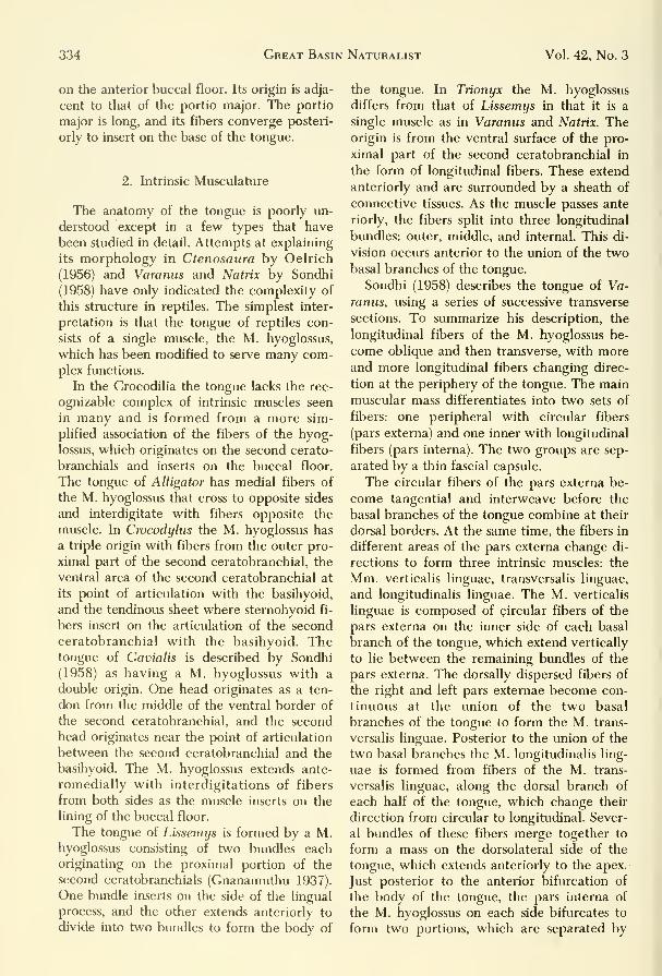
334 Great Basin Naturalist Vol. 42, No. 3
on the anterior buccal floor. Its origin is adja-
cent to that of the portio major. The portio
major is long, and its fibers converge posteri-
orly to insert on the base of the tongue.
2. Intrinsic Musculature
The anatomy of the tongue is poorly un-
derstood except in a few types that have
been studied in detail. Attempts at explaining
its morphology in Ctenosaura by Oelrich
(1956) and Varanus and Matrix by Sondhi
(1958) have only indicated the complexity of
this structure in reptiles. The simplest inter-
pretation is that the tongue of reptiles con-
sists of a single muscle, the M. hyoglossus,
which has been modified to serve many com-plex functions.
In the Crocodilia the tongue lacks the rec-
ognizable complex of intrinsic muscles seen
in many and is formed from a more sim-
plified association of the fibers of the hyog-
lossus, which originates on the second cerato-
branchials and inserts on the buccal floor.
The tongue of Alligator has medial fibers of
the M. hyoglossus that cross to opposite sides
and interdigitate with fibers opposite the
muscle. In Crocodylus the M. hyoglossus has
a triple origin with fibers from the outer pro-
ximal part of the second ceratobranchial, the
ventral area of the second ceratobranchial at
its point of articulation with the basihyoid,
and the tendinous sheet where sternohyoid fi-
bers insert on the articulation of the second
ceratobranchial with the basihyoid. Thetongue of Gavialis is described by Sondhi
(1958) as having a M. hyoglossus with a
double origin. One head originates as a ten-
don from the middle of the ventral border of
the second ceratobranchial, and the second
head originates near the point of articulation
between the second ceratobranchial and the
basihyoid. The M. hyoglossus extends ante-
romedially with interdigitations of fibers
from both sides as the muscle inserts on the
lining of the buccal floor.
The tongue of Lissemys is formed by a M.hyoglossus consisting of two bundles eachoriginating on the proximal portion of the
second ceratobranchials (Gnanamuthu 1937).
One bundle inserts on the side of the lingual
process, and the other extends anteriorly to
divide into two bundles to form the body of
the tongue. In Trionyx the M. hyoglossus
differs from that of Lissemys in that it is a
single muscle as in Varanus and Matrix. Theorigin is from the ventral surface of the pro-
ximal part of the second ceratobranchial in
the form of longitudinal fibers. These extend
anteriorly and are surrounded by a sheath of
connective tissues. As the muscle passes ante
riorly, the fibers split into three longitudinal
bundles: outer, middle, and internal. This di-
vision occurs anterior to the union of the twobasal branches of the tongue.
Sondhi (1958) describes the tongue of Va-
ranus, using a series of successive transverse
sections. To summarize his description, the
longitudinal fibers of the M. hyoglossus be-
come oblique and then transverse, with moreand more longitudinal fibers changing direc-
tion at the periphery of the tongue. The mainmuscular mass differentiates into two sets of
fibers: one peripheral with circular fibers
(pars externa) and one inner with longitudinal
fibers (pars interna). The two groups are sep-
arated by a thin fascial capsule.
The circular fibers of the pars externa be-
come tangential and interweave before the
basal branches of the tongue combine at their
dorsal borders. At the same time, the fibers in
different areas of the pars externa change di-
rections to form three intrinsic muscles: the
Mm. verticalis linguae, transversalis linguae,
and longitudinalis linguae. The M. verticalis
linguae is composed of circular fibers of the
pars externa on the inner side of each basal
branch of the tongue, which extend vertically
to lie between the remaining bundles of the
pars externa. The dorsally dispersed fibers of
the right and left pars externae become con-
tinuous at the union of the two basal
branches of the tongue to form the M. trans-
versalis linguae. Posterior to the union of the
two basal branches the M. longitudinalis ling-
uae is formed from fibers of the M. trans-
versalis linguae, along the dorsal branch of
each half of the tongue, which change their
direction from circular to longitudinal. Sever-
al bundles of these fibers merge together to
form a mass on the dorsolateral side of the
tongue, which extends anteriorly to the apex.
Just posterior to the anterior bifurcation of
the body of the tongue, the pars interna of
the M. hyoglossus on each side bifurcates to
form two portions, which are separated by

September 1982 Tanner, Avery: Buccal Floor of Reptiles 335
some of the bundles and the M. verticalis
linguae.
Each prong terminates with the dimin-
ishing of the longitudinal and circular bun-
dles and the insertion of their obliquely di-
rected fibers on the epithelium of the tongue.
Natrix (Xenochrophis) has been described
by Sondhi (1958) as having a M. hyoglossus
similar to that of the lizard Varanus. In Nat-
rix the M. hyoglossus envelopes the second
ceratobranchial at each side of the origin.
Unlike that in Varanus, the M. hyoglossus of
Natrix becomes ventromedial to the cerato-
branchial and combines with its opposite
member far posterior to the basihyoid. The
M. hyoglossus also divides into parts externa
and interna, but in the substance of the
tongue rather than at its base as in Varanus.
A number of longitudinal fibers of the M.
hyoglossus separate from the rest of the pars
externa at the periphery of the tongue to
form the partes externa and interna. This
change in direction of the fibers is directly
associated with the formation of the Mm.verticalis linguae, transversalis linguae, and
longitudinalis linguae the same as in Varanus. The only difference is that the fibers
of the M. longitudinalis linguae are formed
more anteriorly in the body of the tongue in
Natrix than in Varanus.
Varkey (1979) describes the tongue of Ne-
rodia cyckrprion as being formed of intrinsic
tongue muscles and the M. hyoglossus. Hedescribes the M. hyoglossus as long, slender,
paired retractor muscles making up the bulk
of the tongue. They arise from the medial
edge of the posterior tips of the ceratobran-
chials of the hyoid, pass rostrally, laterally,
and ventrally to the intrinsic tongue muscles,
and are pressed so closely together with themas to be almost indistinguishable. The hyog-
lossus muscles attach to the hyoid comua, the
tongue sheath, the oral mucosa, the fascia
medial, and just posterior to the lateral sub-
lingual glands.
The intrinsic musculature of the tongue of
Liduinura roseofusca has been described byHershkowitz (1941) as consisting of five dis-
tinct bimdles. In the posterior part of the
tongue all but the M. verticalis are present.
The M. transversus inferioris forms a sheet on
the ventral side of the tongue extending dor-
sally along the lateral side to meet the ven-
tral extension of the M. transversus
superioris.
The M. transversus superioris occupies
most of the dorsal part of the tongue deep to
the superficial muscle, the M. lingualis,
which is restricted to the most dorsal muscu-
lar layer of the free, unforked part of the
tongue.
The M. verticalis forms a midsaggital ling-
ual septum, thin toward the anterior and
thick at the posterior end. The fibers of the
M. verticalis run at right angles to those of
the Mm. t. superioris and t. inferioris. Dor-
sally its bundles interweave with those of the
M. lingualis.
The ceratoglossus muscles form a pair of
central muscles extending the entire length of
the organ and forming most of the cross sec-
tion of the tongue.
Posterior to the bifurcation of the tongue
into terminal prongs, the Mm. verticalis ling-
uae and transversalis linguae intersect at
right angles. Thus, in section the tongue can
be divided into four quarters composed of
bundles of the M. longitudinalis linguae and
the pars interna.
At the anterior tip of the tongue, a dorsal
and a ventral notch occur medially. The dor-
sal notch deepens to separate the bases of the
terminal prongs. At this point the bundles of
the M. longitudinalis linguae of each side di-
vide into smaller bundles and intermingle an-
teriorly toward the tips of the prongs to ter-
minate in the connective tissues of the lingual
epithelium (Fig. 26).
The M. hyoglossus in Chamaeleo brevi-
cornis originates on the tip of the distal end
of the first ceratobranchial. A small cartila-
ginous knob on the end of the ceratobran-
chial, which appears to be a remnant of the
epibranchial, also serves as a point of origin
for many fibers. The first and second comuaextend anterolaterally from the basihyal;
therefore the M. hyoglossus, in its contracted
position, extends from its origin medially to
the lingual process, where it makes a right
angle turn to follow the lingual process into
the tongue and to its insertion in the con-
nective tissue surrounding the tongue. Uponreaching the tongue, the M. hyoglossus di-
vides into the two sections described by Son-
dhi (1958) as the pars externa and a medial
longitudinal part, the pars interna. A series of
circular fibers, which are a part of the sheath.
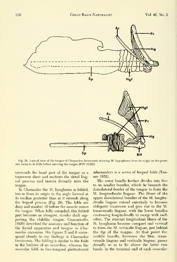
336 Great Basin Naturalist Vol. 42, No. 3
Fig. 28. Lateral view of the tongue of Chamaelon brevicarnis showing M. hypoglossus from its origin on the poste-
rior comu to its folds before entering the tongue (BYU 12422).
surroLuids the basal part of the tongue as a
transverse sheet and encloses the distal ling-
ual process and inserts dorsally into the
tongue.
In Chamaeleo the M. hyoglossus is folded,
less so from its origin to the angle formed at
its median posterior than as it extends along
the lingual process (Fig. 28). The folds are
deep and number 10 before the muscle enters
the tongue. When fully extended, this folded
part becomes an elongate, slender shaft sup-
porting the clublike tongue. Gnanamuthu(1930) described the anatomy and function of
the hyoid apparatus and tongue in Cha-maeleo cacaratus. His figures 5 and 6 corre-
spond closely to our findings in Chamaeleobrevicomis. The folding is similar to the folds
in the bellows of an accordian, whereas the
muscular folds in free-tongued plethodontid
salamanders is a series of looped folds (Tan-
ner 1952).
The outer bundle further divides into five
to six smaller bundles, which lie beneath the
dorsolateral border of the tongue to form the
M. longitudinalis linguae. The fibers of the
upper dorsolateral bundles of the M. longitu-
dinalis linguae extend anteriorly to becomeobliquely transverse and give rise to the M.transversalis linguae, with the lower bundles
continuing longitudinally to merge with each
other. The internal longitudinal fibers of the
M. hyoglossus become compact and vertical
to form the M. verticalis linguae, just behind
the tip of the tongue. At that point the
middle bundle, between the Mm. trans-
versalis linguae and verticalis linguae, passes
dorsally so as to lie above the latter two
bands. In the terminal end of each muscular

September 1982 Tanner, Avery: Buccal Floor of Reptiles 337
CBl
Fig. 29. Ventral view of hyoid apparatus and associated structures: A, Crotalus v. lutosus; B, Pituophis m. desert-
icola. HG—m. hyoglossus, t—tongue.
prong extending into the tongue from each In Figure 29 the general structural rela-
side, the various bundles dwindle and insert tionships of the hyoid, tongue and M. hyo-
in the subepithelial connective tissue of the glossus are depicted for the genera Crotalus
tongue. «ind Pituophis.

338 Great Basin Naturalist Vol. 42, No. 3
In summary, the intrinsic muscles of the
tongue are actually fibers of the hyoglossal
muscles that extend in varying directions.
Unfortunately, the remainder of our knowl-
edge of the tongue and related structures is
incomplete. Many structures such as lingual
glands, glottis, trachea, and their associated
muscles and nerves have not been fully in-
vestigated in all groups. Zug (1971) has stud-
ied the arterial patterns in iguanids, Winokur(1974) has studied the buccal mucosae in tur-
tles, and Schumacher (1973) has examined
the hyolaryngeal muscles and skeleton in tur-
tles and crocodilians.
VIII. Innervation of Buccal FloorMusculature
A. General
The innervation of muscles in reptiles has
been generally neglected, and for this reason
it is difficult to homologize their detailed
musculature. Detailed descriptions of the
nerve patterns in the buccal floor of reptiles
are available from the following workers: Os-
awa (1898), Watkinson (1906), Reese (1915),
Willard (1915), Poglayen-Neuwall (1953,
1954), Oelrich (1956), Schumacher (1956,
1973), Sondhi (1958), and Rieppel (1978,
1981). Soliman (1964) describes and figures
the nerves in the head of Chelydra serpentina
and provides colored plates depicting the
nerves entering the muscles associated withthe buccal floor and the tongue. Islam (1955)
and Islam and Ashig (1972) describe the cra-
nium and cranial nerves of Uromastyx hard-
wicki, and Renous-Lecru (1972) discusses the
branchial plexus in Agama and Chalarodon.All these workers indicate that in reptiles
the IXth (glossopharyngeal), Xth (vagus),
Xlth (spinal accessory), and Xllth (hyoglossal)
cranial nerves usually occur in close associ-
ation and form a glossohyoidean plexus.
Some uniformity does exist in the innervation
of the throat muscles of reptiles, as demon-strated by the fact that in all reptiles the Vth(trigeminal) cranial nerve innervates the M.mylohoideus anterior, the Vllth (facial) in-
nervates the Mm. mylohyoideus posterior
and constrictor colli, and the Xllth (hypog-lossal) and anterior spinal nerves innervate
the M. constrictor colli.
B. Cranial Nerves
Oelrich (1956) presented a clear picture of
the pattern of cranial nerves in the iguanid
Ctenosaura. He found the following nerves
innervating muscles of the buccal floor. Asimilar pattern in all cases has been described
for Anolis by Willard (1915), for the trige-
minal in turtles by Poglayen-Neuwell (1953),
and for Varanus by Watkinson (1906).
1. N. trigeminus: A branch of the trige-
minal nerve (ramus ad musculum mylohyoi-
deum) passes through the posterior mylo-
hyoid foramen to enter the lateral fibers of
the first mandibulohyoid muscle and termi-
nates anteriorly on the M. intermandibularis
posterior. A second branch, the anterior
mylohyoid nerve, emerges on the medial side
of the mandible from the anterior mylohyoidforamen of the splenial bone to pass over the
M. mandibulohyoideus I to enter the ventral
surface of the M. intermandibularis anterior.
A section of the mandibular ramus continues
anteriorly to the lingual ramus of the hypog-lossal nerve, where the latter passes through
the anterior inferior alveolus foramen of the
dentary to divide into two branches. The an-
terior glandular branch passes the ventral
surface of the M. intermandibularis anterior,
whereas the posterior branch enters the Mm.intermandibularis anterior and genioglossus.
2. N. facialis: The facial nerve divides into
a hyoid ramus that innervates a part of the
M. intermandibularis that inserts on the ret-
roarticular process of the mandible. It also in-
nervates the M. constrictor colli and the pos-
terior border of the M. intermandibularis.
3. N. glossopharyngealis: The M. hyog-
lossus is innervated by a ramus formed frombranches of the glossopharyngeal and hypog-lossal nerves.
4. N. hypoglossalis: There are four small
ventral branches of the hypoglossal nerve
that innervate the M. mandibulohyoideus I.
The hypoglossal divides into three mainbranches at the point where the Mm. gen-
ioglossus and hyoglossus join. These branches
include the ramus lingualis lateralis, whichextends anterolaterally to enter the insertion
of the M. genioglossus and medial and lateral
areas of the M. genioglossus. It next emerges
to join the lingual ramus of the trigeminal

September 1982 Tanner, Avery: Buccal Floor of Reptiles 339
nerve, which then enters the tongue. The re-
maining two branches, intermediahs and me-
diahs, go directly to the tongue, where they
innervate its musculature.
Watkins6n (1906) described the nerve pat-
terns seen in Varanus and found the
following:
a. N. trigeminus: There are three main
branches of the trigeminal nerve (rami op-
thalmicus, maxillaris, and mandibularis);
however, only the ramus mandibularis goes
to the buccal floor, where it has three
branches.
The first branch, the ramus ad musculum
mylohyoideus, originates from that part of
the ramus mandibularis (portio alveolaris in-
ferior) that lies within the alveolar surface of
the dentary. It emerges to proceed posteri-
orly, with branches going to the Mm. mylo-
hyoidei posterior as profundus and
superficialis.
The second branch, the ramus muscularis
et glandularis, also arises from the portio al-
veolaris inferior of the mandibular ramus. Abranch extends to the Mm. mentalis super-
ficialis, mentalis profundus anterior, and
mentalis profimdus posterior. It also enters
the portiones major and minor of the M.
genioglossus.
A third branch, the ramus lingualis, origi-
nates from the ramus mandibularis before the
latter enters the alveolar canal. This branch
emerges from the canal to pass along the
ventral buccal floor, where it joins the ramus
lingualis anterior of the hypoglossal nerve. It
enters the lingual sheath and then the tongue,
extending to the anteriormost extremity of
the terminal prongs to innervate, with the
hypoglossal nerve, the bundles of the M.hyoglossus.
2. N. facialis: The facial nerve emerges
from the cranium and divides into an anterior
branch, the ramus palatinus, and a posterior
branch, the ramus hyomandibularis. Tlie lat-
ter branch extends posteriorly as the ramus
hyoideus to innervate the Mm. geniolateralis
and constrictor colli.
3. N. hypoglossus: The hypoglossal nerve
extends obliquely posterior along the dorsal
side of the neck to the buccal floor, where it
divides into two branches, each of which fur-
ther subdivide into two branches. One branch
forms the rami ad musculum geniotrachealis,
and the second branch gives rise to the rami
linguales anterior and posterior.
The ramus ad musculum geniohyoideum
extends obliquely over the M. ceratohyoideus
to form two branches that innervate the mid-
dorsal region of the M. geniohyoideus and
lateral surface of the M. constrictor colli,
respectively.
The ramus ad musculum ceratohyoideum
et musculum mandibulotrachealis extends
from the M. interportalis to the M. cerato-
hyoideus, innervating these and also sending
branches to the Mm. cornuohyoideus and
mandibulotrachealis.
The third branch (ramus lingualis anterior)
originates from the hypoglossal nerve and ex-
tends along the lateral border of the tongue
to eventually anastomose with the ramus
lingualis of the mandibular ramus of the
trigeminal nerve. As it does so, it sends
branches to the sublingual glands and termi-
nates in the M. genioglossus. A small branch
also extends to both the Mm. gen-
ioceratoideus and mandibuloproximalis.
The ramus ad musculum mandibulohyoi-
deum is derived from the hypoglossal nerve
before the branching of the ramus lingualis
posterior. It innervates the M.mandibulohyoideus.
Two other branches derived from the hy-
poglossal (lingual accessorii) innervate the
posterior part of the base of the tongue. A fi-
nal branch, the ramus lingualis posterior, is
the terminal portion of the hypoglossal
nerve. It also innervates the basal area of the
tongue.
Some information is available for other liz-
ards such as Chamaeleo and Calotes (Gnana-
muthu 1937), in which the formation of the
lingual nerve varies. The lingual branch of
the hypoglossal in Chamaeleo is separated
from the glossohyoidean plexus and forms
two branches, the rami linguale lateralis and
medialis. The ramus lingualis lateralis extends
posteriorly to innervate the M. genioglossus,
and the main branch anastomoses with the
lingual branch of the Vth cranial nerve; to-
gether they penetrate the M. hyoglossus and
join the ramus lingualis medialis that enters
and innervates the M. hyoglossus. This same
branch in the anterior region of the buccal
floor unites with the combined lingual
branch and with it also enters the tongue. In

340 Great Basin Naturalist Vol. 42, No. 3
Calotes the lingual branch of the hyoglossal
nerve extends one branch to the M. gen-
ioglossus and one to the tongue. The mainbranch unites with a ramus of the trigeminal
to penetrate the tongue and there subdivides
into many branches for innervation of the
tongue muscles.
Sondhi (1958) gives the following nerve
pattern for the buccal floor of Matrix (Xe-
nochrophis), a natricid snake:
1. N. trigeminus: The ramus mandibularis
of the trigeminal nerve sends three branches
to the buccal floor. The first branch (ramus
ad musculum mylohyoideum) originates from
the mandibular ramus immediately after the
latter enters the alveolar canal of the dentary
as the portio alveolaris inferior. It divides
into two branches, one innervating the M.mylohyoideus posterior profundus and the
other the M. mylohyoideus posterior.
A second branch, the ramus muscularis et
glandularis, originates from the portio alveol-
aris inferior of the mandibular ramus. After
emerging from the mandible, it extends me-dially to provide branches for the Mm. inter-
maxillaris, genioglossus portio major, men-talis profundus anterior, and mentalis
profimdus posterior.
The third branch (ramus lingualis) arises
from the portio alveolaris inferior of the
mandibular ramus after the mandibularis et
glandularis. It unites with the ramus lingualis
of the hyoglossal nerve and extends medially
to the lingual sheath and M. hyoglossus.
2. N. facialis: The facial nerve emergesfrom the foramen prooticum and extends to
the M. mylohyoideus posterior as the ramushyomandibularis, which has two branches to
that muscle.
3. N. hypoglossal: The hypoglossal nervehas three main branches, including the ramusdescendens that originates as a thin branchextending posteriomedially to innervate the
ventral surface of the Mm. omohyoideus,sternohyoideus, and sternothyroideus.
The second branch is the main stem of the
hypoglossal nerve, which forms the ramuslingualis posterior. It extends forward as twobranches, one entering the body and theother the base of the tongue.
The third branch, the ramus ad musculumgeniolateralis, originates in the hypoglossalnerve almost opposite the ramus lingualis
posterior and innervates the M. gen-iolateralis. Distally the hypoglossal bifurcates
into two branches, an inner ramus ad muscu-lum mandibulotrachealis and an outer ramusad musculum geniohyoideum. The inner divi-
sion extends anteriorly and medially to in-
nervate the posterior part of the M. man-dibulotrachealis. The outer branch extends
anterolaterally to form two branches that in-
nervate the M. geniohyoideus.
Langebartel (1968) has summarized the in-
nervation of the muscles of the buccal floor
in other snakes. The mandibular division of
the trigeminal nerve innervates the M. inter-
mandibularis and parts of the tongue. The fa-
cial innervates part of the Mm. constrictor
colli and the cervicomandibularis and sends
some branches to the tongue. Some branches
from glossopharyngeal and the vagus in-
nervate the M. ceratomandibularis, but only
one branch innervates the M. hyotrachealis.
The hypoglossal nerve innervates the Mm.geniohyoideus, ceratomandibularis, and ster-
nohyoideus. The Mm. genioglossus and hy-
poglossus are innervated by an anterior
branch of the hypoglossal nerve. Very com-monly the glossopharyngeal, vagus, and hy-
poglossal nerves combine to innervate the
lingual sheath and the Mm. genioglossus andhypoglossus. The hypoglossal may also haveanterior and posterior branches that enter the
tongue. Last, an anterior branch of the hypo-glossal unites with a branch of the trigeminal
to innervate the Mm. genioglossus and gen-
iotrachealis. Varkey (1979) describes the in-
nervation of muscles in Nerodia, but does not
attempt to identify the nerves.
Soliman (1964) describes and figures the
cranial nerves of Chelydra serpentina. Col-
ored plates depict the various nerves entering
the muscles associated with the buccal floor
and the tongue.
Trionyx has been described by Sondhi
(1958), who indicates the existence of the fol-
lowing nerve pattern, comparable to that re-
ported for Chelydra:
1. N. trigeininus: The mandibular ramus of
the trigeminal nerve has two branches, in-
cluding the ramus ad musculum mylohyoi-
deum that arises in Varantis and Matrix, andthe ramus lingualis. The former branch ex-
tends posteriorly along the medial side of the
mandible to divide into two branches, one of
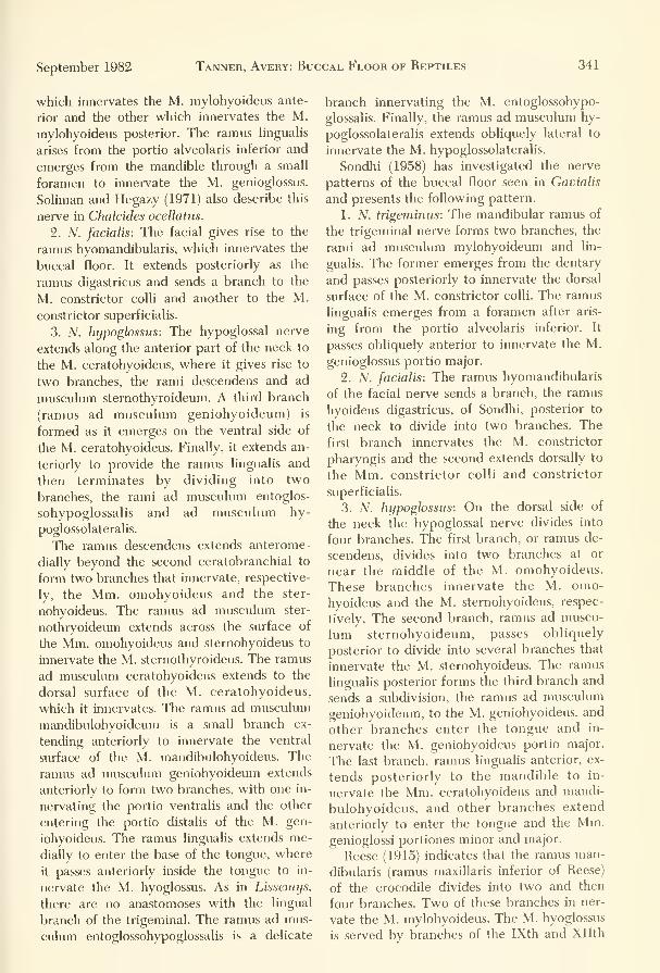
September 1982 Tanner, Avery: Buccal Floor of Reptiles 341
which innervates the M. mylohyoideus ante-
rior and the other which innervates the M.
mylohyoideus posterior. The ramus hnguaUs
arises from the portio alveolaris inferior and
emerges from the mandible through a small
foramen to innervate the M. genioglossus.
Sohman and Hegazy (1971) also describe this
nerve in Chalcides ocellatus.
2. N. facialis: The facial gives rise to the
ramus hyomandibularis, which innervates the
buccal floor. It extends posteriorly as the
ramus digastricus and sends a branch to the
M. constrictor colli and another to the M.
constrictor superficialis.
3. iV. hypoglossus: The hypoglossal nerve
extends along the anterior part of the neck to
the M. ceratohyoideus, where it gives rise to
two branches, the rami descendens and ad
muscukun stemothyroideum. A third branch
(ramus ad musculum geniohyoideum) is
formed as it emerges on the ventral side of
the M. ceratohyoideus. Finally, it extends an-
teriorly to provide the ramus lingualis and
then terminates by dividing into two
branches, the rami ad musculum entoglos-
sohypoglossalis and ad musculum hy-
poglossolateralis.
The ramus descendens extends anterome-
dially beyond the second ceratobranchial to
form two branches that innervate, respective-
ly, the Mm. omohyoideus and the ster-
nohyoideus. The ramus ad musculum ster-
nothryoideum extends across the surface of
the Mm. omohyoideus and sternohyoidevis to
innervate the M. sternothyroideus. The ramus
ad musculum ceratohyoideus extends to the
dorsal surface of the M. ceratohyoideus,
which it innervates. The ramus ad musculum
mandibulohyoideum is a small branch ex-
tending anteriorly to innervate the ventral
surface of tlie M. mandibulohyoideus. The
ramus ad musculum geniohyoideum extends
anteriorly to form two branches, with one in-
nervating the portio ventralis and the other
entering the portio distalis of the M. gen-
iohyoideus. The ramus lingualis extends me-
dially to enter the base of the tongue, where
it passes anteriorly inside the tongue to in-
nervate the M. hyoglossus. As in Lissemys,
there are no anastomoses with the lingual
branch of the trigeminal. The ramus ad mus-
culum entoglossohypoglossalis is a delicate
branch innervating the M. entoglossohypo-
glossalis. Finally, the ramus ad musculum hy-
poglossolateralis extends obliquely lateral to
innervate the M. hypoglossolateralis.
Sondhi (1958) has investigated the nerve
patterns of the buccal floor seen in Gavialis
and presents the following pattern.
1. N. trigeminus: The mandibular ramus of
the trigeminal nerve forms two branches, the
rami ad musculum mylohyoideum and lin-
gualis. The former emerges from the dentary
and passes posteriorly to innervate the dorsal
surface of the M. constrictor colli. The ramus
lingualis emerges from a foramen after aris-
ing from the portio alveolaris inferior. It
passes obliquely anterior to innervate the M.
genioglossus portio major.
2. N. facialis: The ramus hyomandibularis
of the facial nerve sends a branch, the ramus
hyoideus digastricus, of Sondhi, posterior to
the neck to divide into two branches. The
first branch innervates the M. constrictor
pharyngis and the second extends dorsally to
the Mm. constrictor colli and constrictor
superficialis.
3. N. hypoglossus: On the dorsal side of
the neck the hypoglossal nerve divides into
four branches. The first branch, or ramus de-
scendens, divides into two branches at or
near the middle of the M. omohyoideus.
These branches innervate the M. omo-
hyoideus and the M. sternohyoideus, respec-
tively. The second branch, ramus ad muscu-
lum sternohyoideum, passes obliquely
posterior to divide into several branches that
innervate the M. sternohyoideus. The ramus
lingualis posterior forms the third branch and
sends a subdivision, the ramus ad musculum
geniohyoideum, to the M. geniohyoideus, and
other branches enter the tongue and in-
nervate the M. geniohyoideus portio major.
The last branch, ramus lingualis anterior, ex-
tends posteriorly to the mandible to in-
nervate the Mm. ceratohyoideus and mandi-
bulohyoideus, and other branches extend
anteriorly to enter the tongue and the Mm.genioglossi portiones minor and major.
Reese (1915) indicates that the ramus man-
dibularis (ramus maxillaris inferior of Reese)
of the crocodile divides into two and then
four branches. Two of these branches in ner-
vate the M. mylohyoideus. The M. hyoglossus
is served bv branches of the IXth and Xllth

342 Great Basin Naturalist Vol. 42, No. 3
nerves. The hypoglossal nerve also sends
branches to the Mm. omohyoideus, ster-
nohyoideus, geniohyoideus and ge-nioglossus.
B. Spinal Nerves
Oelrich (1956) reports that in Ctenosaura
the first spinal nerve innervates the ventral
part of the M. omohyoideus and the dorsal
part of the stemohyoideus. Sondhi (1958) in-
dicates that in Varanus and Matrix the united
stems of the first and second spinal nerves
anastomose with the hypoglossal nerve andextend posteriorly in the neck to send small
branches to the Mm. stemohyoideus and ster-
nothyroideus and a large branch to the M.omohyoideus. Some of the succeeding spinal
nerves also innervate the M. constrictor colli.
In Natrix, as in Varanus, the first and sec-
ond spinal nerves innervate parts of the M.constrictor colli. In some other snakes manyspinal nerves innervate the Mm. neuroman-dibularis, costomandibularis, costo-cutanei in-
ferior and superior, omohyoideus, ster-
nohyoideus, and transverus branchialis.
In Trionyx, Gavialis, and Crocodylus the
united stem of the first and second spinal
nerves irmervates the M. constrictor colli,
whereas in Crocodylus numerous branches of
first, second, and third spinal nerves in-
nervate the smaller ventral muscles of the
neck.
IX. Discussion
An examination of the preceding descrip-
tions show that the information on the hyoidand associated structures was widely scat-
tered and incomplete. Although morphologyis one of the oldest branches of biology, there
is an absence of complete accounts of the
gross anatomy of the buccal floor of reptiles
as a class. Similar gaps in our knowledge exist
for other anatomical areas of the reptilian
body. In spite of our acceptance of some rep-
tilian ancestral stocks as being the lines of de-
scent for birds and mammals, anatomists havenot vigorously pursued studies to show phy-logenetic relationships. The lack of a com-plete understanding of these groups is as-
tounding considering the importantphylogenetic position of reptiles.
Despite the lack of information, some gen-
eralizations can be made. As indicated bySondhi (1958), the buccal floor in many rep-
tiles has three functions: (1) it participates in
the act of inspiration and expiration, (2) it
aids in the capture and the deglutition of
food, and (3) it provides the mechanisms of
tongue movement. To Sondhi's list should beadded two additional functions: (4) behav-
ioral display and (5) sensory reception.
The important role of the buccal area as a
respiratory throat pump has been explored
by Gnanamuthu (1937), who demonstrated
the part played in Hemidactylus. He states.
The contraction of the thorax expeUing air would result
in the inflation of the buccal cavity, and when next the
thorax relaxes this impure air may be taken into the
lungs again, because the thoracic contraction and expan-
sion follows each other so rapidly. However, the eleva-
tion of the rnouth floor and tongue through the aid of
transverse and hyoid muscles just when the thorax con-
tracts serves to expel the vitiated air effectively out of
the body.
Respiratory mechanisms in reptiles vary
widely. Calotes utilizes the limbs of the ante-
rior cornua and the attached muscles to ac-
tively raise and lower the throat. The posi-
tions of cornua and ceratobranchials andassociated muscles in Varanus indicate a
change of the volume of the throat caused bydilation and compression of the floor of the
mouth.
Among the testudines, the posterior part of
the M. hyoglossus and the entire Mm. cerato-
hyoideus, entoglossohypoglossalis, and hypo-
glossolateralis utilize the jointed basihyoid
and hypoglossum to move the throat up anddown as one solid piece. Although these
structures may not be important in respira-
tion (Mitchell and Morehouse 1863), there is
reason to believe that both aquatic and ter-
restrial turtles pump the throat to exchangewater and air in the nasal canals and buccal
cavity for sensory functions (McCutcheon1943). In Figure 19 we attempt to reproduce
the extensive fimbriations on the bucco-pharyngeal floor of Trionyx. The total func-
tion of these numerous filaments may not be
fully understood, but seemingly they are im-
portant in aquatic respiration (Girgis 1961).
In snakes, inspiration and expiration are
accomplished by the muscles of the bodywall compressing the lungs for expiration and
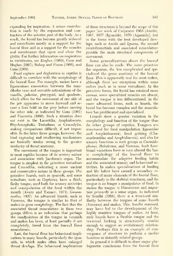
September 1982 Tanner, Avery: Buccal Floor of FIeptiles 343
expanding for inspiration. A minor contribu-
tion is made by the expansion and con-
traction of the anterior part of the body. As a
result, the hyoid has become greatly reduced
and contributes mostly as a support for the
buccal floor and as a support for the muscles
and membranes that open and close the
glottis. For further information on respiration
in vertebrates, see Hughes (1963), Gans and
Hughes (1967), Bishop and Foxon (1968), and
Gans (1969).
Food capture and deglutition in reptiles is
difficult to correlate with the morphology of
the buccal floor. For example, snakes have a
ligamentous connection between the man-
dibular rami and movable articulations of the
maxilla, palatine, pterygoid, and quadrate,
which allow for the movement of one side of
the jaw apparatus to move forward and se-
cure a firm hold on the prey before moving
the other side, as indicated by Gans (1961)
and Frazzetta (1966). Such a situation does
not exist in the Lacertilia, Amphisbaenia,
Rhychocephalia, Testudines, or Crocodylia,
making comparisons difficult, if not impos-
sible. In the latter three groups, however, the
food capturing and swallowing mechanisms
are basically similar owing to the greater
similarity of throat anatomy.
The movement of the tongue is important
in most reptiles because of its sensory nature
and association with Jacobson's organ. Thetongue is simplest in the primitive testudines
and Crocodilia, indicating a more ancient
and conservative nature in these groups. The
primitive lizards, such as iguanids, and some
testudines, such as Gopherus, have a thick,
fleshy tongue, used both for sensory activities
and manipulation of the food within the
mouth (Avery and Tanner, 1971; Gnana-muthu, 1937). An advanced lizards, such as
Varanus, the tongue is similar to that of
snakes in gross morphology. The fact that the
associated throat musculature in these two
groups differs is an indication that perhaps
the manipulation of the tongue in varanids
and snakes has been, at least partially if not
completely, freed from the buccal floor
musculature.
Last, the buccal floor has behavioral impli-
cations in many lizards, particularly the igua-
nids, in which males often have enlarged
throat dewlaps. The behavioral implications
of these structures is beyond the scope of this
paper [see work of Carpenter 1965 (Anolis),
1967, 1977 (Iguanids), 1970 (Agamids)], but
in the forms with the best developed dew-
laps, such as Anolis and Iguana, the second
ceratobranchials and associated musculature
provide the main structural components of
movement.
Some generalizations about the buccal
floor can also be made. The more primitive
the organism, the less complicated and spe-
cialized the gross anatomy of the buccal
floor. This is apparently true for most orders,
although there are exceptions within some
orders (such as in some testudines). In the
primitive forms, the hyoid has retained more
cornua, some specialized muscles are absent,
and the tongue is less differentiated. In the
more advanced forms, such as lizards, the
hyoid has become complex and the muscula-
ture has proliferated and specialized.
Lizards show a greater variation in the
morphology and function of the tongue than
do other groups of reptiles. Tongues are
structured for food manipulation (Iguanidae
and Amphisbaenia), food getting (Cha-
maeleonidae and Amphisbaenia), and also for
sensory functions in such groups as Cnemido-
phorus, Heloderma, and Varanus. Such func-
tional variations have in turn altered the bas-
ic morphology of the buccal floor to
accommodate the adaptive feeding habits
and the associated sensory and behavioral ac-
tivities. In snakes specializations of feeding
and life habits have caused a secondary re-
duction of many elements of the buccal floor,
particularly in the skeletal structures, and the
tongue is no longer a manipulator of food. In
snakes the tongue is filamentous and impor-
tant primarily as a sense organ. As indicated
by Sondhi (1958), there is a structural sim-
ilarity between the tongues of some lizards
(Varanus) and snakes. This, Sondhi reasoned,
may have led to the development of the
highly sensitive tongues of snakes. At least,
such lizards have a flexible tongue and the
terminal forking is structurally similar
enough to suggest an evolutionary relation-
ship. Perhaps this is an example of con-
vergence of structure to perform a similar
function in distantly related groups.
In general it is difficult to draw major phy-
logenetic conclusions from the buccal floor

344 Great Basin Naturalist Vol. 42, No. 3
because the scope of such a study is neces-
sarily hmited to one specialized area and can
be misleading. When hyoid elements are lost,
the associated muscles are also lost or maybecome unrecognizable. Thus the implication
of presence or absence of structures is also
misleading. Future morphological phyloge-
netic studies in the area of the buccal floor
should be supplemented by embryological in-
formation, as indicated by DeBeer (1930,
1951) and Edgeworth (1935). Such research
will provide clues as to which structures have
been lost, fused, readapted or never possessed
by an organism.
Acknowledgments
We wish to thank those who have aided us
during the preparation of this study, particu-
larly our own institutions who have provided
space and secretarial help and have permit-
ted us to dissect materials from their collec-
tions (Brigham Young University Life Science
Museum, BYU, and Southern Connecticut
State College, SCSC). We have also received
study materials from the following: Dr.
George R. Zug, U.S. National Museum; Dr.
Carl Cans, University of Michigan; Dr. AlanE. Leviton, California Academy of Sciences;
Dr. Richard G. Zweifel, American Museumof Natural History, and Mr. Jose P. Rosado,
Museimi of Comparative Zoology. We re-
ceived, on exchange, specimens from Dr. F.
H. Rehmani, Zoologische Museum, Kanpur,India. These were very helpful as com-parative materials, and for their use we are
indeed grateful.
We have used freely skeletal and dissected
material used in previous studies at our labo-
ratories. These have served us not only as
source materials, but also for the comparativestudies.
We are grateful to Dr. Robert M. Wino-kur, who read the manuscript and providedhelpful suggestions in those areas where Che-lonians are discussed.
In the preparation of this study we haverelied heavily on the studies of Gnanamuthu(1937), Langebartel (1968), and Sondhi(1958). Because these are basic studies deal-
ing in some detail with the buccal area, werefer to them repeatedly. Without them this
study would have been extremely difficult.
Bibliography and Literature Cited
Adams, L. A. 1919. A memoir on the phylogeny of the
jaw muscles in recent and fossil vertebrates. Ann.
N.Y. Acad. Sci. 28:51-166.
1925. Correlations of the musculature and the
movements of the skull in Natrix, with some sug-
gestions of homology in the lacertilian. Morph.41:159-181.
Albright, R. G., and E. N. Nelson. 1959. Cranial kinet-
ics of the generalized colubrid snake Elaphe obso-
leta quadrivittata, I and II.J.
Morph.105:193-192.
Anthony,J.,
and R. G. Serra. 1949. Sur une particula-
rite remarguable del'appareil de la morsure chez
un serpent Aglyphe de I'Amerique tropicale,
Xenodon merremii. Rev. Brasil. Biol.
9(2): 153-160.
1950. Anatomic de I'appareil de la morsure chez
Ennectes rnurinus. Rev. Brasil. Biol. 10(l):23-24.
Ashley, L. M. 1955. Laboratory anatomy of the turtle.
Wm. C. Brown Company Publishers, Dubuque,49 pp.
Avery, D. F., and W. W. Tanner. 1964. The osteology
and myology of the head and thorax regions of
the obesiis group of the genus Sauromalus Dume-ril (Iguanidae). BYU Sci. Bull., Biol. Ser.
5(3): 1-30.
1971. Evolution of the iguanine lizards (Sauria,
Iguanidae) as determined by osteological andmyological characters. BYU Sci. Bull., Biol. Ser.
12(3): 1-79.
Auen, E. L., and D. a. Langebartel. 1977. The cranial
nerves of the colubrid snakes Elaphe and Tham-nophis.
J.Morph. 154(2):205-222.
Barrows, S., and H. M. Smith. 1947. The skeleton of
the lizard Xenosaurus grandis (gray). Univ. Kan-
sas Sci. Bull. 31(2,12):227-275.
Beddard, F. E. 1905a. Some notes on the cranial os-
teology of the Mastigure Wxard—Uromastix. Proc.
Zool. Soc. London 1905:1-9.
1905b. A contribution to the anatomy of the
frilled lizard (Chlamydosaunis kingi) and someother Agamidae. Proc. Zool. Soc. London1905:9-22.
1907. Contributions to the knowledge of the sys-
tematic arrangement and anatomy of certain gen-
era and species of Squamata. Proc. Zool. Soc,
London 1907:35-59.
Bellairs, a. d'A. 1950. Observations on the cranial
anatomy of Anniella and a comparison with that
of other burrowing lizards. Proc. Zool. Soc. Lon-
don. 119:887-904.
Bergman, R. A. M. 1961. The anatomy of some Vipe-
ridae. Acta Morph. Neerl-Scand 4(3): 195-230.
1965. The anatomy of Calamaria multipunctata
(Boie). Bull. Nat. Mus. Singapore 33(7):35-66.
Bishop, I. R., and G. E. H. Foxon. 1968. The mecha-nism of breathing in the South American lung-
fish, Lepidosiren paradox: a radiological study.J.
Zool. London. 154:263-271.
BissiNGER, B. E., AND C. A. SiMON. 1979. Comparison of
tongue extrusions in representatives of six fami-
lies of lizards.J.
Herpetology 13(2):113-139.

September 1982 Tanner, Avery: Buccal Floor of Reptiles 345
BoGERT, C. M. 1954. The systematic position of Lantha-
nottis and the affinities of the angiiinoniorphan
lizards. Bull. Amer. Mus. of Nat. Hist., Vol.
105(1).
BojANUs, L. H. 1819. Anatome testudinis europaeae.
1970 ed. Society for the Study of Amphibians and
Reptiles. Vol. VI, 179 pp.
BoLTT, R. E., AND R. F. Ewer. 1964. The functional
anatomy of the head of the puff adder, Bitis arie-
tans (Merr).J.Morph. 114:38-106.
Bradley, O. C. 1903. The muscles of mastication and
the movements of the skull in Lacertilia. Zool.
Fb., Abt. Anat. 18:475-488.
Branch, W. R. 1982. Hemipeneal morphology of the
platynotan lizards.J.
Herpetology, 16(1): 16-38.
Brock, G. T. 1938. The cranial muscles of the gecko, a
general account with a comparison of the mus-
cles in other Gnathostomes. Proc. Zool. Soc. Lon-
don, Series B. 180,761.
Bullock, R. E., and W. W. Tanner. 1966. A com-
parative osteological study of two species of
Colubridae {Pituophis and Thamnophis). BYU Sci.
Bull.. Biol. Ser. 8(3): 1-29.
Byerly, T. G. 1926. Tlie myology of Sphenodon. Univ.
Iowa Study Nat. Hist. 11.
Camp, C. L. 1923. Classification of the lizards. Bull.
Amer. Mus. Nat. Hi.st. 48(11):289-481.
Carpenter, C. C. 1965. The display of the Cocos Island
Anole. Herpetologica 21(4):256-260.
1967. Aggression and social structure in Iguanid
lizards. Univ. of Missouri Press, Columbia.
1977. The aggressive displays of three species of
South American iguanid lizards of the genus
Tropidurus. Herpetologica 33(3).
Carpenter, C. C J.A. Badham, and B. Kimble. 1970.
Behavior patterns of three species of Amphibo-
lurus (Agamidae). Copeia (3)497-505.
Chaine,J.
1902. Considerations sur la constitution mus-
culaire de la region sus-hyoidienne chez les ver-
tebres en general. Ann. Sci. Natur. 16:375-393.
Chiasson, R. B. 1962. Laboratory anatomy of the alliga-
tor. Wm. C. Brown Company Publishers, Du-
buque, 56 pp.
Cope, E. D. 1892. The osteology of the Lacertilia. Proc.
Amer. Philos. Soc. .30(1.38): 185-221.
1898(1900). Crocodiles, lizards, and snakes of
North America, .^nn. Rept. U.S. Nat. Mus., pp.
953-9,54.
Co.x, D. C, and W. W. Tanner. 1977. Osteology and
myology of the head and neck regions of Calli-
saiints, Cophosaurtis, Holbrookia, and Uma (Rep-
tilia: Iguanidae). Great Basin Nat. 37(l):.3.5-56.
Cowan, I. M., and W. B. M. Hick. 1951. A comparative
study of the mvology of the head region in three
.species of Tliamnophis (Reptilia, Ophibia). Trans.
Roy. Soc. Canada 45:19-60.
Criley, B. B. 1968. The cranial osteology of gerrhonoti-
form lizards. Amer. Midi. Nat. 80(1): 199-219.
Cundall, D. L. 1974. The cranial osteology amd myol-
ogy of the green snakes, genus Opheodn/s. Diss.
Abstr. Int. B. Sci. Eng. 35(6):3109-3110.
Davis, D. D. 1934. The collared lizard, a laboratory
guide. Macmillan, New York, 57 pp.
DeBeer, G. R. 1930. Embryology and evolution. Claren-
don Press, Oxford.
1937. The development of the vertebrate skull.
Clarendon Press, Oxford.
1951. Embrvos and ancestors. Clarendon Press,
Oxford.
DeVis, C. W. 1883. Morphology of Chlamijdosaurus
kingi. Proc. Linn. Soc. New South Wales.
1883:1-320.
DuDA, W. 1965. On the cranial osteology of Agama tit-
berciilata with a note on the comparative study
of the skulls in the family Agamidae. Vestn.
Ceshosl. Zool. Spalc. 29:156-174.
Dullemeijer, p. 1956. The fimctional morphology of the
head of the common viper, Vipera berus (Linn.)
Arch. Neerl. Zool. 12:387-497.
1959. A comparative functional-anatomical study
of the heads of some Viperidae. Morph. Jahrb.
99:881-985.
Edgeworth, F. H. 1931. On the development of the ex-
ternal ocular masticatory and hyoid muscles of
Sphenodon punctattis. Proc. Zool. Soc. London.
1931:803-808, 817-818.
The cranial muscles of vertebrates. Univ. of Cam-bridge Press. Cambridge, England, 494 pp.
El Toubi, M. R. 1938. The osteology of the lizard
Scinciis scinciis (Linn.) Bull. Fac. Sci. Cairo.
Fouad I Univ. 14(1938):5-38.
1947a. Some observations on the osteology of the
lizard Agama steilio (Linn.).J.
Morph.
81:135-149.
1947b. Notes on the cranial osteology of Uromas-
tix aegijptia (Forskal). Bull. Fac. Sci. Fouad I
Univ. 25(1947):l-3.
El Toubi, M. R., and A. M. Kamal. 1952. The cranial
osteology of Egyptian Geckos. Bull, de I'institut
Fouad lerdu Desert. ll(1947):l-3.
1959a. The development of the skull in Chalcides
ocellatus.J.Morph. 105:55-104.
1959b. The development of the skull of Chalcides
ocellatus. I. The development of the chondrocra-
nium.J.Morph. 104:269-306.
El Toubi, M. R., and I. M. A. Majid. 1961. A com-
parative study of the cranial osteology of the
Egyptian vipers. J.Arib. Vet. Med. Assoc.
21:303-325.
Eyal-Giladi, H. 1964. The development of the chondro-
cranium of Agama steilio. Acta Zool. Stockh.
45:139-165.
Evans, H. E. 1955. The osteology of a worm snake,
Tijphlops jamaicensis (Shaw). Anat. Rec.
122(3):.381-396.
Fanghella, C, D. F. Avery, and W. W. Tanner. 1975.
Urosaurus and its phylogenetic relationship to
Uta as determined by osteology and myology
(Reptilia: Iguanidae). Great Basin Nat.
35:245-268.
Fisher, D. L., and W. W. Tanner. 1970. Osteological
and myological comparisons of the head and tho-
rax regions of Cnemidophorus tigris septentrion-
alis Burger and Ameiva undulata parva Barbour
and Noble (Familv Teiidae). BYU Sci. Bull., Biol.
Ser. 11(1):1-41.
Frank, G. H., and A. L. Smit. 1974. The early ontogeny
of the Columella auris of Crocodilus niloticus and
its bearing on problems concerning the upper

346 Great Basin Naturalist Vol. 42, No. 3
end of the reptilian hyoid arch. Zool. Africana
9:59-88.
Frazzetta, T. 1966. Studies on the morphology and
function of the skull in the Boidae (Serpentes),
Part II. Morphology and function of the jaw ap-
paratus in Python sebae and Python molurus.J.
Morph. 118:217-296.
FuRBRiNGER, M. 1922. Das Zungenbein der Wirbeltiere.
Abhandl. Heidenberger Akad. Wiss. Nath-natur-
wiss., KI. B., Abh. 11:1-164.
Gandolfi, H. 1908. Die zunge der Agamidae und Igua-
nidae. Zool. Anz. 32:569-580.
Gans, C. 1961. The feeding mechanisms of snakes andits possible evolution. Ameri. Zool. 1:217-227.
1969. Bullfrog {Rana catesbeiana) ventilation:
how does the frog breathe? Science 1963:
1223-1225.
Cans, C, and G. M. Hughes. 1967. The mechanism of
lung ventilation in the tortoise Testudo gracca
Linne.J.
Exp. Biol. 47:1-20.
Gasc,J.
P. 1968. Contribution a I'osteologie et a la myo-logie de Dibamus novaeguineae Gray (Sauria,
Reptilia), discussion systematique. Ann. Sci. Nat.
Zool. Biol. Anim. 10:127-150.
George,J.
C. 1948. The muscular system of Uromastix
hardwickii (Gray).J.
Univ. Bombay. 17(3): 1-17.
1954. On the cranial osteology of Uromastix.J.
Anim. Morph. and Phys. l(2):23-29.
George,J.
C., and R. V. Shad. 1954. The myology of
the head and neck of the Indian pond turtle, Lis-
semys punctata granoso (Schoepff).J.
Anim.Morph. Physiol. 1:1-12.
1955. The myology of the head and neck of the
Indian tortoise, Testudo elegans.J.Anim. Morph.
Physiol. 2:1-13.
Gibson, F. W. 1966. Head muscles of Boa constrictor.
Zoologica 51:29-48.
GiRGis, S. 1961. Contribution a I'osteologie et a la myo-logie de Dibamus novaeguineae Gray (Sauria,
Reptilia), discussion systematique. Comp. Bio-
chem. Physiol. 3:206-217.
Gnanamuthu, C. p. 1930. The anatomy and mechanismof the tongue of Chamaeleon carcaratus (Mer-
rem). Proc. Zool. Soc. London. Part II, 467-486.
1937. Comparative study of the hyoid and tongueof some typical genera of reptiles. Proc. Zool.
Soc. London, Ser. B. 107:1-63.
CrOPPERT, E. 1900. Der Kehlkopr der Amphibien undReptilien. Morph. Jahrb. 28:1-27.
1903. Bedeutung der Zunge fiir den SekundarenGaumen imd Ductus Naso-pharyngeus. Morph.Jahrb. 31:311-357.
Graper, L. 1932. Die das zungenbein und zunge bewe-genden muskeln der Schildkroten I. Jena Zschr.
Natura, 66:169-198.
Haas, G. 1930. tJber die kaumuskulatur und die
schadelmenchanik einiger wuhlschlangen. Zool.
Jahrb. Anat. Jena. 52:95-218.
1981. Die kiefermuskulatur und die
schadelmenchanik der schlangen in vergleichen-
der darstellung. Zool. Jahrb. Anat. Jena.53:127-198.
1931b. Liber die morphologic der kiefermuskula-
tur und die schadelmechanik einiger schlangen.
Zool. Jahrb. Anat. Jena. 54:333-416.
1952. The head muscles of the genus Causus(Ophidia, Solenoglypha) and some remarks on the
origin of the Solenoglypha. Proc. Zool. Soc. Lon-don. 122:573-592.
1960. On the trigeminus muscles of the lizards
Xenosaurus grandis and Shinisaurus crocodilurus.
Amer. Mus. Novitates 2017:1-54.
1968. Anatomical observations on the head of
Anomalepis aspinosus (Typhlopidae, Ophibia).
Acta Zoologica 49:63-1.39.
1973. Muscles of the jaws and associated struc-
tures in the Rhynchocephalia and Squamata. Bi-
ology of the reptilia. Vol. 4 Academic Press, Lon-
don and New York. 285-490.
Hacker, G., and G. H. Schumacker. 1955. Die muskeln
und fascien des mundbodens bei Testudo graeca.
Anat. Ang. 101:294-305.
Harris, V. A. 1963. The anatomy of the rainbow lizard,
Agama agama. Hutchison Tropical Monographs.Anchor Press, 104 pp.
Hershkowitz,J. 1941. On the finer structure of the
tongue of Lichanura roseofusca. J. Morph.68(l):71-79.
Heymans,J. C. 1970. Sur le complexe musculaire de
I'appareil de morsure de natriciteres (Colu-
bridae). Natur. Belg. 51(7):323-332.
1975. La musculature mandibulaire et le groupeparotidien des aparallactinae and atractaspinae.
Rev. Zool. Afr. 89(4):889-905.
Howes, G. B., and H. H. Swinnerton. 1901. On the de-
velopment of the skeleton of the tuatara, Spheno-
don punctatus, with remarks on the young, on
the hatching, and on the hatching hour. Trans.
Zool. Soc. London. 16:1-86.
Hughes, G. M. 1963. Comparative physiology of respira-
tion. Harvard Univ. Press. Cambridge,Massachusetts.
loRDANSKY, N. N. 1970. Structure and biomechanical
analysis of functions of the jaw muscles in the liz-
ards! Anat. Anz. 127(4),,38.3-413.
Islam, A. 1955. The skull of Uromastix hardwickii
(Gray). Biologica Lahore. 1(2): 141-196.
Islam, A., and S. Ashig. 1972. The cranial nerves of
Uromastix hardwicki Gary. Biologica (Lahore)
18(l):51-73.
Iyer, M. M. N. 1942. The habits, external features, andskeleton system of Calotes versicolor. Part 1.
J.
Mysore Univ., Sec. B. 3(1942): 15.3-170.
1943. The habits, external features and skeletal
system of Calotes versicolor (Daud). Part II. TheSkull.
J.Mysore Univ., Sec. B. 4(1943):115-151.
Jenkins, R. L., and W. W. Tanner. 1968. Osteology and
Myology of Phrynosoma p. platyrhinus Girard
and Phrynosoma p. hernandesi Girard. BYU Sci.
Bull., Biol. Ser. 9(4): 1-34.
Johnson, C. E. 1922. Branchial derivatives in turtles.J.
Morph. 36:299-329.
JoLLiE, M.J.
1960. The head skeleton of the lizard. Acta
Zool. Stockh. 41:1-64.
Kallius, E. 1901. Beitrage zur Entwicklung der Zunge.
I. Teil Amphibian und Reptilien. Anat. Hefte
16:529-760.
Kamal, a. M., and H. G. Hammouda. 1965. The devel-
opment of the skull of Psammophis sibilans.J.
Morph. 116:197-296.

September 1982 Tanner, Avery: Buccal Floor of Reptiles 347
Kamal, a. M., H. G. Hammouda, and F. M. Mokhtar.
1970. The development of the osteocranium of
the Egyptian cobra. III. The otic capsule, palate,
temporal bones, lower jaw, and hyoid apparatus
of the osteocranium. Acta Zool. (Stockholm)
51(l-2):31-42.
Kardong, K. V. 1973. Lateral jaw and throat muscula-
ture of the cottonmouth snake Agkistrodon pisci-
vorus. Gegenbaurs Morph. Jahrb. Leipzig
119(.3):316-335.
1974. Kinesis of the jaw apparatus during the
strike in the cottonmouth snake Agkistrodon pis-
civortis. Forma et Functio. 7(4):327-354.
1977. Kinesis of the jaw apparatus during swal-
lowing in the cottonmouth snake Agkistrodon pis-
civorus. Copeia 1977(2):338-348.
Kesteven,J.
L. 1944. The evolution of the skull and the
cephalic muscles. A comparative study of their
development and adult morphology. MemoirAustralian Mus., Sydney 8(3):237-264.
Kingman, R. H. 1932. A comparative study of the skull
in the genus Eumeces of the family Scincidae.
Bull. Univ. Kansas. 33(1932):273-295.
Kluge, a. 1962. The comparative osteology of the eu-
blepharid lizard genus Coleonyx Gray.J.
Morph.
110:299-332.
KocHVA, E. T. 1958a. The head muscles of Vipera pa-
laestinae and their relation to the venom gland.J.
Morph. 102:23-.54.
1958b. On the lateral jaw musculature of the
solenoglypha with remarks on some other snakes.
J.Morph. 110:227-284.
Kroll,J.
C. 1973. Taste buds in the oral epithelium of
the blind snake, Leptotyphops dulcis (Reptilia
Leptotyphopidae). Southwestern Nat.
17:365-370.
Langebartel, D. a. 1968. The hyoid and its associated
muscles in snakes. Illinois Biological MonographNo. 38. Univ. of Illinois Press, Urbana.
Lakjer, T. 1927. Studien iiber die Gaumenregion bei
Sauriem im Vergleich mit Anamniem und Sau-
ropsiden. Zool. Fb., Abt. Anat. 49:57-356.
Lasana, M. 1834. Essai sur I'os hyoido de quelques rep-
tiles. Memoire della r. Accademia della Science
de Torino. 37: 1-25.
LiEM, K. F., H. Marx, and G. B. Rabb. 1971. The vipe-
rid snake Azemiops: its comparative cephalic
anatomy and phvlogenetic position in relation to
Viperinae and Crotalinae. Fieldiana Zool.
59(2):67-126.
Lightoller, G. H. S. 1939. Probable homologues. Astudy of the comparative anatomy of the man-
dibular and hyoid arches and their musculature.
Part I. Comparative myology. Trans. Zool. Soc.
London 24:,349-444.
List,J. C. 1966. Comparative osteology of the snake
families Typhlopidae and Leptotyphlopidae. Il-
linois Biological Monographs. No. 36. Univ. of Il-
linois Press, 112 pp.
LuBOSCH, W. 1932. Bemerkungen iiber die zungenmus-
kidatur de Chamaleons. Morph. Jahrb.
71:158-170.
1933. Untersuchungen iiber die vis-
ceralmusculatur der sauropiden. Morph. Jahrb.
72:584-666.
Mahendra, B. C. 1947. Contributions to the bionomics,
anatomy, reproduction, and development of the
Indian house gecko, Hemidactyhis flaviviridis
Ruppel. Part IV. The respiration and vocal or-
gans. Proc. Ind. Acad. Sci. 25:57-73.
1949. The skull of the Indian house gecko Hemi-
dactyhis flaviviridus Ruppel. Proc. Zool. Soc.
Calcutta. 2(1949):29-42.
Malam, M. E. 1941. Cranial anatomy of the genus Ger-
rhosaurus. South AfricanJ.
Sci. 37:192-217.
McCutcheon, F. H. 1943. The respiratory mechanism
in turtles. Phys. Zool. 16(3):225-263.
McDowell, S. B., and C. M. Bogert. 1954. The sys-
tematic position of Lanthanotus and the affinities
of the Anguinomorphan Lizards. Bull. Amer.
Mus. Nat. Hist. 105(l):l-42.
McDowell, S. B. 1972. The evolution of the snakes and
its bearing on snake origins. Evol. Biol.
6:191-273.
McKay, W. I. 1889. The osteology amd myology of the
death adder {Acanthrophis antarctica). Proc.
Linn. Soc. New South Wales 4:898-986.
MiNOT, C. S. 1880. Studies on the tongue of reptiles and
birds. Anniv. Mem. Boston Soc. Nat. Hist. 20 pp.
Mitchell, S. W., and G. R. Morehouse. 1863. Re-
searches upon the anatomy and physiology of res-
piration in the Chelonia. Smithson. Contrib. to
Knowledge. 13(159):429.
Mivart, S. G. 1867. Notes on the myology of Iguana tii-
berculata. Proc. Zool. Soc. London.
1867:766-787.
1870. On the myology of Chamaeleon parsonii.
Proc. Zool. Soc. London. 1870:850-890.
D. F., and W. W. Tanner. 1970. A comparative
study of the head and thoracic osteology and
myology of the skinks, Ewneces gilberti Van Den-
burgh and Eumeces skiltonionus (Baird and Gi-
rard). BYU Sci. Bull., Biol. Ser. 12(2): 1-32.
L. 1913. Das kopfskelett von Dermochelys co-
riacea Linn. Zool. jbb. Abt. Anat. u. Ont. d.
Tiere. 33:1-238.
NoNOYAMA,J.
1936. The distribution of the tastebuds on
the tongue of some Reptiles.J.
Sci. Hiroshima
Univ., Ser B, Dir. 15:57-66.
NoRRis, K. S., AND C. H. Lowe. 1951. A study of the os-
teology and musculature of Phrynosoma m'caUii
pertinent to its systematic status. Bull. Chicago
Acad. Sci. 9:117-125.
Oelrich, T. M. 1956. The anatomy of the head of
Ctenosaura pectinata (Iguanidae). Misc. Publ.
Mus. Zool. Univ. Michigan. 94:1-122.
Oldham,J.C, and H. H. Smith. 1975. Laboratory anat-
omy of the Iguana. Dubuque, Iowa, Wm. C.
Brown.
Oldham,J.C, H. M. Smith, and S. A. Miller. 1970. A
laboratory prospectus of snake anatomy. Stipes
Publishing Co., Champaign, Illinois, 98 pp.
Osawa, G. 1898. Beitrage zur anatomic der Hatteria
punctata. Archiv. mikroskopische anatomic.
54:481-676.
Parker, W. K. 1880. On the structure and development
of the skull in the Lacertilia, Part I: on the skull
of the common lizards Lacerta agilis, L. viridis,
and Zootoca vivipara. Philos. Trans. Royal Soc.
London. 170:595-640.
Nash,
Nick,

348 Great Basin Naturalist Vol. 42, No. 3
Perkier, E. 1902. Sur revolution des formations bran-
chiales chez le lezard et I'orvet. Comptes Rendus
Acad. Sci., Paris 135:62-63.
Peyer, B. 1912. Die entwicklung des schadelskelets von
Vipera aspis. Morph. Jahrb. 44:563-621.
Ping, Chi. 1932. Anatomy of the tongue and its adjacent
muscles of Hemidactylus bouriggii. Nat. Sci.
8:98-110.
Poglayen-Neuwall, I. 1953. Untersuchungen der Kie-
fermuskulatur und deren Innervation bei
Schildkroten. Acta. Zool. 34:241-292.
1954. Die Kiefermuskulatur der Eidechsen undihre Innervation. Zeitschrift fur Wiss-
enschaftliche Zoologie. 158:79-132.
Presch, W. 1971. Tongue structure of the teiid lizard
genera Ameiva and Cnemidophorus with a reallo-
cation of Ameiva vanzoi.J.
Herpetology.
5{3-4):182-185.
Pringle,J.
A. 1954. Cranio-development in some South
African snakes. Proc. Zool. Soc. London.123:813-865.
Rao, M. K. M., and L. S. Ramaswami. 1952. The fully
formed chondrocranium of Mabuia with an ac-
count of the adult osteocranium. Acta Zool.
33:209-275.
Rathke, H. 1839. Entwickelungsgechichte der Natter
{Coluber natrix). Bomtrager, Konigsberg.
Rathor, M. S. 1969. The morphology of the Indian sand
lizard Ophiomoriis tridactylus (Blyth) Boulenger.
Gengenbaurs Morphol. Jahrb. 114(2): 161-168.
Reese, A. M. 1915. The alligator and its allies. G. P. Put-
nam and Sons, New York and London, 358 pp.1932. The development of the snake's tongue.
Anat. Anz. 74:207-218.
Reese, H. M. 1923. The osteology of the Tegu. Tupi-
nambis nigropunctatus.J.Morph. 38:1-17.
Renous, S. 1977. Musculature of the buccal floor of
Bipes canaUculatus (Reptilia:Amphisbaenia).
Copeia 1977(3):464-471.
Renous-Lecuru, S. 1972. Discussion sur les variations
du plexis brachial chez deux lacertiliens: Againa
agama Linn. (Agamides et Chalarodon mada-gascariensis Peters (Iguanides). Gegenbaurs.Morph. Jahrb. 118(1):81-104.
Rice, E. L. 1920. The development of the skull in the
skink, Eumeces guinqialineatus. Joum. Morph.34:119-220.
RiCHTER, H. 1933. Das Zungebein und seine Musculatur
bei den Lacertilia vera. Jena Z. Naturw.66(3):395-480.
RiEPPEL, O. 1978. The throat musculature of Sphenodon,
with comments on the primitive character states
of the throat muscles in lizards. Dept. Zool. Nat.
Hist. Basel, Switzerland. Anat. Anz. 144:429-440.
1979. A cladistic classification of primitive snakes
based on skull structure. Z. Zool. Syst. Evolu-
tions-Forsch. 17(2): 140-150.
1981. The hyobranchial skeleton in some kttle-
known lizards and snakes.J.
Herpetology
15(4):433-440.
RoBisoN, W. G., AND W. W. Tanner. 1962. A com-parative study of the species of the genus Crota-
phijtus Holbrook (Iguanidae). BYU Sci. Bull.,
Biol. Ser. 2(1): 1-31.
RoMER, A. S. 1956. Osteology of the reptiles. Univ. of
Chicago Press, Chicago, 772 pp.
Rosenberg, H. I. 1968. Cephalic anatomy of Bungarus
fasciatus and morphology of the venom gland of
some elapid snakes. Diss. Abstracts. B29(2):817-818. Univ. microfilms, 68-11:529.
Rosenberg, H. I., and C. Cans. 1976. Lateral head mus-
cles of Elachistodon westermanni Reinhardt (Rep-
tilia:Serpentes). CanadianJ.
Zool. 54(4):510-521.
Saint-Remy, G., and a. Prenant. 1904. Recherches sur
le developpement des derives branchiaux chez les
sauriens et les ophidiens. Arch. Biol. 20:145-216.
Sanders, A. 1870. Notes on the myology of Platydac-
tylus japonicus. Proc. Zool. Soc. London1870:413-426.
1872. Notes on the myology of Liolepis belli.
Proc. Zool. Soc. London 1872:154-183.
1874. Notes on the myology of Phrynosoma cor-
natum. Proc. Zool. Soc. London 1874:71-89.
Savage,J.
M. 1963. Studies on the lizard family Xantu-
sudae IV, the genera. Contributions in Science,
Los Angeles County Museum, No. 71.
Schumacher, G. H. 1956. Kritische Stellungnahme zu
der Arbeit von Ivo Poglayen-Neuwall
"Untersuchungen der Kiefermuskulatur und de-
ren Innervation bei Schildkroten." Acta Zool.
Stockh. 37:1-42.
1973. The head muscles and hyolaryngeal skele-
ton of turtles and crocodilians. Biology of the
Reptilia, Vol. 4, Academic Press, London andNew York, 101-199.
Secoy, D. M. 1971. The myology of Sceloporus clarki
ckirki Baird and Girard (Reptilia:Igiianidae). BYUSci. Bull, Biol. Ser. 14(1): 1-22.
Sewertzoff, S. 1929. Zur Entwickiungsgeschichte der
zunge bei den Reptilien. Acta Zool. Stockholm.
10:231-341.
Shah, R. V. 1963. The neck musculature of a cryptodire
(Deirochelys) and a pleurodire (Chelodina) com-
pared. Bull. Mus. Comp. Zool. 129(6):343-368.
Sidky,J.
A. 1967. The carotid sinus in lizards with an
anatomical survey of the ventral neck region.J.
Morph. 121(4):31 1-321.
Siebenrock, F. 1892a. Das Skelettvon Uroplates fim-
briatus Schneid. Ann. Naturhist. Mus. Wien8:517-536.
1892b. Zur Kenntniss des Kopfskelettes der
Scincoiden, Anguiden und Gerhosaursiden. Ann.
Naturhist. Mus. Wien 7:16.3-195.
1893. Das Skelett von Brooksesia superciliaris
Kuhl. Sitzungebar Akad. Wiss. Wien102:20,5-292.
1894. Skelett der Lacerta simonyi Steind. Sitz
Ber. Akad. Wiss. Wien 103:205-292.
1895. Das Skelett der Agamiden. Sitz Ber. Akad.
Wiss. Wien 104:1,1089-1196.
1898. IJber den Bau und die Entwicklung des
Zungenbeinapparates der Schildkroten. Ann. Na-
turhist. Hofmus. Wien 13:424-437.
1899. liber den Kehlkopf und die luftrohre der
schildkroten. Akademie der Wissenschaften in
Wein. Mathem-Naturw. Class. 108(1 ):58.3-595.
1900. Der zungenbeinapparat und kehlkopf
sammt luftrohre von Testtido calearata Schneid.

September 1982 Tanner, Avery: Buccal Floor of Reptiles 349
Akadeniie den wissenschafteii in Wien. Matheni-
Naturw. Class. 109(l):l-8.
SiNiTsiN, D. T. 1928. The types of skulls in the family
Teiidae. Zool. .\nz., Leipzig. 76:232-236.
Sm.\li.\n, O. 1885. Beitrag zur anatomie des Amphis-
baenoiden. Zeitschr. Wiss. Zool. 42:126-202.
Smith, H. M., and R. W.a.rner. 1948. Evolution of the
ophidian hvobranchium and its bearing on classi-
fication. Herpetologica 4(6): 189-193.
SoLiMAN, M. A. 1964. Die Kopfnerven der Schildkroten.
Zeitschr. Wi.ss. Zool. 169:216-312.
SoLi.M.^x, M. A., AND M. A. Hegazy. 1971. The cranial
nerves of Chalcides ocellatus. II The nervus trige-
minus. Bull. Fac. Sci. Cairo Univ. 43:63-98.
SoNDHi, K. C. 1958. The hyoid and associated structures
in some Indian reptiles. ,\nn. Zool. Agra.
2(11): 155-240.
Stannius, H. 1856. Handbuch der Zootomie. 2. Teil: Die
Wirbelthiere. Aufl. Veit und Comp., Berlin.
SuKHANOv, V. 1961. Some problems of phylogeny and
svstems of Lacertilia. Zoologichesku Zhurnal,
Moscow.
Tan.ner, W. W. 1952. A comparative study of the throat
musculature of the Plethodontidae of Me.xico and
Central America. Univ. Kans. Sci. Bull. 34(Pt. 2,
No. 10):583-677.
TiLAK, R. 1964a. The osteology of Uromastix harduickii
(Gray). Zool. Anz. 173:403-426.
1964b. The hyoid apparatus of Uromastix hard-
ivickii (Gray). Sci. Cult. 30(5):244-245.
ToERiEN, M.J.
1950. The cranial morphology of the Cal-
ifornia lizard Anniella pulchra Grav. South Afri-
canJ.
Sci. 46:321-342.
ToRNiER, G. 1904. Bau und betatigung der kopflappen
mid halsluftsacke bei Chamaleonen. Zool. Jahrb.
Anat. 21:1-40.
Underwood, G. 1967. A contribution to the classifica-
tion of snakes. British Museum (Natural History)
Pub. No. 653, 179 pp.
Van Bourgondien, T. M., and R. C. Bothner. 1969. Acomparative studv of the arterial svstems of somenew world Crotalinae (Reptilia:Ophidia). Anier.
Midland Nat. 81(1):107-147.
Varkey, a. 1979. Comparative cranial myology of North
American natricine snakes. Milwaukee Public
Museum. Publ. in Biology and Geology 4, iv, 70
pp. 60 fig.
Versluys,J.
1898. Die mittlere und aussere Ohrsphare
der Lacertilia und Rhynchycephalia. Inaug. Dis-
sert. Giessen, Gustave Fisher, Jena 1-247.
1904. Entwicklung der columella auris bei den
Lacertilien. Ein Beitrag zur Kenntniss der
schallaeitenden .\pparate und des Zungebeinbo-
gens bei den Sauropsiden. Zool. Jahrb. Abt. Anat.
u. Ont. 19:107-188.
1936. Kranium and Visceralskelett der Sauropsi-
den, I. Reptilien. Handb. d. vgl. Anat. d. Wirbel-
tiere 6, 1936 herausgeg. von Bolk, Goppert, Kal-
lius, Lubosch. Berlin und Wien.
Walter, F. 1887. Das Visceralskelett und seine Mus-
kulatur bie den einheimischen Amphibian und
Reptilien. Jena Zsch. Naturw. 21:1-45.
Watkinson, G. G. 1906. The cranial nerves of Varaniis
bivittatus. Morph. Jahrb. 35:450^72, pis. 11-13.
Weaver, W. G., Jr. 1965. The cranial anatomy of the
hognosed snakes (Heterodon). Bull. Florida State
Mus. 9(7):275-305.
Webb, M. 1951. The cranial anatomy of the South Afri-
can geckos Palmatogecko rangei (Anderson) and
Oedura karroica (Hewitt). Ann. Univ. Stel-
lenbosch47A(5): 133-165.
Weiner, N.J.,
AND H. M. Smith. 1965. Com-parative osteology and classification of the crotaphyti-
form lizards. Amer. Midi. Nat. 73(1): 170-187.'
WiLLARD, W. A. 1915. Cranial nerves of Anolis caroH-
nensis. Bull. Mus. Comp. Zool. 59:17-116.
WiNOKUR, R. M. 1974. (Abstract) Adaptive modifications
of the buccal mucosae in turtles. Am. Zool.
13:1347-8.
Young, E. H. E. 1942. The cranial morphology of the
California lizard Xantusio vigilis. South AfricanJ.
Med. Sci. 7th Supplement. 1942:19-32.
Zangerl, R. 1944. Contribution to the osteology of the
skull of the Amphisbaenidae. Amer. Midi. Nat.
3I(4):417-454.
Zav.\ttari, E. 1908. Materiali per lo studio dell-osso
ioide dei sauri. .\tti della R. accademia delle Sci-
enze di Torino. 43:706-713.
ZuG, G. R. 1971. The distribution and patterns of the
major arteries of the iguanids and comments on
the intergeneric relationships of iguanids (Rep-
tilia:Lacertilia). Smithsonian Contrib. Zool.
85:1-23.