Embed Size (px)
Citation preview
Ichnos, 20:157–163, 2013Copyright c© Taylor & Francis Group, LLCISSN: 1042-0940 print / 1563-5236 onlineDOI: 10.1080/10420940.2013.842903
Byssal Attachment Etchings: A New Bioerosion Traceon Recent Oysters
M. V. Romero,1,2,3 S. S. Brezina,4 C. Bremec,1,2,3 and S. Casadıo1,4
1Consejo Nacional de Investigaciones Cientıficas y Tecnicas (CONICET), Buenos Aires, Argentina2Instituto Nacional de Investigacion y Desarrollo Pesquero (INIDEP), Mar del Plata, Argentina3Instituto de Investigaciones Marinas y Costeras (UNMdP-CONICET), Mar del Plata, Argentina4Instituto de Investigacion en Paleobiologıa y Geologıa, Universidad Nacional de Rıo Negro (UNRN),Gral. Roca, Argentina
Byssate bivalves can be attached to hard substrates by byssalthreads. Dissolution of the substrate to which they are attachedmay leave superficial bioerosion traces. This study reports thescars produced by byssus of Mytilus edulis and Aulacomya atrarecorded in shells of Ostrea puelchana. Oyster valves were cut andgold-sputtered prior to scanning electron microscope observation.Each byssal thread leaves a characteristic scar on shells substrate.They etch an irregular trace on the substrate which may reacha diameter of several centimeters. These etching-traces compriseshallow round/oval holes of variable number and placement. Abundle of pits corresponding to fibers that compose the thread corewas identified in the interior of each hole. We suggest that this tracecould be included in the ethological class Fixichnia. The descriptionof this trace would allow adjusting the composition of fossilassemblages and therefore paleoenvironmental interpretations inthose cases in which mytilid shells have not been preserved. Therecognition of this trace along with other ones of tracemakerswith reotaxis or positive phototaxis could be useful in inferringthe life habit of fossil biogenic substrates and in reconstructingtheir taphonomic history.
Keywords Oysters, Fixichnia, Byssal Etchings, SW Atlantic Ocean,Argentina
INTRODUCTIONNumerous styles of attachment have evolved in hard sub-
strates communities and were described in detail by Bromleyand Heinberg (2006). Several nonencrusting organisms canattach to hard substrates in the same way as encrusters butusing organic compounds secreted by specific structures (Taylorand Wilson, 2003). These organisms may show permanent or
Address correspondence to M. V. Romero. Inidep, Paseo VictoriaOcampo 1. CP 7600, Mar del Plata, Argentina. E-mail: [email protected]
temporary attachment to the substrate and different strategies forfixation, including adherence by means of soft-parts anatomicalstructures (e.g., echinoderm podia, gastropod foot) and chemicaladhesives (e.g., gastropod pedal glue, bivalve byssus), all ofthem producing shallow trace fossils (Bromley and Heinberg,2006). Attachment etchings are trace-fossil cavities produced bythe shallow excavation of a holdfast form into a hard substrate(Tapanila and Ekdale, 2007). Palmer and Plewes (1993) inferredthat the dissolution of underlying substrate (e.g., shells) wheresessile organisms are attached may be a common event.
Since the Precambrian, mucins were involved in the de-velopment of different activities and functions that allowedthe successful radiation in Bivalvia; initial pedal attachmentstructures were no more than adhesive mucins that were theevolutionary analogs of byssi (Prezant, 1990). Yonge (1962)proposed that byssal attachment evolved by neoteny frombyssate larval stages of burrowing bivalves. This probablyoccurred in endobyssate forms (e.g., Modiolus-like forms),which living partly or entirely buried in sediment were theancestors of major epibyssate bivalves and may be associated togrowing colonization of epifaunal environments; nevertheless,evolutionary transition from infaunal to epifaunal life habits inBivalvia was possibly polyphyletic (Stanley, 1972).
In Mytilidae, the byssus is confirmed by a root attached tothe byssal retractor muscle, a stem that extends from the rootand byssal threads. The stem is embedded in the byssal retractormuscle near the base of the foot (Silverman and Roberto, 2007).Elastic structures known as byssal threads are developed fromthe stem and they measure between 0.1 and 0.15 mm in diameterand 2 to 4 cm long (Coyne et al., 1997). Each byssal thread hasa collagen inner core surrounded by a hardened proteinaceouscortex (Silverman and Roberto, 2007) that expands into anadhesive plaque adhering to the substratum (Wiegemann, 2005);its size depends on the individual size and the age of thebyssus (Crisp et al., 1985, in Silverman and Roberto, 2007).
157
158 M. V. ROMERO ET AL.
The thread is divided in a proximal and a distal portion, whichshow different morphological, mechanical, and compositionalproperties (Harrington and Waite, 2008; Harrington et al., 2009).The former is closer to the organism and is more elastic andcommonly less tough than the distal portion (Waite et al., 1998;Wiegemann, 2005). The distal portion is composed of denselypacked straight fibers between 7 and 9 nm in diameter (Waiteet al., 1998). Prior to the attachment of a new thread, the musselfoot removes attached microfouling and surrounding particlesabrading the surface (Wiegemann, 2005). Byssal threads aresecreted sequentially in many directions, allowing mechanicalstability to resist the water movement (Waite, 2002).
There are no descriptions of marks attributable to anchoringbivalve byssi. Different patterns of attachment etchings havebeen described, such as circular marks assignable to organicstructures. Examples are circular arrangements of microscopicpits generated by the pedicle of articulate brachiopods orring-shaped imprints produced by anomid bivalves cementedto a lithic or shelly substrate (Donovan and Pickerill, 1999).Nevertheless, bivalves as tracemakers in shells and othercalcium carbonate substrates on the sea-bed have been widelystudied by palaeontologists and most of these studies arereferred to dwelling traces (Kelly and Bromley, 1984; Wilsonand Palmer, 1988; Kleemann, 1996; Ekdale and Bromley,2001; Zonneveld, 2001; Bassi et al., 2011). However, otherbivalve ethological traces are less frequent in the paleontologicalliterature (Buatois and Mangano, 2011).
Superficial etching scars is one of the six main groups ofbioerosion structures proposed by Ekdale et al. (1984) based onmorphology and ethology. Anchoring etchings were not con-templated by Seilacher (1953) in his ethological classificationof trace fossils. Gibert et al. (2004) proposed a new ethologicalclass named Fixichnia to include these superficial structuresproduced by epifaunal organisms to anchor or fix themselvesto the substrate using soft or skeletal body parts. Ichnotaxasuch as Centrichnus Bromley and Martinell (1991), Podichnus(Bromley and Surlyk, 1973), Renichnus (Mayoral, 1987a),Leptichnus (Taylor et al., 1999), and Stellichnus (Mayoral,1987b) have been considered Fixichnia and their correspondingtracemakers have been identified.
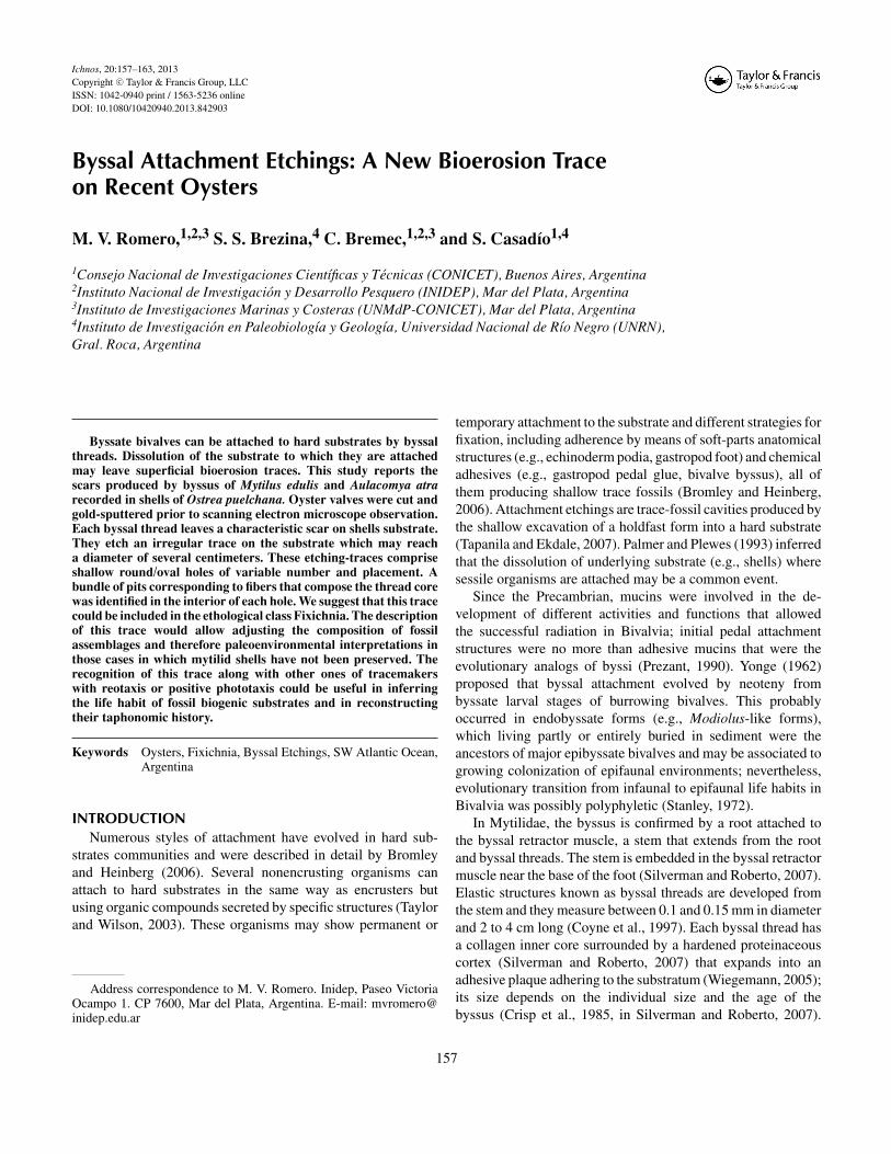
This study reports the etchings produced by the byssus ofMytilus edulis (Linnaeus, 1758) and Aulacomya atra (Molina,1782), both byssate bivalves were attached to shells of Ostreapuelchana (d’Orbigny, 1842) (Fig. 1). The preferential settle-ment of the byssus and the byssal etchings on different areas ofthe left and right valves was assessed.
MATERIALS AND METHODS
Study AreaO. puelchana specimens were collected in San Matıas Gulf
(SMG, 40º 42’ - 42º 41’ S; 63º 45’ - 65º 09’ W). SMG is asemi-enclosed area of the Argentinean shelf. It shows particularoceanographic features (Guerrero and Piola, 1997); maximum
depths are nearly 200 m in the central area (Parker et al., 1997)and the regime is macrotidal (Servicio de Hidrografia Naval,2010). The average salinity is high (33.84) and the averageannual temperature is 13.25 ± 0.20◦C with strong thermal strat-ification mainly in summer (Rivas, 1990). The bottom is sandywith high contents of silt and clay (Parker et al., 1997). Oysterbanks grow on sand or sand-gravel substrates over 10 m deep(Escofet et al., 1978).
SamplingA total of 142 oysters were collected in February 2009 in
two natural banks located at northwest of SMG, called El Buque(EB, 40◦ 50’ S; 65◦ 10’ W) and Zona de Colectores (ZC, 40◦
56’ S; 65◦ 06’ W), at a depth of 12 and 18 m, respectively,at low tide. Samples were taken randomly within the mostdensely packed zone of each bank. To avoid loss of macrofaunaassociated to the valve, each oyster was stored individually ina plastic bag. The valves were fixed in 5% seawater formalinand 15 days later they were stored in 70% alcohol. Mytilidaespecimens were assigned to Mytilus edulis and Aulacomyaatra. The marks produced by their byssal attachment on theexternal surfaces of the valves were recognized under binocularmicroscope. Samples for scanning electron microscope (SEM)analysis were obtained using a Silicon Carbide abrasive cutterby carefully cutting small pieces of valves which containedpreviously identified traces. After gold sputtering, the sampleswere visualized at the LIMF-Universidad Nacional de La Platausing a Fei Quanta 200 SEM in low vacuum mode. A series ofmagnifications were selected with images having a scale rangingfrom 1 mm to 500 μm.
Areas were defined within each valve to test preferentialsettlement of byssate bivalves. Zonification maps of both valveswere performed. This map reflects dissimilar morphologicalfeatures of the valves that may influence the settlement of bivalvelarvae. The left valve exterior was divided into six areas: apex(10%), platform (5%), anterior margin (15%), ventral margin(25%), posterior margin (15%), and center (30%). There is noplatform in the right valves, so the areas into which they weredivided are apex (10%), anterior margin (15%), ventral margin(30%), posterior margin (15%), and center (30%). Percentageswere calculated in relation with the total valve area (100%)following Romero et al. (in press).
The coverage percentage of byssal threads and plaquesand/or etchings was recorded in the zonification maps of eachvalve. The Wilcoxon paired-sample test (Zar, 1999) was usedto compare the coverage percentage of byssal threads andplaques and/or etchings between external surfaces of both oystervalves. A goodness of fit test was performed in order to assesspossible preferential location of byssus and their byssal etchingson different areas within left and right valves. The expectedfrequencies were calculated using a correction coefficient totake into account the different surface assigned to each area.This coefficient is different for each area and it corresponds to
BYSSAL ATTACHMENT ETCHINGS 159
FIG. 1. Byssate bivalves and byssus attached to shells of Ostrea puelchana d’Orbigny 1842. (A) Aulacomya atra (Molina, 1782); (B) Mytilus edulis (Linnaeus,1758); (C) bunch of mytilids covering oyster valves; and (D) detail of mussel byssus fixed on oyster valve. Scale bars = 2 cm. (See Color Plate I.)
the proportion between the percentage assigned to one areaand the percentage of the total valve area (e.g., correctioncoefficient for apex = 10/100). When more than 20% of theexpected frequencies were less than five, valve areas weregrouped to avoid this test becomes inaccurate. Yates’s correctionfor continuity was applied in those cases in which there is onlyone degree of freedom, and relatively small samples (Zar, 1999).The null hypothesis was the random distribution of byssus andtheir etchings on valves at significance level α = 0.05.
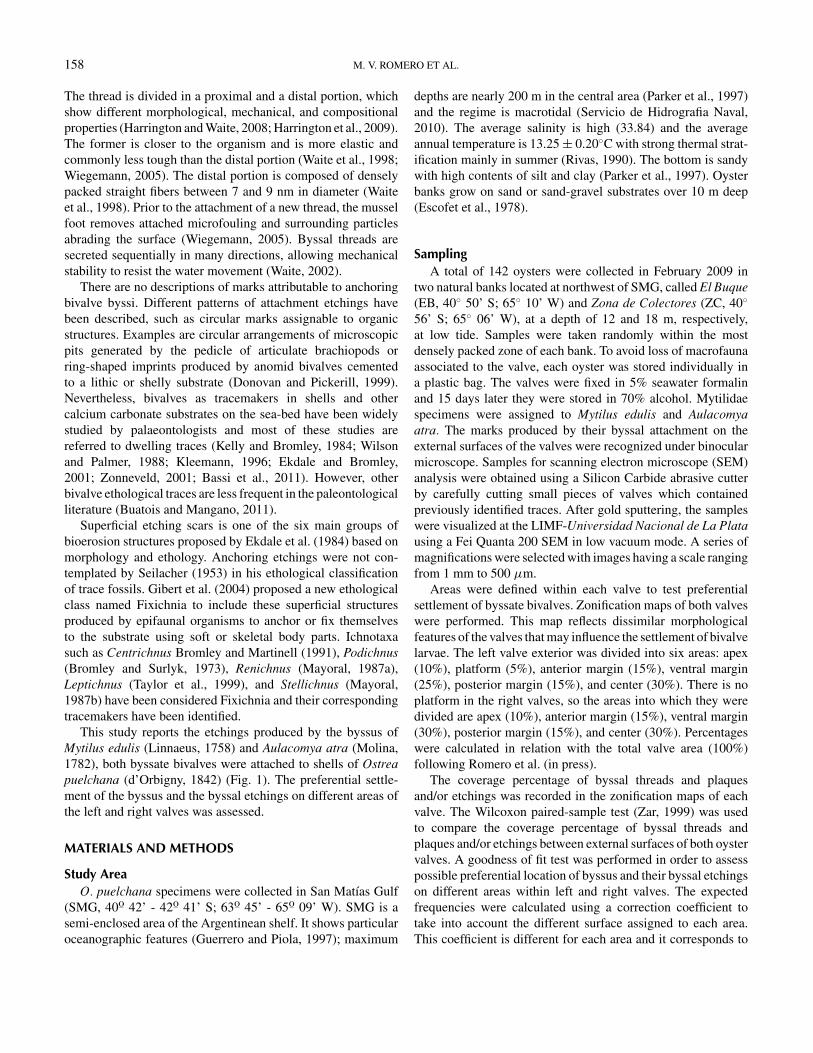
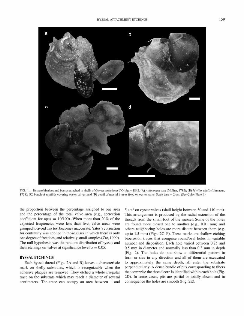
BYSSAL ETCHINGSEach byssal thread (Figs. 2A and B) leaves a characteristic
mark on shelly substrates, which is recognizable when theadhesive plaques are removed. They etched a whole irregulartrace on the substrate which may reach a diameter of severalcentimeters. The trace can occupy an area between 1 and
5 cm2 on oyster valves (shell height between 50 and 110 mm).This arrangement is produced by the radial extension of thethreads from the small foot of the mussel. Some of the holesare found more closed one to another (e.g., 0.01 mm) andothers neighboring holes are more distant between them (e.g.up to 1.5 mm) (Figs. 2C–F). These marks are shallow etchingbioerosion traces that comprise round/oval holes in variablenumber and disposition. Each hole varied between 0.25 and0.5 mm in diameter and normally less than 0.3 mm in depth(Fig. 2). The holes do not show a differential pattern inform or size in any direction and all of them are excavatedto approximately the same depth; all enter the substrateperpendicularly. A dense bundle of pits corresponding to fibersthat comprise the thread core is identified within each hole (Fig.2D). In some cases, pits are partial or totally absent and inconsequence the holes are smooth (Fig. 2E).
160 M. V. ROMERO ET AL.
FIG. 2. Byssus and byssal etchings on O. puelchana valves. (A) View of proteinaceous cortex of byssal threads and adhesive plaques using stereoscopicmicroscope. (B) SEM image showing byssal threads and adhesive plaques. (C) A byssal thread initially detaching from the oyster valve and three round/ovalbyssal etchings. (D) Dense bundle of pits corresponding to fibers that compose the thread core within each byssal etchings. (E, F) Pattern of variation of size andshape in byssal etchings.
BYSSAL ATTACHMENT ETCHINGS 161
Twenty-eight percent of oysters examined presentedmytilids/byssi attached, and in 18% only the byssate bivalvetrace was registered. The Wilcoxon paired-sample test indicatedsignificant differences between left and right valves based oncoverage of byssal threads and plaques and/or etchings. Thistrace was recorded in both valves, although left valves weremore colonized by mytilids (Z = 2.214; p = 0.027) and theiretchings occupied a greater area than on the right valves (Z =2.17; p = 0.030). The Wilcoxon test was significant at levelp < 0.05.
Differential location of byssus and byssal etchings ondifferent areas within left and right valves was not detected(p > 0.05 for both cases and right and left valves).
The bioerosive trace described in this contribution presentsdistinctive characteristics and it represents a new ethologicalsuperficial structure produced by byssate bivalve. This tracewould be assigned into the class Fixichnia within the recentethological classification in use. This class includes superficial(epigenic) traces produced by sessile epiliths that anchor or fixthemselves to the substrate using soft or skeletal body parts(Gibert et al., 2004).
Podichnus is a biogenic structure that evidences the fixationof brachiopods by the pedicles (Bromley and Surlyk, 1973).The byssal etchings are easily differentiated from Podichnusbecause they show a chaotic pattern in different directionsand are more widely scattered than the borings produced bybrachiopod pedicles, which shows a circular pattern of smallholes that increase in size and become more skewed toward theouter edge of the trace (Taylor and Wilson, 2003). The byssaletchings unequivocally did not identify a single tracemaker,while in Podichnus each brachiopod “footprint” identifies justone individual (Bromley and Surlyk, 1973).
Centrichnus is attributed to cementation of anomiid bivalvesas well as the attachment of verrucid cirripedes to a hardsubstrate (Bromley and Martinell, 1991).The difference betweenCentrichnus and byssate bivalves traces lies primarily in thatthe former is characterized by a series of concentric rings orarcs that demonstrate the accretionary growth of skeletal hardpart of the tracemaker. Centrichnus eccentricus represents thebyssal cementation of anomiid bivalves in which the right valveis completely and permanently attached to the substrate by acalcified byssus (Bromley and Martinell, 1991). This mode ofattachment leaves a more compact and oval trace, drop shaped,curved about a center reminding of the tracemaker’s valve shape.The newest growth increment towards the ventral margin of thelower valve is marked by a concentric etching groove in thesubstrate. Centrichnus concentricus presents a hole or centraldepression but with an oval and crenulate perimeter and asurrounding platform that usually reflects the plate-ornament ofthe verrucid barnacle and the concentric growth-lines (Bromleyand Martinell, 1991). Moreover, Centrichnus is larger than thetrace characterized here (i.e., maximum observed diameter =8.5 mm in C. concentricus and 10 mm in C. eccentricus), andeach trace would correspond with only one individual.
Leptichnus (Taylor et al., 1999) shows a similar pattern ofseveral and shallow pits (i.e., by detachment of encrusted sheetsof cheilostomate zooids), but each scar is eliptical and keeps anuniseriate pattern with regular distance between excavated pitswith a more symmetric arrangement compared with byssatebivalve traces. Renichnus is assigned to vermetid gastropods(Mayoral, 1987a). This trace is easily distinguished from thebyssal marks because they are deeper, with crescent or kidneyshaped depressions, and in its more advanced stage it describesthe shape of the gastropod.
Finally, Stellichnus (Mayoral, 1987b) is a star-shaped systemof shallow grooves radiating from a central area. This mor-phology is reminiscent of ctenostomate bryozoans belonging toVinellidae. Although this trace shows perforations in the centerand at the sides of the grooves, they are smaller than the byssalscars (i.e., diameter between 5 and 45 μ). Moreover, Stellichnusshows a more regular arrangement than the byssus trace.
CONCLUSIONSThis article describes a new ethological superficial etchings
produced by byssate bivalves classified as Fixichnia. The recordof this endolithic bioerosion trace produced by a bivalve byssusmay allow adjusting the composition of fossil assemblagesand therefore paleoecological environmental interpretations,especially in those cases in which mytilid shells have not beenpreserved. In addition, the recognition of this trace, along withother ones whose tracemarkers display reotaxis or positivephototaxis, could be useful to infer the life habit of fossilsubstrates and reconstruct their taphonomic history.
The shells of O. puelchana record a set of bioerosivetraces which represent several ichnoguilds that reflect differentbehavior, trophic levels and depths in which were placed onthe substrate (i.e., tiers). Along with byssal etchings, tracesfrom shallowest to intermediate deeper tiers (see Bromley andAsgaard, 1993) have been frequently recorded (i.e., Leptichnusisp., Radulichnus isp., Entobia isp., Gastrochaenolites isp., andSemidendrina isp.). In all these cases, etchings are sufficientlydeep to pass through the oyster periostracum and leavepreservable traces when this layer is lost by physical andbiological erosion.
According to Bromley and Asgaard (1993), Radulichnusinopinatus (Voigt, 1977) and Gnathichnus pentax (Bromley,1975; absent in this study) are considered to be traces that makeup the most superficial ichnoguilds, while Renichnus arcuatus(Mayoral, 1987a) and Centrichnus eccentricus (Bromley andMartinell, 1991) are almost as superficial as the former,constituting a different ichnoguild. Probably, these ichnospeciesand byssal etchings represent an ecologic unit according to thedepth at which they occur and because the tracemakers aresessile mollusks that produced superficial traces by attachmentto the substrate. However, during the oyster colonization bydifferent taxa this trace may be generated by detachment ofthe byssal plaques anytime. Although these traces compose a
162 M. V. ROMERO ET AL.
shallow tier it does not imply that they originate at the beginningof a sequence of colonization, suggesting that the generaltiering scheme proposed by Bromley and Asgaard (1993) doesnot necessarily carry temporal meanings in the colonizationsequence which in some cases is very complex to identify.
Most of Paleozoic epifaunal bivalves were byssate; onlya few groups (e.g., Japanese Permian forms) are known tohave attached by cementation (Stanley, 1972). Furthermore, thisbioerosion trace should be commonly found in fossil record.
Naming this new trace is controversial. The InternationalCode of Zoological Nomenclature (ICZN, 1999) suggests thatit is not advisable to assign names on the basis of unfossilizedmaterial. However, according to Nielsen et al. (2003) a newbioerosive trace must be named regardless of the age of thematerial in which it is registered, allowing the discovery ofbehaviors of living organisms and facilitate communicationbetween biologists and geologists to work in this field. Wefollowed this approach to include a trace described from recentmaterial into an informal ethological classification based onfossil material.
ACKNOWLEDGMENTSThe manuscript greatly benefited from comments and
suggestions by Dr. M. Griffin. This is Instituto Nacional deInvestigacion y Desarrollo Pesquero (INIDEP) Contribution No.1798. Financial support PIP 112-201101-00566. This researchwas supported by a PhD Fellowship from Consejo Nacional deInvestigaciones Cientıficas y Tecnologicas (CONICET) to M.V. Romero.
REFERENCESBassi, D., Humblet, M., and Iryu, Y. 2011. Recent ichnocoenosis in deep water
macroids, Ryukyu islands, Japan. Palaios, 26: 232–238.Bromley, R. G. 1975. Comparative analysis of fossil and recent echinoid
bioerosion. Palaeontology, 18: 725–739.Bromley, R. G. and Asgaard, U. 1993. Endolithic community replacement on a
Pliocene rocky coast. Ichnos, 2: 93–116.Bromley, R. G. and Heinberg, C. 2006. Attachment strategies of organisms on
hard substrates: A palaeontological view. Palaeogeography, Palaeoclima-tology, Palaeoecology, 232: 429–453.
Bromley, R. G. and Martinell, J. 1991. Centrichnus, new ichnogenus forcentrically patterned attachment scars on skeletal substrates. Bulletin ofthe Geological Society of Denmark, 38: 243–252.
Bromley, R. G. and Surlyk, F. 1973. Borings produced by brachiopod pedicles,fossil and Recent. Lethaia, 6: 349–365.
Buatois, L. A. and Mangano, M. G. 2011. Ichnology: Organism-substrateInteractions in Space and Time. Oxford, Cambridge University Press,358 p.
Coyne, K. J., Qin, X., and Waite, J. H. 1997. Extensible collagen in musselbyssus: A natural block copolymer. Science, 277: 1830–1832.
Crisp, D. J., Walker, G., Young, G. A., and Yule, A. B. 1985. Adhesion andsubstrate choice in mussels and barnacles. Journal of Colloid and InterfaceScience, 104: 40–50.
Donovan, S. K. and Pickerill, R. K. 1999. Fossils explained 26: Trace fossils4-borings. Geology Today, 15: 197–200.
d’Orbigny, A. 1842. Voyage dans l’Amerique Meridionale (Le Bresil, LaRepublique Orientale de L’Uruguay, La Republique Argentine, La Patago-nie, La Republique du Chili, La Republique de Bolivie, La Republique du
Perou, execute pendant les anne 1826–1833. In Bertrand, C.P. (ed.), ChezVe Levrault, Tome 3, 4th part, Paleontologie, Paris, 188 p.
Ekdale, A. A., Bromley, R. G., and Pemberton, S. G. 1984. Ichnology, the useof trace fossils in sedimentology and stratigraphy. Society of EconomicPaleontologists and Mineralogists, Short Course 15, 317 p.
Ekdale, A. A. and Bromley, R. G. 2001. Bioerosional innovation for livingin carbonate hardgrounds in the Early Ordovician of Sweden. Lethaia, 34:1–12.
Escofet, A., Orensanz, J. M., Olivier, S. R., and Scarabino, V. 1978. Biocenologıabentonica del Golfo de San Matıas (Rio Negro, Argentina): Metodologia,experiencias, y resultados del estudio ecologico de un gran espaciogeografico de America Latina. Anales del Centro de Ciencias del Mar yLimnologıa, Universidad Nacional Autonoma de Mexico, 5: 59–81.
Gibert, J. M., Domenech, R., and Martinell, J. 2004. An ethological frameworkfor animal bioerosion trace fossils upon mineral substrates with proposal ofa new class, fixichnia. Lethaia, 37: 429–437.
Guerrero, R. and Piola, A. R. 1997. Masas de agua en la plataforma continental.In Boschi, E.E. (ed.), El Mar Argentino y sus recursos pesqueros Tomo 1.INIDEP, Mar del Plata, 222 p.
Harrington, M. J., Gupta, H. S., Fratzl, P., and Waite, J. H. 2009. Collageninsulated from tensile damage by domains that unfold reversibly: In situX-ray investigation of mechanical yield and damage repair in the musselbyssus. Journal of Structural Biology, 167: 47–54.
Harrington, M. J. and Waite, J. H. 2008. Short-order tendons: liquid crystalmesophases, metal-complexation and protein gradients in the externalizedcollagens of mussel byssal threads. In Scheibel, T. (ed.), Fibrous Proteins(30–45). Landes Bioscience, Austin, Texas.
Kelly, S. R. A. and Bromley, R. G. 1984. Ichnological nomenclature of clavateborings. Palaeontology, 27: 793–807.
Kleemann, K. H. 1996. Biocorrosion by bivalves. Marine Ecology, 17: 145–158.Linnaeus, C. 1758. Systema naturae per regna tria naturae, secundum classes,
ordines, genera, species, cum characteribus, differentiis, synonymis, locis.Editio decima, reformata. Holmiae, Impensis Direct. Laurentii Salvii,824 p.
Mayoral, E. 1987a. Accion bioerosiva de Mollusca (Gastropoda, Bivalvia) enel Plioceno inferior de la Cuenca del Bajo Guadalquivir. Revista Espanolade Paleontologıa, 2: 49–58.
———. 1987b. Stellichnus nov. ichnogen., huellas de incrustacion atribuidas aParavinella nov. gen. (Bryozoa, Ctenostomata) de la formacion Arenas deHuelva (Plioceno inferior) en la Cuenca del Bajo Guadalquivir (Espana).Revista Espanola de Paleontologıa, 2: 33–40.
Molina, I. J. 1782. Saggio sulla Storia Naturale del Chili. Bologna, Stamperiadu S. Tommaso d’Aquino, 306 p.
Nielsen, K. S. S., Nielsen, J. K., and Bromley, R. G. 2003. Palaeoecologicaland ichnological significance of microborings in quaternary Foraminifera.Palaeontologia Electronica, 6 (2): 1–13.
Palmer, T. and Plewes, C. 1993. Borings and bioerosion in fossils. GeologyToday, 9: 138–142.
Parker, G., Paterlini, M. C., and Violante, R. A. 1997. El fondo marino. InBoschi, E.E. (ed.), El Mar Argentino y sus recursos pesqueros Tomo 1.INIDEP, Mar del Plata, 222 p.
Prezant, R. S. 1990. Form, function and phylogeny of bivalve mucins. InMorton, B. (ed.), The Bivalvia: Proceedings of a Memorial Symposiumin Honor of Sir Charles Maurice Yonge (1899–1986) at the 9th InternationalMalacological Congress, 1986. Edinburgh, Scotland, 83: 95.
Rivas, A. L. 1990. Heat balance and annual variation of mean temperature inthe North Patagonian gulfs. Oceanologica Acta, 13: 265–272.
Romero, M. V., Brezina, S. S., Hernandez D., Casadıo, S., and Bremec, C.In press. Differential settlement of associated species on Ostrea puelchanad’Orbigny, 1842 in Patagonia (Argentina). American Malacological Bul-letin.
Seilacher, A. 1953. Studien zur Palichnologie, I. Uber die Methoden der Palich-nologie. Neues Jahrbuch fur Geologie und Palaentologie. Abhandlungen,98: 87–124.
BYSSAL ATTACHMENT ETCHINGS 163
Servicio de Hidrografıa Naval. 2010. Tabla de Marea 5, Armada Argentina,176 p.
Silverman, H. G. and Roberto, F. F. 2007. Understanding marine musseladhesion. Marine Biotechnology, 9: 661–681.
Stanley, S. M. 1972. Functional morphology and evolution of byssally attachedbivalve mollusks. Journal of Paleontology, 46: 165–212.
Tapanila, L. and Ekdale, A. A. 2007. Early history of symbiosis in livingsubstrates: Trace-fossil evidence from the marine record. In Miller III, W.(ed.), Trace Fossils: Concepts, Problems, Prospects. Elsevier, Amsterdam,637 p.
Taylor, P. D., Wilson, M. A., and Bromley, R. G. 1999. Leptichnus, a newichnogenus for etchings made by cheilostome bryozoans into calcareoussubstrates. Palaeontology, 42: 595–604.
Taylor, P. D. and Wilson, M. A. 2003. Palaeoecology and evolution of marinehard substrate communities. Earth-Science Reviews, 62: 1–103.
Voigt, E. 1977. On grazing traces produced by the radula of fossil and recentgastropods and chitons. In Crimes, T.P. and Harper, J.C. (eds.), Trace Fossils2, Geological Journal, Special issue 9: 335–346.
Waite, J. H. 2002. Adhesion a la moule. Integrative and Comparative Biology,42: 1172–1180.
Waite, J. H., Qin, X., and Coyne, K. J. 1998. The pecutiar collagens of musselbyssus. Matrix Biology, 17: 93–106.
Wiegemann, M. 2005. Adhesion in blue mussels (Mytilus edulis) and barnacles(genus Balanus): Mechanisms and technical applications. Aquatic Sciences,67: 166–176.
Wilson, M. A. and Palmer, T. J. 1988. Nomenclature of a bivalve boring from theUpper Ordovician of the midwestern United States. Journal of Paleontology,62: 306–308.
Yonge, C. M. 1962. On the primitive significance of the byssus in the Bivalviaand its effects in evolution. Journal of the Marine Biological Association ofthe United Kingdom, 42: 112–125.
Zar, J. H. 1999. Biostatistical Analysis. Prentice-Hall, Englewodo Cliffs, NewJersey.
Zonneveld, J. P. 2001. Middle Triassic biostromes from the Liard Formation,British Columbia, Canada: Oldest examples from the Mesozoic of NWPangea. Sedimentary Geology, 145: 317–341.


