Embed Size (px)
Citation preview

FULL PAPER
DOI: 10.1002/ejic.200600626
Cages on Surfaces: Thiol Functionalisation of CoIII Sarcophagine Complexes
Jack M. Harrowfield,[a,b] George A. Koutsantonis,*[b] Heinz-Bernhard Kraatz,[c]
Gareth L. Nealon,[b] Grzegorz A. Orlowski,[c] Brian W. Skelton,[b] and Allan H. White[b]
Keywords: Sarcophagine / Amino acids / Disulfides / Cobalt / Self-assembled monolayers / Electrodeposition
A number of new (sarcophagine)cobalt compounds contain-ing oligopeptide substituents terminated by a mercaptoethyl-amide unit, [Co(R)(R�)sar]2
6+ (R = NH2, CH3; R� = NHpeptide-CONHCH2CH2S–), have been prepared utilising the carb-oxymethyl complexes [Co(R){N(CH2CO2H)}sar]3+ (R = NH2,CH3), and subsequently characterised. The amino acid resi-dues, terminating with cystamine groups, were introducedinto the carboxymethyl complexes using standard peptidecoupling chemistry. The solid-state structures of [Co(CH3)-{N(CH2CO2H)}sar]Cl4·H2O, [Co(CH3){N(CH2CO2H)2}sar]Cl3·2H2O and [Co(CH3){N(CH2CO2)2}sar]Cl·6.5H2O were deter-mined by single-crystal X-ray diffraction experiments, thefirst two forming lattices containing homochiral sheets of cat-ions in planes parallel to [0,0,1]. In the structure of [Co(CH3)-{N(CH2CO2H)}sar]Cl4·H2O, the cations are of the same abso-lute configuration, but this alternates from one sheet tothe next. In the dihydrate, [Co(CH3){N(CH2CO2H)2}sar]-Cl3·2H2O, the structure appears to be very similar. The sheetalternation is different from the structure of the monohydrate,[Co(CH3){N(CH2CO2H)}sar]Cl4·H2O. The deprotonated im-
Introduction
Reagent immobilisation on surfaces is a sophisticatedpathway to materials with a wide range of applications,[1,2]
heterogeneous catalysis being one obvious application, suchapplications depending on the functionality introducedwith the bound reagent, as well as upon the ease and conve-nience of the immobilisation procedure and the stability ofthe final product. Given the remarkable stability and variedelectronic, magnetic and redox properties of metal com-plexes of the macrobicyclic polyamines known as “sarcoph-
[a] Institut de Science et d’Ingénierie Supramoléculaires, UniversitéLouis Pasteur,8, allée Gaspard Monge, 67083 Strasbourg, FranceFax: +33-3-90245140E-mail: [email protected]
[b] Chemistry, M313, School of Biomedical, Biomolecular andChemical Sciences, University of Western Australia,Crawley 6009, AustraliaFax: +61-8-64887247E-mail: [email protected]
[c] Department of Chemistry, University of Saskatchewan,110 Science Place, Saskatoon, S7N 5C9, CanadaFax: +1-306-966-4730E-mail: [email protected] information for this article is available on theWWW under http://www.eurjic.org or from the author.
Eur. J. Inorg. Chem. 2007, 263–278 © 2007 Wiley-VCH Verlag GmbH & Co. KGaA, Weinheim 263
idodiacetate complex, [Co(CH3){N(CH2CO2)2}sar]Cl·6.5H2O,was prepared in an unrelated reaction; the structure displayshydogen-bonding interactions involving the carboxylategroups and coordinated NH units. The solution electrochem-istry of these compounds has been investigated using bothcyclic voltammetry and differential pulse voltammetry. Thecompounds exhibited pH-dependent, quasi-reversible redoxbehaviour. A monolayer was prepared by the interaction of[Co(R)sar(GlyNHCH2CH2S–)]2
6+ and a gold microelectrodeusing both conventional “self assembly” (SAM) and electro-deposition (EDM) techniques. Surface cyclic voltammogramswere measured, which, after a period of induction associatedwith surface reorganisation, were typical of an orderedmonolayer. The monolayer was characterised by XPS, show-ing peaks assigned to the presence of S, Co, N, O and C.Ellipsometry gave a film thickness of 7�1 Å, consistent withthe formation of a mono- rather than a multi-layer.
(© Wiley-VCH Verlag GmbH & Co. KGaA, 69451 Weinheim,Germany, 2007)
agines” (Figure 1),[3] these are species of particular appealas entities for attachment to surfaces and for various relatedapplications.[4] Reduction potentials for readily accessiblespecies span a range of 2 V,[5] subject to modification in aninterfacial environment,[6] and outer-sphere redox processesinvolving Co complexes are, for example, known to be rapidsteps in reactions leading to photoinduced hydrogen pro-duction[7–10] and the reduction of oxygen to hydrogen per-oxide.[11] Of practical importance in relation to immobili-sation of such complexes is the facile synthesis of the ligandin forms with reactive “external” functional groups R (Fig-ure 1).[12]
Figure 1. Trivial nomenclature used throughout this work.
G. A. Koutsantonis et al.FULL PAPERLimited earlier work on surface-bound cage complexes
has involved modification of electrodes by impregnation ofthin Nafion films on graphite with simple cage complexes,[5]
electropolymerisation of a thiophenylmethylamino cagecomplex on Pt,[12] and carbodiimide coupling of carboxylgroups on partially oxidised graphite to a diamino cagecomplex.[13] Amphiphilic cages[14,15] have also proved tohave interesting biological properties, probably associatedwith their ability to be bound within membranes.[15] As asimple approach to the immobilisation of cage complexeson surfaces, we are currently exploring the use of the well-established surface chemistry of disulfides[1] to form mono-layers of (sarcophagine)cobalt complexes on gold. This isbased on the introduction of peptide substituents associatedwith cystamine units onto the cage, a facile synthesis whenstarting from readily synthesised glycylated derivatives.[16]
The use of peptide-based tethering groups is advantageousas the length of the spacer between the surface and the cagemoiety can be increased incrementally with relative ease,using common peptide synthetic techniques,[17] and tightlypacked monolayers can be formed, presumably by hydrogenbonding between the amide groups.[18] A similar approachhas been applied to the synthesis of ferrocene-decoratedpeptides as probes for elucidation of the mechanism(s) ofelectron transfer in proteins.[19]
Our immediate aim in the present work was to synthesisea series of disulfides bearing peptido cage complex substitu-ents, evaluate their solution electrochemical characteristics,and determine a suitable method for their immobilisationonto gold electrodes. Because only a single disulfide substit-uent is required for surface tethering, our focus has been onobtaining derivatives of the monosubstituted cage complex[Co(CH3)(NH2)sar]3+, though the more complicated syn-theses of monofunctionalised derivatives of [Co(NH2)2-sar]3+ have also been investigated. These CoIII complexesexist in stable enantiomeric forms and thus the formation ofdiastereomeric forms of their oligopeptide derivatives wasanticipated, though prior work suggested that differencesbetween such diastereomers should be small.[12]
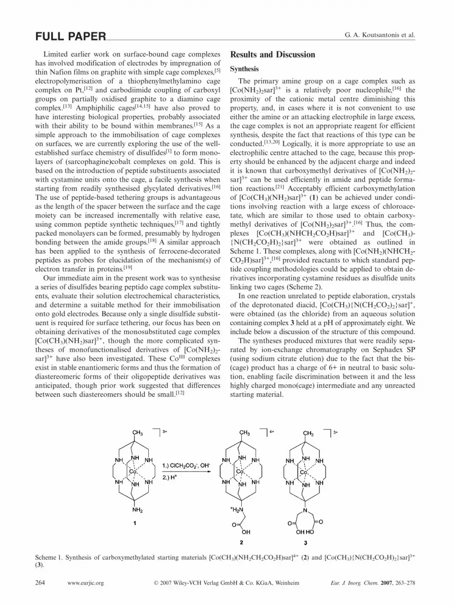
Scheme 1. Synthesis of carboxymethylated starting materials [Co(CH3)(NH2CH2CO2H)sar]4+ (2) and [Co(CH3){N(CH2CO2H)2}sar]3+
(3).
www.eurjic.org © 2007 Wiley-VCH Verlag GmbH & Co. KGaA, Weinheim Eur. J. Inorg. Chem. 2007, 263–278264
Results and Discussion
Synthesis
The primary amine group on a cage complex such as[Co(NH2)2sar]3+ is a relatively poor nucleophile,[16] theproximity of the cationic metal centre diminishing thisproperty, and, in cases where it is not convenient to useeither the amine or an attacking electrophile in large excess,the cage complex is not an appropriate reagent for efficientsynthesis, despite the fact that reactions of this type can beconducted.[13,20] Logically, it is more appropriate to use anelectrophilic centre attached to the cage, because this prop-erty should be enhanced by the adjacent charge and indeedit is known that carboxymethyl derivatives of [Co(NH2)2-sar]3+ can be used efficiently in amide and peptide forma-tion reactions.[21] Acceptably efficient carboxymethylationof [Co(CH3)(NH2)sar]3+ (1) can be achieved under condi-tions involving reaction with a large excess of chloroace-tate, which are similar to those used to obtain carboxy-methyl derivatives of [Co(NH2)2sar]3+.[16] Thus, the com-plexes [Co(CH3)(NHCH2CO2H)sar]3+ and [Co(CH3)-{N(CH2CO2H)2}sar]3+ were obtained as outlined inScheme 1. These complexes, along with [Co(NH2)(NHCH2-CO2H)sar]3+,[16] provided reactants to which standard pep-tide coupling methodologies could be applied to obtain de-rivatives incorporating cystamine residues as disulfide unitslinking two cages (Scheme 2).
In one reaction unrelated to peptide elaboration, crystalsof the deprotonated diacid, [Co(CH3){N(CH2CO2)2}sar]+,were obtained (as the chloride) from an aqueous solutioncontaining complex 3 held at a pH of approximately eight. Weinclude below a discussion of the structure of this compound.
The syntheses produced mixtures that were readily sepa-rated by ion-exchange chromatography on Sephadex SP(using sodium citrate elution) due to the fact that the bis-(cage) product has a charge of 6+ in neutral to basic solu-tion, enabling facile discrimination between it and the lesshighly charged mono(cage) intermediate and any unreactedstarting material.

Cages on Surfaces: Thiol Functionalisation of CoIII Sarcophagine Complexes FULL PAPER
Scheme 2. Synthetic methodology used for the preparation of the disulfide complexes.
The method commonly employed to isolate CoIII com-plexes of the present type from Sephadex SP column elu-ates, including absorbing them on Dowex 50W strong acidresin, re-eluting with HCl and then concentrating to dry-ness,[16] proved to lead here to some hydrolysis of the pep-tides. Fortunately, the complexes could be readily isolatedfrom the citrate eluates by precipitation as their very insolu-ble picrates. These could readily be converted into the per-chlorates, preferred to the chlorides for electrochemicalstudies involving gold electrodes. It is worth noting that,despite the modest isolated yields reported for the com-pounds, the major band in the ion-exchange chromatog-raphy was always the disubstituted disulfide, indicating arelatively efficient reaction to give the desired product.
NMR Spectroscopy
All compounds synthesised (Table 1) exhibited NMRspectra consistent with their proposed structures. Evidencefor the introduction of the carboxymethyl substituent to
Eur. J. Inorg. Chem. 2007, 263–278 © 2007 Wiley-VCH Verlag GmbH & Co. KGaA, Weinheim www.eurjic.org 265
give the compounds 2 and 3 was observed with peaks be-tween δ ≈ 3.6 and 3.7 ppm in their 1H NMR and two peaksat δ = 43.6, 52.5 (–CH2–) and δ = 172.5, 176.8 (C=O) ppmin their 13C NMR spectra. The compounds 4, 7 and 8 dis-played a single amide –NH– peak (δ = 7.9–8.0 ppm) in their1H NMR and a single C=O peak (δ ≈ 170 ppm) in their13C NMR spectra due to their CSA moieties. The com-pounds 5, 6, 9 and 10 displayed two peaks in both their1H NMR (δ = 7.8–8.2 ppm) and 13C NMR (δ = 168.7–172.2 ppm) spectra, corresponding to the inequivalent NHand carbonyl centres of the amino acid (Gly or Ala) andCSA. The complex multiplets observed in the 1H NMRspectra in the region between δ ≈ 2 and 3.5 ppm due to themethylene group resonances of the [Co(CH3)(NH2)sar]3+
and [Co(NH2)2sar]3+ moieties sometimes obscured otherpeaks corresponding to the Gly1 –CH2– group and/or the–CH2–N group of CSA. The presence and position of theobscured peaks was deduced from the overall integration ofthe protons in this region, as well as cross-peaks observedin the 2D NMR experiments. 13C NMR spectra reflected

G. A. Koutsantonis et al.FULL PAPERthe complete asymmetry of the compounds, except for thecompounds 8, 9 and 10 in which two cage methylene signalswere coincident. This was determined from the high inten-sity of the composite peak relative to the other cage methyl-ene signals and by 2D NMR experiments. It is presumedthat the use of racemic [Co(CH3)(NHCH2CO2H)sar]3+ as areactant leads to the production of a mixture of meso andrac forms of the bis(cage) peptides but only in the case ofcompound 6 was any spectroscopic evidence for the pres-ence of such a mixture obtained. The 13C NMR spectrumof this complex shows some doubling of signals, similar tothat observed in a dipeptide derivative of racemic[Co(NH2)(NHCH2CO2H)sar]3+.[21] This could reflect someracemisation of the amino acid during the synthesis but theproduct presently obtained by reaction of ∆-[Co(NH2)(NHCH2CO2H)sar]3+ with (-Ala)2CSA showsonly sharp singlets in its 13C NMR spectrum, indicatingthat this is not the case.
Table 1. List of compounds synthesised.
Compound R Cage R�
2 –CH3 racemic –Gly-OH3 –CH3 racemic –N(CH2CO2H)2
4 –CH3 racemic –Gly-CSA-5 –CH3 racemic –Gly-Gly-CSA-6 –CH3 racemic –Gly-Ala-CSA-7 –NH2 racemic –Gly-CSA-8 –NH2 ∆-enantiomer –Gly-CSA-9 –NH2 ∆-enantiomer –Gly-Gly-CSA-
10 –NH2 ∆-enantiomer –Gly-Ala-CSA-
Mass Spectrometry
Electrospray mass spectrometry (ESMS) has been recog-nised as a valuable method for obtaining molecular weightsof cage complexes with little to no fragmentation.[22] ESMS
Table 2. Crystal data, details of data collection and structure refinement for the complexes.
Complex [2]Cl4·H2O [3]Cl3·2H2O [3–2H]Cl·6.5H2O
Empirical formula C17H40Cl4CoN7O3 C19H43Cl3CoN7O6 C19H50ClCoN7O10.5
M 591.3 639.9 639.0Crystal system triclinic monoclinic monoclinicSpace group P1̄ P21/c P21/ca [Å] 9.1450(7) 9.9703(5) 16.918(1)b [Å] 11.4703(9) 9.9864(5) 14.243(1)c [Å] 12.521(9) 27.8730(10) 25.024(2)α [°] 83.594(1) 90 90β [°] 87.671(1) 93.379(1) 103.099(5)γ [°] 79.557(1) 90 90V [Å3] 1283.30(9) 2770.4(2) 5872.9(7)Dcalcd. [g cm–3] 1.53 1.51 1.445Z 2 4 8Size [mm] 0.45�0.37�0.28 0.43�0.43�0.18 0.65�0.40�0.22µο [mm–1] 1.12 0.96 0.74Trans. (min./max.) 0.87 0.81 0.882Θmax. [°] 64 70 63Nt 17618 55294 154432N (Rint.) 8747 (0.022) 11863 (0.37) 19162 (0.035)No 7177 8958 15820R 0.034 0.050 0.055Rw (nw) 0.073 (3.2) 0.095 (7) 0.102 (28)T [K] 170 298 100
www.eurjic.org © 2007 Wiley-VCH Verlag GmbH & Co. KGaA, Weinheim Eur. J. Inorg. Chem. 2007, 263–278266
spectra of compounds 2 and 3 display peaks consistent withthe molecular ions corresponding to a loss of H+ from thecation, and some peaks consistent with an additional asso-ciation of chloride anions. ESMS spectra of compounds 4–10 in several cases display multiple peaks, corresponding tothe molecular ion in charge states of 2+, 3+ and 4+, whichare assigned as successive losses of H+ from the parent ion,sometimes accompanied by association of a perchlorateanion. Loss of H+ from a coordinated –NH– group of thecage is known and the resolution of the measurements hereare sufficient to distinguish this possibility from that of Co-III reduction to CoII in the mass spectrometer, as has alsobeen observed.[22] There also appears to be no cleavage ofthe disulfide link during volatilisation.
Structure Determinations of Salts of Cations 2 and 3
The crystal structure determinations, modelledas [Co(CH3)(NH2CH2CO2H)sar]Cl4·H2O, [2]Cl4·H2O,[Co(CH3){N(CH2CO2H)2}sar]Cl3·2H2O, [3]Cl3·2H2O, and[Co(CH3){N(CH2CO2)2}sar]Cl·6.5H2O, [3–2H]Cl·6.5H2Oconfirm the nature of the functionalisation (indicated bythe given formulae) of the original cage complex[Co(CH3)(NH2)sar]3+ expected on the basis of the methodof synthesis and spectroscopic measurements; in the firsttwo complexes, one, in the latter, two, formula units, devoidof crystallographic symmetry, comprise the asymmetric unitof the structure. The experimental details and refinementdetails are collected in Table 2, while structural representa-tions showing the atom numbering schemes of the cationsare given in Figure 2. Table 3 collects pertinent hydrogen-bonding data for the complexes. In [2]Cl4·H2O, the O- andN-sites of the glycyl functional are both protonated,whereas in [3]Cl3·2H2O, although a zwitterionic form ispossible, the two carboxyl groups appear to retain their pro-

Cages on Surfaces: Thiol Functionalisation of CoIII Sarcophagine Complexes FULL PAPER
Figure 2. Structural representation of the cations in complexes (a) [Co(CH3)(NH2CH2CO2H)sar]Cl4·H2O [2]Cl4·H2O, (b) [Co(CH3)-{N(CH2CO2H)2}sar]Cl3·2H2O, [3]Cl3·2H2O and (c) one of the two cations in [Co(CH3){N(CH2CO2)2}sar]Cl·6.5H2O, [3–2H]Cl·6.5H2O.Probability ellipsoids shown at 50 [(a), (c)] and 20% (b) amplitude.
tons and the N-centre is unprotonated. This is despite thefact that the two complexes were isolated under essentiallyidentical conditions from quite concentrated HCl solutions,though of course it need not be the case that the dominantspecies in solution are those which crystallise. The carboxyl-ate groups are deprotonated in [3–2H]Cl·6.5H2O. All cationcores are of lel3 conformation.
A view of the lattice of [2]Cl4·H2O along the a axis shows[Figure 3(a)], edge-on, sheets of cations lying in planes par-allel to [0,0,1] which appear to form pairs separated by
Eur. J. Inorg. Chem. 2007, 263–278 © 2007 Wiley-VCH Verlag GmbH & Co. KGaA, Weinheim www.eurjic.org 267
sheets of chloride ions. Within any given sheet, all cationsare of the same absolute configuration, which alternatesfrom one sheet to the next, and all adopt the lel3 conforma-tion. As has been discussed in detail elsewhere,[23] this con-formation of amine cage complexes of CoIII is frequentlyassociated with “hydrogen-bonding chelation” in which twocoordinated NH centres on an open octahedral edge bindto the one unit, very commonly a chloride ion, just as isthe case here [Figure 3(b)]. Each of the three chloride ionsinvolved in such chelation by every cation has a third con-

G. A. Koutsantonis et al.FULL PAPERTable 3. Solution electrochemical data for compounds 2–10. AllE1/2 values are referenced to the Ag/AgCl reference electrode. Scanrate 100 mVs–1. Errors are the standard deviations from five mea-surements.
Compound E1/2 [V] ∆E [V] Ia/Ic
2 –0.552(2) 0.074(3) 13 –0.607(2) 0.073(2) 14 –0.565(3) 0.12(1) 15 –0.562(5) 0.09(1) 16 –0.563(2) 0.09(1) 0.97 –0.494(6) 0.118(5) 0.88 –0.497(2) 0.122(2) 0.89 –0.497(4) 0.116(5) 0.810 –0.496(5) 0.15(1) 0.8
Figure 3. (a) View along the a-axis of the lattice of [Co(CH3)(NH2CH2CO2H)sar]Cl4·H2O, [2]Cl4·H2O; (b) representations of[Co(CH3)(NH2CH2CO2H)sar]Cl4·H2O, [2]Cl4·H2O, showing close contacts.
www.eurjic.org © 2007 Wiley-VCH Verlag GmbH & Co. KGaA, Weinheim Eur. J. Inorg. Chem. 2007, 263–278268
tact at a similar or slightly shorter distance to oxygen, intwo cases that of water molecules and in the third (theshortest of all contacts), that of a carboxyl group and speci-fically of its hydroxy unit. [The presence of the proton onthe carboxyl group is clearly reflected in the asymmetry ofthe C–O distances of 1.198(2) and 1.327(3) Å.] The sheetsof chloride ions which appear to separate the double sheetsof cations contain this last “chelated” chloride ion plus thefourth chloride ion, which has one short contact[2.975(2) Å] to N of the pendent arm plus some rather re-mote CH contacts. Both can be considered as part of acentrosymmetric unit involving enantiomeric cations. It isalso possible to identify centrosymmetric units linking the

Cages on Surfaces: Thiol Functionalisation of CoIII Sarcophagine Complexes FULL PAPERdouble sheets where the water molecule and one of the che-lated chloride ions with which it interacts provide hydrogen-bond bridges between the cations. The water moleculeserves further as part of a hydrogen-bond network (involv-ing its second chelated chloride neighbour) that links the
Figure 4. (a) Lattice of [Co(CH3){N(CH2CO2H)2}sar]Cl3·2H2O, [3]Cl3·2H2O, along the a-axis; (b). Close contacts for [Co(CH3)-{N(CH2CO2H)2}sar]Cl3·2H2O, [3]Cl3·2H2O.
Eur. J. Inorg. Chem. 2007, 263–278 © 2007 Wiley-VCH Verlag GmbH & Co. KGaA, Weinheim www.eurjic.org 269
homochiral columns of cations running parallel to the a-axis within any cation sheet. Hydrogen bonding, presum-ably to be regarded as “charge-assisted” within the formallyionic lattice, would appear to be the principal force de-termining the form of the lattice array.

G. A. Koutsantonis et al.FULL PAPERThe lattice of [Co(CH3){N(CH2CO2H)2}sar]Cl3·2H2O,
[3]Cl3·2H2O, shows many close similarities to that of [2]-Cl4·H2O [Figure 4(a)]. When viewed again along the a-axis,homochiral cation sheets, parallel to [0,0,1], can be dis-cerned edge-on and pairs of such sheets can be consideredto be separated by undulating layers of chloride ions andwater molecules. Such paired sheets are of opposite chiral-ity, but sheets separated by the chloride/water layer arehomochiral, so that the sheet alternation here is...(∆ Λ)(Λ ∆)(∆ Λ)... and not ...∆ Λ ∆ Λ ∆ Λ... as in (2)Cl4·H2O.
A network of conventional hydrogen bonds involving theNH, chloride and water entities serves to link cations of thesame and opposite chirality but there is also more convinc-ing evidence here that CH···O and even stacking interac-tions may play some role in the lattice construction [Fig-ure 4(b)]. In this regard, the iminodiacetate pendent groupin (3)Cl3·2H2O has several interesting features. The protonsare clearly associated with O(n02), on the basis of differencemap and geometrical evidence [C(102)–O(101, 202)1.201(3), 1.309(3); C(202)–O(201, 202) 1.204(4),1.314(4) Å]. The N-atom and the two “internal” O-atomsfrom separate carboxylate units form a nearly equilateraltriangle ca. 2.7 Å on edge, indicating that one proton mayin fact interact with all three, in a mode similar to that inwhich iminodiacetate can act as a tridentate ligand towardsa metal ion. The external carboxyl hydroxy O-atom of theiminodiacetate entity has a relatively short contact[2.993(2) Å] to a chloride ion but is also within 3.056(3) Åof an ethylene bridge C-atom and within 3.470(4) Å of themethyl group carbon atom of different adjacent cations ofthe same chirality. The internal carboxyl carbonyl O-atomhas, in addition to its N- and O-contacts mentioned above,two relatively short contacts [3.084(3), 3.184(3) Å] to car-bon atoms of an ethylene bridge on another cation of thesame chirality. The internal carboxyl hydroxy O-atom hasa contact of 3.265(3) Å to a cap methylene C-atom, and thecarboxyl group of which it is part lies in a plane parallel tothat of another carboxyl group such that there are mutualC···O contacts (stacks?) of 3.376(3) Å, both these contactsinvolving an enantiomorphic cation. The external carboxylcarbonyl O-atom has a contact of 3.328(3) Å to an ethylenebridge C-atom and probably a more significant contact of3.119(3) Å to a water oxygen atom which is hydrogen-bonded to a symmetry-related water molecule and thusconnected to an enantiomorphic cation. The latter contacts,coupled to the stacking just mentioned, give rise to an infi-nite column parallel to a, linking sheets of both chiralities.In view of the nonlocation of the water molecule hydrogenatoms, and the lack of close contacts to other charged orpolar components, reflected in (very) high displacement pa-rameters for the residues assigned as oxygen atoms, hydro-gen bonding plays a less dominant role in the lattice arrayseen for (3)Cl3·2H2O although clearly it remains a majorinfluence.
In (3–2H)Cl·6.5H2O, the pendents are deprotonated, anddevoid of any polar NH or OH hydrogen atoms; rather,the pendent groups themselves are now charged and are
www.eurjic.org © 2007 Wiley-VCH Verlag GmbH & Co. KGaA, Weinheim Eur. J. Inorg. Chem. 2007, 263–278270
competitive with the halide ions (now deficient in numbers,there being only one per cation) in their interactions withthe core NH sites and the water molecules. In each of thecations, one chloride ion is chelated between one pair ofunprimed and primed NH groups from adjacent cagestrings, one single carboxylate oxygen atom from the alter-nate cation of the asymmetric unit between a second, and acarboxylate pair, also from the alternate cation (symmetry-related) between the third. “Chelation” does not appear toimpact on the carboxylate geometries in the manner thatprotonation does, there being no substantial distinction be-tween the geometries of O,CO- and O,OC-chelated carbox-ylate groups, C–O tightly ranged between 1.252 and1.265(3) [� � 1.258(4) Å], O–C–O similarly 123.6–124.7(2)[� � 124.1(5)°]. The interactions of each of the chlorideions are again {(chelate-H)2 plus water molecule-H}. Theinteractions of the carboxylate oxygen atoms are such that
Figure 5. View of the unit cell of [3–2H]Cl·6.5H2O along the c-axis(top) and perpendicular to one of the columns of the centrosym-metric pairs (bottom).

Cages on Surfaces: Thiol Functionalisation of CoIII Sarcophagine Complexes FULL PAPEReach is associated with two hydrogen atoms: O(1101, 2201)are associated with/chelated by 2� NH, O(1202, 1202,2102, 2102) are each associated with one NH group andone water molecule hydrogen atom, and O(1102, 2202) eachwith a pair of water molecule hydrogen atoms, the overallresult being the formation of columns of centrosymmetricpairs of complex cations [Co(1)···Co(2) 8.3943(6) Å] alongc; the pairs sandwich pairs of chloride ions, with associatedCo(1)···Co(2) (x, 1½ – y, z – ½) 8.6617(6) Å involving cat-ions of opposite chirality (Figure 5). A number of the watermolecules (7, 9, 12, 13) are involved in hydrogen-donor in-teractions only to other water molecules, none forming ag-gregates in isolation, but, rather, linking the columns to-gether.
Solution Electrochemistry
The solution electrochemistry of compounds 2–10 wasinvestigated by cyclic voltammetry (CV) and differentialpulse voltammetry (DPV) performed in aqueous solutionusing NaClO4 as the supporting electrolyte, and selecteddata are presented in Table 3. Figure 6 depicts typical CVand DPV data for two of the complexes, 4 and 7. The elec-trochemical behaviour of the compounds was found to bepH-sensitive and was often complicated by adsorption ef-fects, particularly at Au or Pt working electrodes, which isconsistent with previous work performed on the parentcages.[24] Adsorption behaviour at an Au electrode was con-firmed by quartz crystal microbalance (QCM) measure-ments, whereby an increase in mass at the working electrodewas observed at the potential corresponding to the pre-waveattributed to an adsorption phenomenon. In order to ob-tain reproducible results, the electrochemistry was per-formed in aqueous NaClO4 adjusted to pH = 7.3 utilisinga tris(hydroxymethyl)aminomethane (TRIS)/HClO4 buffer.As the data in Table 3 show, the compounds display quasi-reversible redox behaviour, as determined from the ∆E val-ues (� 0.059 V) and the peak current ratio Ia/Ic being lowerthan unity for most of the compounds. It is known that theE1/2 values for the cages are sensitive to the nature of theapical substituents[24] and this is evident for the compoundssynthesised here. The E1/2 for compound 2 with one glycylsubstituent is shifted by 55 mV to a more positive potentialthan that for compound 3, which is terminated with an im-inodiacetate moiety. The nature of the terminal group incompounds 4–10 also affects the E1/2 value in that there isa significant shift (ca. 60–70 mV) to more positive poten-tials when the methyl group at the apex of the cage is re-placed by an amino group. It is worth noting, however, thatthe E1/2 value does not change when moving from the Gly-CSA to Gly-Gly-CSA and Gly-Ala-CSA derivatives if thegroup (–CH3 or –NH2) at the apex is held constant. Thisimplies that changing the nature of the substituent beyondthe initial –Gly- group does not affect the electronic envi-ronment about the Co centre and therefore explains the ab-sence of any significant differences between diastereomers.
Eur. J. Inorg. Chem. 2007, 263–278 © 2007 Wiley-VCH Verlag GmbH & Co. KGaA, Weinheim www.eurjic.org 271
The lack of an effect on the resulting E1/2 value is importantwhen considering the possible use of the complexes as elec-trocatalysts.
Figure 6. (a) Solution CVs of compounds 4 and 7 with GC workingelectrode at pH = 7.3, vs. Ag/AgCl (100 mVs–1, 0.1 NaClO4).(b) DPVs of the reduction processes (scan rate 20 mVs–1, pulseamplitude 50 mV).
Surface Electrochemistry
Preparation of gold microelectrodes modified by a filmof one of the cage disulfides was achieved using both con-ventional “self-assembly” (SAM) by soaking the electrodesin aqueous solutions of the disulfides for 5 d, and by elec-trodeposition (EDM).[18] All surface cyclic voltammograms(CVs) were carried out in H2O and in the presence of 2
NaClO4 as the supporting electrolyte in order to minimisethe iR drop. Determination of the surface concentration ofthe cage was achieved by integration of the Faradaic peakcurrents of the cyclic voltammograms. Some of the charac-teristics of the film are shown in Table 4.
During repeated experiments it became clear that thefilms that formed immediately after the electrodepositionand washing steps displayed some unusual characteristics.CVs performed on the fresh films often displayed very small

G. A. Koutsantonis et al.FULL PAPERTable 4. Comparison of electrochemical data and surface coverageusing ED and SA films for compound 4. Efwhm values reported forreduction waves. Values in parentheses indicate standard deviationsfrom five measurements.
Tech- E° [mV] ∆E Ia/Ic Efwhm Specific area Γ [mol cm–2]
nique [mV] [mV] [Å2]
ED –619(4) 56(8) 0.94(8) 195(12) 150(30) 1.1(2) � 10–10
SA –602(20) 66(6) 0.84(4) 193(15) 150(40) 1.2(3) � 10–10
or unobservable current peaks corresponding to the cagespecies, and if they were present, the E1/2 values were shiftedto more negative potentials than the normal “equilibrium”potentials. It was found that repeated cycling of the poten-tial, at scan rates from 0.1 to 10 Vs–1, for periods of up toand above 30 min produced CVs that displayed sharper andmore intense peak currents, with a reduction in chargingcurrent (Figure 7). This effect could be explained by a ran-dom distribution of molecules on the surface at the start ofthe electrochemical experiment, which gradually changes togive a more ordered film. It is worth pointing out that theelectrostatic repulsion between the cationic Co conjugates
Figure 7. (a) Plot of repeated CVs vs. time for compound 4 immediately after ED and washing (10 Vs–1) [Note: Potential axis reversedfor clarity in (a) and (c)]; (b) plot (a) viewed along the current/potential plane; (c): same as (a) but for SA; (d) plot (c) viewed along thecurrent/potential plane.
www.eurjic.org © 2007 Wiley-VCH Verlag GmbH & Co. KGaA, Weinheim Eur. J. Inorg. Chem. 2007, 263–278272
is likely to induce an initial disorder in the film as the Coheadgroups maximise their physical separation. Electro-chemical cycling might allow greater penetration of theanions into the film, and the enhanced charge neutralitywould promote better packing within the film. As the filmbecomes more ordered, the Co centres become more ther-modynamically homogeneous, which helps explain the re-duction in peak half-widths during the experiment. A moreordered film would also help to explain the lower chargingcurrent observed with time, as a more effective “blockinglayer” is formed. A similar effect has been observed in filmsproduced from immobilised NiII/III redox species[25]
whereby a reduction in capacitive current and sharper Fara-daic current peaks were observed with longer exposuretimes of the modified electrode to the disulfide solution. Tointerpret the results, the authors proposed a processwhereby an initially random (disordered) film was replacedby a more ordered and compact film by replacement of sur-face molecules present in defective sites with fresh mole-cules in solution, whilst the total surface coverage (Γ) re-mained constant. Whilst replacement of surface-boundmolecules with those in solution is clearly not possible here,
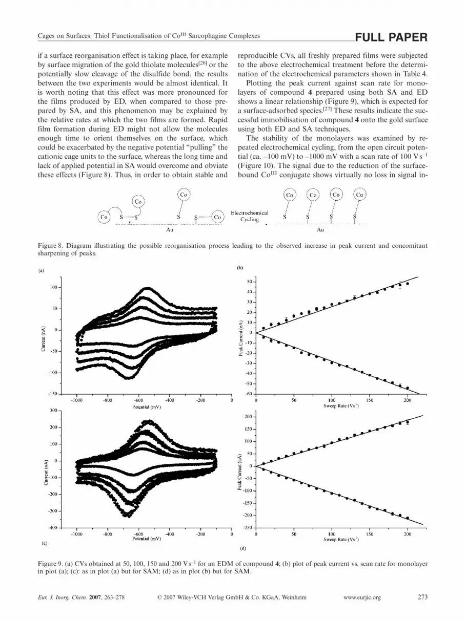
Cages on Surfaces: Thiol Functionalisation of CoIII Sarcophagine Complexes FULL PAPERif a surface reorganisation effect is taking place, for exampleby surface migration of the gold thiolate molecules[26] or thepotentially slow cleavage of the disulfide bond, the resultsbetween the two experiments would be almost identical. Itis worth noting that this effect was more pronounced forthe films produced by ED, when compared to those pre-pared by SA, and this phenomenon may be explained bythe relative rates at which the two films are formed. Rapidfilm formation during ED might not allow the moleculesenough time to orient themselves on the surface, whichcould be exacerbated by the negative potential “pulling” thecationic cage units to the surface, whereas the long time andlack of applied potential in SA would overcome and obviatethese effects (Figure 8). Thus, in order to obtain stable and
Figure 8. Diagram illustrating the possible reorganisation process leading to the observed increase in peak current and concomitantsharpening of peaks.
Figure 9. (a) CVs obtained at 50, 100, 150 and 200 Vs–1 for an EDM of compound 4; (b) plot of peak current vs. scan rate for monolayerin plot (a); (c): as in plot (a) but for SAM; (d) as in plot (b) but for SAM.
Eur. J. Inorg. Chem. 2007, 263–278 © 2007 Wiley-VCH Verlag GmbH & Co. KGaA, Weinheim www.eurjic.org 273
reproducible CVs, all freshly prepared films were subjectedto the above electrochemical treatment before the determi-nation of the electrochemical parameters shown in Table 4.
Plotting the peak current against scan rate for mono-layers of compound 4 prepared using both SA and EDshows a linear relationship (Figure 9), which is expected fora surface-adsorbed species.[27] These results indicate the suc-cessful immobilisation of compound 4 onto the gold surfaceusing both ED and SA techniques.
The stability of the monolayers was examined by re-peated electrochemical cycling, from the open circuit poten-tial (ca. –100 mV) to –1000 mV with a scan rate of 100 Vs–1
(Figure 10). The signal due to the reduction of the surface-bound CoIII conjugate shows virtually no loss in signal in-

G. A. Koutsantonis et al.FULL PAPER
Figure 10. Repeated electrochemical cycling for film of compound 4 (100 Vs–1). (Note: Potential axis reversed for clarity.).
tensity upon repeated cycling for 100 s, indicating a stablemonolayer has been formed.
As the data in Table 4 indicates, the monolayers formedusing both the ED and SA techniques display very similarproperties. The experimentally determined values for bothfilms are virtually identical. Both films exhibit a reversiblereduction of the CoIII cage at E° = –619(4) mV vs. Ag/AgCl, which is anodically shifted compared to the solutionelectrochemistry. The peak separation is nonzero but signif-icantly reduced compared to the solution electrochemistry,indicating potentially an increase in the reversibility of theprocess on the surface. This also signifies that there is somebarrier for electron transfer through the spacer. As wouldbe expected, the molecular footprint is larger than the morecompact and neutral ferrocene conjugates.[18,28] This can berationalised considering the electrostatic repulsion between
Figure 11. (a) XPS of the Co(2p3) region; (b) XPS of the S(2p) region. Closed circles represent experimental spectrum, solid line representscalculated spectrum and dashed lines indicate deconvoluted peaks.
www.eurjic.org © 2007 Wiley-VCH Verlag GmbH & Co. KGaA, Weinheim Eur. J. Inorg. Chem. 2007, 263–278274
the positively charged (sarcophagine)CoIII conjugates onthe surface, and the large Efwhm of ca. 200 mV is consistentwith this, as theoretical treatments[29,30] predict a broaden-ing of Faradaic peaks when the film consists of repulsiveentities. However, it is important to point out that the foot-print and thus the surface concentration for films depositedelectrochemically and prepared by self-assembly are iden-tical. All of the experimentally determined values for E1/2,and importantly, the surface concentration and the relatedspecific area of the molecule are in agreement between thetwo techniques. This shows that both methods can be usedto generate monolayers of the compounds on a gold elec-trode.
SAMs of compound 4 on Au-coated silicon wafers werealso prepared, displaying contact angles of 57(2)° (advanc-ing) and 26(5)° (receding), indicating the presence of a polar

Cages on Surfaces: Thiol Functionalisation of CoIII Sarcophagine Complexes FULL PAPERspecies on the surface. Evidence for a monolayer was ob-tained through the use of ellipsometry, which indicated afilm thickness of (7�1 Å) which is lower than the valueexpected for a fully extended molecule of compound 4 (ca.15 Å). A low value for the film thickness could be due tothe molecules lying at an angle to the surface and/or poorsurface coverage, but it does suggest that the surface ismodified by a monolayer and not a multilayer.
An XPS analysis of a SAM prepared on an Au-coatedsilicon substrate of compound 4 was undertaken in orderto define the nature of the adsorption of the compoundonto the gold surface. Disulfide chemisorption onto gold isgenerally believed to occur through a process in which thedisulfide bond is broken to give two chemically independentmolecules on the surface, bound as thiolates.[26] Peaks cor-responding to S, Co, N, O and C are observed in the XPSspectrum which demonstrates the presence of compound 4on the substrate. Figure 11 shows the major peak in theS(2p) region at 162 eV, which is consistent with the forma-tion of an Au–S bond,[31] allowing us to conclude that thecompound is bound as a thiolate on the surface. The Cobinding energy of 781.5 eV is higher than the reported bind-ing energy of 780.5 eV for the related [Co(NO2)2sar]Cl3,[32]
but does fall within the range of binding energy values forthe CoIII N6 complexes [Co(en)3]Cl3 and [Co(NH3)6]Cl3 of780.2–781.8 eV,[32,33] which indicates the cobalt atom ispresent as CoIII.
Conclusions
In this paper we have described the synthesis and solu-tion electrochemical behaviour of a series of cobalt cagecomplexes bridged with short disulfide-containing peptides.In this way we have demonstrated the efficacy of this syn-thetic strategy which will allow us to tune “tail” length in anumber of applications. We have also shown that it is pos-sible, at least in one case, to immobilise a complex on agold substrate utilising the disulfide linkage present. We be-lieve that this undergoes facile cleavage to give initially spe-cies which lack ordering and subsequent electrochemicalcycling allows them to attain thermodynamically stable sur-face ordering, although this phenomenon requires furtherstudy.
The surface attachment of these peptide-tethered sar-cophagine complexes has allowed us to form modified sur-faces containing redox-active centres and promise entry toan exciting range of applications.
Experimental SectionGeneral: Nuclear magnetic resonance (NMR) spectra were ac-quired with a Bruker ARX 300 (1H at 300 MHz and 13C at75.5 MHz), Bruker AV 500 (1H at 500.13 MHz and 13C at125.8 MHz), or Bruker AV 600 (1H at 600.13 MHz and 13C at150.9 MHz). Chemical shifts for samples measured in D2O are ex-pressed in ppm relative to an internal standard of acetone whichwas taken as being δ = 2.22 ppm for 1H NMR spectra and δ =30.89 ppm for 13C NMR relative to TMS. Chemical shifts for sam-
Eur. J. Inorg. Chem. 2007, 263–278 © 2007 Wiley-VCH Verlag GmbH & Co. KGaA, Weinheim www.eurjic.org 275
ples measured in [D6]DMSO were referenced to the residual solventpeak. Assignments were made with the aid of either the DEPT orHSQC techniques. Mass spectra were recorded using the electro-spray technique (positive ion trap) with a VG Autospec instrumentor QSTAR XL-MS/MS. Microanalyses for carbon, nitrogen andhydrogen were carried out by The Australian National UniversityMicroanalytical Service. All samples were thoroughly dried undervacuum at 50 °C for at least 4 h prior to their analysis. Ion ex-change chromatography was performed under gravity flow usingSP Sephadex C25 (Na+ form, 200–400 mesh) or H+ form Dowex50Wx2 (200–400 mesh) cation exchange resin. Deionised water wasused in all preparations. DMF was predried with molecular sieves(4 Å) prior to use. Dichloromethane was dried by distillation fromCaH2 and stored over molecular sieves (3 Å). Trisodium citrate(Na3Cit) (Ajax), chloroacetic acid (Ajax), sodium picrate (BDH),N-[3-(dimethylamino)propyl]-N�-ethylcarbodiimide hydrochloride(EDC·HCl) (Aldrich), cystamine dihydrochloride (CSA·2HCl) (Al-drich), diisopropylethylamine (DIPEA) (Lancaster), N-hydroxysuc-cinimide (HOSu) (Aldrich), 2-benzotriazole-N,N,N,N,-tetrameth-yluronium hexafluorophosphate (HBTU) (Auspep), trifluoroaceticacid (TFA) (Aldrich), Boc-protected amino acids (Auspep), and tri-fluoromethanesulfonic (triflic) acid (Lancaster) were used as re-ceived. Solution electrochemical experiments were carried out atroom temperature (23�1 °C) with a MacLab Potentiostat orCHInstruments potentiostat model 660B or 440 in 0.1 Na-ClO4(aq) adjusted to pH = 7.3 with tris(perchlorate) buffer, and ananalyte concentration of 1 m. A three-electrode cell system con-sisting of glassy carbon (BAS, 3.0 mm diameter) as the workingelectrode, Pt wire as the counter electrode and Ag/AgCl (3.0 KCl)as the reference electrode was employed. The carbon electrodeswere cleaned by polishing with a micro-cloth pad with Al2O3 slurry(0.05 µm) and washed thoroughly with water before use. For thecyclic voltametric (CV) studies the scan rate was 100 mVs–1 andfor the DPV experiments a scan rate of 20 mVs–1 and pulse ampli-tude of 50 mV was used. Cyclic voltammograms (CVs) werescanned in the potential range of 0.2 to –1.0 V vs. Ag/AgCl.[Co(CH3)(NH2)sar]Cl3·HCl·0.5H2O,[34] rac- and ∆-[Co(NH2)-(NH2CH2CO2H)sar]Cl3·2HCl,[16] [Boc-Gly-CSA]2 and [Boc-Ala-CSA]2[35] were all synthesised as reported previously.
Structure Determinations: Full spheres of CCD area-detector dif-fractometer data were measured (ω-scans; monochromatic Mo-Kα
radiation, λ = 0.71073 Å). Nt(otal) reflections were obtained, thesemerging to N unique (Rint cited) after “empirical”/multiscan ab-sorption correction (proprietary software), No with F � 4σ(F) con-sidered “observed” and used in the full-matrix least-squares refine-ments on F2, refining anisotropic displacement parameter forms,(x,y,z,Uiso)H being included constrained at estimates. Conventionalresiduals R, Rw {weights: [σ2(F2) + nwF2]–1} are cited at conver-gence. Neutral atom complex scattering factors were employedwithin the context of the Xtal 3.7 program system.[36] Pertinent re-sults are given above and in Tables 2 and 3 and Figure 2.In (3)Cl4·2H2O, a pair of substantial difference map residues wereassigned as water molecule oxygen atoms, site occupancies unityafter trial refinement, albeit with high displacement parameters,associated hydrogen atoms not being located. CCDC-605503to -605505 contain the supplementary crystallographic data for thispaper. These data can be obtained free of charge from The Cam-bridge Crystallographic Data Centre via www.ccdc.cam.ac.uk/data_request/cif.
Electrodeposition: All electrodeposition experiments were per-formed using a three-electrode cell system consisting of Au (25 µmdiameter) as the working electrode, Pt wire as the counter electrodeand Ag wire as the reference electrode. Electrodeposition was ac-

G. A. Koutsantonis et al.FULL PAPERcomplished by placing a freshly oxidised microelectrode (electro-chemical cycling from –0.4 to +1.2 V vs. Pt wire in 0.5 H2SO4)into a 1 m solution of the analyte in 0.1 NaClO4(aq) and apply-ing –1.5 V for 30 min. After this period, the electrode was washedwith water, then suspended in a vigorously stirred beaker of waterfor 10 min, and then 2 NaClO4(aq) for 30 s. All electrochemicalmeasurements were performed using a custom-built potentiostat in2 NaClO4(aq) using a three-electrode cell system consisting of Au(25 µm diameter) as the working electrode, Pt wire as the counterelectrode and Ag/AgCl as the reference electrode, which was placedin a separate cell and connected to the analyte cell with a salt bridge(KNO3). The electrochemical cell was enclosed in a grounded Fara-day cage, and all electrolytes were degassed with a flow of N2(g)
prior to the experiments, and an N2(g) blanket maintained through-out the course of the measurements. All electrochemical measure-ments were performed on at least five different microelectrodes toensure reproducibility.
Self-Assembly: Monolayers were formed by soaking a freshlycleaned electrode (electrochemical cycling from –0.4 to +1.2 V vs.Pt wire in 0.5 H2SO4) in a 1 m aqueous solution of the complexfor 5 d. After this period, the electrodes were treated exactly asabove for the electrodeposited materials.
X-ray Photoelectron Spectroscopy: XPS measurements were per-formed using an Axis-165 X-ray Photoelectron Spectrometer(Kratos Analytical) with a monochromatic Al-Ka X-ray source(1486.7 eV). Survey spectra (0–1100 eV) were taken at constantanalyser pass energy of 160 eV and high-resolution spectra of Au4f,C1s, N1s, O1s, S2p and Co2p, were acquired with a pass energy of20 eV and a down time of 200 ms. The take-off angle measured asthe angle between the film surface and the photoelectron energyanalyser was 90°. The typical operating pressure in the analysingchamber was ca. 5·10–10 Torr. Number of scans for high resolutionrequired obtaining high signal/noise ratio varied from 4 for Au to40 for Co, and 60 for S. The binding energies were referenced toAu4f7/2 at 84.0 eV.
[Co(CH3)(NH2CH2CO2H)sar]Cl4·H2O (2)Cl4·H2O and [Co(CH3)-{N(CH2CO2H)2}sar]Cl3·2H2O (3)Cl3·2H2O: [Co(CH3)(NH2)sar]-Cl3·HCl·0.5H2O (2.10 g, 4.0 mmol) was dissolved in H2O (ca.20 mL) in a three-necked round-bottomed flask, and the pH ad-justed to ca. 10 by the addition of NaOH(aq). A solution of Na-CO2CH2Cl (25 g, 0.21 mol) in water (ca. 100 mL) was then added,and the pH again adjusted to ca. 10 by addition of more Na-OH(aq). The solution was then heated at 80 °C under Ar(g) for7 d, and the pH maintained at ca. 9–10 by dropwise addition ofNaOH(aq). The reaction was monitored on micro-columns of Na+
form SP C25 Sephadex by elution with 0.1 Na3Cit. More Na-CO2CH2Cl was added after 3 d (25 g, 0.21 mmol) and 5 d (10 g,0.09 mmol), and after 7 d, the reaction mixture was acidified withacetic acid (pH � 5), diluted to 2 L in water, and applied to acolumn of H+ form Dowex 50Wx2 cation exchange resin. The col-umn was washed with water (500 mL), 1 HCl (500 mL, to removeNa+) and finally the products eluted with 3 HCl. The fractioncontaining the products was then dried in the rotary evaporator,and the solid thus obtained dissolved into water (1 L) and appliedto a column of Na+ form Sephadex cation exchange resin. Elutionwith 0.05 Na3Cit produced three bands, the first being a minoryellow band, the second the major orange band, and the third aminor orange band. The three bands were applied to H+ formDowex 50Wx2 cation exchange resin separately, and treated withwater (500 mL), 1 HCl (500 mL) and eluted with 3 HCl. Theproduct from Band 1 was dried in a rotary evaporator, dissolved inhot water and precipitated by the addition of EtOH and finally
www.eurjic.org © 2007 Wiley-VCH Verlag GmbH & Co. KGaA, Weinheim Eur. J. Inorg. Chem. 2007, 263–278276
Et2O and cooling in the freezer. The product was collected by suc-tion filtration, washed with a little cold EtOH, then finally Et2O,and dried under vacuum to give (3)Cl3·2H2O (0.22 g, 0.35 mmol)as orange crystals. 1H NMR (300 MHz, D2O): δ = 0.9 (s, 3 H,–CH3), 2.4–3.5 (m, 24 H, cage –CH2–), 3.6 (s, 4 H, 2 iminodiacet-ate –CH2–). 13C NMR (75.5 MHz, D2O): δ = 20.1 (–CH3), 42.3 (Cof methyl cage cap), 52.1 (cage –CH2–), 52.5 (iminodiacetate–CH2–), 54.4, 54.7, 55.2 (cage –CH2–), 64.5 (C of amino cage cap),176.8 (C=O). ES-MS: m/z = 243.64 [C19H39CoN7O4 – H+]2+,486.21 [C19H39CoN7O4 – 2 H+]+, 522.22 [C19H39CoN7O4 – H+ +Cl–]+. C19H43Cl3CoN7O6 [(3)Cl3·2H2O, 630.88]: calcd. C 36.17, H6.87, N 15.54; found C 36.49, H 6.52, N 15.70. Crystals of (3)Cl3·2H2O suitable for an X-ray structure determination were grownby slow concentration of a 2 HCl(aq) solution of the complex.The product from Band 2 was treated as above for Band 1 to give(2)Cl4·H2O (1.26 g, 2.13 mmol) as orange crystals. 1H NMR(500 MHz, D2O): δ = 0.93 (s, 3 H, –CH3), 2.4–3.5 (m, 24 H, cage–CH2–), 3.69 (AB q, 2 H, Gly –CH2–). 13C NMR (126 MHz, D2O):δ = 19.9 (–CH3), 42.8 (C of methyl cage cap), 43.6 (Gly –CH2–),52.0, 55.1, 55.2, 55.3 (cage –CH2–), 60.8 (C of amino cage cap),172.5 (C=O). ES-MS: m/z = 464.21 [C17H37CoN7O2 – H+ + Cl–]+,428.23 [C17H37CoN7O2 – 2 H+]+, 500.16 [C17H37CoN7O2 +2 Cl–]+. C17H40Cl4CoN7O3 [(2)Cl4·H2O, 591.29]: calcd. C 34.53, H6.82, N 16.58; found C 34.58, H 6.72, N 16.62. Crystals of (2)Cl4·H2O suitable for an X-ray structure determination were grownby slow concentration of a 2 HCl(aq) solution of the complex.The triflate of [Co(CH3)(NHCH2CO2H)sar]3+ was obtained by dis-solution of the chloride in neat triflic acid at ca. 50 °C whilst aconstant stream of N2 was bubbled though the solution for at least1 h. The orange solution thus obtained was then cooled on ice andEt2O added slowly with stirring to precipitate the triflate as a yel-low-orange powder, which was collected by suction filtration andwashed with copious quantities of Et2O. The product from Band 3was determined from 1H NMR spectroscopy to be the starting ma-terial (0.35 g, 0.67 mmol).
[Co(CH3)(Gly-NHCH2CH2S-)sar]2(ClO4)6·HClO4 (4)(ClO4)6·HClO4: [Co(CH3)(NH2CH2CO2H)sar](CF3SO3)4 (0.60 g,0.58 mmol) was dissolved in DMF (30 mL), and N-hydroxysuc-cinimide (0.067 g, 0.58 mmol) was added to the orange solution,followed by DIPEA (0.4 mL, 2.3 mmol) and EDC·HCl (1.0 g,5.2 mmol) and the orange mixture was stirred for 10 min. Cysta-mine dihydrochloride (CSA·2HCl) (0.063 g, 0.28 mmol) was thenadded, causing a precipitate to form immediately, which redissolvedafter ca. 10 min to give an almost clear solution. The mixture wasstirred for 24 h, during which time a white precipitate had formed.The reaction mixture was then diluted into water (500 mL) andapplied to a column of Na+ form SP Sephadex SP 25 resin, whichwas washed with water (500 mL), then elution was commencedwith 0.05–0.2 Na3Cit, which began to remove four bands, thefourth by far the major component. Band 4 was treated with aconcentrated aqueous solution of sodium picrate (NaPic), until theprecipitation of a very fine bright yellow solid ceased. The precipi-tate was collected on a sintered glass frit, at first by gravity, andthen suction filtration. This material was washed well with water,and the moist yellow solid obtained was then suspended in water(ca. 100 mL) containing ca. 3 mL of concd. HClO4(aq), and to thiswas added EtOAc and the mixture stirred vigorously. Stirring wasstopped periodically and the top layer of EtOAc decanted off andreplaced with fresh solvent, until all of the solid had dissolved,leaving an orange aqueous phase, which was subsequently transfer-red to a separating funnel and extracted with EtOAc until the or-ganic phase was colourless. The aqueous phase was then concen-trated in a rotary evaporator until precipitation of an orange solid

Cages on Surfaces: Thiol Functionalisation of CoIII Sarcophagine Complexes FULL PAPERcommenced, then placed on ice to complete the precipitation. Theorange precipitate was collected on a sintered glass frit and washedwith EtOH and then Et2O and air-dried to give (4)(ClO4)6·HClO4
(0.32 g, 0.19 mmol). The sample was recrystallised from warmH2O/EtOH by the addition of Et2O. 1H NMR 600 MHz, [D6]-DMSO): δ = 0.8 (s, 6 H, 2 –CH3), 2.20–2.48 and 2.85–3.15 (m, 48H, 2 cage –CH2–), 2.53 (t, ca. 2 H, 2 cage pendent –NH–), 2.79 (t,4 H, 2 –CH2–S), 3.18 (m, 4 H, 2 Gly1 –CH2–), 3.39 (q, 4 H, 2CSA –CH2–N), 6.5 (br., 12 H, cage –NH–), 7.95 (t, 2 H, 2 CSAamide NH). 13C NMR (151 MHz, [D6]DMSO): δ = 19.5 (–CH3),37.0 (CSA –CH2–S), 37.8 (CSA –CH2–N), 42.1 (C of methyl cagecap), 44.3 (Gly1 –CH2–), 52.0, 53.5, 54.0, 54.2 (cage –CH2–), 60.3(C of amino cage cap), 171.0 (C=O). ES-MS: m/z = 486.22[C38H82Co2N16O2S2 – 4 H+]2+. C38H83Cl7Co2N16O30S2 [(4)-(ClO4)6·HClO4, 1674.32]: calcd. C 27.26, H 5.00, N 13.38; found C27.43, H 5.38, N 13.31.
[Co(CH3)(Gly-Gly-NHCH2CH2S-)sar]2(ClO4)6·HClO4·0.5H2O (5)-(ClO4)6·HClO4·0.5H2O: [Co(CH3)(NH2CH2CO2H)sar](CF3SO3)4
(0.80 g, 0.78 mmol) was dissolved in DMF (20 mL), and DIPEA(0.14 mL, 0.80 mmol) added, followed by N-hydroxysuccinimide(0.138 g, 1.20 mmol), followed by HBTU (0.445 g, 1.17 mmol) andthe orange solution was stirred for 20 min. In a separate round-bottomed flask, Boc-Gly-CSA (0.177 g, 0.380 mmol) was dissolvedin CH2Cl2 (3 mL) containing TFA (1.5 mL) and the solution stirredfor 1 h, during which time, an oily material separated from themixture. The solvents were removed under reduced pressure, andthe oily residue redissolved in DMF (6 mL) containing an excessof DIPEA (0.36 mL), and then added to the orange cage solution,which was then stirred at room temp. for 18 h. The workup pro-cedure was identical to that of compound 4 above, to give (5)-(ClO4)6·HClO4·0.5H2O as an orange solid (0.43 g, 0.24 mmol). Thesample was recrystallised from warm H2O by the addition ofEtOH, to give an orange oil which solidified upon standing at roomtemp. for several hours. 1H NMR (600 MHz, [D6]DMSO): δ = 0.80(s, 6 H, 2 –CH3), 2.20–2.50 and 2.85–3.15 (m, 48 H, 2 cage–CH2–), 2.60 (t, 2 H, 2 cage pendent –NH–), 2.76 (t, 4 H, 2 –CH2–S), 3.21 (dq, 4 H, 2 Gly1 –CH2–), 3.36 (m, 4 H, 2 CSA –CH2–N),3.73 (d, 4 H, 2 Gly2 –CH2–), 6.51 (br. s, 6 H, 2 cage NH), 6.56 (br.s, 6 H, 2 cage NH), 8.00 (t, 2 H, 2 Gly2 amide NH), 8.08 (t, 2 H,2 CSA amide NH). 13C NMR (151 MHz, [D6]DMSO): δ = 19.5(–CH3), 36.9 (CSA –CH2–S), 38.0 (CSA –CH2–N), 41.5 (Gly2
–CH2–), 42.1 (C of methyl cage cap), 44.3 (Gly1 –CH2–), 52.1, 53.6,54.0, 54.1 (cage –CH2–), 60.3 (C of amino cage cap), 168.9, 171.2(C=O). ES-MS: m/z = 362.51 [C42H88Co2N18O4S2 – 3 H+]3+,395.83 [C42H88Co2N18O4S2 – 2 H+ + ClO4
–]3+, 272.13 [C42H88Co2-N18O4S2 – 2 H+]4+, 543.25 [C42H88Co2N18O4S2 – 4 H+]2+.C42H90Cl7Co2N18O32.5S2 [(5)(ClO4)6·HClO4·0.5H2O, 1797.43]:calcd. C 28.06, H 5.05, N 14.03; found C 28.14, H 5.43, N 13.99.
[Co(CH3)(Gly-Ala-NHCH2CH2S-)sar]2(ClO4)6·HClO4·H2O (6)-(ClO4)6·HClO4·H2O: The procedure used was the same as that forcompound 5 above. From [Co(CH3)(NH2CH2CO2H)sar]-(CF3SO3)4 (0.86 g, 0.84 mmol), DMF (15 mL), DIPEA (0.15 mL,0.86 mmol + 0.5 mL, 3 mmol), N-hydroxysuccinimide (0.11 g,0.96 mmol), HBTU (0.35 g, 0.92 mmol), Boc-Ala-CSA (0.20 g,0.40 mmol), was obtained (6)(ClO4)6·HClO4·H2O (0.36 g,0.20 mmol). The sample was recrystallised from warm H2O by theaddition of EtOH. 1H NMR (600 MHz, [D6]DMSO): δ = 0.79 (s,6 H, 2 cage –CH3), 1.22 (d, 6 H, 2 Ala –CH3), 2.20–2.50 and 2.85–3.15 (m, 48 H, 2 cage –CH2–), 2.56 (AB q, 2 H, 2 cage pendent–NH–), 2.76 (m, 4 H, 2 –CH2–S), 3.17 (m, 4 H, 2 Gly1 –CH2–),3.25–3.50 (m, 4 H, 2 CSA –CH2–N), 4.28 [sext, 2 H, Ala2
–CHα–), 6.51 (br. s, 6 H, 2 cage –NH–), 6.56 (br. s, 6 H, 2 cage–NH–), 7.85 (d, 2 H, 2 Ala2 amide NH), 8.16 (t, 2 H, 2 CSA amide
Eur. J. Inorg. Chem. 2007, 263–278 © 2007 Wiley-VCH Verlag GmbH & Co. KGaA, Weinheim www.eurjic.org 277
NH). 13C NMR (151 MHz, [D6]DMSO): δ = 18.9 (Ala –CH3), 19.5(cage –CH3), 36.9 (CSA –CH2–S), 37.9 (CSA –CH2–N), 42.0 (C ofmethyl cage cap), 44.4 (Gly1 –CH2–), 47.7 (Ala2 –CHα–), 52.2,53.6, 53.9, 54.0 (cage –CH2–), 60.2 (C of amino cage cap), 170.1,172.1 (C=O). Note: Some peaks were doubled due to the presenceof meso and rac compounds but these doubled peaks are reportedwith a single chemical shift in order to simplify the analysis of thespectra. ES-MS: m/z = 371.85 [C44H92Co2N18O4S2 – 3 H+]3+,557.27 [C44H92Co2N18O4S2 – 4 H+]2+. C44H95Cl7Co2N18O33S2
[(6)(ClO4)6·HClO4·H2O, 1834.49]: calcd. C 28.81, H 5.22, N 13.74;found C 28.82, H 5.41, N 13.69.
[Co(NH2)(Gly-NHCH2CH2S-)sar]2(ClO4)6·HClO4·2H2O (7)-(ClO4)6·HClO4·2H2O: The procedure used was the same as thatfor compound 4 above. From [Co(NH3)(NH2CH2CO2H)-sar](CF3SO3)5 (2.55 g, 2.16 mmol), DMF (15 mL), DIPEA(0.76 mL, 4.4 mmol + 0.5 mL, 3 mmol), N-hydroxysuccinimide(1.01 g, 8.8 mmol), EDC·HCl (1.7 g, 8.9 mmol), CSA·2HCl (0.22 g,1.0 mmol), was obtained (7)(ClO4)6·HClO4·2H2O (0.45 g,0.26 mmol). The sample was recrystallised from warm H2O by theaddition of EtOH. 1H NMR (600 MHz, [D6]DMSO): δ = 1.72 (br.,4 H, 2 –NH2), 2.25–2.50 and 2.75–3.20 (m, 48 H, 2 cage –CH2–),2.53 (t, 1 H, 2 Gly1 –NH–), 2.79 (t, 4 H, 2 –CH2–S), 3.16 (m, 4 H,2 Gly1 –CH2–), 3.39 (q, 4 H, 2 CSA –CH2–N), 6.53 (br. s, 6 H, 2cage NH), 6.59 (br. s, 6 H, 2 cage NH), 7.95 (t, 2 H, 2 CSA amideNH). 13C NMR (151 MHz, [D6]DMSO): δ = 37.0(–CH2–S), 37.7 (CSA –CH2–N), 44.3 (Gly1 –CH2–), 52.1, 54.0,54.1, 54.5 (cage –CH2–), 56.9, 60.3 (cage C), 170.9 (Gly1 C=O).ES-MS: m/z = 325.15 [C36H80Co2N18O2S2 – 3 H+]3+, 244.12[C36H80Co2N18O2S2 – 2 H+]4+, 487.22 [C36H80Co2N18O2S2 –4 H+]2+, 358.47 [C36H80Co2N18O2S2 – 2 H+ + ClO4
–]3+.C36H85Cl7Co2N18O32S2 [(7)(ClO4)6·HClO4·2H2O, 1712.33]: calcd.C 25.25, H 5.00, N 14.72; found C 25.44, H 4.87, N 14.57.
∆-[Co(NH2)(Gly-NHCH2CH2S-)sar]2(ClO4)6·HClO4·2H2O (8)-(ClO4)6·HClO4·2H2O: The procedure used was the same as that forcompound 4 above. From [Co(NH3)(NH2CH2CO2H)sar]Cl5·3H2O(0.50 g, 0.75 mmol), DMF (15 mL), DIPEA (0.26 mL, 1.5 mmol +0.5 mL, 3 mmol), N-hydroxysuccinimide (0.17 g, 1.5 mmol),EDC·HCl (0.29 g, 1.5 mmol), CSA·2HCl (0.084 g, 0.37 mmol), wasobtained (8)(ClO4)6·HClO4·2H2O (0.42 g, 0.24 mmol). The samplewas recrystallised from warm H2O by the addition of EtOH. 1HNMR (600 MHz, [D6]DMSO): δ = 2.40–2.65 and 2.90–3.35 (m, 48H, 2 cage –CH2–), 2.79 (t, 4 H, 2 –CH2–S), 3.17 (m, 4 H, 2 Gly1
–CH2–), 3.41 (q, 4 H, 2 CSA –CH2–N), 6.57 (br. s, 6 H, 2 cage–NH–), 6.84 (br. s, 6 H, 2 cage –NH–), 7.97 (t, 2 H, 2 CSA amideNH), 8.36 (br. s, 4 H, 2 free –NH2). 13C NMR (151 MHz, [D6]-DMSO): δ = 36.9 (CSA –CH2–S), 37.8 (CSA –CH2–N), 44.3(Gly1 –CH2–), 50.9, 52.1, 53.6 (2 C) (cage –CH2–), 55.6, 60.5 (C),170.2 (C=O). ES-MS: m/z = 244.16 [C36H80Co2N18O2S2 – 2 H+]3+,358.51 [C36H80Co2N18O2S2 – 2 H+ + ClO4
–]3+. C36H85Cl7Co2-N18O32S2 [(8)(ClO4)6·HClO4·2H2O, 1712.33]: calcd. C 25.25, H5.00, N 14.72; found C 24.99, H 5.02, N 14.35.
∆-[Co(NH2)(Gly-Gly-NHCH2CH2S-)sar]2(ClO4)6·4HClO4 (9)-(ClO4)6·4HClO4: The procedure used was the same as that for com-pound 5 above. From [Co(NH3)(NH2CH2CO2H)sar]Cl5·3H2O(0.50 g, 0.75 mmol), DMF (15 mL), DIPEA (0.26 mL, 1.5 mmol +0.5 mL, 3 mmol), N-hydroxysuccinimide (0.17 g, 1.5 mmol),EDC·HCl (0.29 g, 1.5 mmol), Boc-Gly-CSA (0.17 g, 0.36 mmol),was obtained (9)(ClO4)6·4HClO4 (0.21 g, 0.10 mmol). The samplewas recrystallised from warm H2O by the addition of EtOH. 1HNMR (600 MHz, [D6]DMSO): δ = 2.40–2.70 and 2.90–3.35 (m, 48H, 2 cage –CH2–), 2.76 (t, 4 H, 2 –CH2–S), 3.24 (s, 4 H, 2 Gly1
–CH2–), 3.36 (q, 4 H, 2 CSA –CH2–N), 3.74 (d, 4 H, 2 Gly2

G. A. Koutsantonis et al.FULL PAPER–CH2–), 6.59 (br. s, 6 H, 2 cage –NH–), 6.85 (br. s, 6 H, 2 cage–NH–), 8.01 (s, 2 H, 2 Gly2 amide NH), 8.11 (t, 2 H, 2 CSA amideNH), 8.41 (br. s, 4 H, 2 free –NH2). 13C NMR (151 MHz,[D6]DMSO): δ = 36.9 (CSA –CH2–S), 37.9 (CSA –CH2–N), 41.6(Gly2 –CH2–), 44.3 (Gly1 –CH2–), 50.9, 52.1, 53.4 (2 C) (cage–CH2–), 55.5, 60.4 (C), 168.7, 170.6 (C=O). ES-MS: m/z = 544.24[C40H86Co2N20O4S2 – 4 H+]2+. C40H90Cl10Co2N20O44S2 [(9)(ClO4)6·4HClO4, 2091.78]: calcd. C 22.97, H 4.34, N 13.39; foundC 23.20, H 4.47, N 13.38.
∆-[Co(NH2)(Gly-Ala-NHCH2CH2S-)sar]2(ClO4)6·2HClO4·H2O(10)(ClO4)6·2HClO4·H2O: The procedure used was the same asthat for compound 5 above. From [Co(NH3)(NH2CH2CO2H)-sar](CF3SO3)5 (1.00 g, 0.85 mmol), DMF (15 mL), DIPEA(0.30 mL, 1.7 mmol + 0.50 mL, 3 mmol), N-hydroxysuccinimide(0.11 g, 0.96 mmol), HBTU (0.35 g, 0.92 mmol), Boc-Ala-CSA(0.20 g, 0.40 mmol), was obtained (10)(ClO4)6·2HClO4·H2O(0.42 g, 0.22 mmol). The sample was recrystallised from warm H2Oby the addition of EtOH. 1H NMR (600 MHz, [D6]DMSO): δ =1.22 (d, 6 H, 2 Ala –CH3), 2.40–2.70 and 2.85–3.40 (m, 48 H, 2cage –CH2–), 2.76 (m, 4 H, 2 –CH2–S), 3.17 (AB q, 4 H, 2 Gly1
–CH2–), 3.25–3.45 (m, 4 H, 2 CSA –CH2–N), 4.30 (quint, 2 H, 2Ala2 –CHα–), 6.54 (br. s, 6 H, 2 cage –NH), 6.80 (br. s, 6 H, 2cage –NH), 7.85 (d, 2 H, 2 Ala2 amide NH), 8.20 (t, 2 H, 2 CSAamide NH), 8.41 (br. s, 4 H, 2 free –NH2). 13C NMR (151 MHz,[D6]DMSO): δ = 19.00 (Ala2 –CH3), 36.92 (CSA –CH2–S), 37.87(CSA –CH2–N), 44.47 (Gly1 –CH2–), 47.73 (Ala2 –CHα–), 51.20,52.32, 53.28 (cage –CH2–), 55.62, 60.34 (cage C), 169.90, 172.12(C=O). ES-MS: m/z = 372.51 [C42H90Co2N20O4S2 – 3 H+]3+,279.64 [C42H90Co2N20O4S2 – 2 H+]4+. C42H94Cl8Co2N20O37S2
[(10)(ClO4)6·2HClO4·H2O, 1936.93]: calcd. C 26.04, H 4.89, N14.46; found C 26.24, H 5.01, N 14.28.
Acknowledgments
We thank the University of Western Australia for partial fundingof this research and NSERC for financial support. H.-B. K. is theCanadian Research Chair in Biomaterials. G. L. N. was the holderof an Australian Postgraduate Award. We also thank Dr. DimitreKarpuzov, University of Alberta, for the XPS measurements.
[1] J. C. Love, L. A. Estroff, J. K. Kriebel, R. G. Nuzzo, G. M.Whitesides, Chem. Rev. 2005, 105, 1103.
[2] M.-C. Daniel, D. Astruc, Chem. Rev. 2004, 104, 293.[3] A. M. Sargeson, Pure Appl. Chem. 1986, 58, 1511.[4] A. M. Sargeson, Coord. Chem. Rev. 1996, 151, 89.[5] M. H. Jensen, P. Osvath, A. M. Sargeson, J. Ulstrup, J. Elec-
troanal. Chem. 1994, 377, 131.[6] J. T. Hupp, H. Y. Liu, P. A. Lay, W. H. F. Petri, A. M.
Sargeson, M. J. Weaver, J. Electroanal. Chem. Interfacial Elec-trochem. 1984, 163, 371.
[7] I. I. Creaser, A. Hammershoei, A. Launikonis, A. W. H. Mau,A. M. Sargeson, W. H. F. Sasse, Photochem. Photobiol. 1989,49, 19.
[8] C. Konigstein, A. W. H. Mau, P. Osvath, A. M. Sargeson,Chem. Commun. 1997, 423.
www.eurjic.org © 2007 Wiley-VCH Verlag GmbH & Co. KGaA, Weinheim Eur. J. Inorg. Chem. 2007, 263–278278
[9] A. Launikonis, P. A. Lay, A. W. H. Mau, A. M. Sargeson,W. H. F. Sasse, Sci. Pap. Inst. Phys. Chem. Res. (Jpn.) 1984,78, 198.
[10] A. W. H. Mau, W. H. F. Sasse, I. I. Creaser, A. M. Sargeson,Nouv. J. Chim. 1986, 10, 589.
[11] L. R. Gahan, T. W. Hambley, A. M. Sargeson, M. R. Snow,Inorg. Chem. 1982, 21, 2699.
[12] S. Burnet, M.-H. Choi, P. S. Donnelly, J. M. Harrowfield, I.Ivanova, S.-H. Jeong, Y. Kim, M. Mocerino, B. W. Skelton,A. H. White, C. C. Williams, Z.-L. Zeng, Eur. J. Inorg. Chem.2001, 1869.
[13] A. S. Paxinos, H. Gunther, D. J. M. Schmedding, H. Simon,Bioelectrochem. Bioenerg. 1991, 25, 425.
[14] J. M. Harrowfield, G. A. Koutsantonis, G. L. Nealon, B. W.Skelton, A. H. White, Eur. J. Inorg. Chem. 2005, 2384.
[15] G. W. Walker, R. J. Geue, A. M. Sargeson, C. A. Behm, DaltonTrans. 2003, 2992.
[16] P. S. Donnelly, J. M. Harrowfield, B. W. Skelton, A. H. White,Inorg. Chem. 2000, 39, 5817.
[17] M. Bodanszky, A. Bodanszky, The Practice of Peptide Synthe-sis, 2nd ed., Springer Verlag, Berlin, 1994.
[18] G. A. Orlowski, S. Chowdhury, Y.-T. Long, T. C. Sutherland,H.-B. Kraatz, Chem. Commun. 2005, 1330.
[19] H.-B. Kraatz, J. Inorg. Organomet. Polym. 2005, 15, 83.[20] D. W. Conrad, H. Zhang, D. E. Stewart, R. A. Scott, J. Am.
Chem. Soc. 1992, 114, 9909.[21] P. S. Donnelly, J. M. Harrowfield, J. Chem. Soc., Dalton Trans.
2002, 906.[22] S. F. Ralph, M. M. Sheil, L. A. Hick, R. J. Geue, A. M.
Sargeson, J. Chem. Soc., Dalton Trans. 1996, 4417.[23] J. M. Harrowfield, Supramol. Chem. 2006, 18, 125 and refer-
ences cited therein.[24] A. M. Bond, G. A. Lawrance, P. A. Lay, A. M. Sargeson, Inorg.
Chem. 1983, 22, 2010.[25] K. V. Gobi, T. Okajima, K. Tokuda, T. Ohsaka, Langmuir
1998, 14, 1108.[26] A. Ulman, Chem. Rev. 1996, 96, 1533 and references cited
therein.[27] A. J. Bard, L. R. Faulkner, Electrochemical Methods, 2nd ed.,
John Wiley & Sons, New York, 2001.[28] I. Bediako-Amoa, T. C. Sutherland, C.-Z. Li, R. Silerova, H.-
B. Kraatz, J. Phys. Chem. B 2004, 108, 704.[29] E. Laviron, J. Electroanal. Chem. 1979, 100, 263.[30] H. Matsuda, K. Aoki, K. Tokuda, J. Electroanal. Chem. 1987,
217, 15.[31] T. Ishida, N. Choi, W. Mizutani, H. Tokumoto, I. Kojima, H.
Azehara, H. Hokari, U. Akiba, M. Fujihira, Langmuir 1999,15, 6799.
[32] A. A. Achilleos, L. R. Gahan, T. W. Hambley, P. C. Healy,D. M. Weedon, Inorg. Chim. Acta 1989, 157, 209.
[33] Y. Okamoto, H. Nakano, T. Imanaka, S. Teranishi, Bull. Chem.Soc. Jpn. 1975, 48, 1163.
[34] R. J. Geue, T. W. Hambley, J. M. Harrowfield, A. M. Sargeson,M. R. Snow, J. Am. Chem. Soc. 1984, 106, 5478.
[35] S. Chowdhury, G. Schatte, H.-B. Kraatz, Dalton Trans. 2004,1726.
[36] S. R. Hall, D. J. Du Boulay, R. E. Olthof-Hazekamp, XTAL3.7, The University of Western Australia, 2001.
Received: May 3, 2006Published Online: November 9, 2006


