Embed Size (px)
Citation preview
Special FocuS Review
www.landesbioscience.com virulence 1
virulence 4:8, 1–30; November 15, 2013; © 2013 landes Bioscience
Special FocuS Review
Introduction: Biological Warfare and Bioterrorism Agents
In recent years, the possibility of biological warfare and bio-terrorism has become of increasing concern to both military planners and civil defense authorities. The mailing of anthrax spore containing letters to destinations within the United States in 2001 brought the sudden realization that bioterrorism is not merely a theoretical threat but a real and present danger. Since then, much thought and planning has gone into defining possible biowarfare and bioterrorism agents. There are six requirements for these agents that are relevant here:
1) A high degree of morbidity and lethality.2) Highly infectious microbes or highly toxic substances.3) Easy to distribute widely in an active form.4) Easy to produce in bulk and store until delivered.5) Reasonably hardy in the environment after distribution.6) Bacteria should be genetically engineered to be resistant to
known antibiotic drugs.The 2001 bioterrorist attacks in the USA using anthrax spores
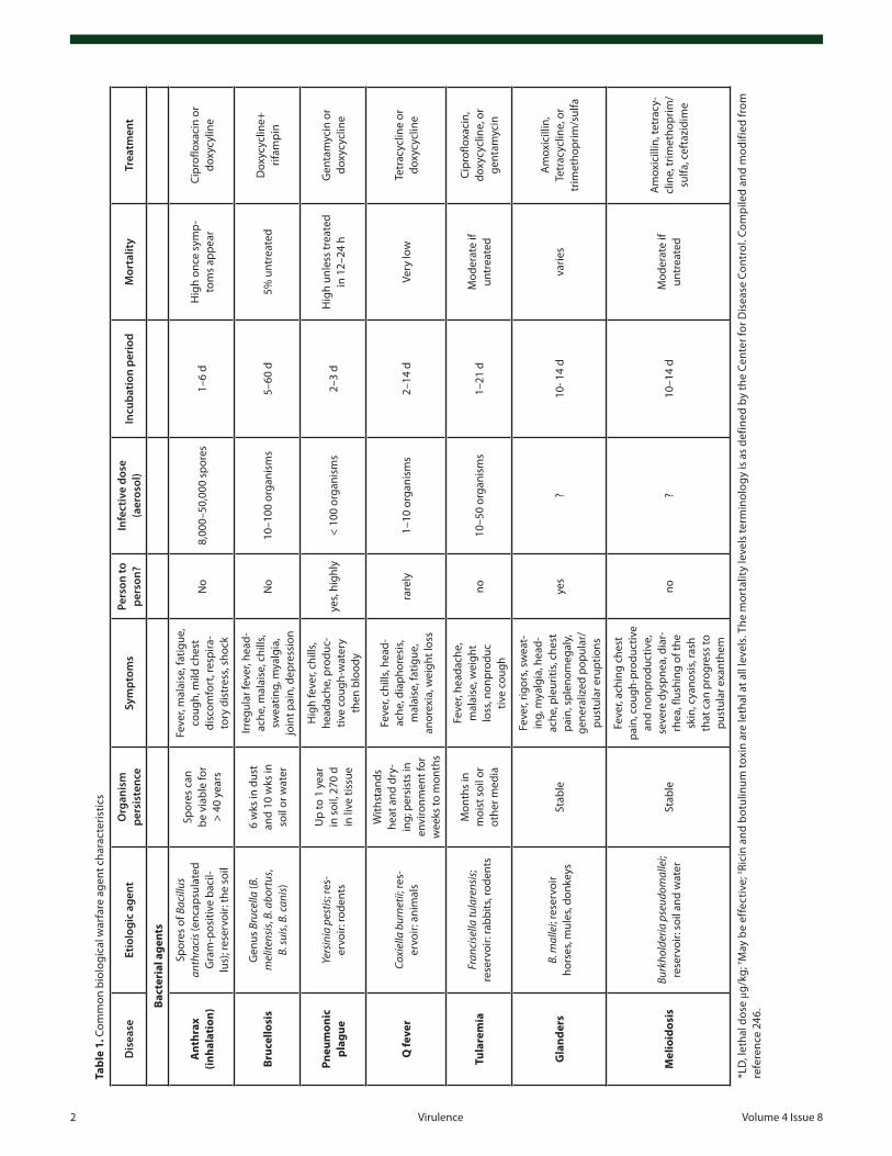
and spreading medium the US Postal Service have once more emphasized the need of early detection and decontamination of critical facilities in the shortest possible time. During the recent decade there has been a remarkable progress in the detection, pro-tection, and decontamination of biological warfare agents since various and sophisticated detection/decontamination methods have been developed and implemented. Nevertheless the threat of biological warfare agents and their possible use in bioterrorist attacks still remains a leading cause of concern in the global com-munity. Furthermore, in the past decade there have been threats to the global society due to the emergence of new infectious dis-eases and/or re-emergence of old infectious diseases that were considered eliminated. Adding to the milieu the observed global rise in the antimicrobial resistance, the preparedness of societies against these agents becomes obvious. Under these circumstances it becomes obvious that the field requires better knowledge about the disease agents, more research, better training and diagnostic facilities, and improved public health system1 (see Table 1).
The emergence of bacterial strains that are resistant to all known antibiotics represents a major challenge to human health. One of the most common bacteria, Staphylococcus aureus has
*Correspondence to: Michael R Hamblin; Email: [email protected]: 06/03/2013; Revised: 09/10/2013; Accepted: 09/12/2013http://dx.doi.org/10.4161/viru.26475
Can biowarfare agents be defeated with light?Fatma vatansever1,2, cleber Ferraresi1,3,4,5, Marcelo victor pires de Sousa1,6, Rui Yin1,2,7, ardeshir Rineh1, 8, Sulbha K Sharma1,9,
and Michael R Hamblin1,2,10,*
1wellman center for photomedicine; Massachusetts General Hospital; Boston Ma uSa; 2Harvard Medical School; Department of Dermatology; Boston, Ma uSa; 3laboratory of electro-thermo-phototherapy; Department of physical Therapy; Federal university of São carlos; São paulo, Brazil; 4post-Graduation program in Biotechnology; Federal university of São carlos; São paulo, Brazil; 5optics Group; physics institute of Sao carlos; university of São paulo; São carlos, Brazil; 6laboratory of Radiation Dosimetry
and Medical physics; institute of physics, São paulo university, São paulo, Brazil; 7Department of Dermatology; Southwest Hospital; Third Military Medical university; chongqing, pR china; 8School of chemistry; university of wollongong; wollongong, NSw australia; 9Raja Ramanna centre for advanced Technology; indore, india;
10Harvard-MiT Division of Health Sciences and Technology; cambridge, Ma uSa
Keywords: biowarfare, bioterrorism, photocatalysis, photocatalytic inactivation, microbial cells, ultraviolet light, germicidal ultraviolet, photo inactivation, UV dosimeters, titanium dioxide, psorales, photodynamic therapy, blue light inactivation
Biological warfare and bioterrorism is an unpleasant fact of 21st century life. Highly infectious and profoundly virulent diseases may be caused in combat personnel or in civilian populations by the appropriate dissemination of viruses, bac-teria, spores, fungi, or toxins. Dissemination may be airborne, waterborne, or by contamination of food or surfaces. coun-termeasures may be directed toward destroying or neutral-izing the agents outside the body before infection has taken place, by destroying the agents once they have entered the body before the disease has fully developed, or by immuniz-ing susceptible populations against the effects. a range of light-based technologies may have a role to play in biodefense countermeasures. Germicidal uv (uvc) is exceptionally active in destroying a wide range of viruses and microbial cells, and recent data suggests that uvc has high selectivity over host mammalian cells and tissues. Two uva mediated approaches may also have roles to play; one where uva is combined with titanium dioxide nanoparticles in a process called photoca-talysis, and a second where uva is combined with psoralens (puva) to produce “killed but metabolically active” microbial cells that may be particularly suitable for vaccines. Many micro-bial cells are surprisingly sensitive to blue light alone, and blue light can effectively destroy bacteria, fungi, and Bacillus spores and can treat wound infections. The combination of photosen-sitizing dyes such as porphyrins or phenothiaziniums and red light is called photodynamic therapy (pDT) or photoinactiva-tion, and this approach cannot only kill bacteria, spores, and fungi, but also inactivate viruses and toxins. Many reports have highlighted the ability of pDT to treat infections and stimulate the host immune system. Finally pulsed (femtosecond) high power lasers have been used to inactivate pathogens with some degree of selectivity. we have pointed to some of the ways light-based technology may be used to defeat biological warfare in the future.
2 virulence volume 4 issue 8
Dis
ease
Etio
logi
c ag
ent
Org
anis
m
pers
iste
nce
Sym
ptom
sPe
rson
to
pers
on?
Infe
ctiv
e do
se
(aer
osol
)In
cuba
tion
peri
odM
orta
lity
Trea
tmen
t
Bact
eria
l age
nts
Ant
hrax
(in
hala
tion
)
Spor
es o
f Bac
illus
an
thra
cis (
enca
psul
ated
G
ram
-pos
itive
bac
il-lu
s); r
eser
voir:
the
soil
Spor
es c
an
be v
iabl
e fo
r >
40 y
ears
Feve
r, m
alai
se, f
atig
ue,
coug
h, m
ild c
hest
di
scom
fort
, res
pira
-to
ry d
istr
ess,
shoc
k
No
8,00
0–50
,000
spo
res
1–6
dH
igh
once
sym
p-to
ms
appe
arci
prof
loxa
cin
or
doxy
cylin
e
Bruc
ello
sis
Gen
us B
ruce
lla (B
. m
elite
nsis
, B. a
bort
us,
B. su
is, B
. can
is)
6 w
ks in
dus
t an
d 10
wks
in
soil
or w
ater
irreg
ular
feve
r, he
ad-
ache
, mal
aise
, chi
lls,
swea
ting,
mya
lgia
, jo
int p
ain,
dep
ress
ion
No
10–1
00 o
rgan
ism
s5–
60 d
5% u
ntre
ated
Dox
ycyc
line+
rif
ampi
n
Pneu
mon
ic
plag
ueYe
rsin
ia p
estis
; res
-er
voir:
rode
nts
up
to 1
yea
r in
soi
l, 27
0 d
in li
ve ti
ssue
Hig
h fe
ver,
chill
s, he
adac
he, p
rodu
c-tiv
e co
ugh-
wat
ery
then
blo
ody
yes,
high
ly<
100
orga
nism
s2–
3 d
Hig
h un
less
trea
ted
in 1
2–24
hG
enta
myc
in o
r do
xycy
clin
e
Q fe
ver
Coxi
ella
bur
netii
; res
-er
voir:
ani
mal
s
with
stan
ds
heat
and
dry
-in
g; p
ersi
sts
in
envi
ronm
ent f
or
wee
ks to
mon
ths
Feve
r, ch
ills,
head
-ac
he, d
iaph
ores
is,
mal
aise
, fat
igue
, an
orex
ia, w
eigh
t los
s
rare
ly1–
10 o
rgan
ism
s2–
14 d
very
low
Tetr
acyc
line
or
doxy
cycl
ine
Tula
rem
iaFr
anci
sella
tula
rens
is;
rese
rvoi
r: ra
bbits
, rod
ents
Mon
ths
in
moi
st s
oil o
r ot
her m
edia
Feve
r, he
adac
he,
mal
aise
, wei
ght
loss
, non
prod
uctiv
e co
ugh
no10
–50
orga
nism
s1–
21 d
Mod
erat
e if
untr
eate
d
cipr
oflo
xaci
n,
doxy
cycl
ine,
or
gent
amyc
in
Gla
nder
sB.
mal
lei;
rese
rvoi
r ho
rses
, mul
es, d
onke
ysSt
able
Feve
r, rig
ors,
swea
t-in
g, m
yalg
ia, h
ead-
ache
, ple
uriti
s, ch
est
pain
, spl
enom
egal
y,
gene
raliz
ed p
opul
ar/
pust
ular
eru
ptio
ns
yes
?10
- 14
dva
ries
am
oxic
illin
, Te
trac
yclin
e, o
r tr
imet
hopr
im/s
ulfa
Mel
ioid
osis
Burk
hold
eria
pse
udom
alle
i; re
serv
oir:
soil
and
wat
erSt
able
Feve
r, ac
hing
che
st
pain
, cou
gh-p
rodu
ctiv
e an
d no
npro
duct
ive,
se
vere
dys
pnea
, dia
r-rh
ea, f
lush
ing
of th
e sk
in, c
yano
sis,
rash
th
at c
an p
rogr
ess
to
pust
ular
exa
nthe
m
no?
10–1
4 d
Mod
erat
e if
untr
eate
d
am
oxic
illin
, tet
racy
-cl
ine,
trim
etho
prim
/su
lfa, c
efta
zidi
me
*lD
, let
hal d
ose
μg/k
g; † M
ay b
e ef
fect
ive;
‡ Rici
n an
d bo
tulin
um to
xin
are
leth
al a
t all
leve
ls. T
he m
orta
lity
leve
ls te
rmin
olog
y is
as
defin
ed b
y th
e ce
nter
for D
isea
se c
ontr
ol. c
ompi
led
and
mod
ified
from
re
fere
nce
246.
Tabl
e 1.
com
mon
bio
logi
cal w
arfa
re a
gent
cha
ract
eris
tics
www.landesbioscience.com virulence 3
Tabl
e 1.
com
mon
bio
logi
cal w
arfa
re a
gent
cha
ract
eris
tics
(con
tinue
d)
Dis
ease
Etio
logi
c ag
ent
Org
anis
m
pers
iste
nce
Sym
ptom
sPe
rson
to
pers
on?
Infe
ctiv
e do
se
(aer
osol
)In
cuba
tion
peri
odM
orta
lity
Trea
tmen
t
Vira
l age
nts
Smal
lpox
vario
la, p
oxvi
rus
fam
-ily
; res
ervo
ir: h
uman
sve
ry s
tabl
e
Feve
r, rig
ors,
seve
re
head
ache
, bac
kach
e,
mal
aise
, vom
iting
, de
liriu
m, a
cute
pap
ular
de
rmat
itis
on th
e fa
ce,
hand
s, an
d fo
rear
ms
whi
ch is
spr
eadi
ng to
th
e lo
wer
ext
rem
ities
yes,
high
lya
ssum
ed lo
w
(10–
100
orga
nism
s)7–
17 d
Hig
h to
mod
erat
eci
dofo
vir†
Vene
zuel
an
vira
l en
ceph
alit
is
vee
viru
s, an
art
hor-
podb
orne
alp
havi
rus;
re
serv
oir:
rode
nt-m
osqu
ito
cycl
es; t
rans
mis
sion
th
roug
h m
osqu
itos
Rela
tivel
y un
stab
le in
the
envi
ronm
ent
Feve
r, rig
ors,
seve
re
head
ache
, pho
toph
o-bi
a, m
alai
se, n
ause
a,
vom
iting
, dia
rrhe
a
low
10–1
00 o
rgan
ism
s1–
5 d
varie
sSu
ppor
tive
care
Vira
l hem
or-
rhag
ic fe
vers
vHF
viru
s, lip
id-e
nvel
oped
vi
ruse
s w
ith s
ingl
e-st
rand
ed R
Na
fam
ilies
Rela
tivel
y un
stab
le in
the
envi
ronm
ent
Feve
r, m
alai
se, m
yalg
ia,
pros
trat
ion,
vas
cula
r pe
rmea
bilit
y m
ay
pres
ent a
s co
njuc
tival
in
ject
ion
and
pete
chia
l he
mor
rhag
e an
d pr
ogre
ss to
muc
ous
mem
bran
e he
mor
-rh
age
and
shoc
k
mod
erat
e
1–10
org
anis
ms;
a
ll vH
F tr
ansm
it-te
d vi
a ae
roso
ls,
exce
ptio
n de
ngue
4–21
d5–
90%
cas
e fa
talit
y ra
te d
epen
ding
on
the
viru
s
Riba
virin
or s
up-
port
ive
care
Ebol
a
Four
viru
ses:
Bun
dibu
gyo
viru
s, eb
ola
viru
s, Su
dan
viru
s, an
d Ta
ï For
est v
irus
of th
e ge
nus
ebol
aviru
s, fa
mily
Filo
virid
ae;
rese
rvoi
r: fr
uit b
aths
Pt
erop
odid
ae fa
mily
, pl
ants
, art
hrop
ods,
bird
s
Stab
le
inte
nse
wea
knes
s, m
uscl
e pa
in, h
ead-
ache
, soa
r thr
oat,
vom
iting
, dia
rrhe
a,
rash
, im
paire
d ki
dney
an
d liv
er fu
nctio
ns
yes
?1–
21 d
90%
fata
lity
Lass
a
lass
a vi
rus,
a m
embe
r of
are
navi
ridae
viru
s fa
mily
, si
ngle
-str
ande
d RN
a
viru
s; re
serv
oir:
rode
nts
Stab
le
Feve
r, re
tros
tern
al p
ain,
so
re th
roat
, bac
k pa
in,
coug
h, a
bdom
inal
pai
n,
vom
iting
, dia
rrhe
a,
faci
al s
wel
ling,
pro
tein
-ur
ia, m
ucos
al b
leed
ing,
he
arin
g lo
ss, t
rem
ors
yes
?1–
3 w
eeks
mod
erat
eRi
bavi
rin o
r sup
-po
rtiv
e ca
re
*lD
, let
hal d
ose
μg/k
g; † M
ay b
e ef
fect
ive;
‡ Rici
n an
d bo
tulin
um to
xin
are
leth
al a
t all
leve
ls. T
he m
orta
lity
leve
ls te
rmin
olog
y is
as
defin
ed b
y th
e ce
nter
for D
isea
se c
ontr
ol. c
ompi
led
and
mod
ified
from
re
fere
nce
246.
4 virulence volume 4 issue 8
Tabl
e 1.
com
mon
bio
logi
cal w
arfa
re a
gent
cha
ract
eris
tics
(con
tinue
d)
Dis
ease
Etio
logi
c ag
ent
Org
anis
m
pers
iste
nce
Sym
ptom
sPe
rson
to
pers
on?
Infe
ctiv
e do
se
(aer
osol
)In
cuba
tion
peri
odM
orta
lity
Trea
tmen
t
Toxi
ns
Botu
lism
cl
ostr
idiu
m‡
Gro
up o
f sev
en to
xins
pr
oduc
ed b
y Cl
ostr
idiu
m
botu
linum
; res
ervo
ir:
soil,
ani
mal
s, fis
h
wee
ks in
no
n-m
ovin
g w
ater
and
soi
l
Dro
opin
g ey
elid
s, ge
nera
l wea
knes
s, di
zzin
ess,
dry
mou
th
and
thro
at, b
lurr
ed
and
doub
le v
isio
n, p
ro-
gres
sive
des
cend
ing
sym
met
rical
par
alys
is
no0.
001
mg/
kg *
lD50
12–3
6 h
up to
se
vera
l day
sH
igh
with
out
resp
irato
ry s
uppo
rta
ntito
xin,
sup
-po
rtiv
e ca
re
Rici
n‡
Der
ived
from
the
bean
s of
the
cast
or p
lant
Ri
cinu
s com
mun
is;
rese
rvoi
r: ca
stor
bea
ns
Stab
le
aero
sol r
oute
: fev
er,
ches
t tig
htne
ss,
coug
h, h
ypot
herm
ia;
ora
l rou
te: g
astr
o-in
test
inal
hem
orrh
age
no3–
5 ul
/kg
lD50
18–2
4 h
Hig
hin
hala
tion:
sup
port
-iv
e; c
are;
Gi:
lava
ge,
char
coal
, cat
hart
ics
Stap
hylo
cocc
al
Ente
roto
xin
Bpr
oduc
ed b
y S.
aure
us;
Resi
stan
t to
free
zing
Hea
t-st
able
Sudd
en o
nset
of
feve
r, ch
ills,
head
ache
, m
yalg
ias,
non-
prod
uctiv
e co
ugh
no30
ug/
pers
on
inca
paci
tatio
n3–
12 h
aft
erin
hala
tion
< 1%
Supp
ortiv
e ca
re
Saxi
toxi
nM
arin
e di
nofla
gella
tes
of th
e ge
nus
Gon
yaul
ax;
rese
rvoi
r: sh
ellfi
shSt
able
Seve
re to
life
-th
reat
enin
g pa
ra-
lytic
neu
rom
ascu
lar
cond
ition
, res
pira
tory
pa
raly
sis
and
failu
re
no?
10 m
in–s
ever
al h
ours
af
ter i
nges
tion
low
Supe
ract
ivat
edch
arco
al
T-2
Myc
otox
ins
tric
hoth
ecen
e
a g
roup
of 4
0 co
mpo
unds
pr
oduc
ed b
y m
olds
of
the
genu
s Fu
sariu
m
Stab
le fo
r yea
rs
at ro
om te
mp
Skin
pai
n, re
dnes
s, ne
cros
is, s
logh
ing
of
epid
erm
is, w
heez
ing,
ch
est p
ain,
hem
opty
sis
nom
oder
ate
min
utes
to h
ours
mod
erat
eSu
ppor
tive
care
*lD
, let
hal d
ose
μg/k
g; † M
ay b
e ef
fect
ive;
‡ Rici
n an
d bo
tulin
um to
xin
are
leth
al a
t all
leve
ls. T
he m
orta
lity
leve
ls te
rmin
olog
y is
as
defin
ed b
y th
e ce
nter
for D
isea
se c
ontr
ol. c
ompi
led
and
mod
ified
from
re
fere
nce
246.
developed resistance to β-lactams (known as methicillin-resistant S. aureus or MRSA) and its vancomy-cin-resistant counterpart (VRSA) have been isolated form infected patients in various parts of the world. Other spe-cies, such as Streptococcus pyogenes, are highly virulent and systemic infection can result in death in times as short as 48 h. As a consequence, antibiotic-resistant microorganisms are poten-tially near-ideal biological weapons that could be used either by enemy combatants on foreign battlefields or by terrorists who have infiltrated the country. Antibiotic-resistant, virulent strains of common microorganisms are particularly attractive as terrorist weapons because no security screen-ing is in effect for common species. Even if detected, the antibiotic-resis-tant nature of the microorganism would initially remain hidden and no alarms would be raised until large-scale contamination and infection had occurred. These issues make it imper-ative that broadly-based alternative strategies be developed for the neu-tralization of drug-resistant biological pathogens.
The deliberate creation of pan-resistant bacterial strains is forbidden in laboratories in most Western coun-tries, but the techniques of genetic engineering are relatively well under-stood and could easily be replicated in countries that are rumored to sponsor terrorism. Therefore effective counter-measures against biological weapons should be able to deal with multiple classes of biological agents regardless of whether they have been engineered to be resistant to all known antibiotics.
There are many potential bioter-rorism agents such as bacteria, viruses, fungi and toxins that can be spread by air, water or food. In this context, we emphasize some of these microor-ganisms due their elevated capabilities of being lethally dangerous or easily dispersible:
1) In gram-negative bacteria, Francisella tularensis causes tularemia or rabbit fever, which is debilitating or even fatal.2 Brucella melitensis is also gram-negative and responsible for the
www.landesbioscience.com virulence 5
contagious disease of brucellosis in sheep, goats, cattle, and in humans causing fever, sweats, anorexia, fatigue, malaise, weight loss, and depression.3 A third gram-negative bacterium is Yersinia pestis, which infects humans and other animals causing plague or “the black death”. This bacterium is primarily a disease of rodents or other wild mammals that usually is transmitted by fleas and often is fatal. Human Yersinia infection takes three main forms: pneumonic, septicemia, and bubonic plagues.4 A fourth gram-negative species is Burkholderia pseudomallei, which causes glan-ders in animals and melioidosis in humans with a mortality rate of 20–50%.5
2) Among the gram-positive bacteria, S. aureus is the most well-known bacterium and is frequently found in the human respiratory tract and on the skin causing skin infections and respiratory diseases beyond promote infections through potent protein toxins produced by it. In addition, MRSA is a widespread antibiotic-resistant strain and has become a major problem in hospitals in the United States.6 S. pyogenes is also a gram-positive bacterium that causes invasive and severe infection including sep-sis and osteomyelitis partly due to its ability to carry out hemoly-sis releasing hemoglobin.7
3) Bacillus anthracis, Bacillus cereus, and Bacillus thuringi-ensis are gram-positive bacteria that produce hardy endospores that can be easily disseminated. B. cereus is endemic and can be transmitted through food while B. thuringiensis produces intra-cellular protein crystals toxic to a wide number of insect larvae. B. anthracis is a rod-shaped bacterium that causes anthrax disease and often is lethal. In addition, these bacteria are similar because they can produce spores and thus infect larger areas in bioterror-ism actions.8
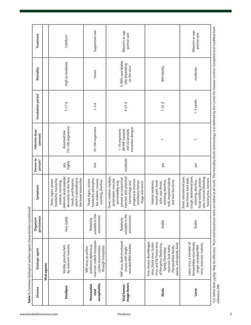
4) Viruses such the etiologic agents of Variola, Ebola, and Lassa are very dangerous. Variola virus is the etiological agent of smallpox, causes 20–30% mortality, and persists in an infectious state for many days in dried crusts from skin lesions as well as in fluid from vesicles.9 Ebola virus causes severe hemorrhagic fever in humans and primates resulting in mortality rates between 80–90%.9 Lassa virus causes Lassa fever that is endemic in West Africa, infecting 2 million people per year and resulting in 5000–10 000 fatalities annually.9
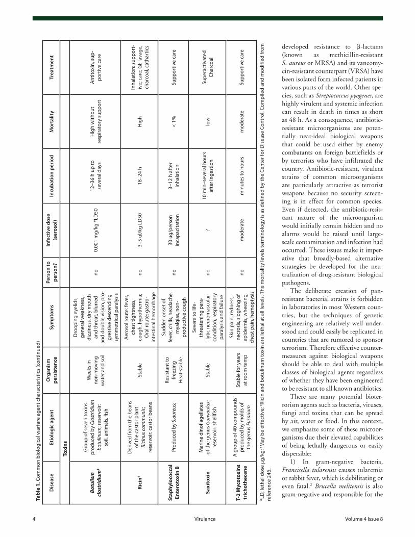
5) Clostridium botulinum is a gram-positive anaerobic bacte-rium and produces the most potent known neurotoxin respon-sible for botulism, which promotes neuromuscular weakness or paralysis.10
Historical evidence of the use of biological warfare is some-what sketchy. In April and May 1979, an unusual anthrax epidemic occurred in Sverdlovsk, Union of Soviet Socialist Republics. Soviet officials attributed it to consumption of con-taminated meat but US agencies attributed it to inhalation of spores accidentally released at a military microbiology facility in the city. Epidemiological data show that most victims worked or lived in a narrow zone extending from the military facility to the southern city limit. Further south, livestock died of anthrax along the extended axis of the zone. The zone paralleled the northerly wind that prevailed shortly before the outbreak. It was concluded that the escape of an aerosol of anthrax pathogen at the military facility caused the outbreak.11
The difficulty faced in decontaminating the environment from biological weapons agents can be illustrated by the his-torical story of Gruinard Island. British military scientists from Porton Down in 1942, during the Second World War, had tested methods to disseminate anthrax spores on a remote and unin-habited island off the Scottish coast. Military scientists exploded a series of anthrax-spore laden bombs, testing their killing effi-ciency using sheep.12 Initial efforts to decontaminate the island after the biological warfare trials failed due to the high durabil-ity of anthrax spores. After 44 years of complete quarantine, Gruinard Island was finally decontaminated in 1986 with 280 tons of formaldehyde diluted in seawater being sprayed over all 196 hectares of the island and the worst-contaminated topsoil around the dispersal site being physically removed.13 A flock of sheep was then placed on the island and remained healthy.14
In Kosovo, rural villagers reported an unusual massive die-off of mice and rats in the summer of 1999 in war-devastated areas. Clusters and small outbreaks of a human disease with fever, lymphadenopathy, and ulcerations of skin and mucosa occurred, which were initially diagnosed as tonsillitis until tularemia was suspected clinically. Rumors started to circulate in some villages that wells had been deliberately contaminated with the pathogen. The Albanian authorities asked World Health Organization to send in a Global Outbreak Alert and Response Network (GOARN) team in order to help in the diagnostics and to investigate the origin and cause of this “unusual” tulare-mia epidemic. Since the strain was thought to be Biovar B (the endemic European strain) rather than the more virulent Biovar A, the epidemic was attributed to war-related destruction of the ecosystem and infrastructure leading to an increased population density of rodents and producing human cases of tularemia.15
There have been some documented occurrences of bioterror-ism. In 1984, two large cohorts of salmonellosis cases (a total of 751 individuals) occurred in The Dalles, Oregon. The size and nature of this outbreak initiated a criminal investigation. The cause only became known when the Federal Bureau of Investigation (FBI) investigated a nearby cult (Rajneeshees) for other criminal violations. In October 1985, a vial containing a culture of Salmonella Typhimurium was discovered by authori-ties in the Rajneeshee clinic laboratory.16 As gastroenteritis cases occurred in increasing numbers, health authorities closed all salad bars in The Dalles.
In 1996 between 29 October and 1 November 1996, 12 clini-cal laboratory workers at the St. Paul Medical Center in Dallas, TX developed severe acute diarrheal illness as a result of eating muffins and doughnuts left in their break room on 29 October. Shigella dysenteriae type 2 was cultured from 8 patients that was identical to the laboratory stock strain (some of which was miss-ing) by pulsed field gel electrophoresis and it was concluded the pastries had been deliberately contaminated.17
On 4 October 2001, a case of inhalational anthrax was reported in a 63-year-old male in Florida. Authorities initially announced this individual had probably contracted the illness by hunting. There were two further cases in Florida, and a fourth case of cutaneous anthrax was identified in a female employee at NBC news in New York City (NYC). Investigators then realized
6 virulence volume 4 issue 8
that exposures had occurred from anthrax-containing letters sent in the mail. On 15 October, the Senate Majority Leader received an anthrax-containing letter, which led to the closure of the Hart Senate Office Building in Washington, DC.18 By the end of the year, anthrax-laden letters had caused 22 cases of anthrax (10 confirmed inhalational and 12 cutaneous, of which 7 were confirmed and 5 suspected) and 5 deaths, mostly among postal workers and mail handlers.19 A twelfth case of cutaneous anthrax occurred in March 2002 in a Texas laboratory where the anthrax samples were processed.20
The mode of dispersal of a biological weapons agent may to some extent depend on whether the biological agent is being used as a form of biological warfare or as bioterrorism. In warfare it is more likely that the agent will be dispersed from an aircraft, loaded into a bomb or an explosive shell that can be directed toward enemy forces, while in bioterrorism it is more likely to be surreptitiously released into a subway tunnel or other enclosed space, or introduced into the water or food supply, or even sent through the mail. Therefore the countermeasures chosen may have to take into account widely differing environments that the agent may be in.
Countermeasures against biological weapons agents can be divided into three broad classes. The first broad class is what can be loosely described as disinfectants, or in other words, treat-ments that can destroy or neutralize the agent in a wide range of inorganic, organic, or living environments before the agent has had a chance to come into contact with human beings in a suf-ficiently large dose to cause infection of harm. The second broad class consists of treatments that can kill or neutralize the agent after it has come into contact with human beings, either before or after infection or intoxication has become established, and this class may include some drugs that can reduce symptoms without destroying the agent. The third broad class consists of strategies to vaccinate or immunize people who have been exposed to the agent, or who are at risk of exposure, in order to avoid infection or to reduce the severity of the consequences of exposure.
It is the hypothesis of the present review that light-based approaches can be effective in all three of these broad classes of countermeasures, and moreover that many of these light-based approaches can be effective against all known classes of biological
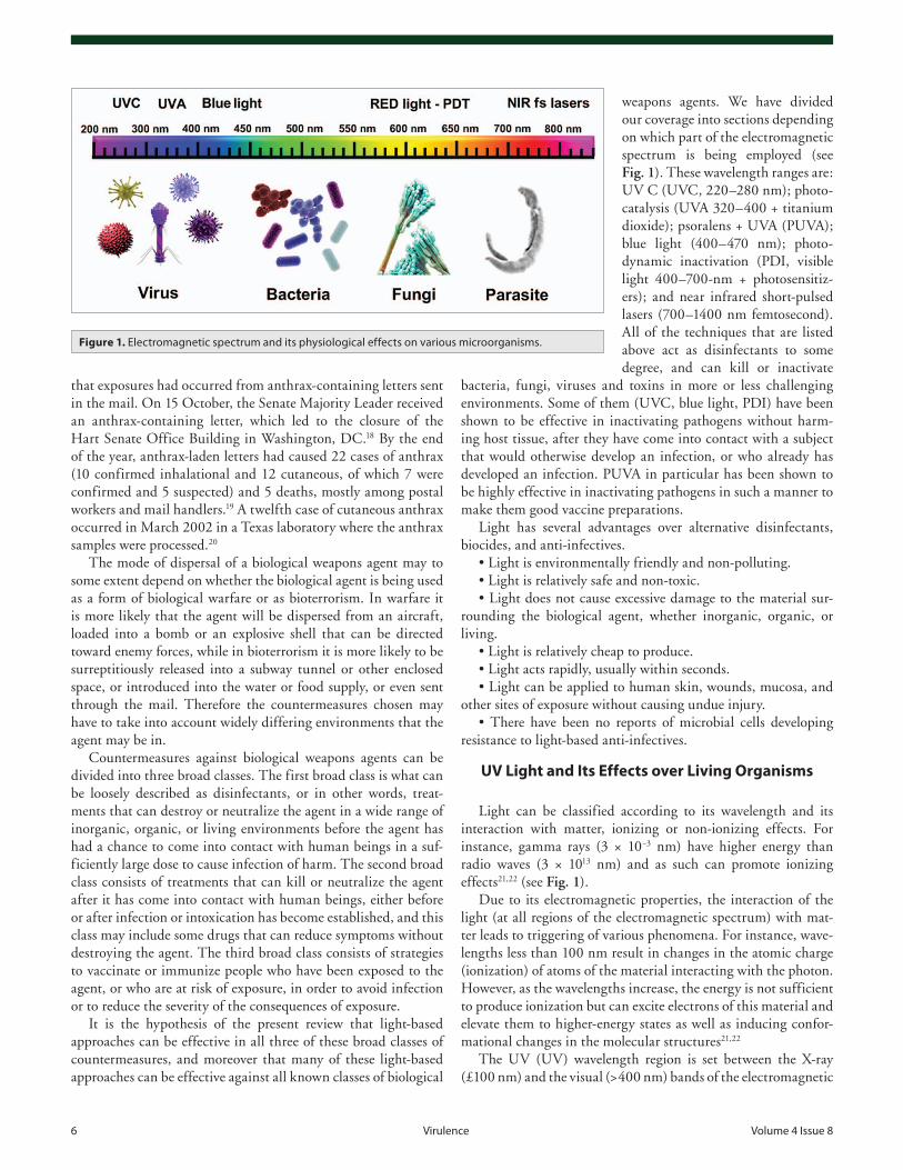
weapons agents. We have divided our coverage into sections depending on which part of the electromagnetic spectrum is being employed (see Fig. 1). These wavelength ranges are: UV C (UVC, 220–280 nm); photo-catalysis (UVA 320–400 + titanium dioxide); psoralens + UVA (PUVA); blue light (400–470 nm); photo-dynamic inactivation (PDI, visible light 400–700-nm + photosensitiz-ers); and near infrared short-pulsed lasers (700–1400 nm femtosecond). All of the techniques that are listed above act as disinfectants to some degree, and can kill or inactivate
bacteria, fungi, viruses and toxins in more or less challenging environments. Some of them (UVC, blue light, PDI) have been shown to be effective in inactivating pathogens without harm-ing host tissue, after they have come into contact with a subject that would otherwise develop an infection, or who already has developed an infection. PUVA in particular has been shown to be highly effective in inactivating pathogens in such a manner to make them good vaccine preparations.
Light has several advantages over alternative disinfectants, biocides, and anti-infectives.
• Light is environmentally friendly and non-polluting.• Light is relatively safe and non-toxic.• Light does not cause excessive damage to the material sur-
rounding the biological agent, whether inorganic, organic, or living.
• Light is relatively cheap to produce.• Light acts rapidly, usually within seconds.• Light can be applied to human skin, wounds, mucosa, and
other sites of exposure without causing undue injury.• There have been no reports of microbial cells developing
resistance to light-based anti-infectives.
UV Light and Its Effects over Living Organisms
Light can be classified according to its wavelength and its interaction with matter, ionizing or non-ionizing effects. For instance, gamma rays (3 × 10⁻³ nm) have higher energy than radio waves (3 × 10¹³ nm) and as such can promote ionizing effects21,22 (see Fig. 1).
Due to its electromagnetic properties, the interaction of the light (at all regions of the electromagnetic spectrum) with mat-ter leads to triggering of various phenomena. For instance, wave-lengths less than 100 nm result in changes in the atomic charge (ionization) of atoms of the material interacting with the photon. However, as the wavelengths increase, the energy is not sufficient to produce ionization but can excite electrons of this material and elevate them to higher-energy states as well as inducing confor-mational changes in the molecular structures21,22
The UV (UV) wavelength region is set between the X-ray (£100 nm) and the visual (>400 nm) bands of the electromagnetic
Figure 1. electromagnetic spectrum and its physiological effects on various microorganisms.
www.landesbioscience.com virulence 7
spectrum. As such, UV light can be classified into four wave-lengths according to its interaction with molecules: vacuum UV (VUV) at 100–200 nm; UV C (UVC) at 200–280 nm; UV B (UVB) at 280–315 nm; and UV A (UVA) at 315–400 nm.21-24 The main physiologic effects, steaming from the photonic energy, can be described as:
• VUV light: including wavelengths <200 nm; is harmful due to its capability of immediate reaction with oxygen atoms and organic molecules even at low doses.
• UVC light: wavelength range lies between 200 and 280 nm; this electromagnetic spectrum has biocidal effects and generally is reported as “germicidal” or more usually “ultraviolet germi-cidal irradiation” (UVGI).
• UVB light: comprises wavelengths between 280 to 315 nm; these photons are known for “sun burning” of the skin and have been implicated in photocarcinogenesis and photoaging.
• UVA light: comprises wavelengths between 315 to 400 nm; it is becoming realized that the shorter UVA wavelengths (called UVA1, 315–340 nm) can have also have detrimental effects on the skin due to production of reactive oxygen species.
Energetically UVC is very important in the context of inac-tivation of microorganisms, since UVC directly affects deoxyri-bonucleic acid (DNA) and ribonucleic acid (RNA) by inducing molecular transformation (i.e., producing photoproducts in the genetic material). The pyrimidines and purines can absorb UV light and this way DNA and RNA can be inactivated by UV light, especially UVC at 254 nm by oxidation of these bases or through base dimerization and formation of cis-syn cyclobutane pyrimidine dimmers in the DNA molecules.3,21,22 When DNA is damaged it becomes very difficult for the nucleic acids to repli-cate, and if replication does occur, it often produces a defect that prevents the bacterium from being viable.23,25 There are six pos-sible photoproduct “defects” in the DNA induced by UV light: thymine–thymine dimer; cytosine–cytosine dimer; cytosine–thymine dimer; uracil–uracil dimer; uracil–thymine dimer; and uracil–cytosine dimer.21
What are the factors governing the effective photonic interac-tion with living organisms? The Grotthus–Draper law (first law of photochemistry) states that photons must be absorbed for the photochemical reaction to occur and the Stark–Einstein law (sec-ond law of photochemistry) states that, if a photon is absorbed, then only one photon should be enough for a photoproduct for-mation.26 On the other hand, it is well known that microbial inactivation is dose dependent process (Bunsen–Roscoe reciproc-ity law) based on the UV intensity in the irradiation area.21 UV light (as also applies to all wavelengths) has energy measured in joules (J), power in watts (W), area irradiated in cm2 or m2, time of irradiation in seconds (s), irradiance (W/area), and fluence or dose (J/area) for calculation of dose-response. In addition, envi-ronmental condition such as humidity, temperature, and parti-cle size also affect the dose-response and need to be considered, although the duration of exposure required for lethal effect of UVC is short.24
UVC light (200 to 280 nm) is the most used light for inac-tivation of microorganisms.3,9,25,27-35 This inactivation can use monochromatic or polychromatic light sources. Indeed, the main
difference between these UVC lamps is that monochromatic lamps such as mercury lamp emitting at 254 nm cause genetic damage to microorganism, whereas polychromatic sources with other UV regions also affect aromatic proteins (i.e., can also affect function and structure of microbial proteins which depends on primary, secondary, and tertiary structures).21
UV light sourcesThe main source of UV light used to kill microorganisms has
been produced by mercury vapor arc lamps for a long time, pre-dominately at a wavelength of 253.7 nm (UVC electromagnetic spectrum)24 (see Table 2). This kind of lamp is low-pressure mer-cury (Hg) and are 30% efficient at converting input power to UVC at 253.7 nm.36 Currently, and owing to its wider application ranges, there is a need for UVC light to be emitted from lamps or devices containing non-toxic materials with better efficiency and lower costs to make them more affordable, owing to the potential risks of mercury lamps being broken and exposing its hazardous material to the environment. In this context, light-emitting diode (LED) and xenon lamps have gained prominence.36
A UVC LED has been tested in a single-pass flow-through device. Unfortunately, the LED is very inefficient at producing UVC radiation (0.3%). However, arrays of LEDs can be more efficient and produce the expected inactivation.36 The xenon lamp emits a peak wavelength at 240 nm. This lamp can have a total emission of 10 W of which approximately 1.4 W is UVC radiation. This lamp is a non-toxic alternative to mercury but it produces ozone, which is a strong oxidant and toxic air pollut-ant.36 Thus, more research needs to be done in order to improve LED efficiency and/or discover others sources of UV light.36
UV light as a viable decontamination technique for poten-tial biological warfare agents
The first observation how microorganisms respond to light was in nineteenth century with experiments using sunlight and inactivation or disinfection of test tubes containing Pasteur solu-tion. At this time it was already known that inactivation or dis-infection of surfaces was dependent on intensity, duration, and wavelength of the light, starting the concept of dose-response. Especially in this context, it was observed differences of sensitiv-ity between different bacteria.23
Since the last century the source of light used to kill micro-organisms have been the low-pressure mercury (Hg) lamps emit-ting primarily a short wave (254 nm) of UVC electromagnetic spectrum.23 UVC light affects pyrimidines, purines, and flavins promoting the formation of dimers in RNA (uracil and cytosine) and DNA (thymine and cytosine), which promotes inactivation of many microorganisms. Thus, UVC is an established means of disinfection and can be used to kill agents causing many infec-tious diseases.21,23 There have been some studies to determine which wavelength in the UVC region is actually best to inacti-vate microorganisms. Lakretz et al.37 compared UV wavelengths between 220 and 280 nm and concluded that 254 and 270 nm were better at carrying out bacterial inactivation and biofilm disruption than 239 and 280 nm. Medium pressure mercury lamps emit a wider range of wavelengths than low pressure lamps including lines between 365 nm and 578 nm and it has been claimed that they are actually better than low pressure lamps at
8 virulence volume 4 issue 8
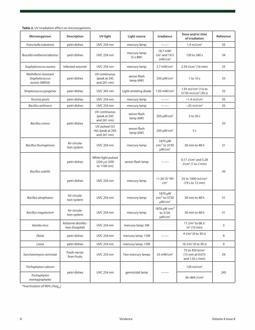
Table 2. uv irradiation effect on microorganisms
Microorganism Description UV light Light source IrradianceDose and/or time
of irradiationReference
Francisella tularensis petri dishes uvc 254 nm mercury lamp ——- 1.4 mJ/cm2 35
Brucella melitensis/abortus petri dishes uvc 254 nmmercury lamp
(5 x 8w)
18.7 mw/cm2 and 19.5
mw/cm2
120 to 240 s 34
Staphylococcus aureus infected wounds uvc 254 nm mercury lamp 2.7 mw/cm2 2.59 J/cm2 (16 min) 25
Methillicin-resistant Staphylococcus aureus (MRSa)
petri dishesuv continuous
(peak at 245 and 261 nm)
xenon flash lamp (6w)
250 µw/cm2 1 to 10 s 33
Streptococcus pyogenes petri dishes uvc 265 nm light-emitting diode 1.93 mw/cm2 1.93 mJ/cm2 (1s) to57.95 mJ/cm2 (30 s)
32
Yersinia pestis petri dishes uvc 254 nm mercury lamp ——- ~1.4 mJ/cm2 35
Bacillus anthracis petri dishes uvc 254 nm mercury lamp ——- ~25 mJ/cm2 35
Bacillus cereus petri dishes
uv continuous (peak at 245 and 261 nm)
xenon flash lamp (6w)
250 µw/cm2 5 to 20 s
33uv pulsed (53
Hz) (peak at 245 and 261 nm)
xenon flash lamp (6w)
250 µw/cm2 5 s
Bacillus thuringiensesair circula-tion system
uvc 254 nm mercury lamp1870 µw
cm/2 to 3720 µw/cm2
30 min to 48 h 31
Bacillus subtilis
petri disheswhite light pulsed
(250 μs) (200 to 1100 nm)
xenon flash lamp ——-0.17 J/cm2 and 5.28 J/cm2 (1 to 2 min)
30
petri dishes uvc 254 nm mercury lamp~1.34.10−3w/
cm2
25 to 1000 mJ/cm2 (19 s to 12 min)
Bacillus atrophaeusair circula-tion system
uvc 254 nm mercury lamp1870 µw
cm/2 to 3720 µw/cm2
30 min to 48 h 31
Bacillus megateriumair circula-tion system
uvc 254 nm mercury lamp1870 µw cm/2
to 3720 µw/cm2
30 min to 48 h 31
Variola virusairborne disinfec-
tion (hospital)uvc 254 nm mercury lamp: 5w
17 J/m2 to 68 J/m2 (10 min)
3
Ebola petri dishes uvc 254 nm mercury lamp: 15w ——-4 J/m2 (0 to 30 s)
9
Lassa petri dishes uvc 254 nm mercury lamp: 15w 16 J/m2 (0 to 30 s) 9
Saccharomyces cerivisiaeFresh nectar from fruits
uvc 254 nm Two mercury lamps 25 mw/cm2
75 to 450 kJ/m2 (15 min at 0.073 and 1.02 l/min)
29
Trichophyton rubrum
petri dishes uvc 254 nm germicidal lamp ——-
120 mJ/cm2
245Trichophyton
mentagrophytes36–864 J/cm2
*inactivation of 90% (1log10)
www.landesbioscience.com virulence 9
inactivating pathogens.38 There have also been studies aimed at comparing pulsed with continuous wave (CW) UV light. Using 365-nm LEDs, Li et al. showed39 that pulsing at 100 Hz was superior to CW for inactivating E. coli and C. alibicans biofilms. Moreover pulsed xenon light technology (broad spectrum includ-ing both UV and visible) has also been much studied40 for micro-bial inactivation.
Due to its killing effects on microorganisms, other applica-tions of the UVC have been extended into the food processing industry, disinfecting heating cooling coils, ventilating and air-conditioning systems, whole room/surface disinfection, and into killing of all human pathogens (bacterial, viral, and protozoan) transmitted via water.21,23,24,36
Considering food-processing, UVC has shown a great poten-tial for surface disinfection of fresh cut fruit and vegetables, reducing deterioration, prolonging storage life, and becoming a viable alternative to chemical sanitizers as titanium dioxide (TiO2) and chlorine.21 It is important highlight that UV treat-ment is increasingly common because the process is effective against a wide range of microorganisms, overdose is not possible, chemical residues or by-products are avoided, and water quality is unaffected and therefore UV treatment has also been an impor-tant tool for water and wastewater treatments.21
Another significant use of UV light is air disinfection because a wide variety of fungal, bacterial, and viral pathogens may be transmitted by airborne droplets as e.g., Mycobacterium tubercu-losis, influenza viruses, SARS corona virus, Aspergillus spp, and Legionella spp.21 UV has successfully reduced the concentration of airborne microorganisms in operating rooms during surgery. The installation of UV light in air handling units and ventilation systems reduced the concentration of airborne bacteria and fungi in indoor air as well as the total amount of bacteria collected at the edge of the surgical site was significantly reduced.21 These results foreshadowed the use of UV light in 1935, specifically UVC in the ducts of ventilation systems.23
The initial success of air disinfection by UVC in surgical rooms stimulated an expansion of UVC application in hospi-tals. For instance, UVC light sources were arranged such that
to provide a kind of “light curtain” and prevent respiratory cross-infections in infant wards23 and in neonatal intensive care units; UVC was used successfully for coil cleaning and promot-ing significantly the reduction of tracheal microbial coloniza-tion, as well as ventilator-associated pneumonia and the use of antibiotics.36
UVC can be used for whole room disinfection, cleaning the air and surfaces under this light. Generally, air disinfection by UVC is accomplished through: irradiation on the upper-room air; irradiation of the entire room; or irradiation of the air that passes through enclosed air-circulation and heating, ventilation, and air-conditioning systems.23 For faster results, high-powered lamps that generate high fluence levels can be used for whole room disinfection, but in unoccupied spaces in order to prevent erythema to the skin and photokeratitis in humans36 or when people wear specific clothes for their protection.23 Currently in the United States, UVC has been installed in air-handling units in heating, ventilating, and air conditioning systems to irradiate the surfaces of the coil and disinfect system components.36
Although biosafety is a public health concern, most of the attention is cornered to hospital environments and microbiology laboratories, and bioterrorism concerns have not so far become familiar to the public.23,24 However, the technology and methods used in health care facilities and laboratories can also help against potential bioterrorism agents that cause anthrax, smallpox, viral hemorrhagic fevers, pneumonic plague, glanders, tularemia, drug-resistant tuberculosis, influenza pandemics, and severe acute respiratory syndrome to mention a few.24
Biological UV dosimetersIt is widely accepted that biological UV dosimetry is an indic-
ative tool for assessing the UV radiation impact on health and ecosystems. The accumulated data indicates that standard UV treatments that are effective against B. subtilis spores are likely also to be sufficient to inactivate B. anthracis spores and that the spores of standard B. subtilis strains could reliably be used as a biodosimetry model for the UV inactivation of B. anthracis spores.41 There are several studies now utilizing the concept of “biological UV dosimeters” as indicators of UV exposure where
Table 2. uv irradiation effect on microorganisms
Microorganism Description UV light Light source IrradianceDose and/or time
of irradiationReference
Aspergillus fumigates
water disinfection uvc 254 nm mercury lamp
0.83 mw/cm2 12.45 mJ/cm2
28Aspergillus flavus 0.83 mw/cm2 16.6 mJ/cm2
Aspergillus niger 0.83 mw/cm2 20.75 mJ/cm2
Clostridium botu-linum toxin
petri dishes uvc 254 nm germicidal lamp15 ergs.
mm−2/s or 1.5 µw/mm2
675 to 900 ergs/mm2 or 67.5 to 90 µJ/mm2 27
*inactivation of 90% (1log10)
(continued)
10 virulence volume 4 issue 8
bacteria such as E. coli and B. subtilis have been used as sensing elements.42
UV radiation is estimated to be one of the most important risk factors for nonmelanoma and melanoma skin cancers. In a study Moehrle et al.43 assessed the annual occupational UV expo-sure of mountain guides that were using spore film test chambers containing spores of B. subtilis as UV dosimeter-agents that have a spectral sensitivity profile similar to erythema-weighted data (calculated from spectroradiometric measurements). In the study nine mountain guide instructors carried dosimeters on the sides of their heads in a total of 1406 working days throughout a year. During the study period the dosimeters were changed monthly.43 In another study by the same group44 they tested the practical application of the “biological UV dosimeters” on 11 persons in a span of 43 d, under different UV exposure conditions that were spread over five different geographical regions. The mixed cohort included four professional lifeguards of a swimming pool who carried the dosimeters attached to their shoulders or to their head-caps for 11 d; three mountain guides that attached the dosimeters laterally to their heads on 27 different occasions of mountaineering activity in different mountain regions; four ski instructors who carried lateral head dosimeters during eight days of skiing in the Alps. The conclusion of the study was that B. subtilis spore film dosimeters can effectively be used as per-sonal “solar UV exposure detectors”.
In a different study Vähävihu et al.45 assessed the viability of personal UV dosimeters; where UVB dose exposure during a 13-d heliotherapy for atopic dermatitis using B. subtilis spore film dosimeters with UV meter, and diary records was used. In addition, correlation between personal UVB dose exposure and changes in serum 25-hydroxyvitamin D (25[OH]D) was studied over a set of 21 adult cohorts in the Canary Islands. The study concluded that the increase in serum 25(OH)D correlates with the UVB exposure length, and that spore films are feasible and reliable in vivo tools to be used as personal UV dosimeters in field conditions.45
Bacterial resistance to UV irradiation: effective internal repair mechanism
Studies have been revealing that bacterial spores possess an enormous resistance to UV radiation46-49 which is a source of concern to some degree. Even more interestingly dormant spores of the various Bacillus species, including B. subtilis, are shown to be 5 to 50 times more resistant to UV radiation than are the corresponding growing cells.50-52 This resistance arises largely due to the use of a unique DNA repair enzyme called Spore Photoproduct Lyase (SP lyase) which apparently repairs specific UV-induced DNA lesions through an radical-based mechanism. The interesting thing about this repair mecha-nism is that, unlike DNA photolyases, SP lyase belongs to the emerging superfamily of radical S-adenosyl-l-methionine (SAM) enzymes and uses a (4Fe-4S)+ cluster and SAM to ini-tiate the repair reaction (where the DNA lesion recognition and binding site involves a β-hairpin structure).46 It has been shown that SAM and the cysteine residue are perfectly posi-tioned at the active and as such facilitate the hydrogen atom abstraction (from the dihydrothymine residue of the lesion) and
subsequently donation to the α-thyminyl radical moiety. Based on structural and biochemical characterizations of mutant pro-teins, the researchers were able to substantiate the role of this cysteine residue in the enzymatic mechanism of action. The proposed structure reveals how SP lyase combines specific fea-tures of radical SAM and DNA repair enzymes, in enabling a complex radical-based repair reaction to occur.46 In essence, the SP lyase repairs the UV-induced thymine dimmer (a spore pho-toproduct (SP)) in germinating endospores and, as such, it is responsible for the strong UV resistance of the endospores. SP lyase is a radical S-adenosyl-l-methionine (SAM) enzyme that is using the (4Fe-4S)+ cluster in reducing SAM and generating the catalytic 5′-deoxyadenosyl radical (5′-dA•).53 A very recent publication by Young et al. is revealing that two conserved tyro-sines may be also critical for the enzymes catalytic activity. The one tyrosine in B. subtilis SPL, Y99(Bs), is downstream of the cysteine, suggesting that SP lyase uses a novel hydrogen atom transfer (HAT) pathway and with a pair of cysteine and tyrosine residues regenerates the SAM. The second tyrosine, Y97(Bs), has a structural role and serves to facilitate the SAM bind-ing. In fact, the researchers think that it may also contribute to the SAM regeneration process by interacting with the puta-tive Y99(Bs) and/or 5′-dA intermediates, and thus lowering the energy barrier for the second H abstraction step.53
In essence, the observed remarkable resistance of the bacte-rial spores to chemical and physical stresses, including exposure to UV radiation, arises as a result of a unique photochemistry of spore DNA that generates and accumulates the spore photo-product 5-thyminyl-5,6-dihydrothymine and coupled with the capabilities of efficient repair of the accumulated damage by the enzyme SP lyase this unique viability effect comes to life. As such the observed elevated spore UV resistance corner stones can be listed as:
• Photochemistry of the DNA within spores: UV generates few (if any) cyclobutane dimers, but rather the spore photoprod-uct 5-thyminyl-5,6-dihydrothymine. As such, it is an exclusive DNA photodamage product in bacterial endospores and a radical S-adenosylmethionine enzyme (SAM) and the SP lyase (at the bacterial early germination phase) repairs it.
• The DNA repair effect (in particular SP lyase repair), dur-ing spore germination process: the unique UV photochemistry of spore DNA is largely due to its saturation with a group of small, acid-soluble proteins (SASP) that are unique to spores and whose binding alters the DNA conformation and as such its photo-chemistry. This SP-specific repair is also unique to spores and is performed by a light-independent SP-lyase, an iron-sulfur protein that utilizes S-adenosylmethionine to catalyze SP monomeriza-tion without DNA backbone cleavage.47,50,52
Resistance of vegetative bacteria to UV photoinactivation can also be developed. The bacterial growth rate strongly affects the sensitivity to UVC,54 and bacteria isolated from a high-alti-tude extreme environment were more resistant to UV.55 There are UV-inducible DNA repair systems such as those found in E. coli mutants deficient in induction of mutations by UV light.56 Nucleotide excision repair involving the products of the uvrA, uvrB, and uvrC genes, and the error-prone repair in association
www.landesbioscience.com virulence 11
with the umuDC gene products is also known to occur.57 The lat-ter process, the SOS response is triggered by the activated RecA* protein, which facilitates the autocleavage of the UmuD protein to yield the active UmuD9 C-terminal fragment.
Clearly once the potential of UV light to kill microorganisms like bacteria, viruses, fungi was understood; there has been an increasing interest to improve the light utilization. We highlight below some studies which used UV light to kill various micro-organisms in water, air, food, or in experimental models and demonstrate that UV light can be a viable tool against a possible bioterrorist action using these microorganisms.
Germicidal UV for Infections
Although it has been known for the past 100 years that UVC irradiation is highly germicidal, the use of UVC irradia-tion for prevention and treatment of localized infections is still in the very early stages of development. Our laboratory has per-formed several studies designed to show that UVC irradiation can be used in vivo to treat mouse models of infections caused by virulent and pathogenic microorganisms.58 UVC treatment (2.59 J/cm2) of partial thickness skin abrasions in mice infected with Pseudomonas aeruginosa increased the survival rate of mice by 58.3% (P = 0.0023).25 When the same treatment was applied to mice with abrasions infected with S. aureus, the wound heal-ing rate was increased by 31.2% (P < 0.00001). In mice with wounds and burns infected with a virulent strain of Acinetobacter baumannii isolated from US soldiers in Iraq, UVC was able to reduce the bacterial burden by >90%.59 Although DNA lesions were observed by immunofluorescence in the surrounding mouse skin immediately after a UVC exposure of 3.24 J/cm2, the lesions were extensively repaired within 72 h. UVC was also successfully employed to treat a cutaneous Candida albicans fungal infection in mouse burns.60
Photocatalytic Inactivation of Biological Warfare Agents: Titania Photocatalysis
The ability of titanium dioxide (TiO2) to act as a photocata-
lyst has been reported since 192961 (and references therein). In 1972, Fujishima and Honda62 first reported the photoelectrolysis of water at a semiconductor electrode. This property was then utilized to catalyze the oxidation of pollutants.63,64 Photocatalytic surfaces can be manufactured into construction and building materials65 and some of the commercial uses include self-cleaning windows and self-cleaning glass covers for road lights61
One of the most important aspects of TiO2 photocatalysis is
that the process, just like the photoelectric effect, depends entirely on the energy of the incident photons and not (to a first approxi-mation) on their intensity.66 This suggests that, if there are even just few photons of required energy, they can induce photoca-talysis; a phenomenon that has enormous practical implications.
There are three main polymorphs of TiO2: anatase, rutile,
and brookite; in all the three forms, titanium (Ti4+) atoms are coordinated to six oxygen atoms (O2−) and are forming the TiO
6
octahedra. Typically TiO2 is an n-type semiconductor because of
its oxygen deficiency, a fact having a leading role in the photocat-alytic processes and mechanisms. The bandgap energy (energy required to promote an electron) of TiO
2 is of 3.0 eV for the
rutile, 3.2 eV for anatase, and ~3.2 eV for brookite polymorphs, which means that photocatalysis can be activated by photons with a wavelength shorter than 385 nm (i.e., UVA). The adsorp-tion of a photon with sufficient energy promotes an electron from the valence band to the conduction band leaving a posi-tively charged hole in the valence band. The hole may be filled by migration of an electron from an adjacent molecule, leaving that molecule with a hole, and so on. And when electrons reach the surface, they can react with O
2 to produce superoxide radical
anion (O2
•−), and the photogenerated holes can react with water to produce hydroxyl radicals (•OH). On the other hand, O
2•− can
react further to form H2O
2 and more •OH. As such, the photo-
catalytic process implies photon-assisted generation of catalyti-cally active ROS rather than an action of the light as a catalyst in the reaction (Fig. 2).
The majority of studies have shown that anatase is the most effective photocatalyst while rutile is less active. Differences are probably due to differences in the extent of recombination of e− and hole between the two forms.67 However, studies have shown that mixtures of anatase and rutile were more effective photocat-alysts than 100% anatase and were more efficient for inactivating viruses.67
The mechanistic description of the TiO2 photocatalysis pro-cess can be detailed as follows, where e−
CB is the electron gener-
ated at the conduction band, h+VB
is the hole generated (and left) at the valence band. A recent paper68 suggests that the mecha-nism could be better characterized as “proton-coupled electron transfer”:
TiO2 + hv → h+
VB + e−
CB
h+VB
+ e−CB
→ energy (recombination process)e−
CB + O
2 → O
2•− (superoxide radical)
h+VB
+ H2O → H + •OH (hydroxyl radical)
•OH protein/lipid layer → H2O + CO
2
O2
•− + H+ → •OOH (hydroperoxyl radical)O
2•− + protein →→ CO
2 + H
2O
•OOH + protein/lipid layer → CO2 + H
2O
•OOH + •OOH → O2 + H
2O
2 (hydrogen peroxide)
H2O
2 + e− → HO− + •OH
One can say that TiO2 is a chemically stable and inert material,
and can continuously exert antimicrobial effects when illumi-nated. The energy source could be even the solar light; therefore, TiO
2 photocatalysts are also useful in remote areas where elec-
tricity is insufficient. However, because of its large band gap for excitation, only biohazardous UV (UV) wavelengths can excite TiO
2, which limits its application in living environments. To cir-
cumvent this problem impurity doping, through metal coating and controlled calcination, has been successfully used to modify the TiO
2 and to expand its absorption wavelengths to the visible
light region (discussed below).Matsunaga and colleagues69,70 were the first to use TiO
2 pho-
tocatalysis to kill microorganisms. This subject area has recently been comprehensively reviewed71,72 and the effect of key variables on the effectiveness has been studied.73
12 virulence volume 4 issue 8
Previous studies have investigated the antibacterial abilities of visible light-responsive photocatalysts using the model bac-teria Escherichia coli and human pathogens. They have shown that modified TiO
2 photocatalysts significantly reduced the
numbers of surviving bacterial cells in response to visible light illumination.
Bacterial inactivation studies have confirmed that even with significantly lower levels of TiO
2 generated radical scavengers,
i.e., ROS, illumination with far-UV light can successfully pro-mote microorganisms inactivation.74 Spore forming bacteria of Bacillus strains were investigated for demonstrating photocata-lytic disinfection effects with relatively good results.75 Armon et al. studied the photocatalytic inactivation of spores of B. sub-tilis and B. cereus (as a model for the main biological warfare element B. anthracis76) where the spore-forming B. cereus is genetically very closely related to B. anthracis whereas B. subtilis is highly resistant to variety of stress factors.77
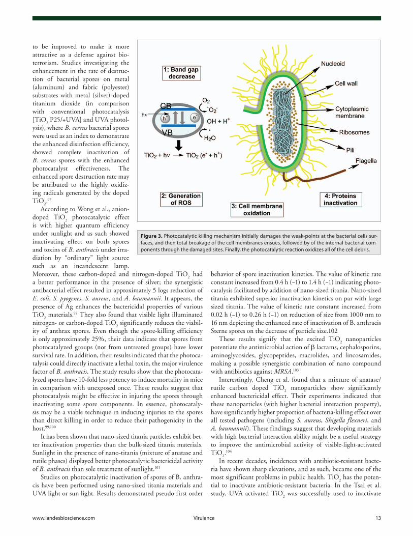
It has been suggested that the photocatalytic killing mecha-nism initially damages the weak-points at the bacterial cell surface before total breakage of the cell membranes. The internal bacte-rial components then leak from the cells through the damaged sites. And finally the photocatalytic reaction oxidizes all of the cell debris. In essence, the killing mechanism with TiO
2 involves
degradation of the cell wall and cytoplasmic membrane due to the production of ROS such as hydroxyl radicals and hydrogen peroxide. This initially leads to leakage of cellular contents then cell lysis and may be followed by complete mineralization of the organism. Killing is most efficient when there is close contact between the organisms and the TiO
2 catalyst71 (Fig. 3).
Huang et al.78 demonstrated with E. coli that TiO2 treated
cells continue to lose their viability even after the UV-irradiation stops, indicating that reactions in the media continue to propa-gate even after the UV-irradiation stops. Once the lethal oxida-tion reactions are initiated by the TiO
2 photocatalytic reaction,
the damaging effects propagate in the dark via the Fenton reac-tion or free radical chain reactions of lipid peroxidation due to ROS.79 The results suggest that initial oxidative damage happens on the cell wall (where the TiO
2 photocatalytic surface makes
the first contact). The cells that sustained the initial oxidative damage insult on their cell walls are still viable, however, though
localized, elimination of the cell-wall protection makes these cells susceptible to ensuing oxidative damages to the underlying cytoplasmic membrane. Overall, the photocatalytic action pro-gressively increases the cell permeability ending in free efflux of the intracellular contents, thus, eventually leading to cell death. Also, it is plausible that TiO
2 can gain access into the membrane-
damaged cells and generates a direct insult on the intracellular components, thus, accelerating the cell death.80,81
In summary, visible light-responsive TiO2 photocatalysts are
more convenient than the traditional UV light-responsive TiO2
photocatalysts because they do not require harmful UV light irradiation to function. These photocatalysts, thus, provide a promising and feasible approach for disinfection of pathogenic bacteria, facilitating the prevention of infectious diseases.82
By contrast, recombination of the photogenerated charge car-riers is a major limitation in the use of TiO
2 as a photocatalyst
and an initiator in the photocatalytic process, and, as such, is an important agent in combating biowarfare. Since the excited e- in the recombination process relaxes back to the valence band (either non-radiatively or radiatively, dissipating its energy as light or heat) without reacting with the possible biological sites (and thus not initiating the photocatalytic process—a bulk recombination process), there are several strategies developed to prevent this from happening and to improve the photocata-lytic efficiency. To enhance the charge separation of the e− and holes and to reduce the likelihood of bulk recombination, termed photoelectrocatalysis, it is possible to apply an electric field.83,84 Other approaches used to achieve improved efficiency include either chemical modifications (by incorporating additional com-ponents in the TiO
2 structure, termed as doping) or increasing
the surface area and porosity of the photocatalyst.85-89
In some cases carbon has been used as a dopant and as such allowing not only visible light absorption but also “injecting” active trap sites within the TiO
2 bands, thus increasing the life-
time of the photogenerated charge carriers.85
TiO2 can be used in combination with some of the noble
metals, such as Ag, Au, and Pt, which enhance the photocata-lytic efficiency under visible light due to “injecting” traps for the electrons and promoting the interfacial charge transfer, and thus delaying the recombination process of the electron–hole pair.90-94
Data accumulated thus far shows that TiO2 exhibits a strong
visible-light induced anti-microbial activity when modified by doping or used in combination. Sulfur-doped TiO
2 is shown to
have strong antibacterial effect.95 Carbon-doped TiO2 and TiO
2
modified with platinum (IV) chloride complexes used as suspen-sion or immobilized at surfaces (infected with the microorgan-isms) show remarkable anti-bactericidal effects. The detrimental effect of the photocatalysts induced with visible light on various microorganism groups such as bacteria (i.e., E. coli, S. aureus, Enterococcus faecalis) or fungi (i.e., Aspergillus niger, C. albicans) and utilizing modified TiO
2 showed increased effect over these
microorganisms in the order: A. niger, C. albicans > E. faecalis, S. aureus > E. coli.96
TiO2 photocatalysis with UV (UVA) light has proven to be
a highly effective process for complete inactivation of airborne microbes. However, the overall efficiency of the technology needs
Figure 2. photocatalytic effect of the Tio2: a process where photon-assisted generation of catalytically active RoS is generated rather than an action of the light as a catalyst in the reaction.
www.landesbioscience.com virulence 13
to be improved to make it more attractive as a defense against bio-terrorism. Studies investigating the enhancement in the rate of destruc-tion of bacterial spores on metal (aluminum) and fabric (polyester) substrates with metal (silver)-doped titanium dioxide (in comparison with conventional photocatalysis [TiO
2 P25/+UVA] and UVA photol-
ysis), where B. cereus bacterial spores were used as an index to demonstrate the enhanced disinfection efficiency, showed complete inactivation of B. cereus spores with the enhanced photocatalyst effectiveness. The enhanced spore destruction rate may be attributed to the highly oxidiz-ing radicals generated by the doped TiO
2.97
According to Wong et al., anion-doped TiO
2 photocatalytic effect
is with higher quantum efficiency under sunlight and as such showed inactivating effect on both spores and toxins of B. anthracis under irra-diation by “ordinary” light source such as an incandescent lamp. Moreover, these carbon-doped and nitrogen-doped TiO
2 had
a better performance in the presence of silver; the synergistic antibacterial effect resulted in approximately 5 logs reduction of E. coli, S. pyogenes, S. aureus, and A. baumannii. It appears, the presence of Ag enhances the bactericidal properties of various TiO
2 materials.98 They also found that visible light illuminated
nitrogen- or carbon-doped TiO2 significantly reduces the viabil-
ity of anthrax spores. Even though the spore-killing efficiency is only approximately 25%, their data indicate that spores from photocatalyzed groups (not from untreated groups) have lower survival rate. In addition, their results indicated that the photoca-talysis could directly inactivate a lethal toxin, the major virulence factor of B. anthracis. The study results show that the photocata-lyzed spores have 10-fold less potency to induce mortality in mice in comparison with unexposed once. These results suggest that photocatalysis might be effective in injuring the spores through inactivating some spore components. In essence, photocataly-sis may be a viable technique in inducing injuries to the spores than direct killing in order to reduce their pathogenicity in the host.99,100
It has been shown that nano-sized titania particles exhibit bet-ter inactivation properties than the bulk-sized titania materials. Sunlight in the presence of nano-titania (mixture of anatase and rutile phases) displayed better photocatalytic bactericidal activity of B. anthracis than sole treatment of sunlight.101
Studies on photocatalytic inactivation of spores of B. anthra-cis have been performed using nano-sized titania materials and UVA light or sun light. Results demonstrated pseudo first order
behavior of spore inactivation kinetics. The value of kinetic rate constant increased from 0.4 h (−1) to 1.4 h (−1) indicating photo-catalysis facilitated by addition of nano-sized titania. Nano-sized titania exhibited superior inactivation kinetics on par with large sized titania. The value of kinetic rate constant increased from 0.02 h (−1) to 0.26 h (−1) on reduction of size from 1000 nm to 16 nm depicting the enhanced rate of inactivation of B. anthracis Sterne spores on the decrease of particle size.102
These results signify that the excited TiO2 nanoparticles
potentiate the antimicrobial action of β lactams, cephalosporins, aminoglycosides, glycopeptides, macrolides, and lincosamides, making a possible synergistic combination of nano compound with antibiotics against MRSA.103
Interestingly, Cheng et al. found that a mixture of anatase/rutile carbon doped TiO
2 nanoparticles show significantly
enhanced bactericidal effect. Their experiments indicated that these nanoparticles (with higher bacterial interaction property), have significantly higher proportion of bacteria-killing effect over all tested pathogens (including S. aureus, Shigella flexneri, and A. baumannii). These findings suggest that developing materials with high bacterial interaction ability might be a useful strategy to improve the antimicrobial activity of visible-light-activated TiO
2.104
In recent decades, incidences with antibiotic-resistant bacte-ria have shown sharp elevations, and as such, became one of the most significant problems in public health. TiO
2 has the poten-
tial to inactivate antibiotic-resistant bacteria. In the Tsai et al. study, UVA activated TiO
2 was successfully used to inactivate
Figure 3. photocatalytic killing mechanism initially damages the weak-points at the bacterial cells sur-faces, and then total breakage of the cell membranes ensues, followed by of the internal bacterial com-ponents through the damaged sites. Finally, the photocatalytic reaction oxidizes all of the cell debris.
14 virulence volume 4 issue 8
the antibiotic-resistant bacteria MRSA, multidrug-resistant A. baumannii (MDRAB), and vancomycin-resistant E. faecalis (VRE) in suspension. Their results indicated that TiO
2 reaction
time had the greatest influence on microbial survival, following the TiO
2 exposure in the presence of UVA. TiO
2 in the presence
of UVA effectively reduced the number of antibiotic-resistant microbes in suspension by 1–3 logs.105
Photo activated TiO2 is effective on microorganisms capable
of killing a wide range of gram-negative and gram-positive bac-teria, fungi (both unicellular and filamentous), protozoa, algae, mammalian viruses, and bacteriophages; the killing activity is enhanced by the presence of other antimicrobial agents, such as Cu and Ag.71
The level of UVA disinfection of B. anthracis and B. brevis vegetative cells increased with the presence of the TiO
2 and Ag
photocatalysts, but had little effect on their spores. Bacillus bre-vis spores were slightly more sensitive to UVB and UVC than the spores of Bacillus atrophaeus. Photocatalytic sterilization against spores was strongest in UVC and UVB and weakest in UVA. The rate of inactivation of Bacillus spores was significantly increased by the presence of TiO
2 but was not markedly differ-
ent from that induced by the presence of Ag. Therefore, TiO2/
Ag plus UVA can be used for the sterilization of vegetative cells, while TiO
2 and UVC are effective against spores.106 However, in
a study investigating the effects of toxin- and capsule-encoding plasmids on the kinetics of UV inactivation of various strains of B. anthracis it was found that the plasmids pXO1 and pXO2 had no effect on bacterial UV sensitivity or photoreactivation. Interestingly enough, vegetative cells were capable of photoreac-tivation whereas photo-induced repair of UV damage was absent in B. anthracis Sterne spores107 which shows that B. anthracis makes highly stable and heat-resistant spores that can remain viable for decades.108
Psoralens and UVA
Psoralens are a group of natural furanocoumarins, commer-cially derived from a plant found in Egypt, Ammi majus. They are also present in celery, carrots, parsley, parsnip, and other veg-etables. It has been known since ancient times that consump-tion of these foodstuffs followed by sun exposure can lead to a phototoxic skin reaction similar to sunburn. The combination of psoralen with UVA light (known as PUVA) was first introduced as a medical treatment for psoriasis.109 Patients orally ingested psoralen compounds or alternatively the psoralens were applied topically in a bath.110 The mechanism is that the psoralen mol-ecule has the correct structure and shape to be able to intercalate between the two strands of DNA in the double helix, and upon illumination, induce the formation of covalent inter-strand cross-linking between opposite nucleic acid strands (Fig. 4).
Due to the DNA damaging action PUVA it has been used for the inactivation of bacteria, viruses, and protozoa in platelet and plasma blood component.111 This photochemical inactiva-tion using PUVA has the potential even to produce a new class of vaccines from whole microbes termed “Killed But Metabolically Active” (KBMA). KBMA vaccines are based on whole microbes
that have been inactivated by defined genotoxic methods that leave the organism incapable of productive growth and of causing disease but preserve metabolic activity sufficient to induce immu-nity. These vaccines have two broad applications. First, recom-binant KBMA vaccines encoding selected antigens relevant to infectious disease can be used to elicit a desired immune response. And when derived from attenuated forms of a targeted pathogen the entire antigenic repertoire is presented to the immune system, as here correlate of protection are unknown. In both applications the vaccine is inactivated by a distinct and limited disruption of the vaccine chromosome using photochemical treatment with a psoralen cross-linking agent, impacting an absolute block to DNA replication and possible vaccine outgrowth.112 Initially this technology was developed for killing undetected microbes con-taminating plasma and platelet blood products.113,114
Brockstedt et al.performed a landmark study in KBMA vac-cine approach demonstrating proof of concept for recombinant KBMA Lm vaccines in animal models of infectious disease and cancer.115 KMBA were developed by removing the genes required for nucleotide excision repair (uvrAB) and rendering microbial-based vaccines sensitive to photochemical inactivation with PUVA. Colony formation of these mutants was blocked by infrequent, randomly distributed psoralen crosslinks, though the bacterial population was able to express its genes, synthesize, and secrete proteins. Using the intracellular pathogen Listeria mono-cytogenes as a model platform, recombinant psoralen-inactivated Lm DeltauvrAB vaccines induced potent CD4+ and CD8+ T-cell responses and protected mice against virus challenge in an infec-tious disease model and provided therapeutic benefit in a mouse cancer model. Microbial KBMA vaccines used either as a recom-binant vaccine platform or as a modified form of the pathogen itself may have broad application for the treatment of infectious disease and cancer. This was a new vaccine paradigm for eliciting effector T-cell responses and protective immunity.
In one study116 KMBA B. anthracis vaccines induced a broad and protective immunity against anthrax. In this approach a novel whole-bacterial-cell anthrax vaccine utilizing B. anthra-cis that was KBMA. Vaccine strains that are asporogenic and nucleotide excision repair deficient were engineered, rendering B. anthracis extremely sensitive to photochemical inactivation with amatosalen (S-59) psoralen (Fig. 5A) and UVA light. The workers also introduced point mutations, which allowed inactive but immunogenic toxins to be produced. These photochemically inactivated vaccine strains maintained a high degree of metabolic activity and secreted protective antigen, lethal factor, and edema factor. KBMA B. anthracis vaccines were found to be avirulent in mice and induced less injection site inflammation than recom-binant protective antigen adsorbed to aluminum hydroxide gel. In animals KBMA B. anthracis vaccination produced antibodies against numerous anthrax antigens, including high levels of anti- protective antigen and toxin-neutralizing antibodies and fully protected mice against challenge with lethal doses of toxinogenic unencapsulated Sterne 7702 spores and rabbits against chal-lenge with lethal pneumonic doses of fully virulent Ames strain spores. Guinea pigs vaccinated with KBMA B. anthracis were partially protected against lethal Ames spore challenge, which
www.landesbioscience.com virulence 15
was comparable to vaccination with the licensed vaccine anthrax vaccine adsorbed. Their data demonstrated that KBMA anthrax vaccines are well tolerated and elicit potent protective immune responses. The use of KBMA vaccines may be broadly applicable to bacterial pathogens, especially those for which the correlates of protective immunity are unknown.116 Toward the development of a KBMA B. anthracis vaccine candidate strain, in a different study a plasmid pMAD and a recombinase system Cre-loxP were used to knockout the uvrAB gene of B. anthracis AP422, which lacks both of two plasmids pXO1 and pXO2. The results showed that the constructed B. anthracis AP422ΔuvrAB was inactivated by photochemical treatment (including an exposure in a long-wavelength UVA light and a treatment of 8-Methoxypsoralen [8-MOP]). It was found that found that the killed B. anthra-cis AP422ΔuvrAB maintained a highly metabolic activity for at least 4 h, showing a state of KBMA. Thus, the KBMA strain of B. anthracis AP422ΔuvrAB provided the prospective vaccine candidate strain for anthrax.
Bruhn et al. demonstrated proof-of-concept for a KBMA vac-cine based on a protozoan pathogen. This approach could be a new method for whole-cell vaccination against other complex intracellular pathogens. There are currently no effective vaccines for visceral leishmaniasis, the second most deadly parasitic infec-tion in the world. This was a novel whole-cell vaccine approach using Leishmania infantum chagasi promastigotes treated with the psoralen compound amotosalen (S-59) and low doses of UVA radiation. This treatment generated permanent, covalent DNA cross-links within parasites and results in Leishmania KBMA. In this report, they characterized the in vitro growth characteris-tics of both KBMA L. major and KBMA L. infantum chagasi. Concentrations of S-59 that generated optimally attenuated parasites were identified. Like live L. infantum chagasi, KBMA L. infantum chagasi parasites were able to initially enter liver cells in vivo after intravenous infection. However, whereas live L. infantum chagasi infection leads to hepatosplenomegaly in mice after 6 mo, KBMA L. infantum chagasi parasites were unde-tectable in the organs of mice at this time point. In vitro, KBMA L. infantum chagasi retained the ability to enter macrophages and induce nitric oxide production. These characteristics of KBMA L. infantum chagasi correlated with the ability to prophylacti-cally protect mice via subcutaneous vaccination at levels similar to vaccination with live, virulent organisms. Splenocytes from mice vaccinated with either live L. infantum chagasi or KBMA L. infantum chagasi displayed similar cytokine patterns in vitro. These results suggested that KBMA technology is a potentially safe and effective novel vaccine strategy against the intracellular protozoan L. infantum chagasi.117
Thus several groups have developed recombinant and patho-gen-derived KBMA vaccine from whole microbes which have been shown to be harmless, immunogenic, and correlated with disease-specific prevention or reduction in preclinical animal models of infectious disease which gives a new hope in this direction.
Besides this PUVA has also been used for inactivation of diverse other viruses such as dengue virus, Chikungunya virus, etc. One of the groups used limes and synthetic psoralens to
enhance solar disinfection of water. They performed a laboratory evaluation with norovirus, E. coli, and MS2. They concluded that psoralens and acidic lime extract both interact synergisti-cally with UV radiation to accelerate inactivation of microbes.118 Most of the virus inactivation using psoralens has been done using platelets. In one of the studies transfusion of platelets was done during a Chikungunya virus epidemic in Ile de La Réunion that had been prepared with photochemical pathogen inactiva-tion treatment. It was found that INTERCEPT-CPAs were well tolerated in a broad range of patients, including infants. The inci-dence of acute transfusion reactions (ATR) was low and when present ATRs were of mild severity.119
B19 is a clinically significant virus that can be transmitted through blood transfusion was also inactivated by photochemi-cal treatment. It was stated that under defined conditions,
Figure 4. intercalation of the psoralen molecules between the strands of the double-stranded DNa helix or RNa where upon illumination with uvc light affects pyrimidines, purines, and flavins, thus promot-ing the formation of dimmers in RNa (uracil and cytosine) and DNa (thymine and cytosine), a process which promotes inactivation of many microorganisms.
16 virulence volume 4 issue 8
Figure 5. list of some of the pS compounds discussed in the manuscript.
www.landesbioscience.com virulence 17
photochemical treatment with amotosalen combined with UVA light could be used to inactivate B19.120
Amotosalen (S-59) photochemical inactivation of severe acute respiratory syndrome coronavirus in human platelet concentrates was reported.121 Following photochemical treatment, SARS-CoV was consistently inactivated to the limit of detection in seven independent APC units. No infectious virus was detected after treatment when up to one-third of the APC unit was assayed, dem-onstrating a mean log
10-reduction of >6.2. Potent inactivation of
SARS-CoV therefore extends the capability of the INTERCEPT Blood System in inactivating a broad spectrum of human patho-gens including recently emerging respiratory viruses.
A transfusion trial was performed using platelets photochemi-cally treated for pathogen inactivation using the synthetic psoralen amotosalen HCl.122 Patients with thrombocytopenia were ran-domly assigned to receive either photochemically treated or con-ventional (control) platelets for up to 28 d. Transfusion reactions were fewer following photochemically treated platelets (3.0% pho-tochemically treated vs. 4.4% control, P = 0.02). The incidence of grade 2 bleeding was equivalent for photochemically treated and conventional platelets, although post-transfusion platelet count increments and days to next transfusion were decreased for photo-chemically treated compared with conventional platelets.
In one of the reports immunogenicity and protective efficacy of a psoralen was reported in which dengue-1 virus was inac-tivated which proved to be a vaccine candidate in Aotus nancy-maae monkeys. In this experiment the protective efficacy was tested of a psoralen-inactivated dengue vaccine candidate in non-human primates. Psoralen-inactivated DENV-1 was reported to be immunogenic in Aotus nancymaae with a reduction in days of viremia following experimental challenge.123 Evaluation has also been studied in a novel psoralen-inactivated dengue virus type 1 (DENV-1) vaccine candidate in Mus musculus mice which led to the conclusion that psoralen-inactivated DENV-1 is immuno-genic in mice.124 Poliovirus replication in HeLa cells was reported to be significantly inhibited in infected cells with 4,5′,8-trimeth-ylpsoralen plus long wavelength UV light. When infected cells were exposed to psoralen plus light during peak viral RNA syn-thesis, formation of virus-specific RNAs was inhibited. Viral RNA species that were either formed in vivo in the presence of or treated in vitro with psoralen plus light appeared to have become degraded. Treatment with psoralen plus light in vitro resulted in the loss of infectivity of single-stranded viral RNA.125
It is known that excessive use of PUVA can cause skin can-cer.126 There has been concern expressed that psoralens themselves may be toxic and/or carcinogenic, but it should be emphasized that the use of PUVA to generate vaccines outside the body will not pose this risk of carcinogenicity. Indeed, the therapy known as extracorporeal photophoresis (treating blood outside the body with psoralens and UVA) is widely used for graft-vs-host disease and other indications.127
Blue Light Inactivation of Pathogens
The bacterial agents of bioweapons are often chosen from the bacteria that show antibiotic resistance or that form endospores
and biofilms in order to be more resistant against available anti-bacterial treatment options. It is known that some bacteria can be converted into spore forms that may create deadly diseases in humans. Early symptoms of anthrax, for instance, can last 1 to 6 days and resemble the flu, but once the bugs multiply to large enough numbers, the body goes into shock and death can occur in 24 to 36 h. For these reasons successful phototherapy studies against virulent bacteria, fungi, and viruses are needed to defeat biological warfare.
UV light killing of bacteria is well understood, but this light-mediated antimicrobial effect may not be unique, since current studies indicate that blue light produces a somewhat similar effect. Even when compared with UV irradiation, blue light has been accepted to be much less detrimental to mammalian cells.128,129 Although effects of blue light seem to vary depending on wavelength, dose, and the nature of the bacteria, these wave-lengths appear to exhibit a broad-spectrum antimicrobial effect against both gram-positive and gram-negative bacteria and have been suggested as an alternative treatment modality for treating some methicillin and penicillin resistance bacterial infections.130
As an example, the 405- and 470-nm blue light showed dose-dependent bactericidal effects on P. aeruginosa and S. aureus in vitro. The results of this study indicated that the fluence of 5–15 J/cm2 was the optimal dose of blue light for treatment of P. aeru-ginosa while for S. aureus a 470-nm light was used in a stronger dose (10–15 J/cm2).36,120,121 High-intensity 405-nm light may have application in the medical, military and agricultural fields to combat B. anthracis spore exposure which is known to have endo-spores of comparable robustness to B. cereus and B. subtilis.41,131,132
The underlying proposed mechanism of action is that light may be absorbed by porphyrins produced by bacteria that result in increased free radicals, which may affect cytoplasmic mem-brane proteins and DNA,133 or have a direct effect on photolabile pigments in bacteria.134
Further studies support this opinion, indicating existence of a therapeutic window of blue light for bacterial infections where bacteria are selectively inactivated while host tissue cells are pre-served.135 Promising outcomes have been achieved when clinical trials have been conducted to investigate the use of blue light for Helicobacter pylori.136,137 Although the majority of the publica-tions on the antimicrobial effect of blue light have been confined to in vitro studies,138-141 investigation by Dai et al. demonstrate potential effects of blue light shown effective in acute, potentially lethal P. aeruginosa burn infections in mice.135
As mentioned above, blue light has recently attracted much attention in comparison to photodynamic therapy as an alterna-tive antimicrobial approach142 due to its intrinsic antimicrobial properties without the involvement of added exogenous photo-sensitizers.130 As a result, the use of blue light inactivation is tech-nically easier to carry out since the delivery of photosensitizers to the target microbes embedded deep within biofilms adherent to tissue has been somewhat challenging.
Bacterial spores are capable of extreme resistance to physical insults like heat, ionizing, UV and gamma radiation, osmotic pressure, and desiccation. The spores also protect the bacte-ria from chemical and biological disinfectants such as iodine,
18 virulence volume 4 issue 8
peroxides, and alkylating agents.143 High-intensity, nonionizing blue light with wavelength of 405 nm and fluence of 1.73 kJ/cm2 is capable of inactivate B. cereus, Bacillus megaterium, B. subti-lis, and Clostridium difficile endospores of 4 log
10 colony-forming
units.144
The sporicidal effect of blue light seem to be an oxygen-dependent process since the efficacy of 405-nm blue light ther-apy explained by the presence of endogenous photoexcitation of intracellular chromophores such as coproporphyrin with Soret bands in the 400–420 nm regions of the visible spectrum and the subsequent generation of cytotoxic ROS such as singlet oxygen in Bacillus and Clostridium bacteria. Blue light can not only regu-late bacterial motility, suppress biofilm formation, and potentiate light inactivation of bacteria, but it may also upregulate bacterial virulence factors.145
In spite of the well understood inactivation of pathogenic microbial species used in bioweapons with UV light, visible light has a clear advantage due to well-recognized risk of UV in skin damage and cancer. To what extend UV light can be replaced with visible light in pilot studies and clinical application still remains questionable, but development of narrow-spectrum illu-mination of blue light could be lead to some application like air, contact surface,146,147 and medical instrument disinfection while in the presence of staff and patients which is much more impor-tant for disinfection of bacterial agents in bioweapons.
In comparison with UV, there is less concern about mutagen-esis effects of the blue light over mammalian cells since the blue light absorption by DNA is weak. Although tissue penetration of the blue light is more efficient than UV, several studies have been conducted to further increase its penetration depth and make it compatible with the less common use of red light in antimicro-bial PDT for eradication of Gram positive bacteria in vivo. Since the microbial cells shows some resistance to UV, one question that must be addressed is “Can microbial cells develop resistance to blue light inactivation?” To answer this question, the resis-tance of blue light in microbial cells must be considered.130
Blue light inactivation with some known wavelengths (405 nm, 415 nm, or 470 nm) revealed antimicrobial effects activity as UV in photochemistry studies. For instance blue light with the wavelength of 405 nm showed strong bacterial killing against gram-positive and gram-negative bacteria in vitro.148 As a result of this, investigation by Enwemeka and colleagues149 has indicated that the consecutive delivery of a low light dose was more effec-tive than a single high dose. This observation was suggested to be verified by in vivo studies.
In another study, inactivation of gram-positive bacteria like MRSA with blue light in 405 nm was found to be due to photo-stimulation of porphyrin molecule in an oxygen-dependent pro-cess.141 Porphyins are different in various bacteria; accordingly, slightly different wavelengths may be required to be absorbed by various porphyrins. These is no exogenous delivered photo-sensitizer involved in inactivation of bacterial using blue light which makes it easier to achieve. The wavelength of blue light use in infection treatment should be the wavelength that selec-tively absorbed by the chromophore located inside the patho-genic microbial cells. This idea further was supported since no
activity revealed for inactivation of MRSA with blue light at 430 nm. Thus use of narrowband filters will provide more activity. Although some inactivation was observed at 420 nm, the best activity was found at 405 nm. In this wavelength, a blue light with absolute dose 23.5 J/cm2 caused 2.4 log
10 reductions of
methicillin-resistance S. aureus.150
Enwemeka et al. in another study,138 worked with two differ-ent strains of S. aureus: MRSA US-300 (strain of CA-MRSA) and IS853 (strain of HA-MRSA) in vitro with different wave-length of blue light. The results showed that various wavelength produced a statistically significant dose-dependent reduction in both strains. However, maximum eradication of the CA-MRSA was achieved in 405 nm and HA-MRSA in 470 nm of blue light with 10 min irradiation. The eradication levels increased with increasing the light dose, albeit not linearly. The conclusion of the study was that phototherapy with low dose blue light may be an effective clinical tool for MRSA infections.
Blue light studies with the wavelength of 415 ± 10 nm in a mouse skin abrasion model infected with hospital-acquired MRSA was highly successful135 and results in terms of log-reduc-tion was more effective than that using bacterial suspensions in vitro. As found in this study, the required light fluence was 10- to 100-fold less than the light dose exposure needed for the equiva-lent bacterial inactivation in vitro.151-153 One possible mechanism for this surprising finding would be that the metabolism of bac-terial cells in vivo favored blue-light inactivation compared with broth cultured cells. Possibly in vivo growth promoted the bio-synthesis of intracellular porphyrins, thus making the microbial cells in the tissue more sensitive to blue light than the identical cells growing in liquid growth medium.
The amount of light energy needed to kill biofilm154 and endospores144 is 10-fold higher than that needed to kill vegeta-tive B. cereus and C. difficile cells; therefore, a blue light source with higher intensity is one important aspect of phototherapy. The efficacy of blue light is dependent on the wavelength, the irradiance, the duration of exposure, and the exposed body sur-face area. The phototherapy devices should not produce a lot of heat and should have a stable broad wavelength light output. Therefore, LED with greater efficacy and higher irradiance can be an ideal light source for the phototherapy.
A high intensity prototype blue gallium nitride LED photo-therapy unit has been developed and its efficacy compared with commercially used phototherapy device by measuring both in vitro and in vivo bilirubin photodegradation.155 In this study microhematocrit tubes (44 ± 7% vs. 35 ± 2%) were used for in vitro experiments and for in vivo experiments Gunn rats (30 ± 9% vs. 16 ± 8%) were applied. The LED device with two focused arrays, each with 500 blue LEDs, showed a significantly higher efficacy of bilirubin photodegradation than the conventional phototherapy in both in vitro and in vivo experiment.
Photodynamic Inactivation (PDI) of Biological Warfare Agents
Photodynamic therapy (PDT) is a non-invasive procedure that uses a non-toxic photosensitizer (PS) and harmless visible
www.landesbioscience.com virulence 19
or near-infrared (NIR) light to generate singlet oxygen and other reactive oxygen species (ROS) that react with biomolecules such as nucleic acids, proteins, and unsaturated lipids. In applications of PDT aimed at, for instance, curing cancer the ROS cause damage to these crucial biomolecules within the tumor cells and initiate apoptosis leading to cell death. However, these previously referred to biomolecular targets of PDT (proteins, lipids, nucleic acids) are also major constituents of all the classes of biowarfare agents listed above. Hence PDT can destroy all known biowar-fare agents.
This desirable property of destroying all classes of pathogen is not totally unique to PDT; certain other strong oxidizing agents such as boiling peracetic acid, chlorine dioxide, and cross-linking agents such as glutaraldehyde will also accomplish this feat. It is known that UV radiation and ionizing radiation will destroy bacteria, fungi, spores, and viruses, but not toxins. However, we believe that PDT has the potential to be the most versatile and certainly the most biocompatible strategy to combat biowarfare agents no matter if they are bacteria, viruses, fungi, spores, or even toxins.
PDT using the appropriate choice of photosensitizer and light could be used to destroy pathogens in water, on surfaces such as vehicles and equipment, in food, on skin, in wounds, and even when the agents have established localized infections in humans before systemic invasion has occurred. One important consider-ation in using PDT to decontaminate large surfaces (housing or vehicles) is that the PS can be efficiently activated by sunlight and after destroying all the microorganisms the residual PS will be harmlessly photobleached, and therefore would be considered environmentally friendly compared with alternative disinfec-tants. An additional advantage of PDT is its high level of selec-tivity, achieved through PS that selectively target specific cells or tissue types and the ability to control the illumination area.
Since mid-1990s, antimicrobial photodynamic-inactivation (PDI) and therapy has been developed as a prolific discovery and development platform, exploring many aspects of the microbial phenotype related to multidrug resistance such as efflux systems, biofilms, bacterial spores, and virulence determinants.152
BacteriaIn the 1990s, it was observed that there was a fundamental
difference in susceptibility to PDT between gram-positive and gram-negative bacteria. It was found that, in general, neutral or anionic PS molecules are efficiently bound to and photody-namically inactivate gram-positive bacteria, whereas they are bound, to a greater or lesser extent, only to the outer membrane of gram-negative bacterial cells but do not inactivate them after illumination.156 In order to inactivate gram-negative bacteria it is necessary to use PS with pronounced cationic charge or to take other measures to permeabilize the gram-negative cell wall.157 The high susceptibility of gram-positive species is explained by their physiology, as their cytoplasmic membrane is surrounded by a relatively porous layer of peptidoglycan and lipoteichoic acid that allows PS to cross.156,158 The cell envelope of gram-negative bacteria consists of an inner cytoplasmic membrane and an outer membrane that are separated by the peptidoglycan-containing periplasm. The outer membrane forms a physical and functional
barrier between the cell and its environment. In the outer mem-brane, several different proteins are present. Some of them func-tion as pores to allow passage of nutrients, whereas others have an enzymatic function or are involved in maintaining the structural integrity of the outer membrane and the shape of the bacteria.159
MRSA infections kill 19 000 hospitalized American patients annually; equivalent to the combined number of deaths due to AIDS, tuberculosis, and viral hepatitis.160 In general, MRSA virulence factors are similar to those of S. aureus, with certain MRSA strains appearing to contain particular genetic back-grounds or factors that enhance their virulence and enabling par-ticular clinical syndromes with net effect of creating havoc in the affected communities161-164 (Fig. 6). There have been several pre-vious reports on PDI of MRSA finding the drug-resistant strain to be as sensitive as the naïve strain165 or be slightly less sensitive when compared with wild-type strains.166 However, during the last few years, MRSA resistance has increased due to constant use of antimicrobials.167
During PDI, PDT combined with photosensitizer (PS) tolu-idine blue O (TBO) (Fig. 5I), scientist were able to eliminate 100% of the MRSA in a sample obtained from a human wound with 3 laser exposures of 15 min duration.168 Aluminum disul-fonated phthalocyanine (AlPcS2) (Fig. 5B) was able to kill 3 logs of MRSA after gallium arsenide laser illumination (1.2 J, 11 mW) regardless the grow phase and the presence of horse or human serum as the medium.169,170 Tetrapyrrole-based pho-tosensitizers, such as the porphyrin XF-73 (Fig. 5D) and the phthalocyanine RLP068/Cl (Fig. 5F), can kill multiple logs of MRSA respectively.171,172 In the same experimental conditions RLP068/Cl (but not TBO) was able to kill MRSA.173 Sixteen epidemic strains of MRSA were subjected to PDI with AIPcS2 and all of them were susceptible to killing in a PS concen-tration-dependent manner.174 PDI is effective in MRSA even when carried with non-coherent red light and polyethyleni-mine (PEI)-ce6 (2.7 logs of killing)175 and is useful in wound models.176 Treatment of local MRSA infections was improved when Hematoporphyrin (Hp) was encapsulated in liposomes or micelles.177
S. pyogenes, also known as in group A streptococcus (GAS or group A strep), has been estimated to cause more than 500 000 deaths every year, making it one of the most harmful pathogens in the world.178 Lethal photosensitization of S. pyogenes was per-formed with Indocyanine green (Fig. 5J), a negatively-charged polymethine dye, and a gallium–aluminum–arsenide (Ga–Al–As) NIR-Laser. Killing was 6.8 log, and even at the lowest con-centration (25 μg/ml) killing was 4.7 log (99.99%).179 PDI can be enhanced by PS entrapping.177 A major difficulty in the inactiva-tion of S. pyogenes is the formation of biofilms which are much more resistant to drug attack than isolated form of these bacteria. Hope and Wilson performed an interesting experiment which evaluated real-time PDI of S. pyogenes biofilms. They used Sn (IV) chlorin e6 (SnCe6) (Fig. 5L) as PS and illuminated with 488 nm argon and 543 nm HeNe lasers in a confocal micro-scope. Scanning the biofilm three times for 5 min each, they obtained significant reduction in biofilm fluorescence indicating the inactivation of the biofilm.180
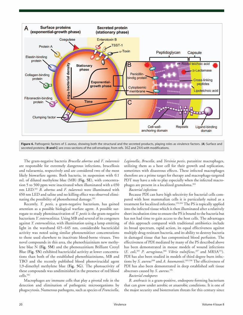
20 virulence volume 4 issue 8
The gram-negative bacteria Brucella abortus and F. tularensis are responsible for extremely dangerous infections, brucellosis and tularaemia, respectively and are considered two of the most likely biowarfare agents. Both bacteria, in suspension with 0.1 mL of diluted methylene blue (MB) (Fig. 5E), with concentra-tion 5 to 500 ppm were inactivated when illuminated with a 650 nm LED.181 B. abortus and F. tularensis were illuminated with 650 nm LED and saline and no killing effect was observed elimi-nating the possibility of photothermal damage.181
Recently, Y. pestis, a gram-negative bacterium, has gained attention as a possible biological warfare agent. A possible sur-rogate to study photoinactivation of Y. pestis is the gram-negative bacterium Y. enterocolitica. Using MB and several of its congeners against Y. enterocolitica, with illumination using a lamp emitting light in the waveband 615–645 nm, considerable bactericidal activity was noted using similar photosensitizer concentrations to those used elsewhere to inactivate blood-borne viruses. Two novel compounds in this area, the phenothiazinium new methy-lene blue N (Fig. 5M) and the phenoxazinium Brilliant Cresyl Blue (Fig. 5N) exhibited bactericidal activity at lower concentra-tions than both of the established phenothiaziniums, MB and TBO and the recently published blood photovirucidal agent 1,9-dimethyl methylene blue (Fig. 5G). The photoactivity of these compounds was undiminished in the presence of red blood cells.182
Macrophages are immune cells that play a pivotal role in the detection and elimination of pathogenic microorganisms by phagocytosis. Numerous pathogens, such as species of Francisella,
Legionella, Brucella, and Yersinia pestis, parasitize macrophages, utilizing them as a host cell for their growth and replication, sometimes with disastrous effects. These infected macrophages therefore are a prime target for therapy and macrophage-targeted PDT may have a role to play especially when the infected macro-phages are present in a localized granuloma.183
Bacterial infectionsBecause PDI can have high selectivity for bacterial cells com-
pared with host mammalian cells it is particularly suited as a treatment for localized infections.159,184 The PS is topically applied into the infected tissue which is then illuminated after a relatively short incubation time to ensure the PS is bound to the bacteria but has not had time to gain access to the host cells. The advantages of this approach compared with traditional antibiotics include its broad spectrum, rapid action, its equal effectiveness against multiply drug-resistant bacteria, and its ability to destroy bacteria in damaged tissue that has compromised blood perfusion. The effectiveness of PDI mediated by many of the PS described above has been demonstrated in mouse models of wound infections (E. coli,185 P. aeruginosa,186 Vibrio vulnificus,187 and MRSA151). PDI has also been studied in models of third degree burn infec-tions by S. aureus188 and A. baumannii.189,190 The effectiveness of PDI has also been demonstrated in deep established soft tissue abscesses caused by S. aureus.191
Bacterial endoporesB. anthracis is a gram-positive, endospore-forming bacterium
that can grow under aerobic or anaerobic conditions. It is one of the major security and bioterrorism threats for this century since
Figure 6. pathogenic factors of S. aureus, showing both the structural and the secreted products, playing roles as virulence factors. (A) Surface and secreted proteins; (B and C) are cross-sections of the cell envelope, from refs. 162 and 244 with modifications.
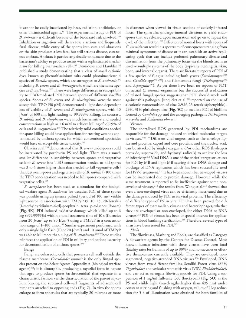
www.landesbioscience.com virulence 21
it cannot be easily inactivated by heat, radiation, antibiotics, or other antimicrobial agents.192 The experimental study of PDI of B. anthracis is difficult because of the biohazard risk involved.193 Inhalation or ingestion will then cause a serious and frequently fatal disease, while entry of the spores into cuts and abrasions on the skin produces a less fatal but still serious disease, cutane-ous anthrax. Anthrax is particularly deadly to humans due to the bacterium’s ability to produce toxins with a sophisticated mecha-nism for killing mammalian cells.194 Demidova and Hamblin195 published a study demonstrating that a class of small cationic dyes known as phenothiazinium salts could photoinactivate 4 species of Bacillus spores, which are surrogates to B. anthracis,196 including B. cereus and B. thuringiensis, which are the same spe-cies as B. anthracis.197 There were large differences in susceptibil-ity to TBO-mediated PDI between spores of different Bacillus species. Spores of B. cereus and B. thuringiensis were the most susceptible. TBO (50 μM) demonstrated a light-dose-dependent loss of viability of B. cereus and B. thuringiensis spores, with 40 J/cm2 of 630 nm light leading to 99.999% killing. In contrast, B. subtilis and B. atrophaeus were much less sensitive and needed concentrations as high as 1.6 mM to achieve killing of >99.9% of cells and B. megaterium.195 The relatively mild conditions needed for spore killing could have applications for treating wounds con-taminated by anthrax spores, for which conventional sporicides would have unacceptable tissue toxicity.195
Oliveira et al.198 demonstrated that B. cereus endospores could be inactivated by porphyrin PS and light. There was a much smaller difference in sensitivity between spores and vegetative cells of B. cereus (the TBO concentration needed to kill spores was 3 to 4 times higher than that needed to kill vegetative cells) than between spores and vegetative cells of B. subtilis (>100 times the TBO concentration was needed to kill spores compared with vegetative cells).195
B. atrophaeus has been used as a simulant for the biologi-cal warfare agent B. anthracis for decades. PDI of these spores was possible using an intense pulsed (period of 100 ms) visible light source in association with TMPyP (5, 10, 15, 20-Tetrakis [1-methylpyridinium-4-yl]-porphyrin tetra p-toluenesulfonate) (Fig. 5K). PDI induced oxidative damage which killed up to 6 log (>99.9999%) within a total treatment time of 10 s (fluencies from 20 J/cm2 up to 80 J/cm2) using a TMPyP in a concentra-tion range of 1–100 μmol.199 Similar experiment performed with only a single light flash (10 or 20 J/cm2) and 10 μmol of TMPyP was able to kill more than 4 log of B. atrophaeus.200 These studies reinforce the application of PDI in military and national security for decontamination of anthrax spores.198
FungiFungi are eukaryotic cells that possess a cell wall outside the
plasma membrane. Coccidioides immitis is the only fungal spe-cies present on the Select Agents Appendix A (biological warfare agents)201: it is dimorphic, producing a mycelial form in nature that ages to produce spores (arthroconidia) that separate in a characteristic fashion via the disarticulation of the parent myce-lium leaving the ruptured cell-wall fragments of adjacent cell remnants attached to opposing ends (Fig. 7). In vivo the spores enlarge to form spherules that are typically 20 microns or more
in diameter when viewed in tissue sections of actively infected hosts. The spherules undergo internal divisions to yield endo-spores that are released upon maturation and go on to repeat the cycle of the infection.202 Infection of normal hosts with spores of C. immitis can result in a spectrum of consequences ranging from minimal symptoms of disease or it can establish an active repli-cating cycle that can include profound pulmonary disease and dissemination from the pulmonary focus via the bloodstream to involve multiple systems of the body (typically meningitis, skin, bone, and internal organs). There are literature reports of PDI of a few species of fungus including both yeasts (Saccharomyces203 and Candida spp204, 205) and filamentous fungi (Trichophyton206 and Aspergillus207). As yet there have been no reports of PDT on actual C. immitis organisms but the successful eradication of related fungal species suggests that PDT should work well against this pathogen. Junqueira et al.208 reported on the use of a cationic nanoemulsion of zinc 2,9,16,23-tetrakis(phenylthio)-29H, 31H-phthalocyanine (Fig. 5C) to mediate PDI of biofilms formed by Candida spp. and the emerging pathogens Trichosporon mucoides and Kodamaea ohmeri.
VirusesThe short-lived ROS generated by PDI mechanisms are
responsible for the damage induced to critical molecular targets in viruses.209,210 Different viral targets, such as the envelope lip-ids and proteins, capsid and core proteins, and the nucleic acid, can be attacked by singlet oxygen and/or other ROS (hydrogen peroxide, superoxide, and hydroxyl radicals) to achieve the loss of infectivity.210 Viral DNA is one of the critical target structures for PDI by MB and light MB causing direct DNA damage and blockage of DNA replication which has been successfully used for HSV-1 treatment.211 It has been shown that enveloped viruses can be inactivated due to protein damage. However, while the same treatment is reported to be ineffective against some non-enveloped viruses,212 the results from Wong et al.213 showed that even a non-enveloped virus can be efficiently inactivated due to the damage induced by PDI to its viral proteins. The efficiency of different types of PS in viral PDI has been proved for dif-ferent types of mammalian viruses and bacteriophages, whether they are enveloped or non-enveloped, for either DNA or RNA viruses.214 PDI of viruses has been of special interest for applica-tions in blood banking sterilization.215 Therefore, several types of virus have been tested for PDI.215
EbolaThe filoviruses, Marburg and Ebola, are classified as Category
A biowarfare agents by the Centers for Disease Control. Most known human infections with these viruses have been fatal (fatality rates for humans of up to 90%) and no vaccines or effec-tive therapies are currently available. They are enveloped, non-segmented, negative-stranded RNA viruses.216 Enveloped, RNA viruses from two different families, Semliki Forest virus (SFV, Togaviridae) and vesicular stomatitis virus (VSV, Rhabdoviridae), and can act as surrogate filovirus models for PDI. Using a sus-pension of 1 mg/ml fullerene C60 (buckyball) (Fig. 5O) as the PS and visible light (wavelengths higher than 495 nm) under constant stirring and flushing with oxygen, values of 7 log reduc-tion for 5 h of illumination were obtained for both families of
22 virulence volume 4 issue 8
enveloped virus.217 VSV plaque forming units were decreased by 5 log using methoxy-polyethylene glycol conjugated fullerene, illuminated by 120 J/cm2 white light.218
Smallpox: Variola major and Variola minor (Orthopoxviruses).It is known that smallpox has been used as a biowarfare
agent in the past. During World War II, scientists from the United Kingdom, the United States, and Japan were involved in research into producing a biological weapon from smallpox.219 In 1992 Soviet defector Ken Alibek confirmed that the Soviet bioweapons program at Zagorsk had produced a large stock-pile—as much as 20 tons—of weaponized smallpox (possibly engineered to resist vaccines), along with refrigerated warheads to deliver it. It is not known whether these stockpiles still exist in Russia. With the breakup of the Soviet Union and unemploy-ment of many of the weapons program’s scientists, there is con-cern that smallpox and the expertise to weaponize it may have become available to other governments or terrorist groups who might wish to use virus as means of biological warfare.220 The last occurrence of endemic smallpox was in Somalia in 1977, and the last human cases were laboratory-acquired infections in 1978. There are four types of Variola major smallpox: ordinary (the most frequent type, accounting for 90% or more of cases); modified (mild and occurring in previously vaccinated persons); f lat; and hemorrhagic (both rare and very severe). Historically, Variola major has an overall fatality rate of about 30%; however, f lat and hemorrhagic smallpox are usually fatal. Present labo-ratory examination of Variola virus requires high-containment (Biosafety Level 4).221
Variola virus is the most notorious poxvirus, a member of a family of large, enveloped DNA viruses. It is generally accepted
that enveloped viruses can be inac-tivated efficiently by singlet oxygen generating agents such as PDI. PDI of HIV-1 by MB/light treatment acts on HIV-1 at different target sites: the envelope and core proteins, and the inner core structures like RNA.222
Four PS (MB, rose bengal [RB] [Fig. 5H], uroporphyrin [UP], and aluminum phthalocynine tetrasul-phonate [AlPcS4]) could inactivate adenovirus. Using MB (2.7 mM) and light (intensity of 106 mW/cm2) produced a complete inactivation of adenovirus after 1 min of exposure: 10 mM of RB was enough for just 0.5 log reduction after 20 min of illumination and complete inactiva-tion was obtained after 30 min PDI with UP; however, AlPcS4 could not completely inactivate adenovirus even when used in 50 mM for 30 min.223
Nucleic acids may be important targets for photoinactivation of
DNA viruses by MB and AlPcS4.224 Photoinactivation of DNA viruses are more efficiently induced by free than by DNA bound porphyrin. Photoreactions of TMPyP and TMPyMPP affect the structural integrity of DNA and also of viral proteins, despite their selective DNA binding.225 The binding of cationic porphy-rins to DNA is presumably due to the electrostatic interaction between the positively-charged substituents in the porphyrin macrocycle and the negatively charged phosphate oxygen atoms of DNA.226
Lassa Virus (Arenavirus) and RVF virus (Bunyaviridae)Lassa virus (LASV) is an Arenavirus that causes Lassa hemor-
rhagic fever in human and non-human primates. Rift Valley fever (RVF) is caused by RVF virus belonging to Bunyaviridae, which is a family of negative-stranded, enveloped RNA viruses. Lassa virus and RVF virus are enveloped RNA viruses that are select agents requiring Biosafety Level 4-equivalent containment.221
Dengue virusDengue and yellow fever viruses belong to the genus Flavivirus
single-stranded RNA viruses. Dengue virus, an enveloped RNA virus, could be inactivated using MB in combination with a LED cluster (mid-peak bandwidth 29 nm, peak 664 nm). The amount of dengue virus reaming was evaluated by plaque forming assays. Dengue virus was completely inactivated within 5 min when the MB concentration was higher than 1.0 μg/mL.227 Lin et al.228 compared light-dependent and light independent inactivation of dengue-2 and other enveloped viruses by the two regio-isomers of carboxyfullerene and found that asymmetric isomer had greater dark activity (even at much higher concentrations than needed for its PDT effect) due to its interaction with the lipid envelope of the virus.
Figure 7. Coccidiodes immitis is the only fungal species present on the Select agents appendix a (biologi-cal warfare agents).201 it is dimorphic, producing a mycelial form in nature that matures to produce spores (arthroconidia) that go on to repeat the cycle of the infection.202
www.landesbioscience.com virulence 23
ToxinsPDI is one of the few anti-
microbial treatments that is also capable of inactivating toxins and secreted virulence factors produced by pathogens. The reactive oxygen species produced during photody-namic action (1O
2 and HO•) can
attack molecular features suscep-tible to oxidation (sulfur atoms, aromatic rings, heterocyclic rings, unsaturated double bonds, amino groups, etc.) present on the toxin molecules themselves. These oxida-tive reactions can disturb the con-formation or alter the functional groups of the toxins and abolish the biological function (Fig. 8). This approach has been well-dem-onstrated in the case of lipopoly-saccharide (LPS, endotoxin from gram-negative bacteria). Komerik et al.229 first showed that TBO and red light could inactivate LPS from E. coli and they also were able to inactivate proteases from P. aerugi-nosa. Gianelli and colleagues230 used MB combined with various light sources to inactivate P. gingivalis LPS adherent to titanium discs, cut from commercial dental implants. Tubby et al.231 studied the ability of MB and red light to inactivate the following secreted virulence factors of S. aureus: V8 protease, α-hemolysin, and sphingomyelin-ase were shown to be inhibited in a dose-dependent manner by exposure to light in the presence of MB. Eubanks et al.232 showed that an actual biowarfare agent, botulinum neurotoxin, could be photoinactivated by exposure to riboflavin and white light. Our laboratory has obtained evidence that two additional microbial toxins, Shiga-like toxin from E. coli O157 and myco-lactone from Mycobacterium ulcerans can be destroyed by expo-sure to benzoporphyrin derivative and red light (manuscript in preparation).
Anti-Microbial Effect of Femtosecond Lasers
It has been proposed that femtosecond lasers, or lasers that maintain a pulse duration of 10−15 s, break down transparent or semitransparent biological tissues due to nonlinear absorption of laser energy with minimal thermal and mechanical effects.233 As a result of the adverse collateral damage possible with other laser systems, the femtosecond laser has been hypothesized to be a new approach for killing pathogens.
Recently, a series of studies reported the efficacy of a visible femtosecond laser or a near-infrared subpicosecond fiber laser on
inactivation of a variety of viral species, including M13 bacterio-phage, tobacco mosaic virus, human papillomavirus, and human immunodefficiency virus.234-240 M13 phages were inactivated by using a very low power (as low as 0.5 nanojoule/pulse) visible femtosecond laser with 425 nm wavelength, 100 fs pulse width, power density ≥ 50 MW/cm2 237.
One group reported241 inactivation of an encephalomyocar-ditis virus, M13 bacteriophage, and Salmonella Typhimurium by a visible femtosecond diode-pumped continuous-wave (CW) mode-locked Ti-sapphire laser. The laser produced a continuous train of 60 fs pulses at a repetition rate of 80 MHz. The exci-tation laser was chosen to operate at a wavelength of λ = 425 nm and with an average power of about 50 mW. It has a pulse width of full-width at half maximum (FWHM) ≅ 100 fs. All the microorganisms were inactivated very efficiently, especially S. Typhimurium. There were different mechanisms of inactivation of different microorganism by femtosecond laser. Inactivation of viruses involves the breaking of hydrogen/hydrophobic bonds or the separation of the weak protein links in the protein shell of a viral particle. On the contrary, inactivation of bacteria is related to the damage of their DNA due to irradiation of a visible fem-tosecond laser.
Figure 8. Diagrammatic representation of the mode of action of several bacterial toxins. (A) Damage to cellular membranes by Staphylococcus aureus toxin. after binding and oligomerization, the stem of the mushroom-shaped toxin heptamer inserts into the target cell and disrupts membrane permeability as depicted by the influx and efflux of ions represented by red and green circles. (B) inhibition of protein synthesis by Shiga toxins (Stx). Holotoxin, which consists of an enzymatically active (A) subunit and five binding (B) subunits, enters cells through the globotriasylceramide (Gb3) receptor. The N-glycosidase activity of the (A) subunit then cleaves an adenosine residue from 28S rRNa, which halts protein synthe-sis. (C) examples of bacterial toxins that activate secondary messenger pathways. Binding of the heat-stable enterotoxins (ST) to a guanylate cyclase receptor results in an increase in cyclic GMp (cGMp) that adversely effects electrolyte flux. By aDp-ribosylation or glucosylation respectively, the c3 exoenzyme (c3) of Clostridium botulinum and the Clostridium difficile toxins a and B (cda and cdB) inactivate the small Rho GTp-binding proteins. cytotoxic necrotizing factor (cNF) of E. coli and the dermonecrotic toxin (DNT) of Bordetella species activate Rho by deamidation.
24 virulence volume 4 issue 8
References1. Thavaselvam D, Vijayaraghavan R. Biological
warfare agents. J Pharm Bioallied Sci 2010; 2:179-88; PMID:21829313; http://dx.doi.org/10.4103/0975-7406.68499
2. Oyston PC. Francisella tularensis: unravelling the secrets of an intracellular pathogen. J Med Microbiol 2008; 57:921-30; PMID:18628490; http://dx.doi.org/10.1099/jmm.0.2008/000653-0
3. McDevitt JJ, Milton DK, Rudnick SN, First MW. Inactivation of poxviruses by upper-room UVC light in a simulated hospital room environment. PLoS One 2008; 3:e3186; PMID:18781204; http://dx.doi.org/10.1371/journal.pone.0003186
4. Achtman M, Zurth K, Morelli G, Torrea G, Guiyoule A, Carniel E. Yersinia pestis, the cause of plague, is a recently emerged clone of Yersinia pseudotuber-culosis. Proc Natl Acad Sci U S A 1999; 96:14043-8; PMID:10570195; http://dx.doi.org/10.1073/pnas.96.24.14043
5. Wuthiekanun V, Peacock SJ. Management of melioidosis. Expert Rev Anti Infect Ther 2006; 4:445-55; PMID:16771621; http://dx.doi.org/10.1586/14787210.4.3.445
6. Klevens RM, Morrison MA, Nadle J, Petit S, Gershman K, Ray S, Harrison LH, Lynfield R, Dumyati G, Townes JM, et al.; Active Bacterial Core surveillance (ABCs) MRSA Investigators. Invasive methicillin-resistant Staphylococcus aureus infec-tions in the United States. JAMA 2007; 298:1763-71; PMID:17940231; http://dx.doi.org/10.1001/jama.298.15.1763
7. Musser JM, Hauser AR, Kim MH, Schlievert PM, Nelson K, Selander RK. Streptococcus pyogenes causing toxic-shock-like syndrome and other inva-sive diseases: clonal diversity and pyrogenic exotoxin expression. Proc Natl Acad Sci U S A 1991; 88:2668-72; PMID:1672766; http://dx.doi.org/10.1073/pnas.88.7.2668
8. Helgason E, Økstad OA, Caugant DA, Johansen HA, Fouet A, Mock M, Hegna I, Kolstø AB. Bacillus anthracis, Bacillus cereus, and Bacillus thuringiensis--one species on the basis of genetic evidence. Appl Environ Microbiol 2000; 66:2627-30; PMID:10831447; http://dx.doi.org/10.1128/AEM.66.6.2627-2630.2000
9. Sagripanti JL, Lytle CD. Sensitivity to ultravio-let radiation of Lassa, vaccinia, and Ebola viruses dried on surfaces. Arch Virol 2011; 156:489-94; PMID:21104283; http://dx.doi.org/10.1007/s00705-010-0847-1
10. Sugiyama H. Clostridium botulinum neurotoxin. Microbiol Rev 1980; 44:419-48; PMID:6252433
11. Meselson M, Guillemin J, Hugh-Jones M, Langmuir A, Popova I, Shelokov A, Yampolskaya O. The Sverdlovsk anthrax outbreak of 1979. Science 1994; 266:1202-8; PMID:7973702; http://dx.doi.org/10.1126/science.7973702
12. Manchee RJ, Broster MG, Melling J, Henstridge RM, Stagg AJ. Bacillus anthracis on Gruinard Island. Nature 1981; 294:254-5; PMID:6795509; http://dx.doi.org/10.1038/294254a0
Another study242 reported the inactivation of murine cytomeg-alovirus (MCMV), an enveloped, double-stranded DNA virus, by a visible (425 nm) femtosecond laser. The results showed that the laser irradiation caused a 5-log reduction in MCMV titer and caused selective aggregation of viral capsid and tegument pro-teins. However, the femtosecond laser did not cause significant changes to the global structure of MCMV virions including membrane and capsid, as assessed by electron microscopy; mean-while, it could not produce the double-strand breaks or crosslink-ing in MCMV genomic DNA.
Manipulation of a near-infrared (NIR) femtosecond laser via impulsive stimulated raman scattering (ISRS) to produce dam-age (e.g., to the protein coat of a virus) is another method for selectively inactivating microorganisms.237
When NIR femtosecond laser induced the inaction of virus and bacteria, its safety to the mammalian cells was considered. The relative research demonstrated that if the wavelength and pulse width of the femtosecond laser were appropriately selected, there was a window in power density that enabled them to achieve selective inactivation of target viruses and bacteria with-out causing cytotoxicity to mammalian cells. It was suggested that this strategy targeted the mechanical (vibrational) properties of microorganisms and thus its antimicrobial efficacy was likely to be unaffected by genetic differences in the microorganisms.238
In the view of the emerging threats from drug resistant patho-gens and microorganisms, developing novel and more effective antimicrobial strategies is an absolute necessity. One such strat-egy is to develop the ultrashort pulsed (USP) laser technology as an effective and chemicals free inactivation technique that can be successfully used over broad spectrum of pathogens, both from bacterial and viral sources.243
In summary, the advantages of such novel laser technologies over the presently prevailing disinfection methods include: they are considered as noninvasive disinfection technologies, because no foreign materials are needed in the disinfection process; they are harmless environmental disinfection methods since no chemicals are used in the pathogen inactivation process; and they are general methods for selective disinfection of pathogens with potentially minimal side effects.241
Conclusion
Recent studies have highlighted the diversity of applications of light-mediated technology against pathogens of all known classes. Wavelengths from the short-UV to the near-infrared (either alone or combined with PS) can be used to kill or inactivate gram-positive and gram-negative bacteria, fungi, endospores, para-sites, viruses, and even protein toxins. The mechanisms of action depend on the different microbial types and the wavelength and presence or not of a PS. The two broad target classes are nucleic acids for UVC and PUVA and oxidizable proteins for photocatal-ysis, PDT, and blue light. The broad occurrence of these biologi-cal targets in bioweapons agents means that the light-mediated technology is highly likely to be very broad-spectrum thus avoid-ing the need to know the identity of the particular agent in any mass biological attack, and also suggests that the development of resistance to light-mediated inactivation is likely to non-existent. Furthermore, light is non-polluting and environmentally friendly, and even if PS need to be used, these compounds are likely to be photodegraded rapidly when the bio-threat has been neutralized thus leaving no lasting pollution. The use of light-based technol-ogy to prevent and treat actual infections suggests that they may be useful to decontaminate humans that have already received exposure to biological agents, without causing undue harm to host tissue. Lastly light-based inactivation may be particularly suitable to form vaccines as they kill pathogens while preserving their antigenicity.
Disclosure of Potential Conflicts of Interest
No potential conflicts of interest were disclosed.
Dedication
We dedicate this work to the great philanthropist Dr Orhan Ocalgiray for his profound contributions to the field of microbiology.
Acknowledgments
Research in the Hamblin laboratory is supported by US NIH grant R01AI050875.
www.landesbioscience.com virulence 25
13. Manchee RJ, Broster MG, Stagg AJ, Hibbs SE. Formaldehyde Solution Effectively Inactivates Spores of Bacillus anthracis on the Scottish Island of Gruinard. Appl Environ Microbiol 1994; 60:4167-71; PMID:16349444
14. Aldhous P. Biological warfare. Gruinard Island handed back. Nature 1990; 344:801; PMID:2109833; http://dx.doi.org/10.1038/344801b0
15. Grunow R, Finke EJ. A procedure for differen-tiating between the intentional release of bio-logical warfare agents and natural outbreaks of disease: its use in analyzing the tularemia outbreak in Kosovo in 1999 and 2000. Clin Microbiol Infect 2002; 8:510-21; PMID:12197873; http://dx.doi.org/10.1046/j.1469-0691.2002.00524.x
16. Török TJ, Tauxe RV, Wise RP, Livengood JR, Sokolow R, Mauvais S, Birkness KA, Skeels MR, Horan JM, Foster LR. A large community outbreak of salmonellosis caused by intentional contamina-tion of restaurant salad bars. JAMA 1997; 278:389-95; PMID:9244330; http://dx.doi.org/10.1001/jama.1997.03550050051033
17. Kolavic SA, Kimura A, Simons SL, Slutsker L, Barth S, Haley CE. An outbreak of Shigella dysenteriae type 2 among laboratory workers due to intentional food contamination. JAMA 1997; 278:396-8; PMID:9244331; http://dx.doi.org/10.1001/jama.1997.03550050058034
18. Hsu VP, Lukacs SL, Handzel T, Hayslett J, Harper S, Hales T, Semenova VA, Romero-Steiner S, Elie C, Quinn CP, et al. Opening a bacillus anthracis-containing envelope, Capitol Hill, Washington, D.C.: the public health response. Emerg Infect Dis 2002; 8:1039-43; PMID:12396912; http://dx.doi.org/10.3201/eid0810.020332
19. Centers for Disease Control and Prevention (CDC). Update: Investigation of bioterrorism-related anthrax, 2001. MMWR Morb Mortal Wkly Rep 2001; 50:1008-10; PMID:11724158
20. Centers for Disease Control and Prevention (CDC). Update: Cutaneous anthrax in a laboratory worker--Texas, 2002. MMWR Morb Mortal Wkly Rep 2002; 51:482; PMID:12064454
21. Cutler TD, Zimmerman JJ. Ultraviolet irradiation and the mechanisms underlying its inactivation of infectious agents. Anim Health Res Rev 2011; 12:15-23; PMID:21676338; http://dx.doi.org/10.1017/S1466252311000016
22. Jagger J. Introduction to research in ultraviolet photo-biology. Prentice-Hall (Englewood Cliffs, N.J), 1967.
23. Reed NG. The history of ultraviolet germicidal irra-diation for air disinfection. Public Health Rep 2010; 125:15-27; PMID:20402193
24. Brickner PW, Vincent RL, First M, Nardell E, Murray M, Kaufman W. The application of ultraviolet germi-cidal irradiation to control transmission of airborne disease: bioterrorism countermeasure. Public Health Rep 2003; 118:99-114; PMID:12690064; http://dx.doi.org/10.1016/S0033-3549(04)50225-X
25. Dai T, Garcia B, Murray CK, Vrahas MS, Hamblin MR. UVC light prophylaxis for cutaneous wound infections in mice. Antimicrob Agents Chemother 2012; 56:3841-8; PMID:22564833; http://dx.doi.org/10.1128/AAC.00161-12
26. Ladd M. Introduction to Physical Chemistry. London, UK: Cambridge University Press, 1998.
27. Durban E, Grecz N. Resistance of spores of Clostridium botulinum 33A to combinations of ultra-violet and gamma rays. Appl Microbiol 1969; 18:44-50; PMID:4896102
28. Nourmoradi H, Nikaeen M, Stensvold CR, Mirhendi H. Ultraviolet irradiation: An effective inactivation method of Aspergillus spp. in water for the control of waterborne nosocomial aspergillosis. Water Res 2012; 46:5935-40; PMID:22985523; http://dx.doi.org/10.1016/j.watres.2012.08.015
29. Guerrero-Beltran J, Welti-Chanes J, Barbosa-Canovas G. UVC light processing of grape, cranberry and grapefruit juices to inactivate Saccharomyces Cerevisiae. J Food Process Eng 2009; 32:916-32; http://dx.doi.org/10.1111/j.1745-4530.2008.00253.x
30. Levy C, Aubert X, Lacour B, Carlin F. Relevant fac-tors affecting microbial surface decontamination by pulsed light. Int J Food Microbiol 2012; 152:168-74; PMID:21924512; http://dx.doi.org/10.1016/j.ijfoodmicro.2011.08.022
31. Luna VA, Cannons AC, Amuso PT, Cattani J. The inactivation and removal of airborne Bacillus atro-phaeus endospores from air circulation systems using UVC and HEPA filters. J Appl Microbiol 2008; 104:489-98; PMID:17927759
32. Dean SJ, Petty A, Swift S, McGhee JJ, Sharma A, Shah S, Craig JP. Efficacy and safety assessment of a novel ultraviolet C device for treating corneal bacterial infections. Clin Experiment Ophthalmol 2011; 39:156-63; PMID:21105972; http://dx.doi.org/10.1111/j.1442-9071.2010.02471.x
33. Umezawa K, Asai S, Inokuchi S, Miyachi H. A com-parative study of the bactericidal activity and daily disinfection housekeeping surfaces by a new por-table pulsed UV radiation device. Curr Microbiol 2012; 64:581-7; PMID:22447288; http://dx.doi.org/10.1007/s00284-012-0110-y
34. Al-Marir A. Ultraviolet C lethal effect on Brucella melitensis. Microbiologica-Quarterly J of Microbiol Sci 2008; 31:47-56
35. Rose LJ, O’Connell H. UV light inactivation of bacterial biothreat agents. Appl Environ Microbiol 2009; 75:2987-90; PMID:19270145; http://dx.doi.org/10.1128/AEM.02180-08
36. Miller SL, Linnes J, Luongo J. Ultraviolet germi-cidal irradiation: future directions for air disinfec-tion and building applications. Photochem Photobiol 2013; 89:777-81; PMID:23581680; http://dx.doi.org/10.1111/php.12080
37. Lakretz A, Ron EZ, Mamane H. Biofouling control in water by various UVC wavelengths and doses. Biofouling 2010; 26:257-67; PMID:20024789; http://dx.doi.org/10.1080/08927010903484154
38. Kalisvaart BF. Re-use of wastewater: preventing the recovery of pathogens by using medium-pressure UV lamp technology. Water Sci Technol 2004; 50:337-44; PMID:15537023
39. Li J, Hirota K, Yumoto H, Matsuo T, Miyake Y, Ichikawa T. Enhanced germicidal effects of pulsed UV-LED irradiation on biofilms. J Appl Microbiol 2010; 109:2183-90; PMID:20854456; http://dx.doi.org/10.1111/j.1365-2672.2010.04850.x
40. Farrell HP, Garvey M, Cormican M, Laffey JG, Rowan NJ. Investigation of critical inter-related fac-tors affecting the efficacy of pulsed light for inacti-vating clinically relevant bacterial pathogens. J Appl Microbiol 2010; 108:1494-508; PMID:19796119; http://dx.doi.org/10.1111/j.1365-2672.2009.04545.x
41. Nicholson WL, Galeano B. UV resistance of Bacillus anthracis spores revisited: validation of Bacillus sub-tilis spores as UV surrogates for spores of B. anthracis Sterne. Appl Environ Microbiol 2003; 69:1327-30; PMID:12571068; http://dx.doi.org/10.1128/AEM.69.2.1327-1330.2003
42. Rettberg P, Cockell CS. Biological UV dosimetry using the DLR-biofilm. Photochem Photobiol Sci 2004; 3:781-7; PMID:15295635; http://dx.doi.org/10.1039/b315950g
43. Moehrle M, Dennenmoser B, Garbe C. Continuous long-term monitoring of UV radiation in professional mountain guides reveals extremely high exposure. Int J Cancer 2003; 103:775-8; PMID:12516097; http://dx.doi.org/10.1002/ijc.10884
44. Moehrle M, Korn M, Garbe C. Bacillus subtilis spore film dosimeters in personal dosimetry for occupa-tional solar ultraviolet exposure. Int Arch Occup Environ Health 2000; 73:575-80; PMID:11100953; http://dx.doi.org/10.1007/s004200000183
45. Vähävihu K, Ylianttila L, Kautiainen H, Tuohimaa P, Reunala T, Snellman E. Spore film dosim-eters are feasible for UV dose monitoring dur-ing heliotherapy. Photochem Photobiol 2010; 86:1174-8; PMID:20573044; http://dx.doi.org/10.1111/j.1751-1097.2010.00769.x
46. Benjdia A, Heil K, Barends TR, Carell T, Schlichting I. Structural insights into recognition and repair of UV-DNA damage by Spore Photoproduct Lyase, a radical SAM enzyme. Nucleic Acids Res 2012; 40:9308-18; PMID:22761404; http://dx.doi.org/10.1093/nar/gks603
47. Yang L, Lin G, Nelson RS, Jian Y, Telser J, Li L. Mechanistic studies of the spore photoproduct lyase via a single cysteine mutation. Biochemistry 2012; 51:7173-88; PMID:22906093; http://dx.doi.org/10.1021/bi3010945
48. Silver SC, Chandra T, Zilinskas E, Ghose S, Broderick WE, Broderick JB. Complete stereospecific repair of a synthetic dinucleotide spore photoproduct by spore photoproduct lyase. J Biol Inorg Chem 2010; 15:943-55; PMID:20405152; http://dx.doi.org/10.1007/s00775-010-0656-8
49. Friedel MG, Berteau O, Pieck JC, Atta M, Ollagnier-de-Choudens S, Fontecave M, Carell T. The spore photoproduct lyase repairs the 5S- and not the 5R-configured spore photoproduct DNA lesion. Chem Commun (Camb) 2006; 445-7; PMID:16493831; http://dx.doi.org/10.1039/b514103f
50. Setlow P. Resistance of spores of Bacillus species to ultraviolet light. Environ Mol Mutagen 2001; 38:97-104; PMID:11746741; http://dx.doi.org/10.1002/em.1058
51. Negrao BL, Crafford D, Viljoen M. The effect of sympathomimetic medication on cardiovascular functioning of children with attention-deficit/hyper-activity disorder. Cardiovasc J Afr 2009; 20:296-9; PMID:19907802
52. Chandra T, Silver SC, Zilinskas E, Shepard EM, Broderick WE, Broderick JB. Spore photoprod-uct lyase catalyzes specific repair of the 5R but not the 5S spore photoproduct. J Am Chem Soc 2009; 131:2420-1; PMID:19178276; http://dx.doi.org/10.1021/ja807375c
53. Yang L, Nelson RS, Benjdia A, Lin G, Telser J, Stoll S, Schlichting I, Li L. A radical transfer pathway in spore photoproduct lyase. Biochemistry 2013; 52:3041-50; PMID:23607538; http://dx.doi.org/10.1021/bi3016247
54. Bucheli-Witschel M, Bassin C, Egli T. UV-C inac-tivation in Escherichia coli is affected by growth conditions preceding irradiation, in particular by the specific growth rate. J Appl Microbiol 2010; 109:1733-44; PMID:20629801
55. Zenoff VF, Heredia J, Ferrero M, Siñeriz F, Farías ME. Diverse UV-B resistance of culturable bacte-rial community from high-altitude wetland water. Curr Microbiol 2006; 52:359-62; PMID:16604419; http://dx.doi.org/10.1007/s00284-005-0241-5
56. Steinborn G. Uvm mutants of Escherichia coli K12 deficient in UV mutagenesis. I. Isolation of uvm mutants and their phenotypical characterization in DNA repair and mutagenesis. Mol Gen Genet 1978; 165:87-93; PMID:362169; http://dx.doi.org/10.1007/BF00270380
57. Munoz-Najar U, Vijayakumar MN. An operon that confers UV resistance by evoking the SOS mutagenic response in streptococcal conjugative transposon Tn5252. J Bacteriol 1999; 181:2782-8; PMID:10217768
58. Dai T, Vrahas MS, Murray CK, Hamblin MR. Ultraviolet C irradiation: an alternative antimicro-bial approach to localized infections? Expert Rev Anti Infect Ther 2012; 10:185-95; PMID:22339192; http://dx.doi.org/10.1586/eri.11.166
26 virulence volume 4 issue 8
59. Dai T, Murray CK, Vrahas MS, Baer DG, Tegos GP, Hamblin MR. Ultraviolet C light for Acinetobacter baumannii wound infections in mice: potential use for battlefield wound decontamination? J Trauma Acute Care Surg 2012; 73:661-7; PMID:22929495; http://dx.doi.org/10.1097/TA.0b013e31825c149c
60. Dai T, Kharkwal GB, Zhao J, St Denis TG, Wu Q, Xia Y, Huang L, Sharma SK, d’Enfert C, Hamblin MR. Ultraviolet-C light for treatment of Candida albicans burn infection in mice. Photochem Photobiol 2011; 87:342-9; PMID:21208209; http://dx.doi.org/10.1111/j.1751-1097.2011.00886.x
61. Hashimoto K, Irie H, Fujishima A. TiO2 Photocatalysis: A Historical Overview and Future Prospects. Jpn J Appl Phys 2005; 44:8269-85; http://dx.doi.org/10.1143/JJAP.44.8269
62. Fujishima A, Honda K. Electrochemical photoly-sis of water at a semiconductor electrode. Nature 1972; 238:37-8; PMID:12635268; http://dx.doi.org/10.1038/238037a0
63. Carey JH, Lawrence J, Tosine HM. Photodechlorination of PCB’s in the presence of tita-nium dioxide in aqueous suspensions. Bull Environ Contam Toxicol 1976; 16:697-701; PMID:828069; http://dx.doi.org/10.1007/BF01685575
64. Frank SN, Bard AJ. Heterogeneous photocatalytic oxidation of cyanide ion in aqueous solutions at tita-nium dioxide powder. J Am Chem Soc 1977; 99:303-4; http://dx.doi.org/10.1021/ja00443a081
65. Chen J, Poon CS. Photocatalytic construction and building materials: from fundamentals to applica-tions. Build Environ 2009; 44:1899-906; http://dx.doi.org/10.1016/j.buildenv.2009.01.002
66. Fujishima A, Rao TN, Tryk DA. Titanium dioxide photocatalysis. J Photochem Photobiol Photochem Rev 2000; 1:1-21; http://dx.doi.org/10.1016/S1389-5567(00)00002-2
67. Lilja M, Forsgren J, Welch K, Astrand M, Engqvist H, Strømme M. Photocatalytic and antimicro-bial properties of surgical implant coatings of titanium dioxide deposited though cathodic arc evaporation. Biotechnol Lett 2012; 34:2299-305; PMID:22941372; http://dx.doi.org/10.1007/s10529-012-1040-2
68. Schrauben JN, Hayoun R, Valdez CN, Braten M, Fridley L, Mayer JM. Titanium and zinc oxide nanoparticles are proton-coupled electron transfer agents. Science 2012; 336:1298-301; PMID:22679095; http://dx.doi.org/10.1126/science.1220234
69. Matsunaga T, Tomoda R, Nakajima T, Wake H. Photoelectrochemical sterilization of micro-bial cells by semiconductor powders. FEMS Microbiol Lett 1985; 29:211-4; http://dx.doi.org/10.1111/j.1574-6968.1985.tb00864.x
70. Matsunaga T, Tomoda R, Nakajima T, Nakamura N, Komine T. Continuous-sterilization system that uses photosemiconductor powders. Appl Environ Microbiol 1988; 54:1330-3; PMID:3046487
71. Foster HA, Ditta IB, Varghese S, Steele A. Photocatalytic disinfection using titanium dioxide: spectrum and mechanism of antimicrobial activ-ity. Appl Microbiol Biotechnol 2011; 90:1847-68; PMID:21523480; http://dx.doi.org/10.1007/s00253-011-3213-7
72. McCullagh C, Robertson JM, Bahnemann DW. K. RP. The application of TiO2 photoca-talysis for disinfection of water contaminated with pathogenic micro-organisms: a review. Res Chem Intermed 2007; 33:359-75; http://dx.doi.org/10.1163/156856707779238775
73. Cushnie TP, Robertson PK, Officer S, Pollard PM, McCullagh C, Robertson JM. Variables to be consid-ered when assessing the photocatalytic destruction of bacterial pathogens. Chemosphere 2009; 74:1374-8; PMID:19101016; http://dx.doi.org/10.1016/j.chemosphere.2008.11.012
74. Ireland JC, Klostermann P, Rice EW, Clark RM. Inactivation of Escherichia coli by titanium dioxide photocatalytic oxidation. Appl Environ Microbiol 1993; 59:1668-70; PMID:8390819
75. Pham HN, McDowell T, Wilkins E. Photocatalytically-mediated disinfection of water using TiO2 as a catalyst and spore-forming Bacillus pumilus as a model. J Environ Sci and Health 1995; A30:627-36
76. Inglesby TV. Anthrax: A possible case history. Emerg Infect Dis 1999; 5:556-60; PMID:10458965; http://dx.doi.org/10.3201/eid0504.990419
77. Armon R, Weltch-Cohen G, Bettane P. Disinfection of Bacillus spp. spores in drinking water by TiO2 photocatalysis as a model for Bacillus anthracis. Water Supply 2004; 4:7-14
78. Maness PC, Smolinski S, Blake DM, Huang Z, Wolfrum EJ, Jacoby WA. Bactericidal activity of pho-tocatalytic TiO(2) reaction: toward an understand-ing of its killing mechanism. Appl Environ Microbiol 1999; 65:4094-8; PMID:10473421
79. Maness PC, Smolinski S, Blake DM, Huang Z, Wolfrum EJ, Jacoby WA. Bactericidal activity of pho-tocatalytic TiO(2) reaction: toward an understand-ing of its killing mechanism. Appl Environ Microbiol 1999; 65:4094-8; PMID:10473421
80. Pelaez M, Nolan NT, Pillai SC, Seery MK. A review on the visible light active titanium dioxide photocataly-sis for e\nviromental applications. Appl Catal B 2012; http://dx.doi.org/10.1016/j.apcatb.2012.05.036
81. Gogniat G, Thyssen M, Denis M, Pulgarin C, Dukan S. The bactericidal effect of TiO2 photo-catalysis involves adsorption onto catalyst and the loss of membrane integrity. FEMS Microbiol Lett 2006; 258:18-24; PMID:16630249; http://dx.doi.org/10.1111/j.1574-6968.2006.00190.x
82. Liou JW, Chang HH. Bactericidal effects and mechanisms of visible light-responsive titanium dioxide photocatalysts on pathogenic bacteria. Arch Immunol Ther Exp (Warsz) 2012; 60:267-75; PMID:22678625; http://dx.doi.org/10.1007/s00005-012-0178-x
83. Zhang H, Zhao H, Zhang S, Quan X. Photoelectrochemical manifestation of photoelectron transport properties of vertically aligned nanotubular TiO2 photoanodes. Chemphyschem 2008; 9:117-23; PMID:18072232; http://dx.doi.org/10.1002/cphc.200700406
84. Harper JC, Christensen PA, Egerton TA. Effect of catalyst type on the kinetics of photoelectri-cal disinfection of water inoculated with E. coli. J Appl Electrochem 2000; 31:623-8; http://dx.doi.org/10.1023/A:1017539328022
85. Irie H, Watanabe Y, Hashimoto K. Carbon-doped Anatase TiO2 Powders as a Visible-light Sensitive Photocatalyst. Chem Lett 2003; 32:772; http://dx.doi.org/10.1246/cl.2003.772
86. Rhee CH, Bae SW, Lee JS. Template-free Hydrothermal Synthesis of High Surface Area Nitrogen-doped Titania Photocatalyst Active under Visible Light. Chem Lett 2005; 34:660; http://dx.doi.org/10.1246/cl.2005.660
87. Lin L, Lin W, Zhu Y, Zhao B, Xie Y. Phosphor-doped Titania —a Novel Photocatalyst Active in Visible Light. Chem Lett 2005; 34:284; http://dx.doi.org/10.1246/cl.2005.284
88. Tokudome H, Miyauchi M. N-doped TiO2 Nanotube with Visible Light Activity. Chem Lett 2004; 33:1108; http://dx.doi.org/10.1246/cl.2004.1108
89. Irie H, Miura S, Nakamura R, Hashimoto K. A Novel Visible-light-sensitive Efficient Photocatalyst, CrIII-grafted TiO2. Chem Lett 2008; 37:252; http://dx.doi.org/10.1246/cl.2008.252
90. Li XZ, Li FB. Study of Au/Au(3+)-TiO2 photo-catalysts toward visible photooxidation for water and wastewater treatment. Environ Sci Technol 2001; 35:2381-7; PMID:11414049; http://dx.doi.org/10.1021/es001752w
91. Behar D, Rabani J. Kinetics of hydrogen produc-tion upon reduction of aqueous TiO2 nanoparticles catalyzed by Pd(0), Pt(0), or Au(0) coatings and an unusual hydrogen abstraction; steady state and pulse radiolysis study. J Phys Chem B 2006; 110:8750-5; PMID:16640431; http://dx.doi.org/10.1021/jp060971m
92. Zeng Y, Wu W, Lee S, Gao J. Photocatalytic perfor-mance of plasma sprayed Pt-modified TiO2 coat-ings under visible light irradiation. Catal Commun 2007; 8:906-12; http://dx.doi.org/10.1016/j.catcom.2006.09.023
93. Wang W, Zhang J, Chen F, He D, Anpo M. Preparation and photocatalytic properties of Fe3+-doped Ag@TiO2 core-shell nanoparticles. J Colloid Interface Sci 2008; 323:182-6; PMID:18448112; http://dx.doi.org/10.1016/j.jcis.2008.03.043
94. You X, Chen F, Zhang J, Anpo M. A novel deposition precipitation method for preparation of Ag-loaded titanium dioxide. Catal Lett 2005; 102:247-50; http://dx.doi.org/10.1007/s10562-005-5863-5
95. Yu JC, Ho W, Yu J, Yip H, Wong PK, Zhao J. Efficient visible-light-induced photocatalytic disinfection on sulfur-doped nanocrystalline titania. Environ Sci Technol 2005; 39:1175-9; PMID:15773492; http://dx.doi.org/10.1021/es035374h
96. Mitoraj D, Jańczyk A, Strus M, Kisch H, Stochel G, Heczko PB, Macyk W. Visible light inactiva-tion of bacteria and fungi by modified titanium dioxide. Photochem Photobiol Sci 2007; 6:642-8; PMID:17549266; http://dx.doi.org/10.1039/b617043a
97. Vohra A, Goswami DY, Deshpande DA, Block SS. Enhanced photocatalytic inactivation of bacterial spores on surfaces in air. J Ind Microbiol Biotechnol 2005; 32:364-70; PMID:16044291; http://dx.doi.org/10.1007/s10295-005-0006-y
98. Wong MS, Sun DS, Chang HH. Bactericidal per-formance of visible-light responsive titania pho-tocatalyst with silver nanostructures. PLoS One 2010; 5:e10394; PMID:20454454; http://dx.doi.org/10.1371/journal.pone.0010394
99. Kau JH, Sun DS, Huang HH, Wong MS, Lin HC, Chang HH. Role of visible light-activated photo-catalyst on the reduction of anthrax spore-induced mortality in mice. PLoS One 2009; 4:e4167; PMID:19132100; http://dx.doi.org/10.1371/journal.pone.0004167
100. Wong MS, Chu WC, Sun DS, Huang HS, Chen JH, Tsai PJ, Lin NT, Yu MS, Hsu SF, Wang SL, et al. Visible-light-induced bactericidal activity of a nitro-gen-doped titanium photocatalyst against human pathogens. Appl Environ Microbiol 2006; 72:6111-6; PMID:16957236; http://dx.doi.org/10.1128/AEM.02580-05
101. Prasad GK, Agarwal GS, Singh B, Rai GP, Vijayaraghavan R. Photocatalytic inactivation of Bacillus anthracis by titania nanomaterials. J Hazard Mater 2009; 165:506-10; PMID:19056174; http://dx.doi.org/10.1016/j.jhazmat.2008.10.009
102. Prasad GK, Ramacharyulu PV, Merwyn S, Agarwal GS, Srivastava AR, Singh B, Rai GP, Vijayaraghavan R. Photocatalytic inactivation of spores of Bacillus anthracis using titania nanomaterials. J Hazard Mater 2011; 185:977-82; PMID:21035260; http://dx.doi.org/10.1016/j.jhazmat.2010.10.001
103. Roy AS, Parveen A, Koppalkar AR, Prasad M. Effect of Nano - Titanium Dioxide with Different Antibiotics against Methicillin-Resistant Staphylococcus Aureus. J Biomat Nanobiotechnol 2010; 1:37-41; http://dx.doi.org/10.4236/jbnb.2010.11005
104. Cheng CL, Sun DS, Chu WC, Tseng YH, Ho HC, Wang JB, Chung PH, Chen JH, Tsai PJ, Lin NT, et al. The effects of the bacterial interac-tion with visible-light responsive titania photo-catalyst on the bactericidal performance. J Biomed Sci 2009; 16:7; PMID:19272171; http://dx.doi.org/10.1186/1423-0127-16-7
www.landesbioscience.com virulence 27
105. Tsai T-M, Chang H-H, Chang K-C, Liu Y-L, Tseng C-C. A comparative study of the bactericidal effect of photocatalytic oxidation by TiO2 on antibiotic-resistant and antibiotic-sensitive bacteria. J Chem Technol Biotechnol 2010; 85:1642-53; http://dx.doi.org/10.1002/jctb.2476
106. Nhung TT, Nagata H, Takahashi A, Aihara M, Okamoto T, Shimohata T, Mawatari K, Akutagawa M, Kinouchi Y, Haraguchi M. Sterilization effect of UV light on Bacillus spores using TiO(2) films depends on wavelength. J Med Invest 2012; 59:53-8; PMID:22449993; http://dx.doi.org/10.2152/jmi.59.53
107. Knudson GB. Photoreactivation of ultraviolet-irra-diated, plasmid-bearing, and plasmid-free strains of Bacillus anthracis. Appl Environ Microbiol 1986; 52:444-9; PMID:2429617
108. Baweja RB, Zaman MS, Mattoo AR, Sharma K, Tripathi V, Aggarwal A, Dubey GP, Kurupati RK, Ganguli M, Chaudhury NK, et al. Properties of Bacillus anthracis spores prepared under various envi-ronmental conditions. Arch Microbiol 2008; 189:71-9; PMID:17713759; http://dx.doi.org/10.1007/s00203-007-0295-9
109. Melski JW, Tanenbaum L, Parrish JA, Fitzpatrick TB, Bleich HL. Oral methoxsalen photochemotherapy for the treatment of psoriasis: a cooperative clinical trial. J Invest Dermatol 1977; 68:328-35; PMID:864273; http://dx.doi.org/10.1111/1523-1747.ep12496022
110. Salem SA, Barakat MA, Morcos CM. Bath psoralen+ultraviolet A photochemotherapy vs. narrow band-ultraviolet B in psoriasis: a com-parison of clinical outcome and effect on circu-lating T-helper and T-suppressor/cytotoxic cells. Photodermatol Photoimmunol Photomed 2010; 26:235-42; PMID:20831697; http://dx.doi.org/10.1111/j.1600-0781.2010.00525.x
111. Cazenave JP, Waller C, Kientz D, Mendel I, Lin L, Jacquet M, Propst M, Liu W, Corash L, Sundin D, et al. An active hemovigilance program character-izing the safety profile of 7483 transfusions with plasma components prepared with amotosalen and UVA photochemical treatment. Transfusion 2010; 50:1210-9; PMID:20113450; http://dx.doi.org/10.1111/j.1537-2995.2009.02579.x
112. Dubensky TW Jr., Skoble J, Lauer P, Brockstedt DG. Killed but metabolically active vaccines. Curr Opin Biotechnol 2012; 23:917-23; PMID:22608846; http://dx.doi.org/10.1016/j.copbio.2012.04.005
113. Lai C, Cao H, Hearst JE, Corash L, Luo H, Wang Y. Quantitative analysis of DNA interstrand cross-links and monoadducts formed in human cells induced by psoralens and UVA irradiation. Anal Chem 2008; 80:8790-8; PMID:18947205; http://dx.doi.org/10.1021/ac801520m
114. Irsch J, Lin L. Pathogen Inactivation of Platelet and Plasma Blood Components for Transfusion Using the INTERCEPT Blood System™. Transfus Med Hemother 2011; 38:19-31; PMID:21779203; http://dx.doi.org/10.1159/000323937
115. Brockstedt DG, Bahjat KS, Giedlin MA, Liu W, Leong M, Luckett W, Gao Y, Schnupf P, Kapadia D, Castro G, et al. Killed but metabolically active microbes: a new vaccine paradigm for eliciting effec-tor T-cell responses and protective immunity. Nat Med 2005; 11:853-60; PMID:16041382; http://dx.doi.org/10.1038/nm1276
116. Skoble J, Beaber JW, Gao Y, Lovchik JA, Sower LE, Liu W, Luckett W, Peterson JW, Calendar R, Portnoy DA, et al. Killed but metabolically active Bacillus anthracis vaccines induce broad and pro-tective immunity against anthrax. Infect Immun 2009; 77:1649-63; PMID:19168734; http://dx.doi.org/10.1128/IAI.00530-08
117. Bruhn KW, Birnbaum R, Haskell J, Vanchinathan V, Greger S, Narayan R, Chang PL, Tran TA, Hickerson SM, Beverley SM, et al. Killed but metabolically active Leishmania infantum as a novel whole-cell vac-cine for visceral leishmaniasis. Clin Vaccine Immunol 2012; 19:490-8; PMID:22323556; http://dx.doi.org/10.1128/CVI.05660-11
118. Harding AS, Schwab KJ. Using limes and synthetic psoralens to enhance solar disinfection of water (SODIS): a laboratory evaluation with norovirus, Escherichia coli, and MS2. Am J Trop Med Hyg 2012; 86:566-72; PMID:22492137; http://dx.doi.org/10.4269/ajtmh.2012.11-0370
119. Rasonglès P, Angelini-Tibert MF, Simon P, Currie C, Isola H, Kientz D, Slaedts M, Jacquet M, Sundin D, Lin L, et al. Transfusion of platelet components prepared with photochemical patho-gen inactivation treatment during a Chikungunya virus epidemic in Ile de La Réunion. Transfusion 2009; 49:1083-91; PMID:19309473; http://dx.doi.org/10.1111/j.1537-2995.2009.02111.x
120. Sawyer L, Hanson D, Castro G, Luckett W, Dubensky TW Jr., Stassinopoulos A. Inactivation of parvovirus B19 in human platelet concentrates by treatment with amotosalen and ultraviolet A illumination. Transfusion 2007; 47:1062-70; PMID:17524098; http://dx.doi.org/10.1111/j.1537-2995.2007.01237.x
121. Pinna D, Sampson-Johannes A, Clementi M, Poli G, Rossini S, Lin L, Vicenzi E. Amotosalen photochemi-cal inactivation of severe acute respiratory syndrome coronavirus in human platelet concentrates. Transfus Med 2005; 15:269-76; PMID:16101804; http://dx.doi.org/10.1111/j.0958-7578.2005.00588.x
122. McCullough J, Vesole DH, Benjamin RJ, Slichter SJ, Pineda A, Snyder E, Stadtmauer EA, Lopez-Plaza I, Coutre S, Strauss RG, et al. Therapeutic efficacy and safety of platelets treated with a photochemical process for pathogen inactivation: the SPRINT Trial. Blood 2004; 104:1534-41; PMID:15138160; http://dx.doi.org/10.1182/blood-2003-12-4443
123. Maves RC, Oré RM, Porter KR, Kochel TJ. Immunogenicity and protective efficacy of a pso-ralen-inactivated dengue-1 virus vaccine candidate in Aotus nancymaae monkeys. Vaccine 2011; 29:2691-6; PMID:21303709; http://dx.doi.org/10.1016/j.vaccine.2011.01.077
124. Maves RC, Castillo Oré RM, Porter KR, Kochel TJ. Immunogenicity of a psoralen-inactivated dengue virus type 1 vaccine candidate in mice. Clin Vaccine Immunol 2010; 17:304-6; PMID:20007362; http://dx.doi.org/10.1128/CVI.00353-09
125. Nair CN, Davis R. Photochemical inhibition of polio-virus replication by 4,5′,8-trimethylpsoralen plus light. Intervirology 1978; 9:65-75; PMID:201583; http://dx.doi.org/10.1159/000148924
126. Stern RS; PUVA Follow-Up Study. The risk of squamous cell and basal cell cancer associated with psoralen and ultraviolet A therapy: a 30-year pro-spective study. J Am Acad Dermatol 2012; 66:553-62; PMID:22264671; http://dx.doi.org/10.1016/j.jaad.2011.04.004
127. Rangarajan HG, Punzalan RC, Camitta BM, Talano JA. The use of novel Therakos™ Cellex(®) for extra-corporeal photopheresis in treatment of graft-versus-host disease in paediatric patients. Br J Haematol 2013; PMID:23961954; http://dx.doi.org/10.1111/bjh.12535
128. Kleinpenning MM, Smits T, Frunt MH, van Erp PE, van de Kerkhof PC, Gerritsen RM. Clinical and histological effects of blue light on normal skin. Photodermatol Photoimmunol Photomed 2010; 26:16-21; PMID:20070834; http://dx.doi.org/10.1111/j.1600-0781.2009.00474.x
129. Liebmann J, Born M, Kolb-Bachofen V. Blue-light irradiation regulates proliferation and differentia-tion in human skin cells. J Invest Dermatol 2010; 130:259-69; PMID:19675580; http://dx.doi.org/10.1038/jid.2009.194
130. Dai T, Gupta A, Murray CK, Vrahas MS, Tegos GP, Hamblin MR. Blue light for infectious dis-eases: Propionibacterium acnes, Helicobacter pylori, and beyond? Drug Resist Updat 2012; 15:223-36; PMID:22846406; http://dx.doi.org/10.1016/j.drup.2012.07.001
131. Helgason E, Okstad OA, Caugant DA, Johansen HA, Fouet A, Mock M, Hegna I, Kolstø AB. Bacillus anthracis, Bacillus cereus, and Bacillus thuringiensis--one species on the basis of genetic evidence. Appl Environ Microbiol 2000; 66:2627-30; PMID:10831447; http://dx.doi.org/10.1128/AEM.66.6.2627-2630.2000
132. Sagripanti JL, Carrera M, Insalaco J, Ziemski M, Rogers J, Zandomeni R. Virulent spores of Bacillus anthracis and other Bacillus species deposited on solid surfaces have similar sensitiv-ity to chemical decontaminants. J Appl Microbiol 2007; 102:11-21; PMID:17184315; http://dx.doi.org/10.1111/j.1365-2672.2006.03235.x
133. Bertoloni G, Lauro FM, Cortella G, Merchat M. Photosensitizing activity of hematoporphyrin on Staphylococcus aureus cells. Biochim Biophys Acta 2000; 1475:169-74; PMID:10832032; http://dx.doi.org/10.1016/S0304-4165(00)00071-4
134. Eraso AJ, Albesa I. Elevation of alanine amino trans-ferase and aspartate amino transferase produced by pyoverdin, a photolabile pigment of Pseudomonas f lu-orescens. Nat Toxins 1998; 6:61-5; PMID:9888631; h t t p : / / d x . d o i . o r g / 1 0 . 1 0 0 2 / ( S I C I ) 15 2 2 -7189(199804)6:2<61::AID-NT5>3.0.CO;2-#
135. Dai T, Gupta A, Huang YY, Sherwood ME, Murray CK, Vrahas MS, Kielian T, Hamblin MR. Blue Light Eliminates Community-Acquired Methicillin-resistant Staphylococcus aureus in Infected Mouse Skin Abrasions. Photomed Laser Surg 2013; PMID:23406384; http://dx.doi.org/10.1089/pho.2012.3365
136. Ganz RA, Viveiros J, Ahmad A, Ahmadi A, Khalil A, Tolkoff MJ, Nishioka NS, Hamblin MR. Helicobacter pylori in patients can be killed by visible light. Lasers Surg Med 2005; 36:260-5; PMID:15791671; http://dx.doi.org/10.1002/lsm.20161
137. Lembo AJ, Ganz RA, Sheth S, Cave D, Kelly C, Levin P, Kazlas PT, Baldwin PC 3rd, Lindmark WR, McGrath JR, et al. Treatment of Helicobacter pylori infection with intra-gastric violet light phototherapy: a pilot clinical trial. Lasers Surg Med 2009; 41:337-44; PMID:19533762; http://dx.doi.org/10.1002/lsm.20770
138. Enwemeka CS, Williams D, Enwemeka SK, Hollosi S, Yens D. Blue 470-nm light kills methicillin-resistant Staphylococcus aureus (MRSA) in vitro. Photomed Laser Surg 2009; 27:221-6; PMID:19196103; http://dx.doi.org/10.1089/pho.2008.2413
139. Enwemeka CS, Williams D, Hollosi S, Yens D, Enwemeka SK. Visible 405 nm SLD light photo-destroys methicillin-resistant Staphylococcus aureus (MRSA) in vitro. Lasers Surg Med 2008; 40:734-7; PMID:19065556; http://dx.doi.org/10.1002/lsm.20724
140. Guffey JS, Wilborn J. In vitro bactericidal effects of 405-nm and 470-nm blue light. Photomed Laser Surg 2006; 24:684-8; PMID:17199466; http://dx.doi.org/10.1089/pho.2006.24.684
141. Maclean M, MacGregor SJ, Anderson JG, Woolsey G. Inactivation of bacterial pathogens following expo-sure to light from a 405-nanometer light-emitting diode array. Appl Environ Microbiol 2009; 75:1932-7; PMID:19201962; http://dx.doi.org/10.1128/AEM.01892-08
142. Bush K, Courvalin P, Dantas G, Davies J, Eisenstein B, Huovinen P, Jacoby GA, Kishony R, Kreiswirth BN, Kutter E, et al. Tackling antibiotic resistance. Nat Rev Microbiol 2011; 9:894-6; PMID:22048738; http://dx.doi.org/10.1038/nrmicro2693
28 virulence volume 4 issue 8
143. Nicholson WL, Munakata N, Horneck G, Melosh HJ, Setlow P. Resistance of Bacillus endospores to extreme terrestrial and extraterrestrial environ-ments. Microbiol Mol Biol Rev 2000; 64:548-72; PMID:10974126; http://dx.doi.org/10.1128/MMBR.64.3.548-572.2000
144. Maclean M, Murdoch LE, MacGregor SJ, Anderson JG. Sporicidal effects of high-intensity 405 nm vis-ible light on endospore-forming bacteria. Photochem Photobiol 2013; 89:120-6; PMID:22803813; http://dx.doi.org/10.1111/j.1751-1097.2012.01202.x
145. Gomelsky M, Hoff WD. Light helps bacteria make important lifestyle decisions. Trends Microbiol 2011; 19:441-8; PMID:21664820; http://dx.doi.org/10.1016/j.tim.2011.05.002
146. Bache SE, Maclean M, MacGregor SJ, Anderson JG, Gettinby G, Coia JE, et al. Clinical stud-ies of the High-Intensity Narrow-Spectrum light Environmental Decontamination System (HINS-light EDS), for continuous disinfection in the burn unit inpatient and outpatient settings. Burns: journal of the International Society for Burn Injuries 2012; 38:69-76.
147. Maclean M, Macgregor SJ, Anderson JG, Woolsey GA, Coia JE, Hamilton K, Taggart I, Watson SB, Thakker B, Gettinby G. Environmental decon-tamination of a hospital isolation room using high-intensity narrow-spectrum light. J Hosp Infect 2010; 76:247-51; PMID:20864210; http://dx.doi.org/10.1016/j.jhin.2010.07.010
148. Enwemeka CS. Therapeutic blue light: a different ray of light on an age-old problem. Photomed Laser Surg 2006; 24:679; PMID:17199464; http://dx.doi.org/10.1089/pho.2006.24.679
149. Bumah VV, Masson-Meyers DS, Cashin SE, Enwemeka CS. Wavelength and Bacterial Density Influence the Bactericidal Effect of Blue Light on Methicillin-Resistant Staphylococcus aureus (MRSA). Photomed Laser Surg 2013; PMID:23621894; http://dx.doi.org/10.1089/pho.2012.3461
150. Maclean M, MacGregor SJ, Anderson JG, Woolsey G. High-intensity narrow-spectrum light inactivation and wavelength sensitivity of Staphylococcus aureus. FEMS Microbiol Lett 2008; 285:227-32; PMID:18557942; http://dx.doi.org/10.1111/j.1574-6968.2008.01233.x
151. Dai T, Tegos GP, Zhiyentayev T, Mylonakis E, Hamblin MR. Photodynamic therapy for methicillin-resistant Staphylococcus aureus infection in a mouse skin abrasion model. Lasers Surg Med 2010; 42:38-44; PMID:20077489; http://dx.doi.org/10.1002/lsm.20887
152. St Denis TG, Dai T, Izikson L, Astrakas C, Anderson RR, Hamblin MR, Tegos GP. All you need is light: antimicrobial photoinactivation as an evolving and emerging discovery strategy against infectious dis-ease. Virulence 2011; 2:509-20; PMID:21971183; http://dx.doi.org/10.4161/viru.2.6.17889
153. Ragàs X, Dai T, Tegos GP, Agut M, Nonell S, Hamblin MR. Photodynamic inactivation of Acinetobacter baumannii using phenothiazinium dyes: in vitro and in vivo studies. Lasers Surg Med 2010; 42:384-90; PMID:20583252; http://dx.doi.org/10.1002/lsm.20922
154. Song HH, Lee JK, Um HS, Chang BS, Lee SY, Lee MK. Phototoxic effect of blue light on the plank-tonic and biofilm state of anaerobic periodontal pathogens. J Periodontal Implant Sci 2013; 43:72-8; PMID:23678390; http://dx.doi.org/10.5051/jpis.2013.43.2.72
155. Chang YS, Hwang JH, Kwon HN, Choi CW, Ko SY, Park WS, Shin SM, Lee M. In vitro and in vivo efficacy of new blue light emitting diode photo-therapy compared to conventional halogen quartz phototherapy for neonatal jaundice. J Korean Med Sci 2005; 20:61-4; PMID:15716604; http://dx.doi.org/10.3346/jkms.2005.20.1.61
156. Malik Z, Ladan H, Nitzan Y. Photodynamic inac-tivation of Gram-negative bacteria: problems and possible solutions. J Photochem Photobiol B 1992; 14:262-6; PMID:1432395; http://dx.doi.org/10.1016/1011-1344(92)85104-3
157. Sharma SK, Dai T, Kharkwal GB, Huang YY, Huang L, De Arce VJ, Tegos GP, Hamblin MR. Drug discovery of antimicrobial photosensitiz-ers using animal models. Curr Pharm Des 2011; 17:1303-19; PMID:21504410; http://dx.doi.org/10.2174/138161211795703735
158. Minnock A, Vernon DI, Schofield J, Griffiths J, Parish JH, Brown ST. Photoinactivation of bacte-ria. Use of a cationic water-soluble zinc phthalo-cyanine to photoinactivate both gram-negative and gram-positive bacteria. J Photochem Photobiol B 1996; 32:159-64; PMID:8622179; http://dx.doi.org/10.1016/1011-1344(95)07148-2
159. Hamblin MR, Hasan T. Photodynamic therapy: a new antimicrobial approach to infectious dis-ease? Photochem Photobiol Sci 2004; 3:436-50; PMID:15122361; http://dx.doi.org/10.1039/b311900a
160. Klevens RM, Morrison MA, Nadle J, Petit S, Gershman K, Ray S, Harrison LH, Lynfield R, Dumyati G, Townes JM, et al.; Active Bacterial Core surveillance (ABCs) MRSA Investigators. Invasive methicillin-resistant Staphylococcus aureus infec-tions in the United States. JAMA 2007; 298:1763-71; PMID:17940231; http://dx.doi.org/10.1001/jama.298.15.1763
161. Baba T, Takeuchi F, Kuroda M, Yuzawa H, Aoki K, Oguchi A, Nagai Y, Iwama N, Asano K, Naimi T, et al. Genome and virulence determinants of high virulence community-acquired MRSA. Lancet 2002; 359:1819-27; PMID:12044378; http://dx.doi.org/10.1016/S0140-6736(02)08713-5
162. Gordon RJ, Lowy FD. Pathogenesis of methicillin-resistant Staphylococcus aureus infection. Clin Infect Dis 2008; 46(Suppl 5):S350-9; PMID:18462090; http://dx.doi.org/10.1086/533591
163. Miko BA, Hafer CA, Lee CJ, Sullivan SB, Hackel MA, Johnson BM, Whittier S, Della-Latta P, Uhlemann AC, Lowy FD. Molecular characteriza-tion of methicillin-susceptible Staphylococcus aureus clinical isolates in the United States, 2004 to 2010. J Clin Microbiol 2013; 51:874-9; PMID:23284029; http://dx.doi.org/10.1128/JCM.00923-12
164. Lowy FD. How Staphylococcus aureus adapts to its host. N Engl J Med 2011; 364:1987-90; PMID:21612466; http://dx.doi.org/10.1056/NEJMp1100251
165. Maisch T, Bosl C, Szeimies RM, Lehn N, Abels C. Photodynamic effects of novel XF porphyrin derivatives on prokaryotic and eukaryotic cells. Antimicrob Agents Chemother 2005; 49:1542-52; PMID:15793136; http://dx.doi.org/10.1128/AAC.49.4.1542-1552.2005
166. Wainwright M, Phoenix DA, Laycock SL, Wareing DR, Wright PA. Photobactericidal activity of pheno-thiazinium dyes against methicillin-resistant strains of Staphylococcus aureus. FEMS Microbiol Lett 1998; 160:177-81; PMID:9532735; http://dx.doi.org/10.1111/j.1574-6968.1998.tb12908.x
167. Fu XJ, Fang Y, Yao M. Antimicrobial pho-todynamic therapy for methicillin-resistant Staphylococcus aureus infection. Biomed Res Int 2013; 2013:159157; PMID:23555074; http://dx.doi.org/10.1155/2013/159157
168. Hajim KI, Salih DS, Rassam YZ. Laser light com-bined with a photosensitizer may eliminate methicil-lin-resistant strains of Staphylococcus aureus. Lasers Med Sci 2010; 25:743-8; PMID:20552385; http://dx.doi.org/10.1007/s10103-010-0803-z
169. Tubby S, Wilson M, Nair SP. Inactivation of staphylococcal virulence factors using a light-activated antimicrobial agent. BMC Microbiol 2009; 9:211; PMID:19804627; http://dx.doi.org/10.1186/1471-2180-9-211
170. Wilson M, Pratten J. Lethal photosensitisation of Staphylococcus aureus in vitro: effect of growth phase, serum, and pre-irradiation time. Lasers Surg Med 1995; 16:272-6; PMID:7791501; http://dx.doi.org/10.1002/lsm.1900160309
171. Street C, Pedigo L, Gibbs A, Loebel N. Antimicrobial photodynamic therapy for the decolonization of meth-icillin-resistant Staphylococcus aureus from the ante-rior nares. 12th World Congress of the International Photodynamic Association: International Society for Optics and Photonics, 2009:73803B-B-16.
172. Maisch T, Bosl C, Szeimies R-M, Love B, Abels C. Determination of the antibacterial efficacy of a new porphyrin-based photosensitizer against MRSA ex vivo. Photochem Photobiol Sci 2007; 6:545-51; PMID:17487307; http://dx.doi.org/10.1039/b614770d
173. Vecchio D, Dai T, Huang L, Fantetti L, Roncucci G, Hamblin MR. Antimicrobial photodynamic therapy with RLP068 kills methicillin-resistant Staphylococcus aureus and improves wound heal-ing in a mouse model of infected skin abrasion PDT with RLP068/Cl in infected mouse skin abrasion. J Biophotonics 2013; 6:733-42; PMID:22987338; http://dx.doi.org/10.1002/jbio.201200121
174. Griffiths MA, Wren BW, Wilson M. Killing of methicillin-resistant Staphylococcus aureus in vitro using aluminium disulphonated phthalocyanine, a light-activated antimicrobial agent. J Antimicrob Chemother 1997; 40:873-6; PMID:9462440; http://dx.doi.org/10.1093/jac/40.6.873
175. Dai T, Tegos GP, Zhiyentayev T, Mylonakis E, Hamblin MR. Photodynamic therapy for methicillin-resistant Staphylococcus aureus infection in a mouse skin abrasion model. Lasers Surg Med 2010; 42:38-44; PMID:20077489; http://dx.doi.org/10.1002/lsm.20887
176. Zolfaghari PS, Packer S, Singer M, Nair SP, Bennett J, Street C, Wilson M. In vivo killing of Staphylococcus aureus using a light-activated antimicrobial agent. BMC Microbiol 2009; 9:27; PMID:19193212; http://dx.doi.org/10.1186/1471-2180-9-27
177. Tsai T, Yang Y-T, Wang T-H, Chien H-F, Chen C-T. Improved photodynamic inactivation of gram-positive bacteria using hematoporphyrin encapsu-lated in liposomes and micelles. Lasers Surg Med 2009; 41:316-22; PMID:19347938; http://dx.doi.org/10.1002/lsm.20754
178. Cohen-Poradosu R, Kasper DL. Group A strepto-coccus epidemiology and vaccine implications. Clin Infect Dis 2007; 45:863-5; PMID:17806050; http://dx.doi.org/10.1086/521263
179. Omar GS, Wilson M, Nair SP. Lethal photosensiti-zation of wound-associated microbes using indocya-nine green and near-infrared light. BMC Microbiol 2008; 8:111; PMID:18593460; http://dx.doi.org/10.1186/1471-2180-8-111
180. Hope CK, Wilson M. Induction of lethal photosen-sitization in biofilms using a confocal scanning laser as the excitation source. J Antimicrob Chemother 2006; 57:1227-30; PMID:16549510; http://dx.doi.org/10.1093/jac/dkl096
181. Ulianova O, Ulianov S, Li P, Luo Q. Estimation of reactogenicity of preparations produced on the basis of photoinactivated live vaccines against bru-cellosis and tularaemia on the organismic level. 1. Using the LASCA method. Quantum Electron 2011; 41:340-3; http://dx.doi.org/10.1070/QE2011v041n04ABEH014598
182. Wainwright M, Phoenix DA, Smillie TE, Wareing DR. Phenothiaziniums as putative photobactericidal agents for red blood cell concentrates. J Chemother 2001; 13:503-9; PMID:11760214
183. Demidova TN, Hamblin MR. Photodynamic therapy targeted to pathogens. Int J Immunopathol Pharmacol 2004; 17:245-54; PMID:15461858
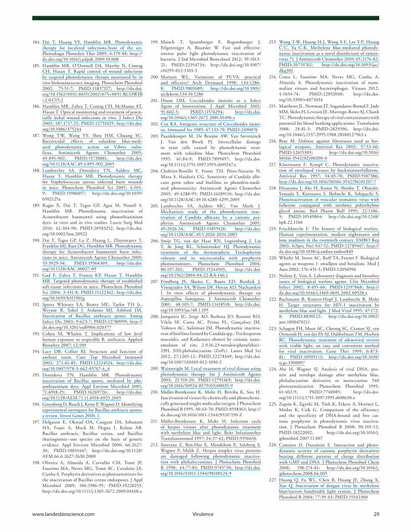
www.landesbioscience.com virulence 29
184. Dai T, Huang YY, Hamblin MR. Photodynamic therapy for localized infections-State of the art. Photodiagn Photodyn Ther 2009; 6:170-88; http://dx.doi.org/10.1016/j.pdpdt.2009.10.008
185. Hamblin MR, O’Donnell DA, Murthy N, Contag CH, Hasan T. Rapid control of wound infections by targeted photodynamic therapy monitored by in vivo bioluminescence imaging. Photochem Photobiol 2002; 75:51-7; PMID:11837327; http://dx.doi.org/10.1562/0031-8655(2002)075<0051:RCOWIB>2.0.CO;2
186. Hamblin MR, Zahra T, Contag CH, McManus AT, Hasan T. Optical monitoring and treatment of poten-tially lethal wound infections in vivo. J Infect Dis 2003; 187:1717-25; PMID:12751029; http://dx.doi.org/10.1086/375244
187. Wong TW, Wang YY, Sheu HM, Chuang YC. Bactericidal effects of toluidine blue-medi-ated photodynamic action on Vibrio vulni-ficus. Antimicrob Agents Chemother 2005; 49:895-902; PMID:15728881; http://dx.doi.org/10.1128/AAC.49.3.895-902.2005
188. Lambrechts SA, Demidova TN, Aalders MC, Hasan T, Hamblin MR. Photodynamic therapy for Staphylococcus aureus infected burn wounds in mice. Photochem Photobiol Sci 2005; 4:503-9; PMID:15986057; http://dx.doi.org/10.1039/b502125a
189. Ragàs X, Dai T, Tegos GP, Agut M, Nonell S, Hamblin MR. Photodynamic inactivation of Acinetobacter baumannii using phenothiazinium dyes: in vitro and in vivo studies. Lasers Surg Med 2010; 42:384-90; PMID:20583252; http://dx.doi.org/10.1002/lsm.20922
190. Dai T, Tegos GP, Lu Z, Huang L, Zhiyentayev T, Franklin MJ, Baer DG, Hamblin MR. Photodynamic therapy for Acinetobacter baumannii burn infec-tions in mice. Antimicrob Agents Chemother 2009; 53:3929-34; PMID:19564369; http://dx.doi.org/10.1128/AAC.00027-09
191. Gad F, Zahra T, Francis KP, Hasan T, Hamblin MR. Targeted photodynamic therapy of established soft-tissue infections in mice. Photochem Photobiol Sci 2004; 3:451-8; PMID:15122362; http://dx.doi.org/10.1039/b311901g
192. Spotts Whitney EA, Beatty ME, Taylor TH Jr., Weyant R, Sobel J, Arduino MJ, Ashford DA. Inactivation of Bacillus anthracis spores. Emerg Infect Dis 2003; 9:623-7; PMID:12780999; http://dx.doi.org/10.3201/eid0906.020377
193. Cohen M, Whalen T. Implications of low level human exposure to respirable B. anthracis. Applied Biosafety 2007; 12:109
194. Lacy DB, Collier RJ. Structure and function of anthrax toxin. Curr Top Microbiol Immunol 2002; 271:61-85; PMID:12224524; http://dx.doi.org/10.1007/978-3-662-05767-4_4
195. Demidova TN, Hamblin MR. Photodynamic inactivation of Bacillus spores, mediated by phe-nothiazinium dyes. Appl Environ Microbiol 2005; 71:6918-25; PMID:16269726; http://dx.doi.org/10.1128/AEM.71.11.6918-6925.2005
196. Greenberg D, Busch J, Keim P, Wagner D. Identifying experimental surrogates for Bacillus anthracis spores: a review. Invest Genet 2010; 1.
197. Helgason E, Okstad OA, Caugant DA, Johansen HA, Fouet A, Mock M, Hegna I, Kolstø AB. Bacillus anthracis, Bacillus cereus, and Bacillus thuringiensis--one species on the basis of genetic evidence. Appl Environ Microbiol 2000; 66:2627-30; PMID:10831447; http://dx.doi.org/10.1128/AEM.66.6.2627-2630.2000
198. Oliveira A, Almeida A, Carvalho CM, Tomé JP, Faustino MA, Neves MG, Tomé AC, Cavaleiro JA, Cunha A. Porphyrin derivatives as photosensitizers for the inactivation of Bacillus cereus endospores. J Appl Microbiol 2009; 106:1986-95; PMID:19228253; http://dx.doi.org/10.1111/j.1365-2672.2009.04168.x
199. Maisch T, Spannberger F, Regensburger J, Felgenträger A, Bäumler W. Fast and effective: intense pulse light photodynamic inactivation of bacteria. J Ind Microbiol Biotechnol 2012; 39:1013-21; PMID:22354734; http://dx.doi.org/10.1007/s10295-012-1103-3
200. Morison WL. Variations of PUVA: practical and effective? Arch Dermatol 1998; 134:1286-8; PMID:9801689; http://dx.doi.org/10.1001/archderm.134.10.1286
201. Dixon DM. Coccidioides immitis as a Select Agent of bioterrorism. J Appl Microbiol 2001; 91:602-5; PMID:11576294; http://dx.doi.org/10.1046/j.1365-2672.2001.01496.x
202. Cox RA. Antigenic structure of Coccidioides immi-tis. Immunol Ser 1989; 47:133-70; PMID:2490076
203. Paardekooper M, De Bruijne AW, Van Steveninck J, Van den Broek PJ. Intracellular damage in yeast cells caused by photodynamic treat-ment with toluidine blue. Photochem Photobiol 1995; 61:84-9; PMID:7899497; http://dx.doi.org/10.1111/j.1751-1097.1995.tb09247.x
204. Chabrier-Roselló Y, Foster TH, Pérez-Nazario N, Mitra S, Haidaris CG. Sensitivity of Candida albi-cans germ tubes and biofilms to photofrin-medi-ated phototoxicity. Antimicrob Agents Chemother 2005; 49:4288-95; PMID:16189110; http://dx.doi.org/10.1128/AAC.49.10.4288-4295.2005
205. Lambrechts SA, Aalders MC, Van Marle J. Mechanistic study of the photodynamic inac-tivation of Candida albicans by a cationic por-phyrin. Antimicrob Agents Chemother 2005; 49:2026-34; PMID:15855528; http://dx.doi.org/10.1128/AAC.49.5.2026-2034.2005
206. Smijs TG, van der Haas RN, Lugtenburg J, Liu Y, de Jong RL, Schuitmaker HJ. Photodynamic treatment of the dermatophyte Trichophyton rubrum and its microconidia with porphyrin photosensitizers. Photochem Photobiol 2004; 80:197-202; PMID:15244503; http://dx.doi.org/10.1562/2004-04-22-RA-146.1
207. Friedberg JS, Skema C, Baum ED, Burdick J, Vinogradov SA, Wilson DF, Horan AD, Nachamkin I. In vitro effects of photodynamic therapy on Aspergillus fumigatus. J Antimicrob Chemother 2001; 48:105-7; PMID:11418518; http://dx.doi.org/10.1093/jac/48.1.105
208. Junqueira JC, Jorge AO, Barbosa JO, Rossoni RD, Vilela SF, Costa AC, Primo FL, Gonçalves JM, Tedesco AC, Suleiman JM. Photodynamic inactiva-tion of biofilms formed by Candida spp., Trichosporon mucoides, and Kodamaea ohmeri by cationic nano-emulsion of zinc 2,9,16,23-tetrakis(phenylthio)-29H, 31H-phthalocyanine (ZnPc). Lasers Med Sci 2012; 27:1205-12; PMID:22278349; http://dx.doi.org/10.1007/s10103-012-1050-2
209. Wainwright M. Local treatment of viral disease using photodynamic therapy. Int J Antimicrob Agents 2003; 21:510-20; PMID:12791463; http://dx.doi.org/10.1016/S0924-8579(03)00035-9
210. Müller-Breitkreutz K, Mohr H, Briviba K, Sies H. Inactivation of viruses by chemically and photochemi-cally generated singlet molecular oxygen. J Photochem Photobiol B 1995; 30:63-70; PMID:8558363; http://dx.doi.org/10.1016/1011-1344(95)07150-Z
211. Müller-Breitkreutz K, Mohr H. Infection cycle of herpes viruses after photodynamic treatment with methylene blue and light. Beitr Infusionsther Transfusionsmed 1997; 34:37-42; PMID:9356656
212. Smetana Z, Ben-Hur E, Mendelson E, Salzberg S, Wagner P, Malik Z. Herpes simplex virus proteins are damaged following photodynamic inactiva-tion with phthalocyanines. J Photochem Photobiol B 1998; 44:77-83; PMID:9745730; http://dx.doi.org/10.1016/S1011-1344(98)00124-9
213. Wong T-W, Huang H-J, Wang Y-F, Lee Y-P, Huang C-C, Yu C-K. Methylene blue-mediated photody-namic inactivation as a novel disinfectant of entero-virus 71. J Antimicrob Chemother 2010; 65:2176-82; PMID:20719762; http://dx.doi.org/10.1093/jac/dkq301
214. Costa L, Faustino MA, Neves MG, Cunha A, Almeida A. Photodynamic inactivation of mam-malian viruses and bacteriophages. Viruses 2012; 4:1034-74; PMID:22852040; http://dx.doi.org/10.3390/v4071034
215. Matthews JL, Newman JT, Sogandares-Bernal F, Judy MM, Skiles H, Leveson JE, Marengo-Rowe AJ, Chanh TC. Photodynamic therapy of viral contaminants with potential for blood banking applications. Transfusion 1988; 28:81-3; PMID:2829396; http://dx.doi.org/10.1046/j.1537-2995.1988.28188127963.x
216. Bray M. Defense against filoviruses used as bio-logical weapons. Antiviral Res 2003; 57:53-60; PMID:12615303; http://dx.doi.org/10.1016/S0166-3542(02)00200-0
217. Käsermann F, Kempf C. Photodynamic inactiva-tion of enveloped viruses by buckminsterfullerene. Antiviral Res 1997; 34:65-70; PMID:9107386; http://dx.doi.org/10.1016/S0166-3542(96)01207-7
218. Hirayama J, Abe H, Kamo N, Shinbo T, Ohnishi-Yamada Y, Kurosawa S, Ikebuchi K, Sekiguchi S. Photoinactivation of vesicular stomatitis virus with fullerene conjugated with methoxy polyethylene glycol amine. Biol Pharm Bull 1999; 22:1106-9; PMID:10549864; http://dx.doi.org/10.1248/bpb.22.1106
219. Frischknecht F. The history of biological warfare. Human experimentation, modern nightmares and lone madmen in the twentieth century. EMBO Rep 2003; 4(Spec No):S47-52; PMID:12789407; http://dx.doi.org/10.1038/sj.embor.embor849
220. Whitby M, Street AC, Ruff TA, Fenner F. Biological agents as weapons 1: smallpox and botulism. Med J Aust 2002; 176:431-3; PMID:12056996
221. Nulens E, Voss A. Laboratory diagnosis and biosafety issues of biological warfare agents. Clin Microbiol Infect 2002; 8:455-66; PMID:12197868; http://dx.doi.org/10.1046/j.1469-0691.2002.00528.x
222. Bachmann B, Knüver-Hopf J, Lambrecht B, Mohr H. Target structures for HIV-1 inactivation by methylene blue and light. J Med Virol 1995; 47:172-8; PMID:8830122; http://dx.doi.org/10.1002/jmv.1890470211
223. Schagen FH, Moor AC, Cheong SC, Cramer SJ, van Ormondt H, van der Eb AJ, Dubbelman TM, Hoeben RC. Photodynamic treatment of adenoviral vectors with visible light: an easy and convenient method for viral inactivation. Gene Ther 1999; 6:873-81; PMID:10505113; http://dx.doi.org/10.1038/sj.gt.3300897
224. Abe H, Wagner SJ. Analysis of viral DNA, pro-tein and envelope damage after methylene blue, phthalocyanine derivative or merocyanine 540 photosensitization. Photochem Photobiol 1995; 61:402-9; PMID:7740085; http://dx.doi.org/10.1111/j.1751-1097.1995.tb08630.x
225. Zupán K, Egyeki M, Tóth K, Fekete A, Herényi L, Módos K, Csík G. Comparison of the efficiency and the specificity of DNA-bound and free cat-ionic porphyrin in photodynamic virus inactiva-tion. J Photochem Photobiol B 2008; 90:105-12; PMID:18222092; http://dx.doi.org/10.1016/j.jphotobiol.2007.11.007
226. Caminos D, Durantini E. Interaction and photo-dynamic activity of cationic porphyrin derivatives bearing different patterns of charge distribution with GMP and DNA. J Photochem Photobiol Chem 2008; 198:274-81; http://dx.doi.org/10.1016/j.jphotochem.2008.04.005
227. Huang Q, Fu WL, Chen B, Huang JF, Zhang X, Xue Q. Inactivation of dengue virus by methylene blue/narrow bandwidth light system. J Photochem Photobiol B 2004; 77:39-43; PMID:15542360
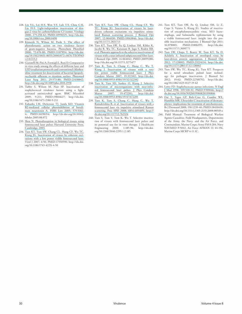
30 virulence volume 4 issue 8
228. Lin Y-L, Lei H-Y, Wen Y-Y, Luh T-Y, Chou C-K, Liu H-S. Light-independent inactivation of den-gue-2 virus by carboxyfullerene C3 isomer. Virology 2000; 275:258-62; PMID:10998325; http://dx.doi.org/10.1006/viro.2000.0490
229. Kömerik N, Wilson M, Poole S. The effect of photodynamic action on two virulence factors of gram-negative bacteria. Photochem Photobiol 2000; 72:676-80; PMID:11107854; http://dx.doi.org/10.1562/0031-8655(2000)072<0676:TEOPAO>2.0.CO;2
230. Giannelli M, Pini A, Formigli L, Bani D. Comparative in vitro study among the effects of different laser and LED irradiation protocols and conventional chlorhex-idine treatment for deactivation of bacterial lipopoly-saccharide adherent to titanium surface. Photomed Laser Surg 2011; 29:573-80; PMID:21438842; http://dx.doi.org/10.1089/pho.2010.2958
231. Tubby S, Wilson M, Nair SP. Inactivation of staphylococcal virulence factors using a light-activated antimicrobial agent. BMC Microbiol 2009; 9:211; PMID:19804627; http://dx.doi.org/10.1186/1471-2180-9-211
232. Eubanks LM, Dickerson TJ, Janda KD. Vitamin B2-mediated cellular photoinhibition of botuli-num neurotoxin A. FEBS Lett 2005; 579:5361-4; PMID:16198354; http://dx.doi.org/10.1016/j.febslet.2005.08.072
233. Shen N. Photodisruption in biological tissues using femtosecond laser pulses Harvard University Press, Cambridge, 2003.
234. Tsen KT, Tsen SW, Chang CL, Hung CF, Wu TC, Kiang JG. Inactivation of viruses by coherent exci-tations with a low power visible femtosecond laser. Virol J 2007; 4:50; PMID:17550590; http://dx.doi.org/10.1186/1743-422X-4-50
235. Tsen KT, Tsen SW, Chang CL, Hung CF, Wu TC, Kiang JG. Inactivation of viruses by laser-driven coherent excitations via impulsive stimu-lated Raman scattering process. J Biomed Opt 2007; 12:064030; PMID:18163846; http://dx.doi.org/10.1117/1.2821713
236. Tsen KT, Tsen SW, Fu Q, Lindsay SM, Kibler K, Jacobs B, Wu TC, Karanam B, Jagu S, Roden RB, et al. Photonic approach to the selective inactivation of viruses with a near-infrared subpicosecond fiber laser. J Biomed Opt 2009; 14:064042; PMID:20059280; http://dx.doi.org/10.1117/1.3275477
237. Tsen K, Tsen S, Chang C, Hung C, Wu T, Kiang J. Inactivation of viruses with a very low power visible femtosecond laser. J Phys Condens Matter 2007; 19:322102; http://dx.doi.org/10.1088/0953-8984/19/32/322102
238. Tsen K, Tsen SD, Sankey O, Kiang J. Selective inactivation of microorganisms with near-infra-red femtosecond laser pulses. J Phys Condens Matter 2007; 19:472201; http://dx.doi.org/10.1088/0953-8984/19/47/472201
239. Tsen K, Tsen S, Chang C, Hung C, Wu T, Ramakrishna B, et al. Inactivation of viruses with a femtosecond laser via impulsive stimulated Raman scattering. Proc SPIE 2008; 6854:68540N; http://dx.doi.org/10.1117/12.762324
240. Tsen S, Tsen Y, Tsen K, Wu T. Selective inactiva-tion of viruses with femtosecond laser pulses and its potential use for in vitro therapy. J Healthcare Engineering 2010; 1:185-96; http://dx.doi.org/10.1260/2040-2295.1.2.185
241. Tsen KT, Tsen SW, Fu Q, Lindsay SM, Li Z, Cope S, Vaiana S, Kiang JG. Studies of inactiva-tion of encephalomyocarditis virus, M13 bacte-riophage, and Salmonella typhimurium by using a visible femtosecond laser: insight into the pos-sible inactivation mechanisms. J Biomed Opt 2011; 16:078003; PMID:21806295; http://dx.doi.org/10.1117/1.3600771
242. Tsen SW, Chapa T, Beatty W, Tsen KT, Yu D, Achilefu S. Inactivation of enveloped virus by laser-driven protein aggregation. J Biomed Opt 2012; 17:128002; PMID:23224114; http://dx.doi.org/10.1117/1.JBO.17.12.128002
243. Tsen SW, Wu TC, Kiang JG, Tsen KT. Prospects for a novel ultrashort pulsed laser technol-ogy for pathogen inactivation. J Biomed Sci 2012; 19:62; PMID:22768792; http://dx.doi.org/10.1186/1423-0127-19-62
244. Lowy FD. Staphylococcus aureus infections. N Engl J Med 1998; 339:520-32; PMID:9709046; http://dx.doi.org/10.1056/NEJM199808203390806
245. Dai T, Tegos GP, Rolz-Cruz G, Cumbie WE, Hamblin MR. Ultraviolet C inactivation of dermato-phytes: implications for treatment of onychomycosis. Br J Dermatol 2008; 158:1239-46; PMID:18410410; http://dx.doi.org/10.1111/j.1365-2133.2008.08549.x
246. Field Manual: Treatment of Biological Warfare Agents Casualties. Field Headquarters, Departments of the Army, the Navy, and the Air Force, and Commandant, Marine Corps; Army FM 8-284, Navy NAVMED P-5042, Air Force AFMAN (I) 44-156, Marine Corps MCRP 4-11.1C.