Embed Size (px)
Citation preview

Carnosine’s Effect on Amyloid Fibril Formation andInduced Cytotoxicity of LysozymeJosephine W. Wu1*, Kuan-Nan Liu2, Su-Chun How2, Wei-An Chen2, Chia-Min Lai2, Hwai-Shen Liu2, Chaur-
Jong Hu3, Steven S. -S. Wang2*
1 Department of Optometry, Central Taiwan University of Science and Technology, Taichung, Taiwan,, 2 Department of Chemical Engineering, National Taiwan University,
Taipei, Taiwan, 3 Department of Neurology, Shuang Ho Hospital, Taipei Medical University, New Taipei City, Taiwan
Abstract
Carnosine, a common dipeptide in mammals, has previously been shown to dissemble alpha-crystallin amyloid fibrils. Todate, the dipeptide’s anti-fibrillogensis effect has not been thoroughly characterized in other proteins. For a more completeunderstanding of carnosine’s mechanism of action in amyloid fibril inhibition, we have investigated the effect of thedipeptide on lysozyme fibril formation and induced cytotoxicity in human neuroblastoma SH-SY5Y cells. Our studydemonstrates a positive correlation between the concentration and inhibitory effect of carnosine against lysozyme fibrilformation. Molecular docking results show carnosine’s mechanism of fibrillogenesis inhibition may be initiated by bindingwith the aggregation-prone region of the protein. The dipeptide attenuates the amyloid fibril-induced cytotoxicity ofhuman neuronal cells by reducing both apoptotic and necrotic cell deaths. Our study provides solid support for carnosine’samyloid fibril inhibitory property and its effect against fibril-induced cytotoxicity in SH-SY5Y cells. The additional insightsgained herein may pave way to the discovery of other small molecules that may exert similar effects against amyloid fibrilformation and its associated neurodegenerative diseases.
Citation: Wu JW, Liu K-N, How S-C, Chen W-A, Lai C-M, et al. (2013) Carnosine’s Effect on Amyloid Fibril Formation and Induced Cytotoxicity of Lysozyme. PLoSONE 8(12): e81982. doi:10.1371/journal.pone.0081982
Editor: Rizwan H. Khan, Aligarh Muslim University, India
Received June 20, 2013; Accepted October 20, 2013; Published December 11, 2013
Copyright: � 2013 Wu et al. This is an open-access article distributed under the terms of the Creative Commons Attribution License, which permits unrestricteduse, distribution, and reproduction in any medium, provided the original author and source are credited.
Funding: This work was supported by the grants from the National Science Council, Taiwan (NSC 101-2221-E-002-150 and NSC 102-2221-E-002-161 to SSW, andNSC 101-2113-M-166-001-MY2 to JWW). The funders had no role in study design, data collection and analysis, decision to publish, or preparation of themanuscript.
Competing Interests: The authors have declared that no competing interests exist.
* E-mail: [email protected] (JWW); [email protected](SSSW)
Introduction
Amyloid diseases, including hemodialysis amyloidosis, type II
(or noninsulin-dependent) diabetes, Parkinson’s disease, transmis-
sible spongiform encephalopathies, Huntington’s disease, and
Alzheimer’s disease, are a group of human diseases that are
characterized by the formation of extracellular insoluble aggre-
gates or deposits (also termed as amyloid fibrils) in certain tissues
and organs [1–4]. While the precursor proteins involved in the
aforesaid diseases share no sequence homology and native
structural motif similarity, they form fibrillar aggregates with
common morphological and histochemical features. For example,
an ordered cross b-sheet-rich secondary structure, fibrillar
morphology with a diameter of 5–15 nm, birefringence to
polarized light upon interaction with Congo Red, fluorescence
after reacting with thioflavin T (ThT), insolubility in most solvents,
and resistance to protease degradation [1,5,6].
The formation of amyloid fibrils or fibril-like aggregates has also
been observed in proteins that are not associated with any form of
disease under certain environmental stresses (e.g., high tempera-
ture, extreme pH, vigorous agitation, and high pressure) [7–9].
Evidence has shown that the morphological, histochemical, and
cytotoxic properties of the aggregates derived from these non-
disease-related proteins are similar to those of disease-associated
amyloid proteins, suggesting that amyloidogenicity or amyloid
fibril-forming propensity is a generic property of all polypeptides
[10,11].
Hen egg-white lysozyme (HEWL), a 129-residue monomeric
protein with molecular weight of 14.3 kD, has been extensively
used as a food preservative owing to its lytic activity against the cell
wall of Gram-positive bacteria [12,13]. Structurally, HEWL, in its
native conformation, is a helix-rich protein (a-helix: ,30%)
containing four disulfide bonds [14,15]. Because of its well-defined
structure information and a high degree of sequence and structural
homology to the human lysozyme [16,17], which is affiliated with
familial lysozyme systemic amyloidosis [18], HEWL has been
widely chosen as a model protein in research relating to the
subjects of protein folding, unfolding, and aggregation. Numerous
studies have demonstrated that HEWL is prone to fibrillate in a
heated and acidic environment [17,19], a concentrated ethanol
solution [20], a concentrated solution of guanidine hydrochloride
[21], and a vigorously agitated condition [22]. Therefore, hen egg-
white lysozyme serves as a nice model system with which to study
in vitro phenomena associated with fibril formation.
To combat amyloid diseases, efforts have been directed toward
seeking or developing a variety of therapeutic strategies [23–25].
An increasing body of evidence points to the possible relation of
fibrillar and/or protofibrillar species derived from amyloid
proteins and the disease pathology; thus, considerable efforts are
underway to screen small inhibitory molecules/compounds that
are capable of counteracting the fibrillogenesis of disease-related
amyloid proteins. A variety of natural or synthesized molecules
PLOS ONE | www.plosone.org 1 December 2013 | Volume 8 | Issue 12 | e81982

and/or compounds have been reported to retard or prevent fibril
formation both in vitro and in vivo [9,26–28].
Carnosine, a naturally-occurring compound discovered by
Gulevitch and Amiragdibi over 100 years ago [29], is found
predominantly in human muscles, heart, liver, brain, kidneys, and
other long-lived tissues. It has been regarded as one of the most
common dipeptides in human and other mammals [30,31].
Carnosine is synthesized from b-alanine and L-histidine, by the
ATP-driven enzyme, carnosine synthetase, and is hydrolyzed by
specific metal ion-dependent homodimeric dipeptidase (carnosi-
nases) in the blood and other tissues [32–34]. In general, the
intramuscular concentration of carnosine is found to be approx-
imately 20–30 mM in human, about twenty-fold higher than that
in mice, ten-fold higher than that in rabbits, and three-fold higher
than that in cows, suggesting a high correlation between maximum
life expectancy of various mammalian species and the concentra-
tion of carnosine present in the body [35–37].
Carnosine has been patented as an eye-drop component used
for the treatment and prevention of senile cataract in Europe [38]
and has also recently been discovered to have physiological and
therapeutic functions (e.g., anti-aging and free-radical scavenging
properties) in the human body [39]. Importantly, carnosine has
been found to disaggregate glycated a-crystallin protein [40] and
to prevent the native protein from forming amyloid fibrils [41]. In
addition, carnosine has been shown to suppress the accumulation
of b-amyloid in the central nervous system of transgenic mice
model for Alzheimer’s disease [42] and attenuate the in vitro
fibrillogenesis of b-amyloid peptide Ab(1–42) [43,44]. In vitro cell
culture studies demonstrated that carnosine can prevent rat brain
endothelial cells [45] and rat PC12 cells from cytotoxic effects
induced by b-amyloid peptides [46]. Based on another study,
carnosine has also been shown to be effective in reducing the
toxicity of prion protein possibly owing to its stimulatory effect in
prion proteolysis and/or decreased glycolysis, which has been
found to be upregulated by prion proteins and contributes toward
protein glycoxidation [47].
Despite ample studies in the literature showing the effects of
carnosine on human and animals, the exact mechanism of how
this dipeptide exerts its effect (in particularly inhibitory activity
against fibrillogenesis) remains elusive. In the present study, we
present a more complete picture of of carnosine’s mechanism of
action in hen egg white lysozyme (HEWL) amyloid fibril inhibition
from the molecular to cellular perspectives. We thoroughly
examined the dose-dependent effect of carnosine on HEWL fibril
formation from the protein structural level via a wide variety of
methods, including several spectroscopic techniques (e.g., thiofla-
vin T fluorescence spectroscopy, Congo red absorption spectros-
copy, far-UV circular dichroism (CD) spectroscopy, and Nile red
fluorescence spectroscopy) and transmission electron microscopy
(TEM). The change in equilibrium thermal denaturation property
of HEWL due to carnosine inhibition, as well as the important
binding interactions by which the dipeptide exerts its inhibitory
effect on amyloid fibrillogenensis, were also investigated. Finally,
we explored the effect of carnosine in preventing SH-SY5Y cell
death induced by HEWL amyloid fibril through a number of cell
toxicity and viability tests (e.g., (4,5-dimethylthiazol-2-yl)2,5-
diphenyltetrazolium bromide (MTT) reduction assay, lactate
dehydrogenase (LDH) release assay, etc.).
Materials and Methods
MaterialsHen egg-white lysozyme (HEWL) (EC 3.2.1.17) was obtained
from Merck (Germany). Hydrochloric acid and sodium chloride
were purchased from Nacalai Tesque, Inc (Japan). Glycine was
obtained from BioBasic, Inc (Canada). Carnosine and other
chemicals, unless otherwise specified, were of analytical grade and
purchased from Sigma (USA).
Preparation of HEWL fibrilsAll HEWL samples (,10 mg/mL or ,0.70 mM) without and
with various concentrations of carnosine were prepared immedi-
ately prior to each experiment via dissolving HEWL powders in
glycine buffer (100 mM glycine/HCl, pH 2.0, 100 mM sodium
chloride and 1.54 mM NaN3), in accordance to previous studies
[48,49], prepared immediately prior to each experiment. In order
to generate amyloid fibrils, each sample (10 mL) was transferred to
a 20 mL glass tube and agitated with polytetrafluoroethylene
(PTFE)-coated micro stirring bars (8 mm61.5 mm) at ,580 rpm
in 55uC [50,51]. At designated time points, the tube was gently
vortexed to well-distribute the HEWL sample solution, and then
an appropriate amount of HEWL sample was withdrawn for the
following measurements.
Thioflavin T (ThT) fluorescence measurementsThe stock solution of ThT at a concentration of 1 mM was
prepared in 95% (v/v) ethanol protected from light prior to use,
and the concentration was determined spectrophotometrically
using the molar extinction coefficient at 416 nm of
26600 M21cm21 [52]. Phosphate buffered saline (136.7 mM
NaCl, 2.68 mM KCl, 0.01 M Na2HPO4, 1.76 mM KH2PO4,
and 1.54 mM NaN3, pH 7.4) was used to dissolve the ThT stock
solution to obtain a ThT working solution at a concentration of
10 mM. HEWL sample solutions (40 mL), with or without
carnosine, taken out at different incubation times were mixed
thoroughly with ThT working solution (960 mL) prior to
measuring the ThT fluorescence emission intensities at 485 nm.
The ThT fluorescence measurements were taken for 60 sec by
exciting the resultant mixtures at 440 nm via Cary Eclipse
fluorescence spectrophotometer (Varian, USA).
Congo red (CR) binding assayFresh solution of CR dye with 10 mM in 100 mM phosphate
buffer (pH 7.4) was diluted from the stock solution prior to use.
The stock solution was prepared in phosphate buffer with 40%
ethanol as described by Klunk et al. [53] and the concentration was
determined spectrophotometrically using the molar extinction
coefficient at 505 nm of 59300 M21cm21 [53]. Aliquots of 20 mL
of fresh CR solution (10 mM) were added to 1980 mL of HEWL
sample solutions followed by vortex-mixing for 15 s and incubat-
ing at room temperature for 30 min. The absorption spectrum of
each sample was recorded over a range of 400–700 nm on a Cary
50 UV-Visible Spectrophotometer (Varian, USA) using a 1-cm
path length quartz cuvette.
Transmission electron microscopy (TEM) analysisAliquots of 5 mL of HEWL samples for TEM analysis were
withdrawn from the working solutions used for ThT fluorescence
measurements at 10 hr incubation and applied to a carbon-
stabilized, formvar coated grid for 30 sec. Excess samples were
removed by applying ashless filter papers to the edge of the grids
and the grids were negatively stained with 2% uranyl acetate in
distilled de-ionized water (Electron Microscopy Sciences, USA) for
another 30 sec. After removing the excess stain, the grids were left
to air-dry for at least 20 min and then examined and
photographed on a Hitachi H-7650 transmission electron micro-
Carnosine Inhibits Fibril Formation and Toxicity
PLOS ONE | www.plosone.org 2 December 2013 | Volume 8 | Issue 12 | e81982

scope with a Gantan model 782 CCD Camera (Tokyo, Japan) at
an accelerating voltage of 100 kV.
Far-UV circular dichroism (CD) spectroscopyThe secondary structural changes of HEWL sample solutions
were evaluated by far-UV CD spectroscopy. CD spectra of
HEWL samples were recorded after diluting 100-fold with de-
ionized water over the wavelength range of 190–260 nm using a J-
815 spectrometer (JASCO, Japan) with a 0.2 cm path length
sample cell. All CD measurements were collected at room
temperature using a bandwidth of 1.0 nm, a step interval of
0.1 nm, and a scanning speed of 50 nm/min. Each CD spectrum
was the average of three scans. The secondary structure contents
of HEWL samples were estimated using the CONTINLL
algorithm with the reference set SP175 available from the
DICROWEB website [54,55].
Nile red binding assayNile red was chosen as a hydrophobic fluorescent dye to avoid
interference from Raman and Rayleigh scattering. Before
measurement, a fresh working solution of Nile red was prepared
by diluting the stock solution (1 mM dissolved in DMSO) to 1 mM
with 100 mM glycine buffer. Aliquots of 20 mL of HEWL samples
with and without carnosine taken at various time points were
immediately mixed with 980 mL of working solution containing
Nile red at 1 mM, which was found to be an optimum
concentration for obtaining significant fluorescence changes
without interference by the inner filter effect [56,57]. The Nile
red fluorescence emission spectra from 600 to 700 nm were
measured at the excitation wavelength of 570 nm on a Cary
Eclipse fluorescence spectrophotometer (Varian, USA) after at
least 15 min incubation in the dark.
Equilibrium thermal denaturation/unfolding and dataanalysis
Denaturation curves were constructed by measuring the
intrinsic fluorescence of HEWL as a function of temperature,
and then subsequently analyzed to yield the thermodynamic
parameters for HEWL unfolding with and without carnosine. In
thermal scans, the thermally-induced unfolding transition of
HEWL samples was determined by monitoring the changes in
intrinsic fluorescence intensity over 20–95uC with a heating rate of
1uC/min and an equilibrium time of 1 min. Intrinsic fluorescence
emission spectra in the range of 300–420 nm were recorded every
2uC at the excitation wavelength of 280 nm on a Cary Eclipse
fluorescence spectrophotometer (Varian, USA). The obtained
thermally-induced unfolding data were analyzed by least-squares
fitting against a two-state transition model via the procedure
described as follows:
For a reversible protein unfolding reaction, the transition curve
between the native and unfolded states is a function of
temperature. The equilibrium constant (KU ) at any temperature
could be calculated from equation (2).
DGU~{RT ln KU~{RT lnfU
fF
~{RT lnfU
1{fU
~
{RT lny{yF
yU{y
ð1Þ
where fF and fU are the fractions of folded and unfolded protein
species at various temperatures, respectively, y is the observed
average emission wavelength (AEW) of HEWL intrinsic fluores-
cence spectrum. yF and yU denote the pre- and post-unfolding
baselines, which are assumed to be linearly dependent on heating
temperature and thus can be represented by yF ~yoF zmF
:T and
yU~yoUzmU
:T , where yoF , yo
U , mF and mU are the intercepts
and slopes of the baselines before and after the transition,
respectively, R is the gas constant, T is the temperature, and DGU
is the Gibbs free energy of thermal unfolding.
Bioinformatic prediction of aggregation sitesVarious aggregation site predictors using protein’s primary
sequence as input have been devised in recent years to predict ‘‘hot
spots’’ or short sequences of amino acids that are prone to
aggregation. Among these, AGGRESCAN [58], AMYLPRED
[59], and TANGO [60] were utilized herein to calculate consensus
potential aggregation sites on HEWL. AGGRESCAN predicts
aggregation hot spots from an aggregation-propensity scale for
amino acids derived from in vivo experiments that involved a highly
amyloidogenic stretch in Ab-42 peptide. AmlyPred incorporates a
variety of algorithms to achieve a more objective prediction of
aggregation sites. These algorithms include secondary structure
[61], amyloidogenic pattern [62], b-aggregate tendency [60],
packing density [63], and conformational energy [64] calculations.
Of the three methods used, TANGO is the only one that accounts
for solution parameters to simulate experimental conditions. We,
therefore, set the prediction parameters to pH 2.0 and 55uC to
match our experimental settings.
Molecular docking of carnosine to lysozymeCarnosine was first built using ChemDraw Ultra (Cambridge
Soft Corp. Cambridge, MA) and optimized in three-dimensional
form in Discovery Studios 2.5.5 (Accelrys Inc., San Diego, CA).
To be consistent with our experimental conditions, we subjected
the X-ray structure (PDB code 2ZQ3) of HEWL to molecular
dynamics (MD) simulations under pH 2.0 and 55uC. The system
reached equilibration around 15 ns (see Fig. S1 of the supporting
information) and a protein structure was extracted at 20 ns for the
docking simulation.
Molecular docking using CDOCKER [65] was implemented to
understand how carnosine inhibit HEWL fibril formation.
CDOCKER is a molecular dynamics (MD) simulated-annealing-
based algorithm incorporated into the Discovery Studios 2.5.5
environment. The grid-based molecular docking method employs
CHARMm force field [66] to perform conformational searches for
optimal binding modes of ligands to proteins or receptors followed
by refinement of poses through minimization. Automated search
for potential binding sites based on surface cavities and ligand
volume yielded two potential binding sites on HEWL with the
input site spheres defined with a grid extension of 8 A and a radius
of 12 A from the center of ligand-binding site. The simulated
annealing protocol consists of first heating the system to 427uC in
2000 steps, followed by annealing in 5000 steps to a final
temperature of 55uC. For each potential binding site, one hundred
random conformations were generated for carnosine based on the
starting structure and allowed to be randomly distributed around
the center of the sites. The final minimization step refined the
docked poses and the ligand-protein interactions. The intramo-
lecular energy for each ligand models were then computed to
produce a total docking energy score (CDOCKER energy).
Cell cultureHuman neuroblastoma SH-SY5Y cells were maintained under
humidified 5% (v/v) CO2/air at 37uC in a mixture of Dulbecco’s
modified Eagle’s medium (DMEM) and Ham’s F12 medium (F12)
supplemented with 10% (v/v) FBS and 100 U/mL penicillin.
Carnosine Inhibits Fibril Formation and Toxicity
PLOS ONE | www.plosone.org 3 December 2013 | Volume 8 | Issue 12 | e81982

3,(4,5-Dimethylthiazol-2-yl)2,5-diphenyltetrazoliumbromide (MTT) reduction assay
SH-SY5Y cells at a density of 16105 cells/mL were plated in
the 96-well plates followed by 24 hr-incubation and various sample
solutions were added to the cells for another 24 hr after plating.
Cell viability of SH-SY5Y cells was measured using the MTT
reduction assay. Viable cells reduce MTT to form blue formazan
crystals, and inhibition of this reaction is indicative of the cellular
redox changes that could result in cytotoxicity [67]. Stock sample
solutions of HEWL with various concentrations of carnosine were
diluted with sterile DMEM-F12 medium. The resultant diluted
solutions were added to SH-SY5Y cells in the 96-well plates. Upon
incubation for 6, 12, and 24 hr, the MTT reduction assay was
performed after removal of the treatment. MTT was added to the
culture medium to yield a final concentration of 0.5 mg/mL and
incubated with cells for 4 hr in a CO2 incubator and 100 mL of a
5:2:3 N,N-dimethylforamide (DMF): sodium dodecylsulfate (SDS):
water solution (pH 4.7) was added to dissolve the formazan
crystals. After 18 hr of incubation in a humidified CO2 incubator,
the absorbance at 585 nm for each well was measured using a
mQuant Microplate reader (Bio-Tek Instruments, VT, USA). Cell
viability was reported relative to control cells that were not
exposed to the HEWL fibril solutions.
Lactate dehydrogenase (LDH) release assayThe level of the cytoplasmic enzyme, lactate dehydrogenase,
released from human neuroblastoma SH-SY5Y cells into
DMEM/F12 culture medium upon cell membrane damage or
lysis was determined for the quantification of cell death using an in
vitro toxicology assay kit (Tox-7) (Sigma, USA) according to the
instructions of the manufacturer. The reduction of NAD+ to
NADH accompanied by the oxidation of lactate to pyruvate is
catalyzed by LDH in the suspension. The resulting NAD+ is
utilized in the stoichiometric conversion of a tetrazolium dye to a
soluble colored formazan derivative, which strongly absorbs at
490–520 nm. Cells were seeded at a density of 16105 cells/mL in
the 96-well plates for 24 hr-incubation under humidified 5% (v/v)
CO2/air at 37uC and the aged HEWL sample without or with
various concentration of carnosine was added to the cells for 6, 12,
and 24 hr. The percentage of LDH release was defined as the ratio
of LDH enzymatic activity of the group treated with aged HEWL
sample to that of the group treated with lysis buffer (maximum
LDH activity). LDH enzymatic activity measurements were
performed following the procedures provided in the manufactur-
er’s instructions (Sigma, USA).
Flow cytometric analysisCells undergoing apoptosis were detected by double staining
with Annexin V-FITC/PI in the dark according to the manufac-
turer’s instructions (Sigma, USA). SH-SY5Y cells were seeded at a
density of 16106 cells/mL in a 6-well plate. After 24 hr-incubation
of cells in a humidified 5% CO2/air incubator and 37uC, the
culture medium was replaced with 1 mL of medium containing
HEWL with various concentrations of carnosine. Upon incubation
in the same culture condition for 24 hr, cells were trypsinized and
washed twice in cold phosphate buffered saline (PBS) under
centrifugation (1400 rpm, 5 min). Cell pellets were suspended in
Annexin V binding buffer at a concentration of 106–107 cells/mL.
100 mL of the resulting cell solution were first mixed with 10 mL of
Annexin V-FITC for 15 min at 0uC in the dark, followed by the
addition of 380 mL of Annexin V binding buffer and 10 mL of
50 mg/mL propidium iodide (PI). The stained cells were imme-
diately analyzed using flow cytometry (Beckman Coulter FC500,
USA). The fluorescence emission signals for annexin V-FITC were
plotted against PI to identify viable cells (i.e. negative for Annexin
V-FITC and PI signals), early apoptotic cells (Annexin V-FITC
signal only) and late apoptotic/necrotic cells (positive for Annexin
V-FITC and PI signals). Analysis was performed using Cytomics
CXP Analysis software (Beckman Coulter, USA). Each measure-
ment was carried out at least three times.
Statistical analysisAll data presented are expressed as the mean 6 the standard
deviation (S.D.) of n independent determinations. Specific n values
(n$5) are reported in the figure legends. To determine whether a
given data set was significantly different from a control data set,
one tailed Student’s t-test assuming unequal variances was
performed. Unless otherwise noted, a criterion of p,0.05 was
employed to determine whether the sample set and the untreated
control set were statistically different. Asterisks above the bars
indicate the significance of the result relative to the untreated
control: a single asterisk indicates a value of p,0.05 and double
asterisks indicate a value of p,0.01. The statistical analyses were
conducted using Excel or KaleidaGraph software.
Results
HEWL fibrillogenesis was affected by carnosine asrevealed by ThT fluorescence and Congo red binding
To assess the formation of fibrillar species at low pH (pH ,2.0)
and high temperature (55uC) under vigorous agitated condition
(,580 rpm), the tinctorial and ultra-structural properties of the
HEWL samples were examined. The samples were first charac-
terized by monitoring the changes in ThT fluorescence emission
and Congo red absorption spectrum, then morphologically
analyzed by TEM.
ThT is a histochemical reagent that stains amyloid deposits and
has been widely used as a standard extrinsic fluorescent dye to
probe for amyloid fibrils [68]. A significant increase in fluores-
cence emission intensity is observed when ThT is added to samples
containing linear array of b-strands. Upon binding to ordered
aggregates of proteins, ThT emits fluorescence intensity that can
be measured and is commonly used as a principal index for
monitoring the kinetics of fibrillogenesis. As shown in Fig. 1, the
change in ThT fluorescence at 485 nm for the control sample of
HEWL (without inhibitor added) was found to follow the
characteristic nucleation-dependent pathway. The duration of
the nucleation phase was approximately 1 hr, and the ThT
fluorescence intensity increased rapidly until reaching its (final)
equilibration plateau at ,500 A.U. after approximately 4 hrs of
incubation.
Using Congo red binding assay, we further determined if the
aggregated species formed in the HEWL sample were fibril-like.
Congo red is another well known histological dye, which is widely
used to probe for the presence of amyloid fibrils. Tissues
containing amyloid deposits stained by Congo red exhibit apple-
green birefringence under the cross polarized light [69,70]. In
addition, binding to amyloid fibrils induces an enhanced
absorbance and red-shift in the maximum absorbance of Congo
red from 490 nm to 540 nm in the absorption spectrum [51,53].
As demonstrated in the time evolved Congo red absorption spectra
of HEWL depicted in the inset of Fig. 1, a considerable increase in
absorbance is detected at around 540 nm accompanied by an
obvious red-shift of the maximum from 494 to 515 nm, indicating
the presence of ordered, b-sheet-rich fibrillar structure in the
HEWL sample. Also, the Congo red absorbance difference at
540 nm was found to increase with incubation time, revealing the
Carnosine Inhibits Fibril Formation and Toxicity
PLOS ONE | www.plosone.org 4 December 2013 | Volume 8 | Issue 12 | e81982

expansion of fibril growth. Our ThT fluorescence and Congo red
absorbance results showed that the aggregates of HEWL formed at
acidic pH and high temperature under vigorous agitation
exhibited fibril-like characteristics.
To explore the effects of carnosine on HEWL fibrillogenesis, we
compared the ThT fluorescence intensities and Congo red
absorption spectra of HEWL samples in the presence and absence
of carnosine. We demonstrate in Fig. 2A that the change of ThT
fluorescence intensity as a function of time in each HEWL sample
(with or without carnosine) can be described by a sigmoidal curve,
which is composed of a lag phase (without noticeable fluorescence
variation), followed by a growth phase (with dramatic fluorescence
increase) and a final equilibrium phase where ThT fluorescence
reaches a plateau. The final equilibrium ThT fluorescence
emission was observed to be negatively correlated with the
concentration of carnosine, with the lowest level of ThT
fluorescence intensity occurring with 50 mM carnosine. For
example, the percentage of reductions calculated by ThT-induced
fluorescence at 10 hr of incubation were found to be ,4.2%,
,21.6%, ,47.0%, ,71.9,% and 91.4% in HEWL samples with
10, 20, 30, 40, and 50 mM carnosine, respectively (the percentage
of reduction in ThT fluorescence intensity = 100%6(ThT
fluorescence intensity of HEWL control sample 2 ThT fluores-
cence intensity of carnosine-containing HEWL sample)/ThT
fluorescence intensity of HEWL control sample).
The Congo red binding assay serves as a complementary
measure to detect the presence of amyloid fibril structure in
carnosine-containing HEWL samples. Congo red absorption
spectra of HEWL with various concentrations of carnosine were
found to be similar to that of the HEWL control at the beginning
of incubation, as shown in the Fig. 2B. As depicted in Fig. 2C, a
marked red shift of the spectral maximum from ,494 nm to
513 nm, accompanied by an obvious increase in absorbance with
an apparent shoulder peak at ,540 nm, was observed in the
absorption spectrum for HEWL with 10 mM carnosine upon 10
hr-incubation. This is akin to the curve obtained for the control
sample of HEWL. However, as the concentration of carnosine was
raised to 50 mM, apart from the disappearance of shoulder peak
at around 540 nm, a significant reduction in both maximum
absorbance and red shift of the maximum were also observed,
signifying a decrease in the formation of amyloid fibrillar species
associated with cross b-pleated sheet. Notably, samples of
carnosine alone (at the concentrations used in this study) did not
quench ThT fluorescence and alter Congo red binding absor-
bance (data not shown). Therefore, our ThT fluorescence emission
and Congo red binding results, evidently, indicated that carnosine
exerts a concentration-dependent attenuating effect on the
aggregation/fibril formation of HEWL.
Electron microscopy analysis of HEWL samples with andwithout carnosine
To further investigate the efficacy of carnosine on HEWL fibril
inhibition, TEM was used to monitor the morphological features
of HEWL samples in the absence and presence of various
concentrations of carnosine after 10 hrs of incubation. As
displayed in Fig. 3 (top left), the negatively stained micrograph
of HEWL control sample contains large quantities of long, un-
branched, fibrils of ,10 nm in diameter and several mm in length,
Figure 1. Amyloid fibrillogenesis kinetics of hen egg-white lysozyme (HEWL). The extent of fibril formation was monitored via Congo redabsorbance at 540 nm and thioflavin T (ThT) fluorescence as a function of incubation time. HEWL samples were dissolved in 100 mM glycine buffer(pH 2.0) and incubated at 55uC with agitation of 580 rpm during the course of experiment. The extent of fibril formation was also monitored viaCongo red absorption spectra shown in the inset (the arrow indicates the increasing incubation time). Data represent the mean ThT fluorescenceintensity of at least 5 independent experiments (n$5). Error bars represent the standard deviation (S.D.) of the fluorescence measurements.doi:10.1371/journal.pone.0081982.g001
Carnosine Inhibits Fibril Formation and Toxicity
PLOS ONE | www.plosone.org 5 December 2013 | Volume 8 | Issue 12 | e81982

which are characteristics of typical amyloid fibrils. We show in
Fig. 3 (top right) that, as compared to the control sample, HEWL
sample with 20 mM carnosine exhibits similar structural mor-
phology, but with less fibril formation. HEWL samples co-
incubated with higher concentrations of carnosine (40 mM and
50 mM as seen in Fig. 3 bottom left and right, respectively)
procured even less fibrils. At the maximum carnosine concentra-
tion tested (50 mM), the fibrils that have formed are short and
sparsely populated. These TEM observations indicate that
carnosine attenuates the amount of HEWL fibrils in a dose-
dependent manner. All in all, our ThT fluorescence, Congo red
binding, and TEM results suggest that exposure of HEWL to
carnosine leads to bona fide inhibition/suppression of amyloid
fibrillogenesis.
Conformational transition of HEWL samples with andwithout carnosine
To examine the effect of carnosine on the conformational
(secondary structural) changes of HEWL, we monitored the far-
UV CD spectra of HEWL in the absence and presence of
carnosine at concentrations of 10–50 mM. As Fig. 4A attests, prior
Figure 2. Effects of carnosine on the kinetics of amyloidfibrillogenesis of HEWL. Inhibition of HEWL fibril formation bycarnosine in a concentration-dependent manner as revealed by (A) ThTfluorescence and (B and C) Congo red absorbance. The Congo redbinding absorbance spectra of HEWL samples were taken at (B) 0 hrand (C) 10 hr of incubation. The samples with and without carnosinewere dissolved in 100 mM glycine buffer (pH 2.0) and incubated at55uC agitated at 580 rpm during the course of the experiment. Eachdata point represents the mean value of at least 5 independentexperiments (n$5). Error bars denote the standard deviation (S.D.) ofthe measurements.doi:10.1371/journal.pone.0081982.g002
Figure 3. Transmission electron micrographs of HEWL sampleswith various concentrations of carnosine. Negatively stainedelectron micrographs of (A) HEWL alone, (B) HEWL co-incubated with20 mM carnosine, (C) HEWL co-incubated with 40 mM carnosine, and(D) HEWL co-incubated with 50 mM carnosine. HEWL samples in theabsence and presence of carnosine were prepared at pH 2.0 andincubated for 10 hr. The scale bar represents 50 nm.doi:10.1371/journal.pone.0081982.g003
Carnosine Inhibits Fibril Formation and Toxicity
PLOS ONE | www.plosone.org 6 December 2013 | Volume 8 | Issue 12 | e81982
to incubation, a characteristic shoulder at ,222 nm and a
prominent minimum at ,208 nm are observed in the far-UV
CD spectra of all HEWL samples, indicating that the native
HEWL is in possession of a predominantly a-helix conformation
[21]. However, upon prolonged incubation, the far-UV CD
spectrum of the control HEWL sample reveals a marked structural
transition, where a prominent change of the secondary structural
composition toward a highly amyloidogenic, yet soluble, b-sheet
rich conformation, is presented in the CD spectrum as a negative
band at 218 nm and a positive ellipticity at around 196 nm.
Comparison of the CD spectra, after 10 hrs incubation for
HEWL samples treated with 10, 30, and 50 mM carnosine
(Figs. 4B, 4C, and 4D, respectively), shows that with increasing
concentration of the inhibitor, less pronounced ellipticity values
of the negative band at ,218 nm can be observed. This
demonstrates that the structural transition perceived in the
HEWL control sample was mitigated upon the addition of
carnosine. To quantitatively compare the differences in the
conformations among the HEWL samples, the secondary
structure contents were estimated by deconvolution of the
acquired CD spectra for all HEWL samples using the CON-
TINLL algorithm available from the DICROWEB server [54].
The resultant time-evolved secondary structure compositions
shown in the insets of Figs. 4A–4D indicate that: (1) As expected,
the control HEWL sample (Fig. 4A) displayed a time-dependent
elevation in b-sheet content that was accompanied by a decrease
in a-helix content, suggesting a transition from a-to-b structure
(2) The b-sheet content reached the highest level of approxi-
mately 41% at 10 hrs after the onset of incubation, which is
about two-fold higher than that at time 0 hr. (3) At 10 hr-
incubation, the HEWL samples with 30 and 50 mM carnosine
(Figs. 4C and 4D) were found to contain a significantly lower b-
sheet fraction relative to the control (,33%, ,19%, and ,41%
for 30 mM carnosine-HEWL mixture, 50 mM carnosine-HEWL
mixture, and the control), demonstrating a positive correlation
between the attenuation of b-sheet content and the concentration
of carnosine. Overall, our far-UV CD results allow us to conclude
that the addition of carnosine is effective in preventing HEWL
from undergoing a-to-b transition, which is closely associated
with the amyloid fibril-forming propensity.
The effects of carnosine on the HEWL tertiary structure/surface hydrophobicity
Using the change in surface hydrophobicity as a gauge, the
effect of carnosine on HEWL tertiary structure was monitored by
the time evolution of Nile red fluorescence emission during the
incubation process. Nile red is a nonionic lipophilic fluorescent
dye, which has been widely used as a probe to study the
intracellular lipid content, polarity of organic solvents, environ-
mental change of biomolecules, and nonionic surfactant micro-
emulsions due to solvatochromism [71]. As illustrated in Fig. 5,
the maximum Nile red fluorescence of the control HEWL sample
shows no noticeable increase (,15–30 A.U.) in the first 1 hr of
incubation, followed by a dramatic increase from 1 to 2 hr of
incubation, and finally reaching an equilibrium plateau (,700
A.U.) after 4 hr of incubation. In addition, a blue-shift in the
wavelength of maximum fluorescence emission (lmax), which is
indicative of the exposure of hydrophobic clusters, was observed
before a significant increase in Nile red fluorescence was detected.
The wavelength of maximum fluorescence emission (lmax)
reached a plateau (from ,661 nm to ,623 nm) at 2 hr of
incubation, suggesting that the shift of lmax is more sensitive than
the enhancement of the Nile red fluorescence emission in probing
tertiary structure changes. A similar trend was also observed in
the HEWL sample containing 10 mM carnosine (data not
shown). However, when a higher concentration of carnosine
was added (e.g., 30 or 50 mM), the maximum Nile red
fluorescence intensity was found to drop considerably and the
blue-shift of lmax was reduced in comparison to that of the
control. For instance, upon incubation for 10 hr, the Nile red
fluorescence intensity and blue-shift in lmax for carnosine
concentration of 30 mM were observed to be ,341 A.U. and
,28 nm, respectively, while for 50 mM carnosine were ,70
A.U. and ,5 nm, respectively.
The effect of carnosine on the thermal denaturationbehavior of HEWL
The magnitudes of the Gibbs free energy changes (DGU) of
thermal denaturation/unfolding were determined through non-
linear regression against the stability curves obtained. Table 1
presents the calculated values of thermodynamic parameters (DGU
and Tm) for all HEWL samples (with and without carnosine). We
first compared the thermostability of HEWL in the acidic (pH 2.0)
and neutral (pH 7.0) conditions by monitoring the changes
average fluorescence emission wavelength (the excitation wave-
length = 280 nm). The thermal denaturation curve of HEWL was
found to shift to the left upon changing incubation condition from
pH 7.0 to pH 2.0 (shown in Fig. S2 of the supporting informa-
tion), indicating that incubation at lower pH renders HEWL less
stable. The values of Tm for HEWL in pH 2.0 and pH 7.0 were
determined to be 50.864.3uC and 70.963.6uC, respectively. For
HEWL alone, the DGU value at pH 2.0 (21.11 kcal/mol) was
found to be less than that observed at pH 7.0 (+4.92 kcal/mol),
implying that a significant smaller thermodynamic barrier to
thermal unfolding exists as a result of the pH decrease from
pH 7.0 to 2.0.
We next examined the thermal denaturation profiles of HEWL
in the presence of various concentrations of carnosine. We
observed that, relative to HEWL at pH 2.0, carnosine increased
the thermal stability of HEWL, and the denaturation curve of
HEWL sample with carnosine shifted towards the higher
temperature region (shown in Fig. S2 of the supporting
information). The calculated Tm for HEWL samples with
carnosine was higher than that without carnosine. For example,
as compared to HEWL alone, the addition of 50 mM carnosine
raised the value of Tm by ,16uC (50.80uC for HEWL alone;
66.80uC for 50 mM carnosine-containing HEWL). Moreover, the
value of DGU was found to increase from 21.11 kcal/mol in the
group with no carnosine added to 20.24 kcal/mol, +0.75 kcal/
mol, or +3.44 kcal/mol upon exposure to 10, 30, or 50 mM
carnosine, respectively.
Potential mechanism of carnosine inhibition of HEWLfibrillogenesis as revealed by molecular docking
To gain molecular insights into how carnosine binds HEWL to
prevent aggregation, we first predicted the potential sites
responsible for the initiation of amyloid fibril. The results of our
consensus aggregation site prediction are depicted in Fig. S3 of the
supporting information. A total of two potential aggregation
regions, spanning residues N27-C30 and N106-A110, were found
based on the primary amino acid sequence of HEWL. These two
regions were mapped onto the protein structure and shown in
surface representation in Fig. 6. From our initial docking
simulations, two potential binding sites (denoted Sites 1 and 2)
were found on the protein. However, only the ligand binding poses
generated from Site 1 had poses that came within close proximity
to one of the potential aggregation regions found by the
Carnosine Inhibits Fibril Formation and Toxicity
PLOS ONE | www.plosone.org 7 December 2013 | Volume 8 | Issue 12 | e81982

aggregation site predictors (see Fig. 6). Therefore, only the ligands
bound to Site 1 were considered for further analysis.
Table 2 shows the top 10 binding poses of Site 1 based on
CDOCKER energy and the protein residues that interacted with
carnosine in each of the potential binding modes. Schematic
representations of the interactions involved in the binding of the
10 poses are shown in Fig. S4. Based on our prediction, a total
of sixteen residues have the potential to be involved in the
binding of carnosine to HEWL in a combination of hydrogen
bond, polar or charged, and cation-pi interactions. Eleven of
these residues (T47, D48, D52, Q57, I58, N59, W62, W63,
A107, W108, and V109) interacted with the ligand in all 10
binding poses examined. Interestingly, three of the eleven
residues (A107, W108, and V109) were also residues that were
predicted to be part of the aggregation-prone region in HEWL
(shown in Fig. S3).
The effect of carnosine on the toxicity of SH-SY5Y cellsinduced by HEWL amyloid fibrils
Given its tight association with the metabolic activities/
functions of mitochondria in living cells, a decrease in the cell’s
ability to reduce MTT was taken as an important indicator of
cytotoxicity mediated by HEWL fibrils [72]. Presented in Fig. 7
are the effects of carnosine on the HEWL fibril-mediated
cytotoxicity in SH-SY5Y cells assessed by MTT reduction assay.
As compared with the un-treated cells (control), exposure of SH-
SY5Y cells to 10 hr-aged HEWL fibrils lowered the cell viability
by ,53%, ,57%, or ,64% for 6, 12, or 24 hr of incubation,
respectively. Comparable percentages of MTT reduction (or cell
viability) results were obtained in the case of HEWL samples co-
incubated with 10 mM carnosine for different periods (,50% for
6 hr, ,53% for 12 hr and ,60% for 24 hr of incubation, as
shown in Fig. 7). However, ,16–17% or ,28–29% of SH-SY5Y
cell viability was restored upon treatment with 20 mM or 30 mM
Figure 4. Representative far-UV CD spectra of HEWL samples. The far-UV CD spectra of (A) HEWL alone, (B) HEWL co-incubated with 10 mMcarnosine, (C) HEWL co-incubated with 30 mM carnosine, and (D) HEWL co-incubated with 50 mM carnosine. The inset illustrates changes in HEWLsecondary structure, showing the contents of a-helix and b-sheet during the fibril formation process.doi:10.1371/journal.pone.0081982.g004
Carnosine Inhibits Fibril Formation and Toxicity
PLOS ONE | www.plosone.org 8 December 2013 | Volume 8 | Issue 12 | e81982

carnosine, respectively. We found that the percentage of MTT
reduction or cell viability rose with increasing concentration of
added carnosine, with the highest cell viability of ,94–97% being
observed in the 50 mM carnosine-containing HEWL sample
(Fig. 7).
The effect of carnosine on the cell membrane integrity ofSH-SY5Y cells treated with HEWL amyloid fibrils asassessed by LDH release/leakage assay
As a complementary measure of cytotoxicity (or cell viability),
the release of the cytosolic enzyme LDH from SH-SY5Y cells after
incubation with each HEWL sample (with or without carnosine)
for 6, 12, or 24 hr was measured. The amount of LDH release,
which is associated with the loss of plasma membrane integrity of
living cells, is considered to be closely correlated with the loss of
cell viability. As a reference, measurement of the total or
maximum LDH (100%) release was obtained from cells treated
with lysis buffer solution. We show in Fig. 8 that, when SH-SY5Y
cells were treated with 10 mg/mL (,0.70 mM) of 10 hr-aged
HEWL, a significant amount of LDH (approximately 72–82% of
LDH release) was detected. Furthermore, the extent of LDH
leakage was observed to be negatively correlated with the
Figure 5. The effects of carnosine on the hydrophobicity of HEWL during incubation. The time-course of the protein surfacehydrophobicity was measured by Nile red-binding fluorescence at different incubation times. Data were presented as maximum Nile red fluorescenceintensity and wavelength of maximum fluorescence emission taken at various incubation time. Each point represents the average of at least 5independent measurements (n$5). HEWL samples were dissolved in 100 mM glycine buffer (pH 2.0) and incubated at 55uC accompanied byagitation of 580 rpm during the course of the experiment (solid symbols represent the wavelength of maximum fluorescence and open symbols areindicative of maximum Nile red fluorescence intensity).doi:10.1371/journal.pone.0081982.g005
Table 1. Thermodynamic parameters for thermal equilibrium unfolding of HEWL in the absence and presence of carnosine.
Thermodynamic parameter HEWL (pH 7.0) HEWL (pH 2.0)HEWL + 10 mMcarnosine (pH 2.0)
HEWL + 30 mMcarnosine (pH 2.0)
HEWL + 50 mMcarnosine (pH 2.0)
Tm (uC) 70.91 50.80 54.01 58.36 66.80
DGU (kcal/mol) 4.92 21.11 20.24 0.75 3.44
doi:10.1371/journal.pone.0081982.t001
Carnosine Inhibits Fibril Formation and Toxicity
PLOS ONE | www.plosone.org 9 December 2013 | Volume 8 | Issue 12 | e81982

concentration of carnosine added. While no statistically differences
in the levels of LDH release were detected between the groups of
HEWL alone and 10 mM carnosine-containing HEWL sample,
co-incubation of 10 mg/mL (,0.70 mM) HEWL with higher
concentrations of carnosine (.20 mM) gave rise to a marked
reduction in the amount of LDH release. As for the group treated
Figure 6. Docking poses of carnosine generated for each of the two predicted binding sites (Sites 1 and 2). The 20 ns HEWLconformation colored by secondary structures is shown as solid ribbon. The consensus aggregation-prone regions predicted by the bioinformaticsaggregation predictors are surface rendered in violet. Inset: the best docked pose (Pose 1; 0.2 A yellow-colored sticks) at Site 1 and the threeinteracting residues (0.12 A atom-colored sticks) of aggregation-prone region.doi:10.1371/journal.pone.0081982.g006
Table 2. Top 10 binding poses of carnosine.
Pose CDOCKER energy* Interaction energy** Interacting HEWL residues
No. (kcal/mol) (kcal/mol) N46 T47 D48 D52 Q57 I58 N59 W62 W63 N106 A107 W108 V109 A110 R112 N113
1 239.9961 236.7313 ! ! ! ! ! ! ! ! ! ! ! ! !
2 238.6158 235.1717 ! ! ! ! ! ! ! ! ! ! !
3 238.6133 235.4767 ! ! ! ! ! ! ! ! ! ! !
4 238.5793 234.3041 ! ! ! ! ! ! ! ! ! ! ! ! ! !
5 238.4724 235.1279 ! ! ! ! ! ! ! ! ! ! !
6 238.3816 235.2141 ! ! ! ! ! ! ! ! ! ! !
7 238.3501 234.9482 ! ! ! ! ! ! ! ! ! ! ! ! ! ! ! !
8 238.2549 233.4443 ! ! ! ! ! ! ! ! ! ! ! ! !
9 238.2086 235.4429 ! ! ! ! ! ! ! ! ! ! !
10 238.1908 233.6293 ! ! ! ! ! ! ! ! ! ! ! ! !
*CDOCKER energy = Ligand intramolecular energy + Interaction energy**Ligand-protein interaction energydoi:10.1371/journal.pone.0081982.t002
Carnosine Inhibits Fibril Formation and Toxicity
PLOS ONE | www.plosone.org 10 December 2013 | Volume 8 | Issue 12 | e81982

with 50 mM carnosine-HEWL mixture, only a low level of LDH
activity was detected in the supernatant demonstrating that
majority of the cells were viable with membrane remaining largely
intact.
To rule out the possibility that the observed cytotoxic inhibition
was mediated by a direct effect of carnosine on the cells, we
performed additional MTT reduction and LDH release experi-
ments for the negative control group consisting of SH-SY5Y cells
incubated in media containing 50 mM carnosine for 6, 12, or 24 hr.
Our results showed that there is almost no difference between the
control group and the group treated with 50 mM carnosine
(p.0.05), indicating that carnosine has a negligible effect on the
cells (as shown in Figs. 7 and 8). Therefore, our observation of the
MTT reduction and LDH release results evidently suggest that
carnosine possesses a protective effect against HEWL fibril-elicited
cytotoxicity on SH-SY5Y cells through its ability to hamper the
formation of HEWL amyloid fibrils.
The effect of carnosine on the pathways of cell deathusing flow cytometry
To examine how the addition of carnosine affects the
mechanism of cell death, we double-stained the SH-SY5Y cells
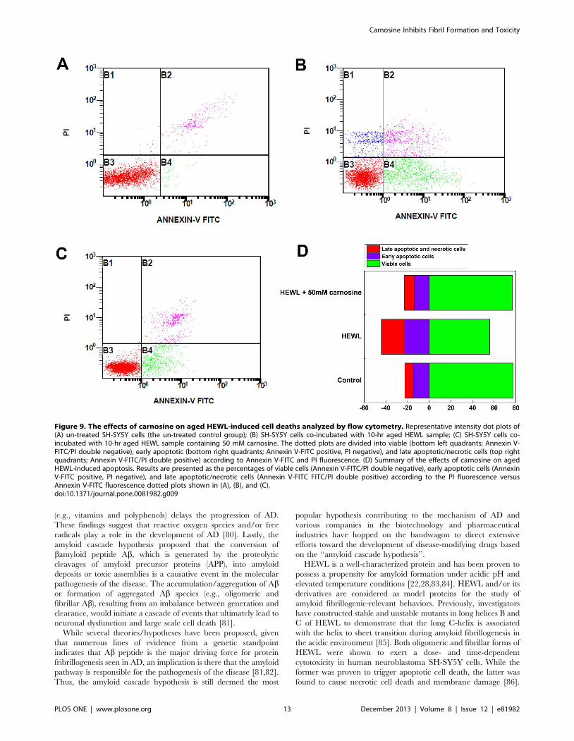
with Annexin-V-FITC and PI. Figs. 9A–9C are the representative
dot plot profiles generated by flow cytometric analysis of the cells.
As seen in Fig. 9A, while the untreated cells (the control) were
primarily Annexin-V-FITC and PI negative, some accidental
random staining with both Annexin-V-FITC and PI was
consistently noticed, probably attributed to the cell death that
occurred as a result of the cell culture operations. Quantitative
analysis of the flow cytometry results revealed that treatment of the
SH-SY5Y cells with the HEWL sample aged for 10 hr evoked
marked staining indicated by the percentage of Annexin V-FITC-
positive cells. The proportion of early apoptotic cells (Annexin V-
FITC positive/PI negative) increased from ,14.7% to ,22.6%
accompanied by a 21.5% decrease (from ,77.5% to ,56.0%) in
viable cell population (Annexin V-FITC negative/PI negative)
upon exposure to the 10-hr aged HEWL sample, as shown in
Figs. 9B and 9D. The Annexin V-FITC/PI staining results also
showed that the addition of the 10-hr aged HEWL sample led to
an increase in the percentage of early apoptotic and necrotic cells
by ,7.9% and ,8.6%, respectively, suggesting that the cell injury
triggered by the aged HEWL sample is slightly more necrotic than
apoptotic. Comparison of Figs. 9B, 9C, and 9D indicates that
the significantly elevated responses in apoptosis and necrosis of
Figure 7. Carnosine’s effect on SH-SY5Y cell viability. The cell viability upon exposure to various HEWL samples was measured by MTTreduction. The SH-SY5Y cells were exposed to 50 mM carnosine alone (the negative control) and HEWL samples aged for 10 hr without or withvarious concentrations of carnosine (10, 20, 30, 40, and 50 mM) for 6, 12, and 24 hr at 37uC in a humidified 5% (v/v) CO2/air environment. The data arepresented as the percentage of MTT reduced by the cells incubated in media containing 50 mM carnosine alone, HEWL alone, or HEWL with variousconcentrations of carnosine. The means 6 S.D. of at least 10 determinations are shown.doi:10.1371/journal.pone.0081982.g007
Carnosine Inhibits Fibril Formation and Toxicity
PLOS ONE | www.plosone.org 11 December 2013 | Volume 8 | Issue 12 | e81982

SH-SY5Y cells to the 10-hr aged HEWL sample were both
depressed upon the addition of 50 mM carnosine. Moreover, as
compared with the untreated cells (the control), similar levels/
percentages of the four different cell populations (viable cells:
negative for Annexin V-FITC and PI signals, early apoptotic cells:
Annexin V-FITC signal only, late apoptotic cells: positive for
Annexin V-FITC and PI signals, and necrotic cells: positive for
Annexin V-FITC and PI signals) were recorded in SH-SY5Y cells
treated with the 10-hr aged HEWL sample containing 50 mM
carnosine. The flow cytometry results support our above-
mentioned MTT reduction and LDH leakage findings that
carnosine, in the concentration range used, is capable of reducing
cell death triggered by the fibrillar species-containing aged HEWL
samples, and that this effect is dosage-dependent.
Discussion
Multiple hypotheses have been proposed for the etiology and
pathogenesis of amyloid diseases, e.g., Alzheimer’s disease (AD).
Of these, the cholinergic hypothesis states that a loss of cholinergic
function associated with acetylcholine (ACh) in the central nervous
system contributes substantially to the cognitive decline observed
in those with AD [73,74]. In addition, cholinergic effects have
been proposed as a potential causative agent for the formation of
plaques and tangles [75]. Another hypothesis, the tau hypothesis
focuses on the microtubule binding tau protein in AD as a
causative factor in amyloidosis. The abnormal or excessive
phosphorylation (hyperphosphorylation) of tau leads to the
transformation of normal tau into paired helical filament, (PHF)-
tau, which accumulate in neuron as neurofibrillary tangles (NFTs)
typically found in histopathological lesions of AD brains [76]. A
third hypothesis describes AD as an inflammatory disorder
involving microglia, astrocytes, and neurons in the inflammatory
processes. Based on this hypothesis, key players that contribute to
the inflammatory responses are the complement system, cytokines,
chemokines, and acute phase proteins [77,78]. A fourth hypothesis
points to oxidative stress as an etiology of AD. Evidence indicates
that AD brains exhibit certain levels of oxidative stress-mediated
injury/damage, which is triggered by the chemical reactions
between reactive oxygen species and/or free radicals and other
molecules (e.g., lipids, proteins, and DNA) [79]. Moreover, there is
indirect evidence demonstrating that treatment of antioxidants
Figure 8. The effects of carnosine on HEWL-induced membrane damage (LDH release into the medium) in SH-SY5Y cells. The cellviability upon exposure to HEWL sample was measured by the LDH release assay. SH-SY5Y cells were incubated with 50 mM carnosine alone (thenegative control) and HEWL samples without or with various concentrations of carnosine (10, 20, 30, 40, and 50 mM) for 6, 12, and 24 hr at 37uC in ahumidified 5% (v/v) CO2/air environment. The percentage of cytotoxicity was evaluated as a ratio of the quantity of LDH released in each sampledivided by the total LDH released by the sample of cells treated with lysis buffer. Quantity of released LDH is estimated by the activity of lactatedehydrogenase in the suspension aliquot from the 96-well plates after 30 min incubation with the appropriate substrate solution. Measurements ofthe means 6 S.D. of at least 8 determinations for each sample were obtained at 490 nm.doi:10.1371/journal.pone.0081982.g008
Carnosine Inhibits Fibril Formation and Toxicity
PLOS ONE | www.plosone.org 12 December 2013 | Volume 8 | Issue 12 | e81982
(e.g., vitamins and polyphenols) delays the progression of AD.
These findings suggest that reactive oxygen species and/or free
radicals play a role in the development of AD [80]. Lastly, the
amyloid cascade hypothesis proposed that the conversion of
bamyloid peptide Ab, which is generated by the proteolytic
cleavages of amyloid precursor proteins (APP), into amyloid
deposits or toxic assemblies is a causative event in the molecular
pathogenesis of the disease. The accumulation/aggregation of Abor formation of aggregated Ab species (e.g., oligomeric and
fibrillar Ab), resulting from an imbalance between generation and
clearance, would initiate a cascade of events that ultimately lead to
neuronal dysfunction and large scale cell death [81].
While several theories/hypotheses have been proposed, given
that numerous lines of evidence from a genetic standpoint
indicates that Ab peptide is the major driving force for protein
fribrillogenesis seen in AD, an implication is there that the amyloid
pathway is responsible for the pathogenesis of the disease [81,82].
Thus, the amyloid cascade hypothesis is still deemed the most
popular hypothesis contributing to the mechanism of AD and
various companies in the biotechnology and pharmaceutical
industries have hopped on the bandwagon to direct extensive
efforts toward the development of disease-modifying drugs based
on the ‘‘amyloid cascade hypothesis’’.
HEWL is a well-characterized protein and has been proven to
possess a propensity for amyloid formation under acidic pH and
elevated temperature conditions [22,28,83,84]. HEWL and/or its
derivatives are considered as model proteins for the study of
amyloid fibrillogenic-relevant behaviors. Previously, investigators
have constructed stable and unstable mutants in long helices B and
C of HEWL to demonstrate that the long C-helix is associated
with the helix to sheet transition during amyloid fibrillogenesis in
the acidic environment [85]. Both oligomeric and fibrillar forms of
HEWL were shown to exert a dose- and time-dependent
cytotoxicity in human neuroblastoma SH-SY5Y cells. While the
former was proven to trigger apoptotic cell death, the latter was
found to cause necrotic cell death and membrane damage [86].
Figure 9. The effects of carnosine on aged HEWL-induced cell deaths analyzed by flow cytometry. Representative intensity dot plots of(A) un-treated SH-SY5Y cells (the un-treated control group); (B) SH-SY5Y cells co-incubated with 10-hr aged HEWL sample; (C) SH-SY5Y cells co-incubated with 10-hr aged HEWL sample containing 50 mM carnosine. The dotted plots are divided into viable (bottom left quadrants; Annexin V-FITC/PI double negative), early apoptotic (bottom right quadrants; Annexin V-FITC positive, PI negative), and late apoptotic/necrotic cells (top rightquadrants; Annexin V-FITC/PI double positive) according to Annexin V-FITC and PI fluorescence. (D) Summary of the effects of carnosine on agedHEWL-induced apoptosis. Results are presented as the percentages of viable cells (Annexin V-FITC/PI double negative), early apoptotic cells (AnnexinV-FITC positive, PI negative), and late apoptotic/necrotic cells (Annexin V-FITC FITC/PI double positive) according to the PI fluorescence versusAnnexin V-FITC fluorescence dotted plots shown in (A), (B), and (C).doi:10.1371/journal.pone.0081982.g009
Carnosine Inhibits Fibril Formation and Toxicity
PLOS ONE | www.plosone.org 13 December 2013 | Volume 8 | Issue 12 | e81982

Others have reported that HEWL fibrils induced extensive
aggregation of human erythrocytes and lipid vesicles, suggesting
that the HEWL fibril-cellular membrane interaction could be the
underlying contributing factor to the disease mechanism [87]. In
addition, Dobson’s group found that by adding fibrils formed by
peptides or full-length HEWL to the native HEWL sample, fibril
formation of the native sample was accelerated. The group also
proved that the b-domain is significant in full-length HEWL fibril
formation [17]. Solubilizing HEWL in highly concentrated
ethanol solutions without heating procured amyloid protofilaments
rapidly owing to the destruction of the helical and tertiary
structures [20]. Results from these and other investigations suggest
that partial unfolding is a prerequisite to fibril formation [21,88–
94].
A number of researchers have hypothesized that all amyloid-
forming polypeptides, regardless of their primary sequence, may
display the same structure-specific cytotoxic effects via common
mechanism(s) [2,95,96]. It has been widely accepted that amyloid
proteins are cytotoxic when present in an aggregated form
containing mature fibrils, protofibrils, and/or low molecular
weight intermediates [97,98]. Moreover, evidence originating
from various studies using cell culture and animal models suggests
that the attenuation of amyloid fibrillogenesis appears to be of
great benefit to the health of the cells [99,100]. To that end,
strategies that utilize agents/compounds to minimize the forma-
tion of aggregated species have been put forth as effective means to
alleviate the pathological effects of amyloids or to counteract the
progression of amyloid diseases [101–103].
Several molecules or compounds, including peptides (or peptide
fragments) and non-peptidic small molecules, have been demon-
strated to potently attenuate or block the aggregation/fibrillogen-
esis and/or cytotoxicity elicited by amyloid-forming peptides and/
or polypeptides [104,105]. Typical examples of peptidic molecules
that have been reported to exhibit inhibitory activity against
amyloid fibril formation are shorter peptide fragments with self-
recognition sequences [106–108] and small heat shock proteins
(e.g., alpha-crystallin Hsp20) [109,110]. On the other hand, non-
peptidic inhibitory molecules consist of a wide range of natural
and synthetic aromatic/phenolic ring-containing polyphenols
[103,111]. Molecules such as the sulfonated dye, Congo red,
and benzofuran-based compounds have been shown to be effective
at reducing fibrillogenesis and cytotoxicity by binding directly to
amyloid fibrils [112,113]. Similar effects were also observed for
certain aromatic/phenolic ring-bearing polyphenols (e.g., nordi-
hydroguaiaretic acid and rosemarinic acid), semisynthetic bacte-
riocidal antibiotics (e.g., rifampicin and its derivatives), surfactants
(e.g., di-C6-PC, di-C7-PC, and n-dodecylhexaoxyethylene glycol
monoether), and others (e.g., nicotine and trehalose) [96,114,115].
Thus, these small molecules could provide a basis for the
development of therapeutics for amyloid diseases.
Carnosine (b-alanyl-L-histidine), a naturally-occurring endoge-
nous di-peptide, is present in surprisingly large amounts in long-
lived human tissues. Numerous lines of evidence have pointed to
the multi-functional significance of carnosine in the human body:
(1) serves as a physiological buffering agent [30,116,117] and a
metal ion (e.g., zinc and copper) chelator [118–120]; (2) possessing
anti-aging functions [121,122], and free-radical scavenging activity
[123,124]; (3) capable of delaying senescence [125] and extending
the life-span of cultured human fibroblasts [36]; (4) able to kill
transformed cells and protect cells against aldehydes and amyloid
peptide fragment [126]. Other investigators have concluded that
carnosine exhibits a well-documented anti-glycating activity
against the glycation of proteins, including low-density lipoproteins,
glucose degradation products, esterase, and histones [127–130].
Using cardiac aspartate aminotransferase (cAAT) as a model,
carnosine was reported to enhance the thermal unfolding and
water accessibility of glycated protein species [131]. It also
mitigates and/or prevents the alteration in electrophoretic
mobility triggered by glyceraldehyde 3-phosphate [132]. Evi-
dence has shown that the methylglyoxal glycation-induced
tryptophan fluorescence polarization and scattered light intensity
enhancements detected in the aggregated a-crystallin protein
were attenuated upon exposure to carnosine [40]. Eye lens
opacity in human was also found to be reversed by carnosine
[133]. Attanasio and co-workers have made an attempt to
explore how both L-form and D-form of carnosine affect the
aggregation of bovine a-crystallin [41]. Apart from retaining the
chaperone activity of bovine a-crystallin and preventing or
reversing the lens opacification, carnosine was observed to have
multiple protective roles against bovine a-crystallin fibrillogen-
esis, including the inhibition of fibril formation and disassembly
of preformed fibrils. In addition, using transgenic 36Tg-AD
mice as a model, the amyloid load of mice brain was found to
decrease upon dietary supplementation of carnosine [42].
Recently, carnosine has been shown to inhibit the in vitro
amyloid fibril formation of Ab(1–42) peptide, probably by
disturbing the hydrogen bond network near residues that play
crucial roles in fibrillogenesis [43] or by impeding the
intermolecular interactions between two key residues (D23 and
K28) located at the adjacent Ab(1–42) monomers [44]. These
findings prompted us to further evaluate carnosine’s effect on
the inhibition of protein fibrillogenesis/aggregation. In this
study, we used hen egg-white lysozyme (HEWL), a well-known
model protein commonly utilized for the study of protein
aggregation, to thoroughly investigate the extent of carnosine
fibril/aggregation inhibition on multiple levels.
We began by demonstrating that the formation of HEWL fibrils
occurred when the samples were incubated at pH 2.0 and 55uC(see Figs. 1 and 3A). We then tested carnosine for its effects on the
in vitro amyloid fibrillogenesis of HEWL. ThT fluorescence, Congo
red binding, and TEM experiments (see Figs. 2 and 3) all revealed
that carnosine exhibits inhibitory activity toward HEWL amyloid
fibril formation, and the said inhibitory effect is dependent upon
both the incubation period and the carnosine concentration
studied (0–50 mM).
The pH-dependent structural stability and/or resistance of
proteins to chemical and thermal denaturation have long been
explored by other studies to understand how change in pH alters
protein conformation [134–136]. We also performed equilibrium
thermal unfolding experiments to compare the susceptibility of the
HEWL structures (in the presence and absence of carnosine) to
denaturation/unfolding by heat. A higher structural stability
against thermal denaturation upon pH decrease was observed in
some proteins [134]. However, in our study, a left-shifted thermal
denaturation curve was observed for HEWL at pH 2.0, indicating
that a decrease in pH from 7.0 to 2.0 gave rise to a lower resistance
to thermal unfolding, which is in agreement with the trend
reported in certain proteins [135]. The addition of carnosine also
tended to lessen the thermally induced destabilization/unfolding
effect of the protein or to augment its thermal stability under the
condition of 55uC and pH 2.0 (see Table 1).
Given that a correlation is believed to exist between amyloid
fibrillogenesis, secondary structural transitions (from either disor-
dered random structure or a-helix-rich conformation to predom-
inantly ordered b-sheet structures) [137,138], and surface hydro-
phobicity, we next recorded the far-UV CD spectra in parallel
with Nile red fluorescence spectra of various HEWL samples to
monitor the changes in structural conformation due to the
Carnosine Inhibits Fibril Formation and Toxicity
PLOS ONE | www.plosone.org 14 December 2013 | Volume 8 | Issue 12 | e81982
addition of carnosine. Our CD results showed that, under the
condition of pH 2.0 and 55uC, carnosine mitigated the a-to-btransition observed in the control or aggregating HEWL sample
(see Fig. 4), thus preventing HEWL from adopting the conforma-
tion comprising primarily b-sheet structure, the suggested second-
ary structure known to be associated with insolubility and protease
resistance [138,139]. In addition, we observed that the control
HEWL and carnosine-containing HEWL samples displayed
markedly different Nile red fluorescence spectra. Compared to
HEWL alone, exposure to carnosine brought about a decrease in
Nile red fluorescence emission and a red-shift in wavelengths of
emission maximum (see Fig. 5). This is an implication that the
exposure of the hydrophobic regions was appreciably suppressed
due to the presence of the dipeptide. It should be noted that,
whether the reduction in Nile red fluorescence emission intensity
observed in the presence of carnosine is due to the stabilization of
HEWL in certain specific conformations with minimized exposure
of hydrophobic clusters or merely an indication of the absence of
exposed hydrophobic clusters cannot be ascertained from the data
obtained so far. However, our results evidently suggest that
amyloid fibrillogenesis of HEWL is undoubtedly hampered when
the protein is co-incubated with carnosine.
The propensity for a diversity of proteins to form fibrillar
aggregates have been a topic intensively explored in both
experimental and theoretical studies alike. Through these studies
it was found that despite the lack of structural or sequence
similarity, these proteins all have the potential to form cross-bstructure characteristic of amyloid fibrils [140], and that only
certain short sequence stretches on the protein chain is important
for the transformation of a protein from its native structure to the
ordered fibrillar structure [62,141,142]. Although the short residue
stretches that are ‘‘aggregation-prone’’ are not similar in sequence,
yet they retain inherent property to form b-sheets under suitable
conditions [143]. It is said that these aggregation-prone sequence
regions are mostly hydrophobic by nature and are usually buried
or partially buried within the proteins [59,142,144]. The amyloid-
promoting regions exhibit intrinsic physicochemical properties
that enhance aggregation by stabilizing b-sheet strands and
promote further self-assembly of the ordered b-structural motifs
into polymers through intermolecular bonding [143]. In the
attempt to better understand the physicochemical and structural
basis for amyloid formation, various groups have devised in silico
algorithms to predict potential amyloid nucleation sites based on
intrinsic properties of the amino acid sequence itself. As each of
these methods make its own assumptions and boasts of its
predictive accuracy, we implemented a consensus approach by
combining the prediction results of several methods (see Mate-rials and Methods) and found two potential aggregation sites,
N27-C30 and N106-A110, on HEWL (see Fig. S3).
To understand how carnosine inhibits HEWL amyloid fibril
formation, we conducted docking studies and analyzed, theoret-
ically, the binding site of carnosine to HEWL using CDOCKER.
Our docking results showed that carnosine initially binds to an
aggregation-prone region through interactions with the following
hydrophobic residues: A107, W108, and V109. Based on the best
binding mode (inset of Fig. 6 and Pose 1 in Fig. S4) derived from
the docking simulation, the interaction pattern involving these
residues include hydrogen bonding with A107 while interactions
with W108 and V109 were charged or polar. Other residues that
prevalently form hydrogen bonds with carnosine were D48, Q57,
and W63. In addition to the hydrogen bonds and charged or polar
interactions, Pose 1 rose above all others owing to an additional
cation-pi interaction that existed between the imidazole ring of
carnosine and R112 of HEWL, which may serve to stabilize the
ligand-protein complex. Based on our molecular docking results,
we believe that carnosine inhibits HEWL aggregation by initially
blocking aggregation-prone site mainly through interactions with
three residues: A107, W108, and V109.
To test that carnosine’s effect against HEWL fibrillogenesis is
not just a generalized phenomenon of dipeptide molecules, we
have performed a couple of control experiments, in which di-
alanine, di-histidine, or di-glycine at 50 mM was incubated with
HEWL and samples were taken at various time points for ThT
binding assay. As illustrated in Fig. S5, the inhibitory potency
against lysozyme fibril formation shows the following order:
carnosine . di-alanine . di-histidine . di-glycine. The reasoning
behind this order of effect can be explained by the relative location
of the potential aggregation-prone region on HEWL, the bulkiness
of the inhibitor’s sidechain, and the extent of interactions involved
in binding carnosine to HEWL to block the aggregation-prone site
from initiating the process of amyloid fibrillogenesis. Through our
bioinformatic prediction and docking studies, we found that the
region spanning residues N106 , A110 to be an aggregation-
prone region and that residues A107 , V109 (which interact with
carnosine) are situated at the opening of the HEWL catalytic cleft.
As can be seen from the best docking pose shown in Fig. 6, the
binding position of carnosine is at the mouth of the catalytic cleft
with the b-alanine inserted into the cleft while the bulky
imidazolium group of histidine is left out sitting at the lip of the
cleft. This orientation maximizes the number of residues and
atomic interactions involved in binding carnosine to HEWL, thus
stabilizing the protein-ligand complex. Whereas histidine plays a
supporting role in anchoring carnosine to the mouth of the cleft,
di-alanine is small enough to enter the cleft and may be less
specific in binding to the aggregation-prone region. However,
based on our docking results, it seems that alanine is involved in
majority of the interactions observed between carnosine and
HEWL (see Fig. S4 of the supporting information); therefore, it is
likely that once di-alanine found its way to the aggregation-prone
site, it is still capable of exerting an anti-fibrillogenic effect on
HEWL to some extent. Having said that alanine participates in
most of the atomic interactions observed between carnosine and
HEWL, it is understandable that di-histidine has less of an
inhibitory effect than di-alanine in preventing HEWL fibril
formation. Based on past studies, it has been found that two
cationic groups in protein or peptide residues can form contact
pairs despite both bearing positive charges [145,146]. Since the
imidazole group in histidine (pK,6) is protonated in low pH
condition, the dipeptide assumes a double positive charge under
the experimental setting explored in our study. According to
Heyda et. al. (2010) [147], the cationic groups in di-histidine and
other histidine-based dipeptides exhibit attractive interactions
regardless of the unfavorable Coulomb repulsion force. Due to the
covalent linkage between the two histidine residues, the contact
pairing of the imidazolium side chains preferably assumes a
parallel or off-parallel stacking orientation, which restricts the
extent of separation between the two groups. This steric constraint
placed on di-histidine meant that the dipeptide nearly folds back
on itself to form cationic pairing, in essence, reducing the
accessibility of atomic interactions that can potentially form with
HEWL if it were laid out with the two imidazolium groups spread
out side-by-side. As for di-glycine, the dipeptide of the smallest
amino acid glycine, our ThT fluorescence results demonstrate that
there is almost no difference in the ThT fluorescence signal
between the HEWL alone sample (the control) and the di-glycine-
containing HEWL sample, suggesting that di-glycine does not
exert an inhibitory action toward HEWL fibrillogenesis.
Carnosine Inhibits Fibril Formation and Toxicity
PLOS ONE | www.plosone.org 15 December 2013 | Volume 8 | Issue 12 | e81982

Having established that carnosine has an inhibitory effect on
HEWL amyloid fibrillogenesis, we next set out to investigate the
effects of carnosine on cell toxicity in the presence of HEWL
aggregates. Previously, it was suggested that amyloid peptides/
proteins demonstrate neuro-cytotoxicity in their aggregation states
[148]. We, therefore, subjected SH-SY5Y human neuroblastoma
cells to co-incubation with 10-hr aged HEWL sample that has
previously been shown by our group to have distinct features
characteristic of amyloid fibril. These features include: adopting
the b-sheet-rich conformation (,41%), having a high affinity for
Congo red, emitting high level of ThT fluorescence, and
displaying fibrillar morphology based on TEM (see Figs. 1, 2, 3,
and 4). Prolonged exposure to the 10-hr aged HEWL sample
considerably decreased the viability of the SH-SY5Y cells. Our
MTT reduction results in concert with the LDH release findings
allow us to conclude that the significant cellular damage elicited by
the aforesaid fibrillar form of HEWL may be inhibited by
carnosine in a concentration-dependent manner (see Figs. 7 and
8). We also carried out flow cytometric analysis, which revealed
that co-incubation with carnosine dipeptide also attenuated the
extent of apoptotic/necrotic cell death induced by the HEWL
sample containing fibrillar species (see Fig. 9). These data are well
corroborated with other findings on the protection of REB4 cells
by carnosine against cytotoxicity triggered by fibrillar Ab(25–35)
[149].
Both MTT and Annexin V/PI apoptosis assay detect cell
viability, but on a different level. Based on the results of our MTT
assay, it can be seen that the pH 2-fibrillized HEWL impaired the
metabolic activity or health of the cells by ,52–64%. However,
with the increased dosage of carnosine, we began to see a recovery
of metabolic activity in the SH-SY5Y cells until close to ,95%
recovery is achieved in the condition where 50 mM carnosine was
added. As MTT assay is only capable of indicating the overall
well-being of a cell population rather than pointing to the more
specific states of cell death (which can be differentiated by the
Annexin V/PI apoptosis assay), we therefore, performed Annexin
V/PI apoptosis assay for a more complete picture of cell viability.
Consistent with previous reports that necrosis is associated with
cytotoxicity elicited by the fibrillar form of HEWL [86], our
Annexin V/PI apoptosis assay also shows that the cytotoxicity
induced by the pH 2.0-fibrillized HEWL is slightly more necrotic,
with an increase of 8.6% necrotic versus 7.9% early apoptotic
cells, when comparing the changes in Figs. 9A, 9B, and 9D. It is
noteworthy to mention that carnosine protects the cells from both
necrosis and apoptosis indiscriminately by maintaining the cell
populations at similar levels as the untreated control group
(Figs. 9A, 9C, and 9D).
In conclusion, the research presented here examines how
carnosine influences in vitro protein fibril formation via several
spectroscopic techniques, TEM, thermally induced unfolding
analysis, and molecular docking. We observed that carnosine has
a concentration-dependent inhibitory activity against b-sheet
formation and in vitro HEWL fibrillogenesis, with the best
inhibition level observed at carnosine concentration of 50 mM.
Moreover, thermally induced equilibrium denaturation analysis
revealed that HEWL exhibited enhanced stability upon the
addition of 50 mM carnosine. The dipeptide exerts its fibril-
inhibitory effect by initially binding to a partially exposed
aggregation-prone region of HEWL, thereby protecting the
protein from amyloid fibril nucleation. In addition, through the
MTT reduction and LDH release assays, we were able to show
that carnosine attenuated the decline in SH-SY5Y cell viability
induced by the aged HEWL samples containing predominantly
HEWL fibrils. The protective action of carnosine against HEWL
fibril induced neural cell death is through both anti-apoptotic and
anti-necrotic activities elicited in the neural-like cells, as evaluated
by the flow cytometric analysis. While the amyloid fibrillogenesis
of hen lysozyme, especially at acidic pH and elevated temperature,
is rarely observed in vivo and further investigation is warranted to
fully decipher the detailed mechanism by which carnosine may
retard the extent of HEWL fibrillogenesis/aggregation and reduce
the cytotoxicity derived from HEWL fibrils, we believe the results
from the current work are still informative and can provide a
model system for future work in this subject matter. It is worth re-
emphasizing that carnosine is currently being used as a component
in eye drops for the prevention and treatment of cataract in
Europe [150]. Along this line, some of the knowledge gained from
this work indicates that carnosine may also be a promising strategy
for ameliorating the progression of protein aggregation diseases
such as amyloidoses.
Supporting Information
Figure S1 The time dependence backbone alpha carbonroot-mean-square deviation (RMSD; nm) of HEWL. The
20 ns simulation was performed in pH 2.0 and 55uC.
(TIF)
Figure S2 Thermal denaturation curves of HEWLsamples. The thermal denaturation curves are obtained by
plotting fractions of denatured/unfolded HEWL under different
conditions (solid black circles: HEWL at pH 7.0; solid red circles:
HEWL at pH 2.0; solid blue squares: HEWL+10 mM carnosine
at pH 2.0; solid green squares: HEWL+30 mM carnosine at
pH 2.0; solid orange diamonds: HEWL+50 mM carnosine at
pH 2.0) against the temperature.
(TIF)
Figure S3 Consensus aggregation site prediction re-vealing two potential aggregation regions on the prima-ry sequence of HEWL.
(TIF)
Figure S4 Two-dimensional schematic representationof the top 10 potential binding modes for carnosine(Poses 1–10).
(TIF)
Figure S5 Time-evolved ThT fluorescence emission of lysozyme
samples containing 50 mM of di-glycine, di-histidine, di-alanine,
and carnosine.
(TIF)
Author Contributions
Conceived and designed the experiments: JWW SSW. Performed the
experiments: KNL SCH CML WAC. Analyzed the data: KNL SCH JWW
SSW. Contributed reagents/materials/analysis tools: CJH HSL. Wrote the
paper: JWW KNL SSW. Discussed the Analyses: HSL Interpreted the
results: HSL.
References
1. Dobson CM (2004) Principles of protein folding, misfolding and aggregation.
Semin Cell Dev Biol 15: 3–16.
2. Wang SSS, Good TA (2005) An overview of Alzheimer’s disease. Journal of the
Chinese Institute of Chemical Engineers 36: 533–559.
Carnosine Inhibits Fibril Formation and Toxicity
PLOS ONE | www.plosone.org 16 December 2013 | Volume 8 | Issue 12 | e81982

3. Liberski PP, Sikorska B, Hauw JJ, Kopp N, Streichenberger N, et al. (2010)Ultrastructural Characteristics (or Evaluation) of Creutzfeldt-Jakob Disease and
Other Human Transmissible Spongiform Encephalopathies or Prion Diseases.
Ultrastructural Pathology 34: 351–361.
4. Kell DB (2010) Towards a unifying, systems biology understanding of large-
scale cellular death and destruction caused by poorly liganded iron:
Parkinson’s, Huntington’s, Alzheimer’s, prions, bactericides, chemical toxicol-ogy and others as examples. Archives of Toxicology 84: 825–889.
5. Ross CA, Poirier MA (2004) Protein aggregation and neurodegenerativedisease. Nat Med 10 Suppl: S10–17.
6. Uversky VN, Fink AL (2004) Conformational constraints for amyloid
fibrillation: the importance of being unfolded. Biochim Biophys Acta 1698:131–153.
7. Chiti F, Dobson CM (2009) Amyloid formation by globular proteins under
native conditions. Nat Chem Biol 5: 15–22.
8. Ferrao-Gonzales AD, Souto SO, Silva JL, Foguel D (2000) The preaggregatedstate of an amyloidogenic protein: hydrostatic pressure converts native
transthyretin into the amyloidogenic state. Proc Natl Acad Sci U S A 97:6445–6450.
9. Wang SSS, Liu KN, Han TC (2010) Amyloid fibrillation and cytotoxicity of
insulin are inhibited by the amphiphilic surfactants. Biochimica Et BiophysicaActa-Molecular Basis of Disease 1802: 519–530.
10. Naeem A, Khan T, Muzaffar M, Ahmad S, Saleemuddin M (2011) A Partially
Folded State of Ovalbumin at Low pH Tends to Aggregate. Cell Biochemistryand Biophysics 59: 29–38.
11. Zhang C, Jackson AP, Zhang ZR, Han Y, Yu S, et al. (2010) Amyloid-Like
Aggregates of the Yeast Prion Protein Ure2 Enter Vertebrate Cells by SpecificEndocytotic Pathways and Induce Apoptosis. Plos One 5: -.
12. De Meulenaer B, Kerkaert B, Mestdagh F (2010) Detection of hen’s egg white
lysozyme in food: Comparison between a sensitive HPLC and a commercialELISA method. Food Chemistry 120: 580–584.
13. Proctor VA, Cunningham FE (1988) The Chemistry of Lysozyme and Its Useas a Food Preservative and a Pharmaceutical. Crc Critical Reviews in Food
Science and Nutrition 26: 359–395.
14. Bolin KA, Millhauser GL (1999) alpha and 3(10): The split personality ofpolypeptide helices. Accounts of Chemical Research 32: 1027–1033.
15. Vaney MC, Maignan S, RiesKautt M, Ducruix A (1996) High-resolution
structure (1.33 angstrom) of a HEW lysozyme tetragonal crystal grown in theAPCF apparatus. Data and structural comparison with a crystal grown under
microgravity from SpaceHab-01 mission. Acta Crystallographica Section D-
Biological Crystallography 52: 505–517.
16. Morozova-Roche LA, Zurdo J, Spencer A, Noppe W, Receveur V, et al. (2000)
Amyloid fibril formation and seeding by wild-type human lysozyme and itsdisease-related mutational variants. Journal of Structural Biology 130: 339–
351.
17. Krebs MRH, Wilkins DK, Chung EW, Pitkeathly MC, Chamberlain AK, et al.(2000) Formation and seeding of amyloid fibrils from wild-type hen lysozyme
and a peptide fragment from the beta-domain. Journal of Molecular Biology
300: 541–549.
18. Pepys MB, Hawkins PN, Booth DR, Vigushin DM, Tennent GA, et al. (1993)
Human Lysozyme Gene-Mutations Cause Hereditary Systemic Amyloidosis.
Nature 362: 553–557.
19. Arnaudov LN, de Vries R (2005) Thermally induced fibrillar aggregation of
hen egg white lysozyme. Biophysical Journal 88: 515–526.
20. Goda S, Takano K, Yamagata Y, Nagata R, Akutsu H, et al. (2000) Amyloidprotofilament formation of hen egg lysozyme in highly concentrated ethanol
solution. Protein Science 9: 369–375.
21. Vernaglia BA, Huang J, Clark ED (2004) Guanidine hydrochloride can induceamyloid fibril formation from hen egg-white lysozyme. Biomacromolecules 5:
1362–1370.
22. Wang SSS, Liu KN, Lee WH (2009) Effect of curcumin on the amyloidfibrillogenesis of hen egg-white lysozyme. Biophysical Chemistry 144: 78–87.
23. Rafii MS, Aisen PS (2009) Recent developments in Alzheimer’s diseasetherapeutics. BMC Med 7: 7.
24. Jakob-Roetne R, Jacobsen H (2009) Alzheimer’s disease: from pathology to
therapeutic approaches. Angew Chem Int Ed 48: 3030–3059.
25. Vardy ERLC, Catto AJ, Hooper NM (2005) Proteolytic mechanisms inamyloid-b metabolism: therapeutic implications for Alzheimer’s disease.
Trends Mol Med 11: 464–472.
26. Lim GP, Calon F, Morihara T, Yang FS, Teter B, et al. (2005) A diet enrichedwith the omega-3 fatty acid docosahexaenoic acid reduces amyloid burden in
an aged Alzheimer mouse model. Journal of Neuroscience 25: 3032–3040.
27. Yang FS, Lim GP, Begum AN, Ubeda OJ, Simmons MR, et al. (2005)Curcumin inhibits formation of amyloid beta oligomers and fibrils, binds
plaques, and reduces amyloid in vivo. Journal of Biological Chemistry 280:5892–5901.
28. Wang SS, Hung YT, Wen WS, Lin KC, Chen GY (2011) Exploring the
inhibitory activity of short-chain phospholipids against amyloid fibrillogenesisof hen egg-white lysozyme. Biochim Biophys Acta 1811: 301–313.
29. Boldyrev AA (2000) Problems and perspectives in studying the biological role of
carnosine. Biochemistry (Mosc) 65: 751–756.
30. Abe H (2000) Role of histidine-related compounds as intracellular proton
buffering constituents in vertebrate muscle. Biochemistry-Moscow 65: 757–
765.
31. Bauer K (2005) Carnosine and homocarnosine, the forgotten, enigmatic
peptides of the brain. Neurochemical Research 30: 1339–1345.
32. Boldyrev A, Bulygina E, Leinsoo T, Petrushanko I, Tsubone S, et al. (2004)
Protection of neuronal cells against reactive oxygen species by carnosine and
related compounds. Comparative Biochemistry and Physiology B-Biochemistry
& Molecular Biology 137: 81–88.
33. Pegova A, Abe H, Boldyrev A (2000) Hydrolysis of carnosine and related
compounds by mammalian carnosinases. Comparative Biochemistry and
Physiology B-Biochemistry & Molecular Biology 127: 443–446.
34. Teufel M, Saudek V, Ledig JP, Bernhardt A, Boularand S, et al. (2003)
Sequence identification and characterization of human carnosinase and a
closely related non-specific dipeptidase. Journal of Biological Chemistry 278:
6521–6531.
35. Hipkiss AR, Brownson C (2000) Carnosine reacts with protein carbonyl groups:
another possible role for the anti-ageing peptide? Biogerontology 1: 217–223.
36. McFarland GA, Holliday R (1994) Retardation of the Senescence of Cultured
Human-Diploid Fibroblasts by Carnosine. Experimental Cell Research 212:
167–175.
37. McFarland GA, Holliday R (1999) Further evidence for the rejuvenating effects
of the dipeptide L-carnosine on cultured human diploid fibroblasts.
Experimental Gerontology 34: 35–45.
38. Babizhayev MA (2011) Designation of imidazole-containing dipeptides as
pharmacological chaperones. Hum Exp Toxicol 30: 736–761.
39. Hipkiss AR (2011) Energy metabolism, proteotoxic stress and age-related
dysfunction - protection by carnosine. Mol Aspects Med 32: 267–278.
40. Seidler NW, Yeargans GS, Morgan TG (2004) Carnosine disaggregates
glycated alpha-crystallin: an in vitro study. Arch Biochem Biophys 427: 110–
115.
41. Attanasio F, Cataldo S, Fisichella S, Nicoletti S, Nicoletti VG, et al. (2009)
Protective Effects of L- and D-Carnosine on alpha-Crystallin Amyloid Fibril
Formation: Implications for Cataract Disease. Biochemistry 48: 6522–6531.
42. Corona C, Frazzini V, Silvestri E, Lattanzio R, La Sorda R, et al. (2011) Effects
of Dietary Supplementation of Carnosine on Mitochondrial Dysfunction,
Amyloid Pathology, and Cognitive Deficits in 3xTg-AD Mice. Plos One 6.
43. Attanasio F, Convertino M, Magno A, Caflisch A, Corazza A, et al. (2013)
Carnosine Inhibits A beta 42 Aggregation by Perturbing the H-Bond Network
in and around the Central Hydrophobic Cluster. Chembiochem 14: 583–592.
44. Aloisi A, Barca A, Romano A, Guerrieri S, Storelli C, et al. (2013) Anti-
aggregating effect of the naturally occurring dipeptide carnosine on abeta1-42
fibril formation. Plos One 8: e68159.
45. Preston JE, Hipkiss AR, Himsworth DT, Romero IA, Abbott JN (1998) Toxic
effects of beta-amyloid(25-35) on immortalised rat brain endothelial cell:
protection by carnosine, homocarnosine and beta-alanine. Neurosci Lett 242:
105–108.
46. Fu QL, Dai HB, Hu WW, Fan YY, Shen Y, et al. (2008) Carnosine protects
against A beta 42-induced neurotoxicity in differentiated rat PC12 cells.
Cellular and Molecular Neurobiology 28: 307–316.
47. Li QQ, Sun YP, Ruan CP, Xu XY, Ge JH, et al. (2011) Cellular prion protein
promotes glucose uptake through the Fyn-HIF-2a-Glut1 pathway to support
colorectal cancer survival. Cancer Sci 102: 400–406.
48. Lowik DWPM, Meijer JT, Roeters M, Viola V, Vriend G, et al. (2007)
Stabilization of peptide fibrils by hydrophobic interaction. Langmuir 23: 2058–
2063.
49. Kato A, Harada A, Azakami H (2008) Amyloid fibril formation of hen
lysozyme depends on the instability of the C-helix (88-99). Bioscience
Biotechnology and Biochemistry 72: 1523–1530.
50. Kroes-Nijboer A, Venema P, Bouman J, van der Linden E (2011) Influence of
Protein Hydrolysis on the Growth Kinetics of beta-Ig Fibrils. Langmuir 27:
5753–5761.
51. Nielsen L, Khurana R, Coats A, Frokjaer S, Brange J, et al. (2001) Effect of
environmental factors on the kinetics of insulin fibril formation: elucidation of
the molecular mechanism. Biochemistry 40: 6036–6046.
52. Darghal N, Garnier-Suillerot A, Salerno M (2006) Mechanism of thioflavin T
accumulation inside cells overexpressing P-glycoprotein or multidrug resis-
tance-associated protein: Role of lipophilicity and positive charge. Biochemical
and Biophysical Research Communications 343: 623–629.
53. Klunk WE, Jacob RF, Mason RP (1999) Quantifying amyloid by Congo red
spectral shift assay. Amyloid, Prions, and Other Protein Aggregates 309: 285–
305.
54. Whitmore L, Wallace BA (2004) DICHROWEB, an online server for protein
secondary structure analyses from circular dichroism spectroscopic data.
Nucleic Acids Research 32: W668–W673.
55. Whitmore L, Wallace BA (2008) Protein secondary structure analyses from
circular dichroism spectroscopy: Methods and reference databases. Biopoly-
mers 89: 392–400.
56. Brown MB, Miller JN, Seare NJ (1995) An Investigation of the Use of Nile-Red
as a Long-Wavelength Fluorescent-Probe for the Study of Alpha(1)-Acid
Glycoprotein-Drug Interactions. Journal of Pharmaceutical and Biomedical
Analysis 13: 1011–1017.
57. Cser A, Nagy K, Biczok L (2002) Fluorescence lifetime of Nile Red as a probe
for the hydrogen bonding strength with its microenvironment. Chemical
Physics Letters 360: 473–478.
Carnosine Inhibits Fibril Formation and Toxicity
PLOS ONE | www.plosone.org 17 December 2013 | Volume 8 | Issue 12 | e81982

58. Conchillo-Sole O, de Groot NS, Aviles FX, Vendrell J, Daura X, et al. (2007)AGGRESCAN: a server for the prediction and evaluation of "hot spots" of
aggregation in polypeptides. BMC Bioinformatics 8: 65.
59. Frousios KK, Iconomidou VA, Karletidi CM, Hamodrakas SJ (2009)
Amyloidogenic determinants are usually not buried. BMC Struct Biol 9: 44.
60. Fernandez-Escamilla AM, Rousseau F, Schymkowitz J, Serrano L (2004)Prediction of sequence-dependent and mutational effects on the aggregation of
peptides and proteins. Nat Biotechnol 22: 1302–1306.
61. Hamodrakas SJ, Liappa C, Iconomidou VA (2007) Consensus prediction of
amyloidogenic determinants in amyloid fibril-forming proteins. Int J BiolMacromol 41: 295–300.
62. Lopez de la Paz M, Serrano L (2004) Sequence determinants of amyloid fibril
formation. Proc Natl Acad Sci U S A 101: 87–92.
63. Galzitskaya OV, Garbuzynskiy SO, Lobanov MY (2006) Prediction of
amyloidogenic and disordered regions in protein chains. PLoS Comput Biol2: e177.
64. Zhang Z, Chen H, Lai L (2007) Identification of amyloid fibril-formingsegments based on structure and residue-based statistical potential. Bioinfor-
matics 23: 2218–2225.
65. Wu G, Robertson DH, Brooks CL 3rd, Vieth M (2003) Detailed analysis ofgrid-based molecular docking: A case study of CDOCKER-A CHARMm-
based MD docking algorithm. J Comput Chem 24: 1549–1562.
66. Brooks BRB, R. E.; . Olafson, B. D.; . States, D. J.; . Swaminathan, S. (1983)
CHARMM: A program for macromolecular energy, minimization, and
dynamics calculations. J Comput Chem 4: 187.
67. Pollack SJ, Sadler IIJ, Hawtin SR, Tailor VJ, Shearman MS (1995) Sulfated
glycosaminoglycans and dyes attenuate the neurotoxic effects of [beta]-amyloidin rat PC12 cells. Neuroscience Letters 184: 113–116.
68. Vassar PS, Culling CFA (1959) Fluorescent Stains, with Special Reference toAmyloid and Connective Tissues. Archives of Pathology 68: 487–498.
69. Ladewig P (1945) Double-Refringence of the Amyloid-Congo-Red-Complex in
Histological Sections. Nature 156: 81–82.
70. Turnell WG, Finch JT (1992) Binding of the Dye Congo Red to the Amyloid
Protein Pig Insulin Reveals a Novel Homology Amongst Amyloid-FormingPeptide Sequences. Journal of Molecular Biology 227: 1205–1223.
71. Geddes CD, Lakowicz JR, Coutinho PJG (2009) Photophysics and BiophysicalApplications of Benzo[,i.a,/i.]phenoxazine Type Fluorophores. Reviews
in Fluorescence 2007: Springer New York. pp. 335–362.
72. Ghanta J, Shen CL, Kiessling LL, Murphy RM (1996) A strategy for designinginhibitors of beta-amyloid toxicity. Journal of Biological Chemistry 271:
29525–29528.
73. Terry AV, Buccafusco JJ (2003) The cholinergic hypothesis of age and
Alzheimer’s disease-related cognitive deficits: Recent challenges and theirimplications for novel drug development. Journal of Pharmacology and
Experimental Therapeutics 306: 821–827.
74. Contestabile A (2011) The history of the cholinergic hypothesis. BehaviouralBrain Research 221: 334–340.
75. Wenk GL (2003) Neuropathologic changes in Alzheimer’s disease. J ClinPsychiatry 64 Suppl 9: 7–10.
76. Williams DR (2006) Tauopathies: classification and clinical update onneurodegenerative diseases associated with microtubule-associated protein
tau. Internal Medicine Journal 36: 652–660.
77. Akiyama H, Barger S, Barnum S, Bradt B, Bauer J, et al. (2000) Inflammationand Alzheimer’s disease. Neurobiol Aging 21: 383–421.
78. Zotova E, Nicoll JAR, Kalaria R, Holmes C, Boche D (2010) Inflammation inAlzheimer’s disease: relevance to pathogenesis and therapy. Alzheimers
Research & Therapy 2.
79. Markesbery WR (1997) Oxidative stress hypothesis in Alzheimer’s disease. Free
Radic Biol Med 23: 134–147.
80. Sano M, Ernesto C, Thomas RG, Klauber MR, Schafer K, et al. (1997) Acontrolled trial of selegiline, alpha-tocopherol, or both as treatment for
Alzheimer’s disease. New England Journal of Medicine 336: 1216–1222.
81. Haass C, Selkoe DJ (2007) Soluble protein oligomers in neurodegeneration:
lessons from the Alzheimer’s amyloid beta-peptide. Nature Reviews MolecularCell Biology 8: 101–112.
82. Hardy J, Selkoe DJ (2002) Medicine - The amyloid hypothesis of Alzheimer’s
disease: Progress and problems on the road to therapeutics. Science 297: 353–356.
83. Sharp JS, Forrest JA, Jones RAL (2002) Surface Denaturation and AmyloidFibril Formation of Insulin at Model Lipid2Water Interfaces. Biochemistry 41:
15810–15819.
84. Hung YT, Lin MS, Chen WY, Wang SS (2010) Investigating the effects of
sodium dodecyl sulfate on the aggregative behavior of hen egg-white lysozyme
at acidic pH. Colloids Surf B Biointerfaces 81: 141–151.
85. Harada A, Azakami H, Kato A (2008) Amyloid fibril formation of hen
lysozyme depends on the instability of the C-helix (88-99). BioscienceBiotechnology and Biochemistry 72: 1523–1530.
86. Gharibyan AL, Zamotin V, Yanamandra K, Moskaleva OS, Margulis BA, et
al. (2007) Lysozyme amyloid oligomers and fibrils induce cellular death viadifferent apoptotic/necrotic pathways. Journal of Molecular Biology 365:
1337–1349.
87. Chaudhary N, Nagaraj R (2009) Hen lysozyme amyloid fibrils induce
aggregation of erythrocytes and lipid vesicles. Molecular and CellularBiochemistry 328: 209–215.
88. Armen RS, Daggett V (2005) Characterization of two distinct beta(2)-
microglobulin unfolding intermediates that may lead to amyloid fibrils of
different morphology. Biochemistry 44: 16098–16107.
89. Chiti F, Mangione P, Andreola A, Giorgetti S, Stefani M, et al. (2001)
Detection of two partially structured species in the folding process of the
amyloidogenic protein beta 2-microglobulin. Journal of Molecular Biology 307:
379–391.
90. Colon W, Lai ZH, McCutchen SL, Miroy GJ, Strang C, et al. (1996) FAP
mutations destabilize transthyretin facilitating conformational changes required
for amyloid formation. Nature and Origin of Amyloid Fibrils 199: 228–242.
91. Fezoui Y, Teplow DB (2002) Kinetic studies of amyloid beta-protein fibril
assembly - Differential effects of alpha-helix stabilization. Journal of Biological
Chemistry 277: 36948–36954.
92. Litvinovich SV, Brew SA, Aota S, Akiyama SK, Haudenschild C, et al. (1998)
Formation of amyloid-like fibrils by self-association of a partially unfolded
fibronectin type III module. Journal of Molecular Biology 280: 245–258.
93. Wiseman RL, Powers ET, Kelly JW (2005) Partitioning conformational
intermediates between competing refolding and aggregation pathways: Insights
into transthyretin amyloid disease. Biochemistry 44: 16612–16623.
94. Yamamoto S, Hasegawa K, Yamaguchi I, Tsutsumi S, Kardos J, et al. (2004)
Low concentrations of sodium dodecyl sulfate induce the extension of beta(2)-
microglobulin-related amyloid fibrils at a neutral pH. Biochemistry 43: 11075–
11082.
95. Canale C, Torrassa S, Rispoli P, Relini A, Rolandi R, et al. (2006) Natively
folded HypF-N and its early amyloid aggregates interact with phospholipid
monolayers and destabilize supported phospholipid bilayers. Biophysical
Journal 91: 4575–4588.
96. Wang W (2005) Protein aggregation and its inhibition in biopharmaceutics.
Int J Pharm 289: 1–30.
97. Walsh DM, Selkoe DJ (2004) Oligomers on the brain: the emerging role of
soluble protein aggregates in neurodegeneration. Protein Pept Lett 11: 213–
228.
98. Haass C, Selkoe DJ (2007) Soluble protein oligomers in neurodegeneration:
lessons from the Alzheimer’s amyloid beta-peptide. Nat Rev Mol Cell Biol 8:
101–112.
99. Khlistunova I, Biernat J, Wang YP, Pickhardt M, von Bergen M, et al. (2006)
Inducible expression of tau repeat domain in cell models of tauopathy -
Aggregation is toxic to cells but can be reversed by inhibitor drugs. Journal of
Biological Chemistry 281: 1205–1214.
100. Roberson ED, Scearce-Levie K, Palop JJ, Yan FR, Cheng IH, et al. (2007)
Reducing endogenous tau ameliorates amyloid beta-induced deficits in an
Alzheimer’s disease mouse model. Science 316: 750–754.
101. Defelice FG, Ferreira ST (2002) Physiopathological modulators of amyloid
aggregation and novel pharmacological approaches in Alzheimer’s disease. An
Acad Bras Cienc 74: 265–284.
102. Estrada LD, Soto C (2007) Disrupting beta-amyloid aggregation for Alzheimer
disease treatment. Curr Top Med Chem 7: 115–126.
103. Porat Y, Abramowitz A, Gazit E (2006) Inhibition of amyloid fibril formation
by polyphenols: structural similarity and aromatic interactions as a common
inhibition mechanism. Chem Biol Drug Des 67: 27–37.
104. Vieira MNN, Figueroa-Villar JD, Meirelles MNL, Ferreira ST, De Felice FG
(2006) Small molecule inhibitors of lysozyme amyloid aggregation. Cell
Biochemistry and Biophysics 44: 549–553.
105. Gazova Z, Bellova A, Daxnerova Z, Imrich J, Kristian P, et al. (2008) Acridine
derivatives inhibit lysozyme aggregation. European Biophysics Journal with
Biophysics Letters 37: 1261–1270.
106. Pallitto MM, Ghanta J, Heinzelman P, Kiessling LL, Murphy RM (1999)
Recognition sequence design for peptidyl modulators of beta-amyloid
aggregation and toxicity. Biochemistry 38: 3570–3578.
107. Soto C, Sigurdsson EM, Morelli L, Kumar RA, Castano EM, et al. (1998)
Beta-sheet breaker peptides inhibit fibrillogenesis in a rat brain model of
amyloidosis: implications for Alzheimer’s therapy. Nat Med 4: 822–826.
108. Hetenyi C, Szabo Z, Klement E, Datki Z, Kortvelyesi T, et al. (2002)
Pentapeptide amides interfere with the aggregation of beta-amyloid peptide of
Alzheimer’s disease. Biochem Biophys Res Commun 292: 931–936.
109. Lee S, Carson K, Rice-Ficht A, Good T (2005) Hsp20, a novel {alpha}-
crystallin, prevents A{beta} fibril formation and toxicity. Protein Science 14:
593–601.
110. Santhoshkumar P, Sharma KK (2004) Inhibition of amyloid fibrillogenesis and
toxicity by a peptide chaperone. Molecular and Cellular Biochemistry 267:
147–155.
111. Taniguchi S, Suzuki N, Masuda M, Hisanaga S, Iwatsubo T, et al. (2005)
Inhibition of heparin-induced tau filament formation by phenothiazines,
polyphenols, and porphyrins. Journal of Biological Chemistry 280: 7614–7623.
112. Klunk WE, Debnath ML, Koros AM, Pettegrew JW (1998) Chrysamine-G, a
lipophilic analogue of Congo red, inhibits A beta-induced toxicity in PC12
cells. Life Sci 63: 1807–1814.
113. Howlett DR, Perry AE, Godfrey F, Swatton JE, Jennings KH, et al. (1999)
Inhibition of fibril formation in beta-amyloid peptide by a novel series of
benzofurans. Biochemical Journal 340: 283–289.
114. Pickhardt A, Biemat J, Khlistunova I, Wang YP, Gazova Z, et al. (2007) N-
phenylamine derivatives as aggregation inhibitors in cell models of Tauopathy.
Current Alzheimer Research 4: 397–402.
Carnosine Inhibits Fibril Formation and Toxicity
PLOS ONE | www.plosone.org 18 December 2013 | Volume 8 | Issue 12 | e81982

115. De Felice FG, Vieira MNN, Meirelles MNL, Morozova-Roche LA, Dobson
CM, et al. (2004) Formation of amyloid aggregates from human lysozyme and
its disease-associated variants using hydrostatic pressure. Faseb Journal 18:
1099–1101.
116. Hitzig BM, Johnson DC, Kazemi H, Burt CT (1987) Differences in
Temperature-Induced Changes in the Pk of Imidazole and the Imidazole
Moiety of Carnosine in Binary Buffer Solutions. Federation Proceedings 46:
1103–1103.
117. Singh SR, Carreiro ST, Chu J, Prasanna G, Niesman MR, et al. (2009) L-
Carnosine: multifunctional dipeptide buffer for sustained-duration topical
ophthalmic formulations. Journal of Pharmacy and Pharmacology 61: 733–
742.
118. Torreggiani A, Fini G, Bottura G (2001) Effect of transition metal binding on
the tautomeric equilibrium of the carnosine imidazolic ring. Journal of
Molecular Structure 565: 341–346.
119. Baran EJ (2000) Metal complexes of carnosine. Biochemistry-Moscow 65: 789–
797.
120. Lenz GR, Martell AE (1964) Metal Complexes of Carnosine. Biochemistry 3:
750–&.
121. Hipkiss AR (2006) Would Carnosine or a Carnivorous Diet Help Suppress
Aging and Associated Pathologies? Understanding and Modulating Aging
1067: 369–374.
122. Stuerenburg HJ (2000) The roles of carnosine in aging of skeletal muscle and in
neuromuscular diseases. Biochemistry-Moscow 65: 862–865.
123. Fu HY, Katsumura Y, Lin MZ, Muroya Y, Hata K, et al. (2009) Free radicalscavenging and radioprotective effects of carnosine and anserine. Radiation
Physics and Chemistry 78: 1192–1197.
124. Nakagawa K, Ueno A, Nishikawa Y (2006) Interactions between carnosine and
captopril on free radical scavenging activity and angiotensin-converting
enzyme activity in vitro. Yakugaku Zasshi-Journal of the Pharmaceutical
Society of Japan 126: 37–42.
125. Yuneva MO, Bulygina ER, Gallant SC, Kramarenko GG, Stvolinsky SL, et al.
(1999) Effect of Carnosine on Age-Induced Changes in Senescence-AcceleratedMice. Journal of Anti-Aging Medicine 2: 337–342.
126. Hipkiss AR (1998) Carnosine, a protective, anti-ageing peptide? Int J Biochem
Cell Biol 30: 863–868.
127. Rashid I, van Reyk DM, Davies MJ (2007) Carnosine and its constituents
inhibit glycation of low-density lipoproteins that promotes foam cell formation
in vitro. Febs Letters 581: 1067–1070.
128. Alhamdani MSS, Al-Azzawie HF, Abbas FKH (2007) Decreased formation of
advanced glycation end-products in peritoneal fluid by carnosine and related
peptides. Peritoneal Dialysis International 27: 86–89.
129. Yan H, Harding JJ (2005) Carnosine protects against the inactivation of
esterase induced by glycation and a steroid. Biochimica Et Biophysica Acta-
Molecular Basis of Disease 1741: 120–126.
130. Gugliucci A, Smith ME, Menini T (2002) Carnosine in physiological
concentrations inhibits advanced glycation of histones by reactive trioses
methylgyoxal and glyceraldehyde. Diabetes 51: A513–A513.
131. Yeargans GS, Seidler NW (2003) Carnosine promotes the heat denaturation of
glycated protein. Biochem Biophys Res Commun 300: 75–80.
132. Seidler NW (2000) Carnosine prevents the glycation-induced changes in
electrophoretic mobility of aspartate aminotransferase. J Biochem Mol Toxicol
14: 215–220.
133. Babizhayev MA, Deyev AI, Yermakova VN, Semiletov YA, Davydova NG, et
al. (2002) Efficacy of N-acetylcarnosine in the treatment of cataracts. Drugs R D
3: 87–103.
134. Rabbani G, Ahmad E, Zaidi N, Fatima S, Khan RH (2012) pH-Induced
molten globule state of Rhizopus niveus lipase is more resistant against thermaland chemical denaturation than its native state. Cell Biochem Biophys 62: 487–
499.
135. Rabbani G, Ahmad E, Zaidi N, Khan RH (2011) pH-dependent conforma-tional transitions in conalbumin (ovotransferrin), a metalloproteinase from hen
egg white. Cell Biochem Biophys 61: 551–560.136. Sen P, Ahmad B, Rabbani G, Khan RH (2010) 2,2,2-Trifluroethanol induces
simultaneous increase in alpha-helicity and aggregation in alkaline unfolded
state of bovine serum albumin. Int J Biol Macromol 46: 250–254.137. Wang SSS, Chen PH, Hung YT (2006) Effects of p-benzoquinone and
melatonin on amyloid fibrillogenesis of hen egg-white lysozyme. Journal ofMolecular Catalysis B-Enzymatic 43: 49–57.
138. Sunde M, Blake CCF (1998) From the globular to the fibrous state: proteinstructure and structural conversion in amyloid formation. Quarterly Reviews of
Biophysics 31: 1–39.
139. Nordstedt C, Naslund J, Tjernberg LO, Karlstrom AR, Thyberg J, et al. (1994)The Alzheimer A beta peptide develops protease resistance in association with
its polymerization into fibrils. Journal of Biological Chemistry 269: 30773–30776.
140. Chiti F, Dobson CM (2006) Protein misfolding, functional amyloid, and human
disease. Annual Review of Biochemistry 75: 333–366.141. Esteras-Chopo A, Serrano L, de la Paz ML (2005) The amyloid stretch
hypothesis: Recruiting proteins toward the dark side. Proceedings of theNational Academy of Sciences of the United States of America 102: 16672–
16677.142. Hamodrakas SJ (2011) Protein aggregation and amyloid fibril formation
prediction software from primary sequence: towards controlling the formation
of bacterial inclusion bodies. Febs Journal 278: 2428–2435.143. Rousseau F, Schymkowitz J, Serrano L (2006) Protein aggregation and
amyloidosis: confusion of the kinds? Current Opinion in Structural Biology 16:118–126.
144. de Groot NS, Pallares I, Aviles FX, Vendrell J, Ventura S (2005) Prediction of
"hot spots" of aggregation in disease-linked polypeptides. Bmc StructuralBiology 5.
145. Magalhaes A, Maigret B, Hoflack J, Gomes JANF, Scheraga HA (1994)Contribution of Unusual Arginine-Arginine Short-Range Interactions to
Stabilization and Recognition in Proteins. Journal of Protein Chemistry 13:195–215.
146. Vondrasek J, Mason PE, Heyda J, Collins KD, Jungwirth P (2009) The
Molecular Origin of Like-Charge Arginine-Arginine Pairing in Water. Journalof Physical Chemistry B 113: 9041–9045.
147. Heyda J, Mason PE, Jungwirth P (2010) Attractive Interactions between SideChains of Histidine-Histidine and Histidine-Arginine-Based Cationic Dipep-
tides in Water. Journal of Physical Chemistry B 114: 8744–8749.
148. Demuro A, Mina E, Kayed R, Milton SC, Parker I, et al. (2005) Calciumdysregulation and membrane disruption as a ubiquitous neurotoxic mechanism
of soluble amyloid oligomers. Journal of Biological Chemistry 280: 17294–17300.
149. Preston JE, Hipkiss AR, Himsworth DTJ, Romero IA, Abbott JN (1998) Toxiceffects of beta-amyloid(25-35) on immortalised rat brain endothelial cell:
protection by carnosine, homocarnosine and beta-alanine. Neuroscience
Letters 242: 105–108.150. Babizhayev MA (2012) Bioactivation antioxidant and transglycating properties
of N-acetylcarnosine autoinduction prodrug of a dipeptide L-carnosine inmucoadhesive drug delivery eye-drop formulation: powerful eye health
application technique and therapeutic platform. Drug Test Anal 4: 468–485.
Carnosine Inhibits Fibril Formation and Toxicity
PLOS ONE | www.plosone.org 19 December 2013 | Volume 8 | Issue 12 | e81982


