Embed Size (px)
Citation preview

Causal Relationship of Susceptibility Genes to IschemicStroke: Comparison to Ischemic Heart Disease andBiochemical DeterminantsPaul Bentley1*, George Peck1, Liam Smeeth2, John Whittaker2, Pankaj Sharma1
1 Imperial College Cerebrovascular Research Unit, Clinical Neurosciences, Charing Cross Hospital, Imperial College London, London, United Kingdom, 2 Department of
Epidemiology and Population Health, London School of Hygiene & Tropical Medicine, London, United Kingdom
Abstract
Interrelationships between genetic and biochemical factors underlying ischemic stroke and ischemic heart disease are poorlyunderstood. We: 1) undertook the most comprehensive meta-analysis of genetic polymorphisms in ischemic stroke to date; 2)compared genetic determinants of ischemic stroke with those of ischemic heart disease, and 3) compared effect sizes of gene-stroke associations with those predicted from independent biochemical data using a mendelian randomization strategy.Electronic databases were searched up to January 2009. We identified: 1) 187 ischemic stroke studies (37,481 cases; 95,322controls) interrogating 43 polymorphisms in 29 genes; 2) 13 meta-analyses testing equivalent polymorphisms in ischemicheart disease; and 3) for the top five gene-stroke associations, 146 studies (65,703 subjects) describing equivalent gene-biochemical relationships, and 28 studies (46,928 subjects) describing biochemical-stroke relationships. Meta-analysesdemonstrated positive associations with ischemic stroke for factor V Leiden Gln506, ACE I/D, MTHFR C677T, prothrombinG20210A, PAI-1 5G allele and glycoprotein IIIa Leu33Pro polymorphisms (ORs: 1.11 – 1.60). Most genetic associations showcongruent levels of risk comparing ischemic stroke with ischemic heart disease, but three genes—glycoprotein IIIa, PAI-1 andangiotensinogen—show significant dissociations. The magnitudes of stroke risk observed for factor V Leiden, ACE, MTHFR andprothrombin, but not PAI-1, polymorphisms, are consistent with risks associated with equivalent changes in activated proteinC resistance, ACE activity, homocysteine, prothrombin, and PAI-1 levels, respectively. Our results demonstrate causalrelationships for four of the most robust genes associated with stroke while also showing that PAI-1 4G/5G polymorphisminfluences cardiovascular risk via a mechanism not simply related to plasma levels of PAI-1 (or tPA) alone.
Citation: Bentley P, Peck G, Smeeth L, Whittaker J, Sharma P (2010) Causal Relationship of Susceptibility Genes to Ischemic Stroke: Comparison to Ischemic HeartDisease and Biochemical Determinants. PLoS ONE 5(2): e9136. doi:10.1371/journal.pone.0009136
Editor: Katrina Gwinn, Baylor College of Medicine, United States of America
Received October 7, 2009; Accepted January 4, 2010; Published February 9, 2010
Copyright: � 2010 Bentley et al. This is an open-access article distributed under the terms of the Creative Commons Attribution License, which permitsunrestricted use, distribution, and reproduction in any medium, provided the original author and source are credited.
Funding: PB & PS are supported by Senior Clinical Fellowships from Dept. of Health (UK). LS is supported by a Senior Clinical Fellowship from the Wellcome Trust.The funders had no role in study design, data collection and analysis, decision to publish, or preparation of the manuscript.
Competing Interests: The authors have declared that no competing interests exist.
* E-mail: [email protected]
Introduction
Stroke is one of the leading causes of death, disability, and
health finance cost in both developed and developing world
countries [1]. Understanding the genetic contributions to ischemic
stroke is important not only so as to explain, or predict, the
minority of cases that occur in the absence of well-established risk
factors, such as smoking, hypertension and diabetes [2], but also to
account for wide variability of stroke incidence within individuals
who do harbour these common, acquired risk-factors [3].
Moreover, appreciating the biochemical basis for risk-associated
genes can motivate novel therapeutic strategies, including
pharmacogenomics [4].
As the cumulative number of studies reporting positive genetic
associations with stroke increases, the main challenges are deciding
which associations are reliable and robust, and then deciphering
the role of putative gene effects in terms of causation [5]. The
present study attempts to address these issues by firstly, presenting
the most comprehensive meta-analysis to date of all candidate
genetic polymorphisms associated with ischemic stroke. Secondly,
we relate these observed gene effect sizes with those predicted from
pathophysiologically-related studies.
Since many of the candidate genes tested for an association with
ischemic stroke have originated from studies in ischemic heart
disease, and given overlapping pathophysiologies of these two
diseases [6], it is meaningful to investigate whether specific genetic
polymorphisms associate with clinical arteriopathic syndromes in
general, e.g. due to a tendency to stiffen arteries [7], or whether
certain genes exert organ-specific effects [8–10]. Furthermore,
where positive associations do exist between genes and stroke it is
critical to validate whether these effects are consistent with the risks
attributed to their putative biological intermediates. For example, if
a stroke-associated gene is also associated with a prothrombotic
tendency, then does the degree of thrombophilia imparted by the
genotype-in-question associate with a similar degree of risk of stroke,
using independent data sets? We attempted to answer this question
for all robust positive gene associations using a method based upon
mendelian randomization [11].
Results
Ischemic Stroke Candidate Gene Meta-AnalysisWe identified 187 candidate genetic polymorphism case-control
studies (References S1), incorporating 37,481 ischemic stroke cases
PLoS ONE | www.plosone.org 1 February 2010 | Volume 5 | Issue 2 | e9136

and 95,322 controls that fulfilled the inclusion criteria. Between
them, 43 polymorphisms were interrogated in 29 genes, with the
mean number of studies per candidate polymorphism being 6.6
(95% CIs 4.4 – 8.8). For 23 out of the 43 candidate polymorphisms
(53%), the combined studies comprised .1000 cases (and .1000
controls) in aggregate. It is these that are focused on in the rest of
the results. Note that these represent 16 out of 29 candidate genes,
because for several genes more than one polymorphism was tested;
PDE 4D (6 SNPs), angiotensinogen (2 SNPs) and HFE (2 SNPs)
being those for which there are .1000 pooled cases. Eighteen
polymorphisms (42% of the total; representing 11 genes) were
investigated by at least one study comprising a total sample size of
.1000.
Of the 23 genetic polymorphisms candidates tested in .1000
cases, six polymorphisms in six genes were found to show an
overall significant effect, with no significant between-study
heterogeneity (Figures 1–7). These were, in order of case-numbers:
factor V Leiden Gln506, angiotensin converting enzyme (ACE)
I/D, methylene tetrahydrofolate reductase (MTHFR) C677T,
prothrombin G20210A, plasminogen activator inhibitor-1 5G
allele and glycoprotein IIIa Leu33Pro. The summary ORs for
these genes ranged from 1.15 (95% CI: 1.06 – 1.25) for ACE I/D,
to 1.60 (95% CI: 1.28 – 2.00) for prothrombin G20210A. The
corresponding population attributable risks for the genes listed
above are, respectively: 1.8%, 3.9%, 3.1%, 1.9%, 11.2% and
5.8% (total: 27.5%). The remainder 17 polymorphisms that were
tested in .1000 pooled cases failed to demonstrate association
with ischemic stroke (Figure 7; Figures S1). Within this group, ten
polymorphisms showed between-study heterogeneity (p,0.05).
Of the 20 candidate polymorphisms that were tested in ,1000
cases, four were found to show positive associations: factor VII
R353Q (793 cases), protein Z G79A (741 cases), glycoprotein 1b-
alpha Met-Thr (564 cases), and intercellular adhesion molecule-1
E469K (356 cases).
For all of the above associations there was no publication bias
towards smaller studies, as indicated by non-significant (p.0.1)
regression intercepts in Egger funnel-plots of effect size against
sample variance [12].
In order to evaluate the relative efficiency of the candidate gene
method over time, we divided studies according to whether our
meta-analysis either identified, or failed to identify, a significant
association, and plotted pooled case numbers for each of the
Figure 1. Forest plots showing positive associations of ischemic stroke with the following genetic polymorphisms: Factor V LeidenArg506Gln.doi:10.1371/journal.pone.0009136.g001
Stroke Genes
PLoS ONE | www.plosone.org 2 February 2010 | Volume 5 | Issue 2 | e9136

largest polymorphisms against publication year (Figure 8 A and
B).This shows that the during the first decade of published studies
(1993 – 2003) candidate polymorphisms were predominantly those
found to be associated with stroke (according to our meta-analysis),
whereas more recently (2004+), an increasing number of studied
cases are for polymorphisms that show no association after
pooling. Such declining success of candidate-gene studies is also
seen by plotting the probability that cases were tested for
polymorphisms that were found after meta-analysis to be
associated, rather than unassociated, over time (Figure 8 C). We
note that there were no correlations between OR magnitude, or
study size, and publication date, when analyzing polymorphisms
individually or grouping as a whole (all p,0.1).
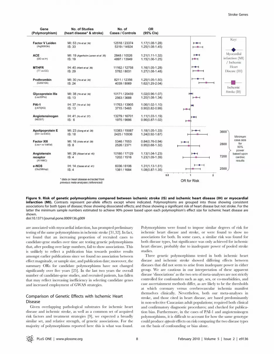
Comparison with Ischemic Heart DiseaseMeta-analyses in myocardial infarction and/or ischemic heart
disease were found [13–22] for thirteen of the genetic polymor-
phisms tested in ischemic stroke for which there were .1000
pooled cases. Comparing odds ratios for each genetic polymor-
phism between ischemic cardiac disease and ischemic stroke
identified four profiles (Figure 9):
1) Polymorphisms associated with risk of both ischemic heart
disease and stroke: factor V Leiden Gln506, ACE I/D,
MTHFR C677T, prothrombin G20210A. The 95%
confidence intervals (CIs) for ORs of ischemic stroke and
cardiac disease overlapped for all of these gene variants.
2) Polymorphisms associated with risk of either ischemic stroke
or myocardial ischemia but not the other disease type – i.e.
dissociations: glycoprotein IIIa Leu33Pro conferring a risk
for stroke, but not ischemic heart disease; PAI-1 4G- versus-
5G associated positively with cardiac disease, but negatively
with stroke, and angiotensinogen M235T posing a risk for
coronary stenosis, but a trend for protection against stroke.
The 95% CIs for risk for all three polymorphisms were non-
overlapping comparing ischemic stroke with ischemic heart
disease, although restricting cardiac datasets to those in
which Caucasian ethnicity was definitely specified results in
marginal overlap for glycoprotein IIIa Leu33Pro (ischemic
heart disease OR: 0.99 – 1.09) and angiotensinogen M235T
(coronary stenosis OR: 1.0 – 1.16).
3) Polymorphisms associated with myocardial ischemia but not
ischemic stroke, but where confidence intervals clearly
overlapped. In these cases, we calculated the minimum case
number required for 90% power (and a= 0.05) assuming
the same effect size as in ischemic heart disease. This showed
that in all cases an inadequate sample size in stroke may
account for the lack of significance here (Figure 9; third
section, final column).
4) Polymorphisms not associated with either ischemic stroke or
heart disease yet tested in .1000 cases of each disease viz.
interleukin-6 G174C [21] and HFE C282Y [22].
Comparison with Biochemically-Predicted RiskFor each of the stroke-associated genetic polymorphisms identified
by our primary meta-analysis we searched for: 1) studies providing
differential measurements of a biochemical marker - i.e. an
intermediate phenotype, IP - related to the genotypic variants of
interest, among subjects without cardiovascular disease, and 2) studies
providing an estimate of ischemic stroke risk based upon incremental
change in the same IP. Such studies were available for the following
polymorphism – IP pairs: factor V Leiden - activated protein C
resistance, ACE D/I - ACE activity, MTHFR - homocysteine
levels, prothrombin G20210A - prothrombin levels, and PAI-1 4G/
5G - PAI-1 levels.
The number of studies providing differential IP levels according to
genotype ranged between 3 and 70, incorporating between 843 and
46,743 healthy subjects, for each genotype of interest. We performed
meta-analyses that pooled these biomarker level differences for each
Figure 2. Forest plots showing positive associations of ischemic stroke with the following genetic polymorphisms: ACE D/I.doi:10.1371/journal.pone.0009136.g002
Stroke Genes
PLoS ONE | www.plosone.org 3 February 2010 | Volume 5 | Issue 2 | e9136

of the six genotype – IP pairs (Figures 10 – 15), with the weighted
mean differences, and 95% CIs, summarised in Figure 16 (2nd
column).
The number of studies providing estimates of ischemic stroke
risk relative to incremental changes in the above IPs ranged
between 1 and 9, incorporating between 245 and 1688 cases, and
459 and 30574 controls. Where more than one study existed that
provided an estimate of IP – stroke risk, we performed a meta-
analysis that combined these ORs (Figures 10 and 13 – 15; lower
forest plots), whilst first adjusting each value to the pooled estimate
of biomarker level change for the genotype comparison of interest
(see above), assuming a log-linear relationship. In the case of ACE
activity and homocysteine levels, a single estimate of biomarker -
stroke risk was extracted from a single study [23], and previous
meta-analysis [24], respectively, and then scaled in the same way.
The results of these calculations for expected risk of the genotype
comparisons of interest (Figure 16, 4th column) were then
compared with the observed risk for each of the same genotype
contrasts, based upon data used in the first part of this paper (5th
column).
As an example, Factor V Leiden 506 mutation ArgGln, relative
to wild-type ArgArg, is associated with a weighted mean average of
0.74 APTT ratio decrease in the activated protein C resistance test
in healthy subjects (Figure 10, upper). Independent data from
ischemic stroke case – control studies suggest that a 0.74 decrease
in APTT ratio using the same test is associated with a risk odds-
ratio of 1.31 (1.11 – 1.54) (Figure 10, lower). This estimated OR is
very close to the observed OR of 1.30 (1.11 – 1.51) noted from our
earlier meta-analysis (Figure 1).
For each of the gene – biomarker pairs (Figure 16), the mean
level of expected risk fell close to, and certainly within the 95%
confidence intervals, the observed risk, with the exception of PAI-
1. For this latter pairing, the expected risk of the genotype
comparison 5G5G vs 4G4G based upon PAI-1 levels was
significantly lower than that observed. This arose because whilst
PAI-1 levels are found to be higher in stroke than in controls, the
5G5G genotype – that itself is associated with elevated stroke risk
relative to 4G4G - was associated with a ,25% reduction in PAI-1
levels relative to 4G4G. This result was unaffected by whether
PAI-1 levels were derived from antigen concentration or enzyme
activity data. We also estimated expected stroke risk on the basis of
tPA levels, and its relationship with the PAI-1 4G/5G genotype.
The 5G5G relative to 4G4G genotype was associated with a slight,
albeit non-significant, increase in tPA levels, which itself is
independently associated with stroke. However, the expected
stroke risk using tPA data was also less than that observed.
Discussion
The current study takes a meta-analytic approach to identify the
totality of candidate genetic polymorphisms reliably associated
Figure 3. Forest plots showing positive associations of ischemic stroke with the following genetic polymorphisms: MTHFR C677T.doi:10.1371/journal.pone.0009136.g003
Stroke Genes
PLoS ONE | www.plosone.org 4 February 2010 | Volume 5 | Issue 2 | e9136

with ischemic stroke. Furthermore, we show how these results
compare with the same polymorphisms in ischemic cardiac
disease, and test their causal relationship with likely biochemical
intermediaries. These points are discussed in turn.
Meta-Analysis of Genetic Polymorphisms in IschemicStroke
Since our last comprehensive meta-analysis of gene effects in
ischemic stroke [25], the size of the pooled study sample from
Figure 4. Forest plots showing positive associations of ischemic stroke with the following genetic polymorphisms: prothrombinG20210A.doi:10.1371/journal.pone.0009136.g004
Figure 5. Forest plots showing positive associations of ischemic stroke with the following genetic polymorphisms: PAI 5G allele.doi:10.1371/journal.pone.0009136.g005
Stroke Genes
PLoS ONE | www.plosone.org 5 February 2010 | Volume 5 | Issue 2 | e9136
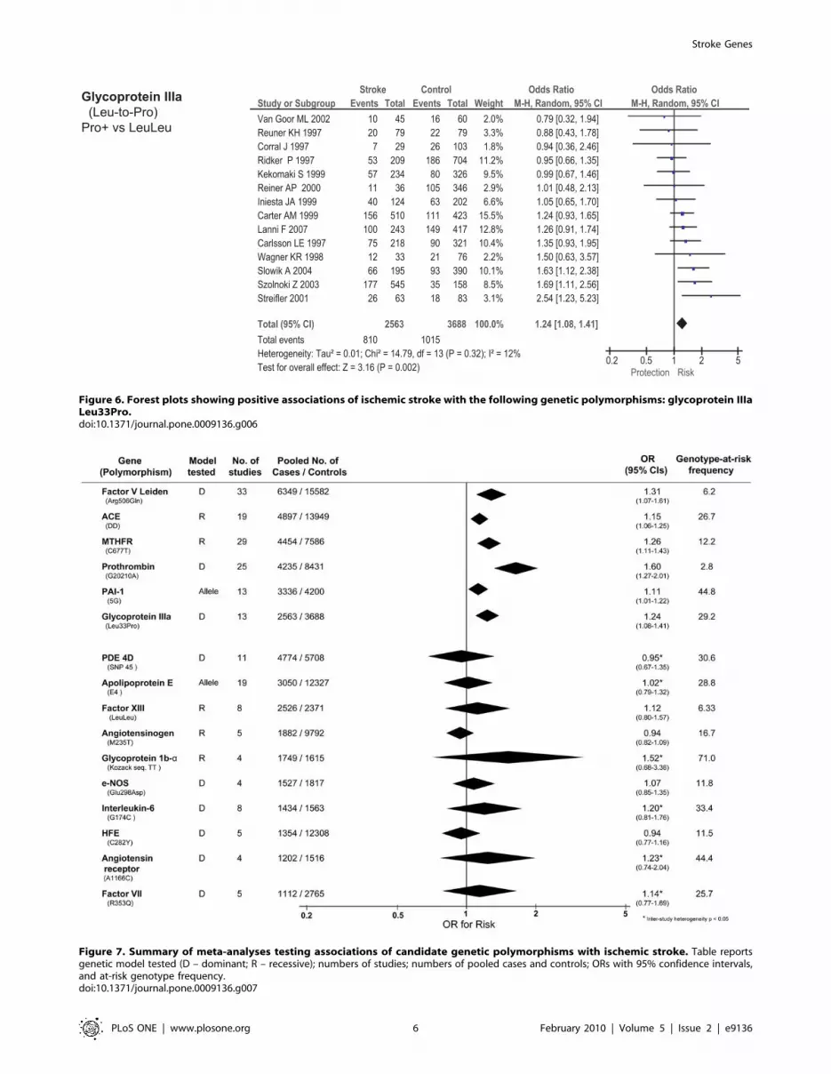
Figure 6. Forest plots showing positive associations of ischemic stroke with the following genetic polymorphisms: glycoprotein IIIaLeu33Pro.doi:10.1371/journal.pone.0009136.g006
Figure 7. Summary of meta-analyses testing associations of candidate genetic polymorphisms with ischemic stroke. Table reportsgenetic model tested (D – dominant; R – recessive); numbers of studies; numbers of pooled cases and controls; ORs with 95% confidence intervals,and at-risk genotype frequency.doi:10.1371/journal.pone.0009136.g007
Stroke Genes
PLoS ONE | www.plosone.org 6 February 2010 | Volume 5 | Issue 2 | e9136

which we were able to extract genetic polymorphism frequency
data has more than doubled (from ,18000 to ,37000 cases), as
has the number of genetic polymorphisms in which more than
1000 cases have been tested for each (from 8 to 23). In order to
increase reliability [5] we confined our results to gene variants
studied in over 1000 patients, and to studies where ischemic stroke
was confirmed radiologically, and that were based predominantly
in Caucasian adults. We discounted studies where there was
publication bias, and applied random-effects models to allow for
between-study heterogeneity. After applying these criteria we
found positive associations with polymorphisms in the following six
genes: Factor V Leiden, ACE, MTHFR, prothrombin, PAI-1, and
glycoprotein-III.
As well as identifying two further genetic associations not
identified reliably in our earlier meta-analysis [25], our study
supports the validity of the meta-analysis technique by finding very
similar effect sizes for the four positive associations identified
previously in the face of an increase in patient numbers by
approximately 50%. There was no reduction in effect size
comparing the current results with those from five years earlier
[25] as is sometimes observed [26]. A difference in our result for
the PAI-1 polymorphism relative to a recent meta-analysis [27]
can be explained by our more rigorous inclusion criteria that
restricted data to Caucasian-predominant populations [28], and
confirmed cerebral infarcts [29].
Whilst the effect size of each positive gene association was small
(odd ratios of 1.11 to 1.60), the overall contribution that genetic
factors make towards stroke is likely to be relatively large given the
frequency of these risk variants in the general population (from 3
to 45% each). The sum of the population attributable risks across
all the gene associations identified here was ,30%. These results
are in keeping with models of common complex diseases in which
relatively small numbers of common polymorphisms, each with
only small hazard ratios, can account for large proportions of
population attributable risk [30]. By contrast, certain well-
described single-gene mutations may confer a high relative risk
of stroke, e.g. CADASIL, but contribute very little to overall stroke
occurrence by virtue of their rarity.
It is likely that future discovery of disease-associated genes will rest
increasingly with genome-wide association studies (GWAS), rather
than candidate-gene strategies [9]. For example, the recent finding
from genome-wide searches that polymorphisms on chromosome 9p
Figure 8. Trends in success of candidate gene approach. A: Numbers of pooled cases published over time testing for polymorphismspositively associated with stroke according to the present meta-analyses. B: As for A, but for polymorphisms found to show no stroke associationaccording to the present meta-analysis. C: Changes in time of probability that cases were tested for polymorphism subsequently found to showassociation (red) or no association (green) with stroke.doi:10.1371/journal.pone.0009136.g008
Stroke Genes
PLoS ONE | www.plosone.org 7 February 2010 | Volume 5 | Issue 2 | e9136
are associated with myocardial infarction, has prompted preliminary
testing of the same polymorphisms in ischemic stroke [31,32]. In fact,
we found that an increasing proportion of recruited cases to
candidate-gene studies over time are testing genetic polymorphisms
that, after pooling over large numbers, fail to show associations. This
is unlikely to reflect a publication bias towards positive results
amongst earlier publications since we found no association between
effect magnitude, or sample size, and publication date; moreover, the
summary ORs for candidate polymorphisms have not changed
significantly over five years [25]. In the last two years the overall
number of candidate-gene studies, and recruited patients, has fallen
that may reflect increasing inefficiency in selecting candidate genes
and increased employment of GWAS strategies.
Comparison of Genetic Effects with Ischemic HeartDisease
Given overlapping pathological substrates for ischemic heart
disease and ischemic stroke, as well as a common set of acquired
risk factors and treatment strategies [9], we expected a broadly
similar set, and relative strength, of genetic associations. For the
majority of polymorphisms reported here this is what was found.
Polymorphisms were found to impose similar degrees of risk for
ischemic heart disease and stroke, or were found to show no
associations for both. In some cases, a similar risk was found for
both disease types, but significance was only achieved for ischemic
heart disease, probably due to inadequate power of pooled stroke
studies.
Three genetic polymorphisms tested in both ischemic heart
disease and ischemic stroke showed differing effects between
diseases that did not seem to arise from inadequate power in either
group. We are cautious in our interpretation of these apparent
disease ‘dissociations’ as the two sets of meta-analyses are not strictly
controlled for confounders such as age, sex, or co-morbidities, and
case ascertainment methods differ, as are likely to be the thresholds
at which coronary versus cerebrovascular ischemia manifest
themselves clinically. Nevertheless, both our meta-analyses in
stroke, and those cited in heart disease, are based predominantly
in non-selective Caucasian adult populations; required both clinical
and confirmatory diagnostic procedures; and checked for publica-
tion bias. Furthermore, in the cases of PAI-1 and angiotensinogen
polymorphisms, it is difficult to account for how the same genotype
could produce opposite effects on risk comparing the two disease types
on the basis of confounding or bias alone.
Figure 9. Risk of genetic polymorphisms compared between ischemic stroke (IS) and ischemic heart disease (IH) or myocardialinfarction (MI). Contrasts represent per-allele effects except where indicated. Polymorphisms are grouped into those showing consistentassociations for both types of disease; those showing dissociated effects; and those showing a significant risk of heart disease but not stroke. For thelatter the minimum sample numbers estimated to achieve 90% power based upon each polymorphism’s effect size for ischemic heart disease areshown.doi:10.1371/journal.pone.0009136.g009
Stroke Genes
PLoS ONE | www.plosone.org 8 February 2010 | Volume 5 | Issue 2 | e9136

Further studies, ideally within the same populations, will be
needed to confirm whether these three gene-disease dissociations
are real, or whether they reflect methodological differences. The
fact that these dissociations relate to proteins in three different
physiological systems, viz. platelets (glycoprotein IIIa), clotting
(PAI-1) and blood pressure (angiotensinogen) - for which there also
polymorphisms showing concordant disease effects, e.g. glycopro-
tein 1b-alpha, factor V Leiden and ACE, respectively – suggests
that differences in risk-profile between ischemic heart disease and
stroke cannot be attributed simply to a single pathophysiology e.g.
hypertension. Differences between ischemic stroke and heart
disease in terms of these three pathophysiologies has support from
Figure 10. Forest plots showing quantitative relationship between genetic polymorphisms and associated biochemical variablesfor: Factor V Leiden and activated Protein C resistance ratio. Additional forest plots are shown in Figures 10, and 13– 15 that relate setchanges in biochemical variables (determined from the first set of meta-analyses within each figure) with risk of stroke. For MTHFR and ACE thisrelationship is determined from a single study each.doi:10.1371/journal.pone.0009136.g010
Figure 11. Forest plots showing quantitative relationship between genetic polymorphisms and associated biochemical variablesfor: ACE D/I and ACE activity. Additional forest plots are shown in Figures 10, and 13– 15 that relate set changes in biochemical variables(determined from the first set of meta-analyses within each figure) with risk of stroke. For MTHFR and ACE this relationship is determined from asingle study each.doi:10.1371/journal.pone.0009136.g011
Stroke Genes
PLoS ONE | www.plosone.org 9 February 2010 | Volume 5 | Issue 2 | e9136

other sources. For example, ischemic stroke relative to ischemic
heart disease has a stronger relationship with hypertension
[33,34], whilst the protective profile of anti-platelet and throm-
bolytic drugs differs between these two diseases [9,35]. Genetic
influences on ischemia may also differ according to vessel size [10].
Comparison of Genetic Effects with Biochemical Markersof Risk
For each of the positive gene associations with ischemic stroke
that we identified, we performed further analyses using separate
data to establish whether the putative biochemical intermediaries
of these gene variants are associated with equivalent quantitative
levels of risk. The method used here was based upon mendelian
randomization in which one starts with an observational
association between an environmental (e.g. biochemical) factor
and disease, and then secondarily investigates whether a
concordant level of risk occurs for a genotype that simulates the
environmental factor - thereby making a stronger case for the
factor being causative of the disease [36]. In the current paper, we
start with positive gene-stroke associations and then subsequently
interrogate independent biochemical data with the expectation of
finding concordant levels of risk.
For the four strongest positive gene-stroke associations– factor V
Leiden, MTHFR, ACE and prothrombin - concordance between
observed risk and that predicted from their associated biochemical
changes was found. Moreover, the mean levels of predicted and
observed risks for these genotypes lay very close to each other.
Importantly therefore, we show here, for the first time, that the
Figure 12. Forest plots showing quantitative relationship between genetic polymorphisms and associated biochemical variablesfor: MTHFR and homocysteine levels. Additional forest plots are shown in Figures 10, and 13– 15 that relate set changes in biochemical variables(determined from the first set of meta-analyses within each figure) with risk of stroke. For MTHFR and ACE this relationship is determined from asingle study each.doi:10.1371/journal.pone.0009136.g012
Stroke Genes
PLoS ONE | www.plosone.org 10 February 2010 | Volume 5 | Issue 2 | e9136

four genetic variants most reliably associated with ischemic stroke
are also associated with biochemical changes that themselves are
related to equivalent levels of risk. This concordance both validates
our original gene – stroke positive associations, and furthermore,
suggests that the risk imparted by each genotype variant is the
direct consequences of each gene’s understood biochemical
actions. The fact that each of these genes exerts biochemical or
haematological changes that are measurable systemically (i.e. from
venous plasma samples), rather than being specifically cerebro-
vascularly based, is in keeping with our other finding that these
four gene variants exert similar levels of risk on ischemic heart
disease as on ischemic stroke. In the case of the concordant
relationship between MTHFR genotype and homocysteine in
their separate associations with stroke risk, we have replicated our
earlier findings in the face of more than a fourfold increase in
meta-analysis size [11].
In contrast to concordant gene-biochemical risk estimates
observed for the four largest genetic associations, we observed a
discordant gene – biochemical relationship for the PAI-1 4G/5G
polymorphism. Specifically, the variant 5G5G, relative to 4G4G,
was associated with an elevated risk of ischemic stroke, but decreased
PAI levels. However, in separate case-control studies, stroke - as well
as atherothrombosis and ischemic cardiac disease [37,38] - is
associated with increased PAI-1 levels. Indeed we saw earlier how in
ischemic heart disease, it is the 4G allele - associated with higher
PAI-1 levels – that is associated with risk. The expected risk of stroke
of the 4G/5G PAI polymorphism was also less than that observed
using tPA levels, that are strongly influenced by PAI-1 levels.
There are several possible explanations for the apparent PAI-1
gene-biochemical paradox in the case of stroke. Firstly, it is possible
that the PAI-1 5G allele association with stroke is false, e.g. because
of reporting bias. However, it is unclear why such a false association
should emerge in the opposite direction to that expected from
ischemic cardiac disease and PAI-1 level data, and Egger’s
regression test argues against publication bias here [12]. Secondly,
the finding that 5G polymorphism depresses PAI-1 levels over the
course of the subject’s life may have different pathophysiological
implications, e.g. by predisposing to stroke, than the same
depression of PAI-1 levels found at a single point in later life, when
this is found to be protective [36]. A third possibility is that the PAI-
1 genetic 4G/5G polymorphism is associated with a brain-specific
factor that influences stroke more strongly than does its actions on
PAI-1 levels. This may occur because of linkage disequilibrium with
an as-yet unidentified gene [39], or because of pleiotropy [36]. For
example, while raised PAI-1 levels in plasma may raise thrombotic
risk, raised PAI-1 levels in carotid vessel wall may serve to stabilise
atheromatous plaques [40]. If the PAI 4G/5G polymorphism
influences both tissue and plasma PAI-1 levels, whilst the latter is
also unduly influenced by environmental variables [41], this could
explain the paradox. Furthermore, postulating that PAI-1 4G/5G
exerts different phenotypic effects on cerebral versus other vascular
beds might explain why its influences on ischemic stroke and
ischemic heart disease are in opposite directions.
ConclusionThe current study provides a comprehensive meta-analysis of
common genetic polymorphisms associated with ischemic stroke
that were identified through the candidate gene approach. The
results serve as an important comparator to emerging genome-wide
association studies [9,31,32], which would be expected to converge
Figure 13. Forest plots showing quantitative relationship between genetic polymorphisms and associated biochemical variablesfor: Prothrombin G20210A and prothrombin levels. Additional forest plots are shown in Figures 10, and 13– 15 that relate set changes inbiochemical variables (determined from the first set of meta-analyses within each figure) with risk of stroke. For MTHFR and ACE this relationship isdetermined from a single study each.doi:10.1371/journal.pone.0009136.g013
Stroke Genes
PLoS ONE | www.plosone.org 11 February 2010 | Volume 5 | Issue 2 | e9136

upon similar genetic associations to those shown here, as well as to
identify genes not previously implicated with cardiovascular disease
pathogenesis. Since a major purpose of elucidating genetic
influences on stroke (as for any complex disease) is to gain insights
into its pathophysiology, we show here how pooled gene association
data can be meaningfully compared with separate data relating the
same genetic effects with both a pathophysiologically-related disease
(here, ischemic cardiac disease) and biochemical intermediaries.
Methods
1: Genetic Polymorphism – Ischemic Stroke AssociationMeta-Analysis
Electronic databases (Medline; EMBASE; Google Scholar) were
searched upto January 1, 2009 for all case-control studies
evaluating any candidate genetic polymorphism in ischemic
stroke. Search words used were: cerebrovascular disease, brain infarction,
stroke and cerebral ischemia in combination with polymorphism, genetic,
mutation, genotype and genes. All languages were searched and
translated when necessary. Additional studies were sought from
references, citations and from the PubMed option ‘Related
Articles’, for each identified study.
Inclusion criteria were studies that: 1) employed case-control
methods where ischemic stroke was analyzed as a dichotomous trait;
both retrospective and prospective cohort designs were included; 2)
confirmed the diagnosis of ischemic stroke with neuroimaging, and
3) were based in Caucasian populations, so as to minimise inter-
racial heterogeneity [28]. Studies were excluded if: 1) patients were
aged under 18 years; 2) only quantitative traits or intermediate
phenotypes were being investigated, or 3) genotype frequency was
not reported. For duplicate publications, the smaller data set(s) were
discarded. For studies with more than one control group, the most
appropriate control group was used.
Data were analyzed using software for preparing Cochrane
reviews (Review Manager, v5; Comprehensive Meta Analysis
v2.2.023). For each genetic polymorphism for which data were
Figure 14. Forest plots showing quantitative relationship between genetic polymorphisms and associated biochemical variablesfor: PAI-1 5G/4G and PAI-1 levels. Additional forest plots are shown in Figures 10, and 13– 15 that relate set changes in biochemical variables(determined from the first set of meta-analyses within each figure) with risk of stroke. For MTHFR and ACE this relationship is determined from asingle study each.doi:10.1371/journal.pone.0009136.g014
Stroke Genes
PLoS ONE | www.plosone.org 12 February 2010 | Volume 5 | Issue 2 | e9136

available from at least two studies, a meta-analysis was carried out.
For each gene variant, a pooled odds ratio (OR) and 95% confidence
intervals were calculated using random-effects [42] models, to
estimate association strength. Tests for heterogeneity [43], and
publication bias [12] were performed with significance set at
p,0.05. Random-effects results are reported throughout. In order to
improve reliability, we only present here results for polymorphisms
tested in .1000 pooled cases [5]. The proportion of cases in the
population that could be attributed to a particular genetic variant
(population attributable risk or PAR) was estimated as follows:
PAR = 100 6 [Prevalence(OR – 1)/(Prevalence (OR – 1)+1)]. The
prevalence of exposure was estimated as the genotype frequency
among pooled controls.
We also sought to determine temporal trends in the success of the
candidate-gene approach by determining the pooled number of cases
recruited into studies that tested for polymorphisms that were either
associated, or not associated, with stroke according to our meta-
analysis, and then plotting this against study publication year.
Moreover, for every publication year, we calculated the probability
that cases were tested for a polymorphism that demonstrated either a
significant, or no, association by our meta-analysis. This analysis is
restricted to polymorphisms for which there were .1000 pooled cases.
2: Ischemic Stroke – Ischemic Heart Disease GeneticComparison
For each genetic polymorphism tested in ischemic stroke
(with .1000 cases), we searched the literature for the most recent
meta-analysis testing for the same polymorphism in myocardial
infarction and/or ischemic heart disease, based predominantly in
Caucasian populations. For all polymorphisms showing a positive
association with ischemic stroke there was an equivalent published
meta-analysis in ischemic heart disease/myocardial infarction. For
each polymorphism for which a cardiac meta-analysis existed we
calculated the pooled random-effects OR of ischemic stroke for the
equivalent genotype (or per-allele) comparison using our own
meta-analysis.
3: Gene – Intermediate Phenotype ComparisonFor each positive genetic association identified from the meta-
analysis in part 1, we performed a separate analysis that produced
an estimate of expected risk based upon genotype – biochemical,
and biochemical – stroke, association studies, using the principle of
mendelian randomisation [11]. Firstly, we searched the medical
literature for two further types of study: 1) those relating each
genetic polymorphism with a quantitative measure of the most
strongly-associated biochemical or hematological marker - i.e. an
intermediate phenotype, IP, for the gene in question - in
populations free from cardiovascular disease, and 2) those relating
an incremental change of the same IP with risk of ischemic stroke.
For example, for the ACE polymorphism we searched for: 1) (ACE
OR angiotensin converting enzyme) AND (gene OR genetic OR genotype
OR polymorphism OR mutation), in combination with (ACE OR
angiotensin converting enzyme) AND (activity OR level); and 2) (ACE OR
angiotensin converting enzyme) AND (activity OR level) in conjunction
Figure 15. Forest plots showing quantitative relationship between genetic polymorphisms and associated biochemical variablesfor: PAI-1 5G/4G and tPA levels. Additional forest plots are shown in Figures 10, and 13– 15 that relate set changes in biochemical variables(determined from the first set of meta-analyses within each figure) with risk of stroke. For MTHFR and ACE this relationship is determined from asingle study each.doi:10.1371/journal.pone.0009136.g015
Stroke Genes
PLoS ONE | www.plosone.org 13 February 2010 | Volume 5 | Issue 2 | e9136

with (cerebrovascular disease OR brain infarction OR stroke OR cerebral
ischemia). As for part 1, we restricted studies to those based in adults
from predominantly Caucasian populations.
From these two sets of studies we performed two types of
analyses: 1) From the first set of studies we extracted from each the
difference in levels (or biological activity) of the IP between the two
homozygous variants, or between the wild-type and heterozygous
variants in cases of dominant polymorphisms. These values were
entered into a meta-analysis to obtain a weighted mean average
using a random-effects model. For those IPs where different
measurement units had been employed between studies (viz. ACE
activity; prothrombin levels, plasminogen-activator inhibitor-1
levels and tissue plasminogen activator levels) we calculated the
percentage change for the rarer, relative to the commoner,
genotype. 2) From the second set of studies we obtained an OR of
ischemic stroke for a given change in the level of that IP used in
the first analysis. Where studies reported different ORs for
different ranges of IP level, we chose the OR reported for the
range closest to that applying to the control group (i.e. healthy
population). The ORs between studies were pooled using a generic
inverse variance procedure in which the logarithms of the ORs are
weighted according to variance in a random-effects model. Since
the ORs reported between these studies usually refer to different
amounts of IP change, we first scaled the OR values for each study
in proportion to the pooled change in IP level for the genotype
comparison of interest (from the first analysis) assuming a log-
linear relationship [11]. For the relationship between homocyste-
ine levels and ischemic stroke we used a summary OR from a
previous meta-analysis [24].
Supporting Information
Figures S1 Supplementary Figures 1–10: Forest plots of genetic
polymorphisms tested in more than 1000 pooled cases but showing
no association with ischemic stroke.
Found at: doi:10.1371/journal.pone.0009136.s001 (0.08 MB
PDF)
References S1 Supplementary References
Found at: doi:10.1371/journal.pone.0009136.s002 (0.29 MB
PDF)
Acknowledgments
We are grateful for helpful suggestions from Aroon Hingorani & Juan
Pablo Casas during the course of this work.
Figure 16. Comparison of estimated risk with observed risk for ischemic stroke-associated genetic polymorphisms. Estimated ORs(green) are calculated from 1) meta-analyses relating risk-genotype with biochemical variation (DX), and 2) single studies or meta-analyses relatingbiochemical variation with stroke risk (scaled log-linearly). Observed ORs are derived from meta-analyses of the equivalent genotype contrast usingthe current datasets.doi:10.1371/journal.pone.0009136.g016
Stroke Genes
PLoS ONE | www.plosone.org 14 February 2010 | Volume 5 | Issue 2 | e9136

Author Contributions
Conceived and designed the experiments: PB LS PS. Performed the
experiments: PB GP PS. Analyzed the data: PB GP JW PS. Wrote the
paper: PB GP LS JW PS.
References
1. Johnston SC, Mendis S, Mathers CD (2009) Global variation in stroke burdenand mortality: estimates from monitoring, surveillance, and modelling. Lancet
Neurol 8: 345–354.
2. Chiuve SE, Rexrode KM, Spiegelman D, Logroscino G, Manson JE, et al.(2008) Primary prevention of stroke by healthy lifestyle. Circulation 118:
947–954.3. Arason GJ, Kramer J, Blasko B, Kolka R, Thorbjornsdottir P, et al. (2007)
Smoking and a complement gene polymorphism interact in promoting
cardiovascular disease morbidity and mortality. Clin Exp Immunol 149:132–138.
4. Arnett DK, Baird AE, Barkley RA, Basson CT, Boerwinkle E, et al. (2007)American Heart Association Council on Epidemiology and Prevention;
American Heart Association Stroke Council; Functional Genomics andTranslational Biology Interdisciplinary Working Group; Relevance of genetics
and genomics for prevention and treatment of cardiovascular disease.
Circulation 115: 2878–901.5. Ioannidis JP, Boffetta P, Little J, O’Brien TR, Uitterlinden AG, et al. (2008)
Assessment of cumulative evidence on genetic associations: interim guidelines.Int J Epidemiol 37: 120–132.
6. Fisher M, Folland E (2008) Acute ischemic coronary artery disease and ischemic
stroke: similarities and differences. Am J Ther 15: 137–149.7. Kingwell B, Boutouyrie P (2007) Genetic influences on the arterial wall. Clin
Exp Pharmacol Physiol 34: 652–657.8. Jood K, Ladenvall P, Tjarnlund-Wolf A, Ladenvall C, Andersson M, et al.
(2005) Fibrinolytic gene polymorphism and ischemic stroke. Stroke 36:
2077–2081.9. Ikram MA, Seshadri S, Bis JC, Fornage M, DeStefano AL, et al. (2009)
Genomewide association studies of stroke. N Engl J Med 360: 1718–1728.10. Rao R, Tah V, Casas JP, Hingorani A, Whittaker J, et al. (2009) Ischaemic
stroke subtypes and their genetic basis: a comprehensive meta-analysis of smalland large vessel stroke. Eur Neurol 61: 76–86.
11. Casas JP, Bautista LE, Smeeth L, Sharma P, Hingorani AD (2005)
Homocysteine and stroke: evidence on a causal link from mendelianrandomisation. Lancet 365: 224–232.
12. Egger M, Davey SG, Schneider M, Minder C (1997) Bias in meta-analysisdetected by a simple, graphical test. BMJ 315: 629–634.
13. Ye Z, Liu EH, Higgins JP, Keavney BD, Lowe GD, et al. (2006) Seven
haemostatic gene polymorphisms in coronary disease: meta-analysis of 66,155cases and 91,307 controls. Lancet 367(9511): 651–8.
14. Agerholm-Larsen B, Nordestgaard BG, Tybjaerg-Hansen A (2000) ACE genepolymorphism in cardiovascular disease: meta-analyses of small and large studies
in whites. Arterioscler Thromb Vasc Biol 20(2): 484–92.15. Klerk M, Verhoef P, Clarke R, Blom HJ, Kok FJ, et al. (2002) MTHFR Studies
Collaboration Group. MTHFR 677C--.T polymorphism and risk of coronary
heart disease: a meta-analysis. JAMA 288(16): 2023–31.16. Xu MQ, Ye Z, Hu FB, He L (2007) Quantitative assessment of the effect of
angiotensinogen gene polymorphisms on the risk of coronary heart disease.Circulation 116(12): 1356–66.
17. Song Y, Stampfer MJ, Liu S (2004) Meta-analysis: apolipoprotein E genotypes
and risk for coronary heart disease. Ann Intern Med 141(2): 137–47.18. Voko Z, Bereczky Z, Katona E, Adany R, Muszbek L (2007) Factor XIII
Val34Leu variant protects against coronary artery disease. A meta-analysis.Thromb Haemost 97(3): 458–63.
19. Ntzani EE, Rizos EC, Ioannidis JP (2007) Genetic effects versus bias forcandidate polymorphisms in myocardial infarction: case study and overview of
large-scale evidence. Am J Epidemiol 165(9): 973–84.
20. Casas JP, Bautista LE, Humphries SE, Hingorani AD (2004) Endothelial nitricoxide synthase genotype and ischemic heart disease: meta-analysis of 26 studies
involving 23028 subjects. Circulation 109(11): 1359–65.21. Sie MP, Sayed-Tabatabaei FA, Oei HH, Uitterlinden AG, Pols HA, et al. (2006)
Interleukin 6 -174 g/c promoter polymorphism and risk of coronary heart
disease: results from the Rotterdam study and a meta-analysis. ArteriosclerThromb Vasc Biol 26(1): 212–7.
22. Ellervik C, Birgens H, Tybjaerg-Hansen A, Nordestgaard BG (2007)Hemochromatosis genotypes and risk of 31 disease endpoints: meta-analyses
including 66,000 cases and 226,000 controls. Hepatology 46(4): 1071–80.
23. Brenner D, Labreuche J, Poirier O, Cambien F, Amarenco P GENICInvestigators (2005) Renin-angiotensin-aldosterone system in brain infarction
and vascular death. Ann Neurol 58(1): 131–8.
24. Wald DS, Law M, Morris JK (2002) Homocysteine and cardiovascular disease:
evidence on causality from a meta-analysis. BMJ 325: 1202.
25. Casas JP, Hingorani AD, Bautista LE, Sharma P (2004) Meta-analysis of genetic
studies in ischemic stroke: thirty-two genes involving approximately 18,000 cases
and 58,000 controls. Arch Neurol 61: 1652–1661.
26. Zintzaras E, Lau J (2008) Synthesis of genetic association studies for pertinent
gene-disease associations requires appropriate methodological and statistical
approaches. J Clin Epidemiol 61: 634–645.
27. Tsantes AE, Nikolopoulos GK, Bagos PG, Tsiara CG, Kapsimali V, et al. (2007)
Plasminogen activator inhibitor-1 4G/5G polymorphism and risk of ischemic
stroke: a meta-analysis. Blood Coagul Fibrinolysis 18: 497–504.
28. Ariyaratnam R, Casas JP, Whittaker J, Smeeth L, Hingorani AD, et al. (2007)
Genetics of ischaemic stroke among persons of non-European descent: a meta-
analysis of eight genes involving approximately 32,500 individuals. PLoS Med 4:
e131.
29. Peck G, Smeeth L, Whittaker J, Casas JP, Hingorani A, et al. (2008) The
genetics of primary haemorrhagic stroke, subarachnoid haemorrhage and
ruptured intracranial aneurysms in adults. PLoS ONE 3: e3691.
30. Yang Q, Khoury MJ, Friedman J, Little J, Flanders WD (2005) How many genes
underlie the occurrence of common complex diseases in the population?
Int J Epidemiol 34: 1129–1137.
31. Matarın M, Brown WM, Scholz S, Simon-Sanchez J, Fung HC, et al. (2007) A
genome-wide genotyping study in patients with ischemic stroke: initial analysis
and data release. Lancet Neurol 6: 414–420.
32. Karvanen J, Silander K, Kee F, Tiret L, Salomaa V, et al. (2009) MORGAM
Project. The impact of newly identified loci on coronary heart disease, stroke
and total mortality in the MORGAM prospective cohorts. Genet Epidemiol 33:
237–246.
33. Kannel WB, Wolf PA, Verter J, McNamara PM (1970) Epidemiologic
assessment of the role of blood pressure in stroke. The Framingham study.
JAMA 214: 301–310.
34. Law MR, Morris JK, Wald NJ (2009) Use of blood pressure lowering drugs in
the prevention of cardiovascular disease: meta-analysis of 147 randomised trials
in the context of expectations from prospective epidemiological studies. BMJ
338: b1665.
35. Wolff T, Miller T, Ko S (2009) Aspirin for the primary prevention of
cardiovascular events: an update of the evidence for the U.S. Preventive Services
Task Force. Ann Intern Med 150: 405–410.
36. Davey Smith G, Ebrahim S (2003) ‘Mendelian randomization’: can genetic
epidemiology contribute to understanding environmental determinants of
disease? Int J Epidemiol 32: 1–22.
37. Hamsten A, de Faire U, Walldius G, Dahlen G, Szamosi A, et al. (1987)
Plasminogen activator inhibitor in plasma: risk factor for recurrent myocardial
infarction. Lancet 2: 3–9.
38. Thogersen AM, Jansson JH, Boman K, Nilsson TK, Weinehall L, et al. (1998)
High plasminogen activator inhibitor and tissue plasminogen activator levels in
plasma precede a first acute myocardial infarction in both men and women:
evidence for the fibrinolytic system as an independent primary risk factor.
Circulation 98: 2241–2247.
39. Attia J, Thakkinstian A, Wang Y, Lincz L, Parsons M, et al. (2007) The PAI-1
4G/5G gene polymorphism and ischemic stroke: an association study and meta-
analysis. J Stroke Cerebrovasc Dis 16: 173–179.
40. Hoekstra T, Geleijnse JM, Kluft C, Giltay EJ, Kok FJ, et al. (2003) 4G/4G
genotype of PAI-1 gene is associated with reduced risk of stroke in elderly. Stroke
34: 2822–2828.
41. Henry M, Tregouet DA, Alessi MC, Aillaud MF, Visvikis S, et al. (1998)
Metabolic determinants are much more important than genetic polymorphisms
in determining the PAI-1 activity and antigen plasma concentrations: a family
study with part of the Stanislas Cohort. Arterioscler Thromb Vasc Biol 18:
84–91.
42. DerSimonian R, Laird N (1986) Meta-analysis in clinical trials. Control Clin
Trials 7: 177–188.
43. Deeks JJ, Altman DG, Bradburn MJ (2001) Statistical methods for examining
heterogeneity and combining results from several studies in a meta-analysis. In:
Egger M, Davey Smith G, Altman DG, eds. Systematic Reviews in Health Care:
Meta-analysis in Context. Annapolis Junction, Md: BMJ Publishing Group;
2001.
Stroke Genes
PLoS ONE | www.plosone.org 15 February 2010 | Volume 5 | Issue 2 | e9136


