Embed Size (px)
Citation preview
Vaccine 23 (2004) 769–779
CD80 and CD86, but not CD154, augment DNA vaccine-inducedprotection in experimental bovine tuberculosis
Alexander C. Mauea,e, W. Ray Watersb, Mitchell V. Palmerb, Diana L. Whippleb,F. Chris Minionc, Wendy C. Brownd, D. Mark Estesa,e,∗
a Department of Molecular Microbiology and Immunology, University of Missouri, Columbia, MO 65211, USAb Bacterial Diseases of Livestock Research Unit, United States Department of Agriculture, Agricultural Research Service,
National Animal Disease Center, Ames, IA 50010, USAc College of Veterinary Medicine, Veterinary Microbiology and Preventive Medicine, Iowa State University, Ames, IA 50011, USA
d Department of Veterinary Microbiology and Pathology, College of Veterinary Medicine, Washington State University, Pullman, WA 99164, USAe Program for the Prevention of Animal Infectious Diseases, Department of Veterinary Pathobiology, University of Missouri, Columbia, MO 65211, USA
Received 20 May 2004; received in revised form 2 July 2004; accepted 7 July 2004Available online 9 August 2004
A
imulatorym odels. Ther rate that thec erculosis,eB determinet 154. Co-a AT-6D f the lungsa ctively, theser e responsest l bovinet©
K
1
m
tBT
-gesse of
flex-etheir
sme
0d
bstract
DNA vaccination is known to elicit robust cellular and humoral responses to encoded antigen. The co-administration of costolecules CD80 (B7-1), CD86 (B7-2) and CD154 (CD40L) has been shown to enhance immune responses in several murine m
ole of specific costimulatory molecules in non-rodent species remains incompletely characterized. In these studies, we demonsto-administration of CD80 and CD86, but not CD154, to an existing candidate subunit DNA vaccine (ESAT-6) against bovine tubnhances protection after aerosol challenge with virulentMycobacterium bovis. Additionally, we have shown that vaccination withM. bovisCG is protective against tuberculosis following aerosol challenge in cattle. Two independent trials were conducted in cattle to
he adjuvant effect of encoded antigen + CD80/CD86 and directly compare the adjuvant activities of CD80/CD86 to those of CDdministration of either CD80/CD86 or CD154 enhanced ESAT-6-specific IFN-� responses as compared to animals vaccinated with ESNA alone. However, following aerosol challenge, only animals vaccinated with CD80/CD86 possessed decreased pathology ond associated lymph nodes, as measured by gross examination, radiographic lesion morphometry and bacterial recovery. Colleesults demonstrate that the co-administration of costimulatory molecules with a protective antigen target enhances bovine immuno DNA vaccination, and that CD80/CD86 is superior to CD154 in augmenting DNA vaccine-induced protection in experimentauberculosis.
2004 Elsevier Ltd. All rights reserved.
eywords:Costimulatory molecules; CpG ODN; Adjuvant
. Introduction
DNA vaccination has been shown to generate cell-ediated and humoral immune responses to encoded antigen
∗ Corresponding author. Present address: Department of Pediatrics andhe Sealy Center for Vaccine Development, University of Texas Medicalranch, 2.330F Children’s Hospital, 301 University Boulevard, Galveston,X 77555, USA. Tel.: +1 409 772 0434; fax: +1 409 772 0460.
E-mail address:[email protected] (D.M. Estes).
in several different species[1–4]. In addition to the induction of immunity, DNA vaccination offers several advantawhen compared to conventional vaccines, such as eaproduction, stability, cost effectiveness and the overallibility of the vaccine platform[1,2,4,5]. DNA vaccines aralso attractive candidates for disease prevention due toability to potentially enhance immunity via CD8+ T cellsthrough the MHC class I pathway[1,2,4]. Several studiein mice have shown that DNA vaccination provides solevel of protective immunity against infectious agents[2,6].
264-410X/$ – see front matter © 2004 Elsevier Ltd. All rights reserved.oi:10.1016/j.vaccine.2004.07.019
770 A.C. Maue et al. / Vaccine 23 (2004) 769–779
While these experiments have demonstrated the utility ofDNA vaccination as a means of eliciting cell-mediated andhumoral responses, it has been difficult to generate compa-rable immune responses to DNA vaccines in humans andlarge animals[3,5,7]. Progress towards successful DNA vac-cines in non-rodent species has been hampered by severalfactors. DNA vaccination of large animals often requiresrelatively large amounts of plasmid DNA administered inmultiple doses[8–10]. Previous studies have also shown thatindividuals vaccinated with DNA are relatively low respon-ders[8–10]. Additionally, there remains relatively little ef-ficacy data on DNA vaccines following infectious challengedue to the relatively high cost of large animal studies. Re-cently, studies in cattle have shown that co-administrationof plasmid-encoded FLT-3L and GM-CSF enhanced bovineCD4+ T cell responses to DNA vaccination[11]. Similarly,plasmid-encoded bovine CD40L (CD154) used as an adju-vant and vaccine-targeting molecule, increased antibody re-sponses to bovine herpesvirus 1 glycoprotein D in sheep[12].CpG oligodinucleotides (ODN), when used in the correctspecies-specific context[13], have been shown to possess ad-juvant activity by inducing the production of inflammatorycytokines from bovine peripheral blood mononuclear cells(PBMC) in vitro [14]. Thus, the co-administration of molec-ular adjuvants may represent a means of enhancing immuner
ciesh e re-s roleo ru-m ctiveo dedb cedc inet D28s cci-n arc ucle-o ic Tl nsesm pro-t ec , thusr ulesc resseb e-s chim-p es toH las-m thec o en-h ,f rdingt vac-c
After assessing the potential of CD80 and CD86 asmolecular adjuvants, we directly compared the effects ofco-administering plasmid-encoded CD154 against encodedCD80 and CD86. CD154 is expressed rapidly on T cellsfollowing activation[21,22] and has a role in the genera-tion of both cell-mediated and thymus dependent humoralresponses, as well as regulation of antigen presenting cell(APC) activity [23,24]. Ligation of CD40 by CD154 stim-ulates immature APCs to differentiate, produce cytokinesand upregulate the expression of costimulatory molecules[23,24], thus creating a more efficient APC. Several ro-dent studies have examined the effect of plasmid-expressedCD154 on DNA vaccination. Mendoza et al.[25] found thatthe co-administration of CD154 enhanced IgG2a productionand CTL responses to plasmid-encoded�-galactosidase, sug-gesting a role in augmenting cell-mediated responses. Simi-larly, mice vaccinated with a HIV DNA vaccine and a CD154expression plasmid possessed antigen-specific increases inIgG2a and IFN-� production and CTL activity[26]. The lat-ter study also determined that co-administration of CD154 in-creased antigen-specific Th2 responses. The aforementionedstudy in ruminants regarding CD154 and DNA vaccinationrevealed the potential of CD154 as an adjuvant to enhancehumoral responses[12], but to date, little data has been pub-lished examining the effect of CD154 on cell-mediated re-s .
v uirec nity.R inm tec-t T-6)pii AT-6 cedb[ argea ine,C s us-i sultss dingC s ap nsesi
2
2
byD on,U byD on,U idsh r
esponses to DNA vaccination in non-rodent species.While previous studies in outbred large animal spe
ave concluded that it is possible to enhance immunponses to DNA vaccination using various strategies, thef costimulatory molecules in enhancing immunity ininants remains incompletely characterized. The objef this study was to evaluate the ability of plasmid-encoovine CD80 and CD86 to augment DNA vaccine-induell-mediated immunity in a model of experimental bovuberculosis. It has been demonstrated previously that Cignaling is required for immune responses to DNA vaation[15]. In rodents, DNA vaccination using a co-lineonstruct encoding CD86 and a suboptimal influenza nprotein epitope resulted in an enhancement of cytotox
ymphocyte (CTL) activity that was >80% of the respoeen with influenza virus-infected controls[16]. Similarly,ice vaccinated with plasmid encoding HIV-1 envelope
ein possessed enhanced IFN-� and CTL responses with tho-administration of a separate plasmid encoding CD86evealing that adjuvant effects with costimulatory molecan be seen whether or not the sequences are co-expy the same construct[17,18]. Enhancement of immune rponses via CD86 costimulation has also been shown inanzees, which developed more robust T cell responsIV-1 envelope protein after receiving CD86-encoding pid [17]. Additional studies in mice have shown that
o-administration of encoded CD80 may also be used tance CTL responses to DNA vaccination[19,20]. To date
ew if any studies have been conducted in cattle regahe use of DNA-encoded CD80 and CD86 to enhanceine responses.
d
ponses in large animals following infectious challengeChallenge withMycobacterium tuberculosisor M. bo-
is represent established models of infection that reqell-mediated immune responses for protective immuoles for CD4+ and CD8+ T cells have been delineatedice, with Th1-mediated responses correlating with pro
ion[27]. The early secretory antigenic target-6kDa (ESArotein is recognized by bovine T cells early duringM. bovis
nfection[28], resulting in the release of IFN-� [29,30], mak-ng it an attractive candidate for vaccine development. ES
DNA vaccination of mice resulted in significantly reduacterial recovery following challenge withM. tuberculosis
31]. To test the utility of these combined approaches in lnimals, we vaccinated cattle with an ESAT-6 DNA vaccpG ODN and plasmid-encoded costimulatory molecule
ng a prime and single boost strategy. Collectively, our reuggest that a DNA vaccine composed of plasmids encoD80 and CD86 administered with CpG ODN provideractical means of generating improved immune respo
n large animals.
. Materials and methods
.1. Plasmid DNA, CpG ODN and rESAT-6
Plasmid encoding bovine CD80 was kindly providedr. Chris J. Howard (Institute of Animal Health, ComptK). Plasmid encoding bovine CD86 was kindly providedr. Keith R. Parsons (Institute of Animal Health, ComptK). The construction of bovine CD80 and CD154 plasmas been described previously[32,33]. The plasmid fo
A.C. Maue et al. / Vaccine 23 (2004) 769–779 771
bovine CD86 encodes the majority of the open readingframe described in GenBank (accession AJ291475 andAY533858) to 1092 nucleotides. The clone used for thesestudies lacks seven amino acids in the N-terminal region ofthe protein (residues 22–28). Expression of bovine CD80,CD86 and CD154 was confirmed in transfected COS-7cells by flow cytometry (data not shown) as previouslydescribed for dendritic cells (DC)[34]. Transfections wereperformed according to the manufacturer’s instructions(Lipofectamine, Invitrogen, Carlsbad, CA). Plasmid encod-ing ESAT-6 was constructed by amplifying the completeopen reading frame of ESAT-6 from pA using forward primer(5′-GAGAGATCTCATGACAGAGCAGCAGTGGAATTT-C-3′) and reverse primer (5′-GGCAGATCTCTATGCGA-ACATCCCAGTG-3′) as described previously[35]. Theamplified fragment was digested withBglII and was theninserted into theBamHI site of plasmid VR1012 (Vical Inc.,San Diego, CA) to generate pISM411. Plasmid DNA forimmunization was prepared by transforming plasmids intocompetentEscherichia coliusing the pcDNA3.1 DirectionalTOPO Expression cloning kit (Invitrogen, Carlsbad, CA).Gene inserts were confirmed by automated DNA sequencing(DNA Core Facility, University of Missouri). Plasmid DNAwas purified using the Qiagen Plasmid Giga kit (Qiagen,Valencia, CA) according to the manufacturer’s instructions.
tc.( -G ineP rali ap ease-r ts at− BS.
usly[ nedb enCT thea di ifiedbw mideg an-t
2
P ple-m plex( St.Lw iledd d ofm cilliw id-
dlebrook 7H11 selective media (Becton Dickinson, Cock-eysville, MD).
2.3. Animals and immunizations
2.3.1. DNA vaccine trial #1Eighteen castrated male mixed breed cattle, 400–600 lbs,
were selected based on their negative reactivity to purifiedprotein derivative (PPD) fromM. avium(PPDa) andM. bo-vis (PPDb). Briefly, whole blood was incubated for 24 h invitro in the presence of 20�g/ml PPDb and PPDa (BiocorAnimal Health, Omaha, NE). Supernatants were then har-vested and PPDb-specific IFN-� production was determinedusing the BoviGam ELISA kit (Biocor Animal Health). An-imals negative for prior exposure to mycobacteria were thenrandomly assigned to the following four experimental treat-ment groups: (1)M. bovisBCG strain Pasteur vaccination(n = 4); (2) ESAT-6 DNA vaccination (n = 5); (3) ESAT-6 + CD80/CD86 DNA vaccination (n = 4); (4) pcDNA3.1(empty plasmid) + CD80/CD86 control DNA vaccination(n = 5). DNA vaccinates were immunized intramuscularly(IM) with 2 mg of total plasmid DNA and with 1 mg of ODN2006 emulsified in 2 ml of IFA in the left mid-cervical re-gion. Cattle received an identical booster dose of vaccine 20days post-prime. BCG animals were vaccinated with a singled 7
s re-g ses.A is-s cor-d andu
20 lbs,
m basedo omM Av nedtbDv (= 54c -m Aa hel a-t .01 forB vac-c cteda r toe imalc UseC
CpG ODN were manufactured by Oligos EWilsonville, OR). ODN 2006 (TCGTCGTTTTGTCTTTGTCGTT) has been shown to stimulate bovBMC in vitro [14] and augment cellular and humo
mmune responses in vivo[36]. ODN 2006 containshosphothioate-modified backbone that renders it nuclesistant. CpG ODN were stored as lyophilized aliquo80◦C until use and then were resuspended in sterile PPurified rESAT-6 was obtained as described previo
35]. Briefly, the coding sequence for ESAT-6 was obtaiy PCR amplification and cloned into pTrcHis B (Invitrogorp., San Diego, CA) to form pISM403. Induction ofE. coliOP 10 cells containing pISM403 was accomplished byddition of 1 mM isopropyl-thio-�-d-galactopyransoide an
ncubating for 6–8 h. The recombinant protein was pury metal chelate chromatography as described[35]. Purityas assessed using sodium dodecyl sulfate polyacrylael electrophoresis and immunoblotting with anti-Xpress
ibodies (Invitrogen).
.2. Bacteria
M. bovisstrain 1315 and attenuatedM. bovisBCG strainasteur were grown in Middlebrook 7H9 media supented with 10% oleic acid–albumin–dextrose com
Difco, Detroit, MI) plus 0.05% Tween 80 (Sigma,ouis, MO) as described previously[37]. M. bovis 1315as originally isolated in 1995 from an infected white-taeer [38]. Challenge and vaccination inocula consisteid-log growth phase mycobacteria. Enumeration of baas confirmed using serial dilution plate counting on M
ose of 4.0×10 colony forming units (CFU)M. bovisBCGtrain Pasteur administered IM in the left mid-cervicalion at the time DNA vaccinates received their booster doll immunizations were conducted at the University of Mouri, Columbia. Immunizations were conducted in acance with the guidelines of the institutional animal carese policies of the University of Missouri, Columbia.
.3.2. DNA vaccine trial #2Twenty cattle (females and castrated males), 400–60
ixed breed cattle from the same herd, were selectedn their negative reactivity to purified protein derivative fr. avium(PPDa) andM. bovis(PPDb), as described in DN
accine trial 1. Naıve animals were then randomly assigo the following five experimental treatment groups: (1)M.ovisBCG strain Pasteur vaccination (n = 4); (2) ESAT-6NA vaccination (n = 4); (3) ESAT-6 + CD80/CD86 DNAaccination (n= 4); (4) ESAT-6 + CD154 DNA vaccinationn4); (5) pcDNA3.1 (empty plasmid) + CD80/CD86/CD1
ontrol DNA vaccination (n = 4). DNA vaccinates were imunized intramuscularly with 2 mg of total plasmid DNnd with 1 mg of ODN 2006 emulsified in 2 ml of IFA in t
eft mid-cervical region. At the time of the first immunizion, BCG animals were vaccinated once with a dose of 1×07 CFUM. bovisBCG strain Pasteur. All cattle, exceptCG vaccinates, received an identical booster dose ofine 20 days post-prime. All immunizations were condut the National Animal Disease Center, Ames, IA. Prioxperimentation, a protocol detailing procedures and anare was approved by the Institutional Animal Care andommittee (IACUC).
772 A.C. Maue et al. / Vaccine 23 (2004) 769–779
2.4. Aerosol challenge of vaccinated cattle
Aerosol challenge of cattle withM.boviswas conducted atthe National Animal Disease Center, Ames, IA. Cattle weresedated with 2.5 mg xylazine (Bayer Corp., Shawnee Mis-sion, KS) given intravenously (IV). 3.6× 10³ CFU and 1.0× 10³ CFU of virulentM. bovis1315 was administered to an-imals, in vaccine trials 1 and 2, respectively, using a modifiedcommercially available Equine Aeromask (Trudell MedicalInternational, London, Ont., Canada) aerosol delivery systemcomprised of a jet nebulizer (Whisper Jet, Marquest Medi-cal Products, Englewood, CO), holding chamber and mask,that has been described previously[38]. This system gener-ates a particle size of <5�m, which is necessary to penetrateterminal airways of the lung. Following challenge, seda-tion was reversed with IV administration of 5 ml tolazoline(100 mg/ml) (Lloyd Laboratories, Shenandoah, IA). 95–100days after challenge, infected cattle were euthanized. Eu-thanasia was conducted by IV administration of sodium pen-tobarbital (Sleepaway, Fort Dodge Laboratories, Fort Dodge,IA). Aerosol challenge and housing of cattle in biosafetylevel 3 (BL-3) facilities was conducted in accordance with theguidelines provided by the Association for the Assessmentand Accreditation of Laboratory Animal Care International(AAALAC). Prior to experimentation, a protocol detailingp tionalA
2
96-w dw I1 vines Mn lc andpr itroa ands ayedf u-p a aree ativec l)w . Re-c eB a).
2
erei Ac-c ury,N inga -
tions. Monoclonal antibodies (Mab) CC302 and CC330 werekindly provided by Dr. Chris J. Howard. Briefly, triplicatewells of 96-well MultiScreen Immobilon-P plates (Millipore,Bedford, MA) were coated with 100�l of 8 �g/ml anti-bovine IFN-� MAb CC330 for 2 h at room temperature, andwashed six times with PBS/0.05% Tween 20 (PBST). Plateswere blocked with PBS containing 1% bovine serum albumin(BSA) for 2 h at room temperature. After washing with PBST,2 × 105 PBMC were added in 100�l volumes containing ei-ther cRPMI alone, rESAT-6 (4�g/ml), or pokeweed mitogen(1�g/ml). Plates were incubated for 36 h at 37◦C. Followingincubation, plates were washed six times with PBST, oncewith distilled water, six times with PBST, then twice withPBS. Plates were then incubated for 2 h at room tempera-ture with 100�l of 5 �g/ml biotinylated mouse anti-bovineIFN-� MAb CC302 diluted in PBS/1% BSA. Plates werewashed and individual IFN-� secreting cells stained using theHistoMark Biotin-Streptavidin Kit (Kirkegaard & Perry Lab-oratories Inc., Gaithersburg, MD) according to the manufac-turer’s instructions. Plates were washed twice with distilledwater and then dried. IFN-� spot-forming cells were enumer-ated using a standard dissection microscope. For each animal,the mean number of spots of negative control wells was sub-tracted from the number of spots in test wells to determine themean number of ESAT-6-specific IFN-� spot-forming cells.
2
nd1t bot-t d at3( Ar-l ellc -wellp thei ionc d asm
2
d toa aptedf es( dlea sys-t , butl mmi ; (4)> co-a n thef ns;( oci;(
rocedures and animal care was approved by the Institunimal Care and Use Committee (IACUC).
.5. IFN-� ELISA
Whole blood cultures were performed in round bottomell plates (200�l/well) by mixing 190�l heparinized blooith 10�l of antigen in complete RPMI (cRPMI, RPM640 supplemented with 10% heat-inactivated fetal boerum, 2 mMl-glutamine, 1 mM sodium pyruvate, 0.1 mon-essential amino acids and 50�g/ml gentamicin). Finaoncentration of antigen in wells for rESAT-6, PPDbokeweed mitogen (PWM) stimulation was 4, 5 and 1�g/ml,espectively. Whole blood was cultured for 24 h in vt 37◦C. After incubation, supernatants were removedtored at−80◦C prior to use. Supernatants were then assor IFN-� production using the BoviGam ELISA kit in dlicate according to the manufacturers’ instructions. Datxpressed as an ELISA index (experimental value/negontrol (media only)×100) or pg/ml. Concentrations (pg/mere based on linear regression from a standard curveombinant bovine IFN-� was kindly provided by Dr. Lornabiuk (University of Saskatchewan, Saskatoon, Canad
.6. IFN-� Elispot assay
Three weeks following DNA vaccine boost, PBMC wsolated from heparinized blood by centrifugation overupaque (Accurate Chemical & Scientific Corp., WestbY). ESAT-6-specific IFN-� production was assayed usn ELISPOT assay as described[11,36]with some modifica
.7. Lymphocyte proliferation assay
Heparinized whole blood was diluted 1:4 in cRPMI a00�l was added to 100�l of antigen diluted in cRPMI in
riplicate, at concentrations described above, in roundom 96-well plates. Whole blood cultures were incubate7◦C for 7 days with the addition of 0.5�Ci [3H] thymidinespecific activity, 6.7 Ci/mM, Amersham Life Science,ington Heights, IL) during the last 18–20 h of culture. Wontents were harvested onto glass fiber filters with a 96late harvester (EG & G Wallac, Gaithersburg, MD), and
ncorporated radioactivity measured by liquid scintillatounting. Treatments were run in triplicate and presenteean cpm.
.8. Histopathology
Lungs and mediastinal lymph nodes were subjectesemi-quantitative gross pathology scoring system ad
rom Vordemeier et al.[39] on coded samples. Lung lobleft cranial, left caudal, right cranial, right caudal, midnd accessory) were subjected to the following scoring
em: (0) no visible lesions; (1) no external gross lesionsesions seen upon slicing; (2) <5 gross lesions of <10n diameter; (3) >5 gross lesions of <10 mm in diameter1 distinct gross lesion of >10 mm in diameter; (5) grosslescing lesions. Lymph node pathology was based o
ollowing scoring system: (0) no necrosis or visible lesio1) small focus (1–2 mm in diameter); (2) several small f3) extensive necrosis.
A.C. Maue et al. / Vaccine 23 (2004) 769–779 773
2.9. Radiographic lesion morphometry
To reinforce pathology findings, all lung lobes were ra-diographed after sacrifice using a MinXray machine (ModelHF-100, Diagnostic Imaging, Rapid City, SD) with 3MAsymetrix Detail Screens and Ultimate 2000 film (3M Ani-mal Care Products, St. Paul, MN). After development, radio-graphs were scanned to create digital images. Radiographiclesions were then identified, outlined and measured usingImage Pro Plus (Media Cybernetics, Silver Spring, MD)software. Affected area was divided by total lung area thenmultiplied by 100 to determine percent affected lung. Resultsfor individual animals are presented as the mean percent af-fected lung for all lung lobes.
2.10. Bacterial recovery
For the determination of bacterial load in the lung andlymph nodes, the right caudal and middle lung lobes andmediastinal lymph nodes were isolated from infected ani-mals and weighed. Previous experiments have determinedwhich lung lobes are most affected following aerosol chal-lenge with 103 CFU of M. bovis1315 for this duration ofinfection[38]. Lung lobes were homogenized in phenol rednutrient broth in a 1:1 (w/v) ratio in an industrial food pro-cessor (Hobart, Model No. HCM62-1, Gill Marketing Co.,C res g ac 0t 0a ates( eksa CFUp .
2
ysiso are( eeng -pa oupsw
3
3rs
atese d thea at-mr hal-
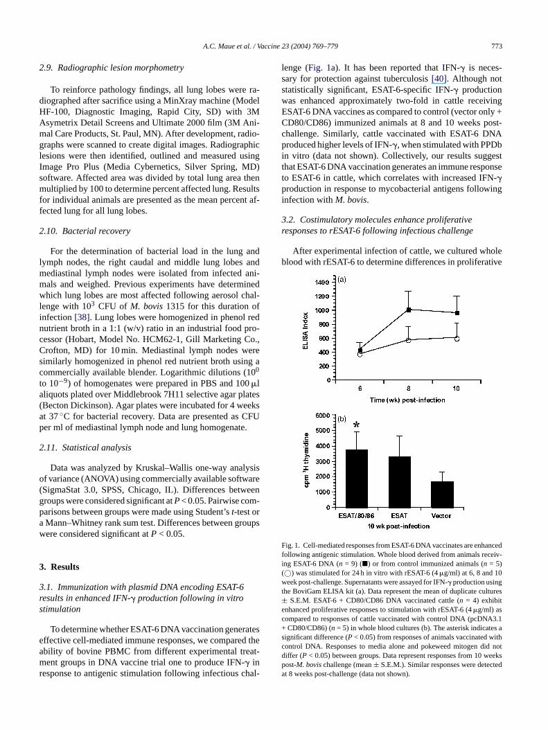
lenge (Fig. 1a). It has been reported that IFN-� is neces-sary for protection against tuberculosis[40]. Although notstatistically significant, ESAT-6-specific IFN-� productionwas enhanced approximately two-fold in cattle receivingESAT-6 DNA vaccines as compared to control (vector only +CD80/CD86) immunized animals at 8 and 10 weeks post-challenge. Similarly, cattle vaccinated with ESAT-6 DNAproduced higher levels of IFN-�, when stimulated with PPDbin vitro (data not shown). Collectively, our results suggestthat ESAT-6 DNA vaccination generates an immune responseto ESAT-6 in cattle, which correlates with increased IFN-�production in response to mycobacterial antigens followinginfection withM. bovis.
3.2. Costimulatory molecules enhance proliferativeresponses to rESAT-6 following infectious challenge
After experimental infection of cattle, we cultured wholeblood with rESAT-6 to determine differences in proliferative
F ancedfollowing antigenic stimulation. Whole blood derived from animals receiv-ing ESAT-6 DNA (n = 9) (�) or from control immunized animals (n = 5)(©) was stimulated for 24 h in vitro with rESAT-6 (4�g/ml) at 6, 8 and 10week post-challenge. Supernatants were assayed for IFN-� production usingthe BoviGam ELISA kit (a). Data represent the mean of duplicate cultures± S.E.M. ESAT-6 + CD80/CD86 DNA vaccinated cattle (n = 4) exhibitenhanced proliferative responses to stimulation with rESAT-6 (4�g/ml) ascompared to responses of cattle vaccinated with control DNA (pcDNA3.1+ CD80/CD86) (n = 5) in whole blood cultures (b). The asterisk indicates asignificant difference (P < 0.05) from responses of animals vaccinated withcontrol DNA. Responses to media alone and pokeweed mitogen did notdiffer (P < 0.05) between groups. Data represent responses from 10 weekspost-M. bovischallenge (mean± S.E.M.). Similar responses were detectedat 8 weeks post-challenge (data not shown).
rofton, MD) for 10 min. Mediastinal lymph nodes weimilarly homogenized in phenol red nutrient broth usinommercially available blender. Logarithmic dilutions (10
o 10−9) of homogenates were prepared in PBS and 10�lliquots plated over Middlebrook 7H11 selective agar plBecton Dickinson). Agar plates were incubated for 4 wet 37◦C for bacterial recovery. Data are presented aser ml of mediastinal lymph node and lung homogenate
.11. Statistical analysis
Data was analyzed by Kruskal–Wallis one-way analf variance (ANOVA) using commercially available softwSigmaStat 3.0, SPSS, Chicago, IL). Differences betwroups were considered significant atP< 0.05. Pairwise comarisons between groups were made using Student’st-test orMann–Whitney rank sum test. Differences between grere considered significant atP < 0.05.
. Results
.1. Immunization with plasmid DNA encoding ESAT-6esults in enhanced IFN-� production following in vitrotimulation
To determine whether ESAT-6 DNA vaccination generffective cell-mediated immune responses, we comparebility of bovine PBMC from different experimental treent groups in DNA vaccine trial one to produce IFN-� in
esponse to antigenic stimulation following infectious c
ig. 1. Cell-mediated responses from ESAT-6 DNA vaccinates are enh
774 A.C. Maue et al. / Vaccine 23 (2004) 769–779
responses between treatment groups. Previous experimentshave shown that T cells from infected humans, cattle andmice, proliferate in response to restimulation with rESAT-6in vitro. The results presented inFig. 1b reveal that ESAT-6+ CD80/CD86 DNA vaccinates show statistically significantproliferation of PBMC (P< 0.05) at 10 weeks post-infectionin response to restimulation with rESAT-6 when compared toanimals vaccinated with control DNA. Animals that receivedESAT-6 DNA alone did not possess significant differencesover control animals. This effect is most likely attributableto small group sizes and variation in individual animal re-sponses. Similar results were seen with PPDb restimulationamongst treatment groups (data not shown). These resultssuggest that ESAT-6 + CD80/CD86 DNA vaccination resultsin an enhanced proliferative response to ESAT-6 as comparedto control vaccinates.
3.3. ESAT-6 + CD80/CD86 DNA vaccinates showreduced pathology in the lungs following aerosolchallenge with M. bovis
Previous experiments have determined the location ofM.bovisafter aerosol challenge (103 CFU) to be restricted to thelung and regional lymph nodes draining the lung[38]. To as-sess disease severity, lungs and lymph nodes were subjectedt attleiB ver-a NAv con-t lung,f inv
rings etry
TS
V raphic (%)
D
D
Dystem each
l ndividuaccina s
CG va DNAv ificant d AT-6D
CG va D154D t, respe
to quantify lung lesions.Table 1illustrates the findings ofradiographic lesion morphometry from vaccination trial oneand two.M. bovisBCG and ESAT-6 + CD80/CD86 DNAvaccinates show comparable levels of affected lung area af-ter virulentM. bovischallenge for approximately 95 daysin trial one. Cattle vaccinated with control DNA or ESAT-6 DNA alone, possessed the highest mean percent affectedlung. Table 2summarizes the percent affected lung on anindividual lobe basis. ESAT-6 + CD80/CD86 DNA andM.bovisBCG vaccinates possessed the lowest mean percentaffected lung on all of the lung lobes. Similar results wereobtained regarding the number of radiographic lesions in thelung (Table 1). Low bacterial recovery was obtained fromthe lung homogenates of all treatment groups at this doseand time point (data not shown). Due to variation in indi-vidual responses, significant differences were not detected,although these results suggest that ESAT-6 + CD80/CD86DNA vaccination achieves reduced pathology compared toESAT-6 DNA vaccination alone following infectious chal-lenge withM. bovis.
3.4. ESAT-6 + CD80/CD86 DNA vaccinates possessreduced pathology and bacterial recovery frommediastinal lymph nodes
sub-j stem[v (0 trola 86D odew tes.T nesu attle
o a previously described pathology scoring system for cnfected with virulentM. bovis [39]. As shown inTable 1,CG and ESAT-6 + CD80/CD86 DNA vaccinates had oll lower levels of lung pathology compared to other Daccinates. According to the scoring of disease severity,rol animals possessed the most severe pathology of theollowed by animals vaccinated with ESAT-6 DNA aloneaccine trial one.
To reinforce the findings of this semi-quantitative scoystem, we next employed radiographic lesion morphom
able 1ummary of pathological findings in the lung ofM. bovis-infected animals
accination group Mean disease scorea Number of radiog
NA vaccine trial #1BCG 0.30± 0.30 1.50± 1.50ESAT/CD80/86 0.45± 0.26 1.25± 0.94ESAT 1.24± 0.61 8.00± 6.09Vector 1.44± 0.42 5.80± 1.98
NA vaccine trial #2BCG 0.00± 0.00∗ 0.50± 0.28∗∗ESAT/CD80/86 1.10± 0.19 10.70± 2.32ESAT/CD154 1.85± 0.73 15.70± 5.51ESAT 1.55± 0.61 22.50± 7.80
ata are presented as group mean (±S.E.M.).a Lungs were subjected to a semi-quantitative pathology scoring s
ung lobe then averaging the values to represent the entire organ for i∗ Single asterisk indicates a significant difference (P< 0.05) from BCG v
∗∗ Double asterisk indicates a significant difference (P < 0.05) from Baccinates using a Mann–Whitney rank sum test and indicates a signNA alone using Kruskal–Wallis one-way ANOVA.
∗∗∗ The triple asterisk indicates a significant difference (P < 0.05) from BNA vaccinates using Student’st-test or a Mann–Whitney rank sum tes
lesions Animals with radiographic lesions Affected lung
1/4 0.107± 0.1072/4 0.115± 0.0874/5 1.274± 1.0135/5 0.824± 0.481
2/4 0.009± 0.007∗∗∗4/4 0.220± 0.0464/4 1.755± 1.6503/4 0.838± 0.380
described previously[39]. Mean disease scores were determined by scoringals within each group.tes compared to animals receiving ESAT-6 DNA alone using Student’t-test.
ccinates compared to ESAT-6 + CD80/CD86 and ESAT-6 + CD154ifference (P < 0.05) from BCG vaccinates compared to animals receiving ES
ccinates compared with either ESAT-6 + CD80/CD86 or ESAT-6 + Cctively.
Following challenge, mediastinal lymph nodes wereected to a previously described pathology scoring sy39]. Results are summarized inTable 3. All ESAT-6 DNAaccinates possessed significantly reduced pathologyP <.05) of the mediastinal lymph node compared to connimals in trial one. Additionally, ESAT-6 + CD80/CDNA vaccinates possessed statistically lower lymph neights (P < 0.05) compared to the other DNA vaccinao determine the protective efficacy of the DNA vaccised in trial one, mediastinal lymph nodes of infected c
A.C. Maue et al. / Vaccine 23 (2004) 769–779 775
Table 2Distribution of radiographic lesions per lung lobe
Vaccination group Left cranial Right cranial Left caudal Right caudal and middle Accessory
DNA vaccine trial #1BCG 0.000± 0.000 0.000± 0.000 0.154± 0.154 0.377± 0.377 0.000± 0.000ESAT/CD80/86 0.121± 0.121 0.305± 0.189 0.000± 0.000 0.143± 0.143 0.000± 0.000ESAT 1.778± 1.677 1.078± 0.983 0.652± 0.404 1.042± 0.643 1.822± 1.822Vector 1.495± 1.248 1.244± 0.909 1.165± 0.930 1.007± 0.470 0.085± 0.085
DNA vaccine trial #2BCG 0.000± 0.000 0.000± 0.000 0.009± 0.009 0.038± 0.038 0.000± 0.000ESAT/CD80/86 0.086± 0.041 0.355± 0.216 0.222± 0.099 0.439± 0.162 0.000± 0.000ESAT/CD154 0.937± 0.910 3.529± 3.337 2.387± 2.188 1.308± 1.248 0.616± 0.573ESAT 0.232± 0.078 1.643± 1.074 1.173± 0.479 0.731± 0.429 0.409± 0.367
Data are presented as group mean (±S.E.M.).
Table 3Summary of pathology in the mediastinal lymph node
Vaccination group Mean disease scorea Lymph node weight (g) CFU (ml)
DNA vaccine trial #1BCG 0.75± 0.75∗ 11.22± 3.32 186.3± 156.2ESAT/CD80/86 1.00± 0.52∗ 6.65± 1.37∗∗∗ 1150.0± 386.2ESAT 1.00± 0.58∗ 43.94± 21.66 2044.0± 1279.6Vector 2.60± 0.20 26.30± 10.10 2832.0± 2293.6
DNA vaccine trial #2BCG 0.25± 0.25∗∗ 10.00± 1.34b 1362.5± 1215.7ESAT/CD80/86 2.00± 0.00 14.62± 3.05 1395.0± 612.0ESAT/CD154 2.25± 0.25 33.12± 12.65 2592.5± 1024.3ESAT 2.25± 0.25 58.17± 31.38 3252.5± 1804.9
Data are presented as group mean (±S.E.M.).a Mediastinal lymph nodes were subjected to a semi-quantitative pathology scoring system described previously[39].b Indicates a significant difference (P < 0.05) from BCG vaccinates compared to ESAT-6 + CD154 DNA vaccinates using a pairwise comparison with a
Mann–Whitney rank sum test.∗ A significant difference (P < 0.05) from vaccinates compared to animals receiving control DNA using Student’st-test.
∗∗ A significant difference (P < 0.05) from BCG vaccinates compared to animals receiving ESAT-6 DNA alone using Student’st-test.∗∗∗ A significant difference (P< 0.05) from ESAT-6 + CD80/CD86 DNA vaccinates compared to animals receiving ESAT-6 DNA alone or vector DNA usingpairwise comparisons with a Mann–Whitney rank sum test.
were homogenized for the recovery ofM. bovisat approx-imately 95 days post-infection. Logarithmic dilutions ofhomogenates were plated onto Middlebrook 7H11 platesand incubated for 4 weeks at 37◦C, after which CFU werecounted. Although not statistically significant, ESAT-6 +CD80/CD86 DNA vaccinates showed a trend towards re-duced mean bacterial recovery from mediastinal lymph nodesas compared to other DNA vaccinates in trial one (Table 3).Similar results were obtained with the same cultures at 8 week(data not shown).
3.5. Cell-mediated responses to ESAT-6 DNAvaccination are enhanced with co-administration ofeither CD80/86 or CD154
After assessing the potential of CD80 and CD86 to aug-ment the bovine immune response to DNA vaccination, wedirectly compared the adjuvant activities of CD80/CD86 toplasmid-encoded CD154 in our second vaccine trial. To de-termine differences in priming cell-mediated immunity, weexamined the ability of bovine PBMC from different exper-
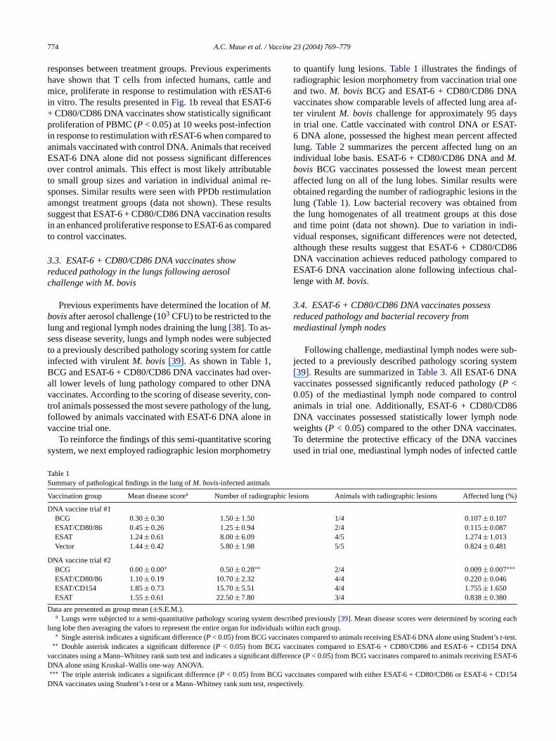
imental treatment groups to produce IFN-� in response toantigenic stimulation before and after aerosol challenge withM.bovis. Modest increases in ESAT-6-specific IFN-� produc-tion were evident in animals receiving ESAT-6 DNA vaccines2 weeks following boost (Fig. 2a). Animals receiving DNAvaccine co-administered with either CD80/CD86 or CD154produced greater mean amounts of IFN-� compared to an-imals receiving no costimulatory molecules. IFN-� produc-tion was reduced 3 weeks post-boost, but was enhanced invaccinates receiving either CD80/CD86 or CD154 followingaerosol challenge withM. bovis. ESAT-6 + CD80/CD86 andESAT-6 + CD154 DNA vaccinates produced approximatelyfive- and eight-fold greater mean amounts of IFN-� com-pared to animals receiving ESAT-6 DNA alone, respectively.DNA vaccination with CD154 also increased the frequencyof IFN-� producing PBMC as revealed by ESAT-6-specificELISPOT (Fig. 2b). At 3 weeks post-boost, animals receivingCD154 DNA possessed greater mean numbers of IFN-� spot-forming cells (SFC). Differences between and within groupswere not statistically significant due to low group numbersand variation in individual responses. However, these data
776 A.C. Maue et al. / Vaccine 23 (2004) 769–779
suggest that DNA-encoded CD80/CD86 and CD154 mayserve as adjuvants to augment IFN-� responses to DNA vac-cination.
3.6. Co-administration of CD154 does not reduce levelsof pathology or bacterial titers in the lung followingaerosol challenge with M. bovis
To assess disease severity, lungs were subjected to theaforementioned semi-quantitative pathology scoring system.Similar to results seen in DNA vaccine trial 1, ESAT-6 +CD80/CD86 DNA vaccinates possessed the lowest meandisease score of the lung compared with animals receivingESAT-6 + CD154 DNA or ESAT-6 DNA alone (Table 1). Thevalidity of these results was reinforced by enumeration of ra-diographic lesions from infected lungs. Radiographic anal-ysis revealed that ESAT-6 + CD80/CD86 DNA vaccinationresulted in an approximate two-fold reduction in detectablelung lesions, as compared to immunization with ESAT-6DNA alone (Table 1). Radiographic analysis also revealedthat ESAT-6 + CD80/CD86 DNA vaccinates possessed thelowest mean total percent affected lung among DNA vacci-
F torym e toa romEv4pwrvcDaim
nates (Table 1). This trend was also observed on an individuallung lobe basis, with ESAT-6 + CD80/CD86 DNA vaccinateshaving the lowest percent affected lung on all of the lobes an-alyzed (Table 2). The only statistically significant differencesin the lung were observed inM. bovisBCG vaccinates.M.bovisBCG vaccinates possessed significantly lower levels ofpercent affected lung (P < 0.05) compared to DNA vacci-nates. Similarly, animals vaccinated withM. bovisBCG pos-sessed statistically significant reductions in mean number ofradiographic lesions (P < 0.05) compared to all DNA vacci-nates, suggesting thatM. bovisBCG vaccination is protectiveagainstM. bovisaerosol challenge in cattle.
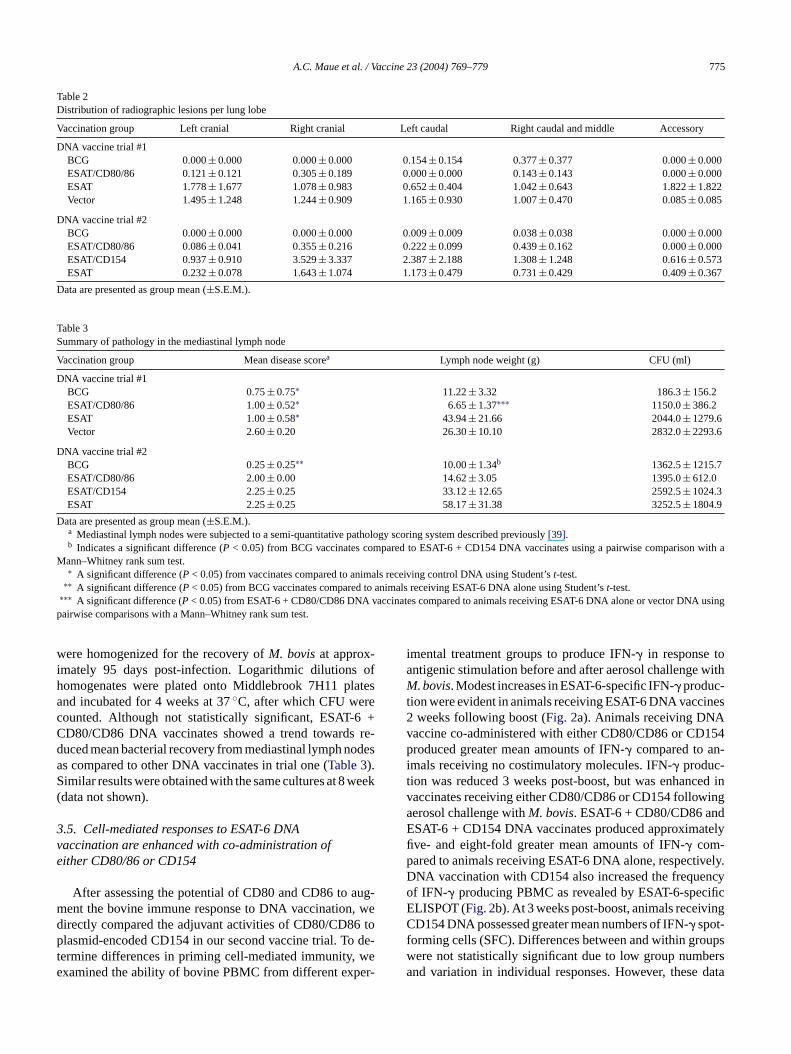
Modest reductions in bacterial recovery were detectedfrom the lungs (Fig. 3) of infected animals co-administeredcostimulatory molecules with ESAT-6 DNA, as compared toanimals immunized with ESAT-6 DNA alone. Although notstatistically significant, ESAT-6 + CD80/CD86 DNA vacci-nates possessed approximately 10- and 40-fold fewer meanCFU/ml in right caudal and middle lung lobes, as compared toESAT-6 + CD154 DNA vaccinates and animals administeredESAT-6 DNA alone, respectively. Collectively, these resultssuggest that although CD154 is capable of enhancing immuneresponses to ESAT-6, CD80/CD86 is superior in generatingprotective immune responses in the lung followingM. bovischallenge.
3rm
sub-j NAv ongstD ly
ig. 2. ESAT-6 DNA vaccination with plasmid-encoded costimulaolecules enhances IFN-� responses after antigenic stimulation relativnimals immunized with ESAT-6 DNA alone. Whole blood derived fSAT-6 + CD80/CD86 DNA vaccinates (n = 4), ESAT-6 + CD154 DNAaccinates (n= 4) or from animals vaccinated with ESAT-6 DNA alone (n=
) was stimulated for 24 h in vitro with rESAT-6 (4�g/ml) at day 0, 2 weeksost-boost, 3 weeks post-boost and 2 weeks post-challenge. Supernatantsere assayed for IFN-� production using the BoviGam ELISA kit (a). Dataepresent the mean (±S.E.M.) of duplicate wells. ESAT-6 + CD154 DNAaccinates (n = 4) display an increase in IFN-� spot-forming cells (SFC) asompared to other DNA vaccinates (b). PBMC from ESAT-6 + CD80/CD86NA vaccinates (n = 4), ESAT-6 + CD154 DNA vaccinates (n = 4) or fromnimals vaccinated with ESAT-6 DNA alone (n= 4) was stimulated for 36 h
n vitro with rESAT-6 (4�g/ml) at 3 weeks post-boost. Data represent theean (±S.E.M.) of triplicate cultures.
F c-t (4 lungl Mid-d ined.D p.
.7. DNA vaccination with CD80/CD86 results ineduced pathology and bacterial recovery from theediastinal lymph nodes
Following challenge, mediastinal lymph nodes wereected to pathology scoring. ESAT-6 + CD80/CD86 Daccinates possessed the lowest level of pathology amNA vaccinates (Table 3). In contrast to trial one, the on
ig. 3. ESAT-6 + CD80/CD86 DNA vaccinates (n= 4) display reduced baerial titers in the lung compared to ESAT-6 + CD154 DNA vaccinatesn =). Approximately, 100 days post-challenge, right caudal and middle
obes were homogenized and logarithmic dilutions were plated ontolebrook 7H11 agar plates for 4 weeks, after which titers were determata represent the mean (±S.E.M.) from animals in each treatment grou
A.C. Maue et al. / Vaccine 23 (2004) 769–779 777
significant difference in lymph node weights was in com-parison betweenM. bovisBCG vaccinates and animals vac-cinated with ESAT-6 + CD154 DNA (Table 3). To com-pare the protective efficacy of ESAT-6 + CD80/CD86 andESAT-6 + CD154 DNA vaccines, mediastinal lymph nodesof infected cattle were homogenized for the recovery ofM. bovisat approximately 100 days post-challenge. Loga-rithmic dilutions of homogenates were plated onto Middle-brook 7H11 plates and incubated for 4 weeks at 37◦C, afterwhich CFU were counted. Modest, not statistically signifi-cant, reductions in bacterial recovery were detected from themediastinal lymph nodes (Table 3) of infected animals co-administered CD80/CD86 with ESAT-6 DNA, as comparedto animals immunized with ESAT-6 DNA alone. Bacterial re-covery was reduced in mediastinal lymph nodes with ESAT-6+ CD80/CD86 DNA vaccinates possessing an approximatetwo-fold reduction in mean bacterial titers compared to otherDNA vaccinates. Similar results were obtained with 8 weekcultures (data not shown).
4. Discussion
The activation of naıve T lymphocytes is known to requireat least two signaling events to occur. The first signal involvesT HCm stim-u -cc cy-c duc-tv uner thecA trin-s hu-m efi andC y inc
dC uner f-e ngew stt ani-m T-6D ateds weres ac-c elativel iona tinall SAT-6 the
lung was not significant at the dose and timepoint used in trialone.
After evaluating the use of CD80/86 as molecular adju-vants, we next conducted a similar DNA vaccine trial to com-pare adjuvant abilities of CD80/CD86 with those of CD154.Previous studies have determined that DNA vaccination withCD154 can enhance immune responses in rodents and largeanimals [12,25,26]. CD154 interactions with its receptor,CD40, on APC have been shown to result in the upregulationof costimulatory molecules, namely B7 molecules, as wellas the production of pro-inflammatory cytokines, such as IL-12 [23,24]. Vaccination with plasmid-encoded CD154 gen-erated enhanced IFN-� responses to ESAT-6 following boostand challenge timepoints. Surprisingly, following challenge,ESAT-6 + CD154 DNA vaccinates possessed greater levels ofpathology in the lung and higher bacterial titers in the lung andmediastinal lymph node when compared to animals receivingESAT-6 + CD80/CD86 DNA. Comparisons between controlimmunized animals were excluded in vaccine trial two as theinfection was uncharacteristic of non-vaccinated cattle. Inboth trials, in addition to statistically significant differences,modest differences were observed in the several measuredparameters that resulted in a clear trend towards protection.Larger differences may become evident with greater num-bers of animals within groups, but small group numbers area re-q iffer-e 154i thei onw
+C ofn Tcv nde us-c s ex-p l re-s s ofD us-c pro-d oss-p ra-t titu-t ex-p mid-e ns toa n-h cci-n ow-e en-h rt re-vt ug-g ,
CR recognition of a foreign antigen presented by a Molecule. The second signal is mediated through a colatory receptor. In naıve T cells, this signal is primarily reeived through CD28-CD80/CD86 interactions[41–43]. Theombination of these signals drives T cells into the cellle, induces differentiation and augments cytokine proion, leading to an effective immune response[41–43]. Pre-ious studies conducted in mice have shown that immesponses to DNA vaccination can be improved witho-administration of costimulatory molecules[16–20,25,26].dditionally, CpG ODN have been shown to possess inic adjuvant abilities that induce immune responses inans, cattle and rodents[13]. Our study explores for th
rst time, the adjuvant effects of plasmid-encoded CD80D86 versus CD154 on DNA vaccine-induced immunitattle.
Our first DNA vaccine trial with CpG ODN, ESAT-6 anD80/CD86 resulted in a marked enhancement of imm
esponses, assessed by IFN-� production, lymphocyte proliration and reduced pathology following aerosol challeith virulent M. bovis. Notably, the results from this fir
rial suggest that immunity can be generated in largeals by a single prime/boost vaccination regimen. ESANA delivered in the absence of CD80 and CD86 generimilar immune responses; however, major differenceseen upon post-mortem examination with ESAT-6 DNA vinates possessing more severe pathology and greater rung lesion area. ESAT-6 + CD80/CD86 DNA vaccinatlso resulted in reduced bacterial recovery from medias
ymph nodes, as compared to animals vaccinated with EDNA alone or control cattle. Bacterial recovery from
n inevitable limitation of large animal experiments thatuire high level bio-containment. Regardless, these dnces suggest that, in cattle, CD80/86 is superior to CD
n augmenting DNA vaccine-induced protection despitenduction of enhanced IFN-� responses to recall stimulatiith rESAT-6.Presumably, the combined action of our ESAT-6
D80/CD86 DNA vaccine is to enhance the activationaıve T lymphocytes, resulting in an effector pool ofells capable of controlling infection with virulentM. bo-is. In DNA vaccination, plasmid DNA is internalized axpressed by two main cells types: dendritic cells and mle cells. It has been shown previously that muscle cellressing costimulatory molecules cannot prime T celponses[44], suggesting that DC are the key mediatorNA vaccine-induced immune responses. However, mle cells may present antigen to activated T cells oruce exogenous antigen for DC to internalize and crresent[45,46]. Previous work conducted in our labo
ory has shown that immature bovine DC do not consively express relatively high levels of CD80, but doress relatively high levels of CD86, suggesting that plasncoded costimulatory molecules may represent a meactivate T cells more efficiently[34]. We observed an eancement of immune responses to ESAT-6 by DNA vaates immunized with either CD80/CD86 or CD154; hver, CD154 immune responses did not correlate withanced protection against tuberculosis. A recent repoealed that CD4+ and CD8+ T cell responses againstM.uberculosisoccur normally in the absence of CD154, sesting alternative ligands for CD40[47]. Other infections
778 A.C. Maue et al. / Vaccine 23 (2004) 769–779
with microbes such asListeria monocytogenes[48] and lym-phocytic choriomeningitis virus[49], have shown that CD8+T cell priming occurs independently of CD40/CD154 inter-actions. Demangel et al.[50] found that mice immunizedwith BCG-infected CD40-stimulated DC had enhanced im-mune responses toM. tuberculosisinfection; however, thisenhancement did not correlate with increased protection fol-lowing infectious challenge, suggesting that additional strate-gies were needed. These studies also demonstrated that stimu-lation via CD40 also resulted in increased expression of IL-10mRNA in vitro [50] and that autocrine expression of IL-10in vivo impaired DC responses to mycobacteria[51]. Thebasis for the effects of CD154-enhanced IFN-� responses,but reduced elimination of bacteria and increased pathol-ogy observed in our vaccination trial will require furtherstudy.
MHC class I-restricted CD8+ CTL have been shown tohave a role in protection against tuberculosis in animalsand man[27]. DNA vaccination is an ideal strategy to tar-get these T cells, as endogenous antigen, produced by cellsthat internalize plasmid DNA, is presented via MHC class Imolecules[52]. One model regarding the generation of im-munologic memory hypothesizes that only a brief encounterwith antigen is necessary for the formation of CD8+ effec-tor/memory T lymphocytes and that a programmed divisiono sult-is n ef-f atei losism thea ns m ofbp n ourl attle[
ationo ine-i par-t ur-t cos-t d inc lly,wn engew e-s e ef-f ion,a be-l id-e ccinep ro-b ppli-c siticd
Acknowledgments
The authors thank Nathan Horman, Larry Wright, JohnKent and Dr. Jean Laufer for animal care and handling; Re-becca Lyon, Jody Mentele, Ryan Cook, Jessica Pollock, Pe-ter Lasley, Josh Pitzer, David Garcia-Tapia, K.C. Lambertand Monica R. Foote for technical assistance. This researchwas supported by the United States Department of Agricul-ture Specific Cooperative Agreement #58-1940-0-008 andthe Program for the Prevention of Animal Infectious Dis-eases (PPAID) at the University of Missouri, Missouri.
References
[1] Gurunathan S, Klinman DM, Seder RA. DNA vaccines: immunology,application, and optimization. Ann Rev Immunol 2000;18:927–74.
[2] Shedlock DJ, Weiner DB. DNA vaccination: antigen presentationand the induction of immunity. J Leukoc Biol 2000;68(6):793–806.
[3] van Drunen Littel-van den Hurk S, Gerdts V, Loehr BI, et al. Recentadvances in the use of DNA vaccines for the treatment of diseasesof farmed animals. Adv Drug Deliv Rev 2000;43(1):13–28.
[4] Cohen AD, Boyer JD, Weiner DB. Modulating the immune responseto genetic immunization. Faseb J 1998;12(15):1611–26.
[5] Beard CW, Mason PW. Out on the farm with DNA vaccines. NatBiotechnol 1998;16(13):1325–8.
[6] Sharma AK, Khuller GK. DNA vaccines: future strategiesBiol
cine
ificine.
cell-DNA
[ i-with
[ e-age-the
l re-unol
[ k S.onses–96.
[ ts.
[ wnm-xynu-ron
[ cine-e reg-
[ H.uno-unol
ccurs independently of further antigenic stimulation, reng in a stable pool of memory cells[53,54]. Therefore, aufficient priming event may be capable of generating aector pool of activated T cells able to control or eradicnfectious agents. CTL generated in response to tubercu
ay directly kill infected cells through the secretion ofnti-microbial protein, granulysin[55]. Granulysin has beehown to possess this activity against a broad spectruacteria in vitro by inducing membrane instability[55]. Thisrotein has been identified in humans and recent work i
aboratory has identified a homologue for this gene in c56].
In conclusion, our data suggest that the co-administrf CD80 and CD86, but not CD154, enhances DNA vacc
nduced protection to experimental bovine tuberculosis,icularly at the level of lung and lymph node pathology. Fhermore, we have established that the addition of theseimulatory molecules enables immunity to be generateattle following a single prime/boost regimen. Additionae have shown for the first time thatM. bovisBCG vacci-ation is protective against experimental aerosol challith virulentM. bovis. To our knowledge, this study reprents the first large animal experiments to examine thects of plasmid-encoded CD80/CD86 on DNA vaccinats well as a direct comparison with CD154. Currently, we
ieve that the co-administration of CpG ODN with plasmncoded CD80 and CD86 may represent a practical valatform for use in large animals, capable of generatingust, long-lived immune responses, which may also be aable to the prophylaxis of other bacterial, viral and paraiseases.
and relevance to intracellular pathogens. Immunol Cell2001;79(6):537–46.
[7] Scheerlinck JY. Genetic adjuvants for DNA vaccines. Vac2001;19(17–19):2647–56.
[8] Wang R, Doolan DL, Le TP, et al. Induction of antigen-speccytotoxic T lymphocytes in humans by a malaria DNA vaccScience 1998;282(5388):476–80.
[9] Wang R, Epstein J, Baraceros FM, et al. Induction of CD4(+) Tdependent CD8(+) type 1 responses in humans by a malariavaccine 2001;98(19):10817–22.
10] Arulkanthan A, Brown WC, McGuire TC, Knowles DP. Based immunoglobulin G1 isotype responses induced in cattleDNA expressing msp1a ofAnaplasma marginale. Infect Immun1999;67(7):3481–7.
11] Mwangi W, Brown WC, Lewin HA, et al. DNA-encoded ftal liver tyrosine kinase 3 ligand and granulocyte macrophcolony-stimulating factor increase dendritic cell recruitment toinoculation site and enhance antigen-specific CD4+ T celsponses induced by DNA vaccination of outbred animals. J Imm2002;169(7):3837–46.
12] Manoj S, Griebel PJ, Babiuk LA, van Drunen Littel-van den HurTargeting with bovine CD154 enhances humoral immune respinduced by a DNA vaccine in sheep. J Immunol 2003;170(2):989
13] Krieg AM. CpG motifs in bacterial DNA and their immune effecAnn Rev Immunol 2002;20:709–60.
14] Zhang Y, Shoda LK, Brayton KA, Estes DM, Palmer GH, BroWC. Induction of interleukin-6 and interleukin-12 in bovine B lyphocytes, monocytes, and macrophages by a CpG oligodeocleotide (ODN 2059) containing the GTCGTT motif. J InterfeCytokine Res 2001;21(10):871–81.
15] Horspool JH, Perrin PJ, Woodcock JB, et al. Nucleic acid vacinduced immune responses require CD28 costimulation and arulated by CTLA4. J Immunol 1998;160(6):2706–14.
16] Iwasaki A, Stiernholm BJ, Chan AK, Berinstein NL, Barber BEnhanced CTL responses mediated by plasmid DNA immgens encoding costimulatory molecules and cytokines. J Imm1997;158(10):4591–601.
A.C. Maue et al. / Vaccine 23 (2004) 769–779 779
[17] Kim JJ, Bagarazzi ML, Trivedi N, et al. Engineering of in vivoimmune responses to DNA immunization via codelivery of costim-ulatory molecule genes. Nat Biotechnol 1997;15(7):641–6.
[18] Kim JJ, Nottingham LK, Wilson DM, et al. Engineering DNA vac-cines via co-delivery of co-stimulatory molecule genes. Vaccine1998;16(19):1828–35.
[19] Chan K, Lee DJ, Schubert A, et al. The roles of MHC class II,CD40, and B7 costimulation in CTL induction by plasmid DNA. JImmunol 2001;166(5):3061–6.
[20] Corr M, Tighe H, Lee D, et al. Costimulation provided byDNA immunization enhances antitumor immunity. J Immunol1997;159(10):4999–5004.
[21] Castle BE, Kishimoto K, Stearns C, Brown ML, Kehry MR. Regula-tion of expression of the ligand for CD40 on T helper lymphocytes.J Immunol 1993;151(4):1777–88.
[22] Roy M, Waldschmidt T, Aruffo A, Ledbetter JA, Noelle RJ. Theregulation of the expression of gp39, the CD40 ligand, on normaland cloned CD4+ T cells. J Immunol 1993;151(5):2497–510.
[23] Grewal IS, Flavell RA. CD40 and CD154 in cell-mediated immunity.Annu Rev Immunol 1998;16:111–35.
[24] van Kooten C, Banchereau J. CD40-CD40 ligand. J Leukoc Biol2000;67(1):2–17.
[25] Mendoza RB, Cantwell MJ, Kipps TJ. Immunostimulatory effects ofa plasmid expressing CD40 ligand (CD154) on gene immunization.J Immunol 1997;159(12):5777–81.
[26] Ihata A, Watabe S, Sasaki S, et al. Immunomodulatory effect of aplasmid expressing CD40 ligand on DNA vaccination against humanimmunodeficiency virus type-1. Immunology 1999;98(3):436–42.
[27] Flynn JL, Chan J. Immunology of tuberculosis. Annu Rev Immunol2001;19:93–129.
[ AT-6
[ Di-AT-6
[ en
Im-
[ J.se-
[ etics
[ bi-B-gy
[ od-re-
[ bac-oes
[ ownTh1ttsial8.
[ li-f
[38] Palmer MV, Waters WR, Whipple DL. Aerosol delivery ofvirulent Mycobacterium bovis to cattle. Tuberculosis (Edinb)2002;82(6):275–82.
[39] Vordermeier HM, Chambers MA, Cockle PJ, Whelan AO, SimmonsJ, Hewinson RG. Correlation of ESAT-6-specific gamma interferonproduction with pathology in cattle followingMycobacterium bovisBCG vaccination against experimental bovine tuberculosis. InfectImmun 2002;70(6):3026–32.
[40] Cooper AM, Dalton DK, Stewart TA, Griffin JP, Russell DG, OrmeIM. Disseminated tuberculosis in interferon gamma gene-disruptedmice. J Exp Med 1993;178(6):2243–7.
[41] Carreno BM, Collins M. The B7 family of ligands and its receptors:new pathways for costimulation and inhibition of immune responses.Ann Rev Immunol 2002;20:29–53.
[42] Chambers CA, Allison JP. Co-stimulation in T cell responses. CurrOpin Immunol 1997;9(3):396–404.
[43] Sharpe AH, Freeman GJ. The B7-CD28 superfamily. Nat Rev Im-munol 2002;2(2):116–26.
[44] Iwasaki A, Torres CA, Ohashi PS, Robinson HL, Barber BH. Thedominant role of bone marrow-derived cells in CTL induction fol-lowing plasmid DNA immunization at different sites. J Immunol1997;159(1):11–4.
[45] Ulmer JB, Deck RR, Dewitt CM, Donnhly JI, Liu MA. Generationof MHC class I-restricted cytotoxic T lymphocytes by expression ofa viral protein in muscle cells: antigen presentation by non-musclecells. Immunology 1996;89(1):59–67.
[46] Ulmer JB, Deck RR, DeWitt CM, et al. Expression of a viral proteinby muscle cells in vivo induces protective cell-mediated immunity.Vaccine 1997;15(8):839–41.
[47] Lazarevic V, Myers AJ, Scanga CA, Flynn JL. CD40, but noton-–
[ TheIm-
[ medan-
l re-
[ Brit-une
[ en-tion
l IL-
[ withres-t viral
[ ialcells.
[ s re-an-
[ n, aty. J
[ tionnol
28] Pollock JM, Andersen P. Predominant recognition of the ESprotein in the first phase of interferon withMycobacterium bovisincattle. Infect Immun 1997;65(7):2587–92.
29] van Pinxteren LA, Ravn P, Agger EM, Pollock J, Andersen P.agnosis of tuberculosis based on the two specific antigens ESand CFP10. Clin Diagn Lab Immunol 2000;7(2):155–60.
30] Buddle BM, Parlane NA, Keen DL, et al. Differentiation betweMycobacterium bovisBCG-vaccinated andM. bovis-infected cattleby using recombinant mycobacterial antigens. Clin Diagn Labmunol 1999;6(1):1–5.
31] Kamath AT, Feng CG, Macdonald M, Briscoe H, Britton WDifferential protective efficacy of DNA vaccines expressingcreted proteins ofMycobacterium tuberculosis. Infect Immun1999;67(4):1702–7.
32] Parsons KR, Howard CJ. Cloning of cattle CD80. Immunogen1999;49(3):231–4.
33] Hirano A, Brown WC, Estes DM. Cloning expression andological function of the bovine CD40 homologue: role inlymphocyte growth and differentiation in cattle. Immunolo1997;90(2):294–300.
34] Bajer AA, Garcia-Tapia D, Jordan KR, et al. Peripheral bloderived bovine dendritic cells promote IgG1-restricted B cellsponses in vitro. J Leukoc Biol 2003;73(1):100–6.
35] Menon SA, Wannemuehler MJ, Mahairas GG, Minion FC. Mycoterial ESAT-6 protein enhances mouse IFN-gamma responses tMy-coplasma hyopneumoniaeP71 protein. J Interferon Cytokine R2002;22(7):807–13.
36] Zhang Y, Palmer GH, Abbott JR, Howard CJ, Hope JC, BrWC. CpG ODN 2006 and IL-12 are comparable for priminglymphocyte and IgG responses in cattle immunized with a rickeouter membrane protein in alum. Vaccine 2003;21(23):3307–1
37] Bolin CA, Whipple DL, Khanna KV, Risdahl JM, Peterson PK, Motor TW. Infection of swine withMycobacterium bovisas a model ohuman tuberculosis. J Infect Dis 1997;176(6):1559–66.
CD40L, is required for the optimal priming of T cells and ctrol of aerosolM. tuberculosisinfection. Immunity 2003;19(6):82335.
48] Grewal IS, Borrow P, Pamer EG, Oldstone MB, Flavell RA.CD40-CD154 system in anti-infective host defense. Curr Opinmunol 1997;9(4):491–7.
49] Whitmire JK, Flavell RA, Grewal IS, Larsen CP, Pearson TC, AhR. CD40-CD40 ligand costimulation is required for generatingtiviral CD4 T cell responses but is dispensable for CD8 T celsponses. J Immunol 1999;163(6):3194–201.
50] Demangel C, Palendira U, Feng CG, Heath AW, Bean AG,ton WJ. Stimulation of dendritic cells via CD40 enhances immresponses toMycobacterium tuberculosisinfection. Infect Immun2001;69(4):2456–61.
51] Demangel C, Bertolino P, Britton WJ. Autocrine IL-10 impairs ddritic cell (DC)-derived immune responses to mycobacterial infecby suppressing DC trafficking to draining lymph nodes and loca12 production. Eur J Immunol 2002;32(4):994–1002.
52] Hassett DE, Zhang J, Whitton JL. Neonatal DNA immunizationa plasmid encoding an internal viral protein is effective in the pence of maternal antibodies and protects against subsequenchallenge. J Virol 1997;71(10):7881–8.
53] Kaech SM, Ahmed R. Memory CD8+ T cell differentiation: initantigen encounter triggers a developmental program in naiveNat Immunol 2001;2(5):415–22.
54] van Stipdonk MJ, Lemmens EE, Schoenberger SP. Naive CTLquire a single brief period of antigenic stimulation for clonal expsion and differentiation. Nat Immunol 2001;2(5):423–9.
55] Ernst WA, Thoma-Uszynski S, Teitelbaum R, et al. GranulysiT cell product, kills bacteria by altering membrane permeabiliImmunol 2000;165(12):7102–8.
56] Endsley JJ, Furrer JL, Endsley MA, et al. Characterizaof bovine homologues of granulysin and NK-lysin. J Immu2004;173(4):2607–14.


