Embed Size (px)
Citation preview

Developmental Biology 351 (2011) 35–45
Contents lists available at ScienceDirect
Developmental Biology
j ourna l homepage: www.e lsev ie r.com/deve lopmenta lb io logy
Cell proliferation in the absence of E2F1-3
Pamela L. Wenzel a,b,1,2, Jean-Leon Chong a,b,2, M. Teresa Sáenz-Robles c, Antoney Ferrey a,b,John P. Hagan a,b,1, Yorman M. Gomez a,b, Ravi Rajmohan a,b, Nidhi Sharma a,b, Hui-Zi Chen a,b,James M. Pipas d, Michael L. Robinson d,⁎, Gustavo Leone a,b,⁎a Department of Molecular Virology, Immunology and Medical Genetics, Comprehensive Cancer Center, College of Medicine, The Ohio State University, Columbus, OH 43210, USAb Department of Molecular Genetics, College of Biological Sciences, The Ohio State University, Columbus, OH 43210, USAc Department of Biological Sciences, University of Pittsburgh, Pittsburgh, PA 15260, USAd Department of Zoology, Miami University, Oxford, OH 45056, USA
⁎ Corresponding authors. M.L. Robinson is to be conta258 Pearson Hall, Miami University, Oxford, OH 45056,Leone, Human Cancer Genetics Program, Departmentnology and Medical Genetics, Department of MolecuUniversity, Comprehensive Cancer Center, 460W. 12th AFax: +1 614 688 4181.
E-mail addresses: [email protected] (M.L. [email protected] (G. Leone).
1 Present address: Department of Medicine, DivisChildren's Hospital and Department of BiochemistryHarvard Medical School, Boston, MA 02115, USA.
2 These authors contributed equally to this work.
0012-1606/$ – see front matter. Published by Elsevierdoi:10.1016/j.ydbio.2010.12.025
a b s t r a c t
a r t i c l e i n f oArticle history:Received for publication 4 June 2010Revised 6 December 2010Accepted 15 December 2010Available online 23 December 2010
Keywords:ProliferationCell cycleE2FRbLensRepressionCell survivalTranscription
E2F transcription factors regulate the progression of the cell cycle by repression or transactivation of genesthat encode cyclins, cyclin dependent kinases, checkpoint regulators, and replication proteins. Although someE2F functions are independent of the Retinoblastoma tumor suppressor (Rb) and related family members,p107 and p130, much of E2F-mediated repression of S phase entry is dependent upon Rb. We previouslyshowed in cultured mouse embryonic fibroblasts that concomitant loss of three E2F activators withoverlapping functions (E2F1, E2F2, and E2F3) triggered the p53-p21Cip1 response and caused cell cycle arrest.Here we report on a dramatic difference in the requirement for E2F during development and in cultured cellsby showing that cell cycle entry occurs normally in E2f1-3 triply-deficient epithelial stem cells and progenitorsof the developing lens. Sixteen days after birth, however, massive apoptosis in differentiating epithelium leadsto a collapse of the entire eye. Prior to this collapse, we find that expression of cell cycle-regulated genes inE2F-deficient lenses is aberrantly high. In a second set of experiments, we demonstrate that E2F3 ablationalone does not cause abnormalities in lens development but rescues phenotypic defects caused by loss of Rb, abinding partner of E2F known to recruit histone deacetylases, SWI/SNF and CtBP-polycomb complexes,methyltransferases, and other co-repressors to gene promoters. Together, these data implicate E2F1-3 inmediating transcriptional repression by Rb during cell cycle exit and point to a critical role for their repressivefunctions in cell survival.
cted at Department of Zoology,USA, Fax: +1 513 529 6900. G.of Molecular Virology, Immu-lar Genetics, The Ohio Stateve. Columbus, OH 43210, USA.
inson),
ion of Hematology/Oncology,and Molecular Pharmacology,
Inc.
Published by Elsevier Inc.
Introduction
Cell cycle entry is guarded by cyclin dependent kinases (Cdk)which, upon activation by mitogenic signals, phosphorylate theRetinoblastoma pocket proteins, Rb, p107, and p130, and cause therelease and accumulation of sequestered E2F transcription factors.E2Fs consist of a family of repressors and activators that togethercoordinately regulate cellular proliferation by controlling the tran-
scriptional activity of over 130 known target genes that function toinitiate the G1/S transition, DNA synthesis, DNA repair and mitosis(Bracken et al., 2004). Rb is thought to inhibit E2F activity andexpression of cell cycle-regulated genes by association with co-repressors such as histone deacetylases, SWI/SNF and CtBP-polycombcomplexes, histone methyltransferases, and DNA methyltransferases(Dahiya et al., 2001; Luo et al., 1998; Nielsen et al., 2001; Robertsonet al., 2000; Vandel et al., 2001; Zhang et al., 2000). Classic paradigmsof cell cycle regulation have consistently portrayed the threeactivators, E2F1, E2F2, and E2F3, as the final components of theCdk-Rb signaling cascade that execute the transcriptional programnecessary to commit cells to enter S phase. Although Rb-mediated cellcycle regulation has been studied extensively, the evidence that E2F1-3 are required for expression of genes critical for proliferation is basedalmost exclusively on analyses of lower eukaryotes and in vitro cellculture systems (Bracken et al., 2004; Hallstrom and Nevins, 2009;Morris et al., 2000; Rowland et al., 2002). Only recently has it beendemonstrated that E2F1-3 are not essential for cell cycle entry, andthat proliferation can proceed without E2F1-3 in the majority of celltypes present during organ development and embryogenesis (Chen
36 P.L. Wenzel et al. / Developmental Biology 351 (2011) 35–45
et al., 2009; Chong et al., 2009). In fact, there is now good evidence tosuggest that these E2Fs are most critical for regulating survival in vivo(Chen et al., 2009; Chong et al., 2009).
The highly organized cellular architecture of the developing lensmakes it an attractive system for the in vivo study of cell cycle anddifferentiation. Simply based upon spatial separation, epithelial cellsthat are proliferating can be distinguished easily from those that areexiting the cell cycle and terminally differentiating. At approximately10 days of murine embryonic development (E10), morphological lensformation begins with the invagination of the surface ectodermoverlying the optic cup to form the lens pit, which subsequently closesto form the lens vesicle (Lovicu et al., 2004). Although initially allepithelial cells lining the lens vesicle maintain the capacity toproliferate, as development progresses, cycling cells are confined toa ring of epithelium slightly anterior to the lens equator. At theequatorial region (or bow) of the lens, rapidly proliferating cells moveinto a transition zone, wherein they begin to exit the cell cycle anddifferentiate. As these cells migrate toward the cortex of the lens, theyterminally differentiate into fiber cells, losing their nuclei andorganelles and gaining the translucent properties necessary for vision.
To test whether loss of E2F activators impact cell cycle entry invivo, we examined the effects of E2F1, E2F2, and E2F3 triple deficiencyduring development in murine pre- and post-natal lenses. We findthat morphological lens development occurs relatively normallythrough late gestation, illustrating that E2F activators are unnecessaryfor proliferation of lens epithelial cells. During migration of theepithelial cells to the equatorial zone, however, DNA double-strandbreaks develop, p21Cip1 is upregulated, and cells exhibit signs ofapoptotic cell death. Further, we find a dramatic increase inexpression of E2F target genes in E2f-triply deficient lenses, pointingto the shared function that E2F1, 2, and 3 play in repressingexpression of cell cycle genes during maturation of the lensepithelium. Aberrant expression of genes required for cell cycle exitand lens differentiation, along with upregulation of p19Arf and otherE2F target genes, together culminates in collapse of the entire lensarchitecture between one to two weeks after birth. Importantly, wedemonstrate a nearly complete rescue of Rb-deficiency phenotypes,including proliferation defects, with conditional ablation of E2F3,pointing to a context-dependent switch in the function of “activating”E2Fs to repress or activate transcription. These data illustrate that E2F-mediated activation of cell cycle regulated genes is not required forproliferation, and point to a critical role for E2F1-3 in cell survival andtranscriptional repression in vivo.
Materials and methods
Mouse strains and genotyping
E2f1, E2f2, E2f3 knockout mice and cry-cre (MLR10) transgenic(Zhao et al., 2004) and ROSA26 reporter (Gt(ROSA)26Sortm1Sor)(Soriano, 1999) mice were maintained on a mixed 129SvEv; C57BL/6;FVB background. PCR primers for genotyping are listed in Fig. S1.
Proliferation assays
Proliferation of lens epithelial or fiber cells was measured byincorporation of BrdU. Briefly, pregnant dams or pups were injectedwith BrdU 2 hours prior to harvest at 100 μg/g body weight. Tissuefrom embryos or neonates was fixed in 10% buffered formalin,dehydrated, and processed with paraffin. Sections were cut at 5-μmthickness and BrdU was detected by immunofluorescent staining.
Histology and immunofluorescent staining
Lens architecture was histologically examined by hematoxylin andeosin staining of lens sections. Proliferation was measured by
immunostaining with BrdU-specific antibodies (DAKO; Bu20a), andapoptosis was detected by TUNEL assays using the Apoptag PlusPeroxidase In Situ Apoptosis Detection Kit (Chemicon International).DNA double-strand breaks were detected as a measure of DNAdamage by immunofluorescent staining with phospho-histone H2AX(Ser139) antibodies (Upstate, NY; clone JBW301). Other antibodiesused include PCNA (Santa Cruz Biotechnology, PC10), Mcm3 (SantaCruz Biotechnology, N-19), and cleaved caspase-3 (Cell SignalingTechnology, Asp175). A minimum of three sections from each lenswere quantified by comparison of the number of positive cells to thetotal number of nuclei in epithelial or fiber cells. Lens-specific markersof differentiation, including α-, β-, and γ-crystallin antibodies were agift from Dr. S. Zigler.
Detection of β-galactosidase activity
Cry-cre mice were crossed to the ROSA26 reporter (Gt(ROSA)26Sortm1Sor) line to test the activity of cre as described previously(Robinson et al., 1995). Briefly, eyes were surgically removed fromembryos, fixed in 2% paraformaldehyde in 0.1 M phosphate buffer pH7.3, washed in PBS, and incubated overnight in the dark at roomtemperature in X-gal staining medium (5 mM potassium ferricya-nide, 5 mM potassium ferrocyanide, 1 mg/ml X-gal, 0.1% deoxycho-late, 0.2% Nonidet P-40, and 2 mM MgCl2 in PBS). Eyes werepost-fixed the next day in 10% buffered formalin for 2 hours andprocessed for paraffin embedding, sectioning, and counterstainingwith nuclear fast red.
Global gene expression analysis
Cry-Cre+/−; E2f1−/−; E2f2−/−; E2f3LoxP/LoxP and E2f1+/+; E2f2−/−;E2f3LoxP/LoxP lenses were microdissected from the eye at E17.5 and P0with 18-gauge needles. RNA from both lenses of each embryo orneonate was isolated by TRIzol purification and processed forhybridization to Affymetrix Mouse Genome 430 2.0 Arrays. Expres-sion values were adjusted by quantile normalization and logtransformation with RMAExpress, and data were analyzed withBRB-ArrayTools 3.7.0. (Figs. S2, S3). Class comparison was used toselect genes differentially expressed at a significance level ofpb0.0001 or pb0.001, and gene set expression comparisons wereused to identify gene ontologies and pathways impacted by loss of E2F(Fig. S4). DAVID (http://david.abcc.ncifcrf.gov) was used to furtherevaluate subsets of genes. CBRC TFSearch (http://www.cbrc.jp/research/db/TFSEARCH.html) and GATHER (http://gather.genome.duke.edu) aided in the identification of genes containing E2Fconsensus binding sites. Microarray data presented in this study hasbeen deposited in the GEO public repository (GSE16533).
Real-time RT-PCR
Both lenses from each embryo or neonate were placed in onemicrocentrifuge tube and homogenized in TRIzol reagent to obtaintotal RNA (Life Technologies). Five μg of total RNAwas used to generatecDNA using Superscript III reverse transcriptase (Invitrogen). Quan-titative RT-PCR was performed using the BioRad iCycler PCR machine.Each PCR reaction contained 0.5 μl of cDNA template, primers at aconcentration of 100 nM, and 1× of SYBR Green Reaction Mix(BioRad). Reactions were performed in triplicate in a total volume of25 μl and data were analyzed using the ΔCt method, where GAPDHserved as the internal control. Each PCR reaction generated only theexpected amplicon as shown by the melting-temperature profiles ofthe final products and gel electrophoresis. Primer sequences are listedin Fig. S1.
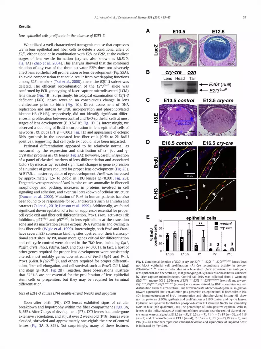
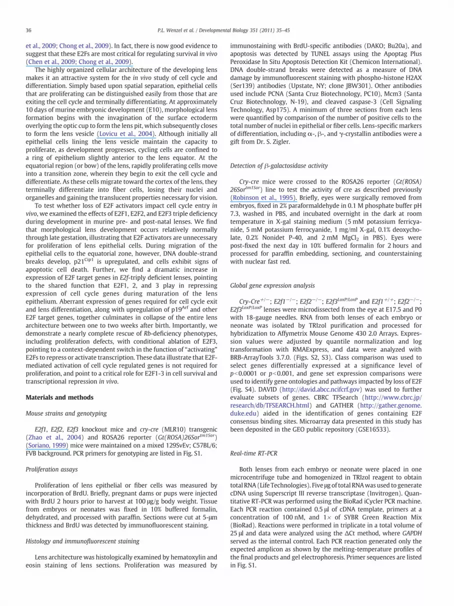
Fig. 1. Conditional deletion of E2f3 in cry-cre;E2f1−/−;E2f2−/−;E2f3LoxP/LoxP lenses doesnot block epithelial cell proliferation. (A) Cre recombinase activity in cry-cre;ROSA26Sortm1Sor mice is detectable as a blue stain (LacZ expression) in embryoniclens epithelial and fiber cells. (B) PCR genotyping of E2f3 on lens or head tissue collectedby laser capture microdissection. Control tail DNA was collected from a weanlingE2f3LoxP/−mouse. (C) E13.5 lenses of E2f1−/−;E2f2−/−;E2f3LoxP/LoxP (control) and cry-cre;E2f1−/−;E2f2−/−;E2f3LoxP/LoxP (cry-cre) mice were stained by H&E to examine nucleardistribution and lens architecture. Blue arrow indicates direction of epithelial migrationtoward equatorial line; ant, anterior; pos, posterior; ep, epithelium; fi, fiber cells; ir, iris.(D) Immunodetection of BrdU incorporation and phosphorylated histone H3 shownormal patterns of DNA synthesis and proliferation in E16.5 control and cry-cre lenses.Epithelial cells positive for BrdU or phospho-histone H3 stain red. Nuclei are stained byDAPI in blue (top quadrants). (E) The percentage of BrdU-positive epithelial cells inlenses at the indicated ages. A minimum of three sections near the central plane of cry-cre lenses were analyzed at E13.5 (n=5), E16.5 (n=7), P1 (n=7), P7 (n=3), and P16(n=3) and of control lenses at E13.5 (n=4), E16.5 (n=2), P1 (n=2), P7 (n=3), andP16 (n=4). Error bars represent standard deviation and significance of unpaired t-testis indicated by **pb0.01.
37P.L. Wenzel et al. / Developmental Biology 351 (2011) 35–45
Results
Lens epithelial cells proliferate in the absence of E2F1-3
We utilized a well-characterized transgenic mouse that expressescre in lens epithelial and fiber cells to delete a conditional allele ofE2f3, either alone or in combination with E2f1 or E2f2, at the earlieststages of lens vesicle formation (cry-cre, also known as MLR10;Fig. 1A) (Zhao et al., 2004). This analysis showed that the combineddeletion of any two of the three activator E2Fs does not adverselyaffect lens epithelial cell proliferation or lens development (Fig. S5A).To avoid compensation that could result from overlapping functionsamong E2F members (Tsai et al., 2008), the entire E2f1-3 subset wasdeleted. The efficient recombination of the E2f3LoxP allele wasconfirmed by PCR-genotyping of laser capture microdissected (LCM)lens tissue (Fig. 1B). Surprisingly, histological examination of E2f1-3deficient (TKO) lenses revealed no conspicuous change in lensarchitecture prior to birth (Fig. 1C). Direct assessment of DNAreplication and mitosis by BrdU incorporation and phosphorylatedhistone H3 (P-H3), respectively, did not identify significant differ-ences in proliferation between control and TKO epithelial cells at moststages of lens development (E13.5-P16; Fig. 1D, E). Interestingly, weobserved a doubling of BrdU incorporation in lens epithelial cells ofnewborn TKO pups (P1, p=0.002; Fig. 1E) and appearance of ectopicDNA synthesis in the associated lens fiber cells (0.5% to 2% BrdUpositive), suggesting that cell cycle exit could have been impacted.
Perinatal differentiation appeared to be relatively normal, asmeasured by the expression and distribution of α-, β-, and γ-crystallin proteins in TKO lenses (Fig. 2A); however, careful inspectionof a panel of classical markers of lens differentiation and associatedfactors by microarray revealed significant changes in gene expressionof a number of genes required for proper lens development (Fig. 2B).At E17.5, a master regulator of eye development, Pax6, was increasedby approximately 1.5- to 2-fold in TKO lenses (pb0.001, Fig. 2B).Targeted overexpression of Pax6 in mice causes anomalies in fiber cellmorphology and packing, increases in proteins involved in cellsignaling and adhesion, and eventual breakdown of cellular structure(Duncan et al., 2000). Mutation of Pax6 in human patients has alsobeen found to be responsible for ocular disorders such as aniridia andcataract (Cai et al., 2010; Hanson et al., 1999). Additionally, we foundsignificant downregulation of a tumor suppressor essential for propercell cycle exit and fiber cell differentiation, Prox1. Prox1 activates Cdkinhibitors, p27Kip1 and p57Kip2, in lens epithelium at the transitionzone and its inactivation causes ectopic DNA synthesis and cycling inlens fiber cells (Wigle et al., 1999). Interestingly, both Pax6 and Prox1have several E2F consensus binding sites upstream of their transcrip-tional start sites. By P0, many more genes critical for differentiationand cell cycle control were altered in the TKO lens, including Gja1,Pdgfrl, Cryl1, Pitx3, Pdgfra, Gja3, and Six3 (pb0.001). In fact, a host ofother genes required for proper lens development were consistentlyaltered, most notably genes downstream of Pax6 (Itgb1 and Pxn),Prox1 (Cdkn1b (p27Kip1)), and others required for proper differenti-ation, fiber cell elongation, and cell survival, such as Foxe3, Cdh1, Maf,and Mafb (pb0.01, Fig. 2B). Together, these observations illustratethat E2F1-3 are not essential for the proliferation of lens epithelialstem cells or progenitors but they may be required for terminaldifferentiation.
Loss of E2F1-3 causes DNA double-strand breaks and apoptosis
Soon after birth (P0), TKO lenses exhibited signs of cellularbreakdown and hypotrophy within the fiber compartment (Figs. 3A,B, S5B). After 7 days of development (P7), TKO lenses had undergoneextensive vacuolation, and at just over 2 weeks old (P16), lenses wereclouded, shriveled and approximately one-eighth the size of controllenses (Fig. 3A–D, S5B). Not surprisingly, many of these features
Fig. 2. Loss of E2f1-3 causes transcriptional deregulation of a subset of lens-specific genes. (A) Immunostaining ofα-, β-, and γ-crystallin in control and cry-cre lenses. The distributionof positive staining ranges from the anterior face of the lens to the transition zone. β- and γ-crystallins are only detectable in the fiber cell compartment. Lowmagnification images ofimmunostaining with α-, β-, and γ-crystallin antibodies show specificity for lens. Surrounding retina provides reference for background levels of fluorescence. (B) Lensdifferentiation markers were analyzed by hierarchical clustering of gene expression and visualized by heatmap. Statistically significant changes (pb0.001) were determined by classcomparison analysis and are indicated by red boxes (upregulated) and green boxes (downregulated).
38 P.L. Wenzel et al. / Developmental Biology 351 (2011) 35–45
resembled murine models of Pax6 overexpression (Duncan et al.,2000). Given the protracted nature of TKO lens breakdown, weexamined the integrity of genomic DNA and apoptosis in TKO lensesprior to birth, before the manifestation of these phenotypes.
Immunohistochemical staining of E13.5 lenses showed a markedincrease in cells that stained positive for phosphorylated histoneH2AX (γ-H2AX). Positivity for γ-H2AX was particularly acute innewly differentiating fiber cells at the equatorial plane of the TKO

Fig. 3. Loss of E2F1-3 causes degradation and opacification of the lens. (A) Hematoxylinand eosin staining of lenses from of E2f1−/−;E2f2−/−;E2f3LoxP/LoxP (control) and cry-cre;E2f1−/−;E2f2−/−;E2f3LoxP/LoxP (cry-cre) embryos (E16.5) and neonates (P1, P7 and P16).(B) High magnification of P1 control and cry-cre lenses. Note the prominent vacuolationin the cortex of the TKO fiber compartment. (C) control and cry-cre mice and (D)dissected lenses from control and cry-cre mice at 3 weeks old. Normal adult lenses arespherical and transparent, whereas TKO lenses are distinguished by decreased size,irregular surface and opacity (cataract).
39P.L. Wenzel et al. / Developmental Biology 351 (2011) 35–45
lenses, whereas γ-H2AX positivity was high in both TKO and controllenses during late stages of fiber cell differentiation (Fig. 4A). DNAdouble-strand breaks in terminally differentiating fiber cells withinthe lens cortex are presumably a result of normal nuclear degener-ation that must occur for maturation of the translucent fibers.Apoptotic cell death, as measured by TUNEL and cleaved caspase-3immunofluorescence, was apparent in the epithelium and fibercompartments of E13.5 TKO lenses but not in control lenses (Fig. 4B,C, and Fig. S5C). At E16.5, apoptosis was most pronounced near theequatorial transition zone of E16.5 TKO lenses, consistent with thedistribution of γ-H2AX-positive cells (Fig. 3D). As a result, the anteriorperimeter of E17.5 TKO lens sections contained 36% fewer epithelialcells than control lenses (p=0.0002; Fig. S5D). From these observa-tions, we conclude that postnatal architectural collapse of TKO lensesis accompanied by massive breakdown of genetic material andinitiation of an apoptotic program.
E2F target gene expression is upregulated with ablation of E2F“activators”
In order to identify the molecular pathways impacted by loss ofE2F1-3 in vivo,we compared global gene expression profiles in control(E2f2−/−) and TKO lenses at two developmental stages, E17.5 and P0,which precede the physical breakdown of the mutant lens. Weimplemented unsupervised class comparison analyses of all probesetson the array to identify genes differentially expressed between lenses.We also used an unbiased approach similar to Gene Set EnrichmentAnalysis (Mootha et al., 2003) to identify a priori defined groups ofgenes that were significantly differentially expressed. Expressionchanges observed among samples were attributable to two variables:developmental time (E17.5 vs. P0) and genotype (control vs. TKO).Comparison of control lenses at the two developmental stagesidentified 408 genes that were downregulated and 174 that wereupregulated in P0 lenses (N 1.5 fold and pb0.001) (Fig. 5A, Fig. S3).The expression of known E2F target genes, as defined by expressionand chromatin immunoprecipitation assays, decreased with age,consistent with a decrease in proliferation as lenses develop fromE17.5 to birth (Black et al., 2005; Bracken et al., 2004; Kong et al.,2007; Ren et al., 2002; Vernell et al., 2003; Weinmann et al., 2002;Wells et al., 2002; Xu et al., 2007) (Fig. 5B, Fig. S2A). Comparisonbetween control and TKO lenses revealed a significant number of geneexpression changes at both developmental time points analyzed(Fig. 5A, Figs. S2, S4). Significantly changed gene ontology categoriesillustrated a bias for differential expression of genes involved inmetabolism and DNA processing (Eisen et al., 1998) (Least square andKolmogorov–Smirnov permutation values of pb0.001) (Figs. S2B, S4).Genes that were downregulated in TKO lenses, depicted in the lowerthird of the heatmap in Fig. 5A, were enriched for functions in themitochondrion and oxidation reduction at E17.5 and, at P0, for roles inthe extracellular region, including proteins critical for cell adhesionand structural matrix, such as thrombospondin, procollagen, albumin,nidogen, perlecan, and laminin (Fig. S6). In contrast, genes contributingto DNA replication were uniformly upregulated in TKO tissues,including E2F targets such as chromatin licensing and DNA replicationfactor 1 (Cdt1) and the minichromosome maintenance deficient genes(Mcm2, Mcm3, Mcm4, Mcm5, Mcm7, Mcm8). In fact, expression ofnearly all E2F target genes was significantly higher in TKO than incontrol lenses at both E17.5 and P0 (Fig. 5C, D). Quantitative analysisof gene expression by real-time RT PCR was used to validate a subsetof changes identified by Affymetrix oligo arrays (Fig. 5D). Tocharacterize the spatial nature of E2F target misexpression, lenseswere sectioned and probedwith antibodies specific for several knowndownstream effectors of E2F. As shown in Fig. 5E, Mcm3 and PCNAprotein levels were undetectable in control E17.5 lens fibers, whereasin TKO tissues these proteins were apparent in fiber cells andconspicuously elevated in the epithelium. Together, the gene

40 P.L. Wenzel et al. / Developmental Biology 351 (2011) 35–45
expression data and protein immunostaining of E2F targets points to acentral role for E2F1-3 in transcriptional repression.
Activation of pro-apoptotic gene expression in the p53 pathway
Previously, we identified a role for E2F in regulation of the p53–p21Cip1 axis (Sharma et al., 2006; Timmers et al., 2007). Thisrelationship between E2F ablation and upregulation of p53 activitywas further supported by recent evidence implicating the deacetylaseSirtuin 1 as a critical modulator of p53 in the murine retina (Chenet al., 2009). In the current study, we observe increased expression ofa number of regulators and downstream effectors of p53 (Fig. 6A).
Most notably, p21Cip1 and p19Arf were significantly upregulated in TKOlenses (Fig. 6B–D). Ectopic expression of p21Cip1 protein was observedin the epithelium, loosely corresponding to an area approximately 60degrees above the lens equator (just anterior to TUNEL-positiveenrichment depicted in Fig. 4D) (Fig. 6C). In contrast, p19Arf waslocalized to the fiber compartment, and appeared throughout the fibercytoplasm and in the nuclei of nucleated lens fibers (Fig. 6C). Thefiber-specific distribution of p19Arf is intriguing, not only for itsrecognized role in stabilizing p53 through inhibition of Mdm2, butalso as a modulator of proliferation. It has been demonstrated in themurine eye that p19Arf regulates Pdgf signaling, a pathway essentialfor cycling of the lens epithelium. Pdgfra and Pdgfrl are bothmisexpressed in TKO lenses, suggesting either that E2F directlyregulates these genes or that p19Arf is playing a role in suppression ofcell cycling via Pdgfr modulation (Fig. 2B). This cascade could bestimulated by signals initiated by p21Cip1 in the epithelium or byectopic proliferation and DNA synthesis in the fiber compartment(Figs. 4C, 6C).
Rb deficiency provides context for E2F's role in proliferation
The Rb tumor suppressor is a known regulator of E2Fs that iscritical for the balanced proliferation of epithelial cells of the lens(Jacks et al., 1992). Rb-null embryos exhibit ectopic cell proliferationinmultiple tissues, including lens cells destined to exit the cell cycle atthe equatorial plane. The loss of E2f3 suppresses the ectopicproliferation in Rb-deficient lenses (Saavedra et al., 2002; Zieboldet al., 2001) without impacting overall development of the lens,presumably because other E2F family members could compensate forloss of E2F3 by providing redundant functions needed for basal levelsof cell proliferation in Rb/E2f3 doubly-deficient lenses. While thesefindings have been interpreted as evidence that E2f3 is critical for cellproliferation, this inference disregards the possibility that therequirements for normal and ectopic cell proliferation caused by Rbdeficiency could differ. Thus, our current observation that E2F1-3repress E2F targets and are dispensable for cell proliferation appearsto contradict the general conclusions drawn from previous analyses oflenses that were singly or doubly deficient for Rb and E2f3 (Saavedraet al., 2002; Ziebold et al., 2001). We therefore sought to explore themechanistic relationship between Rb and E2F3 using the cry-cre invivo system, such that results could be uniformly compared acrossdifferent genetic configurations. We found that deletion of Rb incry-cre;RbLoxP/LoxP embryos resulted in ectopic proliferation andapoptosis of fiber cells, culminating in a profound disruption of lensarchitecture (Fig. 7A–D). As shown in Fig. 5, loss of E2f3 suppressedthe ectopic proliferation and apoptosis of Rbmutant fiber cells, but didnot decrease proliferation or cell numbers below that observed inwildtype lenses (Figs. 7C, 5D). These results parallel previous observationsin Rb−/− embryos (Saavedra et al., 2002; Ziebold et al., 2001) but
Fig. 4. Loss of E2F1-3 causes increased cell death without affecting differentiation. (A)Epithelial cells of E17.5 E2f1−/−;E2f2−/−;E2f3LoxP/LoxP (control) and cry-cre;E2f1−/−;E2f2−/−;E2f3LoxP/LoxP (cry-cre) lenses stained with antibodies for γ-phosphorylatedH2AX, a form of H2AX protein recruited to DNA double-strand breaks. Note that H2AX-positivity is present during normal degradation of nuclear contents, required fororganelle degradation and maturation of lens fiber cells. Aberrant positivity wasobserved near the bow region of the mutant lenses. (B) TUNEL staining at E16.5 showsapoptosis in the lens. ep, epithelium; fi, fiber cells; ir, iris. (C) Quantification of thepercentage of TUNEL-positive cells shows elevated levels of cell death in the epitheliumand fiber compartment. A minimum of three sections near the central plane wereanalyzed for cry-cre lenses (grey bars) at E13.5 (n=9), E16.5 (n=7), P1 (n=7), P7(n=3), and P16 (n=3) and for control lenses (black bars) at E13.5 (n=4), E16.5(n=2), P1 (n=3), P7 (n=3), and P16 (n=4). ep, epithelium; fi, fiber cells; ir, iris.Error bars represent standard deviation. Significance of unpaired t-test indicated by*pb0.05, **pb0.01, and ***pb0.001. (D) Spatial distribution of TUNEL-positive cells incontrol lenses (black bars) and cry-cre lenses (grey bars); note the increased apoptosisnear the transition zones of cry-cre lens equators.
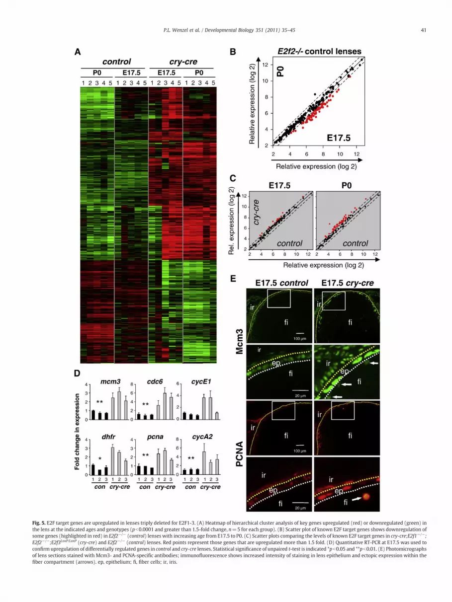
Fig. 5. E2F target genes are upregulated in lenses triply deleted for E2F1-3. (A) Heatmap of hierarchical cluster analysis of key genes upregulated (red) or downregulated (green) inthe lens at the indicated ages and genotypes (pb0.0001 and greater than 1.5-fold change, n=5 for each group). (B) Scatter plot of known E2F target genes shows downregulation ofsome genes (highlighted in red) in E2f2−/− (control) lenses with increasing age from E17.5 to P0. (C) Scatter plots comparing the levels of known E2F target genes in cry-cre;E2f1−/−;E2f2−/−;E2f3LoxP/LoxP (cry-cre) and E2f2−/− (control) lenses. Red points represent those genes that are upregulated more than 1.5 fold. (D) Quantitative RT-PCR at E17.5 was used toconfirm upregulation of differentially regulated genes in control and cry-cre lenses. Statistical significance of unpaired t-test is indicated *pb0.05 and **pb0.01. (E) Photomicrographsof lens sections stained with Mcm3- and PCNA-specific antibodies; immunofluorescence shows increased intensity of staining in lens epithelium and ectopic expression within thefiber compartment (arrows). ep, epithelium; fi, fiber cells; ir, iris.
41P.L. Wenzel et al. / Developmental Biology 351 (2011) 35–45

Fig. 6. Cell cycle inhibitors are activated by loss of E2F. (A) Heatmap rendering of changes in p53 signaling as determined by global gene expression profiling. Statistically significantchanges (pb0.001) that were 1.5-fold or greaterwere determined by class comparison analysis and are indicated by red boxes (upregulated) and green boxes (downregulated). (B) Geneexpression changes in key p53 targetswere validated by realtime RT PCR. Statistical significance of unpaired t-test is indicated *pb0.05. (C) Immunodetection of p19Arf and p21Cip1 on lenssections confirms activation of these downstream targets of p53. (D) Quantification of immunostaining of p21Cip1 in epithelial cells and of p19Arf in fiber cells. Statistical significance ofunpaired t-test is indicated *pb0.05 and **pb0.01.
42 P.L. Wenzel et al. / Developmental Biology 351 (2011) 35–45
refine and extend the analysis by tissue-specific ablation to demon-strate that Rb functions in the lens are cell autonomous. Together withthe analysis of E2f triply-deficient lenses, these latter results supportthe idea that E2F3 promotes ectopic cell proliferation specifically inthe context of Rb deficiency.
Discussion
Contrary to many previous studies performed in cell culture, thedata presented here suggest that E2F1-3 are not required forproliferation of lens epithelial cells (Sharma et al., 2006; Timmers

43P.L. Wenzel et al. / Developmental Biology 351 (2011) 35–45
et al., 2007; Wu et al., 2001). Instead, we show that E2F1-3 play acritical role in cell survival. It remains to be determined whether E2Fsare required in other tissues of the adult mouse, but it would appearfrom parallel analyses of neuroectodermal derivatives of the retinaand endodermal derivatives of the small intestine (Chen et al., 2009;Chong et al., 2009) that epithelial lineages in general do not requireE2F1-3 to proliferate but do require them for cell survival. Indeed, it is
Fig. 7. Cry-cremediated deletion of RbLoxP recapitulates hallmark phenotypes of Rb-null lenseBrdU incorporation illustrates the dramatic defect in the pattern of DNA synthesis in cry-crconcurrent loss of E2f3. ep, epithelium; fi, fiber cells; ir, iris. (B) TUNEL detection of apoptoticphenotype by loss of E2f3. (C) The percentage of TUNEL- and BrdU-positive cells in lenses ocells of cry-cre; RbLoxP/LoxP but not cry-cre; RbLoxP/LoxP; E2f3LoxP/LoxP lenses. (D) Quantificationrepresent standard deviation. Significance of unpaired t-test indicated by **pb0.01 and ***p
also very likely that the majority of embryonic cell types do notrequire E2F1-3 for proliferation since triple mutant embryos cansurvive to E9.5 (Chong et al., 2009).
Since lens morphogenesis proceeds normally in the absence ofE2F1-3 until late gestation, it is unlikely that the transcriptionalcontrol of fiber cell differentiation is disrupted prior to this stage.Primary fiber cell differentiation appears to have taken place normally
s and confirms cell autonomous function of Rb in the E13.5 lens. (A) Immunodetection ofe; RbLoxP/LoxP lenses. Cellular organization within the fiber compartment is restored bycells and staining with hematoxylin and eosin show an almost complete rescue of the Rbf indicated genotypes shows a profound increase in cell death and proliferation in fiberof the percentage of TUNEL- and BrdU-positive cells in the lens epithelium. Error barsb0.001.
44 P.L. Wenzel et al. / Developmental Biology 351 (2011) 35–45
and major crystallin proteins are present with no noticeabledisruption of distribution within the lens. In late gestation, however,there are some significant changes in the abundance of key transcriptsin lens development (Fig. 2B). Most notably, lens epithelial cellassociated transcripts (Pax6 and connexin 43 (Gja1)) were elevatedand lens fiber cell associated transcripts (Prox1 and connexin 46(Gja3)) were reduced. These changes in gene expression mayrepresent a fundamental shift in the requirement of E2Fs in secondaryversus primary fiber cell differentiation, or may result as a secondaryeffect of decreased lens cell survival. Whether any of these transcriptsrepresent direct E2F target genes will require further investigation.
Our results support the view that E2F1-3 family membersnormally function as transcriptional repressors at a time in develop-ment when cells are exiting the cell cycle (Aslanian et al., 2004; Leoneet al., 2000). We show that loss of E2F1-3 in the lens results in ectopicexpression of E2F target genes, many of which are required fornucleotide metabolism and DNA synthesis. It is interesting to notethat upregulation of Mcm3 and PCNA was particularly acute in cellstransitioning to G0 and that the Cdk inhibitors p27 and p57 weredownregulated (Figs. 2B, 5E). Further, this inappropriate expressionof E2F targets was accompanied by a doubling of the number ofepithelial cells incorporating BrdU and the appearance of DNAreplication in fiber cells that are quiescent in normal lenses(Fig. 1D). Given recent work establishing redundant roles for E2Fsin embryonic development (Hurst et al., 2008; Tsai et al., 2008), wewould suggest that overlapping functions of E2F1-3 lie primarily ingene repression.
The analysis of Rb and Rb-E2f3 mutant lenses suggests that thepreviously described requirement for E2Fs in proliferation andtranscriptional activation may be restricted to specific cellularcontexts where Rb protein is inactivated. Such situations couldinclude abnormal proliferation in response to genetic alterations inRb or normal proliferation induced in response to liver damage,immune cell activation, and acute growth factor activation. Earlierwork has characterized the lenses of Rb-E2F3-deficient embryos(Saavedra et al., 2002; Ziebold et al., 2001), yet the conditionaldeletion strategy here demonstrates for the first time that theapoptotic defect found in Rb-null lenses is not a secondary defectcaused by placental deficiencies (de Bruin et al., 2003; Wenzel et al.,2007; Wu et al., 2003) but rather is due to a cell autonomousrequirement for Rb function to balance transcriptional activation byE2F3. In summary, the analysis of TKO lenses provides compelling invivo evidence for a role of E2F1, E2F2 and E2F3 in transcriptionalrepression and cell survival during normal developmental programs,and for a role in transcriptional activation and cell proliferation duringspecialized circumstances restricted to inactivation of Rb and itsassociated repressive cofactors, including histone deacetylases, SWI/SNF, lysine methyltransferases, arginine methyltransferases, and DNAmethyltransferases.
Supplementarymaterials related to this article can be found onlineat doi:10.1016/j.ydbio.2010.12.025.
Conflict of interest statement
The authors declare that they have no competing financialinterests.
Acknowledgments
We thank J. Moffitt and L. Rawahneh for histological support. Thiswork was funded by NIH grants to G.L. (R01CA85619, R01CA82259,R01HD047470, P01CA097189) andM.L.R. (R01EY012995) and an NIHtraining grant to P.L.W. (5 T32 CA106196-04). G.L. is the recipient ofThe Pew Charitable Trust Scholar Award and the Leukemia &Lymphoma Society Scholar Award.
References
Aslanian, A., Iaquinta, P.J., Verona, R., Lees, J.A., 2004. Repression of the Arf tumorsuppressor by E2F3 is required for normal cell cycle kinetics. Genes Dev. 18,1413–1422.
Black, E.P., Hallstrom, T., Dressman, H.K., West, M., Nevins, J.R., 2005. Distinctions in thespecificity of E2F function revealed by gene expression signatures. Proc. Natl Acad.Sci. USA 102, 15948–15953.
Bracken, A.P., Ciro, M., Cocito, A., Helin, K., 2004. E2F target genes: unraveling thebiology. Trends Biochem. Sci. 29, 409–417.
Cai, F., Zhu, J., Chen, W., Ke, T., Wang, F., Tu, X., Zhang, Y., Jin, R., Wu, X., 2010. A novelPAX6 mutation in a large Chinese family with aniridia and congenital cataract. Mol.Vis. 16, 1141–1145.
Chen, D., Pacal, M., Wenzel, P., Knoepfler, P.S., Leone, G., Bremner, R., 2009. Division andapoptosis of E2f-deficient retinal progenitors. Nature 462, 925–929.
Chong, J.L., Wenzel, P.L., Saenz-Robles, M.T., Nair, V., Ferrey, A., Hagan, J.P., Gomez, Y.M.,Sharma, N., Chen, H.Z., Ouseph, M., Wang, S.H., Trikha, P., Culp, B., Mezache, L.,Winton, D.J., Sansom, O.J., Chen, D., Bremner, R., Cantalupo, P.G., Robinson, M.L.,Pipas, J.M., Leone, G., 2009. E2f1-3 switch from activators in progenitor cells torepressors in differentiating cells. Nature 462, 930–934.
Dahiya, A., Wong, S., Gonzalo, S., Gavin, M., Dean, D.C., 2001. Linking the Rb andpolycomb pathways. Mol. Cell 8, 557–569.
de Bruin, A., Wu, L., Saavedra, H.I., Wilson, P., Yang, Y., Rosol, T.J., Weinstein, M.,Robinson, M.L., Leone, G., 2003. Rb function in extraembryonic lineages suppressesapoptosis in the CNS of Rb-deficient mice. Proc. Natl Acad. Sci. 100, 6546–6551.
Duncan, M.K., Kozmik, Z., Cveklova, K., Piatigorsky, J., Cvekl, A., 2000. Overexpression ofPAX6(5a) in lens fiber cells results in cataract and upregulation of (alpha)5(beta)1integrin expression. J. Cell Sci. 113 (Pt 18), 3173–3185.
Eisen, M.B., Spellman, P.T., Brown, P.O., Botstein, D., 1998. Cluster analysis and display ofgenome-wide expression patterns. Proc. Natl Acad. Sci. USA 95, 14863–14868.
Hallstrom, T.C., Nevins, J.R., 2009. Balancing the decision of cell proliferation and cellfate. Cell Cycle 8, 532–535.
Hanson, I., Churchill, A., Love, J., Axton, R., Moore, T., Clarke, M., Meire, F.,van Heyningen, V., 1999. Missense mutations in the most ancient residues of thePAX6 paired domain underlie a spectrum of human congenital eye malformations.Hum. Mol. Genet. 8, 165–172.
Hurst, C.D., Tomlinson, D.C., Williams, S.V., Platt, F.M., Knowles, M.A., 2008. Inactivationof the Rb pathway and overexpression of both isoforms of E2F3 are obligate eventsin bladder tumours with 6p22 amplification. Oncogene 27, 2716–2727.
Jacks, T., Fazeli, A., Schmitt, E.M., Bronson, R.T., Goodell, M.A., Weinberg, R.A., 1992.Effects of an Rb mutation in the mouse. Nature 359, 295–300.
Kong, L.J., Chang, J.T., Bild, A.H., Nevins, J.R., 2007. Compensation and specificity offunction within the E2F family. Oncogene 26, 321–327.
Leone, G., Nuckolls, F., Ishida, S., Adams, M., Sears, R., Jakoi, L., Miron, A., Nevins, J.R.,2000. Identification of a novel E2F3 product suggests a mechanism for determiningspecificity of repression by Rb proteins. Mol. Cell. Biol. 20, 3626–3632.
Lovicu, F.J., Steven, P., Saika, S., McAvoy, J.W., 2004. Aberrant lens fiber differentiation inanterior subcapsular cataract formation: a process dependent on reduced levels ofPax6. Investig. Ophthalmol. Vis. Sci. 45, 1946–1953.
Luo, R.X., Postigo, A.A., Dean, D.C., 1998. Rb interacts with histone deacetylase to represstranscription. Cell 92, 463–473.
Mootha, V.K., Lindgren, C.M., Eriksson, K.F., Subramanian, A., Sihag, S., Lehar, J.,Puigserver, P., Carlsson, E., Ridderstrale, M., Laurila, E., Houstis, N., Daly, M.J.,Patterson, N., Mesirov, J.P., Golub, T.R., Tamayo, P., Spiegelman, B., Lander, E.S.,Hirschhorn, J.N., Altshuler, D., Groop, L.C., 2003. PGC-1alpha-responsive genesinvolved in oxidative phosphorylation are coordinately downregulated in humandiabetes. Nat. Genet. 34, 267–273.
Morris, L., Allen, K.E., La Thangue, N.B., 2000. Regulation of E2F transcription by cyclinE-Cdk2 kinase mediated through p300/CBP co-activators. Nat. Cell Biol. 2, 232–239.
Nielsen, S.J., Schneider, R., Bauer, U.M., Bannister, A.J., Morrison, A., O'Carroll, D.,Firestein, R., Cleary, M., Jenuwein, T., Herrera, R.E., Kouzarides, T., 2001. Rb targetshistone H3 methylation and HP1 to promoters. Nature 412, 561–565.
Ren, B., Cam, H., Takahashi, Y., Volkert, T., Terragni, J., Young, R.A., Dynlacht, B.D., 2002.E2F integrates cell cycle progression with DNA repair, replication, and G(2)/Mcheckpoints. Genes Dev. 16, 245–256.
Robertson, K.D., Ait-Si-Ali, S., Yokochi, T., Wade, P.A., Jones, P.L., Wolffe, A.P., 2000.DNMT1 forms a complex with Rb, E2F1 and HDAC1 and represses transcriptionfrom E2F-responsive promoters. Nat. Genet. 25, 338–342.
Robinson, M.L., Overbeek, P.A., Verran, D.J., Grizzle, W.E., Stockard, C.R., Friesel, R.,Maciag, T., Thompson, J.A., 1995. Extracellular FGF-1 acts as a lens differentiationfactor in transgenic mice. Development 121, 505–514.
Rowland, B.D., Denissov, S.G., Douma, S., Stunnenberg, H.G., Bernards, R., Peeper, D.S.,2002. E2F transcriptional repressor complexes are critical downstream targets ofp19(ARF)/p53-induced proliferative arrest. Cancer Cell 2, 55–65.
Saavedra, H.I., Wu, L., de Bruin, A., Timmers, C., Rosol, T.J., Weinstein, M., Robinson, M.L.,Leone, G., 2002. Specificity of E2F1, E2F2 and E2F3 in mediating Rb function. CellGrowth Differ. 13, 215–225.
Sharma, N., Timmers, C., Trikha, P., Saavedra, H.I., Obery, A., Leone, G., 2006. Control ofthe p53-p21CIP1 Axis by E2f1, E2f2, and E2f3 is essential for G1/S progression andcellular transformation. J. Biol. Chem. 281, 36124–36131.
Soriano, P., 1999. Generalized lacZ expression with the ROSA26 Cre reporter strain.Nat. Genet. 21, 70–71.
Timmers, C., Sharma, N., Opavsky, R., Maiti, B., Wu, L., Wu, J., Orringer, D., Trikha, P.,Saavedra, H.I., Leone, G., 2007. E2f1, E2f2, and E2f3 control E2F target expressionand cellular proliferation via a p53-dependent negative feedback loop. Mol. Cell.Biol. 27, 65–78.

45P.L. Wenzel et al. / Developmental Biology 351 (2011) 35–45
Tsai, S.Y., Opavsky, R., Sharma, N., Wu, L., Naidu, S., Nolan, E., Feria-Arias, E., Timmers, C.,Opavska, J., de Bruin, A., Chong, J.L., Trikha, P., Fernandez, S.A., Stromberg, P., Rosol,T.J., Leone, G., 2008. Mouse development with a single E2F activator. Nature 454,1137–1141.
Vandel, L., Nicolas, E., Vaute, O., Ferreira, R., Ait-Si-Ali, S., Trouche, D., 2001.Transcriptional repression by the retinoblastoma protein through the recruitmentof a histone methyltransferase. Mol. Cell. Biol. 21, 6484–6494.
Vernell, R., Helin, K., Muller, H., 2003. Identification of target genes of the p16INK4A-pRB-E2F pathway. J. Biol. Chem. 278, 46124–46137.
Weinmann, A.S., Yan, P.S., Oberley, M.J., Huang, T.H., Farnham, P.J., 2002. Isolatinghuman transcription factor targets by coupling chromatin immunoprecipitationand CpG island microarray analysis. Genes Dev. 16, 235–244.
Wells, J., Graveel, C.R., Bartley, S.M., Madore, S.J., Farnham, P.J., 2002. The identificationof E2F1-specific target genes. Proc. Natl Acad. Sci. USA 99, 3890–3895.
Wenzel, P.L., Wu, L., de Bruin, A., Chong, J.L., Chen, W.Y., Dureska, G., Sites, E., Pan, T.,Sharma, A., Huang, K., Ridgway, R., Mosaliganti, K., Sharp, R., Machiraju, R., Saltz, J.,Yamamoto, H., Cross, J.C., Robinson, M.L., Leone, G., 2007. Rb is critical in amammalian tissue stem cell population. Genes Dev. 21, 85–97.
Wigle, J.T., Chowdhury, K., Gruss, P., Oliver, G., 1999. Prox1 function is crucial for mouselens-fibre elongation. Nat. Genet. 21, 318–322.
Wu, L., Timmers, C., Maiti, B., Saavedra, H.I., Sang, L., Chong, G.T., Nuckolls, F.,Giangrande, P., Wright, F.A., Field, S.J., Greenberg, M.E., Orkin, S., Nevins, J.R.,Robinson, M.L., Leone, G., 2001. The E2F1-3 transcription factors are essential forcellular proliferation. Nature 414, 457–462.
Wu, L., de Bruin, A., Saavedra, H.I., Starovic, M., Trimboli, A., Yang, Y., Opavska, J., Wilson,P., Thompson, J.C., Ostrowski, M.C., Rosol, T.J., Woollett, L., Weinstein, M., Cross, J.C.,Robinson, M.L., Leone, G., 2003. Extra-embryonic function of Rb is essential forembryonic development and viability. Nature 421, 942–947.
Xu, X., Bieda, M., Jin, V.X., Rabinovich, A., Oberley, M.J., Green, R., Farnham, P.J., 2007.A comprehensive ChIP-chip analysisof E2F1, E2F4, andE2F6 innormal and tumor cellsreveals interchangeable roles of E2F family members. Genome Res. 17, 1550–1561.
Zhang, H.S., Gavin, M., Dahiya, A., Postigo, A.A., Ma, D., Luo, R.X., Harbour, J.W., Dean,D.C., 2000. Exit from G1 and S phase of the cell cycle is regulated by repressorcomplexes containing HDAC-Rb-hSWI/SNF and Rb-hSWI/SNF. Cell 101, 79–89.
Zhao, H., Yang, Y., Rizo, C.M., Overbeek, P.A., Robinson, M.L., 2004. Insertion of a Pax6consensus binding site into the alphaA-crystallin promoter acts as a lens epithelialcell enhancer in transgenic mice. Investig. Ophthalmol. Vis. Sci. 45, 1930–1939.
Ziebold, U., Reza, T., Caron, A., Lees, J.A., 2001. E2F3 contributes both to theinappropriate proliferation and to the apoptosis arising in Rb mutant embryos.Genes Dev. 15, 386–391.


