Embed Size (px)
Citation preview

Review ArticleCellular Players in Skeletal Muscle Regeneration
Laura Cristina Ceafalan,1,2 Bogdan Ovidiu Popescu,2,3 and Mihail Eugen Hinescu1,2
1 Department of Cell Biology and Histology, School of Medicine, “Carol Davila” University of Medicine and Pharmacy,8 Eroii Sanitari, 050474 Bucharest, Romania
2Department of Molecular Medicine and Neuroscience, “Victor Babes,” Institute of Pathology, 99-101 Splaiul Independentei,050096 Bucharest, Romania
3 Department of Neurology, Colentina Clinical Hospital (CDPC), School of Medicine, “Carol Davila” University of Medicine andPharmacy, 19-21 Sos. Stefan cel Mare, 020125 Bucharest, Romania
Correspondence should be addressed to Bogdan Ovidiu Popescu; [email protected]
Received 7 December 2013; Revised 12 January 2014; Accepted 28 January 2014; Published 23 March 2014
Academic Editor: P. Bryant Chase
Copyright © 2014 Laura Cristina Ceafalan et al.This is an open access article distributed under the Creative Commons AttributionLicense, which permits unrestricted use, distribution, and reproduction in anymedium, provided the originalwork is properly cited.
Skeletal muscle, a tissue endowed with remarkable endogenous regeneration potential, is still under focused experimentalinvestigation mainly due to treatment potential for muscle trauma and muscular dystrophies. Resident satellite cells with stem cellfeatures were enthusiastically described quite a long time ago, but activation of these cells is not yet controlled by any medicalinterventions. However, after thorough reports of their existence, survival, activation, and differentiation there are still manyquestions to be answered regarding the intimate mechanism of tissue regeneration. This review delivers an up-to-date inventoryof the main known key players in skeletal muscle repair, revealed by various models of tissue injuries in mechanical trauma, toxiclesions, andmuscular dystrophy. A better understanding of the spatial and temporal relationships between various cell populations,with different physical or paracrine interactions and phenotype changes induced by local or systemic signalling, might lead to amore efficient approach for future therapies.
1. Introduction
Adult mammalian skeletal muscle is a dynamic tissue interms of remodelling, repair, and regeneration.The cells mayundergo physiological changes based on everyday physicalactivity (atrophy, hypertrophy, or fibre type switch). Adultskeletal muscle cells are also able to repair focal damagesinduced by muscle contraction to the sarcolemma or myofib-rils, with no inflammatory reaction and preservation of thehistological features.
Moreover, due to the superficial location, skeletal muscletissue is constantly subjected to different grades of traumaticinjuries that may cause necrosis of entire cells or only of fibresegments. New myofibres will be formed in the process ofmuscle regeneration.
Skeletal muscle regeneration is a complex phenomenonthat involves many regulatory processes that require a closecollaboration of two major cellular categories: stem/progeni-tor cells and surrounding supporting interstitial cells. Bydirect contact or by releasing soluble factors, different types
of interstitial cells are responsible either for the maintenanceof the stem cell niche in the normal tissue or for recruitingof different pools of stem/progenitor cells during muscleregeneration.
This review focuses on recent advances in the cellular andmolecular biology of skeletal muscle regeneration based oncell populations described to play a role in this process. This“social” context is summarized in Table 1 and Figure 1.
2. Steps in Skeletal Muscle Regenerationfollowing Acute Injuries
Mechanical acute injuries lead to muscle fiber destructionby disruption of plasma membrane and basal lamina, sub-sequent calcium inflow, and necrosis by autodigestion oreventually apoptosis.
Animal studies provided evidence that the healing pro-cess after direct trauma requires three steps following necrosis,interrelated and time dependent, as described below [1, 2].
Hindawi Publishing CorporationBioMed Research InternationalVolume 2014, Article ID 957014, 21 pageshttp://dx.doi.org/10.1155/2014/957014

2 BioMed Research International
Terminalbutton
Terminalbutton
Terminalbutton
endo
endo
Capillary
MCP-1, TNF𝛼, VEGF, IGF-1,FGF-2, PDGF-BB, HGF, IL-6,IL-8 TNF𝛼
Pericyte/mesoangioblast
Tcf4
SC
SC
IL-6IL-10IL-1CCL2
M2
LysisiNOS
M1
SK-34
NeMPO
Myoblast
Myoblast
TNF𝛼,
TNF𝛼,
TNF𝛼,
VEGF,Ang-1
Mast cell
Myotube
chymase,tryptase,IL-1, IL-6
TNF𝛼, histamine,
Schwanncell
CTNFNGFIGF-1
Schwanncell
CTNFNGFIGF-1
Schwanncell
CTNFNGFIGF-1fib
SDF-1?
Muscle-derivedstem cells
PICs
MPs
ACs
SMC
MECs
Bonemarrow-derived stemcells
AC 133+
(MDSCs)
adip
TC
PDGFR𝛼
IL1𝛽
IFN𝛾
IFN𝛾,
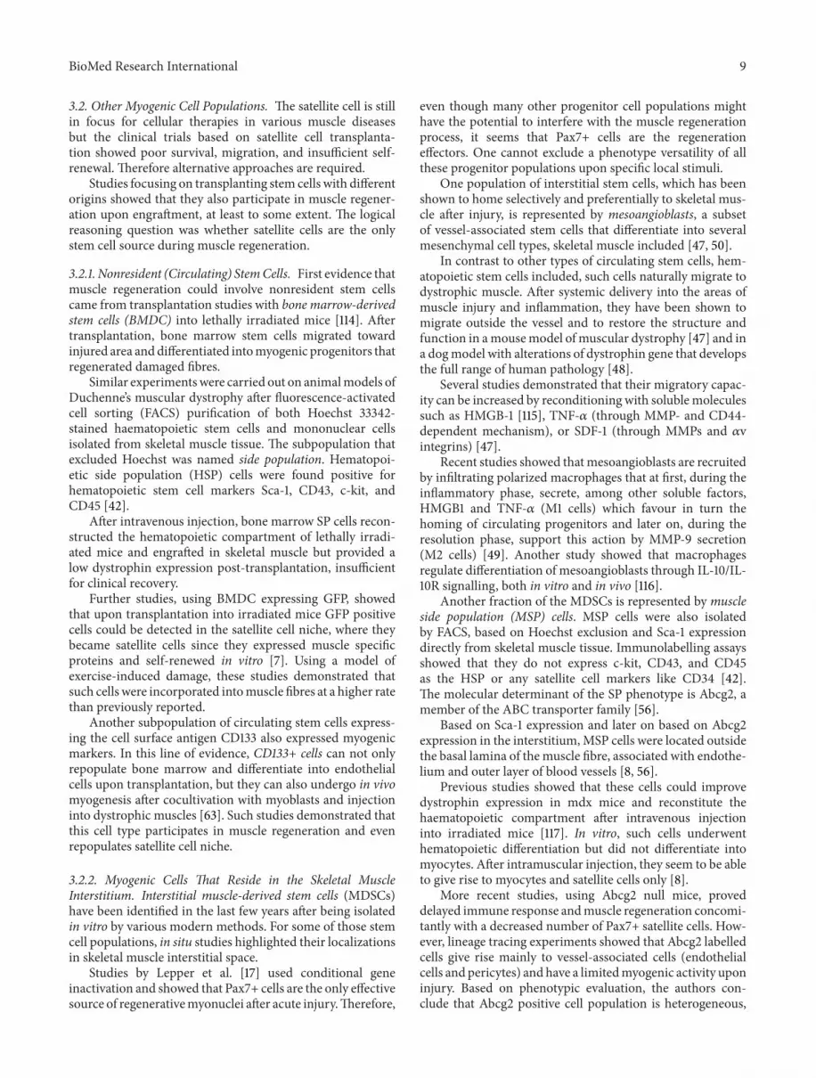
Figure 1: Synoptic view on the skeletal muscle interstitial space. Different stem cell populations become activated after acute injury (green),differentiate, and fuse to form myotubes with the support provided by various interstitial and blood derived cells (blue) either by physicalcontact or paracrine signalling. Satellite cell (SC); endothelial cell (endo); adipocyte (adip); mesenchymal progenitors (MPs) include thePDGFR𝛼+ progenitors, the FAPs, and Tie2+; skeletogenic progenitors; PW1+/Pax7–interstitial cells (PICs); SK-34 cells (SK-34); telocytes(TCs); SDF-1 skeletal muscle-derived fibroblast (fib SDF-1); macrophages (M1/M2); neutrophils (Ne); myogenic endothelial cells (MECs);Tcf4 positive fibroblasts (Tcf4); adventitial cells (ACs); smooth muscle cells (SMC).
2.1. Degeneration and Inflammatory Response. It starts withinthe first minutes following injury and lasts for up to 2 weeks.The affected site is invaded by leukocytes and macrophages,actively secreting cytokines and growth factors that not onlyamplify the inflammatory response, but also take part in thesecond phase of muscle regeneration.
2.2. Regeneration/Repair Phase. It initiates in the first weekafter injury and peaks at 2 weeks, and consists of threemajor stages starting with the activation and differentiationof muscle stem cells followed by maturation of the myofibresand paralleled by formation of new vessels by angiogenesisto revascularize the newly formed myofibres. Those key pro-cesses are orchestrated by a large panel of signals originatingin the blood stream or in the local cellular environment.
2.3. Scar Formation. It begins during the 2nd week afterinjury and increases over time. The appearance of scar tissueimpairs complete muscle regeneration.
Naturally, this time line can vary greatly depending onspecies andwithin the same species depending on injury typeand severity or even on the individual metabolic state.
3. Muscle Stem/Progenitor Cells
3.1. Satellite Cells. Themost studied and commonly acceptedprogenitor cell population in postnatal skeletal muscle isstill represented, even after 50 years since their discovery,by the satellite cells [3]. Such cells were originally identifiedby electron microscopy based on their particular location,accompanying adult skeletal muscle fibres, unsheathed by

BioMed Research International 3
Table1:Ph
enotypicsyno
psisof
thec
ellsfoun
din
skele
talm
uscle
interstitium.
Celltypes
Markers
Negative
markers
Reference
Secreted
molecules
Reference
Stem
cells
Resid
ent
Satellitecells∗#
Pax7,Pax3,Myf5,Ba
rx2,M-cadherin
,CD
206,CC
L2/C
CR2,CT
Rs,C
XCR4
,c-M
et,
CD34,p75NTR
,Tie-2,𝛼
7-intergin,
𝛽1-integrin
,VEC
AM-1,syn
decan-3and
synd
ecan-4,N
CAM/CD56,caveolin
-1,
Nestin
,SM/C-2.6,P
W1,HMGB1
CD45,Sca-1,
desm
in,M
yHC
[15,20–4
1]
Non
specified
locatio
n
Muscle
-derived
side
popu
latio
ncells
(MSP
cells)∗
Abcg2,Sca-1,CD
11,G
r-1,CD
5c-kit,CD
43,
CD45,C
D34.
[8,42,43]
PICs
#PW
1,Sca-1,CD
34,vim
entin
PDGFR𝛼
[43,44
]Slow
-adh
eringste
mcells
(SASC
s)∗
Notch1,ST
AT3,Msx1,Pax3,M
MP2
[45]
Sk-34myoendo
thelial
progenito
rs#
CD34,Sca-1,c-M
et
CD45,C
D14,
CD31,C
D49,
CD144Flk1,
MyoD,m
yf-5,
myf-6,
myogenin,
M-cadherin
,Pax-3,Pax-7.
[46]
Vessel-
associated
Mesoang
ioblasts
(vessel-a
ssociatedste
mcells)∗
E-selectin,𝛽
7,integrin,A
lCAM,C
D44
,cav1,
FGFR
1,CX
CR-4,T
NF-R,
TNFR
1a,IL-10R,
IL-4R,
RAGE,
TLR4
(HMGB1
rec),C
D13
CD34,C
D45,
CD117
CD31
[47–49]
HMGB1,V
EGF,
CXCL
12/SDF1,
CCL2
,VEG
FB,
bFGF,FG
F7,
PDGFAA,
HDGF
[49,50]
Myogenice
ndothelialcells
(MEC
s)#
CD56,C
D34,Pax7,vonWillebrand
factor,
VE-cadh
erin/CD144,UEA
-1receptor
CD45
[51,52]
Peric
ytes∗#
CCR2
,Abcg2,A
lkalinep
hosphatase,𝛼
SMA,
desm
in,N
G2,am
inop
eptid
aseA
/B,R
GS5,
CD146,PD
GFR𝛼and𝛽,N
G2
vWF,CD
31,
CD34,C
D144,
CD45,C
D56,
Pax7
[52–60]
Ang
1[29]
Adventitialcells
(ACs
)#CD
34,Sca-1,Flk1,CD
140b
c-kit,CD
31,
CD146,CD
45[61,62]

4 BioMed Research International
Table1:Con
tinued.
Celltypes
Markers
Negative
markers
Reference
Secreted
molecules
Reference
Bloo
d-deriv
edAC
133+
cells
CD133,CD
34,C
D90,C
D45
[63]
Bone
marrow-derived
SPcells
c-kit,CD
43,C
D45,Sca-1
B220,M
ac-1,
Gr-1,CD
4,CD
5,CD
8,CD
34[42]
Interstitialcells
Vascular
cells
Endo
thelialcells
vonWillebrand
factor,C
D146,CD
31,C
D34,
CD144,CD
105,Ab
cg2,Tie-2
CCR2
[52,54,56,
64–6
6]
MCP
-1,T
NF𝛼
,VEG
F,IG
F-1,
FGF-2,
PDGF-BB
,HGF,
IL-6,IL-8,
TNF𝛼
[29,33,67–
69]
Smoo
thmuscle
cells
SMA,C
CR2,𝛼-actin,caldesm
on,calpo
nin
[59,70,71]
Ang
1[29]
Nerve
cells
Schw
anncells
NCA
M/CD56,nestin
S-100,GFA
P,Kr
ox24
cytokeratin
[72–77]
CTNF,IG
F-1,
NGF
[78]
Perin
euria
lcells
Epith
elialmem
branea
ntigen
(EMA)
GLU
T-1,cla
udin-1,Z
O-1,con
nexins,
occlu
din,
vinculin,talin,desmin,titin,
spectrin,C
D34,V
E-cadh
erin,A
QP-1,
tenascin-C
cytokeratin
S-100,CD
57,
neurofi
laments
[74,79,80]
Neurons
(axon)
Nf200,𝛽
IIItub
ulin
[76]
Neuregu
lin,
calcito
ningene
related
peptide
(CGRP
)
[76]

BioMed Research International 5
Table1:Con
tinued.
Celltypes
Markers
Negative
markers
Reference
Secreted
molecules
Reference
Resid
entcells
inmuscle
conn
ectiv
etissues
heaths
Fibrob
lasts
CCR2
,vim
entin
,procollagen,
ET-TR,
FSP-1,
SMA
[60,81–85]
PDGFAA,
MCP
-1,T
NF𝛼
,Ang
-1
[29,50,67,
69]
Tcf4+fib
roblasts#
Tcf4,P
DGFR𝛼
Pax7,M
yoD,
F4/80
[86,87]
SDF-1+
muscle
-derived
fibroblasts∗
SDF-1
[24]
MPs FA
Ps∗#
Sca-1,PD
GFR𝛼,C
D34
CD45,C
D31,𝛼
7integrin
[10,56,83,
88]
IL6
[83]
PDGFR𝛼+∗
#PD
GFR𝛼,vim
entin
NG2,SM
A,
Pax7,
M-cadherin
[10]
Tie2+skele
togenic
progenito
rs∗#
Tie2,Sca-1,P
DFG
R𝛼,C
D29,C
D34
CD31,C
D45,
CD11b,CX
CR4,
c-kit,NG2
[89]
Telocytes(TC
s)∗#
c-kit,CD
34,vim
entin
,PDGFR𝛼/𝛽
[90,91]
VEG
F[91]
Bloo
d-deriv
edconn
ectiv
etissuec
ells
Macroph
ages
M1
CD68,C
D86,C
D40
,CD14,M
HCI/I
I,CC
R2[49,71]
TNF𝛼
,IL1𝛽,
TNF𝛼
,IL-6,
TNF-𝛼,IL-
1𝛽,
IL-12,IFN-𝛾,
NO,V
EGF,
HMGB1,C
CL2,
MCP
-1
[49,67]
Macroph
ages
M2
CD163,CD
36,C
D163,CD
206,MHCI/I
I,CD
14,𝛼
V-integrin,𝛼
V𝛽3I,V
CAM-1,
mem
brane-bo
undCX
3CL1,ICA
M-1
PECA
M-1
[49,92]
IGF1,IL10,
MMP-9,IL10
andLo
w:IL-6,
TNF-𝛼,IL-
1𝛽,
IFN-𝛾
[49]
Mastcells
c-kit,mastcelltrip
tase
[93,94]
TNF𝛼
,histam
ine,
chym
ase,
tryptase,IL-1,
IL-6
[67,93,95]
Polim
orph
onuclear
CXCR
2,CD
11b,C
D45
[96]
MPO
[97]

6 BioMed Research International
Table1:Con
tinued.
Celltypes
Markers
Negative
markers
Reference
Secreted
molecules
Reference
Myoblasts
Myogenin,
Pax3
(cyclin
gmyoblasts)
,𝛼-actin,m
yosin
heavychain,
CCR1
and
CCR5
,CCR
2.gp130andLIFR
,NCA
M/CD56,V
LA-4,C
X3CR
1,LFA-
1and
PECA
M-1,Sca-1,T
ie-2,p75NTR
,desmin,
Myf5,HMGB1,T
LR4,nestin
Pax7,C
CR2
[28,29,34,
39,73,78,
88,92,98–
103]
TNF𝛼
,INF𝛾
,VEG
F,Ang
-1G-C
SF/G
-CSFR
[104,105]
Myocytes/Myotubes
Myogenin,
MRF
4,CN
TFR,
NCA
M/CD56,
nestin,R
AGE
[34,73,78,
102,103]
TNF𝛼
[104]
∗
Invitro
,#in
situ.

BioMed Research International 7
their basal lamina. It was estimated that such cells account for2–5% of identifiable nuclei [4] located under the basal laminain adult muscle [5].
Satellite cells are responsible for the early growth of themyofibre and then they become mitotically quiescent [4].Throughout adult life they are frequently recruited eitherfor fibre maintenance or, when needed, for cell hypertrophyand focal repair through proliferation and fusion with themyofibre [6]. During adult muscle regeneration they differ-entiate to myogenic precursor cells (MPCs) which will dividerepeatedly before fusing into myotubes.
Early histological studies estimated that the proportion ofsatellite cells drops from 30–35% in the postnatal life to 1–4% in the adult life in mice [6]. Following studies suggestedthat in growing muscle there are two subpopulations ofsatellite cells: a fast-dividing subpopulation, responsible forfibre growth and a slow-dividing one that could function asthe source of the former or could be formed by differentcells. The overall satellite cell number decrease over timecould be explained by the waste of the fast-dividing subset asthey change from asymmetric to symmetric division, so thatmost adult satellite cells will derive from the slow-dividingpopulation. However, in normal adult muscle this populationwill remain constant even after recurrent cycles of necrosis-regeneration, which clearly suggests that the satellite cell poolis maintained by self-renewal.
At first, satellite cells were considered asmuscle precursorcells derived from a population of circulating bone marrow[7] or resident stem cells [8]. Previous studies using eitherbone marrow-derived cells or dissociated satellite cells didnot show a significant contribution to the satellite cellcompartment in animalmodels ofmuscle-induced injury andthey required a large number of transplanted cells [7].
Themesenchymalmultipotent stem cell nature of satellitecells was also suggested by further studies based on theirosteogenic and adipogenic differentiation potential, besidesthe well-known myogenic one [9]. Recently, this theorystarted to be questioned as other mesenchymal progenitors,expressing PDGFR𝛼 and located in the interstitium, repre-sent the only cell population in the adult skeletal musclecapable of differentiation along adipogenic [10] or osteogeniclineage [11].
Though, stem cell core features like proliferation, self-renewal, and differentiation capacity were eventually demon-strated over the years for the satellite cells through variousin vitro or in vivo studies [12]. One of the most convincingevidences in this respect was based on in vivo transplantationof single fibres where no more than seven satellite cellsregenerated and repopulated radiation-ablated muscles ofdystrophic mdx-nude mice [13]. However, differences havebeen noted regarding the behaviour of satellite cells depend-ing on the donor muscle group, which were suggested toresult from local environmental factors.
Such studies brought into light two very importantaspects regarding satellite cells and their proper function interms of activation and recruitment during tissue regenera-tion: the heterogeneity of this population and the importanceof the stem cell niche.
3.1.1. Phenotype. Many recent studies focusing on the iden-tification and prospective isolation of satellite cells reportedthe expression of various markers on satellite cells [14].Among them, paired box transcription factor Pax7 is theonly marker specifically expressed on both quiescent andactivated satellite cells. Previous studies on Pax7-null miceproved that the muscle develops, but the postnatal growth iscompromised; thus, Pax7 appears to be essential for satellitecell formation [15]. Unexpected evidence came from a recentstudy demonstrating that when Pax7 is inactivated in adult-hood, the satellite cells can still support muscle regeneration[16]. Apparently, Pax7 is required in the perinatal life onlyuntil satellite cells become quiescent. This study points outan interesting facet of any adult stem cell system; that is,the genetic requirement changes with age, so basically stemcells also do have an age. However, if Pax7 expression isrequired in skeletal muscle acute injury, regeneration is stilla matter of debate [17]. Very recent data on conditionalknock-out mice conclude that Pax7 is a prerequisite duringregenerative myogenesis [18] for satellite cells proliferationand differentiation regardless of age [19].
Most of the other markers, presented in Table 2, are alsoexpressed on other cell types present in the adult skeletalmuscle and therefore their presence should be correlatedwith the specific location of such cells, under the basallamina of skeletal muscle fibres. Before activation, quiescentsatellite cells do not express muscle-specific proteins, like themembers of the myogenic-regulatory-factor family, but thephenotype changes upon activation.
Recently, by developing a new monoclonal antibody,SM/C-2.6, Fukada et al. [36] identified quiescentM-cadherinpositive satellite cells in their location and further enrichedthem from adult mouse muscle. Functional studies were alsoperformed and proved that this antibody could be useful as apowerful tool for future investigations.
3.1.2. Heterogeneity. Proofs for the diversity of the myogeniccompartment have been provided by both in vitro and in vivomodels [6, 110].
In vitro approaches based on single muscle fibre culturein suspension proved that satellite cells proliferate at differentrates and define high (HPC) and low (LPC) proliferative rateclones, with fixed ratio at single fibre level [110]. The studysuggested that HPC represent a source of adipogenic tissuewithin the skeletal muscle in pathological conditions as theyspontaneously differentiated to adipocytes, but they can beconditioned towards a massive myogenic differentiation ifcocultured with LPC.The existence of such a paracrine effectmight explain why such a spontaneous adipogenic trigger ofdifferentiation is not present in healthy muscles.
More recent studies suggested that all those differencesare orchestrated by the special environment provided bythe niche. The niche is a dynamic microenvironment; it notonly maintains stem-cell quiescence, but also manages theactivation of stem cells when required.
Apparently, the orientation of the division plane withinthe niche is responsible for maintaining this diversity; planardivision generates two identical daughter cells, while apical-basal division generates asymmetric cell fates.

8 BioMed Research International
Table 2: Satellite cell markers.
Early myogenic markers
Pax7 Paired domain transcription factors [15]Pax3 Paired domain transcription factors [20]Myf5 Myogenic regulatory factor [21]
M-cadherin Cell adhesion protein [8, 23]
Transcription factors Msx1 Homeobox transcription factor [45, 99]Barx2 Homeobox transcription factor [22]
Receptors
CD206 Mannose receptor [101]CCL2/CCR2 CC chemokine MCP-1 [101]
CTRs Calcitonin receptors [106]CXCR4 Chemokine receptor [24]c-Met Tyrosine kinase receptor for HGF [25]
p75NTR Neurotrophin receptor [28]Tie-2 Tyrosine kinase receptor for angiopoietin 1 [29]
Adhesion molecules
CD34 Single-pass transmembrane sialomucin [8, 26, 27]𝛼7-intergin Cell surface attachment receptor [107]𝛽1- integrin Cell surface attachment receptor [40]VECAM-1 Adhesion molecule [41]
Syndecan-3/4 Transmembrane heparan sulfate proteoglycans [32, 108]NCAM/CD56 Neural cell adhesion molecule [33, 34]
Other
caveolin-1 Membrane protein [31, 35]lamin A/C and emerin Nuclear envelope proteins [31]
Nestin Intermediate filament protein [109]SM/C-2.6 [36]
PW1 Cell stress mediator [37]HMGB1 High mobility group box 1 protein [103]
The intrinsic heterogeneity was demonstrated by immun-ohistological analysis of phenotypic expression, especiallybased on myogenic regulatory factors (MRFs). Thus, thebasal cell expresses only Pax7, maintains a stem cell identity,and functions as a stem cell reservoir, whereas the cell thatloses contact with the basal lamina commits to a myo-genic fate and has a different profile, expressing both Pax7and Myf5 [111]. Pax7+/Myf5+ cells can undergo limitedsymmetric proliferation to generate myonuclei. This pro-cess is carefully regulated by Notch signalling pathway inorder to maintain the self-renewal property of satellite stemcells under normal conditions. Previous studies showedthat Notch-1 promotes the proliferation of satellite cells(Pax3+/Desmin−/Myf5−/MyoD−), whereas its inhibition byNumb, which prevents Notch translocation into the nucleus,leads to the commitment of the progenitor cells to themyogenic cell fate (Pax3−/Desmin+/Myf5+) and to theirmyogenic differentiation [39].
More recently, other satellite cell subpopulations havebeen defined based on variation in the expression of nonspe-cific myogenic markers such as nestin [109], CXCR-4 and 𝛽1-integrin [40], or ABCG2 and Syndecan-4 [108].
Poor survival and engraftment upon satellite cells trans-plantation could also suggest that this heterogeneous popu-lation could contain only a small proportion of cells with realstem cell features [112].
3.1.3. Activation Milestones. Satellite cell activation anddifferentiation during regeneration recapitulate embryonic
developmental steps based on similar regulatory mecha-nisms, but in a completely different environment.
The activation of satellite cells and their subsequentdifferentiation along the myogenic lineage are controlled byvarious myogenic regulatory factors (MRFs): Myf5, MyoD,myogenin, and MRF4. The activation of surviving satellitecells takes place during muscle fibre degeneration. By asym-metric division, committed satellite cells already expressMyf5[111]. Shortly after activation,MyoD is rapidly induced in vivoin satellite cells that are selected for differentiation and thecells migrate out of the sublaminar niche. The proliferatingcells known as MPCs or myoblasts downregulate Pax7 andcommit to myogenic differentiation, expressing myogenin[100] and MRF4 [99, 102]. They undergo multiple rounds ofmitosis before terminal differentiation. During terminal dif-ferentiation,myoblasts withdraw from the cell cycle, elongate,express muscle-specific genes at high levels (𝛼-actin, myosinheavy chain), and fuse to multinucleatedmuscle cells to formthe mature muscle fibre [101].
Regarding the activation trigger, there are still manysignals that could be taken into account.
Intrinsic signals could include the production of sphin-gosine-1-phosphate from the inner leaflet of the plasmamem-brane [113]. However, its synthesis is mandatorily neededfor entering the cell cycle. Extrinsic signals could be eithermechanical, which, in turn, can trigger synthesis of nitricoxide that leads to hepatocyte growth factor and follistatinrelease or other promyogenic growth factors and cytokinesinvolved in satellite cell activation [102].
BioMed Research International 9
3.2. Other Myogenic Cell Populations. The satellite cell is stillin focus for cellular therapies in various muscle diseasesbut the clinical trials based on satellite cell transplanta-tion showed poor survival, migration, and insufficient self-renewal. Therefore alternative approaches are required.
Studies focusing on transplanting stem cells with differentorigins showed that they also participate in muscle regener-ation upon engraftment, at least to some extent. The logicalreasoning question was whether satellite cells are the onlystem cell source during muscle regeneration.
3.2.1. Nonresident (Circulating) StemCells. First evidence thatmuscle regeneration could involve nonresident stem cellscame from transplantation studies with bone marrow-derivedstem cells (BMDC) into lethally irradiated mice [114]. Aftertransplantation, bone marrow stem cells migrated towardinjured area anddifferentiated intomyogenic progenitors thatregenerated damaged fibres.
Similar experimentswere carried out on animalmodels ofDuchenne’s muscular dystrophy after fluorescence-activatedcell sorting (FACS) purification of both Hoechst 33342-stained haematopoietic stem cells and mononuclear cellsisolated from skeletal muscle tissue. The subpopulation thatexcluded Hoechst was named side population. Hematopoi-etic side population (HSP) cells were found positive forhematopoietic stem cell markers Sca-1, CD43, c-kit, andCD45 [42].
After intravenous injection, bone marrow SP cells recon-structed the hematopoietic compartment of lethally irradi-ated mice and engrafted in skeletal muscle but provided alow dystrophin expression post-transplantation, insufficientfor clinical recovery.
Further studies, using BMDC expressing GFP, showedthat upon transplantation into irradiated mice GFP positivecells could be detected in the satellite cell niche, where theybecame satellite cells since they expressed muscle specificproteins and self-renewed in vitro [7]. Using a model ofexercise-induced damage, these studies demonstrated thatsuch cells were incorporated intomuscle fibres at a higher ratethan previously reported.
Another subpopulation of circulating stem cells express-ing the cell surface antigen CD133 also expressed myogenicmarkers. In this line of evidence, CD133+ cells can not onlyrepopulate bone marrow and differentiate into endothelialcells upon transplantation, but they can also undergo in vivomyogenesis after cocultivation with myoblasts and injectioninto dystrophic muscles [63]. Such studies demonstrated thatthis cell type participates in muscle regeneration and evenrepopulates satellite cell niche.
3.2.2. Myogenic Cells That Reside in the Skeletal MuscleInterstitium. Interstitial muscle-derived stem cells (MDSCs)have been identified in the last few years after being isolatedin vitro by various modern methods. For some of those stemcell populations, in situ studies highlighted their localizationsin skeletal muscle interstitial space.
Studies by Lepper et al. [17] used conditional geneinactivation and showed that Pax7+ cells are the only effectivesource of regenerativemyonuclei after acute injury.Therefore,
even though many other progenitor cell populations mighthave the potential to interfere with the muscle regenerationprocess, it seems that Pax7+ cells are the regenerationeffectors. One cannot exclude a phenotype versatility of allthese progenitor populations upon specific local stimuli.
One population of interstitial stem cells, which has beenshown to home selectively and preferentially to skeletal mus-cle after injury, is represented by mesoangioblasts, a subsetof vessel-associated stem cells that differentiate into severalmesenchymal cell types, skeletal muscle included [47, 50].
In contrast to other types of circulating stem cells, hem-atopoietic stem cells included, such cells naturally migrate todystrophic muscle. After systemic delivery into the areas ofmuscle injury and inflammation, they have been shown tomigrate outside the vessel and to restore the structure andfunction in amousemodel ofmuscular dystrophy [47] and ina dogmodel with alterations of dystrophin gene that developsthe full range of human pathology [48].
Several studies demonstrated that their migratory capac-ity can be increased by reconditioning with solublemoleculessuch as HMGB-1 [115], TNF-𝛼 (through MMP- and CD44-dependent mechanism), or SDF-1 (through MMPs and 𝛼vintegrins) [47].
Recent studies showed thatmesoangioblasts are recruitedby infiltrating polarized macrophages that at first, during theinflammatory phase, secrete, among other soluble factors,HMGB1 and TNF-𝛼 (M1 cells) which favour in turn thehoming of circulating progenitors and later on, during theresolution phase, support this action by MMP-9 secretion(M2 cells) [49]. Another study showed that macrophagesregulate differentiation of mesoangioblasts through IL-10/IL-10R signalling, both in vitro and in vivo [116].
Another fraction of the MDSCs is represented by muscleside population (MSP) cells. MSP cells were also isolatedby FACS, based on Hoechst exclusion and Sca-1 expressiondirectly from skeletal muscle tissue. Immunolabelling assaysshowed that they do not express c-kit, CD43, and CD45as the HSP or any satellite cell markers like CD34 [42].The molecular determinant of the SP phenotype is Abcg2, amember of the ABC transporter family [56].
Based on Sca-1 expression and later on based on Abcg2expression in the interstitium,MSP cells were located outsidethe basal lamina of themuscle fibre, associated with endothe-lium and outer layer of blood vessels [8, 56].
Previous studies showed that these cells could improvedystrophin expression in mdx mice and reconstitute thehaematopoietic compartment after intravenous injectioninto irradiated mice [117]. In vitro, such cells underwenthematopoietic differentiation but did not differentiate intomyocytes. After intramuscular injection, they seem to be ableto give rise to myocytes and satellite cells only [8].
More recent studies, using Abcg2 null mice, proveddelayed immune response andmuscle regeneration concomi-tantly with a decreased number of Pax7+ satellite cells. How-ever, lineage tracing experiments showed that Abcg2 labelledcells give rise mainly to vessel-associated cells (endothelialcells and pericytes) and have a limitedmyogenic activity uponinjury. Based on phenotypic evaluation, the authors con-clude that Abcg2 positive cell population is heterogeneous,

10 BioMed Research International
including MSP cells together with other vascular-interstitialcells and also circulating progenitor cells, which home toskeletal muscle upon injury [56].
PW1/PICs. Another population of muscle-derived stemcells is PW1+/Pax7− interstitial cells (PICs). They were firstdetected in the interstitium of skeletal muscle tissue whilestudying the effect of TNF on muscle homeostasis andstem cells. Such cells express PW1, a cell stress mediatorimplicated in TNF-NF𝜅B signalling and p53-mediated cellstress pathways, along with Sca-1 and CD34. Apparently, theoverall effect of TNF𝛼 administration is a delay in skeletalmuscle regeneration and this population was the one torespond to TNF𝛼 by caspase activation. Moreover, the regen-eration impairment could be overcome by caspase inhibitionsuggesting at least a regulatory role for these cells [43].
New data proved that such cells do not express othermuscle satellite cells markers such as Pax7 or MyoD andbased on lineage tracing experiments they are not derivedfrom satellite cell lineage. In vitro testing showed that theyare bipotential progenitors giving rise to both smooth andskeletal muscle cells. Moreover, they also presented a highself-renewal capacity, as another stem cell feature.
Functional studies demonstrated that they are also myo-genic in vivo, participating in tissue regeneration.
Studies on constitutive Pax7 mutant mice showed adecrease in satellite cells, as Pax7 is required for satellite cellformation, but with a proportional increase in PICs duringpostnatal growth. In vitro testing demonstrated that, in suchcases, PICs could only differentiate into smooth muscle cells,suggesting the requirement of Pax7 for their enrolment intoskeletal muscle lineage [37].
Recently, a study of Pannerec et al. [44] demonstratedthat PICs share the mesenchymal stem cell profile andcan also differentiate into adipose cells. Such data openthe discussion on to what extent this population overlapswith other populations that have been newly described inthe skeletal muscle interstitium, such as, for instance, theinterstitial adipogenic progenitors.
SK-34 cells are another population of MDSCs isolatedby FACS from mouse skeletal muscle tissue. They are CD34positive cells located in the interstitium, outside the basallamina,which donot expressmyogenicmarkers (MyoD,myf-5, myf-6, myogenin, M-cadherin, Pax-3, or Pax-7) or CD45.Most of them were positive for Sca-1 and negative for otherendothelial markers (CD14, CD31, CD49, CD144, and Flk1)and showed multilineage potential (myogenic, endothelial,and adipogenic) [46].
CD34 positive cells isolated from GFP transgenic micewere transplanted into tibialis anterior (TA) muscles ofmale immune-deficientNOD/SCIDmice.The results showedthat they are able to differentiate into myocytes, vascular,and Schwann cells in vivo, contributing to the coordinatedreconstitution of muscle fibres, blood vessels (pericytes,smooth muscle cells, and endothelial cells), and peripheralnerves, with significant structural and functional recoveryafter transplantation [118].
SASCs—slow-adhering stem cells—were isolated in vitrobased on a special preplating technique from the injuredmuscle. This heterogeneous population showed an increased
proportion of Sca-1 and CD34 positive cells, an increasedmigration, proliferation, and differentiation potential, andbetter engraftment in mdx/scid mice. Such cultures alsoshowed an upregulation of multiple genes responsible formultipotency, development, and muscle regeneration likeNotch1, STAT3, Msx1, Pax3, and MMP2 [45].
3.2.3. Vascular Progenitor Cells. One of the newest emergingconcepts in stem cell biology is that blood vessels representa systemic source of progenitor cells [52, 119]. Multipotentadult stem cells have been isolated from all layers of bloodvessel wall in skeletalmuscle and various other organs: intimacontains endothelial progenitor cells (MECs), media of smallblood vessels contains pericytes, and in large vessels smoothmuscle cell progenitors (SMPCs), and the outermost layercontains adventitial cells (ACs) [57, 61, 120]. The intersectionof these two research areas, vascular progenitors and skeletalmuscle biology, opens new and exciting perspectives forskeletal muscle regeneration.
(1) Myogenic Endothelial Cells (MECs). MECs have beenrecently identified in situ by confocal microscopy based oncoexpression of satellite cells markers (Pax7 and CD56) andendothelial markers (von Willebrand factor, VE-cadherin(CD144), UEA-1 receptor, and CD34) in between skeletalmuscle fibres from human muscle biopsies, where bloodvessels reside.
Subsequently, CD56+CD34+CD144+CD45− cells havebeen isolated by FACS as a scant cell population. Theyproliferated and survived in long-term cultures and werenot tumorigenic. Upon transplantation, they were able toregenerate muscle fibres in SCID mice skeletal muscles aftercardiotoxin-induced injury more effectively than skeletalmyoblasts. They could be clonally cultured and showed myo-genic osteogenic and chondrogenic differentiation potential[51, 52].
(2) Pericytes. Pericytes are the mural cells of the smallest divi-sion of the vascular system, the microvessels. They wereregarded for a long time as structural elements, providingstability for these vessels and also being endowed with con-tractile properties. Not only are they enclosed in the endothe-lial cells basal lamina, but they even establish close contactswith endothelial cells. The interplay between endothelial andpericytes has recently come into focus as a central processin the regulation of vascular formation, stabilization, andmaturation as long as disputed processes such as tissueremodelling and repair rely on angiogenesis.
Nowadays, pericytes emerge as a heterogeneous popula-tion in terms of origin, morphology, and marker expression[53].
In addition to their ability to modulate their phenotypealong the pericyte-smooth muscle cell axis during vesselgrowth and remodelling, pericytes have greater phenotypicplasticity, being viewed as multipotent progenitor cells withcapacity to differentiate into adipocytes, osteoblasts, andchondrocytes [121].
Several studies reported their involvement in repairprocesses in various injured tissues as they become activated

BioMed Research International 11
anddifferentiated into adipocytes, chondrocytes, Leydig cells,and even myoblasts [55, 122].
This process can be regarded as differentiation, as peri-cytes have stemcell features, or transdifferentiation of a differ-entiated cell toward a different phenotype, and this is clearlya matter of debate. However, a hypothetical continuum, frommesenchymal cells to extracellular matrix secreting fibrob-lasts and to blood vessel contractile phenotypes (pericytes orsmooth muscle cells), could be taken into consideration.
Perivascular CD146+ pericytes isolated from skeletalmuscle and nonmuscle tissues showed mesenchymal stemcells features with long-term myogenic potential both invitro and in vivo, after transplantation into SCID-mdx orSCID-NOD cardiotoxin injured mice [57]. Lineage tracingexperiments with tissue nonspecific alkaline phosphataseCreERT2 mice proved that alkaline phosphatase positivevessel-associated cells-pericytes contribute to postnatal mus-cle growth and satellite cells pool and their contribution ishighly increased during tissue regeneration [58].
4. Cells of the Stem Cell Niches
Most data on potential stem/progenitor cells populationsinvolved in skeletal muscle regeneration come from in vitrostudies after isolation, characterization, and transplantation.Less is known about their distribution and themicroenviron-ment needed for their maintenance, activation, and differen-tiation upon injury. Up to now, there are three potential stemcell niches that could be taken into consideration in skeletalmuscle tissue: the satellite cell niche, specific for skeletalmuscle, and two vascular stem cell niches (“universal” stemcell niches), one in the subendothelial zone and the second inthe adventitial vasculogenic zone [119].
Muscle satellite cells niche is a polarized microenviron-ment structured by the basal lamina unsheathing the musclefibre. The basal lamina anchors the basal side of satellite cellsthrough laminin—𝛼7𝛽1 integrin receptor [111]. The apicalpole of satellite cells is adjacent to the myofibre, where it isanchored by cell adhesion molecule M-cadherin. Integrityand composition of this niche impact on the repair processby providing the appropriate migration substrate and signalsfor satellite cells. Direct contact along with soluble factorsreleased by neighbouring nonmuscle cells represents signalsthat are conferred to satellite cells [33, 123]. Most likely, directcontact with the myofibre is necessary in order to maintainthem in a quiescence state, with low requirement for growthfactors.Thematuration compartment involves different typesof immune and stromal cells that will provide both cell-to-cellcontact and soluble factors required for satellite cell activa-tion, proliferation, and differentiation. Additional support isprovided by the particularly rich capillary network. There isa close association between satellite cells and blood vessels,with 88% of the satellite cells at less than 21 𝜇m away froma capillary [29, 124]. Besides being a nutrient supply, somestudies suggest that there is a crosstalk between activatedsatellite cells and endothelial cells during differentiation that
supports angiomyogenesis, most probably through solublefactors secretion [33]. Endothelial cells will provide insulin-like growth factor (IGF)-1, fibroblast growth factor (FGF),hepatocyte growth factor (HGF), and vascular endothelialgrowth factor (VEGF). In turn, differentiating myoblastspromote angiogenesis.
Another important regulation seems to be dependenton the neuromuscular junction [35] and periendothelialcells. Periendothelial cells promote the return to quiescenceof a subset of satellite/myogenic cells and maintain theirquiescence through Angiopoietin-1/Tie-2 signalling [125].
A new line of evidence suggested that in skeletal mus-cle the perivascular compartment represents a complexmicroenvironment, with more elaborated functions. A newconcept emerged; that is, the subendothelial zone in smallvessels and especially the area between media and adventitiaand tunica adventitia itself in large vessels provide niche-like environments for resident progenitor cells involved ingrowth, remodelling, and repair of the blood vessel wall[119, 126].
Adventitial Cells (ACs). So far, the adventitia was consideredonly as an assembly of fibroblasts, nerves, and microvesselstravelling through an extracellular matrix. Recent studiesproved that progenitor cells with the ability to form vascularstructures are present in bothmurine andhumanblood vesseladventitia and in both arterial and venous blood vessel fromvarious locations. In arteries wall, they were detected at theborder zone between media and adventitia in the so called“vasculogenic zone” based on marker profile. CD34+/CD31−cells represent a source of endothelial progenitor cells (VW-EPCs) that could form capillary sprouts expressing VEGFR2(KDR), Tie2, VE-cadherin, occludin, and CEACAM1 asthey become engaged on the endothelial cell lineage. More-over, this zone contains other cell subpopulations (CD45+mononuclear cells and CD68+macrophages) suggesting thatmost probably they all derive from a resident, scarce adultmultipotent cell population [64].
Other studies proved that adventitial layer contains clus-ters of CD34+, Sca1+, c-kit-, Flk1+, and CD140b+ cells. Theyseem to have a differentiation potential toward endothelialcells, mural cells, adipocytes, and osteogenic cells [62, 127].
CD34+/CD31− progenitors were also identified in humansaphenous vein adventitia, around vasa vasorum. Such cellscoexpressed pericytes markers such as NG2 and PDGFR𝛽.These saphenous vein-derived progenitor cells (SVPs) werefurther sorted by FACS, propagated, and characterized invitro, proving clonogenic and multilineage potential [120].
It is still not clear yet if the same progenitors act as bothvascular and tissue-specific progenitors nor if different cellpopulations settle in large, individual vessels or intraorganvessels.
However, the adventitia environment should be alsotaken into account not only for the angiogenic/vasculogenicpotential but also for analysing the regeneration processin skeletal muscle, as long as there is clear evidence thatsome vessel-associated cells could participate in this complexprocess.

12 BioMed Research International
5. Signalling Cells for Myogenic Stem CellActivation/Differentiation and Angiogenesis
Stem cell activation and angiogenesis are central, complex,and coordinated processes for muscle regeneration, far frombeing reasonably elucidated.
Besides searching for the most appropriate type of stemcell for therapy, there is a need to understand/investigatewhat is the proper microenvironment for resident stem cellspreservation and differentiation. First, in order to investigatethe stimuli that drive stem cells into myogenesis, one shouldidentify the interstitial cells that might provide the properenvironment for the maintenance of a potential interstitialstem cell population and for their directed differentiationduring muscle regeneration.
Many physical and soluble signals are needed in order tomaintain a balance between proliferation and differentiationin order to restore normal tissue architecture.
Such signals derive either frommuscle cells after injury, orfrom inflammatory or stromal cells, at their turn influencedby injury.
5.1. Inflammatory Cells and Cytokines (Paracrine Function).The inflammatory response following acute injury undergoesa series of carefully regulated steps to efficiently recover tissuehomeostasis.
During inflammatory stage different classes of leukocyteswill be sequentially recruited by the sarcoplasmic proteinsreleased by myofibre necrosis and they will constantly play arole throughout the entire process. Neutrophils are the first tocome, followed by monocytes that will become macrophagesas soon as they reach the muscle interstitium.
Recent studies demonstrated that inflammation promotesinjury, but equally the inflammatory response is criticalto skeletal muscle regeneration. However, the underlyingmolecular mechanisms still remain largely elusive.
Infiltrated leukocytes and macrophages, besides remov-ing of necrotic debris, release a large array of growth factorsand cytokines involved in attracting myogenic stem cells andpromoting angiogenesis.
5.1.1. Neutrophils. The infiltrating neutrophils contribute tosarcolemma lysis, and this process seems to be mediatedby myeloperoxidase (MPO). Muscle injury typically inducesa local increase in MPO activity that reflects neutrophilactivation, extravasation, and cytotoxicity. The number ofneutrophils increases two hours after the acute injury, butthey become undetectable 3 days later. Their involvementin striated muscle regeneration or remodelling relies on theoxidative or proteolytic modification of damaged tissue, toallow phagocytosis of debris [97]. Recent studies showedthat muscle regeneration is slower after toxic injury precededby intraperitoneal injections of antisera to neutrophils andmonocytes in order to deplete the phagocytes [128].
5.1.2. Macrophages. Macrophages represent the most impor-tant cell population throughout the first days after injury,being responsible not only for the removal of cellular debrisand apoptotic cells but also for the release of specificcytokines.
Recent data estimated the evolution of the numbers ofmacrophages after acute injury in wild-type mice skeletalmuscles. Apparently they peaked after 3 days after injury,slowly decreased up to 7 days, and returned to baseline after2 weeks following injury [34].
During the first 24 hours, the injured muscle will recruitCX3CR1lo/Ly-6C+ blood monocytes [129]. Within the mus-cle they release proinflammatory cytokines (TNF𝛼, INF𝛾,and IL1𝛽) that amplify tissue damage [101], by aNO-mediatedmechanism generated by inducible nitric oxide synthase(iNOS) [130]. These cells were named CD68+ inflammatory(M1) cells and they are activated by INF𝛾. In the next days,as the phagocytosis ends, they will suffer a phenotypic andfunctional switch by the intervention of a set of cytokinesIL-4, IL-10, and IL-13, to the activated (M2) cells, withincreased expression of the mannose receptor CD206. Suchcellswere foundnear the regeneratingmuscle fibres.There arethree subclasses of M2 cells, based on functional and molec-ular features: M2a macrophages, CD68−/CD163−/CD206+induced by IL-4 and IL-13, functioning in advanced stages ofhealing and tissue repair; M2bmacrophages that release anti-inflammatory cytokines like IL10; and M2c macrophages,CD68−/CD163+/CD206+, induced by IL-10. They can inac-tivate the M1 phenotype by IL-4 and IL-10 production toreduce muscle damage and promote myogenic differentia-tion, myofibre growth, and membrane repair [101].
According to recent findings, macrophages play a centralrole in controlling skeletalmuscle regeneration by supportingmuscle healing through remodelling the extracellular matrixand angiogenesis [131]. Both human and animal studiessuggest that such cells have also a direct influence on otherimmune cells and progenitor/stem cells proliferation andmigration and delay in differentiation of satellite cells bysecretion of various cytokines and growth factors [49].
Recently, IL-10 was proposed as a key player in survivaland differentiation of transplanted mesoangioblasts, bothin vitro and in vivo. IL-10 is actively produced by M2macrophages and they represent the major source of IL-10 aslong as their depletion restricts the expression of this cytokine[116].
Many studies indicate that TNF𝛼, another proinflamma-tory cytokine, not only has a role in activating leukocytes andadhesion molecules on endothelial cells and controlling thesynthesis of other cytokines and receptors [67], but affectsmuscle repair as well [132]. TNF𝛼 level rises in skeletalmuscle with a peak at 24 hours after crush injury especiallyin activated leukocytes and macrophages [67]. However,TNF𝛼 levels remain raised for 2 weeks after acute injury,even though inflammatory phase declines, and this rise isparalleled by an increased expression of type I TNF𝛼 receptorin injured muscle fibres [133].
It has been equally demonstrated that TNF𝛼 activatessatellite cells to enter the cell cycle and accelerates G
1-to-
S phase transition [104]. Studies on TNF-𝛼 null mutantsshowed lower level expression of proliferation and earlydifferentiation transcription factors (MyoD andMEF-2) thanwild-type after acute injury [101].

BioMed Research International 13
On the other hand, TNF𝛼 also provides inhibitory effectsduring transition from early differentiation to terminal differ-entiation by expression ofmuscle-specific genes andmyotubeformation. Such influences are carried on through NF-𝜅Bactivation that will furthermore stimulate the production ofother inflammatory cytokines such as CCL2 and IL6 [134].However, by activation of p38MAPK, an alternate signallingpathway, TNF𝛼 together with IL1, synthetized by invadingmacrophages aswell, can promotemuscle differentiation [133,135].
More recent studies demonstrated that high mobilitygroup box 1 (HMGB1) and TNF𝛼 secreted by local M1 cellsand of MMP-9 by M2 cells are responsible for recruitingmyogenic stem cells, including vessel-associated stem cells(mesoangioblasts), which have been shown to preferentiallyhome to injured skeletal muscle [49].
Even though so many studies proved multiple effectsof TNF𝛼, no defects in muscle repair or regeneration werereported in TNF𝛼 null mutant mice after muscle crushinjury. One possible explanation relies on a compensatorymechanism based on cytokine system redundancy [67].
Another proinflammatory cytokine with similar effectson muscle regeneration is IL-6. Synthesis of IL-6 is stimu-lated in macrophages by the NF-𝜅B mechanism which alsomediates the positive effects on myoblasts proliferation andtransition to early differentiation and the inhibitory effects onterminal differentiation and cell fusion [101].
In vitro studies suggested the existence of a crosstalkbetween p38 MAPK and NF-𝜅B signalling pathways withactivation of p38 preceding that of NF-𝜅B during myoblastsdifferentiation and the induction of IL-6 as an effector of themyogenic mechanism [136].
IFN-𝛾 expression was not only found to correlate withthe accumulation of macrophages after acute muscle injuries,but it was also expressed in T-cells, natural killer cells,and myoblasts. Functional in vivo studies with blockingantibodies or INF-𝛾 null mice showed impaired muscleregeneration due to restricted cell proliferation and accel-erated fibrosis. Those effects can be explained not only byimpaired macrophage activation but also by direct influenceon myogenic cells. In vitro testing showed that, by blockingINF-𝛾 receptor on C2C12 muscle cell line, their proliferationand fusion are greatly reduced [137].
Macrophages also produce TGF𝛽. TGF𝛽 levels increaserapidly upon acute injuries in parallel with macrophageinfiltration. It functions as a potent inhibitor of myogenicdifferentiation and promoter of fibrosis [88].
Functional studies on regenerating muscle based onneutralization of TGF-𝛽1 in vivo have been shown to lead toa reduction of the diameter of regenerating myofibres [129].
CC chemokines like MCP-1 (CCL2), MIP-1𝛼 (CCL3), andMIP-1𝛽(CCL4) are greatly upregulated following experimen-tal muscle injury as they were shown to induce myoblastsproliferation.
MCP-1 a member of the CC chemokine family and thereceptor CC chemokine receptor 2 (CCR2) are expressed pri-marily not only on macrophages [71] but also on fibroblasts,smooth muscle cells, or endothelial cells, even though somestudies suggest conflicting results [34, 69].
Recovery of muscle structure after acute injury is sig-nificantly impaired in mice lacking its primary receptor,CCR2 [71], primarily by limited recruitment of mono-cytes/macrophages to damaged muscles [34].
MCP-1 is a chemokine known to be majorly involvedin macrophage recruitment and activation. The absence ofMCP-1 in MCP-1−/−mice was followed by impaired macro-phage recruitment, muscle regeneration, and adipocyte accu-mulation but not as much as in CCR2−/−mice [71].
Recent data showed that MCP-1 also promotes angio-genesis [138]. However, MCP-1−/− mice presented a capil-lary density comparable to wild-type, suggesting that fewermacrophages are involved in angiogenesis than in mus-cle regeneration [71]. MCP-1 expression was increased inischemic muscle after femoral artery excision, where angio-genesis and muscle regeneration occurred, but not in thenonischemic muscles, where collateral arteries formed byarteriogenesis [139].
Previous studies performed in vitro suggested that theangiogenic effect is either based on TGF𝛽 by recruitingvascular smooth muscle cells and mesenchymal cells towardendothelial cells or by upregulating hypoxia-inducible factor1𝛼 and subsequent VEGF-A production [140]. In wild-typemice tissue VEGF level decreased after cardiotoxin-inducedinjury followed by necrosis and inflammation and it wasrestored within 7 days but it remained significantly reducedin mice lacking the CCR2 until day 21 [68].
Even if macrophages produce VEGF, they are not themajor source during skeletal muscle regeneration in acuteinjuries as long as VEGF level did not correlate with the3rd day peak in macrophage infiltration. Maximum capillarydensity was obtained only when VEGF level was restored tobaseline and then decreased to control when fibres musclereached normal cross-sectional area. Such results demon-strate that restoration of tissue VEGF levels is a CCR2-dependent process during skeletal muscle regeneration.
CCR2 is expressed only by proinflammatory M1macrophages that are initially recruited to injured tissueswhile anti-inflammatory M2 macrophages do not expressCCR2 and seem to be important in angiogenesis [71].
Recently, it has been demonstrated that intramuscularF4/80 macrophage in injured muscle is the major cellularsource of IGF-1 [34]. Many studies have proven increasedIGF-1mRNA levels duringmuscle regeneration. In vitro stud-ies also showed that IGF-1 stimulates myoblast proliferationand differentiation.
Studies performed on CCR2−/− mice with impairedrecruitment of circulating monocytes proved that the expres-sion of IGF-1 was significantly higher in wild-type micecorrelated withmacrophage infiltration—upregulation at day1 that peaks at day 3 and gradually falls toward day 21. Whencompared with intraperitoneal macrophages during acuteperitonitis, the level of IGF-I produced by intramuscularmacrophages was considerably higher.
It was previously demonstrated that local expression ofIGF-1 speeded up the regeneration of skeletal muscle afterinjury by activating satellite cells, increasing the recruitmentof other stem cells, and improving the survival of motor neu-rons. By modulating the inflammatory response and limiting

14 BioMed Research International
fibrosis, IGF-1 maintains the balance between inflammationand connective tissue remodelling [141].
Macrophages also express urokinase-type plasminogenactivator (uPA). Studies on uPA null mice have shownimpaired macrophage accumulation and muscle regenera-tion. Novak et al. [142] demonstrated that transgenic miceexpressing only macrophage-derived uPA obtained by cross-breeding mice overexpressing macrophage-derived uPA anduPA null showed normal levels of macrophage accumulation,angiogenesis, and tissue repair after acute injuries. One of thesuggested mechanisms includes the proteolytic activation ofhepatocyte growth factor that promotes myoblast prolifera-tion [143].
Besides the paracrine function defined by secretion of thislarge array of soluble factors, macrophages have been shownto establish direct cell-cell contacts with myogenic cells invitro [92]. Such studies demonstrated that macrophages canrescuemyogenic cells from apoptosis. Normally, adultmusclefibres are resistant to proapoptotic signals, but such signalscan function in myogenic cells during muscle regenerationin order to control the number and the quality of the newlyformed myofibres. This study evaluated the expression andfunction of four prosurvival cell-cell adhesion molecularsystems on macrophages and myogenic cells in vitro and invivo—VCAM-1-VLA-4, ICAM-1-LFA-1, PECAM-1-PECAM-1, and CX3CL1-CX3CR1 to prove that macrophages rescuedifferentiated myotubes from apoptosis probably in order toshelter them until they anchor themselves in the growingextracellular matrix.
5.1.3.Mast Cells. There are also reports onmast cells potentialrole in normal skeletal muscle regeneration [95]. In thenormal tissue, the number ofmast cells is very low. One studyused a mild injury model such as saline injection to prove therecruitment of mast cells from the circulation in the injectedmuscle, 8 hours after the injection.
Mast cells release not only histamine and proteases suchas chymase and tryptase but also proinflammatory cytokinessuch as TNF𝛼, IL-1, and IL-6, which rise early after acutedamage.
It appears that mast cells are involved in activation andproliferation of endothelial cells and smooth muscle cellsand by tryptase release they can also activate fibroblaststhrough the cleavage of protease-activated receptor-2 (PAR-2). Recent studies demonstrated the activation and expressionof this receptor in rat skeletal muscle that promotedmyoblastproliferation in vitro [93]. In vivo, mast cells can indirectlyinfluence myoblast proliferation by modulating macrophagerecruitment [94].
5.2. Stromal Cells. Stromal cells represent the most flexibleplayer in themyogenic cell behaviourmodulation due to theirdistribution and high mobility in the interstitial tissue. Basedon recent evidences, some distinct populations have beendescribed and they seem to be involved in adjusting stemcell quiescence, self-renewal, differentiation, and apoptosis.To what extent those populations overlap or if they representdistinct cell populations was only marginally approached.Some of such studies, as in the case of stem cell populations,
have been performed in vitro, after FACS isolation, so theirdistribution is not clear yet.
5.2.1. Telocytes. Recently, we demonstrated the presence of anew type of interstitial cell in adult skeletal muscle tissue, thetelocyte (TC) [90]. At present, the exact role of TCs is stillunder investigation, but based on morphologic assessmentassumptions have been formulated. TCs seem to connectcells of a variety of types present in the muscular tissue bytheir long, thin cell projections, named telopodes. Due totheir long-distance connections, TCs might play an essentialpart in integrating signals for skeletal muscle regulation,remodelling, and regeneration. TCs might provide not onlyparacrine signalling by releasing growth factors but alsoa framework for myogenic progenitor cells guidance dur-ing migration and differentiation after activation. Electronmicroscopy on normal skeletal muscle samples showed thatTCs are often located close to blood vessels, nerve endings,and satellite cells or even putative progenitor cells andtherefore they might offer guidance and paracrine supportwithin the stem cell niche.
5.2.2. Tcf4 Positive Fibroblasts. Recently a new regulatorymechanism of myogenesis was described in mouse tissue.This mechanism involves Tcf4 positive fibroblasts that residein themuscle connective tissue sheaths [86]. UsingTcf4-GFP-Cre mice, the study demonstrated that muscle fibroblastsregulate muscle fibre type development and maturation.
Subsequent studies showed that the reciprocal interactionof such cells with the main stem cells in adult muscle—thesatellite cells—contributes to efficient muscle regeneration.Experimental ablation of satellite cells not only impairs mus-cle regeneration but also interferes with fibroblasts functionleading to an increase in connective tissue. By ablating Tcf4+fibroblasts, satellite cells differentiate prematurely and theregenerated myofibres are smaller [87].
5.2.3. Mesenchymal Progenitors (MPs). Other quiescent cellpopulations residing in muscle interstitium that are activatedafter tissue damage were called fibro/adipogenic progen-itors (FAPs) [83]. FAPs were recently identified as mes-enchymal progenitors expressing PDGFR𝛼, Sca-1, and CD34distinct from canonical satellite cells that, upon activation,do not generate myofibres. Similar studies showed thatPDGFR𝛼+ mesenchymal progenitors can also differentiatetoward osteoblastic and smoothmuscle-like cells [10]. Appar-ently, their number increases in regeneratingmuscles withoutdifferentiation [83, 144]; their differentiation appears to beinhibited by direct contact with regenerating myofibres thatmost likely provide a local microenvironment that main-tains their undifferentiated status [10, 145]. In cocultivationexperiments, they were shown to stimulate the differentia-tion of primary myogenic progenitors, so they have beenassumed to represent a transient source of prodifferentiationsignals for proliferating myogenic progenitors during muscleregeneration. In degenerating skeletal muscles, FAPs are themajor contributors to ectopic fat cell formation. It was notyet investigated if those adipocytes inhibit myogenesis, ashas been suggested to happen in other tissues. It is also not

BioMed Research International 15
clear yet if FAPs represent a completely distinct populationor they are only a phonotypical variant of the adipocyteprogenitors described in the white adipose tissue [144]. Basedon shared PDGFR𝛼 and Sca-1 expression and the adipogenicpotential, the same question could be raised regarding therecently described Tie2+ skeletogenic progenitors [89]. Suchcells were identified in the skeletal muscle interstitium anddistributed around blood vessels, and they represent themajor source for heterotopic ossification due to their highosteogenic potential.
So far, such data could only imply the presence of a ratherheterogeneous mesenchymal progenitor cell population withdifferent subtypes selected by the microenvironment pro-vided by specific conditions of the muscle tissue.
5.2.4. Skeletal Muscle-Derived Fibroblast. Another studyreported the selection in culture of a skeletal muscle-derived fibroblast population that expressed SDF-1. The con-ditioned media collected from such cultures chemoattractedCXCR4 positive murine satellite cells. Neutralization of SDF-1 decreased this effect; therefore the authors concluded thatthe process involved SDF-1/CXCR4+ signalling pathway [24].Either those muscle-derived fibroblasts represent a distinctinterstitial cell population or they overlap with the onespreviously presented was not investigated up to now.
There is a clear tendency to include all connective tissuecells into the ambiguous family of fibroblasts, but based on theresults presented so far a general reassessment of interstitialcells, from morphological, phenotypical, and differentiationpoints of view, would be required, probably not only for theskeletal muscle connective tissue.
5.3. Vascular Cells. During regeneration, angiogenesis andmyogenesis are coordinated processes. Upon injury, destruc-tion of vascular network allows a direct crosstalk betweenexposed vascular cells and potential myogenic progenitors:angiogenesis is an absolute requirement for tissue repair and,on the other hand, vascular cells provide an extensive array ofpotent soluble factors.
Endothelial Cells. Endothelial cells stimulate growth of satel-lite cells through the secretion of a variety of growth factors,including IGF-1, VEGF, FGF-2, PDGF-BB, and HGF [33].They also secrete cytokines like IL-6, IL-8, and TNF𝛼 [29].
Acute injuries with necrosis/inflammation will also leadto endothelial cell destruction and subsequent decrease insoluble factors synthetized by this cell population, such asVEGF [68]. VEGF levels will be restored in conjunction withmuscle regeneration and capillary network reconstruction inorder to support myoblast growth and survival.
Endothelial cells also produce MCP-1 which promotesrecruitment and activation of macrophages [69] (see above).
Periendothelial cells are represented by smoothmuscle cellsorpericytes and endomysial interstitial cells that stabilize theblood vessel. It was demonstrated, both in vitro and in vivo,that such cells promote the return to quiescence of muscleprogenitors by angiopoietin-1 release, through Angiopoietin-1/Tie-2 signalling system. Angiopietin-1 inhibits growth,proliferation, and differentiation of myogenic cells. Tie-2
expression increases as myogenic cells return to quiescence.In vivo functional studies with blocking antibodies againstAng-1/Tie-2 drove satellite cells back into the cell cycle.Thus,the recovery of vascular integrity very probably becomes asignal for ending myogenesis [29].
5.4. Peripheral Nerve Cells
5.4.1. Motor Neurons. Studies comparing muscle regenera-tion in different mouse injury models showed that myoblastdifferentiation and subsequent fusion occur more rapidly inmyotoxic injuries than after crushing. The major differencebetween the two models is that not only blood vessels butalso peripheral nerves are affected during skeletal muscletraumatic injuries. In order to obtain full regeneration, rein-nervation is mandatory; otherwise it can lead to progressivemuscle atrophy [146].
Reinnervation takes place starting from the surrounding,not damaged tissue, and accounts directly for the molecularprofile of the skeletal muscle fibre generating either slow-twitch/oxidative or fast-twitch/glycolytic myofibres. Eventhough myofibre recovery from atrophy after innervationreestablishment was thought to rely on satellite cell incor-poration, recent studies on mice proved that this connectiondoes not directly stimulate satellite cells activation, as long asthere was no significant difference between the reinnervatedand contralateral muscle [147]. Moreover, this experimen-tal approach proved that even though after reinnervationsatellite cells are activated, expressing MyoD, they do notupregulate myogenin, as a differentiation marker.
Such results clearly suggest that the crosstalk betweennerves andmuscle fibres duringmuscle regeneration relies onother factors that accompany muscle and nerve injuries andcan spread in other distant locations, either as soluble factorssecreted by nonneuronal cells of the peripheral nerve, or eventhe cells themselves. This subject was scarcely approached upto now.
5.4.2. Nonneuronal Cells. One nonneuronal source of solublefactors is represented by Schwann cells of adult peripheralnerves and such an example is ciliary neurotrophic factor(CNTF). CNTF is abundantly released during muscle dam-aged accompanied by nerve injury. Previous studies sug-gested that CNTF administration duringmuscle regenerationfavours myotube differentiation [78].
Recently it was proven that CNTF supports myogenicprogenitor cell viability in vitro by the PI3-Akt signallingpathway [148].
As expected, also the CTNF receptor alpha (CTNFR𝛼)expression increases after crushing injuries involving bothmuscle and nerve. However, according to a recent studyCTNFR𝛼 is needed primarily for neuronal regenerationrather than regeneration of the muscle itself [149].
Such results clearly support the concept of nerve-musclecrosstalk following traumatic injuries.
Another neurotrophic factor that also promotes myo-blasts viability is insulin-like growth factor-1 (IGF-1). Oneimportant source could be represented by Schwann cells asstated by a study on extraocular muscle force regulation.

16 BioMed Research International
Apparently muscle cells preferentially extract IGF from thenerve than from the systemic source [150].
Upon transection, the expression levels of another neu-rotrophic factor, nerve growth factor (NGF), significantlyincrease in the peripheral nerve in all nonneuronal cells [151].Nerve growth factor has been shown to stimulate myoblastproliferation and fusion in vitro, and also in vivo, but to alesser extent [150].
NGF has been tested in other studies for modulatingmuscle-derived stem cell behaviour prior to transplantation.Apparently, in vitro direct stimulation reduces differentiationpotential but increases the engraftment of MDSC upontransplantation into dystrophic mdx mice [152].
Evenwithout nerve injury, there is a constant remodellingof the nerve terminal during degeneration and regenerationof the segment of the postsynaptic muscle fibre, as thisconnection is mandatory for functional recovery of themuscle tissue [153]. Studies on muscle fibre laser ablationand vital imaging proved that nerve terminal and Schwanncells remain in contact with the synaptic basal lamina. Asmuscle fibres regenerate, AchRs accumulate in themembraneto the synaptic sites. New junctions will be established bynew extensions of the nerve terminal, guided by processesof terminal Schwann cells outside the area of the originalcontact.
6. Concluding Remarks
The reorganization of muscle extracellular matrix after injuryis mandatory for providing the appropriate scaffold for theregenerated myofibres and their precise spatial organization.
The fine line between efficient tissue regeneration andscar formation relies on mechanisms that are still largelyunknown. In the last few years, multiple resident cell pop-ulations that support muscle progenitor cell activation weretaken into account when regeneration-therapy hypotheseswere imagined.However, keeping these cell types balance andcontrol is clearly the key for tissue homeostasis and efficientregeneration.
Connective tissue is one of the most abundant but alsothe most unpredictable type of tissues in terms of cellularand extracellular molecular composition that varies greatlyaccording to mechanical and soluble stimuli. This tissue typenot only has no problem in regenerating itself but also couldhelp or completely impair, by scar formation, the function ofneighbouring tissues after various organ injuries.
Development of new therapeutic targets requires broad-ening of the horizon of cellular therapy approaches beyondstem/progenitor cells toward a thorough investigation of theenvironmental changes and signals provided by other celltypes in or surrounding the niche, taking into account thetrivial interstitial cells.
Conflict of Interests
The authors declare that there is no conflict of interestsregarding the publication of this paper.
Acknowledgment
This study was also partially supported by the RomanianNational Research Council (CNCS) Grant no. 32/27.10.2011(PN-II-RU-TE-2011-3-0206).
References
[1] J. Huard, Y. Li, and F. H. Fu, “Muscle injuries and repair: currenttrends in research,” Journal of Bone and Joint Surgery A, vol. 84,no. 5, pp. 822–832, 2002.
[2] I. Stratos, R. Rotter, C. Eipel, T. Mittlmeier, and B. Vollmar,“Granulocyte-colony stimulating factor enhances muscle pro-liferation and strength following skeletal muscle injury in rats,”Journal of Applied Physiology, vol. 103, no. 5, pp. 1857–1863, 2007.
[3] A. Mauro, “Satellite cell of skeletal muscle fibers,”The Journal ofBiophysical and Biochemical Cytology, vol. 9, pp. 493–495, 1961.
[4] E. Schultz, M. C. Gibson, and T. Champion, “Satellite cellsare mitotically quiescent in mature mouse muscle: an EM andradioautographic study,” Journal of Experimental Zoology, vol.206, no. 3, pp. 451–456, 1978.
[5] M. Ontell, K. C. Feng, K. Klueber, R. F. Dunn, and F. Taylor,“Myosatellite cells, growth, and regeneration in murine dys-trophic muscle: a quantitative study,” Anatomical Record, vol.208, no. 2, pp. 159–174, 1984.
[6] P. S. Zammit, “All muscle satellite cells are equal, but are somemore equal than others?” Journal of Cell Science, vol. 121, no. 18,pp. 2975–2982, 2008.
[7] M. A. LaBarge and H. M. Blau, “Biological progression fromadult bone marrow to mononucleate muscle stem cell tomultinucleate muscle fiber in response to injury,” Cell, vol. 111,no. 4, pp. 589–601, 2002.
[8] A. Asakura, P. Seale, A. Girgis-Gabardo, and M. A. Rudnicki,“Myogenic specification of side population cells in skeletalmuscle,” Journal of Cell Biology, vol. 159, no. 1, pp. 123–134, 2002.
[9] A. Asakura, M. Komaki, and M. A. Rudnicki, “Muscle satel-lite cells are multipotential stem cells that exhibit myogenic,osteogenic, and adipogenic differentiation,”Differentiation, vol.68, no. 4-5, pp. 245–253, 2001.
[10] A. Uezumi, S. Fukada, N. Yamamoto, S. Takeda, and K.Tsuchida, “Mesenchymal progenitors distinct from satellite cellscontribute to ectopic fat cell formation in skeletal muscle,”Nature Cell Biology, vol. 12, no. 2, pp. 143–152, 2010.
[11] T. Oishi, A. Uezumi, A. Kanaji et al., “Osteogenic differentiationcapacity of human skeletal muscle-derived progenitor cells,”PLoS ONE, vol. 8, no. 2, Article ID e56641, 2013.
[12] F. Relaix and P. S. Zammit, “Satellite cells are essential forskeletal muscle regeneration: the cell on the edge returns centrestage,” Development, vol. 139, no. 16, pp. 2845–2856, 2012.
[13] C. A. Collins, I. Olsen, P. S. Zammit et al., “Stem cell function,self-renewal, and behavioral heterogeneity of cells from theadult muscle satellite cell niche,” Cell, vol. 122, no. 2, pp. 289–301, 2005.
[14] H. Yin, F. Price, and M. A. Rudnicki, “Satellite cells and themuscle stem cell niche,” Physiological Reviews, vol. 93, no. 1, pp.23–67, 2013.
[15] P. Seale, L. A. Sabourin, A. Girgis-Gabardo, A. Mansouri,P. Gruss, and M. A. Rudnicki, “Pax7 is required for thespecification of myogenic satellite cells,” Cell, vol. 102, no. 6, pp.777–786, 2000.

BioMed Research International 17
[16] C. Lepper, S. J. Conway, and C. Fan, “Adult satellite cells andembryonic muscle progenitors have distinct genetic require-ments,” Nature, vol. 460, no. 7255, pp. 627–631, 2009.
[17] C. Lepper, T. A. Partridge, andC. Fan, “An absolute requirementfor pax7-positive satellite cells in acute injury-induced skeletalmuscle regeneration,” Development, vol. 138, no. 17, pp. 3639–3646, 2011.
[18] S. Gunther, J. Kim, S. Kostin et al., “Myf5-positive satellite cellscontribute to Pax7-dependent long-term maintenance of adultmuscle stem cells,” Cell Stem Cell, vol. 13, no. 5, pp. 590–601,2013.
[19] J. von Maltzahn, A. E. Jones, R. J. Parks, and M. A. Rudnicki,“Pax7 is critical for the normal function of satellite cells in adultskeletalmuscle,” Proceedings of the National Academy of Sciencesof the United States of America, vol. 110, no. 41, pp. 16474–16479,2013.
[20] M. Buckingham, L. Bajard, T. Chang et al., “The formation ofskeletal muscle: from somite to limb,” Journal of Anatomy, vol.202, no. 1, pp. 59–68, 2003.
[21] D. D. W. Cornelison and B. J. Wold, “Single-cell analysis ofregulatory gene expression in quiescent and activated mouseskeletal muscle satellite cells,” Developmental Biology, vol. 191,no. 2, pp. 270–283, 1997.
[22] R. Meech, K. N. Gonzalez, M. Barro et al., “Barx2 is expressedin satellite cells and is required for normal muscle growth andregeneration,” Stem Cells, vol. 30, no. 2, pp. 253–265, 2012.
[23] A. Irintchev, M. Zeschnigk, A. Starzinski-Powitz, and A.Wernig, “Expression pattern of M-cadherin in normal, den-ervated, and regenerating mouse muscles,” DevelopmentalDynamics, vol. 199, no. 4, pp. 326–337, 1994.
[24] M. Z. Ratajczak, M. Majka, M. Kucia et al., “Expression offunctional CXCR4 by muscle satellite cells and secretion ofSDF-1 by muscle-derived fibroblasts is associated with thepresence of both muscle progenitors in bone marrow andhematopoietic stem/progenitor cells inmuscles,” StemCells, vol.21, no. 3, pp. 363–371, 2003.
[25] R. E. Allen, S. M. Sheehan, R. G. Taylor, T. L. Kendall, and G.M. Rice, “Hepatocyte growth factor activates quiescent skeletalmuscle satellite cells in vitro,” Journal of Cellular Physiology, vol.165, no. 2, pp. 307–312, 1995.
[26] J. R. Beauchamp, L. Heslop, D. S. W. Yu et al., “Expression ofCD34 and Myf5 defines the majority of quiescent adult skeletalmuscle satellite cells,” Journal of Cell Biology, vol. 151, no. 6, pp.1221–1234, 2000.
[27] L. A. S. Alfaro, S. A. Dick, A. L. Siegel et al., “CD34 promotessatellite cell motility and entry into proliferation to facilitateefficient skeletal muscle regeneration,” Stem Cells, vol. 29, no.12, pp. 2030–2041, 2011.
[28] E. Colombo, S. Romaggi, E. Medico et al., “Human neu-rotrophin receptor p75NTR defines differentiation-orientedskeletal muscle precursor cells: implications formuscle regener-ation,” Journal of Neuropathology and Experimental Neurology,vol. 70, no. 2, pp. 133–142, 2011.
[29] R. Abou-Khalil, R. Mounier, and B. Chazaud, “Regulation ofmyogenic stem cell behavior by vessel cells: the “menage a trois”of satellite cells, periendothelial cells and endothelial cells,” CellCycle, vol. 9, no. 5, pp. 892–896, 2010.
[30] D. J. Burkin and S. J. Kaufman, “The 𝛼7𝛽1 integrin in muscledevelopment and disease,”Cell and Tissue Research, vol. 296, no.1, pp. 183–190, 1999.
[31] V. F. Gnocchi, R. B. White, Y. Ono, J. A. Ellis, and P. S.Zammit, “Further characterisation of the molecular signature
of quiescent and activated mouse muscle satellite cells,” PLoSONE, vol. 4, no. 4, Article ID e5205, 2009.
[32] D. D.W. Cornelison, M. S. Filla, H. M. Stanley, A. C. Rapraeger,and B. B. Olwin, “Syndecan-3 and syndecan-4 specifically markskeletal muscle satellite cells and are implicated in satellite cellmaintenance and muscle regeneration,”Developmental Biology,vol. 239, no. 1, pp. 79–94, 2001.
[33] C. Chiristov, F. Chretien, R. Abou-Khalil et al., “Muscle satellitecells and endothelial cells: close neighbors and privilegedpartners,” Molecular Biology of the Cell, vol. 18, no. 4, pp. 1397–1409, 2007.
[34] H. Lu, D. Huang, N. Saederup, I. F. Charo, R. M. Ransohoff, andL. Zhou, “Macrophages recruited via CCR2 produce insulin-like growth factor-1 to repair acute skeletal muscle injury,” TheFASEB Journal, vol. 25, no. 1, pp. 358–369, 2011.
[35] R. W. ten Broek, S. Grefte, and J. W. von den Hoff, “Regulatoryfactors and cell populations involved in skeletal muscle regen-eration,” Journal of Cellular Physiology, vol. 224, no. 1, pp. 7–16,2010.
[36] S. Fukada, S. Higuchi, M. Segawa et al., “Purification andcell-surface marker characterization of quiescent satellite cellsfrom murine skeletal muscle by a novel monoclonal antibody,”Experimental Cell Research, vol. 296, no. 2, pp. 245–255, 2004.
[37] K. J. Mitchell, A. Pannerec, B. Cadot et al., “Identificationand characterization of a non-satellite cell muscle residentprogenitor during postnatal development,” Nature Cell Biology,vol. 12, no. 3, pp. 257–266, 2010.
[38] J. E. Anderson, “The satellite cell as a companion in skeletalmuscle plasticity: currency, conveyance, clue, connector andcolander,” Journal of Experimental Biology, vol. 209, part 12, pp.2276–2292, 2006.
[39] I. M. Conboy and T. A. Rando, “The regulation of Notch signal-ing controls satellite cell activation and cell fate determinationin postnatal myogenesis,” Developmental Cell, vol. 3, no. 3, pp.397–409, 2002.
[40] M. Cerletti, S. Jurga, C. A.Witczak et al., “Highly efficient, func-tional engraftment of skeletal muscle stem cells in dystrophicmuscles,” Cell, vol. 134, no. 1, pp. 37–47, 2008.
[41] T. L. Jesse, R. LaChance, M. F. Iademarco, and D. C. Dean,“Interferon regulatory factor-2 is a transcriptional activator inmuscle where it regulates expression of vascular cell adhesionmolecule-1,” Journal of Cell Biology, vol. 140, no. 5, pp. 1265–1276, 1998.
[42] E. Gussoni, Y. Soneoka, C. D. Strickland et al., “Dystrophinexpression in the mdx mouse restored by stem cell transplan-tation,” Nature, vol. 401, no. 6751, pp. 390–394, 1999.
[43] V. Moresi, A. Pristera, B. M. Scicchitano et al., “Tumor necrosisfactor-𝛼 inhibition of skeletal muscle regeneration is mediatedby a caspase-dependent stem cell response,” Stem Cells, vol. 26,no. 4, pp. 997–1008, 2008.
[44] A. Pannerec, L. Formicola, V. Besson, G. Marazzi, and D. A.Sassoon, “Defining skeletal muscle resident progenitors andtheir cell fate potentials,”Development, vol. 140, no. 14, pp. 2879–2891, 2013.
[45] X. Mu, G. Xiang, C. R. Rathbone et al., “Slow-adhering stemcells derived from injured skeletal muscle have improvedregenerative capacity,” The American Journal of Pathology, vol.179, no. 2, pp. 931–941, 2011.
[46] T. Tamaki, A. Akatsuka, K. Ando et al., “Identification ofmyogenic-endothelial progenitor cells in the interstitial spacesof skeletal muscle,” Journal of Cell Biology, vol. 157, no. 4, pp.571–577, 2002.

18 BioMed Research International
[47] B. G. Galvez, M. Sampaolesi, S. Brunelli et al., “Completerepair of dystrophic skeletal muscle by mesoangioblasts withenhanced migration ability,” Journal of Cell Biology, vol. 174, no.2, pp. 231–243, 2006.
[48] M. Sampaolesi, S. Blot, G.D’Antona et al., “Mesoangioblast stemcells ameliorate muscle function in dystrophic dogs,” Nature,vol. 444, no. 7119, pp. 574–579, 2006.
[49] K. Lolmede, L. Campana, M. Vezzoli et al., “Inflammatoryand alternatively activated human macrophages attract vessel-associated stem cells, relying on separate HMGB1- andMMP-9-dependent pathways,” Journal of Leukocyte Biology, vol. 85, no.5, pp. 779–787, 2009.
[50] D. Galli, A. Innocenzi, L. Staszewsky et al., “Mesoangioblasts,vessel-associated multipotent stem cells, repair the infarctedheart by multiple cellular mechanisms: a comparison withbone marrow progenitors, fibroblasts, and endothelial cells,”Arteriosclerosis, Thrombosis, and Vascular Biology, vol. 25, no.4, pp. 692–697, 2005.
[51] B. Zheng, B. Cao, M. Crisan et al., “Prospective identificationof myogenic endothelial cells in human skeletal muscle,”NatureBiotechnology, vol. 25, no. 9, pp. 1025–1034, 2007.
[52] C. Chen, M. Corselli, B. Peault, and J. Huard, “Human blood-vessel-derived stem cells for tissue repair and regeneration,”Journal of Biomedicine and Biotechnology, vol. 2012, Article ID597439, 9 pages, 2012.
[53] A. Armulik, A. Abramsson, and C. Betsholtz, “Endothe-lial/pericyte interactions,”CirculationResearch, vol. 97, no. 6, pp.512–523, 2005.
[54] R. G. Bagley, W. Weber, C. Rouleau, and B. A. Teicher, “Peri-cytes and endothelial precursor cells: cellular interactions andcontributions to malignancy,” Cancer Research, vol. 65, no. 21,pp. 9741–9750, 2005.
[55] A. Dellavalle, M. Sampaolesi, R. Tonlorenzi et al., “Pericytes ofhuman skeletal muscle are myogenic precursors distinct fromsatellite cells,”Nature Cell Biology, vol. 9, no. 3, pp. 255–267, 2007.
[56] M. J. Doyle, S. Zhou, K. K. Tanaka et al., “Abcg2 labelsmultiple cell types in skeletal muscle and participates in muscleregeneration,” Journal of Cell Biology, vol. 195, no. 1, pp. 147–163,2011.
[57] M. Crisan, S. Yap, L. Casteilla et al., “A perivascular origin formesenchymal stem cells in multiple human organs,” Cell StemCell, vol. 3, no. 3, pp. 301–313, 2008.
[58] A. Dellavalle, G. Maroli, D. Covarello et al., “Pericytes residentin postnatal skeletal muscle differentiate into muscle fibres andgenerate satellite cells,” Nature Communications, vol. 2, no. 1,article 499, 2011.
[59] D. O. Traktuev, S. Merfeld-Clauss, J. Li et al., “A population ofmultipotent CD34-positive adipose stromal cells share pericyteand mesenchymal surface markers, reside in a periendothe-lial location, and stabilize endothelial networks,” CirculationResearch, vol. 102, no. 1, pp. 77–85, 2008.
[60] D. Sun, C. O. Martinez, O. Ochoa et al., “Bone marrow-derivedcell regulation of skeletal muscle regeneration,” The FASEBJournal, vol. 23, no. 2, pp. 382–395, 2009.
[61] M. Corselli, C. W. Chen, B. Sun et al., “The tunica adventitiaof human arteries and veins as a source of mesenchymal stemcells,” Stem Cells and Development, vol. 21, no. 8, pp. 1299–1308,2012.
[62] M. W. Majesky, X. R. Dong, V. Hoglund, G. Daum, and W. M.Mahoney Jr., “The adventitia: a progenitor cell niche for thevessel wall,”Cells TissuesOrgans, vol. 195, no. 1-2, pp. 73–81, 2012.
[63] Y. Torrente, M. Belicchi, M. Sampaolesi et al., “Human cir-culating AC133+ stem cells restore dystrophin expression andameliorate function in dystrophic skeletal muscle,”The Journalof Clinical Investigation, vol. 114, no. 2, pp. 182–195, 2004.
[64] E. Zengin, F. Chalajour, U. M. Gehling et al., “Vascular wallresident progenitor cells: a source for postnatal vasculogenesis,”Development, vol. 133, no. 8, pp. 1543–1551, 2006.
[65] S. E. Duff, C. Li, J. M. Garland, and S. Kumar, “CD105 is impor-tant for angiogenesis: evidence and potential applications,”TheFASEB Journal, vol. 17, no. 9, pp. 984–992, 2003.
[66] A. Schrage, C. Loddenkemper, U. Erben et al., “Murine CD146is widely expressed on endothelial cells and is recognized by themonoclonal antibodyME-9F1,”Histochemistry and Cell Biology,vol. 129, no. 4, pp. 441–451, 2008.
[67] R. A. Collins and M. D. Grounds, “The role of tumor necrosisfactor-alpha (TNF-𝛼) in skeletal muscle regeneration: studies inTNF-𝛼(-/-) and TNF-𝛼(-/-)/LT-𝛼(-/-) mice,” Journal of Histo-chemistry and Cytochemistry, vol. 49, no. 8, pp. 989–1001, 2001.
[68] O. Ochoa, D. Sun, S. M. Reyes-Reyna et al., “Delayed angiogen-esis and VEGF production in CCR2-/- mice during impairedskeletal muscle regeneration,”American Journal of Physiology—Regulatory Integrative and Comparative Physiology, vol. 293, no.2, pp. R651–R661, 2007.
[69] S. L. Deshmane, S. Kremlev, S. Amini, and B. E. Sawaya,“Monocyte chemoattractant protein-1 (MCP-1): an overview,”Journal of Interferon and Cytokine Research, vol. 29, no. 6, pp.313–326, 2009.
[70] S. S. M. Rensen, P. A. F. M. Doevendans, and G. J. J. M. van Eys,“Regulation and characteristics of vascular smooth muscle cellphenotypic diversity,” Netherlands Heart Journal, vol. 15, no. 3,pp. 100–108, 2007.
[71] C. O. Martinez, M. J. McHale, J. T. Wells et al., “Regulation ofskeletal muscle regeneration by CCR2-activating chemokines isdirectly related to macrophage recruitment,” American Journalof Physiology—Regulatory Integrative and Comparative Physiol-ogy, vol. 299, no. 3, pp. R832–R842, 2010.
[72] J. K. Daniloff, G. Levi, and M. Grumet, “Altered expression ofneuronal cell adhesion molecules induced by nerve injury andrepair,” Journal of Cell Biology, vol. 103, no. 3, pp. 929–945, 1986.
[73] D. Ikova, T. Soukup, and J. Mokry, “Nestin expressionreflects formation, revascularization and reinnervation of newmyofibers in regenerating rat hind limb skeletal muscles,” CellsTissues Organs, vol. 189, no. 5, pp. 338–347, 2009.
[74] E. Perentes, Y. Nakagawa, G. W. Ross, C. Stanton, and L. J.Rubinstein, “Expression of epithelial membrane antigen in per-ineurial cells and their derivatives. An immunohistochemicalstudy with multiple markers,” Acta Neuropathologica, vol. 75,no. 2, pp. 160–165, 1987.
[75] Y.-G. Chen andT.M. Brushart, “The effect of denervatedmuscleand schwann cells on axon collateral sprouting,” Journal ofHandSurgery, vol. 23, no. 6, pp. 1025–1033, 1998.
[76] C.Webber and D. Zochodne, “The nerve regenerative microen-vironment: early behavior and partnership of axons andSchwann cells,” Experimental Neurology, vol. 223, no. 1, pp. 51–59, 2010.
[77] J. A. Blake and M. R. Ziman, “The characterisation of Pax3expressant cells in adult peripheral nerve,” PLoS ONE, vol. 8,no. 3, Article ID e59184, 2013.
[78] K. Kami, Y. Morikawa, M. Sekimoto, and E. Senba, “Geneexpression of receptors for IL-6, LIF, and CNTF in regeneratingskeletal muscles,” Journal of Histochemistry and Cytochemistry,vol. 48, no. 9, pp. 1203–1213, 2000.
BioMed Research International 19
[79] M. Yamamoto, N. Okui, M. Tatebe, T. Shinohara, and H.Hirata, “Regeneration of the perineurium after microsurgicalresection examined with immunolabeling for tenascin-C andalpha smooth muscle actin,” Journal of Anatomy, vol. 218, no.4, pp. 413–425, 2011.
[80] S. Pina-Oviedo and C. Ortiz-Hidalgo, “The normal and neo-plastic perineurium: a review,”Advances in Anatomic Pathology,vol. 15, no. 3, pp. 147–164, 2008.
[81] T. Goodpaster, A. Legesse-Miller, M. R. Hameed, S. C. Aisner,J. Randolph-Habecker, and H. A. Coller, “An immunohis-tochemical method for identifying fibroblasts in formalin-fixed, paraffin-embedded tissue,” Journal of Histochemistry andCytochemistry, vol. 56, no. 4, pp. 347–358, 2008.
[82] E. Alt, Y. Yan, S. Gehmert et al., “Fibroblasts sharemesenchymalphenotypes with stem cells, but lack their differentiation andcolony-forming potential,” Biology of the Cell, vol. 103, no. 4, pp.197–208, 2011.
[83] A. W. B. Joe, L. Yi, A. Natarajan et al., “Muscle injury activatesresident fibro/adipogenic progenitors that facilitate myogene-sis,” Nature Cell Biology, vol. 12, no. 2, pp. 153–163, 2010.
[84] F. Strutz, H. Okada, C. W. Lo et al., “Identification andcharacterization of a fibroblast marker: FSP1,” Journal of CellBiology, vol. 130, no. 2, pp. 393–405, 1995.
[85] A. S. Brack,M. J. Conboy, S. Roy et al., “IncreasedWnt signalingduring aging alters muscle stem cell fate and increases fibrosis,”Science, vol. 317, no. 5839, pp. 807–810, 2007.
[86] S. J.Mathew, J.M.Hansen, A. J.Merrell et al., “Connective tissuefibroblasts andTcf4 regulatemyogenesis,”Development, vol. 138,no. 2, pp. 371–384, 2011.
[87] M.M.Murphy, J. A. Lawson, S. J.Mathew, D. A. Hutcheson, andG.Kardon, “Satellite cells, connective tissue fibroblasts and theirinteractions are crucial for muscle regeneration,” Development,vol. 138, no. 17, pp. 3625–3637, 2011.
[88] K. K. Long, M. Montano, and G. K. Pavlath, “Sca-1 is negativelyregulated by TGF-𝛽1 inmyogenic cells,”TheFASEB Journal, vol.25, no. 4, pp. 1156–1165, 2011.
[89] M. N. Wosczyna, A. A. Biswas, C. A. Cogswell, and D. J.Goldhamer, “Multipotent progenitors resident in the skeletalmuscle interstitium exhibit robust BMP-dependent osteogenicactivity and mediate heterotopic ossification,” Journal of Boneand Mineral Research, vol. 27, no. 5, pp. 1004–1017, 2012.
[90] L. M. Popescu, E. Manole, C. S. Serboiu et al., “Identification oftelocytes in skeletal muscle interstitium: implication for muscleregeneration,” Journal of Cellular and Molecular Medicine, vol.15, no. 6, pp. 1379–1392, 2011.
[91] L. C. Suciu, B. O. Popescu, S. Kostin, and L. M. Popescu,“Platelet-derived growth factor receptor-beta-positive telocytesin skeletal muscle interstitium,” Journal of Cellular and Molecu-lar Medicine, vol. 16, no. 4, pp. 701–707, 2012.
[92] C. Sonnet, P. Lafuste, L. Arnold et al., “Human macrophagesrescue myoblasts and myotubes from apoptosis through a setof adhesion molecular systems,” Journal of Cell Science, vol. 119,part 12, pp. 2497–2507, 2006.
[93] E. Duchesne, M. Tremblay, and C. H. Cote, “Mast celltryptase stimulatesmyoblast proliferation, amechanism relyingon protease-activated receptor-2 and cyclooxygenase-2,” BMCMusculoskeletal Disorders, vol. 12, article 235, 2011.
[94] E. Duchesne, P. Bouchard, M. P. Roussel, and C. H. Cote, “Mastcells can regulate skeletal muscle cell proliferation by multiplemechanisms,”Muscle & Nerve, vol. 48, no. 3, pp. 403–414, 2013.
[95] Y. Kharraz, J. Guerra, C. J. Mann, A. L. Serrano, and P. Munoz-Canoves, “Macrophage plasticity and the role of inflammationin skeletal muscle repair,”Mediators of Inflammation, vol. 2013,Article ID 491497, 9 pages, 2013.
[96] G. L. Warren, L. O’Farrell, M. Summan et al., “Role of CCchemokines in skeletal muscle functional restoration afterinjury,” American Journal of Physiology—Cell Physiology, vol.286, no. 5, pp. C1031–C1036, 2004.
[97] H. X. Nguyen, A. J. Lusis, and J. G. Tidball, “Null mutationof myeloperoxidase in mice prevents mechanical activation ofneutrophil lysis of muscle cell membranes in vitro and in vivo,”Journal of Physiology, vol. 565, part 2, pp. 403–413, 2005.
[98] L. Yahiaoui, D. Gvozdic, G. Danialou, M.Mack, and B. J. Petrof,“CC family chemokines directly regulate myoblast responses toskeletal muscle injury,” Journal of Physiology, vol. 586, no. 16, pp.3991–4004, 2008.
[99] S. B. P. Charge and M. A. Rudnicki, “Cellular and molecularregulation of muscle regeneration,” Physiological Reviews, vol.84, no. 1, pp. 209–238, 2004.
[100] P. S. Zammit, J. P. Golding, Y. Nagata, V. Hudon, T. A. Partridge,and J. R. Beauchamp, “Muscle satellite cells adopt divergentfates: amechanism for self-renewal?” Journal of Cell Biology, vol.166, no. 3, pp. 347–357, 2004.
[101] J. G. Tidball and S. A. Villalta, “Regulatory interactions betweenmuscle and the immune system during muscle regeneration,”American Journal of Physiology—Regulatory Integrative andComparative Physiology, vol. 298, no. 5, pp. R1173–R1187, 2010.
[102] F. le Grand and M. A. Rudnicki, “Skeletal muscle satellite cellsand adult myogenesis,” Current Opinion in Cell Biology, vol. 19,no. 6, pp. 628–633, 2007.
[103] R. deMori, S. Straino, A. di Carlo et al., “Multiple effects of highmobility group box protein 1 in skeletal muscle regeneration,”Arteriosclerosis, Thrombosis, and Vascular Biology, vol. 27, no.11, pp. 2377–2383, 2007.
[104] Y. Li, “TNF-𝛼 is a mitogen in skeletal muscle,”American Journalof Physiology—Cell Physiology, vol. 285, no. 2, pp. C370–C376,2003.
[105] M. Hara, S. Yuasa, K. Shimoji et al., “G-CSF influences mouseskeletal muscle development and regeneration by stimulatingmyoblast proliferation,” Journal of Experimental Medicine, vol.208, no. 4, pp. 715–727, 2011.
[106] S. Fukada, A. Uezumi, M. Ikemoto et al., “Molecular signatureof quiescent satellite cells in adult skeletal muscle,” Stem Cells,vol. 25, no. 10, pp. 2448–2459, 2007.
[107] R. I. Sherwood, J. L. Christensen, I. M. Conboy et al., “Isolationof adult mouse myogenic progenitors: functional heterogeneityof cells within and engrafting skeletal muscle,” Cell, vol. 119, no.4, pp. 543–554, 2004.
[108] K. K. Tanaka, J. K. Hall, A. A. Troy, D. D. W. Cornelison, S.M. Majka, and B. B. Olwin, “Syndecan-4-expressing muscleprogenitor cells in the SP engraft as satellite cells during muscleregeneration,” Cell Stem Cell, vol. 4, no. 3, pp. 217–225, 2009.
[109] K. Day, G. Shefer, J. B. Richardson, G. Enikolopov, and Z.Yablonka-Reuveni, “Nestin-GFP reporter expression definesthe quiescent state of skeletal muscle satellite cells,” Develop-mental Biology, vol. 304, no. 1, pp. 246–259, 2007.
[110] C. A. Rossi, M. Pozzobon, A. Ditadi et al., “Clonal characteriza-tion of rat muscle satellite cells: proliferation, metabolism anddifferentiation define an intrinsic heterogeneity,” PLoS ONE,vol. 5, no. 1, Article ID e8523, 2010.

20 BioMed Research International
[111] S. Kuang, K. Kuroda, F. le Grand, and M. A. Rudnicki,“Asymmetric self-renewal and commitment of satellite stemcells in muscle,” Cell, vol. 129, no. 5, pp. 999–1010, 2007.
[112] Z. Qu-Petersen, B. Deasy, R. Jankowski et al., “Identification ofa novel population of muscle stem cells in mice: potential formuscle regeneration,” Journal of Cell Biology, vol. 157, no. 5, pp.851–864, 2002.
[113] D. Danieli-Betto, S. Peron, E. Germinario et al., “Sphingosine 1-phosphate signaling is involved in skeletalmuscle regeneration,”American Journal of Physiology—Cell Physiology, vol. 298, no. 3,pp. C550–C558, 2010.
[114] G. Ferrari, G. Cusella-De Angelis, M. Coletta et al., “Muscleregeneration by bone marrow-derived myogenic progenitors,”Science, vol. 279, no. 5356, pp. 1528–1530, 1998.
[115] R. Palumbo, M. Sampaolesi, F. de Marchis et al., “ExtracellularHMGB1, a signal of tissue damage, induces mesoangioblastmigration and proliferation,” Journal of Cell Biology, vol. 164, no.3, pp. 441–449, 2004.
[116] L. Bosurgi, G. Corna, M. Vezzoli et al., “Transplanted mesoan-gioblasts require macrophage IL-10 for survival in a mousemodel of muscle injury,” The Journal of Immunology, vol. 188,no. 12, pp. 6267–6277, 2012.
[117] H. Kawada andM. Ogawa, “Bonemarrow origin of hematopoi-etic progenitors and stem cells inmurinemuscle,”Blood, vol. 98,no. 7, pp. 2008–2013, 2001.
[118] T. Tamaki, Y. Uchiyama, Y. Okada et al., “Functional recovery ofdamaged skeletal muscle through synchronized vasculogenesis,myogenesis, and neurogenesis by muscle-derived stem cells,”Circulation, vol. 112, no. 18, pp. 2857–2866, 2005.
[119] D. Tilki, H. Hohn, B. Ergun, S. Rafii, and S. Ergun, “Emergingbiology of vascular wall progenitor cells in health and disease,”Trends in Molecular Medicine, vol. 15, no. 11, pp. 501–509, 2009.
[120] P. Campagnolo, D. Cesselli, A. Al Haj Zen et al., “Human adultvena saphena contains perivascular progenitor cells endowedwith clonogenic and proangiogenic potential,” Circulation, vol.121, no. 15, pp. 1735–1745, 2010.
[121] C. Farrington-Rock, N. J. Crofts, M. J. Doherty, B. A. Ashton,C. Griffin-Jones, and A. E. Canfield, “Chondrogenic and adi-pogenic potential of microvascular pericytes,” Circulation, vol.110, no. 15, pp. 2226–2232, 2004.
[122] G. Paul, I. Ozen, N. S. Christophersen et al., “The adulthuman brain harbors multipotent perivascular mesenchymalstem cells,” PLoS ONE, vol. 7, no. 4, Article ID e35577, 2012.
[123] I. H. Conboy, M. J. Conboy, G. M. Smythe, and T. A. Rando,“Notch-mediated restoration of regenerative potential to agedmuscle,” Science, vol. 302, no. 5650, pp. 1575–1577, 2003.
[124] B. Chazaud, C. Sonnet, P. Lafuste et al., “Satellite cells attractmonocytes and use macrophages as a support to escape apop-tosis and enhance muscle growth,” Journal of Cell Biology, vol.163, no. 5, pp. 1133–1143, 2003.
[125] R.Mounier, F. Chretien, and B. Chazaud, “Blood vessels and thesatellite cell niche,”Current Topics inDevelopmental Biology, vol.96, pp. 121–138, 2011.
[126] A. Pacilli and G. Pasquinelli, “Vascular wall resident progenitorcells. A review,” Experimental Cell Research, vol. 315, no. 6, pp.901–914, 2009.
[127] J. N. Passman, X. R. Dong, S. Wu et al., “A sonic hedgehogsignaling domain in the arterial adventitia supports residentSca1+ smooth muscle progenitor cells,” Proceedings of theNational Academy of Sciences of the United States of America,vol. 105, no. 27, pp. 9349–9354, 2008.
[128] J. G. Tidball, “Inflammatory processes in muscle injury andrepair,” American Journal of Physiology—Regulatory Integrativeand Comparative Physiology, vol. 288, no. 2, pp. R345–R353,2005.
[129] L. Arnold, A. Henry, F. Poron et al., “Inflammatory monocytesrecruited after skeletal muscle injury switch into antiinflamma-tory macrophages to support myogenesis,” Journal of Experi-mental Medicine, vol. 204, no. 5, pp. 1057–1069, 2007.
[130] S. A. Villalta, H. X. Nguyen, B. Deng, T. Gotoh, and J. G.Tidbal, “Shifts in macrophage phenotypes and macrophagecompetition for arginine metabolism affect the severity ofmuscle pathology in muscular dystrophy,” Human MolecularGenetics, vol. 18, no. 3, pp. 482–496, 2009.
[131] L. Bosurgi, A. A. Manfredi, and P. Rovere-Querini, “Macro-phages in injured skeletal muscle: a perpetuum mobile causingand limiting fibrosis, prompting or restricting resolution andregeneration,” Frontiers in Immunology, vol. 2, article 62, 2011.
[132] G. L.Warren, T. Hulderman, N. Jensen et al., “Physiological roleof tumor necrosis factor alpha in traumatic muscle injury,”TheFASEB Journal, vol. 16, no. 12, pp. 1630–1632, 2002.
[133] S. Chen, E. Gerken, Y. Zhang et al., “Role of TNF-𝛼 signaling inregeneration of cardiotoxin-injured muscle,” American Journalof Physiology—Cell Physiology, vol. 289, no. 5, pp. C1179–C1187,2005.
[134] R. C. J. Langen, J. L. J. van der Velden, A. M. W. J. Schols, M. C.J. M. Kelders, E. F. M.Wouters, and Y.M.W. Janssen-Heininger,“Tumor necrosis factor-alpha inhibits myogenic differentiationthroughMyoD protein destabilization,”TheFASEB Journal, vol.18, no. 2, pp. 227–237, 2004.
[135] S. Chen, B. Jin, and Y. Li, “TNF-𝛼 regulates myogenesis andmuscle regeneration by activating p38 MAPK,” American Jour-nal of Physiology—Cell Physiology, vol. 292, no. 5, pp. C1660–C1671, 2007.
[136] B. Baeza-Raja and P. Munoz-Canoves, “p38 MAPK-inducednuclear factor-kappaB activity is required for skeletal muscledifferentiation: role of interleukin-6,” Molecular Biology of theCell, vol. 15, no. 4, pp. 2013–2026, 2004.
[137] M. Cheng, M. Nguyen, G. Fantuzzi, and T. J. Koh, “Endogenousinterferon-𝛾 is required for efficient skeletal muscle regenera-tion,”American Journal of Physiology—Cell Physiology, vol. 294,no. 5, pp. C1183–C1191, 2008.
[138] J. Ma, Q. Wang, T. Fei, J. J. Han, and Y. Chen, “MCP-1 mediatesTGF-𝛽-induced angiogenesis by stimulating vascular smoothmuscle cell migration,” Blood, vol. 109, no. 3, pp. 987–994, 2007.
[139] P. K. Shireman, V. Contreras-Shannon, O. Ochoa, B. P. Karia,J. E. Michalek, and L. M. McManus, “MCP-1 deficiency causesaltered inflammation with impaired skeletal muscle regenera-tion,” Journal of Leukocyte Biology, vol. 81, no. 3, pp. 775–785,2007.
[140] K. H. Hong, J. Ryu, and K. H. Han, “Monocyte chemoattrac-tant protein-1-induced angiogenesis is mediated by vascularendothelial growth factor-A,” Blood, vol. 105, no. 4, pp. 1405–1407, 2005.
[141] L. Pelosi, C. Giacinti, C. Nardis et al., “Local expression ofIGF-1 accelerates muscle regeneration by rapidly modulatinginflammatory cytokines and chemokines,” The FASEB Journal,vol. 21, no. 7, pp. 1393–1402, 2007.
[142] M. L. Novak, S. C. Bryer, M. Cheng et al., “Macrophage-specificexpression of urokinase-type plasminogen activator promotesskeletal muscle regeneration,” The Journal of Immunology, vol.187, no. 3, pp. 1448–1457, 2011.

BioMed Research International 21
[143] T. H. Sisson, M. Nguyen, B. Yu, M. L. Novak, R. H. Simon,and T. J. Koh, “Urokinase-type plasminogen activator increaseshepatocyte growth factor activity required for skeletal muscleregeneration,” Blood, vol. 114, no. 24, pp. 5052–5061, 2009.
[144] M. S. Rodeheffer, “Tipping the scale: muscle versus fat,” NatureCell Biology, vol. 12, no. 2, pp. 102–104, 2010.
[145] A. L. Moyer and K. R. Wagner, “Regeneration versus fibrosis inskeletal muscle,” Current Opinion in Rheumatology, vol. 23, no.6, pp. 568–573, 2011.
[146] A. M. Czerwinska, W. Streminska, M. A. Ciemerych, and I.Grabowska, “Mouse gastrocnemius muscle regeneration aftermechanical or cardiotoxin injury,” Folia Histochemica et Cyto-biologica, vol. 50, no. 1, pp. 144–153, 2012.
[147] Z. Zhou, C. P. Cornelius, M. Eichner, and A. Bornemann,“Reinnervation-induced alterations in rat skeletalmuscle,”Neu-robiology of Disease, vol. 23, no. 3, pp. 595–602, 2006.
[148] K. Hiatt, D. Lewis, M. Shew, K. Bijangi-Vishehsaraei, and S.Halum, “Ciliary neurotrophic factor (CNTF) promotes skeletalmuscle progenitor cell (MPC) viability via the phosphatidyli-nositol 3-kinase-Akt pathway,” Journal of Tissue Engineering andRegenerative Medicine, 2012.
[149] N. Lee, R. P. Spearry, K. M. Leahy et al., “Muscle ciliary neu-rotrophic factor receptor alpha promotes axonal regenerationand functional recovery following peripheral nerve lesion,”Journal of Comparative Neurology, vol. 521, no. 13, pp. 2947–2965, 2013.
[150] J. Menetrey, C. Kasemkijwattana, C. S. Day et al., “Growthfactors improve muscle healing in vivo,” Journal of Bone andJoint Surgery B, vol. 82, no. 1, pp. 131–137, 2000.
[151] R. Heumann, S. Korsching, C. Bandtlow, and H. Thoenen,“Changes of nerve growth factor synthesis in nonneuronal cellsin response to sciatic nerve transection,” Journal of Cell Biology,vol. 104, no. 6, pp. 1623–1631, 1987.
[152] M. Lavasani, A. Lu, H. Peng, J. Cummins, and J. Huard, “Nervegrowth factor improves the muscle regeneration capacity ofmuscle stem cells in dystrophic muscle,” Human Gene Therapy,vol. 17, no. 2, pp. 180–192, 2006.
[153] Y. Li and W. J. Thompson, “Nerve terminal growth remodelsneuromuscular synapses in mice following regeneration of thepostsynaptic muscle fiber,” The Journal of Neuroscience, vol. 31,no. 37, pp. 13191–13203, 2011.

Submit your manuscripts athttp://www.hindawi.com
Hindawi Publishing Corporationhttp://www.hindawi.com Volume 2014
Anatomy Research International
PeptidesInternational Journal of
Hindawi Publishing Corporationhttp://www.hindawi.com Volume 2014
Hindawi Publishing Corporation http://www.hindawi.com
International Journal of
Volume 2014
Zoology
Hindawi Publishing Corporationhttp://www.hindawi.com Volume 2014
Molecular Biology International
Hindawi Publishing Corporationhttp://www.hindawi.com
GenomicsInternational Journal of
Volume 2014
The Scientific World JournalHindawi Publishing Corporation http://www.hindawi.com Volume 2014
Hindawi Publishing Corporationhttp://www.hindawi.com Volume 2014
BioinformaticsAdvances in
Marine BiologyJournal of
Hindawi Publishing Corporationhttp://www.hindawi.com Volume 2014
Hindawi Publishing Corporationhttp://www.hindawi.com Volume 2014
Signal TransductionJournal of
Hindawi Publishing Corporationhttp://www.hindawi.com Volume 2014
BioMed Research International
Evolutionary BiologyInternational Journal of
Hindawi Publishing Corporationhttp://www.hindawi.com Volume 2014
Hindawi Publishing Corporationhttp://www.hindawi.com Volume 2014
Biochemistry Research International
ArchaeaHindawi Publishing Corporationhttp://www.hindawi.com Volume 2014
Hindawi Publishing Corporationhttp://www.hindawi.com Volume 2014
Genetics Research International
Hindawi Publishing Corporationhttp://www.hindawi.com Volume 2014
Advances in
Virolog y
Hindawi Publishing Corporationhttp://www.hindawi.com
Nucleic AcidsJournal of
Volume 2014
Stem CellsInternational
Hindawi Publishing Corporationhttp://www.hindawi.com Volume 2014
Hindawi Publishing Corporationhttp://www.hindawi.com Volume 2014
Enzyme Research
Hindawi Publishing Corporationhttp://www.hindawi.com Volume 2014
International Journal of
Microbiology


