Embed Size (px)
Citation preview
Citation: Fujise, K.; Noguchi, S.;
Takeda, T. Centronuclear Myopathy
Caused by Defective Membrane
Remodelling of Dynamin 2 and BIN1
Variants. Int. J. Mol. Sci. 2022, 23,
6274. https://doi.org/10.3390/
ijms23116274
Academic Editors: Kunihiro
Tsuchida, Vincenzo Sorrentino
and Marta Murgia
Received: 9 April 2022
Accepted: 1 June 2022
Published: 3 June 2022
Publisher’s Note: MDPI stays neutral
with regard to jurisdictional claims in
published maps and institutional affil-
iations.
Copyright: © 2022 by the authors.
Licensee MDPI, Basel, Switzerland.
This article is an open access article
distributed under the terms and
conditions of the Creative Commons
Attribution (CC BY) license (https://
creativecommons.org/licenses/by/
4.0/).
International Journal of
Molecular Sciences
Review
Centronuclear Myopathy Caused by Defective MembraneRemodelling of Dynamin 2 and BIN1 VariantsKenshiro Fujise 1 , Satoru Noguchi 2 and Tetsuya Takeda 3,*
1 Departments of Neuroscience and Cell Biology, Howard Hughes Medical Institute,Yale University School of Medicine, New Haven, CT 06520-8001, USA; [email protected]
2 National Institute of Neuroscience, National Center of Neurology and Psychiatry (NCNP),Tokyo 187-8502, Japan; [email protected]
3 Department of Biochemistry, Faculty of Medicine, Dentistry and Pharmaceutical Sciences,Okayama University, Shikata-cho 2-5-1, Kita-ku, Okayama 700-8558, Japan
* Correspondence: [email protected]; Tel.: +81-86-235-7125; Fax: +81-86-235-7126
Abstract: Centronuclear myopathy (CNM) is a congenital myopathy characterised by centralisednuclei in skeletal myofibers. T-tubules, sarcolemmal invaginations required for excitation-contractioncoupling, are disorganised in the skeletal muscles of CNM patients. Previous studies showedthat various endocytic proteins are involved in T-tubule biogenesis and their dysfunction is tightlyassociated with CNM pathogenesis. DNM2 and BIN1 are two causative genes for CNM that encodeessential membrane remodelling proteins in endocytosis, dynamin 2 and BIN1, respectively. In thisreview, we overview the functions of dynamin 2 and BIN1 in T-tubule biogenesis and discuss howtheir dysfunction in membrane remodelling leads to CNM pathogenesis.
Keywords: centronuclear myopathy; T-tubules; dynamin; BIN1; membrane remodelling
1. Introduction
Centronuclear myopathy (CNM) is a hereditary muscular disorder that is diagnosed bythe clinical features of congenital myopathy and pathological characteristic of centralisednuclei in the skeletal muscle biopsy [1]. Clinical features of CNM patients are broadspectra of onset age and symptoms, and the disease course of an individual patient isoften unpredictable. Seven causative genes for CNM, MTM1, SPEG, BIN1, DNM2, RYR1,TTN and CCDC78, have been identified [2–5]. Among these CNM causative genes, MTM1,BIN1 or DNM2 variants cause disorganisation of T-tubules (transverse tubules) and triadsin the skeletal muscle, suggesting their function in a common pathway during T-tubulebiogenesis (Figure 1) [6]. DNM2 and BIN1 encode essential membrane remodelling proteins,dynamin 2 and BIN1 (also called amphiphysin II), respectively, and they are required forT-tubule biogenesis in skeletal muscle development [6,7]. BIN1- and DNM2-associatedCNM patients show normal or slightly elevated levels of serum creatine kinase and slowlyprogressive muscle weakness [8–11]. This review will overview the functions of dynamin 2and BIN1 in T-tubule biogenesis and discuss possible pathogenic mechanisms of CNMcaused by their membrane remodelling defects, aiming for compensating other excellentreviews [2,12–16].
Int. J. Mol. Sci. 2022, 23, 6274. https://doi.org/10.3390/ijms23116274 https://www.mdpi.com/journal/ijms
Int. J. Mol. Sci. 2022, 23, 6274 2 of 14Int. J. Mol. Sci. 2022, 22, x FOR PEER REVIEW 3 of 15
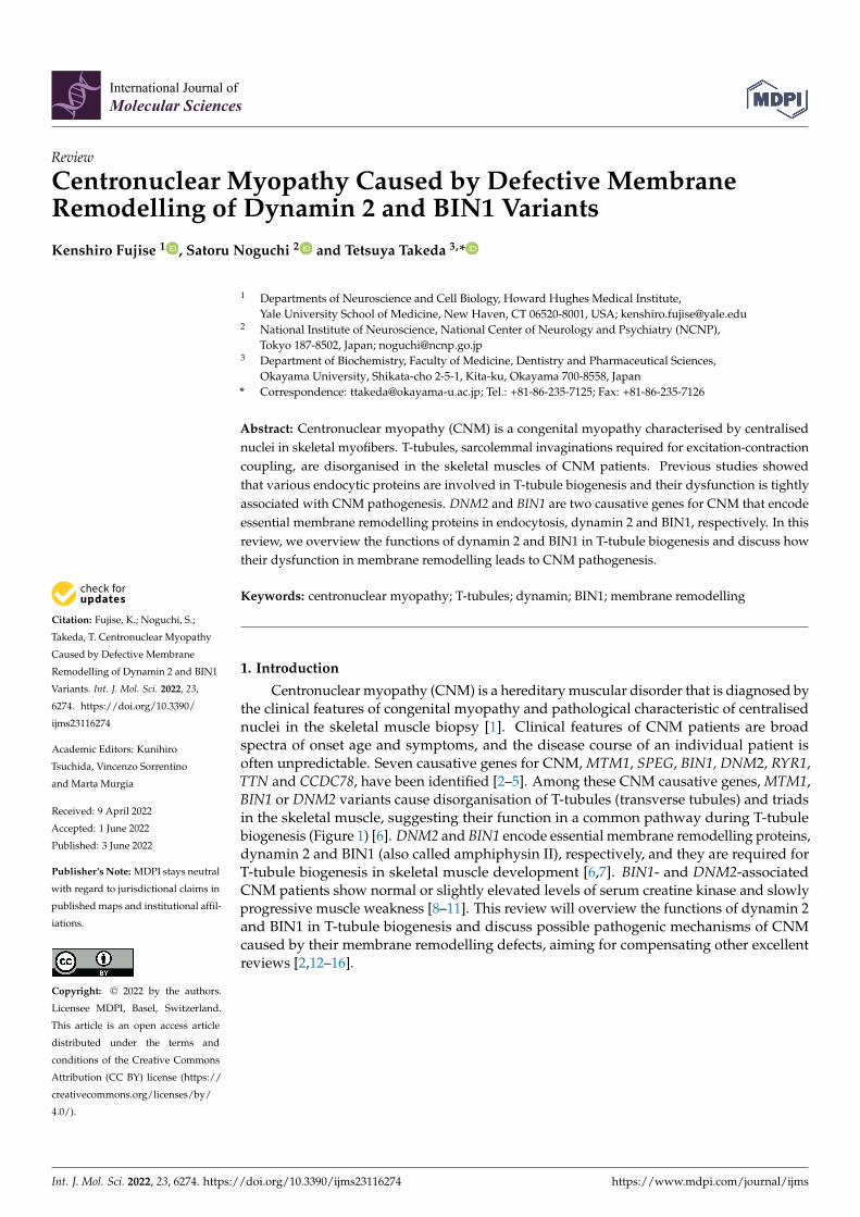
Figure 1. Functions of MTM1, BIN1 and DNM2 in T-tubule biogenesis. CNM causative genes
MTM1, BIN1 and DNM2 contribute to T-tubule biogenesis in a common pathway by respectively
regulating lipid homeostasis, membrane deformation and T-tubule stabilisation.
2. T-Tubules: Sarcolemmal Invaginations Essential for E-C Coupling
Rapid and coordinated contraction of striated muscles is achieved by coupled volt-
age- and calcium-dependent processes called excitation-contraction (E-C) coupling [17].
T-tubules are sarcolemmal invaginations required for the E-C coupling in both skeletal-
and cardiac muscles [6]. In skeletal muscle cells, T-tubules are associated with sarcoplas-
mic reticulum (SR) on either side to form closely apposed membrane contacts termed
“triad”, whereas, in cardiac muscle cells, their contact occurs only on one side to form
“diad”. T-tubules are enriched with specific lipids such as PI(4,5)P2 (phosphatidylinositol
4,5-bisphosphate) and cholesterol and they serve as a platform for localizing T-tubule spe-
cific ion channels or lipid-binding proteins [18–21]. In the E-C coupling, excitation (depo-
larisation) of the T-tubule membrane induces conformational changes of a voltage-gated
L-type calcium channel DHPR (dihydropyridine receptors), which in turn opens RyR1
(ryanodine receptor 1), a Ca2+ channel on SR, to allow Ca2+ release from SR to induce mus-
cle contraction [22]. In the skeletal muscle cells, DHPR directly interacts with RyR1 to en-
able rapid signal transmission (within 2 ms) [23–25]. Thus, the structural and functional
integrity of T-tubules is crucial for proper E-C coupling of the skeletal muscles. Not sur-
prisingly, abnormalities in T-tubule structures cause various muscle diseases including
congenital myopathies [26].
3. BIN1: A BAR Domain Protein-Inducing Membrane Curvature
3.1. BIN1 Functions in T-Tubule Biogenesis
BIN1 (Bridging Integrator 1) belongs to the conserved BAR domain superfamily that
senses and induces membrane curvature [27–29]. BIN1 contains an N-terminal amphi-
pathic helix Bin/Amphiphysin/Rvs-homology (N-BAR) domain that forms a “crescent-
shaped” dimer, and its positively charged concave surface binds to negatively charged
phospholipids to induce membrane curvature [30]. Human and mouse BIN1 are alterna-
tively spliced to express tissue-specific isoforms [27,31–33]. The skeletal muscle-specific
BIN1 isoform, isoform 8, has been shown to localise on T-tubules [33]. Conditional Bin1
knockout mice in skeletal muscle exhibit neonatal lethality [34] and acute knock-down of
BIN1 in adult mice caused structural and functional defects of T-tubules [35], indicating
that BIN1 plays essential roles in the development and maintenance of the skeletal muscle.
The BIN1 ortholog in Drosophila, Amph, is also required for muscle contraction, but not
for synaptic vesicle trafficking, suggesting that it has a similar function as human BIN1
[36].
Figure 1. Functions of MTM1, BIN1 and DNM2 in T-tubule biogenesis. CNM causative genes MTM1,BIN1 and DNM2 contribute to T-tubule biogenesis in a common pathway by respectively regulatinglipid homeostasis, membrane deformation and T-tubule stabilisation.
2. T-Tubules: Sarcolemmal Invaginations Essential for E-C Coupling
Rapid and coordinated contraction of striated muscles is achieved by coupled voltage-and calcium-dependent processes called excitation-contraction (E-C) coupling [17]. T-tubulesare sarcolemmal invaginations required for the E-C coupling in both skeletal- and cardiacmuscles [6]. In skeletal muscle cells, T-tubules are associated with sarcoplasmic reticulum(SR) on either side to form closely apposed membrane contacts termed “triad”, whereas, incardiac muscle cells, their contact occurs only on one side to form “diad”. T-tubules areenriched with specific lipids such as PI(4,5)P2 (phosphatidylinositol 4,5-bisphosphate) andcholesterol and they serve as a platform for localizing T-tubule specific ion channels or lipid-binding proteins [18–21]. In the E-C coupling, excitation (depolarisation) of the T-tubulemembrane induces conformational changes of a voltage-gated L-type calcium channelDHPR (dihydropyridine receptors), which in turn opens RyR1 (ryanodine receptor 1), aCa2+ channel on SR, to allow Ca2+ release from SR to induce muscle contraction [22]. In theskeletal muscle cells, DHPR directly interacts with RyR1 to enable rapid signal transmission(within 2 ms) [23–25]. Thus, the structural and functional integrity of T-tubules is crucialfor proper E-C coupling of the skeletal muscles. Not surprisingly, abnormalities in T-tubulestructures cause various muscle diseases including congenital myopathies [26].
3. BIN1: A BAR Domain Protein-Inducing Membrane Curvature3.1. BIN1 Functions in T-Tubule Biogenesis
BIN1 (Bridging Integrator 1) belongs to the conserved BAR domain superfamily thatsenses and induces membrane curvature [27–29]. BIN1 contains an N-terminal amphipathichelix Bin/Amphiphysin/Rvs-homology (N-BAR) domain that forms a “crescent-shaped”dimer, and its positively charged concave surface binds to negatively charged phospho-lipids to induce membrane curvature [30]. Human and mouse BIN1 are alternativelyspliced to express tissue-specific isoforms [27,31–33]. The skeletal muscle-specific BIN1isoform, isoform 8, has been shown to localise on T-tubules [33]. Conditional Bin1 knockoutmice in skeletal muscle exhibit neonatal lethality [34] and acute knock-down of BIN1 inadult mice caused structural and functional defects of T-tubules [35], indicating that BIN1plays essential roles in the development and maintenance of the skeletal muscle. The BIN1ortholog in Drosophila, Amph, is also required for muscle contraction, but not for synapticvesicle trafficking, suggesting that it has a similar function as human BIN1 [36].
Int. J. Mol. Sci. 2022, 23, 6274 3 of 14
BIN1 isoform 8 consists of four functional domains: H0, N-BAR, PI and Src homology3 (SH3) domains from N- to C-terminus [27] (Figure 2). H0 is an amphipathic helix that isfolded and inserted into one leaflet of the membrane to initiate oligomerisation of N-BARdomains and membrane tubulation [37,38]. N-BAR domain of BIN1 induces clusteringof PI(4,5)P2 and in turn, recruits a downstream partner dynamin 2 to enhance membranetubulation in T-tubule biogenesis [39–41]. Thus, BIN1 contributes to T-tubule biogenesis byregulating lipid composition and protein interaction in a positive feedback manner. N-BARdomain of BIN1 also interacts with F-actin to regulate its organisation via stabilisation orbundle formation of actin filaments [42]. Actin regulatory function of BIN1 is requiredfor proper T-tubule biogenesis in cardiac muscle cells [43]. In contrast, the formation ofBIN1-mediated T-tubule like structures (TLS) in mouse myoblast C2C12 cells is antagonisedby actin polymerisation [44]. The PI domain that exists only in BIN1 isoform 8 interactswith PI(4,5)P2 [44]. Neuronal BIN1 isoform 1 that lacks the PI domain diffusely localisesin the cytoplasm of CHO cells, suggesting essential roles of the PI domain in membraneinvaginations required for T-tubule biogenesis [19]. Indeed, skipping of the PI domain inBIN1 by dysregulated alternative splicing causes aberrant T-tubule formation in CNM andmyotonic dystrophy [45,46]. Lack of the PI domain does not affect muscle development perse, but it causes defects in the formation of T-tubule network and muscle regeneration dueto a reduced pool of satellite cells [33]. The C-terminal SH3 domain of BIN1 interacts withPR domain-containing proteins such as dynamin 2 [9,47]. The SH3 domain of BIN1 alsobinds to its PI domain intramolecularly to form a closed auto-inhibitory conformation [41].The autoinhibition of BIN1 is released upon PI(4,5)P2 binding to the PI domain that in turnrecruits its partner proteins dynamin 2 and myotubularin to the PI(4,5)P2-rich membranedomains [41,48,49]. Interestingly, deletion of exon 20 that encodes the SH3 domain ofBIN1 causes defects in T-tubule formation at E18.5 embryonic muscle fibres, but the triadstructures in adult skeletal muscle are not affected [33]. This result suggests that the BIN1SH3 domain is required for T-tubule formation, but not for its maintenance, at the earlystages of skeletal muscle development.
Int. J. Mol. Sci. 2022, 22, x FOR PEER REVIEW 4 of 15
BIN1 isoform 8 consists of four functional domains: H0, N-BAR, PI and Src homology
3 (SH3) domains from N- to C-terminus [27] (Figure 2). H0 is an amphipathic helix that is
folded and inserted into one leaflet of the membrane to initiate oligomerisation of N-BAR
domains and membrane tubulation [37,38]. N-BAR domain of BIN1 induces clustering of
PI(4,5)P2 and in turn, recruits a downstream partner dynamin 2 to enhance membrane
tubulation in T-tubule biogenesis [39–41]. Thus, BIN1 contributes to T-tubule biogenesis
by regulating lipid composition and protein interaction in a positive feedback manner. N-
BAR domain of BIN1 also interacts with F-actin to regulate its organisation via stabilisa-
tion or bundle formation of actin filaments [42]. Actin regulatory function of BIN1 is re-
quired for proper T-tubule biogenesis in cardiac muscle cells [43]. In contrast, the for-
mation of BIN1-mediated T-tubule like structures (TLS) in mouse myoblast C2C12 cells is
antagonised by actin polymerisation [44]. The PI domain that exists only in BIN1 isoform
8 interacts with PI(4,5)P2 [44]. Neuronal BIN1 isoform 1 that lacks the PI domain diffusely
localises in the cytoplasm of CHO cells, suggesting essential roles of the PI domain in
membrane invaginations required for T-tubule biogenesis [19]. Indeed, skipping of the PI
domain in BIN1 by dysregulated alternative splicing causes aberrant T-tubule formation
in CNM and myotonic dystrophy [45,46]. Lack of the PI domain does not affect muscle
development per se, but it causes defects in the formation of T-tubule network and muscle
regeneration due to a reduced pool of satellite cells [33]. The C-terminal SH3 domain of
BIN1 interacts with PR domain-containing proteins such as dynamin 2 [9,47]. The SH3
domain of BIN1 also binds to its PI domain intramolecularly to form a closed auto-inhib-
itory conformation [41]. The autoinhibition of BIN1 is released upon PI(4,5)P2 binding to
the PI domain that in turn recruits its partner proteins dynamin 2 and myotubularin to
the PI(4,5)P2-rich membrane domains [41,48,49]. Interestingly, deletion of exon 20 that en-
codes the SH3 domain of BIN1 causes defects in T-tubule formation at E18.5 embryonic
muscle fibres, but the triad structures in adult skeletal muscle are not affected [33]. This
result suggests that the BIN1 SH3 domain is required for T-tubule formation, but not for
its maintenance, at the early stages of skeletal muscle development.
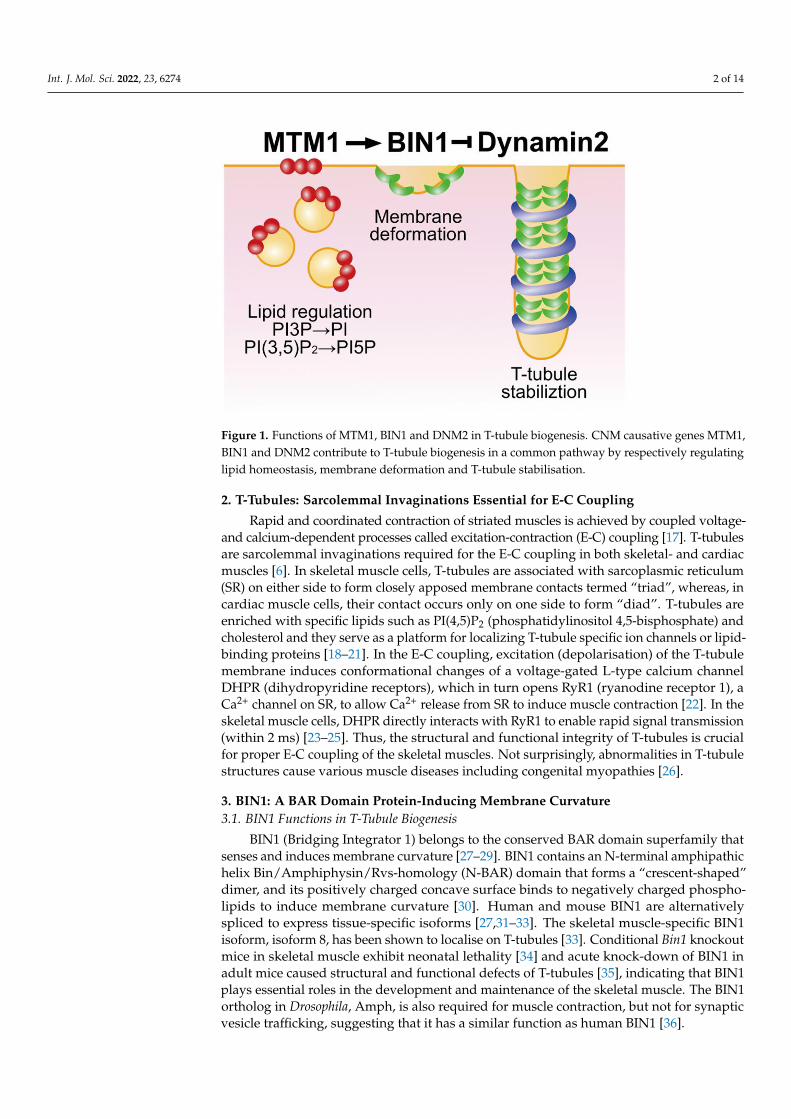
Figure 2. Domain structures of dynamin 2 and BIN1. Schematic illustrations of domain structures
and CNM-associated mutations in dynamin 2 and BIN1.
3.2. CNM Pathogenesis Caused by Defective Membrane Remodelling of BIN1 Variants
Multiple pathogenic BIN1 variants have been identified in CNM patients (Figure 2).
CNM-associated variants in the H0 helix, K21del, R24C and K35N, have been reported to
cause abnormalities in T-tubule structures due to decreased abilities to generate mem-
brane curvature [44,48]. CNM-associated variants in the N-BAR domain, D151N and
R154Q, are defective both in membrane binding and in curvature sensing possibly due to
oligomerisation defects [33,44]. D151N is also defective in the clustering of PI(4,5)P2 both
in cellulo and in vitro systems using a flat membrane sheet [39]. Another variant in the N-
BAR domain, D149N, exhibits decreased membrane deformation abilities in cellulo [40].
Since membrane tubulation defects of K35N and D149N can be restored by supplement-
ing with PI(4,5)P2, these variants are deficient in recruiting PI(4,5)P2 probably due to re-
duced membrane binding affinity [40]. CNM-associated variant IVS10-1G>A in exon 11
causes deletion of the PI domain, resulting in defective triad formation both in humans
Figure 2. Domain structures of dynamin 2 and BIN1. Schematic illustrations of domain structuresand CNM-associated mutations in dynamin 2 and BIN1.
3.2. CNM Pathogenesis Caused by Defective Membrane Remodelling of BIN1 Variants
Multiple pathogenic BIN1 variants have been identified in CNM patients (Figure 2).CNM-associated variants in the H0 helix, K21del, R24C and K35N, have been reported tocause abnormalities in T-tubule structures due to decreased abilities to generate membranecurvature [44,48]. CNM-associated variants in the N-BAR domain, D151N and R154Q, aredefective both in membrane binding and in curvature sensing possibly due to oligomerisa-tion defects [33,44]. D151N is also defective in the clustering of PI(4,5)P2 both in cellulo andin vitro systems using a flat membrane sheet [39]. Another variant in the N-BAR domain,D149N, exhibits decreased membrane deformation abilities in cellulo [40]. Since membranetubulation defects of K35N and D149N can be restored by supplementing with PI(4,5)P2,these variants are deficient in recruiting PI(4,5)P2 probably due to reduced membranebinding affinity [40]. CNM-associated variant IVS10-1G>A in exon 11 causes deletion of
Int. J. Mol. Sci. 2022, 23, 6274 4 of 14
the PI domain, resulting in defective triad formation both in humans and dogs [45]. Thus,CNM-associated variants in H0, N-BAR and PI domains are likely to induce abnormalT-tubule structures due to their membrane deformation disabilities.
Two recessive CNM variants of BIN1, Q434X and K436X, that partially truncate theSH3 domain shows suppressed interaction with dynamin 2 [9,41,47]. In the skeletal musclebiopsies from CNM patients with these variants, abnormal T-tubule morphology withaggregated caveolae-positive membranous structures is observed [49]. Partial truncation ofthe SH3 domain by Q434X and K436X variants also keeps BIN1 in a constitutively openconformation with altered membrane deformation abilities [41]. The loss of autoinhibitionby the CNM mutant BIN1 also causes enhanced interaction with myotubularin, which is aphosphatidylinositol-3-phosphatase for PI(3)P or PI(3,5)P2 encoded by a CNM causativegene MTM1 [50]. The SH3 domain of BIN1 also interacts with N-WASP, an activator ofArp2/3 dependent actin polymerisation [51]. BIN1 mutants with truncated SH3 showsuppressed N-WASP interaction and induce collapsed T-tubule structures [51]. Thus, thestructural abnormalities of T-tubules caused by CNM-associated BIN1 variants are causedby abnormal protein–protein and/or protein–lipid interactions.
4. Dynamin: A Membrane Fission Catalyser in Endocytosis4.1. Structure and Function of Dynamin
Dynamin is a large GTPase essential for membrane fission in clathrin-dependent andindependent endocytic pathways [52–54]. There are three dynamin isoforms in mammals:dynamin 1 and 3, two tissue-specific isoforms highly expressed in neurons, and dynamin2, a ubiquitously expressed isoform [55–57]. These isoforms are similar in amino acidsequences and share the same functional domains: G, middle, pleckstrin homology (PH),GTPase effector (GE) and PR domains from N- to C-terminus (Figure 2). The G domainis responsible for GTP binding and hydrolysis [58]. The middle and GED form a “stalk”structure that serves as interacting platforms in the formation of dimer or tetramer [59]. PHdomain binds to negatively charged phospholipids such as PI(4,5)P2 and plays a role inclustering the phosphoinositides [60,61]. PH domain also senses membrane curvature bybeing hydrophobically inserted into the lipid bilayer [62]. Furthermore, the PH domaincan bind to stalk structure intramolecularly to form autoinhibitory “closed” conformationthat prevents untimely self-assembly [63]. The C-terminal PR domain binds to other SH3domain-containing proteins such as BIN1, amphiphysin 1, and endophilin [9,64,65]. PRdomain is also involved in actin organisation at invadosomes, membranous protrusionsrequired for myoblast fusion [64,66].
Structural studies using cryo-EM, X-ray crystallography and high-speed atomic forcemicroscopy (HS-AFM) gave mechanistic insights into dynamin-mediated membrane fission.Dynamin exists as a tetramer in a physiological condition in the absence of lipids [63], whileit assembles into a helical polymer at the neck of endocytic pits [65] or on membrane tubulesreconstituted in vitro from liposomes [67,68]. Conformational changes of dynamin helicalpolymer coupled with binding and hydrolysis of GTP promote membrane constrictionand fission [69,70]. Although precise mechanisms of the dynamin-mediated membranefission are still under debate, a few decades of studies in the past strongly support thefollowing consensus views: (1) Dynamin polymerises into a helical polymer in the absenceof GTP; (2) the dynamin polymer constricts in the presence of GTP and (3) dynamin severmembrane upon GTP hydrolysis [52]. Various models for dynamin-mediated membranefission have been proposed such as the “constrictase model” in which the dynamin helicalpolymer constricts and mechanically severs the membrane and the “two-stage model” inwhich constriction and dissociation of dynamin helical polymer are required for membranecleavage [52]. By using HS-AFM, we and other groups observed cluster formation bydynamin helices upon GTP hydrolysis [71,72]. We also observed that membrane fissionoccurs between the clustered dynamin helices proposing a novel “clusterase model” [72].GTP hydrolysis also causes the twisting motion of the dynamin helical polymer thatprovides torsion at the neck of the endocytic pits to promote membrane fission [73,74].
Int. J. Mol. Sci. 2022, 23, 6274 5 of 14
Thus, dynamin severs membrane by a combination of various mechanical stresses causedby structural changes and depolymerisation upon GTP hydrolysis.
4.2. Dynamin 2 Functions in T-Tubule Biogenesis
Dynamin 2 is ubiquitously expressed in various tissues, but its expression level isrelatively high in skeletal muscles [75]. In skeletal muscles, dynamin 2 localises to T-tubulesat the early stages of development and regulates T-tubule orientation [34,76]. In celluloreconstitution assay for T-tubule-like structures (TLS) revealed that dynamin 2 is requiredfor stabilisation of the TLS [47]. GTPase activity of dynamin 2 is inhibited by BIN1 in astoichiometry-dependent manner to allow dynamin 2 to stabilise TLS (Figure 1) [34,47].CNM-associated BIN1 mutants with partially truncated SH3 domain fail to bind to dynamin2 and induce TLS formation [47]. The expression level of BIN1 is increased as skeletalmuscle development progresses, while that of dynamin 2 remains unchanged [19]. Thus, itis interesting to speculate that BIN1 contributes not only to membrane tubulation per se butalso supports dynamin 2-mediated membrane stabilisation by suppressing GTPase activityto organise the T-tubule system during the normal development of skeletal muscles.
4.3. Dysregulation of T-Tubule Function by CNM-Associated Dynamin 2 Variants
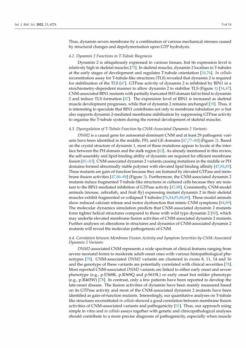
DNM2 is a causal gene for autosomal-dominant CNM and at least 29 pathogenic vari-ants have been identified in the middle, PH, and GE domains [47,77–80] (Figure 2). Basedon the crystal structure of dynamin 1, most of these mutations appear to locate at the inter-face between the PH domain and the stalk region [63]. As already mentioned in this review,the self-assembly and lipid-binding ability of dynamin are required for efficient membranefission [81–83]. CNM-associated dynamin 2 variants causing mutations in the middle or PHdomains formed abnormally stable polymer with elevated lipid binding affinity [47,84,85].These mutants are gain-of-function because they are featured by elevated GTPase and mem-brane fission activities [47,86–88] (Figure 3). Furthermore, the CNM-associated dynamin 2mutants induce fragmented T-tubule-like structures in cultured cells because they are resis-tant to the BIN1-mediated inhibition of GTPase activity [47,88]. Consistently, CNM-modelanimals (mouse, zebrafish, and fruit fly) expressing mutant dynamin 2 in their skeletalmuscles exhibit fragmented or collapsed T-tubules [76,84,85,88,89]. These model animalsshow reduced calcium release and motor dysfunction that mimic CNM symptoms [84,89].The molecular dynamics simulation predicts that CNM-associated dynamin 2 mutantsform tighter helical structures compared to those with wild type dynamin 2 [90], whichmay underlie elevated membrane fission activities of CNM-associated dynamin 2 mutants.Further analyses on alterations in structures and dynamics of CNM-associated dynamin 2mutants will reveal the molecular pathogenesis of CNM.
4.4. Correlation between Membrane Fission Activity and Symptom Severities by CNM-AssociatedDynamin 2 Variants
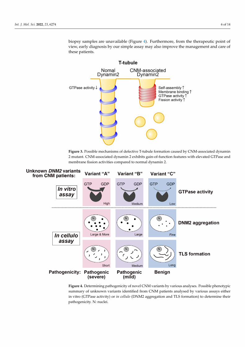
DNM2-associated CNM represents a wide spectrum of clinical features ranging fromsevere neonatal forms to moderate adult-onset ones with various histopathological phe-notypes [78]. CNM-associated DNM2 variants are clustered in exons 8, 11, 14 and 16and the genotype of these variants are potentially correlated with clinical severities [78].Most reported CNM-associated DNM2 variants are linked to either early onset and severephenotype (e.g., p.E368K, p.R369Q and p.S619L) or early onset but milder phenotype(e.g., p.R465W) [78]. In contrast, only a few patients have been reported to develop thelate-onset disease. The fission activities of dynamin have been mainly measured basedon its GTPase activity and most of the CNM-associated dynamin 2 mutants have beenidentified as gain-of-function mutants. Interestingly, our quantitative analyses on T-tubulelike structures reconstituted in cellulo showed a good correlation between membrane fissionactivities of CNM-associated variants and pathogenicity [91]. Thus, our approach usingsimple in vitro and in cellulo assays together with genetic and clinicopathological analysesshould contribute to a more precise diagnosis of pathogenicity, especially when muscle
Int. J. Mol. Sci. 2022, 23, 6274 6 of 14
biopsy samples are unavailable (Figure 4). Furthermore, from the therapeutic point ofview, early diagnosis by our simple assay may also improve the management and care ofthese patients.
Int. J. Mol. Sci. 2022, 22, x FOR PEER REVIEW 6 of 15
membrane fission [73,74]. Thus, dynamin severs membrane by a combination of various
mechanical stresses caused by structural changes and depolymerisation upon GTP hy-
drolysis.
4.2. Dynamin 2 Functions in T-Tubule Biogenesis
Dynamin 2 is ubiquitously expressed in various tissues, but its expression level is
relatively high in skeletal muscles [75]. In skeletal muscles, dynamin 2 localises to T-tu-
bules at the early stages of development and regulates T-tubule orientation [34,76]. In cel-
lulo reconstitution assay for T-tubule-like structures (TLS) revealed that dynamin 2 is re-
quired for stabilisation of the TLS [47]. GTPase activity of dynamin 2 is inhibited by BIN1
in a stoichiometry-dependent manner to allow dynamin 2 to stabilise TLS (Figure 1)
[34,47]. CNM-associated BIN1 mutants with partially truncated SH3 domain fail to bind
to dynamin 2 and induce TLS formation [47]. The expression level of BIN1 is increased as
skeletal muscle development progresses, while that of dynamin 2 remains unchanged
[19]. Thus, it is interesting to speculate that BIN1 contributes not only to membrane tubu-
lation per se but also supports dynamin 2-mediated membrane stabilisation by suppress-
ing GTPase activity to organise the T-tubule system during the normal development of
skeletal muscles.
4.3. Dysregulation of T-Tubule Function by CNM-Associated Dynamin 2 Variants
DNM2 is a causal gene for autosomal-dominant CNM and at least 29 pathogenic var-
iants have been identified in the middle, PH, and GE domains [47,77–80] (Figure 2). Based
on the crystal structure of dynamin 1, most of these mutations appear to locate at the in-
terface between the PH domain and the stalk region [63]. As already mentioned in this
review, the self-assembly and lipid-binding ability of dynamin are required for efficient
membrane fission [81–83]. CNM-associated dynamin 2 variants causing mutations in the
middle or PH domains formed abnormally stable polymer with elevated lipid binding
affinity [47,84,85]. These mutants are gain-of-function because they are featured by ele-
vated GTPase and membrane fission activities [47,86–88] (Figure 3). Furthermore, the
CNM-associated dynamin 2 mutants induce fragmented T-tubule-like structures in cul-
tured cells because they are resistant to the BIN1-mediated inhibition of GTPase activity
[47,88]. Consistently, CNM-model animals (mouse, zebrafish, and fruit fly) expressing
mutant dynamin 2 in their skeletal muscles exhibit fragmented or collapsed T-tubules
[76,84,85,88,89]. These model animals show reduced calcium release and motor dysfunc-
tion that mimic CNM symptoms [84,89]. The molecular dynamics simulation predicts that
CNM-associated dynamin 2 mutants form tighter helical structures compared to those
with wild type dynamin 2 [90], which may underlie elevated membrane fission activities
of CNM-associated dynamin 2 mutants. Further analyses on alterations in structures and
dynamics of CNM-associated dynamin 2 mutants will reveal the molecular pathogenesis
of CNM.
Figure 3. Possible mechanisms of defective T-tubule formation caused by CNM-associated dynamin2 mutant. CNM-associated dynamin 2 exhibits gain-of-function features with elevated GTPase andmembrane fission activities compared to normal dynamin 2.
Int. J. Mol. Sci. 2022, 22, x FOR PEER REVIEW 7 of 15
Figure 3. Possible mechanisms of defective T-tubule formation caused by CNM-associated dynamin
2 mutant. CNM-associated dynamin 2 exhibits gain-of-function features with elevated GTPase and
membrane fission activities compared to normal dynamin 2.
4.4. Correlation between Membrane Fission Activity and Symptom Severities by CNM-
Associated Dynamin 2 Variants
DNM2-associated CNM represents a wide spectrum of clinical features ranging from
severe neonatal forms to moderate adult-onset ones with various histopathological phe-
notypes [78]. CNM-associated DNM2 variants are clustered in exons 8, 11, 14 and 16 and
the genotype of these variants are potentially correlated with clinical severities [78]. Most
reported CNM-associated DNM2 variants are linked to either early onset and severe phe-
notype (e.g., p.E368K, p.R369Q and p.S619L) or early onset but milder phenotype (e.g.,
p.R465W) [78]. In contrast, only a few patients have been reported to develop the late-
onset disease. The fission activities of dynamin have been mainly measured based on its
GTPase activity and most of the CNM-associated dynamin 2 mutants have been identified
as gain-of-function mutants. Interestingly, our quantitative analyses on T-tubule like
structures reconstituted in cellulo showed a good correlation between membrane fission
activities of CNM-associated variants and pathogenicity [91]. Thus, our approach using
simple in vitro and in cellulo assays together with genetic and clinicopathological analyses
should contribute to a more precise diagnosis of pathogenicity, especially when muscle
biopsy samples are unavailable (Figure 4). Furthermore, from the therapeutic point of
view, early diagnosis by our simple assay may also improve the management and care of
these patients.
Figure 4. Determining pathogenicity of novel CNM variants by various analyses. Possible pheno-
typic summary of unknown variants identified from CNM patients analysed by various assays ei-
ther in vitro (GTPase activity) or in cellulo (DNM2 aggregation and TLS formation) to determine their
pathogenicity. N: nuclei.
4.5. Other Functions of Dynamin 2 in Skeletal Muscle
Figure 4. Determining pathogenicity of novel CNM variants by various analyses. Possible phenotypicsummary of unknown variants identified from CNM patients analysed by various assays eitherin vitro (GTPase activity) or in cellulo (DNM2 aggregation and TLS formation) to determine theirpathogenicity. N: nuclei.
Int. J. Mol. Sci. 2022, 23, 6274 7 of 14
4.5. Other Functions of Dynamin 2 in Skeletal Muscle
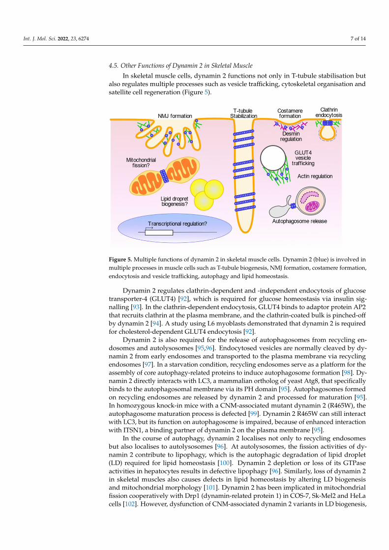
In skeletal muscle cells, dynamin 2 functions not only in T-tubule stabilisation butalso regulates multiple processes such as vesicle trafficking, cytoskeletal organisation andsatellite cell regeneration (Figure 5).
Int. J. Mol. Sci. 2022, 22, x FOR PEER REVIEW 8 of 15
In skeletal muscle cells, dynamin 2 functions not only in T-tubule stabilisation but
also regulates multiple processes such as vesicle trafficking, cytoskeletal organisation and
satellite cell regeneration (Figure 5).
Figure 5. Multiple functions of dynamin 2 in skeletal muscle cells. Dynamin 2 (blue) is involved in
multiple processes in muscle cells such as T-tubule biogenesis, NMJ formation, costamere for-
mation, endocytosis and vesicle trafficking, autophagy and lipid homeostasis.
Dynamin 2 regulates clathrin-dependent and -independent endocytosis of glucose
transporter-4 (GLUT4) [92], which is required for glucose homeostasis via insulin signal-
ling [93]. In the clathrin-dependent endocytosis, GLUT4 binds to adaptor protein AP2 that
recruits clathrin at the plasma membrane, and the clathrin-coated bulk is pinched-off by
dynamin 2 [94]. A study using L6 myoblasts demonstrated that dynamin 2 is required for
cholesterol-dependent GLUT4 endocytosis [92].
Dynamin 2 is also required for the release of autophagosomes from recycling endo-
somes and autolysosomes [95,96]. Endocytosed vesicles are normally cleaved by dynamin
2 from early endosomes and transported to the plasma membrane via recycling endo-
somes [97]. In a starvation condition, recycling endosomes serve as a platform for the as-
sembly of core autophagy-related proteins to induce autophagosome formation [98]. Dy-
namin 2 directly interacts with LC3, a mammalian ortholog of yeast Atg8, that specifically
binds to the autophagosomal membrane via its PH domain [95]. Autophagosomes formed
on recycling endosomes are released by dynamin 2 and processed for maturation [95]. In
homozygous knock-in mice with a CNM-associated mutant dynamin 2 (R465W), the au-
tophagosome maturation process is defected [99]. Dynamin 2 R465W can still interact with
LC3, but its function on autophagosome is impaired, because of enhanced interaction with
ITSN1, a binding partner of dynamin 2 on the plasma membrane [95].
In the course of autophagy, dynamin 2 localises not only to recycling endosomes but
also localises to autolysosomes [96]. At autolysosomes, the fission activities of dynamin 2
contribute to lipophagy, which is the autophagic degradation of lipid droplet (LD) re-
quired for lipid homeostasis [100]. Dynamin 2 depletion or loss of its GTPase activities in
hepatocytes results in defective lipophagy [96]. Similarly, loss of dynamin 2 in skeletal
muscles also causes defects in lipid homeostasis by altering LD biogenesis and mitochon-
drial morphology [101]. Dynamin 2 has been implicated in mitochondrial fission cooper-
atively with Drp1 (dynamin-related protein 1) in COS-7, Sk-Mel2 and HeLa cells [102].
However, dysfunction of CNM-associated dynamin 2 variants in LD biogenesis, lipoph-
agy or mitochondrial fission and their implications in CNM pathogenesis remains to be
elucidated.
Dynamin 2 is also implicated in cytoskeletal regulation, especially in the organisation
of actin. Dynamin 2 regulates intracellular trafficking of the GLUT4-containing vesicles
by controlling actin polymerisation [93]. The actin regulation by dynamin 2 is also
Figure 5. Multiple functions of dynamin 2 in skeletal muscle cells. Dynamin 2 (blue) is involved inmultiple processes in muscle cells such as T-tubule biogenesis, NMJ formation, costamere formation,endocytosis and vesicle trafficking, autophagy and lipid homeostasis.
Dynamin 2 regulates clathrin-dependent and -independent endocytosis of glucosetransporter-4 (GLUT4) [92], which is required for glucose homeostasis via insulin sig-nalling [93]. In the clathrin-dependent endocytosis, GLUT4 binds to adaptor protein AP2that recruits clathrin at the plasma membrane, and the clathrin-coated bulk is pinched-offby dynamin 2 [94]. A study using L6 myoblasts demonstrated that dynamin 2 is requiredfor cholesterol-dependent GLUT4 endocytosis [92].
Dynamin 2 is also required for the release of autophagosomes from recycling en-dosomes and autolysosomes [95,96]. Endocytosed vesicles are normally cleaved by dy-namin 2 from early endosomes and transported to the plasma membrane via recyclingendosomes [97]. In a starvation condition, recycling endosomes serve as a platform for theassembly of core autophagy-related proteins to induce autophagosome formation [98]. Dy-namin 2 directly interacts with LC3, a mammalian ortholog of yeast Atg8, that specificallybinds to the autophagosomal membrane via its PH domain [95]. Autophagosomes formedon recycling endosomes are released by dynamin 2 and processed for maturation [95].In homozygous knock-in mice with a CNM-associated mutant dynamin 2 (R465W), theautophagosome maturation process is defected [99]. Dynamin 2 R465W can still interactwith LC3, but its function on autophagosome is impaired, because of enhanced interactionwith ITSN1, a binding partner of dynamin 2 on the plasma membrane [95].
In the course of autophagy, dynamin 2 localises not only to recycling endosomesbut also localises to autolysosomes [96]. At autolysosomes, the fission activities of dy-namin 2 contribute to lipophagy, which is the autophagic degradation of lipid droplet(LD) required for lipid homeostasis [100]. Dynamin 2 depletion or loss of its GTPaseactivities in hepatocytes results in defective lipophagy [96]. Similarly, loss of dynamin 2in skeletal muscles also causes defects in lipid homeostasis by altering LD biogenesisand mitochondrial morphology [101]. Dynamin 2 has been implicated in mitochondrialfission cooperatively with Drp1 (dynamin-related protein 1) in COS-7, Sk-Mel2 and HeLacells [102]. However, dysfunction of CNM-associated dynamin 2 variants in LD biogenesis,
Int. J. Mol. Sci. 2022, 23, 6274 8 of 14
lipophagy or mitochondrial fission and their implications in CNM pathogenesis remains tobe elucidated.
Dynamin 2 is also implicated in cytoskeletal regulation, especially in the organisationof actin. Dynamin 2 regulates intracellular trafficking of the GLUT4-containing vesicles bycontrolling actin polymerisation [93]. The actin regulation by dynamin 2 is also requiredfor insulin-dependent exocytosis of GLUT4 to supply intracellular membrane componentsto T-tubules [103–105]. Expression of CNM-associated mutant dynamin 2 disrupts de novoactin filament formation in muscle cells [93]. Consistently, in the CNM model mouseexpressing CNM mutant dynamin 2 (R465W), translocation of GLUT4 to the plasmamembrane is impaired due to disorganised actin filaments, and abnormal perinuclearaccumulation of GLUT4 is observed in CNM patient’s muscle biopsy [93].
Actin regulation by dynamin 2 is also required for skeletal muscle development inmyoblast fusion [64,66] and the formation of neuromuscular junctions (NMJ) [106]. Invado-somes are actin-rich membrane protrusions required for degradation of the extracellularmatrix (ECM), and they play essential roles in myoblast fusion and NMJ formation [107].In invadosomes, dynamin 2 is involved in actin organisation either by itself via the PRdomain [64] or with its interacting proteins such as Tks5 (tyrosine kinase substrate with5 SH3 domain) [66,106]. Dynamin 2 is also required for the formation and function ofinvadosomes cooperatively with various BAR domain proteins such as BIN1 [108], en-dophilin [109] and pacsin 2 [110]. Expression of CNM-associated dynamin 2 mutant (A618T)in C2C12 cells enhances formation of invadosomes with abnormal matrix degradation byinducing F-actin bundles [106].
Costameres, sub-sarcolemmal adhesion sites associated with Z-lines in skeletal muscle,play mechanical and signalling roles during muscle contraction [111]. Costameres consist ofmultiple components such as integrin [112], actin [113], clathrin [114] and dynamin 2 [115]and they are required for the stabilisation of skeletal muscle fibres by attaching sarcolemmato myofibrils [111]. Dynamin 2 regulates clathrin plaque formation in costameres byinteracting with desmin and N-WASP [114,115]. In the CNM-model mouse expressingdynamin 2 mutant and the CNM patient’s biopsy, costameres are defected because ofdisorganised desmin filaments and clathrin plaques [114,116].
The nuclear positioning to the periphery of skeletal muscle cells requires crosslinkingof myofibrils by desmin which is regulated by the arp2/3 complex [117]. Dynamin 2 isrequired for peripheral nuclear positioning by interacting with N-WASP, an activator ofthe Arp2/3 complex [51,118–120]. CNM mutant dynamin 2 localises around centralisednuclei and their size and numbers are impaired in the adult skeletal muscles in Dnm2-KImice [121,122]. These abnormal nuclei are possibly produced by defective regenerationof satellite cells due to decreased transcription [123]. However, it is still unclear how thefunction of dynamin 2 around the nuclei is impaired. Further analyses are required forunveiling yet unknown transcriptional regulation by dynamin 2.
4.6. Therapeutic Approaches for CNM
CNM-associated dynamin 2 variants cause gain-of-function features in membranefission activities because of elevated GTPase activity [47,86–88]. Likewise, overexpressionof wild-type dynamin 2 also induces CNM phenotypes such as muscle weakness, abnormalhistology and altered T-tubule structures in mice and Drosophila [79,89,119]. Based on thesefindings, gene silencing approaches are developed to reduce or normalise the expressionlevel of dynamin 2 using AAV-mediated expression of shRNA targeting Dnm2 mRNAor antisense oligonucleotides against Dnm2 pre-mRNA and mRNA [124–126]. Thesegene silencing approaches improve CNM phenotypes of moderate Dnm2R465W/+ andsevere Dnm2S619L/+ mouse models [124–126]. The expression level of dynamin 2 protein isincreased in muscle lysates from Mtm1-KO mouse and XLMTM1 patients [127]. Therefore,gene silencing approaches targeting Dnm2 also improved the CNM symptoms in Mtm1-KOmice [127,128]. As already mentioned in this review, BIN1 negatively regulates GTPaseactivities of dynamin 2 in a stoichiometry dependent manner [34,47]. Skeletal muscle-
Int. J. Mol. Sci. 2022, 23, 6274 9 of 14
specific Bin1-KO mouse shows CNM phenotypes including reduced muscle mass and force,and T-tubule abnormalities with a slight increase of dynamin 2 protein level [34,129]. Thus,downregulation of dynamin 2 by gene silencing tunes its relative amount for BIN1 proteinresulting in normal survival, muscular force and triad structures [34,129]. In zebrafish,knockout of a CNM causal gene SPEG (striated preferentially expressed protein kinase)that encodes a myosin light chain kinase family protein show T-tubule abnormalities withthe increased expression level of dynamin 2 protein [130]. Since SPEG has been shown tointeract with MTM1 [5], SPEG may regulate dynamin 2 function together with MTM1 andBIN1 in skeletal muscle. Although it is still unclear if SPEG is also a negative regulator ofdynamin 2, gene silencing of DNM2 may be a potential therapeutic approach for CNMcaused by variants in DNM2 gene as well as for CNM associated with variants in othergenes such as MTM1, BIN1, SPEG. Indeed, a clinical trial using investigational antisensemedicine DYN101 is ongoing for DNM2-associated CNM (NCT04033159).
5. Perspectives
In this review, we overviewed the function of BIN1 and dynamin 2 in T-tubule biogen-esis and discussed possible molecular mechanisms of CNM pathogenesis caused by theirmembrane remodelling defects. Abnormal membrane remodelling by CNM-associatedvariants of BIN1 and dynamin 2 has been greatly elucidated using multidisciplinary ap-proaches. However, the impact of CNM-associated variants on multifunctional featuresof dynamin 2 at various cellular organelles is still largely unknown. A comprehensiveunderstanding of dysregulated functions of dynamin 2 in the multiple cellular processesmay contribute to a better elucidation of pathomechanisms of CNM and the develop-ment of more precise diagnosis, management and care of CNM patients. Although wefocused on the T-tubule biogenesis by BIN1 and dynamin 2, there are a variety of otherproteins involved in T-tubule formation, and many of them are associated with musclediseases [6,131]. A more comprehensive understanding of protein functions that affectT-tubule formation is required for a better understanding of the CNM pathogenesis causedby abnormal membrane remodelling.
Author Contributions: K.F., S.N. and T.T. wrote and edited the manuscript. K.F. designed the figures.All authors have read and agreed to the published version of the manuscript.
Funding: This research was funded by JSPS KAKENHI, Grant numbers 18K07198, 19KK0180, TheTakeda Science Foundation, Wesco Scientific Promotion Foundation and Ryobi Teien Memory Founda-tion for T.T. This research was also funded by Intramural Research Grant for Neuronal and PsychiatricDisorders of NCNP (29-4, 2-5 for T.T. and Ichizo Nishino, 2-6, 3-9 for S.N.), and AMED under GrantNumbers JP19ek0109285h0003 for Ichizo Nishino and S.N.
Acknowledgments: The authors would like to thank Ichizo Nishino (NCNP) and Kohji Takei(Okayama University) for their scientific insights during our study.
Conflicts of Interest: The authors declare no conflict of interest.
References1. Jungbluth, H.; Wallgren-Pettersson, C.; Laporte, J. Centronuclear (myotubular) myopathy. Orphanet. J. Rare Dis. 2008, 3, 26.
[CrossRef] [PubMed]2. Jungbluth, H.; Gautel, M. Pathogenic mechanisms in centronuclear myopathies. Front. Aging Neurosci. 2014, 6, 339. [CrossRef]
[PubMed]3. Romero, N.B. Centronuclear myopathies: A widening concept. Neuromuscul. Disord. 2010, 20, 223–228. [CrossRef] [PubMed]4. Majczenko, K.; Davidson, A.E.; Camelo-Piragua, S.; Agrawal, P.B.; Manfready, R.A.; Li, X.; Joshi, S.; Xu, J.; Peng, W.; Beggs, A.H.;
et al. Dominant mutation of CCDC78 in a unique congenital myopathy with prominent internal nuclei and atypical cores. Am. J.Hum. Genet. 2012, 91, 365–371. [CrossRef]
5. Agrawal, P.B.; Pierson, C.R.; Joshi, M.; Liu, X.; Ravenscroft, G.; Moghadaszadeh, B.; Talabere, T.; Viola, M.; Swanson, L.C.;Haliloglu, G.; et al. SPEG interacts with myotubularin, and its deficiency causes centronuclear myopathy with dilated cardiomy-opathy. Am. J. Hum. Genet. 2014, 95, 218–226. [CrossRef] [PubMed]
6. Al-Qusairi, L.; Laporte, J. T-tubule biogenesis and triad formation in skeletal muscle and implication in human diseases. Skelet.Muscle 2011, 1, 26. [CrossRef] [PubMed]
Int. J. Mol. Sci. 2022, 23, 6274 10 of 14
7. Dowling, J.J.; Gibbs, E.M.; Feldman, E.L. Membrane traffic and muscle: Lessons from human disease. Traffic 2008, 9, 1035–1043.[CrossRef]
8. Bitoun, M.; Maugenre, S.; Jeannet, P.Y.; Lacene, E.; Ferrer, X.; Laforet, P.; Martin, J.J.; Laporte, J.; Lochmuller, H.; Beggs, A.H.; et al.Mutations in dynamin 2 cause dominant centronuclear myopathy. Nat. Genet. 2005, 37, 1207–1209. [CrossRef]
9. Nicot, A.S.; Toussaint, A.; Tosch, V.; Kretz, C.; Wallgren-Pettersson, C.; Iwarsson, E.; Kingston, H.; Garnier, J.M.; Biancalana, V.;Oldfors, A.; et al. Mutations in amphiphysin 2 (BIN1) disrupt interaction with dynamin 2 and cause autosomal recessivecentronuclear myopathy. Nat. Genet. 2007, 39, 1134–1139. [CrossRef]
10. Fischer, D.; Herasse, M.; Bitoun, M.; Barragan-Campos, H.M.; Chiras, J.; Laforet, P.; Fardeau, M.; Eymard, B.; Guicheney, P.;Romero, N.B. Characterization of the muscle involvement in dynamin 2-related centronuclear myopathy. Brain 2006, 129,1463–1469. [CrossRef]
11. Bohm, J.; Yis, U.; Ortac, R.; Cakmakci, H.; Kurul, S.H.; Dirik, E.; Laporte, J. Case report of intrafamilial variability in autosomalrecessive centronuclear myopathy associated to a novel BIN1 stop mutation. Orphanet. J. Rare Dis. 2010, 5, 35. [CrossRef][PubMed]
12. Gómez-Oca, R.; Cowling, B.S.; Laporte, J. Common Pathogenic Mechanisms in Centronuclear and Myotubular Myopathies andLatest Treatment Advances. Int. J. Mol. Sci. 2021, 22, 11377. [CrossRef] [PubMed]
13. Dowling, J.J.; Weihl, C.C.; Spencer, M.J. Molecular and cellular basis of genetically inherited skeletal muscle disorders. Nat. Rev.Mol. Cell Biol. 2021, 22, 713–732. [CrossRef] [PubMed]
14. Zhao, M.; Maani, N.; Dowling, J.J. Dynamin 2 (DNM2) as Cause of, and Modifier for, Human Neuromuscular Disease. Neurothera-peutics 2018, 15, 966–975. [CrossRef]
15. Cowling, B.S.; Toussaint, A.; Muller, J.; Laporte, J. Defective membrane remodeling in neuromuscular diseases: Insights fromanimal models. PLoS Genet. 2012, 8, e1002595. [CrossRef]
16. Durieux, A.C.; Prudhon, B.; Guicheney, P.; Bitoun, M. Dynamin 2 and human diseases. J. Mol. Med. 2010, 88, 339–350. [CrossRef]17. Franzini-Armstrong, C. The relationship between form and function throughout the history of excitation-contraction coupling.
J. Gen. Physiol. 2018, 150, 189–210. [CrossRef]18. Rosemblatt, M.; Hidalgo, C.; Vergara, C.; Ikemoto, N. Immunological and biochemical properties of transverse tubule membranes
isolated from rabbit skeletal muscle. J. Biol. Chem. 1981, 256, 8140–8148. [CrossRef]19. Lee, E.; Marcucci, M.; Daniell, L.; Pypaert, M.; Weisz, O.A.; Ochoa, G.C.; Farsad, K.; Wenk, M.R.; De Camilli, P. Amphiphysin 2
(Bin1) and T-tubule biogenesis in muscle. Science 2002, 297, 1193–1196. [CrossRef]20. Flucher, B.E. How is SR calcium release in muscle modulated by PIP(4,5)2? J. Gen. Physiol 2015, 145, 361–364. [CrossRef]21. Barrientos, G.; Llanos, P.; Hidalgo, J.; Bolanos, P.; Caputo, C.; Riquelme, A.; Sanchez, G.; Quest, A.F.; Hidalgo, C. Cholesterol
removal from adult skeletal muscle impairs excitation-contraction coupling and aging reduces caveolin-3 and alters the expressionof other triadic proteins. Front. Physiol. 2015, 6, 105. [CrossRef] [PubMed]
22. Calderon, J.C.; Bolanos, P.; Caputo, C. The excitation-contraction coupling mechanism in skeletal muscle. Biophys. Rev. 2014, 6,133–160. [CrossRef] [PubMed]
23. Fill, M.; Copello, J.A. Ryanodine receptor calcium release channels. Physiol. Rev. 2002, 82, 893–922. [CrossRef] [PubMed]24. Cui, Y.; Tae, H.S.; Norris, N.C.; Karunasekara, Y.; Pouliquin, P.; Board, P.G.; Dulhunty, A.F.; Casarotto, M.G. A dihydropyridine
receptor alpha1s loop region critical for skeletal muscle contraction is intrinsically unstructured and binds to a SPRY domain ofthe type 1 ryanodine receptor. Int. J. Biochem. Cell Biol. 2009, 41, 677–686. [CrossRef]
25. Hu, H.; Wang, Z.; Wei, R.; Fan, G.; Wang, Q.; Zhang, K.; Yin, C.C. The molecular architecture of dihydropyrindine receptor/L-typeCa2+ channel complex. Sci. Rep. 2015, 5, 8370. [CrossRef]
26. Dowling, J.J.; Lawlor, M.W.; Dirksen, R.T. Triadopathies: An emerging class of skeletal muscle diseases. Neurotherapeutics 2014, 11,773–785. [CrossRef]
27. Prokic, I.; Cowling, B.S.; Laporte, J. Amphiphysin 2 (BIN1) in physiology and diseases. J. Mol. Med. 2014, 92, 453–463. [CrossRef]28. Frost, A.; Unger, V.M.; De Camilli, P. The BAR domain superfamily: Membrane-molding macromolecules. Cell 2009, 137, 191–196.
[CrossRef]29. Suetsugu, S.; Toyooka, K.; Senju, Y. Subcellular membrane curvature mediated by the BAR domain superfamily proteins. Semin
Cell Dev. Biol. 2010, 21, 340–349. [CrossRef]30. Casal, E.; Federici, L.; Zhang, W.; Fernandez-Recio, J.; Priego, E.M.; Miguel, R.N.; DuHadaway, J.B.; Prendergast, G.C.; Luisi, B.F.;
Laue, E.D. The crystal structure of the BAR domain from human Bin1/amphiphysin II and its implications for molecularrecognition. Biochemistry 2006, 45, 12917–12928. [CrossRef]
31. Sakamuro, D.; Elliott, K.J.; Wechsler-Reya, R.; Prendergast, G.C. BIN1 is a novel MYC-interacting protein with features of atumour suppressor. Nat. Genet. 1996, 14, 69–77. [CrossRef] [PubMed]
32. Butler, M.H.; David, C.; Ochoa, G.C.; Freyberg, Z.; Daniell, L.; Grabs, D.; Cremona, O.; De Camilli, P. Amphiphysin II (SH3P9;BIN1), a member of the amphiphysin/Rvs family, is concentrated in the cortical cytomatrix of axon initial segments and nodes ofranvier in brain and around T tubules in skeletal muscle. J. Cell Biol. 1997, 137, 1355–1367. [CrossRef] [PubMed]
33. Prokic, I.; Cowling, B.S.; Kutchukian, C.; Kretz, C.; Tasfaout, H.; Gache, V.; Hergueux, J.; Wendling, O.; Ferry, A.; Toussaint, A.;et al. Differential physiological role of BIN1 isoforms in skeletal muscle development, function and regeneration. Dis. Model.Mech. 2020, 13, dmm044354. [CrossRef] [PubMed]
Int. J. Mol. Sci. 2022, 23, 6274 11 of 14
34. Cowling, B.S.; Prokic, I.; Tasfaout, H.; Rabai, A.; Humbert, F.; Rinaldi, B.; Nicot, A.S.; Kretz, C.; Friant, S.; Roux, A.; et al.Amphiphysin (BIN1) negatively regulates dynamin 2 for normal muscle maturation. J. Clin. Investig. 2017, 127, 4477–4487.[CrossRef] [PubMed]
35. Tjondrokoesoemo, A.; Park, K.H.; Ferrante, C.; Komazaki, S.; Lesniak, S.; Brotto, M.; Ko, J.K.; Zhou, J.; Weisleder, N.; Ma, J.Disrupted membrane structure and intracellular Ca2+ signaling in adult skeletal muscle with acute knockdown of Bin1. PLoS ONE2011, 6, e25740. [CrossRef]
36. Razzaq, A.; Robinson, I.M.; McMahon, H.T.; Skepper, J.N.; Su, Y.; Zelhof, A.C.; Jackson, A.P.; Gay, N.J.; O’Kane, C.J. Amphiphysinis necessary for organization of the excitation-contraction coupling machinery of muscles, but not for synaptic vesicle endocytosisin Drosophila. Genes Dev. 2001, 15, 2967–2979. [CrossRef]
37. Isas, J.M.; Ambroso, M.R.; Hegde, P.B.; Langen, J.; Langen, R. Tubulation by amphiphysin requires concentration-dependentswitching from wedging to scaffolding. Structure 2015, 23, 873–881. [CrossRef]
38. Adam, J.; Basnet, N.; Mizuno, N. Structural insights into the cooperative remodeling of membranes by amphiphysin/BIN1. Sci.Rep. 2015, 5, 15452. [CrossRef]
39. Picas, L.; Viaud, J.; Schauer, K.; Vanni, S.; Hnia, K.; Fraisier, V.; Roux, A.; Bassereau, P.; Gaits-Iacovoni, F.; Payrastre, B.; et al.BIN1/M-Amphiphysin2 induces clustering of phosphoinositides to recruit its downstream partner dynamin. Nat. Commun. 2014,5, 5647. [CrossRef]
40. Gowrisankaran, S.; Wang, Z.; Morgan, D.G.; Milosevic, I.; Mim, C. Cells Control BIN1-Mediated Membrane Tubulation byAltering the Membrane Charge. J. Mol. Biol. 2020, 432, 1235–1250. [CrossRef]
41. Wu, T.; Baumgart, T. BIN1 membrane curvature sensing and generation show autoinhibition regulated by downstream ligandsand PI(4,5)P2. Biochemistry 2014, 53, 7297–7309. [CrossRef] [PubMed]
42. Drager, N.M.; Nachman, E.; Winterhoff, M.; Bruhmann, S.; Shah, P.; Katsinelos, T.; Boulant, S.; Teleman, A.A.; Faix, J.; Jahn, T.R.Bin1 directly remodels actin dynamics through its BAR domain. EMBO Rep. 2017, 18, 2051–2066. [CrossRef] [PubMed]
43. Hong, T.; Yang, H.; Zhang, S.S.; Cho, H.C.; Kalashnikova, M.; Sun, B.; Zhang, H.; Bhargava, A.; Grabe, M.; Olgin, J.; et al. CardiacBIN1 folds T-tubule membrane, controlling ion flux and limiting arrhythmia. Nat. Med. 2014, 20, 624–632. [CrossRef] [PubMed]
44. Wu, T.; Shi, Z.; Baumgart, T. Mutations in BIN1 associated with centronuclear myopathy disrupt membrane remodeling byaffecting protein density and oligomerization. PLoS ONE 2014, 9, e93060. [CrossRef]
45. Bohm, J.; Vasli, N.; Maurer, M.; Cowling, B.S.; Shelton, G.D.; Kress, W.; Toussaint, A.; Prokic, I.; Schara, U.; Anderson, T.J.;et al. Altered splicing of the BIN1 muscle-specific exon in humans and dogs with highly progressive centronuclear myopathy.PLoS Genet. 2013, 9, e1003430. [CrossRef]
46. Fugier, C.; Klein, A.F.; Hammer, C.; Vassilopoulos, S.; Ivarsson, Y.; Toussaint, A.; Tosch, V.; Vignaud, A.; Ferry, A.; Messaddeq, N.;et al. Misregulated alternative splicing of BIN1 is associated with T tubule alterations and muscle weakness in myotonic dystrophy.Nat. Med. 2011, 17, 720–725. [CrossRef]
47. Fujise, K.; Okubo, M.; Abe, T.; Yamada, H.; Nishino, I.; Noguchi, S.; Takei, K.; Takeda, T. Mutant BIN1-Dynamin 2 complexesdysregulate membrane remodeling in the pathogenesis of centronuclear myopathy. J. Biol. Chem. 2021, 296, 100077. [CrossRef]
48. Bohm, J.; Biancalana, V.; Malfatti, E.; Dondaine, N.; Koch, C.; Vasli, N.; Kress, W.; Strittmatter, M.; Taratuto, A.L.; Gonorazky, H.;et al. Adult-onset autosomal dominant centronuclear myopathy due to BIN1 mutations. Brain 2014, 137, 3160–3170. [CrossRef]
49. Toussaint, A.; Cowling, B.S.; Hnia, K.; Mohr, M.; Oldfors, A.; Schwab, Y.; Yis, U.; Maisonobe, T.; Stojkovic, T.; Wallgren-Pettersson, C.;et al. Defects in amphiphysin 2 (BIN1) and triads in several forms of centronuclear myopathies. Acta Neuropathol. 2011, 121,253–266. [CrossRef]
50. Royer, B.; Hnia, K.; Gavriilidis, C.; Tronchere, H.; Tosch, V.; Laporte, J. The myotubularin-amphiphysin 2 complex in membranetubulation and centronuclear myopathies. EMBO Rep. 2013, 14, 907–915. [CrossRef]
51. Falcone, S.; Roman, W.; Hnia, K.; Gache, V.; Didier, N.; Laine, J.; Aurade, F.; Marty, I.; Nishino, I.; Charlet-Berguerand, N.; et al.N-WASP is required for Amphiphysin-2/BIN1-dependent nuclear positioning and triad organization in skeletal muscle and isinvolved in the pathophysiology of centronuclear myopathy. EMBO Mol. Med. 2014, 6, 1455–1475. [CrossRef] [PubMed]
52. Antonny, B.; Burd, C.; De Camilli, P.; Chen, E.; Daumke, O.; Faelber, K.; Ford, M.; Frolov, V.A.; Frost, A.; Hinshaw, J.E.; et al.Membrane fission by dynamin: What we know and what we need to know. EMBO J. 2016, 35, 2270–2284. [CrossRef] [PubMed]
53. Ferguson, S.M.; De Camilli, P. Dynamin, a membrane-remodelling GTPase. Nat. Rev. Mol. Cell Biol. 2012, 13, 75–88. [CrossRef][PubMed]
54. Schmid, S.L.; Frolov, V.A. Dynamin: Functional design of a membrane fission catalyst. Annu. Rev. Cell Dev. Biol. 2011, 27, 79–105.[CrossRef] [PubMed]
55. Cao, H.; Garcia, F.; McNiven, M.A. Differential distribution of dynamin isoforms in mammalian cells. Mol. Biol. Cell 1998, 9,2595–2609. [CrossRef]
56. Cook, T.; Mesa, K.; Urrutia, R. Three dynamin-encoding genes are differentially expressed in developing rat brain. J. Neurochem.1996, 67, 927–931. [CrossRef] [PubMed]
57. Cook, T.A.; Urrutia, R.; McNiven, M.A. Identification of dynamin 2, an isoform ubiquitously expressed in rat tissues. Proc. Natl.Acad. Sci. USA 1994, 91, 644–648. [CrossRef]
58. Reubold, T.F.; Eschenburg, S.; Becker, A.; Leonard, M.; Schmid, S.L.; Vallee, R.B.; Kull, F.J.; Manstein, D.J. Crystal structure of theGTPase domain of rat dynamin 1. Proc. Natl. Acad. Sci. USA 2005, 102, 13093–13098. [CrossRef]
Int. J. Mol. Sci. 2022, 23, 6274 12 of 14
59. Faelber, K.; Posor, Y.; Gao, S.; Held, M.; Roske, Y.; Schulze, D.; Haucke, V.; Noe, F.; Daumke, O. Crystal structure of nucleotide-freedynamin. Nature 2011, 477, 556–560. [CrossRef]
60. Zhenga, J.; Cahill, S.M.; Lemmon, M.A.; Fushmana, D.; Schlessinger, J.; Cowburn, D. Identification of the binding site for acidicphospholipids on the pH domain of dynamin: Implications for stimulation of GTPase activity. J. Mol. Biol. 1996, 255, 14–21.[CrossRef]
61. Bethoney, K.A.; King, M.C.; Hinshaw, J.E.; Ostap, E.M.; Lemmon, M.A. A possible effector role for the pleckstrin homology (PH)domain of dynamin. Proc. Natl. Acad. Sci. USA 2009, 106, 13359–13364. [CrossRef] [PubMed]
62. Mehrotra, N.; Nichols, J.; Ramachandran, R. Alternate pleckstrin homology domain orientations regulate dynamin-catalyzedmembrane fission. Mol. Biol. Cell 2014, 25, 879–890. [CrossRef] [PubMed]
63. Reubold, T.F.; Faelber, K.; Plattner, N.; Posor, Y.; Ketel, K.; Curth, U.; Schlegel, J.; Anand, R.; Manstein, D.J.; Noe, F.; et al. Crystalstructure of the dynamin tetramer. Nature 2015, 525, 404–408. [CrossRef] [PubMed]
64. Zhang, R.; Lee, D.M.; Jimah, J.R.; Gerassimov, N.; Yang, C.; Kim, S.; Luvsanjav, D.; Winkelman, J.; Mettlen, M.; Abrams, M.E.; et al.Dynamin regulates the dynamics and mechanical strength of the actin cytoskeleton as a multifilament actin-bundling protein.Nat. Cell Biol. 2020, 22, 674–688. [CrossRef]
65. Takei, K.; McPherson, P.S.; Schmid, S.L.; De Camilli, P. Tubular membrane invaginations coated by dynamin rings are induced byGTP-gamma S in nerve terminals. Nature 1995, 374, 186–190. [CrossRef]
66. Chuang, M.C.; Lin, S.S.; Ohniwa, R.L.; Lee, G.H.; Su, Y.A.; Chang, Y.C.; Tang, M.J.; Liu, Y.W. Tks5 and Dynamin-2 enhance actinbundle rigidity in invadosomes to promote myoblast fusion. J. Cell Biol. 2019, 218, 1670–1685. [CrossRef]
67. Takei, K.; Haucke, V.; Slepnev, V.; Farsad, K.; Salazar, M.; Chen, H.; De Camilli, P. Generation of coated intermediates ofclathrin-mediated endocytosis on protein-free liposomes. Cell 1998, 94, 131–141. [CrossRef]
68. Sweitzer, S.M.; Hinshaw, J.E. Dynamin undergoes a GTP-dependent conformational change causing vesiculation. Cell 1998, 93,1021–1029. [CrossRef]
69. Chappie, J.S.; Acharya, S.; Leonard, M.; Schmid, S.L.; Dyda, F. G domain dimerization controls dynamin’s assembly-stimulatedGTPase activity. Nature 2010, 465, 435–440. [CrossRef]
70. Ganichkin, O.M.; Vancraenenbroeck, R.; Rosenblum, G.; Hofmann, H.; Mikhailov, A.S.; Daumke, O.; Noel, J.K. Quantification anddemonstration of the collective constriction-by-ratchet mechanism in the dynamin molecular motor. Proc. Natl. Acad. Sci. USA2021, 118, e2101144118. [CrossRef]
71. Colom, A.; Redondo-Morata, L.; Chiaruttini, N.; Roux, A.; Scheuring, S. Dynamic remodeling of the dynamin helix duringmembrane constriction. Proc. Natl. Acad. Sci. USA 2017, 114, 5449–5454. [CrossRef] [PubMed]
72. Takeda, T.; Kozai, T.; Yang, H.; Ishikuro, D.; Seyama, K.; Kumagai, Y.; Abe, T.; Yamada, H.; Uchihashi, T.; Ando, T.; et al. Dynamicclustering of dynamin-amphiphysin helices regulates membrane constriction and fission coupled with GTP hydrolysis. eLife 2018,7, e30246. [CrossRef] [PubMed]
73. Roux, A.; Uyhazi, K.; Frost, A.; De Camilli, P. GTP-dependent twisting of dynamin implicates constriction and tension inmembrane fission. Nature 2006, 441, 528–531. [CrossRef] [PubMed]
74. Cheng, X.; Chen, K.; Dong, B.; Yang, M.; Filbrun, S.L.; Myoung, Y.; Huang, T.X.; Gu, Y.; Wang, G.; Fang, N. Dynamin-dependentvesicle twist at the final stage of clathrin-mediated endocytosis. Nat. Cell Biol. 2021, 23, 859–869. [CrossRef] [PubMed]
75. Diatloff-Zito, C.; Gordon, A.J.; Duchaud, E.; Merlin, G. Isolation of an ubiquitously expressed cDNA encoding human dynaminII, a member of the large GTP-binding protein family. Gene 1995, 163, 301–306. [CrossRef]
76. Cowling, B.S.; Toussaint, A.; Amoasii, L.; Koebel, P.; Ferry, A.; Davignon, L.; Nishino, I.; Mandel, J.L.; Laporte, J. Increasedexpression of wild-type or a centronuclear myopathy mutant of dynamin 2 in skeletal muscle of adult mice leads to structuraldefects and muscle weakness. Am. J. Pathol. 2011, 178, 2224–2235. [CrossRef]
77. Biancalana, V.; Romero, N.B.; Thuestad, I.J.; Ignatius, J.; Kataja, J.; Gardberg, M.; Heron, D.; Malfatti, E.; Oldfors, A.; Laporte, J.Some DNM2 mutations cause extremely severe congenital myopathy and phenocopy myotubular myopathy. Acta Neuropathol.Commun. 2018, 6, 93. [CrossRef]
78. Bohm, J.; Biancalana, V.; Dechene, E.T.; Bitoun, M.; Pierson, C.R.; Schaefer, E.; Karasoy, H.; Dempsey, M.A.; Klein, F.; Dondaine, N.;et al. Mutation spectrum in the large GTPase dynamin 2, and genotype-phenotype correlation in autosomal dominant centronu-clear myopathy. Hum. Mutat. 2012, 33, 949–959. [CrossRef]
79. Casar-Borota, O.; Jacobsson, J.; Libelius, R.; Oldfors, C.H.; Malfatti, E.; Romero, N.B.; Oldfors, A. A novel dynamin-2 genemutation associated with a late-onset centronuclear myopathy with necklace fibres. Neuromuscul. Disord. 2015, 25, 345–348.[CrossRef]
80. Hohendahl, A.; Roux, A.; Galli, V. Structural insights into the centronuclear myopathy-associated functions of BIN1 and dynamin2. J. Struct. Biol. 2016, 196, 37–47. [CrossRef]
81. Marks, B.; Stowell, M.H.; Vallis, Y.; Mills, I.G.; Gibson, A.; Hopkins, C.R.; McMahon, H.T. GTPase activity of dynamin andresulting conformation change are essential for endocytosis. Nature 2001, 410, 231–235. [CrossRef] [PubMed]
82. Warnock, D.E.; Hinshaw, J.E.; Schmid, S.L. Dynamin self-assembly stimulates its GTPase activity. J. Biol. Chem. 1996, 271,22310–22314. [CrossRef] [PubMed]
83. Vallis, Y.; Wigge, P.; Marks, B.; Evans, P.R.; McMahon, H.T. Importance of the pleckstrin homology domain of dynamin inclathrin-mediated endocytosis. Curr. Biol. 1999, 9, 257–260. [CrossRef]
Int. J. Mol. Sci. 2022, 23, 6274 13 of 14
84. Gibbs, E.M.; Davidson, A.E.; Telfer, W.R.; Feldman, E.L.; Dowling, J.J. The myopathy-causing mutation DNM2-S619L leads todefective tubulation in vitro and in developing zebrafish. Dis. Model. Mech. 2014, 7, 157–161. [CrossRef]
85. Zhao, M.; Smith, L.; Volpatti, J.; Fabian, L.; Dowling, J.J. Insights into wild-type dynamin 2 and the consequences of DNM2mutations from transgenic zebrafish. Hum. Mol. Genet. 2019, 28, 4186–4196. [CrossRef]
86. Wang, L.; Barylko, B.; Byers, C.; Ross, J.A.; Jameson, D.M.; Albanesi, J.P. Dynamin 2 mutants linked to centronuclear myopathiesform abnormally stable polymers. J. Biol. Chem. 2010, 285, 22753–22757. [CrossRef]
87. Kenniston, J.A.; Lemmon, M.A. Dynamin GTPase regulation is altered by PH domain mutations found in centronuclear myopathypatients. EMBO J. 2010, 29, 3054–3067. [CrossRef]
88. Chin, Y.H.; Lee, A.; Kan, H.W.; Laiman, J.; Chuang, M.C.; Hsieh, S.T.; Liu, Y.W. Dynamin-2 mutations associated with centronuclearmyopathy are hypermorphic and lead to T-tubule fragmentation. Hum. Mol. Genet. 2015, 24, 5542–5554. [CrossRef]
89. Kutchukian, C.; Szentesi, P.; Allard, B.; Trochet, D.; Beuvin, M.; Berthier, C.; Tourneur, Y.; Guicheney, P.; Csernoch, L.; Bitoun, M.;et al. Impaired excitation-contraction coupling in muscle fibres from the dynamin2(R465W) mouse model of centronuclearmyopathy. J. Physiol. 2017, 595, 7369–7382. [CrossRef]
90. Hinostroza, F.; Neely, A.; Araya-Duran, I.; Maraboli, V.; Canan, J.; Rojas, M.; Aguayo, D.; Latorre, R.; Gonzalez-Nilo, F.D.;Cardenas, A.M. Dynamin-2 R465W mutation induces long range perturbation in highly ordered oligomeric structures. Sci. Rep.2020, 10, 18151. [CrossRef]
91. Fujise, K.; Okubo, M.; Abe, T.; Yamada, H.; Takei, K.; Nishino, I.; Takeda, T.; Noguchi, S. Imaging-based evaluation of pathogenicityby novel DNM2 variants associated with centronuclear myopathy. Hum. Mutat. 2021, 43, 169–179. [CrossRef] [PubMed]
92. Antonescu, C.N.; Diaz, M.; Femia, G.; Planas, J.V.; Klip, A. Clathrin-dependent and independent endocytosis of glucose transporter4 (GLUT4) in myoblasts: Regulation by mitochondrial uncoupling. Traffic 2008, 9, 1173–1190. [CrossRef] [PubMed]
93. Gonzalez-Jamett, A.M.; Baez-Matus, X.; Olivares, M.J.; Hinostroza, F.; Guerra-Fernandez, M.J.; Vasquez-Navarrete, J.; Bui, M.T.;Guicheney, P.; Romero, N.B.; Bevilacqua, J.A.; et al. Dynamin-2 mutations linked to Centronuclear Myopathy impair actin-dependent trafficking in muscle cells. Sci. Rep. 2017, 7, 4580. [CrossRef] [PubMed]
94. Al-Hasani, H.; Kunamneni, R.K.; Dawson, K.; Hinck, C.S.; Muller-Wieland, D.; Cushman, S.W. Roles of the N- and C-termini ofGLUT4 in endocytosis. J. Cell Sci. 2002, 115, 131–140. [CrossRef] [PubMed]
95. Puri, C.; Manni, M.M.; Vicinanza, M.; Hilcenko, C.; Zhu, Y.; Runwal, G.; Stamatakou, E.; Menzies, F.M.; Mamchaoui, K.; Bitoun, M.;et al. A DNM2 Centronuclear Myopathy Mutation Reveals a Link between Recycling Endosome Scission and Autophagy. Dev.Cell 2020, 53, 154–168.e6. [CrossRef] [PubMed]
96. Schulze, R.J.; Weller, S.G.; Schroeder, B.; Krueger, E.W.; Chi, S.; Casey, C.A.; McNiven, M.A. Lipid droplet breakdown requiresdynamin 2 for vesiculation of autolysosomal tubules in hepatocytes. J. Cell Biol. 2013, 203, 315–326. [CrossRef] [PubMed]
97. Mesaki, K.; Tanabe, K.; Obayashi, M.; Oe, N.; Takei, K. Fission of tubular endosomes triggers endosomal acidification andmovement. PLoS ONE 2011, 6, e19764. [CrossRef] [PubMed]
98. Birgisdottir, A.B.; Johansen, T. Autophagy and endocytosis—Interconnections and interdependencies. J. Cell Sci. 2020,133, jcs228114. [CrossRef]
99. Durieux, A.C.; Vassilopoulos, S.; Laine, J.; Fraysse, B.; Brinas, L.; Prudhon, B.; Castells, J.; Freyssenet, D.; Bonne, G.; Guicheney, P.;et al. A centronuclear myopathy—Dynamin 2 mutation impairs autophagy in mice. Traffic 2012, 13, 869–879. [CrossRef]
100. Kounakis, K.; Chaniotakis, M.; Markaki, M.; Tavernarakis, N. Emerging Roles of Lipophagy in Health and Disease. Front. CellDev. Biol. 2019, 7, 185. [CrossRef]
101. Tinelli, E.; Pereira, J.A.; Suter, U. Muscle-specific function of the centronuclear myopathy and Charcot-Marie-Tooth neuropathy-associated dynamin 2 is required for proper lipid metabolism, mitochondria, muscle fibers, neuromuscular junctions andperipheral nerves. Hum. Mol. Genet. 2013, 22, 4417–4429. [CrossRef] [PubMed]
102. Lee, J.E.; Westrate, L.M.; Wu, H.; Page, C.; Voeltz, G.K. Multiple dynamin family members collaborate to drive mitochondrialdivision. Nature 2016, 540, 139–143. [CrossRef] [PubMed]
103. Marette, A.; Burdett, E.; Douen, A.; Vranic, M.; Klip, A. Insulin induces the translocation of GLUT4 from a unique intracellularorganelle to transverse tubules in rat skeletal muscle. Diabetes 1992, 41, 1562–1569. [CrossRef] [PubMed]
104. Wang, W.; Hansen, P.A.; Marshall, B.A.; Holloszy, J.O.; Mueckler, M. Insulin unmasks a COOH-terminal Glut4 epitope andincreases glucose transport across T-tubules in skeletal muscle. J. Cell Biol. 1996, 135, 415–430. [CrossRef]
105. Ploug, T.; van Deurs, B.; Ai, H.; Cushman, S.W.; Ralston, E. Analysis of GLUT4 distribution in whole skeletal muscle fibers:Identification of distinct storage compartments that are recruited by insulin and muscle contractions. J. Cell Biol. 1998, 142,1429–1446. [CrossRef]
106. Lin, S.S.; Hsieh, T.L.; Liou, G.G.; Li, T.N.; Lin, H.C.; Chang, C.W.; Wu, H.Y.; Yao, C.K.; Liu, Y.W. Dynamin-2 Regulates PostsynapticCytoskeleton Organization and Neuromuscular Junction Development. Cell Rep. 2020, 33, 108310. [CrossRef]
107. Paterson, E.K.; Courtneidge, S.A. Invadosomes are coming: New insights into function and disease relevance. FEBS J. 2018, 285,8–27. [CrossRef]
108. Cao, F.; Zhou, Y.; Liu, X.; Yu, C.H. Podosome formation promotes plasma membrane invagination and integrin-beta3 endocytosison a viscous RGD-membrane. Commun. Biol. 2020, 3, 117. [CrossRef]
109. Ochoa, G.C.; Slepnev, V.I.; Neff, L.; Ringstad, N.; Takei, K.; Daniell, L.; Kim, W.; Cao, H.; McNiven, M.; Baron, R.; et al. A functionallink between dynamin and the actin cytoskeleton at podosomes. J. Cell Biol. 2000, 150, 377–390. [CrossRef]
Int. J. Mol. Sci. 2022, 23, 6274 14 of 14
110. Li, J.; Fujise, K.; Wint, H.; Senju, Y.; Suetsugu, S.; Yamada, H.; Takei, K.; Takeda, T. Dynamin 2 and BAR domain protein pacsin 2cooperatively regulate formation and maturation of podosomes. Biochem. Biophys. Res. Commun. 2021, 571, 145–151. [CrossRef]
111. Ervasti, J.M. Costameres: The Achilles’ heel of Herculean muscle. J. Biol. Chem. 2003, 278, 13591–13594. [CrossRef] [PubMed]112. Peter, A.K.; Cheng, H.; Ross, R.S.; Knowlton, K.U.; Chen, J. The costamere bridges sarcomeres to the sarcolemma in striated
muscle. Prog. Pediatr. Cardiol. 2011, 31, 83–88. [CrossRef] [PubMed]113. Rybakova, I.N.; Patel, J.R.; Ervasti, J.M. The dystrophin complex forms a mechanically strong link between the sarcolemma and
costameric actin. J. Cell Biol. 2000, 150, 1209–1214. [CrossRef] [PubMed]114. Franck, A.; Laine, J.; Moulay, G.; Lemerle, E.; Trichet, M.; Gentil, C.; Benkhelifa-Ziyyat, S.; Lacene, E.; Bui, M.T.; Brochier, G.;
et al. Clathrin plaques and associated actin anchor intermediate filaments in skeletal muscle. Mol. Biol. Cell 2019, 30, 579–590.[CrossRef]
115. Vassilopoulos, S.; Gentil, C.; Laine, J.; Buclez, P.O.; Franck, A.; Ferry, A.; Precigout, G.; Roth, R.; Heuser, J.E.; Brodsky, F.M.; et al.Actin scaffolding by clathrin heavy chain is required for skeletal muscle sarcomere organization. J. Cell Biol. 2014, 205, 377–393.[CrossRef]
116. Massana Munoz, X.; Buono, S.; Koebel, P.; Laporte, J.; Cowling, B.S. Different in vivo impacts of dynamin 2 mutations implicatedin Charcot-Marie-Tooth neuropathy or centronuclear myopathy. Hum. Mol. Genet. 2019, 28, 4067–4077. [CrossRef]
117. Roman, W.; Martins, J.P.; Carvalho, F.A.; Voituriez, R.; Abella, J.V.G.; Santos, N.C.; Cadot, B.; Way, M.; Gomes, E.R. Myofibrilcontraction and crosslinking drive nuclear movement to the periphery of skeletal muscle. Nat. Cell Biol. 2017, 19, 1189–1201.[CrossRef]
118. Yarar, D.; To, W.; Abo, A.; Welch, M.D. The Wiskott-Aldrich syndrome protein directs actin-based motility by stimulating actinnucleation with the Arp2/3 complex. Curr. Biol. 1999, 9, 555–558. [CrossRef]
119. Machesky, L.M.; Mullins, R.D.; Higgs, H.N.; Kaiser, D.A.; Blanchoin, L.; May, R.C.; Hall, M.E.; Pollard, T.D. Scar, a WASp-relatedprotein, activates nucleation of actin filaments by the Arp2/3 complex. Proc. Natl. Acad. Sci. USA 1999, 96, 3739–3744. [CrossRef]
120. Egile, C.; Loisel, T.P.; Laurent, V.; Li, R.; Pantaloni, D.; Sansonetti, P.J.; Carlier, M.F. Activation of the CDC42 effector N-WASP bythe Shigella flexneri IcsA protein promotes actin nucleation by Arp2/3 complex and bacterial actin-based motility. J. Cell Biol.1999, 146, 1319–1332. [CrossRef]
121. Kierdaszuk, B.; Berdynski, M.; Karolczak, J.; Redowicz, M.J.; Zekanowski, C.; Kaminska, A.M. A novel mutation in the DNM2gene impairs dynamin 2 localization in skeletal muscle of a patient with late onset centronuclear myopathy. Neuromuscul. Disord.2013, 23, 219–228. [CrossRef] [PubMed]
122. Fongy, A.; Falcone, S.; Laine, J.; Prudhon, B.; Martins-Bach, A.; Bitoun, M. Nuclear defects in skeletal muscle from a Dynamin2-linked centronuclear myopathy mouse model. Sci. Rep. 2019, 9, 1580. [CrossRef] [PubMed]
123. Almeida, C.F.; Bitoun, M.; Vainzof, M. Satellite cells deficiency and defective regeneration in dynamin 2-related centronuclearmyopathy. FASEB J. 2021, 35, e21346. [CrossRef] [PubMed]
124. Buono, S.; Ross, J.A.; Tasfaout, H.; Levy, Y.; Kretz, C.; Tayefeh, L.; Matson, J.; Guo, S.; Kessler, P.; Monia, B.P.; et al. Reducingdynamin 2 (DNM2) rescues DNM2-related dominant centronuclear myopathy. Proc. Natl. Acad. Sci. USA 2018, 115, 11066–11071.[CrossRef] [PubMed]
125. Trochet, D.; Prudhon, B.; Beuvin, M.; Peccate, C.; Lorain, S.; Julien, L.; Benkhelifa-Ziyyat, S.; Rabai, A.; Mamchaoui, K.; Ferry, A.;et al. Allele-specific silencing therapy for Dynamin 2-related dominant centronuclear myopathy. EMBO Mol. Med. 2018, 10,239–253. [CrossRef]
126. Munoz, X.M.; Kretz, C.; Silva-Rojas, R.; Ochala, J.; Menuet, A.; Romero, N.B.; Cowling, B.S.; Laporte, J. Physiological impact anddisease reversion for the severe form of centronuclear myopathy linked to dynamin. JCI Insight 2020, 5, e137899. [CrossRef]
127. Cowling, B.S.; Chevremont, T.; Prokic, I.; Kretz, C.; Ferry, A.; Coirault, C.; Koutsopoulos, O.; Laugel, V.; Romero, N.B.; Laporte, J.Reducing dynamin 2 expression rescues X-linked centronuclear myopathy. J. Clin. Investig. 2014, 124, 1350–1363. [CrossRef]
128. Tasfaout, H.; Lionello, V.M.; Kretz, C.; Koebel, P.; Messaddeq, N.; Bitz, D.; Laporte, J.; Cowling, B.S. Single Intramuscular Injectionof AAV-shRNA Reduces DNM2 and Prevents Myotubular Myopathy in Mice. Mol. Ther. 2018, 26, 1082–1092. [CrossRef]
129. Silva-Rojas, R.; Nattarayan, V.; Jaque-Fernandez, F.; Gomez-Oca, R.; Menuet, A.; Reiss, D.; Goret, M.; Messaddeq, N.;Lionello, V.M.; Kretz, C.; et al. Mice with muscle-specific deletion of Bin1 recapitulate centronuclear myopathy and acutedownregulation of dynamin 2 improves their phenotypes. Mol. Ther. 2022, 30, 868–880. [CrossRef]
130. Espinosa, K.G.; Geissah, S.; Groom, L.; Volpatti, J.; Scott, I.C.; Dirksen, R.T.; Zhao, M.; Dowling, J.J. Characterization of a novelzebrafish model of SPEG-related centronuclear myopathy. Dis. Model. Mech. 2022, 15, dmm049437. [CrossRef]
131. Hall, T.E.; Martel, N.; Ariotti, N.; Xiong, Z.; Lo, H.P.; Ferguson, C.; Rae, J.; Lim, Y.W.; Parton, R.G. In vivo cell biological screeningidentifies an endocytic capture mechanism for T-tubule formation. Nat. Commun. 2020, 11, 3711. [CrossRef] [PubMed]


