Embed Size (px)
Citation preview

Ceramide-1-phosphate inhibits cigarettesmoke-induced airway inflammation
Kristin Baudiß1, Cemil Korcan Ayata1, Zsofia Lazar1, Sanja Cicko1,Jessica Beckert1, Anja Meyer1, Andreas Zech1, Rodolfo Paula Vieira1,Robert Bittman2, Antonio Gómez-Muñoz3, Irmgard Merfort4 and Marco Idzko1
Affiliations: 1Dept of Pneumology, COPD and Asthma Research Group, University Hospital Freiburg, Freiburg,Germany. 2Dept of Chemistry and Biochemistry, Queens College, City University of New York, Flushing, NY,USA. 3Dept of Biochemistry and Molecular Biology, University of the Basque Country, Bilbao, Spain. 4Dept ofPharmaceutical Biology and Biotechnology, Institute of Pharmaceutical Sciences, University of Freiburg,Freiburg, Germany.
Correspondence: Marco Idzko, Dept of Pneumology, COPD and Asthma Research Group (CARG), UniversityHospital Freiburg, Killianstrasse 5, 79106 Freiburg, Germany. E-mail: [email protected]
ABSTRACT Sphingolipids are involved in the pathogenesis of inflammatory diseases. The centralmolecule is ceramide, which can be converted into ceramide-1-phosphate (C1P). Although C1P can exertanti- and pro-inflammatory effects, its influence on cigarette smoke (CS)-induced lung inflammation isunknown. We aimed to clarify the role of C1P in the pathogenesis of CS-triggered pulmonary inflammationand emphysema in humans and mice.
The effects of C1P were addressed on CS-induced lung inflammation in C57BL/6 mice, CS extract-triggered activation of human airway epithelial cells (AECs) and neutrophils from patients with chronicobstructive pulmonary disease. Differential cell counts in bronchoalveolar lavage fluid were determined byflow cytometry and pro-inflammatory cytokines were measured by ELISA. Expression and DNA bindingof nuclear factor (NF)-κB and neutral sphingomyelinase (nSMase) were quantified by PCR, electrophoreticmobility shift and fluorometric assays.
C1P reduced CS-induced acute and chronic lung inflammation and development of emphysema inmice, which was associated with a reduction in nSMase and NF-κB activity in the lungs. nSMase activityin human serum correlated negatively with forced expiratory volume in 1 s % predicted. In humanAECs and neutrophils, C1P inhibited CS-induced activation of NF-κB and nSMase, and reducedpro-inflammatory cytokine release.
Our results suggest that C1P is a potential target for anti-inflammatory treatment in CS-induced lunginflammation.
@ERSpublicationsCeramide-1-phosphate, a potential target for anti-inflammatory treatment in cigarette smoke-induced lung inflammation http://ow.ly/E7tUS
Copyright ©ERS 2015
This article has supplementary material available from erj.ersjournals.com
Received: April 30 2014 | Accepted after revision: Nov 6 2014
Support statement: This study was funded by the Deutsche Forschungsgemeinschaft DFG (grant ID 7/8-1). Z. Lazar wasa recipient of the European Respiratory Society Long-term Research Fellowship (grant ID 50-2012). Fundinginformation for this article has been deposited with FundRef.
Conflict of interest: None declared.
Eur Respir J 2015; in press | DOI: 10.1183/09031936.00080014 1
ORIGINAL ARTICLEIN PRESS | CORRECTED PROOF
. Published on January 22, 2015 as doi: 10.1183/09031936.00080014ERJ Express
Copyright 2015 by the European Respiratory Society.

IntroductionChronic obstructive pulmonary disease (COPD) is the third most common cause of death in the westerncommunity. Airflow limitation and chronic inflammatory response in the airways and the lungs aretriggered by the inhalation of noxious particles or gases, commonly cigarette smoke (CS) [1]. Thisinflammation is characterised by the accumulation of macrophages and neutrophils in the lungs. Oxidativestress in the airways is an essential component of disease pathogenesis. It activates the transcription factornuclear factor (NF)-κB in inflammatory and structural cells, inducing the production of pro-inflammatorycytokines and chemokines, such as tumour necrosis factor (TNF)-α, interleukin (IL)-1β and IL-8 [2]. Thechronic inflammatory cascade leads to alveolar wall destruction and the development of emphysema [3, 4].
Recent reports suggest that sphingolipids play a role in the pathogenesis of airway inflammation [5, 6].The central molecule ceramide triggers apoptosis of pulmonary epithelial cells, and increased pulmonarylevels of ceramide lead to the development of emphysema in mice [5, 7]. Furthermore, elegant in vitro andin vivo experiments emphasise that ceramide and its derivatives modulate chronic pulmonaryinflammation in bronchial asthma [8, 9] and cystic fibrosis [6]. Ceramide is de novo generated by serinepalmitoyl transferase, ceramide synthase or via membrane sphingomyelin hydrolysis by sphingomyelinase(SMase). It can be directly phosphorylated by ceramide kinase to ceramide-1-phosphate (C1P) [10].Exogenous C1P prevents ceramide generation and inhibits the apoptotic effect of ceramide in alveolarmacrophages [11, 12] and may block the neutral (n)SMase2-dependent apoptosis of human airwayepithelial cells (AECs) [13]. Furthermore, phospho-ceramide analogue-1, an analogue of C1P, inhibitsTNF-α release in RAW264.7 mouse macrophages [14]. However, a detailed study unravelling the role ofC1P in CS-induced lung inflammation in mice and humans is still missing.
The aim of this study was to investigate the effects of natural and synthetic C1P products on murinemodels of CS-induced emphysema, inflammatory response of human AECs and blood neutrophils fromCOPD patients. The underlying working mechanisms of C1P were explored by measuring DNA bindingand mRNA expression of NF-κB and by assessing nSMase activity.
Materials and methodsA detailed description of the materials and methods can be found in the supplementary material.
ReagentsC8-C1P was purchased from Cayman Chemical (Ann Arbor, MI, USA) and aliquoted in ethanol.C16-C1P was acquired from Matreya LLC (Pleasant Gap, PA, USA) and solubilised in sterile nanopurewater on ice using a probe sonicator as previously described [15]. Fresh lipopolysaccharide (LPS)-freecigarette smoke extract (CSE) was generated by bubbling smoke from one commercial cigarette (Marlboro;Philip Morris GmbH, Munich, Germany) at a constant rate through 10 mL RPMI with 0.1% fetal calfserum and penicillin/streptomycin. One burned cigarette is equivalent to 100% of CSE medium.
MiceC57BL/6 mice (6–8 weeks old) were bred at animal facilities of the University Hospital Freiburg (Freiburg,Germany). All experiments were approved by the animal ethics committee of the German government.
Mouse models of acute and chronic CS-induced lung inflammationCS-induced lung inflammation was induced as previously described [16, 17]. C1P was administered aseither intratracheal or oral instillations.
Histological analysis of lungsFormalin-fixed, paraffin-embedded lung sections were stained with haematoxylin and eosin. Mean linearintercept (Lm) and internal surface area of the lung (ISA) were calculated as previously described [17].
Cell culture experimentsHuman bronchial BEAS-2B and alveolar A549 epithelial cells were cultured as previously described [18].5×105 cells·well−1 were seeded in a six-well plate and pre-treated with C1P prior to CSE stimulation. 6 hlater, supernatants were collected for cytokine measurement. To measure NF-κB activation, cells werestimulated with 5% CSE for 10 min.
Isolation and in vitro stimulation of human neutrophilsNeutrophils were isolated using Pancoll gradient (PAN-Biotech GmbH, Aidenbach, Germany), asdescribed previously [5], from 29 stable COPD patients (Global Initiative for Chronic Obstructive LungDisease (GOLD) stages I, II, III and IV: n=6, n=8, n=6 and n=9, respectively), eight healthy ex-smokersand 15 healthy nonsmokers at the Dept of Pulmonology, University Hospital Freiburg. This study was
2 DOI: 10.1183/09031936.00080014
LUNG BIOLOGY AND SMOKING | K. BAUDIß ET AL.

approved by the local ethics committee and all participants gave written informed consent (subjectcharacteristics are shown in the supplementary material). The purity of neutrophils was >98% as assessedon cytospin slides. Cells were seeded into 24-well plates (1×10⁶ cells·well−1) in RPMI1640 culture mediumand treated with C1P or vehicle for 1 h prior to 5% CSE for 18 h. Supernatants were collected for cytokinemeasurements.
Sphingomyelinase fluorometric assaynSMase activity was measured in murine lung tissue and in human sera with the SMase fluorometric assaykit (Cayman Chemical).
Other molecular and cellular analysesDetailed information can be found in the supplementary material.
Statistical analysisGroups were compared using one-way ANOVA followed by the Bonferroni post hoc test; correlation wasanalysed with Pearson correlations (GraphPad Prism, San Diego, CA, USA). A p-value <0.05 wasconsidered significant.
ResultsExogenous C1P inhibits CS-induced lung inflammationSimilar to previous findings, mice exposed to the smoke of five cigarettes per day for 3 consecutive daysdisplayed higher numbers of leukocytes (neutrophils and macrophages) (fig. 1a), higher total number ofcells (mean±SD 113400±16486) and increased levels of pro-inflammatory cytokines in bronchoalveolarlavage fluid (BALF) (fig. 1b and c) compared to air-exposed controls with a total mean±SD of cells inBALF of 33000±10816. Intrapulmonary application of C8-C1P (2.0 µg·kg−1) or C16-C1P (1.6 µg·kg−1)30 min prior to CS exposure attenuated inflammation as shown by a decrease in BALF leucocytosis andtotal BALF cell number (smoke/C16-C1P: mean±SD 44400±16211; smoke/C8-C1P: mean±SD 50400±14449), and a fall in concentrations of BALF cytokines, notably IL-1β, IL-6, keratinocyte chemoattractant(KC) and macrophage inflammatory protein (MIP)-2. Moreover, leukocyte infiltration in the tissue wasalso reduced (fig. 1d–g).
d) e) f) g)
75 000a)
50 000
25 000
0
BALF
cel
l num
ber
Macrophages Neutrophils
30b)
15
0
BALF
cyt
okin
e co
ncen
trat
ion
pg·m
L-1
IL-1 IL-6
#
# #
#
#
*
***
*
#### ##
600c)
300
0
BALF
cyt
okin
e co
ncen
trat
ion
pg·m
L-1
KC MIP-2
# #
# #*
*
Air/vehicleSmoke/vehicleSmoke/C16-C1P 1 µMSmoke/C8-C1P 1 µM
FIGURE 1 Administration of ceramide-1-phosphate (C1P) prior to cigarette smoke (CS) inhalation in a 3-day acute mouse model reduces airway inflammation.Prior to each CS challenge, C57BL/6 mice were treated intratracheally for 3 days with C1P (1 µM) or vehicle. Directly after the last CS challenge animals weresacrificed. a) Bronchoalveolar lavage fluid (BALF) differential cell count was measured by flow cytometry. Concentrations of b) interleukin (IL)-1β and IL-6,and c) keratinocyte chemoattractant (KC) and macrophage inflammatory protein (MIP)-2 were determined in BALF by ELISA. d–g) Representative images ofhaematoxylin eosin-stained lung sections for d) air/vehicle, e) smoke/vehicle, f ) smoke/C16-C1P 1 µM and g) smoke/C8-C1P 1 µM. Data are presented as mean±SEM. n=4–5 mice in each group. One representative experiment out of three is shown. *: p<0.05, **: p<0.01 smoke/vehicle versus air/vehicle; #: p<0.05, ##:p<0.01 smoke/C1P versus smoke/vehicle. Scale bars=50 µm.
DOI: 10.1183/09031936.00080014 3
LUNG BIOLOGY AND SMOKING | K. BAUDIß ET AL.
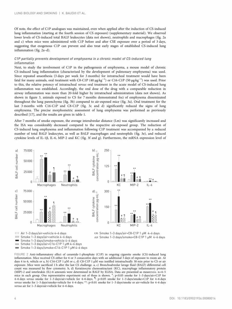
Of note, the effect of C1P analogues was maintained, even when applied after the induction of CS-inducedlung inflammation (starting at the fourth session of CS exposure) (supplementary material). We observedlower levels of CS-induced total BALF leukocytes (data not shown), neutrophils and macrophages (fig. 2aand c) when mice were administered with C1P before and after CSE exposure over a period of 3 days,suggesting that exogenous C1P can prevent and also treat early stages of established CS-induced lunginflammation (fig. 2a–d).
C1P partially prevents development of emphysema in a chronic model of CS-induced lunginflammationNext, to study the involvement of C1P in the pathogenesis of emphysema, a mouse model of chronicCS-induced lung inflammation (characterised by the development of pulmonary emphysema) was used.Since repeated anaesthesia (3 days per week for 3 months) for intratracheal treatment would have beenfatal for many animals, oral treatment with C8-C1P (40 µg·kg−1) or C16-C1P (50 µg·kg−1) was used. Priorto this, the relative potency of intratracheal versus oral treatment in the acute model of CS-induced lunginflammation was established. Accordingly, the oral dose of the drug with a comparable reduction inairway inflammation was more than 20-fold higher by intratracheal administration (data not shown). Asshown in figure 3, animals exposed to CS for 7 months demonstrated foci of emphysema disseminatedthroughout the lung parenchyma (fig. 3b) compared to air-exposed mice (fig. 3a). Oral treatment for thelast 3 months with C16-C1P and C8-C1P (fig. 3c and d) significantly reduced the signs of lungemphysema. The precise morphometric assessment of lung emphysema was performed as previouslydescribed [17], and the results are given in table 1.
After 7 months of smoke exposure, the average interalveolar distance (Lm) was significantly increased andthe ISA was considerably decreased compared to the respective air-exposed group. The reduction ofCS-induced lung emphysema and inflammation following C1P treatment was accompanied by a reducednumber of total BALF leukocytes, as well as BALF macrophages and neutrophils (fig. 3e), and reducedcytokine levels of IL-1β, IL-6, MIP-2 and KC (fig. 3f and g). Furthermore, the mRNA expression level of
BA
LF
ce
ll n
um
be
r
Macrophages Neutrophils KC MIP-2 IL-6
KC MIP-2 IL-6
75 000a)
50 000
25 000
0
BA
LF
cyt
ok
ine
co
nce
ntr
ati
on
pg
·mL
-1
300d)
150
0
****
**
**
#
¶
¶#
¶
¶¶#
#
#
Macrophages Neutrophils
BA
LF
ce
ll n
um
be
r
75 000c)
50 000
25 000
0
**
**
****
¶
¶#
#**
**
****
**
**
BA
LF
cyt
ok
ine
co
nce
ntr
ati
on
pg
·mL
-1
250b)
125
0
¶¶
##
#**
**
**
**
**
Air 1–3 days/air+vehicle 4–6 days
Smoke 1–3 days/air+vehicle 4–6 days
Smoke 1–3 days/air+C8-C1P 1 µM 4–6 days
Smoke 1–3 days/smoke+C8-C1P 1 µM 4–6 daysSmoke 1–3 days/smoke+vehicle 4–6 daysSmoke 1–3 days/air+C16-C1P 1 µM 4–6 days
Smoke 1–3 days/smoke+C16-C1P 1 µM 4–6 days
FIGURE 2 Anti-inflammatory effect of ceramide-1-phosphate (C1P) in ongoing cigarette smoke (CS)-induced lunginflammation. Mice received CS either for 6 or 3 consecutive days with an additional 3 days of exposure to room air. Atdays 4 to 6, vehicle or a, b) C16-C1P 1 µM or c, d) C8-C1P 1 µM was instilled intratracheally 30 min prior to CS or airexposure. Mice were sacrificed 1 h after the last CS challenge. a, c) Bronchoalveolar lavage fluid (BALF) differential cellcount was measured by flow cytometry. b, d) Keratinocyte chemoattractant (KC), macrophage inflammatory protein(MIP)-2 and interleukin (IL)-6 amounts were determined in BALF by ELISA. Data are presented as mean±SEM. n=4–5mice in each group. One representative experiment out of three is shown. #: p<0.05 smoke for 1–3 days/air+C1P for4–6 days versus smoke for 1–3 days/air+vehicle for 4–6 days; ¶: p<0.05 smoke for 1–3 days/smoke+C1P for 4–6 daysversus smoke for 1–3 days/smoke+vehicle for 4–6 days; **: p<0.01 smoke for 1–3 days/smoke or air+vehicle for 4–6 daysversus air for 1–3 days/air+vehicle for 4–6 days.
4 DOI: 10.1183/09031936.00080014
LUNG BIOLOGY AND SMOKING | K. BAUDIß ET AL.

180 000
120 000
60 000
Total cellcount
Macrophages
MIP-2 KC MMP-12 CXCL13
Neutrophils IL-1β IL-60
Air
7 m
on
ths/
veh
icle
3 m
on
ths
BA
LF
ce
ll
nu
mb
er
200
100
50
0
10
5
0
g)
e)
BA
LF
cyt
ok
ine
co
nce
ntr
ati
on
p
g·m
L-1
f)
Re
lati
ve
exp
ressio
n
h)
100
0
BA
LF
cyt
ok
ine
co
nce
ntr
ati
on
p
g·m
L-1
Sm
ok
e 7
mo
nth
s/
veh
icle
3 m
on
ths
Sm
ok
e 7
mo
nth
s/
C1
6-C
1P
3 m
on
ths
Sm
ok
e 7
mo
nth
s/
C8
-C1
P 3
mo
nth
s
Ne
ga
tive
co
ntr
ol
a)
i)
b) c) d)
Anti-CXCR5 Anti-CXCL 13 Anti-CD20 Anti-CD3ε
***
***
***
***
***
# #
***
***
**
***
###
###
######
######
### ###
###
###
Air 7 months/vehicle 3 months
Smoke 7 months/vehicle 3 monthsSmoke 7 months/C8-C1P 3 monthsSmoke 7 months/C16-C1P 3 months
### ###
######
### ###
FIGURE 3 Ceramide-1-phosphate (C1P) reduces the development of emphysema in a mouse model of chronic cigarette smoke (CS)-induced lung inflammation.C57BL/6 mice were exposed to CS for 7 months and received C1P (10 µM) or vehicle orally for the last 3 months. Lungs were isolated 1 day after the last smokechallenge and fixed under 20 cm formalin column for 2 days before being processed for histological emphysema analysis. a–d) Representative histological slides fora) air 7 months/vehicle 3 months, b) CS 7 months/vehicle 3 months, c) CS 7 months/C16-C1P 3 months and d) CS 7 months/C8-C1P 3 months. e)Bronchoalveolar lavage fluid (BALF) differential cell count, f ) interleukin (IL)-1β and IL-6, and g) macrophage inflammatory protein (MIP)-2 and keratinocytechemoattractant (KC). n=8–10 mice in each group. h) Quantified PCR analysis of matrix metalloproteinase (MMP)-12 and CXCL13 mRNA expression in7 months chronic CS mice model. n=5–7 mice in each group. i) Representative images of immunohistochemistry staining of formalin-fixed, paraffin-embeddedlung lobes for rabbit anti-mouse CXCR5, goat anti-mouse CXCL13, rabbit anti-mouse CD20, biotin armenian hamster anti-mouse CD3ε antibody. No stainingwas detected using secondary antibody alone as negative control. Data are presented as mean±SEM. **: p<0.01, ***: p<0.001 CS 7 months/vehicle 3 months versusair 7 months/vehicle 3 months; #: p<0.05, ###: p<0.001 CS 7 months/C1P 3 months versus CS 7 months/vehicle 3 months. Scale bars=100 µm.
DOI: 10.1183/09031936.00080014 5
LUNG BIOLOGY AND SMOKING | K. BAUDIß ET AL.

matrix metalloproteinase (MMP)-12 was significantly higher after 7 months in CS-exposed mice comparedto air-exposed mice. Administration of C1P attenuated MMP-12 mRNA expression in CS-induced lunginflammation (fig. 3h). Additional immunohistochemistry staining of CD3 and CD20 identifiedlymphocytes and the appearance of lymphoid aggregates and follicles in lung tissue of mice. No lymphoidfollicles were found in air-exposed mice (fig. 3i). The presence of lymphoid aggregates in CS-exposed micewas reduced by treatment with C1P (fig. 3i). Our study showed an increase in the B-cell attractingchemokines CXCL13 and CXCR5, which are found in areas of inducible bronchus-associated lymphoidtissue (iBALT), in the lungs of CS-exposed mice after 7 months. In general, we were not able to detect thetypical formation of iBALT in C1P treated mice (fig. 3i). Hence, C1P reduces the development of iBALTin a mouse model of chronic CS-induced lung inflammation due to attenuated mRNA expression ofCXCL13 (fig. 3h).
C1P reduces CS-induced NF-κB expression and activity in lung tissue and human AEC linesCS exposure is associated with the activation of the NF-κB signalling pathway in the lungs of humans andmice [19, 20]. Thus, we next questioned whether C1P can decrease CS-induced NF-κB DNA binding inmurine lung tissue. As shown in figure 4a and b, both C8-C1P and C16-C1P partially inhibited thepronounced NF-κB DNA binding in the lung tissue induced by acute CS exposure. Furthermore,quantified PCR analysis revealed that the protective role of C1P can also be linked to the reduced mRNAexpression of NF-κB1 (fig. 4c) and NF-κB target genes, including MMP-9, MYD88 and TNFreceptor-associated factor (TRAF) 2 expression (fig. S1).
To exclude an indirect effect of C1P, we analysed the influence of C1P on NF-κB DNA binding in A549and BEAS-2B cells. For this purpose, cells were pre-incubated with the indicated concentration of C8-C1P,C16-C1P or vehicle prior to stimulation with CSE (5%) for 10 min. Upon CSE stimulation, strong NF-κBDNA binding was observed in BEAS-2B (fig. 4d) and A549 (fig. 4e) cells compared to vehicle-treatedcells. Interestingly, both C1P analogues significantly reduced CSE-induced NF-κB DNA binding in adose-dependent manner (fig. 4f and g).
Effect of C1P on CS-induced cytokine production and reactive oxygen species release in humanAEC linesBased on the observation that C1P reduces CS-induced NF-κB DNA binding in human AECs in vitro, wethen investigated whether C1P consequently modulates production of pro-inflammatory cytokines (IL-6/IL-8) in AECs. Therefore, A549 and BEAS-2B cells were either treated with the indicated concentration ofC8-C1P, C16-C1P or vehicle for 1 h prior to CSE stimulation. Supernatants were collected after 6 h andcytokines were quantified. As shown in figure 5, C8-C1P and C16-C1P significantly inhibited theCSE-induced production of IL-6 and IL-8. Additional reactive oxygen species (ROS) production by humanAECs was determined. C1P was able to diminish CSE-induced ROS release in A549 and BEAS-2B (fig. S2).
C1P attenuates CSE-induced NF-κB expression and inhibits IL-8 production in human neutrophilsBesides AECs, neutrophils are also a major source of airway IL-8 in COPD. Therefore, we investigated theeffect of C1P on CSE-induced IL-8 production in human neutrophils isolated from nonsmoking controls,ex-smoking controls and COPD patients. In line with the findings from AECs, C16-C1P and C8-C1Pdose-dependently inhibited CSE-induced release of IL-8 by neutrophils of control subjects and COPDpatients with mild-to-moderate (GOLD stages I and II) and severe (GOLD stages III and IV) disease
TABLE 1 Morphometric assessment of the effects of ceramide-1-phosphate (C1P) on theprogression of emphysematous changes caused by cigarette smoke (CS) exposure
Groups Exposure/treatment Results at 7 months
0–4 months 5–7 months Lm µm ISA cm2
A Air/- Air/vehicle 20.6±0.8 748±65B CS/- CS/vehicle 32.3±3.2* 492±70*C CS/- CS/C16-C1P 25.1±2.2*,# 590±42*D CS/- CS/C8-C1P 27.4±1.3*,# 609±131*
Data are presented as mean±SD. Tissue sections from formalin-fixed and paraffin-embedded lungs werestained with haematoxylin-eosin. Mean linear intercept (Lm) describes the average distance betweenalveolar walls in parenchyma. Internal surface area of the lungs (ISA) was performed at 25 cm of formalindistending pressure. n=5 mice per group. *: p<0.05, CS 7 months/vehicle 3 months versus air 7 months/vehicle 3 months; #: p<0.05, CS 7 months/C1P 3 months versus CS 7 months/vehicle 3 months.
6 DOI: 10.1183/09031936.00080014
LUNG BIOLOGY AND SMOKING | K. BAUDIß ET AL.

(fig. 6a–d). The efficacy of C1P treatment in blocking IL-8 production was similar between the controlsubjects and COPD patients with mild-to-moderate (GOLD stages I and II) and severe (GOLD stages IIIand IV) disease. Transcriptional analysis revealed that C1P also reduced CSE-induced NF-κB1 level inneutrophils of COPD patients (fig. 6e).
Increased nSMase activity/expression in murine lung tissue with emphysema and CS-exposedhuman granulocytes and seraIt has previously been reported that CSE increases nSMase activity in AECs, resulting in an increased levelof ceramide and epithelial cell apoptosis [5, 13]. Thus, we analysed nSMase activity in the lungs of micewith CS-induced lung inflammation/emphysema, as well as in the serum of 22 COPD patients, eight
5.0b)a)
p65/p50 2.5
0.0Rela
tive
expr
essi
on
30c)
15
0
NF-
B1
rela
tive
expr
essi
on
2.0
1.5
1.0
0.5
f)
Rela
tive
expr
essi
on
1.5
1.0
0.5
g)
Rela
tive
expr
essi
on
1 2 3 4
VehicleCSC8-C1P µMC16-C1P µM
+---
++--
-+1-
-+-1
VehicleCSC8-C1P µMC16-C1P µM
+---
++--
-+
0.1-
-+1-
-+1-
-+-
0.1
-+-1
-+-
10
**
#
##
***
VehicleVehicle/CS
C8-C1P 1 µM/CSC16-C1P 1 µM/CS
d)
e)
p65/p50
1 2 3 4 5 6 7 8
p65/p50
Air 7 months/vehicle 3 monthsSmoke 7 months/vehicle 3 monthsSmoke 7 months/C16-C1P 3 monthsSmoke 7 months/C8-C1P 3 months
VehicleVehicle/CSEC8-C1P 0.1 µM/CSEC8-C1P 1 µM/CSEC8-C1P 10 µM/CSEC16-C1P 0.1 µM/CSEC16-C1P 1 µM/CSEC16-C1P 10 µM/CSE
FIGURE 4 Ceramide-1-phosphate (C1P) treatment inhibits nuclear factor (NF)-κB DNA binding and NF-κB1expression. a) NF-κB DNA binding in a mouse model of acute cigarette smoke (CS)-induced lung inflammationmeasured by electrophoretic mobility shift assay (EMSA). b) Quantification of the bands shown in a. c) Downregulationof NF-κB1 expression in chronic CS-induced lung inflammation assessed by quantified PCR. GAPDH was used as areference gene. Human d) BEAS-2B and e) A549 airway epithelial cell lines were treated with C16-C1P and C8-C1P for1 h prior to 5% cigarette smoke extract (CSE) stimulation for 10 min. Equal amounts of protein from total cell extractswere analysed for NF-κB DNA binding by EMSA. One representative EMSA out of three is shown. Quantification ofthe bands out of the three EMSAs is shown for f ) BEAS-2B and g) A549. Data are presented as mean±SEM. **: p<0.01CS 7 months/vehicle 3 months versus air 7 months/vehicle 3 months; ***: p<0.001 CS 7 months/vehicle 3 months versusair 7 months/vehicle 3 months; #: p<0.001 CS 7 months/C1P 3 months versus CS 7 months/vehicle 3 months.
DOI: 10.1183/09031936.00080014 7
LUNG BIOLOGY AND SMOKING | K. BAUDIß ET AL.

ex-smoker controls and 11 nonsmoker controls. As shown in figure 7, increased nSMase activity wasdetected in lung tissue of CS-exposed mice compared to air-exposed littermates (fig. 7a) and also in thesera of COPD patients compared to nonsmokers and ex-smoker controls (fig. 7b). Interestingly, there wasa significant negative correlation between human serum nSMase levels and post-bronchodilator forcedexpiratory volume in 1 s (FEV1) % predicted in COPD patients (r=−0.57; p<0.01) (fig. S3a) and also inthe entire study population (r=−0.75; p<0.001) (fig. S3b).
Finally, we addressed the modulatory effect of C1P on CS-triggered nSMase activity/expression in thelung tissue of mice and human neutrophils. C8-C1P and C16-C1P significantly attenuated CS-inducednSMase activity in murine lung (fig. 7a) and nSMase2 mRNA expression in human neutrophils of COPDpatients (fig. 7c).
DiscussionSphingolipids, such as sphingosine-1-phosphate (S1P) and C1P, have been implicated in the regulation ofimmune response during infections, as well as in inflammatory disorders such as atherosclerosis,rheumatoid arthritis, asthma, acute respiratory distress syndrome or cystic fibrosis [9, 21–24]. It isremarkable that while the role of S1P and S1P receptor-agonists/antagonists in the pathogenesis ofinflammatory disorders has been extensively studied in vivo using translational mouse models of humandiseases [9, 10, 25], the knowledge concerning the function of exogenous C1P in modulating immuneresponses is, at present, mostly limited to in vitro observations [26, 27]. Both pro- and anti-inflammatorybehaviour of exogenous C1P has been described. For instance, C1P stimulates eicosanoid (e.g.prostaglandin E2) biosynthesis, and promotes phagocytosis of neutrophils, migration of macrophages anddegranulation of mast cells [15, 22, 24, 28]. Conversely, C1P inhibits LPS-triggered production ofpro-inflammatory cytokines such as TNF-α, IL-6 or IL-5/IL-13 in macrophages or mast cells, as well asreducing acid SMase activity [10, 22, 23, 29].
Here, we report for the first time that both the synthetic C1P analogue C8-C1P and the natural analogueC16-C1P exert anti-inflammatory effects in a mouse model of CS-induced lung inflammation andemphysema. Intrapulmonary and systemic oral treatment attenuates CS-induced recruitment of leukocytes tothe lung and production of pro-inflammatory cytokines, such as KC, MIP-2, IL-6 and IL-1β, as well as
50
100
150
***
***
***
***
***
***
##
###
#####
##
##
#
# ## #
c)
A5
49
IL
-8 p
rod
ucti
on
p
g·m
L-1
150
250
350d)
BE
AS
-2B
IL
-8
pro
du
cti
on
pg
·mL
-1
2000
2500
3000a)
A5
49
IL
-6 p
rod
ucti
on
p
g·m
L-1
500
1250
2000b)
BE
AS
-2B
IL
-6p
rod
ucti
on
pg
·mL
-1
Vehicle
CSE
C16-C1P µM
C8-C1P µM
+
-
-
-
+
+
-
-
-
+
0.1
-
-
+
1
-
-
+
-
0.1
-
+
-
1
+
+
-
-
-
+
0.1
-
-
+
1
-
-
+
-
0.1
-
+
-
1
Vehicle
CSE
C16-C1P µM
C8-C1P µM
+
-
-
-
FIGURE 5 Ceramide-1-phosphate (C1P) administration prior to cigarette smoke extract (CSE) stimulation reducesinterleukin (IL)-6 and IL-8 production in airway epithelial cells. C1P 0.1 µM and 1 µM were given to confluenta, c) A549 and b, d) BEAS-2B cells 1 h prior to 5% CSE stimulation for 6 h. The supernatant was taken and the contentof a, b) IL-6 and c, d) IL-8 was measured by ELISA. Data are presented as mean±SEM. n=10. ***: p<0.001 vehicle/CSEversus vehicle/vehicle; #: p<0.05, ##: p<0.01, ###: p<0.001 C1P/CSE versus vehicle/CSE.
8 DOI: 10.1183/09031936.00080014
LUNG BIOLOGY AND SMOKING | K. BAUDIß ET AL.

development of lung emphysema. In addition, CS-induced mRNA levels of MMP-9 and MMP-12 arediminished by administration of C1P. Therefore, C1P could prevent tissue injury and remodelling due to thereduced matrix proteolysis. Furthermore, we recognised a link between iBALT formation and emphysemadevelopment that supports the study of BRACKE et al. [30], who described an increase of CXCL13 in patientswith COPD and how crucial CXCL13 is in the development of CS-induced lymphoid follicles in mice.Treatment with C1P attenuates the presence of CS-induced accumulation of lymphocytes and CXCL13, andits receptor CXCR5.
Mechanistically, we provide evidence that C1P reduces CS-induced expression and DNA binding ofNF-κB in structural lung cells in vivo and in vitro. In connection with this, nSMase2 has recently beenreported to play a role in CS-triggered apoptosis of AECs [31]. Therefore, it is of significance that C1Pinhibits CS-stimulated nSMase activity and expression in murine lungs and human neutrophils,respectively. We observed increased nSMase activity in the serum of patients with COPD compared tocorresponding controls, which was negatively correlated to FEV1 % pred. Overall, our findings suggest thatsynthetic and natural C1P analogues might be considered as new therapeutic agents for CS-induced lunginflammation and emphysema.
Vehicle
CSE
C16-C1P µM
C8-C1P µM
+
-
-
-
600
300
Co
ntr
ol
no
nsm
ok
ers
IL-8
pro
du
cti
on
pg
·mL
-1
0
500
250
0
500
c)
20Vehicle/medium
Vehicle/CSE
C16-C1P 1 µM/CSE
C8-C1P 1 µM/CSE10
NF
-κB
1 r
ela
tive
exp
ressio
n
of
hu
ma
n n
eu
tro
ph
ils
0
e)
Co
ntr
ol
ex-s
mo
ke
rs
IL-8
pro
du
cti
on
pg
·mL
-1
d)
CO
PD
GO
LD
I/I
I
IL-8
pro
du
cti
on
pg
·mL
-1
a)
CO
PD
GO
LD
III
/IV
IL-8
pro
du
cti
on
pg
·mL
-1
b)
250
0
1400
700
0
+
+
-
-
-
+
1
-
-
+
10
-
-
+
-
1
-
+
-
10
+
+
-
-
-
+
1
-
-
+
10
-
-
+
-
1
-
+
-
10
Vehicle
CSE
C16-C1P µM
C8-C1P µM
+
-
-
-
** * * * * **
* * * *
¶ ¶
#
* * * *
FIGURE 6 Ceramide-1-phosphate (C1P) reduces interleukin (IL)-8 and nuclear factor (NF)-κB expression in humanneutrophils stimulated with cigarette smoke. C1P (1 µM and 10 µM) was administered to isolated human neutrophils 1 hbefore stimulation with 5% cigarette smoke extract (CSE). 18 h later IL-8 in the supernatant was quantified by ELISA. a)Chronic obstructive pulmonary disease (COPD) Global Initiative for Chronic Obstructive Lung Disease (GOLD) I/IIpatients, b) COPD GOLD III/IV patients, c) control nonsmokers, and d) control ex-smokers. e) Neutrophils isolatedfrom COPD patients were stimulated with 1 µM C1P 1 h prior to CSE for 3 h and gene expression analysis for NF-κB1was performed by quantified PCR. GAPDH was used as a reference gene. Data are presented as mean±SEM. *: p<0.05,**: p<0.01 C1P/vehicle versus vehicle/CSE; #: p<0.05 vehicle/CSE versus vehicle; ¶: p<0.05 C1P/CSE versus vehicle/CSE.
DOI: 10.1183/09031936.00080014 9
LUNG BIOLOGY AND SMOKING | K. BAUDIß ET AL.

NF-κB is one of the key transcription factors involved in cellular responses to CS in the lungs [19, 20, 32].Its activation leads to the induction of multiple genes associated with innate immunity, cellularinflammation and repair processes. In the present study, CS-induced NF-κB DNA binding and NF-κBexpression in the lungs were reduced when mice were treated with C8-C1P or C16-C1P, suggesting thatC1P might exert its anti-inflammatory capacity by suppressing the NF-κB pathway. However, the decreasein NF-κB DNA binding might also be due to the attenuation in CS-induced lung inflammation observedupon C1P treatment. Thus, to better define the capacity of C1P to modulate CS-induced NF-κBtransformation, in vitro experiments with CSE-stimulated human A549 and BEAS-2B AECs wereperformed. Strikingly, both C8-C1P and C16-C1P were dose-dependently able to attenuate CSE-triggeredtranslocation of NF-κB into the nucleus in epithelial cell lines, implying that C1P, via interfering with theNF-κB pathway, regulates CS-induced lung inflammation. In this context, C1P reduces CS-triggeredinduction of NF-κB target genes expression such as Mmp9, Myd88 and Traf2 (supplementary material).Unfortunately, TNF receptor 1 and 2 were not significantly increased in our mouse models (data notshown), thus it was not possible to determine the effect of C1P on mRNA expression.
Interestingly, similar effects of C8-C1P and/or C16-C1P on LPS-induced NF-κB activation in humanembryonic kidney cells and primary human peripheral blood mononuclear cells have been demonstrated[33]. However, other studies reported that high concentrations of C16-C1P (15 or 20 µM) stimulated NF-κBactivation in the alveolar rat macrophage cell line NR8383 [12] and murine macrophage cell line J774A.1[15]. Nevertheless, GOLDSMITH et al. [14] did not observe any effect of phospho-ceramide analogue-1, asynthetic C1P analogue, on LPS-induced NF-κB activation in the murine macrophage RAW264.7 cell line.
An explanation for this discrepancy might be the different C1P concentration (1–20 µM) [12, 15, 33] andthe C1P analogue, as well as the different cell types used in these studies. Of note, the solvent in whichC1P is dispersed has been reported to be critical for its pro- or anti-inflammatory action. For instance,C1P was often dissolved in either methanol/dodecane [11] or ethanol/dodecane [34], and dodecane isknown to influence the effect of C1P [29]. To avoid this, we used C16-C1P sonicated in sterile water orC8-C1P dispersed in ethanol/PBS or medium (1:2000–1:200000 dilution).
Since NF-κB pathway activation leads to the induction of pro-inflammatory cytokines in various cell types[4, 18, 23, 35], we investigated whether the CSE-induced production of IL-8 and IL-6 by A549 andBEAS-2B cells is also regulated by C1P. Indeed, C8-C1P and C16-C1P dose-dependently inhibitedCSE-triggered production of IL-8 and IL-6 in AECs.
Furthermore, we have shown an increase of ROS level produced by A549 and BEAS-2B when they werestimulated with CSE. The reduced ROS release through C1P stimulation could prevent cellular damage oraccumulation of oxidative stress.
Nevertheless, oxidative stress in the airways following CS exposure also causes activation of an endogenousmembrane nSMase. CHEN et al. [35] found that peptidoglycan- or LPS-stimulated RAW264.7 macrophages
25.0a) b)
12.5
0.0
nS
Ma
se
acti
vity
(n
no
l·h
–1·m
L–
1)
in m
uri
ne
lu
ng
s
20
Air 7 months/vehicle 3 months
Smoke 7 months/vehicle 3 months
Smoke 7 months/C16-C1P 3 months
Smoke 7 months/C8-C1P 3 months
10
Control
ex-smoker
COPD Control
nonsmoker
0
nS
Ma
se
acti
vity
(n
no
l·h
–1·m
L–
1)
in h
um
an
lu
ng
s
c) 5.0
2.5
0.0
nSMase2
re
lati
ve
exp
ressio
n i
n h
um
an
n
eu
tro
ph
ils
Vehicle/medium
Vehicle/CSE
C16-C1P 1 µM/CSE
C8-C1P 1 µM/CSE
**
###
#
¶
¶ §
§
+
FIGURE 7 Increased neutral sphingomyelinase (nSMase) activity in murine lungs and sera of patients with chronic obstructive pulmonary disease (COPD).C57BL/6 mice were exposed to cigarette smoke (CS) for 7 months and received ceramide-1-phosphate (C1P) (10 µM) or vehicle orally for the last 3 months.a) Lungs were isolated and nSMase activity was quantified with the SMase fluorometric assay kit. b) nSMase activity in serum from 22 patients with COPD,eight healthy control ex-smokers and 11 control nonsmokers were determined. c) nSMase2 expression of stimulated human neutrophils isolated from patientswith COPD measured by quantified PCR. ß2-microglobulin was used as a reference gene. Data are presented as mean±SEM. n=5 mice in each group. ‐ ‐ ‐ ‐ ‐:detection limit of the nSMase fluorometric assay. **: p<0.01 CS 7 months/vehicle 3 months versus air 7 months/vehicle 3 months; #: p<0.01, ###: p<0.001 CS7 months/C1P 3 months versus CS 7 months/vehicle 3 months; ¶: p<0.01 control ex-smokers/control nonsmokers versus COPD; +: p<0.01 vehicle/cigarettesmoke extract (CSE) versus vehicle/medium; §: p<0.01 C1P/CSE versus vehicle/CSE.
10 DOI: 10.1183/09031936.00080014
LUNG BIOLOGY AND SMOKING | K. BAUDIß ET AL.

release ceramide through the activation of nSMase, which further initiates the NF-κB pathway [35]. Thus,the stimulation of nSMase seems to be required for CS-induced apoptosis of AECs [13, 31]. Of note,FILOSTO et al. [31] found higher levels of nSMase2 in lung tissue of emphysema patients in comparison tocontrol subjects. Thus, nSMase has recently gained attention as a new therapeutic strategy for CS-inducedlung inflammation and lung injury. Strikingly, we found elevated nSMase activity in the lungs of micechronically exposed to CS and, more importantly, significantly higher levels of nSMase in the serum ofpatients with COPD compared to never- and former smokers. Notably, enzyme activity was negativelycorrelated with airflow limitation (FEV1 % pred), further underlining the potential role of nSMase in thepathogenesis of COPD. Moreover, treatment of mice with both C1P analogues decreased nSMase activity inthe lungs, demonstrating an additional anti-inflammatory action of C1P. We suggest that the inhibition ofnSMase by C1P decreases ceramide level and consequently influences NF-κB signalling and thedevelopment of emphysema. However, to fully clarify whether ceramide or one of its metabolites regulatesthese effects, further studies investigating the total ceramide level in the context of CS-induced lunginflammation and the treatment with C1P are needed. Interestingly, an inhibitory effect of C1P haspreviously been reported for another isoform of the enzyme, the acid SMase in murine bone marrowmacrophages [11], although its activity is not modified in CS-induced lung inflammation [31].Furthermore, it has been shown that C1P can directly interact with serine palmitoyl transferase to block itsactivity [12]. Hence, we suggest C1P might also inhibit the nSMase isoform due to interaction with thisceramide-generating enzyme. Further analysis could reveal the level of C1P, ceramide, S1P, ceramide kinase,SMase and glycosylceramide synthase in the lungs and plasma of healthy controls compared to COPDpatients, leading us to fully understand the complex metabolism of sphingolipids. Additionally, it would beinteresting to investigate how exogenous C1P modifies the level of these lipids so that a concrete target canbe found for future drug development.
It is worth mentioning that the anti-inflammatory effect of C1P on CS-induced lung inflammation is notlimited to the modulation of structural cells, as both C8-C1P and C16-C1P also reduce CSE-triggeredupregulation of nSMase and NF-κB expression, and consequently IL-8-production, in human neutrophils.Neutrophils are known to play an essential role in the pathogenesis of COPD and, moreover, themodulation of the CXCR2/IL-8 axis has previously been implicated as a therapeutic strategy for COPD[36]. Thus, our observation that C1P inhibits CS-induced IL-8 production in AECs and neutrophilsfurther strengthens our assumption that C1P might be a new therapeutic target for COPD. The potentialof C1P to dampen pro-inflammatory cytokine production has also been reported for LPS-induced TNF-α,MIP-2, IL-6 and IL-8 in human embryonic kidney cells, human blood mononuclear cells, endothelial cellsand mastocytes [27, 33, 37].
COPD is a complex obstructive pulmonary disease characterised by irreversible and progressive airflowlimitation. Although the effect of C1P on the progression of COPD was characterised by pulmonaryinflammation, air space enlargement and parenchymal destruction, there is still need for further analysis ofthe respiratory function of mice to consider the clinical relevance of how C1P treatment affects respiratorymechanics.
In summary, we have demonstrated for the first time that C1P analogues inhibit CS-induced lunginflammation and emphysema in mouse models of COPD. Mechanistically, we showed that C1P interfereswith CS-induced activation of the NF-κB pathway, consequently inhibiting pro-inflammatory cytokineproduction (such as IL-6 and IL-8) by human AECs and blood neutrophils, and also attenuatingCS-induced induction of nSMase activity in the lungs and neutrophils of COPD patients. Thus, C1Panalogues might hold promise as novel treatment options in COPD.
AcknowledgementsWe would like to thank Barbara Schuler (Dept of Pharmaceutical Biology and Biotechnology, University HospitalFreiburg, Freiburg, Germany) for assistance in performing the electrophoretic mobility shift assays and MadelonHossfeld (Dept of Pneumology, COPD and Asthma Research Group, University Hospital Freiburg, Freiburg, Germany)for support with the immunohistochemistry.
References1 Global Initiative for Chronic Obstructive Lung Disease. Global strategy for the diagnosis, management, and
prevention of chronic obstructive pulmonary disease. Updated 2014. www.goldcopd.com/uploads/users/files/GOLD_Report_2014_Oct30.pdf Date last updated: 2014. Date last accessed: January 8, 2014.
2 Barnes PJ, Shapiro SD, Pauwels RA. Chronic obstructive pulmonary disease: molecular and cellular mechanisms.Eur Respir J 2003; 22: 672–688.
3 McDonough JE, Yuan R, Suzuki M, et al. Small-airway obstruction and emphysema in chronic obstructivepulmonary disease. N Engl J Med 2011; 365: 1567–1575.
4 Mukaida N, Mahe Y, Matsushima K. Cooperative interaction of nuclear factor-kappa B- and cis-regulatoryenhancer binding protein-like factor binding elements in activating the interleukin-8 gene by pro-inflammatorycytokines. J Biol Chem 1990; 265: 21128–21133.
DOI: 10.1183/09031936.00080014 11
LUNG BIOLOGY AND SMOKING | K. BAUDIß ET AL.

5 Petrache I, Natarajan V, Zhen L, et al. Ceramide upregulation causes pulmonary cell apoptosis andemphysema-like disease in mice. Nat Med 2005; 11: 491–498.
6 Becker KA, Riethmuller J, Luth A, et al. Acid sphingomyelinase inhibitors normalize pulmonary ceramide andinflammation in cystic fibrosis. Am J Respir Cell Mol Biol 2010; 42: 716–724.
7 Schweitzer KS, Hatoum H, Brown MB, et al. Mechanisms of lung endothelial barrier disruption induced bycigarette smoke: role of oxidative stress and ceramides. Am J Physiol Lung Cell Mol Physiol 2011; 301: L836–L846.
8 Niwa S, Urtz N, Baumruker T, et al. Ovalbumin-induced plasma interleukin-4 levels are reduced in ceramidekinase-deficient DO11.10 RAG1-/- mice. Lipids Health Dis 2010; 9: 1.
9 Idzko M, Hammad H, van Nimwegen M, et al. Local application of FTY720 to the lung abrogates experimentalasthma by altering dendritic cell function. J Clin Invest 2006; 116: 2935–2944.
10 Chalfant CE, Spiegel S. Sphingosine 1-phosphate and ceramide 1-phosphate: expanding roles in cell signaling.J Cell Sci 2005; 118: 4605–4612.
11 Gomez-Munoz A, Kong JY, Salh B, et al. Ceramide-1-phosphate blocks apoptosis through inhibition of acidsphingomyelinase in macrophages. J Lipid Res 2004; 45: 99–105.
12 Granado MH, Gangoiti P, Ouro A, et al. Ceramide 1-phosphate inhibits serine palmitoyltransferase and blocksapoptosis in alveolar macrophages. Biochim Biophys Acta 2009; 1791: 263–272.
13 Levy M, Khan E, Careaga M, et al. Neutral sphingomyelinase 2 is activated by cigarette smoke to augmentceramide-induced apoptosis in lung cell death. Am J Physiol Lung Cell Mol Physiol 2009; 297: L125–L133.
14 Goldsmith M, Avni D, Levy-Rimler G, et al. A ceramide-1-phosphate analogue, PCERA-1, simultaneouslysuppresses tumour necrosis factor-alpha and induces interleukin-10 production in activated macrophages.Immunology 2009; 127: 103–115.
15 Arana L, Ordonez M, Ouro A, et al. Ceramide 1-phosphate induces macrophage chemoattractant protein-1release: involvement in ceramide 1-phosphate-stimulated cell migration. Am J Physiol Endocrinol Metab 2013; 304:E1213–E1226.
16 Lucattelli M, Cicko S, Muller T, et al. P2X7 receptor signaling in the pathogenesis of smoke-induced lunginflammation and emphysema. Am J Respir Cell Mol Biol 2011; 44: 423–429.
17 Cicko S, Lucattelli M, Muller T, et al. Purinergic receptor inhibition prevents the development of smoke-inducedlung injury and emphysema. J Immunol 2010; 185: 688–697.
18 Libermann TA, Baltimore D. Activation of interleukin-6 gene expression through the NF-kappa B transcriptionfactor. Mol Cell Biol 1990; 10: 2327–2334.
19 Vlahos R, Bozinovski S, Jones JE, et al. Differential protease, innate immunity, and NF-kappaB induction profilesduring lung inflammation induced by subchronic cigarette smoke exposure in mice. Am J Physiol Lung Cell MolPhysiol 2006; 290: L931–L945.
20 Di Stefano A, Caramori G, Oates T, et al. Increased expression of nuclear factor-κB in bronchial biopsies fromsmokers and patients with COPD. Eur Respir J 2002; 20: 556–563.
21 Teichgraber V, Ulrich M, Endlich N, et al. Ceramide accumulation mediates inflammation, cell death andinfection susceptibility in cystic fibrosis. Nat Med 2008; 14: 382–391.
22 Lamour NF, Chalfant CE. Ceramide-1-phosphate: the “missing” link in eicosanoid biosynthesis and inflammation.Mol Interv 2005; 5: 358–367.
23 Matsusaka T, Fujikawa K, Nishio Y, et al. Transcription factors NF-IL6 and NF-kappa B synergistically activatetranscription of the inflammatory cytokines, interleukin 6 and interleukin 8. Proc Natl Acad Sci USA 1993; 90:10193–10197.
24 Nixon GF. Sphingolipids in inflammation: pathological implications and potential therapeutic targets. Br JPharmacol 2009; 158: 982–993.
25 Fyrst H, Saba JD. An update on sphingosine-1-phosphate and other sphingolipid mediators. Nat Chem Biol 2010;6: 489–497.
26 Arana L, Gangoiti P, Ouro A, et al. Ceramide and ceramide 1-phosphate in health and disease. Lipids Health Dis2010; 9: 15.
27 Gomez-Munoz A, Gangoiti P, Arana L, et al. New insights on the role of ceramide 1-phosphate in inflammation.Biochim Biophys Acta 2013; 1831: 1060–1066.
28 Granado MH, Gangoiti P, Ouro A, et al. Ceramide 1-phosphate (C1P) promotes cell migration involvement of aspecific C1P receptor. Cell Signal 2009; 21: 405–412.
29 Tauzin L, Graf C, Sun M, et al. Effects of ceramide-1-phosphate on cultured cells: dependence on dodecane in thevehicle. J Lipid Res 2007; 48: 66–76.
30 Bracke KR, Verhamme FM, Seys LJ, et al. Role of CXCL13 in cigarette smoke-induced lymphoid follicle formationand chronic obstructive pulmonary disease. Am J Respir Crit Care Med 2013; 188: 343–355.
31 Filosto S, Castillo S, Danielson A, et al. Neutral sphingomyelinase 2: a novel target in cigarette smoke-inducedapoptosis and lung injury. Am J Respir Cell Mol Biol 2011; 44: 350–360.
32 Comer DM, Kidney JC, Ennis M, et al. Airway epithelial cell apoptosis and inflammation in COPD, smokers andnonsmokers. Eur Respir J 2013; 41: 1058–1067.
33 Hankins JL, Fox TE, Barth BM, et al. Exogenous ceramide-1-phosphate reduces lipopolysaccharide(LPS)-mediated cytokine expression. J Biol Chem 2011; 286: 44357–44366.
34 Pettus BJ, Bielawska A, Subramanian P, et al. Ceramide 1-phosphate is a direct activator of cytosolicphospholipase A2. J Biol Chem 2004; 279: 11320–11326.
35 Chen BC, Chang HM, Hsu MJ, et al. Peptidoglycan induces Cyclooxygenase-2 expression in macrophages byactivating the neutral sphingomyelinase-ceramide pathway. J Biol Chem 2009; 284: 20562–20573.
36 Barnes PJ. New anti-inflammatory targets for chronic obstructive pulmonary disease. Nat Rev Drug Discov 2013;12: 543–559.
37 Chiba N, Masuda A, Yoshikai Y, et al. Ceramide inhibits LPS-induced production of IL-5, IL-10, and IL-13 frommast cells. J Cell Physiol 2007; 213: 126–136.
12 DOI: 10.1183/09031936.00080014
LUNG BIOLOGY AND SMOKING | K. BAUDIß ET AL.


