Embed Size (px)
Citation preview

cGMP modulates spike responses of retinal ganglion cellsvia a cGMP-gated current
FUSAO KAWAI1,2 and PETER STERLING1
1Department of Neuroscience, University of Pennsylvania, Philadelphia2Department of Physiology, Fujita Health University, Toyoake, Aichi, 470-1192, Japan
(Received December 12, 2001;Accepted April 25, 2002)
Abstract
Certain ganglion cells in the mammalian retina are known to express a cGMP-gated cation channel. We foundthat a cGMP-gated current modulates spike responses of the ganglion cells in mammalian retinal slice preparation.In such cells under current clamp, bath application of the membrane-permeant cGMP analog (8-bromo-cGMP,8-p-chlorophenylthio-cGMP) or a nitric oxide donor (sodium nitroprusside, S-nitroso-N-acetyl-penicillamine)depolarized the membrane potential by 5–15 mV, and reduced the amount of current needed to evoke actionpotentials. Similar effects were observed when the membrane potential was simply depolarized by steadycurrent. The responses to cGMP are unaffected by inhibitors of cGMP-dependent protein kinase andCa210calmodulin-dependent protein kinase. The response to cGMP persisted in Ca21-free bath solution withCa21 buffers in the pipette. Under voltage clamp, cGMP analogs did not affect the response kinetics ofvoltage-gated currents. We conclude that cGMP modulates ganglion cell spiking simply by depolarizing themembrane potentialvia the inward current through the cGMP-gated channel. Modulation of this channelviathe long-range NO-synthase amacrine cell may contribute to control of contrast gain by peripheral mechanisms.
Keywords: Retina, Ganglion cell, Guinea pig, Patch clamp, cGMP-gated channel
Introduction
Certain ganglion cells in mammalian retina express a cGMP-gatedcation channel (Ahmad et al., 1994; Kawai & Sterling, 1999). Thischannel is modulated when nitric oxide (NO) released from am-acrine cells (Sandell, 1985; Vincent & Kimura, 1992; Haberechtet al., 1998) stimulates the ganglion cell’s soluble guanylyl cyclase(Ahmad & Barnstable 1993; Gotzes et al., 1998; Kawai & Sterling,1999). The effect of current through this channel had not beenstudied and was complicated to address because cGMP can haveseveral effects. For example, cGMP can modulate voltage-gatedcurrents directly (Levitan & Levitan, 1988) andvia cGMP-dependent protein kinase (PKG) (Paupardin-Tritsch et al., 1986;Sumii & Sperelakis, 1995; Lohmann et al., 1997; Wei et al., 1998).Here we investigated the effect of current through the cGMP-gatedchannel on ganglion cell responses, isolating this effect from thatof the PKG cascade and from the effect of Ca21 enteringvia thecGMP-gated channel on voltage-gated currents.
Methods
Preparation and recording
Slices from adult guinea pig retina were cut at 200mm (Kawai &Sterling, 1999) and viewed on a Zeiss upright microscope withdifferential interference contrast optics (403 water-immersionobjective). Ganglion cells were identified in the slice by theirposition and size. Membrane voltages and currents were recordedin the whole-cell configuration using a patch-clamp amplifier(Axopatch 200A, Axon Instruments, Foster City, CA) linked to acomputer. The current- and voltage-clamp procedures were con-trolled by the pCLAMP software (Axon Instruments). Data werelow-pass filtered (4-pole Bessel type) with a cut-off frequencyof 5 kHz and then digitized at 10 kHz by an analog-to-digitalinterface. All experiments were performed at room temperature(23–258C).
Solutions and drugs
In most experiments, the tissue was perfused with Ames medium(which is buffered with 22.6 mM bicarbonate), bubbled with 95%O205% CO2. In some experiments, Ringer’s solution was usedinstead of Ames’ solution to eliminate Ca21 in the bath. TheRinger’s solution contained (in mM) 135 NaCl; 5 KCl; 10 HEPES;
Address correspondence and reprint requests to: Fusao Kawai, Depart-ment of Physiology, School of Medicine, Fujita Health University, 1-98Dengakugakubo, Kutsukakechou, Toyoake, Aichi, 470-1192, Japan. E-mail:[email protected]
Visual Neuroscience(2002),19, 373–380. Printed in the USA.Copyright © 2002 Cambridge University Press 0952-5238002 $12.50DOI: 10.1017.S0952523802193138
373

and 15 glucose. The solution was adjusted with NaOH to pH 7.4.CdCl2 (200 mM), picrotoxin (100mM), and strychnine (1mM)were also added to block synaptic transmission. The recordingpatch pipette contained (in mM): 140 KCl; 1 CaCl2; 5 EGTA orBAPTA; 10 HEPES; and 2 Mg-ATP. The solution was adjustedwith KOH to pH 7.4. Pipette resistance was about 7 MV.
Test substances were applied either through the bath [8-bromo-cGMP, 8-p-chlorophenylthio-cGMP, 8-p-chlorophenylthio-cAMP,sodium nitroprusside (SNP), or S-nitroso-N-acetyl-penicillamine(SNAP)], via the patch pipette (KT5823, or KN-62). KT5823,KN-62, and S-nitroso-N-acetyl-penicillamine were purchased fromCalbiochem (San Diego, CA). Other chemicals were from Sigma(St. Louis, MO).
Results
Two spike patterns to current injection
Recordings in control solution with synaptic input blocked (CdCl2,200mM 1 picrotoxin, 100mM 1 strychnine, 1mM) showed twotypes of firing. In some cells, small current injections (30 and 100pA) evoked sustained firing, and larger currents (140 pA) gaveinitial spiking that was ultimately blocked—presumably by inac-tivation of Na1 channels at sustained depolarizations above240 mV(Figs. 1A–1C). In other cells, even small current injections evoked
phasic spiking that soon inactivated (Figs. 1D–1F). About three-quarters of the cells (52070) that we recorded in adult retinashowed the tonic spiking; and the rest (18070) were phasic. Thisresembles previous observations on cells in the ganglion cell layerof adult rat (Wang et al., 1997).
Effects of the cGMP-gated current on spike responses
Under voltage clamp, we established that certain cells in theguinea pig retina express a cGMP-gated current. When membrane-permeant analogs of cGMP (1 mM 8-bromo-cGMP or 250mM8-p-chlorophenylthio-cGMP5 pCPT-cGMP) were bath applied toa cell voltage clamped at250 mV, there was a sustained inwardcurrent of approximately 200 pA (10024 cells; data not shown).This matches previous observations in rat retina (Ahmad et al.,1994; Kawai & Sterling, 1999).
Under current clamp, cGMP analogs increased the spike rate.Fig. 2A shows a tonic cell that was depolarized by 15 mV inresponse to 250mM pCPT-cGMP. This raised the spike rate to 45spikes0s (Fig. 2B). Nearly half of the tonic cells (9021) respondedsimilarly. Bath application of 1 mM 8-bromo-cGMP (306 cells),1 mM sodium nitroprusside (SNP; 407 cells), and S-nitroso-N-acetyl-penicillamine (SNAP; 205 cells) produced similar effects.However, 250mM pCPT-cAMP was ineffective (006 cells). Fig. 2Cshows a phasic cell that was depolarized by;10 mV in response
Fig. 1.Action potentials induced by the current injection in the control solution. (A–C) Spike responses of one cell to current injection,respectively, of 30 pA, 100 pA, and 140 pA for 1 s between 0.25 s and 1.25 s. Note that the cell exhibits a tonic firing pattern.(D–F) Different cell shows phasic firing. Spike responses to current injection, respectively, of 30 pA, 100 pA, and 140 pA for 1 sbetween 0.25 s and 1.25 s.
374 F. Kawai and P. Sterling

to 250 mM pCPT-cGMP. This increased the spike rate to 16spikes0s (Fig. 2D; 307 cells).
cGMP also altered the relationship between injected currentand spike rate. Application of pCPT-cGMP (250mM) to tonic cellsmarkedly lowered the amount of current needed to evoke spikes(Fig. 3A). Furthermore, current injections between 10 and 40 pAincreased spike rate almost linearly, the effect peaking at 70 pA (50spikes0s). Larger current injections (80–140 pA) decreased spikerate. The effect of pCPT-cGMP on reducing the current needed forspikes was evident during the first 100 ms, and shifted the dynamicrange of spike rate (Fig. 3B). The currents needed to evoke spikesunder the control and pCPT-cGMP conditions were 40 pA and 10pA, respectively. Spike rates under both conditions increasedalmost monotonically between 10 pA and 140 pA. A similar resultwas obtained in nine cells.
pCPT-cGMP also lowered the thresholds of a phasic cell andincreased the dynamic range. For example, the current thresholdduring 1-s current injection was 100 pA under the control and 20 pAunder pCPT-cGMP (Fig. 3C). Spike rate under the control condi-tion increased monotonically between 100 pA and 150 pA; whereasunder the pCPT-cGMP condition it increased between 20 pA and40 pA, decreasing between 90 pA and 140 pA. The maximum spikerates were 3 spikes0s (at 140 pA) under the control and 22 spikes0s(at 40 pA) under pCPT-cGMP conditions (Fig. 3C). The currentthresholds during the first 100 ms under the control and pCPT-cGMP conditions were 100 pA and 30 pA, respectively (Fig. 3D).Spike rates under both conditions increased almost monotonically.The maximum spike rates under the control and pCPT-cGMP con-ditions were 30 spikes0s (at 140 pA) and 89 spikes0s (at 120 pA),respectively. A similar result was obtained in three cells.
Fig. 2. Bath application of pCPT-cGMP depolarized the resting potentials and increased the spike rates. (A,B) Spike responses of atonic cell to current injection of 50 pA in the control solution (A) and the solution containing 250mM pCPT-cGMP (B). Current wasinjected for 1 s between 0.25 s and 1.25 s. (C,D) Spike responses of a phasic cell to current injection of 100 pA in the control solution(C) and the solution containing 250mM pCPT-cGMP (D).
A cGMP-gated current and spiking responses 375

Effects of steady current injection on spike responses
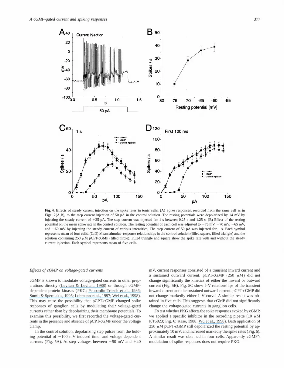
To investigate the mechanism underlying spike modulation bycGMP, we analyzed the effects of resting potential on the spikerate, because cGMP analogs depolarized the resting potentials ofganglion cells (Fig. 2). Fig. 4A shows the spike response to currentinjection recorded from the same cell as in Figs. 2A and 2B. In thecontrol solution, by injecting a steady current to the cell, theresting membrane potential was depolarized to the same voltage(approximately265 mV) as that in Fig. 2B. During a 1-s stepcurrent injection of 50 pA, we observed 42 spikes (Fig. 4A),similar to the number of spikes (44 spikes) at 50 pA under thepCPT-cGMP condition (Fig. 2B), however much larger than thenumber (12 spikes) under the control condition (Fig. 2A). Fig. 4Bshows effects of the resting potential on the mean spike rate in thecontrol solution. As the resting potential was depolarized byinjecting steady current, the spike rate was increased. This suggests
that the steady membrane depolarization increases the response tothe step depolarization.
The stimulus–response relationship under the steady currentinjection was quite similar to that under application of pCPT-cGMP. The steady current injection markedly lowered the meancurrent threshold and shifted the dynamic range of spike rateduring both 1 s (Fig. 4C) and the first 100 ms (Fig. 4D). The meancurrent threshold under the steady current injection was 10 pA(filled triangle in Figs. 4C and 4D), which was identical with that(10 pA) under application of pCPT-cGMP (filled circle in Figs. 4Cand 4D). In Fig. 4C, the mean maximum spike rates were similarunder the steady current injection and under the pCPT-cGMPcondition. In addition, the maximum rates were also quite similarduring the first 100-ms injection under the steady current injectionand under the pCPT-cGMP condition (Fig. 4D). These resultssuggest that pCPT-cGMP modulates spike rate by depolarizing theresting potentialvia the cGMP-gated inward current.
Fig. 3. Effects of pCPT-cGMP on the spike rates. (A,B) Stimulus–response relationships recorded from a tonic cell in the controlsolution (filled square) and the solution containing 250mM pCPT-cGMP (filled circle). (C,D) Stimulus–response relationshipsrecorded from a phasic cell in the control solution (filled square) and the solution containing 250mM pCPT-cGMP (filled circle).
376 F. Kawai and P. Sterling
Effects of cGMP on voltage-gated currents
cGMP is known to modulate voltage-gated currents in other prep-arations directly (Levitan & Levitan, 1988) or through cGMP-dependent protein kinases (PKG; Paupardin-Tritsch et al., 1986;Sumii & Sperelakis, 1995; Lohmann et al., 1997; Wei et al., 1998).This may raise the possibility that pCPT-cGMP changed spikeresponses of ganglion cells by modulating their voltage-gatedcurrents rather than by depolarizing their membrane potentials. Toexamine this possibility, we first recorded the voltage-gated cur-rents in the presence and absence of pCPT-cGMP under the voltageclamp.
In the control solution, depolarizing step pulses from the hold-ing potential of2100 mV induced time- and voltage-dependentcurrents (Fig. 5A). At step voltages between290 mV and140
mV, current responses consisted of a transient inward current anda sustained outward current. pCPT-cGMP (250mM) did notchange significantly the kinetics of either the inward or outwardcurrent (Fig. 5B). Fig. 5C show I–V relationships of the transientinward current and the sustained outward current. pCPT-cGMP didnot change markedly either I–V curve. A similar result was ob-tained in five cells. This suggests that cGMP did not significantlychange the voltage-gated currents in ganglion cells.
To test whether PKG affects the spike responses evoked by cGMP,we applied a specific inhibitor in the recording pipette (10mMKT5823; Fig. 6; Kase, 1988; Wu et al., 1998). Bath application of250mM pCPT-cGMP still depolarized the resting potential by ap-proximately 10 mV, and increased markedly the spike rates (Fig. 6).A similar result was obtained in four cells. Apparently cGMP’smodulation of spike responses does not require PKG.
Fig. 4. Effects of steady current injection on the spike rates in tonic cells. (A) Spike responses, recorded from the same cell as inFigs. 2(A,B), to the step current injection of 50 pA in the control solution. The resting potentials were depolarized by 14 mV byinjecting the steady current of125 pA. The step current was injected for 1 s between 0.25 s and 1.25 s. (B) Effect of the restingpotential on the mean spike rate in the control solution. The resting potential of each cell was adjusted to275 mV,270 mV,265 mV,and 260 mV by injecting the steady current of various intensities. The step current of 50 pA was injected for 1 s. Each symbolrepresents mean of four cells. (C,D) Mean stimulus–response relationships in the control solution (filled square, filled triangle) and thesolution containing 250mM pCPT-cGMP (filled circle). Filled triangle and square show the spike rate with and without the steadycurrent injection. Each symbol represents mean of five cells.
A cGMP-gated current and spiking responses 377

Effects of Ca21 influx through the cGMP-gated channelon spike responses
An accumulation of intracellular Ca21 is also known to modulatevoltage-gated currents in other preparations through Ca21- andcalmodulin-dependent protein kinase (CaM kinase; Browning et al.,1985; Arreola et al., 1998). To examine the possibility that Ca21
influx through the cGMP-gated channel may change spike re-sponses, we first examined effects of cGMP on spike responses inthe Ca21-free Ringer’s solution using the recording pipette con-taining 5 mM EGTA. Even in the Ca21-free solution, bath appli-cation of 250mM pCPT-cGMP depolarized the resting potential byapproximately 10 mV, and increased markedly the spike rates(Fig. 7; n 5 4). A similar result was obtained by using the pipettecontaining 5 mM BAPTA (n 5 3). This suggests that the Ca21
influx is not significantly involved in the modulation by cGMP ofthe spike responses.
To test whether CaM kinase affects the spike responses evokedby cGMP, we applied a specific inhibitor in the recording pipette
(10 mM KN-62; Hidaka & Yokokura, 1996), Bath application of250mM pCPT-cGMP depolarized the resting potential by 5–10 mV,and increased markedly the spike rates (n 5 3; data not shown).Apparently CaM kinase is not essential for cGMP’s modulation ofthe spike responses.
Discussion
In the present study, we studied the effects of cGMP, SNAP, andSNP on spike responses of the ganglion cells in the guinea pigretina. We found that these agents lowered the current threshold bydepolarizing the membrane potential through the cGMP-gatedinward current.
Spiking patterns in the control solution
In the control solution, most cells (74%) in the ganglion cell layershowed tonic firing, the rest (26%) showed phasic. This is similar
Fig. 5. Effects of pCPT-cGMP on the voltage-gated currents. (A,B) Membrane currents induced by depolarization from the holdingvoltage (2100 mV) in the control solution (A) and the solution containing 250mM pCPT-cGMP (B). Command voltages wereincreased in 10-mV steps from290 mV to140 mV. To inhibit the sustained inward current through the cGMP-gated channel, 3 mMCd21 was added to the bath solution. (C) Peak amplitudes of inward and outward currents were measured from (A,B) and plottedagainst the command voltages. Filled square and circle show the amplitude in the control and pCPT-cGMP conditions, respectively.
378 F. Kawai and P. Sterling

to recordings in adult rat retina (Wang et al., 1997). Since our mainpurpose was to examine cGMP effects on spike response, we didnot analyze the mechanisms for tonic0phasic spiking.
We selectively recorded from the cells of the large soma size(approximately 15mm diameter) in the ganglion cell layer, so mostcells recorded in the present experiments are likely to be ganglioncells. However, we cannot completely rule out the possibility thatsome cells recorded here may be displaced amacrine cells, whichare found in the ganglion cell layer (Wang et al., 1997). The phasiccells could be displaced amacrine cells.
Mechanisms underlying the modulation by cGMPof spike activity
Under voltage clamp, bath application of pCPT-cGMP did notchange significantly the response kinetics or voltage dependencyof voltage-gated currents, when the recording pipette containedKT5823. This suggests that PKG is not responsible for the mod-ulation of spike responses in the guinea pig retinal neurons. Bycontrast, in other systems cGMP modulates voltage-gatedINa, ICa,and IK directly or via PKG. Thus cGMP increasesINa in some
Fig. 6. Effects of pCPT-cGMP on spike responses when the recording pipette included KT5823, a selective PKG inhibitor. (A) Spikeresponse to step current injection of 50 pA in the control solution. The step current was injected to a cell for 1 s between 0.25 s and1.25 s. The recording pipette contained 10mM KT5823. Voltage response was recorded 5 min after rupture of the patch membrane.(B) Spike response recorded from the same cell as in (A) in the solution containing 250mM pCPT-cGMP using the pipette containing10mM KT5823. The superfusate was changed 6 min after rupture of the patch membrane. The same step current as in (A) was injectedto the cell.
Fig. 7. Effect of pCPT-cGMP on spike responses in Ca21-free Ringer’s solution. (A) Tonic cell injected with 50 pA, recording pipettecontained 5 mM EGTA. (B) Same cell with 250mM pCPT-cGMP added to bath.
A cGMP-gated current and spiking responses 379

molluscan neurons (Connor & Hockberger, 1984) andICa in others(Paupardin-Tritsch et al., 1986); whereas the Aplysia R15 neuronpCPT-cGMP directly reducesICa (Levitan & Levitan, 1988) and incatfish retinal horizontal cells, cGMP upregulates an inward rec-tifier K 1 current via PKG (Dixon & Copenhagen, 1997).
cGMP can also actvia cAMP-dependent phosphodiesterase(PDE; Beavo et al., 1971a,b; Wexler et al., 1998). Thus, bathapplication of cGMP analogs might modulate the spiking bychanging the cAMP concentration. However, in the present study,bath application of 250mM pCPT-cAMP did not change themembrane potentials or spike rates.
We also examined the effects of intracellular Ca21 accumula-tion on the spike rate. Even in Ca21-free Ringers with the record-ing pipette containing 5 mM EGTA or BAPTA, bath application of250 mM pCPT-cGMP markedly increased the spike rates. Inaddition, when the recording pipette contained 10mM KN-62(specific inhibitor of CaM kinase), application of pCPT-cGMP alsoincreased the spike rates. These results suggest that effects ofcGMP on the spike responses are independent of Ca21 and CaMkinase.
The cGMP-gated current in ganglion cells does not desensitizeover many minutes irrespective of whether the channel was acti-vated by cGMP analogs or SNP (Ahmad et al., 1994; Kawai &Sterling, 1999). The effect of this cGMP-gated current is similar tothat of an injected steady current (Fig. 4C). Apparently, cGMPincreases the spike activity in ganglion cells mainly by modulatingthe resting potentialvia the cGMP-gated inward current.
Physiological function of the cGMP-gated currentin ganglion cells
The retina contains numerous sources of NO, any or all of whichmight modulate the ganglion cell’s cGMP-gated inward current.However, due to its proximity, perhaps the most likely candidate isthe axonal arboration of the amacrine cell whose long axons, richin NO synthase, form a dense plexus in the middle stratum of theinner plexiform layer. NO released in this stratum diffusing radi-ally for at most 10mm would affect all ganglion cell dendrites.Such amacrine cells with long axons probably spike and mightserve as one mechanism by which long-range, lateral connectionsregulate the ganglion cell’s contrast gain (Demb et al., 1999).
Acknowledgments
We thank Drs. M. Freed, A. Kaneko, and E.-I. Miyachi for comments andadvice. This work was supported by National Institute of Health GrantEY08124.
References
Ahmad, I. & Barnstable, C.J. (1993). Differential laminar expression ofparticulate and soluble guanylate cyclase genes in rat retina.Experi-mental Eye Research56, 51–62.
Ahmad, I., Leinders-Zufall, T., Kocsis, J.D., Shepherd, G.M., Zufall,F. & Barnstable, C.J. (1994). Retinal ganglion cells express a cGMP-gated cation conductance activatable by nitric oxide donors.Neuron12,155–165.
Arreola, J., Melvin, J.E. & Begenisich, T. (1998). Differences in
regulation of Ca21-activated Cl2 channels in colonic and parotidsecretory cells.American Journal of Physiology274, C161–166
Beavo, J.A., Hardman, J.G. & Sutherland, E.W. (1971a). Stimulationof adenosine 39,59-monophosphate hydrolysis by guanosine 39,59-monophosphate.Journal of Biological Chemistry246, 3841–3846.
Beavo, J.A., Rogers, N.L., Crofford, O.B., Baird, C.E., Hardman, J.G.,Sutherland, E.W. & Newman, E.V. (1971b). Effects of phospho-diesterase inhibitors on cyclic AMP levels and on lipolysis.Annals ofthe New York Academy of Sciences185, 129–136.
Browning, M.D., Huganir, R. & Greengard, P. (1985). Protein phos-phorylation and neuronal function.Journal of Neurochemistry45,11–23.
Connor, J.A. & Hockberger, P. (1984). A novel membrane sodiumcurrent induced by injection of cyclic nucleotides into gastropod neu-rones.Journal of Physiology(London)354, 139–162.
Demb, J., Haarsma, L., Freed, M.A. & Sterling, P. (1999). Functionalcircuitry of the retinal ganglion cell’s nonlinear receptive field.Journalof Neuroscience19, 9756–9767.
Dixon, D.B. & Copenhagen, D.R. (1997). Metabotropic glutamate receptor-mediated suppression of an inward rectifier current is linkedvia acGMP cascade.Journal of Neuroscience17, 8945–8954.
Gotzes, S., de Vente, J. & Muller, F., (1998). Nitric oxide modulatescGMP levels in neurons of the inner and outer retina in opposite ways.Visual Neuroscience15, 945–955.
Haberecht, M.F., Schmidt, H.H., Mills, S.L., Massey, S.C., Nakane,M. & Redburn-Johnson, D.A. (1998). Localization of nitric oxidesynthase, NADPH diaphorase and soluble guanylyl cyclase in adultrabbit retina.Visual Neuroscience15, 881–890.
Hidaka, H. & Yokokura, H. (1996). Molecular and cellular pharmacol-ogy of a calcium0calmodulin-dependent protein kinase II (CaM kinaseII) inhibitor, KN-62, and proposal of CaM kinase phosphorylationcascades.Advances in Pharmacology36, 193–219.
Kase, H. (1988). New inhibitors of protein kinases from microbial source.In Biology of Actinomycetes. Proceedings of Seventh InternationalSymposium on Biology of Actinomycetes, ed. Okami, Y., Beppu, T.,Ogawara, H., pp. 159–164. Tokyo, Japan: Japan Scientific Societies.
Kawai, F. & Sterling, P. (1999). AMPA receptor activates a G-proteinthat suppresses a cGMP-gated current.Journal of Neuroscience19,2954–2959.
Levitan, E.S. & Levitan, I.B. (1988). A cyclic GMP analog decreases thecurrents underlying bursting activity in the Aplysia neuron R15.Jour-nal of Neuroscience8, 1162–1171.
Lohmann, S.M., Vaandrager, A.B., Smolenski, A., Walter, U. & DeJonge, H.R. (1997). Distinct and specific functions of cGMP-dependentprotein kinases.Trends in Biochemical Science22, 307–312.
Paupardin-Tritsch, D., Hammond, C., Gerschenfeld, H.M., Nairn,A.C. & Greengard, P. (1986). cGMP-dependent protein kinase en-hances Ca21 current and potentiates the serotonin-induced Ca21 cur-rent increase in snail neurones.Nature323, 812–814.
Sandell, J.H. (1985). NADPH diaphorase cells in the mammalian innerretina.Journal of Comparative Neurology238, 466–472.
Sumii, K. & Sperelakis, N. (1995). cGMP-dependent protein kinaseregulation of the L-type Ca21 current in rat ventricular myocytes.Circadian Research77, 803–812.
Vincent, S.R. & Kimura, H. (1992). Histochemical mapping of nitricoxide synthase in the rat brain.Neuroscience46, 755–784.
Wang, G.Y., Ratto, G., Bisti, S. & Chalupa, L.M. (1997). Functionaldevelopment of intrinsic properties in ganglion cells of the mammalianretina.Journal of Neurophysiology78, 2895–2903.
Wei, J.Y., Roy, D.S., Leconte, L. & Barnstable, C.J. (1998). Molecularand pharmacological analysis of cyclic nucleotide-gated channel func-tion in the central nervous system.Progress in Neurobiology56, 37–64.
Wexler, E.M., Stanton, P.K. & Nawy, S. (1998). Nitric oxide depressesGABAA receptor functionvia coactivation of cGMP-dependent kinaseand phosphodiesterase.Journal of Neuroscience18, 2342–2349.
Wu, J., Wang, Y., Rowan, M.J. & Anwyl, R. (1998). Evidence forinvolvement of the cGMP-protein kinase G signaling system in theinduction of long-term depression, but not long-term potentiation, inthe dentate gyrusin vitro. Journal of Neuroscience18, 3589–3596.
380 F. Kawai and P. Sterling


