Embed Size (px)
Citation preview
Chamomile, a novel and selective COX-2 inhibitor with anti-inflammatory activity
Janmejai K Srivastava1, Mitali Pandey1, and Sanjay Gupta1,2,31 Department of Urology, Case Western Reserve University & University Hospitals Case MedicalCenter, Cleveland, OH2 Department of Nutrition, Case Western Reserve University & University Hospitals Case MedicalCenter, Cleveland, OH3 Division of General Medical Sciences, Case Comprehensive Cancer Center, Cleveland, OH
AbstractAims—Inducible cyclooxygenase (COX-2) has been implicated in the process of inflammation andcarcinogenesis. Chamomile has long been used in traditional medicine for the treatment ofinflammatory diseases. In this study we aimed to investigate whether chamomile interferes with theCOX-2 pathway.
Main Methods—We used lipopolysaccharide (LPS)-activated RAW 264.7 macrophages as an invitro model for our studies.
Key Findings—Chamomile treatment inhibited the release of LPS-induced prostaglandin E(2) inRAW 264.7 macrophages. This effect was found to be due to inhibition of COX-2 enzyme activityby chamomile. In addition, chamomile caused reduction in LPS-induced COX-2 mRNA and proteinexpression, without affecting COX-1 expression. The non-steroidal anti-inflammatory drug, sulindacand a specific COX-2 inhibitor, NS398, were shown to act similarly in LPS-activated RAW 264.7cells. Our data suggest that chamomile works by a mechanism of action similar to that attributed tonon-steroidal anti-inflammatory drugs.
Significance—These findings add a novel aspect to the biological profile of chamomile whichmight be important for understanding the usefulness of aqueous chamomile extract in the form oftea in preventing inflammation and cancer.
Keywordsinflammation; cyclooxygenase-2; chamomile; chemoprevention; macrophages; non-steroidal anti-inflammatory agents; carcinogenesis
Correspondence to: Sanjay Gupta, Ph.D., Department of Urology, The James & Eilleen Dicke Research Laboratory, Case WesternReserve University, University Hospitals Case Medical Center, 10900 Euclid Avenue, Cleveland, Ohio 44106, Phone: (216) 368-6162,Fax: (216) 368-0213, [email protected]'s Disclaimer: This is a PDF file of an unedited manuscript that has been accepted for publication. As a service to our customerswe are providing this early version of the manuscript. The manuscript will undergo copyediting, typesetting, and review of the resultingproof before it is published in its final citable form. Please note that during the production process errors may be discovered which couldaffect the content, and all legal disclaimers that apply to the journal pertain.
NIH Public AccessAuthor ManuscriptLife Sci. Author manuscript; available in PMC 2010 November 4.
Published in final edited form as:Life Sci. 2009 November 4; 85(19-20): 663–669. doi:10.1016/j.lfs.2009.09.007.
NIH
-PA Author Manuscript
NIH
-PA Author Manuscript
NIH
-PA Author Manuscript
INTRODUCTIONThere is increasing evidence that longstanding inflammation plays a critical role in the initiationand development of various human illnesses, including cancer (Federico et al., 2007;MacLennan et al., 2006; Khansari et al., 2009). Inflammation and disease are linked throughthe production of inflammatory mediators by macrophages and neutrophils (Federico et al.,2007; O’Shea and Murray, 2008). Inflammation results in induced expression and enzymeactivity of cyclooxygenase-2 (COX-2), which produces inflammatory mediators such asprostaglandin E2 (PGE2) (Hussain et al., 2003). COX-2, unlike COX-1 which is constitutivelyexpressed in most mammalian tissue, is not detectable in normal tissues, but is rapidly inducedby growth factors, tumor promoters, oncogenes and carcinogens (Hussain et al., 2003;Simmons et al., 2004). Aberrant or increased expression of COX-2 has been implicated in thepathogenesis of various inflammatory disorders including lupus, multiple sclerosis, arthritis,Alzheimer’s disease and cancer (Kapoor et al., 2005; Wang et al., 2007). It was observed inearly clinical studies that levels of prostaglandins and COX-2 were higher in tumor tissue thanin normal tissue, suggesting a role of COX-2 in tumorigenesis (Taketo, 1998; Buskens et al.,2002). Experimental studies demonstrate that deletion of COX-2 suppresses the developmentof intestinal polyps in Apc delta716 knockout mice, whereas overexpression of COX-2 issufficient to induce mammary gland tumors in multiparous (nonvirgin) females, suggesting apivotal role of COX-2 in tumorigenesis (Oshima et al., 1996; Liu et al., 2001). In light of theabove findings, COX-2 has become the focal point for the development of anti-inflammatoryand anticancer drugs. Non-steroidal anti-inflammatory drugs (NSAIDs), nonselective non-aspirin NSAIDs and COX-2 selective inhibitors are being widely used for variousinflammatory disorders and cancer prevention (Thun and Blackard, 2009). Selective inhibitorsof COX-2, however, are associated with a small but definite risk of myocardial infarction andstroke. In view of the gastric side-effects of conventional NSAIDs and the recent withdrawalof selective COX-2 inhibitors from the market due to their adverse cardiovascular side-effects,there is considerable impetus to develop alternative anti-inflammatory agents with reducedgastric and cardiovascular side-effects (Ortiz, 2004; Coruzzi et al., 2007). Plant-derived naturalagents may potentially be useful in this regard.
Chamomile has been used for centuries as a medicinal plant for its anti-inflammatory andanalgesic properties (McKay and Blumberg, 2006; Srivastava and Gupta, 2009). It is consumedin the form of tea at a frequency of more than a million cups per day (Speisky et al., 2006).Chamomile has been approved by the German Commission E for oral consumption in themanagement of various inflammatory diseases of the gastrointestinal tract, and for topicalapplication in the treatment of various skin disorders and inflammatory disorders of certainmucosal surfaces, such as the oral cavity and ano-genital areas (Ross, 2008). Recent studieshave demonstrated its antioxidant, hypocholesteroemic, anti-parasitic, anti-aging, andanticancer properties, supporting its longstanding traditional use for treating various humanailments (Babenko and Shakhova, 2006; Lee and Shibamoto, 2002; Srivastava and Gupta,2009). Several constituents of chamomile, including apigenin 7-O-glucoside, luteolin, terpenecompounds, chamazulene, and (−)-alpha-bisabolol, patuletin, quercetin, myricetin, and rutinhave been studied with respect to their anti-inflammatory activities. Of these, chamazulene,alpha-bisabolol, and apigenin have been shown to possess the highest anti-inflammatoryactivity against pro-inflammatory agents (McKay and Blumberg, 2006). The anti-inflammatory effects of azulenes may be related to an influence on the pituitary and adrenalglands, through increased cortisone release and reduced histamine production (Rekka et al.,1996). In cell culture studies, both bisabolol and bisabolol oxide have been shown to inhibit5-lipoxygenase activity (Braga et al., 2009). Apigenin 7-O-glucoside application has beenshown to inhibit skin inflammation caused by application of xanthine-oxidase and cumenehydroperoxide in rats (Fuchs and Milbradt, 1993). In light of accumulated investigativeevidence, we speculated that chamomile may contain constituents that interfere with the actions
Srivastava et al. Page 2
Life Sci. Author manuscript; available in PMC 2010 November 4.
NIH
-PA Author Manuscript
NIH
-PA Author Manuscript
NIH
-PA Author Manuscript
of COX-2. To investigate this hypothesis, we used LPS-activated murine RAW 264.7macrophages as a cell model, since they express high levels of COX-2 and are the most relevantmodel for our studies.
MATERIALS AND METHODSMaterials
Dry chamomile flower of Egyptian origin was purchased from Bec’s Tea Nirvana, Cleveland,Ohio. Cell culture medium, DMEM, fetal bovine serum, penicillin streptomycin cocktail andphosphate buffer saline were purchased from Cellgro Mediatech, Inc. (Herndon, VA).Lipopolysaccharide (LPS, E coli), acetylsalicylic acid, sulindac, arachidonic acid and apigenin7-O-glucoside (>95% pure) were purchased from Sigma (St. Louis, MO). NS-398 waspurchased from Calbiochem. All reagents used in the experiments were of analytical reagentgrade or HPLC grade where applicable.
Preparation of extractsDry chamomile flowers were weighed and crushed to powder with a marble pestle and mortarand a 5% w/v suspension was prepared in a flask by adding hot boiled water. The flask wasthen placed on a shaker (200 rpm) for 4 h and the temperature was maintained at 37°C. Aftershaking, the flask was brought to room temperature the suspension was filtered through a seriesof Whatman filters and finally passed through 0.22 micron filter (Millipore, Billerica, MA).The filtered aqueous extract was freeze dried and stored at −20°C until use. For cell culturestudies, the dried material from aqueous extract was weighed and dissolved in culture mediumto achieve desired concentration.
Cell cultureMurine RAW 264.7 macrophages were obtained from the American Type Culture Collection(ATCC) and cultured in Dulbecco’s modified essential medium in appropriate cultureconditions. Cell stimulation was performed with 1 μg/mL of LPS.
PGE2 releaseTo determine PGE2 accumulation from endogenous arachidonic acid, cells were seeded in 96-well plates (5 × 10⁴/200 μL/well), cultured for two days and, after supernatants were replacedby fresh medium, incubated with or without LPS in the absence or presence of the test agentsfor 24 h. PGE2 was measured in cell culture supernatants and cell lysate of RAW 264.7macrophages by using PGE2 enzyme immunometric EIA kit (Cayman Chemical, Ann Arbor,MI). Experiments were performed at least three times in triplicate.
COX-2 enzyme activityRAW 264.7 cells (1 × 10⁵ cells in a 96 well plate) were pretreated with acetylsalicylic acid(250 μM) for 30 min to irreversibly inactivate COX-1. Thereafter, cells were washed with PBSand fed with fresh medium. Induction of COX-2 was achieved by adding LPS for 24 h. Then,medium was aspirated and cells washed with PBS again and supplied with fresh medium (fetalbovine serum-free). Test compounds were pre-incubated for 30 min before exogenousarachidonic acid was added. After 15 min, supernatants were removed and analyzed byPGE2 enzyme immunometric EIA assay. Experiments were performed at least three times intriplicate.
Cell viability assayCell respiration, an indicator of cell viability, was determined by the mitochondrial-dependentreduction of 3-(4,5-dimethylthiazol-2-yl)-2,5-diphenyl tetrazolium bromide (MTT) to
Srivastava et al. Page 3
Life Sci. Author manuscript; available in PMC 2010 November 4.
NIH
-PA Author Manuscript
NIH
-PA Author Manuscript
NIH
-PA Author Manuscript
formazan. After the supernatants were removed for PGE2 determination, cells were incubatedat 37° with MTT (0.5 mg/mL) for 45 min. The medium was aspirated and cells were solubilizedin dimethyl sulfoxide (250 μL) for at least 2 h in the dark. The extent of reduction of MTT wasquantified by optical density measurement at 550 nm.
Western blot analysisMacrophages, grown in 6-well plates to confluence, were incubated with or without LPS inthe absence or presence of the test agents. Cells were washed with ice-cold PBS and stored at−70° until further analysis. Frozen plates were put on ice and cells were lysed in 1% TritonX-100, 0.15 M NaCl, and 10 mM Tris-HCl pH 7.4 for 30 min. Lysates were homogenizedthrough a 22 G needle and centrifuged at 10,000 g for 10 min at 4°. The supernatants werecollected and protein was measured by the method according to Bradford, 1976. Cell lysates,containing equal amounts of protein, were boiled in SDS sample buffer for 5 min before runningon a 10% SDS–polyacrylamide gel. Proteins were transferred to polyvinylidene fluoridemembranes (Invitrogen, Carlsbad, CA). Membranes were blocked with 5% fat-free dry milkin TBS-T pH 8.0 (Tris-buffered saline [50 mM Tris, pH 8.0, 150 mM NaCl] with 0.1% Tween20) and then incubated with a mouse immunoglobulin G1 against COX-1 (SC-7950) andCOX-2 (SC-7951) or monoclonal anti-β-actin (SC-47778) antibody obtained from SantaCruz,SantaCruz, CA, diluted to 1:250 and incubated overnight at 4°. After washing 3 times withTBS-T, COX-1 and COX-2 was visualized by an anti-mouse IgG:horseradish peroxidaseconjugate and the enhanced chemiluminescence system (ECL™, Amersham PharmaciaBiotech). Signal intensities were evaluated by densitometric analysis (Kodak DigitalScience™ Image Station 2000R Life Science Products).
Reverse transcriptase (RT)-PCR analysisRAW 264.7 cells (5 × 10⁶ cells-10 cm dish) were incubated for 24 h with or without variousconcentrations of chamomile and LPS (1 μg/ml). After washing with PBS twice, total RNAwas isolated from the cell pellet using RNA isolation kit (Invitrogen, CA). The total amountof RNA was determined by absorbance at 260 nm. One microgram (μg) of RNA was reversetranscribed into cDNA using avian myeloblastosis virus (AMV) reverse transcriptase and oligo(dT)15 primer (Promega Co., Madison, WI, USA). The PCR samples contained 50 μl of thereaction mixture, comprised of 50 mM KCl, 5 mM MgCl2, 0.16 mM dNTP, 5.0 units of TaqDNA polymerase (Qiagen, Valencia, CA, USA), and 20 pmol of sense and antisense primersin 10 mM Tris-HCl (pH 8.3). The primer for COX-1 were 5′-ACTGGCTCTGGGAATTTGTG-3′ (sense) and 5′-AGAGCCGCAGGTGATACTGT-3′(antisense), COX-2 were 5′-GGAGAGACTATCAAGATAGT-3′ (sense) and 5′-ATGGTCAGTAGACTTTTACA-3′ (antisense) and those for GAPDH 5′-AGGCCGGTGCTGAGTATGTC-3′ (sense) and 5′-TGCCTGCTTCACCACCTTCT-3′(antisense). The PCR amplification was performed under the following conditions: 38 cyclesof denaturation at 94 °C for 15 sec, annealing at 55 °C for 1 min and extension at 72 °C for 1min, using a thermal cycler (Px2, Thermo Electron Corporation). The amplified PCR productswere run on a 2% agarose gel and visualized by SYBR Gold staining.
Statistical analysisPGE2 determination and cell viability were performed in triplicate. All experiments wererepeated at least twice. Results are expressed as mean values ± SEM. Statistical comparisonswere made by ANOVA followed by a Dunnett’s multiple comparison test. P values <0.05 wereconsidered significant.
Srivastava et al. Page 4
Life Sci. Author manuscript; available in PMC 2010 November 4.
NIH
-PA Author Manuscript
NIH
-PA Author Manuscript
NIH
-PA Author Manuscript
RESULTSHPLC analysis of aqueous chamomile extract demonstrated two major peaks with retentiontimes of 1.179 minutes (27.7%) and 1.520 minutes (63.3%) and five other minor peaks whichtogether constitute 9% of the total flavonoids (Figure 1A). The two major peaks in the aqueouschamomile extract correspond to apigenin 7-O-glucoside (63.3%) and apigenin 7-O-neohespridoside (27.7%). The presence of these constituents in the aqueous chamomile extractwas also confirmed by LC-MS analysis (data not shown).
In the process of standardization of aqueous chamomile extract for ongoing studies, we definedthe doses in equivalent molar concentration corresponding to apigenin 7-O-glucoside, as it isthe major constituent of aqueous chamomile extract. For this, different concentrations ofapigenin 7-O-glucoside were prepared in methanol and subjected to HPLC (Figure 1A). Thepeak area (retention time 1.5 to 1.7 min) were calculated and plotted to obtain a standard curve,which corresponded to the concentration of apigenin 7-O-glucoside in the aqueous extract onthe basis of peak area (Figure 1B).
Next we determined the effect of aqueous chamomile extract on inhibition of endogenousprostaglandin E2 (PGE2) levels in RAW 264.7 macrophages. As shown in figure 1C, treatmentof macrophages with chamomile caused a decrease in endogenous PGE2 levels in RAW 264.7macrophages which was more pronounced at 20 and 40 μg/mL doses of chamomile.Chamomile exposure did not affect cell viability at the test concentration up to 40 g/mL dosesas determined by MTT reduction assay (>95% cell viability; Figure 1D).
In the next series of experiments, we used LPS challenge as treatment of RAW 264.7macrophages, which causes induction of COX-2, and converts LPS-induced endogenousarachidonic acid to PGE2 (Simmons et al., 2004). RAW macrophages treated with LPS (1 g/mL) in the presence of chamomile (5–40 g/ml) for 24 h exhibited a dose-dependent decreasein endogenous PGE2 production (Figure 2A). As shown in figure 2B, exposure of cells tochamomile caused a time-dependent decrease in PGE2 release in the cell culture medium. TheIC50 value was calculated to be 24.0 μg/mL for apigenin 7-O-glucoside corresponding to 15.0μM concentration of aglycone, apigenin.
In an attempt to find the underlying mechanism leading to reduced PGE2 production and releaseafter chamomile exposure, we first examined the influence of chamomile on LPS-inducedCOX-2 mRNA levels. As shown in figure 2C, COX-2 mRNA levels were significantly elevatedafter LPS challenge to RAW 264.7 macrophages. A marked decrease in COX-2 mRNAexpression was noted after treatment of cells with at 20 and 40 μg/mL doses of chamomile.Surprisingly, treatment of RAW 267.4 cells with test doses of chamomile did not cause anysignificant changes in the COX-1 mRNA levels. Since increased COX-2 mRNA steady statelevels may lead to increased COX-2 protein, we examined the influence of chamomile on LPS-induced COX-2 protein expression. As shown in figure 2D, treatment of cells at 5–40 μg/mLdoses of chamomile caused a significant decrease in COX-2 protein expression, whereasCOX-1 protein expression remained unchanged at these doses of chamomile.
Next we determined the efficacy of chamomile in inhibition of COX-2 activity and comparedits effects with those of NS398, a selective COX-2 inhibitor. To determine this, we used RAW264.7 macrophages in which COX-2 was induced by LPS and exogenous arachidonic acid wasadded as substrate. Since exogenous arachidonic acid can be utilized by either COX-1 orCOX-2 to produce PGE2, COX-1 was irreversibly blocked by acetylsalicylic acid beforeCOX-2 was induced with LPS. As shown in figure 3, chamomile was able to inhibit theconversion of exogenous arachidonic acid to PGE2 in a dose-dependent manner whichcorresponded to inhibition of COX-2 activity. The IC50 value determined (28.0 μg/mL forapigenin 7-O-glucoside corresponding to 17.6 μM apigenin) was very similar to that calculated
Srivastava et al. Page 5
Life Sci. Author manuscript; available in PMC 2010 November 4.
NIH
-PA Author Manuscript
NIH
-PA Author Manuscript
NIH
-PA Author Manuscript
for chamomile-dependent inhibition of PGE2 production in LPS-activated RAW 264.7macrophages.
Inhibition of prostaglandin synthesis by direct interference with the cyclooxygenase enzymesis a common mechanism of non-steroidal anti-inflammatory drugs (NSAIDs). Sincechamomile appeared to have actions similar to those of NSAIDs, we hypothesized thatsulindac, an NSAID that inhibits the enzyme activity of COX-1 and COX-2, and NS398, aspecific COX-2 inhibitor, might affect PGE2 production and release in LPS-activated RAW264.7 macrophages in a similar way as chamomile. As shown in figure 4A & B, sulindac andNS398 significantly inhibited endogenous PGE2 production and release in RAW cells withand without LPS treatment, similar to that observed after chamomile treatment. The decreasein PGE2 levels after chamomile treatment corresponded with the decrease in COX-2 mRNAand protein expression; however, the expression of only COX-2 protein was decreased aftertreatment with sulindac and NS398 treatments. Treatment of RAW 264.7 macrophages withsulindac and NS398 did not affect the mRNA COX-2 levels, demonstrating that NSAIDs actionin murine macrophages is not directed towards the transcription of COX-2.
DISCUSSIONThe present study demonstrates that aqueous chamomile extract has the ability to inhibit releaseof PGE2 from LPS activated RAW 264.7 macrophages (IC50 value: 24.0 μg/mL for apigenin7-O-glucoside or 15.0 μM apigenin). The inhibitory activity of chamomile was due to a dose-dependent inhibition of COX-2 enzyme activity (IC50 value: 28.0 μg/mL for apigenin 7-O-glucoside or 17.6 μM apigenin). In addition, chamomile reduced COX-2 mRNA and proteinexpression, but did not affect the activity or expression of COX-1, the constitutive form ofcyclooxygenase. Chamomile inhibits prostaglandin synthesis by a mechanism similar to thatinduced by NSAIDs. Sulindac, an NSAID, and NS398, a specific COX-2 inhibitor, caused asignificant decrease in PGE2 levels and also inhibited COX-2 activity and protein expression,but not COX-2 mRNA expression in RAW 264.7 macrophages, challenged with LPS.
Many phenolic compounds of plant origin, especially flavonoids, possess anti-inflammatory,anti-carcinogenic and free radical scavenging properties and the number of molecules isolatedand characterize continues to increase (Rahman et al, 2006; Stevenson and Hurst, 2007).Previous studies have demonstrated that individual constituents of chamomile such aschalmuzene, luteolin and apigenin are efficacious in inhibiting COX-2, iNOS and leukotrineexpression in cell culture (McKay and Blumberg, 2006). A freeze-dried extract of chamomilehas been shown to suppress the inflammatory effects and leukocyte infiltration in hind pawedema test induced by simultaneous administration of carrageenan and prostaglandin E1 inWistar albino rats (Shipochliev et al., 1981). Ethyl acetate and ethanol extracts of chamomileshowed strong inhibition towards 48/80-induced scratching in mice (Kobayashi et al, 2003).Furthermore, lyophilized ethanol extract of chamomile inhibited carrageenan-induced pawedema in Wistar rats (Al-Hindawi et al., 1989). More recently, we have demonstrated thataqueous and methanolic chamomile extracts possess anticancer effects; specifically, reducedproliferation and enhanced induction of apoptosis in various human cancer cells (Srivastavaand Gupta, 2007). In the current study, we have shown that COX-2 is preferentially inhibitedby chamomile. We also observed that the activities and mechanisms of action of chamomileresemble those of NSAIDs, which have been demonstrated to possess chemopreventiveproperties by their common ability to inhibit prostaglandin synthesis.
Numerous studies have demonstrated that the expression of murine macrophages, COX-2 islargely regulated by transcriptional activation (Kang et al., 2006; Mestre et al., 2001).Lipopolysaccharide and other pro-inflammatory cytokines activate NF-κB which is amammalian transcription factor that regulates several genes important in immunity and
Srivastava et al. Page 6
Life Sci. Author manuscript; available in PMC 2010 November 4.
NIH
-PA Author Manuscript
NIH
-PA Author Manuscript
NIH
-PA Author Manuscript
inflammation. NF-κB binding sites have been identified on the murine COX-2 promoter whichplays a role in LPS-mediated induction of COX-2 in macrophages. In addition, binding ofCCAAT-enhancer-binding proteins (C/EBPs), c-AMP response element binding proteins(CREBs) and c-Jun to COX-2 promoter enhances its transcriptional activation. Further studiesare needed to determine the effect of chamomile on NF-κB, C/EBP, CREB and c-Jun proteins.
In summary, our findings provide insight into the mechanism(s) through which chamomile, asan aqueous infusion in the form of tea, and possibly other related flavonoids, may provebeneficial in the prevention and management of various inflammatory and neoplastic disorders.
CONCLUSIONSThe mechanism of action of chamomile on the inhibition of PGE2 production was due to thesuppression of the COX-2 gene expression and direct inhibition of COX-2 enzyme activity.This may be important in the prevention of inflammation and may contribute to the anti-inflammatory, anti-neoplastic and immunoregulatory effects of chamomile.
AcknowledgmentsThis work was supported by research grants from United States Public Health Services RO1 AT002709 and RO1CA108512. The authors are thankful to Dr Sanjeev Shukla for technical assistance and densitometric analysis.
ReferencesAl-Hindawi MK, Al-Deen IH, Nabi MH, Ismail MA. Anti-inflammatory activity of some Iraqi plants
using intact rats. Journal of Ethnopharmacology 1989;26(2):163–168. [PubMed: 2601356]Babenko NA, Shakhova EG. Effects of Chamomilla recutita flavonoids on age-related liver sphingolipid
turnover in rats. Experimental Gerontology 2006;41(1):32–39. [PubMed: 16183236]Bradford MM. A rapid and sensitive method for the quantitation of microgram quantities of protein
utilizing the principle of protein-dye binding. Analytical Biochemistry 1976;72:248–254. [PubMed:942051]
Braga PC, Dal Sasso M, Fonti E, Culici M. Antioxidant activity of bisabolol: inhibitory effects onchemiluminescence of human neutrophil bursts and cell-free systems. Pharmacology 2009;83(2):110–115. [PubMed: 19096233]
Buskens CJ, Van Rees BP, Sivula A, Reitsma JB, Haglund C, Bosma PJ, Offerhaus GJ, Van LanschotJJ, Ristimäki A. Prognostic significance of elevated cyclooxygenase 2 expression in patients withadenocarcinoma of the esophagus. Gastroenterology 2002;122(7):1800–1807. [PubMed: 12055587]
Coruzzi G, Venturi N, Spaggiari S. Gastrointestinal safety of novel nonsteroidal antiinflammatory drugs:selective COX-2 inhibitors and beyond. Acta Bio Medica 2007;78(2):96–110.
Federico A, Morgillo F, Tuccillo C, Ciardiello F, Loguercio C. Chronic inflammation and oxidative stressin human carcinogenesis. International Journal of Cancer 2007;121(11):2381–2386.
Fuchs J, Milbradt R. Skin anti-inflammatory activity of apigenin-7-glucoside in rats.Arzneimittelforschung 1993;43(3):370–372. [PubMed: 7683883]
Hussain T, Gupta S, Mukhtar H. Cyclooxygenase-2 and prostate carcinogenesis. Cancer Letters 2003;191(2):125–135. [PubMed: 12618325]
Kang YJ, Wingerd BA, Arakawa T, Smith WL. Cyclooxygenase-2 gene transcription in a macrophagemodel of inflammation. Journal of Immunology 2006;177(11):8111–22.
Kapoor M, Shaw O, Appleton I. Possible anti-inflammatory role of COX-2-derived prostaglandins:implications for inflammation research. Current Opinion Investigative Drugs 2005;6(5):461–466.
Khansari N, Shakiba Y, Mahmoudi M. Chronic inflammation and oxidative stress as a major cause ofage-related diseases and cancer. Recent Patents on Inflammation and Allergy Drug Discovery 2009;3(1):73–80. [PubMed: 19149749]
Srivastava et al. Page 7
Life Sci. Author manuscript; available in PMC 2010 November 4.
NIH
-PA Author Manuscript
NIH
-PA Author Manuscript
NIH
-PA Author Manuscript
Kobayashi Y, Nakano Y, Inayama K, Sakai A, Kamiya T. Dietary intake of the flower extracts of Germanchamomile (Matricaria recutita L.) inhibited compound 48/80-induced itch-scratch responses inmice. Phytomedicine 2003;10(8):657–664. [PubMed: 14692726]
Lee KG, Shibamoto T. Determination of antioxidant potential of volatile extracts isolated from variousherbs and spices. Journal of Agricultural Food Chemistry 2002;50(17):4947–4952.
Liu CH, Chang SH, Narko K, Trifan OC, Wu MT, Smith E, Haudenschild C, Lane TF, Hla T.Overexpression of cyclooxygenase-2 is sufficient to induce tumorigenesis in transgenic mice. Journalof Biological Chemistry 2001;276(21):18563–18569. [PubMed: 11278747]
MacLennan GT, Eisenberg R, Fleshman RL, Taylor JM, Fu P, Resnick MI, Gupta S. The influence ofchronic inflammation in prostatic carcinogenesis: a 5-year followup study. Journal of Urology2006;176(3):1012–1016. [PubMed: 16890681]
McKay DL, Blumberg JB. A review of the bioactivity and potential health benefits of chamomile tea(Matricaria recutita L.). Phytotherapy Research 2006;20(7):519–530. [PubMed: 16628544]
Mestre JR, Rivadeneira DE, Mackrell PJ, Duff M, Stapleton PP, Mack-Strong V, Maddali S, Smyth GP,Tanabe T, Daly JM. Overlapping CRE and E-box promoter elements can independently regulateCOX-2 gene transcription in macrophages. FEBS Letters 2001;496(2–3):147–51. [PubMed:11356200]
Ortiz E. Market withdrawal of Vioxx: is it time to rethink the use of COX-2 inhibitors? Journal of ManagedCare Pharmacy 2004;10(6):551–554. [PubMed: 15548128]
O’Shea JJ, Murray PJ. Cytokine signaling modules in inflammatory responses. Immunity 2008;28(4):477–487. [PubMed: 18400190]
Oshima M, Dinchuk JE, Kargman SL, Oshima H, Hancock B, Kwong E, Trzaskos JM, Evans JF, TaketoMM. Suppression of intestinal polyposis in Apc delta716 knockout mice by inhibition ofcyclooxygenase 2 (COX-2). Cell 1996;87(5):803–809. [PubMed: 8945508]
Rahman I, Biswas SK, Kirkham PA. Regulation of inflammation and redox signaling by dietarypolyphenols. Biochemical Pharmacology 2006;72(11):1439–1452. [PubMed: 16920072]
Rekka EA, Kourounakis AP, Kourounakis PN. Investigation of the effect of chamazulene on lipidperoxidation and free radical processes. Research Communication in Molecular Pathology &Pharmacology 1996;92(3):361–364.
Ross SM. Chamomile: a spoonful of medicine. Holistic Nursing Practice 2008;22(1):56–57. [PubMed:18172409]
Shipochliev T, Dimitrov A, Aleksandrova E. Anti-inflammatory action of a group of plant extracts.Preventive Veterinary Medicine 1981;18(6):87–94.
Simmons DL, Botting RM, Hla T. Cyclooxygenase isozymes: the biology of prostaglandin synthesis andinhibition. Pharmacology Reviews 2004;56(3):387–437.
Speisky H, Rocco C, Carrasco C, Lissi EA, López-Alarcón C. Antioxidant screening of medicinal herbalteas. Phytotherapy Research 2006;20(6):462–467. [PubMed: 16619353]
Srivastava JK, Gupta S. Antiproliferative and apoptotic effects of chamomile extract in various humancancer cells. Journal of Agricultural Food Chemistry 2007;55(23):9470–9478.
Srivastava, JK.; Gupta, S. Health promoting benefits of chamomile in the elderly population. In: Watson,Ronald R., editor. Complementary and Alternative Therapies in the Aging Population. Elsevier Inc.,Academic Press; 2009.
Stevenson DE, Hurst RD. Polyphenolic phytochemicals--just antioxidants or much more? Cell Mol LifeSciences 2007;64(22):2900–2916.
Taketo MM. Cyclooxygenase-2 inhibitors in tumorigenesis (Part II). Journal of National Cancer Institute1998;90(21):1609–1620.
Thun MJ, Blackard B. Pharmacologic effects of NSAIDs and implications for the risks and benefits oflong-term prophylactic use of aspirin to prevent cancer. Recent Results in Cancer Research2009;181:215–221. [PubMed: 19213571]
Wang MT, Honn KV, Nie D. Cyclooxygenases, prostanoids, and tumor progression. Cancer MetastasisReviews 2007;26(3–4):525–534. [PubMed: 17763971]
Srivastava et al. Page 8
Life Sci. Author manuscript; available in PMC 2010 November 4.
NIH
-PA Author Manuscript
NIH
-PA Author Manuscript
NIH
-PA Author Manuscript
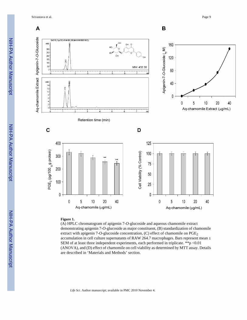
Figure 1.(A) HPLC chromatogram of apigenin 7-O-glucoside and aqueous chamomile extractdemonstrating apigenin 7-O-glucoside as major constituent, (B) standardization of chamomileextract with apigenin 7-O-glucoside concentration, (C) effect of chamomile on PGE2accumulation in cell culture supernatants of RAW 264.7 macrophages. Bars represent mean ±SEM of at least three independent experiments, each performed in triplicate. **p <0.01(ANOVA), and (D) effect of chamomile on cell viability as determined by MTT assay. Detailsare described in ‘Materials and Methods’ section.
Srivastava et al. Page 9
Life Sci. Author manuscript; available in PMC 2010 November 4.
NIH
-PA Author Manuscript
NIH
-PA Author Manuscript
NIH
-PA Author Manuscript
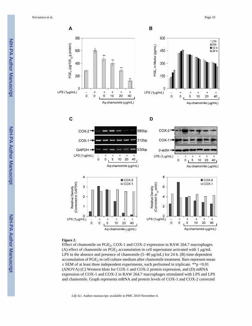
Figure 2.Effect of chamomile on PGE2, COX-1 and COX-2 expression in RAW 264.7 macrophages.(A) effect of chamomile on PGE2 accumulation in cell supernatant activated with 1 μg/mLLPS in the absence and presence of chamomile (5–40 μg/mL) for 24 h. (B) time-dependentaccumulation of PGE2 in cell culture medium after chamomile treatment. Bars represent mean± SEM of at least three independent experiments, each performed in triplicate. **p <0.01(ANOVA) (C) Western blots for COX-1 and COX-2 protein expression, and (D) mRNAexpression of COX-1 and COX-2 in RAW 264.7 macrophages stimulated with LPS and LPSand chamomile. Graph represents mRNA and protein levels of COX-1 and COX-2 corrected
Srivastava et al. Page 10
Life Sci. Author manuscript; available in PMC 2010 November 4.
NIH
-PA Author Manuscript
NIH
-PA Author Manuscript
NIH
-PA Author Manuscript
to GAPDH and β-actin, represents loading controls. Details are described in ‘Materials andMethods’ section.
Srivastava et al. Page 11
Life Sci. Author manuscript; available in PMC 2010 November 4.
NIH
-PA Author Manuscript
NIH
-PA Author Manuscript
NIH
-PA Author Manuscript
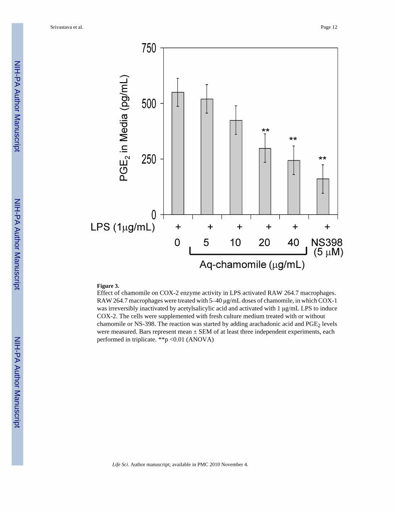
Figure 3.Effect of chamomile on COX-2 enzyme activity in LPS activated RAW 264.7 macrophages.RAW 264.7 macrophages were treated with 5–40 μg/mL doses of chamomile, in which COX-1was irreversibly inactivated by acetylsalicylic acid and activated with 1 μg/mL LPS to induceCOX-2. The cells were supplemented with fresh culture medium treated with or withoutchamomile or NS-398. The reaction was started by adding arachadonic acid and PGE2 levelswere measured. Bars represent mean ± SEM of at least three independent experiments, eachperformed in triplicate. **p <0.01 (ANOVA)
Srivastava et al. Page 12
Life Sci. Author manuscript; available in PMC 2010 November 4.
NIH
-PA Author Manuscript
NIH
-PA Author Manuscript
NIH
-PA Author Manuscript
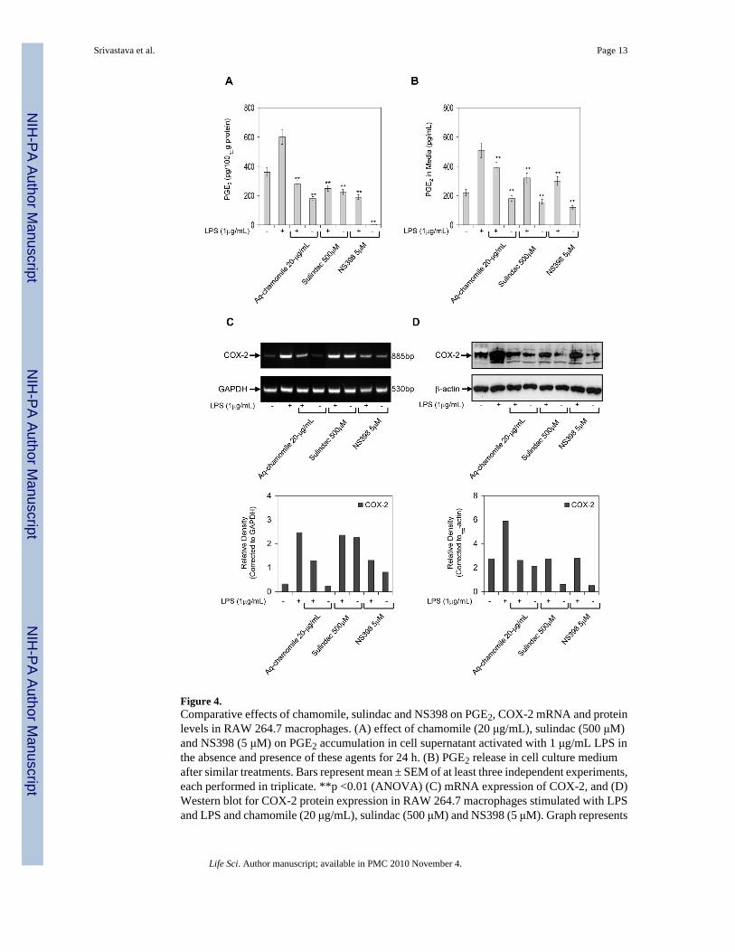
Figure 4.Comparative effects of chamomile, sulindac and NS398 on PGE2, COX-2 mRNA and proteinlevels in RAW 264.7 macrophages. (A) effect of chamomile (20 μg/mL), sulindac (500 μM)and NS398 (5 μM) on PGE2 accumulation in cell supernatant activated with 1 μg/mL LPS inthe absence and presence of these agents for 24 h. (B) PGE2 release in cell culture mediumafter similar treatments. Bars represent mean ± SEM of at least three independent experiments,each performed in triplicate. **p <0.01 (ANOVA) (C) mRNA expression of COX-2, and (D)Western blot for COX-2 protein expression in RAW 264.7 macrophages stimulated with LPSand LPS and chamomile (20 μg/mL), sulindac (500 μM) and NS398 (5 μM). Graph represents
Srivastava et al. Page 13
Life Sci. Author manuscript; available in PMC 2010 November 4.
NIH
-PA Author Manuscript
NIH
-PA Author Manuscript
NIH
-PA Author Manuscript
mRNA and protein levels of COX-2 corrected to GAPDH and β-actin, represents loadingcontrols. Details are described in ‘Materials and Methods’ section.
Srivastava et al. Page 14
Life Sci. Author manuscript; available in PMC 2010 November 4.
NIH
-PA Author Manuscript
NIH
-PA Author Manuscript
NIH
-PA Author Manuscript


