Embed Size (px)
Citation preview

Aquaculture xxx (2015) xxx–xxx
AQUA-631573; No of Pages 10
Contents lists available at ScienceDirect
Aquaculture
j ourna l homepage: www.e lsev ie r .com/ locate /aqua-on l ine
Characteristics of digestive processes in Atlantic salmon (Salmo salar).Enzyme pH optima, chyme pH, and enzyme activities
Åshild Krogdahl ⁎, Anne Sundby, Halvor HolmDepartment of Basic Sciences and Aquatic Medicine, Norwegian University of Life Sciences, School of Veterinary Medicine, PO Box 8146 Dep, N-0033 Oslo, Norway
⁎ Corresponding author. Tel.: +47 2296 4534.E-mail address: [email protected] (Å. Krogdah
http://dx.doi.org/10.1016/j.aquaculture.2015.02.0320044-8486/© 2015 Elsevier B.V. All rights reserved.
Please cite this article as: Krogdahl, Å., et al., Cand enzyme activities, Aquaculture (2015), h
a b s t r a c t
a r t i c l e i n f oArticle history:Received 10 November 2014Received in revised form 13 February 2015Accepted 15 February 2015Available online xxxx
Keywords:Atlantic salmonChyme pHDigestive enzymespH-optimumDiet compositionSalinity
Symptoms indicating digestivemalfunction have been observed in cultivated salmonover the last 10–15 years, atleast partially as a consequence of use of new feed ingredients. The present knowledge onmany aspects of diges-tive functions in fish is limited, in particular for Atlantic salmon. Strengthening is needed to understand themechanisms underlying challenges to gastrointestinal health and to find means to prevent their development.The present paper supplies information regarding digestive enzyme optima and stability, chyme pH and enzymeactivities along the digestive tract of Atlantic salmon. The data presented includes new data from four studies onAtlantic salmon as well as a review of comparative literature from various other fish studies. The four experi-ments comprised salmon of different sizes, kept in salt or fresh water, and fed high fish meal diets with differentprotein to energy ratios (P/E). These studies showed an average pH in the stomach chyme, i.e. across diets andexperiments, of 4.8 (SD = 0.7), whereas the observed pH optimum for pepsin was 3.0. In the intestinal chyme,pH increased throughout the intestine from a mean of 8.1 (SD = 0.23) in the proximal intestine to 8.4 in themid intestine (SD = 0.27) and distal intestine (SD = 0.29). Total proteolytic activity of chyme in the proximalintestine, measured as the sum of the work of all proteolytic enzymes on casein as substrate, showed its maxi-mum at pH 8.8, i.e. clearly above prevailing chyme pH. For the individual proteolytic enzymes, pH optimum var-ied from 7.10 for elastase 1 to 8.98 for trypsin. Chyme pH did not appear to be influenced by fish size, but wassignificantly higher in the fish raised in fresh water than in those raised in salt water. In the intestinal chymepH showed a significant negative regression on diet P/E. For the investigated pancreatic enzymes in the intestinalchyme, the activity in the chyme decreased from proximal to distal regions and faecal activities were very lowcompared to the activities in the proximal intestine. The activity profile for leucine aminopeptidase (LAP), how-ever, trended to increase frommid to distal intestine. Chyme enzyme activities along the intestinal tract did notdiffer clearly between the experiments and no clear relationship between P/E on enzyme activities along the in-testinal tract was observed. Remarkable in vitro stability was observed for the proteolytic enzyme activities.
© 2015 Elsevier B.V. All rights reserved.
1. Introduction
Over the last 10–15 years, plant feed ingredients have replaced fishmeal to a great extent in diets for most cultured, carnivore fish species,including salmonids. At the same time, feed efficiency has decreased,and reduction in both protein and lipid digestibility has been describedin salmon (Krogdahl et al., 2003; Refsie et al., 1998). Increased frequen-cy of symptoms indicating impairment of digestive and immune func-tions of the intestine has also been recorded with some welldocumented in the scientific literature, others only described in reportsfrom animal health authorities. Examples are gastric dilatation, gastriculcers, lipid malabsorption, undigested pellets and intestinal inflamma-tion accompanied by low enzyme activities, low bile salt concentrations,infiltration of immune cells into the lamina propria and submucosa, ap-parent malfunction of water transport, and diarrhoea (Bæverfjord and
l).
haracteristics of digestive prottp://dx.doi.org/10.1016/j.aq
Krogdahl, 1996; Bæverfjord et al., 2006; Bakke et al., 2014; Dale et al.,2009; Krogdahl et al., 2003). These symptoms are suggested to be relat-ed to the plant ingredients' content of antinutrients and other changesin the feeds, such as nutrient content and its physical qualities.
To understandmechanisms behind effects of inclusion of plant feed-stuffs and other nutrient sources that may be used in the future on gas-trointestinal (GI) processes in fish, strengthening of basic knowledge oncharacteristics of the various elements of the digestive apparatus undernormal conditions is required. In spite more than 40 years of commer-cial production of Atlantic salmon,many gaps in the present knowledgeon biochemical and physiological characteristics of digestive processesin the intestine exist. For some other species, more information isavailable. Table 1 presents a comprehensive summary of available infor-mation regarding pH of chyme along the GI tract and pH optima ofproteolytic enzymes. The overview shows a wide range in pH of stom-ach chyme between 2.0 and 7.0, and a variation in optimum forpepsin between pH 1.0 and 3.4. Chyme pH varied between 6.2 and8.2, while intestinal total proteolytic activity optimum varied between
cesses in Atlantic salmon (Salmo salar). EnzymepHoptima, chymepH,uaculture.2015.02.032

Table 1Compiled literature data regarding chyme pH and optimum pH for digestive enzymes in the stomach and intestine.
Omnivorous Stomach Pepsin 5.5–7.0 Charabarti et al. (1995)Carnivorous Stomach Pepsin 5.3–5.5 Charabarti et al. (1995)Asian bony tongue (Scleropages formosus) (Osteoglossidae) Stomach Pepsin 1.5–2.0 Natalia et al. (2004)Atlantic cod (Gadus Morhua) Stomach Pepsin 3.0–3.4 Bjelland et al. (1988)Common dentex (Dentex dentex) Stomach Pepsin 1.5 Perez-Jimenez et al. (2009)Discus fish (Symphysodon aequifasciata) Stomach Pepsin 2 Chong et al. (2002)Gilthead sea bream (Sparus aurata) Stomach Pepsin 2.0–6.0 2 Deguara et al. (2003), Yufera et al. (2004),
Marquez et al. (2012)Japanese eel (Anguilla japonica) Stomach Pepsin 2.0–3.5 1.5 Hidalgo et al. (1999), Chiu and Pan (2002)Pacific bluefin tuna (Thunnus orientalis) Stomach Pepsin 3 de la Parra et al. (2007)Rainbow trout (Oncorhynchus mykiss) Stomach Pepsin 4.9 1.5 Hidalgo et al. (1999), Bucking and Wood (2009)Sea bass (Dicentrarchus labrax L.) Stomach Pepsin 2.7–4.7 Nikolopoulou et al. (2011)Senegalese sole (Solea senegalensis) Stomach Pepsin N6.0 Yufera and Darias (2007)Spiny dog fish (Squalus acanthias), white spotted bamboo shark (Chiloscylliumplagiosu), little skate (Leucoraja erinacea), Clearnose skate (Raja eglanteria)
Stomach Pepsin 2.2–4.0 Wood et al. (2007), Anderson et al. (2010)
Chum salmon (Oncorhyncus keta) Stomach Pepsin 3 Sanchez-Chiang et al. (1987)Turbot (Scophthalmus maximus L.) Stomach Pepsin 1.0–2.0 Munilla-Moran and Saborido-Rey (1996)Asian bony tongue (S. formosus, Osteoglossidae) Intestine Proteolytic 9.5–10.0 Natalia et al. (2004)Atlantic halibut (Hippoglossus hippoglossus L.), Turbot (S. maximus L.) Intestine Proteolytic 9.5–11.0 Glass et al. (1989)Atlantic salmon (Salmo salar) Intestine Proteolytic 6.7–7.8 9.2–11.8 Usher et al. (1990)Big carp (Aristichthys nobilis) Intestine Trypsin 6.8–7.6 8.3 Bitterlich (1985)Common bream (Abaramis brama L.) Intestine Proteolytic 8.0 Kuz'mina et al. (2011)Common dentex (D. dentex) Intestine Proteolytic 8.5–9.0 Perez-Jimenez et al. (2009)Discus fish (S. aequifasciata) Intestine Proteolytic 7.5–9.0,
11.5–12.5Chong et al. (2002)
Dover sole (Solea solea L.) Intestine Proteolytic 7.0–8.0 Clarck et al. (1985)European perch (Perca fluviatilis L.) Intestine Proteolytic 10.0 Kuz'mina et al. (2011)Gilthead sea bream (S. aurata) Intestine Proteolytic 6.5–7.9 9.5–10.0 Munilla-Moran and Saborido-Rey (1996),
Deguara et al. (2003), Marquez et al. (2012)Pacific bluefin tuna (T. orientalis) Intestine Proteolytic 9.0 de la Parra et al. (2007)Roach (Rutilus rutilus L.) Intestine Proteolytic 7.0 Kuz'mina et al. (2011)Sea bass (D. labrax L.) Intestine Proteolytic 7.5–8.2 Nikolopoulou et al. (2011)Silver carp (Hypophthalmichthys molitrix) Intestine Trypsin 6.2–7.2 8.3 Bitterlich (1985)Skipjack tuna (Euthynnus pelamis) Intestine Proteolytic 7.0–8.0 Joakimsson and Nagayama (1990)Spiny dog fish (S. acanthias) Intestine Proteolytic 7.2 Wood et al. (2007)Rainbow Trout (O. mykiss) Intestine Proteolytic 7.1–7.7 8.0–10.0 Dimes et al. (1994), Fard et al. (2007),
Hidalgo et al. (1999), Grabner (1985)Zander (Zander lucioperca L.) Intestine Proteolytic 10.0 Kuz'mina et al. (2011)
2 Å. Krogdahl et al. / Aquaculture xxx (2015) xxx–xxx
7.0 and 12.5. In general, pH optimum for pepsin activity is found to belower than stomach chyme pH inmost species. Also in themid intestineand distal intestine, differences between pH optimum for the proteolyt-ic enzyme activities and measured pH in the chyme are reported(Table 1). In general pH optima for total proteolytic activities alongthe intestinal tract are observed to be higher than the measuredchyme pH.
Differences in chyme pH along the GI tract between Atlantic salmonsmolt kept in freshwater (FW) and salt water (SW) have been observed(Usher et al., 1990). However, no significant differences in the activitiesof digestive enzymes could be measured in these fish. Similar resultswere reported by Fang and Chiou (1989) in a study on tilapia in FWand SW.
The present paper presentswork undertaken over several years con-comitant with an observed necessity to improve assay quality and accu-racy, and to strengthen our knowledge regarding normal characteristicsof GI proteolytic and lipolytic enzymes. The work was essential for themain goal of the research efforts in our group, i.e. for future investigationof effects of new diet ingredients on digestive processes in Atlanticsalmon and other fish species, and hence to secure the health andwellbeing of the fish. Specifically we aimed to establish information es-sential for optimal measurements of enzyme activities along the GItract, such as suitability of available substrates for salmon pepsin forwhich such information was lacking, optimal pH for the digestive en-zymes, as well as their stability during incubation, pH and enzyme pro-files along the GI tract, and to find indications on whether variation inwater salinity, fish size and protein to energy ratio (P/E) might affectpH and enzyme activities along the GI tract. Our focus was onwhat pos-sibly can be considered normal physiological conditions. Accordingly,
Please cite this article as: Krogdahl, Å., et al., Characteristics of digestive proand enzyme activities, Aquaculture (2015), http://dx.doi.org/10.1016/j.aq
the results originate from fish fed the reference diets, i.e. with simplecompositions and in which fish meal was the main protein source.
2. Materials and methods
The basis for the present information was samples and informationcollected from four fish trials, herein named T1–T4. They were all con-ducted mainly for nutritional goals, but the fish fed the reference dietsin these trials were suitable for sampling for the present investigationof biochemical and physiological characteristics of the GI tract. The trialsspanned awide range infish sizes,fish in both fresh and seawater, and awide range in P/E. As all diets were reference diets, the ingredients usedwere similar. The aspects related to the main goals of T1 and T2, i.e. ef-fects of new feed ingredients on production and nutritional aspects,have been published previously by Nordrum et al. (2000, 2003) includ-ing some data for the reference dietswhich are also presented compiledwith results from T3 and T4 in the present paper.
2.1. Experiments and diets
The design of trials T1–T4 is outlined in Table 2. Experiments T1 andT2were conducted in tanks at Nofima's research facility at Sunndalsøra,Norway, T3 at Sjøblink Blokken's research facilities in Sortland, Norway,and T4 at Ræstad FiskAS in Florø, Norway. The two latterwere both con-ducted in net pens. All experimentswere conducted in compliancewithlaws regulating experimentation with live animals in Norway as over-seen by theNorwegian Animal Research Authority (NARA). The four tri-als comprised a total of 10 diets, named D1–D10. Diet compositions are
cesses in Atlantic salmon (Salmo salar). EnzymepHoptima, chymepH,uaculture.2015.02.032

Table 2Outline of experiment design and measurements.
Observations
Trial Diets No. of sampledfish
Duration of feeding,d
Start weight,g
End weight,g
Salinity Enzyme pHoptimum
Enzymestability
ChymepH
Enzymeactivity
T1 D1, D2, D3 45 65 180 300 FW x xT2 D4, D5, D6 15 71 846 1480 SW x xT3 D7, D8, D9 27 177 1240 3950 SW x x x xT4 D10 3 10,000 – SW x
3Å. Krogdahl et al. / Aquaculture xxx (2015) xxx–xxx
given in Table 3. In T1–T3, each diet was fed to fish in three tanks or netpens. The samples taken from T4were taken from three individual fish.
2.2. Sampling for pH measurements and chyme enzyme activities
All sampledfishwere in the fed statewith feed/chymepresent in thewhole GI tract. Following sacrifice, theGI tractwas removed immediate-ly and divided in stomach, proximal intestine (PI) defined as the areafromwhich the pyloric caeca originate, mid intestine (MI) and distal in-testine (DI), as also described by Krogdahl and Bakke-McKellep (2005).The sections were separated by applying clamps to avoid mixing of thecontents between sections. Chyme pH was measured directly in thechyme in the stomach, PI, MI and DI in all the 90 sampled fish using aPHM 210 Meterlab, Radiometer, analytical pH-meter. In the stomachfrom the 10-kg salmon (T4), pHwasmeasured at six different locations,labelled 1–6 from the proximal to the distal part of the organ.
For investigation of pH optimum of enzymes, chyme from the stom-ach and PI in T3 was used.
For collection of chyme samples for enzyme activity studies in T1, T2and T3, the whole GI-tract was dissected, stretched out and wrapped inaluminium foil after removal of other organs, frozen in liquid nitrogenand kept at −80 °C. In T3, faeces were sampled by collecting the faecalpellets evacuated during a meal by sieving the outlet water. The faecalpellet samples were quickly blotted on a paper towel, frozen andlyophilised.
2.3. Preparation of enzyme extracts
In T1, T2 and T3, chyme samples were collected from the frozen GItracts following partial thawing. The tracts were opened longitudinally
Table 3Composition of experimental diets (D1–D10) in the 4 trials (T1–T4).
T1 T2
D1 D2 D3 D4
Ingredients: g kg−1
Wheat 120 120 120Wheat brand 263 263 263 82Fish meal 356 356 356 434Fish oil 246 246 246 295Yttrium oxide 0.001 0.001 0.001 0.001Vit. min 15 15 15 15L-Alanine 3.7
L-Methionine 6.2
L-Cysteine 5.0 5.0
Proximate composition, g kg−1
Water 87 66 66 17Crude protein 297 298 303 379Fat 285 289 293 380NFEStarch 140 151 144 105Ash 52 55 53 65
EnergyGross energy (MJ kg−1) 22.5 23.0 23.3 26.0Protein/energy, g/MJ 13.2 13.1 13 14.5
Please cite this article as: Krogdahl, Å., et al., Characteristics of digestive proand enzyme activities, Aquaculture (2015), http://dx.doi.org/10.1016/j.aq
and the chyme sampled quantitatively from the subsections of thetract: stomach, PI,MI andDI sections. For recording of enzyme activities,theMI andDI sectionswere divided in a proximal and distal part and arereferred to as MI1, MI2, DI1 and DI2, respectively. The chyme sampleswere pooled per tank/pen and lyophilised. Chyme was sampled from 5fish per replicate tank in T1, 5 fish per diet in T2, and from 3 fish perpen in T3. The enzymeswere extracted from the dried samples by stirringwith ice cold distilled water. For all enzymes except lipase, 1:10 (w/v) di-lutions were used in the assays. Following centrifugation at 25.000 ×g for30 min at 4 °C, the supernatants containing the solubilized digestive en-zymes were kept at−20 °C in aliquots of about 1ml until use. For lipase,assay suspensionsweremade of the chyme samples (1:80w/v) in 25mMTris–HCl buffer, pH 8.0, containing 0.5% BRIJ 30, rotated for 10 min andsonicated for 30 s at 0 °C.
2.4. Enzyme assays
As the work presented in this paper took place partly in parallel itwas not possible to optimize the enzyme assays according to optimalpH for the enzymes prior to the work regarding enzyme activitiesalong the GI tract presented herein. A choice had to be made based onavailable information from other fish species. The chosen pH for the in-cubations turned out to be close to the enzyme pH optima in the workpresented herein. An incubation temperature of 37 °C was chosen forthe assays after preliminary evaluation of effect of incubation tempera-ture on enzyme activity showing steadily increasing activity of the di-gestive enzymes with temperature from 0 to 45 °C. Hence, to obtainhigh activities and also to ease comparison with similar studies withhomoeothermic animals, including humans, 37 °C was chosen for theassays.
T3 T4
D5 D6 D7 D8 D9 D10
54 64 64 1682 82 122 122 122
434 434 548 635 696 544295 295 318 265 225 278
0.001 0.00115 15 15 15 15 156.2 3.1
2.5
17 17 86 78 53 80379 379 409 443 493 400380 380 350 325 275 335
72 73 77 114105 105 31 36 36 10865 65 83 81 102 95
26.0 26.0 24.7 24.6 23.8 23.714.6 14.6 16.2 18.0 20.7 16.9
cesses in Atlantic salmon (Salmo salar). EnzymepHoptima, chymepH,uaculture.2015.02.032
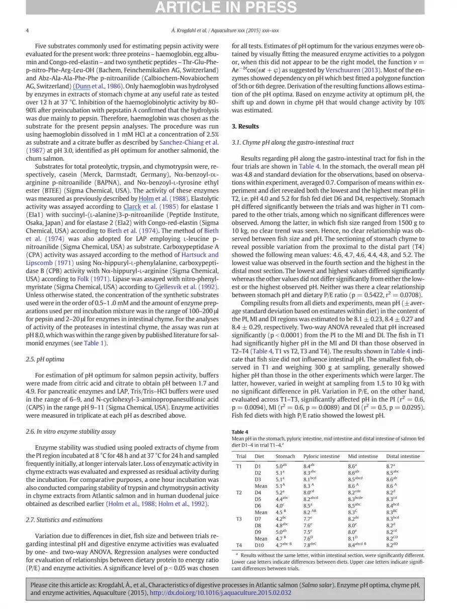
Table 4Mean pH in the stomach, pyloric intestine, mid intestine and distal intestine of salmon feddiet D1–4 in trial T1–4.a
Trial Diet Stomach Pyloric intestine Mid intestine Distal intestine
T1 D1 5.0ab 8.4ab 8.6a 8.7a
D2 5.1a 8.3abc 8.6ab 8.5abc
D3 5.1a 8.1bcd 8.5abcd 8.6ab
Mean 5.1A 8.3 A 8.6 A 8.6 A
T2 D4 5.2a 8.0cd 8.2cde 8.2d
D5 4.4abc 8.2abcd 8.3bcde 8.3cd
D6 4.0c 8.5a 8.5abc 8.4bcd
Mean 4.5 B 8.2 AB 8.3C 8.3BC
T3 D7 4.2bc 7.7e 8.2de 8.3bcd
D8 4.8abc 7.6e 8.0e 8.2d
D9 5.0ab 7.5e 8.0e 8.2cd
Mean 4.7 B 7.6D 8.1D 8.2CD
T4 D10 4.7abc B 7.8deC 8.4abcd B 8.2dD
a Results without the same letter, within intestinal section, were significantly different.Lower case letters indicate differences between diets. Upper case letters indicate signifi-cant differences between trials.
4 Å. Krogdahl et al. / Aquaculture xxx (2015) xxx–xxx
Five substrates commonly used for estimating pepsin activity wereevaluated for the presentwork: three proteins– haemoglobin, egg albu-min andCongo-red-elastin – and two synthetic peptides – Thr-Glu-Phe-p-nitro-Phe-Arg-Leu-OH (Bachem, Feinchemikalien AG, Switzerland)and Abz-Ala-Ala-Phe-Phe p-nitroanilide (Calbiochem-NovabiochemAG, Switzerland) (Dunn et al., 1986). Only haemoglobinwas hydrolysedby enzymes in extracts of stomach chyme at any useful rate as testedover 12 h at 37 °C. Inhibition of the haemoglobinolytic activity by 80–90% after preincubation with pepstatin A confirmed that the hydrolysiswas due mainly to pepsin. Therefore, haemoglobin was chosen as thesubstrate for the present pepsin analyses. The procedure was runusing haemoglobin dissolved in 1 mM HCl at a concentration of 2.5%as substrate and a citrate buffer as described by Sanchez-Chiang et al.(1987) at pH 3.0, identified as pH optimum for another salmonid, thechum salmon.
Substrates for total proteolytic, trypsin, and chymotrypsin were, re-spectively, casein (Merck, Darmstadt, Germany), Nα-benzoyl-DL-arginine p-nitroanilide (BAPNA), and Nα-benzoyl-L-tyrosine ethylester (BTEE) (Sigma Chemical, USA). The activity of these enzymeswasmeasured as previously described by Holm et al. (1988). Elastolyticactivity was assayed according to Clarck et al. (1985) for elastase 1(Ela1) with succinyl-(L-alanine)3-p-nitroanilide (Peptide Institute,Osaka, Japan) and for elastase 2 (Ela2) with Congo-red-elastin (SigmaChemical, USA) according to Bieth et al. (1974). The method of Biethet al. (1974) was also adopted for LAP employing L-leucine p-nitroanilide (Sigma Chemical, USA) as substrate. Carboxypeptidase A(CPA) activity was assayed according to the method of Hartsuck andLipscomb (1971) using Nα-hippuryl-L-phenylalanine, carboxypepti-dase B (CPB) activity with Nα-hippuryl-L-arginine (Sigma Chemical,USA) according to Folk (1971). Lipase was assayed with nitro-phenyl-myristate (Sigma Chemical, USA) according to Gjellesvik et al. (1992).Unless otherwise stated, the concentration of the synthetic substratesused were in the order of 0.5–1 .0 mMand the amount of enzyme prep-arations used per ml incubation mixture was in the range of 100–200 μlfor pepsin and 2–20 μl for enzymes in intestinal chyme. For the analysesof activity of the proteases in intestinal chyme, the assay was run atpH 8.0,whichwaswithin the range given by published literature for sal-monid enzymes (see Table 1).
2.5. pH optima
For estimation of pH optimum for salmon pepsin activity, bufferswere made from citric acid and citrate to obtain pH between 1.7 and4.9. For pancreatic enzymes and LAP, Tris/Tris–HCl buffers were usedin the range of 6–9, and N-cyclohexyl-3-aminopropanesulfonic acid(CAPS) in the range pH 9–11 (Sigma Chemical, USA). Enzyme activitieswere measured in triplicate at each pH as described above.
2.6. In vitro enzyme stability assay
Enzyme stability was studied using pooled extracts of chyme fromthe PI region incubated at 8 °C for 48 h and at 37 °C for 24 h and sampledfrequently initially, at longer intervals later. Loss of enzymatic activity inchyme extracts was evaluated and expressed as residual activity duringthe incubation. For comparative purposes, a one hour incubation wasalso conducted comparing stability of trypsin and chymotrypsin activityin chyme extracts from Atlantic salmon and in human duodenal juiceobtained as described earlier (Holm et al., 1988; Holm et al., 1992).
2.7. Statistics and estimations
Variation due to differences in diet, fish size and between trials re-garding intestinal pH and digestive enzyme activities was evaluatedby one- and two-way ANOVA. Regression analyses were conductedfor evaluation of relationships between dietary protein to energy ratio(P/E) and enzyme activities. A significance level of p b 0.05 was chosen
Please cite this article as: Krogdahl, Å., et al., Characteristics of digestive proand enzyme activities, Aquaculture (2015), http://dx.doi.org/10.1016/j.aq
for all tests. Estimates of pH optimum for the various enzymes were ob-tained by visually fitting the measured enzyme activities to a polygonor, when this did not appear to be the right model, the function v =Ae−btcos(ωt+ φ) as suggested by Verschuuren (2013). Most of the en-zymes showeddependency on pHwhich best fitted a polygone functionof 5th or 6th degree. Derivation of the resulting functions allows estima-tion of the pH optima. Based on enzyme activity at optimum pH, theshift up and down in chyme pH that would change activity by 10%was estimated.
3. Results
3.1. Chyme pH along the gastro-intestinal tract
Results regarding pH along the gastro-intestinal tract for fish in thefour trials are shown in Table 4. In the stomach, the overall mean pHwas 4.8 and standard deviation for the observations, based on observa-tionswithin experiment, averaged 0.7. Comparison of meanswithin ex-periment and diet revealed both the lowest and the highest mean pH inT2, i.e. pH 4.0 and 5.2 for fish fed diet D6 and D4, respectively. StomachpH differed significantly between the trials and was higher in T1 com-pared to the other trials, among which no significant differences wereobserved. Among the latter, in which fish size ranged from 1500 g to10 kg, no clear trend was seen. Hence, no clear relationship was ob-served between fish size and pH. The sectioning of stomach chyme toreveal possible variation from the proximal to the distal part (T4)showed the following mean values: 4.6, 4.7, 4.6, 4.4, 4.8, and 5.2. Thelowest value was observed in the fourth section and the highest in thedistal most section. The lowest and highest values differed significantlywhereas the other values did not differ significantly fromeither the low-est or the highest observed pH. Neither was there a clear relationshipbetween stomach pH and dietary P/E ratio (p = 0.5422, r2 = 0.0708).
Compiling results from all diets and experiments, mean pH (±aver-age standard deviation based on estimateswithin diet) in the content ofthe PI, MI and DI regionswas estimated to be 8.1± 0.23, 8.4± 0.27 and8.4 ± 0.29, respectively. Two-way ANOVA revealed that pH increasedsignificantly (p b 0.0001) from the PI to the MI and DI. The fish in T1had significantly higher pH in the MI and DI than those observed inT2–T4 (Table 4, T1 vs T2, T3 and T4). The results shown in Table 4 indi-cate that fish size did not influence intestinal pH. The smallest fish, ob-served in T1 and weighing 300 g at sampling, generally showedhigher pH than those in the other experiments which were larger. Thelatter, however, varied in weight at sampling from 1.5 to 10 kg withno significant difference in pH. Variation in P/E, on the other hand,evaluated across T1–T3, significantly affected pH in the PI (r2 = 0.6,p = 0.0094), MI (r2 = 0.6, p = 0.0089) and DI (r2 = 0.5, p = 0.0295).Fish fed diets with high P/E ratio showed the lowest pH.
cesses in Atlantic salmon (Salmo salar). EnzymepHoptima, chymepH,uaculture.2015.02.032

Table 5pH optimum for investigated digestive enzymes and pH change which would reduce ac-tivity by 10% on either sides of optimum.a
Enzyme pH optimum pH change for 10% activity change
Pepsin 3.05 0.6Tot prot 8.90 0.9Trypsin 8.98 0.8Chymotrypsin 7.63 0.5Ela1 7.10 0.6Ela2 8.41 0.2CPA 7.30 0.9CPB 7.37 0.7LAP 7.55 0.8Lipase 7.95 0.4
a Tot prot = Total proteolytic activity; Ela1 = elastase 1; Ela2 = elastase 2; CPA =carboxypeptidase A; CPB = carboxypeptidase B; LAP = leucine aminopeptidase.
5Å. Krogdahl et al. / Aquaculture xxx (2015) xxx–xxx
3.2. Enzyme pH optima
The observed relationships between enzyme activities and pH arepresented Fig. 1. Table 5 shows the estimates of the pH optima. Theyare also indicated by vertical lines in Fig. 1. Estimates of the pH rangecovering a 10% shift in activity on one or the other side of the pH opti-mum, indicate the sensitivity of the enzyme activity towards pH shiftsnear the optimum, are also given in Table 5 and indicated in Fig. 1.The pH optimum of pepsin was 3.05 and a shift in pH of 0.6 units re-duced activity by 10%. For the proteases in the intestinal chyme, pH op-timum ranged from 7.10 for Ela1 to 8.98 for trypsin. Overall, the optimafor the various intestinal enzymes showed values spread quite evenlyover the whole range. The narrowest peak was observed for Ela2 forwhich a 10% change in activity would occur by changing pH 0.2 unitsaway from thepHoptimum. In contrast, for total proteolytic and CPA ac-tivity a change of 0.9 pH unit was required for a 10% reduction in
Fig. 1. Effect of pH on activity of pepsin in stomach chyme, and in intestinal chyme on total proteolytic (Tot prot) activity, and on activity of trypsin, chymotrypsin, elastases 1 and 2 (Ela1and Ela2), carboxypeptidases A (CPA) and B (CPB), leucine aminopeptidase (LAP) and lipase in Atlantic salmon. Vertical lines indicate estimated pH optimum for the enzyme (-----),pH 8.0 (……….), and the pH at the lower side of the optimumwhich enzyme activity would be 10% decreased from maximum (-..-..-..-..); grey areas indicate 95% confidence range for ob-served chyme pH (overall mean ± 2 SD) in the stomach chyme for pepsin and chyme of the pyloric section of the intestine for other enzymes. NB: the scale on the x- and y-axis differbetween enzymes.
Please cite this article as: Krogdahl, Å., et al., Characteristics of digestive processes in Atlantic salmon (Salmo salar). EnzymepHoptima, chymepH,and enzyme activities, Aquaculture (2015), http://dx.doi.org/10.1016/j.aquaculture.2015.02.032

05
101520253035404550556065707580859095
100
0 3 6 9 12 15 18 21 24
Rem
ain
ing
act
ivit
y, %
Incubation time, h
Tot prot
Trypsin
Chymotypsin
Ela1
Ela2
CPA
CPB
LAP
Lipase
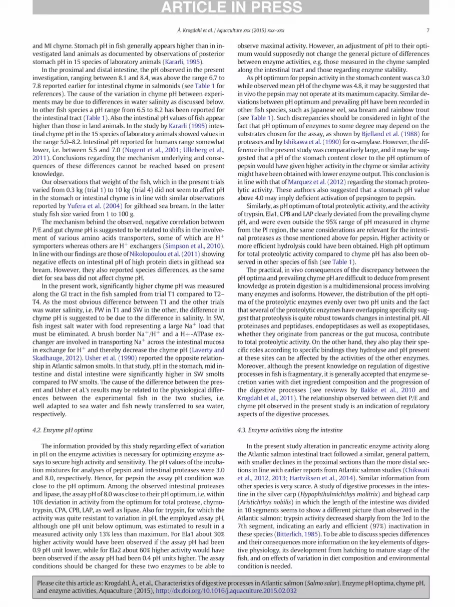
Fig. 3. Remaining activity (% of activity in the pyloric region) as a function of incubationtime for trypsin, chymotrypsin, elastases 1 (Ela1) and 2 Ela2), carboxypeptidases A(CPA) and B (CPB), leucine aminopeptidase (LAP) and lipase in chyme collected fromthe proximal intestine of Atlantic salmon.
6 Å. Krogdahl et al. / Aquaculture xxx (2015) xxx–xxx
activity. For the remainder of the enzymes a change in pH between 0.4and 0.8 pH units would cause a 10% reduction in activity.
3.3. Enzyme activities along the intestinal tract
Activity of all investigated intestinal enzymes, except for LAP, de-clined significantly from the proximal to the most distal intestinalregion. As only minor differences in enzyme activities were seen be-tween diets and trials (T1–T3), the results are presented asmeanswith-in trial (T1–T3) (Fig. 2). For the pancreatic enzymes the activitydecreased less from the PI to theMI2 than fromMI2 to DI2, with the ex-ception of Ela2 for which inactivation seemed greater in the PI than themore distal intestinal sections. Activity of LAP, a brush border enzyme,showed a different development throughout the intestine and in gener-al rather increased in activity along the intestine, especially fromMI2 toDI1. Analysis of relationship between trypsin and total proteolytic activ-ity using all results showed a significant correlation (r2 = 0.73, p =0.004). Dietary P/E did not seem to affect enzyme activity within the in-testinal sections significantly. Moreover, no clear differences were seenin enzyme activities between fish in FW and SW.
Analyses of enzyme activities in faeces, conducted for all enzymesexcept the elastases, LAP and lipase, showed further decline in activitycompared to the activity in DI2 to levels between 2.5% (trypsin) and13% (chymotrypsin) compared to the activity measured in the PI.
3.4. Enzyme stability
When incubated at 8 °C, a high stability of all salmon proteolytic en-zymes was found with approximately 95% of the initial total proteolyticactivity remaining after 48 h of incubation (results not shown). At 37 °C,trypsin showed the highest stability, with more than 92% remainingafter 8 h and 78% after 24 h,whereas 63% and 46%of the total proteolyticactivity remained after 8 h and 24 h (Fig. 3), respectively. Ela1 showedthe lowest stability among the proteases with 23% remaining after 8 h,10% after 24 h. Lipase activity decreased much more rapidly than theproteolytic enzymes and was completely inactivated after 8 h incuba-tion. The high stability of several of the enzymes triggered a pilot com-parison with human trypsin and chymotrypsin known to be inactivatedrapidly during incubation at 37 °C. Both the human enzymes lost about55% of their activities during thefirst 30min incubation. After 60min in-cubation, about 70% of the human enzyme activities were lost while no
0.0
0.1
0.2
0.3
0.4
0.5
0.6
0.7
0.8
0.9
1.0
1.1
1.2
PI MI1 MI2 DI1 DI2
En
zym
e ac
tivi
ty,
OD
/mg
DM
Tot prot
Trypsin
Chymotypsin
Ela1
Ela2
CPA
CPB
LAP
Lipase
Fig. 2. Activity (ΔOD/mg dry matter) of total proteolytic (Tot prot) activity, and of activityof trypsin, chymotrypsin, elastases 1 and2 (Ela1 andEla2), carboxypeptidasesA (CPA) andB (CPB), leucine aminopeptidase (LAP) and lipase of Atlantic salmon. PI = pyloric intes-tine; MI=mid intestine (divided in two sections of equal length,MI1 andMI2); DI= dis-tal intestine (divided in two sections of equal length (DI1 and DI2)).
Please cite this article as: Krogdahl, Å., et al., Characteristics of digestive proand enzyme activities, Aquaculture (2015), http://dx.doi.org/10.1016/j.aq
significant losses of activity were observed for salmon trypsin and chy-motrypsin (Fig. 4).
4. Discussion
4.1. Chyme pH along the GI tract
In the stomach chyme of the Atlantic salmon in the present study,the observed average pH value of 4.8 is very similar to the value of 4.9reported for rainbow trout (Oncorhyncus mykiss) (Bucking and Wood,2009). As can be seen in Table 1, both lower chyme pH and higherchyme pH have been measured in the stomach of other fish species. Inour study, samples were taken from fish in the fed state with contentin the stomach implying that they had consumed feed within a fewhours before sampling. Preliminary observations of pH in the mucuslayer of the stomach wall have shown values lower than 3. This impliesthat time after feeding may influence chyme pH. Such a phenomenonwas demonstrated in a study by Nikolopoulou et al. (2011) on seabass (Dicentrarchus labrax L.) and gilthead sea bream (Sparus aurata).Two hours after feeding, pH values in the stomach were 4.7 and 5.7 insea bass and gilthead sea bream, respectively, declining to 3.0 and3.5 at 10–12 h after feeding. The difference between the specieswas sig-nificant. Moreover, the sea bass showed dietary effects on stomach pH,while only the gilthead sea bream showed dietary effects on pH in the PI
0
10
20
30
40
50
60
70
80
90
100
0 10 20 30 40 50 60
Res
idu
al a
ctiv
ity,
%
Time, min
AS trypsin
AS chymotrypsin
Human trypsin
Human chymotrypsin
Fig. 4. Remaining activity as a function of incubation time for trypsin and chymotrypsinfrom Atlantic salmon (AS) and human.
cesses in Atlantic salmon (Salmo salar). EnzymepHoptima, chymepH,uaculture.2015.02.032
7Å. Krogdahl et al. / Aquaculture xxx (2015) xxx–xxx
and MI chyme. Stomach pH in fish generally appears higher than in in-vestigated land animals as documented by observations of posteriorstomach pH in 15 species of laboratory animals (Kararli, 1995).
In the proximal and distal intestine, the pH observed in the presentinvestigation, ranging between 8.1 and 8.4, was above the range 6.7 to7.8 reported earlier for intestinal chyme in salmonids (see Table 1 forreferences). The cause of the variation in chyme pH between experi-ments may be due to differences in water salinity as discussed below.In other fish species a pH range from 6.5 to 8.2 has been reported forthe intestinal tract (Table 1). Also the intestinal pH values of fish appearhigher than those in land animals. In the study by Kararli (1995) intes-tinal chymepH in the 15 species of laboratory animals showed values inthe range 5.0–8.2. Intestinal pH reported for humans range somewhatlower, i.e. between 5.5 and 7.0 (Nugent et al., 2001; Ulleberg et al.,2011). Conclusions regarding the mechanism underlying and conse-quences of these differences cannot be reached based on presentknowledge.
Our observations that weight of the fish, which in the present trialsvaried from 0.3 kg (trial 1) to 10 kg (trial 4) did not seem to affect pHin the stomach or intestinal chyme is in line with similar observationsreported by Yufera et al. (2004) for gilthead sea bream. In the latterstudy fish size varied from 1 to 100 g.
The mechanism behind the observed, negative correlation betweenP/E and gut chyme pH is suggested to be related to shifts in the involve-ment of various amino acids transporters, some of which are H+
symporters whereas others are H+ exchangers (Simpson et al., 2010).In linewith ourfindings are those of Nikolopoulou et al. (2011) showingnegative effects on intestinal pH of high protein diets in gilthead seabream. However, they also reported species differences, as the samediet for sea bass did not affect chyme pH.
In the present work, significantly higher chyme pH was measuredalong the GI tract in the fish sampled from trial T1 compared to T2–T4. As the most obvious difference between T1 and the other trialswas water salinity, i.e. FW in T1 and SW in the other, the difference inchyme pH is suggested to be due to the difference in salinity. In SW,fish ingest salt water with food representing a large Na+ load thatmust be eliminated. A brush border Na+/H+ and a H+-ATPase ex-changer are involved in transporting Na+ across the intestinal mucosain exchange for H+ and thereby decrease the chyme pH (Laverty andSkadhauge, 2012). Usher et al. (1990) reported the opposite relation-ship in Atlantic salmon smolts. In that study, pH in the stomach, mid in-testine and distal intestine were significantly higher in SW smoltscompared to FW smolts. The cause of the difference between the pres-ent and Usher et al.'s results may be related to the physiological differ-ences between the experimental fish in the two studies, i.e.well adapted to sea water and fish newly transferred to sea water,respectively.
4.2. Enzyme pH optima
The information provided by this study regarding effect of variationin pH on the enzyme activities is necessary for optimizing enzyme as-says to secure high activity and sensitivity. The pH values of the incuba-tion mixtures for analyses of pepsin and intestinal proteases were 3.0and 8.0, respectively. Hence, for pepsin the assay pH condition wasclose to the pH optimum. Among the observed intestinal proteasesand lipase, the assay pH of 8.0was close to their pH optimum, i.e. within10% deviation in activity from the optimum for total protease, chymo-trypsin, CPA, CPB, LAP, as well as lipase. Also for trypsin, for which theactivity was quite resistant to variation in pH, the employed assay pH,although one pH unit below optimum, was estimated to result in ameasured activity only 13% less than maximum. For Ela1 about 30%higher activity would have been observed if the assay pH had been0.9 pH unit lower, while for Ela2 about 60% higher activity would havebeen observed if the assay pH had been 0.4 pH units higher. The assayconditions should be changed for these two enzymes to be able to
Please cite this article as: Krogdahl, Å., et al., Characteristics of digestive proand enzyme activities, Aquaculture (2015), http://dx.doi.org/10.1016/j.aq
observe maximal activity. However, an adjustment of pH to their opti-mum would supposedly not change the general picture of differencesbetween enzyme activities, e.g. those measured in the chyme sampledalong the intestinal tract and those regarding enzyme stability.
As pH optimum for pepsin activity in the stomach contentwas ca 3.0while observedmean pH of the chymewas 4.8, it may be suggested thatin vivo the pepsinmay not operate at itsmaximum capacity. Similar de-viations between pH optimumand prevailing pH have been recorded inother fish species, such as Japanese eel, sea bream and rainbow trout(see Table 1). Such discrepancies should be considered in light of thefact that pH optimum of enzymes to some degree may depend on thesubstrates chosen for the assay, as shown by Bjelland et al. (1988) forproteases and by Ishikawa et al. (1990) forα-amylase. However, the dif-ference in the present studywas comparatively large, and itmay be sug-gested that a pH of the stomach content closer to the pH optimum ofpepsin would have given higher activity in the chyme or similar activitymight have been obtainedwith lower enzyme output. This conclusion isin linewith that of Marquez et al. (2012) regarding the stomach proteo-lytic activity. These authors also suggested that a stomach pH valueabove 4.0 may imply deficient activation of pepsinogen to pepsin.
Similarly, as pH optimumof total proteolytic activity, and the activityof trypsin, Ela1, CPB and LAP clearly deviated from the prevailing chymepH, and were even outside the 95% range of pH measured in chymefrom the PI region, the same considerations are relevant for the intesti-nal proteases as those mentioned above for pepsin. Higher activity ormore efficient hydrolysis could have been obtained. High pH optimumfor total proteolytic activity compared to chyme pH has also been ob-served in other species of fish (see Table 1).
The practical, in vivo consequences of the discrepancy between thepH optima andprevailing chymepH are difficult to deduce frompresentknowledge as protein digestion is a multidimensional process involvingmany enzymes and isoforms. However, the distribution of the pH opti-ma of the proteolytic enzymes evenly over two pH units and the factthat several of the proteolytic enzymes haveoverlapping specificity sug-gest that proteolysis is quite robust towards changes in intestinal pH. Allproteinases and peptidases, endopeptidases as well as exopeptidases,whether they originate from pancreas or the gut mucosa, contributeto total proteolytic activity. On the other hand, they also play their spe-cific roles according to specific bindings they hydrolyse and pH presentat these sites can be affected by the activities of the other enzymes.Moreover, although the present knowledge on regulation of digestiveprocesses in fish is fragmentary, it is generally accepted that enzyme se-cretion varies with diet ingredient composition and the progression ofthe digestive processes (see reviews by Bakke et al., 2010 andKrogdahl et al., 2011). The relationship observed between diet P/E andchyme pH observed in the present study is an indication of regulatoryaspects of the digestive processes.
4.3. Enzyme activities along the intestine
In the present study alteration in pancreatic enzyme activity alongthe Atlantic salmon intestinal tract followed a similar, general pattern,with smaller declines in the proximal sections than themore distal sec-tions in line with earlier reports from Atlantic salmon studies (Chikwatiet al., 2012, 2013; Hartviksen et al., 2014). Similar information fromother species is very scarce. A study of digestive processes in the intes-tine in the silver carp (Hypophthalmichthys molitrix) and bighead carp(Aristichthys nobilis) in which the length of the intestine was dividedin 10 segments seems to show a different picture than observed in theAtlantic salmon; trypsin activity decreased sharply from the 3rd to the7th segment, indicating an early and efficient (97%) inactivation inthese species (Bitterlich, 1985). To be able to discuss species differencesand their consequencesmore information on the key elements of diges-tive physiology, its development from hatching to mature stage of thefish, and on effects of variation in diet composition and environmentalcondition is needed.
cesses in Atlantic salmon (Salmo salar). EnzymepHoptima, chymepH,uaculture.2015.02.032

8 Å. Krogdahl et al. / Aquaculture xxx (2015) xxx–xxx
The fate of LAP along the intestine differed clearly from that of thepancreatic enzymes showing increasing rather than the decreasing ac-tivity. The explanation for this difference is most likely the differencein source of the enzymes, which for LAP is the brush border membrane.The activity in the intestinal chyme stems from enzymes sloughed offwith the surface of the intestinal tract during mucosa renewal. The in-crease in LAP activity between PI and DI is an indication of supply of en-zyme all along the intestinal tract and/or possibly great stability of theenzyme. However, in the terminal section inactivation seemed to takeplace.
Our results regarding the intestinal enzyme activities showed thatonly aminor amount of the enzymes initially secreted into the proximalsection was lost in the faeces in active form. Whether the enzyme pro-teins are digested or reabsorbed and eventually reutilized is notknown. Allegedly, a major part of protein evacuated in faeces stemsfrom endogenous material from the digestive organs and the gut walland includes microbial proteins as well.
In spite quite substantial differences in dietary P/E, variation inchyme enzyme activities was minor and did not appear to be relatedto the variation in P/E. Previous reports of studies with various fish spe-cies show varying effects of dietary protein level on activity of proteasesin intestinal chyme. Studies on Amazon fish (Matrincha, Brycon cf.melanopterus) showed significant but minor elevating effects onchyme enzyme activities related to increase in dietary protein level, aswell dietary lipid and carbohydrate level (Reimer, 1982). Similarly, inan omnivorous tilapia Oreochromis niloticus × Oreochromis aureus dietwith more protein, higher trypsin activity and higher N absorption inthe small intestine was observed (Sklan et al., 2004). This was alsoshown for the Brazilian omnivorous, fresh water fish piracanjuba(Brycon orbignyanus). A diet with 42% protein induced higher intestinalproteolytic activity than those on a diet with 32% protein (Garcia-Carreno et al., 2002). In contrast, work on spotted wolffish (Anarhichasminor) showed an opposite relationship. A high protein diet gave slight-ly lower intestinal protease activity (Papoutsoglou and Lyndon, 2006).Moreover, in a study of silver catfish on feedwith protein levels rangingfrom 20 to 41%, the intestinal alkaline protease activity peaked at a die-tary protein level of 27% (Melo et al., 2012). The work of Kuz'mina et al.(2011) with 4 species of fish on natural diets showed highest alkalineprotease activity in fish fed the diet with the highest protein contentand lowest activity for those on the highest carbohydrate diet. Thebasis for these differences are most likely related to species difference,but insufficient supply of some nutrients, e.g. essential amino acids, aswell as presence of antinutrients, may affect enzyme secretion and ac-tivity (Krogdahl et al., 2010; Nordrum et al., 2000).
Variation in ingredient composition has been reported to affect in-testinal enzyme activity in several fish species, possibly more than var-iation in nutrient composition. Fromour own laboratory, several studiesdocument effects of soybeanmeal and other legumes on activity of pro-teolytic enzymes along the intestine of salmonids, Atlantic cod and seabream (Chikwati et al., 2012, 2013; Couto et al., 2014; Krogdahl et al.,2003; Lilleeng et al., 2007; Nordrum et al., 2000, 2003; Penn et al.,2011; Refstie et al., 2006a,b,c; Romarheim et al., 2006; Sørensen et al.,2011; Srichanun et al., 2014; Zhang et al., 2012), as well as ofantinutrients such as protease inhibitors, saponins and phytic acid(reviewed by Krogdahl et al., 2010). Effects of ingredients have been re-ported also in other studies. In Atlantic cod larvae fed different diets,gene expression patterns of digestive enzymes showed effects of diet(Kortner et al., 2011). Inclusion levels of 30 and 40% soybean meal indiets for juvenile jundia (Ramdia quelen) were reported to reduce intes-tinal proteolytic activity in comparison to diets with 30%meat and bonemeal and fish meal (Lazzari et al., 2010).
Also dietary lipid has been found to affect proteolytic activity in theintestine. The main goal of T2 was to find effects of variation in lipidquality. Inclusion ofmediumchain fatty acidswas found tomarkedly el-evate activities of several proteolytic enzymes in chyme from the prox-imal intestinal region (Nordrum et al., 2003). Also in the herbivorous
Please cite this article as: Krogdahl, Å., et al., Characteristics of digestive proand enzyme activities, Aquaculture (2015), http://dx.doi.org/10.1016/j.aq
Labio rohita and in the omnivorous Puntius gonionotus, increases in die-tary lipid level induced higher intestinal protease activity (Gangadharaet al., 1997; Mohanta et al., 2008), whereas the opposite effect was ob-served in the carnivorous common dentex, Dentex dentex (Perez-Jimenez et al., 2009).
Thus the effects of diet on proteases in the intestinal chyme vary be-tween fish species, with level of protein, lipid and carbohydrates in thediet, and not at least in variation in choice of ingredients for the supplyof nutrients.
4.4. Enzyme stability
The molecular basis for the pronounced stability of salmon proteo-lytic enzymes is not known. The high enzyme activities seen at low tem-peratures in cold-adapted salmon and cod have been attributed tothe three dimensional structure of these psychrophilic enzymes(Ásgeirsson and Bjarnason, 1993; Gudmundsdóttir et al., 1993; Smalaset al., 1994). The presence of fewer sites exposed for autolysis and pos-sibly unknown effects of calcium ions, stabilising the structure of serineproteinases in general, have been suggested to explain differences be-tween bovine and salmon trypsin (Smalas et al., 1994).
5. Conclusions
The present work strengthens knowledge regarding pH in chymealong the intestinal tract and supplies information regarding pH opti-mum and stability of digestive enzymes, i.e. elements necessary for de-velopment of precise and correct assays, for interpretation of results ofobservation of enzyme activities, as well as for evaluation of diet effectson digestive physiology. Thework showed very high in vitro stability forall the digestive enzymes. Optimum pH for pepsin activity was lowerthan the 95% range of pH in gastric chyme. For the digestive enzymesin intestinal chyme, the pH optimum varied within the pH range 7.10to 8.98, and was above the 95% confidence range of the pH in thechyme for trypsin and total proteolytic activity. Chyme pH seemed un-affected by fish size, was higher in fish raised in FW than in those raisedin SW, and showed a negative regression on dietary P/E.
Acknowledgements
Thanks are due to technicians at the Institute of Nutrition at Univer-sity of Oslo and at the Nutrition group at NMBU School of VeterinaryMedicine who skilfully participated in the collection, measurementsand analyses of the many samples investigated in the present workwhich was a spin-off of four feeding experiments conducted under theformer project “Improved resource utilization through better knowl-edge about digestion in fish”, funded by Norwegian Research Council(107524/120) and Biomar AS. Resources for the analyses and labour ofthe present results were from the Institute of Nutrition, University ofOslo and NMBU School of Veterinary Medicine. Many thanks also goto Professor Anne Marie Bakke and Researcher Trond Moksnes Kortnerfor valuable inputs to the manuscript development.
References
Anderson, W.G., Dasiewicz, P.J., Liban, S., Ryan, C., Taylor, J.R., Grosell, M., Weihrauch, D.,2010. Gastro-intestinal handling of water and solutes in three species of elasmo-branch fish, the white-spotted bamboo shark, Chiloscyllium plagiosum, little skate,Leucoraja erinacea and the clear nose skate Raja eglanteria. Comp. Biochem. Physiol.A 155, 493–502.
Ásgeirsson, B., Bjarnason, J.B., 1993. Properties of elastase from Atlantic cod, a cold-adapted proteinase. Biochim. Biophys. Acta 1164, 91–100.
Bæverfjord, G., Krogdahl, Å., 1996. Development and regression of soybean meal inducedenteritis in Atlantic salmon distal intestine. A comparisonwith the intestines of fastedfish. J. Fish Dis. 19, 375–387.
Bæverfjord, G., Refstie, S., Krogedal, P., Åsgård, T., 2006. Low feed pellet water stability andfluctuating water salinity cause separation and accumulation of dietary oil in thestomach of rainbow trout (Oncorhynchus mykiss). Aquaculture 273, 1335–1345.
cesses in Atlantic salmon (Salmo salar). EnzymepHoptima, chymepH,uaculture.2015.02.032
9Å. Krogdahl et al. / Aquaculture xxx (2015) xxx–xxx
Bakke, A.M., Glover, C., Krogdahl, Å., 2010. Feeding, digestion and absorption ofnutrients. In: Grosell, M., Farrell, A.P., Brauner, C.J. (Eds.), The MultifunctionalGut of Fish. Vol 30 in Fish Physiology. Academic Press, San Diego CA USA,pp. 57–110.
Bakke, A.M., Chikwati, E.M., Venold, F.F., Sahlmann, C., Holm, H., Penn, M.H., Oropeza-Moe, M., Krogdahl, Å., 2014. Bile enhances glucose uptake, reduces permeability,and modulates effects of lectins, trypsin inhibitors and saponins on intestinal tissue.Comp. Biochem. Physiol. A 168, 96–109.
Bieth, J., Spiess, B., Wermuth, C.G., 1974. The synthesis and analytical use of a highly sen-sitive and convenient substrate of elastase. Biochem. Med. 11, 350–357.
Bitterlich, G., 1985. Digestive enzyme pattern of two stomachless filter feeders, silver carp,Hypophthalmichthys molitrix Val, and bighead carp, Aristichthys nobilis Rich. J. FishBiol. 27, 103–112.
Bjelland, S., Gildberg, A., Volden, G., 1988. Degradation of human epidermal keratin byfish pepsin. Arch. Dermatol. Res. 280, 119–123.
Bucking, C., Wood, C.M., 2009. The effect of postprandial changes in pH along the gastro-intestinal tract on the distribution of ions between the solid and fluid phases ofchyme in rainbow trout. Aquac. Nutr. 15, 282–296.
Charabarti, I., Gani, A., Chaki, K.K., Mistra, K.K., 1995. Digestive enzymes in 11 freshwaterteleost fish species in relation to food habit and nice segregation. Comp. Biochem.Physiol. 112, 167–177.
Chikwati, E., Sahlmann, C., Penn, M.H., Krogdahl, A., Bakke, A.M., 2013. Alterations in di-gestive physiology during early stages of diet-induced enteritis in Atlantic salmon,Salmo salar L. Aquaculture 402–403, 28–37.
Chikwati, E.M., Venold, F.F., Penn, M.H., Rohloff, J., Refstie, S., Guttvik, A., Hillestad, M.,Krogdahl, Å., 2012. Interaction of soyasaponins with plant ingredients in diets forAtlantic salmon, Salmo salar L. Br. J. Nutr. 107, 1570–1590.
Chiu, S.T., Pan, B.S., 2002. Digestive protease activities of juvenile and adult eel (Anguillajaponica) fed with floating feed. Aquaculture 205, 141–156.
Chong, A.S.C., Hashim, R., Chow-Yang, L., Ali, A.B., 2002. Partial characterization and activ-ities of proteases from the digestive tract of discus fish (Symphysodon aequifasciata).Aquaculture 203, 321–333.
Clarck, J., MacDonald, N.L., Starck, J.R., 1985. Metabolism in marine flatfish-III, measure-ment of elastase activity in the digestive tract of Dover sole (Solea solea L.). Comp.Biochem. Physiol. 81B, 695–700.
Couto, A., Kortner, T.M., Penn, M.H., Østby, G., Bakke, A.M., Krogdahl, Å., Oliva-Teles, A.,2014. Saponins and phytosterols in diets for European sea bass (Dicentrarchus labrax)juveniles: effects on growth, intestinal morphology and physiology. Aquac. Nutr.http://dx.doi.org/10.1111/anu.12146.
Dale, O.B., Torud, B., Kvellestad, A., Koppang, H.S., Koppang, E.O., 2009. From chronic feed-induced intestinal inflammation to adenocarcinoma with metastases in salmonidfish. Cancer Res. 69, 4355–4362.
de la Parra, A.M., Rosas, A., Lazo, J.P., Viana, M.T., 2007. Partial characterization of the di-gestive enzymes of Pacific bluefin tuna Thunnus orientalis under culture conditions.Fish Physiol. Biochem. 33, 223–231.
Deguara, S., Jauncey, K., Agius, C., 2003. Enzyme activities and pH variations in the diges-tive tract of gilthead sea bream. J. Fish Biol. 62, 1033–1043.
Dimes, L.E., Garcia-Carreno, F.L., Haard, N.F., 1994. Estimation of protein digestibility— III,studies on the digestive enzymes from the pyloric caeca of rainbow trout and salmon.Comp. Biochem. Physiol. 109A, 349–360.
Dunn, B.M., Jimenez, M., Parten, B.F., Valler, M.J., Rolph, C.E., Kay, J., 1986. A systematic se-ries of synthetic chromophoric substrates for aspartic proteinases. Biochem. J. 237,899–906.
Fang, L.S., Chiou, S.F., 1989. Effect of salinity on the activities of digestive proteases fromthe tilapia fish, oreochromis-niloticus in different culture environments. Comp.Biochem. Physiol. 93A, 439–443.
Fard, M.R.S., Weisheit, C., Poynton, S.L., 2007. Intestinal pH profile in rainbow troutOncorhynchus mykiss and microhabitat preference of the flagellate Spironucleussalmonis (Diplomonadida). Dis. Aquat. Org. 76, 241–249.
Folk, J.E., 1971. Carboxylase B. In: Boyer, P.D. (Ed.), 3rd ed. The Enzymes vol. 3. AcademicPress, New York, NY, pp. 57–77.
Gangadhara, B., Nandeesha, M.C., Varghese, T.J., Keshavanath, P., 1997. Effect of varyingprotein and lipid levels on the growth of rohu, Labeo rohita. Asian Fish. Sci. 10,139–147.
Garcia-Carreno, F.L., Albuquerque-Cavalcanti, C., del Toro, M.A.N., Zaniboni, E., 2002.Digestive proteinases of Bryon orbignyanus (Characidae, Teleostei): characteristicsand effects of protein quality. Comp. Biochem. Physiol. B Biochem. Mol. Biol. 132,343–352.
Gjellesvik, D.R., Lombardo, D., Walther, B.T., 1992. Pancreatic bile salt dependent lipasefrom cod (Gadus morhua): purification and properties. Biochim. Biophys. Acta 1124,123–134.
Glass, H.J., MacDonald, N.L., Moran, R.M., Stark, J.R., 1989. Digestion of protein in differentmarine species. Comp. Biochem. Physiol. 94B, 607–611.
Grabner, M., 1985. An in vitromethod formeasuring protein digestibility of fish feed com-ponents. Aquaculture 48, 97–110.
Gudmundsdóttir, A., Gudmundsdóttir, E., Óskarsson, S., Bjarnason, J.B., Eakin, A.K., Craik,C.S., 1993. Isolation and characterisation of DNAs from Atlantic cod encoding two dif-ferent forms of trypsinogen. Eur. J. Biochem. 217, 1091–1097.
Hartsuck, I.A., Lipscomb, W.N., 1971. Carboxylase A. In: Boyer, P.D. (Ed.), 3rd ed. TheEnzymes vol. 3. Academic Press, New York, NY, pp. 1–54.
Hartviksen, M., Bakke, A.M., Vecino, J.G., Ringo, E., Krogdahl, A., 2014. Evaluation of the ef-fect of commercially available plant and animal protein sources in diets for Atlanticsalmon (Salmo salar L.): digestive and metabolic investigations. Fish Physiol.Biochem. 40, 1621–1637.
Hidalgo, M.C., Urea, E., Sanz, A., 1999. Comparative study of digestive enzymes in fishwithdifferent nutritional habits. Aquaculture 170, 267–283.
Please cite this article as: Krogdahl, Å., et al., Characteristics of digestive proand enzyme activities, Aquaculture (2015), http://dx.doi.org/10.1016/j.aq
Holm, H., Hanssen, L.E., Krogdahl, Å., Florholmen, J., 1988. High and low inhibitor soybeanmeals affect human duodenal proteinase activity differently: in vivo comparison withbovine serum albumin. J. Nutr. 118, 515–520.
Holm, H., Reseland, J.E., Thorsen, L.I., Flatmark, A., Hanssen, L.E., 1992. Raw soybeans stim-ulate human pancreatic proteinase secretion. J. Nutr. 122, 1407–1416.
Ishikawa, K., Matsui, I., Honda, K., 1990. Substrate-dependent shift of optimum pH in por-cine pancreatic α-amylase–catalysed reactions. Biochemistry 29, 7119–7123.
Joakimsson, K.G., Nagayama, F., 1990. Partial purification and characterisation of protein-ases from the pyloric caeca of skipjack, Euthynnus pelamis. J. Tokyo Univ. Fish. 77,95–104.
Kararli, T.T., 1995. Comparison of the gastrointestinal anatomy, physiology, and biochem-istry of humans and commonly used laboratory animals. Biopharm. Drug Dispos. 16,351–380.
Kortner, T.M., Overrein, I., Oie, G., Kjorsvik, E., Arukwe, A., 2011. The influence of dietaryconstituents on the molecular ontogeny of digestive capability and effects on growthand appetite in Atlantic cod larvae (Gadus morhua). Aquaculture 315, 114–120.
Krogdahl, Å., Bakke-McKellep, A.M., 2005. Fasting and refeeding cause rapid changes inintestinal tissue mass and digestive enzyme capacities of Atlantic salmon (Salmosalar L.) Comp. Biochem. Physiol. 141A, 450–460.
Krogdahl, Å., Bakke-McKellep, A.M., Bæverfjord, G., 2003. Effects of graded levels of stan-dard soybean meal on intestinal structure, mucosal enzyme activities, and pancreaticresponse in Atlantic salmon (Salmo salar L.). Aquac. Nutr. 9, 361–371.
Krogdahl, A., Penn, M., Thorsen, J., Refstie, S., Bakke, A.M., 2010. Important antinutrients inplant feedstuffs for aquaculture: an update on recent findings regarding responses insalmonids. Aquac. Nutr. 41, 333–344.
Krogdahl, Å., Sundby, A., Bakke, A.M., 2011. Gut secretion and digestion. In: Farrell, A.P.(Ed.), Encyclopedia of Fish Physiology. Elsevier, Academic Press (Chapter 74).
Kuz'mina, V.V., Skvortsova, E.G., Zolotareva, G.V., Sheptitskiy, V.A., 2011. Influence of pHupon the activity of glycosidases and proteinases of intestinalmucosa, chyme andmi-crobiota in fish. Fish Physiol. Biochem. 37, 345–353.
Laverty, G., Skadhauge, E., 2012. Adaptation of teleosts to very high salinity. Comp.Biochem. Physiol. A 163, 1–6.
Lazzari, R., Neto, J.R., Pedron, F.D., Loro, V.L., Pretto, A., Gioda, C.R., 2010. Protein sourcesand digestive enzyme activities in jundia (Ramdia quelen). Sci. Agric. 67, 259–266.
Lilleeng, E., Frøystad, M.K., Vekterud, K., Valen, E.C., Krogdahl, A., 2007. Comparison of in-testinal gene expression in Atlantic cod (Gadus morhua) fed standard fish meal orsoybean meal by means of suppression subtractive hybridization and real-timePCR. Aquaculture 267, 269–283.
Marquez, L., Robles, R., Morales, G.A., Moyano, F.J., 2012. Gut pH as a limiting factor for di-gestive proteolysis in cultured juveniles of gilthead sea bream (Sparus aurata). FishPhysiol. Biochem. 38, 859–869.
Melo, J.F.B., Lundstedt, L.M., Moraes, G., Inoue, L.A.K.A., 2012. Effect of different concentra-tions of protein on the digestive system of juvenile silver catfish. Arq. Bras. Med. Vet.Zootec. 64, 450–457.
Mohanta, K.N., Mohanty, S.N., Jena, J.K., Sahu, N.P., 2008. Optimal dietary lipid level of sil-ver barb, Puntius gonionotus fingerlings in relation to growth, nutrient retention anddigestibility, muscle nucleic acid content and digestive enzyme activity. Aquac. Nutr.14, 350–359.
Munilla-Moran, R., Saborido-Rey, F., 1996. Digestive enzymes in marine species. I.Proteinase activities in gut from redfish (Sebases mentella), seabream (Sparus aurata)and turbot (Scophthalmus maximus). Comp. Biochem. Physiol. B 113, 395–402.
Natalia, Y., Hashim, R., Ali, A., Chong, A., 2004. Characterization of digestive enzymes in acarnivorous ornamental fish, the Asian bony tongue Scleropages formosus(Osteoglossidae). Aquaculture 233, 305–320.
Nikolopoulou, D., Moutou, K.A., Fountoulaki, E., Venou, B., Adamidou, S., Alexis, M.N.,2011. Patterns of gastric evacuation, digesta characteristics and pH changes alongthe gastrointestinal tract of gilthead sea bream (Sparus aurata L.) and Europeansea bass (Dicentrarchus labrax L.). Comp. Biochem. Physiol. A Mol. Integr. Physiol158, 406–414.
Nordrum, S., Krogdahl, Å., Røsjø, C., Olli, J.J., Holm, H., 2000. Effects of methionine, cysteineand medium chain triglycerides on nutrient digestibility, absorption of amino acidsalong the intestinal tract and nutrient retention in Atlantic salmon (Salmo salar, L.)under pair feeding regime. Aquaculture 186, 341–360.
Nordrum, S., Olli, J.J., Røsjø, C., Holm, H., Krogdahl, Å., 2003. Effects of graded levels of me-dium chain triglycerides and cysteine on growth, digestive processes and nutrientutilization in seawater reared Atlantic salmon (Salmo salar, L.) under ad libitum feed-ing regime. Aquac. Nutr. 9, 263–274.
Nugent, S.G., Kumar, D., Rampton, D.S., Evans, D.F., 2001. Intestinal luminal pH in inflam-matory bowel disease: possible determinants and implications for therapy withaminosalicylates and other drugs. Gut 48, 571–577.
Papoutsoglou, E.S., Lyndon, A.R., 2006. Digestive enzymes of Anarhichas minor and the ef-fect of diet composition on their performance. J. Fish Biol. 69, 446–460.
Penn,M.H., Bendiksen, E.Å., Campbell, P., Krogdahl, Å., 2011. High dietary level of pea pro-tein concentrate induces intestinal enteropathy in Atlantic salmon (Salmo salar L.).Aquaculture 310, 267–273.
Perez-Jimenez, A., Cardenete, G., Morales, A.E., García-Alcázar, A., Abellán, E., Hidalgo,M.C., 2009. Digestive enzymatic profile of Dentex dentex and response to different di-etary formulations. Comp. Biochem. Physiol. A Mol. Integr. Physiol. 154, 157–164.
Refsie, S., Storebakken, T., Roem, A.J., 1998. Feed consumption and conversion in Atlanticsalmon (Salmo salar) fed diets with fish meal, extracted soybean meal or soybeanmeal with reduced content of oligosaccharides, trypsin inhibitors, lectins and soyaantigens. Aquaculture 162, 301–312.
Refstie, S., Bakke-McKellep, A.M., Penn, M.H., Sundby, A., Shearer, K.D., Krogdahl, A.,2006a. Capacity for digestive hydrolysis and amino acid absorption in Atlantic salmon(Salmo salar) fed diets with soybean meal or inulin with or without addition of anti-biotics. Aquaculture 261, 392–406.
cesses in Atlantic salmon (Salmo salar). EnzymepHoptima, chymepH,uaculture.2015.02.032

10 Å. Krogdahl et al. / Aquaculture xxx (2015) xxx–xxx
Refstie, S., Glencross, B., Landsverk, T., Sørensen, M., Lilleeng, E., Hawkins,W., Krogdahl, Å.,2006b. Digestive function and intestinal integrity in Atlantic salmon (Salmo salar) fedkernel meals and protein concentrates made from yellow or narrow-leafed lupins.Aquaculture 261, 1382–1395.
Refstie, S., Landsverk, T., Bakke-McKellep, A.M., Ringo, E., Sundby, A., Shearer, K.D.,Krogdahl, A., 2006c. Digestive capacity, intestinal morphology, and microflora of 1-year and 2-year old Atlantic cod (Gadus morhua) fed standard or bioprocessed soy-bean meal. Aquaculture 261, 269–284.
Reimer, G., 1982. The influence of diet on the digestive enzymes of the Amazon fishMatrincha, Brycon cf. melanopterus. J. Fish Biol. 21, 637–642.
Romarheim, O.H., Skrede, A., Gao, Y.L., Krogdahl, A., Denstadli, V., Lilleeng, E., Storebakken,T., 2006. Comparison of white flakes and toasted soybean meal partly replacing fishmeal as protein source in extruded feed for rainbow trout (Oncorhynchus mykiss).Aquaculture 256, 354–364.
Sanchez-Chiang, L., Cisternas, E., Ponce, O., 1987. Partial purification of pepsins from adultand juvenile salmon fish Oncorhynchus Keta. Effect of NaCl on proteolytic activities.Comp. Biochem. Physiol. 87B, 793–797.
Simpson, J.E., Walker, N.M., Supuran, C.T., Soleimani, M., Clarke, L.L., 2010. Putative aniontransporter-1 (Pat-1, Slc26a6) contributes to intracellular pH regulation during H+-dipeptide transport in duodenal villous epithelium. Am. J. Physiol. Gastrointest. LiverPhysiol. 298, G683–G691.
Sklan, D., Prag, T., Lupatsch, I., 2004. Structure and function of the small intestine of thetilapia Oreochromis niloticus × Oreochromis aureus (Teleostei, Cichlidae). Aquac. Res.35, 350–357.
Smalas, A., Heimstad, E.S., Hordvik, A., Willasen, N.P., Male, R., 1994. Cold adaptation ofenzymes: structural comparison between salmon and bovine trypsin. Proteins Struct.Funct. Genet. 20, 149–166.
Please cite this article as: Krogdahl, Å., et al., Characteristics of digestive proand enzyme activities, Aquaculture (2015), http://dx.doi.org/10.1016/j.aq
Sørensen, M., Penn, M., El-Mowafi, A., Storebakken, T., Chunfang, C., Øverland, M.,Krogdahl, Å., 2011. Effect of stachyose, raffinose and soya-saponins supplementationon nutrient digestibility, digestive enzymes, gut morphology and growth perfor-mance in Atlantic salmon (Salmo salar, L.). Aquaculture 314, 145–152.
Srichanun, M., Chutima, T., Kortner, T.M., Krogdahl, A., Chotikachind, A., 2014. Effects ofdifferent protein hydrolysate products and levels on growth, survival rate and diges-tive capacity in Asian seabass. Aquaculture 428, 195–202.
Ulleberg, E., Comi, I., Holm, H., Herud, E.B., Jacobsen, M., Vegarud, G.E., 2011. Human gas-trointestinal juices intended for use in in vitro digestion models. Food Dig. 2, 52–61.
Usher, M.L., Talbot, C., Eddy, F.B., 1990. Effects of transfer to seawater on digestion and gutfunction in Atlantic salmon smolts (Salmo salar L.). Aquaculture 90, 85–96.
Verschuuren, G., 2013. Excel simulations: solve problems with excel. Holy Macro! Books.Independent Publishers Group, Chicago, IL, USA (180 pp.).
Wood, C.M., Kajimura, M., Bucking, C., Walsh, P.J., 2007. Osmoregulation, ionoregulationand acid-base regulation by the gastrointestinal tract after feeding in the elasmo-branch (Squalus acanthias). J. Exp. Biol. 210, 1335–1349.
Yufera, M., Darias, M.J., 2007. Changes in the gastrointestinal pH from larvae to adult inSenegal sole (Solea senegalensis). Aquaculture 267, 94–99.
Yufera, M., Fernandez-Diaz, C., Vidaurreta, A., Cara, J.B., Moyano, F.J., 2004. Gastrointesti-nal pH and development of the acid digestion in larvae and early juveniles of Sparusaurata (Pisces: Teleostei). Mar. Biol. 144, 863–869.
Zhang, Y., Øverland, M., Sørensen, M., Penn, M., Mydland, L.T., Shearer, K.D., Storebakken,T., 2012. Optimal inclusion of lupin and pea protein concentrates in extruded diets forrainbow trout (Oncorhynchus mykiss). Aquaculture 344, 100–113.
cesses in Atlantic salmon (Salmo salar). EnzymepHoptima, chymepH,uaculture.2015.02.032


