Embed Size (px)
Citation preview

ORIGINAL PAPER
Characterization of telomere-subtelomere junctionsin Silene latifolia
Received: 3 September 2002 / Accepted: 30 December 2002 / Published online: 13 February 2003� Springer-Verlag 2003
Abstract Telomere-associated regions represent bound-aries between the relatively homogeneous telomeresand the subtelomeres, which show much greater het-erogeneity in chromatin structure and DNA composi-tion. Although a major fraction of subtelomeres isusually formed by a limited number of highly repeatedDNA sequence families, their mutual arrangement,attachment to telomeres and the presence of inter-spersed unique or low-copy-number sequences makethese terminal domains chromosome specific. In thisstudy, we describe the structures of junctions betweentelomeres and a major subtelomeric repeat of the plantSilene latifolia, X43.1. Our results show that on indi-vidual chromosome arms, X43.1 is attached to thetelomere either directly at sites corresponding to nu-cleosome boundaries previously mapped in this se-quence, or via other spacer sequences, both previouslycharacterized and newly described ones. Sites of telo-mere junctions are non-random in all the telomere-as-sociated sequences analysed. These data obtained atthe molecular level have been verified using in situhybridization to metaphase chromosomes and extendedDNA fibres.
Keywords Plant telomere-subtelomere junctions ÆTelomere-associated sequences Æ Fluorescence in situhybridization Æ Extended DNA fibres
Introduction
Telomeres are nucleoprotein structures that form andprotect the ends of eukaryotic chromosomes. TheirDNA component usually consists of tandemly repeated,simple oligonucleotide sequences. These telomeric re-peats (TRs) occur in only a few variants in eukaryotes,e.g. TTAGGG in Homo sapiens, TTTAGGG inArabidopsis thaliana, and TTAGG in Bombyx mori(reviewed by Wellinger and Sen 1997). The lengths oftelomeres can, however, vary widely, not only amongspecies [from hundreds of bp in protists and green algae(Blackburn and Chiou 1981; Douglas et al. 2001) toseveral kb in A. thaliana or Silene latifolia (The Ara-bidopsis Genome Initiative 2000; Riha et al. 1998;Richards and Ausubel 1988) and up to hundreds of kb inNicotiana tabacum and N. sylvestris (Fajkus et al. 1995a;Suzuki et al. 1994)] but also among chromosome ends ina single nucleus (Burr et al. 1992).
In the centromeric direction, the telomere is followedby the subtelomere. The DNA sequences that make upsubtelomeres are able to substitute, at least to someextent, for the protective role of telomeric DNA se-quences in case of telomere loss (Palladino and Gasser1994). Subtelomeres also influence the so-called telo-meric position effect (Gottschling et al. 1990; Pryde andLouis 1999) and thus function as a buffer zone betweenthe telomere and internal chromosome domains(Wellinger and Sen 1997). In subtelomeric regions inplants, both tandem and dispersed repeated sequencesare found in large excess over single-copy or low-copy-number sequences, and are frequently used as markersfor identifying genomic components of hybrid organ-isms in plant breeding and in taxonomic studies(Gebhardt et al. 1995; Vershinin et al. 1996).
Mol Gen Genomics (2003) 269: 13–20DOI 10.1007/s00438-003-0811-9
E. Sykorova Æ J. Cartagena Æ M. Horakova
K. Fukui Æ J. Fajkus
Communicated by M.-A. Grandbastien
E. Sykorova Æ J. Fajkus (&)Institute of Biophysics,Academy of Sciences of the Czech Republic,Kralovopolska 135, 61265 Brno, Czech RepublicE-mail: [email protected].: +4205-41517199Fax: +4205-41211293
E. Sykorova Æ M. Horakova Æ J. FajkusDepartment of Functional Genomics and Proteomics,Masaryk University Brno, Kotlarska,61137 Brno, Czech Republic
J. Cartagena Æ K. FukuiDepartment of Biotechnology,Graduate School of Engineering, Osaka University,2-1 Yamadaoka, 565-0871 Suita, Osaka, Japan

The subtelomere is attached to the telomere by meansof so-called telomere-associated sequences (TASs). Theparticular arrangement of this region has been studied inonly a few cases. For example, the telomere-subtelomerejunction has been analysed in tomato using fluorescencein situ hybridization to extended DNA fibres (EDF-FISH) (Zhong et al. 1998). A modified vectorette ap-proach (Arnold and Hodgson 1991) has been used tocharacterize variants of TASs in barley (Kilian andKleinhofs 1992) and Nicotiana species (Horakova andFajkus 2000; Chen et al. 1997). The attachment of ahighly repetitive subtelomeric sequence, HRS60, totelomeres in tobacco (Fajkus et al. 1995b) and of anrDNA cluster in A. thaliana (Copenhaver and Pikaard1996) has been analysed using direct PCR, which is theconvenient method in cases where information on themacrostructure of the chromosome end is available. InA. thaliana, rDNA genes are located on the ends ofchromosomes 2 and 4, separated from the telomeres by aspacer of about 500 bp. In other cases of subtelomericrepeats where the exact sequence of the junction with thetelomere has been described (HvRT in barley, TAS49 inN. tomentosiformis and HRS60 in N. tabacum), thelinker is formed by a short oligonucleotide sequence.
It has also been shown that different kinds ofchromatin structures are juxtaposed in the TAS region.While the telomeric nucleosomes are characterized byshort periodicity, low stability and a lack of strict posi-tioning (Fajkus et al. 1995a; Makarov et al. 1993; Ros-setti et al. 1998) – features consistent with a recentlyproposed columnar model of telomeric chromatin(Fajkus and Trifonov 2001) – subtelomeric nucleosomesusually show a periodicity similar to that of bulkchromatin, and occupy preferential positions on theDNA sequence (Fajkus et al. 1992, Vershinin andHeslop-Harrison 1998). Moreover, it has been shownrecently that the transition between the telomeric andsubtelomeric modes of nucleosome arrangement is notnecessarily found at the telomere-subtelomere sequencejunction itself, but may occur farther into the subtelo-mere (Sykorova et al. 2001), depending probably on thelength of the telomere, the strength of nucleosome po-sitioning signals in a given subtelomeric sequence, andthe presence of a chromatin boundary element (e.g., amatrix-attachment region-MAR). From the point ofview of possible effects of chromatin structure on geneexpression, the TAS region may thus be an interestingtarget for study, especially with respect to the regulationof the activity of subtelomeric genes. TAS regions maywell be considered as the point at which the sequentiallyand structurally conservative and homogeneous telo-mere meets the subtelomere, which is heterogeneous inthese respects. It has even been suggested in tomato thatall chromosome ends could possess their own uniquearrangement (Zhong et al. 1998).
In the present study we have analysed a subset ofTAS variants in a model dioecious plant, Silene latifolia.This plant possesses relatively short telomeres (Rihaet al. 1998) and a number of subtelomeric repeats (Buzek
et al. 1997). One of the subtelomeric repeats, the X43.1sequence, is a high-copy-number, tandem repeat ob-served in situ at all chromosome ends. This sequence hastherefore been chosen in this work as a suitable candi-date for a TAS, and has been used for PCR-based iso-lation and cloning of other sequences that participate informing the link between the telomere and X43.1 ondifferent chromosome arms.
Materials and methods
Plant material, DNA/RNA isolation
Leaves of Silene latifolia ssp. alba were used for the isolation ofgenomic DNA or total RNA. Genomic DNA was prepared fromcell nuclei as described previously (Fajkus et al. 1995a). Total RNAwas isolated using an RNeasy Mini Kit (Qiagen). Plasmid DNAswere purified using a Qiagen Plasmid Mini Kit.
Primers and probes
The following primers were used for PCR: TELPR (5¢-CCGAA-TTCAACCCTAAACCCTAAACCCTAAACCC-3¢, (Kilian andKleinhofs 1992), X43F (5¢-TCGTACCGGAACCTGTTTCT-3¢)and X43R (5¢-GACCTTCCGAACGGTTGAAA-3¢, provided byK. Riha, Institute of Biophysics, Brno), 19L270 (5¢-TGCTCC-TTATTGCTGGTCACG-3¢), 19L2988 (5¢-TGCAACACAACCC-AAGATTCAC-3¢) and 19U266 (5¢-CGCTCCCGAGCTGATTC-ACC-3¢). To probe the genomic organization of the 19Bstsequence, the following fragments of the 43FtC19 clone were pre-pared: p19/2.7K (2.7 kb) was obtained by PCR using the primers19U266 and 19L2988; p19Bst320 (320 bp) was recovered by di-gestion with BstBI, and p19HL13 (1680 bp) by cleavage withHaeIII (see Fig. 1).
18S rDNA was detected using as a probe the EcoRI fragment ofthe tomato 18S-rDNA gene (kindly provided by Prof. T. Kiss,Institute of Plant Physiology, Szeged, Hungary).
PCR and cloning
PCR on genomic DNA (50 ng) was performed using the protocolrecommended for the Expand High Fidelity PCR system (Roche)with the telomere C-strand primer TELPR (Kilian and Kleinhofs1992) and either X43F or X43R primers specific for both strands ofthe X43.1 sequence. Initial denaturation (94�C, 4 min) was fol-lowed by 10 cycles of denaturation (94�C, 30 s), annealing (56�C,30 s) and extension (68�C, 4 min). A further 25 cycles of PCR wereperformed under the same conditions, increasing the extension timeby 20 s in each successive cycle. The 19Bst sequence was analysedby asymmetric PCR using the 19L270 primer alone for the first 10cycles. Then the second primer (TELPR) was added, and the re-action was continued for 25 cycles under the above conditions.PCR products were either purified using a Qiagen PCR Purificationkit, or separated by agarose electrophoresis and extracted from thegel using a Qiagen Gel Extraction kit. PCR products were thenligated into the EcoRV site of the plasmid pZErO-1 (Invitrogen)and ligation mixtures were used for transformation of XL1-Bluecells (Stratagene). The cloned products were sequenced either di-rectly or, in the case of the clone 43FtC19, after physical mapping;restriction fragments of 43FtC19 obtained by digestion withHaeIII, RsaI, SmaI and NciI (NEB) were isolated, cloned and se-quenced using an AmpliCycle sequencing kit (Perkin Elmer). Toobtain the complete 43FtC19 sequence further primers weredesigned as required (not shown).
14

Analysis of copy numbers and detection of transcripts
For determination of copy numbers, genomic DNA (0.5 and 5 lg)and the probes p19/2.7K, p19Bst320, p19HL13 (0.1 to 500 pg ofDNA) were electrophoresed on 0.7% agarose gels, blotted ontonylon membrane and hybridized with the corresponding radioac-tively labelled probe. A value of 5.6·109 bp for the size of the S.latifolia haploid genome (Vagera et al. 1994) was used to calculatecopy numbers. Total RNA was fractionated on a 1% agarose gel,blotted and hybridized with the probe p19/2.7K. Ready-To-Go([a-32P]dCTP ) DNA Labelling Beads (AP Biotech) were used tolabel DNA probes. Hybridization signals were visualized by auto-radiography or using a Phosphoimager STORM860, and evaluatedwith ImageQuant software (Molecular Dynamics). All standardDNA and RNA manipulations were performed as described inSambrook et al. (1989).
Computer analysis
Sequence analyses were performed using the BLAST (GenBank,http://www.ncbi.nlm.nih.gov/), Genescan (http://genome.dkfz-hei-delberg.de/cgi-bin/GENSCAN/genscan.cgi), MARFinder (http://www.futuresoft.org/MAR-Wiz/), SSEARCH (http://www.ddbj.ni-g.ac.jp/E-mail/homology.html), DNASIS (Hitachi Software Engi-neering) and DNASTAR (DNASTAR Inc.) programs.
Fluorescence in situ hybridization (FISH) on metaphasechromosomes and extended DNA fibres (EDFs)
DNA probes for X43.1, 15Ssp and 19Bst (see above) were amplifiedand labelled by PCR with biotin-16-dUTP (Roche, Germany) ordigoxigenin-11-dUTP (Roche) as described by Ohmido and Fukui(1997). A telomere-specific DNA probe was generated according toIjdo et al. (1991). Metaphase chromosome preparations from roottips of germinated S. latifolia seedlings were made using the en-zymatic maceration and air-drying (EMA) method (Fukui 1996).
FISH was carried out according to the procedures of Ohmidoand Fukui (1997). Biotin-labelled probes were detected using Flu-orescein Avidin DCS (Vector Laboratories) and the signals wereamplified using biotinylated anti-avidin D (Vector Laboratories).For digoxigenin-labelled probes, detection was done using Anti-Dig Rhodamine and signal amplification with sheep anti-TexasRed (Vector Laboratories). Counterstaining was performed using4¢,6-diamidino-2-phenylindole (DAPI). Fluorescent signals wereobserved using a fluorescence microscope (Axioplan, Zeiss) andimages were captured with a cooled CCD (charge-coupled device)camera and analysed using the IPLab Spectrum software.
For preparation of extended DNA fibres (EDFs), cell nucleiwere prepared from fresh S. latifolia leaves according to the
method described by Fransz et al. (1996). Nuclei were resuspendedin 100–150 ll of EDF-PBS (10 mM sodium phosphate pH 7.0,140 mM NaCl). Two microliters of the suspension was pipettedonto one end of an APTES (3-aminopropyltriethoxysilane)-coatedslide (Matsunami Glass Industries) and dried. The nuclear mem-brane was digested by adding 25 ll of STE2 lysis buffer [0.5% (w/v)SDS, 5 mM EDTA, 100 mM TRIS-HCl pH 7.0] to the nuclei for5 min. Then the slide was tilted at an angle of approximately 90� toallow the DNA fibers to be stretched along the surface by fluidflow. The slides were then dried completely, fixed in ethanol:glacialacetic acid (3:1 v/v) for 2 min and then baked at 60�C for 2 h. Theslides were stained with 1 lg/ml YOYO-1 (Molecular Probes) andthe quality of extended DNA fibres was checked under the fluo-rescence microscope. Fixed EDF slides were kept at room tem-perature for up to several weeks. For EDF-FISH, denaturedprobes were directly placed onto EDF slides and the preparationwas denatured for 4 min at 80�C on a hotplate. Hybridization wascarried out for 16–18 h at 37�C. Detection of EDF-FISH signalswas performed as for chromosome FISH, but without the blockingsteps.
Results and discussion
Isolation and analysis of variants of X43.1-telomerejunctions
Analysis of the telomere-subtelomere junction region wasperformed by PCR with the telomere primer TELPR andthe primers X43F andX43R derived from both strands ofthe subtelomeric X43.1 sequence (GenBank AccessionNo. AF251508). This repetitive sequence was previouslyshown to be localized in the terminal regions of allS. latifolia chromosomes (Buzek et al. 1997; Matsunagaet al. 1999), but its distance from the telomere and itsorientation remained unknown. Reaction conditionswere chosen to obtain PCR products of up to 6 kb inlength. PCR using the primer pair TELPR/X43F yieldeda number of discrete bands after electrophoresis on anagarose gel. This mixture of PCR products was clonedinto a plasmid vector, and a set of clones with 0.2- to 4-kbinserts was obtained. Sixteen clones containing an A.thaliana -like TR (3–27 units) at one end and the X43.1sequence at the opposite end were analysed further.Comparison of the clones, and subsequent analyses, re-vealed the presence of nine variants of the TR-X43.1junction which could be sorted into two basic classes(Fig. 2): variants inwhichX43.1was directly attached to aTR and variants in which the connection was indirect. Inindividual variants, the sequence at the TR-X43.1 orTR-spacer junction and the spacer-X43.1 junction isconserved.
Fig. 1 A schematic diagram of the clone 43FtC19 (�4.0 kb) fromthe telomeric end ( left) towards the X43.1 sequence (in grey, seeFig. 2 for details). The black boxes depict regions present in DNAprobes prepared using PCR (p19/2.7K) or restriction enzymedigestion (pBst320, p19HL13). The arrows indicate primerlocations, and the restriction sites used are marked with triangles
15

Direct TR-X43.1 attachment
Three different sites of the direct TR-X43.1 attachmentwere identified in six clones analysed (GenBankAF276798–AF276802, see also Fig. 2). It is known thattwo length variants of the monomeric X43.1 sequenceunit occur in the S. latifolia genome, which differ bythe presence of a 22-bp subrepeat (Buzek et al. 1997;Garrido-Ramos et al. 1999; Sykorova et al. 2001). Bothtypes of monomers are present in diverse arrangementsin the clones analysed here (Fig. 2). Features of theX43.1 chromatin have been described in detail in ourprevious study (Sykorova et al. 2001) including itsdouble nucleosome periodicity, which is similar to thedistribution of nucleosomes on pSC200 and pSC250sequences in rye (Vershinin and Heslop-Harrison 1998).The shorter periodicity resembles the spacing of telo-meric nucleosomes, while the longer is similar to thatof bulk chromatin. Interestingly, in the clones analysedhere (Fig. 2), X43.1 is attached to the telomere at sitescorresponding to previously characterized borders ofpreferred nucleosome positions on X43.1 (Sykorovaet al. 2001). It is therefore plausible that the telomere-specific chromatin structure (Fajkus and Trifonov
2001) is not interrupted at the TR-X43.1 boundary (seebelow).
TR-spacer-X43.1 attachment
In the variants in which a spacer intervenes between TRand X43.1, the spacer sequences are unrelated to eitherTR, X43.1 or other sequences in GenBank. These vari-ants (Fig. 2) can be further subdivided according to thetype of spacer sequence into: (1) short spacer sequence,(2) satellite sequence (other than X43.1), and (3) longspacer sequence variants.
The first group comprises three clones (GenBankAF276803–5), one of which contains a 400-bp spacersequence that is attached to the telomere by degeneratetelomere motifs. The other two clones comprise a shortspacer sequence (142 bp) with repeated degenerateminisatellite motifs (<25 bp). In all three clones thespacer is attached to the shorter variant of the X43.1sequence unit.
The second group is represented by a spacer sequencecomposed of the high-copy-number tandem repeat15Ssp (GenBank AF252330). Three clones analysedhave a variable number of 15Ssp units in a head-to-tail
Fig. 2 Variations in theattachment of the X43.1sequence to the telomere.Individual variants areclassified according to the typeof attachment. Accessionnumbers of the attachmentsequences in GenBank are givenin parentheses. Subgroups(underlined) are characterizedeither by type of spacersequence, or, in the case ofdirect attachment, by thejunction site in the consensusX43.1 sequence. The X43.1sequence occurs in the clones intwo variants that differ by thepresence or absence of a 22-bpsubrepeat at the marked site inthe sequence (315–337 bp)
16
arrangement, which are linked to the telomere at twodifferent sites separated by 73 bp (Fig. 2). All theseclones are characterized by the presence of the longerX43.1 sequence unit. No other spacer sequences arepresent between 15Ssp and TR. This highly conserved,AT-rich sequence (obtained in the initial stages of thisstudy) and its chromatin structure have been charac-terized by our group recently (Sykorova et al. 2001). Theshort nucleosome periodicity and low stability of 15Sspmononucleosomes are reminiscent of telomeric chrom-atin. Moreover, in contrast to all other subtelomericsequences described so far, 15Ssp nucleosomes show nopreference for particular positions, another typical fea-ture of telomeric chromatin.
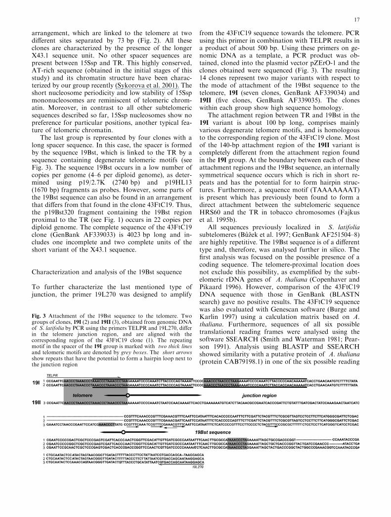
The last group is represented by four clones with along spacer sequence. In this case, the spacer is formedby the sequence 19Bst, which is linked to the TR by asequence containing degenerate telomeric motifs (seeFig. 3). The sequence 19Bst occurs in a low number ofcopies per genome (4–6 per diploid genome), as deter-mined using p19/2.7K (2740 bp) and p19HL13(1670 bp) fragments as probes. However, some parts ofthe 19Bst sequence can also be found in an arrangementthat differs from that found in the clone 43FtC19. Thus,the p19Bst320 fragment containing the 19Bst regionproximal to the TR (see Fig. 1) occurs in 22 copies perdiploid genome. The complete sequence of the 43FtC19clone (GenBank AF339033) is 4023 bp long and in-cludes one incomplete and two complete units of theshort variant of the X43.1 sequence.
Characterization and analysis of the 19Bst sequence
To further characterize the last mentioned type ofjunction, the primer 19L270 was designed to amplify
from the 43FtC19 sequence towards the telomere. PCRusing this primer in combination with TELPR results ina product of about 500 bp. Using these primers on ge-nomic DNA as a template, a PCR product was ob-tained, cloned into the plasmid vector pZErO-1 and theclones obtained were sequenced (Fig. 3). The resulting14 clones represent two major variants with respect tothe mode of attachment of the 19Bst sequence to thetelomere, 19I (seven clones, GenBank AF339034) and19II (five clones, GenBank AF339035). The cloneswithin each group show high sequence homology.
The attachment region between TR and 19Bst in the19I variant is about 100 bp long, comprises mainlyvarious degenerate telomere motifs, and is homologousto the corresponding region of the 43FtC19 clone. Mostof the 140-bp attachment region of the 19II variant iscompletely different from the attachment region foundin the 19I group. At the boundary between each of theseattachment regions and the 19Bst sequence, an internallysymmetrical sequence occurs which is rich in short re-peats and has the potential for to form hairpin struc-tures. Furthermore, a sequence motif (TAAAAAAAT)is present which has previously been found to form adirect attachment between the subtelomeric sequenceHRS60 and the TR in tobacco chromosomes (Fajkuset al. 1995b).
All sequences previously localized in S. latifoliasubtelomeres (Buzek et al. 1997; GenBank AF251504–8)are highly repetitive. The 19Bst sequence is of a differenttype and, therefore, was analysed further in silico. Thefirst analysis was focused on the possible presence of acoding sequence. The telomere-proximal location doesnot exclude this possibility, as exemplified by the subt-elomeric rDNA genes of A. thaliana (Copenhaver andPikaard 1996). However, comparison of the 43FtC19DNA sequence with those in GenBank (BLASTNsearch) gave no positive results. The 43FtC19 sequencewas also evaluated with Genescan software (Burge andKarlin 1997) using a calculation matrix based on A.thaliana. Furthermore, sequences of all six possibletranslational reading frames were analysed using thesoftware SSEARCH (Smith and Waterman 1981; Pear-son 1991). Analysis using BLASTP and SSEARCHshowed similarity with a putative protein of A. thaliana(protein CAB79198.1) in one of the six possible reading
Fig. 3 Attachment of the 19Bst sequence to the telomere. Twogroups of clones, 19I (2) and 19II (3), obtained from genomic DNAof S. latifolia by PCR using the primers TELPR and 19L270, differin the telomere junction region, and are aligned with thecorresponding region of the 43FtC19 clone (1). The repeatingmotif in the spacer of the 19I group is marked with two thick linesand telomeric motifs are denoted by grey boxes. The short arrowsshow repeats that have the potential to form a hairpin loop next tothe junction region
17

frames of 19Bst. The SSEARCH program furtheridentified significant similarity to a hypothetical proteinof Oryza sativa (protein AAD38275.1).
To test for transcription of the hypothetical codingregion of the 43FtC19 clone, the cloned fragment p19/2.7K was used as a probe for hybridization with totalRNA from leaves. A negative result was obtained. The18S rDNA probe was then used for rehybridization (notshown), as a positive control. Of course, our failure tofind 19Bst transcripts in this experiment does not ex-clude the possibility that a region of the 19Bst sequenceforms part of a protein-coding gene, which is silent inleaves either due to its subtelomeric position or the de-velopmental timing of its expression. It is, however,worth noting that both matches in protein databaseswere to hypothetical proteins predicted from genomesequencing data by the same program (Genescan).
Further in silico analysis was focused on the searchfor MAR motifs to evaluate a possible role of 19Bst inbinding to the nuclear matrix. Previous biochemical andcytogenetic data have indicated that telomeres bindchromosomes to the nuclear lamina (Comings 1980;Bass et al. 2000). In a loop model of the higher-orderstructure of chromatin, MARs occur at a frequency ofless than one per 100 kb. Individual chromatin loopsmay, however, differ substantially from this average, notonly between species, but also with respect to chrom-osomal position. It has been observed at meiotic pro-phase that terminally located telomeric chromatin loopsappear to be smaller then loops of the same telomericDNA sequence positioned interstitially (Heng et al.1996). As telomeres are very uniform in sequence, fur-ther MARs should occur in subtelomeres, which arecommonly made up of large blocks of satellite tandemrepeats. Therefore, the software MARFinder (Singhet al. 1997) was employed to find MARs based upon thesimultaneous occurrence of 20 DNA patterns that have
been found in the neighborhood of MARs. It followsfrom the analysis of the 43FtC19 sequence using thissoftware that although several motifs that occur fre-quently in MARs are present, MAR function cannot beattributed to any particular region of the sequence.
In situ analysis of TAS
The above results obtained by molecular approacheswere complemented by cytogenetic data obtained fromfluorescence in situ hybridization (FISH) to metaphasechromosomes and extended DNA fibres (EDF-FISH).The subtelomeric location of the sequence 19Bst on S.latifolia chromosomes was demonstrated using FISH(Fig. 4a). 19Bst exhibited similarly located fluorescentsignals to those reported previously for the 15Ssp se-quence (Sykorova et al. 2001). However, the 15Ssp se-quence, in agreement with its higher copy number, wasfound at subtelomeres of almost all chromosome arms,while the 19Bst signals were observed in the subtelo-meric regions of only about 20 chromosome arms (in-cluding one arm of the X-chromosome and excludingthe Y chromosome). Most of these Bst19-signals prob-ably correspond to TR-proximal parts of 19Bst, whosecopy number (22) exceeds the number of complete 19Bstcopies by four- to fivefold (see above). The strong fluo-rescent signal may indicate the existence of sequencessimilar to Bst19 in subtelomeric regions, although suchsequences have not been found by PCR to date.
The arrangement of 15Ssp and 19Bst sequences rela-tive to X43.1 and telomere sequences along a single ex-tended DNAmolecule can be resolved using EDF-FISH.Figure 4b shows the direct attachment of the X43.1sequence (biotin labelled, green track) to the telomere(digoxigenin labelled, red dot). The appearance of thetelomeric signal as a single dot agreeswith previous resultsof EDF-FISH detection of telomere sequences in rice,which are of comparable size to those in S. latifolia(Ohmido et al. 2000). Furthermore, the X43.1 sequencewas used as reference probe to determine accurately therelative positions of 15Ssp (Fig. 4c) and 19Bst (Fig. 4d)sequences with respect to the telomere. The green signalfor 15Ssp or 19Bst was located between the red signaltrack given by X43.1 and the single red signal dot corre-sponding to the telomere. Thus, using EDF-FISH, both15Ssp and 19Bst sequences can be shown to be positionedbetween the telomere and the X43.1 sequence.
Fig. 4a–d In situ analysis of telomere-associated sequences. Thesubtelomeric location of the newly characterised 19Bst sequencewas demonstrated using FISH on metaphase chromosomes ofS. latifolia (a). Chromosomes X and Y are marked withcorresponding letters. The bar corresponds to 5 lm. Variationsin the relative arrangement of X43.1, 15Ssp, 19Bst and TR werevisualized using EDF-FISH (b – d). The bars represent 1 lm. Thedirect attachment of X43.1 (green signal track) to TR (red signaldot) is shown in b. The 15Ssp sequence ( c) and the Bst19 sequence(d) were detected as green dots between X43.1 (red signal track) andTR (terminal red signal dot)
18

It can be concluded that the spectrum of variation inthe mode of attachment of X43.1 to telomeres involvesboth the new DNA sequences described here, and thepreviously characterized high-copy-number tandem re-peats 15Ssp and X43.1. In the respective repeats, ourprevious data on their chromatin structure (Sykorovaet al. 2001), together with the present detailed informa-tion on sites of their attachment to telomeres, suggestthat a specific chromatin structure, usually ascribed ex-clusively to telomeres, extends to the telomere-associatedregions. The novel 19Bst sequence described here wasshown at both molecular and cytogenetic levels to act asa spacer between X43.1 and the telomere in a subset ofchromosome arms, including one arm of chromosomeX, while it is apparently absent in chromosome Y. Alltelomere-TAS junctions are non-random and highlyconserved in a given type of TAS, which corresponds toour previous findings for two tobacco TASs, HRS60(Fajkus et al. 1995b) and TAS49 (Horakova and Fajkus2000). Taking advantage of the EDF-FISH technique,results from the DNA sequence level could be integratedwith the chromosome picture. Although this study pre-sents only a partial characterization of the structure ofsubterminal chromosome regions, it suggests that thischromosome domain has a highly individual character.
Acknowledgements We thank Dr. Ronald Hancock for criticalreading of the manuscript and Mrs. Jedlickova and Ms. Fridric-hova for technical assistance. This work was supported by theMinistry of Education of the Czech Republic (ProjectMSM143100008) and the Grant Agency of the Czech Republic(Project 204/02/0027).
References
Arnold C, Hodgson IJ (1991) Vectorette PCR: a novel approach togenomic walking. PCR Methods Appl 1:39–42
Bass HW, Riera-Lizarazu O, Ananiev EV, Bordoli SJ, Rines HW,Phillips RL, Sedat JW, Agard DA, Cande WZ (2000) Evidencefor the coincident initiation of homolog pairing and synapsisduring the telomere-clustering (bouquet) stage of meiotic pro-phase. J Cell Sci 113:1033–1042
Blackburn EH, Chiou SS (1981) Non-nucleosomal packaging of atandemly repeated DNA sequence at termini of extrachrom-osomal DNA coding for rRNA in Tetrahymena. Proc NatlAcad Sci USA 78:2263–2267
Burge C, Karlin S (1997) Prediction of complete gene structures inhuman genomic DNA. J Mol Biol 268:78–94
Burr B, Burr FA, Matz EC, Romero-Severson J (1992) Pinningdown loose ends: mapping telomeres and factors affecting theirlength. Plant Cell 4:953–960
Buzek J, Koutnıkova H, Houben A, Rıha K, Janousek B, Siroky J,Grant S, Vyskot B (1997) Isolation and characterization of Xchromosome-derived DNA sequences from a dioecious plantMelandrium album. Chromosome Res 5:57–65
Chen CM, Wang CT, Wang CJ, Ho CH, Kao YY, Chen CC (1997)Two tandemly repeated telomere-associated sequences inNicotiana plumbaginifolia. Chromosome Res 5:561–568
Comings DE (1980) Arrangement of chromatin in the nucleus.Hum Genet 53:131–143
Copenhaver GP, Pikaard CS (1996) RFLP and physical mappingwith an rDNA-specific endonuclease reveals that nucleolus or-ganizer regions of Arabidopsis thaliana adjoin the telomeres onchromosomes 2 and 4. Plant J 9:259–272
Douglas S, Zauner S, Fraunholz M, Beaton M, Penny S, Deng LT,Wu X, Reith M, Cavalier-Smith T, Maier UG (2001) Thehighly reduced genome of an enslaved algal nucleus. Nature410:1091–1096
Fajkus J, Trifonov EN (2001) Columnar packing of telomericnucleosomes. Biochem Biophys Res Commun 280:961–963
Fajkus J, Vyskot B, Bezdek M (1992) Changes in chromatinstructure due to hypomethylation induced with 5-azacytidine orDL-ethionine. FEBS Lett 314:13–16
Fajkus J, Kovarik A, Kralovics R, Bezdek M (1995a) Organizationof telomeric and subtelomeric chromatin in the higher plantNicotiana tabacum. Mol Gen Genet 247:633-638
Fajkus J, Kralovics R, Kovarik A, Fajkusova L (1995b) Thetelomeric sequence is directly attached to the HRS60 subtelo-meric tandem repeat in tobacco chromosomes. FEBS Lett364:33–35
Fransz PF, Alonso-Blanco C, Liharska TB, Peeters AJ, Zabel P, deJong JH (1996) High-resolution physical mapping in Arabid-opsis thaliana and tomato by fluorescence in situ hybridizationto extended DNA fibres. Plant J 9:421–430
Fukui K (1996) Plant chromosomes at meiosis. In: Fukui K, Na-kayama S (eds) Plant chromosomes: laboratory methods. CRCPress, Boca Raton, pp 1–17
Garrido-Ramos MA, de la Herran R, Ruiz Rejon M, Ruiz Rejon C(1999) A subtelomeric satellite DNA family isolated from thegenome of the dioecious plant Silene latifolia. Genome 42:442–446
Gebhardt C, Eberle B, Leonards-Schippers C, Walkemeier B,Salamini F (1995) Isolation, characterization and RFLP linkagemapping of a DNA repeat family of Solanum spegazzinii bywhich chromosome ends can be localized on the genetic map ofpotato. Genet Res 65:1–10
Gottschling DE, Aparicio OM, Billington BL, Zakian VA (1990)Position effect at S. cerevisiae telomeres: reversible repressionof Pol II transcription. Cell 63:751–762
Heng HH, Chamberlain JW, Shi XM, Spyropoulos B, Tsui LC,Moens PB (1996) Regulation of meiotic chromatin loop size bychromosomal position. Proc Natl Acad Sci USA 93:2795–2800
Horakova M, Fajkus J (2000) TAS49—a dispersed repetitivesequence isolated from subtelomeric regions of Nicotianatomentosiformis chromosomes. Genome 43:273–284
Ijdo JW, Wells RA, Baldini A, Reeders ST (1991) Improved telo-mere detection using a telomere repeat probe (TTAGGG)ngenerated by PCR. Nucleic Acids Res 19:4780
Kilian A, Kleinhofs A (1992) Cloning and mapping of telomere-associated sequences from Hordeum vulgare L. Mol Gen Genet235:153–156
Makarov VL, Lejnine S, Bedoyan J, Langmore JP (1993) Nucle-osomal organization of telomere-specific chromatin in rat. Cell73:775–787
Matsunaga S, Kawano S, Michimoto T, Higashiyama T, Nakao S,Sakai A, Kuroiwa T (1999) Semi-automatic laser beam micro-dissection of the Y chromosome and analysis of Y chromosomeDNA in a dioecious plant, Silene latifolia. Plant Cell Physiol40:60–68
Ohmido N, Fukui K (1997) Visual verification of close dispositionbetween a rice A genome-specific DNA sequence (TrsA) and thetelomere sequence. Plant Mol Biol 35:963–968
Ohmido N, Kijima K, Akiyama Y, de Jong JH, Fukui K (2000)Quantification of total genomic DNA and selected repetitivesequences reveals concurrent changes in different DNA fam-ilies in indica and japonica rice. Mol Gen Genet 263:388–394
Palladino F, Gasser SM (1994) Telomere maintenance and generepression: a common end? Curr Opin Cell Biol 6:373–379
Pearson WR (1991) Searching protein sequence libraries: compar-ison of the sensitivity and selectivity of the Smith-Watermanand FASTA algorithms. Genomics 11:635–650
Pryde FE, Louis EJ (1999) Limitations of silencing at native yeasttelomeres. EMBO J 18:2538–2550
Richards EJ, Ausubel FM (1988) Isolation of a higher eukaryotictelomere from Arabidopsis thaliana. Cell 53:127–136
19

Riha K, Fajkus J, Siroky J, Vyskot B (1998) Developmental con-trol of telomere lengths and telomerase activity in plants. PlantCell 10:1691–1698
Rossetti L, Cacchione S, Fua M, Savino M (1998) Nucleosomeassembly on telomeric sequences. Biochemistry 37:6727–6737
Sambrook J, Fritsch EF, Maniatis T (1989) Molecular cloninng: alaboratory manual (2nd edn). Cold Spring Harbor LaboratoryPress, Cold Spring harbor, N.Y.
Singh GB, Kramer JA, Krawetz SA (1997) Mathematical model topredict regions of chromatin attachment to the nuclear matrix.Nucleic Acids Res 25:1419–1425
Smith TF, Waterman MS (1981) Identification of common mo-lecular subsequences. J Mol Biol 147:195–197
Suzuki K, Yamagiwa Y, Matsui T, Yoshida K (1994) Restrictionenzyme-resistant high molecular weight telomeric DNA frag-ments in tobacco. DNA Res 1:129–138
Sykorova E, Fajkus J, Ito M, Fukui K (2001) Transition betweentwo forms of heterochromatin at plant subtelomeres. Chro-mosome Res 9:309–323
The Arabidopsis Genome Initiative (2000) Analysis of the genomesequence of the flowering plant Arabidopsis thaliana. Nature408:796–815
Vagera J, Paulikova D, Dolezel J (1994) The development of maleand female regenerants by in vitro androgenesis in dioeciousplant Melandrium album. Ann Bot 73:455–459
Vershinin AV, Heslop-Harrison JS (1998) Comparative analysis ofthe nucleosomal structure of rye, wheat and their relatives.Plant Mol Biol 36:149–161
Vershinin AV, Alkhimova EG, Heslop-Harrison JS (1996) Mo-lecular diversification of tandemly organized DNA sequencesand heterochromatic chromosome regions in some Triticeaespecies. Chromosome Res 4:517–525
Wellinger RJ, Sen D (1997) The DNA structures at the ends ofeukaryotic chromosomes. Eur J Cancer 33:735–749
Zhong XB, Fransz PF, Wennekes-Eden J, Ramanna MS, vanKammen A, Zabel P, Hans de Jong J (1998) FISH studies re-veal the molecular and chromosomal organization of individualtelomere domains in tomato. Plant J 13:507–517
20


