Embed Size (px)
Citation preview
Plant Disease / July 2005 705
Research
Characterization of the tufB-secE-nusG-rplKAJL-rpoB Gene Cluster of the Citrus Greening Organism and Detection by Loop-Mediated Isothermal Amplification
Mitsuru Okuda, Mitsuhito Matsumoto, and Yuko Tanaka, National Agricultural Research Center for Kyushu Okinawa Region, Suya 2421, Nishigoshi, Kumamoto 861-1192, Japan; Siti Subandiyah, Department of Entomology and Plant Pathology, Gadjah Mada University, Yogyakata 55281, Indonesia; and Toru Iwanami, National Agricul-tural Research Center for Kyushu Okinawa Region, Suya 2421, Nishigoshi, Kumamoto 861-1192, Japan
Citrus greening (CG, Huanglongbing) is a serious disease of citrus that limits pro-duction in many parts of Asia and Africa (2). The causal agents (citrus greening organism, GO) are designated “Candidatus Liberobacter asiaticum” and “Candidatus Liberobacter africanum” for the Asian and African greening species, respectively (6). They are nonculturable phloem-limited bacteria and are transmitted by two distinct insect vectors, Diaphorina citri and Trioza erytreae (2). The Asian greening vector, D. citri, is common in subtropical islands in Japan, rendering trees in those regions susceptible once the pathogen is intro-duced (9). The disease was found for the
first time in the southernmost islands of Japan in 1988 (8). Since then, the disease has spread northward to the main produc-tion areas, posing a great threat to domes-tic citrus cultivation.
Studies of the GO have been hampered by the fact that it cannot be cultured on artificial media. Only a few fragments of genomic DNA have been cloned and se-quenced (4,6,12,14,15). The best charac-terized regions are 16S rDNA and 16S/23S intergenic regions (4,6,14). The nucleotide sequences of these regions of several Japa-nese, Philippine, and Indonesian isolates are identical, similar to those from India and China, and distinct from that from Africa (14). This is consistent with the current status of the GO.
In Japan, where the disease is still lim-ited to relatively few orchards and back-yard gardens, it is managed by removal of infected trees at early stages of infection. To facilitate this practice, a rapid and reli-able assay is indispensable, since the non-specific nature of the foliar symptoms makes greening difficult to distinguish from nutrient deficiencies or other dis-eases. Rapid, simple, and reliable detection methods are also important in GO-
devastated countries like Indonesia. Sero-logical methods have been developed but are inconsistent due to low concentration and uneven distribution of the pathogen in infected citrus. Although amplification of the 16S rDNA fragment by polymerase chain reaction (PCR) is sensitive and re-producible (5), it is applicable only in laboratories equipped with a thermal cy-cler and other basic apparatus for molecu-lar biological experiments and staffed with well-trained personnel.
Loop-mediated isothermal amplification (LAMP) is a new DNA amplification method (11). This method is based on auto-cycling strand displacement DNA synthesis by a DNA polymerase, which has high strand displacement activity, and a set of specially designed inner and outer prim-ers (11). Typically, amplification is com-pleted within 30 min using a simple, af-fordable water bath, which is kept constantly at 65°C. By applying LAMP, the GO might be readily detected in under-equipped laboratories of extension centers and local quarantine offices.
Preliminary investigation revealed that the 16S rDNA and 16S/23S intergenic regions of GO were not suitable for LAMP, because they share homology with plant genomic DNA. In contrast, the nusG-rplKAJL-rpoB gene cluster (15) was more promising because of its specificity, and was chosen for this study. The objectives of this study were (i) to further character-ize the nusG-rplKAJL-rpoB gene cluster by identifying the sequences adjacent to the gene cluster in several isolates from Japan and Indonesia, utilizing thermal asymmetric interlaced PCR (TAIL-PCR) (7), and (ii) to establish a LAMP-based detection method for GO using primers. This method was designed based on the sequence of the conserved region of the nusG-rplKAJL-rpoB gene cluster, and uses a simple stain to visualize products on nylon membranes.
MATERIALS AND METHODS Sample preparation. Leaf samples
were collected from infected citrus trees in different groves on subtropical and tropical islands in Japan and Indonesia (Table 1).
ABSTRACT Okuda, M., Matsumoto, M., Tanaka, Y., Subandiyah, S., and Iwanami, T. 2005. Characterizationof the tufB-secE-nusG-rplKAJL-rpoB gene cluster of the citrus greening organism and detection by loop-mediated isothermal amplification. Plant Dis. 89:705-711.
Thermal asymmetric interlaced polymerase chain reaction (TAIL-PCR) was performed to am-plify the uncharacterized regions adjacent to the nusG-rplKAJL-rpoB gene cluster of citrus greening organism (GO) isolates from different locations in Japan and Indonesia. ConventionalPCR was used to amplify the internal nusG-rplKAJL-rpoB gene cluster of these isolates, and the complete sequence of this 6.1-kb fragment was determined. Comparisons with other bacterial sequences showed that the fragment is the tufB-secE-nusG-rplKAJL-rpoB gene cluster. The organization of this gene cluster is similar to that of the homologous cluster found in Escherichiacoli. Except for three nucleotide changes, the sequence was identical among Japanese and Indo-nesian isolates. A loop-mediated isothermal amplification (LAMP) assay based on the conservedsequence of the nusG-rplKAJL-rpoB gene cluster was developed for the detection of the GO.The LAMP product was rapidly detected on nylon membranes by staining with AzurB. LAMP could detect as low as about 300 copies of the nusG-rplKAJL-rpoB fragment of the Japanese and Indonesian isolates of GO. The LAMP-based detection method, which does not dependupon a thermal cycler and electrophoresis apparatus, will be useful for under-equipped laborato-ries, including those found in extension centers and quarantine offices.
Additional keywords: Diaphorina citri, quantitative PCR
Corresponding author: T. Iwanami E-mail: [email protected]
The nucleotide sequence data reported in this paperwill appear in the DDBJ, EMBL, and GenBanknucleotide sequence databases with the followingaccession number: AY342001.
Accepted for publication 15 January 2005.
DOI: 10.1094 / PD-89-0705 © 2005 The American Phytopathological Society
706 Plant Disease / Vol. 89 No. 7
All field and greenhouse samples from Japan and Indonesia were collected during summer. Total DNA was extracted from about 0.2 g of the leaf midrib of infected
citrus using cetyltrimethylammonium bro-mide (CTAB), as reported previously (10), and suspended with 20 to 200 µl of TE buffer (10 mM Tris-HCl, 1 mM EDTA, pH
8.0). DNA of the established isolates KIN1, OK901, and OKS13 was purified from the infected rough lemon (Citrus jambhiri), which were grown in a green-house at 30°C day and 25°C night.
TAIL-PCR and sequencing of the re-gion adjacent to the tufB-secE-nusG-rplKAJL-rpoB gene cluster. TAIL-PCR (7) was performed to determine the nu-cleotide sequences of the regions adjacent to the nusG-rplKAJL-rpoB gene cluster. The isolate KIN1 was used for the TAIL-PCR experiments. Specific primers GSRV3, GSRV4, and GSRV5 for the re-gion upstream of the known sequence, and GSFW6, GSFW7, and GSFW9 for the region downstream, were used in combina-tion with a short arbitrary primer TAIL2 (Table 2, Fig. 1). The reaction mixtures and thermal cycling conditions are as de-scribed previously (7). Conventional PCR was used to amplify the internal nusG-rplKAJL-rpoB gene cluster, using a primer set of PCFW13 and PCRV2 or PCFW1 and PCRV1, which were designed from the nucleotide sequence of the Indian iso-late (GenBank accession no. M94319) and shown in Table 2. PCR reaction mixture consisted of 1× PCR buffer (10 mM Tris-HCL, 50 mM KCl, 2.5 mM MgCl2, pH 8.3), 200 µM of each dNTP, 2.5 units of AmpliTaq DNA polymerase (Applied Bio-systems, Foster City, CA), 1 µM each of forward and reverse primers, and an arbi-trary amount of DNA template, which had been prepared from the CG-infected citrus leaves from Japan and Indonesia. The am-plifications were performed in a DNA Thermal Cycler 9600 (Applied Biosys-tems) for 40 cycles under the following conditions: 30 s at 94°C, 30 s at 50 to 60°C, depending on the Tm of the primers, and 60 s at 72°C with an initial denatura-tion step of 2 min at 94°C. Both TAIL-PCR and PCR products were directly se-quenced by dideoxy-mediated chain termi-nation (Sanger) method (13) using one of the PCR or sequence primers shown in Table 2, a DNA thermal sequencing kit (Applied Biosystems), and a DNA se-quencer model 373S (Applied Biosys-tems). For the other isolates from Japan and Indonesia, PCR was used to amplify the tufB-secE-nusG-rplKAJL-rpoB gene cluster using primers that were designed from the newly determined sequence of KIN1. These PCR products were directly sequenced.
LAMP. LAMP reactions were con-ducted as reported previously (11), in 10 µl of the mixture containing 1× reaction buffer (20 mM Tris-HCl, 10 mM KCl, 8 mM MgSO4, 10 mM (NH4)2SO4, 0.1% Tween 20, 0.8 M betaine, and 1.4 mM each of dNTPs) together with 8 U of Bst DNA polymerase (New England Biolabs, Beverly, MA), 1.6 µM each of FIP and BIP primers, 0.2 µM each of F3 and B3 prim-ers, and 1 µl of the total DNA from in-fected citrus. FIP, BIP, F3, and B3 for am-
Table 1. Isolates and field sources of citrus greening organism used in this study
Isolates and field sourcesa
Location and year of collection
Host
Japanese isolates and field sources KIN1 Kin, Okinawa, (ref. 14), 1994 Rough lemon (Citrus jambhiri) OK901 Iriomote, Okinawa (ref. 8) 1988 Rough lemon (C. jambhiri) OKS13 Ginowan, Okinawa (ref. 14), 1994 Rough lemon (C. jambhiri) CG4 Motobu, Okinawa ,2001 Tankan (C. tankan) Y02-57 Yoron, Kagoshima, 2002 Tankan (C. tankan) Y02-83 Yoron, Kagoshima, 2002 Oto (C. oto hort. ex Y.) Y03-27 Yoron, Kagoshima, 2003 Kunenbo (C. nobilis var. kunip) OK03-01 Kunigasira, Okinawa, 2003 Shikuwasha (C. depressa) OK03-02 Kunigasira, Okinawa, 2003 Shikuwasha (C. depressa)
Indonesian field sources IDN03-2 Seberkat, Tebes, Sambas, West Kalimantan, 2003 Siem Pontianak (C. reticulata) IDN03-5 Sindhuarjo, Ngaglik, Sleman, Yogyakarta, 2003 Siem Mandarin (C. reticulata) IDN03-7 Karangduwur, Kemili, Purworejo, Central Java, 2003 Siem Mandarin (C. reticulata)
a KIN1, OK901, and OKS13 were isolated by grafting to rough lemon seedlings. Other sources weredirectly obtained from the infected citrus in the fields.
Table 2. Nucleotide sequences and positions of primers used for TAIL-PCR and LAMPa
Name Sequence Positionb
TAIL-PCR primers Tail2 5′-GTNCGASWCANAWGTT-3′ GSFW6 5′-CGTCTCGTCAAGATTGCTATCCG-3′ 3672-3694 GSFW7 5′-GCAAGGTTAGTTCTGTTCATCG-3′ 4016-4037 GSFW9 5′-TCCCGATGAGCGTCCTAATG-3′ 4704-4723 GSRV3 5′-CGAAGAAAACAGACGATACAGGC-3′ 1951-1929 GSRV4 5′-GACCATCAGAGACACAAACC-3′ 1984-1965 GSRV5 5′-TAACTCTACTGGTGTGACACGAC-3′ 2085–2063
PCR primers PCFW13 5′-GCCTGTATCGTCTGTTTTCTTCG-3′ 1929-1951 PCFW1 5′-GTCATCGCATATGGGATGTGGT-3′ 3280-3301 PCRV2 5′-ATTTTAGGAGCAATAACAGGAC-3′ 3781-3760 PCRV1 5′-CGTCTTTAGTCATAAGAG-3′ 4990-4973
Sequence primers SQFW14 5′-GTTAAGTTGCAGATAGAGTCGGG-3′ 2230-2252 SQFW15 5′-GAAGGTTCGGCCTGTTCAATGG-3′ 2593-2614 SQFW16 5′-GAAATTGTTAAAGGCGGGCAG-3′ 2942-2962 SQFW8 5′-GAAGATGCTGGTGCAACGTAG-3′ 4422-4443 SQFW10 5′-ATGGGCGATCTTCCTCTTATGAC-3′ 4960-4982 SQFW11 5′-TCCTAAAATCGTCTATTCACAAAG-3′ 5265-5288 SQFW12 5′-GTTGTCCGCTGCCGATTTGATG-3′ 5310-5331 SQFW19 5′-GTCGATTCTGTGAATAATAATGC-3′ 5611-5633 SQRV9 5′-TCTCCACATCTGTACACTTGAC-3′ 718-697 SQRV10 5′-AACATCCGCACGATTTACTCC-3′ 798-778 SQRV7 5′-AACTCTCCAATCTTAGAACACGC-3′ 1347-1325 SQRV8 5′-TAGACTCATCCCTAACTTGC-3′ 1414-1395
LAMP primers for rplKAJL-rpoB operon rpl-FIP 5′-GCATGCCGAGGATCAATGCCT-TGCTTAAAGAGCGTGC-
TACG-3′ 2727-2802
rpl-BIP 5′-TATGCCTAATGGCACGGGGGTA-AGCTTCATCCGCCTT-CGA-3′
2899-2830
rpl-F3 5′-TGGGTTAAGTGATGCTGTGG-3′ 2701-2721 rpl-B3 5′-CAACAATATCAGCCCCTGCT-3′ 2924-2905
LAMP primers for 16SrDNA region rdna-FIP 5′-TCCGCGATTACTAGCGATTCCGCTCAGTTCGGATTGCA-
CTCT-3′ 45-106
rdna-BIP 5′-TCAGCATGCCGCGGTGAATAAGGCAAAACCAACTCCCA-3′ 180-107 rdna-F3 5′-AGCTAATCCCCAAAAGCCAT-3′ 25-46 rdna-B3 5′-CCTCCTTGCGGTTAGCAC-3′ 85-106
a TAIL-PCR = thermal asymmetric interlaced polymerase chain reaction; LAMP = loop-mediated isothermal amplification.
b Nucleotide positions are numbered following the sequence in the GenBank database AY342001 for TAIL-PCR primers and LAMP primers for nusG-rplKAJL-rpoB gene cluster, and AB019793 for LAMP primers for 16SrDNA region. Nucleotide positions of FIP and BIP indicate target regions ofthe gene cluster, which do not match perfectly with the sequences of the primers because of the pecu-liar structure of the primers.
Plant Disease / July 2005 707
plification of a part of the rplKAJL-rpoB operon or the 16S rDNA region were de-signed using a software PrimerExplore V1 (Fujitsu, Tokyo, Japan; Table 2). Template DNA and primers were denatured at 95°C for 5 min before the other reagents were added. The reaction mixture was incubated at 65°C for up to 1 h.
Comparison of sensitivity of LAMP and PCR. Total DNA from infected citrus leaves was prepared in 10-fold dilution series of 100 ng, 10 ng, 1 ng, 100 pg (0.1 ng), and 10 pg (0.01 ng) per microliter with TE. Using these templates, LAMP was carried out as described above. PCR with GO-specific primers rpl-F3 and rpl-B3 (Table 2) was performed with the ther-mal cycling condition of 35 cycles of 94°C for 30 s, 58°C for 30 s, and 72°C for 30 s after initial denaturation for 2 min. The sensitivity of LAMP was compared with that of PCR.
Visualization of the amplification product by staining. One microliter of DNA dilution series (1, 10, and 100 ng/µl) of citrus genomic DNA (template) or LAMP products were spotted onto nylon membranes (Hybond N+, Amersham Bio-science, Buckinghamshire, UK). The DNA was denatured and fixed to the membrane by soaking in 0.4 M NaOH for 5 min, followed by a brief wash in 2× SSC (0.3 M NaCl and 0.03 M sodium citrate). The membranes were air-dried on paper towels and then soaked in 0.1% AzurB, 0.1%
methylene blue, 0.1% crystal violet, or 1× Mupid Blue (ADVANCE-BIO, Tokyo, Japan) until spots became visible, typically 1 to 2 min. The membranes were rinsed with 50% ethanol and dried.
Sensitivity of LAMP for the GO. The concentration of the GO in total DNA ex-tract was estimated by quantitative PCR using SYBR Green I and Light Cycler system (Roche, Indianapolis, IN). A por-tion of the nusG-rplKAJL-rpoB region was amplified by PCR with GO-specific primers fp1898 and rp1897 (15), and the product was purified using the Montage PCR purification kit (Millipore, Billerica, MA). Tenfold dilution series (1 ng to 0.1 fg per microliter) of the purified product were prepared by diluting with TE after estimating the original concentration with a spectrophotometer. Quantitative PCR was performed on the dilution series, and the standard curve, which relates the cross-ing cycle number to the logarithms of con-centration of the template, was generated. Subsequently, 1 or 10 ng of total DNA from healthy rough lemon was mixed with a diluted series of the template fragment to examine whether DNA of the healthy rough lemon leaves affects the quantitative analysis. After confirming that host DNA has little effect on amplification (see re-sults), quantitative PCR was carried out using the total DNA sample from the GO-infected tissue, and the concentration of GO in the sample was estimated from the
standard curve. The conditions of the quantitative PCR were as follows: 1× reac-tion mixtures (LightCycler-DNA master mix SYBR Green I, Roche) containing 100 ng of sample DNA and specific primers rpl-F1 (5′-TTTCGTTGGGCAGTCT-3′) and rpl-R1 (5′-CCAGCTCGAATACCCT-3′) were put into glass capillaries, and thermal cycling reactions were performed. The primers of rpl-F1 and rpl-R1 were designed from the nucleotide sequence of the nusG-rplKAJL-rpoBC gene cluster (GenBank accession no. M94319; nucleo-tide positions 424 to 439 and 594 to 609, respectively). Reaction conditions were 40 cycles of 95°C for 15 s, 60°C for 5 s, and 72°C for 20 s. The fluorimetric intensity of SYBR Green I was measured at 497 nm during each cycle at 72°C.
RESULTS TAIL-PCR and sequence of the tufB-
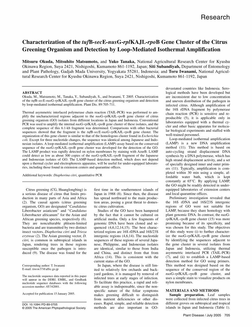
secE-nusG-rplKAJL-rpoB gene cluster. No specific bands were observed after agarose electrophoresis of the first and second TAIL-PCR (Fig. 1B, lanes R5, R4, F6, and F7), presumably due to the low concentration of the GO DNA. A GO-specific band of about 1.9 kbp was ob-served in agarose gels after the third TAIL-PCR with specific primer GSRV3 and the arbitrary primer TAIL2, preceded by the first and second TAIL-PCR using the primer sets GSRV5-TAIL2 and GSRV4-TAIL2, respectively (Fig. 1B). There was
Fig. 1. Amplification of the region adjacent to the nusG-rplKAJL-rpoB gene cluster of the citrus greening organism (GO). A, Annealing positions of the primers used for the specific amplification of the upstream region (TAIL2, GSRV5, GSRV4, GSRV3) and the downstream region (TAIL2, GSFW6, GSFW7, GSFW9). B, Agarose gel electrophoresis of TAIL-PCR products with primer TAIL2 and one of the following specific primers: GSRV5 (indicated as R5),GSRV4 (R4), GSRV3 (R3), GSFW6 (F6), GSFW7 (F7), and GSFW9 (F9). D and H indicate infected tissue and healthy controls, respectively. C, Gene or-ganization of the GO in the vicinity of the nusG-rplKAJL-rpoB gene cluster region revealed by thermal asymmetric interlaced polymerase chain reaction (TAIL-PCR). The regions adjacent to the nusG-rplKAJL-rpoB gene cluster elucidated in this study are shown by thick lines. Sequence identities (%) of the putative genes with the corresponding genes of Escherichia coli are shown.
708 Plant Disease / Vol. 89 No. 7
an expected overlap of nucleotide se-quence between the TAIL-PCR fragment and the 5′ region of the nusG-rplKAJL-rpoB gene cluster, showing the authentic-
ity of the TAIL-PCR product. Likewise, a fragment of about 1.4 kbp, which is lo-cated in the 3′ region of the nusG-rplKAJL-rpoB gene cluster, was specifi-
cally amplified by TAIL-PCR using the primers GSFW6, GSFW7, and GSFW9 in combination with the arbitrary primer TAIL2 (Fig. 1). In addition to these TAIL-PCR amplifications, the internal known region of nusG-rplKAJL-rpoB gene clus-ter was amplified by PCR, and the overall sequence was determined completely, resulting in a fragment of 6,145 bp with a G+C content of 39.7%. Comparison with the nucleotide sequences in the DNA data bank of Japan (DDBJ) suggested that the DNA fragment contains the tufB-secE-nusG-rplKAJL-rpoB gene cluster, which is reminiscent of that of Escherichia coli (3). The percentages of sequence identities between GO and E. coli of each gene are shown in Figure 1.
The 6.1-kbp fragment of the tufB-secE-nusG-rplKAJL-rpoB was amplified by PCR from another two Japanese isolates (OK901 and Y02-57) as well as three In-donesian isolates (In03-2, In03-5, and In03-7; Table 1). The sequences of the isolates within each country were identical, while there were three nucleotide changes at nucleotide positions 1806, 5012, and 6107 between Japanese GO and Indone-sian GO (Table 3).
The sequences of a partial fragment (3.0 kb) of the nusG-rplKAJL-rpoB gene clus-ter of Indian GO as well as a shorter frag-ment (1.7 kb) of the rplKAJL-rpoB operon of African GO had been reported previ-ously (12,15). When compared with Indian GO, there are three nucleotide changes, at nucleotide positions 4439, 4452, and 4453, between Japanese and Indian GO, while the sequence was conserved between Japa-nese and Indonesian GO in these positions (Table 3). Interestingly, all these nucleotide changes were found in the putative genes, not in the intergenic spacer region (Table 3). The sequence of the rplKAJL-rpoBC operon of both Japanese and Indonesian GO showed 81% identity overall at the nucleotide level with that of African GO.
LAMP and its sensitivity. When suit-able primer sets were searched, the com-puter program PrimerExplore V1 showed several candidates for both the rplKAJL-rpoBC operon and the 16SrDNA region. A combination of primers, which had the least probability of dimer formation, was selected for each region, and further ex-periments were conducted using these primer sets (Table 2).
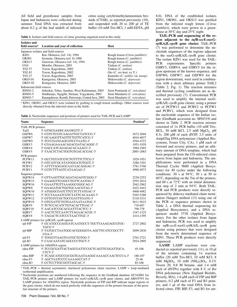
Typical ladder-like bands were observed after agarose gel electrophoresis of the LAMP products of the rplKAJL-rpoBC operon, suggesting successful amplifica-tion of the targeted region (Fig. 2). Suffi-cient amounts of products for agarose gel electrophoresis were obtained in 30 min of incubation. The specific amplification was observed from all field isolates, which had tested positive in PCR detection, as well as from the established isolates kept in the greenhouse (Fig. 2). Ladder-like bands were not observed in agarose gel electro-
Table 3. Differences in the tufB-secE-nusG-rplKAJL-rpoB gene cluster nucleotide sequence among the Japanese (KIN1), Indonesian (IDN03-2), and Indian (Poona) isolates of the citrus greening organism
Nucleotide positionsa and gene
Isolate 1806(nusG) 4439(rplL) 4452(rplL) 4453(rplL) 5012(rpoB) 6107(rpoB)
KIN1 G C T A C A IDN03-2 T C T A T G Poona n.a.b –c A T C n.a.
a Nucleotide positions are numbered following the sequence in the GenBank database AY342001. b n.a. = sequence not available for comparison. c – = gap in the sequence.
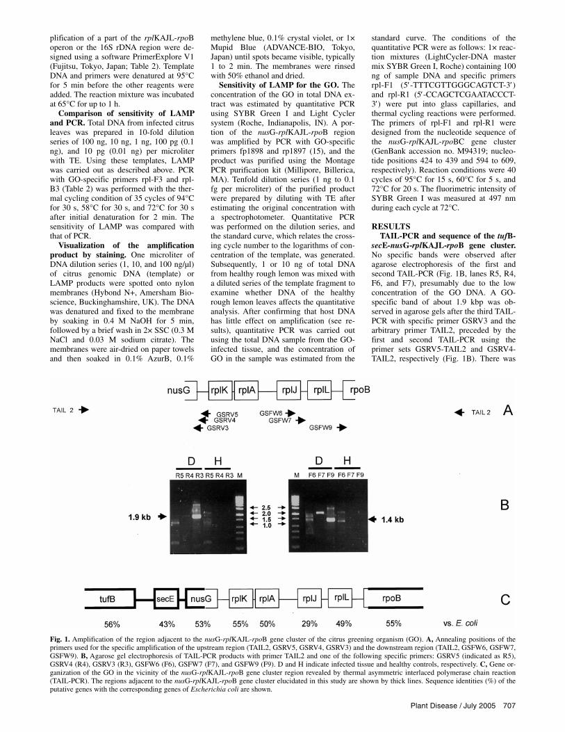
Fig. 3. Agarose gel electrophoresis of loop-mediated isothermal amplification (LAMP) products am-plified with the rpl primer set for the rplKAJL-rpoB operon (A) and polymerase chain reaction (PCR)products amplified with primers F3 and B3 (B). Total DNA of citrus greening organism (GO)-infected leaves (KIN1 and OKS13) were diluted and used as templates: 1, 100 ng; 2, 10 ng; 3, 1 ng; 4, 0.1 ng; 5, 0.01 ng per tube. H and W are 10 ng of total DNA of healthy rough lemon and sterile water, respec-tively. M indicates DNA size markers.
Fig. 2. Agarose gel electrophoresis of loop-mediated isothermal amplification (LAMP) products am-plified with the rpl primer set (rpl) or 16SrDNA primer set (rdna). 100 ng DNA of total DNA from citrus infected with KIN1 (lane 1), OKS13 (lane 2), Y02-57 (lane 3), IDN03-2 (lane 4), IDN03-5 (lane 5), IDN03-7 (lane 6), and from healthy rough lemon (lane 7) were used as templates, respectively. Mindicates DNA size markers.
Plant Disease / July 2005 709
phoresis, when healthy rough lemon DNA was used as template (Fig. 2). When LAMP was tried for a 16S rDNA fragment, ladder-like bands were not as clear as those of the rplKAJL-rpoBC operon, and some nonspecific bands were also present in reactions of the healthy rough lemon DNA (Fig. 2).
LAMP and PCR were tested for 10, 1, 0.1, and 0.01 ng of the total DNA from citrus, which was infected with KIN1 or OKS13 isolate. Both LAMP and PCR could detect GO-specific DNA fragments from 10 and 1 ng of the total DNA (Fig. 3), suggesting that LAMP is at least as sensi-tive as PCR.
Visualization by staining with dyes. In the preliminary experiment, amounts greater than 10 ng of genomic or LAMP-amplified DNA were clearly visualized by spotting nylon membranes and staining with AzurB or Mupid Blue, but not by crystal violet or methylene blue, in 2 min (data not shown). The results indicated that GO DNA can be visualized on the mem-brane by AzurB and Mupid Blue without background staining of the host DNA when the GO DNA is amplified to the concentration of over 10 ng/µl from reac-tion mixture that contains less than 10 ng/µl of the total citrus DNA.
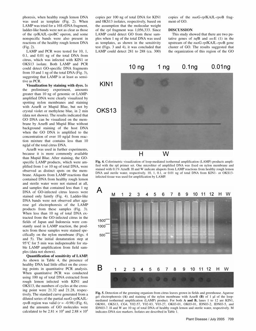
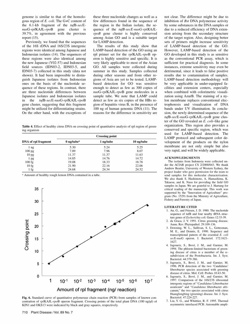
AzurB was used in further experiments, because it is more commonly available than Mupid Blue. After staining, the GO-specific LAMP products, which were am-plified from 1 or 10 ng of total DNA, were observed as distinct spots on the mem-brane. Aliquots from LAMP reactions that contained DNA from healthy rough lemon and sterile water were not stained at all, and samples that contained less than 1 ng DNA of GO-infected citrus leaves were stained only faintly (Fig. 4). Ladder-like DNA bands were not observed after aga-rose gel electrophoresis of the LAMP products from these samples (Fig. 3). When less than 10 ng of total DNA ex-tracted from the GO-infected citrus in the fields of Japan and Indonesia were con-stantly used in LAMP reaction, the prod-ucts from these samples were stained spe-cifically on the nylon membrane (Figs. 4 and 5). The initial denaturation step at 95°C for 5 min was indispensable for sta-ble LAMP amplification from field sam-ples (data not shown).
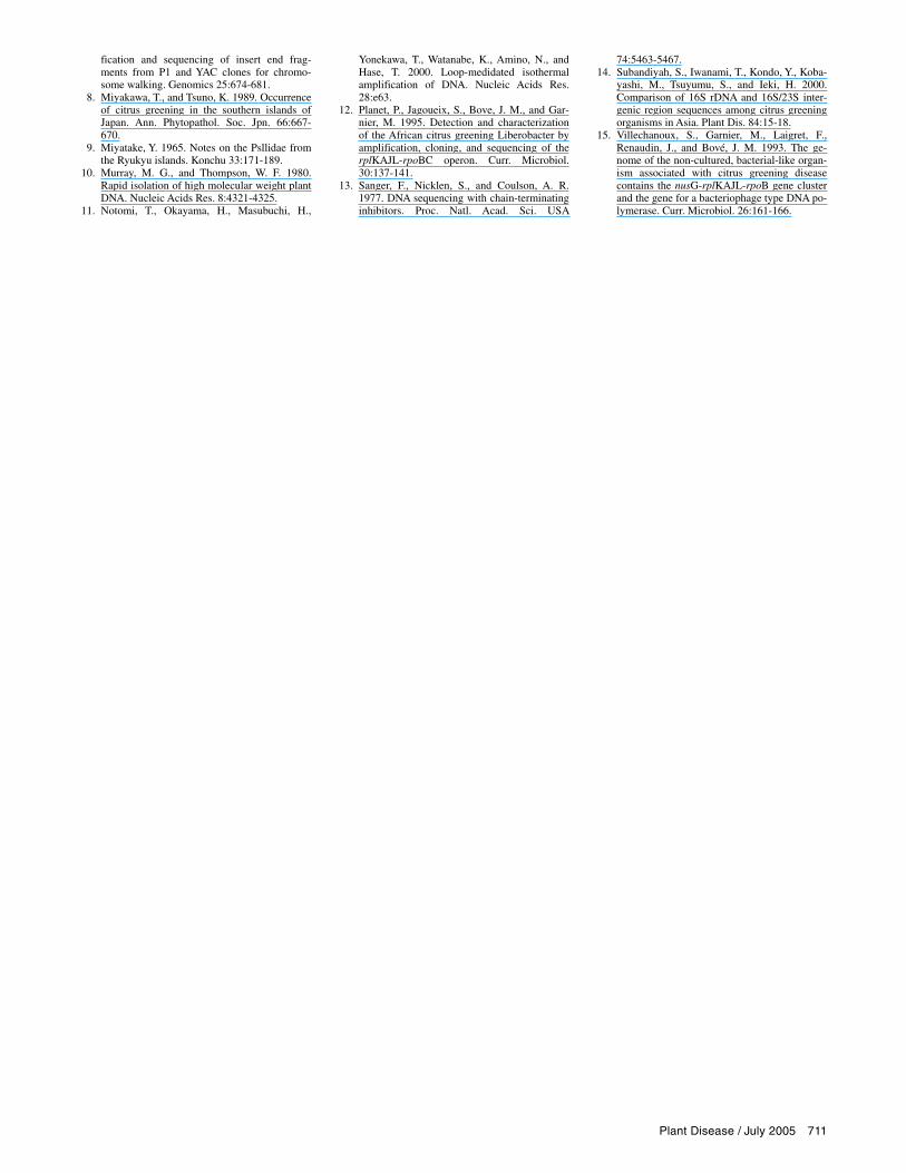
Quantification of sensitivity of LAMP. As shown in Table 4, the presence of healthy DNA had little effect on the cross-ing points in quantitative PCR analysis. When quantitative PCR was conducted using 100 ng of total DNA extracted from rough lemon infected with KIN1 and OKS13, the numbers of cycles at the cross-ing point were 21.32 and 21.28, respec-tively. The standard curve generated from a diluted series of the partial nusG-rplKAJL-rpoB region was valid (r = –0.98) (Fig. 6), and the amounts of GO molecules were calculated to be 2.81 × 10⁴ and 2.88 × 10⁴
copies per 100 ng of total DNA for KIN1 and 0KS13 isolates, respectively, based on the assumption that the molecular weight of the rpl fragment was 1,056,333. Since LAMP could detect GO from these sam-ples when 1 ng of the total DNA was used as templates, as shown in the sensitivity test (Figs. 3 and 4), it was concluded that LAMP could detect 281 to 288 (ca. 300)
copies of the nusG-rplKAJL-rpoB frag-ment of GO.
DISCUSSION This study showed that there are two pu-
tative genes of tufB and secE (1) in the upstream of the nusG-rplKAJL-rpoB gene cluster of GO. The results suggested that the organization of this region of the GO
Fig. 4. Colorimetric visualization of loop-mediated isothermal amplification (LAMP) products ampli-fied with the rpl primer set. One microliter of amplified DNA was fixed on nylon membrane and stained with 0.1% AzurB. H and W indicate aliquots from LAMP reactions from healthy rough lemon DNA and sterile water, respectively. 10, 1, 0.1, or 0.01 ng of total DNA from KIN1- or OKS13-infected tissue was used for amplification by LAMP.
Fig. 5. Detection of the greening organism from citrus leaves grown in fields and greenhouse. Agarose gel electrophoresis (A) and staining of the nylon membrane with AzurB (B) of 1 µl of the loop-mediated isothermal amplification (LAMP) product. For both A and B, lanes 1 to 12 are KIN1, OK901, OKS13, CG4, Y02-57, Y02-83, Y03-27, OK03-01, OK03-01, IDN03-2, IDN03-5, and IDN03-7. H and W are 10 ng of total DNA of healthy rough lemon and sterile water, respectively. M indicates DNA size markers. Isolates are described in Table 1.
710 Plant Disease / Vol. 89 No. 7
genome is similar to that of the homolo-gous region of E. coli. The G+C content of the 6.1-kb fragment of the tufB-secE-nusG-rplKAJL-rpoB gene cluster is 39.7%, in agreement with the previous report (15).
Previously, we found that the sequences of the 16S rDNA and 16S/23S intergenic regions were identical among Japanese and Indonesian isolates (14). The sequences of these regions were also identical among the new Japanese (Y02-57) and Indonesian field sources (IDN03-2, IDN03-5, and IDN03-7) collected in this study (data not shown). It had been impossible to distin-guish Japanese isolates from Indonesian ones on the basis of the nucleotide se-quence of these regions. In contrast, there are three nucleotide differences between Japanese isolates and Indonesian isolates in the tufB-secE-nusG-rplKAJL-rpoB gene cluster, suggesting that this fragment might be utilized for differentiating strains. On the other hand, with the exceptions of
these three nucleotide changes as well as a few differences found in the sequence of the region in the Indian isolate, the se-quence of the tufB-secE-nusG-rplKAJL-rpoB gene cluster is highly conserved among Asian GO and is a suitable target for LAMP detection.
The results of this study show that LAMP-based detection of the GO using an rpl primer set for the rplKAJL-rpoB op-eron is highly sensitive and specific. It is very likely applicable to most of the Asian GO. All samples were collected during summer in this study. Samples collected during other seasons and from other re-gions of Asia are yet to be tested. LAMP-based detection for GO was sensitive enough to detect as few as 300 copies of nusG-rplKAJL-rpoB gene molecules in a sample tube. We note that LAMP could detect as few as six copies of the HBs re-gion of hepatitis virus B, in the presence of 100 ng of human genomic DNA (11). The reasons for the difference in sensitivity are
not clear. The difference might be due to inhibition of Bst DNA polymerase activity by some substances in the DNA samples or due to a reduced efficiency of DNA exten-sion arising from the secondary structure of the target region. Also, designing better sets of primers might increase sensitivity of LAMP-based detection of the GO. However, LAMP-based detection of the GO developed in this study is as sensitive as the conventional PCR assay, which is sufficient for practical diagnosis. In some instances, extreme sensitivity may even be detrimental, since it leads to false positive results due to contamination of samples. LAMP-based detection methodology will be very applicable in under-equipped fa-cilities and extension centers, especially when combined with colorimetric visuali-zation using AzurB. The staining of a ny-lon membrane replaces conventional elec-trophoresis and visualization of DNA bands under UV illumination. In conclu-sion, the newly determined sequence of the tufB-secE-nusG-rplKAJL-rpoB gene clus-ter of the GO revealed an E. coli–like gene organization. This region also provides a conserved and specific region, which was used for LAMP-based detection. The LAMP protocol and subsequent color de-velopment of the products on the nylon membrane are not only simple but also very rapid, and will be widely applicable.
ACKNOWLEDGMENTS The isolates from Indonesia were collected un-
der the ACIAR project CS 2/2000/043. We thank Andrew Beattie, University of Western Sydney, the project leader who gave permission for the team to send samples for this molecular characterization. We also thank S. Hashimoto, A. Hamashima, K. Takaesu, and K. Nasu for providing the field GO samples in Japan. We are grateful to J. Hartung for critical reading of the manuscript. This work was supported by the “Innovation of Agriculture” pro-gram (No. 1529) from the Ministry of Agriculture, Fishery and Forestry of Japan.
LITERATURE CITED 1. An, G., and Friesen, J. D. 1980. The nucleotide
sequence of tufB and four nearby tRNA struc-ture genes of Escherichia coli. Genes 12:33-39.
2. da Graca, J. V. 1991. Citrus greening disease. Annu. Rev. Phytopathol. 29:109-136.
3. Downing, W. L., Sullivan, S. L., Gottesman, M. E., and Dennis, E. 1990. Sequence and transcriptional pattern of the essential E. coli secE-nusG operon. J. Bacteriol. 172:1621-1627.
4. Jagoueix, S., Bové, J. M., and Garnier, M. 1994. The phloem-limited bacterium of green-ing disease of citrus is a member of the α-subdivision of the Protobacteria. Int. J. Syst. Bacteriol. 44:379-386.
5. Jagoueix, S., Bové, J. M., and Garnier, M. 1996. PCR detection of the two ‘Candidatus’ liberobacter species associated with greening disease of citrus. Mol. Cell. Probes 10:43-50.
6. Jagoueix, S., Bové, J. M., and Garnier, M. 1997. Comparison of the 16S/23S ribosomal intergenic regions of “Candidatus Liberobacter asiaticum” and “Candidatus liberobacter afri-canum,” the two species associated with citrus Huanglongbing (greening) disease. Int. J. Syst. Bacteriol. 47:224-227.
7. Liu, Y. G., and Whitttier, R. F. 1995. Thermal asymmetric interlaced PCR: Automable ampli-
Table 4. Effect of healthy citrus DNA on crossing point of quantitative analysis of rpl region of green-ing organism
Crossing point
DNA of rpl fragment 0 ng/tubea 1 ng/tube 10 ng/tube
1 ng 5.30 5.24 5.25 100 pg 7.89 7.96 8.07 10 pg 11.37 11.37 11.63
1 pg 14.85 14.76 14.72 100 fg 19.08 18.33 18.78 10 fg 21.90 22.16 22.06
1 fg 24.68 24.34 24.52
a Amount of healthy rough lemon DNA contained in a tube.
Fig. 6. Standard curve of quantitative polymerase chain reaction (PCR) from samples of known con-centration of rplKAJL-rpoB operon fragment. Crossing points of the total plant DNA (100 ng/µl) of KIN1 and OKS13 were indicated by black and gray squares, respectively.
Plant Disease / July 2005 711
fication and sequencing of insert end frag-ments from P1 and YAC clones for chromo-some walking. Genomics 25:674-681.
8. Miyakawa, T., and Tsuno, K. 1989. Occurrence of citrus greening in the southern islands of Japan. Ann. Phytopathol. Soc. Jpn. 66:667-670.
9. Miyatake, Y. 1965. Notes on the Psllidae from the Ryukyu islands. Konchu 33:171-189.
10. Murray, M. G., and Thompson, W. F. 1980. Rapid isolation of high molecular weight plant DNA. Nucleic Acids Res. 8:4321-4325.
11. Notomi, T., Okayama, H., Masubuchi, H.,
Yonekawa, T., Watanabe, K., Amino, N., and Hase, T. 2000. Loop-medidated isothermal amplification of DNA. Nucleic Acids Res. 28:e63.
12. Planet, P., Jagoueix, S., Bove, J. M., and Gar-nier, M. 1995. Detection and characterization of the African citrus greening Liberobacter by amplification, cloning, and sequencing of the rplKAJL-rpoBC operon. Curr. Microbiol. 30:137-141.
13. Sanger, F., Nicklen, S., and Coulson, A. R. 1977. DNA sequencing with chain-terminating inhibitors. Proc. Natl. Acad. Sci. USA
74:5463-5467. 14. Subandiyah, S., Iwanami, T., Kondo, Y., Koba-
yashi, M., Tsuyumu, S., and Ieki, H. 2000. Comparison of 16S rDNA and 16S/23S inter-genic region sequences among citrus greening organisms in Asia. Plant Dis. 84:15-18.
15. Villechanoux, S., Garnier, M., Laigret, F., Renaudin, J., and Bové, J. M. 1993. The ge-nome of the non-cultured, bacterial-like organ-ism associated with citrus greening disease contains the nusG-rplKAJL-rpoB gene cluster and the gene for a bacteriophage type DNA po-lymerase. Curr. Microbiol. 26:161-166.