Embed Size (px)
Citation preview

Chemically-specific time-resolved surface photovoltage spectroscopy:
Carrier dynamics at the interface of quantum dots attached to a
metal oxide
Ben F. Spencer a,b,⁎, Matthew J. Cliffe a,b, Darren M. Graham a, Samantha J.O. Hardman a,1, Elaine A. Seddon a,b,Karen L. Syres a,2, Andrew G. Thomas a,3, Fausto Sirotti c, Mathieu G. Silly c, Javeed Akhtar d,4, Paul O'Brien d,Simon M. Fairclough e, Jason M. Smith f, Swapan Chattopadhyay b,5, Wendy R. Flavell a
a School of Physics and Astronomy and the Photon Science Institute, The University of Manchester, Manchester M13 9PL, United Kingdomb The Cockcroft Institute, Sci-Tech Daresbury, Keckwick Lane, Daresbury, Warrington WA4 4AD, Cheshire, United Kingdomc Synchrotron SOLEIL, BP 48, Saint-Aubin, F91192 Gif sur Yvette CEDEX, Franced Department of Chemistry, University of Manchester, Oxford Road, Manchester M13 9PL, United Kingdome Department of Chemistry, University of Oxford, South Parks Road, Oxford OX1 3QR, United Kingdomf Department of Materials, University of Oxford, Parks Road, Oxford OX1 3PH, United Kingdom
a b s t r a c ta r t i c l e i n f o
Available online xxxx
Keywords:
Surface photovoltage
Time-resolved photoemission
Semiconductor surface
Carrier dynamics
Photovoltaics
Colloidal quantum dots
We describe a new experimental pump-probe methodology where a 2D delay-line detector enables fast (ns)
monitoring of a narrow XPS spectrum in combination with a continuous pump laser. This has been developed
at the TEMPO beamline at Synchrotron SOLEIL to enable the study of systemswith intrinsically slow electron dy-
namics, and to complement faster measurements that use a fs laser as the pump. We demonstrate its use in a
time-resolved study of the surface photovoltage of them-plane ZnO (1010) surfacewhich shows persistent pho-
toconductivity, requiring monitoring periods on ms timescales and longer. We make measurements from this
surface in the presence and absence of chemically-linked quantum dots (QDs), using type I PbS and type II
CdSe/ZnSe (core/shell) QDs as examples. We monitor signals from both the ZnO substrate and the bound QDs
during photoexcitation, yielding evidence for charge injection from theQDs into the ZnO. The chemical specificity
of the technique allows us to observe differences in the extent towhich theQDsystems are influenced by thefield
of the surface depletion layer at the ZnO surface, which we attribute to differences in the band structure at the
interface.
© 2015 TheUniversity ofManchester. Published by Elsevier B.V. This is anopenaccess articleunder theCCBY license
(http://creativecommons.org/licenses/by/4.0/).
1. Introduction
The study of ‘real-time’ charge carrier dynamics at material inter-
faces is becoming ever more important as next generation technologies
increasingly access nanoscale dimensions. This is particularly the case
for solar harvesting technologies, where a clear understanding of the
dynamics of photoexcited carriers is of key fundamental importance.
One such example is in devices utilising cost-reducing photovoltaic ma-
terials such as light-harvesting colloidal quantum dots (QDs) [1,2]
where a transparent conducting oxide (TCO) such as ZnO is used as
photoanode [3–5]. These QDs have been shown to exhibit multiple ex-
citon generation (MEG, creation ofmore than one pair of charge carriers
froma single photon) for photon energies greater than around twice the
effective band gap. The band gap is typically 1.0 eV, meaning MEG is
present at wavelengths within the solar spectrum, giving potential for
photovoltaic devices with enhanced efficiencies [1]. Considerable effort
is currently being devoted to extending the photoexcited electron-hole
pair lifetime in order to maximise the benefit of MEG. Quantum dot
cores (known as type I QDs) that have been surrounded by a shell are
known as type II QDs, and these are engineered such that the band
Surface Science xxx (2015) xxx–xxx
⁎ Corresponding author at: School of Physics and Astronomy and the Photon Science
Institute, The University of Manchester, Manchester M13 9PL, United Kingdom.
E-mail address: [email protected] (B.F. Spencer).1 Present address: Manchester Institute of Biotechnology, Faculty of Life Sciences,
University of Manchester, 131 Princess Street, Manchester M1 7DN, United Kingdom.2 Present address: Jeremiah Horrocks Institute for Mathematics, Physics and
Astronomy, University of Central Lancashire, Preston PR1 2HE, United Kingdom.3 Present address: School of Materials and the Photon Science Institute, The University
of Manchester, Manchester M13 9PL, United Kingdom.4 Present address: Department of Physics, Nano-Science & Materials Synthesis
Laboratory, COMSATS Institute of Information Technology, Chakshahzad Park Road,
Islamabad 44000, Pakistan.5 Present address: Northern Illinois University and Fermilab, DeKalb, IL 60115, USA.
SUSC-20454; No of Pages 6 March 25, 2015; Model: Gulliver 5
http://dx.doi.org/10.1016/j.susc.2015.03.010
0039-6028/© 2015 The University of Manchester. Published by Elsevier B.V. This is an open access article under the CC BY license (http://creativecommons.org/licenses/by/4.0/).
Contents lists available at ScienceDirect
Surface Science
j ourna l homepage: www.e lsev ie r .com/ locate /susc
Please cite this article as: B.F. Spencer, et al., Chemically-specific time-resolved surface photovoltage spectroscopy: Carrier dynamics at the
interface of quantum dots attached to a metal oxide, Surf. Sci. (2015), http://dx.doi.org/10.1016/j.susc.2015.03.010
structure leads to the photoexcited electrons and holes being separately
trapped in the core and shell or vice versa, reducing the wavefunction
overlap and extending radiative recombination lifetimes. Hence there
is great interest in these type II structures for photovoltaic applications
[6–8]. Here we study the carrier dynamics in heterojunctions formed
both from type II QDs, and from type I QDs that have been shown to ex-
hibit MEG, attached to a semiconductor photoanode, ZnO.
Carrier dynamics at the surface of semiconductors may be probed
using time-resolved surface photovoltage (SPV) spectroscopy [9–11].
The space charge layer at the surface of semiconductors where the
Fermi level is pinned by surface states within the band gap leads to
band bending in this region. For n-type semiconductors the bands
bend upwards due to the presence of a depletion layer, and downwards
in p-type semiconductors due to an accumulation layer. A laser is used
to promote carriers across the band gap, and, in the case of an n-type
semiconductor, electrons and holes migrate away from and towards
the surface respectively (and vice versa in a p-type semiconductor).
This additional electric field reduces the band bending and changes
the energy of the valence band maximum in the space charge region.
A synchronised X-ray source is then used to excite core level photoelec-
trons (using X-ray photoelectron spectroscopy, XPS), as the binding en-
ergy (BE) of the core levels in the space charge region at the surface shift
as the valence band maximum shifts [9–11]. In n-type semiconductors
these core levels shift to higher BE and in p-type semiconductors to
lower BE [10]. By varying the time delay between the laser pump and
XPS probe, the recombination dynamics is monitored over time, be-
cause the band bending and core level binding energies return to equi-
librium as carriers recombine. Thus the recombination dynamics at the
surface are elucidated [10–18].
For the case of colloidal QDs chemically linked to a semiconductor,
this technique then provides a way to monitor the carrier dynamics in
the semiconductor substrate upon photoexcitation of the QDs, i.e., as
additional carriers are injected into the semiconductor conduction
band upon laser illumination of the QDs [19]. Moreover, photoemission
allows chemical specificity because the substrate and QDs can be mon-
itored separately or in some cases simultaneously, simply bymonitoring
different core level photoemission lines. The technique then gives a
chemically-specific diagnostic of charge transfer between the QD and
the substrate.
Laser-pump X-ray-probe SPV experiments using pulsed lasers,
where the pump laser is synchronised to individual X-ray pulses from
a synchrotron, are limited by the time window available, determined
by the synchrotron pulse length and repetition rate [11–13]. In the
case of work by us at the SRS, UK, with the synchrotron operating in
single-bunch mode, the maximum time window available was 320 ns,
i.e., laser pump and X-ray probe pulses were incident every 320 ns
with a set delay time between the two [10]. When measuring the tran-
sient SPV at the Si (111) 7 × 7 surface the dark carrier lifetimewas in ex-
cess of the timewindow, and so amulti-pulsemodel was required to fit
the carrier dynamics since the observed SPV transientwas influenced by
the residual SPV induced by preceding laser pump pulses [10]. Recently
the use of angle-resolved time-of-flight (ARToF) analysers (such as at
the BL07LSU beamline at SPring-8, Japan [14]), has allowed synchroni-
sation of the pump laser pulsewith hybridmode pulses inmultiple syn-
chrotron periods, enabling SPV measurements extending to μs
timescales, in studies of Si (111) [14–16], ZnO (0001) [17], and TiO2
(110) [18].
A different approach is necessary to enable SPV measurements over
much longer timescales (up to ms), which may be required where the
electron dynamics are intrinsically slow, for example due to persistent
photoconductivity (PPC) as in ZnO. This has been enabled at the
TEMPO beamline at Synchrotron SOLEIL, France [20], where pump-
probe measurements are now possible over timescales ranging from
50 ps to ms or longer. Fast (ps–ns) measurements use a pulsed fs laser
in combinationwith hybridmode synchrotron radiation [21]. Dynamics
with slower characteristic times are convenientlymonitored using a CW
laser. This is modulated using the output of a signal generator, where
the period and duty cycle are easily controlled, and a fast XPS detection
system is used to monitor XPS spectra at small time intervals over this
modulation period [21], e.g. a core-level photoemission peak is moni-
tored at 50 ns intervals as the CW laser is switched on and then off
over ms timescales (typically on for 0.5 ms, and off for 0.5 ms). The
key advantage of this system is the access this affords to long monitor-
ing periods, from μs to ms, s and beyond. This is important for the SPV
at the ZnO (1010) surface where ms dynamics are exhibited, due to
PPC controlled by trapping at band gap states associated with ionized
oxygen vacancies [10,19,22,23].
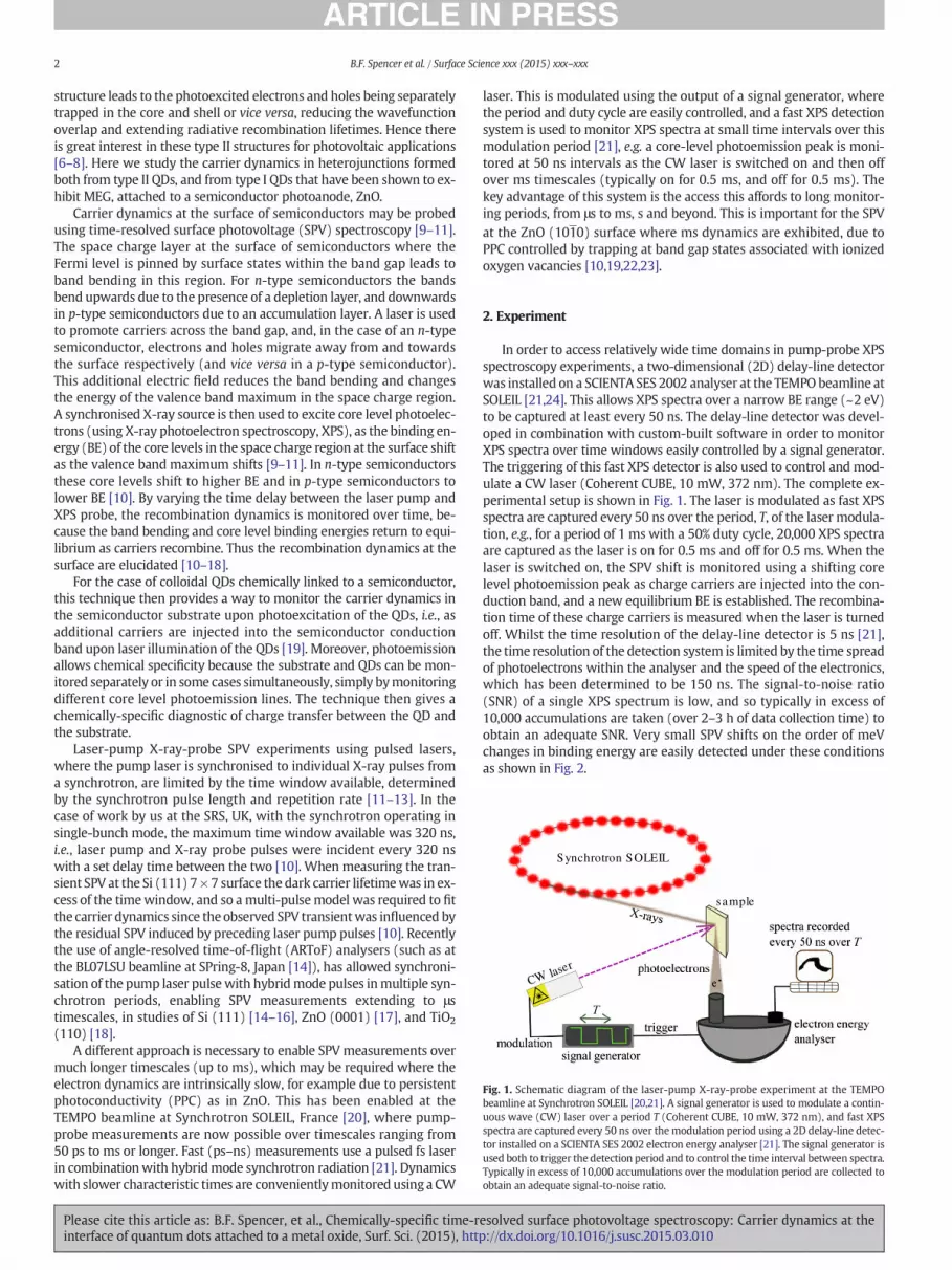
2. Experiment
In order to access relatively wide time domains in pump-probe XPS
spectroscopy experiments, a two-dimensional (2D) delay-line detector
was installed on a SCIENTA SES 2002 analyser at the TEMPO beamline at
SOLEIL [21,24]. This allows XPS spectra over a narrow BE range (~2 eV)
to be captured at least every 50 ns. The delay-line detector was devel-
oped in combination with custom-built software in order to monitor
XPS spectra over time windows easily controlled by a signal generator.
The triggering of this fast XPS detector is also used to control and mod-
ulate a CW laser (Coherent CUBE, 10 mW, 372 nm). The complete ex-
perimental setup is shown in Fig. 1. The laser is modulated as fast XPS
spectra are captured every 50 ns over the period, T, of the laser modula-
tion, e.g., for a period of 1 ms with a 50% duty cycle, 20,000 XPS spectra
are captured as the laser is on for 0.5 ms and off for 0.5 ms. When the
laser is switched on, the SPV shift is monitored using a shifting core
level photoemission peak as charge carriers are injected into the con-
duction band, and a new equilibrium BE is established. The recombina-
tion time of these charge carriers is measured when the laser is turned
off. Whilst the time resolution of the delay-line detector is 5 ns [21],
the time resolution of the detection system is limited by the time spread
of photoelectrons within the analyser and the speed of the electronics,
which has been determined to be 150 ns. The signal-to-noise ratio
(SNR) of a single XPS spectrum is low, and so typically in excess of
10,000 accumulations are taken (over 2–3 h of data collection time) to
obtain an adequate SNR. Very small SPV shifts on the order of meV
changes in binding energy are easily detected under these conditions
as shown in Fig. 2.
Synchrotron SOLEIL
CW laser
sample
Fig. 1. Schematic diagram of the laser-pump X-ray-probe experiment at the TEMPO
beamline at Synchrotron SOLEIL [20,21]. A signal generator is used to modulate a contin-
uous wave (CW) laser over a period T (Coherent CUBE, 10 mW, 372 nm), and fast XPS
spectra are captured every 50 ns over the modulation period using a 2D delay-line detec-
tor installed on a SCIENTA SES 2002 electron energy analyser [21]. The signal generator is
used both to trigger the detection period and to control the time interval between spectra.
Typically in excess of 10,000 accumulations over the modulation period are collected to
obtain an adequate signal-to-noise ratio.
2 B.F. Spencer et al. / Surface Science xxx (2015) xxx–xxx
Please cite this article as: B.F. Spencer, et al., Chemically-specific time-resolved surface photovoltage spectroscopy: Carrier dynamics at the
interface of quantum dots attached to a metal oxide, Surf. Sci. (2015), http://dx.doi.org/10.1016/j.susc.2015.03.010

X-ray photon energies of 200 eV (for the Zn 3d core level) and
280 eV (for the Pb 4f core level) were chosen to maximise surface sen-
sitivity (for examination of the space charge layer) without undue loss
of flux. High-quality XPS spectra of the monitored photoemission
peaks were taken in static mode in order to extract peak fitting param-
eters (e.g., spin-orbit splitting, full width at half maximum (FWHM)).
These parameters were then applied to each fast XPS spectrum across
the laser modulation time window, and the BE positions of the pho-
toemission peaks obtained from the fitting were used to measure
the SPV shift upon, during and after photoexcitation. Static XPS
with and without laser illumination was also used to ensure that
no long-term sample charging was occurring, i.e., that the photo-
emission peaks returned to the original BE positions after the
laser was turned off.
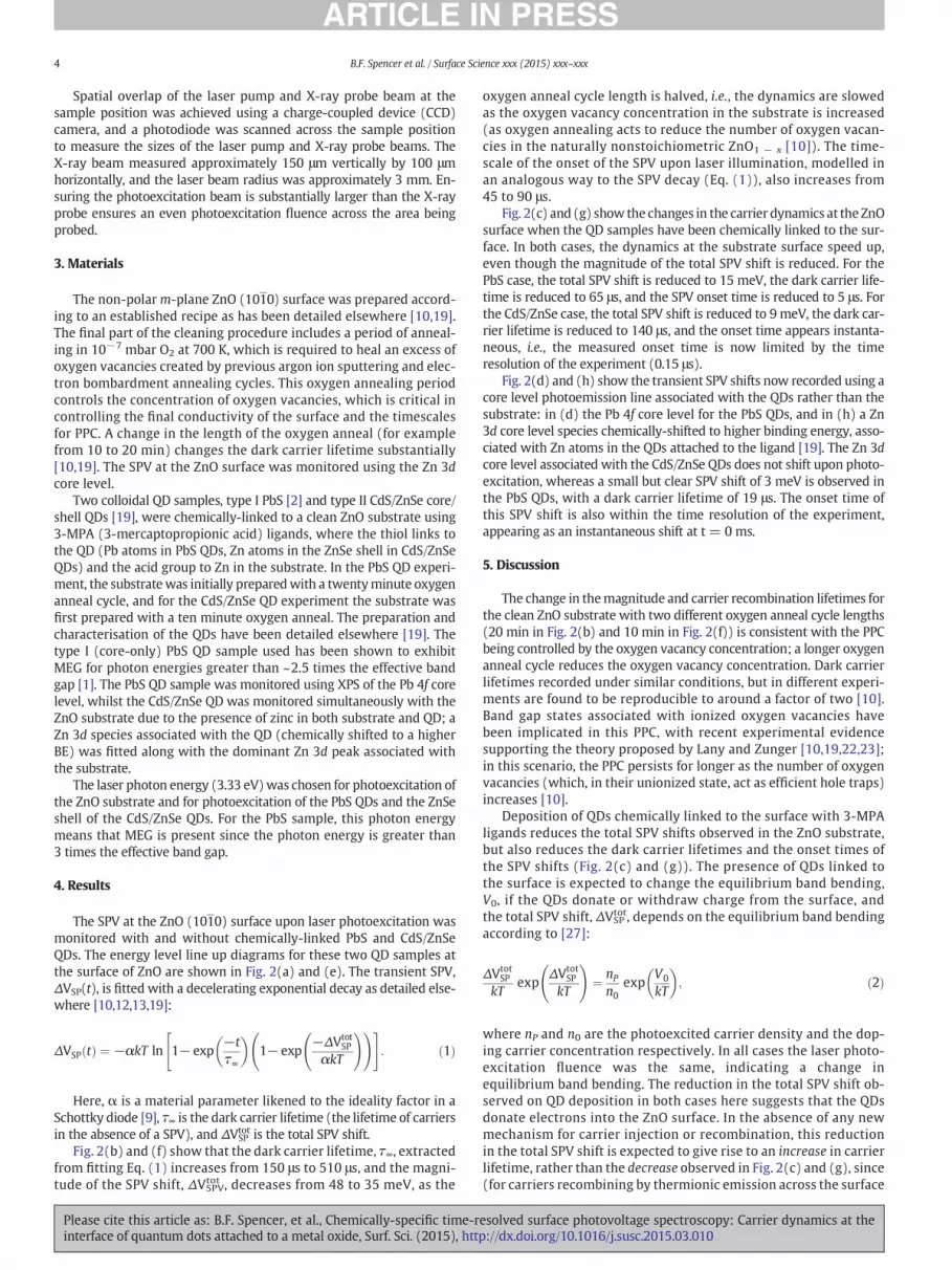
Fig. 2. Transient surface photovoltage (SPV) of quantum dots (QDs) chemically linked to ZnO (1010) with 3-MPA (3-mercaptopropionic acid) ligands. The parameters extracted from
fitting are shown. An X-ray photon energy of 200 eV was used for the Zn 3d core level (b, c, f–h), and a photon energy of 280 eV was used for the Pb 4f core level (d). Arrows denote
laser modulation; the laser is turned on at t = 0 ms, and switched off at T/2, where the period T is 1 ms for the PbS QD case, and 4 ms for the CdS/ZnSe QD case. In all cases laser photo-
excitation at 372 nm (hv=3.33 eV)was used, meaning that both QDs and the ZnO substrate were photoexcited (and for the CdS/ZnSe QDs, the shell of the QDswas photoexcited directly,
enabling charge injection from the shell into the substrate as illustrated in (e)). Energy level diagrams (a) and (e) are adapted from [25,26]. Dashed lines indicate a reduction in the band
bending upon photoexcitation as carriers are promoted (or injected) into the CB. Type I (core only) PbS QDs: (a) energy level line up diagram of the highest occupied molecular orbital
(HOMO) and lowest unoccupied molecular orbital (LUMO) of the QDs with the ZnO conduction band (CB), valence band (VB) and Fermi level (EF); (b) transient SPV for the clean ZnO
substrate, prepared with a twenty minute oxygen anneal cycle; (c) transient SPV of the Zn 3d core level after QD attachment; (d) transient SPV of the Pb 4f core level of the PbS QDs.
Type II (core/shell) CdS/ZnSe QDs: (e) energy level line up diagram, showing that for photon energies less than 2.7 eV, electrons and holes are trapped in the core and shell of the QD
respectively; (f) transient SPV for the clean substrate. A shorter oxygen anneal cycle of 10 min was used, giving an extended dark carrier lifetime compared with (b); (g) transient SPV
of the Zn 3d core level component associated with the substrate (at a binding energy of ~10.18 eV) after QDs have been attached to the surface; (h) transient SPV of the Zn 3d core
level component associated with the ligand/QD (chemically shifted to a higher binding energy of ~10.65 eV), where no SPV shift is observed.
3B.F. Spencer et al. / Surface Science xxx (2015) xxx–xxx
Please cite this article as: B.F. Spencer, et al., Chemically-specific time-resolved surface photovoltage spectroscopy: Carrier dynamics at the
interface of quantum dots attached to a metal oxide, Surf. Sci. (2015), http://dx.doi.org/10.1016/j.susc.2015.03.010
Spatial overlap of the laser pump and X-ray probe beam at the
sample position was achieved using a charge-coupled device (CCD)
camera, and a photodiode was scanned across the sample position
to measure the sizes of the laser pump and X-ray probe beams. The
X-ray beam measured approximately 150 μm vertically by 100 μm
horizontally, and the laser beam radius was approximately 3 mm. En-
suring the photoexcitation beam is substantially larger than the X-ray
probe ensures an even photoexcitation fluence across the area being
probed.
3. Materials
The non-polarm-plane ZnO (1010) surface was prepared accord-
ing to an established recipe as has been detailed elsewhere [10,19].
The final part of the cleaning procedure includes a period of anneal-
ing in 10−7 mbar O2 at 700 K, which is required to heal an excess of
oxygen vacancies created by previous argon ion sputtering and elec-
tron bombardment annealing cycles. This oxygen annealing period
controls the concentration of oxygen vacancies, which is critical in
controlling the final conductivity of the surface and the timescales
for PPC. A change in the length of the oxygen anneal (for example
from 10 to 20 min) changes the dark carrier lifetime substantially
[10,19]. The SPV at the ZnO surface was monitored using the Zn 3d
core level.
Two colloidal QD samples, type I PbS [2] and type II CdS/ZnSe core/
shell QDs [19], were chemically-linked to a clean ZnO substrate using
3-MPA (3-mercaptopropionic acid) ligands, where the thiol links to
the QD (Pb atoms in PbS QDs, Zn atoms in the ZnSe shell in CdS/ZnSe
QDs) and the acid group to Zn in the substrate. In the PbS QD experi-
ment, the substrate was initially preparedwith a twentyminute oxygen
anneal cycle, and for the CdS/ZnSe QD experiment the substrate was
first prepared with a ten minute oxygen anneal. The preparation and
characterisation of the QDs have been detailed elsewhere [19]. The
type I (core-only) PbS QD sample used has been shown to exhibit
MEG for photon energies greater than ~2.5 times the effective band
gap [1]. The PbS QD sample was monitored using XPS of the Pb 4f core
level, whilst the CdS/ZnSe QD was monitored simultaneously with the
ZnO substrate due to the presence of zinc in both substrate and QD; a
Zn 3d species associated with the QD (chemically shifted to a higher
BE) was fitted along with the dominant Zn 3d peak associated with
the substrate.
The laser photon energy (3.33 eV)was chosen for photoexcitation of
the ZnO substrate and for photoexcitation of the PbS QDs and the ZnSe
shell of the CdS/ZnSe QDs. For the PbS sample, this photon energy
means that MEG is present since the photon energy is greater than
3 times the effective band gap.
4. Results
The SPV at the ZnO (1010) surface upon laser photoexcitation was
monitored with and without chemically-linked PbS and CdS/ZnSe
QDs. The energy level line up diagrams for these two QD samples at
the surface of ZnO are shown in Fig. 2(a) and (e). The transient SPV,
ΔVSP(t), is fitted with a decelerating exponential decay as detailed else-
where [10,12,13,19]:
ΔVSP tð Þ ¼ −αkT ln 1− exp−t
τ∞
� �
1− exp−ΔVtot
SP
αkT
! !" #
: ð1Þ
Here, α is a material parameter likened to the ideality factor in a
Schottky diode [9], τ∞ is the dark carrier lifetime (the lifetime of carriers
in the absence of a SPV), and ΔVSPtot is the total SPV shift.
Fig. 2(b) and (f) show that the dark carrier lifetime, τ∞, extracted
from fitting Eq. (1) increases from 150 μs to 510 μs, and the magni-
tude of the SPV shift, ΔVSPVtot , decreases from 48 to 35 meV, as the
oxygen anneal cycle length is halved, i.e., the dynamics are slowed
as the oxygen vacancy concentration in the substrate is increased
(as oxygen annealing acts to reduce the number of oxygen vacan-
cies in the naturally nonstoichiometric ZnO1 − x [10]). The time-
scale of the onset of the SPV upon laser illumination, modelled in
an analogous way to the SPV decay (Eq. (1)), also increases from
45 to 90 μs.
Fig. 2(c) and (g) show the changes in the carrier dynamics at the ZnO
surface when the QD samples have been chemically linked to the sur-
face. In both cases, the dynamics at the substrate surface speed up,
even though the magnitude of the total SPV shift is reduced. For the
PbS case, the total SPV shift is reduced to 15 meV, the dark carrier life-
time is reduced to 65 μs, and the SPV onset time is reduced to 5 μs. For
the CdS/ZnSe case, the total SPV shift is reduced to 9 meV, the dark car-
rier lifetime is reduced to 140 μs, and the onset time appears instanta-
neous, i.e., the measured onset time is now limited by the time
resolution of the experiment (0.15 μs).
Fig. 2(d) and (h) show the transient SPV shifts now recorded using a
core level photoemission line associated with the QDs rather than the
substrate: in (d) the Pb 4f core level for the PbS QDs, and in (h) a Zn
3d core level species chemically-shifted to higher binding energy, asso-
ciated with Zn atoms in the QDs attached to the ligand [19]. The Zn 3d
core level associated with the CdS/ZnSe QDs does not shift upon photo-
excitation, whereas a small but clear SPV shift of 3 meV is observed in
the PbS QDs, with a dark carrier lifetime of 19 μs. The onset time of
this SPV shift is also within the time resolution of the experiment,
appearing as an instantaneous shift at t = 0 ms.
5. Discussion
The change in themagnitude and carrier recombination lifetimes for
the clean ZnO substrate with two different oxygen anneal cycle lengths
(20 min in Fig. 2(b) and 10 min in Fig. 2(f)) is consistent with the PPC
being controlled by the oxygen vacancy concentration; a longer oxygen
anneal cycle reduces the oxygen vacancy concentration. Dark carrier
lifetimes recorded under similar conditions, but in different experi-
ments are found to be reproducible to around a factor of two [10].
Band gap states associated with ionized oxygen vacancies have
been implicated in this PPC, with recent experimental evidence
supporting the theory proposed by Lany and Zunger [10,19,22,23];
in this scenario, the PPC persists for longer as the number of oxygen
vacancies (which, in their unionized state, act as efficient hole traps)
increases [10].
Deposition of QDs chemically linked to the surface with 3-MPA
ligands reduces the total SPV shifts observed in the ZnO substrate,
but also reduces the dark carrier lifetimes and the onset times of
the SPV shifts (Fig. 2(c) and (g)). The presence of QDs linked to
the surface is expected to change the equilibrium band bending,
V0, if the QDs donate or withdraw charge from the surface, and
the total SPV shift, ΔVSPtot, depends on the equilibrium band bending
according to [27]:
ΔVtotSP
kTexp
ΔVtotSP
kT
!
¼nP
n0
expV0
kT
� �
; ð2Þ
where nP and n0 are the photoexcited carrier density and the dop-
ing carrier concentration respectively. In all cases the laser photo-
excitation fluence was the same, indicating a change in
equilibrium band bending. The reduction in the total SPV shift ob-
served on QD deposition in both cases here suggests that the QDs
donate electrons into the ZnO surface. In the absence of any new
mechanism for carrier injection or recombination, this reduction
in the total SPV shift is expected to give rise to an increase in carrier
lifetime, rather than the decrease observed in Fig. 2(c) and (g), since
(for carriers recombining by thermionic emission across the surface
4 B.F. Spencer et al. / Surface Science xxx (2015) xxx–xxx
Please cite this article as: B.F. Spencer, et al., Chemically-specific time-resolved surface photovoltage spectroscopy: Carrier dynamics at the
interface of quantum dots attached to a metal oxide, Surf. Sci. (2015), http://dx.doi.org/10.1016/j.susc.2015.03.010

depletion layer) the photoexcited carrier lifetime, τ, varies with
SPV shift, ΔVSP, according to [12]:
τ ¼ τ∞exp
−ΔVSP
αkT
� �
: ð3Þ
In this model, the dynamic change in the photoexcited carrier life-
time with SPV shift requires the decelerating exponential model given
in Eq. (1), where the lifetime increases as SPV shift is reduced. The fact
that the onset times for the SPV shifts are faster, rather than slower,
when the equilibrium band bending is reduced by attachment of QDs
suggests direct charge injection from the QDs to the ZnO conduction
band (CB) as anticipated according to the energy level line up diagrams
shown in Fig. 2(a) and (e). In the case of CdS/ZnSe QDs the onset ap-
pears instantaneous suggesting that the onset time is now within the
experimental time resolution, and indeed there is evidence that charge
transfer to anoxide substrate in similarQD systemsmayoccur onps and
fs timescales [28–30]. The reduced dark carrier lifetime may indicate
that the QD attachment is providing additional pathways for electron-
hole pair recombination that avoids the long-lived PPC state (e.g. via
new interface states). These changes in the recombination dynamics
are important when considering light harvesting QDs with ZnO as
photoanode as model systems for next-generation photovoltaics.
Transient SPV shifts upon laser illumination are caused by a change
in the band bending at the interface, which is caused by the presence
a depletion layer in the case of n-type ZnO. In the case of QDs, which
are nm-scale semiconductor crystals, intrinsic band bending is not nec-
essarily expected as the size of the QDs is much less than typical deple-
tion layer widths. Therefore, no intrinsic SPV shift is expected when
monitoring core level photoemission lines associated with the QDs.
However, the electric field associated with the surface depletion layer
in the ZnO substrate permeates the medium both above and below
the surface, to an extent dependent on factors such as the dielectric con-
stant and carrier concentration of the medium. We might expect to ob-
serve a SPV shift in the core levels of atoms associated with the QD if
those atoms lie within this field; the size would depend on the position
of the atom in the field, but is clearly expected to be smaller than the
shift we observe at the ZnO surface. It is interesting that a small SPV
shift of 3 meV (in the same direction as the ZnO substrate, i.e. to higher
BE) is observed when monitoring the Pb 4f photoemission line in the
PbS QD sample (Fig. 2(d)). This indicates that the Pb atoms in the QD
lie in the tail of the field associated with the surface depletion layer in
the ZnO substrate. Analysis of this transient gives an onset time within
the time resolution of the experiment, and the decay of the SPV shift
yields a fitted dark carrier lifetime of 19 μs, faster than that observed
in the substrate (65 μs). Both are consistent with fast intrinsic charge
dynamics in PbS, not subject to the PPC observed in ZnO. Interestingly,
a recent study by Neppl et al. [31] observed SPV shifts in N3 dye mole-
cules attached to nanoporous ZnO. It is then interesting that we observe
no SPV shift in the shell of the CdS/ZnSe type II QDs, monitored using a
chemically-shifted component of the Zn 3d core level (Fig. 2(h)); this
may be associated with the potential barrier provided by the ZnSe
shell, which may act to truncate the field associated with the ZnO sub-
strate. We note that in this case photoexcited electrons are more stable
in either the ZnO substrate or the CdS core of the QD than in the ZnSe
shell (Fig. 2(e)). The difference in behaviour between the two QD sys-
tems points to the need for more experiments in this field, particularly
using photon frequencies resonant with the lowest absorption energies
of the QDs. However, the measurements presented here show the
power of soft X-ray pump-probe experiments in allowing us to probe
electron dynamics at these complex interfaces with atomic specificity.
6. Conclusions
A fast XPS technique, where a 2D delay-line detector enables fast
(ns) monitoring of a narrow XPS spectrum at the TEMPO beamline at
Synchrotron SOLEIL [20,21], in combination with a CW laser, has been
developed to enable the study of systems with intrinsically slow
electron dynamics. This has allowed the charge carrier dynamics at
the interface of colloidal QDs chemically-linked to ZnO (1010) to
be monitored on ms timescales, required due to the PPC at the ZnO
(1010) surface [10,19,22,23]. The shift in core level photoemission
lines upon photoexcitation, the SPV shift, has been measured over
time windows of several ms. The chemical specificity of XPS allows for
monitoring of not only the substrate but also of the QDs bound to the
surface. In the case of PbS QDs, the observations of a small SPV shift of
the Pb 4f core level indicates that the QDs lie within the field associated
with the depletion layer at the ZnO surface. However, no transient SPV
is observed when monitoring a Zn 3d core level component associated
with the shell of type II CdS/ZnSe QDs, which may be due to the effect
of the potential barrier at the QD surface provided by this shell. Whilst
further study is required to better understand the charge transfer and
recombination dynamics at these interfaces, our work highlights the
power of laser-pump X-ray probe techniques in measuring photoexcit-
ed carrier dynamics at these interfaces with chemical specificity.
Acknowledgements
The research leading to these results received funding from the
European Community's Seventh Framework Programme (FP7/2007–
2013) under grant agreement no. 226716, allowing access to Synchro-
tron SOLEIL. Work was also supported by EPSRC (UK) under grant no.
EP/K008544/1 and the Cockcroft Institute via its STFC core grant ST/
G008248/1.
References
[1] S.J.O. Hardman, D.M. Graham, S.K. Stubbs, B.F. Spencer, E.A. Seddon, H.T. Fung, S.
Gardonio, F. Sirotti, M.G. Silly, J. Akhtar, P. O'Brien, D.J. Binks, W.R. Flavell, Phys.Chem. Chem. Phys. 13 (2011) 20275.
[2] J. Akhtar, M.A. Malik, P. O'Brien, K.G.U. Wijayantha, R. Dharmadasa, S.J.O. Hardman,
D.M. Graham, B.F. Spencer, S.K. Stubbs, W.R. Flavell, D.J. Binks, F. Sirotti, M. El Kazzi,M. Silly, J. Mater. Chem. 20 (2010) 2336.
[3] P.D.C. King, T.D. Veal, J. Phys. Condens. Matter 23 (2011) 334214.[4] C. Klingshirn, J. Fallert, H. Zhou, J. Sartor, C. Thiele, F. Maier-Flaig, D. Schneider, H.
Kalt, Phys. Status Solidi B Basic Solid State Phys. 247 (2010) 1424.[5] B. Carlson, K. Leschkies, E.S. Aydil, X.Y. Zhu, J. Phys. Chem. C 112 (2008) 8419.
[6] V.I. Klimov, S.A. Ivanov, J. Nanda, M. Achermann, I. Bezel, J.A. McGuire, A. Piryatinski,
Nature 447 (2007) 441.[7] A. Piryatinski, S.A. Ivanov, S. Tretiak, V.I. Klimov, Nano Lett. 7 (2007) 108.
[8] S. Kim, B. Fisher, H.J. Eisler, M. Bawendi, J. Am. Chem. Soc. 125 (2003) 11466.[9] L. Kronik, Y. Shapira, Surf. Sci. Rep. 37 (1999) 1.
[10] B.F. Spencer, D.M. Graham, S.J.O. Hardman, E.A. Seddon, M.J. Cliffe, K.L. Syres, A.G.
Thomas, S.K. Stubbs, F. Sirotti, M.G. Silly, P.F. Kirkham, A.R. Kumarasinghe, G.J.Hirst, A.J. Moss, S.F. Hill, D.A. Shaw, S. Chattopadhyay, W.R. Flavell, Phys. Rev. B 88
(2013) 195301.[11] J.P. Long, H.R. Sadeghi, J.C. Rife, M.N. Kabler, Phys. Rev. Lett. 64 (1990) 1158.
[12] W. Widdra, D. Bröcker, T. Gießel, I.V. Hertel, W. Kruger, A. Liero, F. Noack, V. Petrov,D. Pop, P.M. Schmidt, R. Weber, I. Will, B. Winter, Surf. Sci. 543 (2003) 87.
[13] D. Bröcker, T. Gießel, W. Widdra, Chem. Phys. 299 (2004) 247.
[14] M. Ogawa, S. Yamamoto, Y. Kousa, F. Nakamura, R. Yukawa, A. Fukushima, A.Harasawa, H. Kondoh, Y. Tanaka, A. Kakizaki, I. Matsuda, Rev. Sci. Instrum. 83 (2012).
[15] M. Ogawa, S. Yamamoto, R. Yukawa, R. Hobara, C.H. Lin, R.Y. Liu, S.J. Tang, I. Matsuda,Phys. Rev. B 87 (2013).
[16] M. Ogawa, R.Y. Liu, C.H. Lin, S. Yamamoto, R. Yukawa, R. Hobara, S.J. Tang, I. Matsuda,
Surf. Sci. 624 (2014) 70.[17] R. Yukawa, S. Yamamoto, K. Ozawa, M. Emori, M. Ogawa, S. Yamamoto, K. Fujikawa,
R. Hobara, S. Kitagawa, H. Daimon, H. Sakama, I. Matsuda, Appl. Phys. Lett. 105(2014).
[18] K. Ozawa, M. Emori, S. Yamamoto, R. Yukawa, S. Yamamoto, R. Hobara, K. Fujikawa,
H. Sakama, I. Matsuda, J. Phys. Chem. Lett. 5 (2014) 1953.[19] B.F. Spencer, M.J. Cliffe, D.M. Graham, S.J.O. Hardman, E.A. Seddon, K.L. Syres, A.G.
Thomas, F. Sirotti, M.G. Silly, J. Akhtar, P. O'Brien, S.M. Fairclough, J.M. Smith, S.Chattopadhyay, W.R. Flavell, Faraday Discuss. 171 (2014) 275.
[20] F. Polack, M. Silly, C. Chauvet, B. Lagarde, N. Bergeard, M. Izquierdo, O. Chubar, D.Krizmancic, M. Ribbens, J.P. Duval, C. Basset, S. Kubsky, F. Sirotti, TEMPO: a new
insertion device beam line at SOLEIL for time resolved photoelectron spectroscopy
experiments on solids and interfaces, AIP Proceedings: The 10th InternationalConference on Synchrotron Radiation Instrumentation, 1234, 2010, p. 185.
[21] N. Bergeard, M.G. Silly, D. Krizmancic, C. Chauvet, M. Guzzo, J.P. Ricaud, M. Izquierdo,L. Stebel, P. Pittana, R. Sergo, G. Cautero, G. Dufour, F. Rochet, F. Sirotti, J. Synchrotron
Radiat. 18 (2011) 245.
5B.F. Spencer et al. / Surface Science xxx (2015) xxx–xxx
Please cite this article as: B.F. Spencer, et al., Chemically-specific time-resolved surface photovoltage spectroscopy: Carrier dynamics at the
interface of quantum dots attached to a metal oxide, Surf. Sci. (2015), http://dx.doi.org/10.1016/j.susc.2015.03.010

[22] S. Lany, A. Zunger, Phys. Rev. B 72 (2005) 035215.
[23] S. Lany, A. Zunger, Phys. Rev. Lett. 98 (2007) 045501.[24] G. Cautero, R. Sergo, L. Stebel, P. Lacovig, P. Pittana, M. Predonzani, S. Carrato, Nucl.
Inst. Methods Phys. Res. A 595 (2008) 447.[25] S.H. Wei, A. Zunger, Appl. Phys. Lett. 72 (1998) 2011.
[26] B.R. Hyun, Y.W. Zhong, A.C. Bartnik, L.F. Sun, H.D. Abruna, F.W. Wise, J.D. Goodreau,J.R. Matthews, T.M. Leslie, N.F. Borrelli, ACS Nano 2 (2008) 2206.
[27] J.P. Long, V.M. Bermudez, Phys. Rev. B 66 (2002) 121308.
[28] W.A. Tisdale, K.J. Williams, B.A. Timp, D.J. Norris, E.S. Aydil, X.Y. Zhu, Science 328
(2010) 1543.[29] Y. Yang, W. Rodriguez-Cordoba, X. Xiang, T.Q. Lian, Nano Lett. 12 (2012) 303.
[30] K. Zidek, K.B. Zheng, C.S. Ponseca, M.E. Messing, L.R. Wallenberg, P. Chabera, M.Abdellah, V. Sundstrom, T. Pullerits, J. Am. Chem. Soc. 134 (2012) 12110.
[31] S. Neppl, A. Shavorskiy, I. Zegkinoglou, M. Fraund, D.S. Slaughter, T. Troy, M.P.Ziemkiewicz, M. Ahmed, S. Gul, B. Rude, J.Z. Zhang, A.S. Tremsin, P.-A. Glans, Y.-S. Liu,
C.H. Wu, J. Guo, M. Salmeron, H. Bluhm, O. Gessner, Faraday Discuss. 171 (2014) 219.
6 B.F. Spencer et al. / Surface Science xxx (2015) xxx–xxx
Please cite this article as: B.F. Spencer, et al., Chemically-specific time-resolved surface photovoltage spectroscopy: Carrier dynamics at the
interface of quantum dots attached to a metal oxide, Surf. Sci. (2015), http://dx.doi.org/10.1016/j.susc.2015.03.010


