Embed Size (px)
Citation preview
This article appeared in a journal published by Elsevier. The attachedcopy is furnished to the author for internal non-commercial researchand education use, including for instruction at the authors institution
and sharing with colleagues.
Other uses, including reproduction and distribution, or selling orlicensing copies, or posting to personal, institutional or third party
websites are prohibited.
In most cases authors are permitted to post their version of thearticle (e.g. in Word or Tex form) to their personal website orinstitutional repository. Authors requiring further information
regarding Elsevier’s archiving and manuscript policies areencouraged to visit:
http://www.elsevier.com/copyright

Author's personal copy
Original article
Substituted quinazolines, part 3. Synthesis, in vitro antitumor activity andmolecular modeling study of certain 2-thieno-4(3H)-quinazolinone analogsq
Abdulrahman M. Al-Obaid a, Sami G. Abdel-Hamide a,*, Hassan A. El-Kashef b, Alaa A.-M. Abdel-Aziz a,Adel S. El-Azab a, Hamad A. Al-Khamees a, Hussein I. El-Subbagh a,*
a Department of Pharmaceutical Chemistry, College of Pharmacy, King Saud University, Riyadh 11451, Saudi Arabiab Department of Pharmacology, College of Pharmacy, Mansoura University, Mansoura 35516, Egypt
a r t i c l e i n f o
Article history:Received 5 May 2008Received in revised form 18 July 2008Accepted 1 September 2008Available online 23 September 2008
Keywords:Synthesis2-Thieno-4(3H)-quinazolinonesAntitumor agentsMolecular modeling
a b s t r a c t
The synthesis of some new 2-thieno-4(3H)-quinazolinone derivatives and their biological evaluation asantitumor agents using the National Cancer Institute (NCI) disease oriented antitumor screen protocolare investigated. Compounds 2-(2-thienylcarbonylamino)-5-iodo-N-(4-hydroxyphenyl)-benzamide(16), 2-(2-thieno)-6-iodo-3-phenylamino-3,4-dihydro-quina-zolin-4-one (26), and 2-(2-thieno)-4-[4-sulfonamidobenzylamino]-6-iodo-quinazoline (42), with GI50 values of 12.7, 10.3, 16.9 mM, respec-tively, proved to be the most active members in this study, as compared to the known drug 5-FU.Conformational analysis of the most active molecules using molecular modeling and QSAR techniquesenabled the understanding of the pharmacophoric requirements for 2-thieno-quinzolinone derivativesas antitumor agents. These three quinazolinone analogs (16, 26, 42) could be considered as usefultemplates for future development to obtain more potent antitumor agents.
� 2008 Elsevier Masson SAS. All rights reserved.
1. Introduction
Interests in quinazolines as anticancer agents have furtherincreased since the discovery of raltitrexed (1) and thymitaq (2),(Chart 1). Both compounds proved to be thymidylate enzymeinhibitors [1–3]. 6-Arylamino-7-chloro-quinazoline-5,8-dionederivatives (3) showed potency against cultured human cancer cellline A549 (lung cancer), and stomach cancer SNU-638 [4–6].Indolo-quinazolines 4, 5 (Chart 1) proved active against a panel ofhuman cancer cell lines [7]. Also, 4-anilinoquinazolines representas a new class of antitumor drugs [2,3]. They inhibit the epidermalgrowth factor receptor (EGFR) tyrosine kinase overexpressionthrough the inhibition of EGFR autophosphorylation and EGF-stimulated signal transduction [8,9]. Furthermore, quinazolinesexert their antitumor activity through inhibition of the DNA repairenzyme system [7,10–14]. The present study aimed to synthesizeand evaluate the biological activity of some new quinazolinederivatives as potential antineoplastic agents, as a continuation ofour previous efforts [15,16]. A new series of quinazoline compoundsare designed, in such a way to accommodate a thiophene ringat position 2-, sulphonamides, isothiocyanates, Schiff’s bases or
acylamides at position 3-, ether, thioether, amine bridges or sul-phonamides at position 4- of the quinazoline ring. Thiophene [17],sulphonamide [18], thioether [19], isothiocyanate [18,20], Schiff’sbase and amide [20] functions are known to contribute to theenhancement of the antitumor activity.
2. Results and discussion
2.1. Chemistry
The reaction of 5-iodo-anthranilic acid (6) and 2-thiophenecarbonylchloride (7) afforded the amide analog 8 which was then refluxed inacetic anhydride to obtain the key intermediate 2-(2-thieno)-6-iodo-4H-3,1-benzoxazin-4-one (9) (Scheme 1, Table 1). The latter compoundwas reacted with different aliphatic and aromatic amines in an attemptto obtain 3-substituted-quinazolin-4-ones in different reaction condi-tions, in all cases; the reaction afforded the diamides 10–12 and 14–18instead. Attempts to cyclize the diamides 14–18 to the corresponding4-(3H)-quinazolin-4-one using variety of reaction conditions, includingfusion, were not successful. Compound 13 was obtained in very lowyield by refluxing the corresponding diamide derivative 10 in thionylchloride. The reaction of sulfa derivatives and homosulfanilamide with3,1-benzoxozin-4-one 9 by fusion at high temperature afforded thecorresponding quinazoline-4-one derivatives 19–22 (Scheme 1, Table1). Reaction of 9 with the nucleophilic amino group of homo-sulfanilamide gave 23. The benzoxazine derivative 9 was also reactedwith hydroxylamine hydrochloride in dry pyridine to afford compound24. Condensation of 9 with various hydrazine derivatives afforded
q For parts 1 and 2, see Refs. [15,16].* Corresponding authors. Department of Pharmaceutical Chemistry, College of
Pharmacy, P.O. Box 2457, King Saud University, Riyadh 11451, Saudi Arabia.Tel.: þ966 1 467 7394; fax: þ966 1 467 6383.
E-mail address: [email protected].
Contents lists available at ScienceDirect
European Journal of Medicinal Chemistry
journal homepage: ht tp: / /www.elsevier .com/locate /e jmech
0223-5234/$ – see front matter � 2008 Elsevier Masson SAS. All rights reserved.doi:10.1016/j.ejmech.2008.09.015
European Journal of Medicinal Chemistry 44 (2009) 2379–2391

Author's personal copy
compounds 25–29. The intermediate 9 was also refluxed in formamideto afford 2-(2-thieno)-6-iodo-3,4-dihydro-quinazoline-4-one (30)which upon refluxing in dry xylene containing phosphorous penta-sulfide afforded the corresponding 4-thione analog 31. Alkylation ofcompound 31 with methyl and ethyl iodide afforded the 4-alkylthioanalogs 32 and 33, respectively (Scheme 2, Table 1). The 3-aminoderivative 25 was reacted with phenylisocyanate or isothiocyanate indry dioxane to afford the corresponding urea and thiourea derivatives34, 35. Condensation of 25 with the appropriate aromatic aldehydes inglacial acetic acid afforded the arylidene derivatives 36 and 37.Ethyl acetoacetate was refluxed with 25 in isopropanol to give the3-oxo-butyrylamino derivative 38 (Scheme 2, Table 1). Alkylation ofcompound 30 with ethylbromoacetate in boiling acetone and anhy-drous potassium carbonate afforded the o-alkyl derivative 39 whichwas reacted with hydrazine hydrate in absolute ethanol to form thehydrazide derivative 40. The reaction of 30 with a mixture of phos-phorous oxychloride and phosphorous pentachloride yielded thechloroquinazoline derivative 41 which was refluxed with homo-sulfanilamide or the appropriate sulfa drug in dry pyridine affordedcompounds 42–45, respectively (Scheme 3, Table 1).
2.2. Biological activity
The synthesized compounds (8–45, Schemes 1–3), weresubjected to the NCI’s in vitro, one dose primary anticancer assay,using a 3-cell line panel consisting of MCF-7 (breast), NCI-H460(lung), and SF-268 (CNS) cancers. Compounds which reduce thegrowth of any one of the cell lines to 32% or less are passed on forevaluation in the full panel of 60 cell lines over a 5-log dose range[21–23]. Three response parameters, median growth inhibition(GI50), total growth inhibition (TGI), and median lethal concentra-tion (LC50) were calculated for each cell line [24], using the knowndrug 5-Fluorouracil (5-FU) as a positive control. The NCI antitumordrug discovery screen has been designed to distinguish betweenbroad-spectrum antitumor and tumor or subpanel-selectivecompounds [24]. In the present study, compounds 12, 16, 19, 20, 24,26, 29, 33, 35, 36, 38, 40, 42 and 44 passed primary anticancer assayat an arbitrary concentration of 100 mM. Consequently, these active
compounds were carried over and tested against a panel of 60different tumor cell lines. The tested quinazoline analogs showeda distinctive potential pattern of selectivity as well as a broad-spectrum antitumor activity. With regard to sensitivity againstindividual cell lines (Table 2), compound 16 showed GI50 effec-tiveness against CNS SF-539 and melanoma UACC-257 cancer celllines at concentration of <0.01 mM. Compound 26 showed activityagainst leukemia cell lines HL-60 (TB), K-562 and melanomaSK-MEL-2 with GI50 concentrations of 0.4, 0.3, and 0.3 mM,respectively. Compounds 16, 19, 29, 40, and 44 showed a remark-able activity against melanoma UACC-257 with GI50 concentrationsof <0.01, 0.05, <0.01, <0.01, and 0.04 mM, respectively. Compound35 showed GI50 effectiveness against leukemia K-562 at concen-tration of 0.02 mM. With regard to broad-spectrum antitumoractivity, most of the tested compounds showed GI50, TGI (MG-MID)values of <100 mM, against leukemia, non-small cell lung, colon,CNS, melanoma, ovarian, renal, prostate, and breast cancersubpanel cell lines (Tables 3 and 4). Compounds 16, and 42 showedMG-MID values <100 mM at the three levels of activity, GI50, TGI,and LC50 (Table 3), while compounds 19, 20, 24, 26, 29, 35, 38, 40,42, and 44 showed activity at GI50, and TGI levels. Compound 33was only active at the GI50 level. Compounds 12 and 36 proved to beinactive. Compounds 16, 26, and 42 are the most active members inthis investigation, with GI50 values of 12.7, 10.3, and 16.9 mM,respectively. Compounds 16 and 26 are almost twofolds moreactive than the positive control 5-FU. When a full panel GI50 mean-graph (MG-MID) divided by a particular subpanel GI50 mean-graph,certain ratio will be obtained which helps to predict the selectivityof this compound toward this cell lines subpanel. Ratios of 3–6 areconsidered moderately selective and those with ratios of 6 or moreare taken as selective [23]. Compound 26 proved to be selectivetoward prostate cancer cell lines with selectivity ratio of 6.4.
2.3. Structure–activity correlation
Structure–activity correlation of the synthesized compoundsrevealed that, in the diamide series 10–12 and 14–18, aromaticsubstitution on the benzamide amide function, favors the activity
HN
N
OCH3
H2N
HN
N
O
H2N
NCH3
N
N
HN R2
R3
O
OCl
R1
12
3
R1,R2,R3=H,OCH3,CH3,C2H5,Br
N
NR2
R1
OR3
R4O
N
NR2
R1
OR3
R4NOR
4 5
R1,R2,R3,R4=H,Br,NO2,OMe,alkylandcycloalkylderivatives.
S
N
S HN
O
COOHCOOH
Chart 1.
A.M. Al-Obaid et al. / European Journal of Medicinal Chemistry 44 (2009) 2379–23912380

Author's personal copy
rather than aliphatic substitution. This is obvious upon comparingcompound 16 [N-(4-hydroxy phenyl), GI50 value 12.7 mM], andcompound 10 [N-(2-hydroxy ethyl), value >100 mM]. In thesulfonamide series (19–23), compound 19 bearing unsubstitutedsulfonamide function, which favors the antitumor activity (GI50
value of 24.9 mM), the introduction of 2-thiazolyl moiety to thesulfonamide group, markedly decreased the activity (20,GI50¼ 41.2 mM). Introduction of a spacer methylene functionproduced the homosulfonamide 23 with total loss of activity.Moving the aromatic sulfonamide function from position 3- toposition 4- of the quinazoline ring, favors the anticancer activity, asshown in compound 44 (GI50¼ 23.9 mM) which is the positionalisomer of 20 (GI50¼ 41.2 mM), the same could be found incompound 42 (GI50¼16.9 mM), and its positional isomercompound 23. The introduction of the 3-amino function on thequinazoline ring produced the inactive compound 25, conversion ofthe 3-amino group into aniline function yielded compound 26(GI50¼10.3 mM) as the most active member of this study. Replacingthis 3-aniline function by benzamide, reduced the activity bythreefold (compound 29, GI50¼ 30.2 mM). Aliphatic acylation of the3-amino-quinazoline gave also the active compound 38(GI50¼ 31.1 mM). Further derivatization of the 3-amino group on 25gave the thioureido analog 35 (GI50¼ 24.9 mM), and the inactiveazomethine compound 36. Replacing the 3-amino group of 25 by
3-hydroxyl function (24, GI50¼ 41.1 mM), markedly increased theactivity. Conversion of the inactive ester 39 into its hydrazide 40(GI50¼ 31.1 mM), markedly increased the activity. The aforemen-tioned information is in accordance with the pharmacophoricfeatures essential for antitumor activity, deduced from the overalloutcome of the molecular modeling study (next part).
2.4. Molecular modeling study
Modeling studies are required in order to construct molecularmodels that incorporate all reported experimental evidence. Thesemodels are necessary to obtain a consistent, more precise picture ofthe biological active molecules at the atomic level and furthermore,provide new insights that can be used to design novel therapeuticagents.
2.4.1. Conformational analysisAs an attempt to gain a better insight into the molecular
structures of the active compounds 16, 19, 20, 24, 26, 29, 35, 38, 40,42 and 44, and the inactive compounds 12, 23, 25, 33 and 36,conformational analysis has been performed by use of the MMþ
force-field [25] (calculations in vacuo, bond dipole option for elec-trostatics, Polak–Ribiere algorithm, and RMS gradient of 0.01 kcal/Å mol) as implemented in HyperChem 5.1 [26]. The most stable
I COOH
NH2S
Cl
O
Pyridine6 7
Ac2O,
I
N S
O
O
SOCl2
I
N
N
OCl
S
R NH2,
+
8
Δ
Δ
9
10: R = OH11: R = CH312: R = Et
13
I
NH
O
HNR
O S
I COOH
NH
O S
14: R = H15: R = Cl16: R = OH17: R = OCH318: R = OEt
I
NH
O
HN
O S
R
NH2R
SO2NH2
N
N
OI
N
N
OI SO2NH2
SO2NH-R
NH2
2319: R = H20: R = 2-thiazolyl21: R = 2-pyrimidinyl22: R = guanidinyl
SO2NH-R
SS
NH2
Scheme 1. Synthetic route for the preparation of the target compounds 10–23.
A.M. Al-Obaid et al. / European Journal of Medicinal Chemistry 44 (2009) 2379–2391 2381
Author's personal copy
conformer was fully optimized with AM1 semi-empirical molecularorbital calculation [27]. The global minimum was confirmed as trueminimum not saddle point by the absence of negative eigen valueof the Hessian through frequency calculation (Fig. 1) [28,29]. Thecalculation results classified these compounds into three differentseries, series-A including compounds 12 and 16, series-B includingcompounds 24–26, 29, 35, 36 and 38, and series-C includingcompounds 19, 20, 23, 33, 40, 42 and 44. Each series has its ownstructural similarity which differs from one series to another, asindicated by their molecular parameters. Interestingly, the lowestenergy-minimized structures of all series (A–C) exhibiteda common arrangement of the aryl groups in position 3 (Fig. 1) anda critical distance of such aryl moiety from the quinazoline core.The optimal distance for the best biological activity was found inthe range of 6–7 Å, lower or higher distance values lead toa decrease in activity, as indicated in Figs. 2 and 3.
2.4.2. Influence of lipophilic and steric factorsFurther structural similarity evidence and its influence on
biological activity were obtained from the QSAR-data using theconformations depicted in Fig. 1. QSAR studies showed an optimumhydrophobicity (log P) range of 2.4 (series-A), 3.5–5.5 (series-B)and 3.2–3.9 (series-C) for the active compounds, and log P range of2.0 (series-A), 1.9–2.4 (series-B) and 2.9–3.0 (series-C) for theinactive compounds. Moreover, an optimum dipole moment rangeof 3.2 debye (series-A), 6.8–9.5 (series-B) and 3.1–6.3 (series-C) forthe active compounds, and a dipole moment range of 2.9 (series-A),4.0–5.8 (series-B) and 2.4–3.0 (series-C) for the inactivecompounds. However, direct correlation could be established
between the log P or the dipole moment and antitumor activityamong each series but it seems to be difficult to correlate one seriesto another. These results indicate that the activity of the compounddepends on their lipophilicity. It becomes apparent that log P ordipole moment values are not the sole predicting factor forbiological activity in this study [30]. In addition, despite thevariation of the molecular shapes of these series; measurements ofglobal molecular parameters such as surface area, volume, andrefractivity, also reflect their similarity. The biological inefficacy ofthe inactive compounds could be attributed to the difficulty to crossthe lipid biological membranes due to their physicochemicalproperties.
2.4.3. Flexible alignmentFlexible alignment is an application for flexibly aligning small
molecules. The method accepts as input a collection of smallmolecules with 3D coordinates and computes a collection ofalignments. Each alignment is given a score that quantifies thequality of the alignment in terms of both internal strain and overlapof molecular features. Often, atomic-level details of the structuresof pharmaceutically relevant receptors are not available. In suchcases, 3D alignment (or superposition) of putative ligands can beused to deduce structural requirements for biological activity.Methodologies based upon 3D alignment for finding biologicallyactive ligands generally make use of the qualitative assumptionthat if two ligands have similar biological activity [31–33].
To probe similarity between the three-dimensional structures ofactive compounds 16, 26 and 42, flexible alignment was employed.Our initial approach was to employ MOE/flexible alignment [34] toautomatically generate superpositions of the compounds underinvestigation with minimal user bias. Using MOE/MMFF94, 200conformers of each compound were generated and minimized witha distance-dependant dielectric model. A low energy set of 100 wasselected for further analysis. Conformations of compound 26 weregenerated using distance geometry and optimized with MMFF94[35]. Nine low energy, maximally dissimilar structures wereselected for comparison to the other compounds 16 and 42. Afterassigning MMFF94 charges to all molecules, flexible alignment wasemployed to scan and rank overlays of compounds 16, 26 and 42based on steric, electrostatic field, hydrophobic areas overlap,hydrogen bond donors and acceptors overlap. From the top scoringsuperposition, several sets most consistent with the structure–activity relationships of the three antitumor compounds wereselected and subjected to more refined searching using MOE/flex-ible alignment module [34]. Since the molecules are highly flexible,the limited set of conformers used in the analysis was not capableof achieving complete atom-to-atom superposition. A furtherrefinement was generated (Fig. 4, left panel) by constraining thefunctional group mapping suggested from the initial alignment.The calculated energy difference between the native conformersderived from the flexible alignment and the final conformers inFig. 4 is minimal. In fact, the refined structure of compound 26 was0.2 kcal/mol lower in energy than the starting structure, andcompounds 16 and 42 was 1.0 and 0.5 kcal/mol lower, respectively.A common feature of the MOE-generated alignments is thesuperposition of the aryl groups of the compounds with a slightdeviation (0.2–1.0 Å) Moreover, the NH fragment of the compound26 maps to the NH fragment of other the compounds 16 and 42(0.2–0.9 Å). The thiophene ring and quinazoline nucleus align fairlywell for the three compounds with almost complete superpositionof the thiophene rings of compounds 26, 16 and 42 (0.4 Å).
By the same way, antitumor compounds 26, 16 and inactivecompound 36 were subjected to flexible alignments. As shown inFig. 4 (right panel), all active compounds have the same feature ofalignment as we described above showing four points of similari-ties: thiophene moiety, quinazoline core, NH group directly
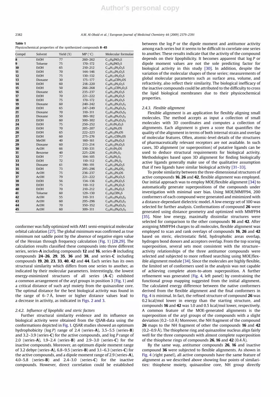
Table 1Physicochemical properties of the synthesized compounds 8–45
Compd Solvent Yield (%) MP (�C) Molecular formulae
8 EtOH 77 260–262 C12H8INO3S9 Toluene 75 170–172 C12H6INO2S10 EtOH 65 210–212 C14H13IN2O3S11 EtOH 70 144–146 C15H15IN2O2S12 EtOH 75 130–132 C16H17IN2O2S13 Dioxane 30 175–177 C14H10ClIN2OS14 EtOH 60 218–220 C18H13IN2O2S15 EtOH 50 266–268 C18H12ClIN2O2S16 Dioxane 65 235–237 C18H13IN2O3S17 EtOH 70 221–222 C19H15IN2O3S18 EtOH 75 170–172 C20H17IN2O3S19 Dioxane 60 240–242 C18H12IN3O3S2
20 EtOH 65 247–249 C21H13IN4O3S3
21 Dioxane 70 310–311 C22H14IN5O3S2
22 Dioxane 50 180–182 C19H14IN5O3S2
23 EtOH 60 300–302 C19H14IN3O3S2
24 EtOH 75 250–252 C12H7IN2O2S25 EtOH 70 205–207 C12H8IN3OS26 EtOH 65 222–223 C18H12IN3OS27 EtOH 60 275–276 C18H11ClIN3OS28 MeOH 40 160–161 C18H10IN5O5S29 Dioxane 60 213–214 C19H12IN3O2S30 AcOH 66 330–331 C12H7IN2OS31 Benzene 40 225–226 C12H7IN2S2
32 EtOH 77 104–105 C13H9IN2S2
33 EtOH 72 110–112 C14H11IN2S2
34 Dioxane 61 190–191 C19H13IN4O2S35 EtOH 73 295–296 C19H13IN4OS2
36 AcOH 75 236–237 C19H12IN3OS37 AcOH 79 221–222 C20H14IN3O2S38 EtOH 56 134–136 C16H12IN3O3S39 EtOH 75 110–112 C16H13IN2O3S40 EtOH 70 210–212 C14H11IN4O2S41 Toluene 60 129–130 C12H6ClIN2S42 AcOH 55 310–311 C19H15IN4O2S2
43 AcOH 60 295–296 C18H13IN4O2S2
44 AcOH 70 350–352 C21H14IN5O2S3
45 AcOH 60 309–311 C22H15IN6O2S2
A.M. Al-Obaid et al. / European Journal of Medicinal Chemistry 44 (2009) 2379–23912382

Author's personal copy
attached to quinazoline moiety as hydrogen bond donor, andaromatic ring attached to such NH moiety. Analysis of inactivemolecule is an important step to understand the essential featuresfor a given activity. It is clear that compound 36 was flexibly alignedin a different manner when compared to the active compoundsexplaining why such compound was void of antitumor activity andshowing the importance of both NH moiety and the aromatic ringattached to this NH group for activity (Fig. 4).
2.4.4. Pharmacophore prediction [33]For pharmacophore generation and prediction, compound 26
(Figs. 5 and 6) served as the reference to which all conformationsof each analog were aligned. All structures were built de novousing 2D/3D editor sketcher in MOE [34]. Conformational modelswere calculated using a 15 kcal energy cut off (minimizationconvergence criteria during conformational analysis: energyconvergence¼ 0.01 kcal/mol, gradient convergence¼ 0.01 kcal/mol). The number of conformers generated for each substrate waslimited to a maximum of 150. All molecules with their associatedconformations were regrouped including the biological data.Hypothesis generation was performed using low energy conformersof the molecules. After assignment of possible pharmacophoreelements for each analog the calculation and analysis were carriedout using the MOE program [34] and a superposition of themolecules, including the assigned elements, was attempted (Figs.5 and 6). Several runs of calculation were repeated to generatepharmacophore maps. For each run a distinct number of specifiedpharmacophore elements were adapted. All adapted modelsshowed that the donor atoms of the NH fragments as hydrophilicelement and the aryl moieties as hydrophobic element were wellsuperimposed within the set distance tolerance. This confirms theimportant role of the hydrophilic and hydrophobic moieties forrecognition and binding to receptor sites.
I
N S
N
OOH
I
N S
N
ONHR
I
N S
O
O
NH2OH
R-NHNH2
HCONH2
I
N S
NH
O
P2S5
I
N S
N
SI
N S
NH
S
24
25: R = H26: R = Ph27: R = 4-ClPh28: R = 2,4-NO2Ph29: R = COPh
9
30
32: R = CH333: R = Et
31
R-Br
I
N SN
ONH
NCX
CHO
H3C
OEt
OO
34: X = 035: X = S
36: R = H37:R = OCH3
38
HNX
I
N SN
ON
R
I
N SN
ONH
O
O
CH3
R
R
Scheme 2. Synthetic route for the preparation of the target compounds 24–38.
I
N S
NH
O
Br OEt
O
I
N S
N
O OEt
O
NH2NH2
I
N S
N
Cl
I
N S
N
O NHNH2
O
I
N S
N
HN
SONH2
NH2
SONH-R
I
N S
N
HN
30 39
POCl3 / PCl5
41 40
4243: R = H44: R = 2-thiazolyl45: R = 2-pyrimidinyl
SO2NH2
SO2NH-R
NH2
Scheme 3. Synthetic route for the preparation of the target compounds 39–45.
A.M. Al-Obaid et al. / European Journal of Medicinal Chemistry 44 (2009) 2379–2391 2383

Author's personal copy
Models for 26 and its analogs 16 and 42 (Fig. 5) possess phar-macophore elements in the aromatic ring attached to NH group (asa hydrogen bond donor) which is out of the thiophene plane as wellas quinazoline core in the same plane of thiophene ring. In contrast,model for 36 (Fig. 6) showed common elements only in thiophenefragment attached to quinazoline core. Since the arylamino part ofcompound 26 plays an important role in activity, model 36 wasexcluded from further considerations. Therefore, model 26 wasconsidered as the representative pharmacophore map of the anti-tumor activity.
According to the pharmacophore generated by MOE [34] theminimal structural requirements for antitumor activity consist ofan aromatic ring (hydrophobic region) attached to NH fragment(H-bonding donor region), and a hydrophobic region representedby quinazoline core and thiophene fragment. For all the activemolecules, reasonable nonbonded distances among the aromaticring, quinazoline core and the thiophene centroid align on thepredicted pharmacophore maps were found (Figs. 2, 3, and 5). Thispharmacophoric assumption was in consistence with biologicaldata.
Table 2Growth inhibitory concentrations (GI50, TGI) and lethal concentration (LC50) of some selected in vitro tumor cell linesa (mM)
Compdb Activity Leukemia CNS Melanoma Breast
CCRF-CEM HL-60 (TB) K-562 RPMI-8226 SF-539 SK-MEL-2 UACC-257 MCF-7 T-47D
16 GI50 ntc 8 24 3 <0.01 16 <0.01 12 2TGI nt 37 d 18 15 30 22 33 11LC50 nt d d 57 62 57 d 94 50
19 GI50 41 20 37 25 nt 22 0.05 23 19TGI d 64 d 62 nt 48 19 65 44LC50
d d d d nt d d d 99
26 GI50 51 0.4 0.3 5 34 0.3 32 2 ntTGI d d 23 d d 2 d d ntLC50
d d d d d d d d nt
29 GI50 39 34 44 23 30 24 <0.01 23 11TGI d d d 58 d 69 31 d 38LC50
d d d d d d d d d
35 GI50 21 1 0.02 19 34 nt 17 29 3TGI 61 22 10 52 d nt 66 88 17LC50
d d 75 d d nt d d d
40 GI50 20 38 37 16 nt 25 <0.01 16 2TGI 88 d d 39 nt 71 d 89 22LC50
d d d 97 nt d d d d
42 GI50 22 30 37 19 11 18 14 0.6 0.2TGI 60 d d 50 31 33 28 16 18LC50
d d d d 85 61 54 d 82
44 GI50 2 32 38 1 32 22 0.04 26 15TGI 4 80 d 3 60 47 28 d 51LC50 9 d d 8 d d 95 d d
a Data obtained based on the NCI’s in vitro disease oriented human tumor cell lines screening protocol.b Compounds passed the in vitro one dose primary anticancer screen.c nt, compound not tested.d Values> 100 mM.
Table 3Median growth inhibitory concentration (GI50, mM) of in vitro subpanel tumor cell lines
Compd Subpanel tumor cell linesa MG-MIDb
I II III IV V VI VII VIII IX
12 83.8� 10.2 96.0� 2.6 c >100 c c c c >100 c
16 11.5� 3.1 13.5� 0.9 15.2� 1.4 12.2� 3.1 11.1� 2.0 13.3� 1.1 13.2� 1.3 8.7� 1.9 11.9� 2.2 12.7� 0.6519 35.3� 4.7 24.0� 1.3 24.8� 2.7 30.3� 3.8 17.6� 2.6 23.5� 2.0 24.7� 1.6 22.7� 3.9 23.3� 2.7 24.9� 1.120 54.0� 4.7 38.7� 8.2 44.7� 9.3 41.4� 11.9 45.2� 10.1 46.5� 9.9 34.2� 4.0 32.9� 5.5 34.8� 10.0 41.2� 2.824 34.3� 3.1 33.2� 3.7 45.8� 9.3 33.2� 3.8 57.3� 7.4 32.1� 4.8 49.8� 9.3 39.9� 12.2 39.4� 4.0 41.1� 2.426 12.7� 7.8 8.6� 3.0 6.5� 2.0 16.3� 5.6 8.5� 4.6 8.6� 4.7 19.7� 9.7 1.61 5.9� 4.11 10.3� 1.829 33.9� 3.6 25.6� 2.3 35.4� 6.2 27.4� 2.2 35.5� 10.9 27.4� 4.6 32.1� 2.5 27.2� 6.6 24.5� 4.8 30.2� 1.833 – 84.2� 10.4 94.6� 3.9 c c 85.2� 14.8 c c 71.4� 18.1 90.6� 3.435 11.5� 4.4 24.0� 4.1 29.5� 2.1 29.2� 6.1 23.1� 3.1 21.5� 3.2 32.5� 5.1 25.8� 1.5 19.4� 4.3 24.9� 1.536 63.4� 22.3 c c c 90.3� 6.8 c c c c c
38 38.5� 4.8 27.5� 3.2 28.1� 2.8 27.9� 4.5 37.7� 9.3 40.9� 12.4 28.9� 3.8 20.6� 6.0 24.9� 3.8 31.1� 2.140 25.4� 4.1 19.4� 1.1 24.3� 2.9 20.7� 2.5 19.4� 3.1 22.3� 2.0 23.1� 2.1 23.5� 1.8 18.7� 4.3 21.5� 0.9542 23.9� 3.2 14.7� 1.6 16.7� 2.7 18.2� 1.6 16.0� 0.88 15.7� 2.0 13.6� 2.9 17.1� 2.2 12.3� 4.2 16.9� 0.9444 21.7� 6.7 23.3� 2.6 24.3� 2.6 27.7� 3.3 18.9� 3.0 25.8� 2.3 28.3� 1.1 13.9� 7.9 25.9� 4.2 23.9� 1.15-FU 15.09� 3.8 c 8.37� 2.6 72.07� 0.01 70.6� 41.5 61.4� 20.2 45.6� 28.8 22.7� 16.8 76.4� 29.1 22.6� 3.6
a I, leukemia; II, non-small cell lung cancer; III, colon cancer; IV, CNS cancer; V, melanoma; VI, ovarian cancer; VII, renal cancer; VIII, prostate cancer; IX, breast cancer.b GI50 full panel mean-graph midpoint (mM).c Values > 100 mM.
A.M. Al-Obaid et al. / European Journal of Medicinal Chemistry 44 (2009) 2379–23912384

Author's personal copy
3. Conclusion
Compounds 2-(2-thienylcarbonylamino)-5-iodo-N-(4-hydroxyphenyl)-benzamide (16), 2-(2-thieno)-6-iodo-3-phenylamino-3,4-dihydro-quinazolin-4-one (26), and 2-(2-thieno)-4-[4-sulfonami-dobenzylamino]-6-iodo-quinazoline (42), with GI50 values of 12.7,10.3, 16.9 mM, respectively, proved to be the most active membersin this study, as compared to the known drug 5-FU. These threequinazoline analogs could be considered as useful templates forfuture development to obtain more potent antitumor agent(s).Flexible alignment was conducted on the basis of experimentaldata from which four featured pharmacophore model was devel-oped. The model was mapped onto the antitumor molecules andcompared with nonactive compounds. This pharmacophore model
could be very useful for the virtual screening in the development ofnew antitumor agents. The overall outcome of this model revealedthat: (i) the quinazolinone ring is a satisfactory backbone for anti-tumor activity, (ii) the presence of aryl moiety at the 3-aminoposition is necessary for the activity as hydrophobic region, (iii) thearyl group at position 3- should be about 6.0–7.0 Å apart from thequinazolinone core (Figs. 2 and 6), (iv) introduction of spacer group,between 3-amino-quinazolinone ring and aryl moiety decreasesthe activity, such as 3-benzoylamino moiety, (v) the presence of onehydrogen atom attached to 3-amino group as a hydrogen bondingdonor is essential for activity and its removal through formation ofbenzylidene moiety diminishes the activity (Figs. 2 and 6), (vi) thepresence of sulfonamide group located on the 3-aryl moiety ofquinazoline is necessary as H-bond region (Figs. 3 and 5) (vii) the
Table 4Total growth inhibitory concentration (TGI, mM) of in vitro subpanel tumor cell lines
Compd Subpanel tumor cell linesa MG-MIDb
I II III IV V VI VII VIII IX
12 c c c c c c c c c c
16 60.6� 14.9(91.0� 7.0)
33.1� 2.8(74.6� 5.1)
34.5� 6.0(74.6� 5.1)
32.4� 5.6(77.6� 7.9)
26.9� 1.5(63.8� 5.9)
35.4� 5.3(72.5� 11.3)
29.3� 2.4(64.1� 6.2)
21.1� 1.4(45.8� 1.5)
36.8� 7.8(79.4� 9.2)
35.0� 2.4(71.8� 2.6)
19 87.7� 7.7 61.3� 6.2 59.3� 10.3(87.1� 6.7)
73.8� 7.0 43.0� 4.1 65.1� 9.6 64.1� 9.1(94.1� 4.4)
58.9� 23.0 62.5� 10.5 63.3� 3.1
20 c 89.5� 5.5 94.6� 5.3 87.7� 9.1 84.8� 8.35 c 84.3� 7.9 c 88.3� 6.0 90.4� 2.224 c 91.2� 4.7 c 94.4� 5.6 97.1� 2.8 87.9� 11.3 c 98.4� 1.5 c 93.3� 2.126 87.1� 12.0 88.0� 5.7 79.2� 14.3 81.9� 13.0 85.9� 14.0 80.1� 15.3 93.1� 6.8 c c 86.7� 3.829 71.5� 14.7 75.3� 7.1 90.0� 5.9 85.8� 7.2 62.1� 8.4 74.4� 10.8 86.7� 7.8 73.3� 26.6 72.5� 12.4 77.6� 3.233 c c c c c c c c c c
35 38.5� 9.5 75.2� 8.0 84.0� 7.9 81.4� 12.5 62.8� 16.4 77.5� 10.5 84.8� 7.7 76.2� 23.8 60.3� 13.0 72.0� 3.636 c c c c c c c c c c
38 c 86.4� 7.2 90.9� 9.1 89.1� 10.9 85.3� 7.1 93.6� 6.4 85.1� 8.2 c 82.1� 7.6 88.6� 2.640 80.5� 10.8 59.6� 5.4 66.9� 9.6 62.1� 7.8 51.1� 4.3 72.5� 9.8 71.1� 8.1 75.3� 24.6 64.7� 13.3 65.9� 3.042 72.3� 10.2 37.0� 3.2
(82.2� 6.1)37.3� 7.9(66.4� 6.5)
42.4� 4.3(89.0� 3.8)
30.5� 1.5(58.3� 2.7)
42.6� 4.9(89.1� 4.9)
36.1� 7.3(88.1� 5.1)
41.8� 11.5(80.8� 19.2)
32.1� 10.6(80.8� 8.1)
40.5� 2.6(79.3� 2.7)
44 59.2� 18.2(69.4� 19.3)
65.2� 8.9(92.4� 5.8)
61.1� 11.2(87.3� 5.0)
68.3� 8.6 45.5� 4.8(92.1� 4.6)
68.9� 8.3 79.6� 6.8 35.9� 17.0(73.0� 27.0)
74.9� 11.2 63.8� 3.5
5-FU c c c c c c c c c c
a For subpanel tumor cell lines, see footnote a of Table 3.b TGI full panel mean-graph midpoint (mM).c Compounds showed values> 100 mM. Median lethal concentration (LC50, mM) of in vitro subpanel tumor cell lines and LC50 full panel mean-graph midpoint (mM) are shownin parentheses.
Fig. 1. Lowest energy conformers of the most active compounds 16, 26, 42 and the inactive compound 36 as representative examples with CPK rendering.
A.M. Al-Obaid et al. / European Journal of Medicinal Chemistry 44 (2009) 2379–2391 2385

Author's personal copy
free sulfonamide function improves the activity, while thesubstituted sulfonamide blunts this action and (viii) regioisomeri-zation of the arylsulfonamide group from position 3- to position 4-of the quinazoline increases the antitumor activity as indicatedupon comparing compounds 23 and 42.
4. Experimental
Unless otherwise specified all chemicals were of commercialgrade, used without further purification and were obtained fromAldrich Chemical Co. (Milwaukee, WI). Solvents used for work ups
were dried over MgSO4, filtered and removed on a rotary evapo-rator. Elemental analyses were performed at College of Pharmacy,King Saud University, Central Laboratory. 1H NMR spectra wereobtained at 500 MHz by JEOL instrument using TMS as internalstandard. Thin layer and flash chromatographies were performedusing E. Merck Silica gel (230–400 mesh). Preparative thin layerchromatography was performed on Harrison model 7924A chro-matotron using Analtech silica gel GF rotors. All modeling studieswere conducted with HyperChem 5.1 package from Hypercube[26]. Flexible alignment and 3D-pharmacophore prediction weregenerated by MOE 2007.09 software [34].
12: >100 μM
I
NH
O
HNEt
O S
16:12.7 μM
I
NH
O
HN
O S
OH
High activity
Am
phip
hilc
regio
n
Hyd
ro
ph
ob
ic reg
io
n
I
N SN
ONH2
25: >100 μM
I
N SN
OHN
29: 30.2 μM
I
N SN
ONH
26:10.3 μM
OHigh activity
Hyd
ro
ph
ob
ic r
eg
ion
Hydrophobic
regio
n
HB region Moderate activity
36: >100 μM
I
N SN
ON
NH
-blo
ckin
g
Dis
tance length
ing
Distance lengthing
Spacer group
Ring closing
Spacer
gro
up
6.7Å
7.1Å 7.0Å
8.4ÅIn
activit
y
Fig. 2. 2D-pharmacophoric design of 3-arylquinazoline (red colors represent the HB regions and green colors represent hydrophobic regions and blue colors represent spacermoiety). (For interpretation of the references to color in this figure legend, the reader is referred to the web version of this article.)
N
N
OI
SO2NH2
S
19: 24.9 μM
N
N
OI
N
N
O
I SO2NH2
23: > 100 μM
20: 41.2 μM
SO2NH
S
S
I
N S
N
HN
42: 16.9 μM
SO2NH2I
N S
N
HN
44: 23.9 μM
SO2NH
NS
Reg
ioiso
merizatio
n
Regioisomerization
Spacer
NH 2
-Blo
ckin
g
NH2-Blocking
6.4Å
6.3Å
7.6Å
7.0Å6.1Å
Inactiv
ity
Moderate activityNS
Hig
h a
ctiv
ity
Hig
h activity
Fig. 3. 2D-pharmacophoric design of the sulfonamido-quinazoline (red colors represent the HB regions and green colors represent hydrophobic regions and blue colors representspacer moiety). (For interpretation of the references to color in this figure legend, the reader is referred to the web version of this article.)
A.M. Al-Obaid et al. / European Journal of Medicinal Chemistry 44 (2009) 2379–23912386

Author's personal copy
4.1. Chemistry
4.1.1. 2-(2-Thienylcarbonylamino)-5-iodo-benzoic acid 82-Thiophenecarbonyl chloride (7.33 g, 0.05 mol) was added
dropwise to a stirred solution of 5-iodo-anthranilic acid (13.15 g,0.05 mol) in pyridine (50 ml) and the reaction mixture was stirred
at room temperature for 2 h. The reaction mixture was poured intocold 5% dilute HCl solution (100 ml). The solid obtained wasfiltered, washed several times with water, dried and crystallizedfrom ethanol (Table 1). 1H NMR (DMSO-d6): d 7.27 (t, 1H, J¼ 4.0 Hz,thiophene-H), 7.70 (d, 1H, J¼ 0.5 Hz, thiophene-H), 7.86–7.88 (dd,1H, J¼ 1.5, J¼ 8.5 Hz, ArH), 7.91 (d, 1H, J¼ 4.0 Hz, thiophene-H),
Fig. 5. The active compounds 16 (red), 26 (green) and 42 (blue), mapped to the pharmacophore model for antitumor activity. Pharmacophore features are color coded: orange forhydrophobics aromatic, blue for a hydrogen bond donor, and violet for a hydrogen bond donor/acceptor feature. The geometries of pharmacophore are shown in the upper rightpanel. (For interpretation of the references to color in this figure legend, the reader is referred to the web version of this article.)
Fig. 4. Flexible alignments of the most active compounds (left panel): 16 (in red), 26 (in green) and 42 (in blue). Right panel showed the flexible alignments of the inactivecompound 36 (in gray) and the active compounds 16 (in red), 26 (in green). (For interpretation of the references to color in this figure legend, the reader is referred to the webversion of this article.)
A.M. Al-Obaid et al. / European Journal of Medicinal Chemistry 44 (2009) 2379–2391 2387
Author's personal copy
8.15 (d, 1H, J¼ 1.5 Hz, ArH), 8.34 (d, 1H, J¼ 8.5 Hz, ArH), 8.96 (br s,1H, NHCO), 12.61 (s, 1H, COOH). Anal. for (C12H8INO3S) C, H, N.
4.1.2. 2-(2-Thieno)-6-iodo-4H-3,1-benzoxazin-4-one 9A mixture of 2-(thiophene-2-carbonylamino)-5-iodo-benzoic
acid (8, 11.19 g, 0.03 mol) and acetic anhydride (30 g, 0.3 mol) washeated under reflux for 4 h. The solvent was removed underreduced pressure. The residue was triturated with petroleum ether40–60. The separated solid was collected by filtration, washed withpetroleum ether 40–60, dried and crystallized from toluene (Table1). 1H NMR (DMSO-d6): d 7.27 (t, 1H, J¼ 4 Hz, thiophene-H), 7.70 (d,1H, J¼ 0.5 Hz, thiophene-H), 7.86–7.88 (dd, 1H, J¼ 1.5, J¼ 8.5 Hz,ArH), 7.91 (d, 1H, J¼ 4.0 Hz, thiophene-H), 8.15 (d, 1H, J¼ 1.5 Hz,ArH), 8.34 (d, 1H, J¼ 8.5 Hz, ArH). Anal. for (C12H6INO2S) C, H, N.
4.1.3. 2-(2-Thienylcarbonylamino)-5-iodo-N-(substituted)-benzamides 10–12
A mixture of 4H-3,1-benzoxazin-4-one derivative (9, 3.55 g,0.01 mol) and appropriate aliphatic amine (0.03 mol) in pyridine(30 ml) was heated under reflux for 3 h. The solvent was thenremoved under reduced pressure. The obtained solid was filtered,washed with diluted HCl and crystallized from the appropriatesolvent. Yield percentage, melting points, crystallization solvent,and molecular formula are shown in Table 1. 1H NMR (DMSO-d6),10: d 3.69–3.81 (q, 2H, J¼ 6 Hz, CONH–CH2CH2OH), 3.55–3.57 (q1,2H, J¼ 6 Hz, CONH–CH2CH2OH), 4.80 (t, 1H, J¼ 6 Hz, OH), 7.27 (t,1H, J¼ 4 Hz, thiophene-H), 7.70 (d, 1H, J¼ 0.5 Hz, thiophene-H),7.86–7.88 (dd, 1H, J¼ 1.5, J¼ 8.5 Hz, ArH), 7.91 (d, 1H, J¼ 4.0 Hz,thiophene-H), 8.15 (d, 1H, J¼ 1.5 Hz, ArH), 8.34 (d, 1H, J¼ 8.5 Hz,ArH), 9.04 (s, 1H, CONH–CH2), 12.57 (s, 1H, Ph–NH–CO). Anal. for(C14H13IN2O3S) C, H, N. 11: d 0.90 (5, 3H, J¼ 7.5 Hz, CH3–CH2CH2NH–), 1.54–1.59 (m, 2H, J¼ 7.5, J¼ 7 Hz, CH3–CH2–CH2NH–), 3.23–3.27(q, 2H, J¼ 7 Hz, CH3–CH2–CH2–NH–), 7.27 (t, 1H, J¼ 4 Hz, thio-phene-H), 7.70 (d, 1H, J¼ 0.5 Hz, thiophene-H), 7.86–7.88 (dd, 1H,J¼ 1.5, H¼ 8.5 Hz, ArH), 7.91 (d, 1H, J¼ 4.0 Hz, thiophene-H), 8.15(d, 1H, J¼ 1.5 Hz, ArH), 8.34 (d, 1H, J¼ 8.5 Hz, ArH), 8.98 (s, 1H,CONH–CH2), 12.59 (s, 1H, Ph–NH–CO). Anal. for (C15H15IN2O2S) C, H,N. 12: d 0.90 (t, 3H, J¼ 7.5 Hz, CH3CH2CH2CH2NH), 1.29–1.37 (m, 2H,J¼ 7.0, 7.5 Hz, CH3–CH2–CH2–CH2–NH–), 1.50–1.56 (m, 2H, J¼ 7.0,7.5 Hz, CH3CH2CH2CH2NH), 3.27–3.31 (m, 2H, J¼ 7.0, J¼ 7.5 Hz,CH3–CH2CH2CH2–NH–), 7.27 (t, 1H, J¼ 4 Hz, thiophene-H), 7.70 (d,1H, J¼ 0.5 Hz, thiophene-H), 7.86–7.88 (dd, 1H, J¼ 1.5, J¼ 8.5 Hz,
ArH), 7.91 (d, 1H, J¼ 4.0 Hz, thiophene-H), 8.15 (d, 1H, J¼ 1.5 Hz,ArH), 8.34 (d, 1H, J¼ 8.5 Hz, ArH), 8.96 (s, 1H, CONH–CH2–), 12.60 (s,1H, PhNHCO–). Anal. for (C16H17IN2O2S) C, H, N.
4.1.4. 2-(2-Thieno)-3-(2-chloroethyl)-4-oxo-6-iodo-3H-quinazoline 13
A mixture of 2-(2-thienylcarbonylamino)-5-iodo-N-(2-hydrox-yethyl)-benzamide (10, 4.16 g, 0.01 mol) and thionyl chloride(23.8 g, 0.2 mol) was heated under reflux for 4 h. The reactionmixture was concentrated in vacuo and poured into ice water. Thesolid obtained was crystallized from dioxane (Table 1). 1H NMR;(DMSO-d6): d 3.87 (t, 2H, J¼ 7 Hz, N–CH2–CH2Cl), 4.5 (t, 2H,J¼ 7 Hz, NCH2CH2–Cl), 7.27 (t, 1H, J¼ 4 Hz, thiophene-H), 7.70 (d,1H, J¼ 0.5 Hz, thiophene-H), 7.86–7.88 (dd, 1H, J¼ 1.5, J¼ 8.5 Hz,ArH), 7.91 (d, 1H, J¼ 4.0 Hz, thiophene-H), 8.15 (d, 1H, J¼ 1.5 Hz,ArH), 8.34 (d, 1H, J¼ 8.5 Hz, ArH). Anal. for (C14H10ClIN2OS) C, H, N.
4.1.5. 2-(2-Thienylcarbonylamino)-5-iodo-N-(4-substitutedphenyl)-benzamides 14–18
A mixture of compound (9, 3.55 g, 0.01 mol) and appropriatearomatic amines (0.20 mol) in dry pyridine (50 ml) was refluxed for8 h. The reaction mixture was cooled and treated with acidulatedice cold water (10%). The separated product was washed severaltimes with water and recrystallized from appropriate solvent (Table1). 1H NMR (DMSO-d6), 14: d 7.16 (t, 1H, J¼ 7.50 Hz, ArH), 7.24–7.25(m, 1H, ArH), 7.39 (t, 2H, J¼ 8.0 Hz, ArH), 7.71–7.75 (m, 3H, ArH),7.89–7.95 (m, 2H, ArH), 8.17 (d, 1H, J¼ 9.0 Hz, ArH), 8.23 (d, 1H,J¼ 1.5 Hz, ArH), 10.62 (s, 1H, NH), 11.64 (s, 1H, NH). Anal. for(C18H13IN2O2S) C, H, N. 15: d 7.24 (t, 1H, J¼ 4.50 Hz, ArH), 7.31 (d, 2H,J¼ 9.0 Hz, ArH), 7.41–7.58 (m, 3H, ArH), 7.85–7.93 (m, 2H, ArH), 8.13(d, 1H, J¼ 9.0 Hz, ArH), 8.2 (d, 1H, J¼ 1.5 Hz, ArH), 10.69 (s, 1H, NH),11.52 (s, 1H, NH). Anal. for (C18H12ClIN2O2S) C, H, N. 16: d 6.77–7.75(dd, 4H, J¼ 9.0 Hz, ArH), 7.23–7.26 (m, 1H, ArH), 7.13 (d, 1H,J¼ 3.00 Hz, ArH), 7.91–7.93 (m, 2H, ArH), 8.23–8.24 (m, 2H, ArH),9.63 (s, 1H, OH), 10.43 (s, 1H, NH), 11.89 (s, 1H, NH). Anal. for(C18H13IN2O3S) C, H, N. 17: d 3.67 (s, 3H, OCH3), 6.96–7.60 (dd, 4H,J¼ 9.0 Hz, ArH), 7.35 (t, 1H, J¼ 4.0 Hz, ArH), 7.7 (s, 1H, ArH),7.90–7.92 (m, 1H, ArH), 8.19–8.23 (m, 2H, ArH), 8.37 (d, 1H,J¼ 1.5 Hz, ArH), 10.51 (br s, 1H, NH), 11.81 (br s, 1H, NH). Anal. for(C19H15IN2O3S) C, H, N. 18: d 1.32 (t, 3H, J¼ 7.0 Hz, OCH2CH3),3.99–4.03 (q, 2H, J¼ 7.0, OCH2CH3), 6.94–7.59 (dd, 4H, J¼ 9.0 Hz,ArH), 7.24 (t, 1H, J¼ 4.50 Hz, ArH), 7.71 (d, 1H, J¼ 3.50 Hz, ArH),
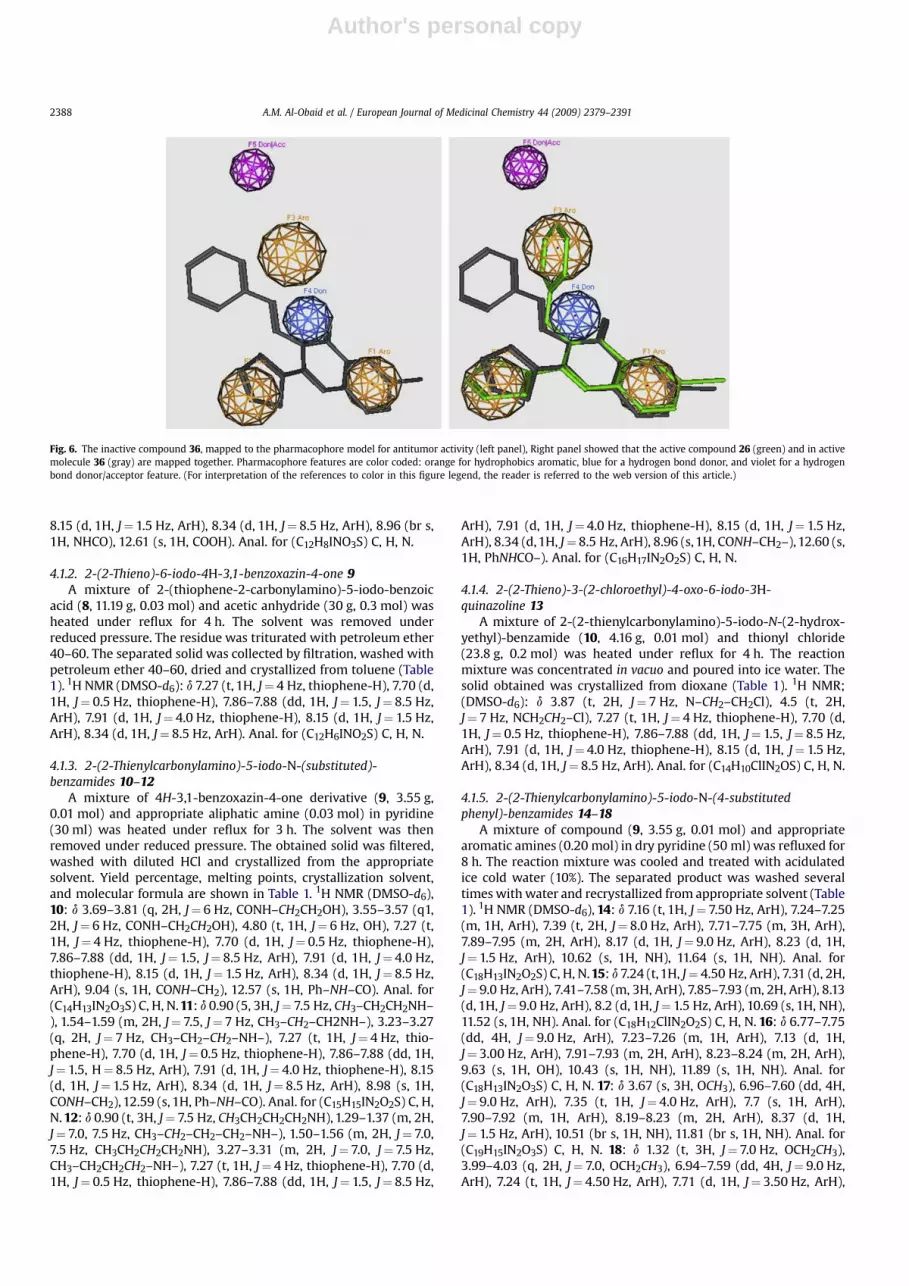
Fig. 6. The inactive compound 36, mapped to the pharmacophore model for antitumor activity (left panel), Right panel showed that the active compound 26 (green) and in activemolecule 36 (gray) are mapped together. Pharmacophore features are color coded: orange for hydrophobics aromatic, blue for a hydrogen bond donor, and violet for a hydrogenbond donor/acceptor feature. (For interpretation of the references to color in this figure legend, the reader is referred to the web version of this article.)
A.M. Al-Obaid et al. / European Journal of Medicinal Chemistry 44 (2009) 2379–23912388

Author's personal copy
7.89–7.92 (m, 3H, ArH), 8.22 (d, 1H, J¼ 8 Hz, ArH), 10.51 (s 1H, NH),11.83 (s, 1H, NH). Anal. for (C20H17IN2O3S) C, H, N.
4.1.6. 2-(Thieno)-6-iodo-3-[4-(substituted sulphonamido)phenyl]-3H-quinazolin-4-one 19–22
A mixture of compound (9, 3.55 g, 0.01 mol) and appropriatesulfa drug (0.01 mol) was fused at 210� in oil bath for 1 h, cooledand triturated with methanol and filtered. The resulting solid waswashed several times with water, dried and recrystallized fromsuitable solvent to obtain compounds 19–22 (Table 1). 1H NMR(DMSO-d6), 19: d 7.24–7.28 (m, 2H, ArH), 7.74–7.77 (m, 2H, ArH),7.90–7.97 (m, 3H, ArH), 8.28–8.29 (m, 2H, ArH), 8.37 (d, 1H,J¼ 9.0 Hz, ArH), 12.05 (br s, 2H, NH2). Anal. for (C18H12IN3O3S2) C, H,N. 20: d 6.81 (d, 1H, J¼ 4.5 Hz, thiazole-H), 7.25 (d, 1H, J¼ 4.5 Hz,thiazole-H), 7.69–7.95 (m, 10H, ArH), 10.27 (br s, 1H, NH). Anal. for(C21H13IN4O3S3) C, H, N. 21: d 6.57 (d, 2H, J¼ 8.5 Hz, ArH), 7.01 (t,1H, J¼ 5.0 Hz, ArH), 7.29 (t, 1H, J¼ 5.0 Hz, ArH), 7.41 (d, 1H,J¼ 5.0 Hz, ArH), 7.62 (d, 2H, J¼ 8.5 Hz, ArH), 7.86–7.88 (dd, 1H,J¼ 1.5, 8.5 Hz, ArH), 8.18–8.20 (m, 2H, ArH), 8.29 (d, 1H, J¼ 8.5 Hz,ArH), 8.35 (d, 1H, J¼ 3.0 Hz, ArH), 8.48 (d, 1H, J¼ 9.0 Hz, ArH), 11.38(br s, 1H, NH). Anal. for (C22H14IN5O3S2) C, H, N. 22: d 3.57 (br s, 1H,NH), 7.28 (t, 1H, J¼ 5.0 Hz, ArH), 7.39 (d, 4H, J¼ 9.0 Hz, ArH),7.91–7.92 (m, 2H, ArH), 8.00–8.02 (m, 2H, ArH), 8.15–8.18 (m, 2H,ArH), 8.32–8.34 (m, 2H, ArH). Anal. for (C19H14IN5O3S2) C, H, N.
4.1.7. 2-(2-Thieno)-6-iodo-3-(4-sulphonamido-benzyl)-3H-quinazolin-4-one 23
Equimolar amounts of compound (9, 3.55, 0.01 mol) andhomosulfanilamide (1.86 g, 0.01 mol) were fused together at 200�
in an oil bath for 1 h. On cooling, the solid mass dissolved in hotglacial acetic acid (50 ml), and filtered. The filtrate was concen-trated in vacuo and the resulting solid was filtered, washed withwater and recrystallized to afford 23 (Table 1). 1H NMR (DMSO-d6):d 4.50 (s, 2H, CH2Ph), 7.25–7.27 (m, 1H, ArH), 7.34 (br s, 2H, NH2),7.54–7.82 (dd, 4H, J¼ 8.5 Hz, ArH), 7.68 (d, 1H, J¼ 5.0 Hz, ArH),7.90–7.92 (m, 2H, ArH), 8.26 (d, 1H, J¼ 3.0 Hz, ArH), 8.34 (d, 1H,J¼ 9.0 Hz, ArH). Anal. for (C19H14IN3O3S2) C, H, N.
4.1.8. 2-(2-Thieno)-6-iodo-3-hydroxy-3,4-dihydro-quinazolin-4-one 24
A mixture of 2-(2-thieno)-6-iodo-4H-3,1-benzoxazin-4-one (9,3.55, 0.01 mol) and hydroxylamine hydrochloride (0.7 g, 0.01 mol)in dry pyridine (35 ml) was heated under reflux for 8 h and thereaction mixture was then concentrated to half its volume. Theseparated solid was filtered, washed with water and crystallized toafford 24 (Table 1). 1H NMR (DMSO-d6): d 5.3 (s, 1H, OH, D2Oexchanged), 7.25 (t, 1H, J¼ 4 Hz, thiophene-H), 7.70 (d, 1H,J¼ 0.5 Hz, thiophene-H), 7.86–7.88 (dd, 1H, J¼ 8.5, 1.5 Hz, ArH), 7.91(d, 1H, J¼ 4.0 Hz, thiophene-H), 8.15 (d, 1H, J¼ 1.5 Hz ArH), 8.34 (d,1H, J¼ 8.5 Hz, ArH). Anal. for (C12H7IN2O2S) C, H, N.
4.1.9. 2-(2-Thieno)-6-iodo-3-substitututed amino-3,4-dihydro-quinazolin-4-one 25–29
A mixture of 2-(2-thieno)-6-iodo-4H-3,1-benzoxazin-4-one (9,3.55 g, 0.01 mol) and the required hydrazine derivative or benzoicacid hydrazide (0.012 mol) in n-butanol (20 ml) was heated underreflux for 6 h. The reaction mixture was concentrated, cooled andthe separated solid was crystallized out from the proper solvent toafford compounds 25–29 (Table 1). 1H NMR (DMSO-d6), 25: d 5.7–5.8 (br s, 2H, NH2, D2O exchanged), 7.27 (t, 1H, J¼ 4 Hz, thiophene-H), 7.73 (d, 1H, J¼ 0.5 Hz, thiophene-H), 7.85–7.87 (dd, 1H, J¼ 8.5,1.5 Hz, ArH), 7.92 (d, 1H, J¼ 4.0 Hz, thiophene-H), 8.1 (d, 1H,J¼ 1.5 Hz, ArH), 8.33 (d, 1H, J¼ 8.5 Hz, ArH). Anal. for (C12H8IN3OS)C, H, N. 26: 6.75 (t, 1H, J¼ 7.5 Hz, ArH), 6.84 (d, 2H, J¼ 8 Hz, ArH),7.17 (t, 1H, J¼ 8 Hz, ArH), 7.22 (t, 1H, J¼ 4 Hz, thiophene-H), 7.66 (d,1H, J¼ 3 Hz, thiophene-H), 7.91 (d, 1H, J¼ 5 Hz, thiophene-H), 7.94–7.96 (dd,1H, J¼ 8.5,1.5 Hz, quinazoline-H), 8.02 (s,1H, quinazoline-H),
8.28 (d, 1H, J¼ 8.5 Hz, quinazoline-H), 8.31 (d, 1H, J¼ 2H, ArH), 10.7(s, 1H, NH). Anal. for (C18H12IN3OS) C, H, N. 27: d 6.78 (d, 2H,J¼ 9 Hz, ArH), 7.17–7.19 (m, 1H, J¼ 4.1 Hz, thiophene-H), 7.23 (d,2H, J¼ 9 Hz, ArH), 7.55 (d, 1H, J¼ 9 Hz, thiophene-H), 7.83–7.84 (dd,1H, J¼ 3.5, 1.5 Hz, thiophene-H), 8.14–8.17 (dd, 1H, J¼ 6.5, 2 Hz,quinazoline-H), 8.24–8.25 (dd, 1H, J¼ 2.5, 1.5 Hz, quinazoline-H),8.35 (d, 1H, J¼ 2 Hz, quinazoline-H), 9.6 (s, 1H, NH). Anal. for(C18H11ClIN3OS) C, H, N. 28: d 7.27 (t, 1H, J¼ 4 Hz, thiophene-H),7.34–8.34 (m, 8H, ArH), 9.5 (s, 1H, NH). Anal. for (C18H10IN5O5S) C,H, N. 29: d 7.24 (t, 1H, J¼ 4 Hz, thiophene-H), 7.41–8.35 (m, 10 H,ArH), 12.31 (br s, 1H, NHC]O, D2O exchanged). Anal. for(C19H12IN3O2S) C, H, N.
4.1.10. 2-(2-Thieno)-6-iodo-3,4-dihydro-quinazolin-4-one 30A mixture of 2-(thieno)-6-iodo-4H-3,1-benzoxazin-4-one (9,
3.55 g, 0.01 mol) and formamide (30 ml) was heated under refluxfor 3 h. On cooling, the separated solid was filtered, washed withwater and crystallized from acetic acid to yield 30 (Table 1). 1H NMR(DMSO-d6): d 7.25 (t, 1H, J¼ 4 Hz, thiophene-H), 7.70 (d, 1H,J¼ 0.5 Hz, thiophene-H), 7.86–7.88 (dd, 1H, J¼ 8.5, 1.5 Hz, quina-zoline-H), 7.91 (d, 1H, J¼ 4.0 Hz, thiophene-H), 8.15 (d, 1H,J¼ 1.5 Hz, quinazoline-H), 8.34 (d, 1H, J¼ 8.5 Hz, quinazoline-H),12.7 (br s, 1H, NH). Anal. for (C12H7IN2OS) C, H, N.
4.1.11. 2-(2-Thieno)-6-iodo-3,4-dihydro-quinazolin-4-thione 31Phosphorous pentasulfide (2.31 g, 0.011 mol) was added to
a solution of 2-(2-thieno)-6-iodo-3,4-dihydro-quinazolin-4-one(30, 3.54 g, 0.01 mol) in xylene (30 ml), and the mixture was heatedunder reflux for 3 h, then filtered while hot. On cooling, theobtained solid was filtered, and washed with water, dried andcrystallized from acetic acid to give 31 (Table 1). 1H NMR (DMSO-d6): d 7.24 (t, 1H, J¼ 4 Hz, thiophene-H), 7.70 (d, 1H, J¼ 0.5 Hz,thiophene-H), 7.86–7.88 (dd, 1H, J¼ 8.5, 1.5 Hz, quinazoline-H), 7.91(d, 1H, J¼ 4.0 Hz, thiophene-H), 8.15 (d, 1H, J¼ 1.5 Hz, quinazoline-H), 8.34 (d, 1H, J¼ 8.5 Hz, quinazoline-H), 12.5 (br s, NH,exchanged). Anal. for (C12H7IN2S2) C, H, N.
4.1.12. 2-(2-Thieno)-4-alkylthio-6-iodo-quinazolines 32, 33A mixture of 2-(2-thieno)-6-iodo-3,4-dihydro-quinazoline-4-
thione (31, 3.7 g, 0.01 mol), the appropriate alkyl halide (0.015 mol)and anhydrous potassium carbonate (2 g) in dry acetone (50 ml)was heated under reflux for 3 h and the reaction mixture wasfiltered while hot. The filtrate was evaporated under vacuum andthe separated solid was washed with water and crystallized fromethanol to give 32, 33 (Table 1). 1H NMR (DMSO-d6), 32: d 2.64 (s,3H, SCH3), 7.27 (t, 1H, J¼ 4 Hz, thiophene-H), 7.70 (d, 1H, J¼ 0.5 Hz,thiophene-H), 7.86–7.88 (dd, 1H, J¼ 8.5, 1.5 Hz, quinazoline-H), 7.91(d, 1H, J¼ 4.0 Hz, thiophene-H), 8.15 (d, 1H, J¼ 1.5 Hz, quinazoline-H), 8.34 (d, 1H, J¼ 8.5 Hz, quinazoline-H). Anal. for (C13H9IN2S2) C,H, N. 33: d 1.2–1.3 (t, 3H, J¼ 10 Hz, CH3–CH2–) 3.01–3.02 (q, 2H,J¼ 10 Hz, CH3–CH–), 7.27 (t, 1H, J¼ 4 Hz, thiophene-H), 7.70 (d, 1H,J¼ 0.5 Hz, thiophene-H), 7.86–7.88 (dd, 1H, J¼ 1.5 Hz, quinazoline-H), 7.91 (d, 1H, J¼ 4.0 Hz, thiophene-H), 8.15 (d, 1H, J¼ 1.5 Hz,quinazoline-H), 8.34 (d, 1H, J¼ 8.5 Hz, quinazoline-H). Anal. for(C14H11IN2S2) C, H, N.
4.1.13. N-(Phenyl)-N0-[2-(2-thieno)-4-oxo-6-iodo-3H-quinazolin-3-yl]-urea (34) and thiourea 35
A mixture of 2-(2-thieno)-6-iodo-3-amino-quinazolin-4-one(25, 3.69 g, 0.01 mol), phenylisocyanate or phenylisothiocyanate(0.015 mol) in dry dioxane (30 ml) was refluxed for 8 h. The excesssolvent was removed and the solid was crystallized from the propersolvent to give compounds 34, 35, respectively (Table 1). 1H NMR(DMSO-d6), 34: d 6.75 (t, 1H, J¼ 7.5 Hz ArH), 6.84 (d, 2H, J¼Hz,ArH), 7.17 (t, 2H, J¼ 7.5 Hz, ArH), 7.22 (t, 1H, J¼ 4 Hz, thiophene-H),7.7 (d, 1H, J¼ 2 Hz, thiophene-H), 7.85 (d, 1H, J¼ 5 Hz, thiophene-H), 7.94–7.96 (dd, 1H, J¼ 7, 2 Hz, quinazoline-H), 8.15 (d, 1H, 1.5 Hz,
A.M. Al-Obaid et al. / European Journal of Medicinal Chemistry 44 (2009) 2379–2391 2389

Author's personal copy
quinazoline-H), 8.34 (d, 1H, 7.5 Hz, quinazoline-H), 10.7 (s, 1H, ]N–NH–CO), 12.05 (s, H, CONHPh). Anal. for (C19H13IN4O2S) C, H, N. 35:d 6.76 (t, 1H, J¼ 7.5 Hz, ArH), 6.84 (d, 2H, J¼ 8 Hz, ArH), 7.17 (t, 2H,J¼ 7.5, ArH), 7.22 (t, 1H, J¼ 4 Hz, thiophene-H), 7.7 (d, 1H, J¼ 2 Hz,thiophene-H), 7.85 (d, 1H, J¼ 5 Hz, thiophene-H), 7.93–7.95 (dd,J¼ 7, 2 Hz, quinazoline-H), 8.09 (d, 1H, 1.5 Hz, quinazoline-H), 8.33(d, 1H, 7.5 Hz, quinazoline-H), 11.7 (s, 1H, ]N–NH–CS–), 12.7 (s, H,CSNHPh). Anal. for (C19H13IN4OS2) C, H, N.
4.1.14. 2-(2-Thieno)-3-arylideneamino-6-iodo-3,4-dihydro-quinazolin-4-ones 36, 37
A mixture of 2-(2-thieno)-3-amino-3,4-dihydro-quinazolin-6-one (25, 3.69 g, 0.01 mol) and the appropriate aldehyde (0.015 mol)in glacial acetic acid (130 ml) was heated under reflux for 8 h. Oncooling, the separated solid was filtered, washed with water andcrystallized from acetic acid to give 36, 37 (Table 1). 1H NMR(DMSO-d6), 36: d 7.23 (t, 1H, J¼ 4 Hz, thiophene-H), 7.3 (m, 3H,ArH), 7.6 (d, 2H, J¼ 7 Hz, ArH), 7.70 (d, 1H, J¼ 0.5 Hz, thiophene-H),7.86–7.88 (dd, 1H, J¼ 8.5, 1.5 Hz, quinazoline-H), 7.91 (d, 1H,J¼ 4.0 Hz, thiophene-H), 8.15 (d, 1H, J¼ 1.5 Hz, quinazoline-H), 8.34(d, 1H, J¼ 8.5 Hz, quinazoline-H), 9.25 (s, 1H, CH]N). Anal. for(C19H12IN3OS) C, H, N. 37: d 3.73 (s, 3H, OCH3), 6.8 (d, 2H, J¼ 8 Hz,ArH), 7.23 (t, 1H, J¼ 4.0 Hz, thiophene-H), 7.5 (d, 2H, J¼ 8 Hz, ArH),7.70 (d, 1H, J¼ 0.5 Hz, thiophene-H), 7.86–7.88 (dd, 1H, J¼ 8.5,1.5 Hz, quinazoline-H), 7.91 (d, 1H, J¼ 4.0 Hz, thiophene-H), 8.15 (d,1H, J¼ 1.5 Hz, quinazoline-H), 8.34 (d, 1H, J¼ 8.5 Hz, quinazoline-H), 9.22 (s, 1H, CH]N). Anal. for (C20H14IN3O2S) C, H, N.
4.1.15. 2-(2-Thieno)-3-(3-oxobutyryl)amino-6-iodo-3,4-dihydro-quinazolin-3-one 38
A mixture of 2-(2-thieno)-6-iodo-3-amino-quinazolin-4-one(25, 3.69 g, 0.01 mol) and ethyl acetoacetate (0.03 mol) in iso-propanol (30 ml), was heated under reflux for 18 h. The reactionmixture was concentrated to third its volume. The separated solidwas filtered, washed with water and crystallized from ethanol togive 38 (Table 1). 1H NMR (CDCl3), d 2.2 (s, 3H, COCH3), 3.6 (s, 2H,COCH2CO–), 7.25 (t, 1H, J¼ 4.0 Hz, thiophene-H), 7.70 (d, 1H,J¼ 0.5 Hz, thiophene-H), 7.86–7.88 (dd, 1H, J¼ 8.5, 1.5 Hz, quina-zoline-H), 7.91 (d, 1H, J¼ 4.0 Hz, thiophene-H), 8.15 (d, 1H,J¼ 1.5 Hz, quinazoline-H), 8.34 (d, 1H, J¼ 8.5 Hz, quinazoline-H),10.7 (s, 1H, ]N–NHCO–). Anal. for (C16H12IN3O3S) C, H, N.
4.1.16. 2-(2-Thieno)-4-(ethoxycarbonylmethyloxy)-6-iodo-3,4-dihydro-quinazoline 39
A mixture of 2-(2-thieno)-6-iodo-3,4-dihydro-quinazolin-3-one(30, 3.54 g, 0.01 mol), ethylbromoacetate (0.015 mol) and anhy-drous potassium carbonate (2.0 g) in dry acetone (50 ml) washeated under reflux for 12 h. The reaction mixture was filteredwhile hot and the filtrate was concentrated in vacuo to give thecrude product which was crystallized from ethanol to give 39 (Table1). 1H NMR (CDCl3), d 1.2 (t, 3H, J¼ 7 Hz, CH3CH2–), 4.17–4.21 (q, 2H,J¼ 7 Hz, CH3CH2–), 5.22 (s, 2H, OCH2CO–), 7.23 (t, 1H, J¼ 5.0 Hz,thiophene-H), 7.70 (d, 1H, J¼ 1.5 Hz, thiophene-H), 7.81–7.83 (dd,1H, J¼ 8.5, 1.5 Hz, quinazoline-H), 7.94–7.95 (d, 1H, J¼ 5 Hz, thio-phene-H), 8.18–8.20 (d, 1H, J¼ 1.5 Hz, quinazoline-H), 8.44 (d, 1H,J¼ 8.5 Hz, quinazoline-H). Anal. for (C16H13IN2O3S) C, H, N.
4.1.17. 4-[2-(2-Thieno)-6-iodo-3H-quinazolin-4-yl-oxy]-acetylhydrazine 40
A solution of the synthesized ester 39 (0.01 mol) and hydrazinehydrate (85%, 5 ml) in ethanol (50 ml) was heated under reflux for3 h. The solvent was evaporated and the obtained residue wasrecrystallized from dioxane to give 40 (Table 1). 1H NMR (DMSO-d6): d 5.1 (s, 2H, CH2CO–), 4.29 (br s, 2H, NH2), 7.17–7.19 (t, 1H,J¼ 4.0 Hz, thiophene-H), 7.43–7.45 (d, 1H, J¼ 0.5 Hz, thiophene-H),7.7–7.71 (dd, 1H, J¼ 8.5, 1.5 Hz, quinazoline-H), 7.95–7.97 (d, 1H,J¼ 4.0 Hz, thiophene-H), 8.04–8.05 (d, 1H, J¼ 8, 1.5 Hz,
quinazoline-H), 8.5 (d, 1H, J¼ 8.5 Hz, quinazoline-H), 9.42 (br s, 1H,NH). Anal. for (C14H11IN4O2S) C, H, N.
4.1.18. 2-(2-Thieno)-4-chloro-6-iodo-quinazoline 41A mixture of 2-(2-thieno)-6-iodo-3,4-dihydro-quinazoline (30,
1.18 g, 0.03 mol), phosphorous oxychloride (5 g, 0.033 mol) andphosphorous pentachloride (1.01 g, 0.05 mol) was heated underreflux in an oil bath for 3 h. The excess phosphorous oxychloridewas removed under reduced pressure and crushed ice (30 g) wasadded to the residue. The separated solid was filtered, washed withwater, dried and crystallized from toluene to give 41 (Table 1). 1HNMR (CDCl3): d 7.25 (t, 1H, J¼ 4 Hz, thiophene-H), 7.8 (d, 1H,J¼ 1.0 Hz, thiophene-H), 7.89–7.91 (dd, 1H, J¼ 8, 1.5 Hz, quinazo-line-H), 8.06 (d, 1H, J¼ 4 Hz, thiophene-H), 8.35 (d, 1H, J¼ 1.5 Hz,quinazoline-H), 8.55 (d, 1H, J¼ 8 Hz, quinazoline-H). Anal. for(C12H6ClIN2S) C, H, N.
4.1.19. 2-(2-Thieno)-4-[4-sulfonamidobenzylamino]-6-iodo-quinazoline 42
A mixture of 2-(2-thieno)-4-chloro-6-iodo-quinazoline 41(1.116 g, 0.003 mol) and homosulfanilamide (0.6 g, 0.003 mol) inpyridine (20 ml) was refluxed for 6 h. The solvent was then evapo-rated under vacuum. The residue was triturated with dilute hydro-chloric acid. The obtained solid was filtered, washed with water andcrystallized from dioxane to afford 42 (Table 1). 1H NMR (DMSO-d6):d 4.6 (s, 2H, CH2Ph), 7.26–7.28 (m, 1H, ArH), 7.36 (br s, 2H, NH2),7.56–7.84 (dd, 4H, J¼ 8.5 Hz, ArH), 7.68 (d, 1H, J¼ 5.9 Hz, ArH), 7.90–7.92 (m, 2H, ArH), 8.26 (d, 1H, J¼ 4.5 Hz, ArH), 8.34 (d, 1H, J¼ 9.0 Hz,ArH), 9.52 (br s, 1H, NH). Anal. for (C19H15IN4O2S2) C, H, N.
4.1.20. 2-(2-Thieno)-4[4-substituted sulfonamido-phenylamino]-6-iodo-quinazoline 43–45
A mixture of 2-(2-thieno)-4-chloro-6-iodo-quinazolin 41(1.12 g, 0.003 mol) and appropriate sulfa drug (0.003 mol) in drypyridine (20 ml) was heated under reflux for 18 h. The solvent wasremoved under vacuum and the separate solid was filtered, washedwith water, dried and recrystallized from suitable solvent to obtaincompounds 43–45 (Table 1). 1H NMR (DMSO-d6), 43: d 7.25–7.29(m, 2H, ArH), 7.75–7.77 (m, 2H, ArH), 7.90–7.97 (m, 3H, ArH), 8.28–8.29 (m, 2H, ArH), 8.37 (d, 1H, J¼ 9.0 Hz, ArH), 9.61 (br s, 1H, NH),12.12 (br s, 2H, NH2). Anal. for (C18H13IN4O2S2) C, H, N. 44: d 6.82 (d,1H, J¼ 4.5 Hz, thiazole-H), 7.25 (d, 1H, J¼ 4.5, thiazole-H), 7.69–7.95(m, 10H, ArH), 9.52 (br s, 1H, NH), 10.37 (br s, 1H, NH). Anal. for(C21H14IN5O2S3) C, H, N. 45: d 6.6 (d, 2H, J¼ 8.0 Hz, ArH), 7.06 (t, 1H,J¼ 5.0 Hz, ArH), 7.29 (t, 1H, J¼ 5.0 Hz, ArH), 7.41 (d, 1H, J¼ 5.0 Hz,ArH), 7.62 (d, 2H, J¼ 8.0 Hz, ArH), 7.85–7.87 (dd, 1H, J¼ 1.5, 8 Hz,ArH), 8.16–8.18 (m, 2H, ArH), 8.29 (d, 1H, J¼ 8 Hz, ArH), 8.36 (d, 1H,J¼ 3.0 Hz, ArH), 8.49 (d, 1H, J¼ 8.0 Hz, ArH), 9.57 (br s, 1H, NH),10.61 (br s, 1H, NH). Anal. for (C22H15IN6O2S2) C, H, N.
4.2. Antitumor screening
Under a sterile condition, cell lines were grown in RPMI 1640media (Gibco, NY, USA) supplemented with 10% fetal bovine serum(Biocell, CA, USA); 5�105 cell/ml was used to test the growth inhi-bition activity of the synthesized compounds. The concentrations ofthe compounds ranging from 0.01 to 100 mM were prepared inphosphate buffer saline. Each compound was initially solubilized indimethyl sulfoxide (DMSO), however, each final dilution containedless than 1% DMSO. Solutions of different concentrations (0.2 ml)were pipetted into separate well of a microtiter tray in duplicate. Cellculture (1.8 ml) containing a cell population of 6�104 cells/ml waspipetted into each well. Controls, containing only phosphate buffersaline and DMSO at identical dilutions, were also prepared in thesame manner. These cultures were incubated in a humidified incu-bator at 37 �C. The incubator was supplied with 5% CO2 atmosphere.
A.M. Al-Obaid et al. / European Journal of Medicinal Chemistry 44 (2009) 2379–23912390
Author's personal copy
After 48 h, cells in each well were diluted 10 times with saline andcounted by using a coulter counter. The counts were corrected forthe dilution.
4.3. Molecular modeling methods
4.3.1. Conformational searchInitial structures for the active molecules 16, 19, 20, 24, 26, 29,
35, 38, 40, 42 and 44, and the inactive molecules 12, 23, 25, 33 and36 were constructed using the HyperChem program version 5.1.The MMþ (calculations in vacuo, bond dipole option for electro-statics, Polake–Ribiere algorithm, and RMS gradient of 0.01 kcal/Å mol) conformational searching in torsional space was performedusing the multiconformer method [36]. Energy minima for theabove compounds were determined by a semi-empirical methodAM1 (as implemented in HyperChem 5.1). The conformations thusobtained were confirmed as minima by vibrational analysis. Atom-centred charges for each molecule were computed from the AM1wave functions (HyperChem 5.1) by the procedure of Orozco andLuque [37], which provides derived charges that resemble thoseobtainable from ab initio 6-31G* calculations. 3D-Pharmacophorecalculation was performed by MOE 2007.09 molecular modelingsoftware [34].
4.3.2. Flexible alignment and pharmacophore predictionFlexible alignment and pharmacophore prediction of
compounds 16, 26, 42 and 36 were carried out with the software‘Molecular Operating Environment’ (MOE of Chemical ComputingGroup Inc., on a Core 2 duo 1.83 GHz workstation). The moleculeswere built using the Builder module of MOE. Their geometry wasoptimized by using the MMFF94 force-field followed by a flexiblealignment using systematic conformational search. Lowest energyaligned conformation(s) were identified through the analysismodule of DSV by Accelrys Inc., [38] and the distances and anglesbetween the pharmacophoric elements were measured.
Acknowledgments
The financial support of King Abdulaziz City for Science andTechnology, Grant APR-23-39, is acknowledged. Thanks are due tothe NCI, Bethesda, MD, for performing the antitumor testing of thesynthesized compounds. Our sincere acknowledgments to Chem-ical Computing Group Inc, 1010 Sherbrooke Street West, Suite 910,Montreal, H3A 2R7, Canada., for their valuable agreement to eval-uate the package of MOE 2007.09 software. The technical assistanceof Mr. Tanvir A. Butt is greatly appreciated.
References
[1] V. Bavetsias, J.H. Marriott, C. Melin, R. Kimbell, Z.S. Matusia, F.T. Boyle,A.L. Jackman, J. Med. Chem. 43 (2000) 1910–1926.
[2] J.B. Smaill, G.W. Rewcastle, J.A. Loo, K.D. Gries, O.H. Chan, E.L. Reyner, E. Lipka,H.D. Showalter, P.W. Vincent, W.L. Elliot, W.A. Denny, J. Med. Chem. 43 (2000)1380–1397.
[3] A. Wissner, D.M. Berger, D.H. Boschelli, M.B. Floyd, L.M. Greenbertger, H. Tsou,E. Upeslacis, Y.F. Wang, N. Zhang, J. Med. Chem. 43 (2000) 3244–3256.
[4] H.J. Park, Y.S. Kim, J.S. Kim, E.J. Lee, Y.J. Yi, H.J. Hwang, M.E. Suh, C.K. Ryu,S.K. Lee, Bioorg. Med. Chem. Lett. 14 (2004) 3385–3388.
[5] Z. Ma, Y. Hano, T. Nomura, Y. Chen, Bioorg. Med. Chem. Lett. 14 (2004) 1193–1196.
[6] N. Malecki, P. Carato, B. Riao, J.F. Goosens, R. Houssin, C. Bailly, J.P. Henichart,Bioorg. Med. Chem. 12 (2004) 641–647.
[7] V.M. Sharma, P. Prasana, K.V. Adi Seshu, C.L.L. Rao, G.S. Kumar,C.P. Narasimhulu, P.A. Babu, R.C. Puranik, D. Subramanyam, A.V. Warlu,S. Rajagopal, K.B.S. Kumar, R. Ajaykumar, R. Rajagopalan, Bioorg. Med. Chem.Lett. 12 (2002) 2303–2307.
[8] A.M.F. Kersemaekers, G.J. Fleuren, E.G. Kenter, L.J. Van den Broek, S.M. Uljee,J. Hermans, M.J. Van de Vijver, Clin. Cancer Res. 5 (1999) 577–586.
[9] M. Maurizi, G. Almadori, G. Ferrandina, M. Distefano, M.E. Romanni, G. Cadoni,P. Benedetti-Panici, G. Paludetti, G. Scambia, S. Mancuso, Br. J. Cancer 74 (1996)1253–1257.
[10] A.L. Harries, Int. J. Radiat. Biol. 48 (1985) 675–690.[11] A.M. Thompson, A.J. Bridges, D.W. Fry, A.J. Kraker, W.A. Denny, J. Med. Chem.
38 (1995) 3780–3788.[12] A.J. Bridges, H. Zhou, D.R. Cody, G.W. Rewcastle, A. McMichael, H.D. Showalter,
D.W. Fry, A.J. Kraker, W.A. Denny, J. Med. Chem. 39 (1996) 267–276.[13] A.E. Wakeling, A.J. Barker, D.H. Davies, D.S. Brown, L.R. Green, S.A. Cartlidge,
J.R. Woodburn, Breast Cancer Res. Treat. 38 (1996) 67–73.[14] R.J. Griffin, S. Srinivasan, K. Bowman, A.H. Calvert, N.J. Curtin, D.R. Newell,
L.C. Pemberton, B.T. Golding, J. Med. Chem. 41 (1998) 5247–5256.[15] S.G. Abdel Hamide, H.A. El-Obeid, K.A. Al-Rashood, A.A. Khalil, H.I. El-Subbagh,
Sci. Pharm. 69 (2001) 351–366.[16] A.A. Khalil, S.G. Abdel Hamide, A.M. Al-Obaid, H.I. El-Subbagh, Arch. Pharm.
Pharm. Med. Chem. 336 (2003) 95–103.[17] H.I. El-Subbagh, S.M. Abu-Zaid, M.A. Mahran, F.A. Badria, A.M. Al-Obaid, J. Med.
Chem. 43 (2000) 2915–2921.[18] H.I. El-Subbagh, A.M. Al-Obaid, Eur. J. Med. Chem. 31 (1996) 1017–1021.[19] H.I. El-Subbagh, M.A. El-Sherbeny, M.N. Nasr, F.E. Goda, F.A. Badria, Boll. Chim.
Farm. 134 (1995) 80–84.[20] H.I. El-Subbagh, W.A. El-Naggar, F.A. Badria, Med. Chem. Res. 3 (1994) 503–516.[21] M.R. Grever, S.A. Schepartz, B.A. Chabner, Semin. Oncol. 19 (1992) 622–638.[22] A. Monks, D. Scudiero, P.J. Skehan, J. Natl. Cancer Inst. 83 (1991) 757–766.[23] M.R. Boyd, K.D. Paull, Drug Rev. Res. 34 (1995) 91–109.[24] P. Skehan, R. Storeng, D. Scudiero, A. Monks, J. McMahon, D. Vistica,
J.R. Warren, H. Bokesch, S. Kenney, M.R. Boyd, J. Natl. Cancer Inst. 82 (1990)1107–1112.
[25] S. Profeta, N.L. Allinger, J. Am. Chem. Soc. 107 (1985) 1907–1918.[26] HyperChem: Molecular Modeling System, Hypercube, Inc., Release 5.1, Florida,
USA, 1997.[27] M.J.S. Dewar, E.G. Zoebisch, E.F. Healy, J.J.P. Stewart, J. Am. Chem. Soc. 107
(1985) 3902–3909.[28] U. El-Ayaan, A.A.-M. Abdel-Aziz, S. Al-Shihry, Eur. J. Med. Chem. 42 (2007)
1325–1333.[29] A.A.-M. Abdel-Aziz, Eur. J. Med. Chem. 42 (2007) 614–626.[30] S.T. Al-Rashood, I.A. Aboldahab, M.N. Nagi, L.A. Abouzeid, A.A.M. Abdel-Aziz,
S.G. Abdel-Hamide, K.M. Youssef, A.M. Al-Obaid, H.I. El-Subbagh, Bioorg. Med.Chem. 14 (2006) 8608–8621.
[31] P. Labute, C. Williams, M. Feher, E. Sourial, J.M. Schmidt, J. Med. Chem. 44(2001) 1483–1490.
[32] S. Kearsley, Tetrahedron Comput. Methodol. 3 (1990) 615–633.[33] W. Gerhard, S. Thomas, B. Fabian, L. Thierry, Drug Discovery Today 13 (2008)
23–29.[34] MOE 2007.9 of Chemical Computing Group. Inc.[35] T.A.J. Halgren, J. Comput. Chem. 17 (1996) 490–519.[36] M. Lipton, W.C.J. Still, J. Comput. Chem. 9 (1988) 343–355.[37] M. Orozco, F.J.J. Luque, J. Comput. Chem. 11 (1990) 909–923.[38] DSV 2008 by Accelrys Software Inc.
A.M. Al-Obaid et al. / European Journal of Medicinal Chemistry 44 (2009) 2379–2391 2391
![Adenosine modulation of D-[3H]aspartate release in cultured retina cells exposed to oxidative stress](https://img.pdfslide.net/doc/110x75/63366da3a1ced1126c0b35b3/adenosine-modulation-of-d-3haspartate-release-in-cultured-retina-cells-exposed.jpg)
![Quantitative determination of reducing sugars, oligosaccharides, and glycoproteins with [3H]borohydride](https://img.pdfslide.net/doc/110x75/635741203cd558f04e04bd4c/quantitative-determination-of-reducing-sugars-oligosaccharides-and-glycoproteins.jpg)
![Synthesis and pharmacological evaluation of thieno[2,3-b]pyridine derivatives as novel c-Src inhibitors](https://img.pdfslide.net/doc/110x75/634bac533093119e280b3106/synthesis-and-pharmacological-evaluation-of-thieno23-bpyridine-derivatives-as.jpg)
![Relevance of the use of [3H]-clonidine to identify imidazoline receptors in the rabbit brainstem](https://img.pdfslide.net/doc/110x75/635276222aff637f8b01ccae/relevance-of-the-use-of-3h-clonidine-to-identify-imidazoline-receptors-in-the.jpg)

![Selective access to N-aryl or N-alkyl derivatives of isoindolo[2,1-b][2,4]benzo(or thieno)diazepines](https://img.pdfslide.net/doc/110x75/634c2ca31983efcda60548dc/selective-access-to-n-aryl-or-n-alkyl-derivatives-of-isoindolo21-b24benzoor.jpg)

![Quantitative autoradiography of 5-[3H]6-cyano-7-nitro-quinoxaline-2,3-dione and (+)-3-[3H]dizocilpine maleate binding in rat vestibular nuclear complex after unilateral deafferentation,](https://img.pdfslide.net/doc/110x75/6360b73f9998b8646c0ef36d/quantitative-autoradiography-of-5-3h6-cyano-7-nitro-quinoxaline-23-dione-and.jpg)
![[3H]acetylcholine synthesis in cultured ciliary ganglion neurons: Effects of myotube membranes](https://img.pdfslide.net/doc/110x75/635c6c4fa3fa66b45c0de58c/3hacetylcholine-synthesis-in-cultured-ciliary-ganglion-neurons-effects-of-myotube.jpg)
![Novel enaminone derived from thieno [2,3-b] thiene: Synthesis, x-ray crystal structure, HOMO, LUMO, NBO analyses and biological activity](https://img.pdfslide.net/doc/110x75/63476d35f88a53192c094f9f/novel-enaminone-derived-from-thieno-23-b-thiene-synthesis-x-ray-crystal-structure.jpg)

![Binding properties of a selective tritiated vasopressin V2 receptor antagonist, [3H]-SR 121463](https://img.pdfslide.net/doc/110x75/6346608c596bdb97a9095c2b/binding-properties-of-a-selective-tritiated-vasopressin-v2-receptor-antagonist.jpg)
![Modulation of [3H]flunitrazepam binding to rat cerebellar benzodiazepine receptors by phosphatidylserine](https://img.pdfslide.net/doc/110x75/63594478ca12adf96502f216/modulation-of-3hflunitrazepam-binding-to-rat-cerebellar-benzodiazepine-receptors.jpg)
![Effects of collagenase on the release of [3H]-noradrenaline from bovine cultured adrenal chromaffin cells](https://img.pdfslide.net/doc/110x75/63552caff4b7d3d11c0c83f3/effects-of-collagenase-on-the-release-of-3h-noradrenaline-from-bovine-cultured.jpg)


![Distribution of cocaine recognition sites in monkey brain: I. In vitro autoradiography with [3H]CFT](https://img.pdfslide.net/doc/110x75/6358095490de00620b040343/distribution-of-cocaine-recognition-sites-in-monkey-brain-i-in-vitro-autoradiography.jpg)
![[3H] muscimol receptors sites in the carp (Cyprinus carpio L.) brain: Binding assay and autoradiographic distribution](https://img.pdfslide.net/doc/110x75/6335f76ecd4bf2402c0b4f62/3h-muscimol-receptors-sites-in-the-carp-cyprinus-carpio-l-brain-binding-assay.jpg)

![Synthesis, Bioactivity, Molecular Docking and POM Analyses of Novel Substituted Thieno[2,3-b]thiophenes and Related Congeners](https://img.pdfslide.net/doc/110x75/6335ab0f64d291d2a30291f6/synthesis-bioactivity-molecular-docking-and-pom-analyses-of-novel-substituted.jpg)

![Glutamate receptor agonists release [3H]GABA preferentially from horizontal cells](https://img.pdfslide.net/doc/110x75/631d0b22665120b3330c268c/glutamate-receptor-agonists-release-3hgaba-preferentially-from-horizontal-cells.jpg)
![Monocyte chemotactic protein-1 provokes mast cell aggregation and [3H]5HT release](https://img.pdfslide.net/doc/110x75/634811a4031992cdcf01d95c/monocyte-chemotactic-protein-1-provokes-mast-cell-aggregation-and-3h5ht-release.jpg)

![Synthesis and Antimicrobial Activity of Some New Thieno[2,3-b]thiophene Derivatives](https://img.pdfslide.net/doc/110x75/6356521adffe30f4b50c1442/synthesis-and-antimicrobial-activity-of-some-new-thieno23-bthiophene-derivatives.jpg)
![ChemInform Abstract: Ecofriendly Synthesis of Thieno[2,3-b]pyridines Derivatives](https://img.pdfslide.net/doc/110x75/6323aad34d8439cb620d0465/cheminform-abstract-ecofriendly-synthesis-of-thieno23-bpyridines-derivatives.jpg)


![SYNTHESIS OF NEW POLYFUNCTIONALLY SUBSTITUTED PYRIDAZINES, PHTHALAZINES, CINNOLINES AND THIENO[3,4-c]PYRIDAZINES](https://img.pdfslide.net/doc/110x75/635788ee3fe58b1a610675f4/synthesis-of-new-polyfunctionally-substituted-pyridazines-phthalazines-cinnolines.jpg)