Embed Size (px)
Citation preview

lable at ScienceDirect
Pediatric Neurology xxx (2015) 1e7
Contents lists avai
Pediatric Neurology
journal homepage: www.elsevier .com/locate/pnu
Original Article
Child Neuroanatomical, Neurocognitive, and Visual AcuityOutcomes With Maternal Opioid and PolysubstanceDetoxification
Kristine B. Walhovd PhD a,b,*, Astrid Bjørnebekk PhD a,b, Kristin Haabrekke c,d,Torill Siqveland PhDd, Kari Slinning PhD c,d, Egil Nygaard PhD a,Anders M. Fjell PhD a,b, Paulina Due-Tønnessen MDa,e, Atle Bjørnerud PhD a,f,Vibeke Moe PhD c,d
aResearch Group for Lifespan Changes in Brain and Cognition, Department of Psychology, University of Oslo, NorwaybDepartment of Physical Medicine and Rehabilitation, Unit of Neuropsychology, Oslo University Hospital, Oslo, Norwayc The Center for Child and Adolescent Mental Health, Eastern and Southern Norway, Oslo, NorwaydDepartment of Psychology, University of Oslo, Oslo, NorwayeDepartment of Radiology, Section of Neuroradiology, Oslo University Hospital, Rikshospitalet, Oslo, Norwayf Intervention Center, Oslo University Hospital, Rikshospitalet, Oslo, Norway
Article HistReceived S* Commu
PsychologyE-mail a
0887-8994/$http://dx.doi
abstract
BACKGROUND: Maternal opioid and polysubstance use du
ring pregnancy is associated with an increased risk of childneurocognitive and visual problems and neuroanatomical differences. We hypothesized that, in contrast to findingsfrom a previous study of children born to mothers not detoxified, children born to detoxified mothers would notshow gross neuroanatomical and neurocognitive differences. METHODS: Mothers with opioid and polysusbstanceabuse problems and their infants (n ¼ 11 þ 12) were recruited from residential treatment institutions. Comparisonmothers and infants (n ¼ 12 þ 12) were recruited from child health centers. The studies were approved by theRegional Committee of Medical Research Ethics. Children had magnetic resonance imaging scanning, neurocognitive,and visual acuity testing at 4.5 years. Neuroanatomical, cognitive, and visual acuity characteristics were comparedacross groups by analysis of variance and general linear models. RESULTS: There were no significant differences acrossgroups in neuroanatomical volumes, or cortical thickness, area, or volume. There were no differences in generalneurocognitive functioning, but significantly lower left eye visual acuity, and a trend toward lower binocular visualacuity, in the drug-exposed relative to the comparison group. CONCLUSIONS: The present study does not demonstrategross differences relative to a comparison group in neuroanatomical and general neurocognitive characteristics ofchildren born to mothers with opioid and polysubstance abuse who were detoxified during pregnancy. However,visual acuity was significantly lower in the drug-exposed group, requiring attention. There is a pressing need foradditional and larger studies of long-term and specific child outcomes in this at-risk group.Keywords: opioid, detoxification, brain, MRI, neurocognitive, vision, development, outcome
Pediatr Neurol 2015; -: 1-7� 2015 Elsevier Inc. All rights reserved.
Introduction
Children born to women using opioids and illicitdrugs during pregnancy are at increased risk for
ory:eptember 4, 2014; Accepted in final form November 18, 2014nications should be addressed to: Walhovd; Department of; University of Oslo; PO Box 1094 Blindern; 0317 Oslo.ddress: [email protected]
- see front matter � 2015 Elsevier Inc. All rights reserved..org/10.1016/j.pediatrneurol.2014.11.008
neuropsychological and mental health difficulties.1-6
Although some of these difficulties may be associatedwith increased postnatal risk,7 maternal opioid andpolysubstance abuse may also directly affect the developingcentral nervous system prenatally.8-12 A few years ago, wepublished the first articles showing that children born tomothers with opioid and polysubstance abuse duringpregnancy who were raised by adoptive parents inoptimized environments nonetheless showed significantlylower neuroanatomical volumes, white matter

K.B. Walhovd et al. / Pediatric Neurology xxx (2015) 1e72
microstructural maturation, and neurocognitive functionthan a comparison group.13,14 We have also recently docu-mented altered neural tract development in methadone-exposed children.15
It is not clear to what extent the observed group’s dif-ferences and difficulties are due to the direct teratogeniceffects of opioid and polysubstance exposure during preg-nancy, the indirect effects of psychosocial risk associatedwith the lifestyle of maternal substance use, or geneticvulnerabilities. In all likelihood, no human clinical study canfully disentangle these effects. Opioid maintenance therapy(OMT) has been the preferred treatment for opioid depen-dence during pregnancy since the early 1970s, and recentnumbers suggest that maternal opioid use is rising.16 Thus itis paradoxical that we know little of the long-term devel-opment of children born to opioid-dependent women.17 Inaddition to OMT, one option for opioid- and substance-dependent pregnant women may be detoxification. Thesafety of detoxification has been debated, but few studiesexist to document outcomes. Recent exceptions report sig-nificant increases in birth weight and gestational age rela-tive to children born to mothers with illicit drug use atdelivery.18,19 However, long-term outcomes are unknown.
In the present article, we examine brain and neuro-cognitive outcomes of children born to mothers who werehospitalized and detoxified during their pregnancies, hencereducing prenatal opioid and drug exposure. The parentsretained custody after birth (see the following section).Although lessening prenatal exposure, postnatal environ-ments are assumed to retain risk factors. We describe thebrain and neurocognitive outcomes of these children at age4.5 years. Furthermore, we discuss these data relative tobrain and neurocognitive outcomes of the children in ourprevious study cohort, who had drug exposure throughoutmuch of their fetal life, but whose postnatal environmentswere optimized. Gross neuroanatomical differences andneurocognitive correlates were found in children withopioid and polysubstance exposure throughout preg-nancy,13 and there are known central nervous systempathways that may cause these directly prenatally.8-10
Hence, our hypothesis was that the present children,whom had considerably less prenatal exposure, wouldevince less neuroanatomical and neurocognitive differencesdespite less optimized postnatal environments.
Participants and methods
The sample consists of mothers and their infants born in between2004 and 2008. A more detailed description of the sample and birthoutcomes is given elsewhere.19 The focus of this article is neuroana-tomical and neurocognitive outcomes of children whose mothers weredetoxified during pregnancy relative to a nonrisk comparison group. Themothers in the substance-associated risk group were recruited from fivedifferent residential treatment institutions in Norway. The mothers inthe comparison group were recruited from child health centers in Oslo.Originally, 33 mothers of 34 children were recruited for the study groupand 30 for the comparison group. In the present sample, we includedonly children who had neurocognitive testing at 4.5 years (risk groupn ¼ 22, comparison group n ¼ 26), who consented to magnetic reso-nance imaging (MRI) scanning (risk n¼ 18, comparison n¼ 18). For someof these, usable MRI data were not obtained (risk n ¼ 1, comparisonn ¼ 6) because they did not complete the scanning (e.g. expressed fear oflying down in the scanner, scanning noises, excess movement). Hence,useable MRI data were obtained for 29 children (risk n ¼ 17, comparison
n¼ 12). Furthermore, in the risk group, we included only childrenwhosemothers themselves had reported using illicit drugs during pregnancyand underwent detoxification. For one of the mothers of these 17 chil-dren, data on drug use were missing, and three mothers stated that theyhad not used any illicit drugs during their pregnancies (e.g., one usedprescription methadone on a daily basis throughout pregnancy, two saidthe used because of their residential treatment partner’s drug use or fearof relapse). One child in the risk group had a venous malformation in theleft orbita, also affecting soft tissue of the left eyeball. There was a lefttemporal lobe meningoencephalocele and dysplastic changes in thesame area, previously documented and likely present at birth. Neuraltube defects may in and of themselves be associated with prenatal drugexposure, including opioids,20 and one case of spina bifida was includedin an independent sample of children prenatally exposed to maternalopioid and polysubstance abuse previously published.13 However,because the present case involved anomalies in the cerebrum, we choseto exclude this child from the present analyses. Hence, data for 12 chil-dren were included in the risk group. There was one fraternal twinpregnancy in the risk group; all others were singleton pregnancies. Asubset of analyses on birth parameters was rerun with and without thetwins included. At the time of the 4.5-year follow-up, three of the chil-dren included in the risk group were in foster care, whereas the otherslived with their biological parents. A flow chart depicting the study andparticipant exclusion/inclusion is given in Supplementary Figure 1. Thestudy was approved by the Regional Committee of Research Ethics, andparents and caregivers gave informed consent.
Residential treatment and detoxification
In Norway, there are currently multiple treatment opportunities forpregnant womenwith substance dependence. For pregnant womenwhoare already enrolled in the OMT program, it is recommended that theycontinue the medication during pregnancy,21,22 although the womenalso have the option of tapering off if theywish. Multiple inpatient clinicsspecialize in medically supervised detoxification in a residential settingwhere pregnant women with untreated substance dependence getmedical and psychological support to become drug-free during theirpregnancies. Pregnant women in OMT who wish to taper off as well aswomenwith opioid and polysubstance dependence who are not in OMTcan voluntarily receive help in these residential clinics. In addition,Norwegian legislation since 1996 (cf. Social Service Law x 6-2a, replacedby the Act for Municipal Health and Care Services, Section 10-3 inJanuary 2012) authorizes detention of pregnant substance-using womenin residential treatment to protect the fetus. In general, the institutions inthe study provide medical supervision of the mothers where possibleabstinence is monitored closely. To prevent severe abstinence, opioidagonists and pain relief medication are prescribed in a transitional phaseand tapered off. When treating a pregnant woman with substancedependence, her individual state and situation is taken into carefulconsideration. Hence, a detailed common detoxification protocol unfor-tunately cannot be provided, except in the situations described here.Close monitoring as well as a supporting environment are provided.While staying in residential care, the mothers and in some cases theirpartners live together with other families. They receive help and guid-ance from professional therapists with regard to nutrition, house-keeping, and economy as well as social interaction and psychologicaltreatment. The parents have the possibility of staying in the residenceswith their children up to 1 year after birth.
Maternal, drug exposure, and birth characteristics
Of the 11 women included in the risk group, sevenwere in residentialtreatment on a voluntarily basis, whereas four were admitted to treat-ment based on the Social Service Law x6-2a. All mothers gave writtenconsent to participate in the study. The mean number of days of preg-nancy at the time of admission was 149 (standard deviation [SD] ¼ 69,range 64-255). Three of the mothers were admitted into treatment intheir first trimester (�84 days), four in their second trimester (85-182 days), and four in their third trimester (�183) days. All weredetoxified as part of the institutional treatment.

K.B. Walhovd et al. / Pediatric Neurology xxx (2015) 1e7 3
Data on substance abuse were collected in pregnancy, usually duringthe third trimester, through personal interviews using The EuropeanAddiction Severity Index, fifth edition.23 A structured interviewproviding amore thorough assessment of the use of substances, nicotine,and alcohol during pregnancy was designed for the purpose of the cur-rent study and also administered. None of the 12 women in the com-parison group reported use of illicit substances during their pregnancies.Two reported sporadic smoking (one two to three times per week, andone two to three times per month), restricted to the first trimester. Forthe first trimester, some maternal alcohol use was reported for eightcomparison children: three, less than once a month; three, one to threetimes a month; one, once a week; and one, two to three times a week. Bymaternal report, the following data were obtained for the risk group:eight of the children were exposed to opioid use (i.e., heroin in the firsttrimester, two also in the second trimester, and for one extending intothe third trimester). Nine of the children were exposed to maternal useof sedatives in the first trimester, and two in the second trimester, andnone in the third trimester. None reported use of cocaine. For six chil-dren, maternal amphetamine use was reported, for one extending intothe second trimester. For nine children, first trimester cannabis use wasreported and for two extending in the second trimester. For four chil-dren, use of additional substances was reported in the first trimester. Forall risk group children, daily or near-daily maternal smoking was re-ported in the first and second trimesters, whereas for five childrenmothers reported no smoking in the third trimester. Maternal alcoholuse was reported for four children, one frequent (six to seven times perweek), and three sporadic (for two, less than once a month, and for one,one to three times per month) in the first trimester only. In both thestudy and comparison groups, all (n ¼ 11þ12 available) reported havingattended all regular maternity checkups. Because women were detoxi-fied during pregnancy, no children were born with Neonatal abstinencesyndrome (NAS). Six of the mothers in the risk group versus none in thecomparison group reported single parenthood. Additional sample char-acteristics are provided in Table 1.
MRI acquisition and analyses
MRI data were collected using a 12-channel head coil on a1.5 T Siemens Avanto scanner (Siemens Medical Solutions, Erlangen,Germany). The pulse sequence used for morphometric analysis was athree-dimensional T1-weighted Magnetization Prepared Rapid GradientEcho (Grappa2). (Please see the supplementary material for additionaldetails on MRI acquisition and analysis.) Only scans deemed free of grossmovement artifacts were included. The image volumes were processed
TABLE 1.Sample Characteristics of the Two Groups
Risk Group (5 F/7 M)
Mean SD Range
Birth weight (g) 3385 459 2450-Birth head circumference (cm) 35.2 1.3 32-37Gestational age (weeks) 39.6 1.1 38-41Maternal education (years) 10.9 2.6 9-18Age at study (months) 55.3 1.1 54-57WWPSI-III IQ 94.9 7.2 86-10Performance IQ 95.6 10.2 75-11Verbal IQ 98.2 8.8 80-11Vision*
Left eye 0.60 0.20 0.20-Right eye 0.59 0.21 0.10-Both eyes 0.65 0.22 0.10-
Abbreviations:F ¼ femaleM ¼ maleSD ¼ standard deviationWWPSI-III ¼ Wechsler Preschool and Primary Scale of Intelligence, third editionP-values are from analyses of variance with group as factor. For the analyses of birth we
* Not available for two in the comparison group. When excluding risk group twins (1 Fand gestational age (M ¼ 40.0, SD ¼ 0.9) remained similar, and P values (n ¼ 10 and n ¼
with the FreeSurfer software package (version 5.3; http://surfer.nmr.mgh.harvard.edu/), including volumetric segmentation,24,25 (seeFig 1 for example segmentations) and cortical surface reconstruction.26-28 In addition, estimated intracranial volume29 was computed. Thecortical reconstruction yields measures of cortical thickness, area, andvolume throughout the cortical mantle. Maps were resampled, mappedto a common surface, smoothed using a circularly symmetric Gaussiankernel with a full-width half-maximum of 15 mm,30 and submitted tostatistical analyses.
Cognitive measures
The Norwegian edition of the Wechsler Preschool and Primary Scaleof Intelligence, third edition,31 was administered.
Visual acuity
A basic measure of visual acuity was obtained by use of the LeaSymbols 10-line folding distance chart (www.good-lite.com), designedfor testing children age 2-4 years. The tests yields a visual acuity score foreach eye as well as for binocular vision. (Please see Supplementarymaterial for additional details on visual acuity testing.) The Lea Chart isconsidered one of the most popular and reliable preliterate acuity charts,and the 15-line version has shown good correspondence with findingson ophthalmological examination, with useful cutoff points having beenfound to be 0.8 where higher sensitivity is preferable, or 0.63 for a goodlevel of specificity.32 Because the equipment was unavailable at the timeof testing, two children in the control group did not complete the visualacuity test.
Statistical analyses
One-way analysis of variance was run to test for differences in birth,demographic, and vision sample characteristics displayed in Table 1. Forsubcortical volumes, univariate analyses of variance were performed withage and sex as covariates to test for group effects, whereas for IQ andcognitive scaled scores, where age and sex are taken into account in thenorm material on which these standardized scores are based, analyses ofvariance were run without covariates. For MRI cortical analyses, separategeneral linear models were run with cortical thickness, area, and volume,respectively, at each vortex across the brain surface as dependent vari-ables, and group as the independent variable of interest with sex and ageincluded as covariates. The results were tested against an empirical null
Comparison Group (3 F/9 M)
Mean SD Range P
3960 3753 346 3060-4316 0.06235.6 1.4 33-38 0.53040.8 0.9 40-42 0.01016.3 2.1 12-19 0.00054.8 0.9 54-56 0.157
7 99.4 8.0 84-111 0.1632 100.7 9.1 86-118 0.2100 100.0 10.0 84-118 0.639
0.80 0.80 0.19 0.50-1.25 0.0290.80 0.71 0.20 0.40-1.00 0.1920.80 0.80 0.09 0.63-1.00 0.063
ight and head circumference, sex was controlled for./1 M), birth weight (M ¼ 3374, SD ¼ 507), head circumference (M ¼ 35.1, SD ¼ 1.4),12) were 0.076, 0.484, and 0.043, respectively.

FIGURE.Sample brain scans and segmentations. The top panel shows samples from a reconstructed scan and whole brain segmentation from one child in the riskgroup; the lower panel shows samples from one child in the comparison group. Sagittal, coronal, and horizontal views are shown from left to right. (Thecolor version of this figure is available in the online edition.)
K.B. Walhovd et al. / Pediatric Neurology xxx (2015) 1e74
distribution of maximum cluster size across 10,000 iterations using ZMonte Carlo simulations as implemented in FreeSurfer33,34 synthesizedwith a cluster-forming threshold of P < 0.05 (two-sided), yieldingcorrection for multiple comparisons across the surface.
Results
One-way analyses of variance showed significant dif-ferences in terms of gestational age and maternal educa-tion in the risk group as well as poorer sight when usingthe left eye in the risk group (P� 0.05, see Table 1 for groupvalues). There was also a trend (P < 0.10) toward signifi-cantly lower birth weight (P ¼ 0.062) and lower score forvisual acuity when using both eyes (P ¼ 0.063). There werenot significant differences (P> 0.10) in head circumferenceat birth; sex distribution; age of testing at 4.5-year follow-up; full-scale, performance, or verbal IQ; or visual acuitywhen only using the right eye. Because group differencesfor visual acuity varied by eye, paired-sample t tests wereperformed to check if there was a significant difference inleft and right eye visual acuity per se, but this was not thecase, either in the sample as a whole (degree offreedom ¼ 21, t ¼ �1.497, P ¼ 0.149) or in the risk group(degree of freedom ¼ 11, t ¼ �0.375, P ¼ 0.715) nor in thecontrol group (degree of freedom ¼ 9, t ¼ �1.595,P ¼ 0.145). Analyses on birth parameters were rerunexcluding the pair of twins in the risk group. The signifi-cant (P < 0.05) differences in gestational age remained, asdid the trend (P < 0.10) toward lower birth weight in therisk group (P ¼ 0.076).
Neuroanatomical volumes, including estimated intra-cranial volume, putamen, pallidum, caudate, hippocam-pus, amygdala, accumbens area, thalamus, cerebellarcortex, and cerebellar white matter of the two groups areshown in Table 2. Univariate analyses of variance
controlling for the effects of sex and age showed no effectof group on either volume.
General linear models with cortical thickness, area, andvolume, respectively, as dependent variables, with sex andgroup as fixed factors, and age as a continuous covariaterevealed no effects of group that survived corrections formultiple comparisons. For descriptive purposes, means andstandard deviations for cortical parcellation volumes for therisk and comparison groups are provided in SupplementaryTable 1. Analysis of variance showed no significant(P < 0.05) difference in volume between groups for eitherparcellation volume. A trend (P < 0.10) was observed forsmaller temporal pole volume in the risk group, but giventhe high number of parcellations, this trend would notsurvive corrections for multiple comparisons.
Discussion
We did in general not find significant cognitive orneuroanatomical differences between a group of childrenborn to mothers with opioid and polysubstance abuseproblems who were detoxified during pregnancy and acomparison group. These findings appear in contrast to thepreviously reported differences for a group of children whowere exposed to opioid and polysubstance abusethroughout pregnancy,13 although a direct quantitativecomparison is hampered by sample differences, such as ageof study. The previously studied group had greater prenatalexposure and generally less optimized prenatal conditions,but was raised in optimized environments. The childrenwere taken into foster care at an early age and later adoptedby those same parents, whose socioeconomic status wassimilar to that of the comparison group.13 The presentlystudied group had less prenatal drug exposure,19 but wasfor the most part raised by the biological mothers, probably

TABLE 2.Neuroanatomical Volumes (mm3) of the Two Groups
Risk Group (5 F/7 M) Comparison Group (3 F/9 M) P
Mean SD Mean SD
Intracranial volume 1,441,676 134,514 1,444,121 126,216 0.232Putamen 11,798 1245 12,280 867 0.696Pallidum 3810 395 3714 363 0.302Caudate 8073 1145 7884 581 0.444Hippocampus 7585 851 7640 935 0.478Amygdala 2571 360 2634 241 0.769Accumbens 1436 145 1444 220 0.969Thalamus 13,544 1265 13,646 1164 0.726Cerebellar cortex 109,845 13,797 109,610 10,695 0.558Cerebellar white 20,715 2564 21,805 2216 0.456Corpus callosum 2332 320 2497 237 0.445
Abbreviations:F ¼ femaleM ¼ maleSD ¼ standard deviationP-values are for the effect of group in univariate analyses of variance with neuroanatomical volume as dependent variable, group as fixed factor, and age and sex as covariates.
K.B. Walhovd et al. / Pediatric Neurology xxx (2015) 1e7 5
in a less optimized postnatal environment. In contrast toour previous study,13 the mothers in the risk group in thisstudy had significantly lower education, for instance. Thattheir children did not show differences in general cognitivefunction and neuroanatomical volumes relative to a nonriskcomparison group, then, may indicate that maternaldetoxification in a residential setting is a promising way offacilitating positive neurodevelopmental outcome of thesechildren.
The reduced opioid and polysubstance exposure mayhave a positive effect on neuroanatomical volumes andcognitive scores. As cell culture and animal studies haveshown, opioids may have negative, including apoptotic,effects9-11 on fetal brain development, and with lessenedexposure, it is possible that these effects can be negligiblerather than pronounced. Furthermore, unlike in our previ-ously studied cohort,13 none of the present children wasbornwith or treated for NAS, the absence of which may alsoinfluence their relative development positively.
However, several uncertainties and limitations remain.First, there is a possibility that the measures we utilized donot capture important differences across groups (e.g., inspecific aspects of attention, executive function, and speedprocessing not measured). For instance, later developingfunctions,35 including executive functions, may be affected,36
and the present study does not address these or other spe-cific cognitive abilities. This is also true for other neuroana-tomical characteristics, and differences may potentially existin, for example, white matter microstructure14,37 or otheraspects of brain anatomy and function. The present study isinadequately powered to rule out subtle differences acrossgroups, and the cognitive scores and neuroanatomical vol-umes in part show tendencies to be lower in the risk group,albeit for the most part far from statistically significant withthese small numbers. However, our previous study13 was notvery differently powered (14 þ 14 compared with 12 þ 12)and showed differences across a number of similar structuresas well as in regional cortical thickness and cognitive func-tion. If such major differences were present in the currentsample, they would likely have been evident even with therelatively small sample, hence it is deemed likely that at leastthe group difference is lower. This could be due in part to a
less well-functioning comparison group. Their cognitivescores are about average for the population norms, but oftenthose who volunteer for research participation show above-average functioning relative to the general population.38-40
The extent to which the previously studied comparisongroup was higher functioning than the present group is,unfortunately, uncertain because the tests used in that studyhad outdated population norms.13 The educational level ofthe mothers in the present comparison group, however, wasabove average, and there is no reason to believe that theirchildren were a poorly functioning group overall.
Given lesser differences relative to a comparison group,several factors could influence this result, in addition to thereduced drug exposure alone. With residential treatment,maternal nutrition and health care are optimized. This mayin turn affect the developing fetus positively. The birthweight of the present risk group was, albeit lower than thatof the comparison group, well within the normal range, aswas gestational age at birth. Birth weight is in and of itself asignificant predictor of later brain development, includinggross brain volume, basal ganglia volumes, and corticalsurface area.41,42 There is reason to believe that the greaterbirth weight in the present risk group relative to the pre-vious may have an effect on increasing later neuroana-tomical volumes.
One difference was found across the presentlycompared groups: risk group children showed poorerperformance on a vision screening test. The results of thevision screening, though, indicated more problems andbelow normal range performance in the drug-exposedgroup. For left eye visual acuity, this group differencereached significance, with poorer results in the risk group.For right eye visual acuity, the difference was not signifi-cant, and for visual acuity when using both eyes, there wasa trend toward group difference. It is a limitation of thepresent study that these results varied, albeit not signifi-cantly, across eyes; there was also notable variance withinthe comparison group. We unfortunately do not have anoverview of the factors causing this variance. However, thelower visual acuity in part observed in the risk group givesreason for concern because vision problems have repeat-edly been shown in opioid-exposed children.43,44 Animal

K.B. Walhovd et al. / Pediatric Neurology xxx (2015) 1e76
studies have shown detrimental effects of prenatal meth-adone on neurotransmitters and mu-receptor affinity45,46
that may have adverse effects on vision. In a recentreport, summed raw scores for picture completion andvocabulary did not deviate across a group of OMT- andnicotine-exposed 4 year olds relative to a comparisongroup.47 It is unknown however whether general cognitivefunction as measured here would deviate in that sample,and comparison of scores is not possible because picturecompletion is not included in the present study. That studyreported deviance in smooth pursuit by eye tracking. Thepresent result as well as those of others for sight suggeststhat visual problems of opioid- and polysubstance-exposed children may be found also at a more basiclevel, that of visual acuity. Visual acuity problems may gounnoticed in small children and potentially also affectcognitive development and performance. We find it likelythat the differences in visual acuity can in part be due toearly drug exposure, and health personnel should be alertto potential visual problems in children exposed to opioidsand other substances in utero, also in cases where expo-sure is reduced by detoxification and NAS is avoided.
The cognitive and neurodevelopmental characteristics ofthese children need to be followed further as more complexneurocognitive functions develop and can be reliably testedonly later.36 The increasing rates of maternal opioid useindicate that reducing the public health burden of maternalopioid use in pregnancy, NAS and associated factors shouldbe of high priority.16 In addition to Patrick et al.’s16 concernsabout treatment costs associated with NAS, it is importantto recognize that costs may extend well beyond longer staysin hospital and special care units.48 Although OMT has beenthe preferred treatment for opioid dependence duringpregnancy since the early 1970s, this study indicates thatmaternal detoxification in a residential settingmay also be aviable option to enhance the outcomes of children.Although a likely contributing factor to the success ofdetoxification here was the long-term individualizedtreatment in a residential setting through the remainder ofpregnancy and birth, this also constitutes a limitation of thepresent research in that we cannot provide a detailedcommon detoxification protocol. The present study lackspower to support strong conclusions, and the need forfurther research to examine the short- and long-termdevelopmental consequences of opioid and polysubstanceabuse, OMT, and maternal detoxification is critical.
Conclusion
In sum, these children born to mothers with opioid andpolysubstance abuse problems who were detoxified in aresidential setting during pregnancy exhibited normalcognitive functioning and not significantly differentneuroanatomical characteristics relative to a comparisongroup at 4.5 years. However, the study indicates also thatthis group of children may exhibit visual acuity problems. Itis important that health personnel are alert to this, and thatchildren are followed for a prolonged period to also detectpossible problems in later neurocognitive development.Although this study of children with a lesser degree ofprenatal drug exposure, in contrast to our previous study ofchildren exposed to drugs throughout pregnancy,13 did not
reveal general differences relative to a comparison group,this should not be taken as an indication that a smallerdegree of drug exposure may not affect brain and cognitivedevelopment. There may still be effects on other and morespecific measures not studied here. Furthermore, there issubstantial heterogeneity in risk groups, which can unfor-tunately not be well-investigated in a small sample such asthe current.
We are grateful to all participating families and children. We thank Bibbi Juell andUnni Rosenkilde for assistance with recruitment of the participating families.Funding source: This work was funded by grants from the Norwegian ResearchCouncil (including project numbers 190411 and 213762) to KBW and AMF; financialsupport was also given to the study by the Regional Center for Child and AdolescentMental Health and the University of Oslo.Financial disclosure: The authors have no financial relationships relevant to thisarticle to disclose.Conflict of Interest: The authors have no conflicts of interest to disclose.
References
1. Walhovd KB, Moe V, Slinning K, et al. Effects of prenatal opiateexposure on brain developmentea call for attention. Nat RevNeurosci. 2009;10:390. PubMed PMID: 19377504. Epub 2009/04/21. eng.
2. Skinner ML, Haggerty KP, Fleming CB, Catalano RF. Predictingfunctional resilience among young-adult children of opiate-dependent parents. J Adolesc Health. 2009;44:283-290. PubMedPMID: 19237115. Pubmed Central PMCID: 2674607. Epub 2009/02/25. eng.
3. Moe V. Foster-placed and adopted children exposed in utero toopiates and other substances: prediction and outcome at four and ahalf years. J Dev Behav Pediatr. 2002;23:330-339. PubMed PMID:12394521. Epub 2002/10/24. eng.
4. Moe V, Slinning K. Prenatal drug exposure and the conceptualiza-tion of long-term effects. Scand J Psychol. 2002;43:41-47. PubMedPMID: 11885759. Epub 2002/03/12. eng.
5. Ornoy A, Segal J, Bar-Hamburger R, Greenbaum C. Developmentaloutcome of school-age children born to mothers with heroin de-pendency: importance of environmental factors. Dev Med ChildNeurol. 2001;43:668-675. PubMed PMID: 11665823. Epub 2001/10/23. eng.
6. Slinning K. Foster placed children prenatally exposed to poly-sub-stanceseattention-related problems at ages 2 and 4 1/2. Eur ChildAdolesc Psychiatry. 2004;13:19-27. PubMed PMID: 14991428. Epub2004/03/03. eng.
7. Hans SL, Jeremy RJ. Postneonatal mental and motor development ofinfants exposed in utero to opioid drugs. Infant Mental HealthJournal. 2001;22:300-315.
8. Garcia-Fuster MJ, Ramos-Miguel A, Rivero G, et al. Regulation ofthe extrinsic and intrinsic apoptotic pathways in the prefrontalcortex of short- and long-term human opiate abusers. Neurosci-ence. 2008;157:105-119. PubMed PMID: 18834930. Epub 2008/10/07. eng.
9. Harlan RE, Song DD. Prenatal morphine treatment and the devel-opment of the striatum. Regulatory Peptides. 1994;54:117-118.
10. Hu S, Sheng WS, Lokensgard JR, Peterson PK. Morphine inducesapoptosis of human microglia and neurons. Neuropharmacology.2002;42:829-836. PubMed PMID: 12015209. Epub 2002/05/17. eng.
11. Wang Y, Han TZ. Prenatal exposure to heroin in mice elicits memorydeficits that can be attributed to neuronal apoptosis. Neuroscience.2009;160:330-338. PubMed PMID: 19272431. Epub 2009/03/11.eng.
12. Bhat R, Chari G, Rao R. Effects of prenatal cocaine, morphine, or bothon postnatal opioid (mu) receptor development. Life Sci. 2006;78:1478-1482. PubMed PMID: 16242731. Epub 2005/10/26. eng.
13. Walhovd KB, Moe V, Slinning K, et al. Volumetric cerebral charac-teristics of children exposed to opiates and other substances inutero. Neuroimage. 2007;36:1331-1344. PubMed PMID: 17513131.
14. Walhovd KB, Westlye LT, Moe V, et al. White matter characteristicsand cognition in prenatally opiate- and polysubstance-exposed

K.B. Walhovd et al. / Pediatric Neurology xxx (2015) 1e7 7
children: a diffusion tensor imaging study. AJNR Am J Neuroradiol.2010;31:894-900. PubMed PMID: 20203117. Epub 2010/03/06. eng.
15. Walhovd KB, Watts R, Amlien I, Woodward LJ. Neural Tract Devel-opment of Infants Born to Methadone-Maintained Mothers. PediatrNeurol. 2012;47:1-6.
16. Patrick SW, Schumacher RE, Benneyworth BD, et al. NeonatalAbstinence Syndrome and Associated Health Care Expenditures:United States, 2000-2009. JAMA. 2012;307:1934-1940. PubMedPMID: 22546608. Epub 2012/05/02. Eng.
17. Wouldes TA, Woodward LJ. Maternal methadone dose duringpregnancy and infant clinical outcome. Neurotoxicol Teratol. 2010;32:406-413. PubMed PMID: 20102736. Epub 2010/01/28. eng.
18. Stewart RD, Nelson DB, Adhikari EH, et al. The obstetrical andneonatal impact of maternal opioid detoxification in pregnancy. AmJ Obstet Gynecol. 2013;209:267.e1-267.e5. PubMed PMID:23727040. Epub 2013/06/04. eng.
19. Haabrekke K, Slinning K, Walhovd KB, et al. The perinatal outcomeof children born to substance-abusing mothers detoxified in resi-dential treatment during pregnancy. J Addict Dis. 2014;33:114-123.
20. Broussard CS, Rasmussen SA, Reefhuis J, et al. Maternal treatmentwith opioid analgesics and risk for birth defects. Am J Obstet Gynecol.2011;204:314.e1-314.e11. PubMed PMID: 21345403. Epub 2011/02/25. eng.
21. Health NDo. National Guidelines for Pregnant Women in Opioid Main-tainance Treatment Norway. Norwegian Directorate of Health; 2011.
22. Norwegian Directorate of Health. National Guidelines for Women inMedicated Assistance Treatment and the Follow-up of the Families UntilSchool-Age. Oslo, Norway: Norwegian Directorate of Health; 2011.
23. McLellan AT, Kushner H, Metzger D, et al. The Fifth Edition of theAddiction Severity Index. J Subst Abuse Treat. 1992;9:199-213.PubMed PMID: 1334156. Epub 1992/01/01. eng.
24. Fischl B, Salat DH, Busa E, et al.Whole brain segmentation: automatedlabeling of neuroanatomical structures in the human brain. Neuron.2002;33:341-355. PubMed PMID: 11832223. Epub 2002/02/08. eng.
25. Fischl B, van der Kouwe A, Destrieux C, et al. Automatically par-cellating the human cerebral cortex. Cereb Cortex. 2004;14:11-22.PubMed PMID: 14654453.
26. Dale AM, Fischl B, Sereno MI. Cortical surface-based analysis. I.Segmentation and surface reconstruction. Neuroimage. 1999;9:179-194. PubMed PMID: 9931268.
27. Fischl B, Sereno MI, Dale AM. Cortical surface-based analysis. II:Inflation, flattening, and a surface-based coordinate system. Neu-roimage. 1999;9:195-207. PubMed PMID: 9931269.
28. Fischl B, Dale AM. Measuring the thickness of the human cerebralcortex from magnetic resonance images. Proc Natl Acad Sci U S A.2000;97:11050-11055. PubMed PMID: 10984517. Pubmed CentralPMCID: 27146.
29. Buckner RL, Head D, Parker J, et al. A unified approach formorphometric and functional data analysis in young, old, anddemented adults using automated atlas-based head size normali-zation: reliability and validation against manual measurement oftotal intracranial volume. Neuroimage. 2004;23:724-738. PubMedPMID: 15488422. Epub 2004/10/19. eng.
30. Fischl B, Sereno MI, Tootell RBH, Dale AM. High-resolution inter-subject averaging and a coordinate system for the cortical surface.Human brain mapping. 1999;8:272-284.
31. Wechsler D. wppsi-III, Norsk versjon. Harcourt Assessment; 2008.32. Bertuzzi F, Orsoni JG, Porta MR, et al. Sensitivity and specificity of a
visual acuity screening protocol performed with the Lea Symbols15-line folding distance chart in preschool children. Acta oph-thalmologica Scandinavica. 2006;84:807-811. PubMed PMID:17083543. Epub 2006/11/07. eng.
33. Hagler Jr DJ, Saygin AP, Sereno MI. Smoothing and cluster thresh-olding for cortical surface-based group analysis of fMRI data. Neu-
roimage. 2006;33:1093-1103. PubMed PMID: 17011792. PubmedCentral PMCID: 1785301. Epub 2006/10/03. eng.
34. Hayasaka S, Nichols TE. Validating cluster size inference: randomfield and permutation methods. Neuroimage. 2003;20:2343-2356.PubMed PMID: 14683734. Epub 2003/12/20. eng.
35. Ornoy A, Daka L, Goldzweig G, et al. Neurodevelopmental andpsychological assessment of adolescents born to drug-addictedparents: effects of SES and adoption. Child Abuse Negl. 2010;34:354-368. PubMed PMID: 20359750. Epub 2010/04/03. eng.
36. Walhovd KB, Tamnes CK, Fjell AM. Brain structural maturation andthe foundations of cognitive behavioral development. Curr OpinNeurol. 2014;27:176-184. PubMed PMID: 24565941. Epub 2014/02/26. eng.
37. Walhovd KB, Watts R, Amlien I, Woodward LJ. Neural tract devel-opment of infants born to methadone-maintained mothers. PediatrNeurol. 2012;47:1-6. PubMed PMID: 22704008. Epub 2012/06/19.eng.
38. Tamnes CK, Østby Y, Walhovd KB, et al. Intellectual abilities andwhite matter microstructure in development: a diffusion tensorimaging study. Human brain mapping. 2010;31:1609-1625. PubMedPMID: 20162594.
39. Walhovd KB, Storsve AB, Westlye LT, et al. Blood markers of fattyacids and vitamin D, cardiovascular measures, body mass index, andphysical activity relate to longitudinal cortical thinning in normalaging. Neurobiol Aging. 2014;35:1055-1064. PubMed PMID:24332985. Epub 2013/12/18. eng.
40. Walhovd KB, Tamnes CK, Bjornerud A, et al. Maturation of Cortico-Subcortical Structural NetworkseSegregation and Overlap ofMedial Temporal and Fronto-Striatal Systems in Development.Cereb Cortex; 2014. PubMed PMID: 24436319. Epub 2014/01/18.Eng.
41. Walhovd KB, Fjell AM, Brown TT, et al. Long-term influence ofnormal variation in neonatal characteristics on human braindevelopment. Proc Natl Acad Sci U S A. 2012;109:20089-20094.PubMed PMID: 23169628. Pubmed Central PMCID: 3523836. Epub2012/11/22. eng.
42. Raznahan A, Greenstein D, Lee NR, et al. Prenatal growth in humansand postnatal brain maturation into late adolescence. Proc Natl AcadSci U S A. 2012;109:11366-11371. PubMed PMID: 22689983.Pubmed Central PMCID: 3396505. Epub 2012/06/13. eng.
43. Gupta M, Mulvihill AO, Lascaratos G, et al. Nystagmus and reducedvisual acuity secondary to drug exposure in utero: long-termfollow-up. J Pediatr Ophthalmol Strabismus. 2012;49:58-63.PubMed PMID: 21417186. Epub 2011/03/23. eng.
44. McGlone L, Hamilton R, McCulloch DL, et al. Visual outcome in in-fants born to drug-misusing mothers prescribed methadone inpregnancy. Br J Ophthalmol. 2014;98:238-245. PubMed PMID:24246372. Epub 2013/11/20. eng.
45. Darmani NA, Schnoll SH, Pandey U, Martin BR. Chronic prenatalmethadone exposure alters central opioid mu-receptor affinity inboth fetal and maternal brain. Neurotoxicol Teratol. 1992;14:265-271. PubMed PMID: 1326078. Epub 1992/07/01. eng.
46. Robinson SE, Maher JR, Wallace MJ, Kunko PM. Perinatal methadoneexposure affects dopamine, norepinephrine, and serotonin in theweanling rat. Neurotoxicol Teratol. 1997;19:295-303. PubMed PMID:9253008. Epub 1997/07/01. eng.
47. Melinder A, Konijnenberg C, Sarfi M. Deviant smooth pursuit inpreschool children exposed prenatally to methadone or buprenor-phine and tobacco affects integrative visuomotor capabilities.Addiction. 2013;108:2175-2182. PubMed PMID: 23734878. Epub2013/06/06. eng.
48. Mactier H. The management of heroin misuse in pregnancy: timefor a rethink? Arch Dis Child Fetal Neonatal Ed. 2011;96:F457-460.PubMed PMID: 20584799. Epub 2010/06/30. eng.

References
1. Jovicich J, Czanner S, Greve D, et al. Reliability in multi-site struc-tural MRI studies: effects of gradient non-linearity correction onphantom and human data. NeuroImage. 2006;30:436-443. PubMedPMID: 16300968.
2. Sled JG, Zijdenbos AP, Evans AC. A nonparametric method forautomatic correction of intensity nonuniformity in MRI data. IEEETrans Med Imaging. 1998;17:87-97. PubMed PMID: 9617910.
3. Fischl B, Salat DH, Busa E, et al. Whole brain segmentation: auto-mated labeling of neuroanatomical structures in the human brain.Neuron. 2002;33:341-355. PubMed PMID: 11832223.
4. Dale AM, Fischl B, Sereno MI. Cortical surface-based analysis. I.Segmentation and surface reconstruction. Neuroimage. 1999;9:179-194. PubMed PMID: 9931268.
5. Fischl B, Sereno MI, Dale AM. Cortical surface-based analysis. II:Inflation, flattening, and a surface-based coordinate system. Neu-roimage. 1999;9:195-207. PubMed PMID: 9931269.
6. Fischl B, Dale AM. Measuring the thickness of the human cerebralcortex from magnetic resonance images. Proc Natl Acad Sci U S A.2000;97:11050-11055. PubMed PMID: 10984517. Pubmed CentralPMCID: 27146.
7. Fischl B, van der Kouwe A, Destrieux C, et al. Automatically par-cellating the human cerebral cortex. Cereb Cortex. 2004;14:11-22.PubMed PMID: 14654453.
8. Desikan RS, Segonne F, Fischl B, et al. An automated labeling systemfor subdividing the human cerebral cortex on MRI scans into gyralbased regions of interest. Neuroimage. 2006;31:968-980. PubMedPMID: 16530430.
9. Fischl B, Salat DH, Busa E, et al. Whole brain segmentation: auto-mated labeling of neuroanatomical structures in the human brain.Neuron. 2002;33:341-355. PubMed PMID: 11832223. Epub 2002/02/08. eng.
10. Fischl B, van der Kouwe A, Destrieux C, et al. Automatically par-cellating the human cerebral cortex. Cereb Cortex. 2004;14:11-22.PubMed PMID: 14654453.
11. Buckner RL, Head D, Parker J, et al. A unified approach formorphometric and functional data analysis in young, old, anddemented adults using automated atlas-based head size normali-zation: reliability and validation against manual measurement oftotal intracranial volume. Neuroimage. 2004;23:724-738. PubMedPMID: 15488422. Epub 2004/10/19. eng.
12. Fischl B, Sereno MI, Tootell RBH, Dale AM. High-resolution inter-subject averaging and a coordinate system for the cortical surface.Human brain mapping. 1999;8:272-284.
13. Bertuzzi F, Orsoni JG, Porta MR, et al. Sensitivity and specificity of avisual acuity screening protocol performed with the Lea Symbols15-line folding distance chart in preschool children. Acta oph-thalmologica Scandinavica. 2006;84:807-811. PubMed PMID:17083543. Epub 2006/11/07. eng.
K.B. Walhovd et al. / Pediatric Neurology xxx (2015) 1e77.e1
Appendix: Participants and methods
A flow chart depicting the overall study and participantexclusion/inclusion is given in Supplementary Figure 1.
Additional information on MRI analyses
The pulse sequence used for morphometric analysis wasa three-dimensional T1-weighted Magnetization PreparedRapid Gradient Echo (Grappa2) with the following param-eters: repetition time/echo time/time to inversion/flipangle ¼ 2400 ms/3.61 ms/1000 ms/8�, matrix 192 � 192,field of view ¼ 240. Each volume consisted of 160 sagittalslices with a voxel size of 1.25 � 1.25 � 1.20 mm. Scan timewas 4 minutes, 18 seconds. One to four MagnetizationPrepared Rapid Gradient Echo series were acquired andreviewed for quality; the best was chosen for analysis. Onlyscans deemed free of gross movement artifacts wereincluded (see Supplementary Figure 1 for exclusions basedon unusable MRIs).
The image volumes were automatically corrected forspatial distortion because of gradient nonlinearity1 and B1field inhomogeneity,2 and resampled to isotropic 1-mmvoxels and processed with the FreeSurfer software pack-age (version 5.3; http://surfer.nmr.mgh.harvard.edu/). Thisprocessing includes removal of non-brain tissue, automatedTalairach transformation, intensity correction, volumetricsegmentation,3 and cortical surface reconstruction4-6 andparcellation.7,8 All volumes were inspected for accuracy.Because no gross errors were found, we did not perform anymanual edits, to avoid possible processor bias in this smallgroup analysis. For subcortical volumes, briefly, the volumesegmentation procedure automatically assigns a neuroan-atomical label to each voxel in an MRI volume based onprobabilistic information automatically estimated from amanually labeled training set.9,10 In addition, estimatedintracranial volume11 was computed. The cortical recon-struction yields measures of cortical thickness, area, andvolume throughout the cortical mantle. Maps wereresampled, mapped to a common surface, smoothed using acircularly symmetric Gaussian kernel with a full-width half-maximum of 15 mm12 and submitted to statistical analyses.
Additional information on testing of visual acuity
The Lea Symbols test of visual acuity consists of fourshapes presumed familiar to childrenda circle, a house, anapple, and a square. The chart is graded in decadic loga-rithmic steps and each of its 10 lines contains five symbols,except for the first, which contains four. In each line, thedistance between the symbols is equal to their width,whereas the distance between one line and the next isequal to the height of the symbols in the lower row. Thesymbols are presented from the top row downward, andthe child is encouraged to name all as they are pointedout.13 Visual acuity was tested at the prescribed distance of
3m, and first assessed in amonocular fashion via occlusionby patching the nontested eye. If a mistake was made, thechild was asked to name the symbols in the line above thelast line in which the error was made. If three of foursymbols were correctly named, the child was asked tocontinue with the following lines. The test was stopped atthe line in which no more than two symbols were identi-fied. The test was repeated with the other eye occluded,and then finally with both eyes open. The visual acuityscore for each eye, as well as for binocular vision, wasdefined as the last row in which at least three symbolswere correctly identified.
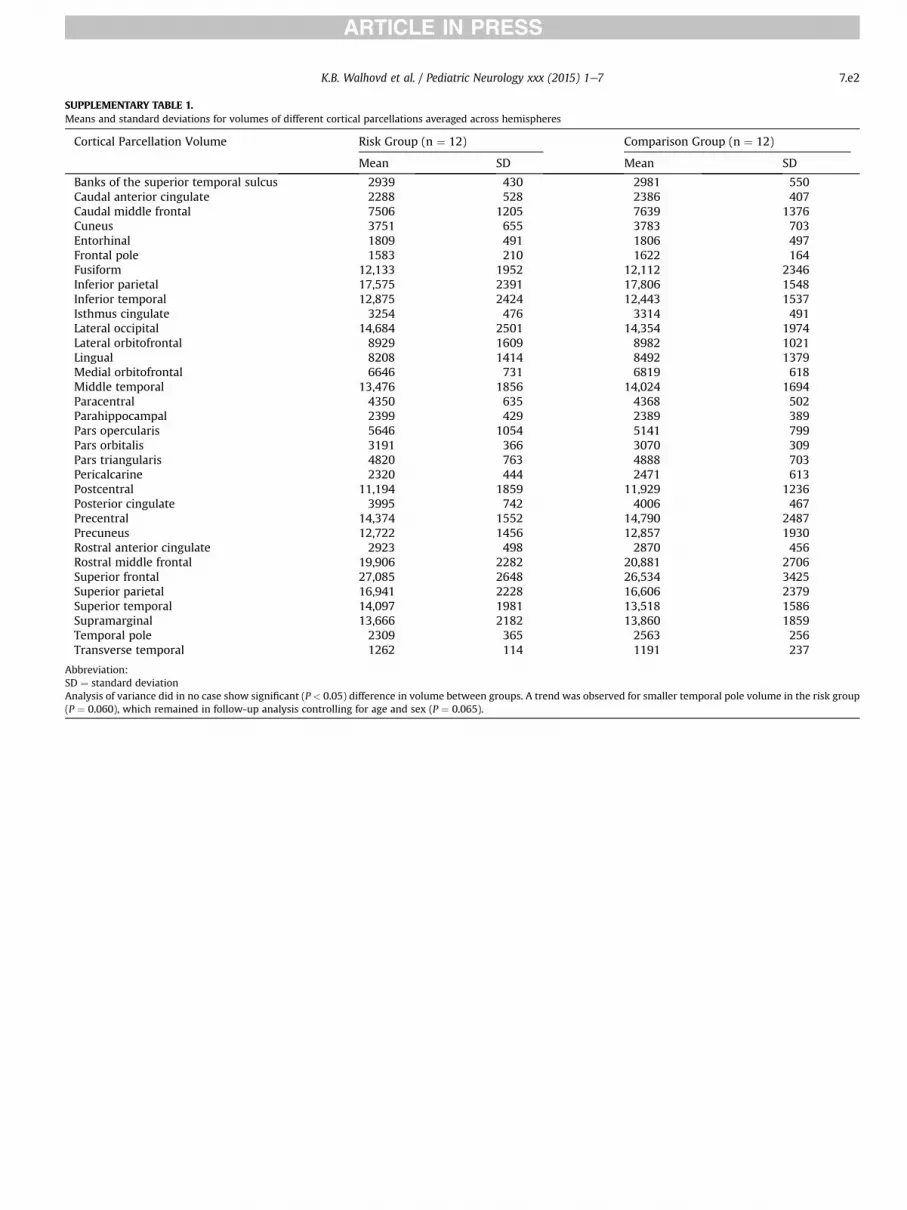
SUPPLEMENTARY TABLE 1.Means and standard deviations for volumes of different cortical parcellations averaged across hemispheres
Cortical Parcellation Volume Risk Group (n ¼ 12) Comparison Group (n ¼ 12)
Mean SD Mean SD
Banks of the superior temporal sulcus 2939 430 2981 550Caudal anterior cingulate 2288 528 2386 407Caudal middle frontal 7506 1205 7639 1376Cuneus 3751 655 3783 703Entorhinal 1809 491 1806 497Frontal pole 1583 210 1622 164Fusiform 12,133 1952 12,112 2346Inferior parietal 17,575 2391 17,806 1548Inferior temporal 12,875 2424 12,443 1537Isthmus cingulate 3254 476 3314 491Lateral occipital 14,684 2501 14,354 1974Lateral orbitofrontal 8929 1609 8982 1021Lingual 8208 1414 8492 1379Medial orbitofrontal 6646 731 6819 618Middle temporal 13,476 1856 14,024 1694Paracentral 4350 635 4368 502Parahippocampal 2399 429 2389 389Pars opercularis 5646 1054 5141 799Pars orbitalis 3191 366 3070 309Pars triangularis 4820 763 4888 703Pericalcarine 2320 444 2471 613Postcentral 11,194 1859 11,929 1236Posterior cingulate 3995 742 4006 467Precentral 14,374 1552 14,790 2487Precuneus 12,722 1456 12,857 1930Rostral anterior cingulate 2923 498 2870 456Rostral middle frontal 19,906 2282 20,881 2706Superior frontal 27,085 2648 26,534 3425Superior parietal 16,941 2228 16,606 2379Superior temporal 14,097 1981 13,518 1586Supramarginal 13,666 2182 13,860 1859Temporal pole 2309 365 2563 256Transverse temporal 1262 114 1191 237
Abbreviation:SD ¼ standard deviationAnalysis of variance did in no case show significant (P < 0.05) difference in volume between groups. A trend was observed for smaller temporal pole volume in the risk group(P ¼ 0.060), which remained in follow-up analysis controlling for age and sex (P ¼ 0.065).
K.B. Walhovd et al. / Pediatric Neurology xxx (2015) 1e7 7.e2

SUPPLEMENTARY FIGURE 1.The flow chart depicts the overall study with inclusion and exclusion of participants for the present article, resulting in the final number of 24 participants(12 control children and 12 children in the risk group).
K.B. Walhovd et al. / Pediatric Neurology xxx (2015) 1e77.e3


