Embed Size (px)
Citation preview
Journal of Cardiology (2008) 52, 140—145
ORIGINAL ARTICLE
Chlamydia pneumoniae IgA and elevatedlevel of IL-6 may synergize to acceleratecoronary artery disease
Hem Chandra Jha (MSc)a, Pragya Srivastava (MSc)a,Rakesh Sarkar (MSc)a, Jagdish Prasad (MS, DNB, MCh)b,Aruna Mittal (PhD)a,∗
a Institute of Pathology-ICMR, Safdarjung Hospital Campus, Post Box No. 4909, New Delhi 110029, Indiab Department of Cardio Thoracic & Vascular Surgery, Safdarjung Hospital, New Delhi 110029, India
Received 11 April 2008; received in revised form 23 May 2008; accepted 20 June 2008Available online 12 August 2008
KEYWORDSIgA serology;IL-2;hsCRP;TNF-�;Physiological andbaseline characteristics
SummaryObjectives: A strong association of cytokines [interleukin (IL)-6, IL-2 and tumornecrosis factor (TNF)-�] with atherosclerosis is well documented. However, theirrole in Chlamydia pneumoniae (Cp)-associated progression of coronary artery dis-ease (CAD) is not well studied. Hence we evaluated the presence of these cytokinesin CAD patients positive for Cp immunoglobulin (Ig) A (low, medium, and high levels)CAD and compared with controls. Also the physiological and baseline characteristicsin IL-6 positive CAD patients were studied.Methods: During the period from March 2005 to June 2007, 192 CAD patients andcontrols attending the cardiology clinic of Safdarjung Hospital, New Delhi, India wereenrolled. ELISA was performed for the estimation of Cp IgA, high sensitive C-reactiveprotein (hsCRP), and cytokines IL-6, IL-2, and TNF-� in CAD patients and controls.Results: IL-6 was significantly higher in CAD patents with medium and higher CpIgA levels (35 vs. 10 and 32 vs. 4) compared to controls. Physiological features andbaseline characteristics of CAD were significantly higher in IL-6 positive CAD patients.Additionally, in CAD patients IL-6 was significantly correlated with hsCRP (p < 0.001,r = 0.42).
Conclusions: Our results lead us to hypothesize the role of IL-6 in the pathogenesisof CAD, which may subsequently get accelerated through Cp infection.© 2008 Japanese College ofreserved.∗ Corresponding author. Tel.: +91 11 26198 402 05;fax: +91 11 26198 401.
E-mail address: amittal [email protected] (A. Mittal).
I
Tc
0914-5087/$ — see front matter © 2008 Japanese College of Cardiolodoi:10.1016/j.jjcc.2008.07.001
Cardiology. Published by Elsevier Ireland Ltd. All rights
ntroduction
he World Health Organization expects cardiovas-ular disease to be a major killer globally owing
gy. Published by Elsevier Ireland Ltd. All rights reserved.
C
tcfsoipintfaditCi(c
scdriIIhIpwiemeI
M
P
Aao2fpm(th
l(
cutpf
iiubnfrtisa
C
VhSatT
Sa
Dp(dC(okiwr
sD
S
SIl
hlamydia pneumoniae and coronary artery disease
o its rapidly increasing prevalence in developingountries [1]. Apart from known conventional riskactors such as dyslepidemia, diabetes, hyperten-ion, and sedentary life style, a strong associationf inflammation and cytokines with atherosclerosiss documented [2,3]. There is strong evidence sup-orting the central role of interleukin (IL)-6 in thenflammatory response as it is the primary determi-ant of C-reactive protein (CRP) which is reportedo be one of the most powerful independent riskactors for coronary heart disease (CHD) [4—6]. Inddition, bacterial pathogens in particular Chlamy-ia pneumoniae (Cp) are reported to be potentnducers of IL-6, which may induce plaque produc-ion and subsequently may cause elevation in serumRP and IL-6 levels [7,8]. Moreover, IL-2 a pro-
nflammatory cytokine secreted by T-helper type 1Th1) lymphocytes has been correlated with variousardiovascular disorders [9].
However, the exact involvement of tumor necro-is factor (TNF)-� levels, a potent inflammatoryytokine and IL-2 in the pathophysiology of cardiacisorders are still undefined [10—12]. Since Cp IgA iseported to be a prevalent marker for CAD patientsn India [13,14], we evaluated the cytokines IL-6,L-2, and TNF-� in CAD patients positive for CpgA with low, medium, and high levels as it willelp in understanding the chronicity of CAD inndia. In addition, taking IL-6 as a marker in CADatients, baseline and physiological characteristicere compared with controls in order to elucidate
ts significance in disease progression. Further tostablish IL-6 as an independent atheroscleroticarker, we evaluated the correlation between lev-
ls of IL-6 and high sensitive (hs)CRP, and levels ofL-6 and IL-2 in CAD patients as well as in controls.
aterials and methods
atients
total of 192 patients (148 males and 44 females)ttending the cardiology out patient departmentf Safdarjung hospital from March 2005 to June007 for suspected CAD and subsequently admittedor angioplasty were enrolled for the study afterrior written consent. In addition, 192 age- and sex-atched healthy controls with no evidence of CAD
142 males and 50 females) were also included in
he study. The study received clearance from theospital ethics review committee.Inclusion criteria: Evidence of CAD required ateast one of the following: (1) significant stenosis70% of luminal diameter) in at least one major
nsvol
141
oronary artery proved by angiography and hadndertaken either percutaneous coronary interven-ion or coronary artery bypass graft (CABG); (2)ositive stress myocardial perfusion imaging studiesor ischemia.
Exclusion criteria: Patients were not includedf any of the following were present: myocardialnfarction or CABG in the preceding 3 months;nstable angina; significant valvular heart disease;lood pressure 180/100 mm. A detailed question-aire was prepared to gather necessary informationrom each patient that included hemoglobin, heartate, blood pressure, blood sugar, blood urea,otal leukocyte count (TLC), smoking habit, alcoholntake habit, lifestyle, migration, socio-economictatus, food habit, and number of previous heartttacks, if any.
ollection of samples
enous blood (2 mL) was collected in non-eparinized tubes from CAD patients and controls.erum was separated within 2 h of blood collectionnd kept at −80 ◦C until used further for detec-ion of antibodies to Cp-IgA, hsCRP, IL-6, IL-2, andNF-�.
erology and antibody level oftherosclerotic marker
etection of antibodies for Cp-specific IgA waserformed using commercially available ELISA kitR-Biopharm AG, Damstadt, Germany) [13]. Weivided the samples into three groups based onp IgA index number, low (1.1—1.6), medium1.7—2.1), and high (more than 2.1). For detectionf antibodies to hsCRP, ELISA was performed usingits (Calibiotech Inc., USA) as per manufacturer’snstructions. Level of the hsCRP (>3 mg/L) in serumas considered as hsCRP positive in dichotomized
esult.The cytokines IL-6, IL-2, and TNF- � were mea-
ured by commercial kit from (Ebiosciences, Saniego, CA, USA) as per manufacturer’s instructions.
tatistical analysis
PSS version 12.0 for Windows (SPSS Inc., Chicago,L, USA) was used for statistical analysis. All sero-ogical results were dichotomized as positive or
egative. For comparing diagnostic assays, chi-quare test, Fisher-exact for binary a relatedariable was used. Simultaneously, an alpha levelf 0.05 was set as the level of significance. Binaryogistic regression was used to evaluate physiologi-
142
Tabl
e1
Low
,m
ediu
man
dhi
ghle
vels
ofCh
lam
ydia
pneu
mon
iae
spec
ific
IgA
inhs
CRP,
IL-6
,IL
-2an
dTN
F-�
grou
pof
coro
nary
arte
rydi
seas
epa
tien
tsan
dco
ntro
ls
Pro-
infl
amm
ator
ym
arke
rsLo
wIg
AM
ediu
mIg
AH
igh
IgA
CAD
-pt
(N=
37)
Cont
rol(
N=
22)
Pva
lue
CAD
-pt
Cont
rol
Pva
lue
CAD
-pt
Cont
rol
Pva
lue
hsCR
P16
(43.
2)06
(27.
2)0.
001*
43(7
0.5)
07(1
9.4)
<0.0
01*
34(5
9.6)
06(3
3.3)
<0.0
01*
IL-6
16(4
3.2)
08(3
6.3)
0.40
535
(57.
3)10
(27.
7)0.
006*
32(5
6.1)
04(2
2.2)
0.01
1*
IL-2
06(1
6.2)
02(9
.1)
0.20
015
(24.
6)06
(16.
6)0.
069
08(1
4)02
(11.
1)0.
358
TNF-
�04
(10.
8)01
(4.5
)0.
244
12(1
9.6)
05(1
3.8)
0.10
406
(10.
5)03
(16.
6)0.
324
CAD
-pt:
coro
nary
arte
rydi
seas
epa
tien
ts,
hsCR
P:hi
ghse
nsit
ive
C-re
acti
vepr
otei
n.*
Sign
ifica
nt,
perc
enta
gein
pare
nthe
sis.
ccbp
R
Caa
IifhtftspvtifrSvfr0e2(
BCI
Awd(hbCtstnaIcIn
H.C. Jha et al.
al and baseline characteristics of CAD. Spearman’sorrelation and t-test was used for the evaluationetween hsCRP and IL-6 as well as IL-6 and IL-2 inatients and healthy controls.
esults
lassification of IgA seropositivity in hsCRPnd pro-inflammatory cytokines (IL-2, IL-6,nd TNF-�) groups
n the group with low Cp-IgA index value consist-ng of 37 CAD patients and 22 controls, positivityor hsCRP (16 vs. 6, respectively) was significantlyigher (p = 0.001) in CAD patients compared to con-rols, however there was no significant differenceor IL-6 (16 vs. 8, respectively), IL-2 (6 vs. 2, respec-ively), and TNF-� (4 vs. 1, respectively). In theecond group with medium IgA index value (61 CADatients and 36 controls) positivity for hsCRP (43s. 7, respectively) and IL-6 (35 vs. 10, respec-ively) was significantly higher (p < 0.001 and 0.006)n CAD patients compared to controls, however dif-erences were not significant for IL-2 (15 vs. 6,espectively) and TNF-� (12 vs. 5, respectively).imilarly in the third group with high IgA indexalue (57 CAD patients and 18 controls) positivityor hsCRP (34 vs. 6, respectively) and IL-6 (32 vs. 4,espectively) was significantly higher (p < 0.001 and.011) in CAD patients compared to controls, how-ver differences were not significant for IL-2 (8 vs., respectively) and TNF-� (6 vs. 3, respectively)Table 1).
aseline and physiological characteristics ofAD patients and controls on the basis of
L-6 positivity
total of 192 CAD patients and 192 controlsere divided on the basis of IL-6 positivity underifferent physiological and baseline characteristicsTable 2). Some physiological features such aseart rate, systolic and diastolic blood pressure,lood sugar, blood urea, and TLC of IL-6 positiveAD patients were significantly higher comparedo IL-6 positive controls. However, hemoglobin wasignificantly lower in IL-6 positive CAD patientshan IL-6 positive controls. Similarly in smokers,on-smokers, alcoholic, non-alcoholic, normal
nd sedentary lifestyle, migrant and non-migrantL-6 positive CAD patients was significantly higherompared to IL-6 positive controls. Additionally,L-6 positive CAD patients were found to be sig-ificantly higher in medium socioeconomic status
Chlamydia pneumoniae and coronary artery disease 143
Table 2 Comparison of baseline and physiological characteristic among IL-6 positive CAD patients and controls
Physiological characteristic Patient mean (±S.D.) Control mean (±S.D.) P value
Hemoglobin (gm/dL) 11.78 ± 2.24 12.5 ± 2.64 <0.001*
Heart rate (beats/min) 78 ± 8 74 ± 5 <0.001*
Systolic blood pressure (mm Hg) 137 ± 22 126 ± 15 <0.001*
Diastolic blood pressure (mm Hg) 90 ± 14 84 ± 9 <0.001*
Blood sugar (mmol/L) 132 ± 22 108 ± 18 <0.001*
Blood urea (mmol/L) 30 ± 12 26 ± 8 <0.001*
TLC (104 cells/L) 7870 ± 1370 7450 ± 1240 0.002*
Baseline characteristic Patient Control P value
Smoker (%) 80 (48.5) 30 (34) 0.029*
Non-smoker (%) 26 (42.6) 09 (12.8) <0.001*
Alcoholic (%) 44 (48.3) 20 (19.6) <0.001*
Non-alcoholic (%) 52 (52) 30 (33.3) 0.010*
Normal LS (%) 39 (52) 22 (23.1) <0.001*
Sedentary LS (%) 56 (47.8) 28 (31.8) 0.001*
Migrant (%) 85 (60.7) 36 (40.9) <0.001*
Non-migrant (%) 37 (44.5) 15 (20.2) 0.001*
LSES (%) 27 (48.2) 08 (29.6) 0.048*
MSES (%) 51 (50.5) 22 (21.7) <0.001*
18 (30) 0.027*
(a0hschI(
C
Arps(nwar
D
Ct[mac
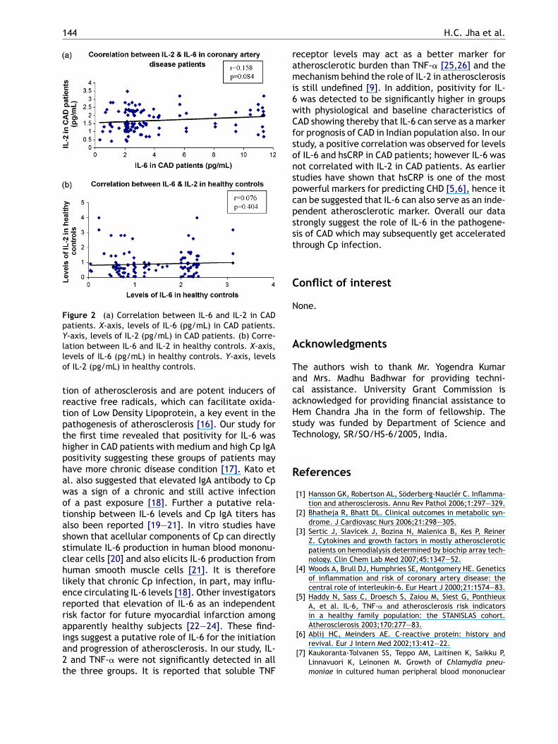
Figure 1 (a) Correlation between IL-6 and hsCRP in CAD
HSES (%) 16 (50)* Significant, percentages in parenthesis.
MSES) (p < 0.001), low socioeconomic status (LSES),nd high socioeconomic status (HSES) (p = 0.048 and.027, respectively) as compared to controls. Foodabit viz. vegetarian and non-vegetarian was notignificant between IL-6 positive CAD patients andontrols in our study (data not shown). Number ofeart attacks was also not significantly different inL-6 positive CAD patients as compared to controlsdata not shown).
orrelation analysis
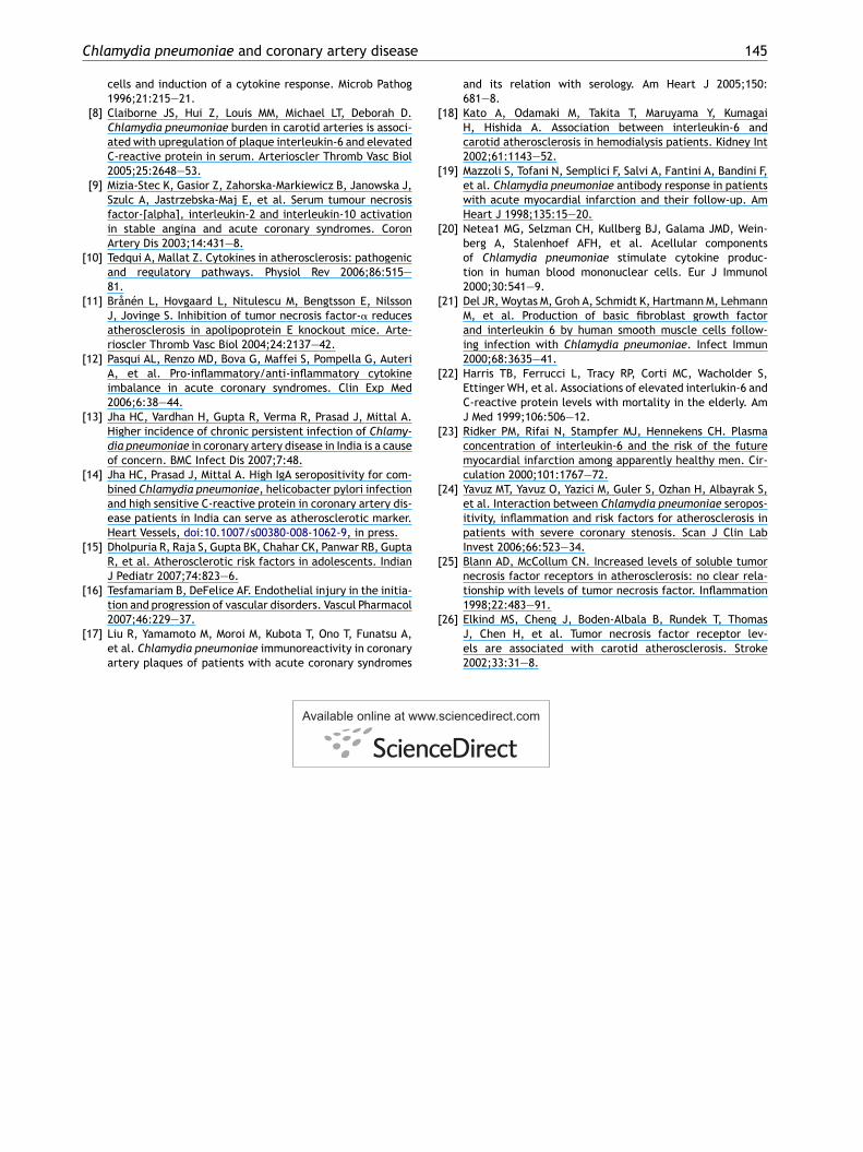
positive and significant correlation (p < 0.001,= 0.424) was found between hsCRP and IL-6 in CADatients (Fig. 1a). However, the correlation was notignificant between IL-6 and IL-2 in CAD patientsp = 0.084, r = 0.158) (Fig. 2a). In healthy controls,o significant correlation (p = 0.995, r = −0.001)as found between hsCRP and IL-6 and for IL-6nd IL-2 (p = 0.404, r = 0.076) (Figs. 1 and 2(b),espectively).
iscussion
AD contributes significantly to morbidity and mor-
ality, particularly in developing countries like India15]. The role of various immuno-inflammatoryediators such as cytokines in the pathogenesis oftherosclerosis is gaining importance since theseytokines have a role in triggering the perpetua-
patients. X-axis, levels of hsCRP (mg/L) in CAD patients.Y-axis, levels of IL-6 (pg/mL) in CAD patients. (b) Corre-lation between IL-6 and hsCRP in healthy controls. X-axis,levels of hsCRP (mg/L) in healthy controls. Y-axis, levelsof IL-6 (pg/mL) in healthy controls.
144
Figure 2 (a) Correlation between IL-6 and IL-2 in CADpatients. X-axis, levels of IL-6 (pg/mL) in CAD patients.Y-axis, levels of IL-2 (pg/mL) in CAD patients. (b) Corre-
rami6wCfsonspcpsst
C
N
A
TacaHsT
R
lation between IL-6 and IL-2 in healthy controls. X-axis,levels of IL-6 (pg/mL) in healthy controls. Y-axis, levelsof IL-2 (pg/mL) in healthy controls.
tion of atherosclerosis and are potent inducers ofreactive free radicals, which can facilitate oxida-tion of Low Density Lipoprotein, a key event in thepathogenesis of atherosclerosis [16]. Our study forthe first time revealed that positivity for IL-6 washigher in CAD patients with medium and high Cp IgApositivity suggesting these groups of patients mayhave more chronic disease condition [17]. Kato etal. also suggested that elevated IgA antibody to Cpwas a sign of a chronic and still active infectionof a past exposure [18]. Further a putative rela-tionship between IL-6 levels and Cp IgA titers hasalso been reported [19—21]. In vitro studies haveshown that acellular components of Cp can directlystimulate IL-6 production in human blood mononu-clear cells [20] and also elicits IL-6 production fromhuman smooth muscle cells [21]. It is thereforelikely that chronic Cp infection, in part, may influ-ence circulating IL-6 levels [18]. Other investigatorsreported that elevation of IL-6 as an independentrisk factor for future myocardial infarction amongapparently healthy subjects [22—24]. These find-
ings suggest a putative role of IL-6 for the initiationand progression of atherosclerosis. In our study, IL-2 and TNF-� were not significantly detected in allthe three groups. It is reported that soluble TNFH.C. Jha et al.
eceptor levels may act as a better marker fortherosclerotic burden than TNF-� [25,26] and theechanism behind the role of IL-2 in atherosclerosis
s still undefined [9]. In addition, positivity for IL-was detected to be significantly higher in groupsith physiological and baseline characteristics ofAD showing thereby that IL-6 can serve as a markeror prognosis of CAD in Indian population also. In ourtudy, a positive correlation was observed for levelsf IL-6 and hsCRP in CAD patients; however IL-6 wasot correlated with IL-2 in CAD patients. As earliertudies have shown that hsCRP is one of the mostowerful markers for predicting CHD [5,6], hence itan be suggested that IL-6 can also serve as an inde-endent atherosclerotic marker. Overall our datatrongly suggest the role of IL-6 in the pathogene-is of CAD which may subsequently get acceleratedhrough Cp infection.
onflict of interest
one.
cknowledgments
he authors wish to thank Mr. Yogendra Kumarnd Mrs. Madhu Badhwar for providing techni-al assistance. University Grant Commission iscknowledged for providing financial assistance toem Chandra Jha in the form of fellowship. Thetudy was funded by Department of Science andechnology, SR/SO/HS-6/2005, India.
eferences
[1] Hansson GK, Robertson AL, Söderberg-Nauclér C. Inflamma-tion and atherosclerosis. Annu Rev Pathol 2006;1:297—329.
[2] Bhatheja R, Bhatt DL. Clinical outcomes in metabolic syn-drome. J Cardiovasc Nurs 2006;21:298—305.
[3] Sertic J, Slavicek J, Bozina N, Malenica B, Kes P, ReinerZ. Cytokines and growth factors in mostly atheroscleroticpatients on hemodialysis determined by biochip array tech-nology. Clin Chem Lab Med 2007;45:1347—52.
[4] Woods A, Brull DJ, Humphries SE, Montgomery HE. Geneticsof inflammation and risk of coronary artery disease: thecentral role of interleukin-6. Eur Heart J 2000;21:1574—83.
[5] Haddy N, Sass C, Droesch S, Zaiou M, Siest G, PonthieuxA, et al. IL-6, TNF-� and atherosclerosis risk indicatorsin a healthy family population: the STANISLAS cohort.Atherosclerosis 2003;170:277—83.
[6] Ablij HC, Meinders AE. C-reactive protein: history andrevival. Eur J Intern Med 2002;13:412—22.
[7] Kaukoranta-Tolvanen SS, Teppo AM, Laitinen K, Saikku P,Linnavuori K, Leinonen M. Growth of Chlamydia pneu-moniae in cultured human peripheral blood mononuclear
C
[
[
[
[
[
[
[
[
[
[
[
[
[
[
[
[
hlamydia pneumoniae and coronary artery disease
cells and induction of a cytokine response. Microb Pathog1996;21:215—21.
[8] Claiborne JS, Hui Z, Louis MM, Michael LT, Deborah D.Chlamydia pneumoniae burden in carotid arteries is associ-ated with upregulation of plaque interleukin-6 and elevatedC-reactive protein in serum. Arterioscler Thromb Vasc Biol2005;25:2648—53.
[9] Mizia-Stec K, Gasior Z, Zahorska-Markiewicz B, Janowska J,Szulc A, Jastrzebska-Maj E, et al. Serum tumour necrosisfactor-[alpha], interleukin-2 and interleukin-10 activationin stable angina and acute coronary syndromes. CoronArtery Dis 2003;14:431—8.
10] Tedqui A, Mallat Z. Cytokines in atherosclerosis: pathogenicand regulatory pathways. Physiol Rev 2006;86:515—81.
11] Brånén L, Hovgaard L, Nitulescu M, Bengtsson E, NilssonJ, Jovinge S. Inhibition of tumor necrosis factor-� reducesatherosclerosis in apolipoprotein E knockout mice. Arte-rioscler Thromb Vasc Biol 2004;24:2137—42.
12] Pasqui AL, Renzo MD, Bova G, Maffei S, Pompella G, AuteriA, et al. Pro-inflammatory/anti-inflammatory cytokineimbalance in acute coronary syndromes. Clin Exp Med2006;6:38—44.
13] Jha HC, Vardhan H, Gupta R, Verma R, Prasad J, Mittal A.Higher incidence of chronic persistent infection of Chlamy-dia pneumoniae in coronary artery disease in India is a causeof concern. BMC Infect Dis 2007;7:48.
14] Jha HC, Prasad J, Mittal A. High IgA seropositivity for com-bined Chlamydia pneumoniae, helicobacter pylori infectionand high sensitive C-reactive protein in coronary artery dis-ease patients in India can serve as atherosclerotic marker.Heart Vessels, doi:10.1007/s00380-008-1062-9, in press.
15] Dholpuria R, Raja S, Gupta BK, Chahar CK, Panwar RB, GuptaR, et al. Atherosclerotic risk factors in adolescents. IndianJ Pediatr 2007;74:823—6.
16] Tesfamariam B, DeFelice AF. Endothelial injury in the initia-
tion and progression of vascular disorders. Vascul Pharmacol2007;46:229—37.17] Liu R, Yamamoto M, Moroi M, Kubota T, Ono T, Funatsu A,et al. Chlamydia pneumoniae immunoreactivity in coronaryartery plaques of patients with acute coronary syndromes
[
Available online at www.
145
and its relation with serology. Am Heart J 2005;150:681—8.
18] Kato A, Odamaki M, Takita T, Maruyama Y, KumagaiH, Hishida A. Association between interleukin-6 andcarotid atherosclerosis in hemodialysis patients. Kidney Int2002;61:1143—52.
19] Mazzoli S, Tofani N, Semplici F, Salvi A, Fantini A, Bandini F,et al. Chlamydia pneumoniae antibody response in patientswith acute myocardial infarction and their follow-up. AmHeart J 1998;135:15—20.
20] Netea1 MG, Selzman CH, Kullberg BJ, Galama JMD, Wein-berg A, Stalenhoef AFH, et al. Acellular componentsof Chlamydia pneumoniae stimulate cytokine produc-tion in human blood mononuclear cells. Eur J Immunol2000;30:541—9.
21] Del JR, Woytas M, Groh A, Schmidt K, Hartmann M, LehmannM, et al. Production of basic fibroblast growth factorand interleukin 6 by human smooth muscle cells follow-ing infection with Chlamydia pneumoniae. Infect Immun2000;68:3635—41.
22] Harris TB, Ferrucci L, Tracy RP, Corti MC, Wacholder S,Ettinger WH, et al. Associations of elevated interlukin-6 andC-reactive protein levels with mortality in the elderly. AmJ Med 1999;106:506—12.
23] Ridker PM, Rifai N, Stampfer MJ, Hennekens CH. Plasmaconcentration of interleukin-6 and the risk of the futuremyocardial infarction among apparently healthy men. Cir-culation 2000;101:1767—72.
24] Yavuz MT, Yavuz O, Yazici M, Guler S, Ozhan H, Albayrak S,et al. Interaction between Chlamydia pneumoniae seropos-itivity, inflammation and risk factors for atherosclerosis inpatients with severe coronary stenosis. Scan J Clin LabInvest 2006;66:523—34.
25] Blann AD, McCollum CN. Increased levels of soluble tumornecrosis factor receptors in atherosclerosis: no clear rela-tionship with levels of tumor necrosis factor. Inflammation
1998;22:483—91.26] Elkind MS, Cheng J, Boden-Albala B, Rundek T, ThomasJ, Chen H, et al. Tumor necrosis factor receptor lev-els are associated with carotid atherosclerosis. Stroke2002;33:31—8.
sciencedirect.com


