Embed Size (px)
Citation preview


¿QUÉ ES LA ELECTROFORESIS?

ELECTROFORESIS DE ADN

CARACTERÍSTICAS DE LA ELECTROFORESIS

AGAROSA
Selección de Concentración de Agarosa
Concentración
Proteína (MW. Daltons)
Polisacárido (MW. Daltons)
ADN (pb)

ACRILAMIDA
Monómeros de acrilamida polimerizan en largas cadenas que están covalentemente unidas por entrecruzamiento
N,N-Metil-bis-acrilamida

ELECTROFORESIS

ELECTROFORESIS HORIZONTAL DE ADN

VISUALIZACIÓN DEL ADN

ELECTROFORESIS CONVENCIONAL
ELECTROFORESIS DE PRODUCTOS DE AMPLIFICACIÓN POR PCR
RECPETOR A2A ADENOSINA
LA ELECTROFORESIS POR SI SÓLA, NO PERMITE CONOCER LA SECUENCIA DE ADN, SÓLO
SEPARAR FRAGMENTOS DE ADN

El DNA puede sufrir un proceso reversible de separación y reasociación de hebras


HIBRIDACIÓN DE ACIDOS NUCLEICOS
La hibridación de ácidos nucleicos (ADN o ARN) es un proceso por el cual se combinan dos cadenas de ácidos nucleicos antiparalelas y con secuencias de bases complementarias en una única molécula de doble cadena, que toma la estructura de doble hélice, donde las bases nitrogenadas quedan ocultas en el interior
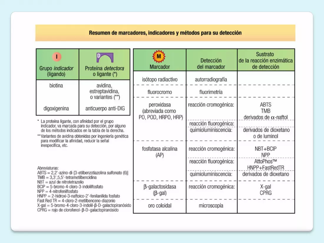
Sustancia radioactivaFluorescenteEnzima

A pesar de su especificidad, las sondas carecen de s u f i c i e n t e s e n s i b i l i d a d p a r a d e t e c t a r l o s microorganismos de una muestra clínica cuando son escasos, por lo que en la actualidad se emplean poco para este fin.
Por el contrario, constituyen un instrumento excelente para identificar un microorganismo una vez aislado por cultivo, ya que se dispone de ADN en abundancia.
R e s u l t a n p a r t i c u l a r m e n t e ú t i l e s p a r a l o s microorganismos cuya identificación por métodos convencionales es difícil o compleja

La hibridación con sondas específicas permite determinar la identidad de un fragmento de DNA o RNA

ANALYZING AND MANIPULATING DNA 537
Alternatively, DNA probes can be used in hybridization reactions with RNArather than DNA to find out whether a cell is expressing a given gene. In this casea DNA probe that contains part of the gene’s sequence is hybridized with RNApurified from the cell in question to see whether the RNA includes nucleotidesequences matching the probe DNA and, if so, in what quantities. In somewhatmore elaborate procedures, the DNA probe is treated with specific nucleasesafter the hybridization is complete, to determine the exact regions of the DNAprobe that have paired with the RNA molecules. One can thereby determine thestart and stop sites for RNA transcription, as well as the precise boundaries ofthe intron and exon sequences in a gene (Figure 8–37).
Today, the positions of intron/exon boundaries are usually determined bysequencing the complementary DNA (cDNA) sequences that represent themRNAs expressed in a cell and comparing them with the nucleotide sequence ofthe genome. We describe later how cDNAs are prepared from mRNAs.
The hybridization of DNA probes to RNAs allows one to determine whetheror not a particular gene is being transcribed; moreover, when the expression ofa gene changes, one can determine whether the change is due to transcriptionalor post-transcriptional controls (see Figure 7–92). These tests of gene expressionwere initially performed with one DNA probe at a time. DNA microarrays nowallow the simultaneous monitoring of hundreds or thousands of genes at a time,as we discuss later. Hybridization methods are in such wide use in cell biologytoday that it is difficult to imagine how we could study gene structure andexpression without them.
Figure 8–35 In situ hybridization to locate specific genes onchromosomes. Here, six different DNA probes have been used to mark thelocations of their respective nucleotide sequences on human chromosome5 at metaphase. The probes have been chemically labeled and detectedwith fluorescent antibodies. Both copies of chromosome 5 are shown,aligned side by side. Each probe produces two dots on each chromosome,since a metaphase chromosome has replicated its DNA and thereforecontains two identical DNA helices. (Courtesy of David C. Ward.)
B
CA
D
E
F
D
C
BF
E
A
C
D
A
F
E
hybridization in 50%formamide at 42oC
hybridization in 50%formamide at 35oC
+
single-strandedDNA probesfor gene A mixture of
single-strandedDNA molecules
B
imperfectbase-pairing
A, C, and E all formstable double helices
only A forms stabledouble helix
Figure 8–36 Stringent versusnonstringent hybridization conditions.To use a DNA probe to find an identicalmatch, stringent hybridization conditionsare used; the reaction temperature iskept just a few degrees below that atwhich a perfect DNA helix denatures inthe solvent used (its melting temperature),so that all imperfect helices formed areunstable. When a DNA probe is beingused to find DNAs with related, as well asidentical, sequences, less stringentconditions are used; hybridization isperformed at a lower temperature, whichallows even imperfectly paired doublehelices to form. Only the lower-temperature hybridization conditions canbe used to search for genes that arenonidentical but related to gene A (C andE in this example).
Existen dos condiciones de hibridación:

TIPOS DE HIBRIDACIÓN
En solución o fase líquida El soporte o
fase sólida

Hibridación en fase líquida

Hibridación en solución (en un tubo)
- Se extrae el ADN del microorganismo, se
Tipos de hibridación
- Las sondas detectan la secuencia complementaria en el genoma de un microorganismo a través de una prueba de hibridación realizada: en solución, sobre un soporte sólido, o in situ
- Se extrae el ADN del microorganismo, se desnaturaliza en solución en un tubo y se añade la sonda marcada con acridina
- Si encuentra su secuencia complementaria se une para formar un fragmento bicatenario
- A continuación, se añade un álcali, que inactiva la acridina cuando la sonda está libre, pero no cuando la sonda ha hibridado con su secuencia, por lo que la luminiscencia medida por un luminómetro corresponde a la sonda hibridada
S e e x t r a e e l A D N d e l m i c r o o r g a n i s m o , s e desnaturaliza en solución en un tubo y se añade la sonda marcada con acridina
Si encuentra su secuencia complementaria se une para f o r m a r u n f r a g m e n t o bicatenario
A continuación, se añade un álcali, que inactiva la acridina cuando la sonda está libre, pero no cuando la sonda ha hibridado con su secuencia, por lo que la luminiscencia medida por un luminómetro cor responde a la sonda hibridada

- Se extrae el ADN después de lisar los microorganismos de una muestra clínica o de un cultivo, se desnaturaliza por calor y se deposita una gota sobre una membrana de nailon o de nitrocelulosa
- La muestra depositada se absorbe y se fija
- Se sumerge la membrana en una solución de hibridación que
contiene la sonda marcada
- Finalizada la hibridación se lava la membrana para eliminar la sonda no unida
- Se detecta la sonda fijada a través del marcador - Se utiliza sobre todo con fines de investigación
Hibridación en fase Sólida


Técnicas basadas en hibridación en fase sólida
In situ
Sourthen blot
Northenblot

El método tipo Southern o Southern blot fue desarrollado por E. M. Southern para la detección de genes específicos en el ADN celular.
El ADN es digerido con una enzima de restricción y los fragmentos son separados por tamaños mediante una electroforesis en un gel. Se realiza tinción con bromuro de etidio para comprobar la calidad de la electroforesis y del ADN.
A continuación los fragmentos de ADN de doble cadena son parcialmente hidrolizados con un ácido débil y desnaturalizados con NaOH para permitir la transferencia. Posteriormente, el ADN es transferido a un filtro de nitrocelulosa, con lo que en el filtro queda representada una réplica de la disposición de los fragmentos de ADN presentes en el gel.
Southern blot

A continuación el filtro se incuba durante un tiempo con la sonda marcada (radiactivamente o con un fluorocromo); durante la incubación la sonda se va hibridando a las moléculas de ADN de cadena sencilla de secuencia complementaria (o muy parecida). La sonda unida al fragmento de ADN complementario se puede visualizar en el filtro de una forma sencilla mediante una exposición a una película de rayos X para el caso de sondas radiactivas o con una película sensible a la luz, para el caso de sondas con fluorocromo.
Southern blot


Northern blot
Es esencialmente idéntica al método de Southern blot, salvo que las moléculas de ácido nucleico de la muestra, en este caso de ARN (total, mensajero, vírico, etc.), se separan por electroforesis en condiciones desnaturalizantes (en presencia de formaldehído, que forma parte de la composición del gel).
Esto es debido a que, a pesar de ser una molécula de una sola cadena, se forman complejas estructuras secundarias que dificultan la migración. Al igual que en el Southern blot, el ARN se transfiere a la membrana de filtro y se hibridan con sondas de ADN marcada.
Durante la electroforesis se pueden observan grandes cantidades de ARNm correspondiente a ribosomal (unidades 28S y 18S).
Tanto la técnica de Southern como de Northern blot se usan poco hoy en día al haber sido sustituidas por técnicas derivadas de la PCR.

ANALYZING AND MANIPULATING DNA 539
produces albumin mRNA. The RNA molecules that hybridize to the labeled DNAprobe on the paper (because they are complementary to part of the normalalbumin gene sequence) are then located by detecting the bound probe byautoradiography or by chemical means (Figure 8–38). The sizes of thehybridized RNA molecules can be determined by reference to RNA standards ofknown sizes that are electrophoresed side by side with the experimental sample.In this way, one might discover that liver cells from the mutant mice make albu-min mRNA in normal amounts and of normal size; alternatively, you might findthat they make it in normal size but in greatly reduced amounts. Another possi-bility is that the mutant albumin mRNA molecules are abnormally short; in thiscase the gel blot could be retested with a series of shorter DNA probes, each cor-responding to small portions of the gene, to reveal which part of the normal RNAis missing.
The original gel-transfer hybridization method, called Southern blotting,analyzes DNA rather than RNA. (It was named after its inventor, and the North-ern and Western blotting techniques were named with reference to it.) Here, iso-lated DNA is first cut into readily separable fragments with restriction nucleases.The double-stranded fragments are then separated on the basis of size by gelelectrophoresis, and those complementary to a DNA probe are identified byblotting and hybridization, as just described for RNA (see Figure 8–38). To char-acterize the structure of the albumin gene in the mutant mice, an albumin-spe-cific DNA probe would be used to construct a detailed restriction map of thegenome in the region of the albumin gene (such a map consists of the pattern ofDNA fragments produced by various restriction nucleases). From this map one
unlabeled RNA or DNA
labeled RNA or DNA of known sizes serving as size markers
electrophoresis
agarosegel
sponge
alkali solution
stack of paper towels
NUCLEIC ACIDS SEPARATEDACCORDING TO SIZE BY AGAROSE-GEL ELECTROPHORESIS
SEPARATED NUCLEIC ACIDS BLOTTED ONTO NITROCELLULOSE PAPER BY SUCTIONOF BUFFER THROUGH GEL AND PAPER
nitrocellulosepaper gel
remove nitrocellulose paper with tightly boundnucleic acids
RADIOLABELED PROBE HYBRIDIZED TO SEPARATED DNA
sealedplasticbag
labeled probe in buffer
LABELED PROBE HYBRIDIZED TOCOMPLEMENTARY DNA BANDSVISUALIZED BY AUTORADIOGRAPHY
positionsof
labeledmarkers
labeledbands
(A) (B) (C)
(D)
(E)
Figure 8–38 Detection of specific RNA or DNA molecules by gel-transferhybridization. In this example, the DNA probe is detected by itsradioactivity. DNA probes detected by chemical or fluorescence methodsare also widely used (see Figure 8–34). (A) A mixture of either single-stranded RNA molecules (Northern blotting) or the double-stranded DNAfragments created by restriction nuclease treatment (Southern blotting) isseparated according to length by electrophoresis. (B) A sheet ofnitrocellulose or nylon paper is laid over the gel, and the separated RNA orDNA fragments are transferred to the sheet by blotting. (C) Thenitrocellulose sheet is carefully peeled off the gel. (D) The sheet containingthe bound nucleic acids is placed in a sealed plastic bag together with abuffered salt solution containing a radioactively labeled DNA probe. Thesheet is exposed to a labeled DNA probe for a prolonged period underconditions favoring hybridization. (E) The sheet is removed from the bag andwashed thoroughly, so that only probe molecules that have hybridized tothe RNA or DNA immobilized on the paper remain attached. Afterautoradiography, the DNA that has hybridized to the labeled probe showsup as bands on the autoradiograph.
For Southern blotting, the strands of the double-stranded DNA moleculeson the paper must be separated before the hybridization process; this isdone by exposing the DNA to alkaline denaturing conditions after the gelhas been run (not shown).

Es una técnica de hibridación que se realiza directamente sobre tejido, células o cromosomas localizados sobre un soporte sólido, habitualmente un portaobjetos.
La visualización puede realizarse por medios isotópicos , enzimáticos o con fluorescencia , siendo evaluada en un microscopio. Su gran virtud es que permite identificar las células donde se produce la hibridación que detecta la alteración genómica o transcripcional.
La visualización de resultados puede realizarse por marcaje con fluorescencia de la sonda (FISH) o cromogénico (CISH, SISH). Este último tiene la ventaja de poder realizarse en microscopio de campo claro y poder almacenarse casi indefinidamente las preparaciones.Los usos más frecuentes en Patología molecular son la detección de amplificaciones (Ej. Her2 en cáncer de mama c-myc en linfoma de Burkitt), traslocaciones ( linfoma folicular, linfoma del manto, sarcomas) y presencia de virus (Epstein-Barr, HPV).
Hibridación in situ

ANALYZING AND MANIPULATING DNA 537
Alternatively, DNA probes can be used in hybridization reactions with RNArather than DNA to find out whether a cell is expressing a given gene. In this casea DNA probe that contains part of the gene’s sequence is hybridized with RNApurified from the cell in question to see whether the RNA includes nucleotidesequences matching the probe DNA and, if so, in what quantities. In somewhatmore elaborate procedures, the DNA probe is treated with specific nucleasesafter the hybridization is complete, to determine the exact regions of the DNAprobe that have paired with the RNA molecules. One can thereby determine thestart and stop sites for RNA transcription, as well as the precise boundaries ofthe intron and exon sequences in a gene (Figure 8–37).
Today, the positions of intron/exon boundaries are usually determined bysequencing the complementary DNA (cDNA) sequences that represent themRNAs expressed in a cell and comparing them with the nucleotide sequence ofthe genome. We describe later how cDNAs are prepared from mRNAs.
The hybridization of DNA probes to RNAs allows one to determine whetheror not a particular gene is being transcribed; moreover, when the expression ofa gene changes, one can determine whether the change is due to transcriptionalor post-transcriptional controls (see Figure 7–92). These tests of gene expressionwere initially performed with one DNA probe at a time. DNA microarrays nowallow the simultaneous monitoring of hundreds or thousands of genes at a time,as we discuss later. Hybridization methods are in such wide use in cell biologytoday that it is difficult to imagine how we could study gene structure andexpression without them.
Figure 8–35 In situ hybridization to locate specific genes onchromosomes. Here, six different DNA probes have been used to mark thelocations of their respective nucleotide sequences on human chromosome5 at metaphase. The probes have been chemically labeled and detectedwith fluorescent antibodies. Both copies of chromosome 5 are shown,aligned side by side. Each probe produces two dots on each chromosome,since a metaphase chromosome has replicated its DNA and thereforecontains two identical DNA helices. (Courtesy of David C. Ward.)
B
CA
D
E
F
D
C
BF
E
A
C
D
A
F
E
hybridization in 50%formamide at 42oC
hybridization in 50%formamide at 35oC
+
single-strandedDNA probesfor gene A mixture of
single-strandedDNA molecules
B
imperfectbase-pairing
A, C, and E all formstable double helices
only A forms stabledouble helix
Figure 8–36 Stringent versusnonstringent hybridization conditions.To use a DNA probe to find an identicalmatch, stringent hybridization conditionsare used; the reaction temperature iskept just a few degrees below that atwhich a perfect DNA helix denatures inthe solvent used (its melting temperature),so that all imperfect helices formed areunstable. When a DNA probe is beingused to find DNAs with related, as well asidentical, sequences, less stringentconditions are used; hybridization isperformed at a lower temperature, whichallows even imperfectly paired doublehelices to form. Only the lower-temperature hybridization conditions canbe used to search for genes that arenonidentical but related to gene A (C andE in this example).


Técnica que permite analizar el nivel de expresión de miles (o todos) los genes de una muestra. Los hay de dos tipos principales: aquellos que utilizan como sondas fragmentos de cDNA y los que usan fragmentos más pequeños de ADN (oligonucleótidos). A estas sondas, colocadas sobre el soporte, se les lanzará el ARN de la muestra que ha sido previamente transformado en cDNA, de tal manera que aquellos genes muy expresados (con gran cantidad de ARN en la muestra original) darán una mayor señal al unirse (hibridación) a su sonda correspondiente, lo cual se detecta por medio de fluorescencia con un lector de luminosidad.
Microarreglos (microarray)
Fig. 4. Representación esquemática del análisis de expresión mediante
microarrays de cDNA. Tras la extracción de ARN, este se transforma en cDNA
y se co-hibrida sobre el microarray con sondas de ADN. Los diferentes colores
tras la hibridación indican el nivel de expresión (rojo: sobreexpresión)
Fig.5. CISH positiva para amplificación de Her2 en cáncer de mama

El resultado del experimento es una “fotografía” del estado de expresión del ARN de los genes de la muestra (tumoral o normal). Se trata por tanto de un método de screening masivo para detectar aquellos genes que se sobre- o infraexpresan en una muestra.
Utilidad en Patología Molecular:
- Detección de mecanismos o vías estimulados o reprimidosen el metabolismo celular (neoplasias o no neoplasias)
- Detección de marcadores tumorales útiles para el diagnóstico yclasificación de las neoplasia
- Detección de marcadores tumorales útiles para el pronóstico ypredicción de respuesta terapéutica de las neoplasia. - Detección de dianas terapéuticas

574 Chapter 8: Manipulating Proteins, DNA, and RNA
to perform in the laboratory; it has displaced Northern blotting as the method ofchoice for quantifying mRNA levels from any given gene.
Microarrays Monitor the Expression of Thousands of Genes atOnce
So far we have discussed techniques that can be used to monitor the expressionof only a single gene (or relatively few genes) at a time. Developed in the 1990s,DNA microarrays have revolutionized the analysis of gene expression by moni-toring the RNA products of thousands of genes at once. By examining theexpression of so many genes simultaneously, we can now begin to identify andstudy the gene expression patterns that underlie cell physiology: we can seewhich genes are switched on (or off) as cells grow, divide, differentiate, orrespond to hormones or to toxins.
DNA microarrays are little more than glass microscope slides studded witha large number of DNA fragments, each containing a nucleotide sequence thatserves as a probe for a specific gene. The most dense arrays may contain tens ofthousands of these fragments in an area smaller than a postage stamp, allowingthousands of hybridization reactions to be performed in parallel (Figure 8–73).Some microarrays are prepared from large DNA fragments that have been gen-erated by PCR and then spotted onto the slides by a robot. Others contain shortoligonucleotides that are synthesized on the surface of the glass wafer with tech-niques similar to those that are used to etch circuits onto computer chips. Ineither case, the exact sequence—and position—of every probe on the chip isknown. Thus, any nucleotide fragment that hybridizes to a probe on the arraycan be identified as the product of a specific gene simply by detecting the posi-tion at which it is bound.
To use a DNA microarray to monitor gene expression, mRNA from the cellsbeing studied is first extracted and converted to cDNA (see Figure 8–43). ThecDNA is then labeled with a fluorescent probe. The microarray is incubated withthis labeled cDNA sample and hybridization is allowed to occur (see Figure 8–73).The array is then washed to remove cDNA that is not tightly bound, and the posi-tions in the microarray to which labeled DNA fragments have bound are identifiedby an automated scanning-laser microscope. The array positions are thenmatched to the particular gene whose sample of DNA was spotted in this location.
Typically the fluorescent DNA from the experimental samples (labeled, forexample, with a red fluorescent dye) are mixed with a reference sample of cDNAfragments labeled with a differently colored fluorescent dye (green, for example).Thus, if the amount of RNA expressed from a particular gene in the cells of inter-est is increased relative to that of the reference sample, the resulting spot is red.Conversely, if the gene’s expression is decreased relative to the reference sample,the spot is green. If there is no change compared to the reference sample, the spot
Figure 8–73 Using DNA microarrays to monitor the expression ofthousands of genes simultaneously. To prepare the microarray, DNAfragments—each corresponding to a gene—are spotted onto a slide by arobot. Prepared arrays are also widely available commercially. In thisexample, mRNA is collected from two different cell samples for a directcomparison of their relative levels of gene expression; the two samples, forexample, could be from cells treated with a hormone and untreated cells ofthe same type. These samples are converted to cDNA and labeled, one witha red fluorochrome, the other with a green fluorochrome. The labeledsamples are mixed and then allowed to hybridize to the microarray. Afterincubation, the array is washed and the fluorescence scanned. In the portionof a microarray shown, which represents the expression of 110 yeast genes,red spots indicate that the gene in sample 1 is expressed at a higher levelthan the corresponding gene in sample 2; green spots indicate thatexpression of the gene is higher in sample 2 than in sample 1. Yellow spotsreveal genes that are expressed at equal levels in both cell samples. Darkspots indicate little or no expression in either sample of the gene whosefragment is located at that position in the array. (Microarray courtesy of J.L. DeRisi et al., Science 278:680–686, 1997. With permission from AAAS.)
robotic ‘printing’ onto glass slide
HYBRIDIZE
WASH
SCAN RED AND GREEN SIGNALS AND COMBINE IMAGES
small region of microarray representing expression of 110 genes from yeast
PCR amplification
collection of gene-specific DNA molecules
cDNA made from sample 1 labeled
with redfluorochrome
cDNA made from sample 2 labeled
with greenfluorochrome

STUDYING GENE EXPRESSION AND FUNCTION 575
is yellow. Using such an internal reference, gene expression profiles can be tab-ulated with great precision.
So far, DNA microarrays have been used to examine everything from thechanges in gene expression that make strawberries ripen to the gene expression“signatures” of different types of human cancer cells (see Figure 7–3); or fromchanges that occur as cells progress through the cell cycle to those made inresponse to sudden shifts in temperature. Indeed, because microarrays allowthe simultaneous monitoring of large numbers of genes, they can detect subtlechanges in a cell, changes that might not be manifested in its outward appear-ance or behavior.
Comprehensive studies of gene expression also provide an additional layerof information that is useful for predicting gene function. Earlier, we discussedhow identifying a protein’s interaction partners can yield clues about that pro-tein’s function. A similar principle holds true for genes: information about agene’s function can be deduced by identifying genes that share its expressionpattern. Using a technique called cluster analysis, one can identify sets of genesthat are coordinately regulated. Genes that are turned on or turned off togetherunder different circumstances are likely to work in concert in the cell: they mayencode proteins that are part of the same multiprotein machine, or proteins thatare involved in a complex coordinated activity, such as DNA replication or RNAsplicing. Characterizing a gene whose function is unknown by grouping it withknown genes that share its transcriptional behavior is sometimes called “guilt byassociation.” Cluster analyses have been used to analyze the gene expressionprofiles that underlie many interesting biological processes, including woundhealing in humans (Figure 8–74).
In addition to monitoring the level of mRNA corresponding to every gene ina genome, DNA microarrays have many other uses. For example, they can beused to monitor the progression of DNA replication in a cell (see Figure 5–32)and, when combined with immunoprecipitation, can pinpoint every position inthe genome occupied by a given gene regulatory protein (see Figure 7–32).Microarrays can also be used to quickly identify disease-causing microbes byhybridizing DNA from infected tissues to an array containing genomic DNAsequences from large collections of pathogens.
Single-Cell Gene Expression Analysis Reveals Biological “Noise”
The methods for monitoring mRNAs just described give average expression lev-els for each mRNA across a large population of cells. By using a fluorescent
Figure 8–74 Using cluster analysis to identify sets of genes that are coordinately regulated. Genes that belong to thesame cluster may be involved in common pathways or processes. To perform a cluster analysis, microarray data are obtainedfrom cell samples exposed to a variety of different conditions, and genes that show coordinate changes in their expressionpattern are grouped together. In this experiment, human fibroblasts were deprived of serum for 48 hours; serum was thenadded back to the cultures at time 0 and the cells were harvested for microarray analysis at different time points. Of the8600 genes analyzed on the DNA microarray, just over 300 showed threefold or greater variation in their expression patternsin response to serum re-introduction. Here, red indicates an increase in expression; green is a decrease in expression. On thebasis of the results of many microarray experiments, the 8600 genes have been grouped in clusters based on similarpatterns of expression. The results of this analysis show that genes involved in wound healing are turned on in response toserum, while genes involved in regulating cell cycle progression and cholesterol biosynthesis are shut down. (From M.B. Eisen et al., Proc. Natl Acad. Sci. U.S.A. 95:14863–14868, 1998. With permission from National Academy of Sciences.)
wound healing genes cell cycle genes
015 min30 min1 h2 h3 h4 h8 h12 h16 h20 h24 h
cholesterol biosynthesisgenes
time
Hap Map