Embed Size (px)
Citation preview
tt
J A C C : C A R D I O V A S C U L A R I N T E R V E N T I O N S V O L . 4 , N O . 8 , 2 0 1 1
© 2 0 1 1 B Y T H E A M E R I C A N C O L L E G E O F C A R D I O L O G Y F O U N D A T I O N I S S N 1 9 3 6 - 8 7 9 8 / $ 3 6 . 0 0
P U B L I S H E D B Y E L S E V I E R I N C . D O I : 1 0 . 1 0 1 6 / j . j c i n . 2 0 1 1 . 0 5 . 0 1 8
Clinical Experience With PercutaneousLeft Ventricular Transapical Access forInterventions in Structural Heart DefectsA Safe Access and Secure Exit
Vladimir Jelnin, MD, Yuriy Dudiy, MD, Bryce N. Einhorn, Itzhak Kronzon, MD,Howard A. Cohen, MD, Carlos E. Ruiz, MD, PHD
New York, New York
Objectives This study sought to evaluate the safety of percutaneous direct left ventricular accessfor interventional procedures.
Background Experience with percutaneous access of the left ventricle (LV) for interventional proce-dures has been limited and associated with a high percentage of major complications. We reportour clinical experience with percutaneous direct LV access for interventional procedures.
Methods Between March 2008 and December 2010, there were 32 percutaneous transapical punc-tures in 28 consecutive patients (16 males, mean age 68.2 � 10.8 years). The delivery sheath sizesranged from 5- to 12-F.
Results All transapical punctures were successfully performed, and safe closure of the access siteswas achieved. Total procedural time was 153.6 � 49.4 min for procedures converted from conven-tional approaches to a transapical approach, 129.5 � 29.6 min for the transapical approach withrans-septal rail support, and 109.3 � 41.4 min for the planned transapical approach. Fluoroscopyime was 61.3 � 26.1 min, 29.7 � 20.8 min, and 27.4 � 21.4 min, respectively. Fluoroscopy time forclosure of mitral paravalvular leaks was reduced by 35%, from 42.6 � 29.9 min to 27.4 � 15.6 min.Complications were observed in 2 patients (7.1%).
Conclusions With meticulous planning, transapical puncture is safe. The transapical access providesa more direct approach to the LV targets for intervention and leads to a significant decrease in theprocedural and fluoroscopy times. Device closure of the direct LV access site is a reliable and safemethod of hemostasis. Placement of a closure device should be considered if sheaths larger than5-F are used. Although we used this technique only for paravalvular leak and LV pseudoaneurysmclosure, it may have application for other percutaneous structural heart interventions. (J Am CollCardiol Intv 2011;4:868–74) © 2011 by the American College of Cardiology Foundation
From Lenox Hill Heart and Vascular Institute of New York, New York, New York. Dr. Kronzon has received speaking honorariafrom Philips Healthcare and is a research grant recipient from GE Healthcare. All other authors have reported that they have norelationships relevant to the contents of this paper to disclose.
Manuscript received March 17, 2011; revised manuscript received May 24, 2011, accepted May 31, 2011.
btv
cihmiiCtipw(eswp
arccusaliiti
pcpcflCflwn
J A C C : C A R D I O V A S C U L A R I N T E R V E N T I O N S , V O L . 4 , N O . 8 , 2 0 1 1 Jelnin et al.
A U G U S T 2 0 1 1 : 8 6 8 – 7 4 Percutaneous Direct Left Ventricular Access
869
For more than half a century, transapical left ventricular (LV)access has been used for diagnostics and hemodynamic assess-ment (1). Recently, direct LV access has also been used forinterventional procedures (2–6). The largest experience haseen gained from the direct surgical exposure of the LVhrough a mini-thoracotomy, mostly for transcatheter aorticalve implantation as well as for other interventions, although
information about the safety of this approach is scarce.
See page 875
The experience with percutaneous access of the LV forinterventional procedures has also been limited and associ-ated with a high percentage of major complications (3–5).In this paper, we report our single-center clinical experienceon percutaneous direct LV access and closure of the accesssite for different transcatheter interventions.
Methods
Patient population. Between March 2008 and December2010, 32 procedures with direct LV access were performed in28 patients undergoing percutaneous closure of mitral paraval-vular leak or LV pseudoaneurysm repair. Sixteen patients(57%) were males, and the mean age was 68.2 � 10.8 years.Twenty-six patients underwent paravalvular leak repair, and 2patients underwent repair of post-myocardial infarction LVpseudoaneurysm.
Fourteen patients had 1 prosthetic valve (2 mechanical, 12biological), and 12 patients had 2 prosthetic valves (9 with bothmechanical, 2 with both biological, and 1 with a mechanicalaortic valve and a biological mitral valve). Patients exhibited amultitude of comorbidities: all patients had congestive heartfailure, 18 (64%) had systemic hypertension, 12 (43%) hadpulmonary hypertension, 11 (40%) experienced atrial fibrilla-tion, 6 (21%) had a permanent pacemaker, 13 (46%) hadextensive coronary artery disease, and 8 (31%) had previouscoronary artery bypass graft surgery. All patients were in NewYork Heart Association functional class II to IV.
All procedures were performed in the dedicated structuralheart catheterization laboratory under general anesthesia.Patients were advised of the procedural risks and options aswell as the off-label use of all closure devices. Signedinformed consent was obtained from all patients before theprocedure. This retrospective study was approved by LenoxHill Hospital’s institutional review board.Imaging echocardiography. All patients had 2-dimensionalechocardiographic evaluation before each procedure. Two-dimensional and real-time 3-dimensional (3D) transesoph-ageal echocardiogram (TEE) was used throughout eachprocedure. An Philips iE-33 with the �7-T to 2-T 3DTEE probe (Philips Healthcare, Andover, Massachusetts)
was used. Three-dimensional modalities included live real-time, real-time 3D zoom, and full-volume acquisition withand without color flow imaging.
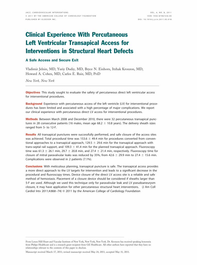
Images were obtained and recorded during each stage ofthe procedure. The operator used the information thusobtained to guide the catheters and devices throughout theprocedure (Fig. 1).
At the end of the procedure, careful echocardiographicevaluation was performed to exclude complications such aspericardial effusion, intracardiac clot, and so forth.Computed tomography angiography. All patients underwentardiac computed tomographic angiography (CTA) (256-sliceCT scanner, Philips Healthcare, Cleveland, Ohio) usingelical scan mode with retrospective electrocardiogram-gatedultiphase reconstruction (16 phases with 6.25% interval
ncrements from RR interval) before the procedure. An Aquar-us workstation (V-3.7.0.14, TeraRecon Inc., San Mateo,alifornia) was used for 3D/4D volume rendering reconstruc-
ion for selecting the optimal entry point from the chest wallnto the LV cavity. This included location of the cardiac apex,apillary muscles, and coronary arteries in relation to the chestall, and the extension of the lung tissue over the LV cavity
Figs. 2 and 3). After the targetntry point was selected and mea-urements obtained, an ink markas placed on the skin of theatient.During the procedure, pre-
cquired CTA (4D volumeendering) was displayed in theatheterization laboratory adja-ent to the fluoroscopy image,sing a grayscale preset thatimulates the fluoroscopy im-ge. The reconstructed images were rotated and angu-ated according to the movement of the fluoroscopymage intensifier, allowing the CTA and the fluoroscopymages to be congruent throughout the procedure. Thisechnique was used for transapical access as well as forntervening on the targeted defects.
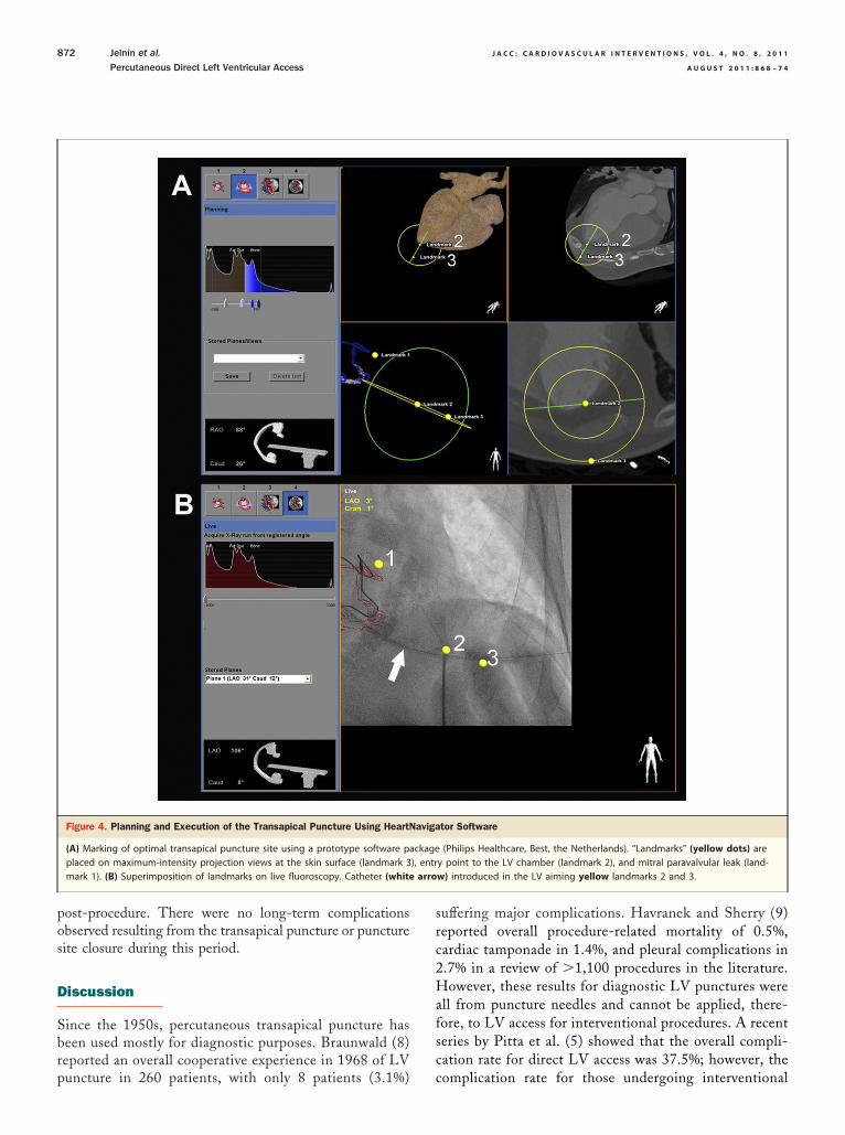
More recently, we have also instituted the use of arototype software “HeartNavigator” (Philips Health-are, Best, the Netherlands) that allows overlay of are-acquired CTA image with live fluoroscopy. Afteroregistration, the overlaid 3D CTA images track theuoroscopy image. Multiple target points marked on theTA image can therefore be accurately projected on the liveuoroscopy indicating the entry point at the skin level, asell as into the myocardial surface, that can guide theeedle access (Fig. 4).
Technique of direct LV access. All procedures were per-formed under general anesthesia. The puncture site wasmarked with ink on the patient’s skin, according to mea-surements obtained from the CTA analysis. If the anterior
Abbreviationsand Acronyms
3D � 3-dimensional
CTA � computedtomographic angiography
LA � left atrium
LV � leftventricle/ventricular
TEE � transesophagealechocardiography
border of the left lung overlapped t
he desired puncture site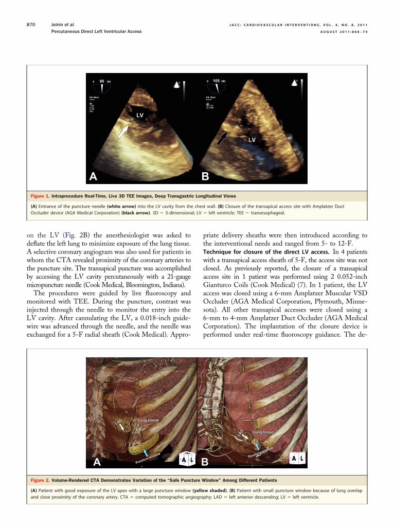
wcaGaOs6Cp
l; LV �
J A C C : C A R D I O V A S C U L A R I N T E R V E N T I O N S , V O L . 4 , N O . 8 , 2 0 1 1
A U G U S T 2 0 1 1 : 8 6 8 – 7 4
Jelnin et al.
Percutaneous Direct Left Ventricular Access
870
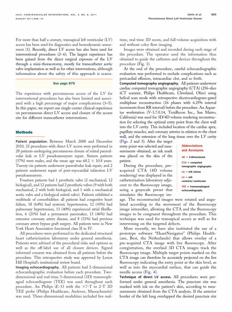
on the LV (Fig. 2B) the anesthesiologist was asked todeflate the left lung to minimize exposure of the lung tissue.A selective coronary angiogram was also used for patients inwhom the CTA revealed proximity of the coronary arteries tothe puncture site. The transapical puncture was accomplishedby accessing the LV cavity percutaneously with a 21-gaugemicropuncture needle (Cook Medical, Bloomington, Indiana).
The procedures were guided by live fluoroscopy andmonitored with TEE. During the puncture, contrast wasinjected through the needle to monitor the entry into theLV cavity. After cannulating the LV, a 0.018-inch guide-wire was advanced through the needle, and the needle wasexchanged for a 5-F radial sheath (Cook Medical). Appro-
Figure 1. Intraprocedure Real-Time, Live 3D TEE Images, Deep Transgastri
(A) Entrance of the puncture needle (white arrow) into the LV cavity from theOccluder device (AGA Medical Corporation) (black arrow). 3D � 3-dimensiona
Figure 2. Volume-Rendered CTA Demonstrates Variation of the “Safe Punc
(A) Patient with good exposure of the LV apex with a large puncture window
and close proximity of the coronary artery. CTA � computed tomographic angiograpriate delivery sheaths were then introduced according tothe interventional needs and ranged from 5- to 12-F.Technique for closure of the direct LV access. In 4 patients
ith a transapical access sheath of 5-F, the access site was notlosed. As previously reported, the closure of a transapicalccess site in 1 patient was performed using 2 0.052-inchianturco Coils (Cook Medical) (7). In 1 patient, the LV
ccess was closed using a 6-mm Amplatzer Muscular VSDccluder (AGA Medical Corporation, Plymouth, Minne-
ota). All other transapical accesses were closed using a-mm to 4-mm Amplatzer Duct Occluder (AGA Medicalorporation). The implantation of the closure device iserformed under real-time fluoroscopy guidance. The de-
gitudinal Views
t wall. (B) Closure of the transapical access site with Amplatzer Ductleft ventricle; TEE � transesophageal.
indow” Among Different Patients
w shaded). (B) Patient with small puncture window because of lung overlap
c Lon
ches
ture W
(yello
phy; LAD � left anterior descending; LV � left ventricle.
J A C C : C A R D I O V A S C U L A R I N T E R V E N T I O N S , V O L . 4 , N O . 8 , 2 0 1 1 Jelnin et al.
A U G U S T 2 0 1 1 : 8 6 8 – 7 4 Percutaneous Direct Left Ventricular Access
871
vice is delivered through the delivery sheath, and after theopening of the distal disk, the device is pulled back untilresistance is felt and the flat disk conforms to the endocar-dial surface. The proper position of the device can beconfirmed by fluoroscopy when visual elongation of thebody of the device during each systolic contraction is noted.Before device release, echocardiographic confirmation of thedevice placement is obtained, and contrast injected throughthe delivery sheath confirms the pericardial position of thesheath. After device release, Surgiflo (Ethicon, Somerville,New Jersey) is injected through the delivery sheath, to fillthe track from the epicardium to the skin.
Results
All transapical accesses were successful, with no complica-tions associated with the percutaneous LV puncture. In all4 patients with 5-F access sheaths, no device closure of theaccess site was performed, and there were no complicationsobserved in this group. In all other patients, the access sitewas successfully closed. There was 1 instance of a smallpericardial effusion documented by echocardiography thatrequired no further intervention. There was 1 procedure-related death in a patient with suprasystemic pulmonary
Figure 3. Planning a Safe Transapical Puncture Using Cardiac CTA Guidan
(A) standard coronal, (B) lateral, and (C) axial views with 3-dimensional (3Dallows the measurement of distances in 3 dimensions from the skin to epic[St. Jude Medical, St. Paul, Minnesota] with paravalvular leak) inside the LVanterior-posterior view (D) and 60° left anterior oblique view (H) demonstratiapex, lung tissue, and identifiable anatomical landmarks such as the sternutool and “slab” view applied. Abbreviations as in Figure 2.
hypertension who developed pulseless electrical activity (
(electromechanical dissociation) after transapical paravalvu-lar leak closure and transapical puncture closure. Two-dimensional echocardiography and emergency thoracotomydid not show any pericardial effusion.
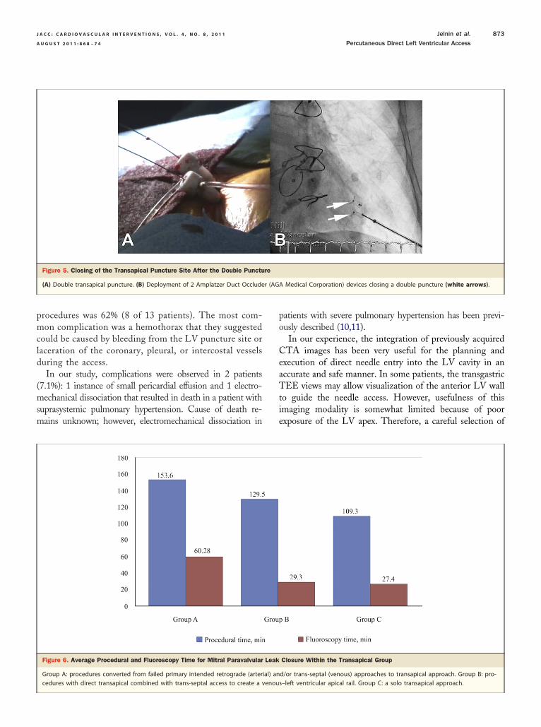
Two patients had repeat transapical access for closure ofadditional paravalvular leaks. In an additional 2 patients, adouble transapical access, for simultaneous deployment andrelease of 2 closure devices for a crescent-shaped paravalvu-lar leak, was performed (Fig. 5).
The patients that underwent paravalvular leak closureprocedures were divided into 3 groups. Group A included 6patients in whom the primary intended retrograde (arterial)and/or trans-septal (venous) approaches failed and proce-dures were converted to the transapical approach. Group Bincluded 10 patients in whom a direct transapical approachcombined with trans-septal access to create a venous–LV apicalrail was initially planned. Group C included 10 patients inwhom a solo transapical approach was used. Average fluoros-copy time was 61.3 � 26.1 min for group A, 29.7 � 20.8 minfor group B, and 27.4 � 21.4 min for group C. Total proceduraltime was 153.6 � 49.4 min for group A, 129.5 � 29.6 min forgroup B, and 109.3 � 41.4 min for group C (Fig. 6).
All patients were clinically followed up to 33 months
et tool overlaid on the image. The red circle with measuring scalesl surface as well as to the target lesion (St. Jude prosthetic mitral valvew arrow). (D and H) 3D volume-rendered reconstruction image inpuncture site on the skin (blue arrow) and its relationship to the cardiac
, F, and G) standard coronal, lateral, and axial views with 3D measuring
ce
) targardia(yellong them. (E
average 13.8 � 9.7 months, range 1 to 33 months)
arro
J A C C : C A R D I O V A S C U L A R I N T E R V E N T I O N S , V O L . 4 , N O . 8 , 2 0 1 1
A U G U S T 2 0 1 1 : 8 6 8 – 7 4
Jelnin et al.
Percutaneous Direct Left Ventricular Access
872
post-procedure. There were no long-term complicationsobserved resulting from the transapical puncture or puncturesite closure during this period.
Discussion
Since the 1950s, percutaneous transapical puncture hasbeen used mostly for diagnostic purposes. Braunwald (8)reported an overall cooperative experience in 1968 of LV
Figure 4. Planning and Execution of the Transapical Puncture Using HeartN
(A) Marking of optimal transapical puncture site using a prototype software pplaced on maximum-intensity projection views at the skin surface (landmark 3mark 1). (B) Superimposition of landmarks on live fluoroscopy. Catheter (white
puncture in 260 patients, with only 8 patients (3.1%)
suffering major complications. Havranek and Sherry (9)reported overall procedure-related mortality of 0.5%,cardiac tamponade in 1.4%, and pleural complications in2.7% in a review of �1,100 procedures in the literature.However, these results for diagnostic LV punctures wereall from puncture needles and cannot be applied, there-fore, to LV access for interventional procedures. A recentseries by Pitta et al. (5) showed that the overall compli-cation rate for direct LV access was 37.5%; however, the
tor Software
(Philips Healthcare, Best, the Netherlands). “Landmarks” (yellow dots) arey point to the LV chamber (landmark 2), and mitral paravalvular leak (land-w) introduced in the LV aiming yellow landmarks 2 and 3.
aviga
ackage), entr
complication rate for those undergoing interventional
r (AG
J A C C : C A R D I O V A S C U L A R I N T E R V E N T I O N S , V O L . 4 , N O . 8 , 2 0 1 1 Jelnin et al.
A U G U S T 2 0 1 1 : 8 6 8 – 7 4 Percutaneous Direct Left Ventricular Access
873
procedures was 62% (8 of 13 patients). The most com-mon complication was a hemothorax that they suggestedcould be caused by bleeding from the LV puncture site orlaceration of the coronary, pleural, or intercostal vesselsduring the access.
In our study, complications were observed in 2 patients(7.1%): 1 instance of small pericardial effusion and 1 electro-mechanical dissociation that resulted in death in a patient withsuprasystemic pulmonary hypertension. Cause of death re-mains unknown; however, electromechanical dissociation in
Figure 5. Closing of the Transapical Puncture Site After the Double Punctu
(A) Double transapical puncture. (B) Deployment of 2 Amplatzer Duct Occlude
Figure 6. Average Procedural and Fluoroscopy Time for Mitral Paravalvular
Group A: procedures converted from failed primary intended retrograde (arter
cedures with direct transapical combined with trans-septal access to create a venoupatients with severe pulmonary hypertension has been previ-ously described (10,11).
In our experience, the integration of previously acquiredCTA images has been very useful for the planning andexecution of direct needle entry into the LV cavity in anaccurate and safe manner. In some patients, the transgastricTEE views may allow visualization of the anterior LV wallto guide the needle access. However, usefulness of thisimaging modality is somewhat limited because of poorexposure of the LV apex. Therefore, a careful selection of
A Medical Corporation) devices closing a double puncture (white arrows).
Closure Within the Transapical Group
d/or trans-septal (venous) approaches to transapical approach. Group B: pro-
re
Leak
ial) an
s–left ventricular apical rail. Group C: a solo transapical approach.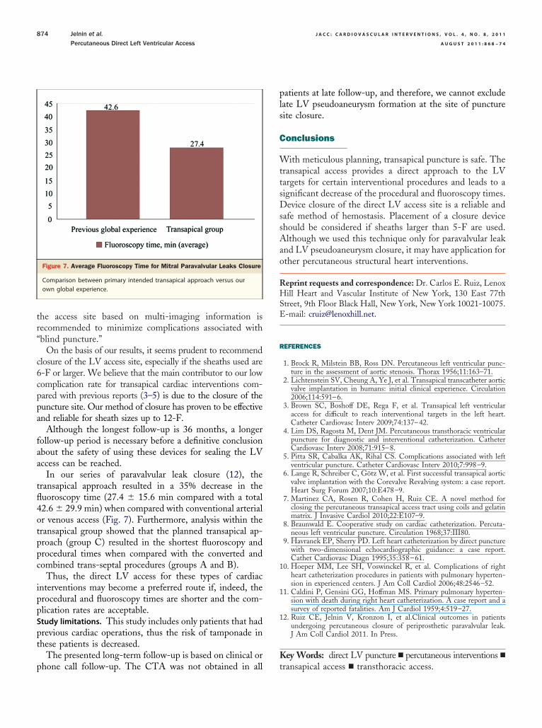
4otppc
ipp
J A C C : C A R D I O V A S C U L A R I N T E R V E N T I O N S , V O L . 4 , N O . 8 , 2 0 1 1
A U G U S T 2 0 1 1 : 8 6 8 – 7 4
Jelnin et al.
Percutaneous Direct Left Ventricular Access
874
the access site based on multi-imaging information isrecommended to minimize complications associated with“blind puncture.”
On the basis of our results, it seems prudent to recommendclosure of the LV access site, especially if the sheaths used are6-F or larger. We believe that the main contributor to our lowcomplication rate for transapical cardiac interventions com-pared with previous reports (3–5) is due to the closure of thepuncture site. Our method of closure has proven to be effectiveand reliable for sheath sizes up to 12-F.
Although the longest follow-up is 36 months, a longerfollow-up period is necessary before a definitive conclusionabout the safety of using these devices for sealing the LVaccess can be reached.
In our series of paravalvular leak closure (12), thetransapical approach resulted in a 35% decrease in thefluoroscopy time (27.4 � 15.6 min compared with a total2.6 � 29.9 min) when compared with conventional arterialr venous access (Fig. 7). Furthermore, analysis within theransapical group showed that the planned transapical ap-roach (group C) resulted in the shortest fluoroscopy androcedural times when compared with the converted andombined trans-septal procedures (groups A and B).
Thus, the direct LV access for these types of cardiacnterventions may become a preferred route if, indeed, therocedural and fluoroscopy times are shorter and the com-lication rates are acceptable.
Study limitations. This study includes only patients that hadprevious cardiac operations, thus the risk of tamponade inthese patients is decreased.
The presented long-term follow-up is based on clinical or
Figure 7. Average Fluoroscopy Time for Mitral Paravalvular Leaks Closure
Comparison between primary intended transapical approach versus ourown global experience.
phone call follow-up. The CTA was not obtained in all
patients at late follow-up, and therefore, we cannot excludelate LV pseudoaneurysm formation at the site of puncturesite closure.
Conclusions
With meticulous planning, transapical puncture is safe. Thetransapical access provides a direct approach to the LVtargets for certain interventional procedures and leads to asignificant decrease of the procedural and fluoroscopy times.Device closure of the direct LV access site is a reliable andsafe method of hemostasis. Placement of a closure deviceshould be considered if sheaths larger than 5-F are used.Although we used this technique only for paravalvular leakand LV pseudoaneurysm closure, it may have application forother percutaneous structural heart interventions.
Reprint requests and correspondence: Dr. Carlos E. Ruiz, LenoxHill Heart and Vascular Institute of New York, 130 East 77thStreet, 9th Floor Black Hall, New York, New York 10021-10075.E-mail: [email protected].
REFERENCES
1. Brock R, Milstein BB, Ross DN. Percutaneous left ventricular punc-ture in the assessment of aortic stenosis. Thorax 1956;11:163–71.
2. Lichtenstein SV, Cheung A, Ye J, et al. Transapical transcatheter aorticvalve implantation in humans: initial clinical experience. Circulation2006;114:591–6.
3. Brown SC, Boshoff DE, Rega F, et al. Transapical left ventricularaccess for difficult to reach interventional targets in the left heart.Catheter Cardiovasc Interv 2009;74:137–42.
4. Lim DS, Ragosta M, Dent JM. Percutaneous transthoracic ventricularpuncture for diagnostic and interventional catheterization. CatheterCardiovasc Interv 2008;71:915–8.
5. Pitta SR, Cabalka AK, Rihal CS. Complications associated with leftventricular puncture. Catheter Cardiovasc Interv 2010;7:998–9.
6. Lange R, Schreiber C, Götz W, et al. First successful transapical aorticvalve implantation with the Corevalve Revalving system: a case report.Heart Surg Forum 2007;10:E478–9.
7. Martinez CA, Rosen R, Cohen H, Ruiz CE. A novel method forclosing the percutaneous transapical access tract using coils and gelatinmatrix. J Invasive Cardiol 2010;22:E107–9.
8. Braunwald E. Cooperative study on cardiac catheterization. Percuta-neous left ventricular puncture. Circulation 1968;37:III80.
9. Havranek EP, Sherry PD. Left heart catheterization by direct puncturewith two-dimensional echocardiographic guidance: a case report.Cathet Cardiovasc Diagn 1995;35:358–61.
10. Hoeper MM, Lee SH, Voswinckel R, et al. Complications of rightheart catheterization procedures in patients with pulmonary hyperten-sion in experienced centers. J Am Coll Cardiol 2006;48:2546–52.
11. Caldini P, Gensini GG, Hoffman MS. Primary pulmonary hyperten-sion with death during right heart catheterization. A case report and asurvey of reported fatalities. Am J Cardiol 1959;4:519–27.
12. Ruiz CE, Jelnin V, Kronzon I, et al.Clinical outcomes in patientsundergoing percutaneous closure of periprosthetic paravalvular leak.J Am Coll Cardiol 2011. In Press.
Key Words: direct LV puncture � percutaneous interventions �
transapical access � transthoracic access.


