Embed Size (px)
Citation preview

Cell Host & Microbe
Resource
Clonal Conditional Mutagenesisin Malaria ParasitesAudrey Combe,1 Donatella Giovannini,1 Teresa Gil Carvalho,1 Stephan Spath,1 Bertrand Boisson,1 Celine Loussert,2
Sabine Thiberge,1 Celine Lacroix,1 Pascale Gueirard,1 and Robert Menard1,*1Institut Pasteur, Unite de Biologie et Genetique du Paludisme, 28 rue du Dr Roux2Institut Pasteur, Imagopole, 25 rue du Dr Roux75724 Paris cedex 15, Paris, France
*Correspondence: [email protected]
DOI 10.1016/j.chom.2009.03.008
SUMMARY
We describe here an efficient method for conditionalgene inactivation in malaria parasites that uses theFlp/FRT site-specific recombination system of yeast.The method, developed in Plasmodium berghei,consists of inserting FRT sites in the chromosomallocus of interest in a parasite clone expressing theFlp recombinase via a developmental stage-specificpromoter. Using promoters active in mosquito midgutsporozoites or salivary gland sporozoites to driveexpression of Flp or its thermolabile variant, FlpL,we show that excision of the DNA flanked by FRT sitesoccurs efficiently at the stage of interest and at unde-tectable levels in prior stages. We applied this tech-nique to conditionally silence MSP1, a gene essentialfor merozoite invasion of erythrocytes. SilencingMSP1 in sporozoites impaired subsequent merozoiteformation in the liver. Therefore, MSP1 plays a dualrole in the parasite life cycle, acting both in liver anderythrocytic parasite stages.
INTRODUCTION
Since its introduction 13 years ago (Wu et al., 1996; van Dijk
et al., 1996; Menard et al., 1997; Crabb et al., 1997), gene target-
ing by homologous recombination in Plasmodium has allowed
important progress in our understanding of protein function in
the parasite. Nonetheless, a major limitation in exploring protein
function by genetic means is that only erythrocytic stages of this
haploid parasite can be transfected (Figure 1). Therefore, the
genes contributing to parasite invasion of and multiplication in
host erythrocytes, which are essential for recombinant selec-
tion, cannot be inactivated and characterized. Also, the genes
having an essential function at multiple stages during the para-
site life cycle cannot be fully addressed by simple gene inactiva-
tion. Thus, conditional manipulation of the genome is required
for a precise understanding of Plasmodium protein function
in vivo.
Systems for regulating gene expression by transcriptional
regulators such as the TET transactivators, first devised in
the related Apicomplexan parasite Toxoplasma gondii (Meiss-
ner et al., 2002), have been adapted to the human pathogen
386 Cell Host & Microbe 5, 386–396, April 23, 2009 ª2009 Elsevier I
Plasmodium falciparum (Meissner et al., 2005). To date,
however, the available tools in P. falciparum only allow for
turning on expression of a transgene, but not for turning off
expression of an endogenous gene. More recently, a method
to regulate protein levels was reported in P. falciparum
(Armstrong and Goldberg, 2007) and in T. gondii (Herm-Gotz
et al., 2007). The method is based on the fusion of an FK506
binding protein (FKBP) destabilization domain (ddFKBP) to
a protein of interest, which remains stable in the presence of the
‘‘shield’’ ligand of the destabilization domain and is degraded
in its absence (Banaszynski et al., 2006). The system appears
to be suitable for dominant-negative studies (Herm-Gotz
et al., 2007) but has not yet proven efficient for conditionally
depleting endogenous products. Importantly, both transcrip-
tional control and protein degradation systems ultimately rely
on an exogenous compound that must be added or removed
to modulate the system, and this might not be ideal for manip-
ulating the genome of the parasite stages that reside in the
mosquito.
By contrast, site-specific recombination (SSR) systems act
directly at the DNA level and do not depend on any exogenous
ligand. In the Flp/FRT SSR system of yeast (Branda and
Dymecki, 2004), the Flp recombinase recognizes the 34 bp
FRT sites and excises any DNA located between two directly
oriented sites (referred to as the FRTed sequence). In a first
attempt to adapt the Flp/FRT SSR system of yeast to P. berghei
(Gil Carvalho et al., 2004), a rodent-infecting Plasmodium
species that offers easy access to all parasite stages, the Flp
gene and the FRTed sequence were inserted in different chro-
mosomes in two different parasite clones. The two clones were
genetically crossed in mosquitoes, where meiosis occurs, allow-
ing a proportion of the haploid progeny (sporozoites) to carry
both the Flp gene and the FRTed sequence. Although efficient,
the cross approach is limited to haploid stages because
diploid/polyploid stages (from zygote to oocyst) still contain
non-FRTed version(s) of the target sequence, and a maximum
of 25% of the subsequent haploid stages (sporozoites onward)
can potentially undergo SSR.
Here, we devised a clonal approach to Flp-mediated SSR
in P. berghei, in which both the recombinase locus and the
FRTed sequence are placed in erythrocytic stages of the same
parasite clone. Such clonal populations undergo SSR with
maximal efficiency only at the stage of interest. In the approach
described here, the parasite stage of interest is the sporozoite,
the stage transmitted by the mosquito to the mammalian host.
nc.

Cell Host & Microbe
Conditional Mutagenesis in P. berghei
Figure 1. Scheme for Flp/FRT-Based Clonal Conditional Mutagen-
esis in Plasmodium
The parasite stages of the Plasmodium parasite during its life cycle in the
mosquito vector and in the mammalian host are shown. In a parasite clone
that harbors the Flp recombinase gene under the control of the promoter active
at the sporozoite stage, the target sequence (dashed line) flanked by FRT sites
(open arrows) is excised at the salivary gland sporozoite stage, leaving a single
FRT site at the excised locus. MG, midgut; SG, salivary gland.
Cell
proportion of TRAP/FlpL(+) and WT parasites before and after
the 10 cycles also indicated that the recombinant locus was
stable and did not impair parasite fitness. TRAP/FlpL(+) parasites
were then cycled through Anopheles stephensi mosquitoes, and
the resulting erythrocytic stages were cloned. Clones having
undergone SSR (Figure 2A) and thus having lost the selectable
marker were readily identified. One such clone, TRAP/FlpL(�),
was characterized (Figure 2C) and used to test the SSR efficiency
of the system in sporozoites.
To monitor TRAP/FlpL-mediated SSR in sporozoites, we
inserted in TRAP/FlpL(�) parasites an FRTed reporter system
at the CS locus that would signal SSR via GFP expression
(Figure 3A). The CS gene, which is expressed in young oocysts
and throughout the sporozoite’s life, is necessary for the forma-
tion of sporozoites inside oocysts and encodes the most abun-
dant protein on the surface of free midgut and salivary gland
sporozoites (Nussenzweig and Nussenzweig, 1985). In the
reporter, the CS promoter was separated from the GFP gene
by an FRTed hDHFR selectable marker; hence, SSR would place
GFP under the control of the CS promoter and render sporozo-
ites fluorescent. The plasmid pGFP/CS containing the reporter
was linearized in the CS promoter sequence and transfected
into TRAP/FlpL(�) parasites, and a parasite population having
integrated the plasmid by single crossover recombination,
named GFP/CS(+), was selected (Figure 3B). This population
was transmitted to mosquitoes kept at 21�C, and at D16,
infected mosquitoes were either maintained at 21�C (CONT) or
placed until D20 at 25�C (SHIF), a temperature at which FlpL
displays higher activity. Approximately 30% and 50% of the
CONT or SHIF midgut sporozoites, respectively, were fluores-
cent at D20 (Figure 3C), and up to 75% of the SHIF salivary gland
sporozoites were fluorescent at D20 (Figure 3D). To assess SSR
levels at the DNA level, we performed PCR using genomic DNA
from midgut and salivary gland sporozoites at D21 (Figure 3E).
Results confirmed that SSR occurred in midgut sporozoites
and that high SSR levels were achieved in SHIF sporozoites
from either mosquito midguts or salivary glands. Importantly,
the number and infectivity of salivary gland sporozoites were
similar in the SHIF and CONT conditions, indicating that the
temperature switch did not impair parasite development. Finally,
the behavior of cloned TRAP/FlpL(�) parasites was indistin-
guishable from that of WT parasites throughout their life cycle
(data not shown), indicating that recombinase expression via
the TRAP promoter was not detrimental to the parasite.
Together, these data indicate that SSR mediated by the TRAP/
FlpL system starts in midgut sporozoites and remains limited at
21�C but can reach high levels in salivary gland sporozoites after
a 25�C temperature shift. Thus, it should be useful for testing the
function of sporozoite proteins in infection of the mammalian
host.
The UIS4/Flp Conditional SystemTo gain versatility, we next aimed at inducing SSR specifically
when the sporozoite has entered the salivary glands of the
mosquito. For this, we constructed a P. berghei clone in which
the recombinase was under the control of the promoter of
UIS4, a gene identified by subtractive suppressive hybridization
as being overexpressed in salivary gland compared to midgut
sporozoites (Matuschewski et al., 2002). RT-PCR experiments
RESULTS
The TRAP/FlpL Conditional SystemBecause our goal was to have SSR repressed in the transfected
erythrocytic stages and completed in salivary gland sporozoites
(Figure 1), we first constructed a P. berghei clone that expresses
the recombinase at the mosquito midgut sporozoite stage. For
this, the recombinasewas expressed via the regulatorysequences
of TRAP, a gene that is weakly expressed in erythrocytic stages
(Robson et al., 1988), is switched on in maturing oocysts (Gil Car-
valho et al., 2004), and induced �10,000-fold in salivary gland
sporozoites compared to erythrocytic stages (Rosinski-Chupin
et al., 2007). To further minimize the risk of premature excision
in erythrocytic stages, we used the FlpL (Low activity) recombi-
nase (Buchholz et al., 1996), whose activity is greatly reduced at
37�C and above—temperatures in erythrocytic stages in the
mammal—and is maintained at 20�C –25�C, temperatures
permissive for parasite development in the mosquito.
The FlpL coding sequence was flanked by 1.5 kb and 0.6 kb of
50 and 30 regulatory sequences of TRAP, respectively, and asso-
ciated with a FRTed hDHFR selectable marker containing its own
(eef1a) promoter and terminator sequences, generating plasmid
pTRAP/FlpL (Figure 2A). The plasmid was linearized in the
50 TRAP regulatory sequence to direct gap repair and plasmid
integration by single crossover recombination at the homologous
chromosomal locus (Nunes et al., 1999) and subsequently trans-
fected into erythrocytic stages of wild-type (WT) P. berghei NK65.
Parasites having integrated the plasmid carrying the hDHFR
resistance cassette, called TRAP/FlpL(+), were readily selected
under pyrimethamine treatment (Figures 2A and 2B). This indi-
cated that the TRAP/FlpL system did not induce major levels of
SSR, which excises the cassette, in erythrocytic stages. To test
whether SSR occurred at low levels, we allowed a mixed popula-
tion of TRAP/FlpL(+) and WT parasites to undergo 10 erythrocytic
cycles in mice in the absence of drug pressure. Southern blot
analysis after the 10 cycles did not detect any marker excision
(Figure 2B), showing that the TRAP/FlpL system did not induce
significant levels of SSR in erythrocytic stages. The similar
Host & Microbe 5, 386–396, April 23, 2009 ª2009 Elsevier Inc. 387

Cell Host & Microbe
Conditional Mutagenesis in P. berghei
Figure 2. Generation of the TRAP/FlpL(�) Receiver Clone
(A) Schematic representation of the TRAP locus in wild-type (WT) P. berghei and the TRAP/FlpL(+) and TRAP/FlpL(�) clones. Plasmid pTRAP/FlpL was linearized
in the TRAP 50 regulatory sequence upstream of the FlpL coding sequence and integrated via single crossover (SCO) recombination at the cognate locus in WT
P. berghei, generating the recombinant locus TRAP/FlpL(+), which, in turn, generates the TRAP/FlpL(�) locus after SSR (marker excision). (Open arrows) FRT site
(34 bp); (thin line with a thin arrow) TRAP 50 upstream sequences (1.5 kb); (thin line with an open lollipop) TRAP 30 downstream sequences (0.6 kb); (gray box)
hDHFR resistance cassette including its own expression sequences (1.6 kb); (thick line) pUC plasmid backbone (2.7 kb). The sizes of the FlpL and TRAP coding
sequences are 1.3 kb and 1.8 kb, respectively. The relevant restriction sites and the size of restriction fragments (in kb) are shown above the corresponding DNA.
B, BamHI; H2, HincII; S, SpeI.
(B) Southern hybridization of genomic DNA of the WT and of a mixture of WT and TRAP/FlpL(+) parasites at a given time (1) and after 10 additional cycles in mice in
the absence of drug (10), digested with HincII and probed with an internal TRAP probe corresponding to most of the TRAP coding sequence (starting upstream of
the SpeI site and ending downstream of the HincII site). The sizes of the bands are shown at the left, and the expected positions of the fragments from the indi-
cated parasite populations are shown at the right. SSR remains undetectable in erythrocytic stages of TRAP/FlpL(+) parasites.
(C) Southern hybridization of genomic DNA of the WT and the TRAP/FlpL(�) clone, digested with SpeI or BamHI and using the internal TRAP probe.
have shown that UIS4 is expressed in P. berghei salivary gland
sporozoites at similar levels as TRAP and �10,000 times more
in salivary gland sporozoites than in erythrocytic stages of the
parasite (Rosinski-Chupin et al., 2007). UIS4 is essential for the
early stages of parasite development in the liver, given that
UIS4 knockout parasites rapidly die after hepatocyte invasion
(Mueller et al., 2005).
We cloned either WT Flp or mutant FlpL between 1.5 kb of 50
regulatory sequence of UIS4 and 0.6 kb of 30 regulatory sequence
of TRAP, along with the FRTed hDHFR marker, generating plas-
mids pUIS4/Flp and pUIS4/FlpL, respectively (Figure 4A). These
plasmids were linearized in the 50 regulatory sequence of UIS4
and separately transfected into WT P. berghei NK65. In both
cases, the expected single crossover homologous recombi-
nants, called UIS4/Flp(+) and UIS4/FlpL(+), respectively, were
selected and cloned. Because SSR did not occur in erythrocytic
stages of the UIS4/Flp clone (see below) and because Flp has
higher activity than FlpL in the 20�C –25�C temperature range
of parasite development in the mosquito (Buchholz et al., 1996),
we investigated SSR efficiency only in sporozoites of the UIS4/
Flp(+) clone (Figure 4B).
The UIS4/Flp(+) parasite clone was transmitted from a mouse
M1 to Anopheles stephensi mosquitoes continuously kept at
388 Cell Host & Microbe 5, 386–396, April 23, 2009 ª2009 Elsevier I
21�C, and at D18, salivary gland sporozoites were transmitted
back to a mouse M2. Marker excision via SSR was monitored
by PCR using genomic DNA from various parasite stages along
the life cycle (Figure 4C). Marker excision was not detected in
erythrocytic stages in mouse M1 and was not detected or was
barely detected in D14 or D18 midgut sporozoites, respectively.
In contrast, marker excision occurred in a majority of both D14
and D18 salivary gland sporozoites, and the nonexcised locus
was not detected in erythrocytic stages in mouse M2. Therefore
the UIS4/Flp system, by mediating little SSR in midgut sporozo-
ites but rapid and efficient SSR in salivary gland sporozoites, is
another valuable system for conditional manipulation of genes
involved in sporozoite infectivity to the mammalian host.
Finally, we selected from the M2 mouse a parasite clone
that had excised the selectable marker, named UIS4/Flp(�)
(Figure 4B) and constructed a derivative clone, named UIS4/
Flp(�)F, by integrating at the DHFR-TS locus a GFP cassette
mediating bright fluorescence at all stages of the parasite via
HSP70 regulatory sequences (Ishino et al., 2006). The phenotype
of the UIS4/Flp(�)F clone was indistinguishable from that of
the WT throughout the life cycle, including normal infectivity of
salivary gland sporozoites (see below). This indicated that Flp
expression via the UIS4 promoter was not detrimental to the
nc.

Cell Host & Microbe
Conditional Mutagenesis in P. berghei
Figure 3. Efficiency of the TRAP/FlpL(�) Conditional System Monitored by a GFP Reporter at the CS Locus
(A) Schematic representations of the CS locus in TRAP/FlpL(�) parasites and in GFP/CS(+) and GFP/CS(�)parasites. Plasmid pGFP/CS was linearized in the CS
50 regulatory sequence and integrated via single crossover (SCO) recombination at the cognate locus in TRAP/FlpL(�) parasites, generating the recombinant
locus GFP/CS(+), which, in turn, generates the GFP/CS(�) locus after SSR (marker excision). In GFP/CS(�) parasites, the residual FRT site is placed immediately
after the start codon of GFP, and the recombinant locus encodes a GFP protein containing a 17-residue extension at the N terminus. Symbols are as in Figure 2,
except: (thin line with a thin arrow) CS 50 upstream sequences (1.3 kb); (thin line with an open lollipop) CS 30 downstream sequences (0.3 kb). The sizes of the CS
and GFP coding sequences are 1 kb and 0.7 kb, respectively. Abbreviations are Figure 1; H3, HindIII; K, KpnI.
(B) Characterization of GFP/CS(+) parasites. Southern hybridization of genomic DNA of WT and of GFP/CS(+) parasites, digested with HindIII, KpnI+HincII, or
SpeI and using internal CS (left panel) or GFP (right panel) probes.
(C and D) Proportion of fluorescent sporozoites in mosquito midguts (C) or salivary glands (D) after transmission of GFP/CS(+) parasites to mosquitoes. Mosqui-
toes were either kept at 21�C (CONT) or placed at 25�C from D16 to D20 (SHIF). Values represent the mean ± SD of three independent parasite cycling
experiments.
(E) SSR efficiency at the CS locus in GFP/CS(+) parasites assessed by PCR, using sense primer PA hybridizing in the CS promoter region and antisense primer PB
hybridizing in the GFP coding sequence, on the genomic DNA of midgut (MG) and salivary gland (SG) sporozoites placed in CONT (21�C) or SHIF (25�C from day
16 to day 20) conditions. The two primers amplify a DNA fragment of 2.2 or 0.6 kb in the absence or presence of SSR, respectively.
parasite and that UIS4 was normally expressed in the recombi-
nant clone and, therefore, that the clone was a suitable recipient
for gene inactivation at the pre-erythrocytic stages of the parasite.
Conditional Mutagenesis of MSP1: MSP1 Is Importantfor Merozoite Formation in the Liver of the HostMerozoite attachment to the erythrocyte surface is mediated by
a family of proteins called merozoite surface proteins (MSPs)
(Cowman and Crabb, 2006). The most studied MSP, MSP1, is
a glycosylphosphatidylinositol (GPI)-anchored protein that
covers the entire surface of free merozoites (Holder et al.,
1985). Antibody inhibition experiments (Siddiqui et al., 1987; de
Koning-Ward et al., 2003; John et al., 2004) and molecular genetic
studies (O’Donnell et al., 2000) have shown that MSP1 is essential
Cell
for merozoite adhesion to erythrocytes. In agreement with this,
our own attempts to inactivate MSP1 in erythrocytic stages of
P. berghei remained unsuccessful (data not shown). In addition,
expression of MSP1 is switched on during the first half and peaks
during the second half of the liver stage developmental process
(Figure 5A). This process, which follows hepatocyte invasion by
sporozoites, consists in karyokinesis without cytokinesis before
budding off of thousands of uninucleate merozoites (Meis and
Verhave, 1988) and lasts �50–�70 hr in rodents.
To test whether MSP1 plays a role in parasite development
inside hepatocytes, we conditionally modified the MSP1 locus
in sporozoites using the UIS4/Flp system. The FRT sites were
thus introduced into the MSP1 locus in erythrocytic stages of
the UIS4/Flp(�)F clone. For this, we constructed the targeting
Host & Microbe 5, 386–396, April 23, 2009 ª2009 Elsevier Inc. 389
Cell Host & Microbe
Conditional Mutagenesis in P. berghei
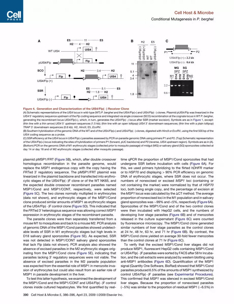
Figure 4. Generation and Characterization of the UIS4/Flp(�) Receiver Clone
(A) Schematic representations of the UIS4 locus in wild-type (WT) P. berghei and the UIS4/Flp(+) and UIS4/Flp(�) clones. Plasmid pUIS4/Flp was linearized in the
UIS4 50 regulatory sequence upstream of the Flp coding sequence and integrated via single crossover (SCO) recombination at the cognate locus in WT P. berghei,
generating the recombinant locus UIS4/Flp(+), which, in turn, generates the UIS4/Flp(�) locus after SSR (marker excision). Symbols are as in Figure 1, except:
(thin line with a thin arrow) UIS4 50 upstream sequences (1.5 kb); (thin line with an open lollipop) UIS4 30 downstream sequences; (thin line with a plain lollipop)
TRAP 30 downstream sequences (0.6 kb). H2, HincII; E5, EcoRV.
(B) Southern hybridization of the genomic DNA of the WT and of the UIS4/Flp(+) and UIS4/Flp(�) clones, digested with HincII or EcoRV, using the first 500 bp of the
UIS4 coding sequence as a probe.
(C) SSR efficiency at the UIS4 locus in UIS4/Flp(+) parasites assessed by PCR on parasite genomic DNA using primers P1 and P2. (Top) Schematic representation
of the UIS4/Flp(+) locus indicating the sites of hybridization of primers P1 (forward, pUC backbone) and P2 (reverse, UIS4 upstream region). Symbols are as in (A).
(Bottom) PCR on the genomic DNA of M1 erythrocytic stages (collected prior to mosquito passage) of midgut (MG) or salivary gland (SG) sporozoites collected at
day 14 or day 18 and of M2 erythrocytic stages (collected after mosquito passage).
plasmid pMSP1/FRT (Figure 5B), which, after double crossover
homologous recombination in the parasite genome, would
replace the MSP1 endogenous copy with the copy having the
FRTed 30 regulatory sequence. The pMSP1/FRT plasmid was
linearized in the plasmid backbone and transfected into erythro-
cytic stages of the UIS4/Flp(�)F clone or of the WT NK65, and
the expected double crossover recombinant parasites named
MSP1/Cond and MSP1/CONT, respectively, were selected
(Figure 5C). The two clones normally multiplied in erythrocytes
(data not shown), and erythrocytic stages of the MSP1/Cond
clone produced similar amounts of MSP1 as erythrocytic stages
of the UIS4/Flp(�)F control clone (Figure 5D). This indicated that
the FRTed 30 heterologous sequence was allowing correct MSP1
expression in erythrocytic stages of the recombinant parasite.
The parasite clones were then separately transferred from a
mouse M1 to mosquitoes and back to a mouse M2. PCR analysis
of genomic DNA of the MSP1/Cond parasites showed undetect-
able levels of SSR in M1 erythrocytic stages but high levels in
D18 salivary gland sporozoites (Figure 5E). As expected, SSR
was not detected in MSP1/CONT salivary gland sporozoites
that lack Flp (data not shown). PCR analysis also showed the
absence of excised parasites in the M2 erythrocytic stages orig-
inating from the MSP1/Cond clone (Figure 5E), indicating that
parasites lacking 30 regulatory sequences were not viable. The
absence of excised parasites in the M2 parasite population
was expected from the essential role of MSP1 in merozoite inva-
sion of erythrocytes but could also result from an earlier role of
MSP1 in parasite development in the liver.
To test this latter hypothesis, we examined the development of
the MSP1/Cond and the MSP1/CONT and UIS4/Flp(�)F control
clones inside cultured hepatocytes. We first quantified by real-
390 Cell Host & Microbe 5, 386–396, April 23, 2009 ª2009 Elsevier I
time qPCR the proportion of MSP1/Cond sporozoites that had
undergone SSR before incubation with cells (Figure 6A). For
this, we used primers hybridizing to the flirted hDHFR marker
or to HSP70 and displaying > 90% PCR efficiency on genomic
DNA of erythrocytic stages, where SSR does not occur. The
numbers of nonexcised or excised MSP1 loci (containing or
not containing the marker) were normalized by that of HSP70
loci, both being single copy, and the percentage of excision at
the MSP1 locus was calculated by the DCT method. The average
proportion of nonexcised loci in the M1 population and in salivary
gland sporozoites was�99% and <5%, respectively (Figure 6A).
Sporozoites of the MSP1/Cond and of the two control clones
were then incubated with HepG2 cells, and the numbers of
developing liver stage parasites (Figure 6B) and of merozoites
released in the culture supernatant (Figure 6C) were counted
by fluorescence microscopy. The MSP1/Cond clone generated
similar numbers of liver stage parasites as the control clones
at 24 hr, 48 hr, 63 hr, and 71 hr (Figure 6B). By contrast, the
MSP1/Cond clone yielded on average 30-fold fewer merozoites
than the control clones at 71 hr (Figure 6C).
To verify that the excised MSP1/Cond liver stages did not
produce MSP1, fluorescent HepG2 cells containing MSP1/Cond
or UIS4/Flp(�)F parasites were sorted by FACS after 58 hr incuba-
tion, and the cell extracts were analyzed by western blotting using
anti-MSP1 antibodies (Figure 6D). Quantification of the MSP1
signal (Quantity One Software, BioRad) showed that MSP1/Cond
parasites produced 6.5% of the amounts of MSP1 synthesized by
control UIS4/Flp(�)F parasites (see Experimental Procedures).
This confirmed that MSP1 was silenced in excised MSP1/Cond
liver stages. Because the proportion of nonexcised parasites
(�5%) was similar to the proportion of residual MSP1 (�6.5%) in
nc.

Cell Host & Microbe
Conditional Mutagenesis in P. berghei
Figure 5. Conditional Mutagenesis of MSP1 Using the UIS4/Flp System
(A) Real-time qPCR analysis of MSP1 and AMA1 relative expression during the P. berghei life cycle, normalized by HSP70 expression. SPZ, sporozoites at day 21
postinfection; 5 hr, 40 hr, and 50 hr, liver stages in HepG2 cells 5 hr, 40 hr, or 50 hr postinfection, respectively; RBC, infected red blood cells. The y axis shows the
log scale of mean normalized expression. Values represent the mean ± SD of two experiments originating from independent batches of mRNA.
(B) Schematic representations of the WT MSP1 locus and of the MSP1 recombinant loci in the MSP1/Cond and MSP1/CONT clones. Plasmid pMSP1/FRT
contains the 30 end (0.6 kb) of the MSP1 coding sequence (open box P1), a flirted fragment including 0.6 kb of TRAP 30 regulatory sequence (plain lollipop)
and the marker cassette (gray box), the plasmid backbone (thick line), and MSP1 30 regulatory sequence (0.6 kb, open lollipop). The linearized plasmid (as indi-
cated) integrated via double crossover (DCO) recombination at the cognate locus into WT or UIS4/Flp(�)F parasites, generating the MSP1/CONT and MSP1/
Cond clones, respectively. H3, HindIII; E5, EcoRV.
(C) Southern hybridization of the genomic DNA of the WT, the MSP1/Cond, and the MSP1/CONT clones, digested with HindIII or EcoRV and using the last 600 bp
of the MSP1 coding sequence as a probe.
(D) Western blot analysis of extracts from MSP1/Cond and UIS4/Flp(�)F merozoites using a mixture of antibodies recognizing MSP1 and AMA1.
(E) SSR efficiency at the MSP1 locus in MSP1/Cond parasites assessed by PCR on parasite genomic DNA using primers P3 and P4. (Top) Schematic represen-
tation of the nonexcised MSP1 locus indicating the sites of hybridization of primers P3 (forward, MSP1 coding sequence) and P4 (reverse, plasmid backbone).
Symbols are as in (A). (Bottom) PCR on the genomic DNA of M1 erythrocytic stages (collected prior to mosquito passage), of salivary gland (SG) sporozoites
collected 18 days after infection, and of M2 erythrocytic stages (collected after mosquito passage). The sizes of the DNA fragments amplified from excised
(SSR+) or nonexcised (SSR�) loci are shown.
the parasite population, the excised parasites appear to produce
virtually no MSP1.
These data thus suggested that MSP1 was involved in the
formation of merozoites inside, or their release from, hepato-
cytes. To distinguish between these two possibilities, we inves-
tigated liver stage development by using transmission electron
microscopy (TEM). HepG2 cells were incubated with sporozoites
from the MSP1/Cond or UIS4/Flp(�)F clones, and after 58 hr
incubation, fluorescent cells were sorted by FACS and pro-
Cell
cessed for TEM. In agreement with the development of WT para-
sites (Meis and Verhave, 1988), the multinucleated control para-
sites generated merozoites of homogenous size and regular
shape that budded off from the periphery of the parasite mass,
progressively filling the parasitophorous vacuole and ultimately
the host cell cytoplasm (Figure 7A). By contrast, the majority of
MSP1/Cond parasites contained numerous vacuoles of various
sizes and generated abnormal progeny (Figures 7B and 7C).
Merozoite buds emerged within the intraparasitic vacuoles,
Host & Microbe 5, 386–396, April 23, 2009 ª2009 Elsevier Inc. 391

Cell Host & Microbe
Conditional Mutagenesis in P. berghei
Figure 6. Development of MSP1/Cond Parasites in HepG2 Cells
(A) Efficiency of SSR at the MSP1 locus assessed by quantitative PCR performed using genomic DNA of M1 erythrocytic stages (M1) or salivary gland sporozoites
(SG SPZ, collected at day 25 postinfection) from the MSP1/Cond clone. The percentage of nonexcised loci was evaluated using primers P5 and P6 (hybridizing
within the selectable marker) and a pair of primers hybridizing to the HSP70 gene for normalization. Values represent the mean ± SD of two independent exper-
iments, each performed in triplicate.
(B) Salivary gland sporozoites (5 3 104) were added to nonconfluent HepG2 cells (multiplicity of infection = 1), and the numbers of maturing parasites were
counted by fluorescence microscopy (for the MSP1/Cond and UIS4/Flp(�)F clones) or by light transmission (for the MSP1/CONT clone) after the indicated incu-
bation times at 37�C. Values represent the mean ± SD of three independent experiments each performed in duplicate.
(C) The experiment was conducted as in (B), and the numbers of hepatic merozoites released in the supernatant were counted.
(D) Western blot analysis of extracts from HepG2 cells infected with MSP1/Cond and UIS4/Flp(�)F sporozoites and sorted 58 hr postinfection or from UIS4/
Flp(�)F merozoites, using antibodies recognizing AMA1 or MSP1. MZ, merozoites; LS, liver stages.
rather than at the parasite periphery, and the internal buds dis-
played abnormal shapes (Figure 7D, left panel), frequently having
an enlarged base or an orientation tangential rather than perpen-
dicular to the plane of the parasite membrane (Figure 7D, right
panel). Therefore, MSP1 appears to play a crucial role in the
formation of merozoites by the liver stage of the parasite.
DISCUSSION
Flp/FRT-Mediated Conditional Mutagenesisin Plasmodium
We have established here a powerful approach for conditionally
modifying the P. berghei genome using the Flp/FRT system.
Unlike the previously described approach that relies on a parasite
cross in the mosquito (Gil Carvalho et al., 2004), the present
‘‘clonal’’ approach places the recombinase cassette and the
FRTed sequence (sequence flanked by FRT sites) in erythrocytic
392 Cell Host & Microbe 5, 386–396, April 23, 2009 ª2009 Elsevier In
stages of the same parasite clone. The clonal parasite population
then undergoes SSR, i.e., excision of the FRTed DNA sequence,
at high efficiency and in a stage-specific manner during the para-
site life cycle. The timing of SSR is controlled by the activity of the
promoter that drives recombinase expression, and its efficiency
can be modulated by temperature when using the thermosensi-
tive FlpL. By inducing SSR in the vast majority of parasites, the
method obviates the need to link the excision process to GFP
expression for recognizing the excised from the nonexcised
parasites (Gil Carvalho et al., 2004), which simplifies the construct
design and shortens the procedure. Bright and constitutive
fluorescence can be expressed in parasites producing the re-
combinase to facilitate characterization and in vivo imaging of
the conditional mutants. Derivatives of the TRAP/FlpL(�) and
UIS4/Flp(�) clones expressing bright GFP fluorescence via the
HSP70 promoter are now available as parasite ‘‘receivers’’ of
conditional alleles of genes of interest.
c.

Cell Host & Microbe
Conditional Mutagenesis in P. berghei
Figure 7. MSP1 Is Essential for Normal Merozoite Formation inside Hepatocytes
(A) Electron micrographs of control UIS4/Flp(�)F liver stages in HepG2 cells collected after 58 hr incubation. Various stages can be observed, from nonseg-
mented parasites (left picture) through budding parasites (second picture) to segmented parasites containing individualized merozoites (two pictures on the right).
Scale bars, 2 mm.
(B) Electron micrographs of MSP1/Cond liver stages in HepG2 cells collected after 58 hr incubation. Numerous large vacuoles are visible in the mutant parasites,
with vacuolated parasites of abnormal shape budding inside of these vacuoles Scale bars, 2 mm.
(C) Quantification of vacuolated EEF for UIS4/Flp(�)F control (n = 49) or MSP1/Cond mutant (n = 54) liver stages in nonsegmented or segmented EEF. V+, vacu-
olated parasites; V�, nonvacuolated parasites; nd, not determined.
(D) (Left)Anenlargedviewof thesquaredarea in the rightmostpicture in (B), showsthe internalbuddingof vacuolated andabnormalmerozoite budswithina vacuole.
(Right) Two sites of aberrant internal budding displaying an enlarged base (top parasite bud) or tangential orientation (bottom parasite bud). Scale bars, 50 nm.
As more Flp-expressing receiver clones will become available,
the approach will only require constructing a targeting plasmid
for creating the conditional allele of the gene of interest in the
appropriate receiver clone. In theory, the 50 regulatory sequence,
the coding sequence or exon(s), or the 30 regulatory sequence of
a target gene can be FRTed, leading to knockout or knockdown
parasites. So far, however, our attempts to insert an FRT site
upstream of the start codon of an essential gene failed. When
targeting MSP1 or AMA1, which are both essential for merozoite
entry into erythrocytes, plasmids designed to place an FRT site
upstream of the start codon normally integrated, but without
retaining the FRT site, despite attempting several positions for
the latter (from –1 to –100 relative to the start codon). Only those
parasites that had corrected the FRT site during plasmid homol-
ogous integration were selected (Nunes et al., 1999), suggesting
that FRT sites upstream of the start codon interfered with gene
expression. We have shown here, using MSP1 as a target, that
flanking the 30 regulatory sequence of a gene with FRT sites in
Cell
the same orientation was an efficient method for conditional
protein depletion. This scheme allowed for normal expression
of the gene before excision and essentially complete knock-
down after excision. This approach has also proved successful
for generating sporozoites depleted of surface proteins of
interest (data not shown), using the TRAP/FlpL system, and
should prove useful for efficient silencing of genes that are
expressed at high levels. Independent work has also succeeded
in placing FRT sites within gene introns without interfering with
gene expression before excision (P. Bhanot, personal communi-
cation), providing a means for conditional inactivation of the
�50% of Plasmodium genes that contain two exons or more.
It also remains possible to flank the entire transcript-generating
sequence by FRT sites—in a single step if the gene is small or in
two steps. Future work should lead to a better knowledge of
where FRT sites can be added innocuously in the Plasmodium
genome and to refined schemes of knockdown- and knockout-
targeting constructs.
Host & Microbe 5, 386–396, April 23, 2009 ª2009 Elsevier Inc. 393

Cell Host & Microbe
Conditional Mutagenesis in P. berghei
Applicability of the Method: What Parasite Stages?In theory, the clonal approach is applicable to any stage of the
parasite during its life cycle. However, with the goal being to
generate mutants that lack a target protein at a desired stage,
the method might not be easily applicable to stages that imme-
diately follow the asexual erythrocytic cycle—primarily the
sexual stages and the zygote, whose genes of interest are
frequently already transcribed during the erythrocytic cycle
and translationally repressed (Mair et al., 2006). Nonetheless,
we believe that the approach should be suitable from the ooki-
nete to the hepatic merozoite stages. Ensuring that parasites
are defective in the target protein at the right time will require
the recombinase to be expressed via a promoter active at the
same time or just before the target gene is expressed. In addi-
tion, the promoter driving recombinase expression should be
tightly repressed before SSR is wanted and highly active after
this, given that large amounts of Flp/FlpL are required for efficient
excision (Branda and Dymecki, 2004). Also, when conditionally
inactivating a constitutively expressed gene, the stability of the
mRNA and of the protein are crucial considerations; if either
one has a long half-life, then DNA excision might not lead imme-
diately to a significant decrease in target protein concentration.
A major limitation of current reverse genetic techniques is
their inability to tackle the function of the proteins that are
essential for the completion of the erythrocytic cycle, most
crucially the step of merozoite entry into erythrocytes. The
SSR methodology might be applicable to the functional analysis
of red blood cell stages in at least two possible ways. First, the
method described here should permit the creation of merozoite
mutants emerging from hepatocytes via SSR induced in the liver
stage of the parasite and the examination of their behavior
during the first round of erythrocyte invasion. Hepatic merozo-
ites can be collected in large amounts, and their interactions
with red blood cells could be imaged and protein function
should be characterized in vitro. Another approach would be
to use FlpL, which is virtually inactive at 38�C and above, in
synchronized cultures of erythrocytic stages of P. falciparum.
FlpL could be constitutively expressed in erythrocytic stages
placed at 38�C without causing SSR, SSR induced by switching
down the temperature to 25�C, and parasites placed back at
37�C. For example, for studies on proteins involved in merozoite
invasion of erythrocytes, parasite cultures would be placed at
low temperature at the early trophozoite stage and back at
37�C at the schizont stage prior to merozoite release. It is likely
that an important proportion of parasites would have undergone
SSR when placed back at 37�C, given that the erythrocytic cycle
lasts 48 hr in P. falciparum and is further extended at lower
temperatures.
MSP1: A Dual Essential Role in the Parasite Life CycleMSP1 is a major surface protein of the Plasmodium merozoite.
MSP1 plays an essential role in merozoite adhesion to erythro-
cytes (Cowman and Crabb, 2006) and is a leading vaccine candi-
date targeting the blood phase of infection (Pleass and Holder,
2005; Epstein et al., 2007). We have shown here, using the
UIS4/Flp conditional system, that MSP1 is also important for
the formation of the first generation of merozoites, a step that
occurs in the host liver after sporozoite inoculation by the
mosquito. Thus, MSP1 plays a dual essential role in the parasite
394 Cell Host & Microbe 5, 386–396, April 23, 2009 ª2009 Elsevier I
life cycle, in the formation of merozoites in the liver, and in their
binding to erythrocytes.
This is reminiscent of the dual role of another major zoite
surface protein of Plasmodium, the circumsporozoite protein
(CSP) of sporozoites (Nussenzweig and Nussenzweig, 1985).
CSP, which, like MSP1, is a GPI-anchored protein, is important
for the sporozoite formation in the mosquito (Menard et al.,
1997) and in sporozoite binding to target cells in the mosquito
salivary glands (Myung et al., 2004) and in the mammalian liver
(Cerami et al., 1992; Sinnis et al., 1994). The abnormal sporogony
(sporozoite formation) inside mosquito oocysts in the absence of
CSP is strikingly similar to the abnormal parasite schizogony
(merozoite formation) inside hepatocytes in the absence of
MSP1. Like MSP1(�) liver stages, CSP(�) oocysts are vacuo-
lated, display internal budding, and generate misshaped
daughter cells. In the absence of CSP, the inner membrane
complex (IMC), which normally docks at restricted areas of the
oocyst plasma membrane and defines the sporozoite bud sites,
quickly underlies the entire plasma membrane of the oocyst (Tha-
thy et al., 2002). Furthermore, in parasite clones that express the
CSP gene via truncated 30 regulatory sequences and produce
5-fold smaller amounts of CSP, oocysts display intermediate
levels of IMC docking at the oocyst plasma membrane and
generate misshaped (fat and short) sporozoites (Thathy et al.,
2002). These data suggest a direct and specific role of CSP in
sporozoite budding. Thus, MSP1 might play the same role in
parasite morphogenesis, in signaling the bud sites at the plasma
membrane of the liver stage, and in correctly shaping the mero-
zoite progeny.
EXPERIMENTAL PROCEDURES
Parasites and Mosquitoes
All parasites are derivatives of Plasmodium berghei strain NK65. Anopheles
stephensi (Sda500 strain) mosquitoes were reared using standard procedures
(Amino et al., 2007). Mosquitoes were fed on infected mice 3–5 days after
emergence and kept at 21�C and 70% humidity. In the case of temperature
switch, mosquito cages were placed at 25�C between day 16 and day 20 after
infection. When using the fluorescence reporter system and the TRAP/FlpL
system, the percentage of fluorescent parasites was determined in midgut
sporozoites and in salivary gland sporozoites at various time points after infec-
tious blood meal by scoring at least 100 sporozoites. For in vitro experiments,
sporozoites were isolated from infected salivary glands 18–25 days after the
infectious blood meal and kept on ice in tissue culture medium with 10% fetal
calf serum (FCS).
Construction of Plasmids
The targeting plasmids used in this study contain the human dihydrofolate
reductase (hDHFR) marker (de Koning-Ward et al., 2000), which is expressed
via the EF1a promoter and the 30 regulatory sequences of the DHFR-TS gene
of P. berghei, the pUC18 backbone, and targeting DNA from P. berghei NK65.
Plasmid pMSP1/FRT contains (1) the last 0.6 kb of the MSP1 coding sequence
immediately followed by the first 16 nucleotides of the TRAP 30 regulatory
region, (2) a first FRT site, (3) the TRAP 30 regulatory region (0.6 kb) starting
at position +16 after the TRAP stop codon, (4) the hDHFR cassette, (5) the
second FRT site, (6) the plasmid backbone, and (7) 0.6 kb of MSP1 30 regula-
tory sequence.
P. berghei Transfection
P. berghei erythrocytic stages were transfected by electroporation using Amaxa
technology as previously described (Janse et al., 2006). Briefly, after overnight
culture (37�C, 10%O2, 5%CO2, 90 rpm) of the blood of infected animals, mature
schizonts were purified using a Nycodenz gradient and collected at room
nc.

Cell Host & Microbe
Conditional Mutagenesis in P. berghei
temperature. The electroporation mix was composed of 107 to 108 merozoites
resuspended in 50 ml, 100 ml of Human T cell Nucleofector solution (Amaxa) and
5 ml of DNA (containing 5 mg of digested DNA in water). Parasites were electro-
porated using the U33 program of the Nucleofector electroporator and immedi-
ately resuspended in PBS and injected intravenously into 3-week-old female
Swiss mice. Recipient mice were treated with pyrimethamine (0.07 mg/ml) in
drinking water, starting 24 hr postelectroporation. At day 6 after electroporation,
the emerging parasite population was transferred to new recipient mice that
were treated again with pyrimethamine. When the parasitemia in parental and
transfer mice were > 1%, infected blood was collected and P. berghei genomic
DNA was extracted and analyzed.
Southern Hybridization
Genomic DNA from P.berghei erythrocytic stages was extracted using the
QIAGEN QIAamp DNA Blood kit, digested with restriction enzymes, separated
by gel electrophoresis, and transferred overnight to a nitrocellulose membrane.
Probes were labeled with [a- 32P]dATP or [a- 32P]dCTP by random priming.
PCR Analysis
PCR analysis was performed using genomic DNA collected from at least 5 3 104
sporozoites by using the QIAamp DNA Mini Kit (QIAGEN), and quantitative PCR
was carried out using a Icycler IQ real-time PCR machine and the iQ� SYBR
green supermix (Biorad). The primers used were: P1, 50-ctggccgtcgttttac-30;
P2, 50-gcatgcatcaaaaaatgaatcc-30; P3, 50-gctgcagtaaaagaacaaattgatgccata-
gaagc-30; and P4, 50-caggaaacagctatgac-30.
Quantitative PCR
Quantitative PCR was carried out using a specific set of primers leading to up to
200 bp amplification products, a Icycler IQ real-time PCR machine, and the iQ
SYBR green supermix (Biorad). Primer sets were selected after determination of
PCR efficiency higher than 90% using the genomic DNA of M1 MSP1/Cond
erythrocytic stages as matrix (P5, 50-gttggttcgctaaactgcatc-30; P6, 50-gaatgga-
gaaccaggtcttc-30). In each experiment, three different amounts of DNA in tripli-
cate were used. To normalize the amounts of a single locus available in genomic
DNA preparations, one set of primers targeting HSP70 (PB001074.01.0; F,
50-tgcagcagataatcaaactc-30; R, 50-acttcaatttgtggaacacc-30 ) locus was used.
Determination of the percentage of nonexcised loci was calculated by the
DCT method using HSP70 as reference.
For qPCR analysis of AMA1 and MSP1 expression during the parasite life
cycle, mRNA from mixed infected red blood cells, sporozoites at day 21 post-
infection, and liver stages in HepG2 cells 5 hr, 40 hr, and 50 hr postinfection
were extracted with TriZol, DNase treated, and cDNAs synthesized with
Superscript II reverse transcriptase (Invitrogen) using random primers. Two
independent RNA preparations were made from two batches of infection.
PCR conditions were one cycle of 95�C for 10 min followed by 40 cycles of
95�C for 15 s, 55�C for 15 s, and 60�C for 45 min). qPCR was performed in trip-
licate with three serial dilutions. The standard curve was analyzed for all
primers and gave amplification efficiencies of 90%–100%. Data were analyzed
with SDS 2.1 software. AMA1 and MSP1 gene expression during the life cycle
were normalized by the hsp70 gene expression. Analysis was performed using
the 2DDCt method (User Bulletin #2, ABI prism 7700, Applied Biosystems)
using the time point 5 hr as reference. The following primers were used for
qPCR analysis: Ama1 forward, ATTTGGGTTGATGGTTATTG; Ama1 reverse,
TCCTTGTCGAAATTTGGTAG; MSP1 forward, TATATTGCAACAGCAGCATC ;
MSP1 reverse, TGGTTGTACTGTTGATGAGG ; HSP70 forward, TGCAGCAGA
TAATCAAACTC; and HSP70 reverse, ACTTCAATTTGTGGAACACC.
Western Blot
For the quantification of the amounts of MSP1 by western blot analysis, the
membrane was incubated with primary antibodies against MSP1 and AMA1
(Rabbit anti-peptides, Eurogentec) for 1 hr and then with horseradish peroxi-
dase-conjugated secondary antibody for 1 hr. After washing, the chemilumi-
nescent signal was detected using the SuperSignal West Femto Maximum
Sensitivity Substrate (Pierce). The levels of MSP1 were quantified by densi-
tometry using the Quantity One analysis software (BioRad). For each band,
the specific densitometry was determined by subtracting the area containing
the band by an adjacent empty area. For each sample, we then calculated
Cell
the ratio of protein levels MSP1/AMA1 and quantified the reduction of MSP1
signal relative to AMA1 in MSP1/Cond parasites.
Cell Infection Assays
Cell infection assays were performed at a multiplicity of infection of 1 by using
8-well permanox Lab-Tek chamber slides, 5 3 104 sporozoites freshly
dissected out from mosquito salivary glands, and HepG2 cells. Sporozoites
were added onto cells, and samples were then placed at 37�C. At various incu-
bation times, supernatant was collected to count the released hepatic mero-
zoites, and samples were fixed with 4% paraformaldehyde (PFA) to count
EEF by fluorescence microscopy.
Transmission Electron Microscopy
GFP-positive infected cells were sorted 58 hr postinfection using the FACS
ARIA (BD Biosciences, San Jose, USA) and were fixed in 2.5% glutaraldehyde
in 0.1 M cacodylate buffer at 4�C for 24 hr. Cell pellets were then embedded in
agarose type IV (Sigma, Chemical Co., Saint Louis, USA). After several washes
in 0.1 M cacodylate buffer, samples were postfixed for 1 hr in the same buffer
containing 1% osmium tetroxide (Merck, Darmstadt, Germany). After dehy-
dration in a graded ethanol series, the samples were embedded in Epon resin.
Contrasted ultrathin sections (60 nm) were observed in a JEM 1010 transmis-
sion electron microscope (Jeol, Tokyo, Japan).
ACKNOWLEDGMENTS
We thank C. Bourgouin, I. Thiery, and the other members of the Centre de
Production et d’Infection des Anopheles (Institut Pasteur) for mosquito rearing.
This work was supported by funds from the Institut Pasteur, the Howard
Hughes Medical Institute, the European Commission (FP6 BioMalPar Network
of Excellence), the Agence Nationale pour la Recherche, and the French
Ministry of Research. R.M. is a Howard Hughes Medical Institute International
Scholar.
Received: December 30, 2008
Revised: February 20, 2009
Accepted: March 27, 2009
Published: April 22, 2009
REFERENCES
Amino, R., Thiberge, S., Blazquez, S., Baldacci, P., Renaud, O., Shorte, S., and
Menard, R. (2007). Imaging malaria sporozoites in the dermis of the mamma-
lian host. Nat. Protoc. 2, 1705–1712.
Armstrong, C.M., and Goldberg, D.E. (2007). An FKBP destabilization domain
modulates protein levels in Plasmodium falciparum. Nat. Methods 4,
1007–1009.
Banaszynski, L.A., Chen, L.-C., Maynard-Smith, L.A., Ooi, A.G., and Wand-
less, T.J. (2006). A rapid, reversible, and tunable method to regulate protein
function in living cells using synthetic small molecules. Cell 126, 995–1004.
Branda, C.S., and Dymecki, S.M. (2004). Talking about a revolution: The
impact of site-specific recombinases on genetic analyses in mice. Dev. Cell
6, 7–28.
Buchholz, F., Ringrose, L., Angrand, P.-O., Rossi, F., and Stewart, A.F. (1996).
Different thermostabilities of FLP and Cre recombinases: Implications for
applied site-specific recombination. Nucleic Acids Res. 24, 4256–4262.
Cerami, C., Frevert, U., Sinnis, P., Takacs, B., Clavijo, P., Santos, M.J., and
Nussenzweig, V. (1992). The basolateral domain of the hepatocyte plasma
membrane bears receptors for the circumsporozoite protein of Plasmodium
falciparum sporozoites. Cell 70, 1021–1033.
Cowman, A.F., and Crabb, B.S. (2006). Invasion of red blood cells by malaria
parasites. Cell 124, 755–766.
Crabb, B.S., Cooke, B.M., Reeder, J.C.,Waller, R.F., Cruana,S.R.,Davern, K.M.,
Wickham, M.E., Brown, G.V., Coppel, R.L., and Cowman, A.F. (1997). Targeted
gene disruption shows that knobs enable malaria-infected red cells to cytoad-
here under physiological shear stress. Cell 89, 287–296.
Host & Microbe 5, 386–396, April 23, 2009 ª2009 Elsevier Inc. 395

Cell Host & Microbe
Conditional Mutagenesis in P. berghei
Epstein, J.E., Giersing, B., Mullen, G., Moorthy, V., and Richie, T.L. (2007).
Malaria vaccines: Are we getting closer? Curr. Opin. Mol. Ther. 9, 12–24.
Gil Carvalho, T., Thiberge, S., Sakamoto, H., and Menard, R. (2004). Condi-
tional mutagenesis using site-specific recombination in Plasmodium berghei.
Proc. Natl. Acad. Sci. USA 101, 14931–14936.
Herm-Gotz, A., Agop-Nersesian, C., Munter, S., Grimley, J.S., Wandless, T.J.,
Frischknecht, F., and Meissner, M. (2007). Rapid control of protein level in the
apicomplexan Toxoplasma gondii. Nat. Methods 4, 1003–1005.
Holder, A.A., Lockyer, M.J., Odink, K.G., Sandhu, J.S., Riveros-Moreno, V.,
Nicholls, S.C., Hillman, Y., Davey, L.S., Tizard, M.L., Schwarz, R.T., et al.
(1985). Primary structure of the precursor to the three major surface antigens
of Plasmodium falciparum merozoites. Nature 317, 270–273.
Ishino, T., Orito, Y., Chinzei, Y., and Yuda, M. (2006). A calcium-dependent
protein kinase regulates Plasmodium ookinetes access to the midgut epithelial
cell. Mol. Microbiol. 59, 1175–1184.
Janse, C.J., Ramesar, J., and Waters, A.P. (2006). High-efficiency transfection
and drug selection of genetically transformed blood stages of the rodent
malaria parasite Plasmodium berghei. Nat. Protoc. 1, 346–356.
John, C.C., O’Donnell, R.A., Sumba, P.O., Moormann, A.M., de Koning-Ward,
T.F., King, C.L., Kazura, J.W., and Crabb, B.S. (2004). Evidence that invasion-
inhibitory antibodies specific for the 19-kDa fragment of merozoite surface
protein-1 (MSP-1 19) can play a protective role against blood-stage Plasmo-
dium falciparum infection in individuals in a malaria endemic area of Africa.
J. Immunol. 173, 666–672.
de Koning-Ward, T.F., Fidock, D.A., Thathy, V., Menard, R., van Spaendonk,
R.M., Waters, A.P., and Janse, C.J. (2000). The selectable marker human dihy-
drofolate reductasse enables sequential genetic manipulation of the Plasmo-
dium berghei genome. Mol. Biochem. Parasitol. 106, 199–212.
de Koning-Ward, T.F., O’Donnell, R.A., Drew, D.R., Thomson, R., Speed, T.P.,
and Crabb, B.S. (2003). A new rodent model to assess blood stage immunity to
the Plasmodium falciparum antigen merozoite surface protein 1-19 reveals
a protective role for invasion inhibitory antibodies. J. Exp. Med. 198, 869–875.
Mair, G.R., Braks, J.A., Garver, L.S., Wiegant, J.C., Hall, N., Dirks, R.W.,
Khan, S.M., Dimopoulos, G., Janse, C.J., and Waters, A.P. (2006). Regulation
of sexual development of Plasmodium by translational repression. Science
313, 667–669.
Matuschewski, K., Ross, J., Brown, S.M., Kaiser, K., Nussenwzeig, V., and
Kappe, S.H. (2002). Infectivity-associated changes in the transcriptional reper-
toire of the malaria parasite sporozoite stage. J. Biol. Chem. 277, 41948–41953.
Meis, J.F., and Verhave, J.P. (1988). Exoerythrocytic development of malarial
parasites. Adv. Parasitol. 27, 1–61.
Meissner, M., Schuter, D., and Soldati, D. (2002). Role of Toxoplasma gondii
myosin A in powering parasite gliding and host cell invasion. Science 298,
837–840.
Meissner, M., Krejany, E., Gilson, P.R., de Koning-Ward, T.F., Soldati, D., and
Crabb, B.S. (2005). Tetracycline analogue-regulated transgene expression in
Plasmodium falciparum blood stages using Toxoplasma gondii transactiva-
tors. Proc. Natl. Acad. Sci. USA 102, 2980–2985.
396 Cell Host & Microbe 5, 386–396, April 23, 2009 ª2009 Elsevier I
Menard, R., Sultan, A.A., Cortes, C., Altszuler, R., van Dijk, M.R., Janse, C.J.,
Waters, A.P., Nussenzweig, R.S., and Nussenzweig, V. (1997). Circumsporo-
zoite protein is required for development of malaria sporozoites in mosquitoes.
Nature 385, 336–340.
Mueller, A.-K., Camargo, N., Kaiser, K., Andorfer, C., Frevert, U., Matuschew-
ski, K., and Kappe, S.H. (2005). Plasmodium liver stage developmental arrest
by depletion of a protein at the parasite-host interface. Proc. Natl. Acad. Sci.
USA 102, 3022–3027.
Myung, J.M., Marshall, P., and Sinnis, P. (2004). The Plasmodium circumspor-
ozoite protein is involved in mosquito salivary gland invasion by sporozoites.
Mol. Biochem. Parasitol. 133, 53–59.
Nunes, A., Thathy, V., Bruderer, T., Sultan, A.A., Nussenzweig, R., and
Menard, R. (1999). Subtle mutagenesis by ends-in recombination in malaria
parasites. Mol. Cell. Biol. 19, 2895–2902.
Nussenzweig, V., and Nussenzweig, R.S. (1985). Circumsporozoite proteins of
malaria parasites. Cell 42, 401–403.
O’Donnell, R.A., Saul, A., Cowman, A.F., and Crabb, B.S. (2000). Functional
conservation of the malaria vaccine antigen MSP-119 across distantly related
Plasmodium species. Nat. Med. 6, 91–95.
Pleass, R.J., and Holder, A.A. (2005). Antibody-based therapies for malaria.
Nat. Rev. Microbiol. 3, 893–899.
Robson, K.J., Hall, J.R., Jennings, M.W., Harris, T.J., Marsh, K., Newbold, C.I.,
Tate, V.E., and Weatherall, D.J. (1988). A highly conserved amino-acid
sequence in thrombospondin, properdin and in proteins from sporozoites
and blood stages of a human malaria parasite. Nature 335, 79–82.
Rosinski-Chupin, I., Chertemps, T., Boisson, B., Perrot, S., Bischoff, E.,
Briolay, J., Couble, P., Menard, R., Brey, P., and Baldacci, P. (2007). Serial
analysis of gene expression in Plasmodium berghei salivary gland sporozoites.
BMC Genomics 8, 466.
Siddiqui, W.A., Tam, L.Q., Kramer, K.J., Hui, G.S., Case, S.E., Yamaga, K.M.,
Chang, S.P., Chan, E.B., and Kan, S.C. (1987). Merozoite surface coat
precursor protein completely protects Aotus monkeys against Plasmodium
falciparum malaria. Proc. Natl. Acad. Sci. USA 84, 3014–3018.
Sinnis, P., Clavijo, P., Fenyo, D., Chait, B.T., Cerami, C., and Nussenzweig, V.
(1994). Structural and functional properties of region II-plus of the malaria
circumsporozoite protein. J. Exp. Med. 180, 297–306.
Thathy, V., Fujioka, H., Gantt, S., Nussenzweig, R., Nussenzweig, V., and
Menard, R. (2002). Levels of circumsporozoite protein in the Plasmodium
oocysts determine sporozoite morphology. EMBO J. 21, 1586–1596.
van Dijk, M.R., Janse, C.J., and Waters, A.P. (1996). Expression of a Plasmo-
dium gene introduced into subtelomeric regions of Plasmodium berghei
chromosomes. Science 271, 662–665.
Wu, Y., Kirkman, L.A., and Wellems, T.E. (1996). Transformation of Plasmo-
dium falciparum malaria parasites by homologous integration of plasmids
that confer resistance to pyrimethamine. Proc. Natl. Acad. Sci. USA 93,
1130–1134.
nc.


