Embed Size (px)
Citation preview

Vol. 64, No. 6JOURNAL OF VIROLOGY, June 1990, p. 2519-25290022-538X/90/062519-11$02.00/0Copyright © 1990, American Society for Microbiology
Cloning and Functional Analysis of Multiply Spliced mRNA Speciesof Human Immunodeficiency Virus Type 1
STEFAN SCHWARTZ,"2 BARBARA K. FELBER,2 DONNA M. BENKO,2 EVA-MARIA FENYO,1AND GEORGE N. PAVLAKIS2*
Department of Virology, School of Medicine, Karolinska Institute, Stockholm S-105 21, Sweden,' and ABL-BasicResearch Program, National Cancer Institute Frederick Cancer Research Facility, Frederick, Maryland 21701-10132
Received 28 December 1989/Accepted 14 February 1990
We have used the polymerase chain reaction technique to clone the small multiply spliced mRNA speciesproduced after infection of human cells by a molecular clone of human immunodeficiency virus type 1 (HIV-1).We identified six Rev-expressing mRNAs, which were generated by the use of two splice acceptors locatedimmediately upstream of the rev AUG. The class of small mRNAs included 12 mRNAs expressing Tat, Rev, andNef. In addition, HIV-1 produced other multiply spliced mRNAs that used alternative splice sites identified bycloning and sequencing. All of these mRNAs were found in the cytoplasm and should be able to produceadditional proteins. The coding capacity of the tat, rev, and nef mRNAs was analyzed by transfection of thecloned cDNAs into human cells. The tat mRNAs produced high levels of Tat, but very low levels of Rev and Nef.All the rev mRNAs expressed high levels of both Rev and Nef and were essential for the production of sufficientamounts of Rev. Therefore, HIV-1 uses both alternatively spliced and bicistronic mRNAs for the productionof Tat, Rev, and Nef proteins.
Human immunodeficiency virus type 1 (HIV-1) has thegeneral structural and functional characteristics of lentivi-ruses (6, 22, 23, 36, 51, 63, 65). Lentiviruses cause slow,debilitating diseases in humans and animals. In many cases,there is a striking inability of the immune system to provideeffective immunity, despite a measurable host immune re-sponse. The long latency period, the chronic course of thedisease, and the failure of the immune response may berelated to the expression strategy and life cycle of theseviruses. Virus expression is determined to a great extent byregulatory factors encoded by short open reading frames inthe viral genome. Therefore, study of the regulatory factorsand their expression is important for understanding the virallife cycle and pathogenesis. For HIV-1, the most extensivelystudied lentivirus, two small regulatory proteins, Tat andRev, have been shown to be essential for replication (12, 17,21, 61). Tat is a potent transactivator of the long terminalrepeat (LTR) promoter that increases the steady-state levelsof all HIV-1 mRNAs (5, 10, 18, 31, 43, 46a, 48, 52, 62, 66).Transactivation of the HIV-1 LTR has been shown to be astable property among various clinical isolates of HIV-1 withdifferent biological characteristics (41, 58). The second es-sential positive activator, Rev, acts through a unique cis-acting element, termed the rev-responsive element (RRE),which is located in the env region (13, 16, 28, 29, 39, 53). Revpromotes the transport of RRE-containing mRNAs out ofthe nucleus (16, 19, 29, 39; B. K. Felber, C. M. Drysdale,and G. N. Pavlakis, submitted for publication). This in-creases the ratio of unspliced to spliced HIV-1 mRNAs inthe infected cell, resulting in high production of viral struc-tural proteins (17-19, 28). A third protein, Nef, has beenproposed to downregulate virus replication and HIV-1 LTR-directed transcription (2, 26, 37, 46), but its function remainscontroversial (30, 33). Regulatory proteins similar in func-tion to those of HIV-1 have been found in other lentiviruses,such as HIV-2 (3, 15, 27), simian immunodeficiency virus
* Corresponding author.
(SIV) (9, 38, 64), visna virus (11, 24, 32, 40), and equineinfectious anemia virus (14, 60).HIV-1 mRNAs expressing Tat and Nef have been de-
scribed (4, 42), but no Rev-specific mRNA had been identi-fied, and the exact mRNA encoding Rev was not known. Inthis study we have identified six novel Rev-expressingHIV-1 mRNAs by cloning cDNAs amplified by the polymer-ase chain reaction (PCR). Extensive analysis of a cDNAlibrary showed that the HIV-1 molecular clone HXB2 pro-duces many different small mRNAs, including three Tat-encoding mRNAs, six Rev-encoding mRNAs, and threeNef-encoding mRNAs. Functional studies revealed that therev mRNAs also produce Nef protein efficiently, while thetat mRNAs produce primarily Tat. These experiments indi-cated that the level of expression of the individual proteinsfrom the small mRNAs depends on the initiator AUG andcan be explained by the scanning model for initiation oftranslation (34, 35).
In addition to the Tat-, Rev-, and Nef-expressing mRNAs,many multiply spliced species containing a small exon in theenv region were detected. These mRNAs would be expectedto express alternative forms of regulatory factors, in additionto the recently described Tev protein (7). We have alsoidentified two new splice acceptors located upstream of thelast exon of tat and rev that would generate mRNAs produc-ing novel forms of Tat or Rev.
MATERIALS AND METHODSRNA extraction. Total RNA was extracted from HXB2-
infected or -transfected cells by the guanidine thiocyanateprocedure (8), and cytoplasmic RNA was prepared as de-scribed before (19).
Synthetic oligonucleotide primers. Seven amplificationprimers were used in this work. NARS and BSS are locatedin exon 1 of the HIV-1 genome (sense strand), SACS islocated in exon 5 (sense strand), 3016S spans the splicedonor at nucleotide (nt) 5591 (sense strand), 3423A is locatedin exon 6D (antisense strand), 3015A is located before thesplice acceptor of exon 7 (antisense strand), and BAMA is
2519
on Decem
ber 15, 2015 by guesthttp://jvi.asm
.org/D
ownloaded from

2520 SCHWARTZ ET AL.
A. HIV-1 GENOME
I gagZZ D'OIII0 .ff I --g
0)0(a 0)o O
Lt u cn) o Cy,* * 4t W) U)
SIcl
I0)
0) u CDID CN
co C
2 3 4 6D2A
5501W 4A
5507i 4B
5523W
C. PCR PRIMERS
-7NARS
-7BSS
7 le q*-SACS 3423A 3015A BAMA
7301 6S
FIG. 1. (A) Organization of the HIV-1 genome. The viral open reading frames and the locations of the restriction sites used for cDNAcloning are indicated. (B) The different exons are represented by solid bars and numbered according to Muesing et al. (42) with modifications.The newly described exons 4A, 4B, 6D, 7A, and 7B are also indicated. Numbers indicate the exact positions of the exons in the HIV-1 (cloneHXB2) genome. (C) The positions of the amplification primers used in this work are indicated here and detailed in Materials and Methods.
located in exon 7 (antisense strand). The locations of theamplification primers are indicated in Fig. 1. The sequencesand the exact positions of the primers in the HXB2 genomeare as follows: NARS, 5'-CTCTAGCAGTGGCGCCCGAACAGGG-3' (nt 173 to 197); BSS, 5'-GGCTTGCTGAAGCGCGCACGGCAAGAGG-3' (nt 246 to 273); SACS, 5'-GAAGAAGCGGAGACAGCGACGAAGAGCTC-3' (nt 5523 to5551); 3016S, 5'-TCATCAAGCTTCTCTATCAAAGCAGT-3' (nt 5568 to 5593); 3423A, 5'-CATCATCGCCCTCTTACTATTACCTC-3' (nt 6195 to 6220); 3015A, 5'-CTGAAACGATAATGGTGAATATCC-3' (nt 7901 to 7924); and BAMA,5'-GCCAAGGATCCGTTCACTAATCGAATGG-3' (nt 8004to 8031). The primer pair BSS-BAMA was used for amplifi-cation of all small multiply spliced mRNAs, primer pairs3016S-BAMA and SACS-3015A were used to amplifycDNAs containing the exon 4 to 7 splice junction, and primerpair NARS-3423A was used to amplify cDNAs containingexon 6D.
Oligonucleotides used as probes were located in exon 2
(EXON2S), the intron between exons 2 and 3 (IN), exon 3(EXON3S), exon 4 (2606S), exon 4A/4B (CO12), exon 5(SACS) (see above), and exon 6D (3311). The sequences andthe positions of these oligonucleotides are as follows:EXON2S, 5'-GGACCAGCAAAGCTCCTCTG-3' (nt 4484to 4503); IN, 5'-CACATCCCACTAGGGGATGC-3' (nt 4752to 4771); EXON3S, 5'-GGACACATAGTTAGCCCTAGG-3' (nt 4962 to 4982); 2606S, 5'-GGTGTCGACATAGCAGAATAGGC-3' (nt 5329 to 5351); C012, 5'-CTTAGGCATCTCCTATGGCA-3' (nt 5502 to 5521); and 3311, 5'-TTCACGTGACTAAACTTCTTACTATGATTATGG-3' (nt 6158 to6190). The numbering system follows the corrected HXB2sequence (44, 49, 50, 51; Pavlakis, unpublished), where +1 isthe mRNA start site.cDNA synthesis and PCR amplification. Total RNA (0.2 to
1 ,ug) was reverse transcribed in the presence of the an-tisense oligonucleotide primer, and the resulting DNA wasamplified in the same tube as follows. The RNA was heatedat 65°C for 5 min and cooled on ice. First-strand synthesis of
rri-
BssHII
B. EXONS
|- tev a
Ivif I-< rev-i nef
vpr , PUvP r
1 289
1
7897
7A7901
7 B7925
7
J. VIROL.
BmHl-...
on Decem
ber 15, 2015 by guesthttp://jvi.asm
.org/D
ownloaded from

MULTIPLY SPLICED mRNAs OF HIV-1 2521
cDNA was carried out in a total volume of 100 ,ul containing50 mM KCI, 10 mM Tris hydrochloride (Tris-HCI, pH 8.3),5 mM MgCl2, 0.01% (wt/vol) gelatin, 200 mM each dNTP, 8U of RNasin (Promega), 10 U of avian myeloblastosis virusreverse transcriptase (Boehringer Mannheim Biochemicals),and 0.5 ,ug of antisense oligonucleotide primer. The reactionmixture was incubated at 45°C for 2 h. PCR amplification(56) of the single-stranded DNA was carried out after addi-tion of 0.5 ,ug of sense oligonucleotide primer and 0.7 U ofThermus aquaticus DNA polymerase (55) (Taq polymerase;Perkin-Elmer Cetus). The reaction mixture was overlaidwith 2 drops of mineral oil (Sigma Chemical Co.) andincubated at 94, 55, and 72°C for 1, 2, and 3 min, respec-
tively. This cycle was repeated 35 times in a DNA thermalcycler (Perkin-Elmer Cetus).
Analysis of PCR products. Ten-microliter portions of eachPCR amplification were analyzed on 5% nondenaturingpolyacrylamide gels. DNA was visualized by UV fluores-cence after staining with ethidium bromide. To denatureDNA before blotting, gels were soaked in 0.5 M NaOH-1.5M NaCl for 10 min, washed twice with H20, and neutralizedin 0.5 M Tris-HCl (pH 7.5)-1.5 M NaCl for 10 min. The DNAwas transferred to nylon membranes by electroblotting at 35mA for 16 h in 2x TBE buffer (0.1 M Tris-HCl [pH 8.3], 0.1M boric acid, 2.0 mM EDTA) at 4°C. After cross-linking ofDNA, the filters were prehybridized for 4 h in 4x SET (0.6M NaCl, 0.12 M Tris-HCl [pH 8.0], 4 mM EDTA) containing0.2% sodium dodecyl sulfate (SDS), 5 mg of salmon spermDNA per ml, and lOx Denhardt solution (2 mg of Ficoll per
ml, 2 mg of polyvinylpyrrolidone per ml, 2 mg of bovineserum albumin per ml). Hybridization was performed over-
night in the same solution plus 10% dextran sulfate. Thefilters were washed in lx SET containing 0.2% SDS andautoradiographed without intensifying screens.
Cloning and sequencing of amplified cDNA. After amplifi-cation of the cDNAs by PCR, the reaction mixture was
extracted with phenol-chloroform and precipitated twicewith ethanol in the presence of 2 M ammonium acetate (pH8.0). The amplification products were redissolved and di-gested with either BssHII alone or BssHII and SacI. cDNAsdigested with BssHII were cloned into a modified Bluescriptvector [pBluescript KS(-); Stratagene] digested withBssHII and SmaI. cDNAs digested with BssHII and Saclwere cloned into the same Bluescript vector digested withBssHII and SacI. The positions of the restriction sites usedfor cloning are indicated in Fig. 1. The modified vectorcontained a BssHII linker inserted between the uniqueHindIll and EcoRI sites in the vector polylinker. PCR-amplified cDNAs covering the exon 4 to 7 splice junctionwere digested with Sacl and cloned into the Sacl and EcoRVsites of the Bluescript KS(-) vector. Escherichia coli DH5acells (Bethesda Research Laboratories) were transformedwith the ligated DNA, and the resulting colonies were replicaplated on nitrocellulose filters. The filters were hybridized todifferent 32P-, 5'-end-labeled oligonucleotide probes. Thehybridization temperature varied between 40 and 65°C,depending on the Tm of the oligonucleotide probe, and wascarried out as described above, except that prehybridizationwas for 1 h and hybridization was for 2 h.
For sequencing, double-stranded plasmid DNA was dena-tured in 0.2 M NaOH for 5 min at room temperature,neutralized by adding 0.1 volume of 3 M sodium acetate (pH5.5), and ethanol precipitated. The pellet was redissolved inSequenase reaction buffer (United States BiochemicalCorp.) together with the appropriate sequencing primer. TheDNA sequences were determined by the dideoxy chain
termination procedure of Sanger et al. (57) with the Seque-nase version 2.0 kit (United States Biochemical) accordingto the supplier's recommendations.
Eucaryotic expression plasmids. Eucaryotic expressionplasmids were constructed by replacing the BssHII-BamHIfragment of plasmid pNL43 (1), which contains an intactprovirus inserted into plasmid pUC18, with the BssHII-BamHI fragments of the various cDNA clones. This pro-vided the cDNAs with the HIV-1 LTR promoter and poly-adenylation signal. After transfections in HeLa cells, theseplasmids expressed individual multiply spliced HIV-1mRNAs which were identical to the ones produced by theintact provirus. This allowed the analysis of expression ofindividual mRNA species. The rev-expressing plasmidpL3crev has been described as pL3ctrs (19).
Cell and transfections. Cells were maintained in Dulbeccomodified Eagle medium supplemented with 10% heat-inacti-vated fetal calf serum. The HeLa-derived cell line HLtatcontains the tat-expressing plasmid pL3tat, which has theHIV-1 LTR promoter linked to the first exon of tat followedby the simian virus 40 polyadenylation site (Felber et al.,submitted). These cells constitutively express a truncated14-kilodalton (kDa) functional form of Tat (one-exon Tat),which can be distinguished from the 16-kDa two-exon Tatexpressed from the complete tat gene in the tat cDNAs. TheHeLa-derived cell line HLfB (B. Mermer, B. K. Felber, M.Campbell, and G. N. Pavlakis, Nucleic Acids Res., in press)contains stably integrated copies of the rev mutant fB (28).Plasmid DNAs (5 ,ug) were transfected into different celllines by the calcium phosphate coprecipitation technique(25) as described before (20). Medium was changed at day 1after transfection, and cells were harvested at day 2 andanalyzed for protein or RNA production.
Labeling and immunoprecipitation. HLtat cells transfectedwith the cDNA clones were metabolically labeled with [35S]cysteine for 3 h. Cells were lysed in 0.5x RIPA buffer (50mM Tris-HCl [pH 7.4], 150 mM NaCl, 1% Triton X-100, 1%sodium deoxycholate, 0.1% SDS). Immunoprecipitationswere carried out with rabbit preimmune (P), anti-Tat (T),anti-Rev (R), and anti-Nef (N) antisera and analyzed on 15%denaturing polyacrylamide gels as described before (Felberet al., submitted).Western immunoblots. The transfected cells were lysed in
0.5x RIPA buffer 2 days after transfection. Proteins wereseparated on SDS-polyacrylamide gels and transferred tonitrocellulose filters. The filters were probed with HIV-1patient sera followed by "25I-labeled protein A as describedbefore (28).
RESULTS
cDNA synthesis and amplification. To investigate the struc-ture of the small multiply spliced HIV-1 mRNAs, we ampli-fied cDNAs from HIV-1-infected or -transfected cells byPCR. The locations of the PCR primers used to amplifycDNAs corresponding to the small mRNAs are indicated inFig. 1C. First, cDNA was synthesized by using reversetranscriptase and the primer BAMA, located in exon 7 at the3' part of the provirus. (The numbering system of exons usedherein follows that of Muesing et al. [42] with modifications.)The synthesized cDNAs were subsequently PCR-amplifiedin the same tube with the same primer in combination withprimer BSS, located in exon 1 (Fig. 1). Primers in exons 1and 7 were selected, since these exons are presumablypresent in all the viral transcripts. In order to distinguishcDNAs differing in size by only a few nucleotides, the
VOL. 64, 1990
on Decem
ber 15, 2015 by guesthttp://jvi.asm
.org/D
ownloaded from
2522 SCHWARTZ ET AL.
... . . ... .. ..
2A1 2 3 4 :;5 7
probes: 2 IN 3 4 4AB 5
0b....
B3 AM A
E t Bi
oxo ri s
*0
I
. .."
3E_:
1.3.4.'7
4,7
.3. 4A4 A,B
_ 2. 5 7
;..XS... 4 B3
-:--- - -I1 5 ")r
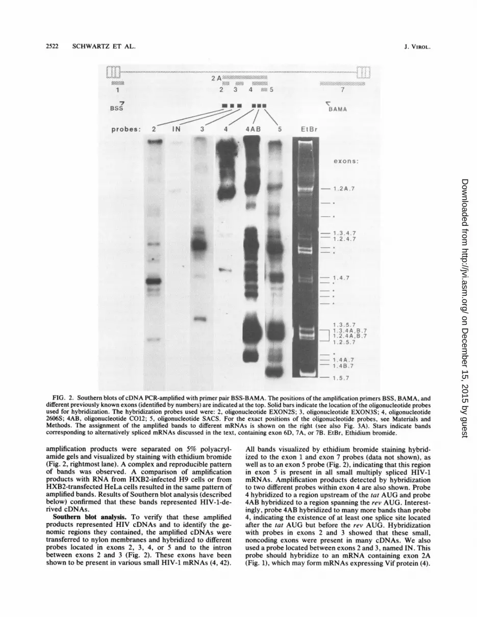
FIG. 2. Southern blots ofcDNA PCR-amplified with primer pair BSS-BAMA. The positions of the amplification primers BSS, BAMA, anddifferent previously known exons (identified by numbers) are indicated at the top. Solid bars indicate the location of the oligonucleotide probesused for hybridization. The hybridization probes used were: 2, oligonucleotide EXON2S; 3, oligonucleotide EXON3S; 4, oligonucleotide2606S; 4AB, oligonucleotide C012; 5, oligonucleotide SACS. For the exact positions of the oligonucleotide probes, see Materials andMethods. The assignment of the amplified bands to different mRNAs is shown on the right (see also Fig. 3A). Stars indicate bandscorresponding to alternatively spliced mRNAs discussed in the text, containing exon 6D, 7A, or 7B. EtBr, Ethidium bromide.
amplification products were separated on 5% polyacryl-amide gels and visualized by staining with ethidium bromide(Fig. 2, rightmost lane). A complex and reproducible patternof bands was observed. A comparison of amplificationproducts with RNA from HXB2-infected H9 cells or fromHXB2-transfected HeLa cells resulted in the same pattern ofamplified bands. Results of Southern blot analysis (describedbelow) confirmed that these bands represented HIV-1-de-rived cDNAs.
Southern blot analysis. To verify that these amplifiedproducts represented HIV cDNAs and to identify the ge-nomic regions they contained, the amplified cDNAs weretransferred to nylon membranes and hybridized to differentprobes located in exons 2, 3, 4, or 5 and to the intronbetween exons 2 and 3 (Fig. 2). These exons have beenshown to be present in various small HIV-1 mRNAs (4, 42).
All bands visualized by ethidium bromide staining hybrid-ized to the exon 1 and exon 7 probes (data not shown), aswell as to an exon 5 probe (Fig. 2), indicating that this regionin exon 5 is present in all small multiply spliced HIV-1mRNAs. Amplification products detected by hybridizationto two different probes within exon 4 are also shown. Probe4 hybridized to a region upstream of the tat AUG and probe4AB hybridized to a region spanning the rev AUG. Interest-ingly, probe 4AB hybridized to many more bands than probe4, indicating the existence of at least one splice site locatedafter the tat AUG but before the rev AUG. Hybridizationwith probes in exons 2 and 3 showed that these small,noncoding exons were present in many cDNAs. We alsoused a probe located between exons 2 and 3, named IN. Thisprobe should hybridize to an mRNA containing exon 2A(Fig. 1), which may form mRNAs expressing Vif protein (4).
J. VIROL.
-7
::j"
....
I*;,.11 "I.1
on Decem
ber 15, 2015 by guesthttp://jvi.asm
.org/D
ownloaded from

MULTIPLY SPLICED mRNAs OF HIV-1 2523
A.
uu-EXONS:
1
N-u-~-tat-
Z1119iYT -re'Igag _ F~I~
2 3 4 6Dm
4 A
4B-5-
mRNAs:
L4
-
lM4A/4B_ Im
_im
-5
- -
_ -
B. OTHER mRNAs
tev4 -
4 A /4B ---
5 m k-
l.w 7
2Ar- -I
-~~~~7-FIG. 3. Small multiply spliced HIV-1 mRN
organization of HIV-1 and the different exons artThe structures of the multiply spliced mRNAs eand Nef identified by cloning and sequencingmRNAs fall into three groups, characterized by4, 4A/4B, or 5. Tat is encoded by a groupcontaining exon 4. Rev is encoded by twocontaining either exon 4A or 4B. The Nef prolthree mRNAs containing exon 5. Each groupmRNA species characterized by the absence or psmall noncoding exons 2 and 3. (B) Structures amultiply spliced mRNAs. Exon 6D-containing cbeen cloned and sequenced (7; unpublished). Theshown to exist by the PCR amplification and hyments. Multiply spliced mRNAs containing theacceptors 7A and 7B, upstream of the previouslyalso indicated. Such mRNAs may produce variatory factors Tat and Rev containing additional
tev- Only one band hybridized to this probe, indicating that onlyv i one exon 2A-containing mRNA was generated. The struc-
ture of this mRNA is shown in Fig. 3B. Although we werevpr-UvPu J able to assign several amplified bands to specific cDNAs
7A -~ ~based on their sizes and hybridization to different probes,7 BA many bands could not be assigned by using these criteria. To
7 investigate the exact nature of the PCR-amplified molecules,we analyzed the amplification products by cloning andsequencing.
Cloning and sequencing of cDNAs. The PCR-amplifiedcDNAs were cloned into a modified Bluescript vector andsequenced by a double-stranded DNA sequencing protocolas described in Materials and Methods. As expected fromthe results of the hybridization experiments, mRNAs gener-
n f, , ated by utilization of additional splice sites between the tatand rev AUGs were identified. We cloned two different typesof cDNAs having rev as the first open reading frame. ThesemRNAs contained either of the two small exons, named 4A
W : Eand 4B, utilizing splice acceptors located 15 and 9 ntupstream of the rev AUG, respectively (Fig. 3; see also Fig.7). These two exons, whose exact locations are shown in
~nef Fig. 1, were found spliced to exon 1 either directly or viaexon 2 or exon 3, generating six different rev mRNAs (Fig.3). These findings verify the results of Si nuclease analysisthat identified exons 4A and 4B in RNA from HIV-1-infectedor -transfected cells (Felber et al., submitted). Others have
L also reported the presence of one splice site in the sameregion between the rev and tat AUGs by Si nuclease
ii3L analysis (54). This proposed splice acceptor corresponds to4B, identified here by cDNA cloning and sequencing.We also cloned and sequenced cDNAs corresponding to
the previously described tat mRNAs containing exon 4 (4,42). These three variant mRNAs are designated here as1.4.7, 1.2.4.7, and 1.3.4.7, according to the exons theycontain (Fig. 3). Sequence analysis of clones containing exon5 confirmed and extended previous studies, which hadidentified two exon 5-containing mRNAs, 1.5.7 and 1.2.5.7(4, 42). In line with observations made for the tat and revmRNAs, the group of mRNAs containing exon 5 was alsofound to consist of three mRNAs (Fig. 3), generated bysplicing of exon 5 to exon 1 directly or via exon 2 or exon 3.
A ; Therefore, all four central overlapping exons of the virus, 4,4A, 4B, and 5, existed in three combinations: spliced to exon
B 1 directly or linked to exon 1 via either exon 2 or 3, yieldinga total of 12 different mRNAs.The 12 tat, rev, and nef mRNA species identified by
cloning and sequencing did not account for the number of[nL cDNA bands amplified by PCR (Fig. 2), suggesting the
presence of additional mRNAs. Since total RNA had beenused in the initial amplifications, it could be argued that not
eAs. (A) Genomic all of the observed cDNAs represented cytoplasmicenshown at,the top mRNAs. To determine whether this was the case, bothncodingTat,tResv, cytoplasmic RNA and total RNA were used for amplifica-are shown. ese tion, and pattems of bands were analyzed in parallel. Thetheir middle exon,
complex profile obtained with total RNA was essentiallyof three mRNAs,identical to that of amplified cytoplasmic RNA. The possi-groups of mRNA, ietclt hto mlfe yolsi N.Tepsi
tein is encoded by bility of a small amount of nuclear contamination in theiconsists of three cytoplasmic fraction could not be ruled out; however, if this?resence of the two had occurred, we would have expected a lower amount of)f additional HIV-1 amplification products in the cytoplasmic fraction. ThesernM A 1 A T n:llNA i.4.oLJ. / nasother species were(bridization experi-alternative spliceknown exon 7, arent forms of regula-amino acids, indi-
cated by striped boxes. Two additional multiply spliced mRNAspredicted to exist in infected cells and designated 1.2A.7 and 1.3A.7are probably expressing Vif and Vpr, respectively. Grey barsindicate exons not entirely cloned.
VOL. 64, 1990
rlrrv
on Decem
ber 15, 2015 by guesthttp://jvi.asm
.org/D
ownloaded from

2524 SCHWARTZ ET AL.
2 A E-' :::::<:::::::.:::w.:-:::2*,5NO' ~42 3 4A %l-
M.
6D
3423A
probes: 4 4AB 5 6DI~~~~~~~~~~ F:
.U.:- 'A.%d&,Amk
6*0
doio ow",
- 1.4.6D.7
1 .3.4 A, B.G6 D.71 .24A,B.6D.71 .3.5.6D.71 2. 5.6D.7
/1 .4B.6D.71.5 SD.7
FIG. 4. Southern blots of cDNA PCR-amplified with primer pair NARS and 3423A. The positions of the amplification primers NARS and3423A and different previously known exons are indicated at the top. Numbers indicate different exons. Solid bars indicate the location ofthe oligonucleotide probes used for hybridization. The hybridization probes used were: 4, oligonucleotide 2606S; 4AB, oligonucleotide C012;5, oligonucleotide SACS; 6D, oligonucleotide 3311. Several bands hybridizing to probe 6D also hybridized to probes located in exons 4,4A/4B, and 5, indicating that exon 6D can be spliced to all four middle exons (Fig. 3B), resulting in additional mRNAs. The assignment ofsome bands is shown on the right.
results suggested that the unidentified cDNA bands repre-sent cytoplasmic mRNAs.
Identification of additional HIV splice sites. To obtain moreinformation about the additional multiply spliced mRNAs,we focused our attention on the region between exons 4 and7. We have recently identified another small exon, namedexon 6D, located within the env gene, as indicated in Fig. 3.We have shown that one mRNA containing exons 1, 4, 6D,and 7 generates a hybrid protein, Tev (7), consisting of thefirst exon of Tat, 38 amino acids of Env, and the second exonof Rev. We reasoned that this exon might be spliced not onlyto exon 4 but also to exons, 5, 4A, and 4B. This couldgenerate a maximum of 12 additional mRNAs, differing fromthe mRNAs depicted in Fig. 3 only by the presence of exon6D spliced to exon 7. To test this hypothesis, we synthesizeda primer located in exon 6D (primer 3423A, Fig. 1C). Thisprimer was used for cDNA synthesis of RNA isolated fromHXB2-infected or -transfected cells and subsequent PCRamplification in combination with the NARS primer locatedin exon 1. The cDNAs were subjected to Southern blotanalysis with probes located in exon 4, 4A/4B, or 5 (Fig. 4).These experiments demonstrated that exon 6D was splicedto exon 4, 4A, 4B, or 5, indicating that at least 4 and possibly12 mRNAs containing exon 6D exist in HXB2-infected cells.The similar results obtained in amplifications with BSS-BAMA and NARS-3423A indicated that mRNAs containingthe small exons 2 and 3 in addition to 6D were also produced.The presence of exon 6D-containing mRNAs can partiallyaccount for the additional unassigned bands detected in Fig.2. The structures and coding potential of four of these
mRNAs (1.4.6D.7, 1.4A.6D.7, 1.4B.6D.7, and 1.5.6D.7) areshown in Fig. 3B. It has been shown that HIV-1-infectedcells produce Tev from 1.4.6D.7 mRNA (7) and that aprotein containing 20 amino acids of Env linked to thesecond exon of Rev can be produced from 1.4A.6D.7,1.4B.6D.7, or 1.5.6D.7 mRNAs (7).To identify any additional splice sites between the splice
donor utilized by exons 4, 4A, 4B, and 5 and the spliceacceptor of exon 7 (nt 5591 to 7925, Fig. 1), we PCR-amplified mRNAs containing this region with two primers,3016S and 3015A, designed to detect alternative splice sites.Primer 3016S extends 4 nt 3' of the splice donor at nt 5591(Fig. 1), and primer 3015A extends 25 nt 5' of the exon 7splice acceptor at nt 7925, thereby selectively amplifyingeither unspliced or alternatively spliced mRNAs in thisregion. cDNA synthesis and PCR amplification with primerpair 3016S and BAMA did not result in amplification prod-ucts corresponding to alternatively spliced mRNAs, indicat-ing that only the previously described splice donor at nt 5591(Fig. iB) was utilized in this region. Sequence analysis ofcDNAs amplified with primers SACS and 3015A revealedthat this splice donor could be spliced to two additionalsplice acceptors, located upstream of the exon 7 spliceacceptor (nt 7925). The corresponding exons were desig-nated 7A and 7B (Fig. 1 and 7), with splice acceptors located28 and 24 nt upstream of exon 7, respectively. Splicing ofthese exons to exon 4 would produce mRNAs with thecapacity to express a Tat protein with an insertion of eightamino acids between the two coding exons or a Tat-Envhybrid protein, respectively. If these exons could be spliced
>,k
*1
7NARS
4^_. t: Ss t- b+v <t< ffl A * w t w - .i
7
exons:
J. VIROL.
amV' .W, .f*,
on Decem
ber 15, 2015 by guesthttp://jvi.asm
.org/D
ownloaded from
MULTIPLY SPLICED mRNAs OF HIV-1 2525
to exons 4A and 4B, the mRNAs produced would expresseither a Rev protein with eight additional amino acids or aRev-Tat hybrid protein. The presence and significance ofsuch proteins in infected cells remains to be investigated.
In order to identify other alternatively spliced smallmRNAs, we replica-plated several hundred colonies fromthe cDNA library and screened them with probes 2, 3, 4,4AB, and 5. Using this approach, we did not identify anyadditional cDNAs. We were also unable to find any mRNAspecies containing both exons 2 and 3, indicating that exons2 and 3 are mutually exclusive within the multiply splicedmRNAs. In addition, no cDNAs containing exons 1, 2, or 3linked directly to exon 7 were found. Results of colonyhybridizations with oligonucleotide 3015A as a probe re-vealed that exon 7A and 7B splice acceptors were present inless than 1% of the cDNA clones, which indicates that thesesplice acceptors are rarely used by the virus.
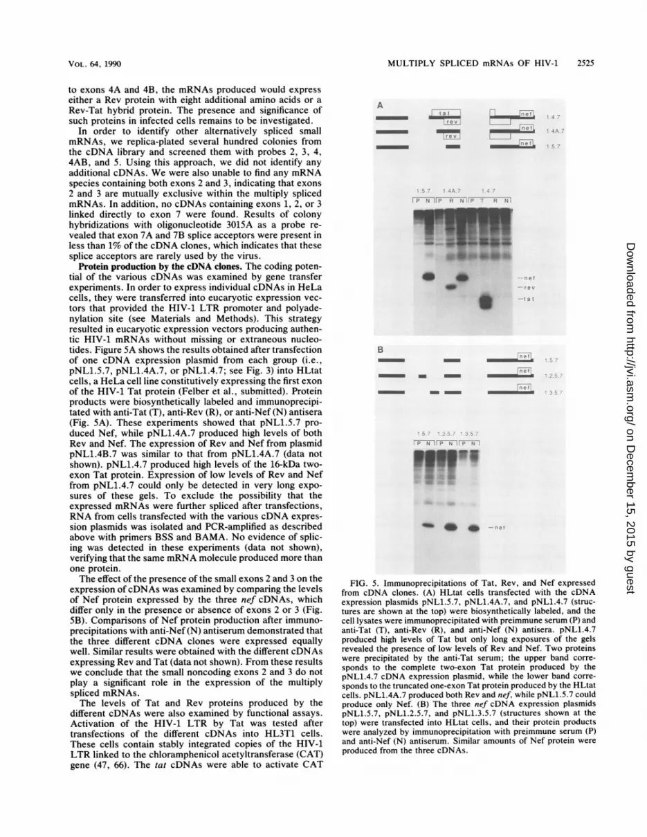
Protein production by the cDNA clones. The coding poten-tial of the various cDNAs was examined by gene transferexperiments. In order to express individual cDNAs in HeLacells, they were transferred into eucaryotic expression vec-tors that provided the HIV-1 LTR promoter and polyade-nylation site (see Materials and Methods). This strategyresulted in eucaryotic expression vectors producing authen-tic HIV-1 mRNAs without missing or extraneous nucleo-tides. Figure 5A shows the results obtained after transfectionof one cDNA expression plasmid from each group (i.e.,pNL1.5.7, pNL1.4A.7, or pNL1.4.7; see Fig. 3) into HLtatcells, a HeLa cell line constitutively expressing the first exonof the HIV-1 Tat protein (Felber et al., submitted). Proteinproducts were biosynthetically labeled and immunoprecipi-tated with anti-Tat (T), anti-Rev (R), or anti-Nef (N) antisera(Fig. 5A). These experiments showed that pNL1.5.7 pro-duced Nef, while pNL1.4A.7 produced high levels of bothRev and Nef. The expression of Rev and Nef from plasmidpNL1.4B.7 was similar to that from pNL1.4A.7 (data notshown). pNL1.4.7 produced high levels of the 16-kDa two-exon Tat protein. Expression of low levels of Rev and Neffrom pNL1.4.7 could only be detected in very long expo-sures of these gels. To exclude the possibility that theexpressed mRNAs were further spliced after transfections,RNA from cells transfected with the various cDNA expres-sion plasmids was isolated and PCR-amplified as describedabove with primers BSS and BAMA. No evidence of splic-ing was detected in these experiments (data not shown),verifying that the same mRNA molecule produced more thanone protein.The effect of the presence of the small exons 2 and 3 on the
expression ofcDNAs was examined by comparing the levelsof Nef protein expressed by the three nef cDNAs, whichdiffer only in the presence or absence of exons 2 or 3 (Fig.SB). Comparisons of Nef protein production after immuno-precipitations with anti-Nef (N) antiserum demonstrated thatthe three different cDNA clones were expressed equallywell. Similar results were obtained with the different cDNAsexpressing Rev and Tat (data not shown). From these resultswe conclude that the small noncoding exons 2 and 3 do notplay a significant role in the expression of the multiplyspliced mRNAs.The levels of Tat and Rev proteins produced by the
different cDNAs were also examined by functional assays.Activation of the HIV-1 LTR by Tat was tested aftertransfections of the different cDNAs into HL3T1 cells.These cells contain stably integrated copies of the HIV-1LTR linked to the chloramphenicol acetyltransferase (CAT)gene (47, 66). The tat cDNAs were able to activate CAT
Atat
- ~~~~~~~~~~~9--~~~~~~~~
-....~n--
net
net....... . ...
net
L7 TP N F; R N P T R Nv
8_Z_-=r14'P ft--_.W,
* ,.24 *** X'Crev
-.1 CS
B-~~~~~ n e
no-T
riotm m
P'N1 PN P N
w |1
FIG. 5. Immunoprecipitations of Tat, Rev, and Nef expressedfrom cDNA clones. (A) HLtat cells transfected with the cDNAexpression plasmids pNL1.5.7, pNL1.4A.7, and pNL1.4.7 (struc-tures are shown at the top) were biosynthetically labeled, and thecell lysates were immunoprecipitated with preimmune serum (P) andanti-Tat (T), anti-Rev (R), and anti-Nef (N) antisera. pNL1.4.7produced high levels of Tat but only long exposures of the gelsrevealed the presence of low levels of Rev and Nef. Two proteinswere precipitated by the anti-Tat serum; the upper band corre-
sponds to the complete two-exon Tat protein produced by thepNL1.4.7 cDNA expression plasmid, while the lower band corre-
sponds to the truncated one-exon Tat protein produced by the HLtatcells. pNL1.4A.7 produced both Rev and nef, while pNL1.5.7 couldproduce only Nef. (B) The three nef cDNA expression plasmidspNL1.5.7, pNL1.2.5.7, and pNL1.3.5.7 (structures shown at thetop) were transfected into HLtat cells, and their protein productswere analyzed by immunoprecipitation with preimmune serum (P)and anti-Nef (N) antiserum. Similar amounts of Nef protein were
produced from the three cDNAs.
VOL. 64, 1990
on Decem
ber 15, 2015 by guesthttp://jvi.asm
.org/D
ownloaded from

2526 SCHWARTZ ET AL.
t 2 3 4 fj
we I --_ *p..P i 0g p 2 C
4A 45 r-v- 5I- I- l_
XIV EXXB2 ATAACAAAAGCCTTAGGCATCTCCTATGGCAGGAAGAAGCONSZNSUS(NA) ???a?aaaAG?cttAGg?atctcctatggcAGgaagaagCONSENSUS(Z) ?taa??aaAGgcttAGgcatctcctatggcAGgaagaag
CONSZNSUS(5IV2) ?ta?a?aAGgggctcgggatatg?tat.a?cgaaagggcCONSENSUS (SIV) cttaaaaAGgg?tt ?gggat ??gttaZtag?agtcac??
_ - _M -- p55g4 g
4_ti do . _. ... p2 4cga
_. ;,---p1 7gag
FIG. 6. Functional assay for the expression of Rev protein by thedifferent cDNA clones. The HLfB cell line containing the rev mutantfB was transfected with the various cDNA clones, and cell lysateswere analyzed for p249ag production by Western immunoblotting as
described in Materials and Methods. Lanes: 1, transfection of HLfBcells with the Rev-expressing plasmid pL3crev as a positive control;2, HLfB cells transfected with 5 jig of pNL1.5.7; 3 and 4, HLfB cellstransfected with 5 ,ug of pNL1.4B.7 or pNL1.4A.7, respectively(trans-complementation by Rev produced by pNL1.4B.7 andpNL1.4A.7 completely restored the wild-type phenotype of themutant virus): 5 and 6, transfection with 15 and 5 ,ug of pNL1.4.7,respectively (lower levels of p24gag were detected, indicating thatpNL1.4.7 produced only very low levels of Rev protein); 7, trans-fection with salmon sperm DNA. HLfB cells do not produce p24gagor p17gag proteins.
expression to a similar extent in these cells, suggesting thathigh levels of functional Tat protein were produced from allcDNAs. As expected, vectors expressing rev or nefcDNAswere unable to activate cat expression, since they all lack anintact tat gene (data not shown).To assay for Rev function, we transfected the cDNA
expression plasmids into HLfB cells (Mermer et al., inpress), which contain stably integrated copies of a rev
mutant molecular clone of HIV-1. These cells produced lowlevels ofp55gag and Env proteins (Fig. 6, lane 7), but did notprocess p55gag to p24gag and pl7gag and did not producevirus particles. As shown in lane 1 of Fig. 6, complementa-tion by Rev protein after transfection of the Rev-producingplasmid pL3crev resulted in an increased production ofp55gag and Env proteins, as well as the production of p24gagand p179ag. We used this complementation assay to studythe expression of Rev protein by the different cDNAs. BothpNL1.4B.7 and pNL1.4A.7 were able to complement the rev
defect in HLfB cells (Fig. 6, lanes 3 and 4, respectively),indicating production of high levels of functional Rev pro-tein. All six different 4A- or 4B-containing cDNAs comple-mented equally well (data not shown). Transfection ofpNL1.4.7, which contains both tat and rev overlappingreading frames, resulted in very low levels of complementa-tion (lanes 5 and 6). When 15 ,ug of plasmid pNL1.4.7 wasused in the transfection (lane 5) instead of 5 Fg (lane 6),higher levels of p24gag and Env proteins were detected,indicating that low levels of Rev protein are produced fromthe tat mRNAs. However, these levels were too low to fullyrestore the wild-type phenotype of the rev mutant provirus inHLfB cells. Therefore, efficient production of Rev requiresthe presence of the rev-specific mRNAs. As expected,Nef-producing pNL1.5.7 did not complement the defect inHLfB cells, since it lacks the rev AUG (lane 2).
DISCUSSIONA surprisingly large number of mRNAs are produced by
HIV-1. The three regulatory proteins Tat, Rev, and Nef are
7A 7B 7I_ I_ I-
EIV 5XD2 AGAGTTAGGCAGGGATATTCACCATTATCGTTTCAGACC
CONSENSUS(NA) agagttAGgcAGggata?tcacc? ?t ?tc?tt ?cAGaccCONSENSUS(Z) agagttAGgcAGggata?tcacct ?tgtc?tt ?cAGacc
CONSENSUS(5IV2) ag?cttAGaaAGggctataggcctgttttctcttccccc
CONSZNSUS(SIV) a ?gttaAG?cAGgg?tataggccagtgttctcttcccc?
FIG. 7. Sequence comparisons of the regions of splice acceptors4A, 4B, 5, 7A, 7B, and 7. The consensus sequences in the regions ofsplice acceptors 4A, 4B, 5, 7A, 7B, and 7 are shown for NorthAmerican (NA) and Zairean (Z) HIV-1 isolates, as well as for HIV-2and SIV as compiled by Myers (44). The nonconserved positions areindicated with ?. Splice acceptors 4A and 4B are conserved amongthe HIV-1 isolates, while only 4A is conserved in HIV-2 and SIV.Both splice acceptors 7A and 7B are conserved among HIV-1isolates as well as in HIV-2 and SIV. The invariable dinucleotides ofthe acceptor sites (i.e., AG) are shown in boldface type. The revinitiator ATG is underlined.
produced from 12 mRNAs. Each protein is produced from agroup of mRNAs consisting of at least three mRNAs dif-fering in the leader sequence preceding the coding exons.This is a result of splicing to the small noncoding exons 2 and3, located in the 5' part of the virus (Fig. 1). Structural andfunctional assays did not reveal any significant differencebetween the Tat-expressing cDNAs or between the Rev- orthe Nef-expressing cDNAs. These results differ from thoseof Muesing et al. (43), who reported that 1.3.4.7 tat cDNAtransactivated the HIV-1 LTR five times better than 1.4.7cDNA.The splice acceptors of exons 2 and 3, at positions 4459
and 4936, respectively, are apparently utilized to producemRNAs encoding Vif and Vpr proteins. The predictedstructures of the vif and vpr mRNAs are shown in Fig. 3B.The production of these mRNAs could potentially decreasethe levels of expression of the proteins encoded downstreamof vif and vpr, e.g., tat, rev, vpu, env, and nef. A possiblerole for the splicing with the splice donors defining exons 2and 3 might be the production of high levels of mRNAsexpressing these downstream proteins. In this regard, it isnoteworthy that mRNAs containing exons similar to 2 and 3in HIV-1 are produced by other lentiviruses such as SIVMAC(9) and visna virus (11).Four new splice sites were identified by cloning and
sequencing, and the corresponding exons were named 4A,4B, 7A, and 7B. The 5' splice sites generating exons 4A and4B are positioned only six nucleotides apart, after the tatAUG and before the rev AUG. Thus, utilization of eithersplice acceptor 4A or 4B allows expression of rev but not tat.A similar mechanism to generate rev mRNA(s) has beenproposed for the SIVMAC virus isolate by Colombini et al.(9). These investigators identified a cDNA clone that uses asplice acceptor between the tat and rev AUGs and isbelieved to represent the Rev-producing mRNA. The obser-vation that cDNAs containing exon 4A or 4B were clonedwith the same frequency after PCR amplification indicatessimilar use of both splice acceptors. In addition, directdetection of exons 4A and 4B in infected or transfected cellsby Si nuclease analysis revealed similar levels of mRNAs
J. VIROL.
on Decem
ber 15, 2015 by guesthttp://jvi.asm
.org/D
ownloaded from

MULTIPLY SPLICED mRNAs OF HIV-1 2527
TABLE 1. Protein production of four different HIV-1 mRNAs
Coding Protein productionmRNAcapacity Tat Rev Nef
1.4.7 tat, rev, nef + +1- +1-1.4A.7 rev, nef - + +1.4B.7 rev, nef - + +1.5.7 nef - - +
containing these exons (Felber et al., submitted). Althoughthe purpose of using two splice acceptors in this region isunclear, it is noteworthy that both splice acceptors are
conserved among different HIV-1 isolates (Fig. 7). In addi-tion, the splice acceptor 4A is also well conserved in thedistantly related lentiviruses HIV-2 and SIV. The spliceacceptor site for exon 4B is conserved only among thedifferent HIV-1 isolates. The newly identified splice accep-
tors were also compared with the consensus splice acceptorsequence (59). The splice acceptor site 4B is in good agree-
ment with the consensus splice acceptor sequence, while 4Ais not.The splice acceptors of exons 7A and 7B are located
before exon 7, the last exon of HIV-1 (Fig. 2). Theseacceptors should be able to generate mRNAs producingisoforms of Tat and Rev or hybrid proteins between the firstexon of Tat and Env or the first exon of Rev and the secondexon of Tat. Both splice acceptors are conserved in HIV-1isolates, as well as in HIV-2 and SIV (Fig. 7). Although thebiological significance of these mRNAs remains to be inves-tigated, it is noteworthy that an alternative splice acceptorhas also been found in the last exon of the SIVMAC genome
(64). This further underlines the similar organization of thelentiviruses.Although the splice sites producing exon 6D are not
conserved in all HIV strains, it is interesting that this exonwas present in many small multiply spliced mRNAs. Thisobservation indicates that the generation of one splice sitemight result in many new alternatively spliced mRNAs. Thismight be an additional mechanism by which lentivirusesdiverge at a high rate. Exons 6D, 7A, and 7B could generateseveral different mRNAs by alternative splicing. Assumingthat exon 6D could be spliced to all 12 mRNA speciesreported here, a total number of 24 differentially splicedsmall mRNAs would be generated. Furthermore, if thesplice acceptors for exons 7A and 7B are utilized in additionto exon 7, a total of 72 differentially spliced small HIV-1mRNAs would be generated. We have previously shownthat one of the 6D-containing mRNAs produces a hybridprotein, Tev (7). The protein products of other exon 6D-containing mRNAs have also been studied (7). Although thesignificance of these mRNA species for the virus remains tobe determined, identification of the alternative splice sitesfurther underlines the complexity of HIV-1 splicing. Theacquisition of regulatory factors such as Rev, which allowsthe production of appropriate quantities of structuralmRNAs, may have allowed the incorporation of additionalsplice signals and the generation of many alternativelyspliced mRNAs. Alternative splicing may offer an evolution-ary advantage to the virus, since proteins are produced frommore than one mRNA. Also, the plasticity of the genomemay allow new combinations to arise and enable viraladaptation to a new environment.
Results of the functional studies of the HIV-1 cDNAclones shown in Fig. 5 and 6 and summarized in Table 1
revealed that tat mRNAs produced very low levels of Revand Nef, while rev mRNAs produced high levels of both Revand Nef proteins. These data are in agreement with thescanning model for initiation of translation (34, 35), accord-ing to which the ribosome binds at the 5' end of the mRNAand scans the RNA for a suitable initiation site. Initiation atthe tat AUG appears to be very efficient, allowing theribosome to read the downstream AUGs for rev and nefonlyin rare instances. This results in high production of Tat butonly low production of Rev and Nef (Fig. 5A). In contrast,the rev AUG seems to allow readthrough of the ribosome toa much higher degree than the tat AUG, resulting in highlevels of both Nef and Rev proteins expressed from the revmRNAs (Fig. SA).
Since tat mRNAs are very poor producers of Rev protein,specific rev mRNAs containing exon 4A or 4B are essentialfor production of sufficient amounts of Rev. Rev producedfrom mRNAs containing either exon 4A or 4B was sufficientto restore virus expression from the rev mutant provirusintegrated in HLfB cells (Fig. 6). These data are in disagree-ment with the results presented by Sadaie et al. (54), whoreported that a derivative of HXB2 containing a pointmutation at the exon 4B splice site displayed a rev mutantphenotype after transfection into COS cells (mutant M43[54]). Our data show that HXB2 can generate anotherequally potent rev mRNA by using the adjacent spliceacceptor, 4A. Therefore, mutant M43 should also produceRev. Accordingly, we were able to show that M43 producedfunctional levels of Rev protein by using the complementa-tion assay described above (data not shown). We also wereable to immunoprecipitate Rev from M43-transfected HeLacells. We concluded that M43 is a rev-deficient but notrev-minus mutant.The multiply spliced mRNAs lack the RRE element and
are present in high levels in the absence of Rev (17, 28). Thecloning and characterization of the majority, if not all, of themultiply spliced HIV-1 mRNAs allowed the generation ofspecific probes for the study of the expression of theindividual species by S1 nuclease analysis (Felber et al.,submitted). This is necessary in order to quantitate thedifferent mRNAs present in infected cells, since PCR ampli-fication results in a nonlinear amplification of target cDNAs.Concerning the reliability of the PCR technique, repetitivecloning and sequencing of the same cDNAs from differentcDNA libraries created by PCR did not reveal any sequenceaberrations. We therefore believe that PCR, as applied here,is a reliable technique for the qualitative analysis of cDNAs.We have shown that HIV-1 produces one group of
mRNAs for each regulatory protein Tat, Rev, and Nef,presumably allowing high levels of expression of each ofthese proteins by the virus. It is interesting that the Nefprotein is produced in high amounts from its own group ofthree monocistronic mRNAs as well as from the six bicis-tronic rev mRNAs. Although HIV-1 produces several bicis-tronic mRNAs, it does not depend on the expression of morethan one protein from each of the mRNAs producing Tat,Rev, and Nef. This is unlike the organization of the humanT-cell leukemia viruses (45), which are proposed to rely on asingle doubly spliced mRNA for the production of both Taxand Rex regulatory proteins.
ACKNOWLEDGMENTS
We are grateful to M. Powers for oligonucleotide synthesis and toJ. Ghrayeb for the Nef antibody. We thank M. Campbell, J.Harrison, and D. Vanderbur for technical assistance, L. Solomin forparticipating in some experiments, and A. Arthur for editing.
VOL. 64, 1990
on Decem
ber 15, 2015 by guesthttp://jvi.asm
.org/D
ownloaded from

2528 SCHWARTZ ET AL.
This research was sponsored by the National Cancer Instituteunder contract N01-CO-74101 with ABL, by the Swedish CancerSociety, and by the Swedish Medical Research Council.
LITERATURE CITED1. Adachi, A., H. E. Gendelman, S. Koenig, T. Folks, R. Willey, A.
Rabson, and M. A. Martin. 1986. Production of acquired immu-nodeficiency syndrome-associated retrovirus in human and non-human cells transfected with an infectious molecular clone. J.Virol. 59:284-291.
2. Ahmad, N., and S. Venkatesan. 1988. Nef protein of HIV-1 is atranscriptional repressor of HIV-1 LTR. Science 241:1481-1485.
3. Arya, S. K., B. Beaver, L. Jagodzinski, B. Ensoli, P. J. Kanki, J.Albert, E. M. Fenyo, G. Biberfeld, J. F. Zagury, F. Laure, M.Essex, E. Norrby, F. Wong-Staal, and R. C. Gallo. 1987. Newhuman and simian HIV-related retrovirus possess functionaltransactivator (tat) gene. Nature (London) 328:548-550.
4. Arya, S. K., and R. C. Gallo. 1986. Three novel genes of humanT-lymphotropic virus type III: immune reactivity of their prod-ucts with sera from acquired immune deficiency syndromepatients. Proc. Natl. Acad. Sci. USA 83:2209-2213.
5. Arya, S. K., C. Guo, S. F. Josephs, and F. Wong-Staal. 1985.Transactivator gene of human T-lymphotrophic virus type III(HTLV-ILI). Science 229:69-73.
6. Barre-Sinoussi, F., J.-C. Cherman, F. Rey, M. T. Nugeyre, S.Chamaret, J. Gruest, C. Dauguet, C. Axler-Blin, F. Brun-Vezinet, C. Rouzious, W. Rozenbaum, and L. Montagnier. 1983.Isolation of a T-lymphotropic retrovirus from a patient at riskfor acquired immune deficiency syndrome (AIDS). Science220:868-871.
7. Benko, D. M., S. Schwartz, G. N. Pavlakis, and B. K. Felber.1990. A novel human immunodeficiency virus type 1 protein,tev, shares sequences with tat, env, and rev proteins. J. Virol.64:2498-2504.
8. Chirgwin, J. M., A. E. Przybyla, R. J. MacDonald, and W. J.Rutter. 1979. Isolation of biologically active ribonucleic acidfrom sources enriched in ribonuclease. Biochemistry 18:5294-5299.
9. Colombini, S., S. K. Arya, M. S. Reitz, L. Jagodzinski, B.Beaver, and F. Wong-Staal. 1989. Structure of simian immuno-deficiency virus regulatory genes. Proc. Natl. Acad. Sci. USA86:4813-4817.
10. Cullen, B. R. 1986. Trans-activation of human immunodefi-ciency virus occurs via a bimodal mechanism. Cell 46:973-982.
11. Davis, J. L., and J. E. Clements. 1989. Characterization of acDNA clone encoding the visna virus transactivating protein.Proc. Natl. Acad. Sci. USA 86:414-418.
12. Dayton, A. I., J. G. Sodroski, C. A. Rosen, W. C. Goh, andW. A. Haseltine. 1986. The trans-activator gene of the humanT-cell lymphotrophic virus type III is required for replication.Cell 44:941-947.
13. Dayton, A. I., E. F. Terwilliger, J. Potz, M. Kowalski, J. G.Sodroski, and W. A. Haseltine. 1988. cis-Acting sequencesresponsive to the rev gene product of the human immunodefi-ciency virus. J. AIDS 1:441-452.
14. Dorn, P. L., and D. Derse. 1988. cis- and trans-acting regulationof gene expression of equine infectious anemia virus. J. Virol.62:3522-3526.
15. Emerman, M., M. Guyader, L. Montagnier, D. Baltimore, andM. Muesing. 1987. The specificity of the human immunodefi-ciency virus type 2 transactivator is different from that of humanimmunodeficiency virus type 1. EMBO J. 6:3755-3760.
16. Emerman, M., R. Vazeux, and K. Peden. 1989. The rev geneproduct of the human immunodeficiency virus affects envelope-specific RNA localization. Cell 57:1155-1165.
17. Feinberg, M. B., R. F. Jarrett, A. Aldovini, R. C. Gallo, and F.Wong-Staal. 1986. HTLV-III expression and production involvecomplex regulation at the levels of splicing and translation ofviral RNA. Cell 46:807-817.
18. Felber, B. K., M. Cladaras, C. Cladaras, C. M. Wright, A. Tse,and G. N. Pavlakis. 1988. Regulation of HIV-1 by viral factors,p. 71-77. In B. R. Franza, B. R. Cullen, and F. Wong-Staal
(ed.), The control of human retrovirus gene expression. ColdSpring Harbor Laboratory, Cold Spring Harbor, N.Y.
19. Felber, B. K., M. Hadzopoulou-Cladaras, C. Cladaras, T. Cope-land, and G. N. Pavlakis. 1989. The rev protein of HIV-1 affectsthe stability and transport of the viral mRNA. Proc. Natl. Acad.Sci. USA 86:1495-1499.
20. Felber, B. K., H. Paskalis, C. Kleinman-Ewing, F. Wong-Staal,and G. N. Pavlakis. 1985. The pX protein of HTLV-1 is atranscriptional activator of its long terminal repeats. Science229:675-679.
21. Fisher, A. G., M. B. Feinberg, S. F. Josephs, M. E. Harper,L. M. Marselle, G. Reyes, M. A. Gonda, A. Aldovini, C. Debouk,R. C. Gallo, and F. Wong-Staal. 1986. The transactivator geneof HTLV-III is essential for virus replication. Nature (London)320:367-371.
22. Gallo, R. C., S. Z. Salahuddin, M. Popovic, G. M. Shearer, M.Kaplan, B. F. Haynes, T. J. Palker, R. Redfield, J. Oleske, B.Safai, G. White, P. Foster, and P. D. Markham. 1984. Frequentdetection and isolation of cytopathic retroviruses (HTLV-III)from patients with AIDS and at risk for AIDS. Science 224:500-503.
23. Gonda, M. A., F. Wong-Staal, R. C. Gallo, J. E. Clements, 0.Narayan, and R. V. Gilden. 1985. Sequence homology andmorphogenic similarity of HTLV-III and visna virus, a patho-genic lentivirus. Science 227:173-177.
24. Gourdou, I., V. Mazarin, G. Querat, N. Sauze, and R. Vigne.1989. The open reading frame S of visna virus genome is atrans-activating gene. Virology 171:170-178.
25. Graham, F. J., and A. J. Van der Eb. 1973. A new technique forthe assay of infectivity of human adenovirus S DNA. Virology52:456-460.
26. Guy, B., M. P. Kieny, Y. Riviere, C. LePeuch, K. Dott, and M.Girard, L. Montagnier, and J. P. Ledcocq. 1987. HIV F/3' orfencodes a phosphorylated GTP-binding protein resembling anoncogene product. Nature (London) 330:266-269.
27. Guyader, M., M. Emerman, P. Sonigo, F. Clavel, L. Montagnier,and M. Alizon. 1987. Genome organization and transactivationof the human immunodeficiency virus type 2. Nature (London)326:662-669.
28. Hadzopoulou-Cladaras, M., B. K. Felber, C. Cladaras, A. Atha-nassopoulos, A. Tse, and G. N. Pavlakis. 1989. The Rev (Trs/Art)protein of human immunodeficiency virus type 1 affects viralmRNA and protein expression via a cis-acting sequence in theenv region. J. Virol. 63:1265-1274.
29. Hammarskjold, M. L., J. Heimer, B. Hammarskjold, I. Sang-wan, L. Albert, and D. Rekosh. 1989. Regulation of humanimmunodeficiency virus env expression by the rev gene product.J. Virol. 63:1959-1966.
30. Hammes, S., E. Dixon, M. Malim, B. Cullen, and W. Greene.1989. Nef protein of human immunodeficiency virus type 1:evidence against its role as a transcriptional inhibitor. Proc.Natl. Acad. Sci. USA 86:9549-9553.
31. Hauber, J., A. Perkins, E. P. Heimer, and B. R. Cullen. 1987.Transactivation of human immunodeficiency virus gene expres-sion is mediated by nuclear events. Proc. Natl. Acad. Sci. USA84:6364-6368.
32. Hess, J. L., J. E. Clements, and 0. Naryan. 1985. cis- andtrans-acting transcriptional regulation of visna virus. Science229:482-485.
33. Kim, S., K. Ikeuchi, R. Byrn, J. Groopman, and D. Baltimore.1989. Lack of a negative influence on viral growth by the nefgene of human immunodeficiency virus type 1. Proc. Natl.Acad. Sci. USA 86:9544-9548.
34. Kozak, M. 1978. How do eucaryotic ribosomes select initiationregions in messenger RNA? Cell 15:1109-1123.
35. Kozak, M. 1989. The scanning model for translation: an update.J. Cell Biol. 108:229-241.
36. Levy, J. A., A. D. Hoffman, S. M. Kramer, J. A. Landis, J. M.Shimabukuro, and L. S. Oshiro. 1984. Isolation of lymphocyto-pathic retroviruses from San Francisco patients with AIDS.Science 225:840-842.
37. Luciw, P. A., C. Cheng-Mayer, and J. A. Levy. 1987. Mutationalanalysis of the human immunodeficiency virus: the orf-B region
J. VIROL.
on Decem
ber 15, 2015 by guesthttp://jvi.asm
.org/D
ownloaded from

MULTIPLY SPLICED mRNAs OF HIV-1 2529
down-regulates virus replication. Proc. Natl. Acad. Sci. USA84:1434-1438.
38. Malim, M., S. Bohnlein, R. Fenrick, S. Y. Le, J. V. Maizel, andB. R. Cullen. 1989. Functional comparison of the rev trans-activators encoded by different primate immunodeficiency virus
species. Proc. Natl. Acad. Sci. USA 86:8222-8226.39. Malim, M. H., J. Hauber, S. Le, J. V. Maizel, and B. R. Cullen.
1989. The HIV-1 rev transactivator acts through a structuredtarget sequence to activate nuclear export of unspliced viralmRNA. Nature (London) 338:254-257.
40. Mazarin, V., I. Gourdou, G. Querat, N. Sauze, and R. Vigne.1988. Genetic structure and function of an early transcript ofvisna virus. J. Virol. 62:4813-4818.
41. Meyerhans, A., R. Cheynier, J. Albert, M. Seth, S. Kwok, J.
Sninsky, L. Morfeldt-Manson, B. Asjo, and S. Wain-Hobson.1989. Temporal fluctuations in HIV quasispecies in vivo are notreflected by sequential HIV isolations. Cell 58:901-910.
42. Muesing, M. A., D. H. Smith, C. D. Cabradilla, C. V. Benton,L. A. Lasky, and D. J. Capon. 1985. Nucleic acid structure andexpression of the human AIDS/lymphadenopathy retrovirus.Nature (London) 313:450-458.
43. Muesing, M. A., D. H. Smith, and D. J. Capon. 1987. Regulationof mRNA accumulation by a human immunodeficiency virustrans-activator protein. Cell 48:691-701.
44. Myers, G. 1989. Human retroviruses and AIDS: a compilationand analysis of nucleic acid and amino acid sequences. LosAlamos National Laboratory, Los Alamos, N.Mex.
45. Nagashima, K., M. Yoshida, and M. Seiki. 1986. A single speciesof pX mRNA of human T-cell leukemia virus type I encodestrans-activator p4Ox and two other phosphoproteins. J. Virol.60:394-399.
46. Niederman, M. J., B. J. Thielan, and L. Ratner. 1989. Humanimmunodeficiency virus type 1 negative factor is a transcrip-tional silencer. Proc. Natl. Acad. Sci. USA 86:1128-1132.
46a.Pavlakis, G. N., and B. K. Felber. 1990. Regulation of expres-sion human immunodeficiency virus. New Biol. 2:20-31.
47. Pavlakis, G. N., B. K. Felber, and C. M. Wright. 1988. A fusionassay for the detection of HIV infected cells, p. 439-445. In D.Bolognesi (ed.), Human retroviruses, cancer and AIDS: ap-proaches to prevention and therapy. Alan R. Liss, Inc., NewYork.
48. Peterlin, B. M., P. A. Luciw, P. J. Barr, and M. D. Walker.1986. Elevated levels of mRNA can account for the trans-activation of human immunodeficiency virus. Proc. Natl. Acad.Sci. USA 83:9734-9738.
49. Ratner, L., A. Fisher, L. L. Jagodzinski, R. S. Liou, H. Mitsuya,R. C. Gallo, and F. Wong-Staal. 1987. Complete nucleotidesequences of functional clones of the virus associated with theacquired immunodeficiency syndrome, HTLV-III/LAV. Hama-tol. Bluttransfus. 31:404-406.
50. Ratner, L., A. Fisher, L. L. Jagodzinski, H. Mitsuya, R. S. Liou,R. C. Gallo, and F. Wong-Staal. 1987. Complete nucleotidesequences of functional clones of the AIDS virus. AIDS Res.Hum. Retroviruses 3:57-69.
51. Ratner, L., W. Haseltine, R. Patarca, K. L. Livak, B. Starcich,S. F. Josephs, E. R. Doran, J. A. Rafalski, E. A. Whitehorn, K.Baumeister, L. Ivanoff, S. Petteway, Jr., M. L. Pearson, J. A.Lautenberger, T. S. Papas, J. Ghrayeb, N. T. Chang, R. C.Gallo, and F. Wong-Staal. 1985. Complete nucleotide sequence
of the AIDS virus, HTLV-III. Nature (London) 313:277-283.52. Rosen, C. A., J. G. Sodroski, and W. A. Haseltine. 1985. The
location of cis-acting regulatory sequences in the human T celllymphotropic virus type III (HTLV-III/LAV) long terminalrepeat. Cell 41:813-823.
53. Rosen, C. A., E. Terwilliger, A. Dayton, J. G. Sodroski, andW. A. Haseltine. 1988. Intragenic cis-acting art gene-responsivesequences of the human immunodeficiency virus. Proc. Natl.Acad. Sci. USA 85:2071-2075.
54. Sadaie, M. R., J. Rappaport, T. Benter, S. F. Josephs, R. Willis,and F. Wong-Staal. 1988. Missense mutations in an infectioushuman immunodeficiency viral genome: functional mapping oftat and identification of the rev splice acceptor. Proc. Natl.Acad. Sci. USA 85:9224-9228.
55. Saiki, R. K., D. H. Gelfand, S. Stoffel, S. J. Scharf, R. Higuchi,G. T. Horn, K. B. Mullis, and H. A. Ehrlich. 1988. Primer-directed enzymatic amplification of DNA with a thermostableDNA polymerase. Science 239:487-491.
56. Saiki, R. K., S. Scharf, F. Faloona, K. B. Mullis, G. T. Horn,H. A. Erlich, and N. Arnheim. 1985. Enzymatic amplification ofP-globin genomic sequences and restriction site analysis fordiagnosis of sickle cell anemia. Science 230:1350-1354.
57. Sanger, F., S. Nicklen, and A. R. Coulson. 1977. DNA sequenc-ing with chain-terminating inhibitors. Proc. Natl. Acad. Sci.USA 74:5463-5467.
58. Schwartz, S., B. K. Felber, E. M. Fenyo, and G. N. Pavlakis.1989. Rapidly and slowly replicating HIV-1 isolates can bedistinguished according to target cell tropism in T-cell andmonocyte cell lines. Proc. Natl. Acad. Sci. USA 86:7200-7203.
59. Shapiro, M. B., and P. Senapathy. 1987. RNA splice junctions ofdifferent classes of eukaryotes: sequence statistics and func-tional implications in gene expression. Nucleic Acids Res.15:7155-7174.
60. Sherman, L., A. Gazit, A. Yaniv, T. Kawakami, J. E. Dahlberg,and S. R. Tronick. 1988. Localization of sequences responsiblefor trans-activation of the equine infectious anemia virus longterminal repeat. J. Virol. 62:120-126.
61. Sodroski, J., W. C. Goh, C. Rosen, A. Dayton, E. Terwilliger,and W. A. Haseltine. 1986. A second post-transcriptional trans-activator gene required for the HTLV-III replication. Nature(London) 321:412-417.
62. Sodroski, J., R. Patarca, C. Rosen, F. Wong-Staal, and W.Haseltine. 1985. Location of the trans-activating region on thegenome of human T-cell lymphotrophic virus type III. Science229:74-77.
63. Sonigo, P., M. Alizon, K. Staskus, D. Klatzmann, S. Cole, 0.Danos, E. Retzel, P. Tioliais, A. Haase, and S. Wain-Hobson.1985. Nucleotide sequence of the visna lentivirus: relationshipto the AIDS virus. Cell 42:369-382.
64. Viglianti, G. A., and J. I. Mullins. 1988. Functional comparisonof transactivation by simian immunodeficiency virus fromrhesus macaques and human immunodeficiency virus type 1. J.Virol. 62:4523-4532.
65. Wain-Hobson, S., P. Sonigo, 0. Danos, S. Cole, and M. Alizon.1985. Nucleotide sequence of the AIDS virus, LAV. Cell40:9-17.
66. Wright, C. M., B. K. Felber, H. Paskalis, and G. N. Pavlakis.1986. Expression and characterization of the trans-activator ofHTLV-III/LAV virus. Science 234:988-992.
VOL. 64, 1990
on Decem
ber 15, 2015 by guesthttp://jvi.asm
.org/D
ownloaded from


