Embed Size (px)
Citation preview

J Physiol 586.11 (2008) pp 2695–2712 2695
Coincidence detection of convergent perforant pathand mossy fibre inputs by CA3 interneurons
Eduardo Calixto1, Emilio J. Galvan2, J. Patrick Card2 and German Barrionuevo2
1Division de Investigaciones en Neurociencias, Instituto Nacional de Psiquiatrıa Ramon de la Fuente, Mexico City, Mexico2Department of Neuroscience, University of Pittsburgh, Pittsburgh, PA, USA
We performed whole-cell recordings from CA3 s. radiatum (R) and s. lacunosum-moleculare
(L-M) interneurons in hippocampal slices to examine the temporal aspects of summation of
converging perforant path (PP) and mossy fibre (MF) inputs. PP EPSPs were evoked from
the s. lacunosum-moleculare in area CA1. MF EPSPs were evoked from the medial extent of the
suprapyramidal blade of the dentate gyrus. Summation was strongly supralinear when examining
PP EPSP with MF EPSP in a heterosynaptic pair at the 10 ms ISI, and linear to sublinear
at longer ISIs. This pattern of nonlinearities suggests that R and L-M interneurons act as
coincidence detectors for input from PP and MF. Summation at all ISIs was linear in voltage clamp
mode demonstrating that nonlinearities were generated by postsynaptic voltage-dependent
conductances. Supralinearity was not detected when the first EPSP in the pair was replaced
by a simulated EPSP injected into the soma, suggesting that the conductances underlying
the EPSP boosting were located in distal dendrites. Supralinearity was selectively eliminated
with either Ni2+ (30 μM), mibefradil (10 μM) or nimodipine (15 μM), but was unaffected by
QX-314. This pharmacological profile indicates that supralinearity is due to recruitment of
dendritic T-type Ca2+channels by the first subthreshold EPSP in the pair. Results with the
hyperpolarization-activated (I h) channel blocker ZD 7288 (50 μM) revealed that I h restricted
the time course of supralinearity for coincidently summed EPSPs, and promoted linear to
sublinear summation for asynchronous EPSPs. We conclude that coincidence detection results
from the counterbalanced activation of T-type Ca2+ channels and inactivation of I h.
(Received 18 February 2008; accepted after revision 3 April 2008; first published online 3 April 2008)
Corresponding author G. Barrionuevo: Department of Neuroscience, A210 Langley Hall, University of Pittsburgh,
Pittsburgh, PA, USA. Email: [email protected]
Damage to the hippocampal formation in humans isknown to produce a deficit in the ability to form long-termepisodic memories (Eichenbaum & Otto, 1992). Althoughthe mechanisms by which the hippocampus supportsmemory formation are still unknown, theoretical andempirical studies provide evidence that the neural networkin area CA3 is capable of generating pattern separatedmemory representations from overlapping neocorticalinputs from the entorhinal cortex (EC) (McNaughton &Morris, 1987; Treves & Rolls, 1994; O’Reilly & McClelland,1994; Leutgeb et al. 2007). Pattern separation may requirethe reconfiguration of coincidentally active CA3 pyramidalcell assemblies by the coactivation of the two convergingexcitatory inputs from the EC. One input is conveyedmonosynaptically via the perforant path (PP), the axonsof the stellate cells in the EC layer II. The other input
E. Calixto and E. J Galvan contributed equally to this work. This paper
has online supplemental material.
is conveyed disynaptically via the mossy fibres (MF),the axons of dentate gyrus (DG) granule cells, whichalso are the targets of the same layer II cells of theEC (Tamamaki & Nojyo, 1993). CA3 pyramidal cellsreceive strong somatic and dendritic inhibitory input fromfeedforward GABAergic interneurons (Buzsaki, 1984;Lawrence & McBain, 2003), which are innervated by theconcurrent excitatory drive from both EC projections. Therecruitment of feed-forward inhibitory interneurons topromote a narrow time window for pyramidal cell firing(Pouille & Scanziani, 2001) also may serve as a means forthe orthogonalization of the CA3 representations. Thisinhibitory influence on the fine tuning of the active CA3pyramidal cell assemblies during pattern separation maydepend, in part, on whether integration of the excitatoryinputs on interneurons is linear or nonlinear.
The ability of interneurons to integrate synaptic inputswith temporal reliability and precision has been attributedto the increased number and faster kinetics of postsynapticAMPA/kainate receptors (Geiger et al. 1997; Nusser et al.
C© 2008 The Authors. Journal compilation C© 2008 The Physiological Society DOI: 10.1113/jphysiol.2008.152751

2696 E. Calixto and others J Physiol 586.11
1998; Walker et al. 2002; Jonas et al. 2004). Previous studieshave also shown that interneurons are endowed with activeconductances in somatic and dendritic compartments,similar to those found in pyramidal cells (Martina et al.2000; Goldberg et al. 2003; Kaiser et al. 2004; Rozsaet al. 2004). These conductances could shape subthresholdEPSPs (Fricker & Miles, 2000; Jonas et al. 2004), andpermit rapid detection of converging excitatory inputs(Galarreta & Hestrin, 2001). Although there have beenextensive characterizations of the morphology, physio-logy and plasticity of CA3 interneurons (for reviews seeFreund & Buzsaki, 1996; McBain et al. 1999; McBain &Fisahn, 2001; Lawrence & McBain, 2003; Jonas et al. 2004),the rules governing synaptic integration of MF and PPinputs to these cells are not known. The purpose of thecurrent study was to assess the properties and mechanismsof temporal integration in radiatum (R) and stratumlacunosum-moleculare (L-M) interneurons in area CA3 bytaking advantage of the ability to stimulate independentlyconvergent MF and PP inputs (Urban & Barrionuevo,1998). The axons of R and L-M interneurons branch in thes. lacunosum-moleculare, s. radiatum and s. pyramidale,and provide feed-forward inhibition to pyramidal cells(Lacaille & Schwartzkroin, 1988b; Williams et al. 1994;Vida et al. 1998). We performed somatic whole-cellrecordings to measure the arithmetic summation of a seriesof pair-wise interactions between subthreshold AMPAMF and PP EPSPs evoked from the medial extent of thesuprapyramidal blade of the dentate gyrus (SDG) atinterstimulus intervals (ISI) ranging from 10 to 100 ms.We found that the prevalent summation rule at the 10 msISI was supralinearity brought about by the counter-balanced activation of T-type Ca2+ and inactivation of hchannels. We propose that PP EPSP and MF EPSP weregenerated from neighbouring synapses located in the s.lacunosum-moleculare of area CA3.
Methods
Slice preparation
Animal use was in accordance with the UniversityInstitutional Animal Care and Use Committee. MaleSprague–Dawley rats (22 ± 4 days old; Zivic MillerCompany) were deeply anaesthetized (Nembutal, i.p.,5 mg per 100 g body weight) and perfused intracardiallywith a modified artificial cerebrospinal fluid (ACSF) inwhich sucrose has been substituted for sodium chloride(concentrations in mm): 230.0 sucrose, 1.9 KCl, 1.2Na2PO4.7H2O, 25.0 NaHCO3, 10.0 glucose, 1.0 CaCl2,4.0 MgCl2, at 4◦C; pH 7.3 maintained with bubbled O2
(95%)/CO2 (5%) at room temperature. Following 1–2 minof perfusion, animals were decapitated, and the brainsremoved. Blocks of tissue containing the hippocampuswere glued to the stage of a Leica VT1000S vibrating
blade microtome and were cut in 350 μm-thick sections.Slices were maintained for at least 60 min in an incubationsolution of the following composition (in mm): 125 NaCl,2.0 KCl, 1.2 NaH2PO4, 25.0 NaHCO3, 10.0 glucose, 1.0CaCl2 and 6.0 MgCl2, pH 7.4 maintained with bubbledO2 (95%)–CO2 (5%) at room temperature. The sliceswere transferred to a submersion recording chamberand superfused at constant flow (2.5 ml min−1) with thefollowing solution (in mm): 125 NaCl, 3.0 KCl, 1.25Na2HPO4, 25 NaHCO3, 2.0 CaCl2, 1.0 MgCl2, 10 glucose,0.01 bicuculine; 0.05 d-2-amino-5-phosphonopentanoicacid (d,l-AP5), pH 7.4. Bath perfusion temperature wasmaintained at 33 ± 1◦C.
Whole-cell recordings were obtained from the somaof putative interneurons in stratum radiatum (R) andstratum lacunosum-moleculare (L-M) of area CA3b of thehippocampus. Cell bodies were localized 60–80 μm fromthe slice surface, and identified visually with infrared videomicroscopy and differential interference contrast optics.Patch pipettes with electrical resistances of 3–6 M� werepulled from borosilicate glass and filled with a solutioncontaining (in mm): 120 potassium methylsulphate, 10KCl, 10 Hepes, 0.5 EGTA, 4.5 Mg.ATP, 0.3 Na2GTP,14 phosphocreatine. Biocytin, 0.5% (Molecular Probes,Eugene, OR, USA) was routinely added to the pipettesolution to allow subsequent morphological identificationand reconstruction of the neurons. Current clamprecordings were obtained with a Cornerstone amplifier(Model: BVC-700A, Dagan Corp., Minneapolis, MN,USA); voltage clamp recording were obtained with anAxoclamp-1D amplified (Axon Instruments, Union City,CA, USA). Signals were low-pass filtered at 3–5 kHz,digitized at 10 or 20 kHz, and stored on disk for off-lineanalysis. Data acquisition and analysis were performedusing LabView (National Instruments, Austin, TX, USA)customized programs.
Electrophysiological measurements of membraneproperties
The membrane potential was measured after initialbreak-in of the cell membrane. After the cell’s membranepotential was stabilized in current-clamp, a series ofinward and outward current steps (500 ms duration;5–20 pA increments; 3–5 sweeps each at 0.2 Hz) wereinjected via the whole-cell pipette to assess input resistance(Ri), action potential (AP) amplitude, action potentialthreshold and afterhyperpolarization (AHP) amplitude.Ri was calculated as the slope of linear fit between voltageand injected current. AP amplitude was measured fromAP threshold to the peak. AP threshold was calculatedusing two measurements as follows (Henze et al. 2000).First, the AP peak was determined by the first derivative ofthe membrane potential. Second, the threshold of the APwas determined by looking back from the AP peak to the
C© 2008 The Authors. Journal compilation C© 2008 The Physiological Society

J Physiol 586.11 Coincidence detection by CA3 interneurons 2697
point where the third derivative of the membrane potentialchanged from negative to positive value. AHP amplitudewas measured from AP threshold to the hyperpolarizationpeak. Classification of the firing pattern was based on theadaptation ratio (AR) of the first to last interspike intervalto quantify the degree of spike adaptation within a sweep(Porter et al. 2001). For this measure, we chose a sweep thatwas evoked by a depolarizing current step of 150 pA abovethe current threshold for the first spike for each cell. Cellswere classified as adapting (AR = 2.1), weakly adapting(AR = 1.2–2.0), and non-adapting (AR = 1.1; Kroner et al.2007).
Stimulation techniques
Extracellular stimulation was performed using bipolarstimulating electrodes made of nichrome wire, 62 μmin diameter. Stimulation consisted of single mono-polar pulses (100–300 μA intensity; 50–100 μs duration)at 0.2 Hz. To activate PP and minimize the spuriousactivation of MF (Henze et al. 1997), the stimulationelectrode was placed in the s. lacunosum-moleculare ofarea CA1 far from area CA3, and close to the hippocampalfissure. In addition, to reduce the probability of antidromicstimulation of CA3 pyramidal cells and of activation ofCA3 collaterals we use low current intensities, which resultin responses with amplitude less than 30% of the thresholdamplitude required to fire the interneurons. In a previousstudy (Berzhanskaya et al. 1998), a current source densityanalysis showed a sink restricted only to an area between 50and 150 μm from the hippocampal fissure. This large sinkwas accompanied by a current source in the s. radiatumand was followed a few milliseconds later by a currentsource in the CA3 cell body layer. Under these conditions,no evidence was seen for current sinks in either the str.radiatum, or the cell body layer (s. pyramidale), indicatingthat the stimulation site in the s. lacunosum-moleculareresults in specific activation of PP synapses. In all of theexperiments the MF pathway was activated by placing thestimulating electrode in the medial extent of the SDG(Fig. 1A and B).
Generation of simulated EPSPs
Simulated EPSPs (simEPSPs) were elicited by somaticcurrent injections generated using customized programswritten in LabView, as previously described (Urban &Barrionuevo, 1998). The current waveform had a timecourse of an α function:
I (t) = Io(t/α)e2αt .
The value of α was between 0.018 and 0.05 and themagnitude was in the range between 0.001 and 0.005. Thiskind of current waveform reproduced accurately the timecourse of somatic EPSPs evoked by synaptic stimulation(Fig. 5A).
Measurement of temporal summation
Temporal summation was assessed off-line as previouslydescribed (Urban & Barrionuevo, 1998). Subthreshold MFand PP AMPA EPSPs with similar amplitudes were evokedalone and also in pairs at interstimulus intervals (ISIs)from 10 to 100 ms in 10 ms increments at a rate of 0.2 Hz.The independence of the inputs was assessed by verifyingthe lack of heterosynaptic pair pulse facilitation (PPF)between MF and PP inputs in voltage clamp conditions(Fig. 1D). In each experiment, we assessed the effect of eachinput on the other by first having MF stimulation precedethe PP stimulation and then reversing the order, whichallowed for the assessment of the effect of prestimulationof each of the inputs on the other. At each ISI, 10combined EPSPs evoked by the paired stimulation wereaveraged. The first EPSP waveform of the pair was digitallysubtracted from the ‘Combined EPSP’ waveform. This‘Subtracted EPSP’ represents the second EPSP evokedby the paired stimulation. We calculated temporalsummation as the ratio of the peak amplitude of thesubtracted EPSP to the peak amplitude of the EPSP elicitedalone from the second (‘Actual EPSP’). A summation ratio(SR) = 1 indicates linear summation (the amplitude of thesecond EPSP in the pair is identical to the amplitude of thesame EPSP evoked singly, i.e. subtracted EPSP = actualEPSP). Under these conditions, the combined effect ofthe subthreshold responses on the postsynaptic cell isdetermined by the arithmetic sum of the individualresponses. When the amplitude of the subtracted EPSP islarger (SR > 1) or smaller (SR < 1) than that of the actualEPSP, summation is supra- or sublinear, respectively. Thatis, the combined effect of the subthreshold responses eitherexceeds or is below the arithmetic sum of the individualresponses, respectively. By calculating the contribution ofthe second EPSP to the combined EPSP, this method allowsus to identify the specific influence of each pathway on theother. We feel that this is particularly important, giventhe anatomical and functional differences between theMF and PP inputs. In experiments in which a simEPSPpreceded either the MF or PP EPSP, SR was calculatedas described above. To test the pharmacological effects ofthe drugs on the SR, single, combined and substractedEPSPs were measured in the presence of the different drugsused (NiCl2, mibefradil, nimodipine or ZD 7288) andstatistically compared to baseline summation.
Statistical analysis
Group measures are expressed as means ± s.e.m. Todetermine the statistical significance of the deviation fromlinear summation, SR values at each ISI interval werecompared to SR values measured at 100 ms ISI; at thisISI, summation was always linear and not significantlydifferent from the arithmetic sum of the individual EPSPs
C© 2008 The Authors. Journal compilation C© 2008 The Physiological Society

2698 E. Calixto and others J Physiol 586.11
elicited alone. In experiments with Ni2+ and ZD 7288,we also assessed the statistical significance of differencesbetween SR in control versus treated slices. Significance wasassessed by Student’s paired t test or repeated-measuresANOVA followed by Dunnett’s test contrasts. In allcases differences were considered significant if P ≤ 0.05.
Figure 1. Synaptic responses in R and L-M interneuronsA, schematic representation of the hippocampal slice preparation depicting the position of the bipolar stimulationelectrodes (Stim.) and the whole-cell recording pipette (Rec.). Perforant path fibres (red lines, PP) were activated fromthe s. lacunosum moleculare in area CA1. Mossy fibres (blue lines, MF) were activated from the suprapyramidalblade of dentate gyrus (SDG). B, schematic diagram of area CA3 showing the typical soma location of R andL-M interneurons relative to the boundary between s. pyramidale and s. lucidum. CA3 area receives convergentexcitatory synaptic inputs from the entorhinal cortex and the dentate gyrus via the PP and the MF, respectively. PPcourses through the s. lacunosum-moleculare making synaptic contacts on the distal dendrites of pyramidal cellsand interneurons. Interneurons receive MF input via axon collaterals in the s. lacunosum moleculare near the hilus(SDG site), and from the MF axon trunks traveling in the s. lucidum (SL site). Recording were obtained from cells withsoma at about 100 μm from the medial extent of the suprapyramidal blade of the dentate gyrus. Boxes indicate thepossibility of neighbouring synapses from PP axons and the thin branches of MF axon collaterals. Abbreviations: A,alveus; SO, s.oriens; SP, s. pyramidale; SL, s. lucidum; SR, s. radiatum; SL-M, s. lacunosum-moleculare; MF, mossyfibres; PP, perforant path. C, averaged traces (n = 10) recorded from one L-M interneuron illustrating the selectivereduction of MF EPSP by DCG-IV (1 μM). Scale: 2 mV, 25 ms. D, lack of interaction between MF and PP inputs asdetermined in heterosynaptic paired-pulse experiments (60 ms ISI) in voltage clamp. The amplitude of MF EPSCpreceded by PP EPSC was 95.3 ± 9.9% of control (MF EPSC elicited alone). The amplitude of PP EPSC preceded byMF EPSC was 98 ± 14.7% of control (PP EPSC elicited alone). Average traces (n = 10). Scale bars 20 pA, 10 ms.
Morphological reconstruction
Following recordings, slices were fixed in cold 4%paraformaldehyde for 72 h, transferred into an antifreezesolution (one-to-one mixture of glycerol and ethyleneglycol in 0.1 m phosphate buffer), and stored at−80◦C. Slices were then cut into 60 μm sections
C© 2008 The Authors. Journal compilation C© 2008 The Physiological Society

J Physiol 586.11 Coincidence detection by CA3 interneurons 2699
on a vibrating blade microtome, reacted with 1%H2O2 and placed in blocking serum with 0.5% TritonX-100 for 2 h at room temperature. Biocytin-labelledneurons were incubated with ABC-peroxidase anddeveloped using the Ni-enhanced DAB chromogen.Interneurons were reconstructed using the Neurolucidatracing system (MicroBrightField, Inc., Williston, VT,USA) on an Axioplan 2 Zeiss microscope equipped withDIC, a 100× (NA = 1.4) planapochromatic lens andadditional Optovar magnification of 1.6× (final opticalmagnification, 1600×; screen magnification, 7200×). Forthe reconstructions, all sections containing the cell wereused.
Drugs
d,l-AP5 and DCG-IV were purchased from TOCRIS(Ellisville, MO, USA). All other drugs were from SigmaChemical Co. (St Louis, MO, USA), and were prepareddaily as concentrated stock solutions in water and dilutedin external recording solution shortly before application.
Results
Anatomical and electrophysiological propertiesof R and L-M interneurons in area CA3
The somata of R and L-M interneurons includedin this analysis were positioned 147 ± 9.0 μm and257.2 ± 10.2 μm from the boundary between s.pyramidale and lucidum, respectively, and approximately100 μm from the medial extent of the suprapyramidalblade of the dentate gyrus (Fig. 1B). Intrinsic membraneproperties and morphological reconstructions wereobtained from 23 R and 56 L-M interneurons.Representative examples of a reconstructed R and L-Minterneurons are shown in Fig. 2A and C, respectively.Given the fact that the analysis focused on the potentialsources of afferent input to R and L-M interneurons, weexcluded the axons from the illustrations of reconstructedneurons in this report.
The majority of R (Fig. 2A) and L-M (Fig. 2B)interneurons were bipolar, with primary dendrites arisingfrom the polar extremes of the somata. The morphologyof each class of cell was consistent with that previouslyreported in the literature. Although the long axis of thedendritic arbors characteristic of each cell differed, thedistal dendrites of each cell type were coextensive withthe PP dorsally, and s. lucidum ventrally. In this regard,the distal branches of dorsal dendrites overlapped the PPat the dorsal blade of the dentate gyrus (compare insetsa and c with B in Fig. 2) and apical dendrites at theventral extent of the arbor invaded stratum lucidum and, insome cases, extended into stratum pyramidale. R and L-M
interneurons had similar passive membrane properties(Table 1). Thirty-three per cent of the cells (8 R and18 L-M interneurons) were adapting (AR = 3.2 ± 0.2).The majority of the cells (60%; n = 10 R and 13L-M interneurons) had a weakly adapting firing pattern
Figure 2. Computer reconstructions of the dendritic tree frombiocytin-filled CA3 interneuronsRepresentative example of one L-M interneuron (A) and one Rinterneuron (B) filled with biocytin. The architecture of the dendriticarbors (mapped in red) typical of R and L-M interneurons included inthis analysis are illustrated in A and C. B illustrates the trajectory of theperforant path (PP) axons revealed by the anterograde transport ofbiocytin from the entorhinal cortex (EC). The dendritic arbors werereconstructed from serial coronal section using Neurolucida software.Reconstructions were remapped onto templates that faithfullyreproduced the location of s. pyramidale (sp.), the dentate gyrus andboundaries of the hippocampal formation in relation to thereconstructed cells. Both classes of interneurons gave rise to profuselybranching dendritic arbors that bridged the area between stratumlucidum (sl) and the suprapyramidal blade of the dentate gyrus (SDG).Dorsally, the distal dendrites were near the medial extent of the SDG,and were coextensive with the PP axons (insets a and c). Ventrally, thedistal dendrites of these arbors were present within s. lucidum, andcoextensive with the MF axons (insets b and d, arrows). Markerbars = 500 μm for A and C, 100 μm for insets a, c and d, and 50 μmfor insert b.
C© 2008 The Authors. Journal compilation C© 2008 The Physiological Society

2700 E. Calixto and others J Physiol 586.11
Table 1. Intrinsic Membrane Properties of R and L-M interneurons
Membrane Input Time Action potential Action potential Action potential fAHPInterneuron type potential resistance constant amplitude duration threshold amplitude
(mV) (M�) (ms) (mV) (mV) (mV) (mV)
R (n = 23) −69 ± 0.5 190 ± 14 24.7 ± 0.91 79 ± 0.8 0.9 ± 0.09 − 45 ± 0.8 12.01 ± 0.3L-M (n = 56) −68 ± 0.4 174 ± 10 21.2 ± 1.17 78 ± 1.1 1.0 ± 0.01 −46 ± 1.0 10.08 ± 0.4
Table 2. Properties of EPSPs in R and L-M interneurons (Vm = −69 mV)
Synaptic Onset 20–80% τ
input latency Rise-time decay Amplitude(ms) (ms) (ms) (mV)
MF EPSP (n = 20) 3.3 ± 0.7 2.0 ± 0.1 28.0 ± 3.5 3.2 ± 0.6PP EPSP (n = 20) 3.2 ± 0.1 1.8 ± 0.1 27.0 ± 1.6 3.3 ± 1.2
(AR = 1.7 ± 0.75). Only a small number of cells (7%;n = 5), all R interneurons, showed non adapting firingproperties (AR = 1.1 ± 0.05) (see online Supplementalmaterial Fig. 1).
Synaptic excitation of R and L-M interneurons
In the presence of bicuculline and AP5, subthreshold MFand PP AMPA EPSPs (range 3–6 mV) were recorded at aresting potential of −69 mV (Table 2). Figure 1C showstypical PP and MF EPSPs evoked in one L-M interneuron.PP EPSPs were evoked from s. lacunosum-molecularein area CA1 (Fig. 1A). MF EPSPs were evoked fromthe medial extent of the suprapyramidal blade ofdentate gyrus (Fig. 1A). Application of the agonistfor group-II metabotropic receptors (2S,2′R,3′R)-2-(2′3′-dicarboxycyclopropyl) glycine (DCG-IV; 1 μm) didnot affect PP EPSP (88.8 ± 4.4% of control; P > 0.05;n = 7; Fig. 1C) but significantly reduced MF EPSP(36.9 ± 1.8% of control; P < 0.001; n = 45; Fig. 1C).Similar MF EPSP reduction with DCG-IV has beenpreviously reported for the compound (Toth et al. 2000;Alle et al. 2001) and the unitary MF EPSC (Alle et al.2001) in interneurons. We tested the MF EPSP sensitivityto DCG-IV data in each set of experiments but not in everyrecorded interneuron. In some experiments, MF axonsin the s. lucidum were surgically transected by a micro-incision made under visual control after the slices wereplaced in the recording chamber (see online Supplementalmaterial, Fig. 2A and B).
Supralinear summation of coincidently arrivingsubthreshold MF and PP inputs to R and L-Minterneurons
In our previous work using the same experimentalapproach, we concluded that the sublinear summationof PP and MF EPSP in CA3 pyramidal cells results fromthe activation of transient potassium channels by the MF
input (Urban & Barrionuevo, 1998). In the present work,we investigated whether temporal integration of PP EPSPand MF EPSP in CA3 interneurons also is shaped byvoltage-dependent conductances. Therefore, we examinedthe temporal summation of subthreshold AMPA EPSPsfrom convergent MF and PP inputs in 16 interneurons (10L-M and 6 R interneurons). As shown in Fig. 3A and B,each input was stimulated alone and also in conjunctionwith stimulation of the other input at ISIs ranging from10 ms to 100 ms with interval increments of 10 ms. Ineach cell, MF stimulation preceded PP stimulation (‘MFEPSP first’), and then the order was reversed (‘PP EPSPfirst’). The distribution of SR values as a function of ISIsfrom 10 to 70 ms revealed a highly nonlinear patternof synaptic summation with a progression from supra-linear to sublinear summation as the ISI was increased(Fig. 3). Given that the SRs for MF and PP responses werestatistically equivalent across all ISIs, regardless of whichinput was stimulated first, the integration of these inputsdoes not depend on the temporal order of their activation.At the 10 ms ISI the magnitude of the second EPSP in thepair was significantly larger than the same EPSP evokedat 100 ms ISI when summation was linear. When MFEPSP preceded PP EPSP, SR was 1.36 ± 0.06 (P < 0.001);a comparable supralinearity was observed at this ISIwhen PP EPSP preceded MF EPSP (SR = 1.41 ± 0.08;P < 0.001). The boosting to the second EPSP in the pair wasno longer detected at the 20 ms ISI, and instead summationat this ISI was linear (SR = 1.13 ± 0.09 with MF EPSPfirst; P > 0.05; SR = 1.07 ± 0.06 with PP EPSP first;P > 0.05). The selective enhancement of summation fornear synchronous EPSPs suggests that one main dendriticoperation in R and L-M interneurons is coincidencedetection (Konig et al. 1996). Indeed, for intervals between30 and 60 ms, there was a gradual reduction in themagnitude of the second EPSP in the pair (Fig. 3B). Themaximum sublinearity for both input permutations was atthe 60 ms ISI (SR = 0.47 ± 0.09, P < 0.001 with MF EPSPfirst; SR = 0.51 ± 0.10, P < 0.001 with PP EPSP first).At ISIs longer than 60 ms, summation gradually became
C© 2008 The Authors. Journal compilation C© 2008 The Physiological Society

J Physiol 586.11 Coincidence detection by CA3 interneurons 2701
linear, and at the 100-ISI the second EPSP in the pairwas not significantly different from the same EPSP elicitedsingly (SR = 0.97 ± 0.02, MF EPSP first; SR = 0.93 ± 0.03,PP EPSP first; P > 0.05). Together these data demonstratethat small EPSPs (3–6 mV at the soma) from the firstinput are able to provide a substantial boost (∼35%) tonear synchronous EPSPs from the second input therebypotentially increasing the probability of postsynaptic firingin response to the conjoint input pattern.
Figure 3. Coincidence detection of PP and MF inputs in area CA3 interneuronsA, representative experiment from one L-M interneuron depicting nonlinearities in the temporal summation ofMF and PP EPSP. EPSPs are averaged traces (n = 10) evoked alone (‘Actual’), and in pairs (‘Combined EPSP’)at the indicated ISIs. Right traces are averaged waveforms (n = 15) resulting from the digital subtraction (‘Sub-tracted’ EPSP) at the indicated ISIs. At 10 ms ISI, the subtracted EPSP is significantly larger than the correspondingEPSP evoked singly (supralinear summation) whereas at 60 ms ISI, the EPSP was significantly reduced (sublinearsummation). Dotted line indicates the amplitude of the subtracted EPSP at the 100 ms ISI. B, population data(n = 16) showing a biphasic pattern of nonlinearities in EPSP summation. The ratio of the amplitude of subtractedEPSP to the amplitude of the second EPSP elicited alone was plotted as a function of ISIs ranging from 10 to 100 ms.Summation at 10 ms ISI was supralinear (summation ratio was increased by an average of 36% for PP EPSP first,and 41% for MF EPSP first). In contrast, summation at ISIs between 40 and 70 ms was sublinear (summation ratiowas reduced by an average of 40% with PP EPSP or MF EPSP first). At the 100 ms ISI summation was linear (size ofsubtracted EPSP was not significantly different from actual EPSP). ∗Significant, P < 0.05. Error bars indicate S.E.M.Scale bars 2 mV, 20 ms.
Coincidence detection is mediated by postsynapticconductances in distal dendrites
Whole cell recordings and confocal imaging studies haveprovided direct evidence indicating the presence of activeconductances in interneuron dendrites and soma (Martinaet al. 2000; Kaiser et al. 2001; Rozsa et al. 2004; Goldberget al. 2003). Therefore, it is likely that subthreshold synapticactivation of a conductance with rapid activation kineticscould cause the supralinearity in temporal summation
C© 2008 The Authors. Journal compilation C© 2008 The Physiological Society

2702 E. Calixto and others J Physiol 586.11
between nearly coincident MF and PP EPSP. To testthis hypothesis we performed similar experiments insomatic voltage clamp conditions (V h = −70 mV). Lowamplitude AMPA PP EPSC and MF EPSC (49 ± 6.1 pA and46 ± 3.7 pA, respectively, n = 9; Fig. 4A) summed linearlyat all ISIs regardless of the activation order. At the 10 msISI, the SR was 0.98 ± 0.06 when MF was stimulated firstand 0.89 ± 0.04 when PP was stimulated first (P > 0.05;n = 9; Fig. 4B). At the 60 ms ISI, SR was 1.00 ± 0.07with MF EPSC first and 0.92 ± 0.09 with PP EPSC first(P > 0.05 n = 9; Fig. 4B). These results indicate that themembrane properties of the R and L-M interneurons playa critical role in the detection of temporally coincident MFand PP inputs.
To gain insight into the relative contribution ofsomatic and dendritic conductances to the nonlinearitiesin summation, we substituted for the first EPSPin the pair a simulated EPSP (simEPSP) generatedby somatically injecting synaptic-like currents via thewhole cell pipette (see Methods). As shown in Fig. 5Aand B, current injections were adjusted to elicitsimEPSPs with comparable amplitude (3.1 ± 0.1 mV;n = 10) and time course (20–80% rise = 2.1 ± 0.2 ms;τ decay = 26.1 ± 2.1 ms; n = 10) to match the synapticallyevoked EPSPs (Table 2). SR values at the 10 ms ISIcalculated with simEPSP first (n = 10) were linearwith MF or PP EPSP (0.94 ± 0.05 and 0.89 ± 0.06,
Figure 4. Coincidence detection is due to postsynaptic conductancesA, same experimental protocol described in Fig. 3 but performed in voltage clamp (Vh = −70 mV). Representativeexamples of an experiment depicting linear summation between MF and PP EPSCs (PP was stimulated first) in oneL-M interneuron. Average PP and MF EPSC waveforms from 10 traces. Combined EPSC shows that the amplitudeof the second EPSC is unchanged at the indicated ISI. Similar results were obtained when MF was stimulated first.B, group data showing that EPSC summation remained linear at all ISIs regardless of the order of input stimulation(n = 9). Error bars indicate S.E.M. Scale bars 20 pA, 20 ms.
respectively; P > 0.05; Fig. 5B). At longer ISIs, summationwas sublinear and had a magnitude and time coursecomparable to that yielded with summed EPSPs at thesame ISIs. Summation reached a sublinear peak at the50 ms ISI (SR = 0.63 ± 0.06; P < 0.001 with MF EPSP;SR = 0.62 ± 0.05; P < 0.001 with PP EPSP) and graduallybecame linear at 100 ms ISI (0.99 ± 0.01; P > 0.05 withMF EPSP; SR = 0.99 ± 0.01; P > 0.05 with PP EPSP;Fig. 5B). Previous studies have shown that the amplitudeof artificial EPSPs generated at the pyramidal cell somais reduced exponentially as it backpropagates to thedendritic tree (Stuart & Spruston, 1998; Williams & Stuart,2000b). Furthermore, calcium transients in the inter-neuron dendrite are seldom evoked by backpropagatingaction potentials (Goldberg et al. 2004). Therefore, thelack of EPSP boosting when simEPSP was appliedfirst suggests that coincidence detection is the result ofvoltage-dependent conductances in distal dendrites. Wealso conclude that sublinearity is produced by outwardcurrents through voltage dependent channels localized inor near the soma.
The recruitment of supralinear boosting by thenear synchronous activation of both inputs requiresneighbouring synaptic positions on the dendritic tree(Cash & Yuste, 1999; Tamas et al. 2002; Polsky et al.2004; Gasparini & Magee, 2006; Losonczy & Magee, 2006;Carter et al. 2007). As indicated by our reconstructions of
C© 2008 The Authors. Journal compilation C© 2008 The Physiological Society

J Physiol 586.11 Coincidence detection by CA3 interneurons 2703
the dendritic arbors, R and L-M interneurons have longbranches extending into the s. lacunosum moleculare, in aregion coextensive with the trajectory of PP fibres, whichraises the possibility of synaptic activation of these neuronsby PP synapses. Some of the same dendrites’ branchesin the s. lacunosum-moleculare extending near the SDGcould receive input from the MF travelling to the s. lucidumor from MF collateral plexuses in the hilus of DG (Acsadyet al. 1998; Claiborne et al. 1986; Fig. 1B). However, theanatomical reconstructions also indicate that R and L-Minterneurons have dendrites that extend into s. lucidumand, occasionally, into the s. pyramidale (Fig. 2A and C),and could receive additional MF input via en passantsynapses or filipodial extensions from MF boutons on CA3pyramidal cells (Acsady et al. 1998). Synapses from MFaxons in the s. lucidum and PP axons are placed far aparton dendritic trees (< 200 μm) that emerge from parentdendrites arising from opposite poles of the interneuronsoma. Given that temporally coincidental but spatiallyseparated inputs sum linearly or sublinearly (Cash & Yuste,1999; Tamas et al. 2002; Polsky et al. 2004; Gasparini &Magee, 2006; Losonczy & Magee, 2006; Carter et al. 2007),we were concerned that the concomitant activation of MFsynapses in the s. lucidum could reduce the amount ofsupralinearity. We tested this possibility with a transectionof area CA3 near the infrapyramidal blade of DG and across
Figure 5. The postsynaptic conductancesunderlying coincidence detection are located indistal dendritesA, typical example from a single experiment in one L-Minterneuron in which the first EPSP in the pair wasreplaced by a simulated EPSP (simEPSP) generated bycurrent injection of a synaptic-like waveform into thecell soma (see Methods). Same experimental protocol asdescribed in Fig. 3. Traces are average voltage traces(n = 15) elicited singly. At 10 and 100 ms ISI, thesubtracted EPSP had a magnitude similar to the EPSPevoked singly (‘Actual’). Similar results were obtainedwith simEPSP preceding PP EPSPs. Dotted line indicatesthe amplitude of subtracted EPSP at the 100 ms ISI. B, incontrast to the case with EPSP summation, the meandistribution of summation ratios with simEPSP firstshows no significant deviation from linearity at the10 ms ISI (n = 10). At longer ISIs, summation withsimEPSPs had a similar pattern of linearity (10–20 msISIs), and sublinearity (from 30 ms to 60 ms ISI for PPEPSP; from 30 to 50 ms ISI for MF EPSP) compared tosummation between EPSPs shown in Fig. 3. ∗Significant,P < 0.05. Error bars indicate S.E.M. Scale bars 2 mV,20 ms.
the s. radiatum, s. lucidum and s. pyramidale all the waythrough the s. oriens and alveus (Henze et al. 1997; seeonline Supplemental material, Fig. 2A and B). Transectionof the MF trunks did not change significantly the supra-linear summation between PP and MF (1.33 ± 0.09 withMF EPSP first; n = 8; P > 0.05; 1.32 ± 0.05 with PP EPSPfirst, n = 8; P > 0.05) (see online Supplemental material,Fig. 2C and D). Therefore, the supralinear summation wereport is unlikely to be complicated by the activation ofMF synapses in the s. lucidum.
Coincidence detection results from activationof the T-type calcium channel
Numerous observations support the contention thatinward currents supplied by the low-threshold T-type Ca2+
(ICaT) channels activated by subthreshold synaptic inputsunderlie supralinear boosting (Markram & Sakmann,1994; Magee & Johnston, 1995; Gillessen & Alzheimer,1997; Randall & Tsien, 1997; Urban et al. 1998; Foehringet al. 2000). Early reports indicate the presence oflow-threshold Ca2+ channels in the dendrites of L-Minterneurons in area CA1 (Lacaille & Schwartzkroin,1988a; Fraser & MacVicar, 1991). To examine whether thesupralinear summation resulting from nearly coincidentactivation of MF and PP inputs is due inward currents
C© 2008 The Authors. Journal compilation C© 2008 The Physiological Society

2704 E. Calixto and others J Physiol 586.11
provided by ICaT, we bath-applied low concentrationsof NiCl2 (30 μm). In contrast to previous studies inCA3 pyramidal cells (Urban et al. 1998; Breustedt et al.2003; but see Gasparini et al. 2001), Ni2+ produceda small but significant reduction in EPSC amplitudewith respect to control (EPSC = 77 ± 8.4%, P < 0.05,n = 5; PP EPSC = 71.7 ± 17%, P < 0.05, n = 5; data notshown) indicating that R-type Ca2+ channels contribute tobaseline glutamatergic transmission at MF and PPsynapses on R and L-M interneurons. Figure 6B show thatat the 10 ms ISI, Ni2+ changed summation from supra-linearity into a significant sublinearity with MF EPSPfirst (SR = 0.79 ± 0.05; n = 10; P < 0.001), and with PPEPSP first (SR = 0.88 ± 0.09; n = 10; P < 0.001). Ni2+ alsoaffected summation at the 20 ms ISI. At this ISI, therewas a significant sublinearity regardless of the activationorder of the inputs (SR = 0.52 ± 0.06 with MF EPSP first,P < 0.001; SR = 0.68 ± 0.08 with PP EPSP first, P < 0.001;n = 7; Fig. 6C). In contrast, Ni2+ did not affect significantlythe sublinearity at longer intervals (SR = 0.36 ± 0.02 with
Figure 6. Coincidence detection relies on subthreshold activation of calcium currentsA, average traces (n = 10) are examples from a typical experiment in one L-M interneuron. Same experimentalprotocol as described in Fig. 3. At the 10 ms ISI, mibefradil resulted in significant reduction in the amplitude ofsubtracted EPSP (sublinear summation). In contrast, mibefradil did not affect the sublinearity typically present incontrol experiments at ISIs ranging from 50 to 70 ms. B, the mean group distribution of summation ratios revealsa similar block of supralinearity during bath applied Ni2+ (n = 7) and mibefradil (n = 6). Control data are the sameas those shown in Fig. 3B. Dotted line indicates that the amplitude of the subtracted EPSP at the 100 ms ISI isidentical to the amplitude of actual EPSP (linear summation). ∗Significant, P < 0.05. Scale bars 2 mV, 20 ms. Errorbars indicate S.E.M.
MF EPSP first and SR = 0.48 ± 0.27 with PP EPSP first;50 ms ISI; n = 7; P > 0.05; Fig. 6C). In six cells, wealso measured summation in the presence of mibefradil(10 μm), a preferential blocker of the T-type Ca2+channel(Martin et al. 2000; Heady et al. 2001; Wolfart & Roeper,2002). Unlike Ni2+, Mibefradil did not affect significantlythe EPSC amplitude (EPSC = 121.1 ± 21.6%; P > 0.05; PPEPSC = 109.8 ± 22.59%; P > 0.05; n = 6; data not shown)but produced a significant increase in input resistance(55.2 ± 4.3%; P < 0.001; Fig. 6A) most likely due to blockof K+ channels (Heady et al. 2001). Mibefradil alsoabolished the supralinearity at the 10 ms ISI with MFEPSP first (SR = 0.81 ± 0.06; P < 0.001; n = 6; Fig. 6), andPP EPSP first (SR = 0.72 ± 0.04, P < 0.001; Fig. 6A andB) but did not affect the sublinearity at longer intervals(SR = 0.75 ± 0.05 with MF EPSP first; SR = 0.60 ± 0.03with PP EPSP first; P > 0.02). Because neither Ni2+ normibefradil is a specific T-channel blocker, these resultsindicate that coincidence detection also could be the resultof R-type Ca2+ currents. However, nimodipine (15 μm),
C© 2008 The Authors. Journal compilation C© 2008 The Physiological Society
J Physiol 586.11 Coincidence detection by CA3 interneurons 2705
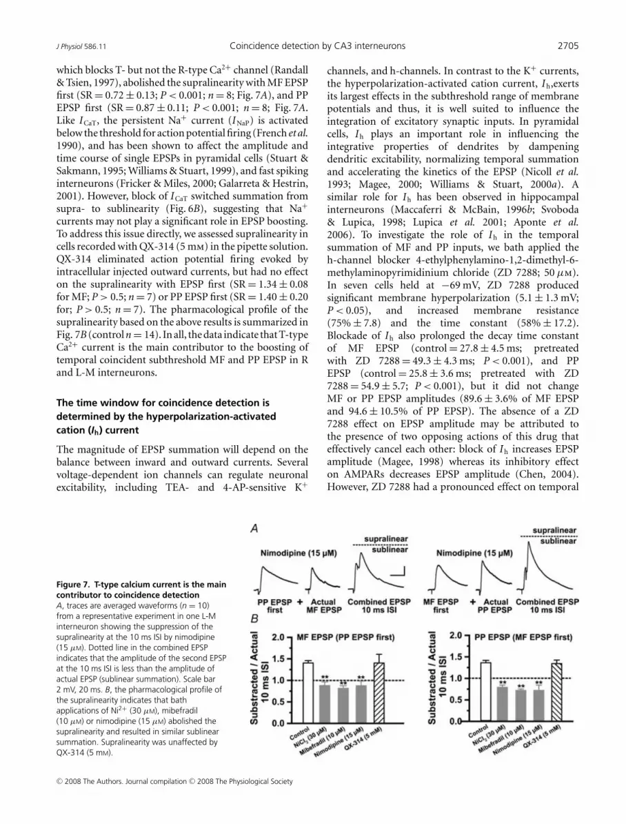
which blocks T- but not the R-type Ca2+ channel (Randall& Tsien, 1997), abolished the supralinearity with MF EPSPfirst (SR = 0.72 ± 0.13; P < 0.001; n = 8; Fig. 7A), and PPEPSP first (SR = 0.87 ± 0.11; P < 0.001; n = 8; Fig. 7A.Like ICaT, the persistent Na+ current (INaP) is activatedbelow the threshold for action potential firing (French et al.1990), and has been shown to affect the amplitude andtime course of single EPSPs in pyramidal cells (Stuart &Sakmann, 1995; Williams & Stuart, 1999), and fast spikinginterneurons (Fricker & Miles, 2000; Galarreta & Hestrin,2001). However, block of ICaT switched summation fromsupra- to sublinearity (Fig. 6B), suggesting that Na+
currents may not play a significant role in EPSP boosting.To address this issue directly, we assessed supralinearity incells recorded with QX-314 (5 mm) in the pipette solution.QX-314 eliminated action potential firing evoked byintracellular injected outward currents, but had no effecton the supralinearity with EPSP first (SR = 1.34 ± 0.08for MF; P > 0.5; n = 7) or PP EPSP first (SR = 1.40 ± 0.20for; P > 0.5; n = 7). The pharmacological profile of thesupralinearity based on the above results is summarized inFig. 7B (control n = 14). In all, the data indicate that T-typeCa2+ current is the main contributor to the boosting oftemporal coincident subthreshold MF and PP EPSP in Rand L-M interneurons.
The time window for coincidence detection isdetermined by the hyperpolarization-activatedcation (Ih) current
The magnitude of EPSP summation will depend on thebalance between inward and outward currents. Severalvoltage-dependent ion channels can regulate neuronalexcitability, including TEA- and 4-AP-sensitive K+
Figure 7. T-type calcium current is the maincontributor to coincidence detectionA, traces are averaged waveforms (n = 10)from a representative experiment in one L-Minterneuron showing the suppression of thesupralinearity at the 10 ms ISI by nimodipine(15 μM). Dotted line in the combined EPSPindicates that the amplitude of the second EPSPat the 10 ms ISI is less than the amplitude ofactual EPSP (sublinear summation). Scale bar2 mV, 20 ms. B, the pharmacological profile ofthe supralinearity indicates that bathapplications of Ni2+ (30 μM), mibefradil(10 μM) or nimodipine (15 μM) abolished thesupralinearity and resulted in similar sublinearsummation. Supralinearity was unaffected byQX-314 (5 mM).
channels, and h-channels. In contrast to the K+ currents,the hyperpolarization-activated cation current, Ih,exertsits largest effects in the subthreshold range of membranepotentials and thus, it is well suited to influence theintegration of excitatory synaptic inputs. In pyramidalcells, Ih plays an important role in influencing theintegrative properties of dendrites by dampeningdendritic excitability, normalizing temporal summationand accelerating the kinetics of the EPSP (Nicoll et al.1993; Magee, 2000; Williams & Stuart, 2000a). Asimilar role for Ih has been observed in hippocampalinterneurons (Maccaferri & McBain, 1996b; Svoboda& Lupica, 1998; Lupica et al. 2001; Aponte et al.2006). To investigate the role of Ih in the temporalsummation of MF and PP inputs, we bath applied theh-channel blocker 4-ethylphenylamino-1,2-dimethyl-6-methylaminopyrimidinium chloride (ZD 7288; 50 μm).In seven cells held at −69 mV, ZD 7288 producedsignificant membrane hyperpolarization (5.1 ± 1.3 mV;P < 0.05), and increased membrane resistance(75% ± 7.8) and the time constant (58% ± 17.2).Blockade of Ih also prolonged the decay time constantof MF EPSP (control = 27.8 ± 4.5 ms; pretreatedwith ZD 7288 = 49.3 ± 4.3 ms; P < 0.001), and PPEPSP (control = 25.8 ± 3.6 ms; pretreated with ZD7288 = 54.9 ± 5.7; P < 0.001), but it did not changeMF or PP EPSP amplitudes (89.6 ± 3.6% of MF EPSPand 94.6 ± 10.5% of PP EPSP). The absence of a ZD7288 effect on EPSP amplitude may be attributed tothe presence of two opposing actions of this drug thateffectively cancel each other: block of Ih increases EPSPamplitude (Magee, 1998) whereas its inhibitory effecton AMPARs decreases EPSP amplitude (Chen, 2004).However, ZD 7288 had a pronounced effect on temporal
C© 2008 The Authors. Journal compilation C© 2008 The Physiological Society

2706 E. Calixto and others J Physiol 586.11
summation. Figure 8 shows that supralinearity at the10 ms ISI was enhanced (SR = 1.68 ± 0.17 with MFEPSP first, P < 0.05; SR = 1.59 ± 0.11 with PP EPSP first,P < 0.05; n = 7), and remained significantly increasedup to the 30 ms ISI with MF EPSP first (1.38 ± 0.14;P < 0.05), and up to the 40 ms ISI with PP EPSP first(1.23 ± 0.13; P < 0.05).
The widening of the time window for EPSPboosting increased the cell’s overall responsiveness toasynchronous inputs, thereby degrading coincidencedetection. Furthermore, ZD 7288 changed summationfrom sublinear to linear between 50 and 70 ms ISIswith MF EPSP first (SR = 0.92 ± 0.12 at 50 ms ISI;P > 0.05; and SR = 0.85 ± 0.15 at 70 ms ISI with P > 0.05;Fig. 8B), and with PP EPSP first (SR = 1.08 ± 0.14 at50 ms ISI, P > 0.05; and SR = 0.91 ± 0.09, P > 0.05 at70 ms ISI; Fig. 8B). Consequently, Ih limited the temporalwindow for coincidence detection by restricting the timecourse of supralinearity and promoting linear to sublinearsummation for asynchronous inputs. By controlling thetemporal integration window, Ih is critically involved indetermining the temporal precision of the interneuronresponse to inputs from PP and MF. Together ourdata suggest that R and L-M interneurons are ableto detect millisecond shifts in the timing betweentemporally correlated subthreshold EPSPs through the
Figure 8. The temporal window for coincidence detection is determined by IhA shows representative traces (n = 10) from one experiment from one L-M interneuron treated with ZD 7288.Summation at the 40 ms ISI for MF EPSP first (left traces), and at the 40 ms ISI for PP EPSP first (right traces) aresupralinear. Dotted line in the combined EPSP indicates the actual supralinearity at different ISIs. B, the mean group(n = 7) distribution of summation ratios during bath applied ZD 7288 (50 μM) revealed that the magnitude ofsupralinearity at the 10 ms ISI is significantly larger than control. Control data are the same as those shown inFig. 3B. Supralinearity remained significant at the 30 ms ISI for MF EPSP first, and at the 40 ms ISI for PP EPSP first(for more details see Results). ZD 7288 also abolished the sublinearity present in control experiments at ISIs from50 and 70 ms (Fig. 3); instead summation ratios at these intervals were linear. ∗Significant P < 0.05. Error barsindicate S.E.M. Scale 2 mV, 20 ms.
counterbalanced activation of ICaT and inactivation ofIh. A prediction from this hypothesis is that inputs willsum linearly at the 10 ms ISI if both conductances areblocked. In six cells, summation was measured in thepresence of NiCl2 (30 μm), and during the subsequentaddition of ZD 7288 (50 μm) to the perfusion bath.As reported above, block of ICaT by Ni2+ produced asignificant sublinearity at the 10 ms ISI with MF EPSP first(SR = 0.79 ± 0.08; P < 0.05; n = 4; Fig. 9A) and with PPEPSP first (SR = 0.8 ± 0.16 C; P < 0.05; n = 4; Fig. 9B).In agreement with the prediction, summation becamelinear following the block of Ih (SR = 0.92 ± 0.12 for MFEPSP first; n = 4; SR = 0.93 ± 0.17 for PP EPSP first; n = 4;Fig. 9A and B).
Discussion
This study provides evidence that R and L-M interneuronsin area CA3 possess voltage-dependent conductanceswhich play a central role in shaping the temporalintegration of converging EPSPs. Furthermore, theprominent supralinear boosting within a narrow timewindow suggests that these interneurons act as coincidentdetectors for subthreshold input from PP and MF.Previous studies in fast spiking (i.e. non-adapting)interneurons have ascribed coincidence detection of EPSPs
C© 2008 The Authors. Journal compilation C© 2008 The Physiological Society

J Physiol 586.11 Coincidence detection by CA3 interneurons 2707
to increased number and faster kinetics of AMPA/kainatereceptors (Geiger et al. 1997; Nusser et al. 1998; Walkeret al. 2002; Jonas et al. 2004). The novel finding reportedhere is that adapting R and L-M interneurons areable to detect millisecond shifts in the timing betweentemporally correlated subthreshold EPSPs by the wayof counterbalanced activation of voltage-dependent ICaT
and Ih channels. Specifically, we found that summationwas supralinear for near coincident EPSPs (10 ms ISI).Non-coincident EPSPs summed linearly (20–30 ms ISI)to sublinearly (40–70 ms ISI). The prominent supralinearboosting within a narrow time span suggests that theintegrative properties of R and L-M interneurons favourcoincident detection for subthreshold EPSPs from PP andMF.
Characterization of R and L-M interneurons
The anatomical and electrophysiological properties ofR and L-M interneurons reported here have beenextensively characterized by previous work in areas CA1and CA3 (Williams et al. 1994; Khazipov et al. 1995;Chitwood & Jaffe, 1998; Chitwood et al. 1999; Savic et al.2001). Specifically, our interneurons correspond to the‘stellate cells’ in area CA1 (Lacaille & Schwartzkroin,1988a,b), and the interneurons at the s. radiatum–s.lacunosum-moleculare border (Kawaguchi & Hama, 1987;Kunkel et al. 1988; Williams et al. 1994; Perez et al.
Figure 9. Coincident inputs sum linearly in the absence of both ICaT and IhA, average traces (n = 5) from a representative experiment in one L-M interneuron showing the sublinearsummation produced by Ni2+ and the reversal to linear summation by the subsequent application of ZD 7288(grey trace). Scale 2 mV, 25 ms. B, the mean summation data (10 ms ISI; n = 6) show the switch from sublinearityto linearity by the addition of ZD 2788 to the bath perfusion containing Ni2+. Dotted line indicates the amplitudeof the second EPSP in the pair (‘Actual’ EPSP). ∗Significant, P < 0.05. Error bars indicate S.E.M.
2001). The spatial distribution of their dendritic treesin our cells also exhibit similarities with interneuronswith the soma residing in the s. lacunosum-moleculareand radiatum described by others (Woodson et al. 1989;Gulyas et al. 1993; McBain & Dingledine, 1993; Arancioet al. 1994; Poncer et al. 1995; Maccaferri & McBain,1996a,b; McMahon & Kauer, 1997) including the Schaffercollateral/commissural and perforant pathway-associatedinterneuron (Vida et al. 1998; Somogy & Klausberger,2005). In area CA1, L-M interneurons are activated by PPinput at a shorter latency and in a feed-forward manner,with respect to pyramidal cells indicating that these cellsmediate feed-forward inhibition (Williams et al. 1994).The firing properties of CA3 R and L-M interneuronsdescribed here have adapting properties similar to theinterneurons described by Chitwood & Jaffe, 1998). Inter-esting, L-M interneurons receive weak inhibitory inputfrom other interneurons, which could potentially allowfor a strong synaptic drive from their excitatory inputs(Williams et al. 1994).
Dependence of coincidence detectionon ICaT activation
Previous studies in pyramidal cells that reportedcoincidence detection using a similar paired stimulationprotocol revealed the participation of regenerative Na+
currents (Margulis & Tang, 1998), QX314 sensitive
C© 2008 The Authors. Journal compilation C© 2008 The Physiological Society

2708 E. Calixto and others J Physiol 586.11
conductances (Nettleton & Spain, 2000) or NMDARactivation (Polsky et al. 2004) in the boosting of thesummed response. In fast spiking interneurons, themillisecond range in the temporal resolution of inputsynchronicity is attributable to the properties of post-synaptic receptors (Geiger et al. 1997; Nusser et al.1998; Walker et al. 2002; Jonas et al. 2004), and theactivation of voltage-dependent Na+ channels (Galarreta& Hestrin, 2001). In the present study, supralinearitywas unaffected by QX-314 but was sensitive to Ni2+ andmibefradil indicating an involvement of T- and/or R-typecalcium currents. By contrast to R-type, T-type channelscan be activated from only a few millivolts positive to−70 mV producing transient Ca2+ currents at potentialsclose to the cell’s resting potential, typically −69 mV inour experiments. Furthermore, nimodipine, which doesnot affect the R-type channel (Randall & Tsien, 1997;Heady et al. 2001; Yasuda et al. 2003; Metz et al. 2005),abolished the supralinear boosting at a concentrationof 15 μm. Based on a Hill slope of 1.61 and an IC50
value of 8.2 μm (Allen et al. 1993) we estimated that thisconcentration was sufficient to block 78% of ICaT. Together,these pharmacological characteristics suggest that ICaT
is responsible for the majority of the inward currentsfor EPSP amplification in R and L-M interneurons. Thedensity of T-type channels in pyramidal cells increaseswith distance from the soma (Magee & Johnston, 1995),and ICaT is a prominent contributor to dendritic Ca2+
influx (Kavalali et al. 1997). These findings are consistentwith the lack of supralinear summation with the somaticinjections of simEPSP reported here. The dependenceof ICaT in coincidence detection also is in agreementwith its role in the amplification of subthreshold EPSPsduring propagation to the pyramidal cell soma (Sutor& Zieglagansberger, 1987; Deisz et al. 1991; Markram& Sakmann, 1994; Magee & Johnston, 1995; Gillessen& Alzheimer, 1997; Andreasen & Lambert, 1998; Urbanet al. 1998), and in neocortical interneurons (Goldberget al. 2004). Interestingly, the time course of supralinearitydescribed in the present work can be accounted for by thetime-to-peak activation (∼10–30 ms) and time constantof inactivation (∼30–50 ms) of dendritic ICaT (Magee &Johnston, 1995; Mouginot et al. 1997). A similar timedependence of the ICaT-mediated enhancement of thesecond EPSP in a pair also has been observed in neocorticalpyramidal cells (Deisz et al. 1991).
Dendritic compartmentalization of supralinearsummation
The summation characteristics of convergent sub-threshold inputs are not solely determined by theirrelative timing, and the contribution of voltage-dependentconductances but, also by the dendritic localization andspatial separation between the synapses (Koch et al. 1983).
In CA3 pyramidal cells, MF and PP inputs located farapart on the apical dendritic tree summate sublinearly atshort ISIs with MF prestimulation (Urban & Barrionuevo,1998). In contrast, synchronous inputs to neighbouringpostsynaptic domains in thin branches of hippocampaland neocortical pyramidal neurons are able to summatesupralinearly (Segev & London, 2000; Ariav et al. 2003;Polsky et al. 2004; Losonczy & Magee, 2006; Carteret al. 2007). For example, coincidental inputs to dendriticspines in the CA1 pyramidal cell show strong nonlinearityonly if their spatial distribution is within 150 μm orless (Losonczy & Magee, 2006). However, this spatialrequirement may be different for the spineless dendrite ofthe interneuron. Although we have no direct anatomicalevidence for the localization of MF and PP synapses, ourreconstructions of the interneuron dendritic tree suggest asimilar anatomical arrangement formed by neighbouringsynapses from PP axons and the thin branches of MFaxon collaterals near the hilus. The observation thatsupralinear summation was unchanged after transectionof the s. lucidum suggests that input to R and L-M inter-neurons elicited from the granule cells in the SDG siteis largely mediated by axonal projections confined todendrites near the hilus, in close proximity to PP synapses.However, as it is unavoidable for projection neurons invitro, the negative finding may reflect truncation of MF bythe slicing procedure, so the addition of the transectionthrough the s. lucidum makes no additional impact on thecircuitry of the slice.
In theory, pairs of coincident EPSPs from inputs inclose spatial proximity would summate sublinearly due toshunting of the synaptic currents caused by the largeincrease in membrane conductance when both inputs areactive. However, since the time course of the conductancethat underlies AMPAR mediated synaptic transmissionis several times faster than 10 ms (Geiger et al. 1997), itis unlikely that the two conductance changes overlappedsignificantly at this ISI. Indeed, summation was linear inthe absence of ICaT and Ih. However, synapses not onlyimpose conductance changes but also inject currents ontohighly localized dendritic regions. If PP and MF afferentsmake synapses close to each other, then their coincidentalinput mediated by current through AMPA receptors wouldproduce a sufficient depolarization to recruit dendriticICaT thereby enhancing the second EPSP in the pair (Deiszet al. 1991). In particular, a high density distribution ofICaT channels clustered near the synaptic sites, as shownin thalamocortical cells (Williams & Stuart, 2000b),could give rise to separate integrative compartments,each functioning as an independent supralinearcomputational unit (Segev & London, 2000; Ariav et al.2003; Polsky et al. 2004; Losonczy & Magee, 2006). In thisscheme, closely spaced supralinear compartments couldgive rise to reciprocal synaptic interactions that promotesymmetrical boosting to near synchronous PP and MF
C© 2008 The Authors. Journal compilation C© 2008 The Physiological Society

J Physiol 586.11 Coincidence detection by CA3 interneurons 2709
inputs. This interpretation is in agreement with thefinding that the supralinear interaction between MF andPP afferents in R and L-M interneurons is symmetricalin time, i.e. prestimulation with either input amplifiesthe second EPSP in the pair. While the strong localdepolarization evoked by the conjoint inputs might haveevoked dendritic calcium spikes, EPSP waveforms at the10 ms ISI were not inflected. However, smooth EPSPsin soma recordings could have resulted from dendriticfiltering of distally located regenerative events.
The role of Ih in the temporal integration of MFand PP inputs
Ih has been shown to control the membrane properties ofinterneurons in the hippocampus (Maccaferri & McBain,1996a; Svoboda & Lupica, 1998; Lupica et al. 2001; Aponteet al. 2006). In the present study, the hyperpolarizingshift observed following treatment with ZD 7288 pointsto a participation of Ih in the membrane conductanceof R and L-M interneurons at potentials near rest. ZD7288 also enhanced and prolonged the supralinearity,resulting in a considerable excitatory synaptic driveduring both nearly coincident and temporally dispersedsynaptic input. These data underscore the critical roleof Ih in determining the temporal fidelity of coincidencedetection, as demonstrated by previous experimental andcomputational studies (Magee, 1998; Jonas et al. 2004;Migliore et al. 2004; Yamada et al. 2005). From the dataobtained with simEPSP first, we conclude that a significantproportion of Ih channels underlying the sublinearity areexpressed at or near the interneuron soma. The block ofICaT also reveals that integration in the region of temporaltransition from supra- to sublinearity (∼20 ms ISI) isthe result of ‘active linearization’ (Cash & Yuste, 1999;Gasparini & Magee, 2006) by the dynamic counteractingactivation of ICaT and inactivation of Ih. For inputs arrivingwith progressively increasing asynchrony the mode ofintegration is sublinear and predominantly shaped by Ih
inactivation. While blockade of Ih substantially reducedsublinearity over a range of ISIs, summation at ISIs upto 90 ms is sublinear in control conditions and in thepresence of ZD 7288. Given these data, Ih cannot beexpected to support this sublinearity (Magee, 1998). TheD-type voltage gated potassium conductance (Storm,1988; Metz et al. 2007) could be a candidate mediator ofsublinear summation at long ISIs.
Functional implications
The time course of the supralinearity indicates that thesecells could be selectively entrained by synaptic burstselicited from the cells in the superficial layers of theEC firing at intervals (10–25 ms) time-locked to theintrinsically generated gamma oscillation (Charpak et al.
1995; Chrobak & Buzsaki, 1998). Because the supra-linear interaction is symmetrical, i.e. it does not dependon the temporal order of cell firing between EC anddentate gyrus, R and L-M interneurons also are tunedto the phasic high frequency discharge (up to 100 Hz)from granule cells (Jung & McNaughton, 1993). Ourdata show that small EPSPs (3–6 mV at the soma) fromthe first input are able to provide a substantial boost(< 35%) to near synchronous EPSPs from the secondinput thereby potentially increasing the probability ofpostsynaptic firing in response to the conjoint input. Thus,coincidence detection of correlated PP and MF inputby R and L-M interneurons will likely synchronize theiroutput targeting both the apical dendrite and soma of CA3pyramidal cells in a feed-forward manner. The increaseddendritic inhibition may gate the PP EPSP (Ang et al.2005) by modulating the distal boosting by ICaT (Urbanet al. 1998); it also may control the induction of long-termsynaptic plasticity in pyramidal cells by regulating thebackpropagation of action potentials. On the otherhand, the stronger feed-forward inhibition in the soma(Pouille & Scanziani, 2001) could constrain the timingof pyramidal cell discharge elicited by the converginginput from the recurrent collaterals during the CA3-generated gamma oscillations. Thus, feed-forwardinhibition may possibly sharpen the focus of the MFinput to pyramidal cells when multiple cortical firingpatterns are imposed to the CA3 network simultaneously,such as during pattern separation. Consistent with thisline of reasoning is the observation that feed-forwardinhibition is a key contributor to the sparsening of odourrepresentations (Perez-Orive et al. 2002).
References
Acsady L, Kamondi A, Syk A, Freund T & Buzsaki G (1998).GABAergic cells are the major postsynaptic targets of mossyfibers in the rat hippocampus. J Neurosci 18, 3386–3403.
Alle H, Jonas P & Geiger JRP (2001). PTP and LTP at ahippocampal mossy fiber-interneuron synapse. Proc NatlAcad Sci U S A 98, 14708–14713.
Allen TG, Sim JA & Brown DA (1993). The whole-cell calciumcurrent in acutely dissociated magnocellular cholinergicbasal forebrain neurones of the rat. J Physiol 460, 91–116.
Andreasen M & Lambert JD (1998). Factors determining theefficacy of distal excitatory synapses in rat hippocampal CA1pyramidal neurones. J Physiol 507, 441–462.
Ang CW, Carlson GC & Coulter DA (2005). Hippocampal CA1circuitry dynamically gates direct cortical inputspreferentially at theta frequencies. J Neurosci 25, 9567–9580.
Aponte Y, Cheng-Chang L, Reisinger E & Jonas P (2006).Hyperpolarization-activated cation channels in fast-spikinginterneurons of rat hippocampus. J Physiol 574, 229–243.
Arancio O, Korn H, Gulyas A, Freund T & Miles R (1994).Excitatory synaptic connections onto rat hippocampalinhibitory cells may involve a single transmitter release site.J Physiol 48, 395–405.
C© 2008 The Authors. Journal compilation C© 2008 The Physiological Society

2710 E. Calixto and others J Physiol 586.11
Ariav G, Polsky A & Schiller J (2003). Submillisecond precisionof the input-output transformation function mediated byfast sodium dendritic spikes in basal dendrites of CA1pyramidal neurons. J Neurosci 23, 7750–7758.
Berzhanskaya J, Urban NN & Barrionuevo G (1998).Pharmacological characterization of the monosynapticperforant path input to CA3 pyramidal neurons.J Neurophysiol 79, 2111–2118.
Breustedt J, Vogt KE, Miller RJ, Nicoll RA & Schmitz D (2003).α1E-Containing Ca2+ channels are involved in synapticplasticity. Proc Natl Acad Sci U S A 100, 12450–12455.
Buzsaki G (1984). Feed-forward inhibition in the hippocampalformation. Prog Neurobiol 22, 131–153.
Carter AG, Soler-Llavina GJ & Sabatini BL (2007). Timing andlocation of synaptic inputs determine modes of subthresholdintegration in striatal medium spiny neurons. J Neurosci 22,8967–8977.
Cash S & Yuste R (1999). Linear summation of excitatoryinputs by CA1 pyramidal neurons. Neuron 22, 383–394.
Charpak S, Pare D & Llinas R (1995). The entorhinal cortexentrains fast CA1 hippocampal oscillations in theanaesthetized guinea-pig: role of the monosynapticcomponent of the perforant path. Eur J Neurosci 7,1548–1557.
Chen C (2004). ZD 7288 inhibits postsynaptic glutamatereceptor-mediated responses at hippocampal perforantpath-granule cell synapses. Eur J Neurosci 19, 643–649.
Chitwood RA, Hubbard A & Jaffe DB (1999). Passiveelectrotonic properties of rat hippocampal CA3interneurones. J Physiol 51, 743–756.
Chitwood RA & Jaffe DB (1998). Calcium-dependentspike-frequency accommodation in hippocampal CA3nonpyramidal neurons. J Neurophysiol 80, 983–988.
Chrobak JJ & Buzsaki G (1998). Gamma oscillations in theentorhinal cortex of the freely behaving rat. J Neurosci 18,388–398.
Claiborne BJ, Amaral DG & Cowan WM (1986). A light andelectron microscopic analysis of the mossy fibers of the ratdentate gyrus. J Comp Neurol 246, 435–458.
Deisz RA, Fortin G & Zielglgansberger W (1991). Voltagedependence of excitatory postsynaptic potentials of ratneocortical neurons. J Neurophysiol 65, 371–382.
Eichenbaum H & Otto T (1992). The hippocampus – Whatdoes it do? Behav Neural Biol 57, 2–36.
Foehring RC, Mermelstein PG, Wen-Jie S, Ulrich S & SurmeierDJ (2000). Unique properties of R-type calcium currents inneocortical and neostriatal neurons. J Neurophysiol 84,2225–2236.
Fraser DD & MacVicar BA (1991). Low-threshold transientcalcium current in rat hippocampal lacunosum-moleculareinterneurons, kinetics and modulation by neurotransmitters.J Neurophysiol 11, 2812–2820.
French CR, Sah P, Buckett KJ & Gage PW (1990). Avoltage-dependent persistent sodium current in mammalianhippocampal neurons. J Gen Physiol 95, 1139–1157.
Freund TF & Buzsaki G (1996). Interneurons of thehippocampus. Hippocampus 6, 347–470.
Fricker D & Miles R (2000). EPSP amplification and theprecision of spike timing in hippocampal neurons. Neuron28, 559–569.
Galarreta M & Hestrin S (2001). Spike transmission andsynchrony detection in networks of GABAergicinterneurons. Science 292, 2295–2299.
Gasparini S, Kasyanov AM, Pietrobon D, Voronin LL &Cherubini E (2001). Presynaptic R-type calcium channelscontribute to fast excitatory synaptic transmission in the rathippocampus. J Neurosci 21, 8715–8721.
Gasparini S & Magee JC (2006). State-dependent dendriticcomputation in hippocampal CA1 pyramidal neurons.J Neurosci 26, 2088–2100.
Geiger JR, Lubke J, Roth A, Frotscher M & Jonas P (1997).Submillisecond AMPA receptor-mediated signaling at aprincipal neuron-interneuron synapse. Neuron 18,1009–1023.
Gillessen T & Alzheimer C (1997). Amplification of EPSPs bylow Ni2+- and amiloride-sensitive Ca2+ channels in apicaldendrites of rat CA1 pyramidal neurons. J Neurophysiol 77,1639–1643.
Goldberg JH, Lacefield CO & Yuste R (2004). Global dendriticcalcium spikes in mouse layer 5 low threshold spikinginterneurons, implications for control of pyramidal cellbursting. J Physiol 558, 465–478.
Goldberg JH, Tamas G & Yuste R (2003). Ca2+ imaging ofmouse neocortical interneuron dendrites: Ia-type K+channels control action potential backpropagation. J Physiol551, 49–65.
Gulyas A, Miles R, Hajos N & Freund TF (1993). Precision andvariability in postsynaptic target selection of inhibitory cellsin the hippocampal CA3 region. Eur J Neurosci 5,1729–1751.
Heady TN, Gomora JC, Macdonald TL & Perez-Reyes E (2001).Molecular pharmacology of T-type Ca2+ channels.Jpn J Pharmacol 85, 339–350.
Henze DA, Gonzalez-Burgos G, Urban NN, Lewis DA &Barrionuevo G (2000). Dopamine increases excitability ofpyramidal neurons in primate prefrontal cortex.J Neurophysiol 84, 2799–2809.
Henze DA, Urban NN & Barrionuevo G (1997). Origin of theapparent asynchronous activity of hippocampal mossyfibers. J Neurophysiol 78, 24–30.
Jonas P, Bischofberger J, Fricker D & Miles R (2004).Interneuron Diversity series. Fast in, fast out – temporal andspatial signal processing in hippocampal interneurons.Trends Neurosci 27, 30–40.
Jung MW & McNaughton BL (1993). Spatial selectivity of unitactivity in the hippocampal granule layer. Hippocampus 3,165–182.
Kaiser KM, Lubke J, Zilberter Y & Sakmann B (2004).Postsynaptic calcium influx at single synaptic contactsbetween pyramidal neurons and bitufted interneurons inlayer 2/3 of rat neocortex is enhanced by backpropagatingaction potentials. J Neurosci 24, 1319–1329.
Kaiser KM, Zilberter Y & Sakmann B (2001). Back-propagatingaction potentials mediate calcium signaling in dendrites ofbitufted interneurons in layer 2/3 of rat somatosensorycortex. J Physiol 535, 17–31.
Kavalali ET, Zhuo M, Bito H & Tsien RW (1997). DendriticCa2+ channels characterized by recordings from isolatedhippocampal dendritic segments. Neuron 18,651–663.
C© 2008 The Authors. Journal compilation C© 2008 The Physiological Society

J Physiol 586.11 Coincidence detection by CA3 interneurons 2711
Kawaguchi Y & Hama K (1987). Two subtypes ofnon-pyramidal cells in rat hippocampal formation identifiedby intracellular recording and HRP injection. Brain Res 411,190–195.
Khazipov R, Congar P & Ben-Ari Y (1995). Hippocampal CA1lacunosum moleculare interneurons: modulation ofmonosynaptic GABAergic IPSCs by presynaptic GABAB
receptors. J Neurophysiol 74, 2126–2137.Koch C, Poggio T & Torre V (1983). Nonlinear interactions in a
dendritic tree: localization, timing, and role in informationprocessing. Proc Natl Acad Sci U S A 80, 2799–2802.
Konig P, Andreas K, Singer E & Singer W (1996). Integrator orcoincidence detector? The role of the cortical neuronrevisited. Trends Neurosci 19, 130–137.
Kroner S, Krimer LS, Lewis D & Barrionuevo G (2007).Dopamine increases inhibition in the monkey dorsolateralprefrontal cortex through cell type-specific modulation oflocal circuit neurons. Cereb Cortex 17, 1020–1032.
Kunkel DD, Lacaille JC & Schwartzkroin PA (1988).Ultrastructure of stratum lacunosum-moleculareinterneurons of hippocampal CA1 region. Synapse 2,382–394.
Lacaille JC & Schwartzkroin PA (1988a). Stratumlacunosum-moleculare interneurons of hippocampal CAlregion. I. Intracellular response characteristics, synapticresponses, and morphology. J Neurosci 8, 1400–1410.
Lacaille JC & Schwartzkroin PA (1988b). Stratumlacunosum-moleculare interneurons of hippocampalCAl region. II lntrasomatic and intradendritic recordings oflocal circuit of synaptic recordings. J Neurosci 8, 1411–1424.
Lawrence JJ & McBain CJ (2003). Interneuron diversity series:Containing the detonation – feedforward inhibition in theCA3 hippocampus. Trends Neurosci 26, 631–640.
Leutgeb JK, Leutgeb S, Moser MB & Moser EI (2007). Patternseparation in the dentate gyrus and CA3 of thehippocampus. Science 315, 961–966.
Losonczy A & Magee J (2006). Integrative properties of radialoblique dendrites in hippocampal CA1 pyramidal neurons.Neuron 50, 291–307.
Lupica CR, Bell JA, Hoffman AF & Watson PL (2001).Contribution of the hyperpolarization-activated current (Ih)to membrane potential and GABA release in hippocampalinterneurons. J Neurophysiol 86, 261–268.
Maccaferri G & McBain CJ (1996a). Long-term potentiation indistinct subtypes of hippocampal non-pyramidal neurons.J Neurosci 16, 5334–5343.
Maccaferri G & McBain CJ (1996b). The hyperpolarization-activated current (Ih) and its contribution to pacemakeractivity in rat CA1 hippocampal stratum oriens-alveusinterneurons. J Physiol 497, 119–130.
McBain CJ & Dingledine R (1993). Heterogeneity of synapticglutamate receptors on CA3 st. radiatum interneurones ofrat hippocampus. J Physiol 462, 373–392.
McBain CJ & Fisahn A (2001). Interneurons unbound. Nat RevNeurosci 2, 11–23.
McBain CJ, Freund TF & Mody I (1999). Glutamatergicsynapses onto hippocampal interneurons: precision timingwithout lasting plasticity. Trends Neurosci 22, 228–235.
McMahon LL & Kauer JA (1997). Hippocampal interneuronsexpress a novel form of synaptic plasticity. J Neurosci 18,295–305.
McNaughton BL & Morris RG (1987). Hippocampal synapticenhancement and information storage within a distributedmemory system. Trends Neurosci 10, 408–415.
Magee JC (1998). Dendritic hyperpolarization-activatedcurrents modify the integrative properties of hippocampalCA1 pyramidal neurons. J Neurosci 18, 7613–7624.
Magee JC (2000). Dendritic integration of excitatory synapticinput. Nat Rev Neurosci 1, 181–190.
Magee JC & Johnston D (1995). Synaptic activation ofvoltage-gated channels in the dendrites of hippocampalpyramidal neurons. Science 268, 301–304.
Margulis M & Tang CM (1998). Temporal integration canreadily switch between sublinear and supralinearsummation. J Neurophysiol 79, 2809–2813.
Markram H & Sakmann B (1994). Calcium transients in dend-rites of neocortical neurons evoked by single subthresholdexcitatory postsynaptic potentials via low-voltage-activatedcalcium channels. Proc Natl Acad Sci U S A 91, 5207–5211.
Martin RL, Lee JH, Cribbs L, Perez-Reyes E & Hanck DA(2000). Mibefradil block of cloned T-type calcium channels.J Pharmacol Exp Ther 295, 302–308.
Martina M, Vida I & Jonas P (2000). Distal initiation and activepropagation of action potentials in interneuron dendrites.Science 287, 295–300.
Metz AE, Jarsky T, Martina M & Spruston N (2005). R-typecalcium channels contribute to afterdepolarization andbursting in hippocampal ca1 pyramidal neurons. J Neurosci25, 5763–5773.
Metz AE, Spruston N & Martina M (2007). Dendritic D-typepotassium currents inhibit the spike afterdepolarization inrat hippocampal CA1 pyramidal neurons. J Physiol 581,175–187.
Migliore M, Messineo L & Ferrante M (2004). Dendritic Ih
selectively blocks temporal summation of unsynchronizeddistal inputs in CA1 pyramidal neurons. J Comput Neurosci16, 5–13.
Mouginot D, Bossu JL & Gahwiler BH (1997). Low-thresholdCa2+ currents in dendritic recordings from purkinje cells inrat cerebellar slice cultures. J Neurosci 17, 160–170.
Nettleton JS & Spain WJ (2000). Linear to supralinearsummation of AMPA-mediated EPSPs in neocorticalpyramidal neurons. J Neurophysiol 83, 3310–3322.
Nicoll A, Larkman A & Blakemore C (1993). Modulation ofEPSP shape and efficacy by intrinsic membraneconductances in rat neocortical pyramidal neurons in vitro.J Physiol 468, 693–710.
Nusser Z, Lujan R, Laube G, Roberts DB, Molnar E & SomogyiP (1998). Cell type and pathway dependence of synapticAMPA receptor number and variability in the hippocampus.Neuron 21, 545–559.
O’Reilly RC & McClelland JL (1994). Hippocampal conjunctiveencoding, storage, and recall, avoiding a trade-off.Hippocampus 4, 661–682.
Perez Y, Morin F & Lacaille JC (2001). A hebbian form oflong-term potentiation dependent on mGluR1a inhippocampal inhibitory interneurons. Proc Natl Acad Sci U SA 98, 9401–9406.
Perez-Orive J, Mazor O, Turner GC, Cassenaer S, Wilson RI &Laurent G (2002). Oscillations and sparsening of odorrepresentations in the mushroom body. Science 297,359–365.
C© 2008 The Authors. Journal compilation C© 2008 The Physiological Society

2712 E. Calixto and others J Physiol 586.11
Polsky A, Mel BW & Schiller S (2004). Computational subunitsin thin dendrites of pyramidal cells. Nat Neurosci 7, 621–627.
Poncer JC, Shinozaki H & Miles R (1995). Dual modulation ofsynaptic inhibition by distinct metabotropic glutamatereceptors in the rat hippocampus. J Physiol 485, 121–134.
Porter JT, Johnson CK & Agmon A (2001). Diverse types ofinterneurons generate thalamus-evoked feedforwardinhibition in the mouse barrel cortex. J Neurosci 21,2699–2710.
Pouille F & Scanziani M (2001). Enforcement of temporalfidelity in pyramidal cells by somatic feed-forwardinhibition. Science 293, 1159–1163.
Randall AD & Tsien RW (1997). Contrasting biophysical andpharmacological properties of T-type and R-type calciumchannels. Neuropharmacology 36, 879–893.
Rozsa B, Zelles T, Vizi ES & Lendvai B (2004). Distance-dependent scaling of calcium transients evoked bybackpropagating spikes and synaptic activity in dendritesof hippocampal interneurons. J Neurosci 24, 661–670.
Savic N, Pedarzani P & Sciancalepore M (2001). Mediumafterhyperpolarization and firing pattern modulation ininterneurons of stratum radiatum in the CA3 hippocampalregion. J Neurophysiol 85, 1986–1997.
Segev I & London M (2000). Untangling dendrites withquantitative models. Science 290, 744–750.
Somogyi P & Klausberger T (2005). Defined types of corticalinterneuron structure space and spike timing in thehippocampus. J Physiol 562, 9–26.
Storm JF (1988). Temporal integration by a slowly inactivatingK+ current in hippocampal neurons. Nature 336, 379–381.
Stuart G & Sakmann B (1995). Amplification of EPSPs byaxosomatic sodium channels in neocortical pyramidalneurons. Neuron 15, 1065–1076.
Stuart G & Spruston N (1998). Determinants of voltageattenuation in neocortical pyramidal neuron dendrites.J Neurosci 18, 3501–3510.
Sutor B & Zieglagansberger W (1987). A low-voltage activated,transient calcium current is responsible for thetime-dependent depolarizing inward rectification of ratneocortical neurons in vitro. Pflugers Arch 410, 102–111.
Svoboda KR & Lupica CR (1998). Opioid inhibition ofhippocampal interneurons via modulation of potassium andhyperpolarization-activated cation (Ih) currents. J Neurosci18, 7084–7098.
Tamamaki N & Nojyo Y (1993). Projection of the entorhinallayer II neurons in the rat as revealed by intracellularpressure-injection of neurobiotin. Hippocampus 3, 471–480.
Tamas G, Szabadics J & Somogyi P (2002). Cell type andsubcellular position dependent summation of unitarypostsynaptic potentials in neocortical neurons. J Neurosci 22,740–747.
Toth K, Suares G, Lawrence JJ, Philips-Tansey E & McBain CJ(2000). Differential mechanisms of transmission at threetypes of mossy fiber synapse. J Neurosci 20, 8279–8289.
Treves A & Rolls ET (1994). Computational analysis of the roleof the hippocampus in memory. Hippocampus 4, 374–391.
Urban NN & Barrionuevo G (1998). Active summation ofEPSPs in CA3 pyramidal neurons. Proc Natl Acad Sci U S A95, 11450–11455.
Urban NN, Henze DA & Barrionuevo G (1998). Amplificationof perforant path EPSPs by sodium and LVA calciumchannels in CA3 pyramidal cells. J Neurophysiol 80,1558–1591.
Vida I, Halasy K, Szinyei C, Somogyi P & Buhl EH (1998).Unitary IPSPs evoked by interneurons at the stratumradiatum-stratum lacunosum-moleculare border in the CA1area of the rat hippocampus in vitro. J Physiol 506, 755–773.
Walker HC, Lawrence JJ & McBain CJ (2002). Activation ofkinetically distinct synaptic conductances on inhibitoryinterneurons by electrotonically overlapping afferents.Neuron 35, 161–171.
Williams SR, Samulack DD, Beaulieu C & Lacaille JC (1994).Membrane properties and synaptic responses ofinterneurons located near the stratumlacunosum-moleculare/radiatum border of area CA1 inwhole cell recordings from rat hippocampal slices.J Neurophysiol 71, 2217–2235.
Williams SR & Stuart GJ (1999). Mechanisms andconsequences of action potential burst firing in ratneocortical pyramidal neurons. J Physiol 521, 467–482.
Williams SR & Stuart GJ (2000a). Site independence of EPSPtime course is mediated by dendritic Ih in neocorticalpyramidal neurons. J Neurophysiol 83, 3177–3182.
Williams SR & Stuart GJ (2000b). Action potentialbackpropagation and somato-dendritic distribution of ionchannels in thalamocortical neurons. J Neurosci 20,1307–1317.
Wolfart J & Roeper J (2002). Selective coupling of T-typecalcium channels to SK potassium channels preventsintrinsic bursting in dopaminergic midbrain neurons.J Neurosci 22, 3404–3413.
Woodson W, Nitecka L & Ben-Ari Y (1989). Organization ofthe GABAergic system in the rat hippocampal formation, aquantitative immunocytochemical study. J Comp Neurol280, 254–271.
Yamada R, Kuba H, Ishii TM & Ohmori H (2005).Hyperpolarization-activated cyclic nucleotide-gated cationchannels regulate auditory coincidence detection in nucleuslaminaris of the chick. J Neurosci 25, 8867–8877.
Yasuda R, Sabatini BL & Svoboda K (2003). Plasticity of calciumchannels in dendritic spines. Nat Neurosci 6, 948–955).
Acknowledgements
We thank Ingeborg Kroener and John Cavaretta for their help
with the interneuron reconstruction and technical support. This
work was supported by NINDS grant NS24288
Supplemental material
Online supplemental material for this paper can be accessed at:
http://jp.physoc.org/cgi/content/full/jphysiol.2008.152751/DC1
and
http://www.blackwell-synergy.com/doi/suppl/10.1113/jphysiol.
2008.152751
C© 2008 The Authors. Journal compilation C© 2008 The Physiological Society


